Antidepressants of different mechanisms of action modulate glucocorticoid receptor function

182
Ann. N.Y. Acad. Sci. 1024: 182–212 (2004). © 2004 New York Academy of Sciences.doi: 10.1196/annals.1321.099
Early Environmental Regulation of Hippocampal Glucocorticoid Receptor Gene Expression
Characterization of Intracellular Mediators andPotential Genomic Target Sites
IAN C.G. WEAVER, JOSIE DIORIO, JONATHAN R. SECKL,a MOSHE SZYF, AND MICHAEL J. MEANEY
McGill Program for the Study of Behavior, Genes and Environment, Douglas Hospital Research Centre, Departments of Psychiatry, and Neurology and Neurosurgery, McGill University, Montreal, Canada, H4H 1R3aMolecular Endocrinology Laboratory, Department of Medicine,University of Edinburgh, Edinburgh, Scotland, United Kingdom EH4 2XU
ABSTRACT: Environmental conditions in early life permanently alter the devel-opment of glucocorticoid receptor gene expression in the hippocampus and hy-pothalamic-pituitary-adrenal responses to acute or chronic stress. In part,these effects can involve an activation of ascending serotonergic pathways andsubsequent changes in the expression of transcription factors that might driveglucocorticoid receptor expression in the hippocampus. This paper summariz-es the evidence in favor of these pathways as well as recent studies describingregulatory targets within the chromatin structure of the promoter region of therat hippocampal glucocorticoid receptor gene.
KEYWORDS: maternal care; stress; glucocorticoid receptor; methylation;epigenomic programming
Several years ago Levine, Denenberg, Zarrow, and colleagues1−6 showed that earlyexperience modified the development of adrenal glucocorticoid responses to a widerange of stressors. These findings clearly demonstrated that even rudimentary adap-tive responses to stress could be modified by environmental events. More recentstudies7 revealed these environmental effects produce sustained alterations in gluco-corticoid receptor gene expression in the hippocampus and frontal cortex, which me-diate glucocorticoid negative feedback regulation of the hypothalamic-pituitary-adrenal (HPA) axis and thus result in stable differences in HPA responses to stress.These studies reflect the plasticity within brain regions that regulate the activity ofthe HPA axis, and provide a model for understanding the processes that contribute
Address for correspondence: Michael J. Meaney, Douglas Hospital Research Center, 6875LaSalle Boulevard, Montreal (Quebec), Canada H4H 1R3. Voice: 514-761-6131 ext. 3938; fax:514-762-3034.
183WEAVER et al.: EARLY ENVIRONMENTAL REGULATION
to individual differences in neuroendocrine function. The importance of this topic isunderscored by the fact that individual differences in adrenal hormone and sympa-thetic responses to stress appear to be of considerable importance in determining thevulnerability to multiple forms of pathology.8−11
ENVIRONMENTAL REGULATION OF HPA AND BEHAVIORAL RESPONSES TO STRESS
Postnatal Handling Studies
Perhaps the strongest evidence for the environmental regulation of the develop-ment of HPA responses to stress comes from the postnatal handling research withrodents. Handling involves a brief (i.e., 3−15 min), daily period of separation of thepup from the mother for the first few weeks of life and results in decreased stressreactivity in adulthood.2−6,12−15 As adults, neonatally handled rats show decreasedfearfulness and more modest pituitary ACTH and adrenal corticosterone responsesto stress; such effects are apparent in animals tested as late as 26 months of age.16,17
The handling effects on the development of HPA responses to stress have impor-tant functional consequences. In the rat, glucocorticoid levels often rise with age andare associated with hippocampal degeneration and the emergence of learning andmemory deficits.18−21 Such age-related increases in basal and stress-inducedpituitary-adrenal activity are significantly less apparent in the handled animals, andthus these animals show little evidence of hippocampal aging.16 Likewise, handledanimals also show more modest stress-induced suppression of immune function bycomparison to non-handled rats.22
Such findings may lead to the conclusion that handled animals are hardier ormore resistant than non-handled animals. But this misses the point. Handled animalsare not better adapted than non-handled animals, they are simply different. The en-vironmental context then serves to determine the adaptive value of increased or de-creased stress reactivity. In the examples cited above, it would appear that thehandled animals are at some advantage by virtue of a more modest HPA response tostress. But this condition is not universal, Laban and colleagues23 found that non-handled animals are more resistant to the induction of experimental allergic enceph-alomyelitis (EAE) than are handled animals. Glucocorticoids are protective against thedevelopment of EAE, which can be fatal.24 Adrenalectomized animals, for example,rarely survive EAE. Hence, the increased HPA responsivity of the non-handled rendersan advantage under these circumstances. The cost of such resistance is an increasedvulnerability to glucocorticoid-induced illness, but it is not difficult to imagine a sce-nario whereby such a cost is an acceptable trade-off. In essence, the handling studiesrepresent a robust example of phenotypic plasticity in the expression of defensive re-sponses to threat. One obvious question concerns the nature of the neurobiologicalmechanisms that mediate such phenotypic variation.
Considering the importance of the corticotropin-releasing hormone (CRH) sys-tems for both behavioral and HPA responses to stress, it is probably not surprisingthat these systems are critical targets for the handling effect on stress reactivity.Adult animals exposed to postnatal handling show decreased CRH mRNA expres-sion in the paraventricular nucleus of the hypothalamus (PVNh) and the central nu-cleus of the amygdala,14,25,26 decreased CRH content in the locus coeruleus,26 and
184 ANNALS NEW YORK ACADEMY OF SCIENCES
decreased CRH receptor levels in the locus coeruleus compared with non-handledrats.26 The release of CRH and the activation of HPA responses to stress are mediat-ed by stress-induced increases in the release of noradrenaline at the level of thePVNh. Indeed, CRH release from the amygdala activates the release of noradrena-line from the locus coeruleus.27 Together, these findings suggest that there would bemore modest CRH-induced activation of the locus coeruleus during stress in the han-dled animals. At least two findings are consistent with this idea. By comparison to non-handled rats, acute stress in handled animals produces (1) a smaller stress-induced in-crease in cFOS immunoreactive neurons in the locus coeruleus28 and (2) more mod-est increases in extracellular noradrenaline levels in the PVNh.29 We propose thatpostnatal handling can decrease the expression of behavioral responses to stress, inpart, by altering the development of the central nucleus of the amygdala–locus coer-uleus CRH system.
Postnatal handling affects the development of neural systems that regulate CRHgene expression. Levels of CRH mRNA and protein in PVNh neurons are subject toinhibitory regulation via glucocorticoid negative feedback.9,30 Handled rats show in-creased negative feedback sensitivity to glucocorticoids.13,14 This effect is, in turn,related to the increased glucocorticoid receptor expression in the hippocampus andfrontal cortex,14,31−33 regions known to mediate the inhibitory effects of glucocorti-coids over CRH synthesis in PVNh neurons.19,30,34−36 The alterations in glucocorti-coid receptor expression are a critical feature for the effect of the early environmenton negative feedback sensitivity and HPA responses to stress; reversing the differ-ences in hippocampal glucocorticoid receptor levels eliminates the differences inHPA responses to stress between handled and non-handled animals.13
CRH activity within the amygdala–locus coeruleus pathway is subject to γ -aminobutyric acid-(GABA-)ergic inhibition.37,38 Interestingly, handled rats alsoshow increased GABAA and benzodiazepine (BZ) receptor levels in the noradrener-gic cell body regions of the locus coeruleus and the nuclei tractus solitarius as wellas in the basolateral and central nucleus of the amygdala.39 These effects are associ-ated with increased expression of the mRNA for the α1 and γ 2 subunit of theGABAA receptor, which together encode for proteins that form the BZ site. Thesefindings suggest that the composition of the GABAA receptor complex in brain re-gions that regulate stress reactivity is influenced by early life events. Handling in-creases α1 and γ 2 subunit expression39,40 and, importantly, this profile is associatedwith increased GABA binding.41,42 Interestingly, in humans, individual differencesin BZ receptor sensitivity are associated with vulnerability for anxiety disorders.43
Together, the effects of handling on glucocorticoid and GABAA/BZ receptor geneexpression could serve to dampen CRH synthesis and release and to decrease the ef-fect of CRH at critical target sites, such as the locus coeruleus. We feel that this mod-el provides a reasonable working hypothesis for the mechanisms underlying thehandling effect on endocrine and behavioral responses to stress.7
WHAT ARE THE CRITICAL FEATURES OF THESEENVIRONMENTAL MANIPULATIONS?
Some years ago Levine and colleagues44 suggested that the effects of handlingwere actually mediated by alterations in mother–infant interactions. Indeed, postna-
185WEAVER et al.: EARLY ENVIRONMENTAL REGULATION
tal handling increases the frequency of pup licking/grooming by mothers.45
We40,46,47 examined this question by attempting to define naturally occurring varia-tions in maternal behavior over the first eight days following birth through the sim-ple, albeit time-consuming, observation of mother–pup interactions in normallyreared animals. There was considerable variation in two forms of maternal behavior:licking/grooming of pups and arched-back nursing.48 Licking/grooming includedboth body as well as anogenital licking. Arched-back nursing, also referred to as“crouching,” is characterized by a dam nursing her pups with her back conspicuous-ly arched and legs splayed outward. While common, it is not the only posture fromwhich dams nurse. A blanket posture represents a more relaxed version of thearched-back position, where the mother is almost lying on the suckling pups. Thisposition provides substantially less opportunity for movements such as nippleswitching. Dams also nurse from their sides and often will move from one postureto another over the course of a nursing bout. Interestingly, the frequency of licking/grooming and arched-back nursing was highly correlated (r = +0.91) across animalsand thus we were able to define mothers according to both behaviors—High or Lowlicking/grooming–arched-back nursing (LG-ABN) mothers. For the sake of most ofthe studies described here, High and Low LG-ABN mothers were identified as fe-males whose scores on both measures were ±1 SD above (High) or below (Low) themean for their cohort.49
The critical question, of course, concerns the potential consequences of these dif-ferences in maternal behavior for the development of behavioral and neuroendocrineresponses to stress. Indeed, if postnatal handling results in more modest behavioraland HPA responses to stress through effects on maternal behavior, then the adult off-spring of animals reared by High LG-ABN mothers should resemble animals han-dled as neonates. As adults, the offspring of High LG-ABN mothers showed reducedplasma ACTH and corticosterone responses to acute stress by comparison to theadult offspring of Low LG-ABN mothers. The High LG-ABN offspring also showedsignificantly increased hippocampal glucocorticoid receptor mRNA expression, en-hanced glucocorticoid negative feedback sensitivity, and decreased hypothalamicCRH mRNA levels. Moreover, the magnitude of the corticosterone response to acutestress was significantly correlated with the frequency of both maternal licking/grooming (r = −0.61) and arched-back nursing (r = −0.64) during the first week of life,as was the level of hippocampal glucocorticoid receptor mRNA and hypothalamicCRH mRNA expression (all r’s > 0.70).46
HOW MIGHT MATERNAL CARE REGULATE GENE EXPRESSIONIN THE OFFSPRING?
The handling paradigm provides a model for understanding the mechanisms bywhich environmental stimuli can regulate neural development and physiology. Thismodel is somewhat unique since most paradigms involving alterations in perinatalenvironmental conditions focus on changes in either synapse formation or neuronsurvival50 that ultimately result in effects at the level of morphology. In contrast,handling affects neurochemical differentiation in the hippocampus, specifically al-tering the sensitivity of hippocampal cells to corticosterone, via an effect on gluco-
186 ANNALS NEW YORK ACADEMY OF SCIENCES
corticoid receptor gene expression and thus receptor density. Such variations inneuronal differentiation underlie important individual differences in tissue sensitiv-ity to hormonal signals and thus represent a biochemical basis for environmental“programming” of neural systems.
The handling effect on the development of glucocorticoid receptor density in thehippocampus shows the common characteristics of a developmental effect. First,there is a specific “critical period” during which the organism is maximally respon-sive to the effects of handling. Second, the effects of handling during the first 21 daysof life on glucocorticoid receptor density endure throughout the life of the animal.Finally, there is substantial specificity to the handling effect. Handling alters the glu-cocorticoid, but not mineralocorticoid, receptor gene expression. Interestingly, glu-cocorticoid and mineralocorticoid receptors are co-expressed in virtually allhippocampal neurons. Thus, the handling effect on gene expression is specific.
Temporal Features of the Handling Effect
Handling during the first week of life is as effective as handling during the entirefirst three weeks of life in reducing adrenal steroid responses to stress51 and in in-creasing hippocampal glucocorticoid receptor density.52 Handling over the secondweek of life is less effective, whereas animals handled between days 15 and 21 donot differ from non-handled animals in glucocorticoid receptor binding. Thus, interms of both HPA activity and glucocorticoid receptor binding, the sensitivity of thesystem to environmental regulation decreases progressively over the first threeweeks of life. Moreover, in comparison to same-aged non-handled animals, handledanimals exhibited significantly increased hippocampal glucocorticoid receptor den-sity as early as day 7 of life and the magnitude of the effect did not increase there-after.52 Thus, glucocorticoid receptor binding capacity appears to be especiallysensitive to environmental regulation during the first week of life. However, pleasenote that these findings do not preclude the possibility of other periods of environ-mental regulation. Indeed, a so-called critical period must be defined not only interms of the target outcome, but also by the relevant input stimulus. Hence, we as-sume that these findings suggest that the critical period for the effect of handling onglucocorticoid receptor gene expression occurs during the first week of life.
The Role of Thyroid Hormones
Handling during the first week of life activates the hypothalamic-pituitary-thyroid axis leading to increased levels of circulating thyroxine (T4) and increasedintracellular levels of the biologically more potent T4 metabolite, triiodothyronine(T3). The pituitary-thyroid axis is a major regulator of HPA development (see Ref.53 for a review). Neonatal treatment with either T4 or T3 resulted in significantly in-creased glucocorticoid receptor binding capacity in the hippocampus in animals ex-amined as adults.54 Like the handling manipulation, neither T4 nor T3 treatmentaffected hypothalamic or pituitary glucocorticoid receptor density. Moreover, ad-ministration of the thyroid hormone synthesis inhibitor, propylthiouracil (PTU), tohandled pups for the first two weeks of life completely blocked the effects of han-dling on hippocampal glucocorticoid receptor binding capacity. These data are con-

187WEAVER et al.: EARLY ENVIRONMENTAL REGULATION
sistent with the idea that the thyroid hormones might mediate, in part at least, theeffects of neonatal handling on the development of the forebrain glucocorticoid re-ceptor system.
Systemic injections of neonatal rat pups represent a rather crude manipulation,particularly procedures involving thyroid hormones. While these data might impli-cate the thyroid hormones, there is no indication that the hippocampus is actually thecritical site of action. To examine whether thyroid hormones might act directly onhippocampal cells we used an in vitro system, involving primary cultures of dissoci-ated hippocampal cells.55 The hippocampal cells were taken from embryonic ratpups (E20) and beginning on the fifth day following plating the cultures were ex-posed to 0, 1, 10, or 100 nM T3. These cells exhibit both mineralocorticoid and glu-cocorticoid receptor binding.56 Indeed, both receptors as well as their mRNAs canbe detected using material from a 60-mm dish. The results of several experimentshave failed to detect any effect of thyroid hormones on glucocorticoid receptor den-sity in cultured hippocampal cells. These in vitro data suggest that (1) the effects ofthe thyroid hormones on the glucocorticoid receptor binding occur at some site distalto the hippocampal cells or (2) thyroid hormones interact at the level of the hippoc-ampus with some other hormonal signal that is obligatory for the expression of thethyroid hormone effect.
The Role of Serotonin
Thyroid hormones have pervasive effects throughout the developing central ner-vous system (CNS) and one such effect involves the regulation of central serotoner-gic neurons.57 Thyroid hormones increase serotonin (5-HT) turnover in thehippocampus of the neonatal rat. 58 Handling also increases hippocampal 5-HTturnover58,59 and thus both manipulations increase serotonergic stimulation of hip-pocampal neurons. There is also direct evidence for an effect of 5-HT on glucocor-ticoid receptor density in the neonatal rat. Lesioning of the raphe 5-HT neurons with5,7-dihydroxytryptamine (5,7-DHT) dramatically reduces the ascending serotoner-gic input into the hippocampus. Rat pups administered 5,7-DHT on the second dayof life showed reduced hippocampal glucocorticoid receptor density as adults.58 In-terestingly, neonatal administration of 5,7-DHT produces only a transient effect,such that by adulthood 5-HT innervations to the hippocampus are restored. The ef-fect of hippocampal glucocorticoid receptor levels, however, persists into adulthood.This finding suggests that the effect of 5-HT on hippocampal glucocorticoid receptorexpression, like handling itself, is unique to the first week of life.
Serotonin significantly increases glucocorticoid receptor density in cultured hip-pocampal cells.56,60 In hippocampal cells cultured in the presence of increasing con-centrations of 5-HT, there was a twofold increase in glucocorticoid receptor binding.The effect of 5-HT was dose related, with an EC50 of 4−5 nM and a maximal effectachieved at 10 nM concentrations that require a four-day treatment period. Shorterperiods of exposure were ineffective, suggesting that the effect of 5-HT involves theincreased synthesis of receptors. In support of this idea, we found that the effect of5-HT on glucocorticoid receptor density in cultured hippocampal cells is blocked byeither actinomycin-D or cycloheximide and is paralleled by an increase in glucocor-ticoid receptor mRNA levels.

188 ANNALS NEW YORK ACADEMY OF SCIENCES
The effect of 5-HT on glucocorticoid receptor expression occurs uniquely in theneuronal cell population. We found no effect of 5-HT on glucocorticoid receptorbinding in hippocampal glial-enriched cell cultures. This finding is not surprising,since our initial studies were performed with cultures composed largely (∼85%) ofneuron-like cells.56 Moreover, the composition of the cultures is unaffected by 5-HTtreatment. We also examined the potential involvement of the glial cells by using aconditioned-medium experiment in which glial-enriched cultures were treated forfive days with 5-HT and the medium was then used to feed neuronal cultures. Thisprocedure had no effect on glucocorticoid receptor density, suggesting that the effectwas not due to a 5-HT–induced glial secretory product.
The effects of 10 nM 5-HT on glucocorticoid receptor density in cultured hippoc-ampal cells are completely blocked by the 5-HT2 receptor antagonists, ketanserin andmianserin.56,60 Moreover, the 5-HT2A agonists 1-(2,5-dimethoxy-4-iodophenyl)-2-aminopropane (DOI), 3-trifluoromethyl-phenylpiperazine monohydrochloride(TFMPP), and quipazine were also effective in increasing glucocorticoid receptorbinding in hippocampal culture, although not as effective as 5-HT. Selective agonistsor antagonists of the 5-HT1A or 5-HT3 receptors have no effect on glucocorticoid re-ceptor binding. Using 125I7-amino-8-iodo-ketanserin as radioligand, we found high-affinity 5-HT2A binding sites in our cultured hippocampal cells.
We then examined the nature of the secondary messenger systems involved in thisserotonergic effect on glucocorticoid receptor binding. Mitchell and colleagues56
found that low nanomolar concentrations of 5-HT (EC50 = 7 nM) produce a fourfoldincrease in cAMP levels in cultured hippocampal cells, with no effect on cGMP lev-els. This increase in cAMP is blocked by ketanserin and at least partially mimickedby quipazine, TFMPP, and DOI. Indeed, there is a strong correlation (+0.97) betweenthe effects of these 5-HT receptor agonists on cAMP and glucocorticoid receptorlevels.
Treatment with the stable cAMP analogue, 8-bromo-cAMP or with 10 µM for-skolin produces a significant increase in glucocorticoid receptor density in culturedhippocampal neurons.60 The effect of 8-bromo-cAMP is concentration related, andthe maximal effect of 8-bromo-cAMP (∼190%) is comparable to that for 5-HT(∼200%). Interestingly, as with 5-HT, the effects of 8-bromo-cAMP on glucocorti-coid receptor mRNA levels and receptor density are not apparent until at least fourdays of treatment.
Taken together, these findings suggest that changes in cAMP concentrations maymediate the effects of 5-HT on glucocorticoid receptor synthesis in hippocampalcells. We60 also found that the cyclic nucleotide–dependent protein kinase inhibitor,H8, completely blocked the effects of 10 nM 5-HT on glucocorticoid receptor bind-ing in hippocampal cell cultures. In contrast, the protein kinase C inhibitor, H7, hadno such effect. These data suggest that activation of protein kinase A is involved inthe serotonergic regulation of hippocampal glucocorticoid receptor development.
These studies involve effects on intact cells with long incubation periods, andthere is ample possibility for an interaction between second messenger systems. Thisissue also arises because the 5-HT2A receptor is linked not to cyclic nucleotide, butphospholipase C–related second messenger systems. In both in vivo and in vitrostudies, 5-HT2A agonists increase both diacylglycerol (DAG) levels and inositolphosphate (IP) metabolism (notably IP1) within hippocampal membranes.61 Thereare numerous examples in the literature of such “crosstalk” between second messen-
189WEAVER et al.: EARLY ENVIRONMENTAL REGULATION
ger systems and the stimulation of IP metabolism via phorbol esters alters cAMPlevels.62 However, in contrast to other compounds, such as glutamate or carbachol,we found that stimulation of IP metabolism by 5-HT in hippocampal slices was rath-er modest in animals during the first week of life.63 Interestingly, the effect of 5-HTon IP metabolism in hippocampal slices is decreased in the handled animals on post-natal day 7, while the stimulation of DAG is slightly enhanced. However, the overallpattern of 5-HT stimulation is weak. This may be due to differences in receptor cou-pling at this time of life. In neonatal rat hippocampi the stimulation of IP metabolismoccurred via 5-HT2C and not 5-HT2A receptors during the first weeks of life.64 Sincethere is little 5-HT2C receptor expression in dorsal hippocampus this may explain theweak stimulation of phospholipase C–related second messenger systems.
These data suggest that 5-HT directly stimulates cAMP formation in hippocampalneurons. This idea is not easy to reconcile with the involvement of a 5-HT2A receptor.However, a number of 5-HT receptors have been cloned and these receptors directlystimulate adenylyl cyclase activity. These include the 5-HT4, 5-HT6, and 5-HT7 recep-tors.65 The mRNAs for each of these receptors is expressed in rat hippocampus. More-over, the 5-HT7 receptor binds ketanserin with high affinity. Interestingly,antidepressants increase glucocorticoid receptor mRNA in cortical and hippocampalcell cultures.66,67 Both the 5-HT6 and 5-HT7 receptors bind various antidepressantswith high affinity and the 5-HT7 receptor shows a high affinity for ketanserin.
To examine the potential involvement of the 5-HT7 receptor in mediating the in-crease in glucocorticoid receptor levels, we68 measured receptor expression in cul-tured hippocampal neurons after treatment with 10 mM 8-bromo-cAMP or withvarious doses of the specific 5-HT7 receptor agonist, 3-(2-aminoethyl)-1H-indole-5-carboxamide maleate (5-carboxamidotryptamine; 5-CT) for seven days. All treat-ments resulted in an increase in glucocorticoid receptor levels. The effect of 5-CTon glucocorticoid receptor expression was blocked by methiothepin. Likewise, 5-CTproduced a significant increase in cAMP levels and the effect was blocked by me-thiothepin. Pindolol, which binds to the 5-HT1A but not the 5-HT7 receptor, had littleeffect. These results further implicate the 5-HT7 receptor. The increase in glucocor-ticoid receptor expression is also mimicked with 5-methoxytryptamine (5-MeOT),an effect blocked with methiothepin as well as H8, a PKA inhibitor. Over the courseof these studies we found that other serotonergic agonists (quipazine, TFMPP, DOI)could partially mimic the 5-HT effect on glucocorticoid receptor levels; this, how-ever, was the first evidence that a more selective serotonergic agonist, 5-CT, couldfully mimic the 5-HT effect. This observation is consistent with the idea that the ef-fect of 5-HT on glucocorticoid receptor expression in hippocampal neurons is medi-ated by a 5-HT7 receptor via activation of cAMP.
Activation of cAMP pathways can regulate gene transcription through effects on anumber of transcription factors, including of course the cAMP-response element bind-ing protein (CREB) through an enhanced phosphorylation of CREB. Phospho-CREB(pCREB) regulates gene transcription through pathways that involve the cofactor,CREB-binding protein (CBP). To further examine the relevant signal transductionpathway, CBP expression was investigated by Western blot analysis. Primary hippoc-ampal cell cultures treated with 10 mM 8-bromo cAMP, 50 nM 5-CT, and 100 nM 5-HT all showed a significant increase CBP expression. Furthermore, the profile ofpCREB was similar to CBP. Treatment of primary hippocampal cell cultures with 50nM 5-CT resulted in a significant increase in phosphorylation of CREB.
190 ANNALS NEW YORK ACADEMY OF SCIENCES
In Vivo 5-HT Effects on Glucocorticoid Receptor Expression
Our in vivo studies59 of 5-HT activity provide some insight into why the hippoc-ampus is selectively affected by handling. In rat pups handled for the first seven daysof life, and sacrificed immediately following handling on postnatal day 7, 5-HT turn-over was significantly increased in the hippocampus, but not in the hypothalamus oramygdala (regions where handling has no effect on glucocorticoid receptor density).These data suggest that handling selectively activates certain ascending 5-HT path-ways and that this effect underlies the sensitivity of this receptor system in specificbrain regions to regulation by environmental events during the first week of life.
Clearly,one concern here is the relationship between our in vitro results and thein vivo condition. Thus, it is reassuring that effects of postnatal handling of rat pupson hippocampal glucocorticoid receptor binding are blocked by concurrent admin-istration of ketanserin.58 Moreover, ketanserin treatment also blocked the effects ofT3 on hippocampal glucocorticoid receptor expression.69 This finding also supportsthe idea that thyroid hormones mediate the handling effect by serving to increase 5-HT activity. We also examined the effects of handling on cAMP levels in hippocam-pal tissue in neonatal rats and found that handling stimulates a fourfold increase incAMP levels.70 These increases in cAMP are almost completely abolished by con-current treatment with either ketanserin or the thyroid hormone synthesis inhibitor,PTU. Thus, to date the results from these in vivo studies certainly appear consistentwith our earlier in vitro experiments.
The regulation of gene transcription by cAMP71−78 is mediated by various tran-scription factors including cyclic nucleotide response element binding proteins(CREBs), cyclic nucleotide response element binding modulators (CREMs), most ofwhich seem to be antagonists for CREBs, and the activating transcription factor fam-ily (ATF-1, ATF-2, ATF-3). In addition to the CREB/CREM-ATF family, nervegrowth factor–inducible factors (NGFI-A and NGFI-B) as well as activator protein-2 (AP-2) are inducible by cAMP.73,79 The promoter region of the human and mouseglucocorticoid receptor gene has been cloned and at least partially sequenced80,81
and contains numerous binding sites for most of these transcription factors, provid-ing a mechanism whereby cAMP might increase glucocorticoid receptor expression.
We70 used a variety of techniques to study potential handling-induced changes inthe expression of these transcription factors in neonatal rat hippocampus. Handlingresulted in no change in NGFI-B mRNA expression, a significant (i.e., two- to three-fold) increase in AP-2 mRNA expression, and a very substantial (i.e., eight- to ten-fold) increase in NGFI-A mRNA levels. The increase in NGFI-A expressionoccurred across all hippocampal cell fields and in virtually every neuron. The in-crease in AP-2 and NGFI-A mRNAs is apparent immediately following the termina-tion of handling, persists for at least three hours, and is associated with an increasein both AP-2 and NGFI-A immunoreactivity, indicating that the increase in mRNAexpression is reflected in changes in protein levels. The handling effects on bothNGFI-A and AP-2 expression are blocked by ketanserin or PTU.
The challenge at this point is to define the molecular targets for the early environ-mental effects. First, we are assuming that one target for regulation is the promoterregion of the glucocorticoid receptor gene. We82 identified and characterized severalnew glucocorticoid receptor mRNAs cloned from rat hippocampus. All encode acommon protein, but differ in their 5′ -leader sequences presumably as a conse-
191WEAVER et al.: EARLY ENVIRONMENTAL REGULATION
quence of alternative splicing of potentially 11 different exon 1 sequences. The al-ternate exon 1 sequences are unlikely to alter the amino acid sequence of theglucocorticoid receptor protein; there is an in-frame stop codon present immediately5′ to the translation initiation site in exon 2, common to all the mRNA variants.From the 10 alternate exon 1 sequences we identified by 5′ -RACE, four correspondto alternative exons 1 sequence previously identified in mouse—exons 11, 15, 19, and110.81,83 Most alternative exons are located in a 3-kb CpG island upstream of exon2 that exhibits substantial promoter activity in transfected cells. Ribonuclease pro-tection assays demonstrated significant levels of six alternative exon 1 sequences invivo in the rat, with differential expression in the liver, hippocampus, and thymuspresumably reflecting tissue-specific differences in promoter activity. Two of the al-ternative exon 1 sequences (exon 16 and 110) were expressed in all tissues examined,together present in 77–87% of total glucocorticoid mRNA. The remaining glucocor-ticoid receptor transcripts contained tissue-specific alternative first exons. Hippoc-ampal RNA contained significant levels of the minor exon 15-, 17-, and 111-containing glucocorticoid receptor mRNA variants that were expressed at either lowor undetectable levels in liver and thymus.
In transient transfection experiments, a construct encoding the whole CpG islandof the glucocorticoid receptor gene, including eight of the alternate exons 1 and thesplice acceptor site within the intron 5′ of exon 2, fused to a luciferase reporter genewithin exon 2, exhibited substantial promoter activity in all cell lines tested. This ac-tivity results from transcripts originating at any point within the CpG island thatare spliced from an appropriate donor site onto the splice acceptor site 5′ to exon2, and represents the sum of the activity of individual promoters on the genomicDNA fragment.
Promoter activity was also associated with particular regions of the CpG island,where the fusion to luciferase was made within specific exon 1 sequences. In thesecases, no splice acceptor site is available within the luciferase gene, and a transcrip-tional fusion is generated between the specific exon 1 and the luciferase reporter; lu-ciferase activity therefore reflects transcription through the specific exon 1. Relativeactivity of these constructs in different cell types was similar with one notable ex-ception, the exon 17 promoter sequence (P17). Interestingly, P17, fused to luciferasewithin exon 17, had the highest activity of any single promoter construct in B103 andC6 cells, both of which are CNS derived. The activity of this construct was low inhepatic cells, in which P16 and P110 had the highest activity. In vivo, glucocorticoidreceptor mRNA transcripts containing exon 17 were present at significant levels inthe hippocampus, but absent from the liver, suggesting that factors present in cellsof CNS origin are responsible for transcription initiation at the promoter upstreamof exon 17 in rat hippocampus.
Interestingly, tissue-specific alternative exon 17 usage was altered by postnatalhandling that, of course, increases glucocorticoid receptor expression in the hippoc-ampus. Handling selectively elevated glucocorticoid receptor mRNA containingexon 17; there was, for example, no effect on exon 110. Predictably, maternal carealso affected the expression of glucocorticoid receptor splice variants. Variants con-taining the exon 17 sequence were significantly increased in the adult offspring ofHigh LG-ABN mothers.
Serotonin appears crucial in mediating the effects of neonatal handling upon glu-cocorticoid receptor expression within the hippocampus. The transcription factors
192 ANNALS NEW YORK ACADEMY OF SCIENCES
NGFI-A and AP-2 are implicated in the induction of glucocorticoid receptor in thehippocampus after handling or following 5-HT treatment. A sequence in the humanglucocorticoid receptor gene that binds AP-2 in vitro84 is completely conserved inthe rat glucocorticoid receptor gene (at 22718).82 Additionally, within the CpG is-land, the glucocorticoid receptor gene contains 16 GC boxes (GGGCGG), includinga sequence exactly matching the consensus binding site for the family of zinc-fingerproteins that includes NGFI-A85 immediately upstream of exon 17. Thus, increasesin AP-2 and NGFI-A induced by neonatal handling could increase transcription froma promoter adjacent to exon 17, leading to increased glucocorticoid receptor mRNA.In previous studies we found that handling increased the binding of both NGFI-Aand AP-2 to a promoter sequence for the human glucocorticoid receptor containingconsensus sequences for both transcription factors (Weaver and colleagues, unpub-lished observations).
So how do these effects result in the long-term differentiation of hippocampalneurons? There are two very intriguing features of the 5-HT effect that bear directlyon the question of the hippocampal cell cultures as a model for neural differentiation.First, the effects of 5-HT on glucocorticoid receptor levels in hippocampal cell cul-tures are restricted to the first three weeks in culture. Thus, cultures treated with 10nM 5-HT for seven days at any time during the first three weeks in culture show asignificant increase in glucocorticoid receptor density; however, the effect is lost af-ter this point. Cultures treated with 10 nM 5-HT for seven days during the third tofourth week following plating show no increase in glucocorticoid receptor binding.Second, and most exciting, the increase in glucocorticoid receptor binding capacityfollowing exposure to 10 nM 5-HT persists after 5-HT removal from the medium—for as long as the cultures can be studied there is a sustained increase in glucocorti-coid receptor levels well past the removal of 5-HT from the medium. We have goneas long as 50 days and have seen no decrease in the magnitude of the 5-HT effect.Thus, the effect of 5-HT on glucocorticoid receptor density observed in hippocampalculture cells mimics the long-term effects of early environmental events.
Thus, we arrive at the most interesting feature of these effects: the finding that theseeffects persist well beyond the period of the treatment. There are at least two possibleexplanations for this finding. First, in vivo the increase in 5-HT turnover associatedwith the handling procedure might be accompanied by an increase in 5-HT innervationof the hippocampus, which persists throughout the life of the animal. The increased 5-HT innervation could then serve to maintain the handling effect. This possibility seemsunlikely. The effect in cell cultures persists in the absence of 5-HT in the medium.Moreover, handling does not permanently alter 5-HT innervation into the dorsal hip-pocampus using either electrochemical59 or immunocytohistochemical (Desjardinsand Meaney, unpublished observations) measures of 5-HT content.
HOW ARE THE EFFECTS OF MATERNAL CARE ON THE OFFSPRINGSUSTAINED INTO ADULTHOOD?
Epigenomic Marking of the Exon 17 Glucocorticoid Receptor Promoter
DNA methylation is a stable, epigenomic mark at CpG dinucleotides, which is as-sociated with stable variations in gene transcription.86−88 Hypomethylation of CpG
193WEAVER et al.: EARLY ENVIRONMENTAL REGULATION
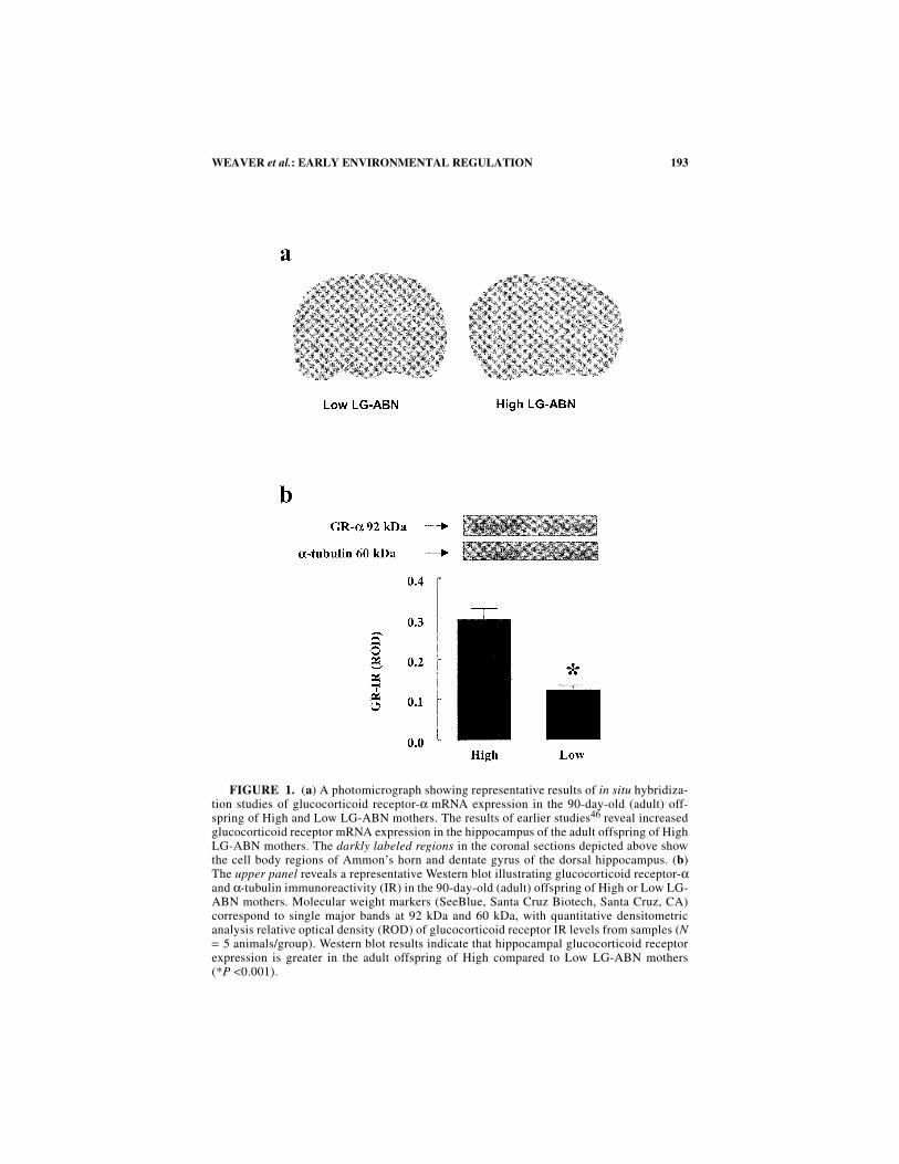
FIGURE 1. (a) A photomicrograph showing representative results of in situ hybridiza-tion studies of glucocorticoid receptor-α mRNA expression in the 90-day-old (adult) off-spring of High and Low LG-ABN mothers. The results of earlier studies46 reveal increasedglucocorticoid receptor mRNA expression in the hippocampus of the adult offspring of HighLG-ABN mothers. The darkly labeled regions in the coronal sections depicted above showthe cell body regions of Ammon’s horn and dentate gyrus of the dorsal hippocampus. (b)The upper panel reveals a representative Western blot illustrating glucocorticoid receptor-αand α-tubulin immunoreactivity (IR) in the 90-day-old (adult) offspring of High or Low LG-ABN mothers. Molecular weight markers (SeeBlue, Santa Cruz Biotech, Santa Cruz, CA)correspond to single major bands at 92 kDa and 60 kDa, with quantitative densitometricanalysis relative optical density (ROD) of glucocorticoid receptor IR levels from samples (N= 5 animals/group). Western blot results indicate that hippocampal glucocorticoid receptorexpression is greater in the adult offspring of High compared to Low LG-ABN mothers(*P <0.001).
194 ANNALS NEW YORK ACADEMY OF SCIENCES
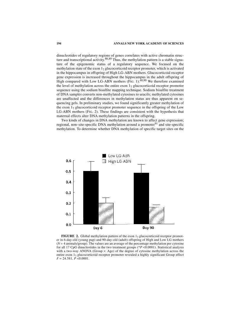
dinucleotides of regulatory regions of genes correlates with active chromatin struc-ture and transcriptional activity.86,89 Thus, the methylation pattern is a stable signa-ture of the epigenomic status of a regulatory sequence. We focused on themethylation state of the exon 17 glucocorticoid receptor promoter, which is activatedin the hippocampus in offspring of High LG-ABN mothers. Glucocorticoid receptorgene expression is increased throughout the hippocampus in the adult offspring ofHigh compared with Low LG-ABN mothers (FIG. 1).40,90 We therefore examinedthe level of methylation across the entire exon 17 glucocorticoid receptor promotersequence using the sodium bisulfite mapping technique. Sodium bisulfite treatmentof DNA samples converts non-methylated cytosines to uracils; methylated cytosinesare unaffected and the differences in methylation status are thus apparent on se-quencing gels. In preliminary studies, we found significantly greater methylation ofthe exon 17 glucocorticoid receptor promoter sequence in the offspring of the LowLG-ABN mothers (FIG. 2). These findings are consistent with the hypothesis thatmaternal effects alter DNA methylation patterns in the offspring.
Two kinds of changes in DNA methylation are known to affect gene expression;regional, non–site-specific DNA methylation around a promoter91 and site-specificmethylation. To determine whether DNA methylation of specific target sites on the
FIGURE 2. Global methylation pattern of the exon 17 glucocorticoid receptor promot-er in 6-day-old (young pup) and 90-day-old (adult) offspring of High and Low LG mothers(N = 4 animals/group). The values are an average of the percentage methylation per cytosinefor all 17 CpG dinucleotides in the two treatment groups (*P <0.0001). Statistical analysiswith a two-way ANOVA (Group × Age) of the degree of cytosine methylation across theentire exon 17 glucocorticoid receptor promoter revealed a highly significant Group effectF = 24.581, P <0.0001.
195WEAVER et al.: EARLY ENVIRONMENTAL REGULATION
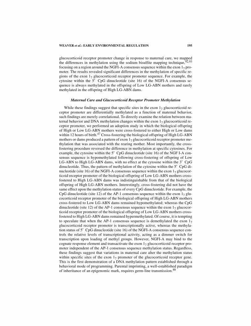
glucocorticoid receptor promoter change in response to maternal care, we mappedthe differences in methylation using the sodium bisulfite mapping technique,92,93
focusing on a region around the NGFI-A consensus sequence within the exon 17 pro-moter. The results revealed significant differences in the methylation of specific re-gions of the exon 17 glucocorticoid receptor promoter sequence. For example, thecytosine within the 5′ CpG dinucleotide (site 16) of the NGFI-A consensus se-quence is always methylated in the offspring of Low LG-ABN mothers and rarelymethylated in the offspring of High LG-ABN dams.
Maternal Care and Glucocorticoid Receptor Promoter Methylation
While these findings suggest that specific sites in the exon 17 glucocorticoid re-ceptor promoter are differentially methylated as a function of maternal behavior,such findings are merely correlational. To directly examine the relation between ma-ternal behavior and DNA methylation changes within the exon 17 glucocorticoid re-ceptor promoter, we performed an adoption study in which the biological offspringof High or Low LG-ABN mothers were cross-fostered to either High or Low damswithin 12 hours of birth.47 Cross-fostering the biological offspring of High LG-ABNmothers or dams produced a pattern of exon 17 glucocorticoid receptor promoter me-thylation that was associated with the rearing mother. Most importantly, the cross-fostering procedure reversed the difference in methylation at specific cytosines. Forexample, the cytosine within the 5′ CpG dinucleotide (site 16) of the NGF I-A con-sensus sequence is hypomethylated following cross-fostering of offspring of LowLG-ABN to High LG-ABN dams, with no effect at the cytosine within the 3′ CpGdinucleotide. Thus, the pattern of methylation of the cytosine within the 5′ CpG di-nucleotide (site 16) of the NGFI-A consensus sequence within the exon 17 glucocor-ticoid receptor promoter of the biological offspring of Low LG-ABN mothers cross-fostered to High LG-ABN dams was indistinguishable from that of the biologicaloffspring of High LG-ABN mothers. Interestingly, cross-fostering did not have thesame effect upon the methylation status of every CpG dinucleotide. For example, theCpG dinucleotide (site 12) of the AP-1 consensus sequence within the exon 17 glu-cocorticoid receptor promoter of the biological offspring of High LG-ABN motherscross-fostered to Low LG-ABN dams remained hypomethylated; whereas the CpGdinucleotide (site 12) of the AP-1 consensus sequence within the exon 17 glucocor-ticoid receptor promoter of the biological offspring of Low LG-ABN mothers cross-fostered to High LG-ABN dams remained hypermethylated. Of course, it is temptingto speculate that when the AP-1 consensus sequence is demethylated the exon 17glucocorticoid receptor promoter is transcriptionally active, whereas the methyla-tion status of 5′ CpG dinucleotide (site 16) of the NGFI-A consensus sequence con-trols the relative levels of transcriptional activity, acting as a dimmer switch fortranscription upon loading of methyl groups. However, NGFI-A may bind to thecognate response element and transactivate the exon 17 glucocorticoid receptor pro-moter independent of the AP-1 consensus sequence methylation status. Regardless,these findings suggest that variations in maternal care alter the methylation statuswithin specific sites of the exon 17 promoter of the glucocorticoid receptor gene.This is the first demonstration of a DNA methylation pattern established through abehavioral mode of programming. Parental imprinting, a well-established paradigmof inheritance of an epigenomic mark, requires germ-line transmission.88

196 ANNALS NEW YORK ACADEMY OF SCIENCES
Maternal Care–Driven Demethylation of the Exon 17Glucocorticoid Receptor Promoter
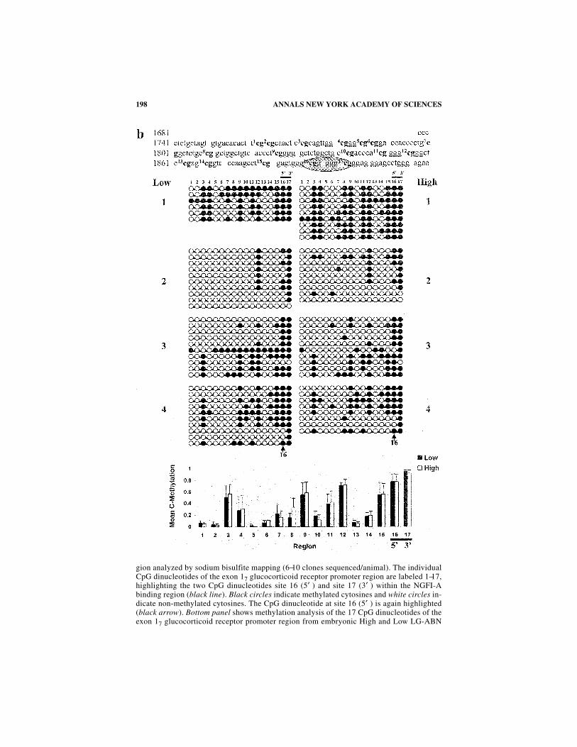
High and Low LG-ABN mothers differ in the frequency of pup licking/groomingand arched-back nursing only during the first week of life.40,46 Thus, we wonderedwhether this period corresponds to the timing for the appearance of the difference inDNA methylation in the offspring. We used the sodium bisulfite mapping techniqueto map precisely the methylation status of the cytosines within the exon 17 glucocor-ticoid receptor promoter over multiple developmental time points (FIG. 3a−e). Thisanalysis demonstrates that just before birth, on embryonic day 20, the entire regionis unmethylated in both groups. Strikingly, one day following birth (postnatal day 1)the exon 17 glucocorticoid receptor promoter is de novo methylated in both groups.The 5′ and 3′ CpG sites of the exon 17 glucocorticoid receptor NGFI-A responseelement in the offspring of both High and Low LG-ABN mothers, which exhibit dif-ferential methylation later in life, are de novo methylated to the same extent (FIG.3b). These data show that both the basal state of methylation and the first wave of denovo methylation after birth occur similarly in both groups. Whereas it is generallyaccepted that DNA methylation patterns are formed prenatally and that de novo me-thylation occurs early in development, there is at least one documented example ofpostnatal de novo methylation of the HoxA5 and HoxB5 genes.94 Since similar anal-yses are not documented for other genes, it is unknown yet whether changes in me-thylation are common around birth or whether they are unique to this glucocorticoidreceptor promoter. Between postnatal day 1 and postnatal day 6, the period when dif-ferences in the maternal behavior of High and Low LG-ABN dams are apparent, dif-ferences in the status of methylation of the exon 17 glucocorticoid receptor developbetween the two groups. For example, the NGFI-A response element 5′ CpG dinu-cleotide (site 16) is demethylated in the High, but not in the Low LG-ABN group(FIG. 5c). This is consistent with data from the cross-fostering experiment, which il-lustrated that the differences between the two groups developed following birth inresponse to maternal behavior. The group difference in CpG dinucleotide methyla-tion then remains consistent through to adulthood (postnatal day 90; FIG. 3c–e). In-terestingly, the CpG dinucleotide (site 12) of the AP-1 consensus sequence withinthe exon 17 glucocorticoid receptor promoter is similarly hypomethylated in theHigh LG-ABN offspring by postnatal day 6, and this hypomethylation is sustainedinto adulthood. Our findings suggest that the group difference in DNA methylationoccurs as a function of a maternal behavior over the first week of life. The results ofearlier studies indicated that the first week of postnatal life is indeed a “critical pe-riod” for the effects of early experience on hippocampal glucocorticoid receptorexpression.95
The results of developmental time-line study are very intriguing. From postnatalday 6, the methylation patterns for each of the 17 individual CpG sites within theexon 17 glucocorticoid receptor promoter do not all remain at the exactly same fre-quency for each developmental time-point (compare FIG. 3c−e). This is consistentwith the model that methylation, like most (if not all) biological processes, is in aconstant flux, but is stably maintained through a dynamic equilibrium. The develop-mental time-line may also help explain why the effect of maternal care on the hip-pocampal glucocorticoid receptor gene activity is not easily reversed when a HighLG-ABN offspring is cross-fostered to a Low LG-ABN mother, in comparison to the
197WEAVER et al.: EARLY ENVIRONMENTAL REGULATION
FIGURE 3. (a) Methylation patterns of the exon 17 glucocorticoid receptor promoterin the hippocampi of ED20 High and Low LG-ABN offspring (N = 5 animals/group). Toppanel shows a sequence map of the exon 17 glucocorticoid receptor promoter including the17 CpG dinucleotides (highlighted in bold) and the NGFI-A binding region (encircled).Middle panel shows bead-on-string representation of the cytosine methylation status of eachof the 17 individual CpG dinucleotides of the exon 17 glucocorticoid receptor promoter re-
198 ANNALS NEW YORK ACADEMY OF SCIENCES
gion analyzed by sodium bisulfite mapping (6−10 clones sequenced/animal). The individualCpG dinucleotides of the exon 17 glucocorticoid receptor promoter region are labeled 1−17,highlighting the two CpG dinucleotides site 16 (5′ ) and site 17 (3′ ) within the NGFI-Abinding region (black line). Black circles indicate methylated cytosines and white circles in-dicate non-methylated cytosines. The CpG dinucleotide at site 16 (5′ ) is again highlighted(black arrow). Bottom panel shows methylation analysis of the 17 CpG dinucleotides of theexon 17 glucocorticoid receptor promoter region from embryonic High and Low LG-ABN
199WEAVER et al.: EARLY ENVIRONMENTAL REGULATION
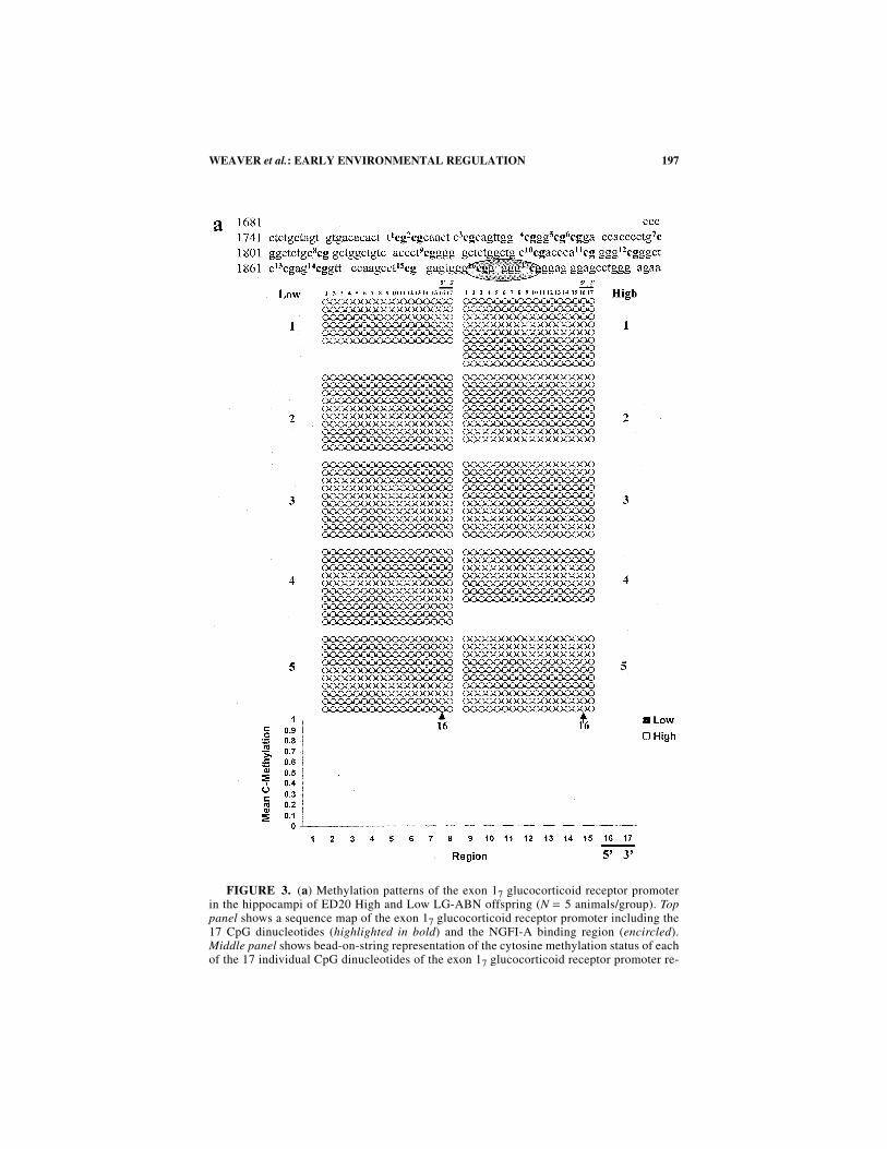
offspring. (b) Methylation patterns of the exon 17 glucocorticoid receptor promoter in thehippocampi of 1-day-old (within one hour after birth) High and Low LG-ABN offspring (N= 4 animals/group). Top panel shows a sequence map of the exon 17 glucocorticoid receptorpromoter including the 17 CpG dinucleotides (highlighted in bold) and the NGFI-A bindingregion (encircled). Middle panel shows bead-on-string representation of the cytosine meth-ylation status of each of the 17 individual CpG dinucleotides of the exon 17 glucocorticoid
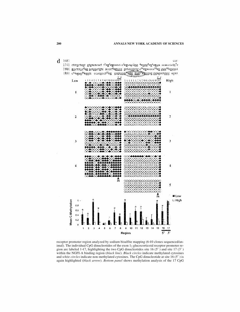
200 ANNALS NEW YORK ACADEMY OF SCIENCES
receptor promoter region analyzed by sodium bisulfite mapping (6−10 clones sequenced/an-imal). The individual CpG dinucleotides of the exon 17 glucocorticoid receptor promoter re-gion are labeled 1−17, highlighting the two CpG dinucleotides site 16 (5′ ) and site 17 (3′ )within the NGFI-A binding region (black line). Black circles indicate methylated cytosinesand white circles indicate non-methylated cytosines. The CpG dinucleotide at site 16 (5′ ) isagain highlighted (black arrow). Bottom panel shows methylation analysis of the 17 CpG
201WEAVER et al.: EARLY ENVIRONMENTAL REGULATION
dinucleotides of the exon 17 glucocorticoid receptor promoter region from postnatal day-1High and Low LG offspring. The two-way ANOVA (Group × Region) revealed a highly sig-nificant effect of Region [F =10.337, P <0.0001]. (c) Methylation patterns of the exon 17glucocorticoid receptor promoter in the hippocampi of 6-day-old (young pup) High and LowLG-ABN offspring (N = 4 animals/group). Top panel shows a sequence map of the exon 17glucocorticoid receptor promoter including the 17 CpG dinucleotides (highlighted in bold)
202 ANNALS NEW YORK ACADEMY OF SCIENCES
substantial increase in hippocampal glucocorticoid receptor gene activity observedwhen a Low LG-ABN offspring is cross-fostered to a High LG-ABN mother. DNAmethylation is a thermodynamically slower process in comparison to active demeth-ylation. This implies that the hippocampal exon 17 glucocorticoid receptor promoter,within the Low LG-ABN mother’s biological offspring, becomes stripped of CpGmethylation by activity-dependent processive demethylation, resulting from the in-
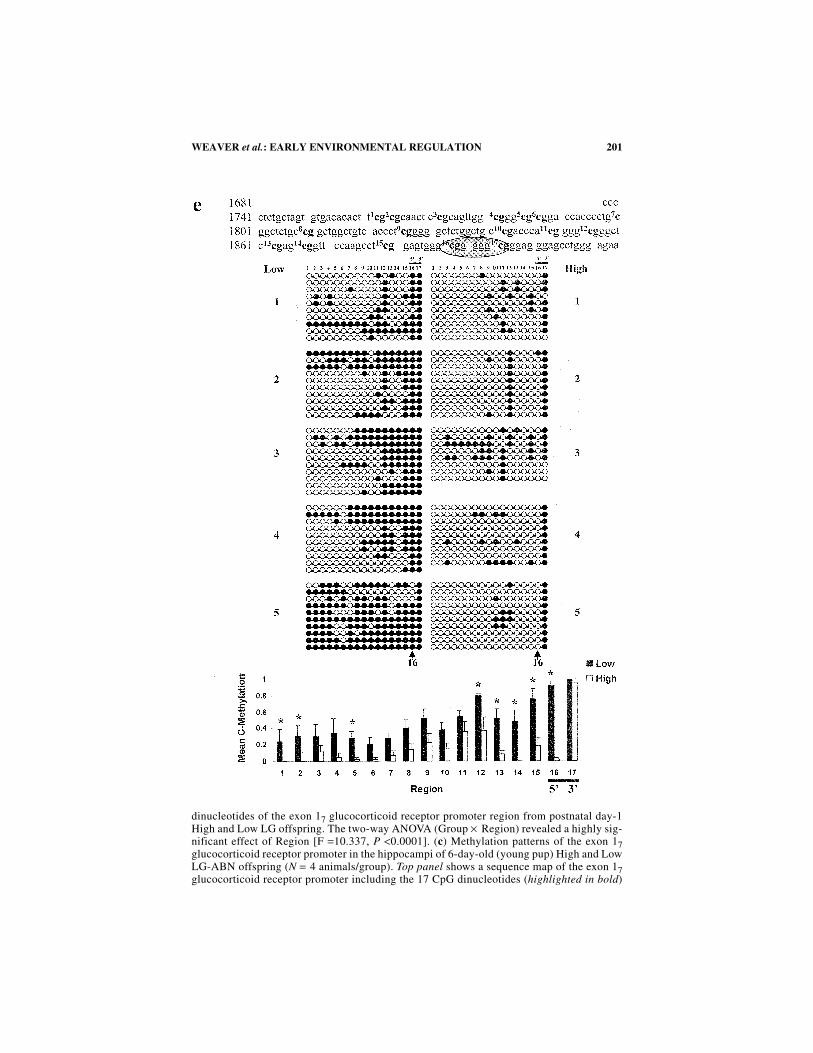
and the NGFI-A binding region (encircled). Middle panel shows bead-on-string representa-tion of the cytosine methylation status of each of the 17 individual CpG dinucleotides of theexon 17 glucocorticoid receptor promoter region analyzed by sodium bisulfite mapping (5−10 clones sequenced/animal). The individual CpG dinucleotides of the exon 17 glucocorti-coid receptor promoter region are labeled 1−17, highlighting the two CpG dinucleotides site16 (5′ ) and site 17 (3′ ) within the NGFI-A binding region (black line). Black circles indi-cate methylated cytosines and white circles indicate non-methylated cytosines. The CpG di-nucleotide at site 16 (5′ ) is again highlighted (black arrow). Bottom panel showsmethylation analysis of the 17 CpG dinucleotides of the exon 17 glucocorticoid receptor pro-moter region of the glucocorticoid receptor from postnatal day-6 High and Low LG-ABNoffspring *P <0.05; *P <0.0001 for site 16 (5′ ) lying within the NGFI-A binding region.The two-way ANOVA (Group × Region) revealed a highly significant effect of both Group[F = 32.569, P <0.0001] and Region [F = 5.353, P <0.0001]. The Group × Region interac-tion effect was not significant [F = 1.265, P = 0.057]. (d) Methylation patterns of the exon17 glucocorticoid receptor promoter in the hippocampi of 21-day-old (weaning age) Highand Low LG-ABN offspring (N = 4−5 animals/group). Top panel shows a sequence map ofthe exon 17 glucocorticoid receptor promoter including the 17 CpG dinucleotides (highlight-ed in bold) and the NGFI-A binding region (encircled). Middle panel shows bead-on-stringrepresentation of the cytosine methylation status of each of the 17 individual CpG dinucle-otides of the exon 17 glucocorticoid receptor promoter region analyzed by sodium bisulfitemapping (6−10 clones sequenced/animal). The individual CpG dinucleotides of the exon 17glucocorticoid receptor promoter region are labeled 1−17, highlighting the two CpG dinucle-otides site 16 (5′ ) and site 17 (3′ ) within the NGFI-A binding region (black line). Blackcircles indicate methylated cytosines and white circles indicate non-methylated cytosines.The CpG dinucleotide at site 16 (5′ ) is again highlighted (black arrow). Bottom panel showsmethylation analysis of the 17 CpG dinucleotides of the exon 17 glucocorticoid receptor pro-moter region from postnatal day 21 High and Low LG-ABN offspring (*P <0.05). The two-way ANOVA (Group × Region) revealed a highly significant effect of both Group [F =150.450, P <0.0001] and Region [F = 12.474, P <0.0001], as well as a significant Group ×Region interaction effect [F = 4.223, P <0.0001]. (e) Methylation patterns of the exon 17glucocorticoid receptor promoter in the hippocampi of 90-day-old (adult) High and LowLG-ABN offspring (N = 5 animals/group). Top panel shows a sequence map of the exon 17glucocorticoid receptor promoter including the 17 CpG dinucleotides (highlighted in bold)and the NGFI-A binding site (encircled). Middle panel shows bead-on-string representationof the cytosine methylation status of each of the 17 individual CpG dinucleotides of the exon17 glucocorticoid receptor promoter region analyzed by sodium bisulfite mapping (8−10clones sequenced/animal). The individual CpG dinucleotides of the exon 17 glucocorticoid re-ceptor promoter region are labeled 1−17, highlighting the two CpG dinucleotides site 16 (5′ )and site 17 (3′ ) within the NGFI-A binding region (black line). Black circles indicate methyl-ated cytosines and white circles indicate non-methylated cytosines. The CpG dinucleotide atsite 16 (5′ ) is again highlighted (black arrow). Bottom panel shows methylation analysis of the17 CpG dinucleotides of the exon 17 glucocorticoid receptor promoter region from postnatalday-90 High and Low LG-ABN offspring *P <0.05; *P <0.0001 for site 16 (5′ ) lying withinthe NGFI-A binding region. The two-way ANOVA (Group × Region) revealed a highly signif-icant effect of Group [F = 104.782, P <0.0001] and Region [F = 11.443, P <0.0001], as well asa significant Group × Region interaction effect [F=2.321, P<0.01].
203WEAVER et al.: EARLY ENVIRONMENTAL REGULATION
tense tactile stimulation exerted toward the pups by the High LG-ABN foster mother.Whereas the hippocampal exon 17 glucocorticoid receptor promoter within the HighLG-ABN mother’s biological offspring passively gains CpG methylation (assumingsome loss of methylation through maternal care by the biological High LG-ABNparent prior to cross-fostering), through a lack of stimulation of processive deme-thylation by the Low LG-ABN adoptive parent. The fact that High and Low LG-ABN offspring differ on epigenomic control of gene activity leaves the reader tospeculate upon why the Low LG-ABN offspring are so plastic in response to the fos-ter, High LG-ABN mother.
Site-Specific Methylation of the Cytosine within the 5′ CpG Dinucleotide(Site 16) of the NGFI-A Response Element
Blocks Transcription Factor Binding
The next question concerns the functional importance of such differences in me-thylation. DNA methylation affects gene expression either by attracting methylatedDNA binding proteins to a densely methylated region of a gene96 or by site-specificinterference with the binding of a transcription factor to its recognition element.97
Our data showing site-specific demethylation of the cytosine within the 5′ CpG di-nucleotide (site 16) of the NGFI-A response element (FIG. 3c) is consistent with thehypothesis that methylation at this site interferes with the binding of NGFI-A proteinto its binding site. To address this question, we determined the in vitro binding ofincreasing concentrations of purified recombinant NGFI-A protein98 to its responseelement under different states of methylation using the electrophilic mobility shiftassay (EMSA) technique with four 32P-labeled synthetic oligonucleotide sequences(FIG. 4a) bearing the NGFI-A binding site that was either (1) non-methylated, (2)methylated in the 3′ CpG site, (3) methylated in the 5′ CpG site, (4) methylated inboth sites, or (5) mutated at the two CpGs with an adenosine replacing the cytosines.NGFI-A formed a protein-DNA complex with the non-methylated oligonucleotide(FIG. 4b, lanes 2−4), while the protein was unable to form a complex with either afully methylated sequence or a sequence that was methylated at the 5′ CpG site (FIG.4b, lanes 10−12, 14−16). Partial activity was seen with the sequence methylated at the3′ CpG site (FIG. 4b, lanes 6−8). The specificity of the protein-DNA interaction isindicated by the fact that the recombinant protein fails to form a complex with themutated NGFI-A response element, even at high protein concentrations (36 pM)(FIG. 4b, lanes 18−20). This difference in binding was confirmed by competition ex-periments (FIG. 4c). NGFI-A recombinant protein was incubated with a labeled, non-methylated oligonucleotide in the presence of increasing concentrations of non-la-beled oligonucleotides containing the NGFI-A consensus sequence that were either3′ CpG methylated, 5′ CpG methylated, methylated at both sites, or mutated at thetwo CpGs with an adenosine replacing the cytosines. As expected, the non-methy-lated oligonucleotide competed with the labeled oligonucleotide protein-DNA com-plex (FIG. 4c, lane 7). The specificity of the protein-DNA interaction is indicated bythe fact that the mutated oligonucleotide is unable to compete away the labeled oli-gonucleotide protein-DNA complex (FIG. 4c lanes 17−19). Neither the oligonucle-otide methylated in both the 3′ and 5′ CpGs nor the 5′ CpG methylatedoligonucleotide were able to compete (FIG. 4c, lanes 11−16). Importantly, the 3′CpG methylated oligonucleotide, which mimics the sequence observed in the off-

204 ANNALS NEW YORK ACADEMY OF SCIENCES
FIGURE 4. (a) The exon 17 glucocorticoid receptor promoter sequence with the NGFI-A binding region (encircled). Beneath is a bead-on-string representation of a synthesized ra-dio-labeled oligonucleotide probe, highlighting the two CpG dinucleotides [ovals representthe cytosines at site 16 (5′ ) and site 17 (3′ )] within the NGFI-A binding region (responseelement, RE). (b) EMSA analysis of protein-DNA complex formation between recombinantpurified NGFI-A protein and radiolabeled oligonucleotide (a) bearing the NGFI-A responseelement containing differentially methylated cytosines within the 5′ CpG (site 16) and 3′CpG (site 17) dinucleotides. Non-methylated cytosines are represented by gray ovals, me-thylated cytosines are shown as black ovals, and white ovals are mutated CpG dinucleotideswith an adenosine replacing the cytosine. The presence of increasing amounts of purifiedNGFI-A protein (9 pM, 18 pM, or 36 pM) is indicated by the black triangle. The black arrowindicates the shift in labeled oligonucleotide mobility. Lane 1: free oligonucleotide non-me-thylated at either dinucleotide. Lanes 2−4: non-methylated oligonucleotide with an increas-ing amount of NGFI-A. Lane 5: free oligonucleotide methylated at the 3′ CpG dinucleotide.Lanes 6−8: oligonucleotide methylated at the 3′ CpG dinucleotide with an increasingamount of NGFI-A. Lane 9: free oligonucleotide methylated at the 5′ CpG dinucleotide.Lanes 10−12: oligonucleotide methylated at the 5′ CpG dinucleotide with an increasingamount of NGFI-A. Lane 13: free oligonucleotide methylated at both 5′ and 3′ CpG dinu-cleotides. Lanes 14−16: oligonucleotide methylated at both 5′ and 3′ CpG dinucleotideswith an increasing amount of NGFI-A. Lane 17: free non-methylated oligonucleotide mu-tated with an adenosine replacing the cytosine in both the 5′ and 3′ CpG dinucleotides.Lanes 18−20: mutated non-methylated oligonucleotide with increasing amount of NGFI-A.Note, methylation of the cytosine within the 3′ CpG dinucleotide reduced binding at thehigher levels of NGFI-A protein, while methylation of the cytosine within the 5′ CpG di-nucleotide completely eliminated protein binding to the NGFI-A binding region (responseelement, RE). (c) EMSA analysis of competition of protein-DNA complex formation be-tween NGFI-A protein and a radiolabeled oligonucleotide probe containing the NGFI-A re-sponse element (RE) (a) by an excess of non-labeled oligonucleotides containingdifferentially methylated cytosines within the 5′ and 3′ CpG dinucleotides of the NGFI-Aresponse element (RE). Non-methylated cytosines are represented by gray ovals, methylated
205WEAVER et al.: EARLY ENVIRONMENTAL REGULATION
spring of High LG-ABN mothers, exhibited substantial competition (FIG. 4c, lanes8−10). The results indicate that while methylation of the cytosine within the 5′ CpGdinucleotide (site 16) reduced NGFI-A protein binding to the same extent as meth-ylation in both CpG sites, methylation of the cytosine within the 3′ CpG dinucle-otide (site 17) only partially reduced NGFI-A protein binding (FIG. 4a, b). Thesedata support the hypothesis that methylation of the cytosine within the 5′ CpG di-nucleotide (site 16) in the NGFI-A response element of the exon 17 glucocorticoidreceptor promoter region in the offspring of Low LG-ABN mothers inhibits NGFI-A protein binding, potentially explaining the reduced glucocorticoid receptor genetranscription in the offspring of the Low LG-ABN mothers.
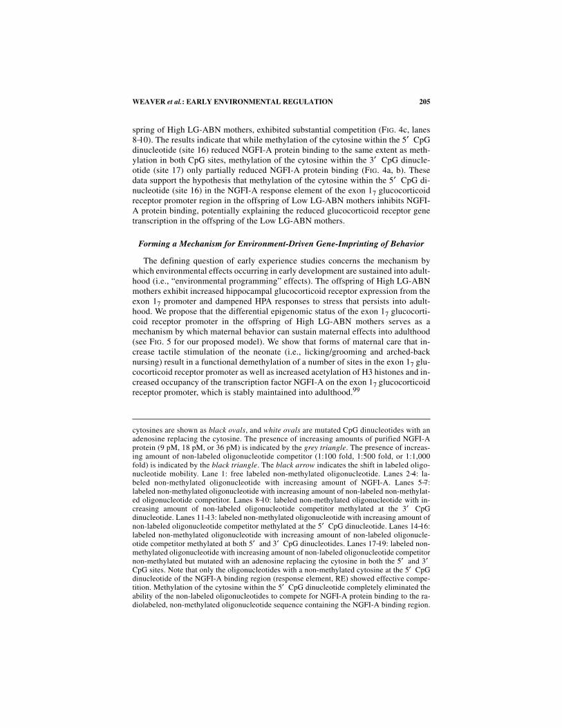
Forming a Mechanism for Environment-Driven Gene-Imprinting of Behavior
The defining question of early experience studies concerns the mechanism bywhich environmental effects occurring in early development are sustained into adult-hood (i.e., “environmental programming” effects). The offspring of High LG-ABNmothers exhibit increased hippocampal glucocorticoid receptor expression from theexon 17 promoter and dampened HPA responses to stress that persists into adult-hood. We propose that the differential epigenomic status of the exon 17 glucocorti-coid receptor promoter in the offspring of High LG-ABN mothers serves as amechanism by which maternal behavior can sustain maternal effects into adulthood(see FIG. 5 for our proposed model). We show that forms of maternal care that in-crease tactile stimulation of the neonate (i.e., licking/grooming and arched-backnursing) result in a functional demethylation of a number of sites in the exon 17 glu-cocorticoid receptor promoter as well as increased acetylation of H3 histones and in-creased occupancy of the transcription factor NGFI-A on the exon 17 glucocorticoidreceptor promoter, which is stably maintained into adulthood.99
cytosines are shown as black ovals, and white ovals are mutated CpG dinucleotides with anadenosine replacing the cytosine. The presence of increasing amounts of purified NGFI-Aprotein (9 pM, 18 pM, or 36 pM) is indicated by the grey triangle. The presence of increas-ing amount of non-labeled oligonucleotide competitor (1:100 fold, 1:500 fold, or 1:1,000fold) is indicated by the black triangle. The black arrow indicates the shift in labeled oligo-nucleotide mobility. Lane 1: free labeled non-methylated oligonucleotide. Lanes 2−4: la-beled non-methylated oligonucleotide with increasing amount of NGFI-A. Lanes 5−7:labeled non-methylated oligonucleotide with increasing amount of non-labeled non-methylat-ed oligonucleotide competitor. Lanes 8−10: labeled non-methylated oligonucleotide with in-creasing amount of non-labeled oligonucleotide competitor methylated at the 3′ CpGdinucleotide. Lanes 11−13: labeled non-methylated oligonucleotide with increasing amount ofnon-labeled oligonucleotide competitor methylated at the 5′ CpG dinucleotide. Lanes 14−16:labeled non-methylated oligonucleotide with increasing amount of non-labeled oligonucle-otide competitor methylated at both 5′ and 3′ CpG dinucleotides. Lanes 17−19: labeled non-methylated oligonucleotide with increasing amount of non-labeled oligonucleotide competitornon-methylated but mutated with an adenosine replacing the cytosine in both the 5′ and 3′CpG sites. Note that only the oligonucleotides with a non-methylated cytosine at the 5′ CpGdinucleotide of the NGFI-A binding region (response element, RE) showed effective compe-tition. Methylation of the cytosine within the 5′ CpG dinucleotide completely eliminated theability of the non-labeled oligonucleotides to compete for NGFI-A protein binding to the ra-diolabeled, non-methylated oligonucleotide sequence containing the NGFI-A binding region.
206 ANNALS NEW YORK ACADEMY OF SCIENCES
FIGURE 5. A model for environmental gene programming. (1) Prior to parturition thehippocampal exon 17 glucocorticoid receptor is entirely non-methylated (FIG. 3a). (2) Fol-lowing birth, the hippocampal exon 17 glucocorticoid receptor becomes hypermethylated(FIG. 3b) and associated with tightly packed histones to form inactive chromatin. (3) In theabsence of High tactile stimulation, the hippocampal exon 17 glucocorticoid receptor of theLow LG-ABN offspring remains hypermethylated and associated with the inactive chroma-tin, which endured into adulthood (4) (compare FIG. 3c−e). (5) High licking/grooming damsstimulate and maintain increased levels of activity-dependent gene expression in the off-spring. The activity-dependent transcription factor NGFI-A actively targets its cognate re-sponse element within the hypermethylated hippocampal exon 17 glucocorticoid receptor.
207WEAVER et al.: EARLY ENVIRONMENTAL REGULATION
Note that the effect of maternal care on glucocorticoid receptor expression is sub-tler (FIG. 1a, b) than the more pronounced effect on the methylation status of the 5′CpG dinucleotide (site 16). However, in previous studies82 we found evidence forthe use of at least three promoters regulating hippocampal glucocorticoid receptorexpression, suggesting that exon 17 is but one of the regulatory sequences determin-ing glucocorticoid receptor expression within the hippocampus.
Environmental Variability Meets Epigenomic Predictability
Further studies are required to determine how maternal behavior alters the epige-nomic status of the exon 17 glucocorticoid receptor promoter. In addition, the exactcausal relationship between DNA methylation and other changes in the epigenomicstatus described here, such as altered histone acetylation and NGFI-A binding, re-mains unclear. Regardless of these as-yet-unanswered questions, our findings pro-vide the first evidence that maternal behavior, early after birth, stably alters theepigenome of the offspring, providing a mechanism for the long-term effects of earlyexperience on gene expression in the adult. These studies offer an opportunity toclearly define the nature of gene–environment interactions during development andhow such effects result in the sustained “environmental programming” of gene ex-pression and function over the life-span. Finally, it is important to note that maternaleffects on the expression of defensive responses, such as increased HPA activity, area common theme in biology,7,100,101 such that the magnitude of the maternal influ-ence on the development of HPA and behavioral responses to stress in the rat shouldnot be surprising. Maternal effects on defensive responses to threat are apparent inplants, insects, and reptiles. Such effects commonly follow from the exposure of themother to the same or similar forms of threat and may represent examples where theenvironmental experience of the mother is translated through an epigenetic mecha-nism of inheritance into phenotypic variation in the offspring. Indeed, maternal ef-fects could result in the transmission of adaptive responses acrossgenerations.7,100,101 Epigenomic modifications of targeted regulatory sequences inresponse to even reasonably subtle variations in environmental conditions mightserve as a major source of epigenetic variation in gene expression and function andultimately as a process mediating such maternal effects. We propose that epigenomicchanges serve as an intermediate process that imprints dynamic environmental ex-periences on the fixed genome resulting in stable alterations in phenotype.
(6) Transcription factors commonly recruit histone modifying proteins, such as histoneacetylase transferase (HAT). DNA methylation is associated with changes in chromatin ac-tivity states that gate accessibility of promoters to transcription factors through effects onhistone acetylation (AC), a marker of active chromatin. Acetylation of the histone tails neu-tralizes the positively charged histones, which disrupts histone binding to negativelycharged DNA and thus promotes transcription factor binding, resulting in transient exon 17glucocorticoid receptor promoter activity. (7) Following processive DNA demethylation(compare FIG. 3b and c), the transcription factor NGFI-A can firmly bind to the demethylat-ed response element (FIG. 4b, c) within the hippocampal exon 17 glucocorticoid receptor andallow stable transcription. The hippocampal exon 17 glucocorticoid receptor of the HighLG-ABN offspring remains hypomethylated into adulthood (8), allowing stable transcrip-tion that is sustained throughout life (FIG. 1a, b).
208 ANNALS NEW YORK ACADEMY OF SCIENCES
REFERENCES
1. DENENBERG, V.H. 1964. Critical periods, stimulus input, and emotional reactivity: Atheory of infantile stimulation. Psychol. Rev. 71: 335−351.
2. DENENBERG, V.H. et al. 1967. Increased adrenocortical activity in the neonatal rat fol-lowing handling. Endocrinology 81: 1047−1052.
3. LEVINE, S. 1966. Infantile stimulation and adaptation to stress. Res. Publ. Assoc. Res.Nerv. Ment. Dis. 43: 280−291.
4. LEVINE, S. 1957. Infantile experience and resistance to physiological stress. Science126: 405.
5. LEVINE, S. 1962. Plasma-free corticosteroid response to electric shock in rats stimu-lated in infancy. Science 135: 795−796.
6. ZARROW, M.X., P.S. CAMPBELL & V.H. DENENBERG. 1972. Handling in infancy:increased levels of the hypothalamic corticotropin releasing factor (CRF) followingexposure to a novel situation. Proc. Soc. Exp. Biol. Med. 141: 356−358.
7. MEANEY, M.J. 2001. Maternal care, gene expression, and the transmission of individualdifferences in stress reactivity across generations. Annu. Rev. Neurosci. 24: 1161−1192.
8. CHROUSOS, G.P. & P.W. GOLD. 1992. The concepts of stress and stress system disor-ders. Overview of physical and behavioral homeostasis. J. Am. Med. Assoc. 267:1244−1252.
9. AKANA, S.F. et al. 1992. Feedback and facilitation in the adrenocortical system:unmasking facilitation by partial inhibition of the glucocorticoid response to priorstress. Endocrinology 131: 57−68.
10. MCEWEN, B.S. & E. STELLAR. 1993. Stress and the individual. Mechanisms leading todisease. Arch. Intern. Med. 153: 2093−2101.
11. SECKL, J.R. & M.J. MEANEY. 1993. Early life events and later development ofischaemic heart disease. Lancet 342: 1236.
12. ADER, R. 1970. The effects of early experience on the adrenocortical response to dif-ferent magnitudes of stimulation. Physiol. Behav. 5: 837−839.
13. MEANEY, M.J. et al. 1989. Neonatal handling alters adrenocortical negative feedbacksensitivity and hippocampal type II glucocorticoid receptor binding in the rat. Neu-roendocrinology 50: 597−604.
14. VIAU, V. et al. 1993. Increased plasma ACTH responses to stress in nonhandled com-pared with handled rats require basal levels of corticosterone and are associated withincreased levels of ACTH secretagogues in the median eminence. J. Neurosci. 13:1097−1105.
15. BHATNAGAR, S. & M.J. MEANEY. 1995. Hypothalamic-pituitary-adrenal function inchronic intermittently cold-stressed neonatally handled and non handled rats. J. Neu-roendocrinol. 7: 97−108.
16. MEANEY, M.J. et al. 1991. Postnatal handling attenuates certain neuroendocrine, ana-tomical, and cognitive dysfunctions associated with aging in female rats. Neurobiol.Aging 12: 31−38.
17. MEANEY, M.J. et al. 1992. Basal ACTH, corticosterone and corticosterone-bindingglobulin levels over the diurnal cycle, and age-related changes in hippocampal type Iand type II corticosteroid receptor binding capacity in young and aged, handled andnonhandled rats. Neuroendocrinology 55: 204−213.
18. LANDFIELD, P.W. & T.A. PITLER. 1984. Prolonged Ca2+-dependent after hyperpolariza-tions in hippocampal neurons of aged rats. Science 226: 1089−1092.
19. SAPOLSKY, R.M., L.C. KREY & B.S. MCEWEN. 1984. Glucocorticoid-sensitive hippoc-ampal neurons are involved in terminating the adrenocortical stress response. Proc.Natl. Acad. Sci. USA 81: 6174−6177.
20. KERR, D.S. et al. 1989. Corticosteroid modulation of hippocampal potentials:increased effect with aging. Science 245: 1505−1509.
21. ISSA, A.M. et al. 1990. Hypothalamic-pituitary-adrenal activity in aged, cognitivelyimpaired and cognitively unimpaired rats. J. Neurosci. 10: 3247−3254.
22. BHATNAGAR, S., N. SHANKS & M.J. MEANEY. 1996. Plaque-forming cell responses andantibody titers following injection of sheep red blood cells in nonstressed, acute,
209WEAVER et al.: EARLY ENVIRONMENTAL REGULATION
and/or chronically stressed handled and nonhandled animals. Dev. Psychobiol. 29:171−181.
23. LABAN, O. et al. 1995. Experimental allergic encephalomyelitis in adult DA rats sub-jected to neonatal handling or gentling. Brain Res. 676: 133−140.
24. MASON, D. 1991. Genetic variation in the stress response: susceptibility to experimen-tal allergic encephalomyelitis and implications for human inflammatory disease.Immunol. Today 12: 57−60.
25. PLOTSKY, P.M. & M.J. MEANEY. 1993. Early, postnatal experience alters hypothalamiccorticotropin-releasing factor (CRF) mRNA, median eminence CRF content andstress-induced release in adult rats. Brain Res. Mol. Brain. Res. 18: 195−200.
26. FRANCIS, D., P.M. PLOTSKY & M.J. MEANEY. 2004. Handling increases CRH mRNAexpression in selected hypothalamic neuronal populations. Submitted for publica-tion.
27. PAGE, M.E. & R.J. VALENTINO. 1994. Locus coeruleus activation by physiological chal-lenges. Brain Res. Bull. 35: 557−560.
28. PEARSON, D. et al. 1997.The effect of postnatal environment on stress-induced changesin hippocampal FOS-like immunoreactivity in adult rats. Soc. Neurosci. Abstr. 23:1849.
29. LIU, D. et al. 2000. Influence of neonatal rearing conditions on stress-induced adreno-corticotropin responses and norepinephrine release in the hypothalamic paraventricu-lar nucleus. J. Neuroendocrinol. 12: 5−12.
30. DE KLOET, E.R. 2000. Stress in the brain. Eur. J. Pharmacol. 405: 187−198.31. MEANEY, M.J. et al. 1985. Early postnatal handling alters glucocorticoid receptor con-
centrations in selected brain regions. Behav. Neurosci. 99: 765−770.32. SARRIEAU, A., S. SHARMA & M.J. MEANEY. 1988. Postnatal development and environ-
mental regulation of hippocampal glucocorticoid and mineralocorticoid receptors.Brain Res. 471: 158−162.
33. O’DONNELL, D. et al. 1994. Postnatal handling alters glucocorticoid, but not mineralo-corticoid messenger RNA expression in the hippocampus of adult rats. Brain Res.Mol. Brain Res. 26: 242−248.
34. JACOBSON, L. & R. SAPOLSKY. 1991. The role of the hippocampus in feedback regula-tion of the hypothalamic-pituitary-adrenocortical axis. Endocr. Rev. 12: 118−134.
35. DIORIO, D., V. VIAU & M.J. MEANEY. 1993. The role of the medial prefrontal cortex(cingulate gyrus) in the regulation of hypothalamic-pituitary-adrenal responses tostress. J. Neurosci. 13: 3839−3847.
36. DE KLOET, E.R. et al. 1998. Brain corticosteroid receptor balance in health and dis-ease. Endocr. Rev. 19: 269−301.
37. OWENS, M.J., M.A. VARGAS & C.B. NEMEROFF. 1993. The effects of alprazolam on cor-ticotropin-releasing factor neurons in the rat brain: implications for a role for CRF inthe pathogenesis of anxiety disorders. J. Psychiatr. Res. 27: 209−220.
38. DE BOER, S.F., J.L. KATZ & R.J. VALENTINO. 1992. Common mechanisms underlyingthe proconflict effects of corticotropin-releasing factor, a benzodiazepine inverseagonist and electric foot-shock. J. Pharmacol. Exp. Ther. 262: 335−342.
39. CALDJI, C. et al. 2000. The effects of early rearing environment on the development ofGABAA and central benzodiazepine receptor levels and novelty-induced fearfulnessin the rat. Neuropsychopharmacology 22: 219−229.
40. CALDJI, C. et al. 1998. Maternal care during infancy regulates the development of neu-ral systems mediating the expression of fearfulness in the rat. Proc. Natl. Acad. Sci.USA 95: 5335−5340.
41. WILSON, M.A. 1996. GABA physiology: modulation by benzodiazepines and hor-mones. Crit. Rev. Neurobiol. 10: 1−37.
42. MEHTA, A.K. & M.K. TICKU. 1999. An update on GABAA receptors. Brain Res. BrainRes. Rev. 29: 196−217.
43. NUTT, D.J. et al. 1992. Clinical correlates of benzodiazepine receptor function. Clin.Neuropharmacol. 15: 679A−680A.
44. HENNESSY, M.B., J. LI & S. LEVINE. 1980. Infant responsiveness to maternal cues inmice of 2 inbred lines. Dev. Psychobiol. 13: 77−84.
210 ANNALS NEW YORK ACADEMY OF SCIENCES
45. LEE, M.H. & D.I. WILLIAMS. 1975. Long term changes in nest condition and pupgrouping following handling of rat litters. Dev. Psychobiol. 8: 91−95.
46. LIU, D. et al. 1997. Maternal care, hippocampal glucocorticoid receptors, and hypotha-lamic-pituitary-adrenal responses to stress. Science 277: 1659−1662.
47. FRANCIS, D. et al. 1999. Nongenomic transmission across generations of maternalbehavior and stress responses in the rat. Science 286: 1155−1158.
48. STERN, J.M. 1997. Offspring-induced nurturance: animal-human parallels. Dev. Psy-chobiol. 31:19−37.
49. CHAMPAGNE, F.A. et al. 2003. Variations in maternal care in the rat as a mediating influ-ence for the effects of environment on development. Physiol. Behav. 79: 359−371.
50. PURVES, D., W.D. SNIDER & J.T. VOYVODIC. 1988. Trophic regulation of nerve cellmorphology and innervation in the autonomic nervous system. Nature 336: 123−128.
51. LEVINE, S. & G.W. LEWIS. 1959. The relative importance of experimenter contact in aneffect produced by extra-stimulation in infancy. J. Comp. Physiol. Psychol. 52: 368−369.
52. MEANEY, M.J. & D.H. AITKEN. 1985. The effects of early postnatal handling on hip-pocampal glucocorticoid receptor concentrations: temporal parameters. Brain Res.354: 301−304.
53. MEANEY, M.J. et al. 1994. Corticosteroid receptors in rat brain and pituitary duringdevelopment and hypothalamic-pituitary-adrenal (HPA) function. In Receptors andthe Developing Nervous System. P.A.Z. McLaughlin, Ed.: 163−202. Chapman andHall. London.
54. MEANEY, M.J., D.H. AITKEN & R.M. SAPOLSKY. 1987. Thyroid hormones influence thedevelopment of hippocampal glucocorticoid receptors in the rat: a mechanism for theeffects of postnatal handling on the development of the adrenocortical stressresponse. Neuroendocrinology 45: 278−283.
55. BANKER, G.A. & W.M. COWAN. 1977. Rat hippocampal neurons in dispersed cell cul-ture. Brain Res. 126: 397−342.
56. MITCHELL, J.B. et al. 1990. Serotonin regulates type II corticosteroid receptor bindingin hippocampal cell cultures. J. Neurosci. 10: 1745−1752.
57. SAVARD, P. et al. 1984. Effect of neonatal hypothyroidism on the serotonin system ofthe rat brain. Brain Res. 292: 99−108.
58. MITCHELL, J.B., L.J. INY & M.J. MEANEY. 1990. The role of serotonin in the develop-ment and environmental regulation of type II corticosteroid receptor binding in rathippocampus. Brain Res. Dev. Brain Res. 55: 231−235.
59. SMYTHE, J.W., W.B. ROWE & M.J. MEANEY. 1994. Neonatal handling alters serotonin(5-HT) turnover and 5-HT2 receptor binding in selected brain regions: relationshipto the handling effect on glucocorticoid receptor expression. Brain Res. Dev. BrainRes. 80: 183−189.
60. MITCHELL, J.B. et al. 1992. Serotonergic regulation of type II corticosteroid receptorbinding in hippocampal cell cultures: evidence for the importance of serotonin-induced changes in cAMP levels. Neuroscience 48: 631−639.
61. SANDERS-BUSH, E., M. TSUTSUMI & K.D. BURRIS. 1990. Serotonin receptors and phos-phatidylinositol turnover. Ann. N.Y. Acad. Sci. 600: 224−235; discussion 235−236.
62. YOSHIMASA, T. et al. 1987. Cross-talk between cellular signalling pathways suggestedby phorbol-ester-induced adenylate cyclase phosphorylation. Nature 327: 67−70.
63. PARENT, M. et al. 2001. Analysis of amino acids and catecholamines, 5-hydrox-ytryptamine and their metabolites in brain areas in the rat using in vivo microdialy-sis.Methods 23: 11−20.
64. IKE, J., H. CANTON & E. SANDERS-BUSH. 1995. Developmental switch in the hippocam-pal serotonin receptor linked to phosphoinositide hydrolysis. Brain Res. 678: 49−54.
65. LUCAS, J.J. & R. HEN. 1995. New players in the 5-HT receptor field: genes and knock-outs. Trends Pharmacol. Sci. 16: 246−252.
66. PEPIN, M.C., S. BEAULIEU & N. BARDEN. 1989. Antidepressants regulate glucocorticoidreceptor messenger RNA concentrations in primary neuronal cultures. Brain Res.Mol. Brain Res. 6: 77−83.
67. PEPIN, M.C., M.V. GOVINDAN & N. BARDEN. 1992. Increased glucocorticoid receptor genepromoter activity after antidepressant treatment. Mol. Pharmacol. 41: 1016−1022.
211WEAVER et al.: EARLY ENVIRONMENTAL REGULATION
68. LAPLANTE, P., J. DIORIO & M.J. MEANEY. 2002. Serotonin regulates hippocampal glu-cocorticoid receptor expression via a 5-HT7 receptor. Brain Res. Dev. Brain Res.139: 199−203.
69. FRANCIS, D. et al. 1996. The role of early environmental events in regulating neuroen-docrine development. Moms, pups, stress, and glucocorticoid receptors. Ann. N.Y.Acad. Sci. 794: 136−152.
70. MEANEY, M.J. et al. 2000. Postnatal handling increases the expression of cAMP-induc-ible transcription factors in the rat hippocampus: the effects of thyroid hormones andserotonin. J. Neurosci. 20: 3926−3935.
71. DE GROOT, R.P. & P. SASSONE-CORSI. 1993. Hormonal control of gene expression: mul-tiplicity and versatility of cyclic adenosine 3′ ,5′ -monophosphate-responsive nuclearregulators. Mol. Endocrinol. 7: 145−153.
72. HABENER, J.F. 1990. Cyclic AMP response element binding proteins: a cornucopia oftranscription factors. Mol. Endocrinol. 4: 1087−1094.
73. IMAGAWA, M., R. CHIU & M. KARIN. 1987. Transcription factor AP-2 mediates induc-tion by two different signal-transduction pathways: protein kinase C and cAMP. Cell51: 251−260.
74. MONTMINY, M.R., G.A. GONZALEZ & K.K. YAMAMOTO. 1990. Regulation of cAMP-inducible genes by CREB. Trends Neurosci. 13: 184−188.
75. SHENG, M. & M.E. GREENBERG. 1990. The regulation and function of c-fos and otherimmediate early genes in the nervous system. Neuron 4: 477−485.
76. VALLEJO, M. 1994. Transcriptional control of gene expression by cAMP-response ele-ment binding proteins. J. Neuroendocrinol. 6: 587−596.
77. WALTON, K.M. & R.P. REHFUSS. 1990. Molecular mechanisms of cAMP-regulatedgene expression. Mol. Neurobiol. 4: 197−210.
78. YAMAMOTO, K.K. et al. 1988. Phosphorylation-induced binding and transcriptionalefficacy of nuclear factor CREB. Nature 334: 494−498.
79. VACCARINO, F.M. et al. 1993. Induction of immediate early genes by cyclic AMP in pri-mary cultures of neurons from rat cerebral cortex. Brain Res. Mol. Brain Res. 19: 76−82.
80. LECLERC, S. et al. 1991. Purification of a human glucocorticoid receptor gene pro-moter-binding protein. Production of polyclonal antibodies against the purified fac-tor. J. Biol. Chem. 266: 8711−8719.
81. STRAHLE, U. et al. 1992. At least three promoters direct expression of the mouse gluco-corticoid receptor gene. Proc. Natl. Acad. Sci. USA 89: 6731−6735.
82. MCCORMICK, J.A. et al. 2000. 5′ -heterogeneity of glucocorticoid receptor messengerRNA is tissue specific: differential regulation of variant transcripts by early-lifeevents. Mol. Endocrinol. 14: 506−517.
83. CHEN, F., C.S. WATSON & B. GAMETCHU. 1999. Multiple glucocorticoid receptor tran-scripts in membrane glucocorticoid receptor-enriched S-49 mouse lymphoma cells.J. Cell. Biochem. 74: 418−429.
84. NOBUKUNI, Y. et al. 1995. Characterization of the human glucocorticoid receptor pro-moter. Biochemistry 34: 8207−8214.
85. CROSBY, S.D. et al. 1991. The early response gene NGFI-C encodes a zinc finger tran-scriptional activator and is a member of the GCGGGGGCG (GSG) element-bindingprotein family. Mol. Cell. Biol. 11: 3835−3841.
86. RAZIN, A. 1998. CpG methylation, chromatin structure and gene silencing—a three-way connection. EMBO J. 17: 4905−4908.
87. SZYF, M. 2001. Towards a pharmacology of DNA methylation. Trends Pharmacol. Sci.22: 350−354.
88. SAPIENZA, C. 1990. Parental imprinting of genes. Sci. Am. 263: 52−60.89. RAZIN, A. & H. CEDAR. 1977. Distribution of 5-methylcytosine in chromatin. Proc.
Natl. Acad. Sci. USA 74: 2725−2728.90. MYERS, M.M. et al. 1989. Relationships between maternal behavior of SHR and WKY
dams and adult blood pressures of cross-fostered F1 pups. Dev. Psychobiol. 22: 55−67.91. KESHET, I., J. YISRAELI & H. CEDAR. 1985. Effect of regional DNA methylation on
gene expression. Proc. Natl. Acad. Sci. USA 82: 2560−2564.92. CLARK, S.J. et al. 1994. High sensitivity mapping of methylated cytosines. Nucleic
Acids Res. 22: 2990−2997.
212 ANNALS NEW YORK ACADEMY OF SCIENCES
93. FROMMER, M. et al. 1992. A genomic sequencing protocol that yields a positive dis-play of 5-methylcytosine residues in individual DNA strands. Proc. Natl. Acad. Sci.USA 89: 1827−1831.
94. HERSHKO, A.Y. et al. 2003. Methylation of HoxA5 and HoxB5 and its relevance toexpression during mouse development. Gene 302: 65−72.
95. MEANEY, M.J. et al. 1996. Early environmental regulation of forebrain glucocorticoidreceptor gene expression: implications for adrenocortical responses to stress. Dev.Neurosci. 18: 49−72.
96. NAN, X. et al. 1998. Transcriptional repression by the methyl-CpG-binding proteinMeCP2 involves a histone deacetylase complex. Nature 393: 386−389.
97. COMB, M. & H.M. GOODMAN. 1990. CpG methylation inhibits proenkephalin geneexpression and binding of the transcription factor AP-2. Nucleic Acids Res. 18:3975−3982.
98. MILBRANDT, J. 1987. A nerve growth factor-induced gene encodes a possible tran-scriptional regulatory factor. Science 238: 797−799.
99. WEAVER, I.C. et al. Epigenomic programming by maternal behavior. Nat. Neurosci. 100. AGRAWAL, A.A. 2001. Phenotypic plasticity in the interactions and evolution of spe-
cies. Science 294: 321−326.101. ROSSITER, M.C. 1999. Maternal Effects as Adaptations. T.A.F. Mousseau, Ed. Oxford
University Press. London.
Copyright © 2022 FDOKUMEN