Selective attention in peacocks during assessment of rival males
Upload
independentCategory
view
3download
0
Yadav et al. BMC Genetics 2014, 15:19http://www.biomedcentral.com/1471-2156/15/19
RESEARCH ARTICLE Open Access
DYZ1 arrays show sequence variation betweenthe monozygotic malesSandeep Kumar Yadav1, Anju Kumari1, Saleem Javed2 and Sher Ali1*
Abstract
Background: Monozygotic twins (MZT) are an important resource for genetical studies in the context of normal anddiseased genomes. In the present study we used DYZ1, a satellite fraction present in the form of tandem arrays on thelong arm of the human Y chromosome, as a tool to uncover sequence variations between the monozygotic males.
Results: We detected copy number variation, frequent insertions and deletions within the sequences of DYZ1 arraysamongst all the three sets of twins used in the present study. MZT1b showed loss of 35 bp compared to that in 1a,whereas 2a showed loss of 31 bp compared to that in 2b. Similarly, 3b showed 10 bp insertion compared to that in 3a.MZT1a germline DNA showed loss of 5 bp and 1b blood DNA showed loss of 26 bp compared to that of 1a blood and1b germline DNA, respectively. Of the 69 restriction sites detected in DYZ1 arrays, MboII, BsrI, TspEI and TaqI enzymesshowed frequent loss and or gain amongst all the 3 pairs studied. MZT1 pair showed loss/gain of VspI, BsrDI, AgsI, PleI,TspDTI, TspEI, TfiI and TaqI restriction sites in both blood and germline DNA. All the three sets of MZT showed differencesin the number of DYZ1 copies. FISH signals reflected somatic mosaicism of the DYZ1 copies across the cells.
Conclusions: DYZ1 showed both sequence and copy number variation between the MZT males. Sequence variationwas also noticed between germline and blood DNA samples of the same individual as we observed at least in one setof sample. The result suggests that DYZ1 faithfully records all the genetical changes occurring after the twining whichmay be ascribed to the environmental factors.
Keywords: Monozygotic twins, Y chromosome, DYZ1
BackgroundThe diverse role of nature and nurture has been ad-dressed on the basis of studies on twins which are likenatural clones. It is believed that the differences betweentwins are largely due to the influence of environmentalfactors. Theoretically, identical twins must be identicalbecause they arise from a single fertilized egg (zygote).However, recent studies have shown that the identicaltwins are not truly identical as they show discerniblevariation in their genotypes [1-3]. The genetic differ-ences between MZ twins represent an example of som-atic mosaicism [4,5]. On the same token; one mayexpect similar mosaicism in the germline samples also.However, this has not been demonstrated unequivocally.During the early stages of life; it is difficult to uncoverdifferences in any of the biological attributes of twins.
* Correspondence: [email protected] Genetics Laboratory, National Institute of Immunology, ArunaAsaf Ali Marg, New Delhi 110067, IndiaFull list of author information is available at the end of the article
© 2014 Yadav et al.; licensee BioMed Central LCommons Attribution License (http://creativecreproduction in any medium, provided the or
However, as twins age, genetic and epigenetic changesaccumulate which cause the differential expression ofgenes in twins [6,7]. Twins have been reported to showcopy number variation for a number of genes [1]. There-fore, the term monozygotic twins (MZT) is more appro-priate rather than the use of identical twins as there areno identical twins in true sense.The reason of monozygotic twinning in human is not
clear. MZ twins result when a fertilized egg or zygotesplits into two embryos. This remarkable event takesplace during the first week after fertilization and canhappen at different times such as at the two cell stageon days 1 to 3, at the early blastocyst stage on days 4 to6 or in the late blastocyst stage on days 7 to 9 [8]. Thefrequency of monozygotic twinning increases 2 to 5times with in vitro fertilization [9,10]. In case of femalemonozygotic twinning, one suggested mechanism is thepreferential inactivation of the normal X in one of thetwins [11,12]. Twinning occurs spontaneously at the rateof about 1 in 80 live births [8,13]. However, monozygotic
td. This is an open access article distributed under the terms of the Creativeommons.org/licenses/by/2.0), which permits unrestricted use, distribution, andiginal work is properly cited.

Yadav et al. BMC Genetics 2014, 15:19 Page 2 of 12http://www.biomedcentral.com/1471-2156/15/19
twinning spontaneously occurs at the rate of about 1 in250 live births [8,14]. The rate of spontaneous twinning ishighest (1 in 11) in Nigeria and lowest (1 in 250) in Japan.The occurrence is about 6 per 1000 in Asia, 10–20 per1000 in Europe and USA and about 40 per 1000 in Africa[8,15].Mammalian Y chromosome originated from an ances-
tral autosome about 300 million years ago is a degener-ated X-chromosome [16]. The human Y chromosome ismale specific, constitutively haploid and largely escapesmeiotic recombination. Lack of recombination wasthought to be responsible for the degeneration of thehuman Y chromosome and loss of Y linked genes, but arecent study showed that during the past 25 millionyears, the human Y chromosome lost only 1 gene [17].Thus, crucial genes seem to have been retained by the Ychromosome.Approximately, 95% (60 Mb) of the human Y chromo-
some represents a male specific region of the Y (MSY).Similarly, 5% (3 Mb) of the human Y chromosome com-prises of pseudo-autosomal region (PAR) necessary for thepairing with the human X chromosome. The human Ychromosome has a high proportion of repeat elements.The satellite sequence DYZ1 constitutes approximately20% of the total Y chromosome [18]. Based on the HaeIIIdigestion of the human genomic DNA, DYZ1 was identi-fied as a 3.4 Kb band in the males [19], which was foundto largely contain a pentameric repeat ‘’TTCCA” [20]. Anormal human Y chromosome contains approximately3000–4300 copies of the DYZ1 arrays [21]. Since DYZ1copies do not participate in recombination, it was deducedto have no functional or evolutionary advantage [16].However, even the most repetitive stretches of DNA havesignificance in the genome as the same are envisaged toabsorb undue mutational load. DYZ1 is now reported toplay a crucial role in chromatin folding and maintenanceof the structural integrity of the Y chromosome, thus hav-ing some functional attributes [21].The major part of human genome is heterochromatic
and environmentally triggered genomic changes are gen-erally absorbed by this region. It is largely expected that,no major change takes place in the arrays of DYZ1 be-cause it does not undergo recombination. However,Since DYZ1 represents heterochromatic region of thehuman Y chromosome, any change taking place betweenthe two males of MZT after twining may in principle bedetected. Mutations occurred during pre-twinning stagewill be present in both the twins while, the ones ac-quired during the later stages in life will differentiatethem from each other. With this premise, we undertookanalysis of DYZ1 between the males of three sets ofMZT. In one set, we analysed DYZ1 arrays in the DNAfrom the semen sample as well. In the present studyblood DNA samples from three pairs of MZ twins were
used. We also collected germline DNA samples fromMZT1. We sequenced and virtually restriction mappedthe 3564 bp unit of DYZ1 arrays. We also calculated thecopy number of DYZ1 amongst the sets of these twinsusing Real Time PCR. The number of “TTCCA” repeatsand its single, double, triple, four and five base pair de-rivatives per 3564 bp unit generate a profile for the re-spective arrays. Difference in the number of TTCCArepeats and its derivatives, copy number variation ofDYZ1 arrays and loss/gain of the restriction enzymesites were compared to uncover differences betweenMZT males. Similarly, comparison was also made be-tween the blood DNA and germline DNA of the MZT1.Our result shows that DYZ1 indeed is capable of faithfullyrecording the sequence variation following the process oftwining. These changes may or may not be exclusively dueto environment, such correlation is possible to establish.This information is envisaged to be useful in the contextof biology, medicine and forensic cases.
Results3564 bp unit of DYZ1 arrayFour PCR amplified fragments (Figure 1, purple) werecloned and several positive clones were sequenced. Wehave taken the consensus of these sequences following thealignment as representative of the majority of the DYZ1 ar-rays. Further, we sequenced PCR amplified products dir-ectly and repeated the process twice and got variationseach time. Keeping that in mind, we have relied on the se-quences obtained from a cloned product over to that ofamplified ones since cloned fragment ensures purity of thetemplate having identical molecules. We have submittedsequences of all such cloned fragments to the GenBankand assigned accession numbers are given herein (MZT_1a Blood [KF941192], MZT_1a Germline [KF941193],MZT_1b Blood [KF941194], MZT_1b Germline [KF941195],MZT_2a [KF941196], MZT_2b [KF941197], MZT_3a[KF941198] and MZT_3b [KF941199]. The number ofhighly abundant “TTCCA” repeats and its single, double,triple, four and five base pair derivatives per 3564 bp unit(HaeIII fragment) of DYZ1 array were counted. The differ-ences in the number of TTCCA repeat unit and its deriva-tives between MZT are highlighted in yellow, green andsky blue for MZT pairs 1, 2 and 3, respectively (Table 1).Germline samples of both the individuals of MZ twin pair1 were used. We also sequenced the 3.56 kb HaeIII frag-ment of DYZ1 array originating from germline DNA ofMZT1 pair, ascertained the number of TTCCA repeatsand its derivatives per 3.56 kb unit of DYZ1 array andcompared the differences between DNA of blood andgermline origin with respect to DYZ1 array. The detailedresult is shown in Table 2 and differences are highlightedin bold. DYZ1 array sequences with adjusted “TTCCA”reading frame are shown in Additional file 1.

Figure 1 Diagrammatic illustration showing PCR strategy for full length amplification of 3.56 Kb unit of DYZ1 array. (A) Cartoondiagram of human Y chromosome with highlighted DYZ1 containing region in purple, (B) a single unit of DYZ1array is shown as a purple bar.Forward and reverse primers are shown above and below the bar. Similarly, amplification products and their corresponding sizes are shownbelow the bar. The amplified fragments were used for subsequent cloning and sequencing.
Yadav et al. BMC Genetics 2014, 15:19 Page 3 of 12http://www.biomedcentral.com/1471-2156/15/19
Multiple sequence alignment of 3564 bp unit of DYZ1 arrayDYZ1 sequences of all the three twin pairs were alignedin pair wise combination using ClustalW software. Fur-ther, DYZ1 array sequence originating from blood andgermline DNA of both the individuals of MZT1 were
Table 1 Number of “TTCCA” repeats and its single base pair dpair derivatives per 3.56 Kb unit of DYZ1 array are shown for
Sr. no. Repeatunit and
derivatives
Number of TTCCA repeat unit
MZT1a MZT1b
1 TTCCA 235 226
2 1 bp derivatives 290 289
ATCCA 9 9
TACCA 3 3
TTACA 16 16
TTCAA 21 21
TTTCA 19 19
TTCTA 25 26
TTCCT 33 33
GTCCA 29 29
TGCCA 10 9
TTGCA 23 22
TTCGA 60 60
TTCCG 17 17
CTCCA 14 14
TCCCA 4 4
TTCCC 7 7
3 2 bp derivatives 142 140
4 3 bp derivatives 33 36
5 4 bp derivatives 9 11
6 5 bp derivatives 1 1
The differences are highlighted in bold.
aligned with each other. Sequence variations (point mu-tations, deletions or insertions) are highlighted in yel-low (Additional files 2 and 3). MZT1b have showndeletions of 20 bp and 15 bp between 390-411 bp and1728–1744 bp positions, respectively with net loss of
erivatives along with double, triple, four and five baseall 3 MZT pairs
s and its derivatives per 3564 bp HaeIII unit of DYZ1 array
MZT2a MZT2b MZT3a MZT3b
221 228 184 190
286 295 245 238
9 9 7 7
3 3 2 2
15 16 12 11
22 23 21 19
18 19 17 15
24 25 24 23
34 30 29 27
30 31 26 26
9 9 8 8
24 25 16 18
55 62 50 51
19 18 14 14
12 14 10 10
4 4 2 2
8 7 7 5
154 139 116 120
32 37 28 28
9 8 9 8
1 1 1 1

Table 2 Number of “TTCCA” repeats and it’s single base pair derivatives along with double, triple, four and five basepair derivatives per 3.56 Kb unit of DYZ1 array are shown for MZT1
Sr. no. Repeatunit and
derivatives
Number of “TTCCA” repeat units and its derivatives per 3.56 Kb unit of DYZ1 array
MZT1a MZT1a MZT1b MZT1b
(Blood DNA) (Germline DNA) (Blood DNA) (Germline DNA)
1 TTCCA 235 234 226 232
2 1 base pair derivatives 290 290 289 288
ATCCA 9 10 9 9
TACCA 3 3 3 4
TTACA 16 15 16 16
TTCAA 21 22 21 22
TTTCA 19 19 19 18
TTCTA 25 25 26 25
TTCCT 33 32 33 32
GTCCA 29 30 29 30
TGCCA 10 10 9 10
TTGCA 23 22 22 22
TTCGA 60 60 60 60
TTCCG 17 17 17 16
CTCCA 14 14 14 14
TCCCA 4 4 4 4
TTCCC 7 7 7 6
3 2 base pair derivatives 142 142 140 140
4 3 base pair derivatives 33 33 36 35
5 4 base pair derivatives 9 9 11 9
6 5 base pair derivatives 1 1 1 1
The differences are highlighted in bold.
Yadav et al. BMC Genetics 2014, 15:19 Page 4 of 12http://www.biomedcentral.com/1471-2156/15/19
35 bp when compared to MZT1a. Similarly, MZT2ashowed deletions of 16 bp and 15 bp between 411–427 bp and 765–781 bp positions, respectively, with netloss of 31 bps when compared to MZT2b. However,MZT1b itself has 5 bp less than the standard 3564 bplength. Further, MZT3b also showed deletion of 1 bpand 5 bp between 1068–1070 bp and 1580–1586 bppositions, respectively, as compared to MZT3a. MZT3balso showed insertion of 1 bp and 15 bp between1668–1669 and 2515–2516 respectively, compared withMZT3a. So, MZT3b showed net insertion of 10 bp ascompared to MZT3a. When blood and germline DYZ1sequence of MZT1 were compared, germline sample ofMZT1a showed deletion of 5 bp between 410–416 bppositions as compared with that of MZT1a bloodDNA. Similarly, MZT1b blood DYZ1 sequence showeddeletion of 15 bp each between 390–406 bp and 1722–1738 bp positions and insertion of 1 bp between 844–845 bp, 3487–3488 bp, 3518–3519 bp and 3540–3549 bppositions as compared to MZT1b germline DYZ1 se-quence. Thus, DYZ1 from MZT1b blood showed net lossof 26 bps.
Restriction mapping of 3564 bp HaeIII fragment ofDYZ1 arrayA 3564 bp unit of DYZ1 array was subjected to virtualrestriction mapping for all the three sets of twins(Table 3). Twin pair 1 showed single loss/gain of MboII,BstXI, BsrDI, DsrI, BsmI and TaqI sites. Likewise, CspCIand TspEI showed loss/gain of these restriction sitestwice. Twin pair 2 showed single loss/gain of DdeI, Bse-MII, TatI, MboII, BccI, SfaNI, TstI, MseI, BsrI, AgsI, PleI,TspDTI, BsaBI, NlaIV, BtgZI, MaeII and double loss/gainof ArsI, AlfI, NlaIII, BcgI, BsrDI, TspGWI, FaiI, TspEI,HinFI, TaqII, MfeI restriction sites while, TfiI and TaqIshowed loss/gain of three and six restriction sites, respect-ively. In twin pair 3, single loss/gain of Cac8I, EcoRI, SetI,AsuII, MboII, BccI, MaeI, RsaI, XmnI, NlaIII, FaiI, PleI,TspDTI, TaqI, HinFI, BtgZI, MaeIII, Tsp45I, HphI, NspIand SphI restriction site were detected. Similarly, ApoI,BcgI and BsrI showed loss/gain of two restriction sites. Asingle enzyme TspEI showed loss of three sites. Out of 69restriction enzymes sites detected in DYZ1, MboII, BsrI,TspEI and TaqI showed frequent loss or gain of the re-striction sites in all the three twin pairs studied. The

Table 3 Virtual restriction mapping of 3.56 Kb unit of DYZ1 array from twin pairs (blood DNA) using restrictionmapper software
Sr. no. Restrictionsite
Sequence Overhang Restriction site frequency
MZT1a MZT1b MZT2a MZT2b MZT3a MZT3b
1 Cac8I GCNNGC 5’ 1 1 1 1 2 1
2 BseYI CCCAGC 5’ 1 1 1 1 1 1
3 Bsp1407I TGTACA 5’ 1 1 1 1 0 0
4 BspHI TCATGA 3’ 1 1 1 1 0 0
5 ClaI ATCGAT 3’ 1 1 1 1 1 1
6 DdeI CTNAG blunt 1 1 0 1 0 0
7 EcoRI GAATTC 5’ 1 1 1 1 2 1
8 ScrFI CCNGG 3’ 1 1 1 1 1 1
9 VspI ATTAAT blunt 1 1 1 1 1 1
10 BseMII CTCAG 5’ 1 1 0 1 0 0
11 BsgI GTGCAG 3’ 1 1 1 1 0 0
12 BtsI GCAGTG 3’ 1 1 1 1 0 0
13 Eco57I CTGAAG 3’ 1 1 1 1 1 1
14 GsuI CTGGAG 3’ 1 1 1 1 0 0
15 Hpy188I TCNGA 3’ 1 1 1 1 0 0
16 MnlI CCTC 3’ 1 1 2 2 0 0
17 PflMI CCANNNNNTGG 3’ 1 1 1 1 1 1
18 SduI GDGCHC blunt 1 1 1 1 0 0
19 BsaBI GATNNNNATC blunt 0 0 1 0 0 0
20 TaqII GACCGA 3’ 0 0 2 0 0 0
21 MfeI CAATTG 5’ 0 0 0 2 0 0
22 DpnI GATC 5’ 2 2 1 1 1 1
23 FokI GGATG 3’ 2 2 2 2 2 2
24 MboI GATC 5’ 2 2 1 1 1 1
25 ArsI GACNNNNNNTTYG 5’ 2 2 0 2 2 2
26 BdaI TGANNNNNNTCA 5’ 2 2 2 2 2 2
27 CspCI CAANNNNNGTGG 5’ 2 0 0 0 0 0
28 Eco57MI CTGRAG 5’ 2 2 2 2 1 1
29 SetI ASST 5’ 2 2 2 2 2 1
30 TspRI CASTG 5’ 2 2 2 2 1 1
31 ApoI RAATTY 3’ 3 3 3 3 4 2
32 AsuII TTCGAA 3’ 3 3 3 3 3 4
33 Eco31I GGTCTC blunt 3 3 3 3 3 3
34 TatI WGTACW 5’ 3 3 3 4 1 1
35 MboII GAAGA 3’ 3 4 4 3 3 4
36 CviJI RGCY blunt 4 4 4 4 3 3
37 MslI CAYNNNNRTG 5’ 4 4 4 4 3 3
38 BccI CCATC 3’ 4 4 5 4 4 5
39 MaeI CTAG 3’ 4 4 4 4 3 4
40 SfaNI GCATC 3’ 4 4 3 4 2 2
41 AlfI GCANNNNNNTGC 3’ 4 4 2 4 2 2
42 TstI CACNNNNNNTCC 3’ 4 4 4 5 4 4
43 RsaI GTAC 3’ 5 5 5 5 2 3
44 BsmAI GTCTC 3’ 5 5 5 5 5 5
45 BstXI CCANNNNNNTGG blunt 5 4 5 5 5 5
Yadav et al. BMC Genetics 2014, 15:19 Page 5 of 12http://www.biomedcentral.com/1471-2156/15/19

Table 3 Virtual restriction mapping of 3.56 Kb unit of DYZ1 array from twin pairs (blood DNA) using restrictionmapper software (Continued)
46 XmnI GAANNNNTTC 5’ 6 6 6 6 5 6
47 MseI TTAA 3’ 6 6 6 5 6 6
48 NlaIII CATG 5’ 7 7 9 7 5 4
49 BcgI CGANNNNNNTGC 5’ 14 14 18 16 10 12
50 BsrDI GCAATG 5’ 15 14 14 16 11 11
51 BsrI ACTGG 5’ 19 18 19 18 12 14
52 TspGWI ACGGA 5’ 19 19 21 19 16 16
53 BsmI GAATGC 5’ 21 20 22 22 13 17
54 AgsI TTSAA 5’ 22 22 24 25 24 20
55 FaiI YATR 3’ 23 23 25 23 17 16
56 PleI GAGTC 3’ 23 23 24 23 19 18
57 TspDTI ATGAA blunt 25 25 23 24 18 17
58 TspEI AATT 5’ 34 32 36 34 32 29
59 TfiI GAWTC 3’ 53 53 51 54 48 48
60 TaqI TCGA blunt 66 67 61 67 54 55
61 HinfI GANTC 5’ 76 76 75 77 67 66
The differences are highlighted in bold.
Yadav et al. BMC Genetics 2014, 15:19 Page 6 of 12http://www.biomedcentral.com/1471-2156/15/19
germline DNA samples studied along with the bloodDNA samples for MZT1a showed single loss/gain of VspI,SetI, AsuII, BsrDI, BsmI, AgsI, PleI,TspDTI,TspEI,TfiI andTaqI restriction sites. Likewise, CspCI and BcgI showeddouble loss/gain of the restriction sites. Further, MZT 1bshowed single loss/gain of VspI, AcyI, HgaI, Hpy99I, DpnI,FokI, MboI, TspRI, SfaNI, BstXI, BsrDI, BsrI, AgsI, FaiI,PleI, TspDTI, TaqI and HinfI, double loss/gain of HaeIVand Hin4I and triple loss/gain of TspEI restriction sites.Out of 63 restriction sites detected in blood and germlineDNA samples of MZT1, VspI ,BsrDI, AgsI, PleI, TspDTI,TspEI, TfiI and TaqI showed loss/gain of restriction sitesin both the individuals of MZT1. Detailed result of virtualrestriction mapping for blood and germline DNA sampleof MZT1 is given in the Table 4.The real restriction mapping experiments did not al-
ways correlate with the virtual restriction mapping databecause in case of virtual restriction mapping, we dealtwith a single array sequence while in case of real restric-tion mapping, we dealt with a pool of DYZ1 array se-quences and average of all of them may be lot moredifferent than that of a single array sequence. Out of sev-eral restriction enzymes like RsaI, BstXI, DpnI, EcoRI,MboI, MboII, XmnI, TatI, MseI, ApoI, MfeI, BseMII,NlaIII and DdeI; DpnI restriction pattern showed vari-ation between blood and germline DNA (Figure 2).Taken together, the blood genomic DNA does not con-tain DpnI site while germline DNA does in MZT1.
DYZ1 copy number variationDYZ1 copy number was calculated using absolute quan-titative PCR following SYBR green chemistry and a
standard curve of cloned DYZ1 plasmid using ten-folddilutions. The dissociation curve, standard curve andamplification plot are given in Figures 3A,B and C, re-spectively. The respective copy number values for alltwin pairs and controls are shown in figures 3D. Twinpair sets 1, 2 and 3 showed differences of 409, 367 and697 of DYZ1 copies, respectively.
Localization of DYZ1 on metaphases/nuclei usingFluorescence in situ HybridizationWe screened approximately 400 nuclei and metaphases.To rule out the possibility of experimental error, twopositive controls (metaphases prepared from normal hu-man blood) were used with the same probe preparation.Following FISH, the nuclei and metaphases showedDYZ1 signal of varying intensity which is due to thevarying number of its copies. The representative FISHpictures are shown in Figure 4.
DiscussionThe genome that we are born with is not the one thatwe die with [1]. This is true for all the cells in our body.So, as we age, environmentally triggered genomic changesaccumulate in our DNA more in the repeat regions. Ac-cordingly then, the difference between the identical twinsincreases as they age. Twins can also begin their lives withsome major differences.MSY region of the human Y chromosome does not
take part in the crossing over, so the DNA comprisingMSY is faithfully passed on from father to son. However,MSY may accumulate mutations during the life time ofan individual. In case of DYZ1, point mutations generate
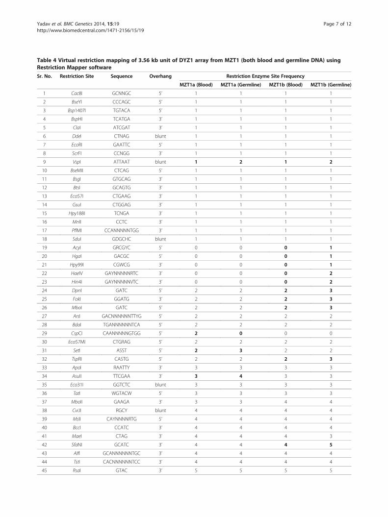
Table 4 Virtual restriction mapping of 3.56 kb unit of DYZ1 array from MZT1 (both blood and germline DNA) usingRestriction Mapper software
Sr. No. Restriction Site Sequence Overhang Restriction Enzyme Site Frequency
MZT1a (Blood) MZT1a (Germline) MZT1b (Blood) MZT1b (Germline)
1 Cac8I GCNNGC 5’ 1 1 1 1
2 BseYI CCCAGC 5’ 1 1 1 1
3 Bsp1407I TGTACA 5’ 1 1 1 1
4 BspHI TCATGA 3’ 1 1 1 1
5 ClaI ATCGAT 3’ 1 1 1 1
6 DdeI CTNAG blunt 1 1 1 1
7 EcoRI GAATTC 5’ 1 1 1 1
8 ScrFI CCNGG 3’ 1 1 1 1
9 VspI ATTAAT blunt 1 2 1 2
10 BseMII CTCAG 5’ 1 1 1 1
11 BsgI GTGCAG 3’ 1 1 1 1
12 BtsI GCAGTG 3’ 1 1 1 1
13 Eco57I CTGAAG 3’ 1 1 1 1
14 GsuI CTGGAG 3’ 1 1 1 1
15 Hpy188I TCNGA 3’ 1 1 1 1
16 MnlI CCTC 3’ 1 1 1 1
17 PflMI CCANNNNNTGG 3’ 1 1 1 1
18 SduI GDGCHC blunt 1 1 1 1
19 AcyI GRCGYC 5’ 0 0 0 1
20 HgaI GACGC 5’ 0 0 0 1
21 Hpy99I CGWCG 3’ 0 0 0 1
22 HaeIV GAYNNNNNRTC 3’ 0 0 0 2
23 Hin4I GAYNNNNNVTC 3’ 0 0 0 2
24 DpnI GATC 5’ 2 2 2 3
25 FokI GGATG 3’ 2 2 2 3
26 MboI GATC 5’ 2 2 2 3
27 ArsI GACNNNNNNTTYG 5’ 2 2 2 2
28 BdaI TGANNNNNNTCA 5’ 2 2 2 2
29 CspCI CAANNNNNGTGG 5’ 2 0 0 0
30 Eco57MI CTGRAG 5’ 2 2 2 2
31 SetI ASST 5’ 2 3 2 2
32 TspRI CASTG 5’ 2 2 2 3
33 ApoI RAATTY 3’ 3 3 3 3
34 AsuII TTCGAA 3’ 3 4 3 3
35 Eco31I GGTCTC blunt 3 3 3 3
36 TatI WGTACW 5’ 3 3 3 3
37 MboII GAAGA 3’ 3 3 4 4
38 CviJI RGCY blunt 4 4 4 4
39 MslI CAYNNNNRTG 5’ 4 4 4 4
40 BccI CCATC 3’ 4 4 4 4
41 MaeI CTAG 3’ 4 4 4 3
42 SfaNI GCATC 3’ 4 4 4 5
43 AlfI GCANNNNNNTGC 3’ 4 4 4 4
44 TstI CACNNNNNNTCC 3’ 4 4 4 4
45 RsaI GTAC 3’ 5 5 5 5
Yadav et al. BMC Genetics 2014, 15:19 Page 7 of 12http://www.biomedcentral.com/1471-2156/15/19

Table 4 Virtual restriction mapping of 3.56 kb unit of DYZ1 array from MZT1 (both blood and germline DNA) usingRestriction Mapper software (Continued)
46 BsmAI GTCTC 3’ 5 5 5 5
47 BstXI CCANNNNNNTGG blunt 5 5 4 5
48 XmnI GAANNNNTTC 5’ 6 6 6 6
49 MseI TTAA 3’ 6 6 6 6
50 NlaIII CATG 5’ 7 7 7 7
51 BcgI CGANNNNNNTGC 5’ 14 12 14 14
52 BsrDI GCAATG 5’ 15 14 14 15
53 BsrI ACTGG 5’ 19 19 18 19
54 TspGWI ACGGA 5’ 19 19 19 19
55 BsmI GAATGC 5’ 21 20 20 20
56 AgsI TTSAA 5’ 22 23 22 23
57 FaiI YATR 3’ 23 23 23 24
58 PleI GAGTC 3’ 23 24 23 24
59 TspDTI ATGAA blunt 25 24 25 24
60 TspEI AATT 5’ 34 35 32 35
61 TfiI GAWTC 3’ 53 52 53 51
62 TaqI TCGA blunt 66 65 67 66
63 HinfI GANTC 5’ 76 76 76 75
The germline samples for remaining two twin pairs (MZT2 and 3) were not available. The differences in restriction site frequency are highlighted in bold.
Yadav et al. BMC Genetics 2014, 15:19 Page 8 of 12http://www.biomedcentral.com/1471-2156/15/19
derivatives of “TTCCA” while insertions and deletionsshift the “TTCCA” frame. Genome tries to neutralize orminimize these changes. In the process, insertion at onepoint may lead to the deletion at another point and viceversa (Additional file 2). Despite these changes in thenumber of “TTCCA” and its derivatives, the overalllength of the array remains almost unchanged. Inde-pendent mutational events may also lead to gain or lossof restriction enzyme sites in DYZ1 array which is evi-dent from the present study.
Figure 2 A representative gel picture showing Restriction Fragment Lfor restriction digestion. The DYZ1 fragments generated using primers Dgermline samples compared to that in blood. However, DYZ1 fragment amblood and germline DNA.
In addition to these, DYZ1 arrays showed copy numbervariation between MZT as uncovered by real time PCR.However, fluorescence signal intensity (Figure 4) of DYZ1probe is not always correlated with its copy number vari-ation. This is because every cell does not contain equalnumber of DYZ1 copies. Similarly, DNA used for quanti-tative Real Time PCR does not contain homogeneouspopulation of DYZ1 sequences. Thus, DYZ1 copies calcu-lated using absolute quantification is the average of theDYZ1 arrays present in the pool of DNA from all the cells.
ength Polymorphism (RFLP) in monozygotic males: DpnI is usedYZ1 A & B, C & D and E & F show presence of restriction site inplified by primers DYZ1 G & H does not contain any DpnI site both in

Figure 3 Copy number estimation of DYZ1 in Monozygotic twin pairs. (A) represents the dissociation curve, (B) the standard plot (C) theamplification plot and (D) shows the number of DYZ1 copies in all three twin pairs along with two control samples.
Yadav et al. BMC Genetics 2014, 15:19 Page 9 of 12http://www.biomedcentral.com/1471-2156/15/19
Analysis of DYZ1 has been pursued in our laboratory inthe context with Sex Chromosome Related Anomalies(SCRA) [22], males exposed to Natural Background Radi-ation (NBR) [21], Arsenic Poisoning [23], Prostate Cancercell lines [18] and Infertility [24]. Significantly, DYZ1 wasfound to show much reduced copies in all these cases.Thus, indeed there exists a correlation between the re-duced copies of the DYZ1 and these abnormal conditions.DYZ1 does not unequivocally differentiate between mono-zygotic twins but the effect of nature vs. nurture on twinscan be studied with respect to DYZ1 arrays. This is be-cause at the timing of twining, the copies in both themales are expected to be identical. Any variation noticedeither in the copies of the arrays or within is ascribable tothe environmental conditions. Thus, present study hasrelevance in the context of changes brought about in theDYZ1 arrays between two males of monozygotic origin.Taken together, the study mainly supports the argu-
ment that, the monozygotic twins are not really identi-cal as evident from this study. Extrapolation of thisstudy in a large number of samples may lead to thediscovery of sufficient genetic variation in the DYZ1arrays from across the samples. This in turn wouldaugment the already existing approaches useful for thediscrimination of identical twins in the context offorensic cases.
ConclusionsDYZ1 arrays have shown variations between the mono-zygotic males. Similarly, sequence variations were alsoestablished between germline and blood DNA samplesof the same individual for one twin pair. This approachis envisaged to be of relevance in biology, medicine andforensic cases if sufficiently large number of samplesboth from blood and germline are analysed.
MethodsSample collection and DNA isolationPresent study was approved by the Institutional HumanEthical Committee of the National Institute of Immun-ology, New Delhi. Peripheral blood lymphocytes (PBLs)were collected from three pairs of male monozygotictwins, with their informed consent. Genomic DNA fromblood was isolated using DNeasy Blood and Tissue kitfrom Qiagen, Germany (Cat no. 29504) and GermlineDNA of MZT1 was isolated following standard protocol[21]. Quality of isolated DNA was checked by electro-phoresis using 1% agarose gel. DNA concentration wasmeasured spectrophotometrically.
End point PCRPCR primers used to amplify the full 3.56 bp HaeIIIfragment of DYZ1 are listed in Table 5 and illustrated

Figure 4 Localization of DYZ1 using FISH. (A) DAPI (4′, 6-diamidino-2-phenylindole) stained metaphases and interphase nuclei are shownhaving green signal of DYZ1 probe by red arrows. (B) The table shows fluorescence intensity per unit area values for each DYZ1 probe signalspot. Note the variation in the DYZ1 probe’s signal intensities across nuclei reflecting copy number variation and somatic mosaicism.
Yadav et al. BMC Genetics 2014, 15:19 Page 10 of 12http://www.biomedcentral.com/1471-2156/15/19
graphically in Figure 1. The end point PCR reactionswere carried out in 20 μl volume containing Pfu DNApolymerase (Biotools, Spain), 5X reaction buffer (Pro-mega, Madison, USA), 200 μM dNTPs (Bio. Basic Inc. To-ronto, Canada) and 100 ng template DNA. The amplifiedproducts were resolved on 1.0% agarose gels, stained withEthidium bromide and visualized under Ultraviolet.
Cloning and DNA sequencingEnd point PCR amplified DYZ1 array fragments re-solved on 1.0% agarose gel were extracted using a kit(Fermentas, Thermo Fischer Scientific). Purified frag-ments of DNA were cloned in blunt end cloning vectors(CloneJet, Fermentas, Thermo Fischer Scientific). Fourrecombinant clones, each representing positive oneswere selected after conducting colony PCR using vector
Table 5 List of primers used for PCR amplification of DYZ1 ar
Serial no. Primer ID Primer sequence L
1 DYZ1 A CCATTCGAGACCGTAGCAATT
3 DYZ1 B ATTTGATGCCATCCCATGAC
5 DYZ1 C TCCTTTGCCTTCCATTCG
6 DYZ1 D TGCAGTCTTTTCCCTTCGAG
7 DYZ1 E ATTGGATGGGATTGGAATGA
9 DYZ1 F TCGAATGGAAGGCAAAGG
10 DYZ1 G CGACTGGTACGGACTCCAT
12 DYZ1 H TGGACAGCCTGGAATAAAGTG
specific forward and reverse primers. Recombinant cloneswere further confirmed by restriction digestion. Fourpurified recombinant clones were sequenced on AppliedBiosystems 3130xl genetic analyzer using ABI ABIPRISM®BigDye® terminator v3.1 cycle sequencing kits (Lifetechnologies, California, USA). PCR conditions were setas 96°C for 1 minute, followed by 25 cycles each consist-ing of 96°C for 10 seconds, 50°C for 5 seconds, and 60°Cfor 4 minutes. After cycle sequencing, extension productswere purified to remove any unincorporated dye-labelledterminators using ethanol–sodium acetate precipitationmethod followed by washing in 70% ethanol. Hi-Di™Formamide (Life technologies, California, USA) wasadded, samples were heat denatured, chilled on iceand loaded onto the 3130xl genetic analyzer, ABI. Thedata was collected using 3130xl Data Collection
ray
ength (bp) Location Orientation
21 35-16 (5’ upstream to HaeIII site) 5’-3’
20 763-782 5’-3’
18 1668-1685 5’-3’
20 2564-2583 5’-3’
20 861-880 3’-5’
18 1669-1686 3’-5’
20 2637-2656 3’-5’
21 3586-3606 3’-5’

Yadav et al. BMC Genetics 2014, 15:19 Page 11 of 12http://www.biomedcentral.com/1471-2156/15/19
Software v3.0. Sequences were analyzed using SequenceScanner software version 1.0 and gene runner softwareversion 3.05.
Restriction mappingThe DYZ1 sequences were subjected to virtual restrictionmapping using Restriction Mapper Software Version 3.0(Tables 3 and 4). To support the virtual restriction map-ping data, we conducted real restriction mapping on PCRamplified product using several restriction enzymes. Thereaction digestions were carried out in 20 μl reaction mix-ture using 1 μg of template DNA and 2 units of enzymefollowing standard protocols (NEB, UK). Digested sampleswere resolved on 2.0% agarose gel and visualized underUV illumination to record the resultant bands.
Copy number estimationNumber of DYZ1 copies was calculated based on ab-solute quantification method using quantitative PCR(qPCR). DNA was used as template and SYBR green(Life Technologies, California, USA) as detection dye.The qPCR reactions were performed on Sequence Detec-tion System 7500 (Life Technologies, California, USA)following 10 fold dilutions of recombinant plasmid con-taining ~3.4 Kb HaeIII fragment of DYZ1 array startingwith 2 × 10⁸ copies and standard curve were generated.All the reactions were carried out in triplicates using threedifferent concentrations of the template DNA. The stand-ard curve has a slope of −3.32 and R2 value of >0.99. Cop-ies of the DYZ1 array were calculated by extrapolation ofthe standard curve obtained with known copies of the re-combinant plasmid. To show the reproducibility of qPCRresults, error bars are shown on top of the graph bars.
Florescence in-situ Hybridization (FISH)Peripheral blood cells cultured in PB-MAX™ KaryotypingMedium (Gibco®, USA) were used for metaphase chromo-some preparation. The cells were grown for 70 hours in5% CO2 environment at 37°C and then treated with colce-mid (3 μg/ml). Treated cells were again incubated for2 hours in 5% CO2 environment at 37°C. After 72 hours,cells were centrifuged at 1800 rpm for 10 minutes at roomtemperature (RT). Harvested cells were resuspended in0.075 M KCl and incubated at RT for 20 minutes in 5%CO2 environment at 37°C. Then added 1 drop of fixativesolution (3:1, methanol: glacial acetic acid) and centrifugedat 1800 rpm for 10 minutes at RT. Discarded the super-natant, resuspended the cell pellet in 10 ml fresh fixativesolution and incubated for 20 minutes at 37°C. Then cen-trifuged cells at 1800 rpm for 10 minutes at RT. Repeatedthe washing step 2 times. Finally, cells were resuspendedin fresh 1 ml fixative and stored at −20°C until used.
20 μl of nuclei suspension in fixative was spread onthe fixative dipped glass slides. Before proceeding fur-ther, slides were kept for 1 week at 37°C for ageing.Slides were then incubated in 70% glacial acetic acid for2 minutes followed by dehydration in 70%, 90% and100% ethanol for 2 minutes each at RT. Slides were airdried and incubated in a solution containing 0.1 mg/mland 0.01 N HCl for 20 minutes. Fixed the metaphasepreparation in 4% paraformaldehyde (prepared in 1XPBS, pH 7.4) for 5 minutes at RT. Slides were washed 2times in PBS followed by once in water. Further, slideswere dehydrated in 70%, 90% and 100% ethanol sequen-tially. Air dried slides were then used for hybridization.FISH was conducted with a labelled clone containing3.56 kb sequence of DYZ1 array. Labelling was donewith biotin-dUTP using a Nick translation kit from Vysis(Illinois, USA). Hybridization, washing, counterstainingand mounting of the slides were conducted followingstandard protocols [25]. The slides were screened underthe Olympus fluorescence microscope (BX 51) fittedwith vertical fluorescence illuminator U-LH100HG UV,excitation and barrier filters and images were capturedwith a charge-coupled device (CCD) camera. Capturedimages were analysed using CytoVision software version3.93 from Applied Imaging Systems.
Additional files
Additional file 1: 3.56 Kb sequence of DYZ1 array from all threetwin pairs, in adjusted frame of “TTCCA”. (A) MZT_1a (Blood), MZT_1a(Germline), MZT_1b (Blood) and MZT_1b (Germline); (B) MZT_2a (Blood)and MZT_2b (Blood) and (C) MZT_3a (blood) and MZT_3b (Blood).
Additional file 2: Multiple sequence alignment (MSA) of 3.56 Kbsequence of DYZ1 array from twin pairs. The regions of nucleotidevariations are highlighted in yellow. (A) MZT1, (B) MZT2 and (C) MZT3.
Additional file 3: Multiple Sequence Alignment (MSA) of 3.56 Kbsequence of DYZ1 array from Blood DNA with Germline DNA ofMZT1. The insertions, deletions and point mutations are highlighted inyellow. (A) MZT1a and (B) MZT1b.
AbbreviationsMZT: Monozygotic twin; NRY: Non recombining region of Y; PAR: Pseudoautosomal region; MSY: Male specific region of Y; DNA: Deoxyribonucleicacid; PBLs: Peripheral blood lymphocytes; PCR: Polymerase chain reaction;RFLP: Restriction fragment length polymorphism; UV: Ultraviolet; qPCR: Quantitativepolymerase chain reaction; FISH: Fluorescence in situ hybridization; MSA: Multiplesequence alignment.
Competing interestsThe authors declare no competing interests.
Authors’ contributionsSKY and SA conceived the study. SKY and AK carried out the experimentsand did in-silico analysis. SKY and SA interpreted the data and wrote themanuscript. SJ provided the twin sample MZT2. MZT1 and 3 were arrangedby SKY. All the authors read and approved the final version of themanuscript.
AcknowledgementsThis work was supported by the Department of Biotechnology, Governmentof India Grant - BT/PR11805/MED/12/424/2009 and BT/PR14102/AAQ/01/438/

Yadav et al. BMC Genetics 2014, 15:19 Page 12 of 12http://www.biomedcentral.com/1471-2156/15/19
2010 to SA and a core grant from DBT, New Delhi to the National Instituteof Immunology, New Delhi. SA is grateful to Department of Science andTechnology, New Delhi, Government of India, for the award of J. C. BoseNational Fellowship. SKY and AK acknowledge the Council of Scientific andIndustrial Research (CSIR), New Delhi, for financial assistance in the form ofSenior Research Fellow (SRF). The funders had no role in study design, datacollection; analysis and interpretation, preparation of the manuscript ordecision to publish. We thank twin brothers for their consent, and Shri KhemSingh Negi for his technical assistance. Equipment donation from the AlexanderVon Humboldt Foundation, Bonn, Germany is gratefully acknowledged. Authorsdeclare no conflicts of interest regarding the submitted manuscript.
Author details1Molecular Genetics Laboratory, National Institute of Immunology, ArunaAsaf Ali Marg, New Delhi 110067, India. 2Jamia Hamdard, Hamdard Nagar,New Delhi 110062, India.
Received: 3 September 2013 Accepted: 31 January 2014Published: 4 February 2014
References1. Bruder CEG, Piotrowski A, Gijsbers AACJ, Andersson R, Erickson S, De Stahl
TD, Menzel U, Sandgren J, Von Tell D, Poplawski A, Crowley M, Crasto C,Partridge EC, Tiwari H, Allison DB, Komorowski J, Van Ommen GJB,Boomsma DI, Pedersen NL, Den Dunnen JT, Wirdefeldt K, Dumanski JP:Phenotypically concordant and discordant monozygotic twins displaydifferent DNA copy-number-variation profiles. Am J Hum Genet 2008,82:763–771.
2. Machin GA: Some causes of genotypic and phenotypic discordance inmonozygotic twin pairs. Am J Med Genet 1996, 61:216–228.
3. Gringras P, Chen W: Mechanisms for differences in monozygous twins.Early Hum Dev 2001, 64:105–117.
4. Youssoufian H, Pyeritz RE: Mechanisms and consequences of somaticmosaicism in humans. Nat Rev Genet 2002, 3:748–758.
5. Erickson RP: Somatic gene mutation and human disease other thancancer. Mutat Res 2003, 543:125–136.
6. Fraga MF, Ballestar E, Paz MF, Ropero S, Setien F, Ballestar ML, Heine-SuñerD, Cigudosa JC, Urioste M, Benitez J, Boix-Chornet M, Sanchez-Aguilera A,Ling C, Carlsson E, Poulsen P, Vaag A, Stephan Z, Spector TD, Wu YZ, PlassC, Esteller M: Epigenetic differences arise during the life time of monozy-gotic twins. Proc Natl Acad Sci U S A 2005, 102:10604–10609.
7. Petronis A: Epigenetics and twins: three variations on the theme.Trends Genet 2006, 22:347–350.
8. Hall JG: Twinning. Lancet 2003, 362:735–743.9. Edwards RG, Mettler L, Walters DE: Identical twins and in vitro fertilization.
J In Vitro Fertil Embryo Trans 1986, 3:114–117.10. Vitthala S, Gelbaya TA, Brison DR, Fitzgerald CT, Nardo LG: The risk of
monozygotic twins after assisted reproductive technology: a systematicreview and meta-analysis. Hum Reprod Update 2009, 15:45–55.
11. Burn J, Corney G: Zygosity determination and the types of twinning. InTwinning and twins. Edited by MacGillivray I, Campbell DM, Thompson B.Chichester: John Wiley and Sons; 1988:7–25.
12. Richards CS, Watkins SC, Hoffman EP, Schneider NR, Milsark IW, Katz KS,Cook JD, Kunkel LM, Cortada JM: Skewed X inactivation in a femalemonozygotic twin results in Duchenne muscular dystrophy. Am J HumGenet 1990, 46:672–681.
13. Nylander PPS: Frequency of multiple births. In Human multiplereproduction. Edited by MacGillivray I, Nylander PPS, Corney G. London: W BSaunders; 1975:87–98.
14. Hall JG: Twins and twinning. Am J Med Gen 1996b, 61:202–204.15. Murphy M, Hey K: Twinning rates. Lancet 1997, 349:1398–1399.16. Lahn BT, Pearson NM, Jegalian K: The human Y chromosome in the light
of evolution. Nat Rev Genet 2001, 2:207–216.17. Hughes JF, Skaletsky H, Brown LG, Pyntikova T, Graves T, Fulton RS, Dugan
S, Ding Y, Buhay CJ, Kremitzki C, Wang Q, Shen H, Holder M, Villasana D,Nazareth LV, Cree A, Courtney L, Veizer J, Kotkiewicz H, Cho TJ, Koutseva N,Rozen S, Muzny DM, Warren WC, Gibbs RA, Wilson RK, Page DC: Strictevolutionary conservation followed rapid gene loss on human andrhesus Y chromosomes. Nature 2012, 483(7387):22–26.
18. Yadav SK, Kumari A, Ali S: Fate of the human Y chromosome linked genesand loci in prostate cancer cell lines DU145 and LNCaP. BMC Genomics2013, 14:323.
19. Cooke HJ: Repeated sequence specific to human males. Nature (London)1976, 262:182–186.
20. Nakahori Y, Mitani K, Yamada M, Nakgome Y: A human Y chromosomespecific repeated DNA family (DYZ1) consists of a tandem array ofpentanucleotides. Nucleic Acid Res 1986, 14:7569–7580.
21. Pathak D, Premi S, Srivastava J, Chandy SP, Ali S: Genomic instability of theDYZ1 repeat in patients with Y chromosome anomalies and malesexposed to Natural Background Radiation. DNA Res 2006, 13:103–109.
22. Bashamboo A, Rahman MM, Prasad A, Chandy SP, Ahmad J, Ali S: Fate ofSRY, PABY, DYS1, DYZ3 and DYZ1 loci in Indian patients harbouring sexchromosomal anomalies. Mol Hum Reprod 2005, 11(2):117–127.
23. Ali S, Ali S: Genetic integrity of the human Y chromosome exposed togroundwater arsenic. BMC Med Genet 2010, 6:3–35.
24. Kumari A, Yadav SK, Ali S: Organizational and functional status of theY-linked genes and loci in the infertile patients having normal spermiogram.PLoS One 2012, 7(7):e41488.
25. Rahman MM, Bashamboo A, Prasad A, Pathak D, Ali S: Organizationalvariation of DYZ1 repeat sequences on the human Y chromosome andits diagnostic potential. DNA Cell Biol 2004, 23(9):561–571.
doi:10.1186/1471-2156-15-19Cite this article as: Yadav et al.: DYZ1 arrays show sequence variationbetween the monozygotic males. BMC Genetics 2014 15:19.
Submit your next manuscript to BioMed Centraland take full advantage of:
• Convenient online submission
• Thorough peer review
• No space constraints or color figure charges
• Immediate publication on acceptance
• Inclusion in PubMed, CAS, Scopus and Google Scholar
• Research which is freely available for redistribution
Submit your manuscript at www.biomedcentral.com/submit
Copyright © 2022 FDOKUMEN