
Dual Mode of glucagon receptor internalization: Role of PKCα, GRKs and β-arrestins
24
Dual Mode of Glucagon Receptor Internalization: Role of PKCα, GRKs and β-arrestins Lada Krilov * , Amy Nguyen * , Teruo Miyazaki, Cecilia G. Unson, Russell Williams, Norman H. Lee ^ , Susan Ceryak ^ , and Bernard Bouscarel # Gastroenterology Research Laboratory, Digestive Diseases Center, Department of Biochemistry and Molecular Biology, The George Washington University, Washington, DC, USA (L.K., A.N., T.M., and B.B.), Department of Pharmacology and Physiology, The George Washington University, Washington, DC, USA (R.W., N.H.L. and S.C.), and Laboratory of Molecular Biology and Biochemistry, The Rockefeller University, New York, NY, USA (C.G.U.) Abstract Glucagon levels are elevated in diabetes and some liver diseases. Increased glucagon secretion leads to abnormal stimulation of glucagon receptors (GRs) and consequent elevated glucose production in the liver. Blocking glucagon receptor signaling has been proposed as a potential treatment option for diabetes and other conditions associated with hyperglycemia. Elucidating mechanisms of GR desensitization and downregulation may help identify new drug targets besides GR itself. The present study explores the mechanisms of GR internalization and the role of PKCα, GPCR kinases (GRKs) and β-arrestins therein. We have reported previously that PKCα mediates GR phosphorylation and desensitization. While the PKC agonist, PMA, did not affect GR internalization when tested alone, it increased glucagon-mediated GR internalization by 25–40% in GR-expressing HEK-293 cells (HEK-GR cells). In both primary hepatocytes and HEK-GR cells, glucagon treatment recruited PKCα to the plasma membrane where it colocalized with GR. We also observed that overexpression of GRK2, GRK3, or GRK5 enhanced GR internalization. In addition, we found that GR utilizes both clathrin- and caveolin-mediated endocytosis in HEK-GR cells. Glucagon triggered translocation of both β-arrestin1 and β-arrestin2 from the cytosol to the perimembrane region, and overexpression of β-arrestin1 and β-arrestin2 increased GR internalization. Furthermore, both β-arrestin1 and β-arrestin2 colocalized with GR and with Cav-1, suggesting the possible involvement of these arrestins in GR internalization. Keywords glucagon receptor; PKCα; GRK; β-arrestin © 2011 Elsevier Inc. All rights reserved. Address all correspondence and requests for reprints to: Dr. Susan Ceryak, Department of Pharmacology and Physiology, The George Washington University, 2300 Eye Street N.W., Washington, DC, 20037. Telephone: (202)-994-3896, [email protected]. * These two authors have equally contributed to this study ^ These two authors have equally contributed to this study # Senior author, deceased Publisher's Disclaimer: This is a PDF file of an unedited manuscript that has been accepted for publication. As a service to our customers we are providing this early version of the manuscript. The manuscript will undergo copyediting, typesetting, and review of the resulting proof before it is published in its final citable form. Please note that during the production process errors may be discovered which could affect the content, and all legal disclaimers that apply to the journal pertain. NIH Public Access Author Manuscript Exp Cell Res. Author manuscript; available in PMC 2012 December 10. Published in final edited form as: Exp Cell Res. 2011 December 10; 317(20): 2981–2994. doi:10.1016/j.yexcr.2011.10.001. NIH-PA Author Manuscript NIH-PA Author Manuscript NIH-PA Author Manuscript
-
Upload
independent -
Category
Documents
-
view
1 -
download
0
Transcript of Dual Mode of glucagon receptor internalization: Role of PKCα, GRKs and β-arrestins
Dual Mode of Glucagon Receptor Internalization: Role of PKCα,GRKs and β-arrestins
Lada Krilov*, Amy Nguyen*, Teruo Miyazaki, Cecilia G. Unson, Russell Williams, Norman H.Lee^, Susan Ceryak^, and Bernard Bouscarel#Gastroenterology Research Laboratory, Digestive Diseases Center, Department of Biochemistryand Molecular Biology, The George Washington University, Washington, DC, USA (L.K., A.N.,T.M., and B.B.), Department of Pharmacology and Physiology, The George WashingtonUniversity, Washington, DC, USA (R.W., N.H.L. and S.C.), and Laboratory of Molecular Biologyand Biochemistry, The Rockefeller University, New York, NY, USA (C.G.U.)
AbstractGlucagon levels are elevated in diabetes and some liver diseases. Increased glucagon secretionleads to abnormal stimulation of glucagon receptors (GRs) and consequent elevated glucoseproduction in the liver. Blocking glucagon receptor signaling has been proposed as a potentialtreatment option for diabetes and other conditions associated with hyperglycemia. Elucidatingmechanisms of GR desensitization and downregulation may help identify new drug targets besidesGR itself. The present study explores the mechanisms of GR internalization and the role of PKCα,GPCR kinases (GRKs) and β-arrestins therein. We have reported previously that PKCα mediatesGR phosphorylation and desensitization. While the PKC agonist, PMA, did not affect GRinternalization when tested alone, it increased glucagon-mediated GR internalization by 25–40%in GR-expressing HEK-293 cells (HEK-GR cells). In both primary hepatocytes and HEK-GRcells, glucagon treatment recruited PKCα to the plasma membrane where it colocalized with GR.We also observed that overexpression of GRK2, GRK3, or GRK5 enhanced GR internalization. Inaddition, we found that GR utilizes both clathrin- and caveolin-mediated endocytosis in HEK-GRcells. Glucagon triggered translocation of both β-arrestin1 and β-arrestin2 from the cytosol to theperimembrane region, and overexpression of β-arrestin1 and β-arrestin2 increased GRinternalization. Furthermore, both β-arrestin1 and β-arrestin2 colocalized with GR and with Cav-1,suggesting the possible involvement of these arrestins in GR internalization.
Keywordsglucagon receptor; PKCα; GRK; β-arrestin
© 2011 Elsevier Inc. All rights reserved.Address all correspondence and requests for reprints to: Dr. Susan Ceryak, Department of Pharmacology and Physiology, The GeorgeWashington University, 2300 Eye Street N.W., Washington, DC, 20037. Telephone: (202)-994-3896, [email protected].*These two authors have equally contributed to this study^These two authors have equally contributed to this study#Senior author, deceasedPublisher's Disclaimer: This is a PDF file of an unedited manuscript that has been accepted for publication. As a service to ourcustomers we are providing this early version of the manuscript. The manuscript will undergo copyediting, typesetting, and review ofthe resulting proof before it is published in its final citable form. Please note that during the production process errors may bediscovered which could affect the content, and all legal disclaimers that apply to the journal pertain.
NIH Public AccessAuthor ManuscriptExp Cell Res. Author manuscript; available in PMC 2012 December 10.
Published in final edited form as:Exp Cell Res. 2011 December 10; 317(20): 2981–2994. doi:10.1016/j.yexcr.2011.10.001.
NIH
-PA Author Manuscript
NIH
-PA Author Manuscript
NIH
-PA Author Manuscript
INTRODUCTIONGlucagon plays an important role in the regulation of blood glucose homeostasis.Dysregulation of glucagon metabolism is associated with several diseases, most importantlywith diabetes, where the basal level of glucagon is elevated and the bihormonal insulin-to-glucagon relationship is altered [1–3]. Chronic hyperglucagonemia leads to increasedhepatic production of glucose, aggravating hyperglycemia in diabetic patients. Suppressionof glucagon secretion by somatostatin improves the condition of diabetic subjects. In recentyears there has been an increased interest in the glucagon receptor (GR) as a therapeutictarget in diabetes management [4]. However, application of GR antagonists in the chronictreatment of diabetes has had limited success thus far [5]. Therefore, gaining a betterunderstanding of the mechanisms of GR desensitization is critical for evaluation of drugefficacy and identification of novel drug targets for diabetes.
The GR is a prototypical, Family B heptahelical G protein-coupled receptor (GPCR).Prolonged agonist stimulation attenuates signaling through GPCRs. This phenomenon,termed homologous desensitization, ensures precise spatiotemporal regulation of signaltransduction by filtering input from multiple receptors and protecting against acute andchronic over-stimulation [6,7]. The first phase of desensitization involves phosphorylationof receptors by GPCR kinases (GRKs). GRK-mediated phosphorylation promotes binding ofβ-arrestins, which results in uncoupling of receptors from G proteins. In addition, it has beensuggested that GRK2 and GRK3 can mediate GPCR desensitization through interaction withreceptors in a phosphorylation-independent manner [8,9]. Other kinases, such as PKC andPKA, mediate desensitization and downregulation of some GPCRs, even in the absence ofrespective ligands (heterologous desensitization).
Previous studies from other laboratories and ours have shown that glucagon-stimulatedcAMP production is diminished upon treatment of cells with glucagon and other hormones,such as angiotensin and vasopressin, as well as with PKC activating agents, suggesting thatGRs undergo both homologous and heterologous internalization [10,11].
Desensitized receptors are sequestered in the cytoplasm within minutes of agoniststimulation. Internalization of receptors is the primary mechanism of signal transductionattenuation and is also crucial for receptor resensitization. Whereas most GPCRs utilizeclathrin-mediated endocytosis, utilization of clathrin-independent pathways, includingcaveolar endocytosis, has been reported [12].
We and others have previously shown that, upon 30 minutes of glucagon stimulation, GRsare internalized in vitro [13,14] and in vivo [15]. The receptor’s carboxyl terminus isrequired for internalization of both the human [13]and rat GR [14]. Phosphorylation ofcertain Ser residues has been shown to be necessary for GR internalization [15]. However,the precise mechanisms of GR internalization are largely unknown. Therefore, the aim of thepresent study was to identify the key pathways of GR internalization. In this study, we haveinvestigated (i) the role of PKC and GRK in GR internalization, (ii) utilization of differentinternalization pathways by GR, and (iii) ability of β-arrestin1 and β-arrestin2 to promoteGR internalization.
MATERIALS AND METHODSMaterials
Cell culture media, Penicillin/Streptomycin, L-glutamine and amino acids were fromCellgro (Kansas City, MO). Nystatin, Filipin III, DABCO, Poly-L-lysine, phenylarsineoxide, Geneticin/G418, Tetracycline, Sodium-butyrate, bovine serum albumin (BSA), 1,4-
Krilov et al. Page 2
Exp Cell Res. Author manuscript; available in PMC 2012 December 10.
NIH
-PA Author Manuscript
NIH
-PA Author Manuscript
NIH
-PA Author Manuscript
diazobicyclo[2,2,2]octane (DABCO), paraformaldehyde, phenylmethylsulphonyl fluoride(PMSF), phosphate-buffered saline (PBS), Tris-buffered saline, and Di(N-succinimidyl)3,3′-dithiodipropionate were from Sigma (St Louis, MO). Glucagon was purchased fromBachem (King of Prussia, PA). 125I-glucagon was purchased from Linco (St. Charles, MO).Phorbol 12-myristate 13-acetate (PMA), Forskolin, Phorbol 12,13-Dibutyrate (PDBu), 4α-phorbol ester, and Mowiol were purchased from Calbiochem (San Diego, CA). Thetransfection reagents Lipofectamine 2000 and OptiMEM were from Invitrogen (Rockville,MD). Lysotracker and Texas Red – Phalloidin and Hoescht 33258, as well as thefluorescently labeled secondary antibodies, goat anti-rabbit Alexa fluor 568, goat anti-mouseAlexa fluor 350, goat anti-mouse Alexa fluor 568 and goat anti-mouse Alexa fluor 488 werefrom Molecular Probes (Carlsbad, CA). The mouse anti-clathrin heavy chain antibody andthe rabbit anti-EEA1 antibody were from Affinity Bioreagents (Golden, CO). The GRantibody (ST-18) was generated in rabbit for an epitope in the last 18 amino acids aspreviously reported [16]. The rabbit anti-caveolin (Cav)-1 antibody and the mouse anti-FLAG antibody were from Sigma (St. Louis, MO). The rabbit anti-PKCα antibody used forWestern blots was from Research Diagnostics (Concord, MA). The rabbit anti-PKCαantibody used for immunoprecipitation experiments was from Cell Signaling Technology(Danvers, MA). The rabbit anti-PKCδ antibody was from Santa Cruz Biotechnology (SantaCruz, CA). The rabbit anti-GAPDH antibody was from Trevigen (Gaithersburg, MD).
Plasmidsβ-arrestin1-GFP and β-arrestin2-GFP plasmids were a generous gift of Dr. Cornelius Krasel(University of Würzburg, Würzburg, Germany). Dominant negative (DN) β-arrestin1(V53D), β-arrestin2 DN (arr2 319–418), GRK2, GRK3 and GRK5 were a generous giftfrom Dr. Jeffrey L. Benovic (Thomas Jefferson University, Philadelphia, PA). PKCα andPKCα DN were kindly provided by Dr. Jae-Won Soh (Inha University, Incheon, SouthKorea). PKCα-YFP was kindly provided by Dr. R. Kubitz (Heinrich-Heine University,Dusseldorf, Germany). Plasmid pcDNA 3.1, used as an empty vector control, was purchasedfrom Invitrogen (Rockville, MD).
Cell culture and transfectionHepatocytes were isolated by the collagenase perfusion technique as previously described[17] from male Golden Syrian hamsters (Harlan Sprague Dawley, Indianapolis, IN, 100–130g body wt) fed a rodent chow diet ad libitum. The cells were cultured in Dulbecco’smodified Eagle’s medium (DMEM) supplemented with 10% fetal bovine serum, L-glutamine (200 μM), MEM non-essential and MEM essential amino acids, Penicillin (5000I.U./mL) and Streptomycin (5000 μg/mL). HEK-293 cells stably expressing the rat glucagonreceptor (HEK-GR, [18] were maintained under the culture conditions described above.HEK-293 cells stably expressing a tetracycline-inducible FLAG-tagged GR (HEK-FLAG-GR) [16]were maintained in DMEM/F-12 (50:50) supplemented with 200 μg/mL Geneticin/G418, 10% fetal calf serum and Penicillin/Streptomycin. The GR expression was induced byincubation of cells in an induction medium, DMEM/F-12 supplemented with sodiumbutyrate, tetracycline, 10% serum and Penicillin (5000 I.U./mL) and Streptomycin (5000 μg/mL) for 24–48 hours. The cells were transfected with 4–8 μg plasmid DNA usingLipofectamine 2000, following the manufacturer’s instructions. Control cells weretransfected with the equivalent amount of vector plasmid (empty vector). Twenty-four hoursafter transfection, the cells were seeded onto poly-L-lysine coated coverslips and glass-bottom dishes (for microscopy) or onto 24-well plates (for binding). On average, thetransfection efficiency was between 60–80%.
Krilov et al. Page 3
Exp Cell Res. Author manuscript; available in PMC 2012 December 10.
NIH
-PA Author Manuscript
NIH
-PA Author Manuscript
NIH
-PA Author Manuscript
GR-GFP C-Terminus Fusion Plasmid ConstructionThe consecutive stop codons of the GR cDNA inserted in pGEM-2 plasmid were abolishedby site-directed mutagenesis (QuikChange XL, Stratagene Corp., La Jolla, CA) usingPAGE-purified primers (sense: 5′-GACAGCCCCACCTGTAAGCGGCCGCAGCTTCC-3′/antisense 5′-GGAAGCTGCGGCCGCTTACAGGTGGGGCTGTC-3′) (Sigma Genosys,The Woodlands, TX). The modified GR cDNA was amplified by PCR using primers(forward: 5′-CATCATCCATCTGACATTGGGACGCGTCG-3′, reverse: 5′-GAATACACGGAATTCCACCATGCTCCTCACC-3′) (Sigma Genosys, The Woodlands,TX). PCR products were resolved on a 1% TBE agarose gel and the mutated GR cDNAinsert band was excised, purified and ligated into the pcDNA3.1/CT-GFP-TOPO expressionvector (Invitrogen/Life Technologies, Carlsbad, CA) following the manufacturer’s protocolto create TOPO/GR. Ligation reactions were verified by EcoRI/NotI digestion as well as bysequencing.
Confocal and fluorescence microscopyThe cells were seeded onto poly-L-lysine coated multi-chambered coverglass, coverslips orglass-bottom 35-mm dishes (for live cell microscopy). The cells were washed twice andserum-starved for one hour prior to treatment with drugs. After treatment, the cells werewashed twice with PBS, fixed in 3.7% paraformaldehyde for 10 min, permeabilized with 1%Triton X-100 (Sigma, St Louis, MO), according to previously reported methods [19]. Thecells were incubated with blocking buffer (8% BSA/PBS) for 1 hour. Primary and secondaryantibodies were diluted in 2% BSA/PBS buffer and incubated with the cells for 1 hour,respectively. Between primary and secondary antibody incubations, the coverslips werewashed 3 times for 5 min. The coverslips were mounted onto glass slides using Mowiol withDABCO. Images were taken either on a confocal laser scanning microscope (OlympusIX-70 microscope and Bio-Rad MRC 1024 confocal laser scanning system) or on afluorescence microscope (Olympus IX-81 fluorescence microscope with a Cool Snap HGPhotometrics CCD camera). For each frame, four to eight planes were acquired and theprojection images were deconvolved using Slidebook 4.1 software (Intelligent ImagingInnovations, Denver, CO). In live cell time lapse experiments, the cells were maintained at37°C using a heated microscope stage, and the images were acquired at 0.5–2 minuteintervals.
Cell fractionationCrude membrane and cytoplasmic fractions were purified from freshly isolated GoldenSyrian hamster hepatocytes. Briefly, cells were washed twice on ice in cold PBS,centrifuged and resuspended in membrane isolation buffer (50 mM Tris HCl, pH 7.4, 2.5mM MgCl2, 1 mM EDTA, 1 mM dithiothreitol (DTT), 5 mM NaF, 1 mM Na3VO4),supplemented with protease inhibitors (Roche Bioscience, Palo Alto, CA), and passedthrough a 26-gauge needle at least 10 times. Samples were cleared of unbroken cells andnuclei by centrifugation at 1000 × g for 5 minute and at 4°C. Crude membrane fractionswere separated from cytoplasmic fractions by ultra-centrifugation at 100,000 × g for 50 minand at 4°C. Crude membrane fractions were resuspended in membrane isolation buffer andall samples were stored at −80°C.
Radioligand bindingIn HEK-GR cells, two methods (Method A and B) were used to measure GR internalizationby radioligand binding, as described previously [20]; [21]. These methods were used ascomplementary approaches. A third method (Method C), which is similar to Method A andto those described previously [20,21], was used with minor modifications in hepatocytessince these cells were in suspension rather than attached (as were HEK-GR cells) and thus,
Krilov et al. Page 4
Exp Cell Res. Author manuscript; available in PMC 2012 December 10.
NIH
-PA Author Manuscript
NIH
-PA Author Manuscript
NIH
-PA Author Manuscript
required a modified protocol. Method A: HEK-GR cells were washed twice and serum-starved in binding buffer for 1 hour and treated with 100 nM glucagon for the indicated timeat 37°C. The cells were washed and binding was performed as previously described [14].Each set of glucagon-treated cells was matched with a set of non-treated cells and thepercentage GR internalization was calculated as the ratio of specific surface binding of 125I-glucagon in glucagon-treated and non-treated cells under the same conditions. Method B:HEK-GR cells seeded onto 24-well plates were washed twice and serum-starved in bindingbuffer (DMEM containing 0.1% BSA and 20 mM HEPES, pH 7.4) for 1 hour at 37 °C. 125I-glucagon was added (30,000 cpm per well) and incubated with the cells for 2 hours at 4°C toestablish equilibrium. The labeled cells were then placed at 37°C for 30 min to allow for GRinternalization. The unbound ligand was removed by washing with binding buffer on ice andsurface-bound 125I-glucagon was stripped with a 5-minute incubation in cold glycine buffer(pH 3) and collected. The cells were then lysed in 0.8 M NaOH to collect theintracellular 125I-glucagon fraction. Nonspecific binding was determined as describedabove. The percentage of internalized receptors was calculated as a ratio ofintracellular 125I-glucagon and the sum of intracellular and surface 125I-glucagon. MethodC: Hepatocytes in suspension were serum starved for 1 hour in binding buffer and thentreated with glucagon, PMA, or forskolin for the indicated time at 37°C. The cells werewashed twice on ice in binding buffer, and then labeled with 125I-glucagon and binding wasperformed as previously described[14]. GR internalization was calculated as in Method A.Measurements for the three Methods were performed in triplicate or quadruplicate. Allvalues were corrected for nonspecific binding, which accounted for 10–30% of totalbinding.
Immunoprecipitation and Western blottingImmunoprecipitation and Western blotting were performed as previously described [14].Briefly, after drug treatment, the cells were washed twice with cold PBS and then incubatedwith the cross-linker 1.25 mM di(N-succinimidyl) 3,3′-dithiodipropionate in serum-freeculture medium for 30 min at room temperature. The cells were then washed twice, scrapedand centrifuged. Cell pellets were resuspended in lysis buffer. The lysates containing 400 μgof protein were precleared with 50 μL of anti-mouse agarose beads (eBiosciences, SanDiego, CA) for 30 min. Supernatants were incubated with the rabbit anti-GR (ST-18),mouse anti-FLAG or anti-PKCα antibodies and immunoprecipitation was performed.Immunoprecipitated proteins were separated on an 8–16% Tris-Glycine gradient gel(Invitrogen, Rockville, MD), and transferred to nitrocellulose Hybond membranes(Amersham Biosciences, Piscataway, NJ). Membranes were blocked in 5% milk dissolvedin wash buffer containing 25 mM Tris, 150 mM NaCl and 0.1% Tween-20 for 1 h. Alkalinephosphatase activity was visualized with Western Lightning ECL detection reagent (PerkinElmer, Boston, MA). The images were scanned and analyzed using ImageQuant software(GE Healthcare, Piscataway, NJ).
Statistical AnalysisThe statistical significance was determined by one-way analysis of variance (ANOVA)followed by either Tukey post-hoc test or Bonferroni’s multiple comparison post-test. Allcalculations were performed using GraphPad Prism 4 (GraphPad Software, San Diego, CA).A p value of <0.05 was considered to be statistically significant.
RESULTSGlucagon receptor internalization involves GRKs and PKCα
PKA and PKC mediate desensitization of many GPCRs through direct phosphorylation ofreceptors or indirectly through activation of GRKs [22,23]. GR’s C-terminus contains
Krilov et al. Page 5
Exp Cell Res. Author manuscript; available in PMC 2012 December 10.
NIH
-PA Author Manuscript
NIH
-PA Author Manuscript
NIH
-PA Author Manuscript
putative consensus sites for PKA and PKC. We have previously shown that PKCα promotesGR phosphorylation and desensitization [10]. To examine the respective roles of PKC andPKA in glucagon-stimulated GR internalization, we incubated HEK-293 cells stablyexpressing rat GR (HEK-GR cells) [18] in the presence or absence of phorbol 12-myristate13-acetate (PMA), a PKC agonist that activates classical (α, β and γ) and novel (δ, ε, η andθ) PKC isoforms, or forskolin (FK), an adenylyl cyclase activator and indirect activator ofPKA.
GR internalization after 30 min of glucagon treatment was measured by 125I-glucagonradioligand binding (Method A) as described in Materials in Methods. Treatment of HEK-GR cells with either PMA (200 nM) or forskolin (1 μM) in the absence of glucagon did nottrigger GR internalization (data not shown). While activation of PKC with PMA increasedglucagon-mediated GR internalization by approximately 20%, indirect activation of PKAwith forskolin had no effect on glucagon-mediated GR internalization (Figure 1A). The datasuggest that PKA does not play a role in GR internalization. To further explore the role ofPKC, we treated HEK-GR cells with the PMA analog phorbol 12,13-dibutyrate (PDBu). Theeffect of PDBu was similar to that of PMA. In contrast, the inactive PMA analog, 4α-phorbol, had no effect (Figure 1B). As we have previously shown specific involvementPKCα in GR desensitization, we suspected that it may also play a role in internalization.Expression of a dominant negative (DN) mutant construct, PKCα DN, attenuated glucagon-induced GR internalization in HEK-GR cells, indicating that PKCα contributes to glucagon-stimulated GR internalization. In contrast, overexpression of PKCα DN did not affect theability of PMA to enhance GR internalization induced by glucagon, suggesting thatadditional PKC isoforms that are activated by PMA may be involved in enhancingglucagon-stimulated GR internalization (Figure 1C). To further explore the involvement ofPKCα in GR internalization, HEK-GR cells were transfected with PKCα-YFP andvisualized by time-lapse fluorescence microscopy in live cells. At 10 and 20 min ofglucagon treatment we observed translocation of PKCα-YFP from the cytoplasm to theplasma membrane, suggesting that glucagon triggers recruitment of PKC to the vicinity ofGRs (Figure 1D). In HEK-293 cells stably expressing FLAG-GR (HEK-FLAG-GR), weobserved colocalization of endogenous PKCα with GR in the plasma membrane at 5 and 10min of glucagon treatment and in the perimembrane region at 10 and 20 min of treatment(Figure 1E, arrows). We confirmed that PKCα interacts with GR by co-immunoprecipitationof PKCα and GR from HEK-FLAG-GR cells. Relative to control conditions (untreatedcells), association of PKCα with GR increased by 50% and 80% upon 30 min treatment with100 nM glucagon and 200 nM PMA, respectively (Figure 1F). Taken together, these datasupport the hypothesis that PKCα interacts with GR after glucagon stimulation and afterPMA stimulation in the absence of glucagon. It is worth mentioning that under basalconditions (control), there is already a certain level of interaction between PKCα and GR inHEK-GR cells, consistent with previous studies reporting the presence of PKCα in both theplasma membrane and cytoplasmic fractions of these cells [10].
GRK-mediated phosphorylation is a critical step in GPCR desensitization. GRKs areinvolved in homologous desensitization, as well as internalization of many GPCRs [24].Recent reports have shown that GRKs regulate receptors that are structurally similar to GR.For example, GRK isoforms 2, 3 and 5 desensitize the vasoactive intestinal polypeptidetype-1 receptor [25]. GRKs represent a novel target for treatment of diabetes. Syntheticpeptides have been used to block GRK2 and GRK3 and restore glucose homeostasis inmouse models of diabetes [26]. In order to investigate their possible role in GRinternalization, we transfected GRK2, GRK3, and GRK5 into HEK-GR cells. Radioligandbinding assays with 125I-glucagon demonstrated a significant increase (~10%) in glucagon-stimulated GR internalization in cells transfected with GRK2, GRK3, or GRK5 compared
Krilov et al. Page 6
Exp Cell Res. Author manuscript; available in PMC 2012 December 10.
NIH
-PA Author Manuscript
NIH
-PA Author Manuscript
NIH
-PA Author Manuscript
with glucagon-stimulated cells transfected with empty vector (control) (Figure 2). Thesefindings suggest that GRKs are involved in internalization in HEK-GR cells.
Glucagon receptor utilizes both clathrin- and caveolin-mediated endocytosisDisruption of clathrin-mediated endocytosis by well-established methods, includingcytosolic acidification, treatment with 0.45 M sucrose and treatment with phenylarsine oxide(PAO), decreased GR internalization at 30 min by up to 70% in 125I-glucagon bindingexperiments (Figure 3A). Trypan blue staining was used to verify that the cell viability wasnot affected under these conditions. Furthermore, GR colocalized with clathrin as early as 5min after glucagon treatment (Figure 3B). Given that the clathrin-disrupting agents failed tocompletely abolish GR internalization, we suspected that GR might utilize additionalinternalization pathway(s). To explore the involvement of clathrin-independent endocytosis,we used the cholesterol depleting agents, Filipin III and Nystatin, which are known todisrupt caveolae [27]. In our experiments, each reagent decreased GR internalization by 10–25% (Figure 3C).
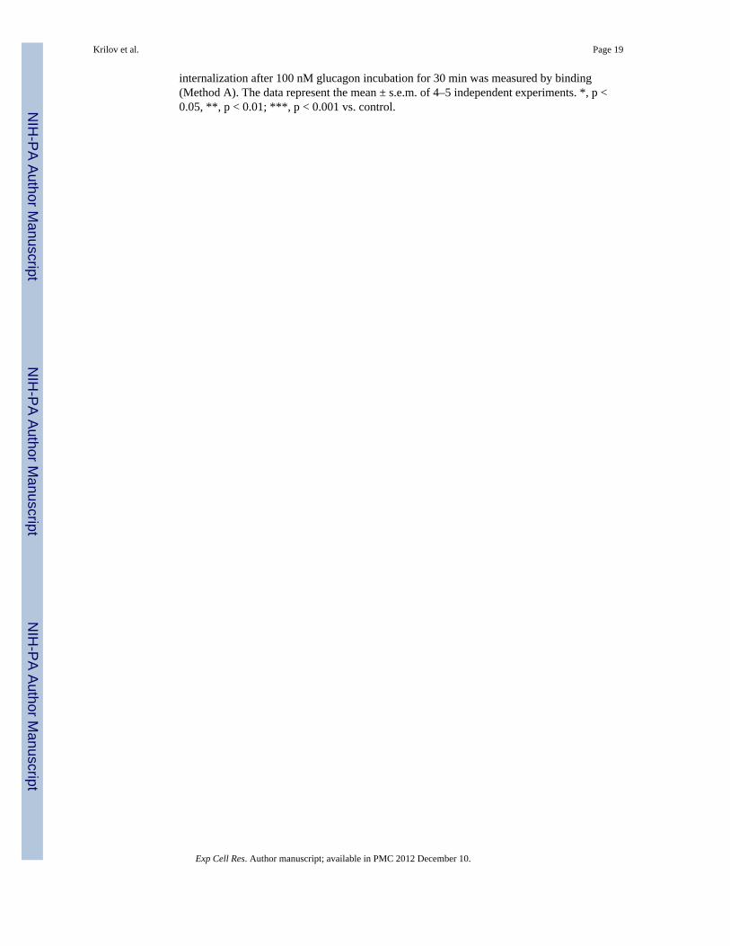
To confirm the involvement of caveolae in GR endocytosis, HEK-FLAG-GR cells weretreated with glucagon and immunostained with the anti-caveolin (Cav)-1 antibody (Figure4A and B). While in untreated cells Cav-1 colocalized with GR at the membrane, inglucagon-treated cells, it also colocalized with internalized GRs (Figure 4A). Furthermore,Cav-1 co-precipitated with GR when GR was immunoprecipitated using anti-FLAG, whichwe have previously shown to be specific for GR IP in HEK-FLAG-GR cells [14](Figure4B). The association transiently increased, with a maximum observed at 5 min of glucagonstimulation (Figure 4B). This association of the GR and Cav-1 returned to the basal level by20 min glucagon stimulation (Figure 4B).
Glucagon induces GR internalization and association of GR with PKCα and caveolin-1 inprimary hepatocytes
To investigate the physiological relevance of our findings, we performed radioligandbinding assays with 125I-glucagon in primary hamster hepatocytes. In these studies, 30-minute incubation with glucagon (0.1–160 nM) resulted in a concentration-dependentdecrease in cell surface GR (up to approximately 40%), indicating receptor internalization(Figure 5A). Hepatocytes were also incubated with 200 nM PMA in the presence or absenceof glucagon (Figure 5A). PMA increased glucagon-stimulated GR internalization from 40%to 70% (Figure 5A), consistent with our results in HEK-GR cells (Figure 1A–C). As wefound PKCα and Cav-1 translocation and association with the GR upon glucagonstimulation in HEK-GR cells (Figures 1E–F, 4A–B), we next examined whether Cav-1 andPKCα translocate to the membrane in hepatocytes upon glucagon stimulation. Uponincubation of the hepatocytes without and with either 100 nM glucagon or 200 nM PMA,the cytoplasmic and crude membrane fractions were separated and the membrane levels ofCav-1 and PKCα were determined by immunoblotting (Figure 5B). After a 5- to 10-minincubation of the cells with glucagon, the Cav-1 and PKCα membrane levels were increasedby 2- to 3-fold and 1.5- to 2.5-fold, respectively (Figure 5B–D). Further, incubation of thehepatocytes with glucagon for 30 and 60 min led to increased membrane levels of PKCα,PKCδ and Cav-1, and decreased cytoplasmic levels of PKCα (Figure 5E, 6 right-handlanes). Of note, Cav-1 was detectable in the cytoplasmic fraction only after an extendedexposure time of the chemiluminescent signal. The blots were also probed with an antibodyagainst the cytosolic marker glyceraldehyde-3-phosphate dehydrogenase (GAPDH) to assessthe purity of the cellular fractionation procedure, as has been reported by others [28,28]. Inorder to verify whether the accumulation of PKCα in the membrane leads to its interactionwith the GR, the receptor was immunoprecipitated from the crude membrane (CM) fraction.PKCα and β-actin, but not PKCδ or Cav-1, co-immunoprecipitated with GR (Figure 5E, 3
Krilov et al. Page 7
Exp Cell Res. Author manuscript; available in PMC 2012 December 10.
NIH
-PA Author Manuscript
NIH
-PA Author Manuscript
NIH
-PA Author Manuscript
left-hand lanes). In these experiments, PKCδ was examined to verify the specificity ofinvolvement of PKCα in co-immunoprecipitation of the GR. The association of GR withPKCα peaked 30 min after the addition of glucagon, whereas the association with β-actindecreased over time and was undetectable after 60 min. The latter is possibly due to β-actinand F-actin distribution changes occurring with glucagon treatment, as we have previouslyreported [14]. As in HEK-GR cells (Figure 1F), a moderate level of interaction betweenPKCα and GR was observed under basal conditions in hepatocytes (Figure 5E), which isconsistent with previous studies reporting the presence of PKCα in both the plasmamembrane and cytoplasmic fractions of these cells [17]. To further examine the kinetics ofGR association with PKCα in hepatocytes, we conducted an additional time-courseexperiment. After respective 20 and 30 min stimulation with 100 nM glucagon, theassociation of PKCα with GR reached a maximum of 40 and 70% above control (Figure 5F).
β-arrestins are involved in GR internalizationClathrin-mediated internalization of most GPCRs requires β-arrestins. We have recentlyreported that, also referred to as arrestin-2 and -3, respectively [29], are required forrecycling of GRs to the plasma membrane in HEK-GR cells [14]. Furthermore, co-transfection of dominant-negative constructs of β-arrestin1 and β-arrestin 2 decreased theamount of glucagon-stimulated GR internalization as compared to cells transfected with anempty vector. In the present study, to further examine their involvement in GRinternalization. β-arrestin1-GFP and β-arrestin2-GFP were transfected into HEK-FLAG-GRcells. Upon 5–20 min of glucagon treatment, colocalization of FLAG-GR with bothtransfected β-arrestins was observed at the plasma membrane level and in the cytoplasm(Figure 6A, arrows), suggesting that the interaction with β-arrestins is maintained afterreceptor internalization. It appeared that, at 10 and 20 min of glucagon treatment, β-arrestinsand GR colocalized predominantly in the perinuclear region (Figure 6A, arrows). Thisobservation was supported by labeling the nuclei with DAPI (Figure 6B, right panel). Time-lapse fluorescence microscopy in live HEK-GR cells transfected with β-arrestins indicatedthat both β-arrestin1-GFP and β-arrestin2-GFP translocated from the cytoplasm to theperinuclear region after treatment with glucagon (Figure 6B, left panels). Bindingexperiments were conducted to further assess the involvement of β-arrestin isoforms in GRinternalization (Figure 6C). GR internalization was significantly increased in cellstransfected with either β-arrestin1 WT or β-arrestin2 WT, as compared to cells transfectedwith an empty vector (EV). However, co-transfection of β-arrestin1 WT and β-arrestin2 WTdid not result in a further increase in GR internalization (Figure 6C).
The role of β-arrestins in caveolae-mediated internalization is unknown. Given that ourfindings indicated that both clathrin- and caveolin-mediated pathways are utilized by GR,we considered the possibility that β-arrestins could be a common component of the twointernalization routes. We observed colocalization of Cav-1 with both β-arrestin1 and β-arrestin2 by fluorescence microscopy at 10 and 20 min after treatment with glucagon for 10and 20 min (Figure 7A, arrows). Co-immunoprecipitation (Co-IP) experiments in total celllysates confirmed an interaction between β-arrestins and Cav-1. In these experiments,glucagon stimulation for 10 or 15 min induced a time-dependent 2- to 3-fold increase in Co-IP of Cav-1 with β-arrestin1 (Figure 7B). Similar results were observed in Co-IPexperiments with Cav-1 with β-arrestin2 following glucagon stimulation (2-fold increasefollowing 10 and 15 min stimulation; data not shown). As expected, Co-IP experiments withan IgG control did not result in appreciable detection of Cav-1 (Figure 7B).
Based on the results of our study, we propose the following hypothetical model for GRinternalization in GR-expressing HEK-293 cells (Figure 8). Upon treatment with glucagon,PKCα is recruited to the plasma membrane to facilitate GR desensitization andinternalization. GRK2, GRK3, and GRK5 may all play a role in GR desensitization. PKCα
Krilov et al. Page 8
Exp Cell Res. Author manuscript; available in PMC 2012 December 10.
NIH
-PA Author Manuscript
NIH
-PA Author Manuscript
NIH
-PA Author Manuscript
possibly facilitates recruitment of GRK2 to the membrane, as has been suggested in theliterature [24]. Desensitized GRs interact with β-arrestin1 and/or β-arrestin2, and thecomplexes are internalized via clathrin-coated pits and eventually targeted to earlyendosomes. Internalized receptors are recycled to the plasma membrane in a β-arrestin-dependent manner, as we have previously established [14]. Alternatively, GRs areinternalized through caveolae. The two internalization pathways are either utilized inparallel or the receptors shuttle from one pathway to the other. The possible point ofinteraction between the two endocytic routes might be vesicles in which β-arrestins andCav-1 colocalize.
DISCUSSIONIn the present study, we confirmed that the GR is internalized upon stimulation withglucagon in GR-transfected HEK-293 cells and in cultured hepatocytes. We found that GRutilizes both clathrin- and caveolin-mediated endocytosis in HEK-GR cells. Our observationthat both β-arrestin1 and β-arrestin2 colocalize with GR suggests that these β-arrestins areinvolved in GR internalization. The two arrestins also colocalized with caveolin-1 upontreatment of cells with glucagon. To our knowledge, this is the first reported putativeinteraction between the caveolin- and clathrin-mediated receptor internalization pathways.We have previously shown that PKC plays a role in GR phosphorylation and desensitization[19]. The results of the present study suggest that PKC also enhances glucagon-stimulatedGR internalization. GRKs are known to promote internalization of GPCRs throughenhancing β-arrestin recruitment and binding to clathrin, but their role in GR internalizationhas not been explored to date. We found that overexpression of either GRK2, GRK3, orGRK5 enhanced glucagon-stimulated GR internalization in HEK-GR cells, indicating thatthese three kinases may be important for GR internalization.
In both HEK-GR cells and hepatocytes, significant GR internalization was observed with 30min of glucagon (100 nM) stimulation. We and others have reported that PMA stimulatesGR desensitization in cultured hepatocytes as well as in HEK-293 cells [10], but the effectof PMA on GR internalization has not been addressed. In the present study we determinedthat PMA alone did not affect GR internalization, but it enhanced glucagon-stimulated GRinternalization in cultured hepatocytes. This phenomenon can be interpreted as a positivefeedback mechanism through PKC, which is activated through GR coupling to Gq. Anotherfeedback pathway could take place through coupling of GR to Gs and activation of PKA.Previous studies suggested that PKA does not play a role in GR desensitization [30]. Theseearlier findings are supported by the present study, in which PKA stimulation, mediated byadenylyl cyclase activation by forskolin, also does not affect GR internalization.
Our immunoblotting and time-lapse fluorescence microscopy studies demonstrated thatglucagon triggered the accumulation of PKCα in the plasma membrane of HEK-GR cellsand in the isolated crude membrane fraction of cultured hepatocytes. The significance of thistranslocation is twofold: first, it represents a step in the activation of PKCα mediated by theinteraction with membrane-bound phosphatidylserine and diacylglycerol, and second, itbrings PKCα into proximity of GR, allowing for interaction of the two proteins. Indeed, weverified that GR associated with PKCα under basal conditions in both HEK-GR cells andhepatocytes. The association transiently increased to a maximum observed 30 min afterstimulation with glucagon. In HEK-GR cells PMA alone also triggered translocation ofPKCα and its association with GR in the plasma membrane. Given that our binding assaysshow lack of effect of PMA on GR internalization, it appears that, in the absence ofglucagon, PKCα does not facilitate GR internalization.
Krilov et al. Page 9
Exp Cell Res. Author manuscript; available in PMC 2012 December 10.
NIH
-PA Author Manuscript
NIH
-PA Author Manuscript
NIH
-PA Author Manuscript
We observed no association of GR with PKCδ in hepatocytes, suggesting that PKCδ is notdirectly involved or associated with GR during glucagon stimulation. Furthermore, we alsodid not observe any increase in glucagon-stimulated GR internalization when PKCδ andPKCζ, representative of novel and classical PKCs, respectively, were overexpressed inHEK-GR cells (data not shown). At this time, we cannot rule out involvement of otherclassic and atypical PKC isoforms in GR internalization following glucagon treatment aloneor in combination with PMA.
GRKs are indispensable for homologous desensitization of GPCRs. Recent studies haveshown that GRKs not only phosphorylate receptors but also modulate signaling throughinteraction with numerous other proteins, including Gα, Gβγ, caveolin, clathrin, PI3K, MEKand Akt [24]. The effect of GRKs on GR desensitization has been addressed to a certainextent [31,32]. These reports examined the role of GRK2 in GR desensitization in the liverin vivo and the roles of GRK2 and GRK3 in PMA-mediated GR desensitization in vitro. Toour knowledge, this is the first report implicating the GRK2, GRK3 and GRK5 in GRinternalization.
Depending on the interplay between the receptor, cell type, receptor ligand, and the nature ofdesensitization, receptors may utilize multiple internalization pathways and even switchfrom one pathway to another during endocytosis. Most GPCRs utilize a clathrin-mediatedinternalization pathway, although usage of alternative pathways, such as internalizationthrough caveloae/lipid rafts, has been described [6,33] and may be associated with differentreceptor functions, e.g., promotion of cell signaling versus receptor degradation,respectively, as reported for the EGF receptor [34]. In the present study, pharmacologicaldisruption of clathrin-coated pits resulted in only partial inhibition of GR internalization inHEK-GR cells, suggesting that the receptor utilizes additional internalization routes. Itshould be noted that the use of pharmacological agents to characterize GR internalizationshould be interpreted with caution as they may lack selectivity and may affect membranestructure and function. Examined under a light microscope, the cell shape appeared normalafter pharmacological disruption of clathrin-coated pits. While the pharmacologicalinhibition regimens chosen in our study are widely used in the literature, hypertonic sucroseand PAO are considered non-selective inhibitors of clathrin-mediated endocytosis [35]. Thecholesterol-depleting agents Nystatin and Filipin III disrupt caveolae and have been utilizedto demonstrate the role of caveolae in GPCR internalization. A significant reduction of GRendocytosis/degradation in the presence of these drugs was observed.
We found that in HEK-FLAG-GR cells, GR colocalizes with Cav-1, a major constituent ofcaveolae, at the plasma membrane and, after stimulation with glucagon, in the cytoplasm. Asimilar interaction pattern with Cav-1 was previously observed for the structurally relatedglucagon-like peptide 1 (GLP-1) receptor expressed in HEK-293 cells [36]. Glucagontriggered recruitment of Cav-1 to the membrane in both hepatocytes and HEK-GR cells,with the same peak time of Cav-1 plasma membrane accumulation (5 min). Additionally, inco-immunoprecipitation studies in HEK-FLAG-GR cells, we found that GR associates withCav-1 in a time-dependent manner. However, under the conditions tested, co-immunoprecipitation of GR and Cav-1 was not found in hepatocytes. This discrepancy couldbe due to the use of different time points examined in HEK-GR cells (5, 10 and 20 min)versus hepatocytes (30 and 60 min) and different fractions analyzed (total lysate for HEKcells versus crude membrane for hepatocytes).
As a scaffolding protein, Cav-1 directly interacts with several signaling molecules, includingPKA, PKC, and Gαs [12,37]. At the plasma membrane, receptors that interact with caveolaeare often part of lipid rafts, which serve as distinct signaling platforms. Our results suggest,for the first time, that GR may be partitioned between lipid rafts and non-raft plasma
Krilov et al. Page 10
Exp Cell Res. Author manuscript; available in PMC 2012 December 10.
NIH
-PA Author Manuscript
NIH
-PA Author Manuscript
NIH
-PA Author Manuscript
membrane domains. Further studies are needed to assess the importance of putative lipid raftlocalization for downstream signaling, in particular ERK1/2 activation, as observed with theGLP-1R [36].
β-arrestins are required for clathrin-mediated internalization and many GPCRs aredesensitized and internalized in a β-arrestin-dependent manner [38]. Our results suggest thatGR interacts with both β-arrestin1 and β-arrestin2 in HEK-GR cells at the plasma membraneand in the cytoplasm. Of note, Merlen et al. [15] did not find evidence of β-arrestin1 or β-arrestin2 involvement in GR internalization in liver cells isolated from rats injected withglucagon. The authors suggested that clathrin-independent internalization pathways (such aslipid rafts) may be relevant in vivo. While there are earlier reports on the requirement for β-arrestins in clathrin-independent internalization [39,40], this is the first study to demonstratecolocalization of β-arrestin1 and β-arrestin2 with Cav-1. These results are intriguing,however, we recognize that β-arrestins cannot be definitively linked to caveolae-mediatedinternalization through colocalization and co-immunoprecipitation experiments alone. As anexample, desensitization, but not internalization, of the M2 muscarinic acetylcholinereceptor was found to require arrestins [41] Therefore, it is possible that the interaction ofcaveolin with β-arrestins occurs at the level of receptor desensitization. However, the factthat the proteins appear to colocalize in the cytosol, suggests that β-arrestins may indeed beinvolved in caveolae-mediated internalization, at least under conditions of β-arrestinoverexpression. We do recognize that it is plausible that alternate internalization pathwayspredominate under β-arrestin1 and β-arrestin2 basal expression levels, as has been seen forthe M2 muscarinic acetylcholine receptor [42].
In the present studies, β-arrestins rapidly accumulated in the perinuclear region in responseto glucagon treatment, which presumably denotes a recycling compartment, as reported forthe N-formyl peptide receptor [43]. Moreover, we recently reported that recycling of GR isabolished by downregulation of β-arrestin1 and β-arrestin2 [14]. Perinuclear targeting of β-arrestins may also be correlated with their ability to act as signaling scaffolds and facilitateactivation of ERK1/2 by glucagon, which has been reported to require clathrin-dependentGR internalization [44]. We also speculate that β-arrestins could mediate GR signaling thatis independent of G protein activation, as recently reported for the beta2-adrenergic receptor[38].
In conclusion, the present study demonstrates that GRK2, GRK3, GRK5, and PKCαstimulate agonist-induced GR internalization in HEK-GR cells. PKCα interacts with the GRin both hepatocytes and HEK-293 cells. Endocytosis of GRs proceeds through both clathrin-and caveolae- dependent pathways. However, further experiments are needed to clarifywhether the receptor shuttles between the two pathways, as has been observed for the AT1receptor [45,46] and the β2-adrenergic receptor [47]. GR internalization in HEK-GR cellsinvolves both β-arrestin1 and β-arrestin2. The intracellular components of GRdesensitization and internalization identified in this study represent potential drug targets fornovel diabetes therapies focused on antagonizing GR signaling.
AcknowledgmentsThe authors wish to thank Drs. Marianne David and Jianping Ming for their technical advice and assistance. We arealso grateful to Drs. Jeffrey L. Benovic, Marc Caron, Cornelius Krasel, Ralf Kubitz, and Jae-Won Soh, forproviding us with the specific plasmids. We thank the Center for Microscopy and Image Analysis, The GeorgeWashington University, for the use of the confocal microscope.
This work was supported by the National Institutes of Health, grant DK56108 to BB.
Krilov et al. Page 11
Exp Cell Res. Author manuscript; available in PMC 2012 December 10.
NIH
-PA Author Manuscript
NIH
-PA Author Manuscript
NIH
-PA Author Manuscript
References1. Reaven GM, Chen YD, Golay A, Swislocki AL, Jaspan JB. Documentation of hyperglucagonemia
throughout the day in nonobese and obese patients with noninsulin-dependent diabetes mellitus. JClin Endocrinol Metab. 1987; 64:106–110. [PubMed: 3536980]
2. Shah P, Vella A, Basu A, Basu R, Schwenk WF, Rizza RA. Lack of suppression of glucagoncontributes to postprandial hyperglycemia in subjects with type 2 diabetes mellitus. J ClinEndocrinol Metab. 2000; 85:4053–4059. [PubMed: 11095432]
3. Unger RH. Role of glucagon in the pathogenesis of diabetes: the status of the controversy.Metabolism: clinical and experimental. 1978; 27:1691–1709. [PubMed: 360007]
4. Petersen KF, Sullivan JT. Effects of a novel glucagon receptor antagonist (Bay 27-9955) onglucagon-stimulated glucose production in humans. Diabetologia. 2001; 44:2018–2024. [PubMed:11719833]
5. McCormack JG, Westergaard N, Kristiansen M, Brand CL, Lau J. Pharmacological approaches toinhibit endogenous glucose production as a means of anti-diabetic therapy. Curr Pharm Des. 2001;7:1451–1474. [PubMed: 11529255]
6. Ferguson SS. Evolving concepts in G protein-coupled receptor endocytosis: the role in receptordesensitization and signaling. Pharmacol Rev. 2001; 53:1–24. [PubMed: 11171937]
7. Moore CA, Milano SK, Benovic JL. Regulation of receptor trafficking by GRKs and arrestins. AnnuRev Physiol. 2007; 69:451–482. [PubMed: 17037978]
8. Dhami GK, Anborgh PH, Dale LB, Sterne-Marr R, Ferguson SS. Phosphorylation-independentregulation of metabotropic glutamate receptor signaling by G protein-coupled receptor kinase 2. JBiol Chem. 2002; 277:25266–25272. [PubMed: 12101219]
9. Freedman NJ, Ament AS, Oppermann M, Stoffel RH, Exum ST, Lefkowitz RJ. Phosphorylation anddesensitization of human endothelin A and B receptors. Evidence for G protein-coupled receptorkinase specificity. J Biol Chem. 1997; 272:17734–17743. [PubMed: 9211925]
10. Ikegami T, Krilov L, Meng J, Patel B, Chapin-Kennedy K, Bouscarel B. Decreased glucagonresponsiveness by bile acids: a role for protein kinase Calpha and glucagon receptorphosphorylation. Endocrinology. 2006; 147:5294–5302. [PubMed: 16916948]
11. Murphy GJ, Hruby VJ, Trivedi D, Wakelam MJ, Houslay MD. The rapid desensitization ofglucagon-stimulated adenylate cyclase is a cyclic AMP-independent process that can be mimickedby hormones which stimulate inositol phospholipid metabolism. Biochem J. 1987; 243:39–46.[PubMed: 3038085]
12. Chini B, Parenti M. G-protein coupled receptors in lipid rafts and caveolae: how, when and why dothey go there? J Mol Endocrinol. 2004; 32:325–338. [PubMed: 15072542]
13. Buggy JJ, Heurich RO, MacDougall M, Kelley KA, Livingston JN, Yoo-Warren H, RossomandoAJ. Role of the glucagon receptor COOH-terminal domain in glucagon-mediated signaling andreceptor internalization. Diabetes. 1997; 46:1400–1405. [PubMed: 9287038]
14. Krilov L, Nguyen A, Miyazaki T, Unson CG, Bouscarel B. Glucagon receptor recycling: role ofcarboxyl terminus, beta-arrestins, and cytoskeleton. Am J Physiol Cell Physiol. 2008; 295:C1230–C1237. [PubMed: 18787074]
15. Merlen C, Fabrega S, Desbuquois B, Unson CG, Authier F. Glucagon-mediated internalization ofserine-phosphorylated glucagon receptor and Gsalpha in rat liver. FEBS Letters. 2006; 580:5697–5704. [PubMed: 17010343]
16. Unson CG. Expression of glucagon receptors in tetracycline-inducible HEK293S GnT1- stable celllines: an approach toward purification of receptor protein for structural studies. Biopolymers.2008; 90:287–296. [PubMed: 18260137]
17. Bouscarel B, Gettys TW, Fromm H, Dubner H. Ursodeoxycholic acid inhibits glucagon-inducedcAMP formation in hamster hepatocytes: a role for PKC. Am J Physiol. 1995; 268:G300–G310.[PubMed: 7864127]
18. Ikegami T, Cypess AM, Bouscarel B. Modulation of glucagon receptor expression and response intransfected human embryonic kidney cells. Am J Physiol Cell Physiol. 2001; 281:C1396–C1402.[PubMed: 11546678]
Krilov et al. Page 12
Exp Cell Res. Author manuscript; available in PMC 2012 December 10.
NIH
-PA Author Manuscript
NIH
-PA Author Manuscript
NIH
-PA Author Manuscript
19. Tao YX, Segaloff DL. Functional characterization of melanocortin-4 receptor mutations associatedwith childhood obesity. Endocrinology. 2003; 144:4544–4551. [PubMed: 12959994]
20. Hipkin RW, Wang Y, Schonbrunn A. Protein kinase C activation stimulates the phosphorylationand internalization of the sst2A somatostatin receptor. J Biol Chem. 2000; 275:5591–5599.[PubMed: 10681540]
21. Innamorati G, Le Gouill C, Balamotis M, Birnbaumer M. The long and the short cycle. Alternativeintracellular routes for trafficking of G-protein-coupled receptors. J Biol Chem. 2001; 276:13096–13103. [PubMed: 11150299]
22. Winstel, R.; Freund, S.; Krasel, C.; Hoppe, E.; Lohse, MJ. Protein kinase cross-talk: membranetargeting of the beta-adrenergic receptor kinase by protein kinase C. Proceedings of the NationalAcademy of Sciences of the United States of America; 1996. p. 2105-2109.
23. Cong M, Perry SJ, Lin FT, Fraser ID, Hu LA, Chen W, Pitcher JA, Scott JD, Lefkowitz RJ.Regulation of membrane targeting of the G protein-coupled receptor kinase 2 by protein kinase Aand its anchoring protein AKAP79. J Biol Chem. 2001; 276:15192–15199. [PubMed: 11278469]
24. Ribas C, Penela P, Murga C, Salcedo A, Garcia-Hoz C, Jurado-Pueyo M, Aymerich I, Mayor F Jr.The G protein-coupled receptor kinase (GRK) interactome: role of GRKs in GPCR regulation andsignaling. Biochimica et Biophysica Acta. 2007; 1768:913–922. [PubMed: 17084806]
25. Shetzline MA, Walker JK, Valenzano KJ, Premont RT. Vasoactive intestinal polypeptide type-1receptor regulation. Desensitization, phosphorylation, and sequestration. J Biol Chem. 2002;277:25519–25526. [PubMed: 11978791]
26. Anis Y, Leshem O, Reuveni H, Wexler I, Ben Sasson R, Yahalom B, Laster M, Raz I, Ben SassonS, Shafrir E, Ziv E. Antidiabetic effect of novel modulating peptides of G-protein-coupled kinasein experimental models of diabetes. Diabetologia. 2004; 47:1232–1244. [PubMed: 15235770]
27. Prevostel C, Alice V, Joubert D, Parker PJ. Protein kinase C(alpha) actively downregulates throughcaveolae-dependent traffic to an endosomal compartment. J Cell Sci. 2000; 113 ( Pt 14):2575–2584. [PubMed: 10862715]
28. Ronnebaum SM, Ilkayeva O, Burgess SC, Joseph JW, Lu D, Stevens RD, Becker TC, Sherry AD,Newgard CB, Jensen MV. A pyruvate cycling pathway involving cytosolic NADP-dependentisocitrate dehydrogenase regulates glucose-stimulated insulin secretion. J Biol Chem. 2006;281:30593–30602. [PubMed: 16912049]
29. Kim YM, Benovic JL. Differential roles of arrestin-2 interaction with clathrin and adaptor protein2 in G protein-coupled receptor trafficking. J Biol Chem. 2002; 277:30760–30768. [PubMed:12070169]
30. Heurich RO, Buggy JJ, Vandenberg MT, Rossomando AJ. Glucagon induces a rapid and sustainedphosphorylation of the human glucagon receptor in Chinese hamster ovary cells. BiochemBiophys Res Commun. 1996; 220:905–910. [PubMed: 8607865]
31. Charbonneau A, Unson CG, Lavoie JM. High-fat diet-induced hepatic steatosis reduces glucagonreceptor content in rat hepatocytes: potential interaction with acute exercise. J Physiol. 2007;579:255–267. [PubMed: 17053032]
32. Tobias ES, Rozengurt E, Connell JM, Houslay MD. Co-transfection with protein kinase D confersphorbol-ester-mediated inhibition on glucagon-stimulated cAMP accumulation in COS cellstransfected to overexpress glucagon receptors. Biochem J. 1997; 326 ( Pt 2):545–551. [PubMed:9291130]
33. Doherty GJ, McMahon HT. Mechanisms of endocytosis. Annu Rev Biochem. 2009; 78:857–902.[PubMed: 19317650]
34. Sigismund S, Argenzio E, Tosoni D, Cavallaro E, Polo S, Di Fiore PP. Clathrin-mediatedinternalization is essential for sustained EGFR signaling but dispensable for degradation. Dev Cell.2008; 15:209–219. [PubMed: 18694561]
35. Ivanov AI. Pharmacological inhibition of endocytic pathways: is it specific enough to be useful?Methods Mol Biol. 2008; 440:15–33. [PubMed: 18369934]
36. Syme G, Rowe P, Martin D, Daly G. Disability in patients with chronic patellofemoral painsyndrome: a randomised controlled trial of VMO selective training versus general quadricepsstrengthening. Man Ther. 2009; 14:252–263. [PubMed: 18436468]
Krilov et al. Page 13
Exp Cell Res. Author manuscript; available in PMC 2012 December 10.
NIH
-PA Author Manuscript
NIH
-PA Author Manuscript
NIH
-PA Author Manuscript
37. Cohen AW, Hnasko R, Schubert W, Lisanti MP. Role of caveolae and caveolins in health anddisease. Physiol Rev. 2004; 84:1341–1379. [PubMed: 15383654]
38. Shenoy SK, Drake MT, Nelson CD, Houtz DA, Xiao K, Madabushi S, Reiter E, Premont RT,Lichtarge O, Lefkowitz RJ. beta-arrestin-dependent, G protein-independent ERK1/2 activation bythe beta2 adrenergic receptor. J Biol Chem. 2006; 281:1261–1273. [PubMed: 16280323]
39. Kohout TA, Lin FS, Perry SJ, Conner DA, Lefkowitz RJ. beta-Arrestin 1 and 2 differentiallyregulate heptahelical receptor signaling and trafficking. Proceedings of the National Academy ofSciences of the United States of America. 2001; 98:1601–1606. [PubMed: 11171997]
40. Zhang J, Ferguson SS, Barak LS, Menard L, Caron MG. Dynamin and beta-arrestin reveal distinctmechanisms for G protein-coupled receptor internalization. J Biol Chem. 1996; 271:18302–18305.[PubMed: 8702465]
41. Pals-Rylaarsdam R, Gurevich VV, Lee KB, Ptasienski JA, Benovic JL, Hosey MM. Internalizationof the m2 muscarinic acetylcholine receptor. Arrestin-independent and -dependent pathways. JBiol Chem. 1997; 272:23682–23689. [PubMed: 9295310]
42. Lee KB, Ptasienski JA, Pals-Rylaarsdam R, Gurevich VV, Hosey MM. Arrestin binding to theM(2) muscarinic acetylcholine receptor is precluded by an inhibitory element in the thirdintracellular loop of the receptor. J Biol Chem. 2000; 275:9284–9289. [PubMed: 10734068]
43. Vines CM, Revankar CM, Maestas DC, LaRusch LL, Cimino DF, Kohout TA, Lefkowitz RJ,Prossnitz ER. N-formyl peptide receptors internalize but do not recycle in the absence of arrestins.J Biol Chem. 2003; 278:41581–41584. [PubMed: 12947104]
44. Dalle S, Longuet C, Costes S, Broca C, Faruque O, Fontes G, Hani EH, Bataille D. Glucagonpromotes cAMP-response element-binding protein phosphorylation via activation of ERK1/2 inMIN6 cell line and isolated islets of Langerhans. J Biol Chem. 2004; 279:20345–20355. [PubMed:14988413]
45. Ishizaka N, Griendling KK, Lassegue B, Alexander RW. Angiotensin II type 1 receptor:relationship with caveolae and caveolin after initial agonist stimulation. Hypertension. 1998;32:459–466. [PubMed: 9740611]
46. Wyse BD, Prior IA, Qian H, Morrow IC, Nixon S, Muncke C, Kurzchalia TV, Thomas WG,Parton RG, Hancock JF. Caveolin interacts with the angiotensin II type 1 receptor during exocytictransport but not at the plasma membrane. J Biol Chem. 2003; 278:23738–23746. [PubMed:12692121]
47. Rybin VO, Xu X, Lisanti MP, Steinberg SF. Differential targeting of beta -adrenergic receptorsubtypes and adenylyl cyclase to cardiomyocyte caveolae. A mechanism to functionally regulatethe cAMP signaling pathway. J Biol Chem. 2000; 275:41447–41457. [PubMed: 11006286]
Krilov et al. Page 14
Exp Cell Res. Author manuscript; available in PMC 2012 December 10.
NIH
-PA Author Manuscript
NIH
-PA Author Manuscript
NIH
-PA Author Manuscript
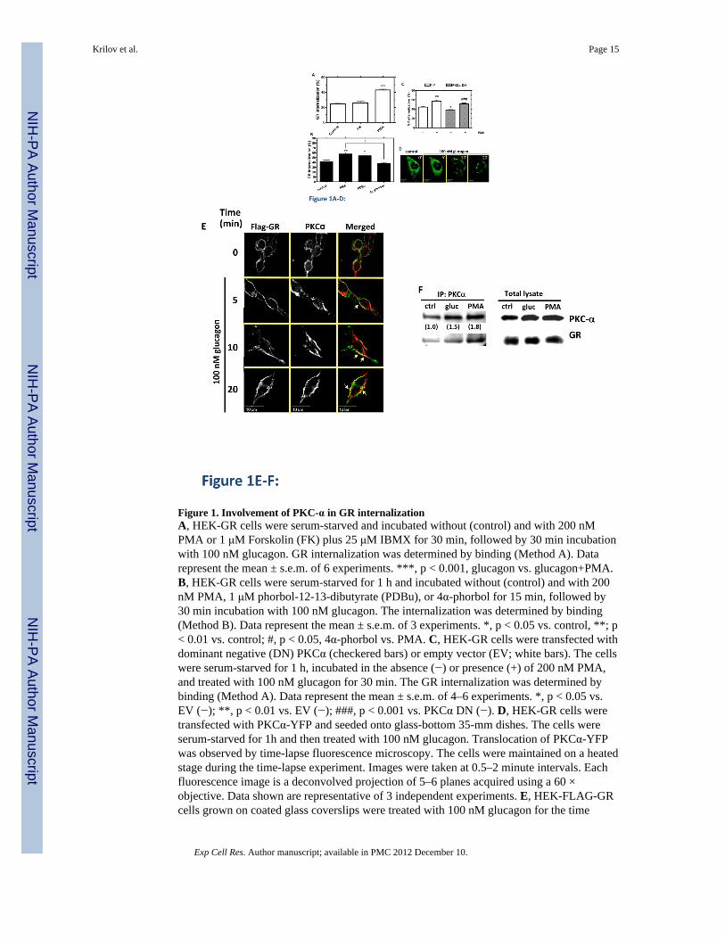
Figure 1. Involvement of PKC-α in GR internalizationA, HEK-GR cells were serum-starved and incubated without (control) and with 200 nMPMA or 1 μM Forskolin (FK) plus 25 μM IBMX for 30 min, followed by 30 min incubationwith 100 nM glucagon. GR internalization was determined by binding (Method A). Datarepresent the mean ± s.e.m. of 6 experiments. ***, p < 0.001, glucagon vs. glucagon+PMA.B, HEK-GR cells were serum-starved for 1 h and incubated without (control) and with 200nM PMA, 1 μM phorbol-12-13-dibutyrate (PDBu), or 4α-phorbol for 15 min, followed by30 min incubation with 100 nM glucagon. The internalization was determined by binding(Method B). Data represent the mean ± s.e.m. of 3 experiments. *, p < 0.05 vs. control, **; p< 0.01 vs. control; #, p < 0.05, 4α-phorbol vs. PMA. C, HEK-GR cells were transfected withdominant negative (DN) PKCα (checkered bars) or empty vector (EV; white bars). The cellswere serum-starved for 1 h, incubated in the absence (−) or presence (+) of 200 nM PMA,and treated with 100 nM glucagon for 30 min. The GR internalization was determined bybinding (Method A). Data represent the mean ± s.e.m. of 4–6 experiments. *, p < 0.05 vs.EV (−); **, p < 0.01 vs. EV (−); ###, p < 0.001 vs. PKCα DN (−). D, HEK-GR cells weretransfected with PKCα-YFP and seeded onto glass-bottom 35-mm dishes. The cells wereserum-starved for 1h and then treated with 100 nM glucagon. Translocation of PKCα-YFPwas observed by time-lapse fluorescence microscopy. The cells were maintained on a heatedstage during the time-lapse experiment. Images were taken at 0.5–2 minute intervals. Eachfluorescence image is a deconvolved projection of 5–6 planes acquired using a 60 ×objective. Data shown are representative of 3 independent experiments. E, HEK-FLAG-GRcells grown on coated glass coverslips were treated with 100 nM glucagon for the time
Krilov et al. Page 15
Exp Cell Res. Author manuscript; available in PMC 2012 December 10.
NIH
-PA Author Manuscript
NIH
-PA Author Manuscript
NIH
-PA Author Manuscript
indicated. The cells were then fixed, permeabilized and immunostained with anti-FLAG(green) and anti-PKCα (red) antibodies. Each fluorescence image is a deconvolvedprojection of 5–6 planes acquired using a 60 × objective. Representative images from 3independent experiments are shown. Arrows indicate colocalization. F, HEK-FLAG-GRcells were incubated without (ctrl) or with either 100 nM glucagon (gluc) or 200 nM PMAfor 30 min. Immunoprecipitation of PKCα and immunoblotting were performed as describedin Materials and Methods. The mean values within parentheses represent the fold increaseabove control determined in the absence of either glucagon or PMA. The value for controlwas arbitrarily set as 1. The image shown is representative of 3 independent experiments.
Krilov et al. Page 16
Exp Cell Res. Author manuscript; available in PMC 2012 December 10.
NIH
-PA Author Manuscript
NIH
-PA Author Manuscript
NIH
-PA Author Manuscript
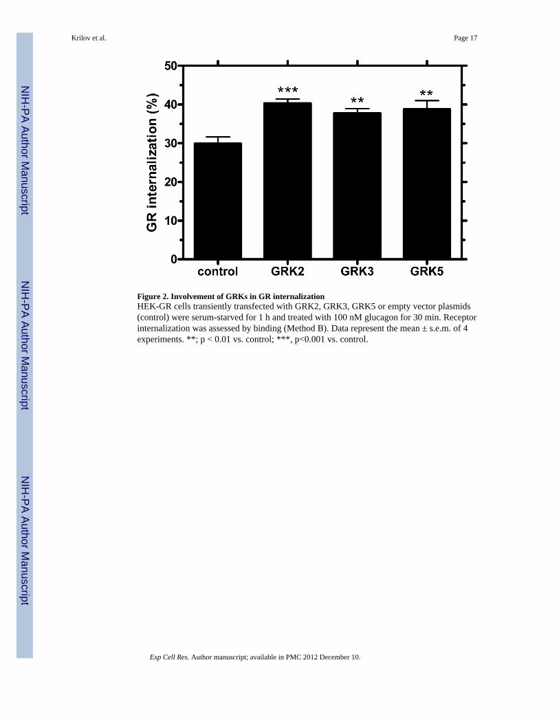
Figure 2. Involvement of GRKs in GR internalizationHEK-GR cells transiently transfected with GRK2, GRK3, GRK5 or empty vector plasmids(control) were serum-starved for 1 h and treated with 100 nM glucagon for 30 min. Receptorinternalization was assessed by binding (Method B). Data represent the mean ± s.e.m. of 4experiments. **; p < 0.01 vs. control; ***, p<0.001 vs. control.
Krilov et al. Page 17
Exp Cell Res. Author manuscript; available in PMC 2012 December 10.
NIH
-PA Author Manuscript
NIH
-PA Author Manuscript
NIH
-PA Author Manuscript
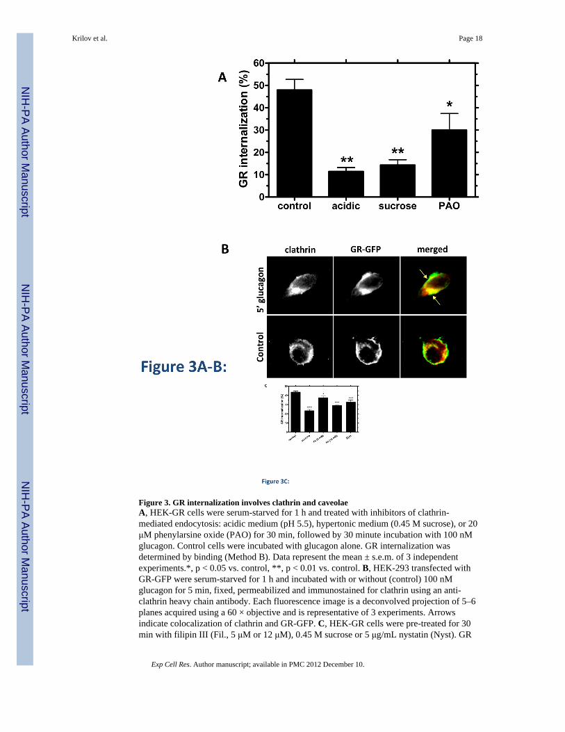
Figure 3. GR internalization involves clathrin and caveolaeA, HEK-GR cells were serum-starved for 1 h and treated with inhibitors of clathrin-mediated endocytosis: acidic medium (pH 5.5), hypertonic medium (0.45 M sucrose), or 20μM phenylarsine oxide (PAO) for 30 min, followed by 30 minute incubation with 100 nMglucagon. Control cells were incubated with glucagon alone. GR internalization wasdetermined by binding (Method B). Data represent the mean ± s.e.m. of 3 independentexperiments.*, p < 0.05 vs. control, **, p < 0.01 vs. control. B, HEK-293 transfected withGR-GFP were serum-starved for 1 h and incubated with or without (control) 100 nMglucagon for 5 min, fixed, permeabilized and immunostained for clathrin using an anti-clathrin heavy chain antibody. Each fluorescence image is a deconvolved projection of 5–6planes acquired using a 60 × objective and is representative of 3 experiments. Arrowsindicate colocalization of clathrin and GR-GFP. C, HEK-GR cells were pre-treated for 30min with filipin III (Fil., 5 μM or 12 μM), 0.45 M sucrose or 5 μg/mL nystatin (Nyst). GR
Krilov et al. Page 18
Exp Cell Res. Author manuscript; available in PMC 2012 December 10.
NIH
-PA Author Manuscript
NIH
-PA Author Manuscript
NIH
-PA Author Manuscript
internalization after 100 nM glucagon incubation for 30 min was measured by binding(Method A). The data represent the mean ± s.e.m. of 4–5 independent experiments. *, p <0.05, **, p < 0.01; ***, p < 0.001 vs. control.
Krilov et al. Page 19
Exp Cell Res. Author manuscript; available in PMC 2012 December 10.
NIH
-PA Author Manuscript
NIH
-PA Author Manuscript
NIH
-PA Author Manuscript
Figure 4. GR associates with caveolin-1A, HEK-FLAG-GR cells were serum-starved for 1 h and treated with 100 nM glucagon forthe time indicated, fixed, permeabilized and immunostained for GR using anti-FLAGantibody (FLAG-GR, green) and Cav-1 using the anti-Cav-1 antibody (cav-1, red). Eachfluorescence image is a deconvolved projection of 5–6 planes acquired using a 60 ×objective. Representative images of 3 independent experiments are shown. Arrows indicatecolocalization of caveolin and FLAG-GR. B, HEK-FLAG-GR were treated with 100 nMglucagon for 0, 5, 10, or 20 min. FLAG-GR was immunoprecipitated using the anti-FLAGantibody. The immunoprecipitated proteins were analyzed by immunoblotting with the anti-GR antibody and the anti-Cav-1 antibody, respectively. The image shown is representativeof 3 independent experiments.
Krilov et al. Page 20
Exp Cell Res. Author manuscript; available in PMC 2012 December 10.
NIH
-PA Author Manuscript
NIH
-PA Author Manuscript
NIH
-PA Author Manuscript
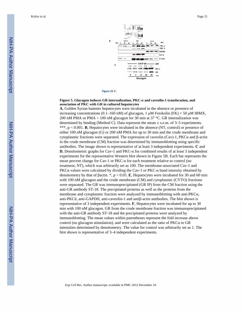
Figure 5. Glucagon induces GR internalization, PKC-α and caveolin-1 translocation, andassociation of PKC with GR in cultured hepatocytesA, Golden Syrian hamster hepatocytes were incubated in the absence or presence ofincreasing concentrations (0.1–160 nM) of glucagon, 1 μM Forskolin (FK) + 50 μM IBMX,200 nM PMA or PMA + 100 nM glucagon for 30 min at 37 °C. GR internalization wasdetermined by binding (Method C). Data represent the mean ± s.e.m. of 3–5 experiments.***, p < 0.001. B, Hepatocytes were incubated in the absence (NT, control) or presence ofeither 100 nM glucagon (G) or 200 nM PMA for up to 30 min and the crude membrane andcytoplasmic fractions were separated. The expression of caveolin (Cav)-1, PKCα and β-actinin the crude membrane (CM) fraction was determined by immunoblotting using specificantibodies. The image shown is representative of at least 3 independent experiments. C andD, Densitometric graphs for Cav-1 and PKC-α for combined results of at least 3 independentexperiments for the representative Western blot shown in Figure 5B. Each bar represents themean percent change for Cav-1 or PKC-α for each treatment relative to control (notreatment, NT), which was arbitrarily set as 100. The membrane-associated Cav-1 andPKCα values were calculated by dividing the Cav-1 or PKC-α band intensity obtained bydensitometry by that of βactin. *, p < 0.05. E, Hepatocytes were incubated for 30 and 60 minwith 100 nM glucagon and the crude membrane (CM) and cytoplasmic (CYTO) fractionswere separated. The GR was immunoprecipitated (GR IP) from the CM fraction using theanti-GR antibody ST-18. The precipitated proteins as well as the proteins from themembrane and cytoplasmic fraction were analyzed by immunoblotting with anti-PKCα,anti-PKCδ, anti-GAPDH, anti-caveolin-1 and antiβ-actin antibodies. The blot shown isrepresentative of 3 independent experiments. F, Hepatocytes were incubated for up to 30min with 100 nM glucagon. GR from the crude membrane fraction was immunoprecipitatedwith the anti-GR antibody ST-18 and the precipitated proteins were analyzed byimmunoblotting. The mean values within parentheses represent the fold increase abovecontrol (no glucagon stimulation), and were calculated as the ratio of PKCα to GRintensities determined by densitometry. The value for control was arbitrarily set as 1. Theblot shown is representative of 3–4 independent experiments.
Krilov et al. Page 21
Exp Cell Res. Author manuscript; available in PMC 2012 December 10.
NIH
-PA Author Manuscript
NIH
-PA Author Manuscript
NIH
-PA Author Manuscript
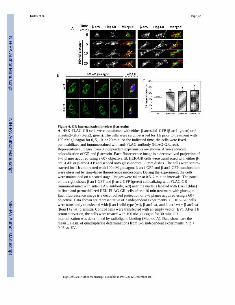
Figure 6. GR internalization involves β-arrestinsA, HEK-FLAG-GR cells were transfected with either β-arrestin1-GFP (β-arr1, green) or β-arrestin2-GFP (β-arr2, green). The cells were serum-starved for 1 h prior to treatment with100 nM glucagon for 0, 5, 10, or 20 min. At the indicated time, the cells were fixed,permeabilized and immunostained with anti-FLAG antibody (FLAG-GR, red).Representative images from 3 independent experiments are shown. Arrows indicatecolocalization of GR and β-arrestin. Each fluorescence image is a deconvolved projection of5–6 planes acquired using a 60× objective. B, HEK-GR cells were transfected with either β-arr1-GFP or β-arr2-GFP and seeded onto glass-bottom 35 mm dishes. The cells were serum-starved for 1 h and treated with 100 nM glucagon. β-arr1-GFP and β-arr2-GFP translocationwere observed by time-lapse fluorescence microscopy. During the experiment, the cellswere maintained on a heated stage. Images were taken at 0.5–2 minute intervals. The panelon the right shows β-arr1-GFP and β-arr2-GFP (green) colocalizing with FLAG-GR(immunostained with anti-FLAG antibody, red) near the nucleus labeled with DAPI (blue)in fixed and permeabilized HEK-FLAG-GR cells after a 10 min treatment with glucagon.Each fluorescence image is a deconvolved projection of 5–6 planes acquired using a 60×objective. Data shown are representative of 3 independent experiments. C, HEK-GR cellswere transiently transfected with β-arr1 wild type (wt), β-arr2 wt, and β-arr1 wt + β-arr2 wt(β-arr1+2 wt) plasmids. Control cells were transfected with an empty vector (EV). After 1 hserum starvation, the cells were treated with 100 nM glucagon for 30 min. GRinternalization was determined by radioligand binding (Method A). Data shown are themean ± s.e.m. of quadruplicate determinations from 3–5 independent experiments. *, p <0.05 vs. EV.
Krilov et al. Page 22
Exp Cell Res. Author manuscript; available in PMC 2012 December 10.
NIH
-PA Author Manuscript
NIH
-PA Author Manuscript
NIH
-PA Author Manuscript
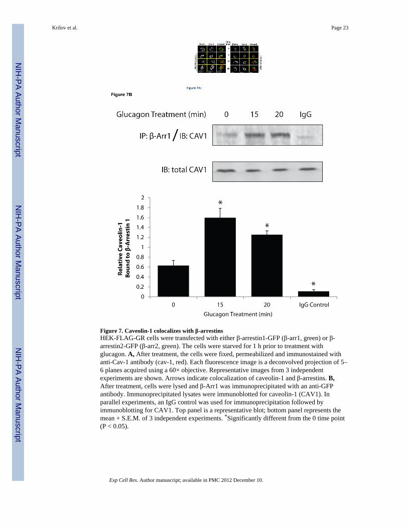
Figure 7. Caveolin-1 colocalizes with β-arrestinsHEK-FLAG-GR cells were transfected with either β-arrestin1-GFP (β-arr1, green) or β-arrestin2-GFP (β-arr2, green). The cells were starved for 1 h prior to treatment withglucagon. A, After treatment, the cells were fixed, permeabilized and immunostained withanti-Cav-1 antibody (cav-1, red). Each fluorescence image is a deconvolved projection of 5–6 planes acquired using a 60× objective. Representative images from 3 independentexperiments are shown. Arrows indicate colocalization of caveolin-1 and β-arrestins. B,After treatment, cells were lysed and β-Arr1 was immunoprecipitated with an anti-GFPantibody. Immunoprecipitated lysates were immunoblotted for caveolin-1 (CAV1). Inparallel experiments, an IgG control was used for immunoprecipitation followed byimmunoblotting for CAV1. Top panel is a representative blot; bottom panel represents themean + S.E.M. of 3 independent experiments. *Significantly different from the 0 time point(P < 0.05).
Krilov et al. Page 23
Exp Cell Res. Author manuscript; available in PMC 2012 December 10.
NIH
-PA Author Manuscript
NIH
-PA Author Manuscript
NIH
-PA Author Manuscript
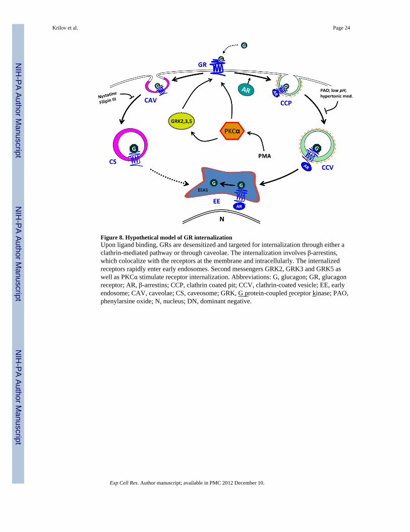
Figure 8. Hypothetical model of GR internalizationUpon ligand binding, GRs are desensitized and targeted for internalization through either aclathrin-mediated pathway or through caveolae. The internalization involves β-arrestins,which colocalize with the receptors at the membrane and intracellularly. The internalizedreceptors rapidly enter early endosomes. Second messengers GRK2, GRK3 and GRK5 aswell as PKCα stimulate receptor internalization. Abbreviations: G, glucagon; GR, glucagonreceptor; AR, β-arrestins; CCP, clathrin coated pit; CCV, clathrin-coated vesicle; EE, earlyendosome; CAV, caveolae; CS, caveosome; GRK, G protein-coupled receptor kinase; PAO,phenylarsine oxide; N, nucleus; DN, dominant negative.
Krilov et al. Page 24
Exp Cell Res. Author manuscript; available in PMC 2012 December 10.
NIH
-PA Author Manuscript
NIH
-PA Author Manuscript
NIH
-PA Author Manuscript


![Ruth Knight presentation.ppt [Compatibility Mode]](https://static.fdokumen.com/doc/165x107/631d5d013ba403638902baaf/ruth-knight-presentationppt-compatibility-mode.jpg)


![Stephen Briggs [Compatibility Mode]](https://static.fdokumen.com/doc/165x107/6324c3005c2c3bbfa802dd10/stephen-briggs-compatibility-mode.jpg)


