Glucagon-like peptide 1 (GLP-1) - Research Collection
60
Research Collection Review Article Glucagon-like peptide 1 (GLP-1) Author(s): Müller, Timo D.; Finan, Brian; Bloom, S.R.; D'Alessio, David; Drucker, Daniel J.; Flatt, P.R.; Gribble, Fiona; Grill, Harvey J.; Habener, Joel F.; Holst, Jens J.; Langhans, Wolfgang; Meier, Juris J.; Nauck, Michael A.; Perez-Tilve, Diego; Pocai, A.; Reimann, Frank; Sandoval, Darleen A.; Schwartz, Thue W.; Seeley, Randy J.; Stemmer, Kerstin; Tang-Christensen, Mads; Woods, Stephen C. ; DiMarchi, Richard D.; Tschöp, Matthias H. Publication Date: 2019-12 Permanent Link: https://doi.org/10.3929/ethz-b-000371733 Originally published in: Molecular Metabolism 30, http://doi.org/10.1016/j.molmet.2019.09.010 Rights / License: Creative Commons Attribution-NonCommercial-NoDerivatives 4.0 International This page was generated automatically upon download from the ETH Zurich Research Collection . For more information please consult the Terms of use . ETH Library
-
Upload
khangminh22 -
Category
Documents
-
view
3 -
download
0
Transcript of Glucagon-like peptide 1 (GLP-1) - Research Collection
Research Collection
Review Article
Glucagon-like peptide 1 (GLP-1)
Author(s): Müller, Timo D.; Finan, Brian; Bloom, S.R.; D'Alessio, David; Drucker, Daniel J.; Flatt, P.R.; Gribble, Fiona;Grill, Harvey J.; Habener, Joel F.; Holst, Jens J.; Langhans, Wolfgang; Meier, Juris J.; Nauck, Michael A.;Perez-Tilve, Diego; Pocai, A.; Reimann, Frank; Sandoval, Darleen A.; Schwartz, Thue W.; Seeley, Randy J.;Stemmer, Kerstin; Tang-Christensen, Mads; Woods, Stephen C. ; DiMarchi, Richard D.; Tschöp, MatthiasH.
Publication Date: 2019-12
Permanent Link: https://doi.org/10.3929/ethz-b-000371733
Originally published in: Molecular Metabolism 30, http://doi.org/10.1016/j.molmet.2019.09.010
Rights / License: Creative Commons Attribution-NonCommercial-NoDerivatives 4.0 International
This page was generated automatically upon download from the ETH Zurich Research Collection. For moreinformation please consult the Terms of use.
ETH Library
Glucagon-like peptide 1 (GLP-1)
T.D. Müller 1,2,3,*, B. Finan 4, S.R. Bloom 5, D. D’Alessio 6, D.J. Drucker 7, P.R. Flatt 8, A. Fritsche 2,9,10,F. Gribble 11, H.J. Grill 12, J.F. Habener 13, J.J. Holst 14, W. Langhans 15, J.J. Meier 16, M.A. Nauck 17,D. Perez-Tilve 18, A. Pocai 19, F. Reimann 11, D.A. Sandoval 20, T.W. Schwartz 21,22, R.J. Seeley 20,K. Stemmer 1,2, M. Tang-Christensen 23, S.C. Woods 24, R.D. DiMarchi 4,25, M.H. Tschöp 2,26,27
ABSTRACT
Background: The glucagon-like peptide-1 (GLP-1) is a multifaceted hormone with broad pharmacological potential. Among the numerousmetabolic effects of GLP-1 are the glucose-dependent stimulation of insulin secretion, decrease of gastric emptying, inhibition of food intake,increase of natriuresis and diuresis, and modulation of rodent b-cell proliferation. GLP-1 also has cardio- and neuroprotective effects, decreasesinflammation and apoptosis, and has implications for learning and memory, reward behavior, and palatability. Biochemically modified forenhanced potency and sustained action, GLP-1 receptor agonists are successfully in clinical use for the treatment of type-2 diabetes, and severalGLP-1-based pharmacotherapies are in clinical evaluation for the treatment of obesity.Scope of review: In this review, we provide a detailed overview on the multifaceted nature of GLP-1 and its pharmacology and discuss itstherapeutic implications on various diseases.Major conclusions: Since its discovery, GLP-1 has emerged as a pleiotropic hormone with a myriad of metabolic functions that go well beyondits classical identification as an incretin hormone. The numerous beneficial effects of GLP-1 render this hormone an interesting candidate for thedevelopment of pharmacotherapies to treat obesity, diabetes, and neurodegenerative disorders
� 2019 The Authors. Published by Elsevier GmbH. This is an open access article under the CC BY-NC-ND license (http://creativecommons.org/licenses/by-nc-nd/4.0/).
Keywords GLP-1; Insulin; Glucagon; Diabetes; Obesity; Incretin
1. FROM THE DISCOVERY OF INSULIN TO THE DISCOVERY OFGLP-1
Maintenance of adequate glucose metabolism is a prerequisite forhuman health, and pathological failure to buffer against prolongedepisodes of hypo- and/or hyperglycemia can result in severe micro-vascular disease, metabolic damage, coma, and death. Unsurprisingly,before the discovery and commercialization of insulin in the 1920’s,juvenile-onset diabetes, with its paucity of endogenous insulin, was a
disease with only a few years between a patient’s diagnosis andpremature demise. The discovery of insulin and its ability to lowerblood glucose transformed juvenile-onset (type-1) diabetes from a fatalto a manageable disease. However, early on, it was noted that insulinderived from pancreatic extracts [1] or as crude insulin preparations [2]sometimes first elevated blood glucose and then later decreased bloodglucose levels. The increase in blood glucose, which peaked around20 min after the administration, was believed to be caused by a toxicfraction resulting from suboptimal insulin purification [2]. The same
1Institute for Diabetes and Obesity, Helmholtz Diabetes Center, Helmholtz Zentrum München, German Research Center for Environmental Health (GmbH), Neuherberg,Germany 2German Center for Diabetes Research (DZD), Neuherberg, Germany 3Department of Pharmacology and Experimental Therapy, Institute of Experimental andClinical Pharmacology and Toxicology, Eberhard Karls University Hospitals and Clinics, Tübingen, Germany 4Novo Nordisk Research Center Indianapolis, Indianapolis, IN,USA 5Division of Diabetes, Endocrinology and Metabolism, Imperial College London, London, UK 6Division of Endocrinology, Duke University Medical Center, Durham, NC,USA 7The Department of Medicine, Lunenfeld-Tanenbaum Research Institute, Mt. Sinai Hospital, University of Toronto, Ontario, M5G1X5, Canada 8SAAD Centre for Pharmacy& Diabetes, Ulster University, Coleraine, Northern Ireland, UK 9Institute for Diabetes Research and Metabolic Diseases of the Helmholtz Center Munich at the University ofTübingen, Tübingen, Germany 10Division of Endocrinology, Diabetology, Vascular Disease, Nephrology and Clinical Chemistry, Department of Internal Medicine, University ofTübingen, Tübingen, Germany 11Metabolic Research Laboratories and Medical Research Council Metabolic Diseases Unit, Wellcome Trust-Medical Research Council,Institute of Metabolic Science, Addenbrooke’s Hospital, University of Cambridge, Cambridge, CB2 0QQ, UK 12Institute of Diabetes, Obesity and Metabolism, Department ofPsychology, University of Pennsylvania, Philadelphia, PA, 19104, USA 13Laboratory of Molecular Endocrinology, Massachusetts General Hospital, Harvard University, Boston,MA, USA 14Novo Nordisk Foundation Center for Basic Metabolic Research, Department of Biomedical Sciences, University of Copenhagen, Copenhagen,Denmark 15Physiology and Behavior Laboratory, ETH Zurich, Schwerzenbach, Switzerland 16Diabetes Division, St Josef Hospital, Ruhr-University Bochum, Bochum,Germany 17Diabetes Center Bochum-Hattingen, St Josef Hospital (Ruhr-Universität Bochum), Bochum, Germany 18Department of Internal Medicine, University of Cincinnati-College of Medicine, Cincinnati, OH, USA 19Cardiovascular & ImmunoMetabolism, Janssen Research & Development, Welsh and McKean Roads, Spring House, PA, 19477,USA 20Department of Surgery, University of Michigan Medical School, Ann Arbor, MI, USA 21Novo Nordisk Foundation Center for Basic Metabolic Research, University ofCopenhagen, DL-2200, Copenhagen, Denmark 22Department of Biomedical Sciences, University of Copenhagen, DK-2200, Copenhagen, Denmark 23Obesity Research,Global Drug Discovery, Novo Nordisk A/S, Måløv, Denmark 24Department of Psychiatry and Behavioral Neuroscience, University of Cincinnati, Cincinnati, OH,USA 25Department of Chemistry, Indiana University, Bloomington, IN, USA 26Division of Metabolic Diseases, Department of Medicine, Technische Universität München,Munich, Germany 27Helmholtz Zentrum München, German Research Center for Environmental Health (GmbH), Neuherberg, Germany
*Corresponding author. Institute for Diabetes and Obesity, Helmholtz Diabetes Center, Helmholtz Zentrum München, German Research Center for Environmental Health(GmbH), Neuherberg, Germany. E-mail: [email protected] (T.D. Müller).
Received July 17, 2019 � Revision received September 10, 2019 � Accepted September 22, 2019 � Available online 30 September 2019
https://doi.org/10.1016/j.molmet.2019.09.010
Review
72 MOLECULAR METABOLISM 30 (2019) 72e130 � 2019 The Authors. Published by Elsevier GmbH. This is an open access article under the CC BY-NC-ND license (http://creativecommons.org/licenses/by-nc-nd/4.0/).www.molecularmetabolism.com
toxic fraction was thought to be responsible for local skin irritations andabscesses that were sometimes observed in patients treated withthese formulations [2]. These observations spurred efforts to optimizethe isolation and purification of insulin from tissue homogenates.Aiming to develop a fast and inexpensive method for commercial insulinpurification, in 1923, Charles Kimball and John Murlin precipitated apancreatic fraction that, after evaporation and reconstitution in water,had a robust hyperglycemic effect when injected into rabbits and dogs[3]. Because the fraction was incapable of decreasing blood glucose,Kimball and Murlin hypothesized that the hyperglycemic effect resultedfrom a secreted factor, one that antagonizes insulin’s hypoglycemiceffect. The factor was named ‘the glucose agonist’, or “glucagon” [3].Over the subsequent decades, substantial research efforts weredirected toward unravelling the molecular underpinnings of glucoseregulation by the two opposing pancreatic hormones (as reviewed in[4,5]). Among the numerous major discoveries made in this regard wasthe finding that the hyperglycemic effect of glucagon resides in itsability to act in the liver to stimulate glycogenolysis and gluconeo-genesis [6e12] despite its seemingly paradoxical ability to stimulateinsulin secretion in humans reported by Ellis Samols in 1965 [13]. In theearly 1950’s, Earl Sutherland and Christian de Duve demonstrated thatpancreatic glucagon production was abolished when the function ofislet a-cells was compromised by treatment with either cobalt orsynthalin A but was preserved upon alloxan-induced impairment of theexocrine pancreatic acinar and islet b-cells [14,15]. Subsequent his-tological studies by Claes Hellerstrom and Bo Hellman divided a-cellsinto a1-and a2-subtypes, but it was not until the early 1960s thatconvincing evidence was provided that a2-cells were the source ofglucagon [16]. This conclusion was based on suppression of a2-cellnumbers in rats and guinea pigs by administration of exogenousglucagon [17,18] plus the strong staining in a2-cells for tryptophan,which is an amino acid present in glucagon. Later, a1-cells were shownby Bo Hellman and Ake Lernmark to be the source of an inhibitor ofinsulin secretion [19]. This was eventually established as somatostatin.In 1959, Roger Unger generated and characterized the first glucagon-detecting antibody and thus paved the path to the development of thefirst radioimmunoassays (RIA) to detect glucagon in blood and tissuesamples [20]. It was subsequently found by Ellis Samols and VincentMarks in 1966 [21] and confirmed by others that glucagon-likeimmunoreactivity was also present in extra-pancreatic tissues, inparticular in the intestine [22e27]. Notably, the circulating glucagon-like material was also detected in 1967 by Samols and Marks inpancreatectomized humans [21] and in the following year by RogerUnger and Isabel Valverde in dogs [27]. This eliminated the pancreasas the origin of this glucagon-like immunoreactivity. Unger thendemonstrated in 1968 that intraduodenal, but not intravenous, glucoseadministration increases circulating glucagon-like immunoreactivity[27], implying that the intestine secretes this glucagon-like material.The intestinal glucagon-like material was heterogeneous, comprisingseveral fractions of different molecular size and with apparentlydistinct biological actions relative to pancreatic-derived glucagon[27,28]. The intestinal glucagon-immunoreactive material did notinduce hyperglycemia when injected into dogs and was devoid ofglycogenolytic effects in the isolated perfused rat liver [27].However, analogous to the effects of glucagon, the intestinal materialstimulated the release of insulin, suggesting that the intestinalglucagon-like fractions are either different substances or differentforms of pancreatic glucagon [27]. Further corroborating the distinctnature of the intestinal and pancreatic glucagon-like material, immu-nocytochemical studies revealed that the intestinal cells that stainedpositive for the glucagon antibody differed from the pancreatic
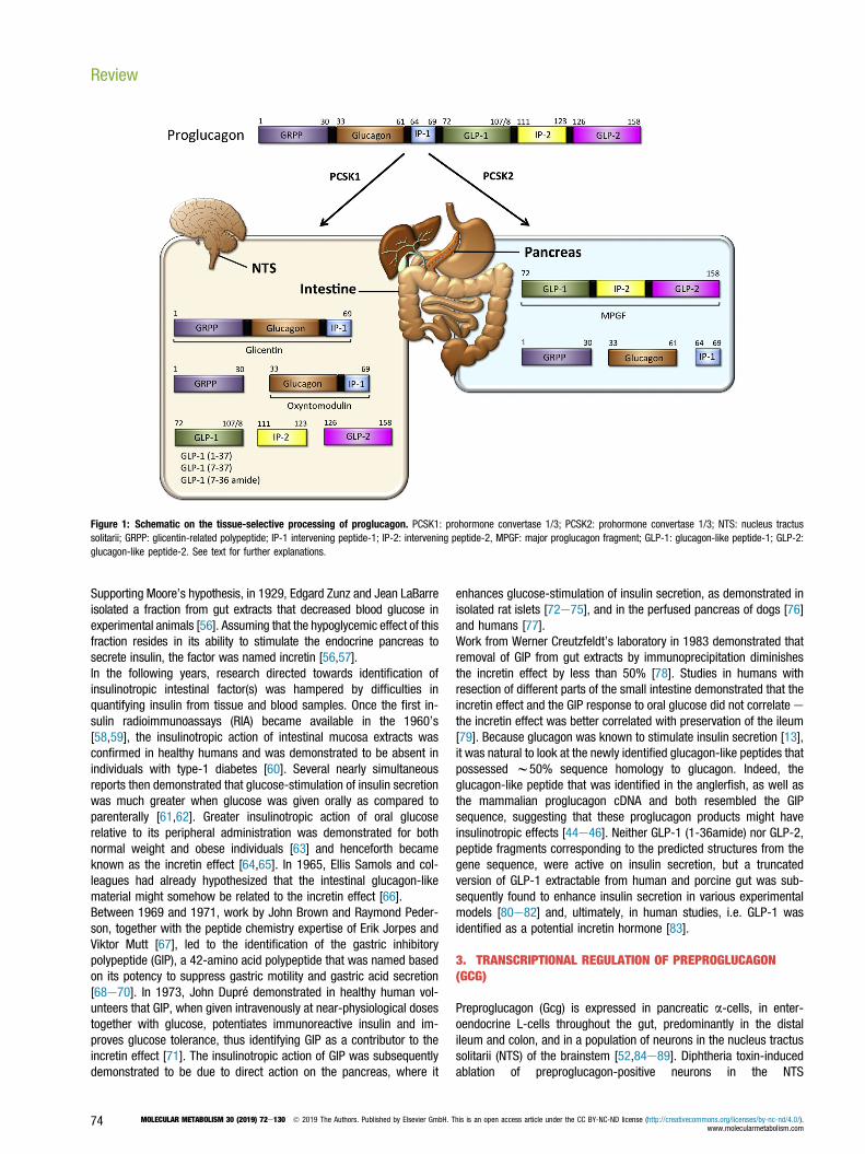
glucagon-producing a-cells in terms of their morphology and ultra-structure [29], classifying these intestinal cells as “L-cells” [30].Glucagon-like immunoreactive material of larger size than of glucagonwas subsequently detected in the pancreas of dogs [31] as well as inislets isolated from birds [32] and guinea pigs [33]. Collectively, thesestudies suggested that glucagon might originate from a larger pre-cursor molecule that is post-translationally cleaved into several frac-tions of distinct size and with different functions. Immunoprecipitationanalysis of rat pancreatic islets identified this precursor as an 18,000molecular weight (MW) protein, now classified as proglucagon [34]. Inthe pancreas, proglucagon was found to be cleaved to produce twofragments, mature glucagon and a 10,000-MW second protein. Thislatter protein lacked the glucagon sequence and was named majorproglucagon fragment (MPGF) [34,35] (Figure 1).In the early 1980s, it was established that the major form of the in-testinal glucagon-immunoreactive material, a peptide designated gli-centin, contained the full glucagon sequence [36e40], and glicentinwas proposed to represent at least a fragment of proglucagon,because it was also identified in the pancreas [41]. Although glicentinwas initially thought to comprise w100 amino acids, its purificationfrom porcine intestine established glicentin as a 69-amino acid peptide[39]. Thus, intestinal proglucagon is cleaved into distinct fractions thatare different from those derived in the pancreas. In 1982, a smallerintestinal form was identified as a 37-amino acid peptide containingthe full 29-amino acid sequence of glucagon with 8 additional aminoacids on its C-terminal end [36], the same as found in the C-terminusof glicentin. Based on its potency to act on oxyntic glands, the 37-amino acid peptide was named oxyntomodulin [42] (Figure 1). Thus,it appeared that proglucagon undergoes a tissue-specific, differentialprocessing leading to the formation of glicentin and oxyntomodulin inthe gut and to glucagon plus the N-terminal fragment of glicentin in thepancreas [34,43] (Figure 1). By using a method that allows the pre-diction of a protein sequence through decoding of recombinant cDNAclones, at the beginning of the 1980s Joel Habener established that adifferent glucagon-related peptide is encoded within the anglerfishpreproglucagon cDNA [44e46]. Two glucagon-related peptides weresubsequently identified in the rat [47,48], hamster [49], bovine [50],and human [51] proglucagon sequence. The two hypothetical peptideswere designated glucagon-like peptides 1 and 2 (GLP-1 and GLP-2)[49] (Figure 1). Radioimmunoassays in tissues from rats [52], pigs[53], and humans [54] then established a distinct profile ofproglucagon-derived peptides (PGDPs) in the pancreas and the in-testine with production of glucagon and one large fragment (majorproglucagon fragment; MPGF) in the pancreas and liberation of smallerGLP-1 immunoreactive peptides in the intestine.
2. IDENTIFICATION OF GIP AND GLP-1 AS GASTROINTESTINALINSULINOTROPIC HORMONES
In 1902, Ernest Bayliss and William Starling identified a substance that isproduced in and secreted from the epithelial cells of the duodenum inresponse to the contact of these cells with acidic chyme [55]. Bayliss andStarling noted that the substance, after being released into the circula-tion, stimulates the pancreas to secrete pancreatic juice; they named thesubstance secretin, thereby identifying the first gastrointestinal hormone[55]. In 1906, Benjamin Moore reported that the glucosuria of diabeticpatients could be ameliorated by repeated oral administration of a pig-derived intestinal mucosal homogenate [55]. Influenced by the work ofBayliss and Starling, Moore hypothesized that the intestinal mucosamembrane produces a substance of the nature of a hormone that de-creases blood glucose via its stimulatory action on the pancreas [55].
MOLECULAR METABOLISM 30 (2019) 72e130 � 2019 The Authors. Published by Elsevier GmbH. This is an open access article under the CC BY-NC-ND license (http://creativecommons.org/licenses/by-nc-nd/4.0/).www.molecularmetabolism.com
73
Supporting Moore’s hypothesis, in 1929, Edgard Zunz and Jean LaBarreisolated a fraction from gut extracts that decreased blood glucose inexperimental animals [56]. Assuming that the hypoglycemic effect of thisfraction resides in its ability to stimulate the endocrine pancreas tosecrete insulin, the factor was named incretin [56,57].In the following years, research directed towards identification ofinsulinotropic intestinal factor(s) was hampered by difficulties inquantifying insulin from tissue and blood samples. Once the first in-sulin radioimmunoassays (RIA) became available in the 1960’s[58,59], the insulinotropic action of intestinal mucosa extracts wasconfirmed in healthy humans and was demonstrated to be absent inindividuals with type-1 diabetes [60]. Several nearly simultaneousreports then demonstrated that glucose-stimulation of insulin secretionwas much greater when glucose was given orally as compared toparenterally [61,62]. Greater insulinotropic action of oral glucoserelative to its peripheral administration was demonstrated for bothnormal weight and obese individuals [63] and henceforth becameknown as the incretin effect [64,65]. In 1965, Ellis Samols and col-leagues had already hypothesized that the intestinal glucagon-likematerial might somehow be related to the incretin effect [66].Between 1969 and 1971, work by John Brown and Raymond Peder-son, together with the peptide chemistry expertise of Erik Jorpes andViktor Mutt [67], led to the identification of the gastric inhibitorypolypeptide (GIP), a 42-amino acid polypeptide that was named basedon its potency to suppress gastric motility and gastric acid secretion[68e70]. In 1973, John Dupré demonstrated in healthy human vol-unteers that GIP, when given intravenously at near-physiological dosestogether with glucose, potentiates immunoreactive insulin and im-proves glucose tolerance, thus identifying GIP as a contributor to theincretin effect [71]. The insulinotropic action of GIP was subsequentlydemonstrated to be due to direct action on the pancreas, where it
enhances glucose-stimulation of insulin secretion, as demonstrated inisolated rat islets [72e75], and in the perfused pancreas of dogs [76]and humans [77].Work from Werner Creutzfeldt’s laboratory in 1983 demonstrated thatremoval of GIP from gut extracts by immunoprecipitation diminishesthe incretin effect by less than 50% [78]. Studies in humans withresection of different parts of the small intestine demonstrated that theincretin effect and the GIP response to oral glucose did not correlate ethe incretin effect was better correlated with preservation of the ileum[79]. Because glucagon was known to stimulate insulin secretion [13],it was natural to look at the newly identified glucagon-like peptides thatpossessed w50% sequence homology to glucagon. Indeed, theglucagon-like peptide that was identified in the anglerfish, as well asthe mammalian proglucagon cDNA and both resembled the GIPsequence, suggesting that these proglucagon products might haveinsulinotropic effects [44e46]. Neither GLP-1 (1-36amide) nor GLP-2,peptide fragments corresponding to the predicted structures from thegene sequence, were active on insulin secretion, but a truncatedversion of GLP-1 extractable from human and porcine gut was sub-sequently found to enhance insulin secretion in various experimentalmodels [80e82] and, ultimately, in human studies, i.e. GLP-1 wasidentified as a potential incretin hormone [83].
3. TRANSCRIPTIONAL REGULATION OF PREPROGLUCAGON(GCG)
Preproglucagon (Gcg) is expressed in pancreatic a-cells, in enter-oendocrine L-cells throughout the gut, predominantly in the distalileum and colon, and in a population of neurons in the nucleus tractussolitarii (NTS) of the brainstem [52,84e89]. Diphtheria toxin-inducedablation of preproglucagon-positive neurons in the NTS
Figure 1: Schematic on the tissue-selective processing of proglucagon. PCSK1: prohormone convertase 1/3; PCSK2: prohormone convertase 1/3; NTS: nucleus tractussolitarii; GRPP: glicentin-related polypeptide; IP-1 intervening peptide-1; IP-2: intervening peptide-2, MPGF: major proglucagon fragment; GLP-1: glucagon-like peptide-1; GLP-2:glucagon-like peptide-2. See text for further explanations.
Review
74 MOLECULAR METABOLISM 30 (2019) 72e130 � 2019 The Authors. Published by Elsevier GmbH. This is an open access article under the CC BY-NC-ND license (http://creativecommons.org/licenses/by-nc-nd/4.0/).www.molecularmetabolism.com
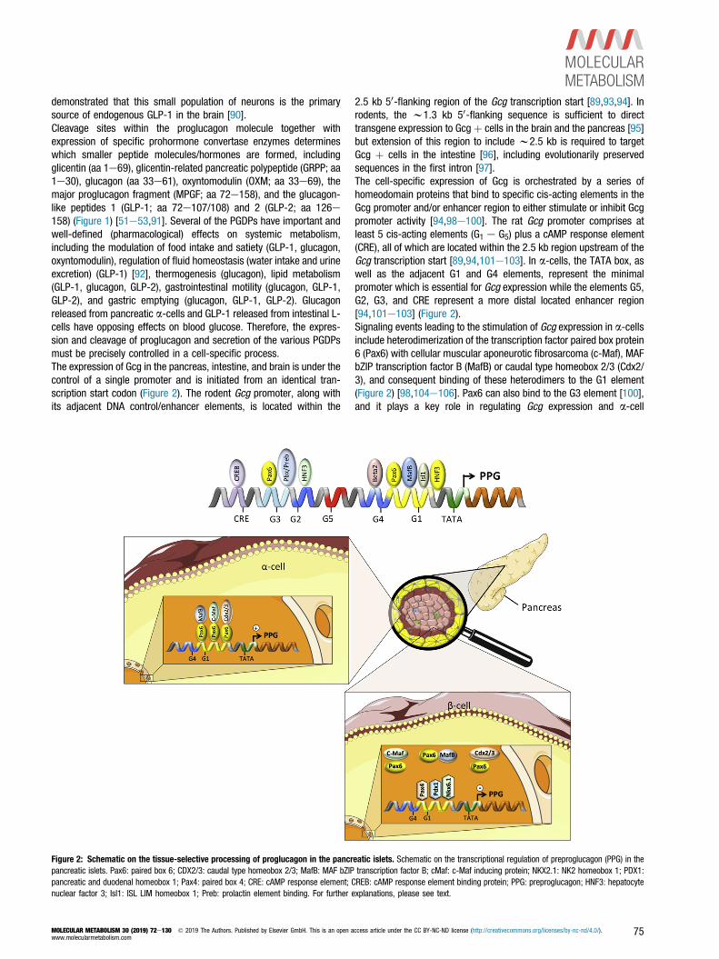
demonstrated that this small population of neurons is the primarysource of endogenous GLP-1 in the brain [90].Cleavage sites within the proglucagon molecule together withexpression of specific prohormone convertase enzymes determineswhich smaller peptide molecules/hormones are formed, includingglicentin (aa 1e69), glicentin-related pancreatic polypeptide (GRPP; aa1e30), glucagon (aa 33e61), oxyntomodulin (OXM; aa 33e69), themajor proglucagon fragment (MPGF; aa 72e158), and the glucagon-like peptides 1 (GLP-1; aa 72e107/108) and 2 (GLP-2; aa 126e158) (Figure 1) [51e53,91]. Several of the PGDPs have important andwell-defined (pharmacological) effects on systemic metabolism,including the modulation of food intake and satiety (GLP-1, glucagon,oxyntomodulin), regulation of fluid homeostasis (water intake and urineexcretion) (GLP-1) [92], thermogenesis (glucagon), lipid metabolism(GLP-1, glucagon, GLP-2), gastrointestinal motility (glucagon, GLP-1,GLP-2), and gastric emptying (glucagon, GLP-1, GLP-2). Glucagonreleased from pancreatic a-cells and GLP-1 released from intestinal L-cells have opposing effects on blood glucose. Therefore, the expres-sion and cleavage of proglucagon and secretion of the various PGDPsmust be precisely controlled in a cell-specific process.The expression of Gcg in the pancreas, intestine, and brain is under thecontrol of a single promoter and is initiated from an identical tran-scription start codon (Figure 2). The rodent Gcg promoter, along withits adjacent DNA control/enhancer elements, is located within the
2.5 kb 50-flanking region of the Gcg transcription start [89,93,94]. Inrodents, the w1.3 kb 50-flanking sequence is sufficient to directtransgene expression to Gcgþ cells in the brain and the pancreas [95]but extension of this region to include w2.5 kb is required to targetGcg þ cells in the intestine [96], including evolutionarily preservedsequences in the first intron [97].The cell-specific expression of Gcg is orchestrated by a series ofhomeodomain proteins that bind to specific cis-acting elements in theGcg promoter and/or enhancer region to either stimulate or inhibit Gcgpromoter activity [94,98e100]. The rat Gcg promoter comprises atleast 5 cis-acting elements (G1 e G5) plus a cAMP response element(CRE), all of which are located within the 2.5 kb region upstream of theGcg transcription start [89,94,101e103]. In a-cells, the TATA box, aswell as the adjacent G1 and G4 elements, represent the minimalpromoter which is essential for Gcg expression while the elements G5,G2, G3, and CRE represent a more distal located enhancer region[94,101e103] (Figure 2).Signaling events leading to the stimulation of Gcg expression in a-cellsinclude heterodimerization of the transcription factor paired box protein6 (Pax6) with cellular muscular aponeurotic fibrosarcoma (c-Maf), MAFbZIP transcription factor B (MafB) or caudal type homeobox 2/3 (Cdx2/3), and consequent binding of these heterodimers to the G1 element(Figure 2) [98,104e106]. Pax6 can also bind to the G3 element [100],and it plays a key role in regulating Gcg expression and a-cell
Figure 2: Schematic on the tissue-selective processing of proglucagon in the pancreatic islets. Schematic on the transcriptional regulation of preproglucagon (PPG) in thepancreatic islets. Pax6: paired box 6; CDX2/3: caudal type homeobox 2/3; MafB: MAF bZIP transcription factor B; cMaf: c-Maf inducing protein; NKX2.1: NK2 homeobox 1; PDX1:pancreatic and duodenal homeobox 1; Pax4: paired box 4; CRE: cAMP response element; CREB: cAMP response element binding protein; PPG: preproglucagon; HNF3: hepatocytenuclear factor 3; Isl1: ISL LIM homeobox 1; Preb: prolactin element binding. For further explanations, please see text.
MOLECULAR METABOLISM 30 (2019) 72e130 � 2019 The Authors. Published by Elsevier GmbH. This is an open access article under the CC BY-NC-ND license (http://creativecommons.org/licenses/by-nc-nd/4.0/).www.molecularmetabolism.com
75

development, because mice lacking Pax6 fail to produce glucagon-producing a-cells [107]. Pax6 also stimulates Gcg expression in theenteroendocrine cells of the gastrointestinal epithelium [108]. Micehomozygous for a dominant negative Pax6 mutation (SEYNeu) haverepressed Gcg expression in enteroendocrine cells in the small andlarge bowel and absence of immunoreactive GLP-1 and GLP-2 [109].Further supporting the role of Pax6 in regulating intestinal Gcgexpression, adenoviral overexpression of Pax6 enhances Gcg promoteractivity and Gcg expression in intestinal enteroendocrine cells such asthe secretin tumor cell line-1 (STC-1) and cells derived from colonictumors of transgenic mice expressing large T antigen under the controlof the proglucagon promoter (GLUTag cells) [108].Other transcriptional mechanisms regulating Gcg expression in a-cellsinclude interaction of Cdx2/3, POU domain transcription factor brain 4(Brn-4), hepatocyte nuclear factor 3 alpha (HNF3a; a.k.a. Foxa1),hepatocyte nuclear factor 3 beta (HNF3b; a.k.a. Foxa2), paired boxprotein 2 (Pax2), neuronal differentiation factor 1/beta 2 (NeuroD/Beta2), and basic helix-loop-helix transcription factor E47 with the G1,G2, G3, or G4 elements (Figure 2) [100,102,104,106,110e118].Emphasizing their role in regulating glycemia, mice lacking Foxa1 orFoxa2 die shortly after birth due to severe hypoglycemia and sub-stantial reduction in Gcg mRNA levels in the pancreas [119,120].Notably, a-cell development is impaired in mice lacking Foxa2 but isnormal in mice lacking Foxa1 [119,120]. These data suggest thatFoxa1 affects glucagon levels via its action on the Gcg promoter, whileFoxa2, in addition to modulating Gcg promoter activity, affects a-celldifferentiation. Interestingly, b-cell specific deletion of Foxa2 also re-sults in postnatal death due to severe hypoglycemia, but the lowglucose level in Foxa2-negative mice seems to be caused by hyper-insulinemia rather than by changes in Gcg expression [121]. The LIMhomeobox protein 1 (Isl1) is ubiquitously expressed in mature endo-crine cells of the pancreas [122], and its ablation in mice results infailure to develop any pancreatic endocrine cell type [123]. In ChineseHamster insulinoma InR1-G9 cells, Isl1 enhances the activity of theGcg promoter [118]. Isl1 also interacts with the enhancer region of thegenes encoding for insulin [124] and somatostatin [125], stimulatingtheir transcription. In islet and intestinal endocrine cell lines, Cdx2/3(the same protein is characterized as Cdx2 in mice and Cdx3 inhamsters) activates Gcg gene transcription via binding to the G1element of the Gcg promoter [98]. Overexpression of Cdx2 in a-cellInR1-G9 cells accordingly increases Gcg expression [113].In both a-cells and intestinal L-cells, Gcg expression is controlled bycertain homeodomain proteins [94,98,100] and by cAMP-activation ofprotein kinase A (PKA), as demonstrated in primary rat intestinal cul-tures [126], isolated pancreatic cell lines [127,128] and enter-oendocrine GLUTag and STC-1 cell lines [129,130]. In STC-1 cells,disruption of the CRE element in the Gcg enhancer only partially bluntsPKA-stimulation of Gcg expression [130,131]. This suggests that PKAalso affects Gcg transcription via CRE-independent mechanisms.Consistent with this, certain effectors of the Wnt pathway, includinglithium and b-catenin, enhance Gcg expression in intestinal but notpancreas-derived cell lines, supposedly via inhibition of the glycogensynthase kinase-3beta (GSK-3beta) [132,133]. PKA has beendemonstrated to inhibit GSK-3beta [134,135], indicating that the CRE-independent PKA stimulation of the Gcg promoter might be mediatedvia negative regulation of the Wnt pathway [132,133]. Other factorsstimulating Gcg expression in the intestine include protein hydrolysates[101] and insulin [133]. Insulin stimulation of intestinal Gcg promoteractivity and of GLP-1 secretion [133] is noteworthy because insulininhibits glucagon production and secretion in isolated rat islets [136]and in hamster islet InR1-G9 cells [137]. While the mechanism
underlying insulin inhibition of pancreatic glucagon production war-rants further clarification, it is thought to be achieved by throughtranscriptional mechanisms via an insulin-responsive element (IRE) inthe Gcg promoter of the a-cells [138].Notably, whereas Gcg is robustly expressed in a-cells, its expression issuppressed in b-cells through binding of pancreatic and duodenalhomeobox 1 (Pdx1), Pax4, and homeobox protein Nkx6.1 to the G1element, thereby competitively preventing the binding of Pax6/Mafheterodimers to the G1 element (Figure 2) [100,139,140]. Whilesubstantial evidence supports a role of Pdx1 in the negative regulationof Gcg expression, Pdx1 immunoreactivity has also been demonstratedin some Gcg expressing L-cells [141]. Overexpression of Pdx1 alone isinsufficient to block Gcg expression in a-cell cultures (aTC-1 cells),isolated murine islets, or GLUTag enteroendocrine cells [142]. Thesedata collectively suggest that Pdx1 requires interaction with othertranscription factors to inhibit Gcg expression.In summary, the cell-type selective expression of Gcg is regulatedthrough more than a dozen transcription factors that selectively bind tocis-acting elements in the Gcg promoter and enhancer regions,thereby either stimulating or inhibiting Gcg expression. Apart from aseries of homeodomain proteins, Gcg expression is also stimulated byPKA in response to increased levels of cAMP [94,99,101,129]. Insulinstimulates intestinal Gcg expression [133] while at the same timeinhibiting Gcg expression in a-cells [89,137,138]. Certain effectors ofthe Wnt pathway further enhance Gcg expression in the intestine butnot in the pancreas [94,132,143].
4. POSTTRANSLATIONAL PROCESSING OF PREPROGLUCAGON
The vast majority of glucagon is produced in the pancreatic a-cells,but, under some conditions, small amounts of glucagon also have beendetected in the intestinal L-cells [87], although the validity of immu-noassays to distinguish different proglucagon products has beenquestioned, and some reports were unable to find genuine glucagon inthe intestine by mass spectrometric analysis [144e146]. Glucagonalso has been detected immunohistochemically in certain Gcg-positiveneurons of the NTS [84e86]. The tissue-specific cleavage of proglu-cagon is orchestrated by the selective expression of the prohormoneconvertase (PC) enzymes. Prohormone convertase 1 (PC1; a.k.a.PCSK1 or PC1/3) is expressed in GCG þ cells in the brain and theintestine, and cleavage of Gcg by PCSK1 results in the liberation ofGLP-1, GLP-2, glicentin, oxyntomodulin, and IP2 (Figure 1) [147e150].In contrast, PC2 (a.k.a. PCSK2) is highly expressed in the pancreas[151], and its expression in a-cells results in cleavage of Gcg into“pancreatic type” glucagon, GRPP, MPGF, and a small interveningpeptide (IP1). Studies in the porcine and human pancreas suggest thatthe PCSK2-liberated PGDPs are all co-secreted in equimolar concen-trations from the islets [152,153]. Underlining the role of PCSK2 inliberation of glucagon via proglucagon processing, PCSK2-deficientmice are slightly hypoglycemic upon fasting, have a reduced rise inblood glucose following intraperitoneal glucose administration, displayimpaired processing of Gcg in the a-cells, and develop a-cell hyper-plasia [154,155]. The hypoglycemia and a-cell hyperplasia seem todirectly result from glucagon deficiency because continuous intra-peritoneal glucagon supplementation is sufficient to correct the hy-poglycemia and the a-cell hyperplasia of Pcsk2�/� mice [156].While PCSK2 is the predominant prohormone convertase in a-cells innon-pathological conditions, a-cell PCSK1 immunoreactivity increasesin rodent models of metabolic stress. a-cell PCSK1 activity and/orexpression is found in embryonic and neonatal mice, with pregnancy,and in models of prediabetes and diabetes [157e160]. In cultured
Review
76 MOLECULAR METABOLISM 30 (2019) 72e130 � 2019 The Authors. Published by Elsevier GmbH. This is an open access article under the CC BY-NC-ND license (http://creativecommons.org/licenses/by-nc-nd/4.0/).www.molecularmetabolism.com
a-cell lines or isolated islets, high-media glucose concentrations in-crease PCSK1 expression and cellular GLP-1 content [161,162]. IsletGLP-1 production is also mediated by the cytokine IL-6, which isreleased in response to exercise, obesity and diabetes [163,164].Lastly, upon streptozotocin-induced destruction of the b-cells, there isan acute increase in islet PCSK1 and Gcg expression and increasedprocessing of proglucagon to GLP-1 [158]. Consistent with this,adenoviral overexpression of PCSK1 in a-cells increases islet GLP-1production and secretion, ultimately leading to enhanced glucose-stimulation of insulin secretion and improved survival of the islets[165]. Further, glucagon receptor KO mice also have compensatoryincreases in a-cell GLP-1, and GLP-1 receptor (GLP-1R) signaling hasbeen reported to contribute to the preserved glucose responses afterstreptozotocin administration [166,167]. Together, these data point toa potential role of the a-cells in compensating for increased b-cellfunctional demand under conditions of insulin resistance, pregnancy,and cellular stress through intra-islet GLP-1 production [168,169].Multiple lines of evidence are accumulating that challenge currentdogma and imply that pancreatic GLP-1 production also has a role,under some circumstances, in regulating insulin secretion via para-crine action [170e172]. Further work is needed to understand thepathological and physiological role for GLP-1 vs. glucagon and para-crine signaling in islet cell development. Glucagon-stimulation of in-sulin secretion is preserved in islets isolated from b-cell-specific GCGRKO mice but is attenuated upon treatment of these islets with exendin(9e39) [173]. Consistent with this, glucagon-stimulation of insulinsecretion is decreased in islets isolated from b-cell-specific GLP-1RKO mice [173]. These data underscore the importance of a-to b-cellcommunication and indicate that glucagon may be the dominant PGDPacting via the GLP-1R on the b-cells to stimulate insulinsecretion [173].Several forms of GLP-1 are processed from proglucagon and vary intheir ability to enhance glucose-induced insulin secretion. The differentforms include GLP-1 (1e37) (or 1-36amide) and two “truncated”forms, GLP-1 (7-36amide) (“amidated GLP-1) and GLP-1 (7e37)(“glycine-extended GLP-1”) (Figure 1) [174]. In humans, nearly allcirculating GLP-1 is one of the truncated forms, with w80% of GLP-1immunoreactivity corresponding to GLP-1 (7-36amide) and w20% tothe glycine-extended GLP-1 (7e37) [175]. The relative abundance ofGLP-1 (7-36amide), GLP-1 (7e37) and GLP-1 (1e37) differs amongspecies [176e178]. Both the longer and the truncated forms of GLP-1are detected in extracts of rat intestine and pancreas [52], in a rat Gcg-producing cell line [179] and upon transfection of rat pituitary or ratinsulinoma cells with a glucagon fusion gene [180]. While GLP-1 (7-36amide) and GLP-1 (7e37) are equally potent to stimulate thesecretion of insulin and c-peptide [181], GLP-1 (1e37) has a muchlower insulinotropic efficacy [80e82].
5. GLP-1 DEGRADATION
Native GLP-1 has a very short half-life, which, depending on thespecies, is around 1e2 min [182e184] and results from two causes:(a) the action of the enzyme dipeptidylpeptidase-4 (DPP-4) and (b)renal elimination. DPP-4 cleaves GLP-1 (7-36amide) and GLP-1 (7e37) at the N-terminal dipeptide to generate GLP-1 (9-36amide) or GLP-1 (9e37), low affinity ligands for the GLP-1 receptor [185e188]. Boththese intact forms as well as inactivated GLP-1 metabolites are alsorapidly cleared from the circulation via the kidneys. In mice, theenzyme neprilysin additionally rapidly degrades the metabolites,making GLP-1 difficult to measure in this species [189]. While GLP-1degradation is unaffected by kidney function, the clearance of both
GLP-1, and to a greater extent its inactive metabolites, is delayed inpatients with renal insufficiency [184].DPP-4 exists in two forms; i.e., it is a membrane-spanning cell surfaceprotein and a circulating protein, and both forms have actions thatextend beyond its proteolytic activity [190]. In the intestine, DPP-4 ishighly expressed in the enterocyte brush border and in endothelial cells[191]. Consequently, as discussed in a comprehensive review [152], alarge portion of intestinal GLP-1 is already degraded in the capillariesof the distal gut with an estimatedw25% of active GLP-1 reaching theliver and only w10e15% reaching the general circulation[152,176,183,191]. Pharmacological inhibition or genetic reduction ofDPP-4 activity preserves much higher circulating levels of intact GLP-1[191,192], and this was demonstrated to potentiate the insulinotropiceffect of GLP-1 in anesthetized pigs (eventually leading to the devel-opment of DPP-4 inhibitors for clinical use) [193]. When administeredi.v., i.p., or s.c. in rats, GLP-1 (7e36 amide) has a half-life of 0.8e4.7 min, 0.6e13.5 min and 4.6e7.1 min, respectively [194].Substantial evidence indicates that the DPP-4-generated GLP-1 me-tabolites (GLP-1 (9-36amide) and GLP-1 (9e37)) have no major role inregulating glucose metabolism [195e197]. However, one report of anexperiment in obese humans suggested that GLP-1 (9-36amide) is aweak insulin secretagogue [198], and administration of GLP-1 (9-36amide) improves glucose handling without affecting insulin secre-tion in anesthetized pigs and in humans [199,200]. GLP-1 (9-36amide)also improves cardiac output in the post-ischemic mouse heart whenadministered during reperfusion, and it affects vasodilation inmesenteric arteries in mice [201]. This scenario contrasts with theapparent lack of effect of high doses of GLP-1 (9-36amide) on glu-coregulation in ob/ob mice or on cognitive function in high-fat fed mice[202,203]. Indeed, there is evidence that truncated GLP-1 (9-36amide)has no effect on glucose clearance or insulin secretion in healthyhumans [196]. In fact, the peptide acts as a weak GLP-1R antagonist,clearly counteracting the biological effects of GLP-1 (7-36amide)in vitro [204].
6. REGULATION OF GLP-1 SECRETION
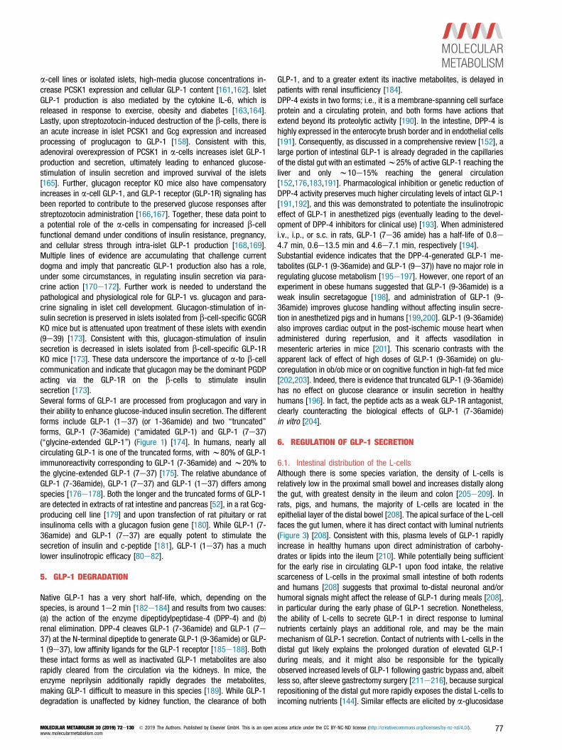
6.1. Intestinal distribution of the L-cellsAlthough there is some species variation, the density of L-cells isrelatively low in the proximal small bowel and increases distally alongthe gut, with greatest density in the ileum and colon [205e209]. Inrats, pigs, and humans, the majority of L-cells are located in theepithelial layer of the distal bowel [208]. The apical surface of the L-cellfaces the gut lumen, where it has direct contact with luminal nutrients(Figure 3) [208]. Consistent with this, plasma levels of GLP-1 rapidlyincrease in healthy humans upon direct administration of carbohy-drates or lipids into the ileum [210]. While potentially being sufficientfor the early rise in circulating GLP-1 upon food intake, the relativescarceness of L-cells in the proximal small intestine of both rodentsand humans [208] suggests that proximal to-distal neuronal and/orhumoral signals might affect the release of GLP-1 during meals [208],in particular during the early phase of GLP-1 secretion. Nonetheless,the ability of L-cells to secrete GLP-1 in direct response to luminalnutrients certainly plays an additional role, and may be the mainmechanism of GLP-1 secretion. Contact of nutrients with L-cells in thedistal gut likely explains the prolonged duration of elevated GLP-1during meals, and it might also be responsible for the typicallyobserved increased levels of GLP-1 following gastric bypass and, albeitless so, after sleeve gastrectomy surgery [211e216], because surgicalrepositioning of the distal gut more rapidly exposes the distal L-cells toincoming nutrients [144]. Similar effects are elicited by a-glucosidase
MOLECULAR METABOLISM 30 (2019) 72e130 � 2019 The Authors. Published by Elsevier GmbH. This is an open access article under the CC BY-NC-ND license (http://creativecommons.org/licenses/by-nc-nd/4.0/).www.molecularmetabolism.com
77
inhibitors, which prevent digestion of starch and oligosaccharides, thusmoving the unprocessed nutrients to more distal parts of the gut. Insome studies, depending on subject characteristics and nutrient load,GLP-1 responses were augmented by acarbose or voglibose (a-glucosidase inhibitors) [217,218]. The commonly observed increase inGLP-1 after bariatric surgery [211e216] is considered a causal factorleading to increased circulating levels of insulin after the surgery. Inline with this notion, blockade of GLP-1R with exendin-9 has beendemonstrated to normalize post-bariatric hyperinsulinemia and alle-viate resulting hypoglycemia after RYGB [219] or gastrectomy [217].Similarly, blockade of GLP-1R with an antagonistic monoclonal anti-body demonstrated an important contribution of enhanced GLP-1secretion to the improved insulin secretion and glucose handling af-ter vertical sleeve gastrectomy (VSG) in preoperatively lean mice [144].The commonly observed rise in GLP-1 following gastric bypass surgeryis also consistent with the robust responses observed in humans upondirect introduction of nutrients, in amounts corresponding to the“physiological malabsorption”, by distal ileal intubation [210]. Thus,increased circulating GLP-1 is invariably reported after bariatric sur-gery [211e216] and after surgical repositioning of the distal gut (so-called ileal interposition), both of which rapidly expose the L-cells toincoming incompletely digested nutrients [144].Enhanced GLP-1 secretion has been suggested to be a major mech-anism underlying enhanced insulin secretion after bariatric surgerybecause blockade of GLP-1 action by treatment with the pharmaco-logical antagonist exendin-9 decreased insulin secretion and amelio-rated post-surgical glucose handling in both mice and humans [144].Nonetheless, a primary role of GLP-1 in post-surgical improvement of
metabolism is controversial. Studies in GLP-1R KO mice documentedthat obese mice exhibit comparable weight loss and improved glucosemetabolism to wildtype controls following VSG [220]. Failure of GLP-1RKO to attenuate the beneficial effects of bariatric surgery on bodyweight may be related to the observation that the body weightdecrease in mice after RYGB may reflect enhanced energy expenditure,whereas in humans and in rats a reduction in food intake is moreimportant [221]. However, sleeve gastrectomy differs markedly fromgastric bypass with faster systemic appearance of ingested glucoseand higher secretion of insulin, GLP-1, PYY, CCK, and ghrelin afterRGYB [222]. Perhaps analogously, rodent experiments have found thatGLP-1R signaling is not required for the weight-reducing effect ofRoux-en-Y Gastric Bypass (RYGB) [223]. Furthermore, GLP-1 inhibitionby exendin (9e39) or administration of a DPP-4 inhibitor does notaffect food intake in humans following RYGB [224]. However, asdemonstrated in that same report, concomitant administration ofexendin9-39 and a DPP-4 inhibitor increased food intake by w20%,suggesting that GLP-1, when potentially acting in concert with othergut hormones, might have an important role in the metabolic benefitsachieved by bariatric surgery [224]. Consistent with this, treatment ofRYGB patients with exendin (9e39) increases the fMRI response toimages of food in the caudate nucleus, and in the insula in the humanbrain in response to consuming palatable food [225].The improved glucose metabolism is mainly due to three closelyinteracting factors: 1) rapid absorption of glucose from the intestinegiving rise to high post-prandial glucose responses, 2) a consequentexaggerated secretion of GLP-1 that act on the b-cells, and 3) radicallyimproved hepatic and subsequently peripheral insulin sensitivity [226].
Figure 3: Schematic on the nutrient-induced stimulation of GLP-1 secretion in the L-cell. CICR: calcium-induced calcium release; LCFA: long-chain fatty acids, GLUT2:glucose transporter 2; GLP-1: glucagon-like peptide-1; GLP-2: glucagon-like peptide-2; OXM: oxyntomodulin; Trpc3: transient receptor potential channel 3; VDCC: voltage-dependent calcium channel; SGLT1: sodium/glucose co-transporter 1. For further explanations, please see text.
Review
78 MOLECULAR METABOLISM 30 (2019) 72e130 � 2019 The Authors. Published by Elsevier GmbH. This is an open access article under the CC BY-NC-ND license (http://creativecommons.org/licenses/by-nc-nd/4.0/).www.molecularmetabolism.com
Like pancreatic a-cells, the intestinal L-cells secrete PGDPs simulta-neously and in equimolar concentrations. An exception is glicentin andits cleavage product oxyntomodulin, the concentrations of which needto be combined to match the amount of secreted GLP-1 [41,53]. Meal-induced GLP-1 secretion has been demonstrated in numerous speciesincluding mice [227], rats [228e232], dogs [233e235] and humans[236e239]. An important consideration when analyzing meal-inducedGLP-1 secretion is whether GLP-1 was measured in systemic or portalvein blood, whether the samples were taken in relation to oral meals orrather to intragastric infusion of liquid (or semiliquid) meals, andwhether total or active GLP-1 was measured. As comprehensivelydiscussed previously [240], an estimate of up to 75% of active GLP-1 isalready degraded in the gut, and, from the amount of GLP-1 reachingthe liver, another 50% is degraded before reaching the systemic cir-culation [183,241]. Only 10e15% of active GLP-1 is believed to reachthe pancreas via the circulation [185,191,241]. As demonstrated inanesthetized pigs, blood concentrations of total and active GLP-1progressively decrease with increasing distance from the site ofsecretion [241]. Levels of total and active GLP-1 are thus highest in theportal vein and lowest in the peripheral venous system [241]. Inanesthetized pigs, baseline levels of total GLP-1 in the portal vein are inthe range of w30 pmol/l and increase up to 150 pmol/l upon treat-ment with neuromedin C, which is a known stimulator of GLP-1secretion [241]. Baseline portal vein levels of active GLP-1 arew10 pmol/l and increase up to w40 pmol/l upon treatment withneuromedin C [241].Systemic plasma levels of total C-terminally amidated GLP-1 correlatewith plasma insulin and thus are low during fasting and increased inresponse to an oral meal [236]. In humans, fasting systemic plasmaconcentrations of “total” GLP-1 (including metabolites generated byDPP-4-mediated degradation) are typically in the range of 5e10 pmol/L and can increase up to 40 pmol/L in response to a meal [236].Plasma systemic concentrations of “intact”, biologically active GLP-1are much lower (fasting: < 2 pmol/l, peak post-nutrient concentra-tions 5e10 pmol/l) [240,242]. The amount of GLP-1 secretion isaffected by the size and composition of a meal [237]. In healthy humanvolunteers, a 520 kcal oral meal induced a greater systemic increaseof plasma systemic total and active GLP-1 than a 260 kcal meal [237].GLP-1 levels are detectable during fasting, indicating that GLP-1 istonically secreted into the general circulation in basal conditions [243].In rats however, there is also evidence for a “preprandial” cephalic-phase GLP-1 reflex [244,245], and the muscarinic cholinergic antag-onist atropine reduces the GLP-1 response to an oral glucose load inhumans [246].Nutrients stimulating GLP-1 secretion include the metabolizablemonosaccharides that include glucose, fructose, and galactose[239,247e250] as well as non-metabolizable monosaccharides suchas methyl-a-glucopyranoside [247], long-chain fatty acids [232,251e253], proteins [228,250,254e257], and certain amino acids [258e260]. In healthy humans, ingestion of carbohydrates or proteinselicits a rapid increase in circulating GLP-1 with a peak 30e60 minfollowing nutrient intake, whereas ingestion of lipids elicits a some-what later but more prolonged (>120 min) increase [238,239]. In rats,portal vein levels of total GLP-1 peak around 15 min after intragastricinfusion of a liquid meal and return to baseline levels after 90e120 min [231]. In rats eating a spontaneous chow meal, increasedportal vein but not systemic levels of active GLP-1 are detectable [261].There is controversy as to whether glucose-induced GLP-1 secretion isdisturbed in patients with type-2 diabetes. A study in w1,500 Danishsubjects suggested that the GLP-1 response to oral glucose is reducedin patients with prediabetes or type-2 diabetes [262]. In contrast, a
meta-analysis of 22 clinical studies revealed no difference in glucose-stimulated GLP-1 secretion between patients with type-2 diabetes andnon-diabetic controls [263,264].
6.2. GLP-1 secretion in response to monosaccharides and othercarbohydratesThe cellular mechanisms underlying glucose-stimulation of GLP-1secretion from L-cells are, at least in part, similar to the stimulationof insulin secretion in the islets. In enteroendocrine GLUTag cells,glucose, and fructose dose-dependently increase GLP-1 secretionthrough closure of ATP-sensitive KATP channels and subsequentmembrane depolarization (Figure 3) [247,249,265]. Glucose-inducedmembrane depolarization entails opening of voltage-dependent Ca2þ
(VDC) channels, and the resulting Ca2þ influx then triggers vesicularexocytosis and secretion of GLP-1 into the circulation (Figure 3) [266].Underlining the role of the KATP channels in mediating this process,glucose-stimulated Ca2þ entry and GLP-1 secretion are mimickedupon treatment of GLUTag cells with the KATP channel inhibitortolbutamide [265]. While the importance of KATP channel activity inmediating GLP-1 release has been confirmed in vitro, its relevance forGLP-1 secretion in vivo is less clear. While sulphonylureas potentlypromote insulin secretion in type-2 diabetic patients via inhibition ofKATP channel activity [267e270], there is no clear evidence that sul-phonylureas affect GLP-1 secretion in humans (as reviewed in [266]).However, the Kir6.2/SUR1 channel complex of the KATP channel ispresent in human L-cells [271] and KATP channel subunits andglucokinase are highly expressed in murine L-cell populations [266]. Insummary, while L-cell depolarization is crucial for GLP-1 secretion, therole of the Kir6.2/SUR1 channel complex of the KATP channels formediating this process in vivo warrants clarification.Monosaccharides demonstrated to stimulate GLP-1 secretion includeglucose, galactose, and fructose [239,247]. Low concentrations ofglucose or methyl-a-glucopyranoside stimulate L-cell electrical activityand promote GLP-1 secretion via sodium-glucose cotransporter(SGLT1)-dependent induction of small inward currents (Figure 3) [247].Preventing luminal glucose absorption by blockade of SGLT1 reducesGLP-1 secretion in the isolated perfused canine ileum [248] and in therat ileum [178] and impairs glucose-stimulation of GLP-1 release inGLUTag cells [247]. Notably, the glucose transporter-2 (GLUT2) hasbeen implicated in glucose-stimulated GLP-1 secretion, as demon-strated by an impaired GLP-1 response to oral glucose in mice defi-cient for GLUT2, and this is accompanied by reduced glucose-stimulated insulin secretion and impaired glucose tolerance [272].However, pharmacological inhibition of active, sodium-coupledglucose transport impaired glucose-stimulated GLP-1 secretionin vitro, whereas inhibition of facilitative GLUT-mediated glucosetransport was without effect [273]. In summary, glucose uptake intothe L-cells seems to be mediated via both, GLUT2 and SGLT1(Figure 3) [274e276], with the electrogenic SGLT1 mediated uptakebeing of particular importance for stimulus secretion coupling.Downstream of glucose-mediated membrane depolarization, vesicularexocytosis of GLP-1 is orchestrated in a Ca2þ dependent mannerinvolving a cellular machinery like that in b-cells [277,278]. Fructosestimulation of GLP-1 secretion has been demonstrated in rats, mice,humans, and GLUTag cells [249,279]. When given orally, fructose is afar less potent GLP-1 secretagogue relative to an isocaloric load ofglucose [249]. Similar findings are reported in humans upon intra-gastric infusion of glucose and fructose at doses that are matched forsweetness [279].A possible role of intestinal sweet-taste receptors in glucose-stimulated GLP-1 secretion remains uncertain. In isolated murine
MOLECULAR METABOLISM 30 (2019) 72e130 � 2019 The Authors. Published by Elsevier GmbH. This is an open access article under the CC BY-NC-ND license (http://creativecommons.org/licenses/by-nc-nd/4.0/).www.molecularmetabolism.com
79
and human L-cell cultures, glucose, and the artificial sweetenersucralose, each stimulates GLP-1 secretion with diminished glucosestimulation of GLP-1 secretion in mice lacking a-gustducin, an integralsweet-taste receptor element [280]. In contrast, there is no effect ofsucralose on GLP-1 secretion in primary L-cell cultures in other reports[281,282], and infusion of artificial sweeteners does not affectglucose-stimulated GLP-1 secretion in healthy human volunteers[283,284].
6.3. GLP-1 secretion in response to dietary lipidsIn addition to sensing carbohydrates and glucose, L-cells, like otherenteroendocrine cells, sense dietary lipids and protein (see next sec-tion) through specific cell-surface receptors, which bind metabolites ofthe dietary lipids and proteins [285] (Figure 4). This occurs mainly atthe basolateral side of the L-cell, i.e. after absorption of the metabolites[285]. In the case of dietary triglycerides, the L-cell responds not onlyto the free fatty acids but also to the other major metabolite product, 2-monoacyl glycerol (2-MAG), which appears to act in synergy [286].Cell lines commonly used to study mechanisms of GLP-1 secretion areGLUTag, STC-1 and human colorectal adenocarcinoma-derived NCI-H716 cells. These cell lines are far from optimal models, as they, forexample, differ in their secretory repertoire; some secreting peptidesthat are not classical L-cell products, such as GIP, glucagon, andsomatostatin, and GLUTag cells lack PYY [287].Fatty acid induction of GLP-1 secretion has been demonstrated in vitrousing murine enteroendocrine STC-1 cells [251] and human intestinalNCI-H716 cells [251,253] and in vivo by direct administration of lipidsinto the duodenum [288,289] or ileum [210]. Dose-dependent fattyacid-induced GLP-1 secretion is evident with a-linolenic acid (C18:3),docosahexanoic acid (C22:6), and palmitoleic acid (C16:1), oleic acid(C18:1), stearic acid (C18:0), and octanoic acid (C8:0) [251] are less
effective. In humans, unsaturated are more effective than saturatedfatty acids [290,291].Induction of GLP-1 secretion by FFA is highly dependent on thecytosolic Ca2þ concentration. Treatment of STC-1, GLUTag, or NCI-H716 cells with long-chain fatty acids potently increases intracellularCa2þ [253,292]. The FFA-induced rise in intracellular Ca2þ is sub-stantially reduced when cells are cultured in a Ca2þ free medium, andis abolished upon treatment of cells with the Ca2þ channel inhibitornicardipine [292] or when using BSA (which binds fatty acids) [251].Collectively, these data suggest that FFAs increase intracellular Ca2þ
by stimulating the influx of Ca2þ via the cell-surface (most likely L-type) Ca2þ channels [292]. Treatment of NCI-H716 or STC-1 cells withthe Ca2þ ionophore ionomycin or with phorbol myristate acetate (PMA)increases cytosolic Ca2þ levels and stimulates GLP-1 secretion in adose-dependent manner [251,253]. The ionomycin-induced increasein Ca2þ influx and GLP-1 secretion is completely abolished upontreatment of cells with the Ca2þ chelator EGTA [251]. In summary,compelling evidence indicates that FFA increase GLP-1 secretion bystimulating extracellular Ca2þ influx via cell-surface Ca2þ channels.Receptors implicated in FFA regulation of GLP-1 secretion includeGPR120 (FFAR4) and GPR40 (FFAR1), both of which are activated bylong-chain fatty acids. GPR120 was previously reported to be co-localized with GLP-1 in colonic enteroendocrine cells [251]. Thiscould point to GPR120 being involved in the stimulation of GLP-1 bylong chain FFAs. However, The GLP-1 response to oleic acid is unal-tered in GPR120-deficient mice, and synthetic GPR120 agonists do notstimulate GLP-1 from primary cell cultures [286]. GPR40, the otherlong chain FFA receptor, is also highly expressed and the mostenriched GPCR in L-cells [266,293]. In contrast to GPR120, the GLP-1response to dietary fat is strongly reduced in GPR40 KO mice ascompared to littermates [286]. Importantly, synthetic GPR40 agonists
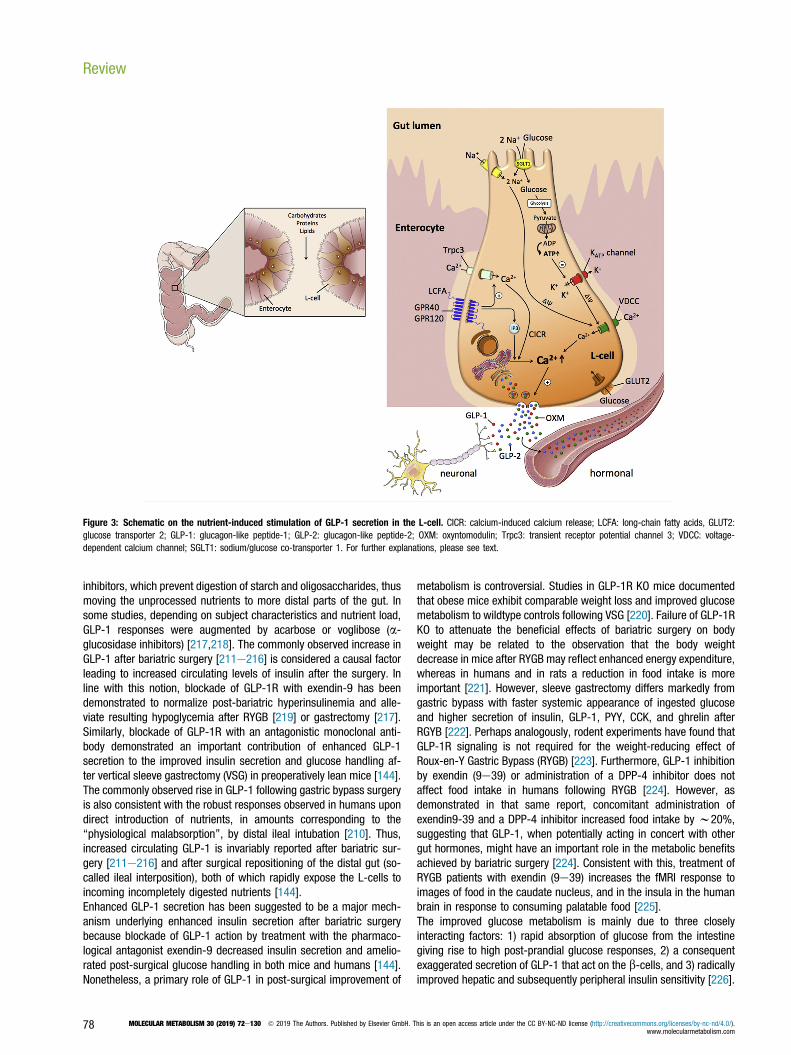
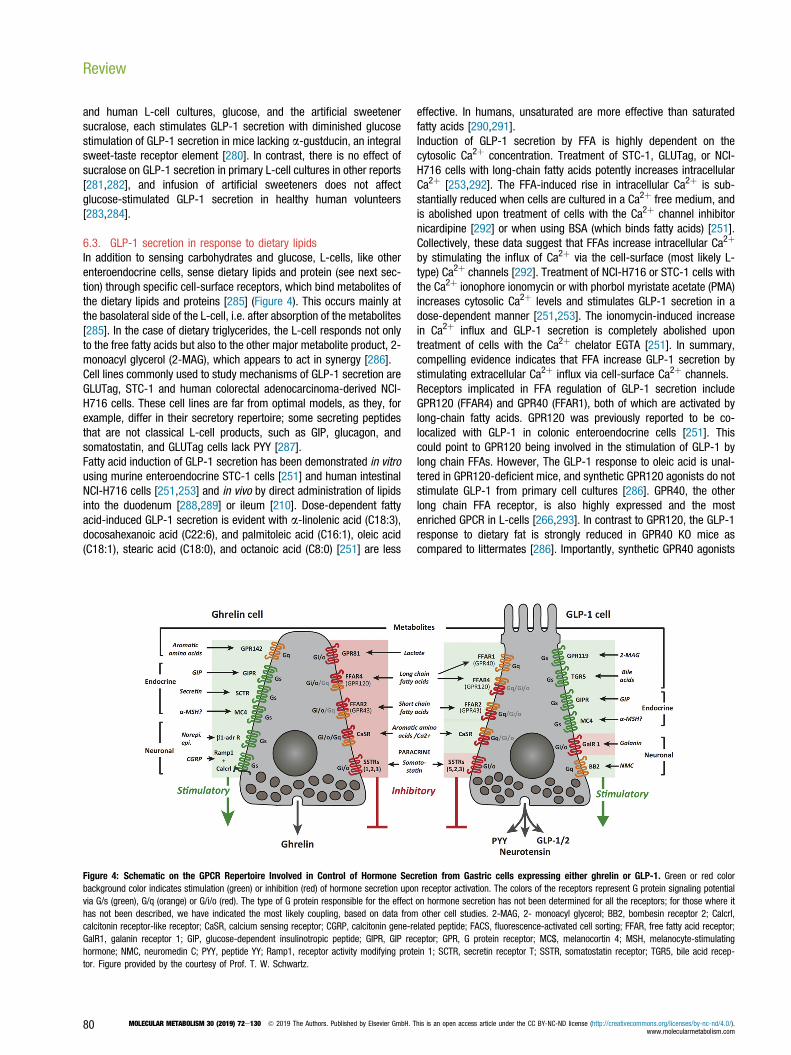
Figure 4: Schematic on the GPCR Repertoire Involved in Control of Hormone Secretion from Gastric cells expressing either ghrelin or GLP-1. Green or red colorbackground color indicates stimulation (green) or inhibition (red) of hormone secretion upon receptor activation. The colors of the receptors represent G protein signaling potentialvia G/s (green), G/q (orange) or G/i/o (red). The type of G protein responsible for the effect on hormone secretion has not been determined for all the receptors; for those where ithas not been described, we have indicated the most likely coupling, based on data from other cell studies. 2-MAG, 2- monoacyl glycerol; BB2, bombesin receptor 2; Calcrl,calcitonin receptor-like receptor; CaSR, calcium sensing receptor; CGRP, calcitonin gene-related peptide; FACS, fluorescence-activated cell sorting; FFAR, free fatty acid receptor;GalR1, galanin receptor 1; GIP, glucose-dependent insulinotropic peptide; GIPR, GIP receptor; GPR, G protein receptor; MC$, melanocortin 4; MSH, melanocyte-stimulatinghormone; NMC, neuromedin C; PYY, peptide YY; Ramp1, receptor activity modifying protein 1; SCTR, secretin receptor T; SSTR, somatostatin receptor; TGR5, bile acid recep-tor. Figure provided by the courtesy of Prof. T. W. Schwartz.
Review
80 MOLECULAR METABOLISM 30 (2019) 72e130 � 2019 The Authors. Published by Elsevier GmbH. This is an open access article under the CC BY-NC-ND license (http://creativecommons.org/licenses/by-nc-nd/4.0/).www.molecularmetabolism.com
are efficacious GLP-1 secretagogues both in vitro in primary cultures,in perfused intestines and in vivo when studied in mice [252,294,295].Treatment of mice with GPR40 agonists [296,297] has been reportedto decrease body weight and food intake in DIO mice, effects notablyabsent in mice deficient for either GPR40 or GLP-1R [296].Accordingly, long chain fatty acids stimulate GLP-1 secretion mainlythrough GPR40. However, the other main metabolite from dietary tri-glycerides, 2-MAG, is also a very powerful GLP-1 secretagogue actingthrough GPR119 [298,299]. Importantly, the GLP-1 response to dietaryfat is vastly reduced not only in GPR40 KO animals but also in GPR119KO mice, and agonists for the Gq-coupled GPR40 act in synergy withthe Gs-coupled GPR119 to robustly stimulate GLP-1 [286,300]. Areceptor-independent stimulation of GLP-1 release from GLUTag cellshas been reported for oleic acid, which stimulates GLP-1 release byuncoupling oxidative phosphorylation and, hence, indirectly stimulatingglycolysis with the resulting activation of the mechanism alluded toabove [301].
6.4. GLP-1 secretion by proteins/amino acidsProtein and amino acid stimulation of GLP-1 secretion has beendemonstrated in murine primary colonic L-cell cultures [260,302], inGLUTag cells [256,259], in human NCI-H716 cells [251,303] and in theisolated perfused rat ileum or colon [250,304] as well as in vivo in mice[305], rats [256,306], and humans [307,308]. In healthy human vol-unteers, a diet with 30% kcal from protein (40% carbohydrates, 30%fat) causes greater GLP-1 secretion than a diet with 10% kcal protein(60% carbohydrates, 30% fat) [308]. Individual amino acids stimu-lating GLP-1 secretion include glutamine, asparagine, phenylalanine,and glycine, with glutamine and glycine being the most potent [258e260]. When orally administered, glutamine also increases circulatingGLP-1 and insulin in lean, obese and type-2 diabetic individuals [307].In human NCI-H716 cells, stimulation of GLP-1 secretion was alsodemonstrated for leucine, isoleucine, valine, skimmed milk, casein,and whey [303]. L-Arginine, a potent insulin secretagogue [309], alsostimulates GLP-1 release from isolated rat intestine, and, when givenorally, augments GLP-1 and insulin levels and improves glucosetolerance in mice, effects that are absent in GLP-1R KO mice [305].Meat hydrolysate stimulates GLP-1 secretion from NCI-H716 cells, aneffect that is not related to changes in proglucagon expression [253].This stimulatory effect can be blocked by pretreatment with the p38inhibitor SB203580, the PI3 kinase inhibitor wortmannin or the MEK1/2inhibitor U0126 [310]. The corn protein zein stimulates GLP-1 secre-tion in GLUTag cells and in the small intestine of anesthetized rats[256], and it stimulates GLP-1 secretion when administered eitherorally [228] or directly into the ileum [257]. Intraluminal administrationof peptones stimulate GLP-1 secretion in the isolated perfused ratileum [250] but not upon ileal perfusion in healthy human volunteers[210]. Pectin stimulates GLP-1 secretion in the isolated perfused ratcolon [304] but not in the isolated rat ileum [250]. The low molecularfraction of wheat protein hydrolysate (LWP) increases GLP-1 secretionin both GLUTag cells and when directly administered in rats [306]. Inrats, the LWP-induced GLP-1 secretion further improves glucosetolerance and enhances insulin secretion, an effect that is blocked bypre-administration of the GLP-1R antagonist exendin (9e39) [306].Protein-stimulation of GLP-1 secretion has also been demonstrated inhumans, with similar GLP-1 responses upon uptake of whey, casein,gluten or cod protein [311e313]. Protein-induction of GLP-1 secretionseems to be dose-dependent, as demonstrated by uptake of isocaloricdiets comprising 14%, 25%, or 50% of energy coming fromproteins [314].
The molecular mechanisms underlying protein stimulation of GLP-1secretion include activation of Ca2þ/calmodulin-dependent kinase II[306]. Substantial evidence supports that peptide-mediated GLP-1secretion is a Ca2þ sensitive process and involves L-cell signaling viathe Ca2þ sensing receptor (CaSR) and the peptide transporter 1(PEPT1) [302]. Consistent with this, glycine-sarcosine (Gly-Sar) stim-ulation of GLP-1 secretion from purified murine L-cell cultures isblocked in the absence of extracellular Ca2þ and is inhibited upontreatment with the L-type Ca2þ-channel blocker nifedipine [302].Oligopeptide stimulation of GLP-1 release is impaired upon treatmentof L-cell cultures with a CaSR antagonist and is ameliorated in micedeficient for the peptide transporter 1 (PEPT1) [302]. Aromatic aminoacids such as phenylalanine, however, also interact withGPR142 [315].
6.5. GLP-1 secretion in response to endocrine factors
6.5.1. Endocrine regulation of intestinal GLP-1 secretionThe intestinal distribution of L-cells, with high abundance in the distalgut and low abundance in the proximal gut, argues for the existence ofa proximal-to-distal coordinating loop in which neuronal and/orendocrine factors arising in the upper intestine affect L-cell GLP-1secretion in the distal region. While such a proximal-distal loopmight indeed exist, it cannot be ruled out that while fewer in number,L-cells in the upper intestine are sufficient for the rapid induction ofGLP-1 secretion following nutrient intake [316e318]. Nonetheless, thepresumed loop (if it exists) would likely be important for the earlypostprandial phase at a time when the L-cells of the distal gut are notyet in direct contact with luminal nutrients. Supporting such neuronal/endocrine regulation of GLP-1 secretion, the L-cells are in closeproximity to both enteric neurons and the intestinal microvasculature[191,319]. Possible neuroendocrine regulation of GLP-1 secretion issupported by studies in rodents in which nutrient flow to the distalintestine is prevented, precluding direct L-cell contact in this part of theintestine to luminal nutrients, [289,320]. Administration of glucose orfat directly into the duodenum of such rodents rapidly stimulates L-cellGLP-1 secretion, with a magnitude comparable to that occurring whennutrients are directly placed into the ileum [289,320]. Because the L-cells co-secrete the PGDPs, factors stimulating GLP-2 or oxy-ntomodulin are also natural secretagogues of GLP-1. Neuronal/endo-crine factors affecting the intestinal release of PGDPs in some speciesinclude GIP, acetylcholine, gastrin-releasing peptide (GRP), insulin,somatostatin, and ghrelin [320e322].In rodents, a biphasic secretion of GLP-1 has been observed; thissuggests a rapid phase of GLP-1 secretion caused by direct stimulationof L-cells in the upper GI tract and a second phase potentially causedby signals from the upper gut reaching the lower small and perhapseven the large intestine [323,324]. GIP and the intramural intestinalautonomous nervous system have been suggested as signalingpathways [323]. Several lines of evidence support a role for GIP in GLP-1 secretion. GIP stimulation of GLP-1 secretion, however, seems to behighly species-specific. Levels of GIP expression (in K-cells) aregreatest in the proximal gut, and circulating levels of GIP rapidly in-crease upon food intake [236,325,326] or when nutrients are placeddirectly into the duodenum [289,323,327]. Secretion ofpreproglucagon-derived peptides is stimulated in rats and in primaryrat L-cell cultures upon treatment with GIP [289,323,327]. In rats,induction of gut glucagon-like immunoreactivity induced by eitherlipids or by physiological concentrations of GIP can be blocked bysubdiaphragmatic vagotomy, suggesting that in rats GIP regulation of
MOLECULAR METABOLISM 30 (2019) 72e130 � 2019 The Authors. Published by Elsevier GmbH. This is an open access article under the CC BY-NC-ND license (http://creativecommons.org/licenses/by-nc-nd/4.0/).www.molecularmetabolism.com
81

GLP-1 secretion requires either afferent or efferent signal transmissionvia the vagus [327]. The importance of such a proximal-to-distal GIP-GLP-1 axis for human physiology is, however, questionable. In healthyand type-2 diabetic humans, it has been consistently demonstrated,that GIP, even in rather high (supraphysiological) doses, does not leadto GLP-1 secretion [328e330]. In humans, GLP-1 secretion inresponse to duodenal glucose delivery does not become robustlystimulated until the delivery rate overcomes the absorptive capacity ofthe duodenum, i.e. until such time as non-absorbed glucose reachesthe jejunum and beyond. In contrast, GIP secretion is stimulated byvery low gastric glucose delivery rates, consistent with the higherabundance of GIP-producing K-cells compared to GLP-1-producing L-cells in the proximal intestine [331]. Direct infusion of glucose into theduodenum at a rate that ensured w total absorption close to theinfusion site (2 kcal/min) triggered robust GIP, but not GLP-1 secretion,whereas both hormones were released when glucose was directlydelivered into the ileum [332]. Another gut peptide that has beenimplicated in a proximal-to-distal loop to stimulate GLP-1 secretion isCCK [333]. However, the concentrations of both GIP and CCK needed tostimulate GLP-1 secretion are not normally reached under physiolog-ical conditions [334].Neurotransmitters expressed in vagal and enteric neurons, includingacetylcholine and GRP, increase GLP-1 secretion, supporting thepossibility of a proximal-distal neuroendocrine loop [335] without theneed of supraphysiological plasma concentrations of proximally-secreted gut peptides. Receptors for acetylcholine, including themuscarinic receptors M1, M2, and M3, are expressed in rat L-cells[319] and human NCI-H716 cells [336]. Treatment of rats with thenonspecific muscarinic receptor antagonist atropine or with the M1selective antagonist pirenzipine, but not treatment with M2 or M3-selective antagonists blunts lipid-induced GLP-1 secretion [319,335].In human NCI-H716 cells, GLP-1 secretion is stimulated by betha-nechol, a nonselective muscarinic agonist, while pretreatment withpirenzipine or the M2 antagonist gallamine inhibits bethanechol-induction of GLP-1 secretion [336]. Acetylcholine also stimulatesGLP-1 secretion in the perfused porcine ileum, and this effect can beblocked by co-infusion of atropine [337]. Albeit with notable physio-logical challenges (atropine powerfully inhibits GI motility), infusion ofatropine also blunts nutrient induced GLP-1 secretion in healthy humanvolunteers [246]. Together, these data suggest that M1 and M2muscarinic receptors are implicated in human L-cell GLP-1 secretion.Interestingly, in rat ileum preparations, administration of atropine wasnot able to block GIP-induced GLP-1 secretion [335]. In the isolatedperfused porcine ileum, GLP-1 secretion is inhibited by electrical nervestimulation or by administration of norepinephrine, effects that can beblocked by co-infusion of the nonselective a-adrenergic receptorantagonist phentolamine [337]. While norepinephrine seemingly in-hibits GLP-1 secretion via its action on a-adrenergic receptors, itssecretion is stimulated by isoproterenol, and this effect can be blockedby co-infusion of the b-adrenergic receptor antagonist propanolol[337]. These data collectively suggest that intestinal GLP-1 secretion isstimulated by cholinergic and b-adrenergic receptor signaling andinhibited by activation of a-adrenergic receptors.GRP is produced and released by GRPergic neurons of the entericnervous system [338]. In rats, infusion of GRP stimulates the secretion ofGLP-1 while administration of the GRP antagonist BW10 blocks GLP-1secretion when fat is directly administered into the duodenum [339].GRP stimulation of GLP-1 secretion has also been demonstrated for ratL-cell cultures and in preparations of rat ileum [323,335]. Notably, GRPregulation of glucose handling is not fully dependent on GLP-1 signaling,because GRP also directly stimulates insulin secretion in the isolated
perfused dog pancreas [340] and further delays gastric emptying[341,342]. Nonetheless, mice deficient for GRP have impaired glucosetolerance, reduced first-phase insulin secretion and impaired GLP-1secretion in response to an oral glucose challenge [343].In summary, there are several mechanisms that may contribute to therapid increase in GLP-1 secretion following nutrient intake. Nutrient-induced GLP-1 secretion can occur from L-cells located in the prox-imal small intestine with induction of GLP-1 secretion as early asdigested nutrients leave the pylorus. The glucose concentration after ameal may exceed the absorptive capacity in the proximal intestine sothat the ingested glucose rapidly reaches the more distally located L-cells. Neuroendocrine reflexes may also trigger GLP-1 secretion inaddition to direct nutrient-induced stimulation of L-cell GLP-1 secretion[344]. Current data suggest that when chyme enters the duodenum ittriggers GIP release in the proximal gut. The local increase in GIPstimulates vagal afferent transmission followed by activation of vagalefferents and enteric neurons that release acetylcholine and/or GRP tostimulate GLP-1 release from the distal gut. When the nutrients sub-sequently reach the distal gut, direct contact with the L-cells thentriggers additional GLP-1 secretion into the circulation (as alsoreviewed in [320]).Other factors influencing GLP-1 secretion include activation of theolfactory receptor OR51E1 using nonanoic acid, which stimulatessecretion of GLP-1 and PYY in human and rodent enteroendocrine L-cells [345]. More recently, ghrelin was identified to stimulate GLP-1secretion in murine and human L-cell cultures [322]. In mice, pe-ripheral administration of ghrelin further enhances glucose-stimulatedGLP-1 secretion and improves glucose tolerance, an effect that isblocked by pre-administration of the ghrelin receptor antagonist D-LysGHRP6 and that is absent in GLP-1R KO mice [322].
6.5.2. Endocrine regulation of central GLP-1 secretionAs discussed above, in addition to enteroendocrine L-cells, GLP-1 isalso produced in a discrete set of non-TH-positive neurons in thecaudal portions of the NTS [86,148,346e348], and these hindbrainGCG þ positive neurons are the primary source of endogenous brainGLP-1 [90]. Either peripheral administration of leptin [349] or gastricballoon distention [350] acutely activates GLP-1-producing neurons inthe NTS, as assessed by cFos immunoreactivity. Direct electricalstimulation of the NTS evokes glutamatergic excitatory post-synapticcurrents (EPSCs) in GCG þ positive neurons [351]. Generation ofmice that express eYFP under control of the Gcg promoter has enabledthe isolation and characterization of NTS GCG þ neurons in ex vivotissue slices [351]. Electrophysiological whole-cell voltage- andcurrent-clamp recordings in horizontal or coronal brainstem slices hasrevealed a rapid leptin-induced depolarization of these NTSGCG þ neurons, thus confirming the ability of leptin to directly stim-ulate central GLP-1 secretion [351]. Of note, the hindbrainGCG þ neurons lack the GLP-1 receptor such that they cannot bedirectly activated by peripherally-derived GLP-1 [351]. In addition,neither electrophysiological administration of PYY, melanotan II, norghrelin stimulates these neurons in isolated NTS brain slices [351]. Incontrast, leptin [351], CCK, and epinephrine [352] stimulate Gcgneurons. In the NTS, neurons expressing GLP-1 also express the leptinreceptor [351,353]. Electrical stimulation of the solitary tract indicatesthat PPG neurons in the NTS are second-order neurons that receivedirect input from vagal afferents. Thus, peripheral endocrine signals,such leptin or GLP-1, can via activation of vagal afferents triggercentral activation of PPG neurons in the NTS [351].CCK-induced firing of the NTS GCG þ neurons can be blocked bytreatment with the glutamate receptor antagonist DNQX or by inhibition
Review
82 MOLECULAR METABOLISM 30 (2019) 72e130 � 2019 The Authors. Published by Elsevier GmbH. This is an open access article under the CC BY-NC-ND license (http://creativecommons.org/licenses/by-nc-nd/4.0/).www.molecularmetabolism.com
of a1-adrenergic signaling [352]. Consistent with these findings,peripherally administered CCK induces cFos immunoreactivity in GLP-1-producing neurons of the hindbrain vagal complex of the NTS [354],and surgical vagal deafferentation reduces CCK-induced NTS neuronalcFos activation by approximately 50% [355]. Thus, these neurons areable to sense and respond to a variety of peripheral signals that help toregulate both short and long-term energy balance. Indeed, similar tochronic blockade of CNS GLP-1R, viral knockdown of GLP– expressingneurons in rats increases body mass, and specifically body adiposity[356]. In a recent report, chemogenetic stimulation of Gcg neuronsreduced food intake without conditioning avoidance, and this occurredwhen the animals were fed or fasted or were fed chow or HFD [357].On the other hand, acute chemogenetic inhibition of these neurons didnot increase ad lib feeding but did increase refeeding after a fast andblocked stress-induced hypophagia [90]. In summary, the gluta-matergic GLP-1-producing neurons in the NTS are activated by mul-tiple peripheral signals and regulate many aspects of feeding behavior.This CNS GLP-1 system does not seem to be activated by peripherally-secreted (endogenous) GLP-1 and therefore may be distinct from theperipheral GLP-1 system.
7. THE GLUCAGON RECEPTOR FAMILY
GLP-1, GLP-2, glucagon, GIP, secretin, and growth hormone-releasinghormone (GHRH) belong to a group of structurally related peptides thatpromote their biological action via binding to structurally similar Gprotein-coupled receptors (GPCRs) of the class B family [358,359]. Allmembers of this family are seven transmembrane Gas-coupled re-ceptors that increase levels of cAMP through activation of adenylatecyclase [359]. Each receptor of this family is concisely named based onits single and unique endogenous ligand (GLP-1R, GLP-2R, GCGR,GIPR, SCTR, and GHRHR). Under physiological conditions, most studiesreport no meaningful cross-reactivity among the peptide ligands andthe receptors of this family [359,360].Cloning of rat and human pancreatic GLP-1R cDNA documented thatligand-induced activation of a single unique GLP-1R increases intra-cellular levels of cAMP and also that GLP-1, but not glucagon, GIP, VIP,or secretin, activates GLP-1R [360e364]. In the pancreas, glucagonhas physiologically relevant cross-reactivity with GLP-1R, with an EC50of 36.4 � 0.22 nM, but there is no affinity of GLP-1 to the glucagonreceptor [365]. The interaction of glucagon with GLP-1R is importantfor insulin secretion in the b-cells [365]. Cloning of the humanpancreatic GLP-1R cDNA was also used to demonstrate comparablebinding affinity of exendin-4 and exendin (9e39) to human GLP-1R[363], and consolidated the work of Jean-Pierre Raufman and JohnEng that identified exendin-4 as a GLP-1 paralog [366] and exendin(9e39) as a GLP-1R antagonist [367].
7.1. Tissue distribution of GLP-1RThe presence of GLP-1R was first demonstrated in rat insulinomaRINm5F [368], RIN5AH [369], and RIN1046-38 cells [82] using cAMPaccumulation assays and radioligand binding. Subsequent studies thenconfirmed the presence of GLP-1R in these and other insulinoma celllines [369e376] as well as in somatostatin-secreting cells [370,375]and in islets isolated from rats [377] and humans [378].Expression of GLP-1R was also demonstrated in the rat lung [379] aswell as in the human brain, kidney, stomach and heart, with noexpression of GLP-1R in liver, skeletal muscle or adipose tissue[380,381] Although early efforts [380,381] failed to consistently detectthe GLP-1 receptor in adipose tissue, more recent studies have
unequivocally identified the GLP-1R expressed in adipocytes. Theserecent studies include the finding of the receptor in differentiatingmouse 3T3-L1 pre-adipocytes [382e384], adipocytes formed fromhuman bone marrow-derived mesenchymal stem cells [385], humanepicardial fat [386], and in human visceral white adipose tissue [387].Nonetheless, GLP-1 and liraglutide induce adipocyte formation in vitroand in vivo, and adipogenesis is decreased in preadipocytes lackingGLP-1R [384]. Autoradiography studies identified binding of [125I]GLP-1 and [125I][Tyr39]exendin-4 in the rat lateral septum, subfornicalregions, thalamus, hypothalamus, interpenduncular nucleus, poster-odorsal tegmental nucleus, area postrema, inferior olive, and NTS[388]. Similar findings on the central distribution of GLP-1R mRNA inrats have been reported using S35-UTP-labeled probes complemen-tary to the PPG and GLP-1R mRNA [347]. In non-human primates, thehighest GLP-1 immunoreactivity has been demonstrated in variousnuclei of the hypothalamus, the area postrema, the NTS, and the dorsalmotor nucleus of the vagus [389]. In the human brain, expression ofGLP-1R mRNA has been reported in the cerebral cortex, hypothalamus,hippocampus, thalamus, caudate-putamen, and globus pallidum[390].A general caveat when analyzing the tissue distribution of the GLP-1Rprotein is the lack of sufficiently selective and commercially availableantibodies [391]. However, Novo Nordisk has recently developedmonoclonal antibodies which, through targeting of the extracellulardomain of GLP-1R, allow the detection of GLP-1R in human and rodenttissues with improved selectivity [392e394]. Using these antibodies,immunohistochemical studies in tissues from humans and non-humanprimates confirmed the presence of GLP-1R in the pancreas with broadexpression in the b-cells, weaker abundance in the acinar cells, and noexpression in the duct cells [392]. GLP-1R was further detected in thearterial walls of the kidney and lung, in heart myocytes of the sinoatrialnode, and in the Brunner’s gland of the duodenum [392]. In heartmyocytes, expression of GLP-1R is restricted to the sinoatrial node andis not observed in other cardiomyocytes [392], which aligns with thescattered expression pattern of GLP-1R in the atrium but not elsewherein the heart [395]. Low expression of GLP-1R occurred in parietal cellsand smooth muscle cells of the stomach and in myenteric plexusneurons of the gut with no expression in the liver and thyroid gland[392]. In mice, GLP-1R-positive cells were identified in several regionsof the telencephalon (olfactory bulb, amygdala, preoptic area, nucleusaccumbens, septum), diencephalon (hypothalamic arcuate nucleus,paraventricular nucleus, dorsomedial nucleus, the lateral hypothalamicnucleus and the supraoptic nucleus) and regions of the mesenceph-alon [393]. In the medulla, GLP-1R was observed in the area postrema,NTS, and lateral reticular nucleus [393].Complementary data on the distribution of GLP-1R have been reportedusing other validated techniques, including in situ hybridization and highlysensitive specific antisera. Further, studies of transgenic animals con-trolling the expression of green fluorescent protein (GFP) downstream ofthe GLP-1R promoter foundGLP-1R promoter activity in pancreaticb- andd-cells, vascular smooth muscle, cardiac atrium, the gastric antrum andpylorus, enteric neurons, and vagal and dorsal root ganglia [395]. In thebrain, major GLP-1R-positive regions include the area postrema, ARC,VMH, and the ventrolateral medulla with lower expression in the PVH andNTS [395,396].While GLP-1R is solidly expressed in the pancreaticb- andd-cells, most studies report absence [397e399] or very restrictedexpression of GLP-1R in a small subset (<10%) ofa-cells [395]. In amorerecent study, detection of GLP-1R using a fluorescent GLP-1R antago-nistic peptide label (Luxendin645) located GLP-1R on the cell surface ofw18% of mouse a-cells [400].
MOLECULAR METABOLISM 30 (2019) 72e130 � 2019 The Authors. Published by Elsevier GmbH. This is an open access article under the CC BY-NC-ND license (http://creativecommons.org/licenses/by-nc-nd/4.0/).www.molecularmetabolism.com
83
7.2. Ligand-induced activation of GLP-1RThe rat and human GLP-1R protein comprises 463 amino acids with90% sequence homology between these species [359,360,364]. GLP-1 binding to and activation of GLP-1R is a complex process that iscomprehensively summarized in previous review articles [359,401]. Asa class B GPCR, GLP-1R comprises seven transmembrane helices(TMH) interconnected by intracellular loops, with a C-terminal intra-cellular domain and a large (w120 amino acid) N-terminal extracel-lular domain (ECD) [401]. Upon biosynthesis of the receptor in theendoplasmic reticulum (ER), the N-terminal ECD of GLP-1R (and of allother class-B GPCRs) contains a short leader sequence encoding for asignal peptide. This signal peptide is of crucial importance for trans-location of the receptor across the ER as well as for trafficking of thereceptor to the cell surface [359,401e404]. Underlining the impor-tance of the signal peptide in receptor trafficking, blocking the signalpeptide through site-directed mutagenesis causes retention of thereceptor within the ER [404]. Following translocation of the receptoracross the ER, the signal peptide is enzymatically cleaved by apeptidase, leaving behind the full length GLP-1R with the N-terminalhelix at the beginning of the ECD and four b-strands forming twoantiparallel sheets that are connected through three disulfide bondsbetween six cysteine (Cys) residues [401]. The disulfide bond betweenCys1 and Cys3 connects the N-terminal a-helix of the receptor to thefirst b-sheet, while the disulfide bond between Cys2 and Cys5 connectstwo b-sheets and the disulfide bond between Cys4 and Cys6 holds thecentral b-sheets in proximity to the C-terminal domain of the receptor[401,405e409].Several studies have aimed to identify the functional GLP-1 motifsorchestrating its interaction with GLP-1R [410e412]. Analysis of thecrystal structure of GLP-1R linked to GLP-1 suggests that certainresidues in the a-helical region of GLP-1 interact with residues in theN-terminal ECD of GLP-1R [407,409]. These data agree with reportsdemonstrating a crucial role of the GLP-1R ECD for recognition by GLP-1 [405,413e416]. Although these studies convincingly demonstratethe importance of the GLP-1R ECD for ligand binding, this interactionseems less important for ligand-induced activation of the receptor[401]. Accordingly, exendin (9e39) and exendin-4 have similar ligandbinding affinities to the human GLP-1R, yet exendin (9e39) is not ableto activate the receptor [363]. Furthermore, despite a low affinity tobind GLP-1R, [Ser(2)]exendin (1e9) is nonetheless able to activate thereceptor with low potency [417]. Taken together, the ligand motifsrequired for receptor binding seem to differ from those motifs requiredfor receptor activation. Indeed, for most (if not all) members of theglucagon receptor family, the activation of the receptor requiresinteraction of the N-terminal residues of the ligand with the trans-membrane helices and extracellular loops of the receptor [401,418e420]. A two-domain model has emerged and suggests that GLP-1binds via its a-helical and C-terminal motifs to the N-terminal ECDof GLP-1R followed by the activation of GLP-1R through binding of theN-terminal residues of GLP-1 with the transmembrane helices andextracellular loops of the receptor [401,421,422].A model for ligand-induced receptor conformation has also beenproposed based on comparative molecular simulations using GLP-1Rbound to positive and negative allosteric modulators [423]. Based onthis model, a negative allosteric modulator binds and deactivates GLP-1R through binding between the cleft of the GLP-1R helices VI and VII,thereby pushing helix VI to an inactive state that prevents association ofthe receptor with the G protein [423]. A positive allosteric modulatorbinds and activates GLP-1R through binding between helix V and VI,thereby creating an intercellular binding site for the G protein [423].Similar conclusions have been reached by analysis of the GLP-1 bound
receptor conformation using cryo-electron microscopy [424] and byanalyzing the crystal structure of GLP-1R bound to truncated peptideagonist [425].
7.3. Ligand-induced biased GLP-1R agonismActivation of GLP-1R stimulates formation of cAMP via Gs signaling,increases intracellular Ca2þ via the Gq/11 pathway, and promotesERK1/2 signaling via recruitment of b-arrestin [426e431]. Despitebinding to the same receptor, different GLP-1R ligands can engageselective pathways to elicit different cellular responses [427,431,432].Such biased agonism is believed to have therapeutic value becausecertain ligands might accentuate specific signaling pathways thatoptimize therapeutic outcome. By assessing cAMP formation, Ca2þ
accumulation and ERK1/2 phosphorylation, biased agonism wasdemonstrated for exendin-4 and oxyntomodulin, which, relative toGLP-1, have a bias for b-arrestin signaling [427,431,432]. In INS-1cells, knockdown of b-arrestin attenuates GLP-1-stimulation of insu-lin secretion and decreases activation of ERK1/2 and CREB [429]. b-arrestin also promotes cell proliferation and survival via mechanismsthat include activation of ERK1/2 [433]. Relative to GLP-1 and exendin-4, the biased GLP-1R agonist exendin P5 favors G protein coupledsignaling over recruitment of b-arrestin [434]. In lean mice, exendin P5improves glucose handling with greater potency than exendin-4 buthas comparable efficacy to exendin-4 when analyzed more chronicallyin DIO and ob/ob mice [434]. In vitro comparison of receptor occupancywith ligand potency and efficacy demonstrated that the bias of exendinP5 results from enhanced efficacy in Gas-mediated cAMP signalingrather than a loss of b-arrestin recruitment [435]. Functional studies ofGLP-1R signaling in combination with three-dimensional modeling ofthe ligandereceptor complex identified the GLP-1R ECD as the keymolecular trigger of biased agonism. Based on this model, the ligand-specific response of the receptor is mediated by a unique receptorconformation/stabilization that results from the specific and individualcontact of the ligand with the receptor [431]. When treated with eitherGLP-1 or exendin P5, the greatest difference in the ligand-bound re-ceptor structure is observed in transmembrane 1 (TM1), the extra-cellular portions of TM6 and TM7, and the ECL3 conformation,suggesting that these ECDs modulate biased signal transduction [435].
7.4. GLP-1R desensitization and resensitizationUpon ligand binding, GPCRs located on the cell surface transduce theextracellular signal to the interior of the cell. At some point, the ligand-induced receptor activation has to be terminated, and the sensitivity ofthe receptor to be activated by its ligand has to be restored. Datarelated to the molecular mechanisms leading to termination of GLP-1Ractivity (desensitization) and subsequent GLP-1R resensitization areincompletely understood and partially conflicting.Desensitization of GPCRs is generally achieved by two families ofserine/threonine kinases, the second-messenger-dependent proteinkinases and the receptor-specific G protein-coupled receptor kinases(GRKs) [436,437]. After ligand-induced receptor activation, these ki-nases phosphorylate the receptor. This induces intracellular recruit-ment of arrestin, which then binds to the GPCR and uncouples thereceptor from its heterotrimeric G proteins [436,437]. Another potentialmode of receptor deactivation is the translocation of the ligandereceptor complex to intracellular compartments (endosomes), suchthat the now intracellular receptor is physically dissociated from themembrane [436,437]. While such ligand-induced receptor internali-zation has been demonstrated for many GPCRs (including GLP-1R), itsimportance for receptor deactivation is controversial and is potentiallyreceptor specific. While some studies support a role of receptor
Review
84 MOLECULAR METABOLISM 30 (2019) 72e130 � 2019 The Authors. Published by Elsevier GmbH. This is an open access article under the CC BY-NC-ND license (http://creativecommons.org/licenses/by-nc-nd/4.0/).www.molecularmetabolism.com
internalization in ligand-induced receptor desensitization [438,439], nosuch role has been demonstrated for the b2 adrenergic receptor (b2-ADR) [440e443] or the receptors for angiotensin1A [444], D1 dopa-mine [445], m2mACh [446], neurokinin 1 [447], histamine H2 [448],secretin [449], or GLP-1 [450e452]. However, although it may not berelevant for receptor inactivation, receptor internalization is neverthe-less important for receptor resensitization. A commonly proposedmodel is that the phosphorylated and arrestin-bound receptor, alongwith a previously membrane-bound G protein-coupled receptorphosphatase (GRP), is engulfed into the maturing endosome. Theacidic pH of the endosome (pHw5e6) then causes a change in thereceptor conformation that enables the GRP to bind and dephos-phorylate the receptor. Following subsequent dissociation of the ligandand the arrestin, the re-sensitized receptor either travels back to theplasma membrane or is degraded in the lysosomes [437,453].Ligand-induced internalization and recycling of GLP-1R has beendemonstrated in Chinese hamster lung fibroblasts [453], in rat insu-linoma cells [429,453,454], and in rat pancreatic BRIN BD11 cells[452]. In vitro studies using 125I-labeled GLP-1 (7-36amide) in ratinsulinoma cells indicate that internalization of the ligand-GLP-1Rcomplex is saturable and time- and temperature-dependent [454].At the plasma membrane, GLP-1R recruitment of arrestin is observedas early as 1 min after stimulation with exendin-4 [452]. Surface re-expression of GLP-1R after ligand-induced GLP-1R endocytosis ap-pears with a half-time of 15 min with no GLP-1R endocytosis upontreatment of cells with exendin (9e39) [453]. Notably, in the endo-somes, the receptoreligand complex co-localizes with adenylatecyclase. Pharmacological inhibition of GLP-1R endocytosis by dynasorattenuates cAMP formation in BRIN BD11 pancreatic b-cells andlowers PKA substrate phosphorylation, resulting in a lesser magnitudeof glucose-stimulated insulin secretion [450]. These data are consis-tent with reports that the internalized GLP-1R is to a certain extent stillcapable of stimulating insulin secretion from pancreatic b-cells [451].Continued cAMP generation after GLP-1R sequestration (internaliza-tion) is accompanied by joint translocation of the activated Gas subunitalong with GLP-1R into the endosome [452]. Interestingly, generationof cAMP by the internalized GLP-1R persists after recruitment ofarrestin, which indicates that arrestin recruitment is not essential forGLP-1R desensitization [452]. In contrast to these findings, siRNAmediated knockdown of b-arrestin in INS-1 cells attenuates GLP-1-induced phosphorylation of CREB, ERK1/2, and IRS-2 and diminishesthe insulinotropic effect of GLP-1 in conditions of low (2.5 mM) andhigh glucose (25 mM) [429]. These studies suggest that recruitment ofb-arrestin following ligand-induced activation of GLP-1R is importantfor the insulinotropic effect of GLP-1. However, knockdown of b-arrestin has minimal effect on GLP-1R internalization or desensitization[429]. In one recent report, exendin-4 analogs were modified on theirN-terminal end to modulate GLP-1R trafficking and/or signaling [455].The insulinotropic efficacy of these biased-signaling molecules wasinversely correlated to the receptor internalization; i.e. insulin releasewas greatest using molecules that retain GLP-1R at the cellsurface [455].In summary, accumulating evidence indicates that ligand-inducedGLP-1R signaling is partially preserved after endocytosis of the re-ceptor. Consistent with this, continuous cAMP formation has beendemonstrated upon internalization of the receptors for parathyroidhormone [456], thyroid-stimulating hormone [457], and sphingosine1-phosphate [458]. The exact mechanisms underlying continuation ofreceptor signaling after sequestration of the ligandereceptor complexseemingly involve preserved association of the receptor with the Gassubunit. The residence time of the ligand interaction with the receptor
is commonly hypothesized to affect the duration an internalized re-ceptor can continue to signal [459].
7.5. Studies using GLP-1R-deficient animalsMice globally deficient for GLP-1R are viable and have no overtmetabolic phenotype when fed a standard chow diet ad libitum [460e462]. However, these mice are mildly hyperglycemic under conditionsof fasting and display impaired glucose clearance during oral andintraperitoneal glucose tolerance tests (OGTT and ipGTT), withdecreased glucose-stimulated insulin secretion [460,461] and normalpre- and postprandial levels of glucagon [461]. Although the incretineffect is diminished in GLP-1R KO mice, these mice have normal bodyweight and food intake on regular chow diets [460e462]. Whenchronically fed a high-fat diet (HFD), GLP-1R KO mice are paradoxicallyleaner than their wild-type controls yet are more glucose intolerant[462]. The molecular mechanism underlying the diminished insulino-tropic response of the GLP-1R KO mice to orally administered glucoselikely reflects loss of GLP-1R-dependent augmentation of insulinsecretion. While GLP-1 enhances proinsulin expression[82,372,373,463,464], there are no differences in pancreatic insulinexpression in GLP-1R KO mice [461]. Consistent with the hypothesisthat GLP-1R is the only endogenous receptor for GLP-1, eitherperipherally [465e467] or centrally [460] applied GLP-1R agonists failto affect glycemic control, body weight, and food intake in miceglobally deficient for GLP-1R. In summary, the most obvious metabolicphenotype of the global germline GLP-1R KO mice is diminished insulinsecretion in response to oral glucose, and a paradoxical protectionfrom diet-induced obesity despite impaired glycemic control.
8. MOLECULAR MECHANISMS UNDERLYING GLP-1-INDUCEDINSULIN SECRETION
Induction of glucose-dependent insulin secretion by GLP-1 has beendemonstrated in vivo in numerous species including rodents [468e471] and humans [83] as well as in the isolated perfused pancreasof rats [81,472e474], dogs [473], pigs [80,475], and humans [77],and in isolated islets [476] and cultured pancreatic cell lines [82,370e373,472,477e479]. GLP-1 (7e37) is roughly 100-fold more potentthan glucagon to stimulate the secretion of insulin [480]. Nevertheless,intra-islet glucagon levels are much higher than circulating levels, andstudies in mice highlight the importance of glucagon, acting throughthe GLP-1 receptor in the control of glucose-stimulated insulinsecretion [173,365]. The key importance of GLP-1 and GIP in stimu-lating insulin secretion under conditions of hyperglycemia is the findingthat oral glucose-induced insulin secretion is more impaired in micethat lack both incretin hormone receptors [481e483].
8.1. Acute insulinotropic effects of GLP-1In b-cells, binding of GLP-1 to its receptor leads to activation ofadenylate cyclase (AC) and subsequently to an increase in cAMP(Figure 5) [82]. Overexpression of GLP-1R in rat insulinoma RIN1046-38 cells results in elevated levels of basal cAMP [484]. While both GLP-1 (1e37) and GLP-1 (7e37) stimulate insulin secretion, GLP-1 (7e37)is efficacious at a lower dose. At a dose of 5 mM, both GLP-1 (1e37)and GLP-1 (7e37) increase cAMP levels in RIN1046-38 cells but at adose of 5 nM, only GLP-1 (7e37) enhances cAMP levels [82].Consistent with the increase in cAMP, GLP-1 stimulation of insulinmRNA is substantially greater upon treatment of RIN1046-38 cells withGLP-1 (7e37) relative to treatment with GLP-1 (1e37) [82]. Furthersupporting the greater insulinotropic potency of GLP-1 (7e37) relativeto GLP-1 (1e37), GLP-1 (7e37) stimulates insulin release in the
MOLECULAR METABOLISM 30 (2019) 72e130 � 2019 The Authors. Published by Elsevier GmbH. This is an open access article under the CC BY-NC-ND license (http://creativecommons.org/licenses/by-nc-nd/4.0/).www.molecularmetabolism.com
85

perfused rat pancreas at a concentration of 50 pM whereas treatmentwith GLP-1 (1e37), even at a dose of 0.5 mM, has no effect on insulinsecretion [81]. Underlining the role of cAMP in the insulinotropic effectof GLP-1, enhanced cAMP hydrolysis through overexpression of thecyclic nucleotide phosphodiesterase 3B (PDE3B) diminishes GLP-1-induced insulin secretion [485]. Insulin and IFG-1 increase levels ofPDE3B, and treatment of hamster clonal b-cells (HIT-T15) with IGF-1deteriorates the insulinotropic effect of GLP-1 [486]. Transgenic miceoverexpressing PDE3B under control of the rat insulin 2 promoter areglucose intolerant and have impaired insulin secretion following anintravenous glucose challenge [487]. In summary, there is substantialevidence indicating that cAMP is an important second messengerdriving the acute insulinotropic effect of GLP-1.The GLP-1-induced rise in cAMP leads to activation of PKA andenhanced signaling via exchange proteins directly activated by cAMP(Epac) (Figure 5) [488e490]. cAMP activates PKA, which phosphory-lates the b2 subunit of the L-type VDC channels and potentially theKir6.2 and SUR1 subunits of the KATP channels [491]. This conse-quently increases the sensitization of the KATP channels to ATP, leadingto closure of the KATP channels, depolarization of the cell membrane,and opening of VDC channels (Figure 5). The subsequent Ca2þ influxpromotes exocytosis of the insulin granules and acute secretion ofinsulin into the circulation. At the same time, the GLP-1-activated PKAinhibits voltage-gated Kþ (Kv)-channels, preventing membrane repo-larization and boosting Ca2þ influx via prolonged opening of the VDCchannels (Figure 5) [492]. Inhibition of PKA in isolated islets attenuatesGLP-1-induced insulin secretion [490].The acute insulinotropic effect of GLP-1 does not fully depend on PKAsignaling [493]. Up to 50% of the GLP-1-induced insulin release can bemediated through signaling via Epac [489,494,495]. Similar to PKA,members of the Epac family contain an evolutionarily conserved cAMPbinding domain which enables the members of this family (Epac1 andEpac2) to regulate diverse biological functions in a cAMP-dependentmanner [496]. Both Epac1 and Epac2 are expressed in the pancreas
[495,497]. In b-cells, the Epac proteins stimulate Ca2þ release fromthe endoplasmic reticulum (ER), increasing insulin secretion byincreasing the intracellular pool of Ca2þ [488]. cAMP activates Epac2through direct binding. In high glucose conditions where there is anenhanced Ca2þ influx into the b-cells via the VDC channels, Epac2opens RYR Ca2þ channels in the ER, further increasing intracellularCa2þ levels, and potentiates insulin exocytosis (Figure 5) [489]. TheGLP-1/Epac2 mediated opening of the RYR Ca2þ channels in the ERdepends on the simultaneous Ca2þ influx via the VDC channels. Thisprocess, generally referred to as calcium-induced calcium release(CICR) (Figure 5) [489,498e500], ensures that the insulinotropic actionof GLP-1 is highly dependent on the ambient glucose concentration.Accordingly, in the isolated perfused rat pancreas, GLP-1 fails tostimulate insulin release at glucose concentrations <2.8 mM, butamplifies insulin release at glucose concentrations>6.6 mM [82,480].Consistent with this, clinical studies demonstrate that GLP-1R agonismimproves glycemic control in patients with type 2 diabetes but withlittle risk of hypoglycemia when GLP-1 is used without accompanyingsulfonylurea therapy [501e507].Beyond its ability to increase the intracellular pool of Ca2þ, GLP-1 alsoaffects insulin secretion via cAMP-mediated regulation of exocytosis(Figure 6). In isolated murine b-cells, GLP-1 stimulates exocytosis ofthe insulin granules and this can be blocked by co-treatment of GLP-1with the adenylate cyclase inhibitor MDL 12330A [508]. As reviewedpreviously [508], the insulin granules are organized in distinct poolsthat vary in their release competence. Most granules (>99%) belong tothe immature “reserve” pool that must undergo a series of maturationprocesses to acquire release-competence. Only 0.5e1% of thegranules belong to the readily releasable pool (RRP), from which only asubset, the immediately releasable pool (IRP), is located in the vicinityof the Ca2þ channels and is released with minimal time delay uponopening of the Ca2þ channels [508]. cAMP enhances insulin exocytosisby promoting maturation of the granules and thus by increasing thepool of release-competent (RRP/IRP) granules [508e510].
Figure 5: Schematic on GLP-1 mediated insulin secretion in the b-cell. GLUT1/2: glucose transporter 1/2; AC: adenylate cyclase; PKA: protein kinase A; Epac2: exchangeprotein activated by cAMP; Pdx-1: pancreatic and duodenal homeobox 1; CICR: calcium-induced calcium release. For further explanations, please see text.
Review
86 MOLECULAR METABOLISM 30 (2019) 72e130 � 2019 The Authors. Published by Elsevier GmbH. This is an open access article under the CC BY-NC-ND license (http://creativecommons.org/licenses/by-nc-nd/4.0/).www.molecularmetabolism.com

cAMP regulates exocytosis via PKA-dependent and -independentmechanisms. GLP-1-stimulation of exocytosis is partially inhibitedwhen isolated mouse b-cells are treated with the PKA inhibitor H89 butis fully inhibited upon co-treatment with H89 with an antiserum againstEpac2 (a.k.a. cAMP-GEFII) [508]. Epac2 binds to the nucleotide bindingdomain-1 of the SUR1 subunit of the KATP channel and this interactionis aborted when levels of cAMP increase (Figure 6) [511]. Apart frombinding SUR1, Epac2 also binds to a series of molecules that potentiallyparticipate in transport, priming and fusion of the insulin vesicles,including Rab3, Rim2, and the Ca2þ sensor Piccolo [493,512,513].Epac2 interaction with Rim2 and Piccolo is crucial for cAMP-inducedCa2þ-dependent insulin secretion and is not blocked by inhibition ofPKA [493,512,513]. A hypothetical model for the regulation ofexocytosis by Epac2 is that the GLP-1-induced rise in cAMP leads to aconformational change of Epac2, which then dissociates from SUR1and stimulates dissociation of Rim2 from the a1.2 subunit of the L-type Ca2þ channel (Figure 6). The dissociated Epac2 then dimerizeswith Rim2 and Piccolo in a Ca2þ-dependent manner followed by in-duction of exocytosis of the insulin granules through binding of theEpac2/Rim2/Piccolo complex to Rab3 which is located at the mem-brane of the insulin granules (Figure 6) [508,511]. Another PKA-independent mechanism of GLP-1 stimulation of insulin secretion in-cludes PKC-dependent membrane depolarization via activation oftransient receptor potential cation channel subfamily M member 4(Trpm4) and Trpm5 [514]. In murine and human islets, GLP-1, viaphospholipase C (PLC), leads to an activation of PKC, followed bymembrane depolarization and stimulation of insulin secretion. Mem-brane depolarization by GLP-1 was mimicked by the PKC activatorphorbol myristate acetate (PMA) and was not affected by treatmentwith either the PKA inhibitor myr-PKI, the KATP channel blockertolbutamide, or the L-type Ca2þ channel inhibitor isradipine [514]. ThePKC-dependent effect of GLP-1 on membrane depolarization wasmediated by activation of Trpm4 and Trpm5, and GLP-1 failed to in-crease electrical activity and insulin secretion in islets isolated fromTrpm4 and Trpm5 KO mice [514]. Another recent report suggests thatincretin-stimulation of insulin secretion requires incorporation ofglutamate in the insulin granules. In murine pancreatic Min6-K8 cells,prevention of cytosolic glutamate production by KO of a key componentof the malate-aspartate shuttle or triple KO of the vesicular glutamatetransporters Slc17a6, Slc17a7, and Slc17a8 impaired GLP-1-inducedinsulin secretion [515].GLP-1 also lowers blood glucose in patients with type 1 diabetes, e.g.via its ability to slow down gastric emptying, indicating that not all ofGLP-1s glycemic effects derive from its action on the b-cell or insulin
action [516,517]. Post-prandial levels of portal blood glucose areelevated and insulin levels decreased in rats with knockdown of GLP-1R in vagal afferent neurons, suggesting that GLP-1 also regulatesblood glucose via activation of vagal afferent GLP-1 receptors [518].These data are consistent with a recent report showing that GLP-1stimulation of insulin secretion requires GLP-1R and neuronal nitricoxide synthase in the enteric nervous system [519]. Moreover, micewith targeted inactivation of GLP-1R in autonomic neurons targeted byPhox2b, including the nodose ganglion, exhibit impaired glucose ho-meostasis, accelerated gastric emptying, and increased levels of GLP-1, glucagon, and insulin, supporting the importance of vagal afferentGLP-1Rs for metabolic homeostasis [520].
8.2. Effects of GLP-1 on (Pro-)insulin synthesisIn addition to the ability of GLP-1 to stimulate insulin secretion viasignaling through PKA and Epac2, GLP-1R agonism also increasesglucose metabolism by promoting insulin synthesis (Figure 5). GLP-1stimulation of insulin gene expression was first reported by DanielDrucker in rat insulinoma RIN1046-38 cells [82] and was subsequentlyconfirmed in several independent studies [372,373,463,464]. In RIN1046-38 cells, the GLP-1-induced rise in insulin mRNA is accompa-nied by an increased expression of GLUT1 and hexokinase1 [464].Treatment with the transcription inhibitor actinomycin D blocks GLP-1stimulation of GLUT1 and hexokinase1 expression in these cellswithout affecting GLP-1 induction of insulin expression [464]. Thesedata led to the hypothesis that the increased insulin mRNA levelsoccurring upon GLP-1 exposure might be due to stabilization of themRNA, whereas the elevated expression of GLUT1 and hexokinase aredue to direct transcriptional stimulation by GLP-1 [464]. Nevertheless,GLP-1 also activates proinsulin gene transcription in islet insulinomacells [372,521]. Notably, induction of proinsulin expression by eitherGLP-1 or forskolin can be blocked by galanin, suggesting that GLP-1stimulation of insulin expression is cAMP dependent [373].GLP-1-induced stimulation of insulin synthesis is initiated by Pdx1, atranscription factor implicated in development of the pancreas [490]and in MODY type-4 diabetes [522,523]. GLP-1 induced activation ofPKA increases Pdx1 expression and translocation of Pdx1 to the nu-cleus, where Pdx1 binds to the insulin promoter to initiate insulinexpression and synthesis (Figure 5) [490,524]. As demonstrated inRIN1046-38 cells, PKA activation of Pdx1, but not glucose-inducedPdx1 nuclear translocation, can be blocked either by PKA inhibitionor by using a cAMP antagonist, implying that GLP-1 regulates the PKA/Pdx1 axis in a cAMP-dependent manner [490]. Accordingly, GLP-1effects on Pdx1 can be mimicked by treatment of RIN1046-38 cells
Figure 6: Schematic on GLP-1 mediated exocytosis of the insulin granules in the b-cell. For further explanations, please see text.
MOLECULAR METABOLISM 30 (2019) 72e130 � 2019 The Authors. Published by Elsevier GmbH. This is an open access article under the CC BY-NC-ND license (http://creativecommons.org/licenses/by-nc-nd/4.0/).www.molecularmetabolism.com
87
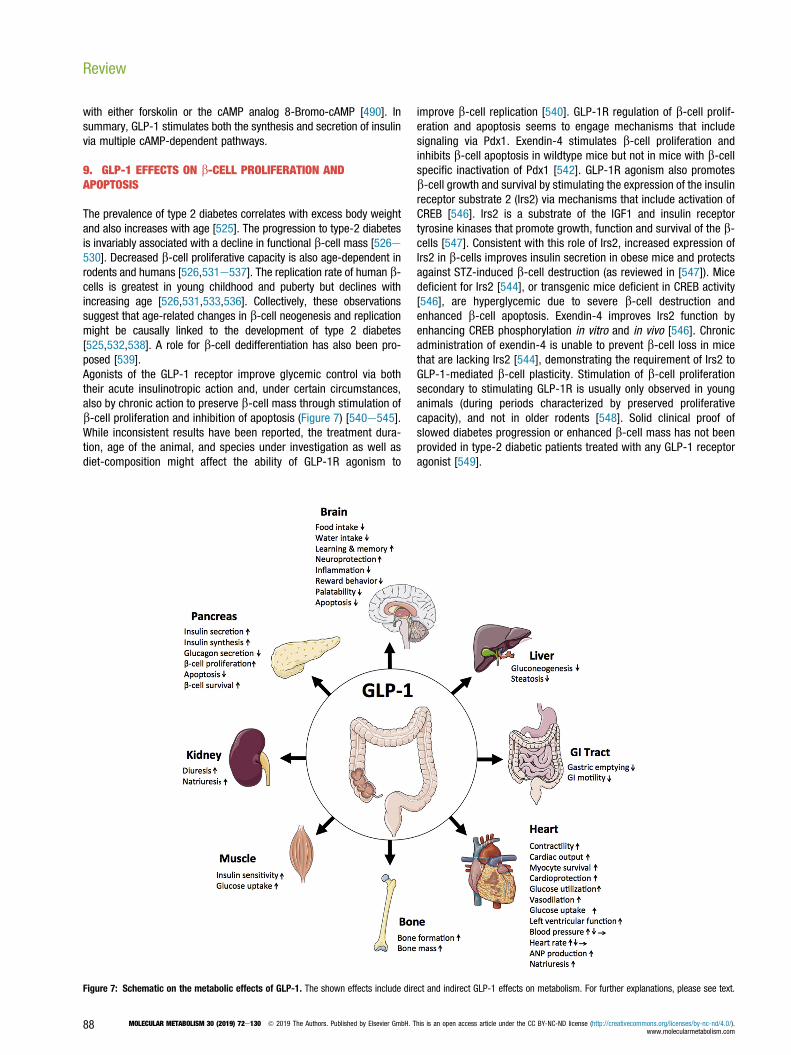
with either forskolin or the cAMP analog 8-Bromo-cAMP [490]. Insummary, GLP-1 stimulates both the synthesis and secretion of insulinvia multiple cAMP-dependent pathways.
9. GLP-1 EFFECTS ON b-CELL PROLIFERATION ANDAPOPTOSIS
The prevalence of type 2 diabetes correlates with excess body weightand also increases with age [525]. The progression to type-2 diabetesis invariably associated with a decline in functional b-cell mass [526e530]. Decreased b-cell proliferative capacity is also age-dependent inrodents and humans [526,531e537]. The replication rate of human b-cells is greatest in young childhood and puberty but declines withincreasing age [526,531,533,536]. Collectively, these observationssuggest that age-related changes in b-cell neogenesis and replicationmight be causally linked to the development of type 2 diabetes[525,532,538]. A role for b-cell dedifferentiation has also been pro-posed [539].Agonists of the GLP-1 receptor improve glycemic control via boththeir acute insulinotropic action and, under certain circumstances,also by chronic action to preserve b-cell mass through stimulation ofb-cell proliferation and inhibition of apoptosis (Figure 7) [540e545].While inconsistent results have been reported, the treatment dura-tion, age of the animal, and species under investigation as well asdiet-composition might affect the ability of GLP-1R agonism to
improve b-cell replication [540]. GLP-1R regulation of b-cell prolif-eration and apoptosis seems to engage mechanisms that includesignaling via Pdx1. Exendin-4 stimulates b-cell proliferation andinhibits b-cell apoptosis in wildtype mice but not in mice with b-cellspecific inactivation of Pdx1 [542]. GLP-1R agonism also promotesb-cell growth and survival by stimulating the expression of the insulinreceptor substrate 2 (Irs2) via mechanisms that include activation ofCREB [546]. Irs2 is a substrate of the IGF1 and insulin receptortyrosine kinases that promote growth, function and survival of the b-cells [547]. Consistent with this role of Irs2, increased expression ofIrs2 in b-cells improves insulin secretion in obese mice and protectsagainst STZ-induced b-cell destruction (as reviewed in [547]). Micedeficient for Irs2 [544], or transgenic mice deficient in CREB activity[546], are hyperglycemic due to severe b-cell destruction andenhanced b-cell apoptosis. Exendin-4 improves Irs2 function byenhancing CREB phosphorylation in vitro and in vivo [546]. Chronicadministration of exendin-4 is unable to prevent b-cell loss in micethat are lacking Irs2 [544], demonstrating the requirement of Irs2 toGLP-1-mediated b-cell plasticity. Stimulation of b-cell proliferationsecondary to stimulating GLP-1R is usually only observed in younganimals (during periods characterized by preserved proliferativecapacity), and not in older rodents [548]. Solid clinical proof ofslowed diabetes progression or enhanced b-cell mass has not beenprovided in type-2 diabetic patients treated with any GLP-1 receptoragonist [549].
Figure 7: Schematic on the metabolic effects of GLP-1. The shown effects include direct and indirect GLP-1 effects on metabolism. For further explanations, please see text.
Review
88 MOLECULAR METABOLISM 30 (2019) 72e130 � 2019 The Authors. Published by Elsevier GmbH. This is an open access article under the CC BY-NC-ND license (http://creativecommons.org/licenses/by-nc-nd/4.0/).www.molecularmetabolism.com

The anti-apoptotic effect of GLP-1R agonism has been demonstratedin vivo in mice [543,550,551] and rats [552,553] as well as in severalrodent [550,551] and human [554,555] cell lines, purified rat b-cells[543], and humans [556]. STZ-induced b-cell apoptosis is reducedupon administration of exendin-4 [543] or GLP-1 (7e36 amide) [551]and treatment with exendin-4 diminishes hyperglycemia induced bySTZ [543]. STZ-induced b-cell apoptosis is accelerated in mice defi-cient for the GLP-1 receptor [543]. In isolated rat b-cells, treatmentwith exendin-4 reduces the apoptotic effect induced by treatment withpro-apoptotic cytokines, such as IL1b, TNFa, and interferon gamma[543]. In mouse pancreatic bTC-6 cells, liraglutide enhances b-cellsurvival via stimulation of anti-apoptotic signaling mechanisms thatinclude stimulation of PI3 kinase-dependent phosphorylation of AKT,leading to inactivation of the pro-apoptotic protein BAD and silencingof FoxO1 [550].An innovative approach to assess human b-cell replication in vivo wasrecently established by the group of Alvin Powers [557]. In this model,human b-cells were grafted under the renal capsule of lean diabeticimmunodeficient NOD scid gamma mice. Using this model, exendin-4was found to enhance b-cell proliferation in juvenile but not adulthuman islets [557]. Notably, the age-dependent decline in the exendin-4 induced b-cell proliferation was not related to changes of GLP-1Rexpression, as also indicated by preservation of exendin-4-inducedinsulin secretion in the adult islets [557]. However, in juvenile butnot adult human islets, exendin-4 stimulated calcineurin/NFATsignaling and enhanced expression of proliferation-promoting factorssuch as NFATC1, FOXM1, and CCNA1 [557]. [557]. Collectively, thesedata suggest that the sensitivity of b-cell proliferative mechanisms torespond to GLP-1R agonism declines with increasing age [557].
10. IMPAIRED RESPONSIVENESS OF GLP-1 TO STIMULATEINSULIN SECRETION
GLP-1-stimulated insulin secretion has an estimated heritability of0.53, as demonstrated in twin studies [558]. The first evidence for aninfluence of genetic background on GLP-1-induced insulin secretionwas found for variation in the transcription factor 7-like 2 (TCF7L2)[559]. Infusion of GLP-1 during a classical hyperglycemic clamp innon-diabetic individuals results in a marked reduction of insulinsecretion in carriers of the TCF7L2 risk allele of SNP rs7903146.Diminished insulin secretion in response to treatment with GLP-1 hasbeen replicated in other studies [560e562].In addition to TCF7L2, several other genetic loci have been identified tobe associated with reduced GLP-1-induced insulin secretion, includingthe loci for GLP-1R [563], wolfram syndrome 1 (WFS1) [564] andchymotrypsinogen B1/2 (CTRB1/2) [565]. The molecular mechanismsleading to diminished GLP-1 responsiveness associated with geneticvariation in TCF7L2 most likely includes alterations of the WNTsignaling pathway and associated b-cell proliferation and insulin geneexpression [566]. Interestingly, genetic variation in a transcriptionalregulator of the insulin gene nuclear receptor subfamily 4 group Amember 3 (Nor-1) is able to rescue TCF7L2-mediated GLP-1 resis-tance [567]. Impairment of proinsulin conversion may be anothermechanism for TCF7L2 associated diminished GLP-1 efficacy [568].Another plausible mechanistic explanation for a diminished “incretineffect” is the TCF7L2-dependent repression of GLP-1R and GIPRexpression on the b-cells [569]. The association between WFS1 var-iants and impaired incretin action may result from alterations ofendoplasmic reticulum homeostasis and, consequently, b-celldysfunction [566]. Recently, using an untargeted integrative genomicsapproach, a set of genes that contained variants associated with GLP-
1-stimulated insulin secretion were described, which at the same timehave the potential to physically interact in the b-cell and are enrichedfor pathways that are important for insulin secretion [570]. Finally, theGLP-1 effects on insulin secretion are dependent on the metabolicstate of the individual. Incretin action may also be reduced duringhyperglycemia and in some individuals with diabetes, prediabetes andinsulin resistance [571].In summary, a reduced incretin effect on insulin secretion is associatedwith both genetic and metabolic alterations. The presence of bothhyperglycemia and genetically determined GLP-1 resistance canpotentiate the impairment of GLP1-induced insulin secretion [572].
11. GLP-1 EFFECTS ON GLUCAGON SECRETION
GLP-1 also decreases blood glucose by suppressing glucagon secre-tion (Figure 7) [475,573,574]. GLP-1 inhibition of glucagon secretionhas been demonstrated in vivo in numerous species including mice[575], dogs [576], and humans [516,517,573,577e581], as well as inthe isolated perfused pancreas of rats [473,474,582], dogs [473], andpigs [475] and in isolated intact murine islets [583]. Clamp studies inpatients with type 2 diabetes indicate that GLP-1 inhibition of glucagonsecretion is equally important for lowering blood glucose as GLP-1stimulation of insulin release [578].The mechanisms underlying GLP-1 suppression of glucagon secretionare complex. In the isolated perfused pig pancreas, GLP-1 dose-dependently stimulates the secretion of somatostatin [475] which is apotent suppressor of glucagon secretion [584]. Somatostatin inhibitsglucagon secretion via paracrine mechanisms and, when blocked,stimulates glucagon release in isolated rat islets [585,586]. In theisolated perfused rat pancreas, co-infusion of GLP-1 with a specificsomatostatin receptor 2 (SSTR2) antagonist (PRL-2903) abolishes theGLP-1-induced suppression of glucagon secretion [582]. While thesedata point to a prominent role of somatostatin in mediating GLP-1suppression of glucagon secretion, treatment of isolated murine is-lets with the SSTR2 antagonist CYN154806 does not fully inhibit GLP-1’s ability to suppress glucagon secretion [583]. Consistent with theconclusion that GLP-1 suppression of glucagon secretion does not fullydepend on somatostatin, GLP-1 effects on glucagon secretion can bemimicked by forskolin-induced changes in cAMP. Accordingly, treat-ment of isolated murine islets with low concentrations of forskolin (1e10 nM) suppresses glucagon secretion by up to 60%, while highconcentrations of forskolin (0.1e10 mM) stimulate glucagon release[583]. Notably, the PKA inhibitor 8-Br-Rp-cAMPS attenuates theinhibitory effect of GLP-1 on glucagon secretion, suggesting that GLP-1suppression of glucagon secretion is PKA-dependent [583]. In intactmurine islets, blockade of N-type Ca2þ channels withu-conotoxin, butnot L-type Ca2þ channels using nifedipine, abolishes low glucose(1 mM) stimulation of glucagon secretion and blunts the GLP-1-mediated inhibitor effects. In summary, these data indicate thatGLP-1 may inhibit a-cell glucagon secretion via PKA-dependentmodulation of N-type Ca2þ channel activity in addition to its para-crine action via somatostatin [583].GLP-1 may also inhibit glucagon secretion indirectly via its insulino-tropic effect on the b-cells. Accordingly, as described in recent reviewarticles [4,574], GLP-1 stimulates both the d-cells to secrete so-matostatin and the b-cells to secrete insulin, amylin, zinc, and GABA,all of which suppress the release of glucagon. In a-cell-derived IN-R1-G9 cells, insulin inhibits glucagon release via activation of phospha-tidylinositol 3-kinase (PI3K). Inhibition of PI3K by wortmannin offsetsthe ability of insulin to suppress glucagon secretion [587]. In a-cells,insulin further enhances translocation of GABA-A receptors [588], and
MOLECULAR METABOLISM 30 (2019) 72e130 � 2019 The Authors. Published by Elsevier GmbH. This is an open access article under the CC BY-NC-ND license (http://creativecommons.org/licenses/by-nc-nd/4.0/).www.molecularmetabolism.com
89
GABA released from b-cells enhances glucose inhibition of glucagonsecretion [589]. Insulin is co-crystalized with Zn2þ in the secretorygranules of the b-cells [590,591] and Zn2þ is co-secreted with insulinunder conditions of hyperglycemia [592,593]. Zn2þ acts in a paracrinemanner on the a-cells to inhibit the secretion of glucagon [593e595].Interestingly, disruption of the intrapancreatic infusion of Zn2þ but notof Zn2þ free insulin accelerates glucagon secretion in rats madediabetic by STZ, indicating that Zn2þ but not insulin is the mainstimulus underlying the Zn2þ-insulin inhibition of glucagon secretion[595]. Consistent with this, glucagon secretion is inhibited upontreatment of a-TC cells with Zn2þ [596]. As demonstrated in isolatedrat a-cells, intact islets and the perfused rat pancreas, Zn2þ inhibitspyruvate-induced glucagon secretion via opening of KATP channels andinhibition of a-cell electrical activity [593]. In summary, there iscredible evidence indicating that Zn2þ that is co-secreted with insulinfrom the b-cells plays a prominent role in inhibiting release of glucagon[593e595]. Amylin, which is also co-secreted with insulin, also affectsglucagon release from the a-cells. Amylin dose-dependently sup-presses arginine-mediated glucagon secretion in rats [597] whereaspharmacological inhibition of amylin signaling enhances glucagonsecretion [598]. Pramlintide, a synthetic amylin receptor agonist, im-proves glycemic control in diabetic patients via inhibition of post-prandial glucagon secretion and inhibition of gastric emptying[599,600]. Interestingly, amylin does not affect glucagon secretion inisolated islets [601] nor in the perfused rat pancreas [602,603],suggesting that amylin regulation of glucagon is not cell autonomous.Notably, while GLP-1 can potentially affect glucagon secretion via itsstimulatory effect on the b-cells in normal physiological conditions,GLP-1 also inhibits glucagon release in type 1 diabetes, thusdemonstrating that GLP-1 inhibition of glucagon secretion does notfully depend on the b-cell secretome [516].In summary, GLP-1 potently inhibits glucagon secretion. This occurs inpart by a paracrine stimulatory effect on the islets to secrete so-matostatin, and perhaps insulin, Zn2þ, GABA, and amylin, at elevatedglucose concentrations. GLP-1 likely suppresses glucagon secretionvia endocrine rather than direct mechanisms. Most studies report theabsence [397e399] or very restricted expression of the GLP-1 re-ceptor in a small subset (10%) of a-cells [395], and treatment ofisolated rat a-cells with GLP-1 enhances rather than inhibits therelease of glucagon [604e606]. GLP-1 may nevertheless also directlyinhibit glucagon secretion, because a-cell-specific GLP-1R KO micehave elevated non-fasting glucagon levels relative to wildtype controlsand female a-cell-specific GLP-1R KO mice are mildly glucose intol-erant with increased secretion of glucagon upon peripheral adminis-tration of glucose [607]. Further supporting a direct GLP-1 inhibition ofglucagon secretion is the fact that such secretion is not prevented inisolated human islets treated with the insulin receptor antagonist S961or the somatostatin receptor 2 antagonist CYN154806 [608].
12. CARDIOVASCULAR EFFECTS OF GLP-1
The presence of GLP-1R mRNA in the heart was first demonstrated inrats [380] and humans [381,609]. Expression was subsequentlyconfirmed at the protein level [201,610] and histologically using micethat express GFP under control of the GLP1-R promoter. In these micescattered GLP-1R expression was detected in cardiomyocytes of theatrium but not in the ventricles, and widespread expression wasdetected in smooth muscle cells of coronary vessels [395]. As dis-cussed in a comprehensive review article [611], the anatomicallocation of GLP-1R within the cardiovascular system has species-related differences. In mice, GLP-1R immunoreactivity using multiple
different antisera is found in the cardiomyocytes, endocardium,microvascular endothelium, and the smooth muscle cells in the cor-onary arteries [201]. In humans, the presence of GLP-1R has primarilybeen demonstrated in the endothelium, the coronary arteries and thesmooth muscle cells [381,612]. Nevertheless, there is little concor-dance between the available localization data from immunohisto-chemistry vs. detection of GLP-1R mRNA in situ hybridization. In viewof the many specificity problems associated with antibodies againstthe GLP-1 receptor, some of the incongruent findings may also resultfrom lack of antisera specificity. Using well-characterized antibodies,expression was noted in the non-human primate and human sinoatrialnode [392], which would be consistent with the effect of GLP-1 onheart rate. Surprisingly, in one recent study, robust expression of theGLP-1 receptor mRNA was also reported in human cardiac ventricles,but the exact localization of the GLP-1R protein could not be deter-mined [613].Although best known for its insulinotropic and weight lowering action,GLP-1R agonism also confers a series of beneficial effects on thecardiovascular system in rodents. These include an increase of car-diomyocyte survival via inhibition of apoptosis [614,615], ameliorationof endothelial dysfunction [612,616], improvement of regional, andglobal cardiac output following injury and heart failure [614,617e619](Figure 7). Reduction of blood pressure is also reported in hypertensivehumans [620]. Prolonged treatment with liraglutide further improvescardiovascular outcome in patients with T2DM [621]. Of note, GLP-1improvement of cardiovascular performance is, at least in part, inde-pendent of its ability to decrease body weight and to improve lipidmetabolism, and GLP-1 may promote its cardiac effects via directaction at GLP-1R as well as indirect mechanisms independent ofcardiac GLP-1R action [201,611,622].
12.1. Effects of GLP-1 on cardiac performance after cardiac injuryEndothelial dysfunction is a common co-morbidity associated withinsulin resistance and T2DM. Treatment of type-2 diabetic patients thatpresent coronary artery disease with GLP-1 improves endothelialfunction without affecting insulin resistance [612]. Similar findingshave been reported for GLP-1 treatment of type-1 diabetes [623] butGLP-1 improvement of endothelial function seems to be related tonative GLP-1 and is not observed when GLP-1R agonists are comparedhead-to-head with equally glucose lowering medications.GLP-1 improvement of endothelial function, assessed by flow-mediated vasodilation, was also demonstrated during a hyperglyce-mic clamp in patients with T2DM and normoglycemic controls [616]. Inrats, GLP-1 (7-36amide) and, perhaps paradoxically, the GLP-1Rantagonist exendin (9e39), dose-dependently cause relaxation ofthe aorta, potentially via elevation of cAMP formation and activation ofKATP channels [624].In dogs with acute myocardial infarction, infusion of GLP-1 improvescardiac performance by increasing myocardial glucose uptake and byenhancing left ventricular function [617]. In the same animal model,infusion of GLP-1 limited myocardial stunning (ventricular dysfunctionwithout myocardial necrosis) following reperfusion [618]. These dataare in agreement with non-controlled clinical studies reporting that72 h of native GLP-1 infusion improved regional and global left ven-tricular performance in patients with acute myocardial infarction andsevere systolic dysfunction [619]. In mice, 7-day treatment with lir-aglutide prior to induction of myocardial infarction increases survivaland reduces cardiac injury and infarction size, while improving cardiacoutput [614]. In the isolated perfused rat heart, acute GLP-1R agonismprotects from ischemia/reperfusion injury [625,626], an effect that canbe blocked by pretreatment with either exendin (9e39), the cAMP
Review
90 MOLECULAR METABOLISM 30 (2019) 72e130 � 2019 The Authors. Published by Elsevier GmbH. This is an open access article under the CC BY-NC-ND license (http://creativecommons.org/licenses/by-nc-nd/4.0/).www.molecularmetabolism.com
inhibitor Rp-cAMP, the PI3 Kinase inhibitor LY294002 or the MAP Ki-nase inhibitor UO126 [625]. The cardioprotective effect of GLP-1Ragonism seems to be, at least in part, independent of GLP-1-induced weight loss, dependent on the canonical GLP-1R, andaccompanied by enhanced expression of cardioprotective genes in theheart, including Akt, GSK3b, PPARD, NRF-2, and HO-1 [614]. Treat-ment of primary neonatal mouse cardiomyocytes with liraglutide in-creases cAMP formation and reduces apoptosis, as indicated byinhibition of TNFa induction of caspase 3 activity [614]. GLP-1R ago-nism has direct cell autonomous anti-apoptotic effects in the rodentheart, and GLP-1 reduces hypoxia-induced apoptosis in isolatedneonatal rat cardiomyocytes, and inhibition of PI3K using LY29400 orMAPK using UO126 blocks the anti-apoptotic effect of GLP-1 [615].GLP-1 has different effects on left ventricular function and cardiaccontractility under healthy vs. pathological conditions. GLP-1-inducedimprovement of left ventricular function has been reported in dogswith cardiomyopathy [617], in patients with acute myocardial infarction[619], and in the isolated mouse heart [201]. At least part of theimprovement in cardiac output may be driven by an increase in heartrate. Other studies, however, report failure of GLP-1 to directlyaccelerate contraction in isolated adult rat cardiomyocytes, notablydespite GLP-1 stimulation of cAMP accumulation [627]. Further,treatment of the healthy isolated rat heart with GLP-1 has been re-ported to reduce left ventricular contractility despite GLP-1 stimulationof myocardial glucose uptake [628]. However, as demonstrated in thesame study, GLP-1 enhances recovery of the heart after ischemia andimproves left ventricular function and myocardial glucose uptake[628]. GLP-1R KO mice have increased baseline left ventriculardeveloped pressure (LVDP) during perfusion [201]. Mice deficient forGLP-1R have decreased resting HR and an increased thickness of theleft ventricular wall relative to wildtype controls [201,629], as well asreduced cardiac contractility when challenged with insulin orepinephrine [629]. Collectively, these data indicate that GLP-1 im-proves cardiac performance and output in the post-ischemic heart butotherwise may decrease cardiac output under non-pathological con-ditions. Although mice with cardiomyocyte-specific deletion of GLP-1Rhave lower basal heart rate (HR), they fully retain the cardioprotectivepotential of GLP-1R agonism following left arterial descending (LAD)coronary artery occlusion [630]. These data suggest that car-diomyocyte GLP-1R signaling is required for regulation of HR but isunrelated to the cardioprotective effects of GLP-1R agonism followingcardiac injury.
12.2. Cardiometabolic effects of GLP-1(9-36amide)Accumulating evidence indicates that some cardiac effects of GLP-1(7-36amide) are actually mediated via the DPP-4-generated GLP-1(9-36amide) and its smaller degradation products and, thus, are in-dependent of GLP-1R signaling. At a dose of 0.3 nmol/L, pretreatmentwith GLP-1 (7-36amide) improves recovery of LVDP after ischemia/reperfusion (I/R) injury in both WT and GLP-1R KO mice [201]. Inter-estingly, a >10 fold greater dose of exendin-4 is needed relative toGLP-1 (7-36amide) to achieve a similar effect in WT mice but still withlower efficacy relative to GLP-1 (7-36amide) in GLP-1R KO mice [201].The superior cardioprotective effect of GLP-1 (7-36amide) relative toexendin-4 and the preservation of efficacy of GLP-1 (7-36amide) butnot exendin-4 in GLP-1R KO mice suggest that some of the car-dioprotective effects of GLP-1 (7-36amide) are independent of GLP-1Rsignaling and are potentially mediated via the DPP-4-cleaved GLP-1metabolite GLP-1 (9-36amide) [201]. Interestingly, when mice arepretreated with either GLP-1 (7-36amide) or GLP-1 (9-36amide), onlyGLP-1 (7-36amide) improves LVDP. However, when administered
during reperfusion, both GLP-1 (7-36amide) and GLP-1 (9-36amide)improve functional recovery from I/R injury in both WT and GLP-1R KOmice [201]. Consistent with these data, while exendin-4 but not GLP-1(9e36) has infarct limiting potential in the isolated rat heart, exendin-4and GLP-1 (9e36) both improve left ventricular performance duringreperfusion [626]. The infarct-limiting potential of exendin-4 is abol-ished by treatment with exendin (9e39), thus indicating that thiscardioprotective effect is GLP-1R-dependent [626]. Treatment of iso-lated mouse cardiomyocytes with either exendin4 or GLP-1 (9-36amide) increases phosphorylation of AKT and ERK [631], both ofwhich are known to positively affect cardiomyocyte growth and sur-vival [632,633].Collectively, these data indicate that pharmacologic administration ofGLP-1 (9-36amide) may have a functional role to improve recoveryfrom I/R injury via GLP-1R-independent mechanisms, whereas GLP-1(7-36amide) but not GLP-1 (9-36amide) affects cardiac contractility viaGLP-1R signaling [201]. Further supporting a cardiometabolic role ofGLP-1 (9-36amide), both GLP-1 (7-36amide) and GLP-1 (9-36amide),but not exendin-4, promote vasodilation in isolated and ex vivo culturedmesenteric blood vessels [201], i.e., the vasodilatory effect of GLP-1(7-36amide) was not different relative to that of GLP-1 (9-36amide)and was likewise preserved in GLP-1R KO mice [201]. Whether the lowendogenous concentration and the restricted pharmacokinetic actionprofile of endogenous GLP-1 metabolites are sufficient to elicit sucheffects under physiological circumstances remains questionable.
12.3. Effect of GLP-1 on heart rate (HR) and blood pressure (BP)GLP-1 effects on HR and BP are species-related. In rats, intravenousinfusion of GLP-1 (7-37amide) or exendin-4 acutely increases HR aswell as systolic, diastolic, and mean arterial BP (Figure 7) [634,635]. Inrats, GLP-1 stimulation of BP peaks around 3e5 min post-injectionand returns to baseline after 25 min [634]. Both central and periph-eral administration of GLP-1R agonists increase HR and BP in rodents[636e638], with induction of c-Fos expression in adrenal medullarycatecholamine neurons and activation of tyrosine hydroxylase, the keyenzyme involved in production of norepinephrine, in the brainstem[638]. These data collectively suggest that some of the acute cardio-vascular effects of GLP-1 might be due to brain enhancement ofcatecholamine outflow and elevation of sympathetic tone. However, inrats, GLP-1 elevation of BP is preserved upon pretreatment withreserpine, propanolol or phentolamine, thus indicating that the hy-pertensive effect of GLP-1 does not exclusively depend on catechol-amine signaling [634]. However, while the hypertensive effect of GLP-1is not abolished by inhibition of b-adrenergic signaling [634], exendin-4 induction of HR is blocked by the b-adrenergic receptor antagonistpropranolol [639], and exendin-4 fails to affect HR in adrenalectomizedrats (as reviewed in [622]). In mice, both acute and chronic centraladministration of exendin-4 increases HR and BP with concomitantdecrease in excitatory glutamatergic and inhibitory glycinergicneurotransmission to preganglionic parasympathetic cardiac vagalneurons. GLP-1 stimulation of HR and BP is mediated via GLP-1Rsignaling because i.v. infusion of exendin (9e39) blocks GLP-1 andexendin-4 induction of HR and BP [635], and these actions are lost inGlp1r�/- mice [640]. Collectively, these data suggest that acute GLP-1effects on HR and BP in normal animals may involve signaling via both,the sympathetic and parasympathetic nervous systems. Not all rodentstudies report elevated BP in response to GLP-1R agonism, but dosecould be a discriminatory factor. While some studies report increasedBP in rodents upon acute or chronic GLP-1R agonism [634e636,638],others report decreased BP, especially in experimental models asso-ciated with the development of hypertension such as salt-sensitive db/
MOLECULAR METABOLISM 30 (2019) 72e130 � 2019 The Authors. Published by Elsevier GmbH. This is an open access article under the CC BY-NC-ND license (http://creativecommons.org/licenses/by-nc-nd/4.0/).www.molecularmetabolism.com
91

db mice and Dahl S rats [641,642]. In mice, liraglutide blunts hyper-tension induced by angiotensin II (Ang-II), an effect that is absent inGLP-1R KO mice, and is also attenuated upon pretreatment of WT micewith exendin (9e39) [640].While in most rodent studies GLP-1 increases HR and BP, i.v. infusionof GLP-1 (7-36amide) acutely increases HR without affecting meanarterial BP in conscious calves [643]. Most human studies report amoderate stimulatory effect of GLP-1 on HR with unchanged or withreduced BP only in hypertensive individuals [620,622,640,644e649].Acute infusion of GLP-1 increases heart rate and cardiac output inhealthy human volunteers, potentially via GLP-1-induced vasodilationin adipose tissue and skeletal muscle [650]. As demonstrated inhealthy human volunteers, GLP-1 infusion acutely increases blood flowin the subcutaneous abdominal adipose tissue and skeletal musclewithout alterations in splanchnic blood flow [650]. Notably, in contrastto GIP [651e654], the GLP-1 induced increase in adipose tissue bloodflow is not dependent on postprandial hyperglycemia and hyper-insulinemia and is observed even under conditions of fasting glucoseand insulin concentrations [650].GLP-1R agonism has also been demonstrated to reduce BP in peoplewith T2DM (as reviewed in [611,622,649]). Notably, in healthy humanvolunteers, acute administration of GLP-1, GLP-1 (9-36amide), orexenatide did not affect blood flow in the mesenteric or renal arteries,excluding splanchnic vasodilatation as a potential mechanism under-lying the chronic hypotensive effect of GLP-1 [644]. In a recent meta-analysis of 60 clinical studies, GLP-1R agonists were demonstrated toreduce diastolic BP with a range of �1.84 to �4.60 mmHg and toslightly increase heart rate by 2e3.35 beats/min [620]. Relative toplacebo, the reduction of diastolic BP only reached significance for thetreatment of exenatide 10 mg/twice daily (�1.08 mmHg) [620]. Thesedata align with an integrated analysis of 36 clinical studies thataffirmed cardiovascular safety with up to 4-years’ treatment witheither DPP-4 inhibitors or GLP-1R agonists [655].The molecular mechanisms underlying GLP-1 improvement of BP areincompletely understood. Potential mechanisms may include GLP-1-induced vasodilation and activation of nitric-oxide-dependentmechanisms [612,640,656]. Indeed, liraglutide stimulates relaxa-tion of the aorta [640] but GLP-1 might also indirectly lower BP via itsability to reduce body weight. GLP-1R agonism could also improve BPvia its ability to stimulate sodium excretion (natriuresis) via thekidneys [657]. The long-term steady-state BP is affected by theintravascular volume, which is influenced by the vascular tone andthe extracellular fluid volume (ECFV); the ECFV is in turn determinedby the sodium balance. Accordingly, when BP increases, the kidneysrespond by increasing natriuresis to decrease BP via reduction of theECFV [92,658,659]. GLP-1R agonists acutely stimulate natriuresis inrodents and may also do so in humans, lowering BP, potentially alsovia reduction of the ECFV [641,642,660,661]. In mice, liraglutidereduces angiotensin II (Ang-II) stimulation of systolic and diastolic BP,an effect that is absent GLP-1R KO mice or by pretreatment withexendin (9e39) or the natriuretic peptide receptor antagonist anantin[640]. In WT mice and in isolated atrial cardiomyocytes, liraglutidestimulates the production and secretion of atrial natriuretic peptide(ANP), and liraglutide fails to induce natriuresis and to lower BP inANP KO mice [640]. Liraglutide stimulation of cardiac ANP secretionis PKA independent but mediated by Epac2 in a cAMP and PLC-dependent manner [640]. However, in humans there is no consis-tent effect of GLP-1 on ANP secretion whereas angiotensin II isgenerally lowered [657,662e664]. In summary, GLP-1R agonismimproves BP through stimulation of natriuresis via Epac2-dependentstimulation of cardiac ANP secretion [640]. Nevertheless, the
majority of hypertensive human subjects treated with liraglutide donot exhibit an increase in circulating ANP [662].
13. GLP-1 EFFECTS ON FOOD INTAKE AND BODY WEIGHT
One of the most frequently described extrapancreatic effects ofpharmacokinetically-optimized GLP-1R analogs is their ability to lowerbody weight via centrally regulated inhibition of food intake (Figure 7).Suppression of food intake by GLP-1R agonism has been demon-strated in numerous species including mice [465e467,665,666], rats[667,668], birds [669,670], pigs [671,672], non-human primates[673,674], and humans [675e678]. The anorexigenic effect of GLP-1Ragonism has been demonstrated in numerous clinical studies and fMRIanalysis support that GLP-1 inhibition of food intake is centrallymediated in healthy individuals and patients with type-2 diabetes[679e684].
13.1. Effect of peripheral administration of GLP-1 analogs on foodintakePeripheral administration of GLP-1 analogs decreases body weight viasuppression of food intake [465e467,665,685]. Depending on themolecule (native GLP-1 or a longer-acting GLP-1 analog) and the routeof administration, GLP-1 and its analogs seem to engage differentsignaling pathways to either directly or indirectly transmit the signal toreduce food intake to the CNS.Central GLP-1R appear to be necessary for the anorectic effect ofperipherally administered GLP-1R agonists in rodents [686e688].When administered s.c., liraglutide fails to inhibit eating in micepossessing a CNS-specific deletion of GLP-1R [688]. Another reportindicates that i.v. infusion of liraglutide inhibits eating in rats by directlyactivating hypothalamic Pomc/Cart neurons in the ARC, without acti-vating GLP-1-producing neurons in the hindbrain, and independent ofGLP-1R signaling in vagal afferents, the area postrema (AP) and thePVN [686]. In rats, central (i.c.v.) administration of exendin (9e39) wasalso sufficient to attenuate the anorexigenic effect of i.p. injected lir-aglutide and exendin-4 [687], and bilateral neurochemical lesions ofthe lateral parabrachial nucleus (IPBN) with ibotenic acid blunted theanorexigenic effect of peripherally administered exendin-4 [689].When applied either s.c. or i.p., exendin-4 and liraglutide inducedacute neuronal activation (measured by activation of cFos) in PVN, AP,and NTS [686,690]. Interestingly, the short-term anorexigenic effectsof i.p. injected low-dose liraglutide and exendin-4 were also attenuatedby subdiaphragmatic vagal deafferentation [687,691], but longer-termeffects of these compounds on food intake did not depend on intactvagal afferents [691]. All of these findings are consistent with theconcept that GLP-1 receptor agonists inhibit eating and reduce bodyweight primarily by acting directly in the brain, and that various brainareas are involved in mediating the eating-inhibitory effects of GLP-1receptor agonists. This presumably reflects the ability of long-actingGLP-1 analogs to enter the brain at areas with an incomplete BBB,such as the median eminence.While these data collectively emphasize the importance of neuronalGLP-1R for the food intake inhibition by long-acting GLP-1 analogs, it isunclear whether and to what extend this accounts for physiologicaldoses of intestinal-derived native GLP-1. When infused into the hepaticportal vein, GLP-1 (7-36amide) increased cFos expression in the NTS,the AP, and the central nucleus of the amygdala, but not in the hy-pothalamic ARC or PVN [692], i.e., native GLP-1 appears to induce anactivation pattern in the brain that is different from the pattern inducedby long-acting GLP-1 receptor agonists. While peripherally adminis-tered GLP-1 seemingly crosses the blood brain barrier (BBB) via simple
Review
92 MOLECULAR METABOLISM 30 (2019) 72e130 � 2019 The Authors. Published by Elsevier GmbH. This is an open access article under the CC BY-NC-ND license (http://creativecommons.org/licenses/by-nc-nd/4.0/).www.molecularmetabolism.com
diffusion, the half-life of GLP-1 is less than 2 min, leading todisagreement regarding whether peripherally derived GLP-1 is whatactivates CNS GLP-1R under physiological conditions.In rats, i.v. administration of GLP-1 at high physiological doses stim-ulates vagal afferents, and this is blocked by pretreatment withexendin (9e39) [693]. While this activation appears to be involved inthe insulinotropic and gastric emptying inhibiting effects of exogenousGLP-1 [693], it does not appear to be crucial for the effect of i.v.administered native GLP-1 on eating [694], because GLP-1 infusionsinto the hepatic portal vein did not lose their potency to reduce foodintake after subdiaphragmatic vagal deafferentation in rats [692,694].Interestingly, however, the food intake inhibition following i.p.administration of a supposedly “physiological” dose of GLP-1 (7-36amide) depends on the integrity of abdominal vagal afferents [694],indicating that peripheral GLP-1 can inhibit eating via at least two partlyseparate pathways. Consistent with a role of vagal afferents and vagalafferent GLP-1 receptors in the eating-inhibitory effect of peripheralendogenous GLP-1, virus-mediated knockdown of GLP-1R in vagalafferent neurons increased meal size, accelerated gastric emptying,and reduced prandial insulin secretion in rats, while not affecting 24 hfood intake or body weight [518]. The latter finding matches the resultsobtained in Phox2b-Cre GLP-1 receptor KO mice, lacking GLP-1 re-ceptors in the autonomic nervous system, which have normal bodymass and composition in response to both chow and HFD [688], yetexhibit partial attenuation of the anorectic and weight loss effects ofperipherally administered GLP-1R agonists [520]. In humans, the effectof i.v. GLP-1 infusion on food intake was lost after truncal vagotomy[695], indicating that vagal afferents are involved in mediating theeffects of circulating native GLP-1 on appetite. In rats, on the otherhand, lesions of the AP or administration of exendin (9e39) into the 4thventricle blocked the food intake inhibition by GLP-1 infused into thehepatic portal vein [261], suggesting that high concentrations ofcirculating GLP-1 act on the hindbrain to reduce food intake. In onestudy GLP-1 infused into the vena cava or hepatic portal vein of ratshad similar effects on food intake [696], whereas in another study inrats GLP-1 infusions into the jugular vein inhibited food intake morepotently than infusions into the hepatic portal vein [697]. These find-ings argue against a hepatic portal site of action of GLP-1 to inhibiteating, and further emphasize that the route of administration is animportant consideration when analyzing food intake inhibition by GLP-1R agonism.The data summarized above were collected in mice, rats, and humans;in addition to varying experimental conditions, species differences mayaccount for some of the discrepancies observed. Nevertheless,collectively these data indicate that vagal afferents are involved inmediating the satiating effects of endogenous peripheral GLP-1 orexogenously administered GLP-1 and the acute inhibition of food intakein response to peripheral GLP-1 receptor agonists under certain con-ditions. However, vagal afferents and vagal afferent GLP-1 receptorsare presumably less important for the effects of peripheral GLP-1 onbody mass.
13.2. Effect of central administration of GLP-1R analogs on foodintakeThe acute anorectic effect of centrally administered GLP-1 wasdescribed simultaneously 1996 by Steve Bloom’s group [668] and byMads Tang-Christensen et al. [92] and was confirmed by others[667,698]. In mice and rats, the short-term inhibition of food intakeinduced by i.c.v. administered GLP-1R agonists is dose-dependent,can be blocked by pretreatment with exendin (9e39) [668,698] andis absent in GLP-1R KO mice [690]. Inhibition of food intake by centrally
administered GLP-1R analogs is accompanied by increased cFosneuronal activity in the PVN, amygdala, NTS, AP, lateral parabrachialnucleus, and the ARC [668,696,698]. These data align with theexpression of GLP-1R throughout the CNS [395,396]. Furthermore, cellbodies that are immunoreactive to GLP-1 antisera are detected in theNTS, and GLP-1 immunoreactive nerve fibers are widely distributedthroughout the brain with highest density in the hypothalamus, thal-amus and septal regions whereas the lowest density is in the cortexand hindbrain [86,698]. Supporting a role of the NTS in mediating GLP-1’s effects on food intake, adeno-associated virus (AAV) mediatedknock-down of GLP-1R in the NTS results in higher food intake andmeal size [699]. Chronic blockade of GLP-1R with intra 3rd ventricularinfusion of exendin (9e39) increases food intake and fat mass [700].Direct intraparenchymal delivery of low, ventricle subthreshold dosesof GLP1R agonists to GLP1R expressing nuclei throughout the brain,including e.g., NTS, lPBN, ventral tegmental area (VTA), PVH, PVT,nucleus accumbens (NAcc), ventral hippocampus, and lateral septumreduced food intake via reductions in meal size. Consistent with this,delivery of exendin (9e39) to these sites increases meal size and foodintake [701e705].Hindbrain GLP-1R activation enhances phosphorylation of PKA andMAPK while decreasing the activity of AMPK in the NTS, and inhibitionof PKA/MAPK activity by administration of Rp-cAMP or UO126 atten-uates food intake inhibition of exendin-4 administered into the 4thventricle [706]. These data suggest that hindbrain GLP-1R activationsuppresses food intake via PKA/MAPK-induced inhibition of AMPK.Diphtheria toxin-induced ablation of GLP-1 producing neurons in theNTS had no effect on either ad libitum food intake or glucose meta-bolism [90]. Nevertheless, in rats, activation of hindbrain GLP-1R viai.c.v. administration of exendin-4 into the 4th ventricle reduces foodintake via reduction of meal frequency; i.e., via increased satiety [706].Intra 4th-ventricular administration of exendin (9e39) or lesions of theAP also block food intake inhibition by GLP-1 infused into the hepaticportal vein (HPV) [261], suggesting that high levels of circulating GLP-1inhibit eating at least in part by acting on the hindbrain.
13.3. Molecular mechanisms underlying food intake inhibition byGLP-1The molecular underpinnings regulating GLP-1 inhibition of food intakeand body weight loss are complex. Undoubtedly, brain GLP-1Rsignaling is crucial for the anorexigenic effect of exogenously admin-istered long-acting GLP-1R agonists, as demonstrated by failure ofliraglutide to suppress food intake in CNS-specific GLP-1R KO mice[688]. Consistent with this, i.c.v. administration of exendin (9e39) issufficient to attenuate the anorexigenic effect of peripherally admin-istered liraglutide and exendin-4 [687]. However, CNS-specific GLP-1RKO mice have normal body mass and composition whether on a chowor HFD [688], suggesting that these receptors may be necessary forthe pharmacological response to GLP-1 but not the physiologicalcontrol of body weight. However, data obtained in KO animals generallyhave to be regarded with caution since compensatory mechanisms,which limit interpretations and affect study outcomes, might develop.Depending on the route of administration (and the GLP-1R agonistused), peripherally administered GLP-1 analogs seem either to actdirectly in the hypothalamus and hindbrain, both areas with incompleteBBB, or else on vagal afferents to transmit the signal to the hindbrainwhich then projects to other key feeding areas in the brain. HindbrainGLP-1R activation enhances phosphorylation of PKA and MAPK whiledecreasing the activity of AMPK in the NTS, and inhibition of PKA/MAPKactivity by administration of Rp-cAMP or UO126 attenuates food intakeinhibition of exendin-4 administered into the 4th ventricle [706]. These
MOLECULAR METABOLISM 30 (2019) 72e130 � 2019 The Authors. Published by Elsevier GmbH. This is an open access article under the CC BY-NC-ND license (http://creativecommons.org/licenses/by-nc-nd/4.0/).www.molecularmetabolism.com
93

data suggest that hindbrain GLP-1R activation suppresses food intakevia PKA/MAPK-induced inhibition of AMPK.The anorexigenic effect of GLP-1R agonism involves the glucose-dependent silencing of central AMPK action. Activity of AMPK isincreased under conditions of fasting [707] and is decreased underconditions of enhanced glucose utilization [708e710]. Consistent withAMPK’s role as a stress- and nutrient-responsive protein kinase,pharmacological, physiological (e.g. fasting-induced), or genetic acti-vation of AMPK stimulates food intake whereas AMPK inhibition de-creases food consumption [707,711]. Central administration of GLP-1reduces the activity (phosphorylation) of AMPK [712,713] and treat-ment of mouse hypothalamic GT1-7 cells with exendin-4 stimulatesglycolysis and decreases phosphorylation of AMPK [714]. Pharmaco-logical activation of AMPK by administration of 5-aminoimidazole-4-carboxamide-1-b-Deribofuranoside (AICAR) in either the 3rd [714]or the 4th ventricle [706] attenuates the anorectic effect of exendin-4injected into the same ventricles. Consistent with these data, i.c.v.administration of the glycolysis inhibitor 2-deoxyglucose (2-DG) like-wise increases activity of AMPK and blunts food intake suppression byi.c.v.-injected exendin-4 [714]. In summary, compelling evidence in-dicates that the anorexigenic effect of central GLP-1 receptor activationis partially mediated by central stimulation of glycolysis andconcomitant silencing of AMPK activity. Accordingly, pharmacologicalactivation of AMPK in the VMH blunts food intake suppression by i.c.v.exendin-4 [714,715] and attenuates central GLP-1 effects on brownand inguinal white adipose tissue [666].The anorexigenic effect of GLP-1 seems to be transduced via GLP-1Rsignaling in several areas of the brain, including both hypothalamicnuclei and the hindbrain. Microinjection of exendin-4 into the NTSreduces intake of palatable high-fat diet and inhibits food rewardbehavior [716], and hindbrain inhibition of GLP-1R via administration ofexendin (9e39) into the 4th ventricle increases food intake [717]. Theanorexigenic and weight-lowering effects of liraglutide are bluntedupon genetic ablation of GLP-1R in glutamatergic neurons, yet fullypreserved when GLP-1R is deleted in GABAergic neurons [718]. Pe-ripheral administration of GLP-1 (7-36amide) enhances neuronal ac-tivity in the VMH [719,720], and direct administration of GLP-1 into theLH, VMH, or DMH of rats acutely decreases food intake [721]. Similarfindings were reported in rats that received direct infusion of GLP-1 (7-36amide) into the PVN [722,723]. In rats, liraglutide decreases foodintake when directly injected into the ARC, LHA, or PVN while directadministration into the VMH decreases body weight without affectingfood intake, but increases the expression of Ucp1 in the brown andinguinal white adipose tissue [666]. The acute effects of exendin-4 toreduce food intake and to improve glucose handling are preservedupon knockdown of GLP-1R in the VMH [715], indicating that the ef-fects of central GLP-1R activation on eating and BAT thermogenesisare mediated by at least partially separate brain areas and circuitries.
13.4. GLP-1 effects on visceral illnessInhibition of food intake can be due to other factors than satiety,including gastric discomfort, e.g., gastroparesis, associated withvisceral illness and stress. Anorexia associated with visceral illnesscan be chemically-induced by administration of lithium chloride (LiCl),a substance known to promote nausea and emesis in humans [724e726] and taste avoidance conditioning and pica in rodents, that do notvomit [727]. Pica is the measure of the amount of kaolin clayconsumed in response to treatments that trigger nausea and vomitingin humans and is a broadly accepted proxy of visceral malaise in non-vomiting rats. Taste avoidance conditioning describes the reduction inthe intake of novel taste or food that has been associated with a
treatment that induces negative consequences e.g., including the ef-fects of peripheral pain, lactose malabsorption or the effects LiCl [728].In rats, peripheral administration of LiCl induces cFos neuronal activityin the IPBN, the AP and the NTS [726,729] with remarkable overlap inthe cFos activity pattern induced by centrally administered GLP-1[726]. Apart from inducing cFos activity in the same brain areas[696,726,730], LiCl and central GLP-1R agonism both decrease lo-comotor activity [668,731], slow GI motility and gastric emptying[573,732], reduce body temperature [732,733], and induce adversegastrointestinal effects including nausea, emesis, and taste avoidance[696,724e726,734e737]. Selective ablation of NTS PPG neuronsprevents stress-induced hyperphagia in mice but has no effect onlong-term ad libitum food intake, body weight, or glucose tolerance[90]. These data suggest that NTS PPG neurons are not essential forlong-term regulation of food intake but are required for the short-termhypophagia in response to stress [738,739].Collectively, these studies highlight that, in some species, GLP-1 andLiCl might engage similar central signaling mechanisms and suggestthat central GLP-1s anorexigenic effect might be influenced by aversivepathways and gastric discomfort. However, there are other data thatwould challenge the simple explanation that the reduction in feedingand body weight resulting from CNS GLP1R agonism is simplyexplained by inducing an aversive visceral state. Targeted agonistdelivery to CNS GLP1-R expressing sites, PVT, NTS, VTA, NAcc, or PBNreduces food intake without the expression of pica, a proxy of visceralillness [704,716,740,741]. In addition, Jelsing et al. report that foodintake inhibition by liraglutide is observed in the absence of reducedgastric emptying effects that some have linked to visceral malaise[742]. Importantly, both van Bloomendaal et al. [682] and ten Kulveet al. [743] provide data to support the hypothesis that food intakereduction after GLP-1 receptor agonism is mediated by actions onappetite- and reward-related areas of the human brain, an interpre-tation also supported by rodent studies [704,716,740,741].
13.5. GLP-1 effects on reward behaviorCentral GLP-1 regulation of food intake is not restricted to GLP-1Rsignaling in the hypothalamus and hindbrain and is not limited tomodulation of homeostatic feeding, i.e. food intake to maintain energyhomeostasis. GLP-1 also affects non-hunger related (hedonic) feedingby targeting brain areas involved in reward, motivation and addiction,such as the VTA, NAcc, lateral septum [701,741,744,745], and PVT[704]. These mesolimbic regions express GLP-1R [347] and receiveprojections from PPG neurons in the NTS [354,741,744]. Peripheraladministration of exendin-4 [691] or direct administration of GLP-1 intothe core of the NAcc [744] increases cFos expression in this region,and direct activation of NAcc GLP-1R reduces food intake while itsdirect inhibition by exendin (9e39) induces hyperphagia [744]. Foodintake inhibition by NAcc GLP-1R is unrelated to taste avoidance [744]and does not induce pica [701,741]. Administration of exendin-4 intothe shell of the NAcc decreases sucrose reward behavior in the pro-gressive ratio (PR) operant conditioning test [701]. Consistent with this,GLP-1R agonism decreases a variety of reward behaviors in rodents,including the motivation to lever press for food reward in an operantconditioning test [746], alcohol intake [747,748], alcohol seekingbehavior [747], amphetamine-induced conditioned place preference(Amp-CPP) [748], and hedonic feeding [748]. The inhibitory effect ofGLP-1R analogs on reward behaviors is blunted in mice with CNS-specific deletion of GLP-1R [748]. Several lines of evidence indicatethat NAcc GLP-1R activation decreases food intake by reducing foodpalatability. In satiated rats given a choice between chow and high-fatdiet (HFD), exendin-4 preferentially reduces consumption of the more
Review
94 MOLECULAR METABOLISM 30 (2019) 72e130 � 2019 The Authors. Published by Elsevier GmbH. This is an open access article under the CC BY-NC-ND license (http://creativecommons.org/licenses/by-nc-nd/4.0/).www.molecularmetabolism.com

palatable HFD [741]. In rats, intra-NAcc injection of exendin (9e39)increases the size of a sucrose meal and accelerates the licking fre-quency during the first meal, an indicator of increased palatability[749]. In summary, GLP-1R agonism decreases homeostatic and he-donic feeding, and modulates food intake not only via the hypothala-mus and the hindbrain but also via signaling in the mesolimbic system,in which GLP-1R activation affects reward behavior and palatability.
13.6. GLP-1 transport across the blood brain barrier (BBB)Several lines of evidence indicate that peripherally administered GLP-1can cross the blood brain barrier (BBB). Studies using a stabilizedradiolabeled (Ser8)GLP-1 revealed rapid uptake into the brain afterintravenous (i.v.) administration without self-inhibition when chal-lenged with increased doses of unlabeled (Ser8)GLP-1 or upon inhi-bition of brain GLP-1 influx upon pretreatment with exendin (9e39).Small peptide GLP-1R analogs including liraglutide, lixisenatide, andexendin-4 have all been demonstrated to cross the BBB upon pe-ripheral administration [750,751], and HPLC analysis confirmed thatmost of the peripherally administered exendin-4 reaches the brainintact [751,752]. While these studies suggest that GLP-1 influx into thebrain might be a GLP-1R-independent process with passive diffusion ofGLP-1 through the BBB [750e752], other studies suggest that uptakeof liraglutide into the brain is GLP-1R-dependent [686,753]. None-theless, brain regions with incomplete BBB, such as the subfornicalregion and the AP, are main targets of peripherally injected 125I-labeled GLP-1 [754].
14. GLP-1 EFFECTS ON WATER INTAKE
GLP-1 inhibition of water intake was first reported by Mads Tang-Christensen in 1996 [92] and was later confirmed by others [755e760]. GLP-1 inhibition of water consumption is independent of foodintake [758] and is blocked by pretreatment with exendin (9e39) [92].In rats, inhibition of drinking by GLP-1 (7e36 amide) is dose-dependent and occurs within 30 min following its central (i.c.v.) orperipheral (i.p.) administration [92]. In one study in healthy humans,i.v. infusion of GLP-1 decreased water consumption by 36% inresponse to i.v. infusion of hypertonic saline (5% NaCl) [755]. Pe-ripheral but not central administration of GLP-1 fails to inhibit angio-tensin (ANG) II-induced drinking in rats that have been treated withmonosodium glutamate (MSG) to induce damage of the ARC and thesensory circumventricular organs, suggesting that peripherallyadministered GLP-1 effects drinking behavior via CNS-dependentmechanisms [760].GLP-1 affects renal function by decreasing water intake as well as bystimulating the excretion of urine and sodium (Figure 7). The natriureticand diuretic effect of GLP-1R agonism was first shown in 1996 [92]and was subsequently confirmed in rodents [642,660,761] and inhealthy, obese, and type-2 diabetic humans [661,663]. While the acutenatriuretic effect of GLP-1 has been solidly confirmed in humans[762e764], 12 wk treatment of T2DM patients with liraglutide or theDPP-4 inhibitor sitagliptin had no effect on markers of renal function,including glomerular filtration rate, renal plasma flow, albumin-to-creatine ratio, and excretion of sodium, potassium, and urea [765].These data suggest that that GLP-1’s effects on renal function might betransient and vanish upon more chronic treatment. In one study inT2DM patients, the natriuretic effect of liraglutide persisted after 3 wktreatment [662].In rats, GLP-1 induced natriuresis and diuresis is accompanied byincreased renal plasma flow and glomerular filtration rate [660,761].Denervation of the kidney results in failure of GLP-1 to increase
glomerular filtration rate and decreases the natriuretic and diureticeffect of GLP-1 [761]. These data suggest that the natriuretic anddiuretic effect of GLP-1 might be due to inhibition of sodium reab-sorption in the proximal tubule and emphasize that GLP-1 requiresintact renal innervation to increase glomerular filtration. Studies inrodents and humans indicate that GLP-1R is expressed in the renalvasculature [392,766e768]. However, treatment of isolated porcineproximal tubular cells with GLP-1 reduced sodium re-absorption [768].Consistent with this, GLP-1, exendin-4, and the DPP-4 inhibitor sita-gliptin reduced proximal tubular sodium reabsorption in rats[660,761,769,770] and humans [663]. In the renal proximal tubule,GLP-1 reduced Naþ/Hþ exchanger isoform 3 (NHE3)-mediated bi-carbonate reabsorption [660]. Consistent with this, exendin-4decreased NHE3-mediated sodium-dependent pH recovery inporcine kidney LLC-PK1 cells [771]. Inhibition of NHE3 activity by GLP-1R agonism seems to be mediated via cAMP activation of PKA, whichphosphorylates NHE3 at its Ser552 and Ser605 residues[660,771,772]. In rodents [660,761] and humans [663,763], GLP-1Ragonism increases renal lithium clearance, a marker for proximaltubular sodium reabsorption. These data suggest that the diuretic andnatriuretic effects of GLP-1 include changes in renal hemodynamicsand reduced sodium reabsorption due to PKA-dependent down-regulation of NHE3 activity in the renal proximal tubule. GLP-1 mightalso affect kidney function via its cardiovascular effects. In rats,infusion of GLP-1 increases renal blood flow and the glomerularfiltration rate [660,761], and this effect is potentially mediated via GLP-1’s ability to increase vasodilation of the afferent renal arteriole. Inwildtype but not GLP-1R KO mice, liraglutide stimulates the secretionof natriuretic peptide (ANP) from the atrial cardiomyocytes and lir-aglutide fails to affect natriuresis and vasorelaxation in mice deficientfor ANP [640].
15. GLP-1 EFFECTS ON ENERGY EXPENDITURE
A role of endogenous GLP-1R signaling in the control of energyexpenditure was inferred from studying mice with congenital GLP-1Rdeficiency. Relative to wildtype controls, lean chow-fed GLP-1R KOmice do not have alterations in energy expenditure, neither when keptat a standard room temperature nor when challenged with moderatecold exposure [773]. However, relative to wildtype controls, diet-induced obese GLP-1R KO mice exhibit reduced energy expenditureat standard ambient room temperature but not at temperatures fa-voring thermoneutrality [774]. Because thermoneutrality minimizes thecontribution of non-shivering thermogenesis to whole-body energyexpenditure and because non-shivering thermogenesis requiresfunctional brown adipose tissue (BAT) [775], these data imply acontribution of endogenous GLP-1R signaling to the control of energyexpenditure, at least in diet-induced obese mice, by regulating BATactivity. This contribution requires GLP-1R signaling in the brain; i.e.,the brain plays a critical role in the control of BAT activity [776] andGLP-1R is expressed in the brain, including in nuclei involved in thecontrol of BAT [347,389]. In keeping with this, site-specific disruptionof GLP-1R on neurons of the DMH of rats results in significant bodyweight gain paralleled by a reduction in energy expenditure and BATheat production [777]. The DMH is part of the neuronal networkinvolved in the control of BAT thermogenesis [776] and receivesinnervation from pre-proglucagon-expressing neurons of the hindbrain[778]. A priori, this positions those neurons as potential contributors tothe neural circuits controlling energy expenditure by regulating BATthermogenesis in rodents. Intriguingly, emerging data involving gain orloss of function from those cells in mice argue against such a
MOLECULAR METABOLISM 30 (2019) 72e130 � 2019 The Authors. Published by Elsevier GmbH. This is an open access article under the CC BY-NC-ND license (http://creativecommons.org/licenses/by-nc-nd/4.0/).www.molecularmetabolism.com
95
contribution. Thus, diphtheria toxin-mediated ablation of those neurons[90] does not result in long-term changes on food intake or bodyweight, which suggests a minimal impact on energy expenditure.Conversely, acute chemogenetic activation of those neurons fails toincrease, and rather transiently reduces, energy expenditure [357].Disregarding differences due to experimental conditions includingacute versus chronic intervention or animal species or strains, thesedata suggest that endogenous GLP-1R signaling in the brain may playa constitutive role in the homeostatic control of BAT thermogenesis,beyond its physiological modulation by brain-derived GLP-1.However, this does not preclude the possibility that exogenousadministration of GLP-1 may regulate energy expenditure by acting onbrain GLP-1R. In fact, an early report in rats described an increase inenergy expenditure during the first hours of the dark cycle followingi.c.v. administration of low doses of GLP-1 (7-37amide) [779].In mice, acute central (i.c.v.) but not peripheral administration of GLP-1increased brown adipose tissue (BAT) temperature and increased theactivity of sympathetic fibers that innervate the inguinal BAT [773].Consistent with these data, central administration of the GLP-1R ag-onists liraglutide and exendin-4 increases energy expenditure and BATthermogenesis through mechanisms that include AMPK signaling inthe mediobasal hypothalamus [666]. Taken together, these studiessuggest that pharmacological activation of brain GLP-1R signalingincreases energy expenditure in mice.Peripheral administration of GLP-1R agonists has led to variableresults with respect to energy expenditure. One study in rats foundan increase of energy expenditure upon i.v. infusion of GLP-1, andwith even greater potency relative to centrally administered GLP-1[780]. These data are consistent with the demonstration that i.v. butnot i.p. administered GLP-1 inhibits eating via activation of hind-brain GLP-1 receptors [261,694] and suggests a similar mechanismfor i.v. GLP-1-stimulation of energy expenditure. In contrast,another study in rats reported an acute decrease of energyexpenditure associated with a decrease in interscapular skin tem-perature following i.p. administration of exendin-4 and this effectwas attenuated when GLP-1R was knocked down on vagal afferentneurons [781]. These data are consistent with reports that theanorexigenic effect of i.p. administered GLP-1 depends on vagalafferents [692,694] and thus suggests a similar mechanism for i.p.GLP-1 effects on energy expenditure. Caudal brainstem delivery ofCocaine- and amphetamine-regulated transcript (CART) induceshypothermia, and this effect is blocked by pretreatment withexendin (9e39) [782]. Several studies in mice [465,467,685,774]and humans [783e787] have failed to detect changes in energyexpenditure following chronic or acute peripheral treatment withdifferent GLP-1R agonists. One human study did report an acutedecrease of diet-induced thermogenesis due to lower carbohydrateoxidation following i.v. infusion of GLP-1 [788].In summary, the experimental evidence supports a contribution ofbrain-GLP-1R signaling in the control of energy expenditure, at least inrodents, via control of BAT activity. The frequently reported absence ofperipherally administered GLP-1 mimetics to regulate energy expen-diture in mice and humans has several implications. First, the BBBmight limit access to brain areas where GLP-1R could potentiallymediate effects on energy expenditure; second, the underlying GLP-1analog, the feeding status of the test subject, and the route ofadministration are important considerations when analyzing GLP-1effects on energy expenditure, and, third, rodents may be uniquelysensitive to mechanisms activating energy expenditure through thesympathetic nervous system.
16. GLP-1 EFFECTS ON GASTRIC EMPTYING
In addition to its direct and vagally mediated insulinotropic action on b-cells, GLP-1 also improves postprandial glucose handling by inhibitionof gastric emptying, decelerating the rate at which glucose is absorbedinto the circulation (Figure 7) [573,789e791]. Inhibition of gastricmotility and gastric acid secretion following treatment with GLP-1Ragonists has been demonstrated in mice [792], rats [793e795],dogs [796e798], pigs [799,800], and humans [573,789e791,799,801e805]. In rats, inhibition of gastric emptying occurs af-ter peripheral or central administration of GLP-1 [795], and this effectis mediated via hindbrain GLP-1R signaling [806]. Vagal afferent GLP-1receptor knockdown accelerates gastric emptying [518] and vagalafferent denervation [795] or peripheral administration of exendin (9e39) [794,795] blocks the effect of centrally or peripherally adminis-tered GLP-1 on gastric emptying. These data are consistent with thefailure of GLP-1 to inhibit gastric acid secretion in vagotomized humansubjects [807] as well as the failure of GLP-1 to inhibit vagally-inducedmotility in the isolated perfused pig antrum [800]. These data suggestthat GLP-1-mediated inhibition of gastric motility includes vagal af-ferents, as well as GLP-1R dependency in the periphery and the brain[795]. Inhibition of adrenergic signaling through combined adminis-tration of phentolamine and propanolol abolishes the inhibitory effect ofGLP-1 on gastric motility, suggesting that GLP-1 inhibits gastricemptying via adrenergic signaling [794]. In fasted rats, co-administration of GLP-1 and GLP-2 leads to a greater inhibition ofsmall bowel motility relative to either monotherapy [793]. Exendin-4fails to affect gastric emptying in GLP-1R KO mice [792]. In healthyhuman volunteers, intravenous GLP-1 decelerates gastric emptyingafter the first meal to a greater extent than after a second meal,indicating that GLP-1 inhibition of gastric emptying undergoes rapiddesensitization [803]. In overweight humans, liraglutide 3 mg solidlyinhibits gastric emptying after 5wk of treatment, but the efficacymarkedly decreases after 16wk of treatment, yet with still significanteffect relative to placebo treated controls [808]. In rats, liraglutide- butnot exendin-4-induced inhibition of gastric emptying is markedlydiminished after 14 wk of treatment [742]. Because gastric emptying isa prerequisite for glycemic rises after meals, which in turn triggerinsulin secretion, deceleration of gastric emptying in response toexogenous GLP-1 leads to lesser post-meal glucose excursions andreduced insulin secretory responses [809]. This has led to doubtsabout the importance of an incretin role for pharmacological GLP-1administration, because incretin hormones should lead to a net in-crease in post-nutrient insulin secretory responses [810].
17. GLP-1 EFFECTS ON THE HYPOTHALAMIC PITUITARYADRENAL (HPA) AXIS
Neurons of the NTS are important in the regulation of thehypothalamus-pituitary-adrenal (HPA) axis in response to stress [811],and evidence indicates that brain GLP-1R signaling plays an integralrole in the acute CNS response to stress and aversive stimuli [811].Preproglucagon-positive neurons of the NTS have dense projections toother brain areas, including the hypothalamic PVN, where theyinnervate CRH-releasing neurons [812]. GLP-1R is expressed in neu-rons of the PVN [347] that co-localize with CRH [813]. Centraladministration of GLP-1 in rats and mice stimulates the HPA axis andincreases secretion of corticosterone, and this is accompanied byincreased cFOS immunoreactivity in CRH-positive neurons in the PVN[668,698,814]. In rodents and humans, peripherally administered
Review
96 MOLECULAR METABOLISM 30 (2019) 72e130 � 2019 The Authors. Published by Elsevier GmbH. This is an open access article under the CC BY-NC-ND license (http://creativecommons.org/licenses/by-nc-nd/4.0/).www.molecularmetabolism.com
GLP-1R agonists transiently stimulate the HPA axis and increasecirculating levels of corticosterone, aldosterone, and ACTH [815e817].Peripheral administration of exendin-4 or liraglutide increase circu-lating corticosterone in mice under basal conditions and also during aforced swimming test [818]. Mice with PVN-selective deletion of GLP-1R (using Sim1 cre) have impaired stress-induced HPA axis activation,and this is further accompanied by protection from stress-inducedweight loss, reduced stress-induced cardiovascular endpoints andreduced anxiety-like behavior [813]. In rats, combined administrationof dexamethasone and exendin-4 leads to a greater anorexigenic effectand greater weight loss relative to treatment with exendin-4 alone[819]. Similar results were reported in mice treated with GLP-1/dexamethasone, an unimolecular peptide-nuclear hormone conjugatedesigned to optimize dexamethasone delivery into GLP-1R positivecells [820]. Ablation of hindbrain catecholamine neurons using anti-dopamine-b-hydroxylase-saporin (DSAP) blunts the ability of exendin-4 to increase corticosterone secretion but interestingly potentiates thefood intake inhibition by exendin-4 [819]. These data suggest thatactivation of the HPA axis antagonizes the satiation effect of GLP-1Ragonism [819].
18. GLP-1 EFFECTS ON LEARNING, MEMORY ANDNEUROPROTECTION
GLP-1R expression has also been demonstrated in the rat [347,821]and mouse [822] hippocampus, a brain region implicated in spatiallearning and memory [823,824]. In rats, central GLP-1R agonismimproves several aspects of learning and memory, as demonstrated byimproved performance in the Morris water maze test and by enhancedlatency in the passive avoidance test [417]. GLP-1’s improvement oflearning and memory can be blocked by pretreatment with exendin(9e39), and is absent in mice deficient for the GLP-1 receptor [417].Targeted hippocampal restoration of GLP-1R expression in otherwiseGLP-1R-deficient mice using AAV-mediated gene transfer reversesGLP-1 improvement of learning and memory in the passive avoidancetest [417].Central GLP-1R signaling has also been demonstrated to have neu-roprotective effects. GLP-1 and exendin-4 enhance differentiation andneurite outgrowth in rat pheochromocytoma (PC12) cells and in humanneuroblastoma SK-N-SH cells [825]. The neuroprotective effect ofGLP-1R agonism is similar to that conferred by nerve growth factor(NGF) and is blocked when PC12 cells are co-incubated with exendin(9e39) [825]. Both GLP-1 and exendin-4 protect cultured hippocampalneurons against glutamate-induced apoptosis [826]. The time andseverity of seizures induced by systemic administration of the neuro-toxin kainic acid is enhanced in GLP-1R KO mice relative to WT controlswhereas the destructive effect of kainic acid is ameliorated in the GLP-1R KO mice upon targeted AAV-mediated restoration of hippocampalGLP-1R expression [417].The molecular mechanisms underlying the neuroprotective effects ofcentral GLP-1R agonism are, at least in part, mediated through theability to increase cAMP formation and subsequently to enhanceactivation of PI3-kinase and ERK. GLP-1R agonism also increasescAMP levels in cultured hippocampal neurons [826] and in PC12 cells[825]. Pharmacological inhibition of either PI3-Kinase or ERK blocksthe stimulatory effect GLP-1 and exendin-4 on neurite outgrowth inPC12 cells [825]. In PC12 cells, GLP-1 stimulation of neurite outgrowthis only partially silenced by treatment with the PKA inhibitor H89,indicating that cAMP-mediated activation of PI3K and ERK followingGLP-1R agonism does not fully depend on PKA signaling, at least inthese cells [825,827].
Huntington’s disease (HD) is characterized by severe neuro-degeneration that is caused by a mutation of the huntingtin protein(HTT) [828]. The neurotoxicity and neurodegeneration induced by themutant HTT protein has been attributed to enhanced oxidative stressand accumulation of HTT due to inappropriate autophagic HTT clear-ance [829]. The prevalence of T2DM is elevated in patients with HD[830], suggesting that impaired insulin sensitivity might be a causalfactor contributing to neurodegeneration in HD patients [830,831].Overexpression of mutant HTT impairs insulin signaling and stimulatesneuronal apoptosis in human SK-N-MC neuronal cells [831]. Treat-ment of SK-N-MC cells with liraglutide improves insulin sensitivity andenhances cell viability, potentially by mechanisms that includeamelioration of neuronal glucotoxicity, improvement of oxidative stressand less aggregation of the mutant HTT through stimulation of AMPK-mediated autophagy [831].Alzheimer’s disease (AD) is characterized by neurodegeneration ofcholinergic neurons in the hippocampus. In a rat model of neuro-degeneration, GLP-1 and exendin-4 ameliorated neurodegeneration byreducing ibotenic acid-induced depletion of cholinergic cell bodies, asdemonstrated by preservation of choline acetyltransferase immuno-reactivity in basal forebrain cholinergic neurons [826]. In rats,administration of GLP-1R agonists into the hippocampus further pre-vented impairment of spatial learning and memory induced by centraladministration of amyloid b (Ab), a protein considered to play acausative role in impairment of learning and memory in Alzheimerdisease [832,833]. Consistent with this, chronic GLP-1R agonism hasbeen demonstrated in several mouse models of AD to prevent thedevelopment of memory deficits [834e836]. Clinical studies evalu-ating the effect of GLP-1R agonism on neurodegeneration associatedwith AD are scarce. AD is frequently reported to be accompanied bydecreased glucose transport across the BBB and 6 month treatment ofAD patients with liraglutide improved BBB glucose transfer relative toplacebo controls [837]. In a double blind placebo controlled study, 12week treatment with liraglutide in patients at risk for AD did not showcognitive differences between the study cohorts [838]. Other clinicaltrials evaluating the neuroprotective effects of GLP-1R agonism on ADare currently ongoing [839]GLP-1 analogs have also proven successful for the treatment of Par-kinson’s Disease (PD). PD is typically characterized by degeneration ofdopaminergic neurons, a condition that can be mimicked in experi-mental animals by administration of the dopaminergic neurotoxinMPTP (1-Methyl-4-phenyl-1,2,3,6-tetrahydropyridin) [840e842]. Inmice, 7-d infusion of exendin-4 into the lateral ventricle was sufficientto protect from MPTP-induced damage of the dopaminergic systemand from the development of locomotor deficits that typically arise fromdopamine deficiency [843]. Similar neuroprotective effects have beenreported in various PD rodent models treated with GLP-1R agonists[844e849]. Consistent with this, exendin-4 increases survival andlevels of tyrosine hydroxylase, the key enzyme for dopamine produc-tion, in primary neuronal cultures treated with the dopaminergic toxin6-hydroxydopamine (6-OHDA) [843]. Several clinical studies haveconfirmed the potential of GLP-1R agonism to ameliorate clinicalsymptoms of PD, including long-lasting improvements of motor andcognitive function [850e853].
19. GLP-1 EFFECTS ON BONE
The first understanding of GLP-1 actions in skeletal physiology arosefrom GLP-1R KO mice, which exhibit a reduction in trabecular bonemass due to a higher osteoclast activity [854]. In rodents, but not inhumans, activation of thyroid GLP-1R leads to C-cell stimulation in the
MOLECULAR METABOLISM 30 (2019) 72e130 � 2019 The Authors. Published by Elsevier GmbH. This is an open access article under the CC BY-NC-ND license (http://creativecommons.org/licenses/by-nc-nd/4.0/).www.molecularmetabolism.com
97
thyroid [855,856], and involvement of calcitonin in GLP-1-mediatedosteoclast activation has been suspected [854]. Furthermore, theseanimals exhibit profound modifications of bone matrix compositionwith a lower collagen maturity that jeopardizes the biomechanicalresponse and hence contributes to skeletal fragility [857]. In com-parison, GIPR KO animals present higher trabecular bone mass butsignificant lower biomechanical resistance due to alterations of tissuemineral density distribution and collagen maturity [858,859]. Doubleincretin receptor knock-out (DIRKO) animals revealed that the GLP-1/GLP-1R pathway was important for the integrity of cortical but nottrabecular bone [860].GLP-1R agonists have been extensively used in animal models ofskeletal fragility. In ovariectomy-induced bone loss, exenatide en-hances bone formation due to osteoblast activation [857]. In hindlimb-unloading rats, exenatide improves bone mass by promoting osteo-genic differentiation of bone marrow stem cells [861]. In type-1 dia-betic models, although liraglutide is less effective than GIP analoguesin promoting osteoblast differentiation, it improves tissue materialbiomechanics by reducing collagen degradation [862]. In type-2 dia-betic models (genetically-inherited or diet-induced obesity), exenatidesignificantly enhances bone strength by increasing trabecular bonemass, bone formation and trabecular microarchitecture. Exenatidesignificantly improves collagen maturity [863,864]. The use of theunimolecular triple agonist GIP-Oxm is also significantly potent inimproving bone strength and restoring trabecular bone mass [865].However, its mechanism of action is linked to reduced bone formationand reduced osteoclast surfaces. GIP-Oxm leads to changes incollagen lamellar orientation, improves collagen maturity and reductionin advanced glycation end-products in the bone matrix. However, it isnot clear whether these ameliorations in bone strength result fromactivation of skeletal GLP-1 receptors or whether it requires extra-skeletal receptors as observed for the GIP/GIPR pathway [866]. Assuch, a stable GIP agonist has been demonstrated to improve bonequality in rats [867], whereas DPP-4 inhibition positively modified bonecomposition but not bone microstructure in high fat fed mice [868].Human studies report less compelling data on the effects of GLP-1 orits analogues in bone remodeling markers or fracture. In contrast toGLP-2 administration, subcutaneous injection of GLP-1 in healthy in-dividuals had no effects on bone formation or resorption markers[869]. In non-diabetic, overweight men, GLP-1 infusion seems toreduce the extent of bone resorption in a similar way to GIP [787].However, in obese, non-diabetic patients under anti-psychotic drugs,exenatide failed to modify significantly the pattern of bone remodeling[870]. In contrast, liraglutide was capable of enhancing bone formationin obese weight-reducing women while having no effects on boneresorption [871]. Data are more controversial in type-2 diabetes andalthough first reports did not find evidence of protective effects of theGLP-1 receptor agonist on bone fracture [872,873], more recent datasuggest that liraglutide and lixisenatide, but not exenatide, contributeto reduce fractures in the diabetic population [874].
20. PHARMACOLOGICAL USE OF GLP-1 ANALOGS TO TREATOBESITY AND DIABETES
The exogenous supplementation of native hormones, commonly pu-rified from tissue homogenates, has led to numerous, seminal dis-coveries, most prominently the identification of insulin in 1921 [875]followed by the discovery of glucagon in 1923 [3]. Despite numeroussuch groundbreaking discoveries, including identifying the intestine asa source of incretins [56], this practice did not necessarily identifyhormones capable of sufficiently lowering body weight.
Its anorexigenic and insulinotropic action make GLP-1 an appealingcandidate for the treatment of obesity and diabetes. The pharmaco-logical value of native GLP-1, however, is limited by a short half-life[182,185,876] and dose-limited adverse gastrointestinal effects[734,877]. A common approach to enhance the therapeutic benefits ofGLP-1-based pharmacology is therefore to improve its pharmacoki-netics. Biochemical modification of the native peptide has enhancedexposure and time of action to improve the metabolic efficacy attolerable doses. Furthermore, these optimized molecules allow for lessfrequent administration, which places less burden on the patient andimproved compliance.
20.1. Optimized GLP-1 monoagonistsSeveral structurally refined GLP-1 derivatives with improved bioavail-ability and sustained action have been developed for the treatment ofT2DM [878]. These drugs include exenatide (Byetta�, short-acting;and Bydureon�, long-acting, AstraZeneca), lixisenatide (Lyxumia�,Sanofi), liraglutide (Victoza�; Novo Nordisk), dulaglutide (Trulicity�;Eli Lilly & Co), albiglutide (Tanzeum�, GlaskoSmithKline), and sem-aglutide (Ozempic�, Novo Nordisk) [878].Exenatide (Byetta� and Bydureon�, AstraZeneca), is a synthetic 39-amino acid peptide (first discovered as exendin-4 in the saliva of thegila monster, Heloderma suspectum) (Figure 8) [879]. The first 30amino residues of exendin-4 have w53% sequence homology tomammalian GLP-1, whereas the C-terminal nonapeptide extensionhas no similarities. Unlike GLP-1, exendin-4 has a glycine at thesecond amino acid position at the N-terminus, which protects thepeptide from DPP-4-mediated degradation and inactivation. In addi-tion, exendin-4 differs from mammalian GLP-1 by a series of aminoacids in the central and C-terminal domains that include Leu10,Lys12, Gln13, Met14, Glu16, Glu17, Tyr19, Arg20, Leu21, Glu24,Lys27, Asn28, and Gly30. The C-terminal end of exendin-4 isextended relative to GLP-1 by 9 amino acids. The C-terminalextension supports a secondary structure by formation of a trypto-phan cage and improves potency at the GLP-1 receptor [880]. Whenadministered either i.v., i.p., or s.c. in rats, the half-life of exendin-4is 18e41 min, 125e174 min, and 90e216 min, respectively [194].Relative to GLP-1 (7e36 amide), the bioavailability of exendin-4 isnearly doubled from 36 to 67% for GLP-1 to 74e76% for exendin-4,with decelerated plasma clearance from 35 to 38 ml/min to 4 e8 ml/min [194]. These pharmacokinetic parameters support twice-daily administration in humans. In patients with T2DM, exenatidedecreases fasting and post-prandial levels of blood glucose [881e884], slows gastric emptying [885], reduces cardiovascular riskfactors [886] and decreases body weight accompanied by suppres-sion of food intake [885e887]. The placebo-subtracted weight lossfrom baseline induced by exenatide is typically in the range of lowsingle-digit percentages. After 82 wk of treatment of T2DM patientswith exenatide, the placebo-subtracted weight loss from baseline isapproximately 5% [886]. A once-weekly formulation of exenatide hasbeen developed by Amylin Pharmaceuticals and has been marketedby AstraZeneca since 2012 under the trade name Bydureon�. Theprimary ingredient in the formulation that allows for extended-releaseis polyD,L-lactide-co-glycolic acid. Together with the other excipientsof the formulation, the pharmacokinetics of exenatide are improvedby a mechanism involving the formation of a depot of microspheresat the injection site, which slows dissipation from the injection site[888]. Compared to classical exenatide given twice daily (BID, theonce-weekly (QW) formulation at a 2 mg dose level provides arelatively stable plasma concentration at steady-state (w300 pg/ml)because of the more continuous delivery of the drug, with equal (or
Review
98 MOLECULAR METABOLISM 30 (2019) 72e130 � 2019 The Authors. Published by Elsevier GmbH. This is an open access article under the CC BY-NC-ND license (http://creativecommons.org/licenses/by-nc-nd/4.0/).www.molecularmetabolism.com
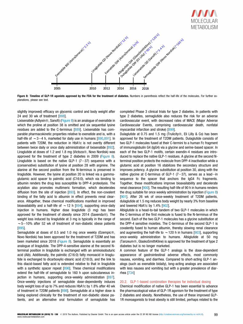
slightly improved) efficacy on glycemic control and body weight after24 and 30 wk of treatment [888].Lixisenatide (Adlyxin�, Sanofi) (Figure 8) is an analogue of exenatide inwhich the proline at position 38 is omitted and six sequential lysineresidues are added to the C-terminus [889]. Lixisenatide has com-parable pharmacokinetic properties relative to exenatide and is, with ahalf-life of w3e4 h, marketed for daily use in humans [890,891]. Inpatients with T2DM, the reduction in HbA1c is not overtly differentbetween twice daily or once daily administration of lixisenatide [892].Liraglutide at doses of 1.2 and 1.8 mg (Victoza�, Novo Nordisk) wasapproved for the treatment of type 2 diabetes in 2009 (Figure 8).Liraglutide is based on the native GLP-1 (7e37) sequence with a(conservative) substitution of lysine at position 28 with arginine. Thealanine at the second position from the N-terminus is preserved inliraglutide. However, the lysine at position 20 is linked via a gamma-glutamic acid spacer to palmitic acid (C16:0), which via binding toalbumin renders the drug less susceptible to DPP-4 proteolysis. Theacylation also promotes multimeric formation, which deceleratesdiffusion from the site of injection [893]. In effect, the non-covalentbinding of the fatty acid to albumin in effect prevents renal clear-ance. Altogether, these chemical modifications manifest in improvedbioavailability and a half-life of w12 h [894], supporting once-dailyinjection in humans. Higher dose liraglutide (3 mg) has beenapproved for the treatment of obesity since 2014 (Saxenda�). Theweight loss induced by liraglutide at 3 mg is typically in the range ofw5e10% after 52 wk of treatment of non-diabetic obese patients[895].Semaglutide at doses of 0.5 and 1.0 mg once weekly (Ozempic�,Novo Nordisk) has been approved for the treatment of T2DM and hasbeen marketed since 2018 (Figure 8). Semaglutide is essentially ananalogue of liraglutide. The DPP-4-sensitive alanine at the second N-terminal position in liraglutide is exchanged with an aminoisobuturicacid (Aib). Additionally, the palmitic (C16:0) fatty monoacid in liraglu-tide is exchanged to dicarboxylic-stearic acid (C18:0), and the link tothis diacid-based fatty acid is extended relative to that in liraglutidewith a synthetic spacer repeat [896]. These chemical modificationsextend the half-life of semaglutide to 160 h upon subcutaneous in-jection in humans, supporting once-weekly administration [897].Once-weekly injections of semaglutide dose-dependently inducesbody weight loss of up to 7% and reduces HbA1c by 1.8% after 40 wkof treatment in T2DM patients [898]. Semaglutide at a higher dose isbeing explored clinically for the treatment of non-diabetic obese pa-tients, and an alternative oral formulation of semaglutide has
completed Phase 3 clinical trials for type 2 diabetes. In patients withtype 2 diabetes, semaglutide also reduces the risk for an adversecardiovascular event, with decreased rates of MACE (Major AdverseCardiovascular Events, comprising cardiovascular death, nonfatalmyocardial infarction and stroke) [899].Dulaglutide at 0.75 and 1.5 mg (Trulicity�, Eli Lilly & Co) has beenapproved for the treatment of T2DM patients. Dulaglutide consists oftwo GLP-1 molecules fused at their C-termini to a human Fc fragmentof immunoglobulin G4 (IgG4) via a glycine and serine-based spacer. Ineach of the two GLP-1 motifs, certain exendin-4 residues are intro-duced to replace the native GLP-1 residues. A glycine at the second N-terminal position protects the molecule from DPP-4 inactivation while aglutamic acid at position 16 stabilizes the secondary structure andimproves potency. A glycine substitution at position 30, along with thenative glycine at C-terminus of GLP-1 (7e37), serves as a leadeinsequence to the spacer that anchors the IgG4 Fc fragments.Together, these modifications improve bioavailability and deceleraterenal clearance [900]. The resulting half-life of 90 h in humans rendersthe drug suitable for once weekly administration by injection (Figure 8)[901]. After 26 wk of once-weekly treatment of T2DM patients,dulaglutide at 1.5 mg reduces body weight by nearly 3% from baselineand lowered HbA1c by 1.4% [902].Albiglutide is a head-to-tail tandem of two GLP-1 molecules in whichthe C-terminus of the first molecule is fused to the N-terminus of thesecond. Each of the two GLP-1 molecules has a glycine substitution atthe DPP-4 sensitive moieties. The C-terminus of the second GLP-1 iscovalently fused to human albumin, thereby slowing renal clearanceand augmenting the half-life to w120 h in humans [903], supportingonce-weekly administration to humans. Albiglutide at 50 mg(Tanzeum�, GlaskoSmithKline) is approved for the treatment of type 2diabetes but is no longer marketed.A common feature of the GLP-1 analogs is the dose-dependentappearance of gastrointestinal adverse effects, most commonlynausea, vomiting, and diarrhea. Compared to short-acting GLP-1 an-alogs (such as exenatide bidaily), long-acting analogs are associatedwith less nausea and vomiting but with a greater prevalence of diar-rhea [734]
20.2. GLP-1-based combination therapies for individual dosingChemical modification of native GLP-1 has been essential to advancethe pharmacological value of GLP-1R agonism for the treatment of type2 diabetes and obesity. Nonetheless, the use of these improved GLP-1R monoagonists to treat obesity is still limited, perhaps related to the
Figure 8: Timeline of GLP-1R agonists approved by the FDA for the treatment of diabetes. Numbers in parenthesis reflect the half-life of the molecules. For further ex-planations, please see text.
MOLECULAR METABOLISM 30 (2019) 72e130 � 2019 The Authors. Published by Elsevier GmbH. This is an open access article under the CC BY-NC-ND license (http://creativecommons.org/licenses/by-nc-nd/4.0/).www.molecularmetabolism.com
99
level of body weight-lowering efficacy achieved with GLP-1R agonismalone. Higher doses of GLP-1R agonists can achieve clinically relevantweight loss, and greater increases in dose levels, in theory, can drivemore weight-lowering efficacy. However, adverse gastrointestinal ef-fects generally preclude the use of higher doses, even with adjusteduptitration dosing algorithms [734,877].Despite the appreciable weight lowering effect of semaglutide, singleuse of GLP-1R monoagonism may still have limited ability to drive bodyweight loss into the w20% range, GLP-1R agonism is a vital ingre-dient that can be used in adjunct with other body weight-loweringhormones to optimize weight loss at tolerable doses. It seems intui-tive to assume that adjunct administration of several independenthormones, each given at a tolerable dose, would enhance metabolicoutcome beyond what can be achieved by each hormone alone. In anideal case, such co-administration would engage complementarysignaling mechanisms such that the magnitude of metabolic benefitsmight be even greater than the sum of the individual monotherapies ei.e., producing synergy. Historic examples of such a co-administrationtherapy for the treatment of obesity include the famous rainbow pills[878]. These pills, which enjoyed great popularity in the 1940’s and1950’s, comprised a series of weight lowering substances such asthyroid hormone, amphetamines, and laxatives. However, adverseeffects on the cardiovascular and brain reward systems led to theirdiscontinued use [878]. A more recent and controlled example of apolypharmacological anti-obesity pharmacotherapy is Qsymia� (VivusInc.), a combination of phentermine and topiramate. Qsymia, anextended-release formulation of this combination, was approved by theFDA for the treatment of obesity in 2012. Placebo-subtracted weightloss achieved by Qsymia is typically in the range of 5e10% after up to56 wk of treatment [904,905]. The combination of GLP-1R analogswith basal insulin has yielded promising effects to accelerate metabolicand glycemic outcomes in patients with type-2 diabetes [906e908].Relative to the monotherapies, the benefits of this combination therapyinclude a greater decrease of HbA1c with an increased likelihood toachieve an HbA1c � 7% [907]. Similar findings are reported for thecombination of insulin with DPP-4 inhibitors [909]. Treatment of type-2diabetic patients with a combination of the DPP-4 inhibitor sitagliptinand liraglutide showed no greater efficacy on glucose control relative totreatment with liraglutide alone, potentially because of GLP-1R satu-ration by the liraglutide monotherapy [910]. One study in type-2 dia-betic patients reported improved glycemic control when exenatide wasadded to ongoing treatment with sitagliptin as compared to switchingfrom sitagliptin to exenatide monotherapy [911].Preclinical studies have evaluated the therapeutic potential of GLP-1based polypharmacology for the treatment of obesity and diabetes.The various approaches include the combination of GLP-1R agonistswith leptin [912,913], salmon calcitonin [914], PYY [915], CCK [916],insulin [917], adrenomedullin [918], and b3-adrenergic receptor ag-onists [919], and antagonists at the cannabinoid receptor 1 (CB1)[920], or agonists at the receptors for melanocortin-4 (MC4R) [685] orfarnesoid-x (FXR) [921]. In summary, a series of GLP-1-based com-bination therapies has been found to offer metabolic benefits greaterthan what is achieved by treatment with each compound alone.
20.3. Unimolecular GLP-1-based polypharmacologyAnother more recently developed concept of polypharmacology is tocombine some of the sequences of several structurally related hor-mones into a single entity of enhanced potency and sustained action.The concept underlying these unimolecular multi-agonists is that asingle molecule with complementary signaling through more than onereceptor, each of which offers beneficial effects on systems
metabolism, would maximize metabolic outcomes at tolerable doses.This strategy has refined the toolbox of weight loss pharmacology, andseveral molecules with agonism at key metabolic receptors are, asreviewed below, currently in clinical evaluation. One might ask why aunimolecular multi-agonist is believed to be superior over the adjunctadministration of the independent hormones? Apart from the possibilitythat acquiring regulatory approval is seemingly easier for a singlemolecule relative to a co-mixture of hormones, it must be consideredthat each molecule in a co-mixture has its own unique pharmacoki-netic profile. Simultaneously injected hormones might differ in theirhalf-life and time of action due to differences in rates of absorption,distribution, metabolism, and clearance, as well as the potential fordrugedrug interaction. While these differences might compromise theoptimal metabolic outcome of a co-mixture of separate molecule en-tities, a single molecule might bypass some of these limitations. With asingle molecule approach, the ratio of activities of the constituents,however, will be fixed, whereas individual combination therapiespresent the potential benefit of being titratable to allow more optimalratio between doses of the agonists. This is particularly importantwhen one of the components has a narrow therapeutic window.
20.3.1. GLP-1 dual- and triple-agonistsThe possibility of using glucagon as a constituent of a multi-functionalsingle molecule for the treatment of chronic metabolic diseases wasinitially counterintuitive due to its established role in raising bloodglucose as part of the counter-regulatory physiological response todefend against insulin-induced hypoglycemia. However, the catabolicqualities of glucagon, notably its effect to decrease hepatic fat andcirculating lipids, could be leveraged for therapeutic benefit in meta-bolic diseases if its diabetogenic action were mitigated. The prospectemerged of using GLP-1 to buffer the hyperglycemic effect of glucagonwhile providing complementary mechanisms to lower body weight,most notably the effects of GLP-1 to suppress appetite to coincide withthe energy expenditure effect of glucagon. Co-infusion of GLP-1 andglucagon to human subjects resulted in decreased food intake, likelysecondary to GLP-1 action, and increased energy expenditure, sug-gested to be driven by glucagon action [783,785]. Chronic co-administration of selective mono-agonists to obese non-human pri-mates resulted in significantly more body weight loss when comparedto the individual treatments, thus demonstrating the translation ofthese acute effects observed in humans and the chronic effectsobserved in primates [673].The structural similarity of GLP-1 and glucagon as well as of theirrespective receptors, along with the inherent low-potency promiscuityof these hormones at the other respective receptors (apart from thepancreas, where glucagon has recently been demonstrated to also acton GLP-1R [173]), suggested the possibility of integrating both activ-ities into a sequence-intermixed single molecule hybrid. Preclinicalproof of concept of enhanced body weight lowering relative to GLP-1mono-therapy without impaired glycemic control was first demon-strated by two independent research efforts. Both of these GLP-1/glucagon co-agonists, one based on a glucagon-derived backbone[922] and the other based on an oxyntomodulin-derived sequence[923], had superior weight loss in obese rodents relative to matchedanalogs that were selective in activity towards GLP-1R. Since theseinitial reports, other medicinal chemistry strategies have beenemployed to design unique GLP-1/glucagon co-agonists, including theuse of exendin-4 derived sequences [924], resulting in a rich pipelineof candidates that are advancing through clinical trials [925]. Twounique compounds have been evaluated in Phase 2 trials (SAR425899and MEDI0382), and although dual-agonism has produced promising
Review
100 MOLECULAR METABOLISM 30 (2019) 72e130 � 2019 The Authors. Published by Elsevier GmbH. This is an open access article under the CC BY-NC-ND license (http://creativecommons.org/licenses/by-nc-nd/4.0/).www.molecularmetabolism.com

results on body weight and glycemic control in diabetic patients[926,927], it is not apparent that these co-agonists are superior toselective GLP-1R agonists.The original rationale for combining GLP-1 and GIP, the other pre-dominant incretin hormone, was to leverage the individual islet effectsof each hormone to impart superior glycemic control. Co-infusion ofGLP-1 and GIP to healthy human subjects has resulted in increasedinsulinotropic effects [928].However, controversy still exists as to whether it is best to thera-peutically promote or inhibit GIP action for beneficial effects on gly-cemic control and body weight [929e931] as both GIPR agonists [932]and antagonists [933,934] have decreased body weight in preclinicalstudies as individual treatments, but also to induce synergistic bodyweight loss when combined with GLP-1R agonists. It is important tonote that recent independent confirmations on the prospects of GIPRagonism have been demonstrated with other GIP analogs [935,936].As with glucagon, the structural similarity of GLP-1 and GIP providedthe opportunity for rationally engineering single molecules of chimericdesign [937]. The first reported GLP-1/GIP co-agonists, which hadprolonged half-lives due to the use of fatty acylation or PEGylation,yielded synergistic effects, lowering body weight in obese rodentsrelative to the individual mono-agonist comparators [465] and having agreater insulinotropic effect in non-human primates. Tirzepatide(LY3298176), a once-weekly GLP-1/GIP co-agonist, was superior to aGLP-1R agonist (dulaglutide) in preclinical studies in terms of bodyweight loss and glycemic control improvement [938]. In the phase 2study of type-2 diabetic patients, Tirzepatide produced a profoundreduction in body weight and improved HbA1c levels, both of whichwere superior relative to effects of Dulaglutide in the active comparatorarm [939], thus demonstrating the translation of the preclinical ob-servations. Whether GIP-based co-agonists can provide greatermaximal clinical efficacy, relative to optimized GLP-1R agonists re-mains unknown, and may be further evaluated in Phase 3 studies.Based on the encouraging efficacy of GLP-1/glucagon and GLP-1/GIPco-agonists, it was rationalized that a single molecule with tripleagonism at all three of these receptors might provide even greaterefficacy than the respective co-agonists. The glucagon componentwould presumably confer energy expenditure benefits that arenoticeably absent from GLP-1 and GIP pharmacology [465,932,938],and thus permit greater body weight lowering potency and efficacy.The GIP component is hypothesized to provide an additional mecha-nism to buffer against the hyperglycemic effects of glucagon [940],thus permitting triple agonists of higher potency at the glucagon re-ceptor that in theory could drive more body weight loss. Indeed, amonomeric peptide with potent and balanced triple agonism of GLP-1R, GIPR, and GcgR induced greater body weight loss than therespective co-agonists and mono-agonists in obese mice [466].Similar preclinical benefits of a triple-acting GLP-1R, GIPR, and GCGRpeptide have been observed in high-fat fed mice [941]. Readouts fromthe first clinical trials with triagonists will determine if such tripleagonism has the curative potential that the preclinical data suggest.
20.3.2. GLP-1-based peptide-nuclear hormone conjugatesAn alternative molecular format to achieve sequence-intermixedchimeric or hybridized peptides is single-molecule fusions or conju-gates. As noted, the sequence similarity within the glucagon super-family of peptides and the structural similarity within the secretinreceptor family served as the molecular basis for engineering chimericpeptides with agonism at multiple receptors and with sizes comparableto the native peptides. Thus, engineering GLP-1-based multi-agoniststhat are hybridized sequences is likely restricted to similarly structured
hormones. However, the theoretical combinations with GLP-1 can beextended to structurally diverse hormones because multi-valentchemical fusions or conjugates are possible where the secondeffector molecule can vary from small oligonucleotides and nuclearhormones to peptides and large proteins.Single molecule fusions of GLP-1 to the peptide hormones gastrin,amylin, and cholecystokinin have been reported, as have fusions of GLP-1 to larger proteins such as FGF21, and to inhibiting antibodies againstPCSK9. GLP-1/gastrin fusions hold potential to re-granulate diseasedislets and improve islet cell health in rodents genetically prone to developdiabetes [942]. Interestingly, modified GLP-1/gastrin fusion moleculesthat also incorporate bioactivity of the related gut hormone xenin havebeen found to have considerable preclinical efficacy [943,944]. GLP-1/amylin fusions have potential in obese diabetic patients by capitalizingon two independent pharmacological functions to reduce food intake andlower blood glucose. Pre-clinical studies with GLP-1/amylin fusions havedemonstrated body weight lowering efficacy in obese rodents [945].Similarly, GLP-1/CCK fusions can capitalize on non-redundant foodintake mechanisms to lower body weight. An optimized GLP-1/CCKfusion had enhanced efficacy to lower body weight in obese rodentsrelative to mono-agonist controls and their co-administration [946,947].FGF21 analogues have been observed to potently lower triglycerides inT2DM patients [948] and to promote energy expenditure in rodents.Thus, GLP-1/FGF21 fusions have the potential, at least in rodents, toprovide greater improvements in dyslipidemia than GLP-1R agonists andhave the potential to drive greater body weight loss through the energyexpenditure mechanism not evident with GLP-1 mono-therapy. GLP-1/FGF21 fusions have been reported to dose-dependently lower bodyweight in obese mice, and co-administration of the respective mono-agonists results in synergistic body weight loss in obese mice (ab-stracts). The combination of GLP-1 and anti-PCSK9 antibodies, like theGLP-1/FGF21 combinations, has great promise in diabetic patients atrisk of cardiovascular disease due to effects on hypercholesterolemia. Apharmaceutically-optimized GLP-1/anti-PCSK9 antibody was reported tohave potent weight lowering efficacy in obese rodents and to have potenteffects to lower cholesterol in cynomolgous monkeys [949]. In a recentphase I clinical study, treatment of overweight and obese patients withGLP-1/anti-PCSK9 decreased LDL cholesterol but failed to improveglucose metabolism [950].Oncolytic drug discovery research has been at the forefront of tissue-targeting strategies as a pharmacological tool to improve the efficacyof immunological agents by empowering them with classical chemo-therapeutic mechanisms, a procedure which also results in animproved therapeutic window for the classical small molecule. Byleveraging the high specificity of the antibodies and employing meta-stable covalent linkages of a cytotoxic payload, this effector molecule istargeted specifically to the antigen-producing cells. Once targeted, thecytotoxic agent is channeled into the cell via the antibody internali-zation process where it is released internally to provide a cytotoxicmechanism that is complementary to the immunological effects of theantibody. A similar concept has emerged in metabolic disease drugdiscovery research, although it is fundamentally different from theoncology field, because the effector molecule is not meant to drive acell-killing effect but a cell-altering effect.GLP-1R analogs have been leveraged for their relatively selectivemodalities of action in pancreatic islets and the central nervous systemto precisely deliver cargo such as antisense oligonucleotides andnuclear hormones to these tissues. The aim is to alter the cellularphysiology of these specific cell subtypes by the action of the effectormolecule while simultaneously capitalizing on the therapeutic benefitsof the GLP-1 targeting molecule. GLP-1/antisense conjugates have
MOLECULAR METABOLISM 30 (2019) 72e130 � 2019 The Authors. Published by Elsevier GmbH. This is an open access article under the CC BY-NC-ND license (http://creativecommons.org/licenses/by-nc-nd/4.0/).www.molecularmetabolism.com
101

been developed to bypass a refractory nature of pancreatic b-cells tothe uptake of antisense oligonucleotides while harnessing the un-tapped potential of these oligonucleotides to repress the expression ofpathological intracellular targets [951]. Conjugates of particular anti-sense oligonucleotides to a GLP-1 analog enhanced the productiveuptake of the oligonucleotide in GLP-1R expressing cells to elicit se-lective and effective gene silencing in vitro and in vivo with limited off-target accumulation of the oligonucleotide. Although the therapeuticbenefits have yet to be reported for a specific GLP-1/antisensemolecule designed to deliver a therapeutically-relevant antisense,the data suggest that GLP-1R is a suitable vector for in vivo tissuetargeting, cellular internalization, and intracellular deposition of a cargomoiety. However, a comprehensive application of GLP-1R mediatedtargeting has yet to be demonstrated though translational pharma-cology experiments.In other pre-clinical experiments, GLP-1 analogs have been utilized toselectively deliver nuclear hormone payloads to cells in a targetedmanner. Estrogen has pleotropic, beneficial effects on metabolism yetthe reproductive endocrine toxicity and tumorigenic potential limits itsuse in chronic settings other than hormone replacement therapy.Emerging preclinical data suggest that a GLP-1/estrogen conjugateenhances the anorectic, body weight lowering, insulinotropic, and isletpreservation effects in mice relative to the GLP-1 analog or estrogenalone [467,952,953]. Importantly, this GLP-1/estrogen conjugate wasdevoid of the uterotrophic effect evident with estrogen alone,demonstrating an improved therapeutic index mediated by the GLP-1component. GLP-1R targeting has also been leveraged to deliver theanti-inflammatory properties of dexamethasone to metabolically rele-vant tissues. Although glucocorticoids are potent anti-inflammatorymolecules, chronic treatment oftentimes results in compromisedmetabolic control, notably due to hepatic action to increase glucoseproduction. In mice, a GLP-1/dexamethasone conjugate circumventedaction in the liver and skeletal system to avoid the deleterious effectson glycemic control and bone turnover, respectively, that is evidentwith systemic dexamethasone action [820]. This GLP-1/dexamethasone conjugate improved the body weight lowering po-tency of the GLP-1 analog alone, partially by alleviating surrogatemarkers of hypothalamic inflammation. Despite these provocativepreclinical findings suggesting a targeting phenomenon that results inenhanced efficacy and improved therapeutic index for these nuclearhormone conjugates, more studies are required to delineate themechanism for the intracellular delivery and release of the cargo alongwith precise pharmacokinetic analyses. Furthermore, this targetingprinciple requires additional validation, particularly in the context ofhuman biology, before tangible conclusions regarding its applicabilityto clinical pharmacology can be made.
21. OUTLOOK
Depending on the molecule (native or recombinant long-acting) and theroute of administration, GLP-1 analogs have broad pleiotropic action onmetabolism. Among the numerous beneficial effects mediated by GLP-1R agonists are regulation of blood glucose, lowering of body weightvia inhibition of food intake and the decrement of gastric motility,stimulation of cell proliferation, reduction of inflammation andapoptosis, improvement of cardiovascular function, and neuro-protection. Recombinant GLP-1R analogs with improved pharmaco-kinetics and sustained action are successful in the treatment of type-2diabetes, and there is considerable and ongoing effort to furtheroptimize their action profile to improve therapeutic outcome and
patient compliance. Novel peptides that combine the pharmacology ofGLP-1 with that of other gut peptides are in clinical evaluation for thetreatment of diabetes and obesity. While long-term clinical studies areongoing, we can be carefully optimistic that improved GLP-1-basedpharmacology can one day be safely applied to further lower bodyweight relative to currently available GLP-1R agonists.
ACKNOWLEDGEMENTS
This work was supported by grants from the Alexander von Humboldt Foundation,
Helmholtz Alliance ICEMED, and Helmholtz Initiative on Personalized Medicine iMed
by Helmholtz Association, and Helmholtz cross-program topic “Metabolic Dysfunc-
tion”. This work was further supported by German Research Foundation Grants DFG-
TS226/1-1, DFG-TS226/3-1, and DFG-TR152 (project no. 239283807), land the
European Research Council ERC AdG HypoFlam 695054, and German Center for
Diabetes Research (DZD e.V.). DJD is supported by a Banting and Best Diabetes
Centre-Novo Nordisk Chair in Incretin Biology and by CIHR Foundation grant 154321.
The figures were made using material provided by Servier Medical Art (Servier),
under consideration of a Creative Commons Attribution 3.0 Unported License.
CONFLICT OF INTEREST
KS, SCW, TWS, WL, PF, FR, FG, JFH, SRB, DD’A, AF, JJH, PDT and AP have nothing to
disclose. DAS receives research support from Novo Nordisk and Medimmune. DJD
served as an advisor or consultant or speaker within the past 12 months to Forkhead
Biotherapeutics, Intarcia Therapeutics, Kallyope, Merck Research Laboratories, Novo
Nordisk, Pfizer, and Sanofi. Neither DJD or his family members hold stock directly or
indirectly in any of these companies. MTC is employee of Novo Nordisk, where part of
the business is related GLP-1R agonists. MAN has been member on advisory boards
or has consulted with AstraZeneca, Boehringer Ingelheim, Eli Lilly, Fractyl, Glax-
oSmithKline, Menarini/Berlin Chemie, Merck, Sharp & Dohme, and Novo Nordisk. He
has received grant support from AstraZeneca, Eli Lilly, Menarini/Berlin-Chemie,
Merck, Sharp & Dohme, Novartis Pharma and Novo Nordisk. He has also served on the
speakers’ bureau of AstraZeneca, Boehringer Ingelheim, Eli Lilly, Menarini/Berlin
Chemie, Merck, Sharp & Dohme, Novo Nordisk, and Sun Pharma. HJG is consultant
and member of the Global Obesity Advisory Board of Novo Nordisk. RS is consultant or
receives research support from Novo Nordisk, Sanofi, Johnson & Johnson, Zafgen,
Kallyope, Scohia, AstraZeneca, Ironwood Pharma, Redesign Health, Pfizer and
GuidePoint Consultants. RDDiM and BF have IP at Indiana University and employment
with Novo Nordisk that pertains to the subject. MHT is a scientific advisor for Erx
Biotech. TDM receives grant support from Novo Nordisk and Sanofi. JJM has received
lecture honoraria and consulting fees from the AstraZeneca, Berlin-Chemie, Boeh-
ringer Ingelheim, Eli Lilly, Merck Sharp & Dohme, Novo Nordisk, Novartis, and Sanofi;
has received reimbursement of congress participation fees and travel expenses from
Merck Sharp & Dohme, Novo Nordisk, and Sanofi; and has initiated projects supported
by Boehringer-Ingelheim, Merck Sharp & Dohme, Novo Nordisk, and Sanofi.
REFERENCES
[1] Collip, J.B., 1923. Delayed manifestation of the physiological effects of insulin
following the administration of certain pancreatic extracts. American Journal
of Physiology 63:391.
[2] Fisher, N.F., 1923. Preparation of insulin. American Journal of Physiology 67:
57.
[3] Kimball, C., Murlin, J., 1923. Aqueous extracts of pancreas III. Some pre-
cipitation reactions of insulin. Journal of Biological Chemistry 58:337e348.
[4] Muller, T.D., Finan, B., Clemmensen, C., DiMarchi, R.D., Tschop, M.H., 2017.
The new biology and pharmacology of glucagon. Physiological Reviews 97:
721e766.
Review
102 MOLECULAR METABOLISM 30 (2019) 72e130 � 2019 The Authors. Published by Elsevier GmbH. This is an open access article under the CC BY-NC-ND license (http://creativecommons.org/licenses/by-nc-nd/4.0/).www.molecularmetabolism.com
[5] Unger, R.H., Orci, L., 1976. Physiology and pathophysiology of glucagon.
Physiological Reviews 56:778e826.
[6] Bürger, M., Brandt, W., 1935. Über das Glukagon (die hyperglykämisierende
Substanz der Pankreas). Zeitschrift für die gesamte experimentelle Medizin
96:375.
[7] Bürger, M., Kramer, H., 1929. Über den hepatischen Angriffspunkt des In-
sulins. Die primäre Paradoxe Insulinhyperglykämie. Zeitschrift für die
gesamte experimentelle Medizin 65:487.
[8] Bürger, M., Kramer, H., 1929. Primäre Hyperglykämie und Glykogenveränder-
ung der Leber als Folge intraportaler Insulininjektion nach Untersuchungen am
Hund. Zeitschrift für die gesamte experimentelle Medizin 67:441.
[9] Exton, J.H., Park, C.R., 1968. Control of gluconeogenesis in liver. II. Effects of
glucagon, catecholamines, and adenosine 3’,5’-monophosphate on gluco-
neogenesis in the perfused rat liver. Journal of Biological Chemistry 243:
4189e4196.
[10] Garcia, A., Williamson, J.R., Cahill Jr., G.F., 1966. Studies on the perfused rat
liver. II. Effect of glucagon on gluconeogenesis. Diabetes 15:188e193.
[11] Struck, E., Ashmore, J., Wieland, O., 1965. Stimulation of gluconeogenesis
by long chain fatty acids and glucagon. Biochemische Zeitschrift 343:107e
110.
[12] Ui, M., Claus, T.H., Exton, J.H., Park, C.R., 1973. Studies on the mechanism
of action of glucagon on gluconeogenesis. Journal of Biological Chemistry
248:5344e5349.
[13] Samols, E., Marri, G., Marks, V., 1965. Promotion of insulin secretion by
glucagon. Lancet 2:415e416.
[14] Vuylsteke, C.A., Cornelius, G., deDuve, C., 1952. Influence du traitment au
cobalt sur le contenu en facteur HG du pancreas de cobaye. Archives
Internationales de Physiologie 60:128.
[15] Fodden, J.H., Read, W.O., 1954. The activity of extracted pancreatic
hyperglycemic-glycogenolytic factor after cobaltous chloride and synthalin A.
Endocrinology 54:303.
[16] Hellerstrom, C., Hellman, B., Petersson, B., Alm, G., 1964. The two types of
pancreatic alpha cells and their relation to glucagon secretion. In: Brolin, S.E.,
Hellman, B., Knutson, H. (Eds.), Structure and Metabolism of the Pancreatic
Islets. Edinburgh & New York: Pergamon Press Ltd. p. 117e29.
[17] Hellerstrom, C., Hellman, B., 1962. Reactions of the two types of A cells in
the islets of Langerhans after administration of glucagon. Acta Endo-
crinologica (Copenhagen) 41:116e122.
[18] Petersson, B., Hellman, B., 1963. Effects of long term administration of
glucagon on the pancreatic islet tissue of rats and Guinea-pigs. Acta Endo-
crinologica (Copenhagen) 44:139e149.
[19] Hellman, B., Lernmark, A., 1969. Inhibition of the in vitro secretion of insulin
by an extract of pancreatic alpha-1 cells. Endocrinology 84:1484e1488.
[20] Unger, R.H., Eisentraut, A.M., Mc, C.M., Keller, S., Lanz, H.C., Madison, L.L.,
1959. Glucagon antibodies and their use for immunoassay for glucagon.
Proceedings of The Society for Experimental Biology and Medicine 102:621e
623.
[21] Samols, E., Marks, V., 1967. New conceptions on the functional significance
of glucagon (pancreatic and extra-pancreatic). Journees Annuelles de Dia-
betologie de l’Hotel-Dieu 7:43e66.
[22] Moody, A.J., Markussen, J., Fries, A.S., Steenstrup, C., Sundby, F., 1970.
The insulin releasing activites of extracts of pork intestine. Diabetologia 6:
135e140.
[23] Murphy, R.F., Elmore, D.T., Buchanan, K.D., 1971. Isolation of glucagon-like
immunoreactivity of gut by affinity chromatography. Biochemical Journal 125:
61Pe62P.
[24] Unger, R.H., Eisentraut, A.M., Sims, K., McCall, M.S., Madison, L.L., 1961.
Sites of origin of glucagon in dogs and humans. Clinical Research 9:53.
[25] Valverde, I., Rigopoulou, D., Marco, J., Faloona, G.R., Unger, R.H., 1970.
Characterization of glucagon-like immunoreactivity (GLI). Diabetes 19:614e
623.
[26] Unger, R.H., Ketterer, H., Eisentraut, A.M., 1966. Distribution of immunoas-
sayable glucagon in gastrointestinal tissues. Metabolism 15:865e867.
[27] Unger, R.H., Ohneda, A., Valverde, I., Eisentraut, A.M., Exton, J., 1968.
Characterization of the responses of circulating glucagon-like immunoreac-
tivity to intraduodenal and intravenous administration of glucose. Journal of
Clinical Investigation 47:48e65.
[28] Valverde, I., Rigopoulou, D., Exton, J., Ohneda, A., Eisentraut, A., Unger, R.H.,
1968. Demonstration and characterization of a second fraction of glucagon-
like immunoreactivity in jejunal extracts. The American Journal of the Medical
Sciences 255:415e420.
[29] Grimelius, L., Capella, C., Buffa, R., Polak, J.M., Pearse, A.G., Solcia, E.,
1976. Cytochemical and ultrastructural differentiation of enteroglucagon and
pancreatic-type glucagon cells of the gastrointestinal tract. Virchows Archiv B
Cell Pathology 20:217e228.
[30] Buffa, R., Capella, C., Fontana, P., Usellini, L., Solcia, E., 1978. Types of
endocrine cells in the human colon and rectum. Cell and Tissue Research
192:227e240.
[31] Rigopoulou, D., Valverde, I., Marco, J., Faloona, G., Unger, R.H., 1970. Large
glucagon immunoreactivity in extracts of pancreas. Journal of Biological
Chemistry 245:496e501.
[32] Tung, A.K., Zerega, F., 1971. Biosynthesis of glucagon in isolated pigeon
islets. Biochemical and Biophysical Research Communications 45:387e395.
[33] Hellerstrom, C., Howell, S.L., Edwards, J.C., Andersson, A., 1972. An
investigation of glucagon biosynthesis in isolated pancreatic islets of Guinea
pigs. FEBS Letters 27:97e101.
[34] Patzelt, C., Tager, H.S., Carroll, R.J., Steiner, D.F., 1979. Identification and
processing of proglucagon in pancreatic islets. Nature 282:260e266.
[35] Patzelt, C., Schug, G., 1981. The major proglucagon fragment: an abundant
islet protein and secretory product. FEBS Letters 129:127e130.
[36] Holst, J.J., 1982. Evidence that enteroglucagon (II) is identical with the C-
terminal sequence (residues 33-69) of glicentin. Biochemical Journal 207:
381e388.
[37] Jacobsen, H., Demandt, A., Moody, A.J., Sundby, F., 1977. Sequence
analysis of porcine gut GLI-1. Biochimica et Biophysica Acta 493:452e459.
[38] Sundby, F., Jacobsen, H., Moody, A.J., 1976. Purification and characteriza-
tion of a protein from porcine gut with glucagon-like immunoreactivity.
Hormone and Metabolic Research 8:366e371.
[39] Thim, L., Moody, A.J., 1981. The amino acid sequence of porcine glicentin.
Peptides 2(Suppl. 2):37e39.
[40] Holst, J.J., 1980. Evidence that glicentin contains the entire sequence of
glucagon. Biochemical Journal 187:337e343.
[41] Moody, A.J., Holst, J.J., Thim, L., Jensen, S.L., 1981. Relationship of gli-
centin to proglucagon and glucagon in the porcine pancreas. Nature 289:
514e516.
[42] Bataille, D., Tatemoto, K., Gespach, C., Jornvall, H., Rosselin, G., Mutt, V.,
1982. Isolation of glucagon-37 (bioactive enteroglucagon/oxyntomodulin)
from porcine jejuno-ileum. Characterization of the peptide. FEBS Letters 146:
79e86.
[43] Thim, L., Moody, A.J., 1982. Purification and chemical characterization of a
glicentin-related pancreatic peptide (proglucagon fragment) from porcine
pancreas. Biochimica et Biophysica Acta 703:134e141.
[44] Lund, P.K., Goodman, R.H., Dee, P.C., Habener, J.F., 1982. Pancreatic
preproglucagon cDNA contains two glucagon-related coding sequences ar-
ranged in tandem. Proceedings of the National Academy of Sciences of the
United States of America 79:345e349.
[45] Lund, P.K., Goodman, R.H., Habener, J.F., 1981. Pancreatic pre-
proglucagons are encoded by two separate mRNAs. Journal of Biological
Chemistry 256:6515e6518.
[46] Lund, P.K., Goodman, R.H., Montminy, M.R., Dee, P.C., Habener, J.F., 1983.
Anglerfish islet pre-proglucagon II. Nucleotide and corresponding amino acid
sequence of the cDNA. Journal of Biological Chemistry 258:3280e3284.
MOLECULAR METABOLISM 30 (2019) 72e130 � 2019 The Authors. Published by Elsevier GmbH. This is an open access article under the CC BY-NC-ND license (http://creativecommons.org/licenses/by-nc-nd/4.0/).www.molecularmetabolism.com
103

[47] Heinrich, G., Gros, P., Habener, J.F., 1984. Glucagon gene sequence. Four of
six exons encode separate functional domains of rat pre-proglucagon.
Journal of Biological Chemistry 259:14082e14087.
[48] Heinrich, G., Gros, P., Lund, P.K., Bentley, R.C., Habener, J.F., 1984. Pre-
proglucagon messenger ribonucleic acid: nucleotide and encoded amino acid
sequences of the rat pancreatic complementary deoxyribonucleic acid.
Endocrinology 115:2176e2181.
[49] Bell, G.I., Santerre, R.F., Mullenbach, G.T., 1983. Hamster preproglucagon
contains the sequence of glucagon and two related peptides. Nature 302:
716e718.
[50] Lopez, L.C., Frazier, M.L., Su, C.J., Kumar, A., Saunders, G.F., 1983.
Mammalian pancreatic preproglucagon contains three glucagon-related
peptides. Proceedings of the National Academy of Sciences of the United
States of America 80:5485e5489.
[51] Bell, G.I., Sanchez-Pescador, R., Laybourn, P.J., Najarian, R.C., 1983. Exon
duplication and divergence in the human preproglucagon gene. Nature 304:
368e371.
[52] Mojsov, S., Heinrich, G., Wilson, I.B., Ravazzola, M., Orci, L., Habener, J.F.,
1986. Preproglucagon gene expression in pancreas and intestine diversifies
at the level of post-translational processing. Journal of Biological Chemistry
261:11880e11889.
[53] Orskov, C., Holst, J.J., Knuhtsen, S., Baldissera, F.G., Poulsen, S.S.,
Nielsen, O.V., 1986. Glucagon-like peptides GLP-1 and GLP-2, predicted
products of the glucagon gene, are secreted separately from pig small in-
testine but not pancreas. Endocrinology 119:1467e1475.
[54] Orskov, C., Holst, J.J., Poulsen, S.S., Kirkegaard, P., 1987. Pancreatic and
intestinal processing of proglucagon in man. Diabetologia 30:874e881.
[55] Moore, B., 1906. On the treatment of Diabetus mellitus by acid extract of
Duodenal Mucous Membrane. Biochemical Journal 1:28e38.
[56] Zunz, E., La Barre, J., 1929. Contributiona a l’etude des variations physi-
ologiques de la secretion interne du pancreas: relations entre les secretions
externe et interne du pancreas. Archives Internationales de Physiologie et de
Biochimie 31:20e44.
[57] La Barre, J., 1932. Sur les possibilite’s d’un traitement du diabète par l’in-
crétine. Bulletin de l’Academie Royale de Medecine de Belgique 12:620e
634.
[58] Yalow, R.S., Berson, S.A., 1959. Assay of plasma insulin in human subjects
by immunological methods. Nature 184(Suppl. 21):1648e1649.
[59] Yalow, R.S., Berson, S.A., 1960. Immunoassay of endogenous plasma insulin
in man. Journal of Clinical Investigation 39:1157e1175.
[60] Dupre, J., Beck, J.C., 1966. Stimulation of release of insulin by an extract of
intestinal mucosa. Diabetes 15:555e559.
[61] Elrick, H., Stimmler, L., Hlad Jr., C.J., Arai, Y., 1964. Plasma insulin response
to oral and intravenous glucose administration. The Journal of Cinical
Endocrinology and Metabolism 24:1076e1082.
[62] McIntyre, N., Holdsworth, C.D., Turner, D.S., 1964. New interpretation of oral
glucose tolerance. Lancet 2:20e21.
[63] Perley, M.J., Kipnis, D.M., 1967. Plasma insulin responses to oral and
intravenous glucose: studies in normal and diabetic sujbjects. Journal of
Clinical Investigation 46:1954e1962.
[64] Nauck, M.A., Meier, J.J., 2018. Incretin hormones: their role in health and
disease. Diabetes Obesity and Metabolism 20(Suppl. 1):5e21.
[65] Nauck, M.A., Meier, J.J., 2016. The incretin effect in healthy individuals and
those with type 2 diabetes: physiology, pathophysiology, and response to
therapeutic interventions. Lancet Diabetes Endocrinology 4:525e536.
[66] Samols, E., Tyler, J., Marri, G., Marks, V., 1965. Stimulation of glucagon
secretion by oral glucose. Lancet 2:1257e1259.
[67] Gregory, R.A., 1977. An assessment of Viktor Mutt’s contributions. Gastro-
enterology 72:786e787.
[68] Brown, J.C., Dryburgh, J.R., 1971. A gastric inhibitory polypeptide. II. The
complete amino acid sequence. Canadian Journal of Biochemistry 49:867e
872.
[69] Brown, J.C., Mutt, V., Pederson, R.A., 1970. Further purification of a poly-
peptide demonstrating enterogastrone activity. Journal of Physiology 209:
57e64.
[70] Brown, J.C., Pederson, R.A., Jorpes, E., Mutt, V., 1969. Preparation of highly
active enterogastrone. Canadian Journal of Physiology and Pharmacology 47:
113e114.
[71] Dupre, J., Ross, S.A., Watson, D., Brown, J.C., 1973. Stimulation of insulin
secretion by gastric inhibitory polypeptide in man. The Journal of Cinical
Endocrinology and Metabolism 37:826e828.
[72] Schafer, R., Schatz, H., 1979. Stimulation of (Pro-)insulin biosynthesis and
release by gastric inhibitory polypeptide in isolated islets of rat pancreas. Acta
Endocrinologica (Copenhagena) 91:493e500.
[73] Schauder, P., Brown, J.C., Frerichs, H., Creutzfeldt, W., 1975. Gastric
inhibitory polypeptide: effect on glucose-induced insulin release from isolated
rat pancreatic islets in vitro. Diabetologia 11:483e484.
[74] Szecowka, J., Grill, V., Sandberg, E., Efendic, S., 1982. Effect of GIP on the
secretion of insulin and somatostatin and the accumulation of cyclic AMP
in vitro in the rat. Acta Endocrinologica (Copenhagen) 99:416e421.
[75] Taminato, T., Seino, Y., Goto, Y., Inoue, Y., Kadowaki, S., 1977. Synthetic
gastric inhibitory polypeptide. Stimulatory effect on insulin and glucagon
secretion in the rat. Diabetes 26:480e484.
[76] Adrian, T.E., Bloom, S.R., Hermansen, K., Iversen, J., 1978. Pancreatic
polypeptide, glucagon and insulin secretion from the isolated perfused canine
pancreas. Diabetologia 14:413e417.
[77] Brunicardi, F.C., Druck, P., Seymour, N.E., Sun, Y.S., Elahi, D.,
Andersen, D.K., 1990. Selective neurohormonal interactions in islet cell
secretion in the isolated perfused human pancreas. Journal of Surgical
Research 48:273e278.
[78] Ebert, R., Unger, H., Creutzfeldt, W., 1983. Preservation of incretin activity
after removal of gastric inhibitory polypeptide (GIP) from rat gut extracts by
immunoadsorption. Diabetologia 24:449e454.
[79] Lauritsen, K.B., Moody, A.J., Christensen, K.C., Lindkaer Jensen, S., 1980.
Gastric inhibitory polypeptide (GIP) and insulin release after small-bowel
resection in man. Scandinavian Journal of Gastroenterology 15:833e840.
[80] Holst, J.J., Orskov, C., Nielsen, O.V., Schwartz, T.W., 1987. Truncated
glucagon-like peptide I, an insulin-releasing hormone from the distal gut.
FEBS Letters 211:169e174.
[81] Mojsov, S., Weir, G.C., Habener, J.F., 1987. Insulinotropin: glucagon-like
peptide I (7-37) co-encoded in the glucagon gene is a potent stimulator of
insulin release in the perfused rat pancreas. Journal of Clinical Investigation
79:616e619.
[82] Drucker, D.J., Philippe, J., Mojsov, S., Chick, W.L., Habener, J.F., 1987.
Glucagon-like peptide I stimulates insulin gene expression and increases
cyclic AMP levels in a rat islet cell line. Proceedings of the National Academy
of Sciences of the United States of America 84:3434e3438.
[83] Kreymann, B., Williams, G., Ghatei, M.A., Bloom, S.R., 1987. Glucagon-like
peptide-1 7-36: a physiological incretin in man. Lancet 2:1300e1304.
[84] Drucker, D.J., Asa, S., 1988. Glucagon gene expression in vertebrate brain.
Journal of Biological Chemistry 263:13475e13478.
[85] Han, V.K., Hynes, M.A., Jin, C., Towle, A.C., Lauder, J.M., Lund, P.K., 1986.
Cellular localization of proglucagon/glucagon-like peptide I messenger RNAs
in rat brain. Journal of Neuroscience Research 16:97e107.
[86] Jin, S.L., Han, V.K., Simmons, J.G., Towle, A.C., Lauder, J.M., Lund, P.K.,
1988. Distribution of glucagonlike peptide I (GLP-I), glucagon, and glicentin in
the rat brain: an immunocytochemical study. Journal of Comparative
Neurology 271:519e532.
Review
104 MOLECULAR METABOLISM 30 (2019) 72e130 � 2019 The Authors. Published by Elsevier GmbH. This is an open access article under the CC BY-NC-ND license (http://creativecommons.org/licenses/by-nc-nd/4.0/).www.molecularmetabolism.com
[87] Sandoval, D.A., D’Alessio, D.A., 2015. Physiology of proglucagon peptides:
role of glucagon and GLP-1 in health and disease. Physiological Reviews 95:
513e548.
[88] Nian, M., Drucker, D.J., Irwin, D., 1999. Divergent regulation of human and
rat proglucagon gene promoters in vivo. American Journal of Physiology 277:
G829eG837.
[89] Philippe, J., 1991. Structure and pancreatic expression of the insulin and
glucagon genes. Endocrine Reviews 12:252e271.
[90] Holt, M.K., Richards, J.E., Cook, D.R., Brierley, D.I., Williams, D.L.,
Reimann, F., et al., 2019. Preproglucagon neurons in the nucleus of the
solitary tract are the main source of brain GLP-1, mediate stress-induced
hypophagia, and limit unusually large intakes of food. Diabetes 68:21e33.
[91] Drucker, D.J., 1990. Glucagon and the glucagon-like peptides. Pancreas 5:
484e488.
[92] Tang-Christensen, M., Larsen, P.J., Goke, R., Fink-Jensen, A., Jessop, D.S.,
Moller, M., et al., 1996. Central administration of GLP-1-(7-36) amide inhibits
food and water intake in rats. American Journal of Physiology 271:R848e
R856.
[93] Lee, Y.C., Brubaker, P.L., Drucker, D.J., 1990. Developmental and tissue-
specific regulation of proglucagon gene expression. Endocrinology 127:
2217e2222.
[94] Jin, T., 2008. Mechanisms underlying proglucagon gene expression. Journal
of Endocrinology 198:17e28.
[95] Efrat, S., Teitelman, G., Anwar, M., Ruggiero, D., Hanahan, D., 1988.
Glucagon gene regulatory region directs oncoprotein expression to neurons
and pancreatic alpha cells. Neuron 1:605e613.
[96] Lee, Y.C., Asa, S.L., Drucker, D.J., 1992. Glucagon gene 5’-flanking se-
quences direct expression of simian virus 40 large T antigen to the intestine,
producing carcinoma of the large bowel in transgenic mice. Journal of Bio-
logical Chemistry 267:10705e10708.
[97] Zhou, L., Nian, M., Gu, J., Irwin, D.M., 2006. Intron 1 sequences are required
for pancreatic expression of the human proglucagon gene. American Journal
of Physiology Regulatory Integrative and Comparative Physiology 290:R634e
R641.
[98] Jin, T., Drucker, D.J., 1996. Activation of proglucagon gene transcription
through a novel promoter element by the caudal-related homeodomain
protein cdx-2/3. Molecular and Cellular Biology 16:19e28.
[99] Knepel, W., Jepeal, L., Habener, J.F., 1990. A pancreatic islet cell-
specific enhancer-like element in the glucagon gene contains two do-
mains binding distinct cellular proteins. Journal of Biological Chemistry
265:8725e8735.
[100] Gosmain, Y., Cheyssac, C., Heddad Masson, M., Dibner, C., Philippe, J.,
2011. Glucagon gene expression in the endocrine pancreas: the role of the
transcription factor Pax6 in alpha-cell differentiation, glucagon biosynthesis
and secretion. Diabetes Obesity and Metabolism 13(Suppl. 1):31e38.
[101] Gevrey, J.C., Malapel, M., Philippe, J., Mithieux, G., Chayvialle, J.A.,
Abello, J., et al., 2004. Protein hydrolysates stimulate proglucagon gene
transcription in intestinal endocrine cells via two elements related to cyclic
AMP response element. Diabetologia 47:926e936.
[102] Herzig, S., Fuzesi, L., Knepel, W., 2000. Heterodimeric Pbx-Prep1 homeo-
domain protein binding to the glucagon gene restricting transcription in a cell
type-dependent manner. Journal of Biological Chemistry 275:27989e27999.
[103] Philippe, J., Drucker, D.J., Habener, J.F., 1987. Glucagon gene transcription
in an islet cell line is regulated via a protein kinase C-activated pathway.
Journal of Biological Chemistry 262:1823e1828.
[104] Gauthier, B.R., Schwitzgebel, V.M., Zaiko, M., Mamin, A., Ritz-Laser, B.,
Philippe, J., 2002. Hepatic nuclear factor-3 (HNF-3 or Foxa2) regulates
glucagon gene transcription by binding to the G1 and G2 promoter elements.
Molecular Endocrinology 16:170e183.
[105] Gosmain, Y., Avril, I., Mamin, A., Philippe, J., 2007. Pax-6 and c-Maf
functionally interact with the alpha-cell-specific DNA element G1 in vivo to
promote glucagon gene expression. Journal of Biological Chemistry 282:
35024e35034.
[106] Andersen, F.G., Heller, R.S., Petersen, H.V., Jensen, J., Madsen, O.D.,
Serup, P., 1999. Pax6 and Cdx2/3 form a functional complex on the rat
glucagon gene promoter G1-element. FEBS Letters 445:306e310.
[107] St-Onge, L., Sosa-Pineda, B., Chowdhury, K., Mansouri, A., Gruss, P., 1997.
Pax6 is required for differentiation of glucagon-producing alpha-cells in
mouse pancreas. Nature 387:406e409.
[108] Trinh, D.K., Zhang, K., Hossain, M., Brubaker, P.L., Drucker, D.J., 2003. Pax-
6 activates endogenous proglucagon gene expression in the rodent gastro-
intestinal epithelium. Diabetes 52:425e433.
[109] Hill, M.E., Asa, S.L., Drucker, D.J., 1999. Essential requirement for Pax6 in
control of enteroendocrine proglucagon gene transcription. Molecular Endo-
crinology 13:1474e1486.
[110] Drucker, D.J., Philippe, J., Jepeal, L., Habener, J.F., 1987. cis-acting DNA
sequence controls glucagon gene expression in pancreatic islet cells.
Transactions of the Association of American Physicians 100:109e115.
[111] Dumonteil, E., Laser, B., Constant, I., Philippe, J., 1998. Differential regu-
lation of the glucagon and insulin I gene promoters by the basic helix-loop-
helix transcription factors E47 and BETA2. Journal of Biological Chemistry
273:19945e19954.
[112] Hussain, M.A., Habener, J.F., 1999. Glucagon gene transcription activation
mediated by synergistic interactions of pax-6 and cdx-2 with the p300 co-
activator. Journal of Biological Chemistry 274:28950e28957.
[113] Jin, T., Trinh, D.K., Wang, F., Drucker, D.J., 1997. The caudal homeobox
protein cdx-2/3 activates endogenous proglucagon gene expression in InR1-
G9 islet cells. Molecular Endocrinology 11:203e209.
[114] Laser, B., Meda, P., Constant, I., Philippe, J., 1996. The caudal-related
homeodomain protein Cdx-2/3 regulates glucagon gene expression in islet
cells. Journal of Biological Chemistry 271:28984e28994.
[115] Philippe, J., Morel, C., Prezioso, V.R., 1994. Glucagon gene expression is
negatively regulated by hepatocyte nuclear factor 3 beta. Molecular and
Cellular Biology 14:3514e3523.
[116] Ritz-Laser, B., Estreicher, A., Gauthier, B., Philippe, J., 2000. The paired
homeodomain transcription factor Pax-2 is expressed in the endocrine
pancreas and transactivates the glucagon gene promoter. Journal of Bio-
logical Chemistry 275:32708e32715.
[117] Ritz-Laser, B., Estreicher, A., Klages, N., Saule, S., Philippe, J., 1999. Pax-6
and Cdx-2/3 interact to activate glucagon gene expression on the G1 control
element. Journal of Biological Chemistry 274:4124e4132.
[118] Wang, M., Drucker, D.J., 1995. The LIM domain homeobox gene isl-1 is a
positive regulator of islet cell-specific proglucagon gene transcription. Journal
of Biological Chemistry 270:12646e12652.
[119] Kaestner, K.H., Katz, J., Liu, Y., Drucker, D.J., Schutz, G., 1999. Inactivation
of the winged helix transcription factor HNF3alpha affects glucose homeo-
stasis and islet glucagon gene expression in vivo. Genes and Development
13:495e504.
[120] Lee, C.S., Sund, N.J., Behr, R., Herrera, P.L., Kaestner, K.H., 2005. Foxa2 is
required for the differentiation of pancreatic alpha-cells. Developmental
Biology 278:484e495.
[121] Sund, N.J., Vatamaniuk, M.Z., Casey, M., Ang, S.L., Magnuson, M.A.,
Stoffers, D.A., et al., 2001. Tissue-specific deletion of Foxa2 in pancreatic
beta cells results in hyperinsulinemic hypoglycemia. Genes and Development
15:1706e1715.
[122] Dong, J., Asa, S.L., Drucker, D.J., 1991. Islet cell and extrapancreatic
expression of the LIM domain homeobox gene isl-1. Molecular Endocrinology
5:1633e1641.
[123] Pfaff, S.L., Mendelsohn, M., Stewart, C.L., Edlund, T., Jessell, T.M., 1996.
Requirement for LIM homeobox gene Isl1 in motor neuron generation reveals
a motor neuron-dependent step in interneuron differentiation. Cell 84:309e
320.
MOLECULAR METABOLISM 30 (2019) 72e130 � 2019 The Authors. Published by Elsevier GmbH. This is an open access article under the CC BY-NC-ND license (http://creativecommons.org/licenses/by-nc-nd/4.0/).www.molecularmetabolism.com
105

[124] Karlsson, O., Thor, S., Norberg, T., Ohlsson, H., Edlund, T., 1990. Insulin
gene enhancer binding protein Isl-1 is a member of a novel class of
proteins containing both a homeo- and a Cys-His domain. Nature 344:
879e882.
[125] Vallejo, M., Penchuk, L., Habener, J.F., 1992. Somatostatin gene upstream
enhancer element activated by a protein complex consisting of CREB, Isl-1-
like, and alpha-CBF-like transcription factors. Journal of Biological Chemistry
267:12876e12884.
[126] Drucker, D.J., Brubaker, P.L., 1989. Proglucagon gene expression is regu-
lated by a cyclic AMP-dependent pathway in rat intestine. Proceedings of the
National Academy of Sciences of the United States of America 86:3953e
3957.
[127] Drucker, D.J., Campos, R., Reynolds, R., Stobie, K., Brubaker, P.L., 1991.
The rat glucagon gene is regulated by a protein kinase A-dependent pathway
in pancreatic islet cells. Endocrinology 128:394e400.
[128] Knepel, W., Chafitz, J., Habener, J.F., 1990. Transcriptional activation of the
rat glucagon gene by the cyclic AMP-responsive element in pancreatic islet
cells. Molecular and Cellular Biology 10:6799e6804.
[129] Drucker, D.J., Jin, T., Asa, S.L., Young, T.A., Brubaker, P.L., 1994. Activation
of proglucagon gene transcription by protein kinase-A in a novel mouse
enteroendocrine cell line. Molecular Endocrinology 8:1646e1655.
[130] Gajic, D., Drucker, D.J., 1993. Multiple cis-acting domains mediate basal and
adenosine 3’,5’-monophosphate-dependent glucagon gene transcription in a
mouse neuroendocrine cell line. Endocrinology 132:1055e1062.
[131] Lu, F., Jin, T., Drucker, D.J., 1996. Proglucagon gene expression is induced
by gastrin-releasing peptide in a mouse enteroendocrine cell line. Endocri-
nology 137:3710e3716.
[132] Ni, Z., Anini, Y., Fang, X., Mills, G., Brubaker, P.L., Jin, T., 2003. Tran-
scriptional activation of the proglucagon gene by lithium and beta-catenin in
intestinal endocrine L cells. Journal of Biological Chemistry 278:1380e1387.
[133] Yi, F., Sun, J., Lim, G.E., Fantus, I.G., Brubaker, P.L., Jin, T., 2008. Cross talk
between the insulin and Wnt signaling pathways: evidence from intestinal
endocrine L cells. Endocrinology 149:2341e2351.
[134] Fang, X., Yu, S.X., Lu, Y., Bast Jr., R.C., Woodgett, J.R., Mills, G.B., 2000.
Phosphorylation and inactivation of glycogen synthase kinase 3 by protein
kinase A. Proceedings of the National Academy of Sciences of the United
States of America 97:11960e11965.
[135] Li, M., Wang, X., Meintzer, M.K., Laessig, T., Birnbaum, M.J.,
Heidenreich, K.A., 2000. Cyclic AMP promotes neuronal survival by phos-
phorylation of glycogen synthase kinase 3beta. Molecular and Cellular
Biology 20:9356e9363.
[136] Chen, L., Komiya, I., Inman, L., McCorkle, K., Alam, T., Unger, R.H., 1989.
Molecular and cellular responses of islets during perturbations of glucose
homeostasis determined by in situ hybridization histochemistry. Proceedings
of the National Academy of Sciences of the United States of America 86:
1367e1371.
[137] Philippe, J., 1989. Glucagon gene transcription is negatively regulated by
insulin in a hamster islet cell line. Journal of Clinical Investigation 84:672e
677.
[138] Philippe, J., 1991. Insulin regulation of the glucagon gene is mediated by an
insulin-responsive DNA element. Proceedings of the National Academy of
Sciences of the United States of America 88:7224e7227.
[139] Gauthier, B.R., Gosmain, Y., Mamin, A., Philippe, J., 2007. The beta-cell
specific transcription factor Nkx6.1 inhibits glucagon gene transcription by
interfering with Pax6. Biochemical Journal 403:593e601.
[140] Ritz-Laser, B., Estreicher, A., Gauthier, B.R., Mamin, A., Edlund, H.,
Philippe, J., 2002. The pancreatic beta-cell-specific transcription factor Pax-
4 inhibits glucagon gene expression through Pax-6. Diabetologia 45:97e
107.
[141] Fujita, Y., Chui, J.W., King, D.S., Zhang, T., Seufert, J., Pownall, S., et al.,
2008. Pax6 and Pdx1 are required for production of glucose-dependent
insulinotropic polypeptide in proglucagon-expressing L cells. American
Journal of Physiology Endocrinology and Metabolism 295:E648eE657.
[142] Flock, G., Cao, X., Drucker, D.J., 2005. Pdx-1 is not sufficient for repression
of proglucagon gene transcription in islet or enteroendocrine cells. Endo-
crinology 146:441e449.
[143] Yi, F., Brubaker, P.L., Jin, T., 2005. TCF-4 mediates cell type-specific
regulation of proglucagon gene expression by beta-catenin and glycogen
synthase kinase-3beta. Journal of Biological Chemistry 280:1457e1464.
[144] Larraufie, P., Roberts, G.P., McGavigan, A.K., Kay, R.G., Li, J., Leiter, A.,
et al., 2019. Important role of the GLP-1 Axis for glucose homeostasis after
bariatric surgery. Cell Reports 26:1399e1408 e1396.
[145] Roberts, G.P., Kay, R.G., Howard, J., Hardwick, R.H., Reimann, F.,
Gribble, F.M., 2018. Gastrectomy with Roux-en-Y reconstruction as a lean
model of bariatric surgery. Surgery for Obesity and Related Diseases 14:
562e568.
[146] Roberts, G.P., Larraufie, P., Richards, P., Kay, R.G., Galvin, S.G.,
Miedzybrodzka, E.L., et al., 2019. Comparison of human and murine
enteroendocrine cells by transcriptomic and peptidomic profiling. Diabetes
68:1062e1072.
[147] Baggio, L.L., Drucker, D.J., 2007. Biology of incretins: GLP-1 and GIP.
Gastroenterology 132:2131e2157.
[148] Larsen, P.J., Tang-Christensen, M., Holst, J.J., Orskov, C., 1997. Distribution
of glucagon-like peptide-1 and other preproglucagon-derived peptides in the
rat hypothalamus and brainstem. Neuroscience 77:257e270.
[149] Tucker, J.D., Dhanvantari, S., Brubaker, P.L., 1996. Proglucagon processing
in islet and intestinal cell lines. Regulatory Peptides 62:29e35.
[150] Vrang, N., Hansen, M., Larsen, P.J., Tang-Christensen, M., 2007. Charac-
terization of brainstem preproglucagon projections to the paraventricular and
dorsomedial hypothalamic nuclei. Brain Research 1149:118e126.
[151] Rouille, Y., Martin, S., Steiner, D.F., 1995. Differential processing of pro-
glucagon by the subtilisin-like prohormone convertases PC2 and PC3 to
generate either glucagon or glucagon-like peptide. Journal of Biological
Chemistry 270:26488e26496.
[152] Holst, J.J., 2007. The physiology of glucagon-like peptide 1. Physiological
Reviews 87:1409e1439.
[153] Holst, J.J., Bersani, M., Johnsen, A.H., Kofod, H., Hartmann, B., Orskov, C.,
1994. Proglucagon processing in porcine and human pancreas. Journal of
Biological Chemistry 269:18827e18833.
[154] Furuta, M., Yano, H., Zhou, A., Rouille, Y., Holst, J.J., Carroll, R., et al., 1997.
Defective prohormone processing and altered pancreatic islet morphology in
mice lacking active SPC2. Proceedings of the National Academy of Sciences
of the United States of America 94:6646e6651.
[155] Furuta, M., Zhou, A., Webb, G., Carroll, R., Ravazzola, M., Orci, L., et al.,
2001. Severe defect in proglucagon processing in islet A-cells of prohormone
convertase 2 null mice. Journal of Biological Chemistry 276:27197e27202.
[156] Webb, G.C., Akbar, M.S., Zhao, C., Swift, H.H., Steiner, D.F., 2002. Glucagon
replacement via micro-osmotic pump corrects hypoglycemia and alpha-cell
hyperplasia in prohormone convertase 2 knockout mice. Diabetes 51:
398e405.
[157] Kilimnik, G., Kim, A., Steiner, D.F., Friedman, T.C., Hara, M., 2010. Intraislet
production of GLP-1 by activation of prohormone convertase 1/3 in pancreatic
alpha-cells in mouse models of ss-cell regeneration. Islets 2:149e155.
[158] Nie, Y., Nakashima, M., Brubaker, P.L., Li, Q.L., Perfetti, R., Jansen, E., et al.,
2000. Regulation of pancreatic PC1 and PC2 associated with increased
glucagon-like peptide 1 in diabetic rats. Journal of Clinical Investigation 105:
955e965.
[159] Thyssen, S., Arany, E., Hill, D.J., 2006. Ontogeny of regeneration of beta-cells
in the neonatal rat after treatment with streptozotocin. Endocrinology 147:
2346e2356.
[160] Wilson, M.E., Kalamaras, J.A., German, M.S., 2002. Expression pattern of
IAPP and prohormone convertase 1/3 reveals a distinctive set of endocrine
Review
106 MOLECULAR METABOLISM 30 (2019) 72e130 � 2019 The Authors. Published by Elsevier GmbH. This is an open access article under the CC BY-NC-ND license (http://creativecommons.org/licenses/by-nc-nd/4.0/).www.molecularmetabolism.com
cells in the embryonic pancreas. Mechanisms of Development 115:171e
176.
[161] McGirr, R., Ejbick, C.E., Carter, D.E., Andrews, J.D., Nie, Y., Friedman, T.C.,
et al., 2005. Glucose dependence of the regulated secretory pathway in
alphaTC1-6 cells. Endocrinology 146:4514e4523.
[162] Whalley, N.M., Pritchard, L.E., Smith, D.M., White, A., 2011. Processing of
proglucagon to GLP-1 in pancreatic alpha-cells: is this a paracrine mecha-
nism enabling GLP-1 to act on beta-cells? Journal of Endocrinology 211:99e
106.
[163] Ellingsgaard, H., Ehses, J.A., Hammar, E.B., Van Lommel, L., Quintens, R.,
Martens, G., et al., 2008. Interleukin-6 regulates pancreatic alpha-cell mass
expansion. Proceedings of the National Academy of Sciences of the United
States of America 105:13163e13168.
[164] Ellingsgaard, H., Hauselmann, I., Schuler, B., Habib, A.M., Baggio, L.L.,
Meier, D.T., et al., 2011. Interleukin-6 enhances insulin secretion by
increasing glucagon-like peptide-1 secretion from L cells and alpha cells.
Nature Medicine 17:1481e1489.
[165] Wideman, R.D., Yu, I.L., Webber, T.D., Verchere, C.B., Johnson, J.D.,
Cheung, A.T., et al., 2006. Improving function and survival of pancreatic
islets by endogenous production of glucagon-like peptide 1 (GLP-1). Pro-
ceedings of the National Academy of Sciences of the United States of
America 103:13468e13473.
[166] Jun, L.S., Millican, R.L., Hawkins, E.D., Konkol, D.L., Showalter, A.D.,
Christe, M.E., et al., 2015. Absence of glucagon and insulin action reveals a
role for the GLP-1 receptor in endogenous glucose production. Diabetes 64:
819e827.
[167] Omar, B.A., Andersen, B., Hald, J., Raun, K., Nishimura, E., Ahren, B., 2014.
Fibroblast growth factor 21 (FGF21) and glucagon-like peptide 1 contribute to
diabetes resistance in glucagon receptor-deficient mice. Diabetes 63:101e
110.
[168] Moffett, R.C., Vasu, S., Thorens, B., Drucker, D.J., Flatt, P.R., 2014. Incretin
receptor null mice reveal key role of GLP-1 but not GIP in pancreatic beta cell
adaptation to pregnancy. PLoS One 9:e96863.
[169] Vasu, S., Moffett, R.C., Thorens, B., Flatt, P.R., 2014. Role of endogenous
GLP-1 and GIP in beta cell compensatory responses to insulin resistance and
cellular stress. PLoS One 9:e101005.
[170] Chambers, A.P., Sorrell, J.E., Haller, A., Roelofs, K., Hutch, C.R., Kim, K.S.,
et al., 2017. The role of pancreatic preproglucagon in glucose homeostasis in
mice. Cell Metabolism 25:927e934 e923.
[171] Lin, H.V., Wang, J., Wang, J., Li, W., Wang, X., Alston, J.T., et al., 2018.
GPR142 prompts glucagon-like Peptide-1 release from islets to improve beta
cell function. Molecular Metabolism 11:205e211.
[172] Traub, S., Meier, D.T., Schulze, F., Dror, E., Nordmann, T.M., Goetz, N., et al.,
2017. Pancreatic alpha cell-derived glucagon-related peptides are required
for beta cell adaptation and glucose homeostasis. Cell Reports 18:3192e
3203.
[173] Capozzi, M.E., Svendsen, B., Encisco, S.E., Lewandowski, S.L., Martin, M.D.,
Lin, H., et al., 2019. Beta Cell tone is defined by proglucagon peptides
through cAMP signaling. JCI Insight 4.
[174] Orskov, C., Bersani, M., Johnsen, A.H., Hojrup, P., Holst, J.J., 1989. Com-
plete sequences of glucagon-like peptide-1 from human and pig small in-
testine. Journal of Biological Chemistry 264:12826e12829.
[175] Orskov, C., Rabenhoj, L., Wettergren, A., Kofod, H., Holst, J.J., 1994. Tissue
and plasma concentrations of amidated and glycine-extended glucagon-like
peptide I in humans. Diabetes 43:535e539.
[176] Hansen, L., Hartmann, B., Bisgaard, T., Mineo, H., Jorgensen, P.N.,
Holst, J.J., 2000. Somatostatin restrains the secretion of glucagon-like
peptide-1 and -2 from isolated perfused porcine ileum. American Journal
of Physiology Endocrinology and Metabolism 278:E1010eE1018.
[177] Mojsov, S., Kopczynski, M.G., Habener, J.F., 1990. Both amidated and
nonamidated forms of glucagon-like peptide I are synthesized in the rat in-
testine and the pancreas. Journal of Biological Chemistry 265:8001e8008.
[178] Kuhre, R.E., Albrechtsen, N.W., Windelov, J.A., Svendsen, B., Hartmann, B.,
Holst, J.J., 2014. GLP-1 amidation efficiency along the length of the intestine
in mice, rats and pigs and in GLP-1 secreting cell lines. Peptides 55:52e57.
[179] Philippe, J., Mojsov, S., Drucker, D.J., Habener, J.F., 1986. Proglucagon
processing in a rat islet cell line resembles phenotype of intestine rather than
pancreas. Endocrinology 119:2833e2839.
[180] Drucker, D.J., Mojsov, S., Habener, J.F., 1986. Cell-specific post-
translational processing of preproglucagon expressed from a
metallothionein-glucagon fusion gene. Journal of Biological Chemistry 261:
9637e9643.
[181] Orskov, C., Wettergren, A., Holst, J.J., 1993. Biological effects and metabolic
rates of glucagonlike peptide-1 7-36 amide and glucagonlike peptide-1 7-37
in healthy subjects are indistinguishable. Diabetes 42:658e661.
[182] Hui, H., Farilla, L., Merkel, P., Perfetti, R., 2002. The short half-life of
glucagon-like peptide-1 in plasma does not reflect its long-lasting beneficial
effects. European Journal of Endocrinology 146:863e869.
[183] Deacon, C.F., Pridal, L., Klarskov, L., Olesen, M., Holst, J.J., 1996. Glucagon-
like peptide 1 undergoes differential tissue-specific metabolism in the
anesthetized pig. American Journal of Physiology 271:E458eE464.
[184] Meier, J.J., Nauck, M.A., Kranz, D., Holst, J.J., Deacon, C.F., Gaeckler, D.,
et al., 2004. Secretion, degradation, and elimination of glucagon-like peptide
1 and gastric inhibitory polypeptide in patients with chronic renal insuffi-
ciency and healthy control subjects. Diabetes 53:654e662.
[185] Deacon, C.F., Johnsen, A.H., Holst, J.J., 1995. Degradation of glucagon-like
peptide-1 by human plasma in vitro yields an N-terminally truncated peptide
that is a major endogenous metabolite in vivo. The Journal of Cinical
Endocrinology and Metabolism 80:952e957.
[186] Kieffer, T.J., McIntosh, C.H., Pederson, R.A., 1995. Degradation of glucose-
dependent insulinotropic polypeptide and truncated glucagon-like peptide 1
in vitro and in vivo by dipeptidyl peptidase IV. Endocrinology 136:3585e3596.
[187] Mentlein, R., Gallwitz, B., Schmidt, W.E., 1993. Dipeptidyl-peptidase IV hy-
drolyses gastric inhibitory polypeptide, glucagon-like peptide-1(7-36)amide,
peptide histidine methionine and is responsible for their degradation in hu-
man serum. European Journal of Biochemistry 214:829e835.
[188] De Meester, I., Korom, S., Van Damme, J., Scharpe, S., 1999. CD26, let it cut
or cut it down. Immunology Today 20:367e375.
[189] Windelov, J.A., Wewer Albrechtsen, N.J., Kuhre, R.E., Jepsen, S.L.,
Hornburg, D., Pedersen, J., et al., 2017. Why is it so difficult to measure
glucagon-like peptide-1 in a mouse? Diabetologia 60:2066e2075.
[190] Drucker, D.J., 2007. Dipeptidyl peptidase-4 inhibition and the treatment of
type 2 diabetes: preclinical biology and mechanisms of action. Diabetes Care
30:1335e1343.
[191] Hansen, L., Deacon, C.F., Orskov, C., Holst, J.J., 1999. Glucagon-like
peptide-1-(7-36)amide is transformed to glucagon-like peptide-1-(9-36)
amide by dipeptidyl peptidase IV in the capillaries supplying the L cells of the
porcine intestine. Endocrinology 140:5356e5363.
[192] Holst, J.J., Deacon, C.F., 1998. Inhibition of the activity of dipeptidyl-
peptidase IV as a treatment for type 2 diabetes. Diabetes 47:1663e1670.
[193] Deacon, C.F., Hughes, T.E., Holst, J.J., 1998. Dipeptidyl peptidase IV inhi-
bition potentiates the insulinotropic effect of glucagon-like peptide 1 in the
anesthetized pig. Diabetes 47:764e769.
[194] Parkes, D., Jodka, C., Smith, P., Nayak, S., Rinehart, L., Gingerich, R., et al.,
2001. Pharmacokinetic actions of exendin-4 in the rat:comparison with
glucagon-like peptide-1. Drug Development Research 53:260e267.
[195] Nauck, M.A., 1998. Glucagon-like peptide 1 (GLP-1): a potent gut hormone
with a possible therapeutic perspective. Acta Diabetologica 35:117e129.
MOLECULAR METABOLISM 30 (2019) 72e130 � 2019 The Authors. Published by Elsevier GmbH. This is an open access article under the CC BY-NC-ND license (http://creativecommons.org/licenses/by-nc-nd/4.0/).www.molecularmetabolism.com
107
[196] Vahl, T.P., Paty, B.W., Fuller, B.D., Prigeon, R.L., D’Alessio, D.A., 2003. Ef-
fects of GLP-1-(7-36)NH2, GLP-1-(7-37), and GLP-1- (9-36)NH2 on intra-
venous glucose tolerance and glucose-induced insulin secretion in healthy
humans. The Journal of Cinical Endocrinology and Metabolism 88:1772e
1779.
[197] Zander, M., Madsbad, S., Deacon, C.F., Holst, J.J., 2006. The metabolite
generated by dipeptidyl-peptidase 4 metabolism of glucagon-like peptide-1
has no influence on plasma glucose levels in patients with type 2 diabetes.
Diabetologia 49:369e374.
[198] Elahi, D., Egan, J.M., Shannon, R.P., Meneilly, G.S., Khatri, A., Habener, J.F.,
et al., 2008. GLP-1 (9-36) amide, cleavage product of GLP-1 (7-36) amide, is
a glucoregulatory peptide. Obesity (Silver Spring) 16:1501e1509.
[199] Deacon, C.F., Plamboeck, A., Moller, S., Holst, J.J., 2002. GLP-1-(9-36)
amide reduces blood glucose in anesthetized pigs by a mechanism that does
not involve insulin secretion. American Journal of Physiology Endocrinology
and Metabolism 282:E873eE879.
[200] Meier, J.J., Gethmann, A., Nauck, M.A., Gotze, O., Schmitz, F., Deacon, C.F.,
et al., 2006. The glucagon-like peptide-1 metabolite GLP-1-(9-36) amide
reduces postprandial glycemia independently of gastric emptying and insulin
secretion in humans. American Journal of Physiology Endocrinology and
Metabolism 290:E1118eE1123.
[201] Ban, K., Noyan-Ashraf, M.H., Hoefer, J., Bolz, S.S., Drucker, D.J., Husain, M.,
2008. Cardioprotective and vasodilatory actions of glucagon-like peptide 1
receptor are mediated through both glucagon-like peptide 1 receptor-
dependent and -independent pathways. Circulation 117:2340e2350.
[202] Parker, J.C., Lavery, K.S., Irwin, N., Green, B.D., Greer, B., Harriott, P., et al.,
2006. Effects of sub-chronic exposure to naturally occurring N-terminally
truncated metabolites of glucose-dependent insulinotrophic polypeptide (GIP)
and glucagon-like peptide-1 (GLP-1), GIP(3-42) and GLP-1(9-36)amide, on
insulin secretion and glucose homeostasis in ob/ob mice. Journal of Endo-
crinology 191:93e100.
[203] Porter, D., Faivre, E., Flatt, P.R., Holscher, C., Gault, V.A., 2012. Actions of
incretin metabolites on locomotor activity, cognitive function and in vivo
hippocampal synaptic plasticity in high fat fed mice. Peptides 35:1e8.
[204] Knudsen, L.B., Pridal, L., 1996. Glucagon-like peptide-1-(9-36) amide is a
major metabolite of glucagon-like peptide-1-(7-36) amide after in vivo
administration to dogs, and it acts as an antagonist on the pancreatic re-
ceptor. European Journal of Pharmacology 318:429e435.
[205] Bryant, M.G., Bloom, S.R., Polak, J.M., Hobbs, S., Domschke, W.,
Domschke, S., et al., 1983. Measurement of gut hormonal peptides in bi-
opsies from human stomach and proximal small intestine. Gut 24:114e119.
[206] Damholt, A.B., Kofod, H., Buchan, A.M., 1999. Immunocytochemical evi-
dence for a paracrine interaction between GIP and GLP-1-producing cells in
canine small intestine. Cell and Tissue Research 298:287e293.
[207] Drucker, D.J., 2006. The biology of incretin hormones. Cell Metabolism 3:
153e165.
[208] Eissele, R., Goke, R., Willemer, S., Harthus, H.P., Vermeer, H., Arnold, R.,
et al., 1992. Glucagon-like peptide-1 cells in the gastrointestinal tract and
pancreas of rat, pig and man. European Journal of Clinical Investigation 22:
283e291.
[209] Larsson, L.I., Holst, J., Hakanson, R., Sundler, F., 1975. Distribution and
properties of glucagon immunoreactivity in the digestive tract of various
mammals: an immunohistochemical and immunochemical study. Histo-
chemistry 44:281e290.
[210] Layer, P., Holst, J.J., Grandt, D., Goebell, H., 1995. Ileal release of glucagon-
like peptide-1 (GLP-1). Association with inhibition of gastric acid secretion in
humans. Digestive Diseases and Sciences 40:1074e1082.
[211] Caiazzo, R., Branche, J., Daoudi, M., Solecki, G., Hubert, T., Quenon, A.,
et al., 2018. Increased postprandial glucagon-like peptide-1 (GLP-1) pro-
duction after endoscopic gastrointestinal bypass using the Cousin lumen-
apposing stent in a porcine model. Endoscopy 50:14e21.
[212] Chambers, A.P., Jessen, L., Ryan, K.K., Sisley, S., Wilson-Perez, H.E.,
Stefater, M.A., et al., 2011. Weight-independent changes in blood glucose
homeostasis after gastric bypass or vertical sleeve gastrectomy in rats.
Gastroenterology 141:950e958.
[213] Chambers, A.P., Stefater, M.A., Wilson-Perez, H.E., Jessen, L., Sisley, S.,
Ryan, K.K., et al., 2011. Similar effects of roux-en-Y gastric bypass and
vertical sleeve gastrectomy on glucose regulation in rats. Physiology and
Behavior 105:120e123.
[214] Cummings, D.E., Overduin, J., Shannon, M.H., Foster-Schubert, K.E.,
Conference, A.B.S.C., 2005. Hormonal mechanisms of weight loss and
diabetes resolution after bariatric surgery. Surgery for Obesity and Related
Diseases 1:358e368.
[215] Peterli, R., Wolnerhanssen, B., Peters, T., Devaux, N., Kern, B., Christoffel-
Courtin, C., et al., 2009. Improvement in glucose metabolism after bariatric
surgery: comparison of laparoscopic Roux-en-Y gastric bypass and laparo-
scopic sleeve gastrectomy: a prospective randomized trial. Annals of Surgery
250:234e241.
[216] Verhaeghe, R., Zerrweck, C., Hubert, T., Trechot, B., Gmyr, V.,
D’Herbomez, M., et al., 2014. Gastric bypass increases postprandial insulin
and GLP-1 in nonobese minipigs. European Surgical Research 52:41e49.
[217] Qualmann, C., Nauck, M.A., Holst, J.J., Orskov, C., Creutzfeldt, W., 1995.
Glucagon-like peptide 1 (7-36 amide) secretion in response to luminal
sucrose from the upper and lower gut. A study using alpha-glucosidase
inhibition (acarbose). Scandinavian Journal of Gastroenterology 30:892e
896.
[218] Seifarth, C., Bergmann, J., Holst, J.J., Ritzel, R., Schmiegel, W., Nauck, M.A.,
1998. Prolonged and enhanced secretion of glucagon-like peptide 1 (7-36
amide) after oral sucrose due to alpha-glucosidase inhibition (acarbose) in
Type 2 diabetic patients. Diabetic Medicine 15:485e491.
[219] Craig, C.M., Liu, L.F., Deacon, C.F., Holst, J.J., McLaughlin, T.L., 2017.
Critical role for GLP-1 in symptomatic post-bariatric hypoglycaemia. Dia-
betologia 60:531e540.
[220] Wilson-Perez, H.E., Chambers, A.P., Ryan, K.K., Li, B., Sandoval, D.A.,
Stoffers, D., et al., 2013. Vertical sleeve gastrectomy is effective in two
genetic mouse models of glucagon-like Peptide 1 receptor deficiency. Dia-
betes 62:2380e2385.
[221] Lutz, T.A., 2018. Considering our methods: methodological issues with ro-
dent models of appetite and obesity research. Physiology & Behavior 192:
182e187.
[222] Svane, M.S., Bojsen-Moller, K.N., Martinussen, C., Dirksen, C., Madsen, J.L.,
Reitelseder, S., et al., 2019. Postprandial nutrient handling and gastroin-
testinal secretion of hormones after Roux-en-Y gastric bypass vs sleeve
gastrectomy. Gastroenterology.
[223] Ye, J., Hao, Z., Mumphrey, M.B., Townsend, R.L., Patterson, L.M.,
Stylopoulos, N., et al., 2014. GLP-1 receptor signaling is not required for
reduced body weight after RYGB in rodents. American Journal of Physiology
Regulatory Integrative and Comparative Physiology 306:R352eR362.
[224] Svane, M.S., Jorgensen, N.B., Bojsen-Moller, K.N., Dirksen, C., Nielsen, S.,
Kristiansen, V.B., et al., 2016. Peptide YY and glucagon-like peptide-1
contribute to decreased food intake after Roux-en-Y gastric bypass surgery.
International Journal of Obesity (London).
[225] Ten Kulve, J.S., Veltman, D.J., Gerdes, V.E.A., van Bloemendaal, L.,
Barkhof, F., Deacon, C.F., et al., 2017. Elevated postoperative endogenous
GLP-1 levels mediate effects of Roux-en-Y gastric bypass on neural
responsivity to food cues. Diabetes Care 40:1522e1529.
[226] Holst, J.J., Madsbad, S., Bojsen-Moller, K.N., Svane, M.S., Jorgensen, N.B.,
Dirksen, C., et al., 2018. Mechanisms in bariatric surgery: gut hormones,
diabetes resolution, and weight loss. Surgery for Obesity and Related Dis-
eases 14:708e714.
[227] Fujii, Y., Osaki, N., Hase, T., Shimotoyodome, A., 2015. Ingestion of coffee
polyphenols increases postprandial release of the active glucagon-like
Review
108 MOLECULAR METABOLISM 30 (2019) 72e130 � 2019 The Authors. Published by Elsevier GmbH. This is an open access article under the CC BY-NC-ND license (http://creativecommons.org/licenses/by-nc-nd/4.0/).www.molecularmetabolism.com
peptide-1 (GLP-1(7-36)) amide in C57BL/6J mice. Journal of Nutrition Sci-
ences 4:e9.
[228] Higuchi, N., Hira, T., Yamada, N., Hara, H., 2013. Oral administration of corn
zein hydrolysate stimulates GLP-1 and GIP secretion and improves glucose
tolerance in male normal rats and Goto-Kakizaki rats. Endocrinology 154:
3089e3098.
[229] Jejelava, N., Kaufman, S., Krieger, J.P., Terra, M.M., Langhans, W.,
Arnold, M., 2018. Intestinal lymph as a readout of meal-induced GLP-1
release in an unrestrained rat model. American Journal of Physiology Reg-
ulatory Integrative and Comparative Physiology.
[230] Moghadam, A.A., Moran, T.H., Dailey, M.J., 2017. Alterations in circadian
and meal-induced gut peptide levels in lean and obese rats. Experimental
Biology and Medicine (Maywood) 242:1786e1794.
[231] Nakajima, S., Hira, T., Hara, H., 2015. Postprandial glucagon-like peptide-1
secretion is increased during the progression of glucose intolerance and
obesity in high-fat/high-sucrose diet-fed rats. British Journal of Nutrition 113:
1477e1488.
[232] Rocca, A.S., LaGreca, J., Kalitsky, J., Brubaker, P.L., 2001. Monounsaturated
fatty acid diets improve glycemic tolerance through increased secretion of
glucagon-like peptide-1. Endocrinology 142:1148e1155.
[233] Deshpande, M.C., Venkateswarlu, V., Mantri, A.H., Gadewar, S.T., 2011.
Targeting enteral endocrinal L-cells with dietary carbohydrates, by increasing
the availability of miglitol in the intestinal lumen, leads to multi-fold
enhancement of plasma glucagon-like peptide-1 levels in non-diabetic ca-
nines. Drug Development and Industrial Pharmacy 37:506e517.
[234] Ionut, V., Liberty, I.F., Hucking, K., Lottati, M., Stefanovski, D., Zheng, D.,
et al., 2006. Exogenously imposed postprandial-like rises in systemic glucose
and GLP-1 do not produce an incretin effect, suggesting an indirect mech-
anism of GLP-1 action. American Journal of Physiology Endocrinology and
Metabolism 291:E779eE785.
[235] Lubbs, D.C., Vester Boler, B.M., Ridge, T.K., Spears, J.K., Graves, T.K.,
Swanson, K.S., 2010. Dietary macronutrients and feeding frequency affect
fasting and postprandial concentrations of hormones involved in appetite
regulation in adult dogs. Journal of Animal Science 88:3945e3953.
[236] Orskov, C., Wettergren, A., Holst, J.J., 1996. Secretion of the incretin hor-
mones glucagon-like peptide-1 and gastric inhibitory polypeptide correlates
with insulin secretion in normal man throughout the day. Scandinavian
Journal of Gastroenterology 31:665e670.
[237] Vilsboll, T., Krarup, T., Sonne, J., Madsbad, S., Volund, A., Juul, A.G., et al.,
2003. Incretin secretion in relation to meal size and body weight in healthy
subjects and people with type 1 and type 2 diabetes mellitus. The Journal of
Cinical Endocrinology and Metabolism 88:2706e2713.
[238] Elliott, R.M., Morgan, L.M., Tredger, J.A., Deacon, S., Wright, J., Marks, V.,
1993. Glucagon-like peptide-1 (7-36)amide and glucose-dependent insuli-
notropic polypeptide secretion in response to nutrient ingestion in man: acute
post-prandial and 24-h secretion patterns. Journal of Endocrinology 138:
159e166.
[239] Herrmann, C., Goke, R., Richter, G., Fehmann, H.C., Arnold, R., Goke, B.,
1995. Glucagon-like peptide-1 and glucose-dependent insulin-releasing
polypeptide plasma levels in response to nutrients. Digestion 56:117e126.
[240] Kuhre, R.E., Wewer Albrechtsen, N.J., Hartmann, B., Deacon, C.F., Holst, J.J.,
2015. Measurement of the incretin hormones: glucagon-like peptide-1 and
glucose-dependent insulinotropic peptide. Journal of Diabetic Complications
29:445e450.
[241] Hjollund, K.R., Deacon, C.F., Holst, J.J., 2011. Dipeptidyl peptidase-4 inhi-
bition increases portal concentrations of intact glucagon-like peptide-1 (GLP-
1) to a greater extent than peripheral concentrations in anaesthetised pigs.
Diabetologia 54:2206e2208.
[242] Holst, J.J., Deacon, C.F., 2005. Glucagon-like peptide-1 mediates the ther-
apeutic actions of DPP-IV inhibitors. Diabetologia 48:612e615.
[243] Toft-Nielson, M., Madsbad, S., Holst, J.J., 1996. The effect of glucagon-like
peptide I (GLP-I) on glucose elimination in healthy subjects depends on the
pancreatic glucoregulatory hormones. Diabetes 45:552e556.
[244] Dailey, M.J., Stingl, K.C., Moran, T.H., 2012. Disassociation between pre-
prandial gut peptide release and food-anticipatory activity. Endocrinology
153:132e142.
[245] Vahl, T.P., Drazen, D.L., Seeley, R.J., D’Alessio, D.A., Woods, S.C., 2010.
Meal-anticipatory glucagon-like peptide-1 secretion in rats. Endocrinology
151:569e575.
[246] Balks, H.J., Holst, J.J., von zur Muhlen, A., Brabant, G., 1997. Rapid oscil-
lations in plasma glucagon-like peptide-1 (GLP-1) in humans: cholinergic
control of GLP-1 secretion via muscarinic receptors. The Journal of Cinical
Endocrinology and Metabolism 82:786e790.
[247] Gribble, F.M., Williams, L., Simpson, A.K., Reimann, F., 2003. A novel
glucose-sensing mechanism contributing to glucagon-like peptide-1 secre-
tion from the GLUTag cell line. Diabetes 52:1147e1154.
[248] Sugiyama, K., Manaka, H., Kato, T., Yamatani, K., Tominaga, M., Sasaki, H.,
1994. Stimulation of truncated glucagon-like peptide-1 release from the
isolated perfused canine ileum by glucose absorption. Digestion 55:24e28.
[249] Kuhre, R.E., Gribble, F.M., Hartmann, B., Reimann, F., Windelov, J.A.,
Rehfeld, J.F., et al., 2014. Fructose stimulates GLP-1 but not GIP secretion in
mice, rats, and humans. American Journal of Physiology Gastrointestinal and
Liver Physiology 306:G622eG630.
[250] Dumoulin, V., Moro, F., Barcelo, A., Dakka, T., Cuber, J.C., 1998. Peptide YY,
glucagon-like peptide-1, and neurotensin responses to luminal factors in the
isolated vascularly perfused rat ileum. Endocrinology 139:3780e3786.
[251] Hirasawa, A., Tsumaya, K., Awaji, T., Katsuma, S., Adachi, T., Yamada, M.,
et al., 2005. Free fatty acids regulate gut incretin glucagon-like peptide-1
secretion through GPR120. Nature Medicine 11:90e94.
[252] Christensen, L.W., Kuhre, R.E., Janus, C., Svendsen, B., Holst, J.J., 2015.
Vascular, but not luminal, activation of FFAR1 (GPR40) stimulates GLP-1
secretion from isolated perfused rat small intestine. Physics Reports 3.
[253] Reimer, R.A., Darimont, C., Gremlich, S., Nicolas-Metral, V., Ruegg, U.T.,
Mace, K., 2001. A human cellular model for studying the regulation of
glucagon-like peptide-1 secretion. Endocrinology 142:4522e4528.
[254] Gunnarsson, P.T., Winzell, M.S., Deacon, C.F., Larsen, M.O., Jelic, K.,
Carr, R.D., et al., 2006. Glucose-induced incretin hormone release and
inactivation are differently modulated by oral fat and protein in mice.
Endocrinology 147:3173e3180.
[255] Hall, W.L., Millward, D.J., Long, S.J., Morgan, L.M., 2003. Casein and whey
exert different effects on plasma amino acid profiles, gastrointestinal hor-
mone secretion and appetite. British Journal of Nutrition 89:239e248.
[256] Hira, T., Mochida, T., Miyashita, K., Hara, H., 2009. GLP-1 secretion is
enhanced directly in the ileum but indirectly in the duodenum by a newly
identified potent stimulator, zein hydrolysate, in rats. American Journal of
Physiology Gastrointestinal and Liver Physiology 297:G663eG671.
[257] Mochida, T., Hira, T., Hara, H., 2010. The corn protein, zein hydrolysate,
administered into the ileum attenuates hyperglycemia via its dual action on
glucagon-like peptide-1 secretion and dipeptidyl peptidase-IV activity in rats.
Endocrinology 151:3095e3104.
[258] Gameiro, A., Reimann, F., Habib, A.M., O’Malley, D., Williams, L.,
Simpson, A.K., et al., 2005. The neurotransmitters glycine and GABA stim-
ulate glucagon-like peptide-1 release from the GLUTag cell line. Journal of
Physiology 569:761e772.
[259] Reimann, F., Williams, L., da Silva Xavier, G., Rutter, G.A., Gribble, F.M.,
2004. Glutamine potently stimulates glucagon-like peptide-1 secretion from
GLUTag cells. Diabetologia 47:1592e1601.
[260] Tolhurst, G., Zheng, Y., Parker, H.E., Habib, A.M., Reimann, F., Gribble, F.M.,
2011. Glutamine triggers and potentiates glucagon-like peptide-1 secretion
by raising cytosolic Ca2þ and cAMP. Endocrinology 152:405e413.
MOLECULAR METABOLISM 30 (2019) 72e130 � 2019 The Authors. Published by Elsevier GmbH. This is an open access article under the CC BY-NC-ND license (http://creativecommons.org/licenses/by-nc-nd/4.0/).www.molecularmetabolism.com
109
[261] Punjabi, M., Arnold, M., Ruttimann, E., Graber, M., Geary, N., Pacheco-
Lopez, G., et al., 2014. Circulating glucagon-like peptide-1 (GLP-1) inhibits
eating in male rats by acting in the hindbrain and without inducing avoidance.
Endocrinology 155:1690e1699.
[262] Faerch, K., Torekov, S.S., Vistisen, D., Johansen, N.B., Witte, D.R.,
Jonsson, A., et al., 2015. GLP-1 response to oral glucose is reduced in
prediabetes, screen-detected type 2 diabetes, and obesity and influenced by
sex: the ADDITION-PRO study. Diabetes 64:2513e2525.
[263] Calanna, S., Christensen, M., Holst, J.J., Laferrere, B., Gluud, L.L., Vilsboll, T.,
et al., 2013. Secretion of glucagon-like peptide-1 in patients with type 2
diabetes mellitus: systematic review and meta-analyses of clinical studies.
Diabetologia 56:965e972.
[264] Matikainen, N., Bogl, L.H., Hakkarainen, A., Lundbom, J., Lundbom, N.,
Kaprio, J., et al., 2014. GLP-1 responses are heritable and blunted in ac-
quired obesity with high liver fat and insulin resistance. Diabetes Care 37:
242e251.
[265] Reimann, F., Gribble, F.M., 2002. Glucose-sensing in glucagon-like peptide-
1-secreting cells. Diabetes 51:2757e2763.
[266] Tolhurst, G., Reimann, F., Gribble, F.M., 2009. Nutritional regulation of
glucagon-like peptide-1 secretion. Journal of Physiology 587:27e32.
[267] Ashcroft, F.M., Gribble, F.M., 1999. ATP-sensitive Kþ channels and insulin
secretion: their role in health and disease. Diabetologia 42:903e919.
[268] Ashcroft, F.M., Rorsman, P., 2013. K(ATP) channels and islet hormone
secretion: new insights and controversies. Nature Reviews Endocrinology 9:
660e669.
[269] Gribble, F.M., Reimann, F., 2003. Sulphonylurea action revisited: the post-
cloning era. Diabetologia 46:875e891.
[270] Henquin, J.C., 1992. The fiftieth anniversary of hypoglycaemic sulphona-
mides. How did the mother compound work? Diabetologia 35:907e912.
[271] Nielsen, L.B., Ploug, K.B., Swift, P., Orskov, C., Jansen-Olesen, I.,
Chiarelli, F., et al., 2007. Co-localisation of the Kir6.2/SUR1 channel complex
with glucagon-like peptide-1 and glucose-dependent insulinotrophic poly-
peptide expression in human ileal cells and implications for glycaemic control
in new onset type 1 diabetes. European Journal of Endocrinology 156:663e
671.
[272] Cani, P.D., Holst, J.J., Drucker, D.J., Delzenne, N.M., Thorens, B.,
Burcelin, R., et al., 2007. GLUT2 and the incretin receptors are involved in
glucose-induced incretin secretion. Molecular and Cellular Endocrinology
276:18e23.
[273] Parker, H.E., Adriaenssens, A., Rogers, G., Richards, P., Koepsell, H.,
Reimann, F., et al., 2012. Predominant role of active versus facilitative
glucose transport for glucagon-like peptide-1 secretion. Diabetologia 55:
2445e2455.
[274] Gorboulev, V., Schurmann, A., Vallon, V., Kipp, H., Jaschke, A., Klessen, D.,
et al., 2012. Na(þ)-D-glucose cotransporter SGLT1 is pivotal for intestinal
glucose absorption and glucose-dependent incretin secretion. Diabetes 61:
187e196.
[275] Moriya, R., Shirakura, T., Ito, J., Mashiko, S., Seo, T., 2009. Activation of
sodium-glucose cotransporter 1 ameliorates hyperglycemia by mediating
incretin secretion in mice. American Journal of Physiology Endocrinology and
Metabolism 297:E1358eE1365.
[276] Mace, O.J., Schindler, M., Patel, S., 2012. The regulation of K- and L-cell
activity by GLUT2 and the calcium-sensing receptor CasR in rat small in-
testine. Journal of Physiology 590:2917e2936.
[277] Ezcurra, M., Reimann, F., Gribble, F.M., Emery, E., 2013. Molecular mech-
anisms of incretin hormone secretion. Current Opinion in Pharmacology 13:
922e927.
[278] Reimann, F., Gribble, F.M., 2016. Mechanisms underlying glucose-
dependent insulinotropic polypeptide and glucagon-like peptide-1 secre-
tion. Journal of Diabetes Investigation 7(Suppl. 1):13e19.
[279] Steinert, R.E., Frey, F., Topfer, A., Drewe, J., Beglinger, C., 2011. Effects of
carbohydrate sugars and artificial sweeteners on appetite and the secretion
of gastrointestinal satiety peptides. British Journal of Nutrition 105:1320e
1328.
[280] Jang, H.J., Kokrashvili, Z., Theodorakis, M.J., Carlson, O.D., Kim, B.J.,
Zhou, J., et al., 2007. Gut-expressed gustducin and taste receptors regulate
secretion of glucagon-like peptide-1. Proceedings of the National Academy of
Sciences of the United States of America 104:15069e15074.
[281] Reimann, F., Habib, A.M., Tolhurst, G., Parker, H.E., Rogers, G.J.,
Gribble, F.M., 2008. Glucose sensing in L cells: a primary cell study. Cell
Metabolism 8:532e539.
[282] Saltiel, M.Y., Kuhre, R.E., Christiansen, C.B., Eliasen, R., Conde-
Frieboes, K.W., Rosenkilde, M.M., et al., 2017. Sweet taste receptor acti-
vation in the gut is of limited importance for glucose-stimulated GLP-1 and
GIP secretion. Nutrients 9.
[283] Ma, J., Chang, J., Checklin, H.L., Young, R.L., Jones, K.L., Horowitz, M.,
et al., 2010. Effect of the artificial sweetener, sucralose, on small intestinal
glucose absorption in healthy human subjects. British Journal of Nutrition
104:803e806.
[284] Maersk, M., Belza, A., Holst, J.J., Fenger-Gron, M., Pedersen, S.B.,
Astrup, A., et al., 2012. Satiety scores and satiety hormone response after
sucrose-sweetened soft drink compared with isocaloric semi-skimmed milk
and with non-caloric soft drink: a controlled trial. European Journal of Clinical
Nutrition 66:523e529.
[285] Husted, A.S., Trauelsen, M., Rudenko, O., Hjorth, S.A., Schwartz, T.W., 2017.
GPCR-mediated signaling of metabolites. Cell Metabolism 25:777e796.
[286] Ekberg, J.H., Hauge, M., Kristensen, L.V., Madsen, A.N., Engelstoft, M.S.,
Husted, A.S., et al., 2016. GPR119, a major enteroendocrine sensor of dietary
triglyceride metabolites coacting in synergy with FFA1 (GPR40). Endocri-
nology 157:4561e4569.
[287] Kuhre, R.E., Wewer Albrechtsen, N.J., Deacon, C.F., Balk-Moller, E.,
Rehfeld, J.F., Reimann, F., et al., 2016. Peptide production and secretion in
GLUTag, NCI-H716, and STC-1 cells: a comparison to native L-cells. Journal
of Molecular Endocrinology 56:201e211.
[288] Roberge, J.N., Brubaker, P.L., 1991. Secretion of proglucagon-derived
peptides in response to intestinal luminal nutrients. Endocrinology 128:
3169e3174.
[289] Roberge, J.N., Brubaker, P.L., 1993. Regulation of intestinal proglucagon-
derived peptide secretion by glucose-dependent insulinotropic peptide in a
novel enteroendocrine loop. Endocrinology 133:233e240.
[290] Thomsen, C., Rasmussen, O., Lousen, T., Holst, J.J., Fenselau, S.,
Schrezenmeir, J., et al., 1999. Differential effects of saturated and mono-
unsaturated fatty acids on postprandial lipemia and incretin responses in
healthy subjects. American Journal of Clinical Nutrition 69:1135e1143.
[291] Thomsen, C., Storm, H., Holst, J.J., Hermansen, K., 2003. Differential effects
of saturated and monounsaturated fats on postprandial lipemia and
glucagon-like peptide 1 responses in patients with type 2 diabetes. American
Journal of Clinical Nutrition 77:605e611.
[292] Sidhu, S.S., Thompson, D.G., Warhurst, G., Case, R.M., Benson, R.S., 2000.
Fatty acid-induced cholecystokinin secretion and changes in intracellular
Ca2þ in two enteroendocrine cell lines, STC-1 and GLUTag. Journal of
Physiology 528(Pt 1):165e176.
[293] Engelstoft, M.S., Schwartz, T.W., 2016. Opposite regulation of ghrelin and
glucagon-like peptide-1 by metabolite G-protein-coupled receptors. Trends in
Endocrinology and Metabolism 27:665e675.
[294] Edfalk, S., Steneberg, P., Edlund, H., 2008. Gpr40 is expressed in enter-
oendocrine cells and mediates free fatty acid stimulation of incretin secretion.
Diabetes 57:2280e2287.
[295] Hauge, M., Vestmar, M.A., Husted, A.S., Ekberg, J.P., Wright, M.J., Di
Salvo, J., et al., 2015. GPR40 (FFAR1) - combined Gs and Gq signaling
Review
110 MOLECULAR METABOLISM 30 (2019) 72e130 � 2019 The Authors. Published by Elsevier GmbH. This is an open access article under the CC BY-NC-ND license (http://creativecommons.org/licenses/by-nc-nd/4.0/).www.molecularmetabolism.com
in vitro is associated with robust incretin secretagogue action ex vivo and
in vivo. Molecular Metabolism 4:3e14.
[296] Gorski, J.N., Pachanski, M.J., Mane, J., Plummer, C.W., Souza, S., Thomas-
Fowlkes, B.S., et al., 2017. GPR40 reduces food intake and body weight
through GLP-1. American Journal of Physiology Endocrinology and Meta-
bolism 313:E37eE47.
[297] Xiong, Y., Swaminath, G., Cao, Q., Yang, L., Guo, Q., Salomonis, H., et al.,
2013. Activation of FFA1 mediates GLP-1 secretion in mice. Evidence for
allosterism at FFA1. Molecular and Cellular Endocrinology 369:119e129.
[298] Hansen, H.S., Rosenkilde, M.M., Holst, J.J., Schwartz, T.W., 2012. GPR119
as a fat sensor. Trends in Pharmacological Sciences 33:374e381.
[299] Mandoe, M.J., Hansen, K.B., Hartmann, B., Rehfeld, J.F., Holst, J.J.,
Hansen, H.S., 2015. The 2-monoacylglycerol moiety of dietary fat appears to
be responsible for the fat-induced release of GLP-1 in humans. American
Journal of Clinical Nutrition 102:548e555.
[300] Hauge, M., Ekberg, J.P., Engelstoft, M.S., Timshel, P., Madsen, A.N.,
Schwartz, T.W., 2017. Gq and Gs signaling acting in synergy to control GLP-1
secretion. Molecular and Cellular Endocrinology 449:64e73.
[301] Clara, R., Langhans, W., Mansouri, A., 2016. Oleic acid stimulates glucagon-
like peptide-1 release from enteroendocrine cells by modulating cell respi-
ration and glycolysis. Metabolism 65:8e17.
[302] Diakogiannaki, E., Pais, R., Tolhurst, G., Parker, H.E., Horscroft, J.,
Rauscher, B., et al., 2013. Oligopeptides stimulate glucagon-like peptide-1
secretion in mice through proton-coupled uptake and the calcium-sensing
receptor. Diabetologia 56:2688e2696.
[303] Chen, Q., Reimer, R.A., 2009. Dairy protein and leucine alter GLP-1 release
and mRNA of genes involved in intestinal lipid metabolism in vitro. Nutrition
25:340e349.
[304] Plaisancie, P., Dumoulin, V., Chayvialle, J.A., Cuber, J.C., 1995. Luminal
glucagon-like peptide-1(7-36) amide-releasing factors in the isolated vasc-
ularly perfused rat colon. Journal of Endocrinology 145:521e526.
[305] Clemmensen, C., Smajilovic, S., Smith, E.P., Woods, S.C., Brauner-
Osborne, H., Seeley, R.J., et al., 2013. Oral L-arginine stimulates GLP-1
secretion to improve glucose tolerance in male mice. Endocrinology 154:
3978e3983.
[306] Kato, M., Nakanishi, T., Tani, T., Tsuda, T., 2017. Low-molecular fraction of
wheat protein hydrolysate stimulates glucagon-like peptide-1 secretion in an
enteroendocrine L cell line and improves glucose tolerance in rats. Nutrition
Research 37:37e45.
[307] Greenfield, J.R., Farooqi, I.S., Keogh, J.M., Henning, E., Habib, A.M.,
Blackwood, A., et al., 2009. Oral glutamine increases circulating glucagon-
like peptide 1, glucagon, and insulin concentrations in lean, obese, and
type 2 diabetic subjects. American Journal of Clinical Nutrition 89:106e113.
[308] Lejeune, M.P., Westerterp, K.R., Adam, T.C., Luscombe-Marsh, N.D.,
Westerterp-Plantenga, M.S., 2006. Ghrelin and glucagon-like peptide 1
concentrations, 24-h satiety, and energy and substrate metabolism during a
high-protein diet and measured in a respiration chamber. American Journal
of Clinical Nutrition 83:89e94.
[309] Floyd Jr., J.C., Fajans, S.S., Conn, J.W., Knopf, R.F., Rull, J., 1966. Stimu-
lation of insulin secretion by amino acids. Journal of Clinical Investigation 45:
1487e1502.
[310] Reimer, R.A., 2006. Meat hydrolysate and essential amino acid-induced
glucagon-like peptide-1 secretion, in the human NCI-H716 enteroendocrine
cell line, is regulated by extracellular signal-regulated kinase1/2 and p38
mitogen-activated protein kinases. Journal of Endocrinology 191:159e170.
[311] Calbet, J.A., Holst, J.J., 2004. Gastric emptying, gastric secretion and
enterogastrone response after administration of milk proteins or their peptide
hydrolysates in humans. European Journal of Nutrition 43:127e139.
[312] Mortensen, L.S., Hartvigsen, M.L., Brader, L.J., Astrup, A., Schrezenmeir, J.,
Holst, J.J., et al., 2009. Differential effects of protein quality on postprandial
lipemia in response to a fat-rich meal in type 2 diabetes: comparison of
whey, casein, gluten, and cod protein. American Journal of Clinical Nutrition
90:41e48.
[313] Nilsson, M., Stenberg, M., Frid, A.H., Holst, J.J., Bjorck, I.M., 2004. Glycemia
and insulinemia in healthy subjects after lactose-equivalent meals of milk
and other food proteins: the role of plasma amino acids and incretins.
American Journal of Clinical Nutrition 80:1246e1253.
[314] Belza, A., Ritz, C., Sorensen, M.Q., Holst, J.J., Rehfeld, J.F., Astrup, A., 2013.
Contribution of gastroenteropancreatic appetite hormones to protein-induced
satiety. American Journal of Clinical Nutrition 97:980e989.
[315] Rudenko, O., Shang, J., Munk, A., Ekberg, J.P., Petersen, N.,
Engelstoft, M.S., et al., 2019. The aromatic amino acid sensor GPR142
controls metabolism through balanced regulation of pancreatic and gut
hormones. Molecular Metabolism 19:49e64.
[316] Theodorakis, M.J., Carlson, O., Michopoulos, S., Doyle, M.E., Juhaszova, M.,
Petraki, K., et al., 2006. Human duodenal enteroendocrine cells: source of
both incretin peptides, GLP-1 and GIP. American Journal of Physiology
Endocrinology and Metabolism 290:E550eE559.
[317] Jorsal, T., Rhee, N.A., Pedersen, J., Wahlgren, C.D., Mortensen, B.,
Jepsen, S.L., et al., 2018. Enteroendocrine K and L cells in healthy and type 2
diabetic individuals. Diabetologia 61:284e294.
[318] Svendsen, B., Pedersen, J., Albrechtsen, N.J., Hartmann, B., Torang, S.,
Rehfeld, J.F., et al., 2015. An analysis of cosecretion and coexpression of gut
hormones from male rat proximal and distal small intestine. Endocrinology
156:847e857.
[319] Anini, Y., Hansotia, T., Brubaker, P.L., 2002. Muscarinic receptors control
postprandial release of glucagon-like peptide-1: in vivo and in vitro studies in
rats. Endocrinology 143:2420e2426.
[320] Lim, G.E., Brubaker, P.L., 2006. Glucagon-like peptide 1 secretion by the L-
cell - the view from within. Diabetes 55:S70eS77.
[321] Lim, G.E., Huang, G.J., Flora, N., LeRoith, D., Rhodes, C.J., Brubaker, P.L.,
2009. Insulin regulates glucagon-like peptide-1 secretion from the enter-
oendocrine L cell. Endocrinology 150:580e591.
[322] Gagnon, J., Baggio, L.L., Drucker, D.J., Brubaker, P.L., 2015. Ghrelin is a
novel regulator of GLP-1 secretion. Diabetes 64:1513e1521.
[323] Brubaker, P.L., 1991. Regulation of intestinal proglucagon-derived peptide
secretion by intestinal regulatory peptides. Endocrinology 128:3175e3182.
[324] Brubaker, P.L., Anini, Y., 2003. Direct and indirect mechanisms regulating
secretion of glucagon-like peptide-1 and glucagon-like peptide-2. Canadian
Journal of Physiology and Pharmacology 81:1005e1012.
[325] Cleator, I.G., Gourlay, R.H., 1975. Release of immunoreactive gastric inhib-
itory polypeptide (IR-GIP) by oral ingestion of food substances. Americas
Journal of Surgery 130:128e135.
[326] Burhol, P.G., Waldum, H.L., Jorde, R., Lygren, I., 1979. The effect of a test
meal on plasma vasoactive intestinal polypeptide (VIP), gastric inhibitory
polypeptide (GIP), and secretin in man. Scandinavian Journal of Gastroen-
terology 14:939e943.
[327] Rocca, A.S., Brubaker, P.L., 1999. Role of the vagus nerve in mediating
proximal nutrient-induced glucagon-like peptide-1 secretion. Endocrinology
140:1687e1694.
[328] Nauck, M.A., Heimesaat, M.M., Orskov, C., Holst, J.J., Ebert, R.,
Creutzfeldt, W., 1993. Preserved incretin activity of glucagon-like peptide 1
[7-36 amide] but not of synthetic human gastric inhibitory polypeptide in
patients with type-2 diabetes mellitus. Journal of Clinical Investigation 91:
301e307.
[329] Mentis, N., Vardarli, I., Kothe, L.D., Holst, J.J., Deacon, C.F., Theodorakis, M.,
et al., 2011. GIP does not potentiate the antidiabetic effects of GLP-1 in
hyperglycemic patients with type 2 diabetes. Diabetes 60:1270e1276.
[330] Fieseler, P., Bridenbaugh, S., Nustede, R., Martell, J., Orskov, C., Holst, J.J.,
et al., 1995. Physiological augmentation of amino acid-induced insulin
secretion by GIP and GLP-I but not by CCK-8. American Journal of Physiology
268:E949eE955.
MOLECULAR METABOLISM 30 (2019) 72e130 � 2019 The Authors. Published by Elsevier GmbH. This is an open access article under the CC BY-NC-ND license (http://creativecommons.org/licenses/by-nc-nd/4.0/).www.molecularmetabolism.com
111
[331] Schirra, J., Katschinski, M., Weidmann, C., Schafer, T., Wank, U., Arnold, R.,
et al., 1996. Gastric emptying and release of incretin hormones after glucose
ingestion in humans. Journal of Clinical Investigation 97:92e103.
[332] Zhang, X., Young, R.L., Bound, M., Hu, S., Jones, K.L., Horowitz, M., et al.,
2019. Comparative effects of proximal and distal small intestinal glucose
exposure on glycemia, incretin hormone secretion, and the incretin effect in
health and type 2 diabetes. Diabetes Care 42:520e528.
[333] Beglinger, S., Drewe, J., Schirra, J., Goke, B., D’Amato, M., Beglinger, C.,
2010. Role of fat hydrolysis in regulating glucagon-like Peptide-1 secretion.
The Journal of Cinical Endocrinology and Metabolism 95:879e886.
[334] Hansen, L., Holst, J.J., 2002. The effects of duodenal peptides on glucagon-
like peptide-1 secretion from the ileum. A duodeno–ileal loop? Regulatory
Peptides 110:39e45.
[335] Herrmann-Rinke, C., Voge, A., Hess, M., Goke, B., 1995. Regulation of
glucagon-like peptide-1 secretion from rat ileum by neurotransmitters and
peptides. Journal of Endocrinology 147:25e31.
[336] Anini, Y., Brubaker, P.L., 2003. Muscarinic receptors control glucagon-like
peptide 1 secretion by human endocrine L cells. Endocrinology 144:3244e
3250.
[337] Hansen, L., Lampert, S., Mineo, H., Holst, J.J., 2004. Neural regulation of
glucagon-like peptide-1 secretion in pigs. American Journal of Physiology
Endocrinology and Metabolism 287:E939eE947.
[338] Moghimzadeh, E., Ekman, R., Hakanson, R., Yanaihara, N., Sundler, F., 1983.
Neuronal gastrin-releasing peptide in the mammalian gut and pancreas.
Neuroscience 10:553e563.
[339] Roberge, J.N., Gronau, K.A., Brubaker, P.L., 1996. Gastrin-releasing peptide
is a novel mediator of proximal nutrient-induced proglucagon-derived peptide
secretion from the distal gut. Endocrinology 137:2383e2388.
[340] Hermansen, K., Ahren, B., 1990. Gastrin releasing peptide stimulates the
secretion of insulin, but not that of glucagon or somatostatin, from the iso-
lated perfused dog pancreas. Acta Physiologica Scandinavica 138:175e179.
[341] Lenz, H.J., 1988. CNS regulation of gastric and autonomic functions in dogs
by gastrin-releasing peptide. American Journal of Physiology 255:G298e
G303.
[342] Yegen, B.C., Gurbuz, V., Coskun, T., Bozkurt, A., Kurtel, H., Alican, I., et al.,
1996. Inhibitory effects of gastrin releasing peptide on gastric emptying in
rats. Regulatory Peptides 61:175e180.
[343] Persson, K., Gingerich, R.L., Nayak, S., Wada, K., Wada, E., Ahren, B., 2000.
Reduced GLP-1 and insulin responses and glucose intolerance after gastric
glucose in GRP receptor-deleted mice. American Journal of Physiology
Endocrinology and Metabolism 279:E956eE962.
[344] Steinert, R.E., Feinle-Bisset, C., Asarian, L., Horowitz, M., Beglinger, C.,
Geary, N., 2017. Ghrelin, CCK, GLP-1, and PYY(3-36): secretory controls and
physiological roles in eating and glycemia in health, obesity, and after RYGB.
Physiological Reviews 97:411e463.
[345] Han, Y.E., Kang, C.W., Oh, J.H., Park, S.H., Ku, C.R., Cho, Y.H., et al., 2018.
Olfactory receptor OR51E1 mediates GLP-1 secretion in human and rodent
enteroendocrine L cells. Journal of Endocrine Society 2:1251e1258.
[346] Llewellyn-Smith, I.J., Reimann, F., Gribble, F.M., Trapp, S., 2011. Pre-
proglucagon neurons project widely to autonomic control areas in the mouse
brain. Neuroscience 180:111e121.
[347] Merchenthaler, I., Lane, M., Shughrue, P., 1999. Distribution of pre-pro-
glucagon and glucagon-like peptide-1 receptor messenger RNAs in the rat
central nervous system. Journal of Comparative Neurology 403:261e280.
[348] Trapp, S., Hisadome, K., 2011. Glucagon-like peptide 1 and the brain: central
actions-central sources? Autonomic Neuroscience 161:14e19.
[349] Elias, C.F., Kelly, J.F., Lee, C.E., Ahima, R.S., Drucker, D.J., Saper, C.B.,
et al., 2000. Chemical characterization of leptin-activated neurons in the rat
brain. Journal of Comparative Neurology 423:261e281.
[350] Vrang, N., Phifer, C.B., Corkern, M.M., Berthoud, H.R., 2003. Gastric
distension induces c-Fos in medullary GLP-1/2-containing neurons. American
Journal of Physiology Regulatory Integrative and Comparative Physiology 285:
R470eR478.
[351] Hisadome, K., Reimann, F., Gribble, F.M., Trapp, S., 2010. Leptin directly
depolarizes preproglucagon neurons in the nucleus tractus solitarius: elec-
trical properties of glucagon-like Peptide 1 neurons. Diabetes 59:1890e
1898.
[352] Hisadome, K., Reimann, F., Gribble, F.M., Trapp, S., 2011. CCK stimulation of
GLP-1 neurons involves alpha1-adrenoceptor-mediated increase in gluta-
matergic synaptic inputs. Diabetes 60:2701e2709.
[353] Garfield, A.S., Patterson, C., Skora, S., Gribble, F.M., Reimann, F.,
Evans, M.L., et al., 2012. Neurochemical characterization of body weight-
regulating leptin receptor neurons in the nucleus of the solitary tract.
Endocrinology 153:4600e4607.
[354] Rinaman, L., 1999. Interoceptive stress activates glucagon-like peptide-1
neurons that project to the hypothalamus. American Journal of Physiology
277:R582eR590.
[355] Baptista, V., Browning, K.N., Travagli, R.A., 2007. Effects of cholecystokinin-
8s in the nucleus tractus solitarius of vagally deafferented rats. American
Journal of Physiology Regulatory Integrative and Comparative Physiology 292:
R1092eR1100.
[356] Louis, D.S., 1990. Evolving concerns relating to occupational disorders of the
upper extremity. Clinical Orthopaedics and Related Research, 140e143.
[357] Gaykema, R.P., Newmyer, B.A., Ottolini, M., Raje, V., Warthen, D.M.,
Lambeth, P.S., et al., 2017. Activation of murine pre-proglucagon-producing
neurons reduces food intake and body weight. Journal of Clinical Investi-
gation 127:1031e1045.
[358] Harmar, A.J., 2001. Family-B G-protein-coupled receptors. Genome Biology
2. REVIEWS3013.
[359] Mayo, K.E., Miller, L.J., Bataille, D., Dalle, S., Goke, B., Thorens, B., et al.,
2003. International Union of Pharmacology. XXXV. The glucagon receptor
family. Pharmacological Reviews 55:167e194.
[360] Thorens, B., 1992. Expression cloning of the pancreatic beta cell receptor for
the gluco-incretin hormone glucagon-like peptide 1. Proceedings of the
National Academy of Sciences of the United States of America 89:8641e
8645.
[361] Dillon, J.S., Tanizawa, Y., Wheeler, M.B., Leng, X.H., Ligon, B.B., Rabin, D.U.,
et al., 1993. Cloning and functional expression of the human glucagon-like
peptide-1 (GLP-1) receptor. Endocrinology 133:1907e1910.
[362] Graziano, M.P., Hey, P.J., Borkowski, D., Chicchi, G.G., Strader, C.D., 1993.
Cloning and functional expression of a human glucagon-like peptide-1 re-
ceptor. Biochemical and Biophysical Research Communications 196:141e
146.
[363] Thorens, B., Porret, A., Buhler, L., Deng, S.P., Morel, P., Widmann, C., 1993.
Cloning and functional expression of the human islet GLP-1 receptor.
Demonstration that exendin-4 is an agonist and exendin-(9-39) an antagonist
of the receptor. Diabetes 42:1678e1682.
[364] van Eyll, B., Lankat-Buttgereit, B., Bode, H.P., Goke, R., Goke, B., 1994.
Signal transduction of the GLP-1-receptor cloned from a human insulinoma.
FEBS Letters 348:7e13.
[365] Svendsen, B., Larsen, O., Gabe, M.B.N., Christiansen, C.B., Rosenkilde, M.M.,
Drucker, D.J., et al., 2018. Insulin secretion depends on intra-islet glucagon
signaling. Cell Reports 25:1127e1134 e1122.
[366] Raufman, J.P., Singh, L., Singh, G., Eng, J., 1992. Truncated glucagon-like
peptide-1 interacts with exendin receptors on dispersed acini from Guinea
pig pancreas. Identification of a mammalian analogue of the reptilian peptide
exendin-4. Journal of Biological Chemistry 267:21432e21437.
[367] Raufman, J.P., Singh, L., Eng, J., 1991. Exendin-3, a novel peptide from
Heloderma horridum venom, interacts with vasoactive intestinal peptide re-
ceptors and a newly described receptor on dispersed acini from Guinea pig
pancreas. Description of exendin-3(9-39) amide, a specific exendin receptor
antagonist. Journal of Biological Chemistry 266:2897e2902.
Review
112 MOLECULAR METABOLISM 30 (2019) 72e130 � 2019 The Authors. Published by Elsevier GmbH. This is an open access article under the CC BY-NC-ND license (http://creativecommons.org/licenses/by-nc-nd/4.0/).www.molecularmetabolism.com
[368] Goke, R., Conlon, J.M., 1988. Receptors for glucagon-like peptide-1(7-36)
amide on rat insulinoma-derived cells. Journal of Endocrinology 116:357e
362.
[369] Orskov, C., Nielsen, J.H., 1988. Truncated glucagon-like peptide-1 (proglu-
cagon 78-107 amide), an intestinal insulin-releasing peptide, has specific
receptors on rat insulinoma cells (RIN 5AH). FEBS Letters 229:175e178.
[370] Fehmann, H.C., Habener, J.F., 1991. Functional receptors for the insulino-
tropic hormone glucagon-like peptide-I(7-37) on a somatostatin secreting cell
line. FEBS Letters 279:335e340.
[371] Fehmann, H.C., Habener, J.F., 1991. Homologous desensitization of the
insulinotropic glucagon-like peptide-I (7-37) receptor on insulinoma (HIT-T15)
cells. Endocrinology 128:2880e2888.
[372] Fehmann, H.C., Habener, J.F., 1992. Insulinotropic hormone glucagon-like
peptide-I(7-37) stimulation of proinsulin gene expression and proinsulin
biosynthesis in insulinoma beta TC-1 cells. Endocrinology 130:159e166.
[373] Fehmann, H.C., Habener, J.F., 1992. Galanin inhibits proinsulin gene
expression stimulated by the insulinotropic hormone glucagon-like peptide-
I(7-37) in mouse insulinoma beta TC-1 cells. Endocrinology 130:2890e
2896.
[374] Gallwitz, B., Witt, M., Folsch, U.R., Creutzfeldt, W., Schmidt, W.E., 1993.
Binding specificity and signal transduction of receptors for glucagon-like
peptide-1(7-36)amide and gastric inhibitory polypeptide on RINm5F insuli-
noma cells. Journal of Molecular Endocrinology 10:259e268.
[375] Gros, L., Demirpence, E., Jarrousse, C., Kervran, A., Bataille, D., 1992.
Characterization of binding sites for oxyntomodulin on a somatostatin-
secreting cell line (RIN T3). Endocrinology 130:1263e1270.
[376] Matsumura, T., Itoh, H., Watanabe, N., Oda, Y., Tanaka, M., Namba, M.,
et al., 1992. Glucagonlike peptide-1(7-36)amide suppresses glucagon
secretion and decreases cyclic AMP concentration in cultured In-R1-G9 cells.
Biochemical and Biophysical Research Communications 186:503e508.
[377] Moens, K., Heimberg, H., Flamez, D., Huypens, P., Quartier, E., Ling, Z.,
et al., 1996. Expression and functional activity of glucagon, glucagon-like
peptide I, and glucose-dependent insulinotropic peptide receptors in rat
pancreatic islet cells. Diabetes 45:257e261.
[378] Fehmann, H.C., Hering, B.J., Wolf, M.J., Brandhorst, H., Brandhorst, D.,
Bretzel, R.G., et al., 1995. The effects of glucagon-like peptide-I (GLP-I) on
hormone secretion from isolated human pancreatic islets. Pancreas 11:196e
200.
[379] Lankat-Buttgereit, B., Goke, R., Fehmann, H.C., Richter, G., Goke, B., 1994.
Molecular cloning of a cDNA encoding for the GLP-1 receptor expressed in rat
lung. Experimental and Clinical Endocrinology 102:341e347.
[380] Bullock, B.P., Heller, R.S., Habener, J.F., 1996. Tissue distribution of
messenger ribonucleic acid encoding the rat glucagon-like peptide-1 re-
ceptor. Endocrinology 137:2968e2978.
[381] Wei, Y., Mojsov, S., 1995. Tissue-specific expression of the human receptor
for glucagon-like peptide-I: brain, heart and pancreatic forms have the same
deduced amino acid sequences. FEBS Letters 358:219e224.
[382] Chen, J., Zhao, H., Ma, X., Zhang, Y., Lu, S., Wang, Y., et al., 2017. GLP-1/
GLP-1R signaling in regulation of adipocyte differentiation and lipogenesis.
Cellular Physiology and Biochemistry 42:1165e1176.
[383] Egan, J.M., Montrose-Rafizadeh, C., Wang, Y., Bernier, M., Roth, J., 1994.
Glucagon-like peptide-1(7-36) amide (GLP-1) enhances insulin-stimulated
glucose metabolism in 3T3-L1 adipocytes: one of several potential extrap-
ancreatic sites of GLP-1 action. Endocrinology 135:2070e2075.
[384] Challa, T.D., Beaton, N., Arnold, M., Rudofsky, G., Langhans, W., Wolfrum, C.,
2012. Regulation of adipocyte formation by GLP-1/GLP-1R signaling. Journal
of Biological Chemistry 287:6421e6430.
[385] Sanz, C., Vazquez, P., Blazquez, C., Barrio, P.A., Alvarez Mdel, M.,
Blazquez, E., 2010. Signaling and biological effects of glucagon-like peptide
1 on the differentiation of mesenchymal stem cells from human bone
marrow. American Journal of Physiology. Endocrinology and Metabolism 298:
E634eE643.
[386] Iacobellis, G., Camarena, V., Sant, D.W., Wang, G., 2017. Human epicardial
fat expresses glucagon-like peptide 1 and 2 receptors genes. Hormone and
Metabolic Research 49:625e630.
[387] Vendrell, J., El Bekay, R., Peral, B., Garcia-Fuentes, E., Megia, A., Macias-
Gonzalez, M., et al., 2011. Study of the potential association of adipose tissue
GLP-1 receptor with obesity and insulin resistance. Endocrinology 152:
4072e4079.
[388] Goke, R., Larsen, P.J., Mikkelsen, J.D., Sheikh, S.P., 1995. Distribution of
GLP-1 binding sites in the rat brain: evidence that exendin-4 is a ligand of
brain GLP-1 binding sites. European Journal of Neuroscience 7:2294e2300.
[389] Heppner, K.M., Kirigiti, M., Secher, A., Paulsen, S.J., Buckingham, R.,
Pyke, C., et al., 2015. Expression and distribution of glucagon-like peptide-1
receptor mRNA, protein and binding in the male nonhuman primate (Macaca
mulatta) brain. Endocrinology 156:255e267.
[390] Alvarez, E., Martinez, M.D., Roncero, I., Chowen, J.A., Garcia-Cuartero, B.,
Gispert, J.D., et al., 2005. The expression of GLP-1 receptor mRNA and
protein allows the effect of GLP-1 on glucose metabolism in the human
hypothalamus and brainstem. Journal of Neurochemistry 92:798e806.
[391] Panjwani, N., Mulvihill, E.E., Longuet, C., Yusta, B., Campbell, J.E.,
Brown, T.J., et al., 2013. GLP-1 receptor activation indirectly reduces hepatic
lipid accumulation but does not attenuate development of atherosclerosis in
diabetic male ApoE(-/-) mice. Endocrinology 154:127e139.
[392] Pyke, C., Heller, R.S., Kirk, R.K., Orskov, C., Reedtz-Runge, S., Kaastrup, P.,
et al., 2014. GLP-1 receptor localization in monkey and human tissue: novel
distribution revealed with extensively validated monoclonal antibody. Endo-
crinology 155:1280e1290.
[393] Jensen, C.B., Pyke, C., Rasch, M.G., Dahl, A.B., Knudsen, L.B., Secher, A.,
2018. Characterization of the glucagonlike peptide-1 receptor in male mouse
brain using a novel antibody and in situ hybridization. Endocrinology 159:
665e675.
[394] Pyke, C., Knudsen, L.B., 2013. The glucagon-like peptide-1 receptor–or not?
Endocrinology 154:4e8.
[395] Richards, P., Parker, H.E., Adriaenssens, A.E., Hodgson, J.M., Cork, S.C.,
Trapp, S., et al., 2014. Identification and characterization of GLP-1 receptor-
expressing cells using a new transgenic mouse model. Diabetes 63:1224e
1233.
[396] Cork, S.C., Richards, J.E., Holt, M.K., Gribble, F.M., Reimann, F., Trapp, S.,
2015. Distribution and characterisation of Glucagon-like peptide-1 receptor
expressing cells in the mouse brain. Molecular Metabolism 4:718e731.
[397] Muraro, M.J., Dharmadhikari, G., Grun, D., Groen, N., Dielen, T., Jansen, E.,
et al., 2016. A single-cell transcriptome atlas of the human pancreas. Cellular
Systems 3:385e394 e383.
[398] Segerstolpe, A., Palasantza, A., Eliasson, P., Andersson, E.M.,
Andreasson, A.C., Sun, X., et al., 2016. Single-cell transcriptome profiling of
human pancreatic islets in health and type 2 diabetes. Cell Metabolism 24:
593e607.
[399] Waser, B., Blank, A., Karamitopoulou, E., Perren, A., Reubi, J.C., 2015.
Glucagon-like-peptide-1 receptor expression in normal and diseased human
thyroid and pancreas. Modern Pathology 28:391e402.
[400] Ast, J., Arvaniti, A., Fine, N.H.F., Nasteska, D., Ashford, F.B., Stamataki, Z.,
et al., 2019. LUXendins reveal endogenous glucagon-like peptide-1 receptor
distribution and dynamics. bioRxiv, 557132.
[401] Moon, M.J., Park, S., Kim, D.K., Cho, E.B., Hwang, J.I., Vaudry, H., et al.,
2012. Structural and molecular conservation of glucagon-like Peptide-1 and
its receptor confers selective ligand-receptor interaction. Frontiers in Endo-
crinology (Lausanne) 3:141.
[402] Alken, M., Rutz, C., Kochl, R., Donalies, U., Oueslati, M., Furkert, J., et al.,
2005. The signal peptide of the rat corticotropin-releasing factor receptor 1
MOLECULAR METABOLISM 30 (2019) 72e130 � 2019 The Authors. Published by Elsevier GmbH. This is an open access article under the CC BY-NC-ND license (http://creativecommons.org/licenses/by-nc-nd/4.0/).www.molecularmetabolism.com
113
promotes receptor expression but is not essential for establishing a functional
receptor. Biochemical Journal 390:455e464.
[403] Couvineau, A., Rouyer-Fessard, C., Laburthe, M., 2004. Presence of a N-
terminal signal peptide in class II G protein-coupled receptors: crucial role for
expression of the human VPAC1 receptor. Regulatory Peptides 123:181e
185.
[404] Huang, Y., Wilkinson, G.F., Willars, G.B., 2010. Role of the signal peptide in
the synthesis and processing of the glucagon-like peptide-1 receptor. British
Journal of Pharmacology 159:237e251.
[405] Parthier, C., Kleinschmidt, M., Neumann, P., Rudolph, R., Manhart, S.,
Schlenzig, D., et al., 2007. Crystal structure of the incretin-bound extracel-
lular domain of a G protein-coupled receptor. Proceedings of the National
Academy of Sciences of the United States of America 104:13942e13947.
[406] Parthier, C., Reedtz-Runge, S., Rudolph, R., Stubbs, M.T., 2009. Passing the
baton in class B GPCRs: peptide hormone activation via helix induction?
Trends in Biochemical Sciences 34:303e310.
[407] Runge, S., Thogersen, H., Madsen, K., Lau, J., Rudolph, R., 2008. Crystal
structure of the ligand-bound glucagon-like peptide-1 receptor extracellular
domain. Journal of Biological Chemistry 283:11340e11347.
[408] Tan, Y.V., Couvineau, A., Murail, S., Ceraudo, E., Neumann, J.M.,
Lacapere, J.J., et al., 2006. Peptide agonist docking in the N-terminal
ectodomain of a class II G protein-coupled receptor, the VPAC1 receptor.
Photoaffinity, NMR, and molecular modeling. Journal of Biological Chemistry
281:12792e12798.
[409] Underwood, C.R., Garibay, P., Knudsen, L.B., Hastrup, S., Peters, G.H.,
Rudolph, R., et al., 2010. Crystal structure of glucagon-like peptide-1 in
complex with the extracellular domain of the glucagon-like peptide-1 re-
ceptor. Journal of Biological Chemistry 285:723e730.
[410] Adelhorst, K., Hedegaard, B.B., Knudsen, L.B., Kirk, O., 1994. Structure-
activity studies of glucagon-like peptide-1. Journal of Biological Chemistry
269:6275e6278.
[411] Gallwitz, B., Witt, M., Paetzold, G., Morys-Wortmann, C., Zimmermann, B.,
Eckart, K., et al., 1994. Structure/activity characterization of glucagon-like
peptide-1. European Journal of Biochemistry 225:1151e1156.
[412] Hinke, S.A., Manhart, S., Speck, M., Pederson, R.A., Demuth, H.U.,
McIntosh, C.H., 2004. In depth analysis of the N-terminal bioactive domain of
gastric inhibitory polypeptide. Life Sciences 75:1857e1870.
[413] Graziano, M.P., Hey, P.J., Strader, C.D., 1996. The amino terminal domain of
the glucagon-like peptide-1 receptor is a critical determinant of subtype
specificity. Receptors and Channels 4:9e17.
[414] Runge, S., Wulff, B.S., Madsen, K., Brauner-Osborne, H., Knudsen, L.B.,
2003. Different domains of the glucagon and glucagon-like peptide-1 re-
ceptors provide the critical determinants of ligand selectivity. British Journal
of Pharmacology 138:787e794.
[415] Van Eyll, B., Goke, B., Wilmen, A., Goke, R., 1996. Exchange of W39 by A
within the N-terminal extracellular domain of the GLP-1 receptor results in a
loss of receptor function. Peptides 17:565e570.
[416] Wilmen, A., Goke, B., Goke, R., 1996. The isolated N-terminal extracellular
domain of the glucagon-like peptide-1 (GLP)-1 receptor has intrinsic binding
activity. FEBS Letters 398:43e47.
[417] During, M.J., Cao, L., Zuzga, D.S., Francis, J.S., Fitzsimons, H.L., Jiao, X.,
et al., 2003. Glucagon-like peptide-1 receptor is involved in learning and
neuroprotection. Nature Medicine 9:1173e1179.
[418] Moon, M.J., Kim, H.Y., Kim, S.G., Park, J., Choi, D.S., Hwang, J.I., et al.,
2010. Tyr1 and Ile7 of glucose-dependent insulinotropic polypeptide (GIP)
confer differential ligand selectivity toward GIP and glucagon-like peptide-1
receptors. Molecules and Cells 30:149e154.
[419] Moon, M.J., Kim, H.Y., Park, S., Kim, D.K., Cho, E.B., Park, C.R., et al., 2012.
Evolutionarily conserved residues at glucagon-like peptide-1 (GLP-1) receptor
core confer ligand-induced receptor activation. Journal of Biological Chem-
istry 287:3873e3884.
[420] Moon, M.J., Lee, Y.N., Park, S., Reyes-Alcaraz, A., Hwang, J.I., Millar, R.P.,
et al., 2015. Ligand binding pocket formed by evolutionarily conserved
residues in the glucagon-like peptide-1 (GLP-1) receptor core domain.
Journal of Biological Chemistry 290:5696e5706.
[421] Al-Sabah, S., Donnelly, D., 2003. A model for receptor-peptide binding at the
glucagon-like peptide-1 (GLP-1) receptor through the analysis of truncated
ligands and receptors. British Journal of Pharmacology 140:339e346.
[422] Lopez de Maturana, R., Willshaw, A., Kuntzsch, A., Rudolph, R., Donnelly, D.,
2003. The isolated N-terminal domain of the glucagon-like peptide-1 (GLP-1)
receptor binds exendin peptides with much higher affinity than GLP-1.
Journal of Biological Chemistry 278:10195e10200.
[423] Song, G., Yang, D., Wang, Y., de Graaf, C., Zhou, Q., Jiang, S., et al., 2017.
Human GLP-1 receptor transmembrane domain structure in complex with
allosteric modulators. Nature 546:312e315.
[424] Zhang, Y., Sun, B., Feng, D., Hu, H., Chu, M., Qu, Q., et al., 2017. Cryo-EM
structure of the activated GLP-1 receptor in complex with a G protein. Nature
546:248e253.
[425] Jazayeri, A., Rappas, M., Brown, A.J.H., Kean, J., Errey, J.C., Robertson, N.J.,
et al., 2017. Crystal structure of the GLP-1 receptor bound to a peptide
agonist. Nature 546:254e258.
[426] Rankovic, Z., Brust, T.F., Bohn, L.M., 2016. Biased agonism: an emerging
paradigm in GPCR drug discovery. Bioorganic and Medicinal Chemistry
Letters 26:241e250.
[427] Koole, C., Wootten, D., Simms, J., Valant, C., Sridhar, R., Woodman, O.L.,
et al., 2010. Allosteric ligands of the glucagon-like peptide 1 receptor (GLP-
1R) differentially modulate endogenous and exogenous peptide responses in
a pathway-selective manner: implications for drug screening. Molecular
Pharmacology 78:456e465.
[428] Montrose-Rafizadeh, C., Avdonin, P., Garant, M.J., Rodgers, B.D., Kole, S.,
Yang, H., et al., 1999. Pancreatic glucagon-like peptide-1 receptor couples to
multiple G proteins and activates mitogen-activated protein kinase pathways
in Chinese hamster ovary cells. Endocrinology 140:1132e1140.
[429] Sonoda, N., Imamura, T., Yoshizaki, T., Babendure, J.L., Lu, J.C.,
Olefsky, J.M., 2008. Beta-Arrestin-1 mediates glucagon-like peptide-1
signaling to insulin secretion in cultured pancreatic beta cells. Proceedings of
the National Academy of Sciences of the United States of America 105:
6614e6619.
[430] Wheeler, M.B., Lu, M., Dillon, J.S., Leng, X.H., Chen, C., Boyd 3rd., A.E.,
1993. Functional expression of the rat glucagon-like peptide-I receptor, ev-
idence for coupling to both adenylyl cyclase and phospholipase-C. Endocri-
nology 133:57e62.
[431] Wootten, D., Reynolds, C.A., Smith, K.J., Mobarec, J.C., Koole, C.,
Savage, E.E., et al., 2016. The extracellular surface of the GLP-1 receptor is a
molecular trigger for biased agonism. Cell 165:1632e1643.
[432] Wootten, D., Savage, E.E., Willard, F.S., Bueno, A.B., Sloop, K.W.,
Christopoulos, A., et al., 2013. Differential activation and modulation of the
glucagon-like peptide-1 receptor by small molecule ligands. Molecular
Pharmacology 83:822e834.
[433] Quoyer, J., Longuet, C., Broca, C., Linck, N., Costes, S., Varin, E., et al.,
2010. GLP-1 mediates antiapoptotic effect by phosphorylating Bad through a
beta-arrestin 1-mediated ERK1/2 activation in pancreatic beta-cells. Journal
of Biological Chemistry 285:1989e2002.
[434] Zhang, H., Sturchler, E., Zhu, J., Nieto, A., Cistrone, P.A., Xie, J., et al., 2015.
Autocrine selection of a GLP-1R G-protein biased agonist with potent anti-
diabetic effects. Nature Communications 6:8918.
[435] Liang, Y.L., Khoshouei, M., Glukhova, A., Furness, S.G.B., Zhao, P.,
Clydesdale, L., et al., 2018. Phase-plate cryo-EM structure of a biased
agonist-bound human GLP-1 receptor-Gs complex. Nature 555:121e125.
[436] Ferguson, S.S., Barak, L.S., Zhang, J., Caron, M.G., 1996. G-protein-coupled
receptor regulation: role of G-protein-coupled receptor kinases and arrestins.
Canadian Journal of Physiology and Pharmacology 74:1095e1110.
Review
114 MOLECULAR METABOLISM 30 (2019) 72e130 � 2019 The Authors. Published by Elsevier GmbH. This is an open access article under the CC BY-NC-ND license (http://creativecommons.org/licenses/by-nc-nd/4.0/).www.molecularmetabolism.com

[437] Krupnick, J.G., Benovic, J.L., 1998. The role of receptor kinases and arrestins
in G protein-coupled receptor regulation. Annual Review of Pharmacology and
Toxicology 38:289e319.
[438] Pak, Y., Kouvelas, A., Scheideler, M.A., Rasmussen, J., O’Dowd, B.F.,
George, S.R., 1996. Agonist-induced functional desensitization of the mu-
opioid receptor is mediated by loss of membrane receptors rather than
uncoupling from G protein. Molecular Pharmacology 50:1214e1222.
[439] Yang, J., Williams, J.A., Yule, D.I., Logsdon, C.D., 1995. Mutation of carboxyl-
terminal threonine residues in human m3 muscarinic acetylcholine receptor
modulates the extent of sequestration and desensitization. Molecular Phar-
macology 48:477e485.
[440] Chuang, D.M., Kinnier, W.J., Farber, L., Costa, E., 1980. A biochemical study
of receptor internalization during beta-adrenergic receptor desensitization in
frog erythrocytes. Molecular Pharmacology 18:348e355.
[441] Hertel, C., Coulter, S.J., Perkins, J.P., 1985. A comparison of catecholamine-
induced internalization of beta-adrenergic receptors and receptor-mediated
endocytosis of epidermal growth factor in human astrocytoma cells. Inhibi-
tion by phenylarsine oxide. Journal of Biological Chemistry 260:12547e
12553.
[442] Homburger, V., Lucas, M., Cantau, B., Barabe, J., Penit, J., Bockaert, J.,
1980. Further evidence that desensitization of beta-adrenergic-sensitive
adenylate cyclase proceeds in two steps. Modification of the coupling and
loss of beta-adrenergic receptors. Journal of Biological Chemistry 255:
10436e10444.
[443] Pippig, S., Andexinger, S., Lohse, M.J., 1995. Sequestration and recycling of
beta 2-adrenergic receptors permit receptor resensitization. Molecular
Pharmacology 47:666e676.
[444] Thomas, W.G., Thekkumkara, T.J., Motel, T.J., Baker, K.M., 1995. Stable
expression of a truncated AT1A receptor in CHO-K1 cells. The carboxyl-
terminal region directs agonist-induced internalization but not receptor
signaling or desensitization. Journal of Biological Chemistry 270:207e213.
[445] Ng, G.Y., Trogadis, J., Stevens, J., Bouvier, M., O’Dowd, B.F., George, S.R.,
1995. Agonist-induced desensitization of dopamine D1 receptor-stimulated
adenylyl cyclase activity is temporally and biochemically separated from
D1 receptor internalization. Proceedings of the National Academy of Sciences
of the United States of America 92:10157e10161.
[446] Pals-Rylaarsdam, R., Xu, Y., Witt-Enderby, P., Benovic, J.L., Hosey, M.M.,
1995. Desensitization and internalization of the m2 muscarinic acetylcholine
receptor are directed by independent mechanisms. Journal of Biological
Chemistry 270:29004e29011.
[447] Garland, A.M., Grady, E.F., Lovett, M., Vigna, S.R., Frucht, M.M., Krause, J.E.,
et al., 1996. Mechanisms of desensitization and resensitization of G protein-
coupled neurokinin1 and neurokinin2 receptors. Molecular Pharmacology 49:
438e446.
[448] Fukushima, Y., Asano, T., Takata, K., Funaki, M., Ogihara, T., Anai, M., et al.,
1997. Role of the C terminus in histamine H2 receptor signaling, desensi-
tization, and agonist-induced internalization. Journal of Biological Chemistry
272:19464e19470.
[449] Holtmann, M.H., Roettger, B.F., Pinon, D.I., Miller, L.J., 1996. Role of receptor
phosphorylation in desensitization and internalization of the secretin receptor.
Journal of Biological Chemistry 271:23566e23571.
[450] Kuna, R.S., Girada, S.B., Asalla, S., Vallentyne, J., Maddika, S.,
Patterson, J.T., et al., 2013. Glucagon-like peptide-1 receptor-mediated
endosomal cAMP generation promotes glucose-stimulated insulin secretion
in pancreatic beta-cells. American Journal of Physiology Endocrinology and
Metabolism 305:E161eE170.
[451] Thompson, A., Kanamarlapudi, V., 2015. Agonist-induced internalisation of
the glucagon-like peptide-1 receptor is mediated by the Galphaq pathway.
Biochemical Pharmacology 93:72e84.
[452] Girada, S.B., Kuna, R.S., Bele, S., Zhu, Z., Chakravarthi, N.R., DiMarchi, R.D.,
et al., 2017. Galphas regulates Glucagon-Like Peptide 1 Receptor-mediated
cyclic AMP generation at Rab5 endosomal compartment. Molecular Meta-
bolism 6:1173e1185.
[453] Widmann, C., Dolci, W., Thorens, B., 1995. Agonist-induced internalization
and recycling of the glucagon-like peptide-1 receptor in transfected fibro-
blasts and in insulinomas. Biochemical Journal 310(Pt 1):203e214.
[454] Goke, R., Richter, G., Goke, B., Trautmann, M., Arnold, R., 1989. Internali-
zation of glucagon-like peptide-1(7-36)amide in rat insulinoma cells.
Research in Experimental Medicine(Berlin) 189:257e264.
[455] Jones, B., Buenaventura, T., Kanda, N., Chabosseau, P., Owen, B.M.,
Scott, R., et al., 2018. Targeting GLP-1 receptor trafficking to improve agonist
efficacy. Nature Communications 9:1602.
[456] Vilardaga, J.P., Romero, G., Friedman, P.A., Gardella, T.J., 2011. Molecular
basis of parathyroid hormone receptor signaling and trafficking: a family B
GPCR paradigm. Cellular and Molecular Life Sciences 68:1e13.
[457] Calebiro, D., Nikolaev, V.O., Gagliani, M.C., de Filippis, T., Dees, C.,
Tacchetti, C., et al., 2009. Persistent cAMP-signals triggered by internalized
G-protein-coupled receptors. PLoS Biology 7:e1000172.
[458] Mullershausen, F., Zecri, F., Cetin, C., Billich, A., Guerini, D., Seuwen, K.,
2009. Persistent signaling induced by FTY720-phosphate is mediated by
internalized S1P1 receptors. Nature Chemical Biology 5:428e434.
[459] Hothersall, J.D., Brown, A.J., Dale, I., Rawlins, P., 2016. Can residence time
offer a useful strategy to target agonist drugs for sustained GPCR responses?
Drug Discovery Today 21:90e96.
[460] Scrocchi, L.A., Brown, T.J., MaClusky, N., Brubaker, P.L., Auerbach, A.B.,
Joyner, A.L., et al., 1996. Glucose intolerance but normal satiety in mice with
a null mutation in the glucagon-like peptide 1 receptor gene. Nature Medicine
2:1254e1258.
[461] Scrocchi, L.A., Marshall, B.A., Cook, S.M., Brubaker, P.L., Drucker, D.J.,
1998. Identification of glucagon-like peptide 1 (GLP-1) actions essential for
glucose homeostasis in mice with disruption of GLP-1 receptor signaling.
Diabetes 47:632e639.
[462] Scrocchi, L.A., Drucker, D.J., 1998. Effects of aging and a high fat diet on
body weight and glucose tolerance in glucagon-like peptide-1 receptor -/-
mice. Endocrinology 139:3127e3132.
[463] Goke, R., Fehmann, H.C., Linn, T., Schmidt, H., Krause, M., Eng, J., et al.,
1993. Exendin-4 is a high potency agonist and truncated exendin-(9-39)-
amide an antagonist at the glucagon-like peptide 1-(7-36)-amide receptor of
insulin-secreting beta-cells. Journal of Biological Chemistry 268:19650e
19655.
[464] Wang, Y., Egan, J.M., Raygada, M., Nadiv, O., Roth, J., Montrose-
Rafizadeh, C., 1995. Glucagon-like peptide-1 affects gene transcription and
messenger ribonucleic acid stability of components of the insulin secretory
system in RIN 1046-38 cells. Endocrinology 136:4910e4917.
[465] Finan, B., Ma, T., Ottaway, N., Muller, T.D., Habegger, K.M., Heppner, K.M.,
et al., 2013. Unimolecular dual incretins maximize metabolic benefits in
rodents, monkeys, and humans. Science Translational Medicine 5,
209ra151.
[466] Finan, B., Yang, B., Ottaway, N., Smiley, D.L., Ma, T., Clemmensen, C., et al.,
2015. A rationally designed monomeric peptide triagonist corrects obesity
and diabetes in rodents. Nature Medicine 21:27e36.
[467] Finan, B., Yang, B., Ottaway, N., Stemmer, K., Muller, T.D., Yi, C.X., et al.,
2012. Targeted estrogen delivery reverses the metabolic syndrome. Nature
Medicine 18:1847e1856.
[468] Ahren, B., Hughes, T.E., 2005. Inhibition of dipeptidyl peptidase-4 augments
insulin secretion in response to exogenously administered glucagon-like
peptide-1, glucose-dependent insulinotropic polypeptide, pituitary adenylate
cyclase-activating polypeptide, and gastrin-releasing peptide in mice.
Endocrinology 146:2055e2059.
[469] Fernandez, J., Valdeolmillos, M., 1999. Glucose-dependent stimulatory effect
of glucagon-like peptide 1(7-36) amide on the electrical activity of pancreatic
beta-cells recorded in vivo. Diabetes 48:754e757.
MOLECULAR METABOLISM 30 (2019) 72e130 � 2019 The Authors. Published by Elsevier GmbH. This is an open access article under the CC BY-NC-ND license (http://creativecommons.org/licenses/by-nc-nd/4.0/).www.molecularmetabolism.com
115
[470] Simonsson, E., Ahren, B., 1998. Potentiated beta-cell response to non-
glucose stimuli in insulin-resistant C57BL/6J mice. European Journal of
Pharmacology 350:243e250.
[471] Ahren, B., Lindskog, S., van Dijk, G., Scheurink, A.J., Steffens, A.B., 1995.
Effects of GLP-1 and 2,5-anhydro-D-mannitol on insulin secretion and
plasma glucose in mice. Endocrine Research 21:583e594.
[472] Fehmann, H.C., Goke, B., Goke, R., Trautmann, M.E., Arnold, R., 1989.
Synergistic stimulatory effect of glucagon-like peptide-1 (7-36) amide and
glucose-dependent insulin-releasing polypeptide on the endocrine rat
pancreas. FEBS Letters 252:109e112.
[473] Kawai, K., Suzuki, S., Ohashi, S., Mukai, H., Ohmori, H., Murayama, Y., et al.,
1989. Comparison of the effects of glucagon-like peptide-1-(1-37) and -(7-
37) and glucagon on islet hormone release from isolated perfused canine and
rat pancreases. Endocrinology 124:1768e1773.
[474] Komatsu, R., Matsuyama, T., Namba, M., Watanabe, N., Itoh, H., Kono, N.,
et al., 1989. Glucagonostatic and insulinotropic action of glucagonlike peptide
I-(7-36)-amide. Diabetes 38:902e905.
[475] Orskov, C., Holst, J.J., Nielsen, O.V., 1988. Effect of truncated glucagon-like
peptide-1 [proglucagon-(78-107) amide] on endocrine secretion from pig
pancreas, antrum, and nonantral stomach. Endocrinology 123:2009e2013.
[476] Gromada, J., Ding, W.G., Barg, S., Renstrom, E., Rorsman, P., 1997. Multisite
regulation of insulin secretion by cAMP-increasing agonists: evidence that
glucagon-like peptide 1 and glucagon act via distinct receptors. Pflügers
Archiv 434:515e524.
[477] Clark, S.A., Burnham, B.L., Chick, W.L., 1990. Modulation of glucose-induced
insulin secretion from a rat clonal beta-cell line. Endocrinology 127:2779e
2788.
[478] Gromada, J., Dissing, S., Bokvist, K., Renstrom, E., Frokjaer-Jensen, J.,
Wulff, B.S., et al., 1995. Glucagon-like peptide I increases cytoplasmic cal-
cium in insulin-secreting beta TC3-cells by enhancement of intracellular
calcium mobilization. Diabetes 44:767e774.
[479] Gromada, J., Dissing, S., Rorsman, P., 1996. Desensitization of glucagon-like
peptide 1 receptors in insulin-secreting beta TC3 cells: role of PKA-
independent mechanisms. British Journal of Pharmacology 118:769e775.
[480] Weir, G.C., Mojsov, S., Hendrick, G.K., Habener, J.F., 1989. Glucagonlike
peptide I (7-37) actions on endocrine pancreas. Diabetes 38:338e342.
[481] Hansotia, T., Baggio, L.L., Delmeire, D., Hinke, S.A., Yamada, Y.,
Tsukiyama, K., et al., 2004. Double incretin receptor knockout (DIRKO) mice
reveal an essential role for the enteroinsular axis in transducing the glu-
coregulatory actions of DPP-IV inhibitors. Diabetes 53:1326e1335.
[482] Hansotia, T., Drucker, D.J., 2005. GIP and GLP-1 as incretin hormones:
lessons from single and double incretin receptor knockout mice. Regulatory
Peptides 128:125e134.
[483] Preitner, F., Ibberson, M., Franklin, I., Binnert, C., Pende, M., Gjinovci, A.,
et al., 2004. Gluco-incretins control insulin secretion at multiple levels as
revealed in mice lacking GLP-1 and GIP receptors. Journal of Clinical
Investigation 113:635e645.
[484] Montrose-Rafizadeh, C., Wang, Y., Janczewski, A.M., Henderson, T.E.,
Egan, J.M., 1997. Overexpression of glucagon-like peptide-1 receptor in an
insulin-secreting cell line enhances glucose responsiveness. Molecular and
Cellular Endocrinology 130:109e117.
[485] Harndahl, L., Jing, X.J., Ivarsson, R., Degerman, E., Ahren, B.,
Manganiello, V.C., et al., 2002. Important role of phosphodiesterase 3B for
the stimulatory action of cAMP on pancreatic beta-cell exocytosis and release
of insulin. Journal of Biological Chemistry 277:37446e37455.
[486] Zhao, A.Z., Zhao, H., Teague, J., Fujimoto, W., Beavo, J.A., 1997. Attenuation
of insulin secretion by insulin-like growth factor 1 is mediated through
activation of phosphodiesterase 3B. Proceedings of the National Academy of
Sciences of the United States of America 94:3223e3228.
[487] Harndahl, L., Wierup, N., Enerback, S., Mulder, H., Manganiello, V.C.,
Sundler, F., et al., 2004. Beta-cell-targeted overexpression of
phosphodiesterase 3B in mice causes impaired insulin secretion, glucose
intolerance, and deranged islet morphology. Journal of Biological Chemistry
279:15214e15222.
[488] Doyle, M.E., Egan, J.M., 2007. Mechanisms of action of glucagon-like
peptide 1 in the pancreas. Pharmacology and Therapeutics 113:546e593.
[489] Holz, G.G., 2004. Epac: a new cAMP-binding protein in support of glucagon-
like peptide-1 receptor-mediated signal transduction in the pancreatic beta-
cell. Diabetes 53:5e13.
[490] Wang, X., Zhou, J., Doyle, M.E., Egan, J.M., 2001. Glucagon-like peptide-1
causes pancreatic duodenal homeobox-1 protein translocation from the
cytoplasm to the nucleus of pancreatic beta-cells by a cyclic adenosine
monophosphate/protein kinase A-dependent mechanism. Endocrinology 142:
1820e1827.
[491] Bunemann, M., Gerhardstein, B.L., Gao, T., Hosey, M.M., 1999. Functional
regulation of L-type calcium channels via protein kinase A-mediated phos-
phorylation of the beta(2) subunit. Journal of Biological Chemistry 274:
33851e33854.
[492] MacDonald, P.E., Wang, X., Xia, F., El-kholy, W., Targonsky, E.D.,
Tsushima, R.G., et al., 2003. Antagonism of rat beta-cell voltage-dependent
Kþ currents by exendin 4 requires dual activation of the cAMP/protein kinase
A and phosphatidylinositol 3-kinase signaling pathways. Journal of Biological
Chemistry 278:52446e52453.
[493] Kashima, Y., Miki, T., Shibasaki, T., Ozaki, N., Miyazaki, M., Yano, H., et al.,
2001. Critical role of cAMP-GEFII–Rim2 complex in incretin-potentiated in-
sulin secretion. Journal of Biological Chemistry 276:46046e46053.
[494] de Rooij, J., Zwartkruis, F.J., Verheijen, M.H., Cool, R.H., Nijman, S.M.,
Wittinghofer, A., et al., 1998. Epac is a Rap1 guanine-nucleotide-exchange
factor directly activated by cyclic AMP. Nature 396:474e477.
[495] Kawasaki, H., Springett, G.M., Mochizuki, N., Toki, S., Nakaya, M.,
Matsuda, M., et al., 1998. A family of cAMP-binding proteins that directly
activate Rap1. Science 282:2275e2279.
[496] Cheng, X., Ji, Z., Tsalkova, T., Mei, F., 2008. Epac and PKA: a tale of two
intracellular cAMP receptors. Acta Biochimica et Biophysica Sinica (Shanghai)
40:651e662.
[497] Leech, C.A., Holz, G.G., Chepurny, O., Habener, J.F., 2000. Expression of
cAMP-regulated guanine nucleotide exchange factors in pancreatic beta-
cells. Biochemical and Biophysical Research Communications 278:44e47.
[498] Holz, G.G., Leech, C.A., Heller, R.S., Castonguay, M., Habener, J.F., 1999.
cAMP-dependent mobilization of intracellular Ca2þ stores by activation of
ryanodine receptors in pancreatic beta-cells. A Ca2þ signaling system
stimulated by the insulinotropic hormone glucagon-like peptide-1-(7-37).
Journal of Biological Chemistry 274:14147e14156.
[499] Kang, G., Chepurny, O.G., Holz, G.G., 2001. cAMP-regulated guanine
nucleotide exchange factor II (Epac2) mediates Ca2þ-induced Ca2þ release in
INS-1 pancreatic beta-cells. Journal of Physiology 536:375e385.
[500] Tsuboi, T., da Silva Xavier, G., Holz, G.G., Jouaville, L.S., Thomas, A.P.,
Rutter, G.A., 2003. Glucagon-like peptide-1 mobilizes intracellular Ca2þ and
stimulates mitochondrial ATP synthesis in pancreatic MIN6 beta-cells.
Biochemical Journal 369:287e299.
[501] Irie, S., Matsumura, Y., Zdravkovic, M., Jacobsen, L.V., Kageyama, S., 2008.
Tolerability, pharmacokinetics and pharmacodynamics of the once-daily
human GLP-1 analog liraglutide in Japanese healthy subjects: a random-
ized, double-blind, placebo-controlled dose-escalation study. International
Journal of Clinical Pharmacology and Therapeutics 46:273e279.
[502] Seino, Y., Rasmussen, M.F., Zdravkovic, M., Kaku, K., 2008. Dose-dependent
improvement in glycemia with once-daily liraglutide without hypoglycemia or
weight gain: a double-blind, randomized, controlled trial in Japanese patients
with type 2 diabetes. Diabetes Research and Clinical Practice 81:161e168.
[503] Vilsboll, T., Zdravkovic, M., Le-Thi, T., Krarup, T., Schmitz, O.,
Courreges, J.P., et al., 2007. Liraglutide, a long-acting human glucagon-like
peptide-1 analog, given as monotherapy significantly improves glycemic
Review
116 MOLECULAR METABOLISM 30 (2019) 72e130 � 2019 The Authors. Published by Elsevier GmbH. This is an open access article under the CC BY-NC-ND license (http://creativecommons.org/licenses/by-nc-nd/4.0/).www.molecularmetabolism.com
control and lowers body weight without risk of hypoglycemia in patients with
type 2 diabetes. Diabetes Care 30:1608e1610.
[504] Ponzani, P., Scardapane, M., Nicolucci, A., Rossi, M.C., 2016. Effectiveness
and safety of liraglutide after three years of treatment. Minerva Endo-
crinologica 41:35e42.
[505] Tanaka, K., Saisho, Y., Kawai, T., Tanaka, M., Meguro, S., Irie, J., et al.,
2015. Efficacy and safety of liraglutide monotherapy compared with met-
formin in Japanese overweight/obese patients with type 2 diabetes. Endo-
crine Journal 62:399e409.
[506] Vilsboll, T., Krarup, T., Madsbad, S., Holst, J.J., 2001. No reactive hypo-
glycaemia in Type 2 diabetic patients after subcutaneous administration of
GLP-1 and intravenous glucose. Diabetic Medicine 18:144e149.
[507] Nauck, M.A., Heimesaat, M.M., Behle, K., Holst, J.J., Nauck, M.S., Ritzel, R.,
et al., 2002. Effects of glucagon-like peptide 1 on counterregulatory hormone
responses, cognitive functions, and insulin secretion during hyperinsulinemic,
stepped hypoglycemic clamp experiments in healthy volunteers. The Journal
of Cinical Endocrinology and Metabolism 87:1239e1246.
[508] Gromada, J., Brock, B., Schmitz, O., Rorsman, P., 2004. Glucagon-like
peptide-1: regulation of insulin secretion and therapeutic potential. Basic and
Clinical Pharmacology and Toxicology 95:252e262.
[509] Renstrom, E., Eliasson, L., Rorsman, P., 1997. Protein kinase A-dependent
and -independent stimulation of exocytosis by cAMP in mouse pancreatic B-
cells. Journal of Physiology 502(Pt 1):105e118.
[510] Gromada, J., Bokvist, K., Ding, W.G., Holst, J.J., Nielsen, J.H., Rorsman, P.,
1998. Glucagon-like peptide 1 (7-36) amide stimulates exocytosis in human
pancreatic beta-cells by both proximal and distal regulatory steps in
stimulus-secretion coupling. Diabetes 47:57e65.
[511] Shibasaki, T., Sunaga, Y., Fujimoto, K., Kashima, Y., Seino, S., 2004.
Interaction of ATP sensor, cAMP sensor, Ca2þ sensor, and voltage-dependent
Ca2þ channel in insulin granule exocytosis. Journal of Biological Chemistry
279:7956e7961.
[512] Fujimoto, K., Shibasaki, T., Yokoi, N., Kashima, Y., Matsumoto, M.,
Sasaki, T., et al., 2002. Piccolo, a Ca2þ sensor in pancreatic beta-cells.
Involvement of cAMP-GEFII.Rim2. Piccolo complex in cAMP-dependent
exocytosis. Journal of Biological Chemistry 277:50497e50502.
[513] Ozaki, N., Shibasaki, T., Kashima, Y., Miki, T., Takahashi, K., Ueno, H., et al.,
2000. cAMP-GEFII is a direct target of cAMP in regulated exocytosis. Nature
Cell Biology 2:805e811.
[514] Shigeto, M., Ramracheya, R., Tarasov, A.I., Cha, C.Y., Chibalina, M.V.,
Hastoy, B., et al., 2015. GLP-1 stimulates insulin secretion by PKC-dependent
TRPM4 and TRPM5 activation. Journal of Clinical Investigation 125:4714e
4728.
[515] Murao, N., Yokoi, N., Honda, K., Han, G., Hayami, T., Gheni, G., et al., 2017.
Essential roles of aspartate aminotransferase 1 and vesicular glutamate
transporters in beta-cell glutamate signaling for incretin-induced insulin
secretion. PLoS One 12:e0187213.
[516] Creutzfeldt, W.O., Kleine, N., Willms, B., Orskov, C., Holst, J.J., Nauck, M.A.,
1996. Glucagonostatic actions and reduction of fasting hyperglycemia by
exogenous glucagon-like peptide I(7-36) amide in type I diabetic patients.
Diabetes Care 19:580e586.
[517] Gutniak, M., Orskov, C., Holst, J.J., Ahren, B., Efendic, S., 1992. Anti-
diabetogenic effect of glucagon-like peptide-1 (7-36)amide in normal sub-
jects and patients with diabetes mellitus. New England Journal of Medicine
326:1316e1322.
[518] Krieger, J.P., Arnold, M., Pettersen, K.G., Lossel, P., Langhans, W., Lee, S.J.,
2016. Knockdown of GLP-1 receptors in vagal afferents affects normal food
intake and glycemia. Diabetes 65:34e43.
[519] Grasset, E., Puel, A., Charpentier, J., Collet, X., Christensen, J.E., Terce, F.,
et al., 2017. A specific gut microbiota dysbiosis of type 2 diabetic mice
induces GLP-1 resistance through an enteric NO-dependent and gut-brain
Axis mechanism. Cell Metabolism 25:1075e1090 e1075.
[520] Varin, E.M., Mulvihill, E.E., Baggio, L.L., Koehler, J.A., Cao, X., Seeley, R.J.,
et al., 2019. Distinct neural sites of GLP-1r expression mediate physiological
versus pharmacological control of incretin action. Cell Reports 27:3371e
3384 e3373.
[521] Hay, C.W., Sinclair, E.M., Bermano, G., Durward, E., Tadayyon, M.,
Docherty, K., 2005. Glucagon-like peptide-1 stimulates human insulin pro-
moter activity in part through cAMP-responsive elements that lie upstream
and downstream of the transcription start site. Journal of Endocrinology 186:
353e365.
[522] Hani, E.H., Stoffers, D.A., Chevre, J.C., Durand, E., Stanojevic, V., Dina, C.,
et al., 1999. Defective mutations in the insulin promoter factor-1 (IPF-1) gene
in late-onset type 2 diabetes mellitus. Journal of Clinical Investigation 104:
R41eR48.
[523] Macfarlane, W.M., Frayling, T.M., Ellard, S., Evans, J.C., Allen, L.I.,
Bulman, M.P., et al., 1999. Missense mutations in the insulin promoter
factor-1 gene predispose to type 2 diabetes. Journal of Clinical Investigation
104:R33eR39.
[524] Wang, X., Cahill, C.M., Pineyro, M.A., Zhou, J., Doyle, M.E., Egan, J.M., 1999.
Glucagon-like peptide-1 regulates the beta cell transcription factor, PDX-1, in
insulinoma cells. Endocrinology 140:4904e4907.
[525] Fujitani, Y., 2018. How does glucagon-like peptide 1 stimulate human beta-
cell proliferation? A lesson from islet graft experiments. Journal of Diabetes
Investigation 9:1255e1257.
[526] Butler, A.E., Janson, J., Bonner-Weir, S., Ritzel, R., Rizza, R.A., Butler, P.C.,
2003. Beta-cell deficit and increased beta-cell apoptosis in humans with type
2 diabetes. Diabetes 52:102e110.
[527] Maclean, N., Ogilvie, R.F., 1955. Quantitative estimation of the pancreatic
islet tissue in diabetic subjects. Diabetes 4:367e376.
[528] Rahier, J., Guiot, Y., Goebbels, R.M., Sempoux, C., Henquin, J.C., 2008.
Pancreatic beta-cell mass in European subjects with type 2 diabetes. Dia-
betes Obesity and Metabolism 10(Suppl. 4):32e42.
[529] Saito, K., Takahashi, T., Yaginuma, N., Iwama, N., 1978. Islet morphometry
in the diabetic pancreas of man. Tohoku Journal of Experimental Medicine
125:185e197.
[530] Westermark, P., Wilander, E., 1978. The influence of amyloid deposits on the
islet volume in maturity onset diabetes mellitus. Diabetologia 15:417e421.
[531] Butler, P.C., Meier, J.J., Butler, A.E., Bhushan, A., 2007. The replication of
beta cells in normal physiology, in disease and for therapy. Nature Clinical
Practice Endocrinology and Metabolism 3:758e768.
[532] Cnop, M., Hughes, S.J., Igoillo-Esteve, M., Hoppa, M.B., Sayyed, F., van de
Laar, L., et al., 2010. The long lifespan and low turnover of human islet beta
cells estimated by mathematical modelling of lipofuscin accumulation. Dia-
betologia 53:321e330.
[533] Meier, J.J., Butler, A.E., Saisho, Y., Monchamp, T., Galasso, R.,
Bhushan, A., et al., 2008. Beta-cell replication is the primary mechanism
subserving the postnatal expansion of beta-cell mass in humans. Diabetes
57:1584e1594.
[534] Chen, H., Gu, X., Liu, Y., Wang, J., Wirt, S.E., Bottino, R., et al., 2011. PDGF
signalling controls age-dependent proliferation in pancreatic beta-cells. Na-
ture 478:349e355.
[535] Chen, H., Gu, X., Su, I.H., Bottino, R., Contreras, J.L., Tarakhovsky, A., et al.,
2009. Polycomb protein Ezh2 regulates pancreatic beta-cell Ink4a/Arf
expression and regeneration in diabetes mellitus. Genes and Development
23:975e985.
[536] Gregg, B.E., Moore, P.C., Demozay, D., Hall, B.A., Li, M., Husain, A., et al.,
2012. Formation of a human beta-cell population within pancreatic islets is
set early in life. The Journal of Cinical Endocrinology and Metabolism 97:
3197e3206.
[537] Teta, M., Long, S.Y., Wartschow, L.M., Rankin, M.M., Kushner, J.A., 2005.
Very slow turnover of beta-cells in aged adult mice. Diabetes 54:2557e
2567.
MOLECULAR METABOLISM 30 (2019) 72e130 � 2019 The Authors. Published by Elsevier GmbH. This is an open access article under the CC BY-NC-ND license (http://creativecommons.org/licenses/by-nc-nd/4.0/).www.molecularmetabolism.com
117

[538] Bonner-Weir, S., 2001. beta-cell turnover: its assessment and implications.
Diabetes 50(Suppl. 1):S20eS24.
[539] Cinti, F., Bouchi, R., Kim-Muller, J.Y., Ohmura, Y., Sandoval, P.R., Masini, M.,
et al., 2016. Evidence of beta-cell dedifferentiation in human type 2 diabetes.
The Journal of Cinical Endocrinology and Metabolism 101:1044e1054.
[540] Arakawa, M., Ebato, C., Mita, T., Hirose, T., Kawamori, R., Fujitani, Y., et al.,
2009. Effects of exendin-4 on glucose tolerance, insulin secretion, and beta-
cell proliferation depend on treatment dose, treatment duration and meal
contents. Biochemical and Biophysical Research Communications 390:809e
814.
[541] Kawamori, D., Shirakawa, J., Liew, C.W., Hu, J., Morioka, T., Duttaroy, A.,
et al., 2017. GLP-1 signalling compensates for impaired insulin signalling in
regulating beta cell proliferation in betaIRKO mice. Diabetologia 60:1442e
1453.
[542] Li, Y., Cao, X., Li, L.X., Brubaker, P.L., Edlund, H., Drucker, D.J., 2005. beta-
Cell Pdx1 expression is essential for the glucoregulatory, proliferative, and
cytoprotective actions of glucagon-like peptide-1. Diabetes 54:482e491.
[543] Li, Y., Hansotia, T., Yusta, B., Ris, F., Halban, P.A., Drucker, D.J., 2003.
Glucagon-like peptide-1 receptor signaling modulates beta cell apoptosis.
Journal of Biological Chemistry 278:471e478.
[544] Park, S., Dong, X., Fisher, T.L., Dunn, S., Omer, A.K., Weir, G., et al., 2006.
Exendin-4 uses Irs2 signaling to mediate pancreatic beta cell growth and
function. Journal of Biological Chemistry 281:1159e1168.
[545] Buteau, J., El-Assaad, W., Rhodes, C.J., Rosenberg, L., Joly, E., Prentki, M.,
2004. Glucagon-like peptide-1 prevents beta cell glucolipotoxicity. Dia-
betologia 47:806e815.
[546] Jhala, U.S., Canettieri, G., Screaton, R.A., Kulkarni, R.N., Krajewski, S.,
Reed, J., et al., 2003. cAMP promotes pancreatic beta-cell survival via CREB-
mediated induction of IRS2. Genes and Development 17:1575e1580.
[547] White, M.F., 2003. Insulin signaling in health and disease. Science 302:
1710e1711.
[548] Tschen, S.I., Dhawan, S., Gurlo, T., Bhushan, A., 2009. Age-dependent
decline in beta-cell proliferation restricts the capacity of beta-cell regener-
ation in mice. Diabetes 58:1312e1320.
[549] Smits, M.M., Tonneijck, L., Muskiet, M.H., Kramer, M.H., Pieters-van den
Bos, I.C., Vendrik, K.E., et al., 2017. Pancreatic effects of liraglutide or
sitagliptin in overweight patients with type 2 diabetes: a 12-week random-
ized, placebo-controlled trial. Diabetes Care 40:301e308.
[550] Kapodistria, K., Tsilibary, E.P., Kotsopoulou, E., Moustardas, P., Kitsiou, P.,
2018. Liraglutide, a human glucagon-like peptide-1 analogue, stimulates
AKT-dependent survival signalling and inhibits pancreatic beta-cell apoptosis.
Journal of Cellular and Molecular Medicine 22:2970e2980.
[551] Sun, L., Dai, Y., Wang, C., Chu, Y., Su, X., Yang, J., et al., 2015. Novel
pentapeptide GLP-1 (32-36) amide inhibits beta-cell apoptosis in vitro and
improves glucose disposal in streptozotocin-induced diabetic mice. Chemical
Biology and Drug Design 86:1482e1490.
[552] Fang, Y., Jiang, D., Wang, Y., Wang, Q., Lv, D., Liu, J., et al., 2018. Neu-
roprotection of rhGLP-1 in diabetic rats with cerebral ischemia/reperfusion
injury via regulation of oxidative stress, EAAT2, and apoptosis. Drug Devel-
opment Research 79:249e259.
[553] Li, D., Shang, Y., Shen, C., Li, L., Zhao, D., Ma, L., et al., 2018. Effects of
Exendin-4 on pancreatic islets function in treating hyperglycemia post severe
scald injury in rats. Journal of Trauma Acute Care Surgery 85:1072e1080.
[554] Khalilnezhad, A., Taskiran, D., 2018. Protective effects of glucagon-like
peptide-1 (GLP-1) analogue exenatide against glucose and fructose-
induced neurotoxicity. International Journal of Neuroscience, 1e24.
[555] Liao, P., Yang, D., Liu, D., Zheng, Y., 2017. GLP-1 and ghrelin attenuate high
glucose/high lipid-induced apoptosis and senescence of human microvas-
cular endothelial cells. Cellular Physiology and Biochemistry 44:1842e1855.
[556] Farilla, L., Bulotta, A., Hirshberg, B., Li Calzi, S., Khoury, N., Noushmehr, H.,
et al., 2003. Glucagon-like peptide 1 inhibits cell apoptosis and improves
glucose responsiveness of freshly isolated human islets. Endocrinology 144:
5149e5158.
[557] Dai, C., Hang, Y., Shostak, A., Poffenberger, G., Hart, N., Prasad, N., et al.,
2017. Age-dependent human beta cell proliferation induced by glucagon-like
peptide 1 and calcineurin signaling. Journal of Clinical Investigation 127:
3835e3844.
[558] Simonis-Bik, A.M., Eekhoff, E.M., de Moor, M.H., Kramer, M.H.,
Boomsma, D.I., Heine, R.J., et al., 2009. Genetic influences on the insulin
response of the beta cell to different secretagogues. Diabetologia 52:2570e
2577.
[559] Schafer, S.A., Tschritter, O., Machicao, F., Thamer, C., Stefan, N.,
Gallwitz, B., et al., 2007. Impaired glucagon-like peptide-1-induced insulin
secretion in carriers of transcription factor 7-like 2 (TCF7L2) gene poly-
morphisms. Diabetologia 50:2443e2450.
[560] Lyssenko, V., Lupi, R., Marchetti, P., Del Guerra, S., Orho-Melander, M.,
Almgren, P., et al., 2007. Mechanisms by which common variants in the
TCF7L2 gene increase risk of type 2 diabetes. Journal of Clinical Investigation
117:2155e2163.
[561] Pilgaard, K., Jensen, C.B., Schou, J.H., Lyssenko, V., Wegner, L., Brons, C.,
et al., 2009. The T allele of rs7903146 TCF7L2 is associated with impaired
insulinotropic action of incretin hormones, reduced 24 h profiles of plasma
insulin and glucagon, and increased hepatic glucose production in young
healthy men. Diabetologia 52:1298e1307.
[562] Villareal, D.T., Robertson, H., Bell, G.I., Patterson, B.W., Tran, H., Wice, B.,
et al., 2010. TCF7L2 variant rs7903146 affects the risk of type 2 diabetes by
modulating incretin action. Diabetes 59:479e485.
[563] Sathananthan, A., Man, C.D., Micheletto, F., Zinsmeister, A.R., Camilleri, M.,
Giesler, P.D., et al., 2010. Common genetic variation in GLP1R and insulin
secretion in response to exogenous GLP-1 in nondiabetic subjects: a pilot
study. Diabetes Care 33:2074e2076.
[564] Schafer, S.A., Mussig, K., Staiger, H., Machicao, F., Stefan, N., Gallwitz, B.,
et al., 2009. A common genetic variant in WFS1 determines impaired
glucagon-like peptide-1-induced insulin secretion. Diabetologia 52:1075e
1082.
[565] t Hart, L.M., Fritsche, A., Nijpels, G., van Leeuwen, N., Donnelly, L.A.,
Dekker, J.M., et al., 2013. The CTRB1/2 locus affects diabetes susceptibility
and treatment via the incretin pathway. Diabetes 62:3275e3281.
[566] Mussig, K., Staiger, H., Machicao, F., Haring, H.U., Fritsche, A., 2010. Genetic
variants affecting incretin sensitivity and incretin secretion. Diabetologia 53:
2289e2297.
[567] Ordelheide, A.M., Gerst, F., Rothfuss, O., Heni, M., Haas, C., Thielker, I.,
et al., 2013. Nor-1, a novel incretin-responsive regulator of insulin genes and
insulin secretion. Molecular Metabolism 2:243e255.
[568] Kirchhoff, K., Machicao, F., Haupt, A., Schafer, S.A., Tschritter, O.,
Staiger, H., et al., 2008. Polymorphisms in the TCF7L2, CDKAL1 and
SLC30A8 genes are associated with impaired proinsulin conversion. Dia-
betologia 51:597e601.
[569] Shu, L., Matveyenko, A.V., Kerr-Conte, J., Cho, J.H., McIntosh, C.H.,
Maedler, K., 2009. Decreased TCF7L2 protein levels in type 2 diabetes
mellitus correlate with downregulation of GIP- and GLP-1 receptors and
impaired beta-cell function. Human Molecular Genetics 18:2388e2399.
[570] Gudmundsdottir, V., Pedersen, H.K., Allebrandt, K.V., Brorsson, C., van
Leeuwen, N., Banasik, K., et al., 2018. Integrative network analysis highlights
biological processes underlying GLP-1 stimulated insulin secretion: a DIRECT
study. PLoS One 13:e0189886.
[571] Herzberg-Schafer, S., Heni, M., Stefan, N., Haring, H.U., Fritsche, A., 2012.
Impairment of GLP1-induced insulin secretion: role of genetic background,
Review
118 MOLECULAR METABOLISM 30 (2019) 72e130 � 2019 The Authors. Published by Elsevier GmbH. This is an open access article under the CC BY-NC-ND license (http://creativecommons.org/licenses/by-nc-nd/4.0/).www.molecularmetabolism.com
insulin resistance and hyperglycaemia. Diabetes Obesity and Metabolism
14(Suppl. 3):85e90.
[572] Heni, M., Ketterer, C., Thamer, C., Herzberg-Schafer, S.A., Guthoff, M.,
Stefan, N., et al., 2010. Glycemia determines the effect of type 2 diabetes
risk genes on insulin secretion. Diabetes 59:3247e3252.
[573] Willms, B., Werner, J., Holst, J.J., Orskov, C., Creutzfeldt, W., Nauck, M.A.,
1996. Gastric emptying, glucose responses, and insulin secretion after a
liquid test meal: effects of exogenous glucagon-like peptide-1 (GLP-1)-(7-36)
amide in type 2 (noninsulin-dependent) diabetic patients. The Journal of
Cinical Endocrinology and Metabolism 81:327e332.
[574] Drucker, D.J., 2018. Mechanisms of action and therapeutic application of
glucagon-like peptide-1. Cell Metabolism 27:740e756.
[575] Fridolf, T., Bottcher, G., Sundler, F., Ahren, B., 1991. GLP-1 and GLP-1(7-36)
amide: influences on basal and stimulated insulin and glucagon secretion in
the mouse. Pancreas 6:208e215.
[576] Freyse, E.J., Becher, T., El-Hag, O., Knospe, S., Goke, B., Fischer, U., 1997.
Blood glucose lowering and glucagonostatic effects of glucagon-like peptide I
in insulin-deprived diabetic dogs. Diabetes 46:824e828.
[577] Hare, K.J., Knop, F.K., Asmar, M., Madsbad, S., Deacon, C.F., Holst, J.J.,
et al., 2009. Preserved inhibitory potency of GLP-1 on glucagon secretion in
type 2 diabetes mellitus. The Journal of Cinical Endocrinology and Meta-
bolism 94:4679e4687.
[578] Hare, K.J., Vilsboll, T., Asmar, M., Deacon, C.F., Knop, F.K., Holst, J.J., 2010.
The glucagonostatic and insulinotropic effects of glucagon-like peptide 1
contribute equally to its glucose-lowering action. Diabetes 59:1765e1770.
[579] Hvidberg, A., Nielsen, M.T., Hilsted, J., Orskov, C., Holst, J.J., 1994. Effect of
glucagon-like peptide-1 (proglucagon 78-107amide) on hepatic glucose
production in healthy man. Metabolism 43:104e108.
[580] Junker, A.E., Gluud, L.L., van Hall, G., Holst, J.J., Knop, F.K., Vilsboll, T.,
2016. Effects of glucagon-like peptide-1 on glucagon secretion in pa-
tients with non-alcoholic fatty liver disease. Journal of Hepatology 64:
908e915.
[581] Plamboeck, A., Veedfald, S., Deacon, C.F., Hartmann, B., Vilsboll, T.,
Knop, F.K., et al., 2015. The role of efferent cholinergic transmission for the
insulinotropic and glucagonostatic effects of GLP-1. American Journal of
Physiology Regulatory Integrative and Comparative Physiology 309:R544e
R551.
[582] de Heer, J., Rasmussen, C., Coy, D.H., Holst, J.J., 2008. Glucagon-like
peptide-1, but not glucose-dependent insulinotropic peptide, inhibits
glucagon secretion via somatostatin (receptor subtype 2) in the perfused rat
pancreas. Diabetologia 51:2263e2270.
[583] De Marinis, Y.Z., Salehi, A., Ward, C.E., Zhang, Q., Abdulkader, F.,
Bengtsson, M., et al., 2010. GLP-1 inhibits and adrenaline stimulates
glucagon release by differential modulation of N- and L-type Ca2þ channel-
dependent exocytosis. Cell Metabolism 11:543e553.
[584] Gerich, J.E., Lorenzi, M., Schneider, V., Karam, J.H., Rivier, J., Guillemin, R.,
et al., 1974. Effects of somatostatin on plasma glucose and glucagon levels
in human diabetes mellitus. Pathophysiologic and therapeutic implications.
New England Journal of Medicine 291:544e547.
[585] Barden, N., Cote, J.P., Lavoie, M., Dupont, A., 1978. Secretion of somato-
statin by rat islets of Langerhans and gastric mucosa and a role for
pancreatic somatostatin in the regulation of glucagon release. Metabolism
27:1215e1218.
[586] Barden, N., Lavoie, M., Dupont, A., Cote, J., Cote, J.P., 1977. Stimulation of
glucagon release by addition of anti-stomatostatin serum to islets of Lang-
erhans in vitro. Endocrinology 101:635e638.
[587] Kaneko, K., Shirotani, T., Araki, E., Matsumoto, K., Taguchi, T.,
Motoshima, H., et al., 1999. Insulin inhibits glucagon secretion by the acti-
vation of PI3-kinase in In-R1-G9 cells. Diabetes Research and Clinical
Practice 44:83e92.
[588] Xu, E., Kumar, M., Zhang, Y., Ju, W., Obata, T., Zhang, N., et al., 2006. Intra-
islet insulin suppresses glucagon release via GABA-GABAA receptor system.
Cell Metabolism 3:47e58.
[589] Wendt, A., Birnir, B., Buschard, K., Gromada, J., Salehi, A., Sewing, S., et al.,
2004. Glucose inhibition of glucagon secretion from rat alpha-cells is
mediated by GABA released from neighboring beta-cells. Diabetes 53:1038e
1045.
[590] Blundell, T.L., Cutfield, J.F., Dodson, E.J., Dodson, G.G., Hodgkin, D.C.,
Mercola, D.A., 1972. The crystal structure of rhombohedral 2 zinc insulin.
Cold Spring Harbor Symposia on Quantitative Biology 36:233e241.
[591] Blundell, T.L., Cutfield, J.F., Dodson, G.G., Dodson, E., Hodgkin, D.C.,
Mercola, D., 1971. The structure and biology of insulin. Biochemical Journal
125:50Pe51P.
[592] Gee, K.R., Zhou, Z.L., Qian, W.J., Kennedy, R., 2002. Detection and imaging
of zinc secretion from pancreatic beta-cells using a new fluorescent zinc
indicator. Journal of the American Chemical Society 124:776e778.
[593] Franklin, I., Gromada, J., Gjinovci, A., Theander, S., Wollheim, C.B., 2005.
Beta-cell secretory products activate alpha-cell ATP-dependent potassium
channels to inhibit glucagon release. Diabetes 54:1808e1815.
[594] Ishihara, H., Maechler, P., Gjinovci, A., Herrera, P.L., Wollheim, C.B., 2003.
Islet beta-cell secretion determines glucagon release from neighbouring
alpha-cells. Nature Cell Biology 5:330e335.
[595] Zhou, H., Zhang, T., Harmon, J.S., Bryan, J., Robertson, R.P., 2007. Zinc, not
insulin, regulates the rat alpha-cell response to hypoglycemia in vivo. Dia-
betes 56:1107e1112.
[596] Gyulkhandanyan, A.V., Lee, S.C., Bikopoulos, G., Dai, F., Wheeler, M.B.,
2006. The Zn2þ-transporting pathways in pancreatic beta-cells: a role for
the L-type voltage-gated Ca2þ channel. Journal of Biological Chemistry 281:
9361e9372.
[597] Gedulin, B.R., Rink, T.J., Young, A.A., 1997. Dose-response for glucagono-
static effect of amylin in rats. Metabolism 46:67e70.
[598] Gedulin, B.R., Jodka, C.M., Herrmann, K., Young, A.A., 2006. Role of
endogenous amylin in glucagon secretion and gastric emptying in rats
demonstrated with the selective antagonist, AC187. Regulatory Peptides 137:
121e127.
[599] Levetan, C., Want, L.L., Weyer, C., Strobel, S.A., Crean, J., Wang, Y., et al.,
2003. Impact of pramlintide on glucose fluctuations and postprandial
glucose, glucagon, and triglyceride excursions among patients with type 1
diabetes intensively treated with insulin pumps. Diabetes Care 26:1e8.
[600] Nyholm, B., Orskov, L., Hove, K.Y., Gravholt, C.H., Moller, N., Alberti, K.G.,
et al., 1999. The amylin analog pramlintide improves glycemic control and
reduces postprandial glucagon concentrations in patients with type 1 dia-
betes mellitus. Metabolism 48:935e941.
[601] Broderick, C.L., Brooke, G.S., DiMarchi, R.D., Gold, G., 1991. Human and rat
amylin have no effects on insulin secretion in isolated rat pancreatic islets.
Biochemical and Biophysical Research Communications 177:932e938.
[602] Inoue, K., Hiramatsu, S., Hisatomi, A., Umeda, F., Nawata, H., 1993. Effects
of amylin on the release of insulin and glucagon from the perfused rat
pancreas. Hormone and Metabolic Research 25:135e137.
[603] Silvestre, R.A., Peiro, E., Degano, P., Miralles, P., Marco, J., 1990. Inhibitory
effect of rat amylin on the insulin responses to glucose and arginine in the
perfused rat pancreas. Regulatory Peptides 31:23e31.
[604] Ding, W.G., Renstrom, E., Rorsman, P., Buschard, K., Gromada, J., 1997.
Glucagon-like peptide I and glucose-dependent insulinotropic polypeptide
stimulate Ca2þ-induced secretion in rat alpha-cells by a protein kinase A-
mediated mechanism. Diabetes 46:792e800.
[605] Dunning, B.E., Foley, J.E., Ahren, B., 2005. Alpha cell function in health and
disease: influence of glucagon-like peptide-1. Diabetologia 48:1700e1713.
[606] Ma, X., Zhang, Y., Gromada, J., Sewing, S., Berggren, P.O., Buschard, K.,
et al., 2005. Glucagon stimulates exocytosis in mouse and rat pancreatic
MOLECULAR METABOLISM 30 (2019) 72e130 � 2019 The Authors. Published by Elsevier GmbH. This is an open access article under the CC BY-NC-ND license (http://creativecommons.org/licenses/by-nc-nd/4.0/).www.molecularmetabolism.com
119

alpha-cells by binding to glucagon receptors. Molecular Endocrinology 19:
198e212.
[607] Zhang, Y., Parajuli, K.R., Fava, G.E., Gupta, R., Xu, W., Nguyen, L.U., et al.,
2019. GLP-1 receptor in pancreatic alpha-cells regulates glucagon secretion
in a glucose-dependent bidirectional manner. Diabetes 68:34e44.
[608] Ramracheya, R., Chapman, C., Chibalina, M., Dou, H., Miranda, C.,
Gonzalez, A., et al., 2018. GLP-1 suppresses glucagon secretion in human
pancreatic alpha-cells by inhibition of P/Q-type Ca(2þ) channels. Physics
Reports 6:e13852.
[609] Wei, Y., Mojsov, S., 1996. Distribution of GLP-1 and PACAP receptors in
human tissues. Acta Physiologica Scandinavica 157:355e357.
[610] Bhashyam, S., Fields, A.V., Patterson, B., Testani, J.M., Chen, L., Shen, Y.T.,
et al., 2010. Glucagon-like peptide-1 increases myocardial glucose uptake
via p38alpha MAP kinase-mediated, nitric oxide-dependent mechanisms in
conscious dogs with dilated cardiomyopathy. Circulatory Heart Failure 3:
512e521.
[611] Sivertsen, J., Rosenmeier, J., Holst, J.J., Vilsboll, T., 2012. The effect of
glucagon-like peptide 1 on cardiovascular risk. Nature Reviews Cardiology 9:
209e222.
[612] Nystrom, T., Gutniak, M.K., Zhang, Q., Zhang, F., Holst, J.J., Ahren, B., et al.,
2004. Effects of glucagon-like peptide-1 on endothelial function in type 2
diabetes patients with stable coronary artery disease. American Journal of
Physiology Endocrinology and Metabolism 287:E1209eE1215.
[613] Baggio, L.L., Yusta, B., Mulvihill, E.E., Cao, X., Streutker, C.J., Butany, J.,
et al., 2018. GLP-1 receptor expression within the human heart. Endocri-
nology 159:1570e1584.
[614] Noyan-Ashraf, M.H., Momen, M.A., Ban, K., Sadi, A.M., Zhou, Y.Q.,
Riazi, A.M., et al., 2009. GLP-1R agonist liraglutide activates cytoprotective
pathways and improves outcomes after experimental myocardial infarction in
mice. Diabetes 58:975e983.
[615] Wang, S.X., Xie, Y., Zhou, X., Sha, W.W., Wang, W.L., Han, L.P., et al., 2010.
Effect of glucagon-like peptide-1 on hypoxia-reoxygenation induced injury in
neonatal rat cardiomyocytes. Zhonghua Xinxueguanbing Zazhi 38:72e75.
[616] Ceriello, A., Esposito, K., Testa, R., Bonfigli, A.R., Marra, M., Giugliano, D.,
2011. The possible protective role of glucagon-like peptide 1 on endothelium
during the meal and evidence for an "endothelial resistance" to glucagon-like
peptide 1 in diabetes. Diabetes Care 34:697e702.
[617] Nikolaidis, L.A., Elahi, D., Hentosz, T., Doverspike, A., Huerbin, R.,
Zourelias, L., et al., 2004. Recombinant glucagon-like peptide-1 increases
myocardial glucose uptake and improves left ventricular performance in
conscious dogs with pacing-induced dilated cardiomyopathy. Circulation 110:
955e961.
[618] Nikolaidis, L.A., Doverspike, A., Hentosz, T., Zourelias, L., Shen, Y.T.,
Elahi, D., et al., 2005. Glucagon-like peptide-1 limits myocardial stunning
following brief coronary occlusion and reperfusion in conscious canines.
Journal of Pharmacology and Experimental Therapeutics 312:303e308.
[619] Nikolaidis, L.A., Mankad, S., Sokos, G.G., Miske, G., Shah, A., Elahi, D., et al.,
2004. Effects of glucagon-like peptide-1 in patients with acute myocardial
infarction and left ventricular dysfunction after successful reperfusion. Cir-
culation 109:962e965.
[620] Sun, F., Wu, S., Guo, S., Yu, K., Yang, Z., Li, L., et al., 2015. Impact of GLP-1
receptor agonists on blood pressure, heart rate and hypertension among
patients with type 2 diabetes: a systematic review and network meta-anal-
ysis. Diabetes Research and Clinical Practice 110:26e37.
[621] Marso, S.P., Daniels, G.H., Brown-Frandsen, K., Kristensen, P., Mann, J.F.,
Nauck, M.A., et al., 2016. Liraglutide and cardiovascular outcomes in type 2
diabetes. New England Journal of Medicine 375:311e322.
[622] Ussher, J.R., Drucker, D.J., 2012. Cardiovascular biology of the incretin
system. Endocrine Reviews 33:187e215.
[623] Ceriello, A., De Nigris, V., Pujadas, G., La Sala, L., Bonfigli, A.R., Testa, R.,
et al., 2016. The simultaneous control of hyperglycemia and GLP-1 infusion
normalize endothelial function in type 1 diabetes. Diabetes Research and
Clinical Practice 114:64e68.
[624] Green, B.D., Hand, K.V., Dougan, J.E., McDonnell, B.M., Cassidy, R.S.,
Grieve, D.J., 2008. GLP-1 and related peptides cause concentration-
dependent relaxation of rat aorta through a pathway involving KATP and
cAMP. Archives of Biochemistry and Biophysics 478:136e142.
[625] Bose, A.K., Mocanu, M.M., Carr, R.D., Brand, C.L., Yellon, D.M., 2005.
Glucagon-like peptide 1 can directly protect the heart against ischemia/
reperfusion injury. Diabetes 54:146e151.
[626] Sonne, D.P., Engstrom, T., Treiman, M., 2008. Protective effects of GLP-1
analogues exendin-4 and GLP-1(9-36) amide against ischemia-reperfusion
injury in rat heart. Regulatory Peptides 146:243e249.
[627] Vila Petroff, M.G., Egan, J.M., Wang, X., Sollott, S.J., 2001. Glucagon-like
peptide-1 increases cAMP but fails to augment contraction in adult rat
cardiac myocytes. Circulation Research 89:445e452.
[628] Zhao, T., Parikh, P., Bhashyam, S., Bolukoglu, H., Poornima, I., Shen, Y.T.,
et al., 2006. Direct effects of glucagon-like peptide-1 on myocardial
contractility and glucose uptake in normal and postischemic isolated rat
hearts. Journal of Pharmacology and Experimental Therapeutics 317:1106e
1113.
[629] Gros, R., You, X., Baggio, L.L., Kabir, M.G., Sadi, A.M., Mungrue, I.N., et al.,
2003. Cardiac function in mice lacking the glucagon-like peptide-1 receptor.
Endocrinology 144:2242e2252.
[630] Ussher, J.R., Baggio, L.L., Campbell, J.E., Mulvihill, E.E., Kim, M.,
Kabir, M.G., et al., 2014. Inactivation of the cardiomyocyte glucagon-like
peptide-1 receptor (GLP-1R) unmasks cardiomyocyte-independent GLP-1R-
mediated cardioprotection. Molecular Metabolism 3:507e517.
[631] Ban, K., Kim, K.H., Cho, C.K., Sauve, M., Diamandis, E.P., Backx, P.H., et al.,
2010. Glucagon-like peptide (GLP)-1(9-36)amide-mediated cytoprotection is
blocked by exendin(9-39) yet does not require the known GLP-1 receptor.
Endocrinology 151:1520e1531.
[632] Rose, B.A., Force, T., Wang, Y., 2010. Mitogen-activated protein kinase
signaling in the heart: angels versus demons in a heart-breaking tale.
Physiological Reviews 90:1507e1546.
[633] Sussman, M.A., Volkers, M., Fischer, K., Bailey, B., Cottage, C.T., Din, S.,
et al., 2011. Myocardial AKT: the omnipresent nexus. Physiological Reviews
91:1023e1070.
[634] Barragan, J.M., Rodriguez, R.E., Blazquez, E., 1994. Changes in arterial blood
pressure and heart rate induced by glucagon-like peptide-1-(7-36) amide in
rats. American Journal of Physiology 266:E459eE466.
[635] Barragan, J.M., Rodriguez, R.E., Eng, J., Blazquez, E., 1996. Interactions of
exendin-(9-39) with the effects of glucagon-like peptide-1-(7-36) amide and
of exendin-4 on arterial blood pressure and heart rate in rats. Regulatory
Peptides 67:63e68.
[636] Barragan, J.M., Eng, J., Rodriguez, R., Blazquez, E., 1999. Neural contri-
bution to the effect of glucagon-like peptide-1-(7-36) amide on arterial blood
pressure in rats. American Journal of Physiology 277:E784eE791.
[637] Griffioen, K.J., Wan, R., Okun, E., Wang, X., Lovett-Barr, M.R., Li, Y., et al.,
2011. GLP-1 receptor stimulation depresses heart rate variability and inhibits
neurotransmission to cardiac vagal neurons. Cardiovascular Research 89:
72e78.
[638] Yamamoto, H., Lee, C.E., Marcus, J.N., Williams, T.D., Overton, J.M.,
Lopez, M.E., et al., 2002. Glucagon-like peptide-1 receptor stimulation in-
creases blood pressure and heart rate and activates autonomic regulatory
neurons. Journal of Clinical Investigation 110:43e52.
[639] Gardiner, S.M., March, J.E., Kemp, P.A., Bennett, T., 2006. Mesenteric
vasoconstriction and hindquarters vasodilatation accompany the pressor
Review
120 MOLECULAR METABOLISM 30 (2019) 72e130 � 2019 The Authors. Published by Elsevier GmbH. This is an open access article under the CC BY-NC-ND license (http://creativecommons.org/licenses/by-nc-nd/4.0/).www.molecularmetabolism.com
actions of exendin-4 in conscious rats. Journal of Pharmacology and
Experimental Therapeutics 316:852e859.
[640] Kim, M., Platt, M.J., Shibasaki, T., Quaggin, S.E., Backx, P.H., Seino, S.,
et al., 2013. GLP-1 receptor activation and Epac2 link atrial natriuretic
peptide secretion to control of blood pressure. Nature Medicine 19:567e575.
[641] Hirata, K., Kume, S., Araki, S., Sakaguchi, M., Chin-Kanasaki, M., Isshiki, K.,
et al., 2009. Exendin-4 has an anti-hypertensive effect in salt-sensitive mice
model. Biochemical and Biophysical Research Communications 380:44e49.
[642] Yu, M., Moreno, C., Hoagland, K.M., Dahly, A., Ditter, K., Mistry, M., et al.,
2003. Antihypertensive effect of glucagon-like peptide 1 in Dahl salt-sensitive
rats. Journal of Hypertension 21:1125e1135.
[643] Edwards, C.M., Edwards, A.V., Bloom, S.R., 1997. Cardiovascular and
pancreatic endocrine responses to glucagon-like peptide-1(7-36) amide in
the conscious calf. Experimental Physiology 82:709e716.
[644] Bremholm, L., Andersen, U.B., Hornum, M., Hilsted, L., Veedfald, S.,
Hartmann, B., et al., 2017. Acute effects of glucagon-like peptide-1, GLP-19-
36 amide, and exenatide on mesenteric blood flow, cardiovascular param-
eters, and biomarkers in healthy volunteers. Physics Reports 5.
[645] Drucker, D.J., Nauck, M.A., 2006. The incretin system: glucagon-like pep-
tide-1 receptor agonists and dipeptidyl peptidase-4 inhibitors in type 2 dia-
betes. Lancet 368:1696e1705.
[646] Kumarathurai, P., Anholm, C., Fabricius-Bjerre, A., Nielsen, O.W.,
Kristiansen, O., Madsbad, S., et al., 2017. Effects of the glucagon-like
peptide-1 receptor agonist liraglutide on 24-h ambulatory blood pressure
in patients with type 2 diabetes and stable coronary artery disease: a ran-
domized, double-blind, placebo-controlled, crossover study. Journal of Hy-
pertension 35:1070e1078.
[647] Kumarathurai, P., Anholm, C., Larsen, B.S., Olsen, R.H., Madsbad, S.,
Kristiansen, O., et al., 2017. Effects of liraglutide on heart rate and heart rate
variability: a randomized, double-blind, placebo-controlled crossover study.
Diabetes Care 40:117e124.
[648] Srivastava, G., Apovian, C.M., 2018. Current pharmacotherapy for obesity.
Nature Reviews Endocrinology 14:12e24.
[649] Ussher, J.R., Drucker, D.J., 2014. Cardiovascular actions of incretin-based
therapies. Circulation Research 114:1788e1803.
[650] Asmar, A., Asmar, M., Simonsen, L., Madsbad, S., Holst, J.J., Hartmann, B.,
et al., 2017. Glucagon-like peptide-1 elicits vasodilation in adipose tissue and
skeletal muscle in healthy men. Physics Reports 5.
[651] Asmar, M., Arngrim, N., Simonsen, L., Asmar, A., Nordby, P., Holst, J.J.,
et al., 2016. The blunted effect of glucose-dependent insulinotropic poly-
peptide in subcutaneous abdominal adipose tissue in obese subjects is partly
reversed by weight loss. Nutrition and Diabetes 6:e208.
[652] Asmar, M., Simonsen, L., Arngrim, N., Holst, J.J., Dela, F., Bulow, J., 2014.
Glucose-dependent insulinotropic polypeptide has impaired effect on
abdominal, subcutaneous adipose tissue metabolism in obese subjects. In-
ternational Journal of Obesity (London) 38:259e265.
[653] Asmar, M., Simonsen, L., Asmar, A., Holst, J.J., Dela, F., Bulow, J., 2016.
Insulin plays a permissive role for the vasoactive effect of GIP regulating
adipose tissue metabolism in humans. The Journal of Cinical Endocrinology
and Metabolism 101:3155e3162.
[654] Asmar, M., Simonsen, L., Madsbad, S., Stallknecht, B., Holst, J.J., Bulow, J.,
2010. Glucose-dependent insulinotropic polypeptide may enhance fatty acid
re-esterification in subcutaneous abdominal adipose tissue in lean humans.
Diabetes 59:2160e2163.
[655] Mannucci, E., Monami, M., 2017. Cardiovascular safety of incretin-based
therapies in type 2 diabetes: systematic review of integrated analyses and
randomized controlled trials. Advances in Therapy 34:1e40.
[656] Chai, W., Dong, Z., Wang, N., Wang, W., Tao, L., Cao, W., et al., 2012.
Glucagon-like peptide 1 recruits microvasculature and increases glucose use
in muscle via a nitric oxide-dependent mechanism. Diabetes 61:888e896.
[657] Asmar, A., Cramon, P.K., Simonsen, L., Asmar, M., Sorensen, C.M.,
Madsbad, S., et al., 2019. Extracellular fluid volume expansion uncovers a
natriuretic action of GLP-1: a functional GLP-1-renal axis in man. The Journal
of Cinical Endocrinology and Metabolism.
[658] Guyton, A.C., Coleman, T.G., Granger, H.J., 1972. Circulation: overall regu-
lation. Annual Review of Physiology 34:13e46.
[659] Ivy, J.R., Bailey, M.A., 2014. Pressure natriuresis and the renal control of
arterial blood pressure. Journal of Physiology 592:3955e3967.
[660] Crajoinas, R.O., Oricchio, F.T., Pessoa, T.D., Pacheco, B.P., Lessa, L.M.,
Malnic, G., et al., 2011. Mechanisms mediating the diuretic and natriuretic
actions of the incretin hormone glucagon-like peptide-1. American Journal of
Physiology Renal Physiology 301:F355eF363.
[661] Gutzwiller, J.P., Tschopp, S., Bock, A., Zehnder, C.E., Huber, A.R.,
Kreyenbuehl, M., et al., 2004. Glucagon-like peptide 1 induces natriuresis in
healthy subjects and in insulin-resistant obese men. The Journal of Cinical
Endocrinology and Metabolism 89:3055e3061.
[662] Lovshin, J.A., Barnie, A., DeAlmeida, A., Logan, A., Zinman, B., Drucker, D.J.,
2015. Liraglutide promotes natriuresis but does not increase circulating
levels of atrial natriuretic peptide in hypertensive subjects with type 2 dia-
betes. Diabetes Care 38:132e139.
[663] Skov, J., Dejgaard, A., Frokiaer, J., Holst, J.J., Jonassen, T., Rittig, S., et al.,
2013. Glucagon-like peptide-1 (GLP-1): effect on kidney hemodynamics and
renin-angiotensin-aldosterone system in healthy men. The Journal of Cinical
Endocrinology and Metabolism 98:E664eE671.
[664] Skov, J., Holst, J.J., Gotze, J.P., Frokiaer, J., Christiansen, J.S., 2014.
Glucagon-like peptide-1: effect on pro-atrial natriuretic peptide in healthy
males. Endocrine Connectivity 3:11e16.
[665] Henderson, S.J., Konkar, A., Hornigold, D.C., Trevaskis, J.L., Jackson, R.,
Fritsch Fredin, M., et al., 2016. Robust anti-obesity and metabolic effects of a
dual GLP-1/glucagon receptor peptide agonist in rodents and non-human
primates. Diabetes Obesity and Metabolism 18:1176e1190.
[666] Beiroa, D., Imbernon, M., Gallego, R., Senra, A., Herranz, D., Villarroya, F.,
et al., 2014. GLP-1 agonism stimulates brown adipose tissue thermogenesis
and browning through hypothalamic AMPK. Diabetes 63:3346e3358.
[667] Donahey, J.C., van Dijk, G., Woods, S.C., Seeley, R.J., 1998. Intraventricular
GLP-1 reduces short- but not long-term food intake or body weight in lean
and obese rats. Brain Research 779:75e83.
[668] Turton, M.D., O’Shea, D., Gunn, I., Beak, S.A., Edwards, C.M., Meeran, K.,
et al., 1996. A role for glucagon-like peptide-1 in the central regulation of
feeding. Nature 379:69e72.
[669] Furuse, M., Matsumoto, M., Okumura, J., Sugahara, K., Hasegawa, S., 1997.
Intracerebroventricular injection of mammalian and chicken glucagon-like
peptide-1 inhibits food intake of the neonatal chick. Brain Research 755:
167e169.
[670] Shousha, S., Nakahara, K., Nasu, T., Sakamoto, T., Murakami, N., 2007.
Effect of glucagon-like peptide-1 and -2 on regulation of food intake, body
temperature and locomotor activity in the Japanese quail. Neuroscience
Letters 415:102e107.
[671] Raun, K., von Voss, P., Knudsen, L.B., 2007. Liraglutide, a once-daily human
glucagon-like peptide-1 analog, minimizes food intake in severely obese
minipigs. Obesity (Silver Spring) 15:1710e1716.
[672] Renner, S., Blutke, A., Dobenecker, B., Dhom, G., Muller, T.D., Finan, B.,
et al., 2018. Metabolic syndrome and extensive adipose tissue inflammation
in morbidly obese Gottingen minipigs. Molecular Metabolism.
[673] Elvert, R., Bossart, M., Herling, A.W., Weiss, T., Zhang, B., Kannt, A., et al.,
2018. Team players or opponents: coadministration of selective glucagon
and GLP-1 receptor agonists in obese diabetic monkeys. Endocrinology 159:
3105e3119.
[674] Scott, K.A., Moran, T.H., 2007. The GLP-1 agonist exendin-4 reduces food
intake in nonhuman primates through changes in meal size. American
MOLECULAR METABOLISM 30 (2019) 72e130 � 2019 The Authors. Published by Elsevier GmbH. This is an open access article under the CC BY-NC-ND license (http://creativecommons.org/licenses/by-nc-nd/4.0/).www.molecularmetabolism.com
121
Journal of Physiology Regulatory Integrative and Comparative Physiology 293:
R983eR987.
[675] Gutzwiller, J.P., Drewe, J., Goke, B., Schmidt, H., Rohrer, B., Lareida, J.,
et al., 1999. Glucagon-like peptide-1 promotes satiety and reduces food
intake in patients with diabetes mellitus type 2. American Journal of Phys-
iology 276:R1541eR1544.
[676] Gutzwiller, J.P., Goke, B., Drewe, J., Hildebrand, P., Ketterer, S.,
Handschin, D., et al., 1999. Glucagon-like peptide-1: a potent regulator of
food intake in humans. Gut 44:81e86.
[677] Verdich, C., Flint, A., Gutzwiller, J.P., Naslund, E., Beglinger, C.,
Hellstrom, P.M., et al., 2001. A meta-analysis of the effect of glucagon-like
peptide-1 (7-36) amide on ad libitum energy intake in humans. The Journal
of Cinical Endocrinology and Metabolism 86:4382e4389.
[678] Flint, A., Raben, A., Astrup, A., Holst, J.J., 1998. Glucagon-like peptide 1
promotes satiety and suppresses energy intake in humans. Journal of Clinical
Investigation 101:515e520.
[679] De Silva, A., Salem, V., Long, C.J., Makwana, A., Newbould, R.D.,
Rabiner, E.A., et al., 2011. The gut hormones PYY 3-36 and GLP-1 7-36
amide reduce food intake and modulate brain activity in appetite centers in
humans. Cell Metabolism 14:700e706.
[680] Schlogl, H., Kabisch, S., Horstmann, A., Lohmann, G., Muller, K., Lepsien, J.,
et al., 2013. Exenatide-induced reduction in energy intake is associated with
increase in hypothalamic connectivity. Diabetes Care 36:1933e1940.
[681] Ten Kulve, J.S., Veltman, D.J., van Bloemendaal, L., Barkhof, F., Drent, M.L.,
Diamant, M., et al., 2016. Liraglutide reduces CNS activation in response to
visual food cues only after short-term treatment in patients with type 2
diabetes. Diabetes Care 39:214e221.
[682] van Bloemendaal, L., RG, I.J., Ten Kulve, J.S., Barkhof, F., Konrad, R.J.,
Drent, M.L., et al., 2014. GLP-1 receptor activation modulates appetite- and
reward-related brain areas in humans. Diabetes 63:4186e4196.
[683] van Bloemendaal, L., Veltman, D.J., ten Kulve, J.S., Drent, M.L., Barkhof, F.,
Diamant, M., et al., 2015. Emotional eating is associated with increased brain
responses to food-cues and reduced sensitivity to GLP-1 receptor activation.
Obesity (Silver Spring) 23:2075e2082.
[684] van Bloemendaal, L., Veltman, D.J., Ten Kulve, J.S., Groot, P.F., Ruhe, H.G.,
Barkhof, F., et al., 2015. Brain reward-system activation in response to
anticipation and consumption of palatable food is altered by glucagon-like
peptide-1 receptor activation in humans. Diabetes Obesity and Metabolism
17:878e886.
[685] Clemmensen, C., Finan, B., Fischer, K., Tom, R.Z., Legutko, B., Sehrer, L.,
et al., 2015. Dual melanocortin-4 receptor and GLP-1 receptor agonism
amplifies metabolic benefits in diet-induced obese mice. EMBO Molecular
Medicine 7:288e298.
[686] Secher, A., Jelsing, J., Baquero, A.F., Hecksher-Sorensen, J., Cowley, M.A.,
Dalboge, L.S., et al., 2014. The arcuate nucleus mediates GLP-1 receptor
agonist liraglutide-dependent weight loss. Journal of Clinical Investigation
124:4473e4488.
[687] Kanoski, S.E., Fortin, S.M., Arnold, M., Grill, H.J., Hayes, M.R., 2011. Pe-
ripheral and central GLP-1 receptor populations mediate the anorectic effects
of peripherally administered GLP-1 receptor agonists, liraglutide and exendin-
4. Endocrinology 152:3103e3112.
[688] Sisley, S., Gutierrez-Aguilar, R., Scott, M., D’Alessio, D.A., Sandoval, D.A.,
Seeley, R.J., 2014. Neuronal GLP1R mediates liraglutide’s anorectic but not
glucose-lowering effect. Journal of Clinical Investigation 124:2456e2463.
[689] Swick, J.C., Alhadeff, A.L., Grill, H.J., Urrea, P., Lee, S.M., Roh, H., et al.,
2015. Parabrachial nucleus contributions to glucagon-like peptide-1 receptor
agonist-induced hypophagia. Neuropdsychopharmacology 40:2001e2014.
[690] Baggio, L.L., Huang, Q., Brown, T.J., Drucker, D.J., 2004. Oxyntomodulin and
glucagon-like peptide-1 differentially regulate murine food intake and energy
expenditure. Gastroenterology 127:546e558.
[691] Labouesse, M.A., Stadlbauer, U., Weber, E., Arnold, M., Langhans, W.,
Pacheco-Lopez, G., 2012. Vagal afferents mediate early satiation and prevent
flavour avoidance learning in response to intraperitoneally infused exendin-4.
Journal of Neuroendocrinology 24:1505e1516.
[692] Baumgartner, I., Pacheco-Lopez, G., Ruttimann, E.B., Arnold, M., Asarian, L.,
Langhans, W., et al., 2010. Hepatic-portal vein infusions of glucagon-like
peptide-1 reduce meal size and increase c-Fos expression in the nucleus
tractus solitarii, area postrema and central nucleus of the amygdala in rats.
Journal of Neuroendocrinology 22:557e563.
[693] Bucinskaite, V., Tolessa, T., Pedersen, J., Rydqvist, B., Zerihun, L.,
Holst, J.J., et al., 2009. Receptor-mediated activation of gastric vagal af-
ferents by glucagon-like peptide-1 in the rat. Neurogastroenterology and
Motility 21:978ee978.
[694] Ruttimann, E.B., Arnold, M., Hillebrand, J.J., Geary, N., Langhans, W., 2009.
Intrameal hepatic portal and intraperitoneal infusions of glucagon-like pep-
tide-1 reduce spontaneous meal size in the rat via different mechanisms.
Endocrinology 150:1174e1181.
[695] Plamboeck, A., Veedfald, S., Deacon, C.F., Hartmann, B., Wettergren, A.,
Svendsen, L.B., et al., 2013. The effect of exogenous GLP-1 on food intake is
lost in male truncally vagotomized subjects with pyloroplasty. American
Journal of Physiology Gastrointestinal and Liver Physiology 304:G1117e
G1127.
[696] Van Dijk, G., Thiele, T.E., Donahey, J.C., Campfield, L.A., Smith, F.J.,
Burn, P., et al., 1996. Central infusions of leptin and GLP-1-(7-36) amide
differentially stimulate c-FLI in the rat brain. American Journal of Physiology
271:R1096eR1100.
[697] Kim, D.H., D’Alessio, D.A., Woods, S.C., Seeley, R.J., 2009. The effects of
GLP-1 infusion in the hepatic portal region on food intake. Regulatory Pep-
tides 155:110e114.
[698] Larsen, P.J., Tang-Christensen, M., Jessop, D.S., 1997. Central adminis-
tration of glucagon-like peptide-1 activates hypothalamic neuroendocrine
neurons in the rat. Endocrinology 138:4445e4455.
[699] Alhadeff, A.L., Mergler, B.D., Zimmer, D.J., Turner, C.A., Reiner, D.J.,
Schmidt, H.D., et al., 2017. Endogenous glucagon-like peptide-1 receptor
signaling in the nucleus tractus solitarius is required for food intake control.
Neuropsychopharmacology 42:1471e1479.
[700] Barrera, J.G., Jones, K.R., Herman, J.P., D’Alessio, D.A., Woods, S.C.,
Seeley, R.J., 2011. Hyperphagia and increased fat accumulation in two
models of chronic CNS glucagon-like peptide-1 loss of function. Journal of
Neuroscience 31:3904e3913.
[701] Dickson, S.L., Shirazi, R.H., Hansson, C., Bergquist, F., Nissbrandt, H.,
Skibicka, K.P., 2012. The glucagon-like peptide 1 (GLP-1) analogue, exendin-
4, decreases the rewarding value of food: a new role for mesolimbic GLP-1
receptors. Journal of Neuroscience 32:4812e4820.
[702] Hsu, T.M., Hahn, J.D., Konanur, V.R., Lam, A., Kanoski, S.E., 2015. Hippo-
campal GLP-1 receptors influence food intake, meal size, and effort-based
responding for food through volume transmission. Neuro-
psychopharmacology 40:327e337.
[703] Mietlicki-Baase, E.G., Ortinski, P.I., Rupprecht, L.E., Olivos, D.R.,
Alhadeff, A.L., Pierce, R.C., et al., 2013. The food intake-suppressive effects
of glucagon-like peptide-1 receptor signaling in the ventral tegmental area
are mediated by AMPA/kainate receptors. American Journal of Physiology
Endocrinology and Metabolism 305:E1367eE1374.
[704] Ong, Z.Y., Liu, J.J., Pang, Z.P., Grill, H.J., 2017. Paraventricular thalamic
control of food intake and reward: role of glucagon-like peptide-1 receptor
signaling. Neuropsychopharmacology 42:2387e2397.
[705] Terrill, S.J., Holt, M.K., Maske, C.B., Abrams, N., Reimann, F., Trapp, S.,
et al., 2019. Endogenous GLP-1 in lateral septum promotes satiety and
suppresses motivation for food in mice. Physiology and Behavior 206:191e
199.
Review
122 MOLECULAR METABOLISM 30 (2019) 72e130 � 2019 The Authors. Published by Elsevier GmbH. This is an open access article under the CC BY-NC-ND license (http://creativecommons.org/licenses/by-nc-nd/4.0/).www.molecularmetabolism.com

[706] Hayes, M.R., Leichner, T.M., Zhao, S., Lee, G.S., Chowansky, A., Zimmer, D.,
et al., 2011. Intracellular signals mediating the food intake-suppressive ef-
fects of hindbrain glucagon-like peptide-1 receptor activation. Cell Meta-
bolism 13:320e330.
[707] Minokoshi, Y., Alquier, T., Furukawa, N., Kim, Y.B., Lee, A., Xue, B., et al.,
2004. AMP-kinase regulates food intake by responding to hormonal and
nutrient signals in the hypothalamus. Nature 428:569e574.
[708] Fan, X., Ding, Y., Brown, S., Zhou, L., Shaw, M., Vella, M.C., et al., 2009.
Hypothalamic AMP-activated protein kinase activation with AICAR amplifies
counterregulatory responses to hypoglycemia in a rodent model of type 1
diabetes. American Journal of Physiology Regulatory Integrative and
Comparative Physiology 296:R1702eR1708.
[709] McCrimmon, R.J., Fan, X., Ding, Y., Zhu, W., Jacob, R.J., Sherwin, R.S.,
2004. Potential role for AMP-activated protein kinase in hypoglycemia
sensing in the ventromedial hypothalamus. Diabetes 53:1953e1958.
[710] McCrimmon, R.J., Shaw, M., Fan, X., Cheng, H., Ding, Y., Vella, M.C., et al.,
2008. Key role for AMP-activated protein kinase in the ventromedial hypo-
thalamus in regulating counterregulatory hormone responses to acute hy-
poglycemia. Diabetes 57:444e450.
[711] Kohno, D., Sone, H., Tanaka, S., Kurita, H., Gantulga, D., Yada, T., 2011.
AMP-activated protein kinase activates neuropeptide Y neurons in the hy-
pothalamic arcuate nucleus to increase food intake in rats. Neuroscience
Letters 499:194e198.
[712] Burmeister, M.A., Ferre, T., Ayala, J.E., King, E.M., Holt, R.M., Ayala, J.E.,
2012. Acute activation of central GLP-1 receptors enhances hepatic insulin
action and insulin secretion in high-fat-fed, insulin resistant mice. American
Journal of Physiology Endocrinology and Metabolism 302:E334eE343.
[713] Seo, S., Ju, S., Chung, H., Lee, D., Park, S., 2008. Acute effects of glucagon-
like peptide-1 on hypothalamic neuropeptide and AMP activated kinase
expression in fasted rats. Endocrine Journal 55:867e874.
[714] Burmeister, M.A., Ayala, J., Drucker, D.J., Ayala, J.E., 2013. Central
glucagon-like peptide 1 receptor-induced anorexia requires glucose
metabolism-mediated suppression of AMPK and is impaired by central
fructose. American Journal of Physiology Endocrinology and Metabolism 304:
E677eE685.
[715] Burmeister, M.A., Brown, J.D., Ayala, J.E., Stoffers, D.A., Sandoval, D.A.,
Seeley, R.J., et al., 2017. The glucagon-like peptide-1 receptor in the
ventromedial hypothalamus reduces short-term food intake in male mice by
regulating nutrient sensor activity. American Journal of Physiology Endocri-
nology and Metabolism 313:E651eE662.
[716] Alhadeff, A.L., Grill, H.J., 2014. Hindbrain nucleus tractus solitarius glucagon-
like peptide-1 receptor signaling reduces appetitive and motivational aspects
of feeding. American Journal of Physiology Regulatory Integrative and
Comparative Physiology 307:R465eR470.
[717] Hayes, M.R., Bradley, L., Grill, H.J., 2009. Endogenous hindbrain glucagon-
like peptide-1 receptor activation contributes to the control of food intake by
mediating gastric satiation signaling. Endocrinology 150:2654e2659.
[718] Adams, J.M., Pei, H., Sandoval, D.A., Seeley, R.J., Chang, R.B.,
Liberles, S.D., et al., 2018. Liraglutide modulates appetite and body weight
through glucagon-like peptide 1 receptor-expressing glutamatergic neurons.
Diabetes 67:1538e1548.
[719] Chaudhri, O.B., Parkinson, J.R., Kuo, Y.T., Druce, M.R., Herlihy, A.H.,
Bell, J.D., et al., 2006. Differential hypothalamic neuronal activation following
peripheral injection of GLP-1 and oxyntomodulin in mice detected by
manganese-enhanced magnetic resonance imaging. Biochemical and Bio-
physical Research Communications 350:298e306.
[720] Parkinson, J.R., Chaudhri, O.B., Kuo, Y.T., Field, B.C., Herlihy, A.H.,
Dhillo, W.S., et al., 2009. Differential patterns of neuronal activation in the
brainstem and hypothalamus following peripheral injection of GLP-1, oxy-
ntomodulin and lithium chloride in mice detected by manganese-enhanced
magnetic resonance imaging (MEMRI). Neuroimage 44:1022e1031.
[721] Schick, R.R., Zimmermann, J.P., vorm Walde, T., Schusdziarra, V., 2003.
Peptides that regulate food intake: glucagon-like peptide 1-(7-36) amide acts
at lateral and medial hypothalamic sites to suppress feeding in rats. Amer-
ican Journal of Physiology Regulatory Integrative and Comparative Physiology
284:R1427eR1435.
[722] McMahon, L.R., Wellman, P.J., 1997. Decreased intake of a liquid diet in
nonfood-deprived rats following intra-PVN injections of GLP-1 (7-36) amide.
Pharmacology Biochemistry and Behavior 58:673e677.
[723] McMahon, L.R., Wellman, P.J., 1998. PVN infusion of GLP-1-(7-36) amide
suppresses feeding but does not induce aversion or alter locomotion in rats.
American Journal of Physiology 274:R23eR29.
[724] Curtis, K.S., Sved, A.F., Verbalis, J.G., Stricker, E.M., 1994. Lithium chloride-
induced anorexia, but not conditioned taste aversions, in rats with area
postrema lesions. Brain Research 663:30e37.
[725] Rabin, B.M., Hunt, W.A., 1992. Relationship between vomiting and taste
aversion learning in the ferret: studies with ionizing radiation, lithium chlo-
ride, and amphetamine. Behavioral and Neural Biology 58:83e93.
[726] Thiele, T.E., Roitman, M.F., Bernstein, I.L., 1996. c-Fos induction in rat
brainstem in response to ethanol- and lithium chloride-induced conditioned
taste aversions. Alcoholism Clinical and Experimental Research 20:1023e
1028.
[727] Mitchell, D., Wells, C., Hoch, N., Lind, K., Woods, S.C., Mitchell, L.K., 1976.
Poison induced pica in rats. Physiology and Behavior 17:691e697.
[728] Pelchat, M.L., Grill, H.J., Rozin, P., Jacobs, J., 1983. Quality of acquired
responses to tastes by Rattus norvegicus depends on type of associated
discomfort. Journal of Comparative Psychology 97:140e153.
[729] Swank, M.W., Bernstein, I.L., 1994. c-Fos induction in response to a
conditioned stimulus after single trial taste aversion learning. Brain Research
636:202e208.
[730] Thiele, T.E., Seeley, R.J., D’Alessio, D., Eng, J., Bernstein, I.L., Woods, S.C.,
et al., 1998. Central infusion of glucagon-like peptide-1-(7-36) amide (GLP-1)
receptor antagonist attenuates lithium chloride-induced c-Fos induction in rat
brainstem. Brain Research 801:164e170.
[731] Meachum, C.L., Bernstein, I.L., 1992. Behavioral conditioned responses to
contextual and odor stimuli paired with LiCl administration. Physiology and
Behavior 52:895e899.
[732] Bernstein, I.L., Chavez, M., Allen, D., Taylor, E.M., 1992. Area postrema
mediation of physiological and behavioral effects of lithium chloride in the rat.
Brain Research 575:132e137.
[733] O’Shea, D., Gunn, I., Chen, X., Bloom, S., Herbert, J., 1996. A role for
central glucagon-like peptide-1 in temperature regulation. Neuroreport 7:
830e832.
[734] Bettge, K., Kahle, M., Abd El Aziz, M.S., Meier, J.J., Nauck, M.A., 2017.
Occurrence of nausea, vomiting and diarrhoea reported as adverse events in
clinical trials studying glucagon-like peptide-1 receptor agonists: A sys-
tematic analysis of published clinical trials. Diabetes Obesity and Metabolism
19:336e347.
[735] Shiomi, M., Takada, T., Tanaka, Y., Yajima, K., Isomoto, A., Sakamoto, M.,
et al., 2018. Clinical factors associated with the occurrence of nausea and
vomiting in type 2 diabetes patients treated with glucagon-like peptide-1
receptor agonists. Journal of Diabetes Investigation.
[736] Thiele, T.E., Van Dijk, G., Campfield, L.A., Smith, F.J., Burn, P., Woods, S.C.,
et al., 1997. Central infusion of GLP-1, but not leptin, produces conditioned
taste aversions in rats. American Journal of Physiology 272:R726eR730.
[737] van Dijk, G., Thiele, T.E., Seeley, R.J., Woods, S.C., Bernstein, I.L., 1997.
Glucagon-like peptide-1 and satiety. Nature 385:214.
[738] Borner, T., Liberini, C.G., Lutz, T.A., Riediger, T., 2018. Brainstem GLP-1
signalling contributes to cancer anorexia-cachexia syndrome in the rat.
Neuropharmacology 131:282e290.
[739] Grill, H.J., Carmody, J.S., Amanda Sadacca, L., Williams, D.L., Kaplan, J.M.,
2004. Attenuation of lipopolysaccharide anorexia by antagonism of caudal
MOLECULAR METABOLISM 30 (2019) 72e130 � 2019 The Authors. Published by Elsevier GmbH. This is an open access article under the CC BY-NC-ND license (http://creativecommons.org/licenses/by-nc-nd/4.0/).www.molecularmetabolism.com
123

brain stem but not forebrain GLP-1-R. American Journal of Physiology
Regulatory Integrative and Comparative Physiology 287:R1190eR1193.
[740] Alhadeff, A.L., Baird, J.P., Swick, J.C., Hayes, M.R., Grill, H.J., 2014.
Glucagon-like Peptide-1 receptor signaling in the lateral parabrachial nucleus
contributes to the control of food intake and motivation to feed. Neuro-
psychopharmacology 39:2233e2243.
[741] Alhadeff, A.L., Rupprecht, L.E., Hayes, M.R., 2012. GLP-1 neurons in the
nucleus of the solitary tract project directly to the ventral tegmental area and
nucleus accumbens to control for food intake. Endocrinology 153:647e658.
[742] Jelsing, J., Vrang, N., Hansen, G., Raun, K., Tang-Christensen, M.,
Knudsen, L.B., 2012. Liraglutide: short-lived effect on gastric emptying - long
lasting effects on body weight. Diabetes Obesity and Metabolism 14:531e
538.
[743] ten Kulve, J.S., Veltman, D.J., van Bloemendaal, L., Barkhof, F., Deacon, C.F.,
Holst, J.J., et al., 2015. Endogenous GLP-1 mediates postprandial reductions
in activation in central reward and satiety areas in patients with type 2
diabetes. Diabetologia 58:2688e2698.
[744] Dossat, A.M., Lilly, N., Kay, K., Williams, D.L., 2011. Glucagon-like peptide 1
receptors in nucleus accumbens affect food intake. Journal of Neuroscience
31:14453e14457.
[745] Terrill, S.J., Jackson, C.M., Greene, H.E., Lilly, N., Maske, C.B., Vallejo, S.,
et al., 2016. Role of lateral septum glucagon-like peptide 1 receptors in food
intake. American Journal of Physiology Regulatory Integrative and Compar-
ative Physiology 311:R124eR132.
[746] Decarie-Spain, L., Fisette, A., Zhu, Z., Yang, B., DiMarchi, R.D., Tschop, M.H.,
et al., 2019. GLP-1/dexamethasone inhibits food reward without inducing
mood and memory deficits in mice. Neuropharmacology 151:55e63.
[747] Egecioglu, E., Steensland, P., Fredriksson, I., Feltmann, K., Engel, J.A.,
Jerlhag, E., 2013. The glucagon-like peptide 1 analogue Exendin-4 attenu-
ates alcohol mediated behaviors in rodents. Psychoneuroendocrinology 38:
1259e1270.
[748] Sirohi, S., Schurdak, J.D., Seeley, R.J., Benoit, S.C., Davis, J.F., 2016.
Central & peripheral glucagon-like peptide-1 receptor signaling differentially
regulate addictive behaviors. Physiology and Behavior 161:140e144.
[749] Dossat, A.M., Diaz, R., Gallo, L., Panagos, A., Kay, K., Williams, D.L., 2013.
Nucleus accumbens GLP-1 receptors influence meal size and palatability.
American Journal of Physiology Endocrinology and Metabolism 304:E1314e
E1320.
[750] Hunter, K., Holscher, C., 2012. Drugs developed to treat diabetes, liraglutide
and lixisenatide, cross the blood brain barrier and enhance neurogenesis.
BMC Neuroscience 13:33.
[751] Kastin, A.J., Akerstrom, V., 2003. Entry of exendin-4 into brain is rapid but
may be limited at high doses. International Journal of Obesity and Related
Metabolic Disorders 27:313e318.
[752] Kastin, A.J., Akerstrom, V., Pan, W., 2002. Interactions of glucagon-like
peptide-1 (GLP-1) with the blood-brain barrier. Journal of Molecular Neuro-
science 18:7e14.
[753] Salinas, C.B.G., Lu, T.T., Gabery, S., Marstal, K., Alanentalo, T., Mercer, A.J.,
et al., 2018. Integrated Brain Atlas for Unbiased Mapping of Nervous System
Effects Following Liraglutide Treatment. Scientific Reports 8:10310.
[754] Orskov, C., Poulsen, S.S., Moller, M., Holst, J.J., 1996. Glucagon-like peptide
I receptors in the subfornical organ and the area postrema are accessible to
circulating glucagon-like peptide I. Diabetes 45:832e835.
[755] Gutzwiller, J.P., Hruz, P., Huber, A.R., Hamel, C., Zehnder, C., Drewe, J.,
et al., 2006. Glucagon-like peptide-1 is involved in sodium and water ho-
meostasis in humans. Digestion 73:142e150.
[756] McKay, N.J., Daniels, D., 2013. Glucagon-like peptide-1 receptor agonist
administration suppresses both water and saline intake in rats. Journal of
Neuroendocrinology 25:929e938.
[757] McKay, N.J., Galante, D.L., Daniels, D., 2014. Endogenous glucagon-like
peptide-1 reduces drinking behavior and is differentially engaged by water
and food intakes in rats. Journal of Neuroscience 34:16417e16423.
[758] McKay, N.J., Kanoski, S.E., Hayes, M.R., Daniels, D., 2011. Glucagon-like
peptide-1 receptor agonists suppress water intake independent of effects on
food intake. American Journal of Physiology Regulatory Integrative and
Comparative Physiology 301:R1755eR1764.
[759] Navarro, M., Rodriquez de Fonseca, F., Alvarez, E., Chowen, J.A., Zueco, J.A.,
Gomez, R., et al., 1996. Colocalization of glucagon-like peptide-1 (GLP-1)
receptors, glucose transporter GLUT-2, and glucokinase mRNAs in rat hy-
pothalamic cells: evidence for a role of GLP-1 receptor agonists as an
inhibitory signal for food and water intake. Journal of Neurochemistry 67:
1982e1991.
[760] Tang-Christensen, M., Vrang, N., Larsen, P.J., 1998. Glucagon-like peptide
1(7-36) amide’s central inhibition of feeding and peripheral inhibition of
drinking are abolished by neonatal monosodium glutamate treatment. Dia-
betes 47:530e537.
[761] Moreno, C., Mistry, M., Roman, R.J., 2002. Renal effects of glucagon-like
peptide in rats. European Journal of Pharmacology 434:163e167.
[762] Muskiet, M.H., Tonneijck, L., Smits, M.M., Kramer, M.H., Diamant, M.,
Joles, J.A., et al., 2016. Acute renal haemodynamic effects of glucagon-like
peptide-1 receptor agonist exenatide in healthy overweight men. Diabetes
Obesity and Metabolism 18:178e185.
[763] Skov, J., Pedersen, M., Holst, J.J., Madsen, B., Goetze, J.P., Rittig, S., et al.,
2016. Short-term effects of liraglutide on kidney function and vasoactive
hormones in type 2 diabetes: a randomized clinical trial. Diabetes Obesity
and Metabolism 18:581e589.
[764] Tonneijck, L., Smits, M.M., Muskiet, M.H.A., Hoekstra, T., Kramer, M.H.H.,
Danser, A.H.J., et al., 2016. Acute renal effects of the GLP-1 receptor agonist
exenatide in overweight type 2 diabetes patients: a randomised, double-
blind, placebo-controlled trial. Diabetologia 59:1412e1421.
[765] Tonneijck, L., Smits, M.M., Muskiet, M.H., Hoekstra, T., Kramer, M.H.,
Danser, A.H., et al., 2016. Renal Effects of DPP-4 Inhibitor Sitagliptin or GLP-
1 Receptor Agonist Liraglutide in Overweight Patients With Type 2 Diabetes:
A 12-Week, Randomized, Double-Blind, Placebo-Controlled Trial. Diabetes
Care 39:2042e2050.
[766] Fujita, H., Morii, T., Fujishima, H., Sato, T., Shimizu, T., Hosoba, M., et al.,
2014. The protective roles of GLP-1R signaling in diabetic nephropathy:
possible mechanism and therapeutic potential. Kidney International 85:579e
589.
[767] Korner, M., Stockli, M., Waser, B., Reubi, J.C., 2007. GLP-1 receptor
expression in human tumors and human normal tissues: potential for in vivo
targeting. Journal of Nuclear Medicine 48:736e743.
[768] Schlatter, P., Beglinger, C., Drewe, J., Gutmann, H., 2007. Glucagon-like
peptide 1 receptor expression in primary porcine proximal tubular cells.
Regulatory Peptides 141:120e128.
[769] Pacheco, B.P., Crajoinas, R.O., Couto, G.K., Davel, A.P., Lessa, L.M.,
Rossoni, L.V., et al., 2011. Dipeptidyl peptidase IV inhibition attenuates blood
pressure rising in young spontaneously hypertensive rats. Journal of Hy-
pertension 29:520e528.
[770] Thomson, S.C., Kashkouli, A., Singh, P., 2013. Glucagon-like peptide-1 re-
ceptor stimulation increases GFR and suppresses proximal reabsorption in
the rat. American Journal of Physiology Renal Physiology 304:F137eF144.
[771] Carraro-Lacroix, L.R., Malnic, G., Girardi, A.C., 2009. Regulation of Naþ/Hþexchanger NHE3 by glucagon-like peptide 1 receptor agonist exendin-4 in
renal proximal tubule cells. American Journal of Physiology Renal Physiology
297:F1647eF1655.
[772] Farah, L.X., Valentini, V., Pessoa, T.D., Malnic, G., McDonough, A.A.,
Girardi, A.C., 2016. The physiological role of glucagon-like peptide-1 in the
Review
124 MOLECULAR METABOLISM 30 (2019) 72e130 � 2019 The Authors. Published by Elsevier GmbH. This is an open access article under the CC BY-NC-ND license (http://creativecommons.org/licenses/by-nc-nd/4.0/).www.molecularmetabolism.com
regulation of renal function. American Journal of Physiology Renal Physiology
310:F123eF127.
[773] Lockie, S.H., Heppner, K.M., Chaudhary, N., Chabenne, J.R., Morgan, D.A.,
Veyrat-Durebex, C., et al., 2012. Direct control of brown adipose tissue
thermogenesis by central nervous system glucagon-like peptide-1 receptor
signaling. Diabetes 61:2753e2762.
[774] Heppner, K.M., Marks, S., Holland, J., Ottaway, N., Smiley, D., Dimarchi, R.,
et al., 2015. Contribution of brown adipose tissue activity to the control of
energy balance by GLP-1 receptor signalling in mice. Diabetologia 58:2124e
2132.
[775] Cannon, B., Nedergaard, J., 2011. Nonshivering thermogenesis and its
adequate measurement in metabolic studies. Journal of Experimental Biology
214:242e253.
[776] Morrison, S.F., Madden, C.J., Tupone, D., 2014. Central neural regulation of
brown adipose tissue thermogenesis and energy expenditure. Cell Meta-
bolism 19:741e756.
[777] Lee, S.J., Sanchez-Watts, G., Krieger, J.P., Pignalosa, A., Norell, P.N.,
Cortella, A., et al., 2018. Loss of dorsomedial hypothalamic GLP-1 signaling
reduces BAT thermogenesis and increases adiposity. Molecular Metabolism
11:33e46.
[778] Shi, X., Chacko, S., Li, F., Li, D., Burrin, D., Chan, L., et al., 2017. Acute
activation of GLP-1-expressing neurons promotes glucose homeostasis and
insulin sensitivity. Molecular Metabolism 6:1350e1359.
[779] Hwa, J.J., Ghibaudi, L., Williams, P., Witten, M.B., Tedesco, R., Strader, C.D.,
1998. Differential effects of intracerebroventricular glucagon-like peptide-1
on feeding and energy expenditure regulation. Peptides 19:869e875.
[780] Osaka, T., Endo, M., Yamakawa, M., Inoue, S., 2005. Energy expenditure by
intravenous administration of glucagon-like peptide-1 mediated by the lower
brainstem and sympathoadrenal system. Peptides 26:1623e1631.
[781] Krieger, J.P., Santos da Conceicao, E.P., Sanchez-Watts, G., Arnold, M.,
Pettersen, K.G., Mohammed, M., et al., 2018. Glucagon-like peptide-1 reg-
ulates brown adipose tissue thermogenesis via the gut-brain axis in rats.
American Journal of Physiology Regulatory Integrative and Comparative
Physiology 315:R708eR720.
[782] Skibicka, K.P., Alhadeff, A.L., Grill, H.J., 2009. Hindbrain cocaine- and
amphetamine-regulated transcript induces hypothermia mediated by GLP-1
receptors. Journal of Neuroscience 29:6973e6981.
[783] Cegla, J., Troke, R.C., Jones, B., Tharakan, G., Kenkre, J., McCullough, K.A.,
et al., 2014. Coinfusion of low-dose GLP-1 and glucagon in man results in a
reduction in food intake. Diabetes 63:3711e3720.
[784] Tan, T., Behary, P., Tharakan, G., Minnion, J., Al-Najim, W.,
Albrechtsen, N.J.W., et al., 2017. The Effect of a Subcutaneous Infusion of
GLP-1, OXM, and PYY on Energy Intake and Expenditure in Obese Volunteers.
The Journal of Cinical Endocrinology and Metabolism 102:2364e2372.
[785] Tan, T.M., Field, B.C., McCullough, K.A., Troke, R.C., Chambers, E.S.,
Salem, V., et al., 2013. Coadministration of glucagon-like peptide-1 during
glucagon infusion in humans results in increased energy expenditure and
amelioration of hyperglycemia. Diabetes 62:1131e1138.
[786] Bagger, J.I., Holst, J.J., Hartmann, B., Andersen, B., Knop, F.K., Vilsboll, T.,
2015. Effect of Oxyntomodulin, Glucagon, GLP-1, and Combined
Glucagon þGLP-1 Infusion on Food Intake, Appetite, and Resting Energy
Expenditure. The Journal of Cinical Endocrinology and Metabolism 100:
4541e4552.
[787] Bergmann, N.C., Lund, A., Gasbjerg, L.S., Meessen, E.C.E., Andersen, M.M.,
Bergmann, S., et al., 2019. Effects of combined GIP and GLP-1 infusion on
energy intake, appetite and energy expenditure in overweight/obese in-
dividuals: a randomised, crossover study. Diabetologia 62:665e675.
[788] Flint, A., Raben, A., Rehfeld, J.F., Holst, J.J., Astrup, A., 2000. The effect of
glucagon-like peptide-1 on energy expenditure and substrate metabolism in
humans. International Journal of Obesity and Related Metabolic Disorders 24:
288e298.
[789] Linnebjerg, H., Park, S., Kothare, P.A., Trautmann, M.E., Mace, K.,
Fineman, M., et al., 2008. Effect of exenatide on gastric emptying and
relationship to postprandial glycemia in type 2 diabetes. Regulatory Peptides
151:123e129.
[790] Little, T.J., Pilichiewicz, A.N., Russo, A., Phillips, L., Jones, K.L., Nauck, M.A.,
et al., 2006. Effects of intravenous glucagon-like peptide-1 on gastric
emptying and intragastric distribution in healthy subjects: relationships with
postprandial glycemic and insulinemic responses. The Journal of Cinical
Endocrinology and Metabolism 91:1916e1923.
[791] Wettergren, A., Schjoldager, B., Mortensen, P.E., Myhre, J., Christiansen, J.,
Holst, J.J., 1993. Truncated GLP-1 (proglucagon 78-107-amide) inhibits
gastric and pancreatic functions in man. Digestive Diseases and Sciences 38:
665e673.
[792] Tatarkiewicz, K., Sablan, E.J., Polizzi, C.J., Villescaz, C., Parkes, D.G., 2014.
Long-term metabolic benefits of exenatide in mice are mediated solely via
the known glucagon-like peptide 1 receptor. American Journal of Physiology
Regulatory Integrative and Comparative Physiology 306:R490eR498.
[793] Bozkurt, A., Naslund, E., Holst, J.J., Hellstrom, P.M., 2002. GLP-1 and GLP-2
act in concert to inhibit fasted, but not fed, small bowel motility in the rat.
Regulatory Peptides 107:129e135.
[794] Giralt, M., Vergara, P., 1998. Sympathetic pathways mediate GLP-1 actions
in the gastrointestinal tract of the rat. Regulatory Peptides 74:19e25.
[795] Imeryuz, N., Yegen, B.C., Bozkurt, A., Coskun, T., Villanueva-
Penacarrillo, M.L., Ulusoy, N.B., 1997. Glucagon-like peptide-1 inhibits
gastric emptying via vagal afferent-mediated central mechanisms. American
Journal of Physiology 273:G920eG927.
[796] Anvari, M., Paterson, C.A., Daniel, E.E., McDonald, T.J., 1998. Effects of GLP-
1 on gastric emptying, antropyloric motility, and transpyloric flow in response
to a nonnutrient liquid. Digestive Diseases and Sciences 43:1133e1140.
[797] Ionut, V., Zheng, D., Stefanovski, D., Bergman, R.N., 2008. Exenatide can
reduce glucose independent of islet hormones or gastric emptying. American
Journal of Physiology Endocrinology and Metabolism 295:E269eE277.
[798] Moore, M.C., Werner, U., Smith, M.S., Farmer, T.D., Cherrington, A.D., 2013.
Effect of the glucagon-like peptide-1 receptor agonist lixisenatide on post-
prandial hepatic glucose metabolism in the conscious dog. American Journal
of Physiology Endocrinology and Metabolism 305:E1473eE1482.
[799] Nagell, C.F., Wettergren, A., Orskov, C., Holst, J.J., 2006. Inhibitory effect of
GLP-1 on gastric motility persists after vagal deafferentation in pigs. Scan-
dinavian Journal of Gastroenterology 41:667e672.
[800] Wettergren, A., Wojdemann, M., Holst, J.J., 1998. Glucagon-like peptide-1
inhibits gastropancreatic function by inhibiting central parasympathetic
outflow. American Journal of Physiology 275:G984eG992.
[801] Halim, M.A., Degerblad, M., Sundbom, M., Karlbom, U., Holst, J.J.,
Webb, D.L., et al., 2018. Glucagon-Like Peptide-1 Inhibits Prandial Gastro-
intestinal Motility Through Myenteric Neuronal Mechanisms in Humans. The
Journal of Cinical Endocrinology and Metabolism 103:575e585.
[802] Nakatani, Y., Maeda, M., Matsumura, M., Shimizu, R., Banba, N., Aso, Y.,
et al., 2017. Effect of GLP-1 receptor agonist on gastrointestinal tract motility
and residue rates as evaluated by capsule endoscopy. Diabetes and Meta-
bolism 43:430e437.
[803] Nauck, M.A., Kemmeries, G., Holst, J.J., Meier, J.J., 2011. Rapid tachy-
phylaxis of the glucagon-like peptide 1-induced deceleration of gastric
emptying in humans. Diabetes 60:1561e1565.
[804] Nauck, M.A., Wollschlager, D., Werner, J., Holst, J.J., Orskov, C.,
Creutzfeldt, W., et al., 1996. Effects of subcutaneous glucagon-like peptide 1
(GLP-1 [7-36 amide]) in patients with NIDDM. Diabetologia 39:1546e1553.
[805] Thazhath, S.S., Marathe, C.S., Wu, T., Chang, J., Khoo, J., Kuo, P., et al.,
2016. The Glucagon-like peptide 1 receptor agonist exenatide inhibits small
intestinal motility, flow, transit, and absorption of glucose in healthy subjects
and patients with type 2 diabetes: a randomized controlled trial. Diabetes 65:
269e275.
MOLECULAR METABOLISM 30 (2019) 72e130 � 2019 The Authors. Published by Elsevier GmbH. This is an open access article under the CC BY-NC-ND license (http://creativecommons.org/licenses/by-nc-nd/4.0/).www.molecularmetabolism.com
125

[806] Hayes, M.R., Skibicka, K.P., Grill, H.J., 2008. Caudal brainstem processing is
sufficient for behavioral, sympathetic, and parasympathetic responses driven
by peripheral and hindbrain glucagon-like-peptide-1 receptor stimulation.
Endocrinology 149:4059e4068.
[807] Wettergren, A., Wojdemann, M., Meisner, S., Stadil, F., Holst, J.J., 1997. The
inhibitory effect of glucagon-like peptide-1 (GLP-1) 7-36 amide on gastric
acid secretion in humans depends on an intact vagal innervation. Gut 40:
597e601.
[808] Halawi, H., Khemani, D., Eckert, D., O’Neill, J., Kadouh, H., Grothe, K., et al.,
2017. Effects of liraglutide on weight, satiation, and gastric functions in
obesity: a randomised, placebo-controlled pilot trial. Lancet Gastroenterology
and Hepatology 2:890e899.
[809] Nauck, M.A., Niedereichholz, U., Ettler, R., Holst, J.J., Orskov, C., Ritzel, R.,
et al., 1997. Glucagon-like peptide 1 inhibition of gastric emptying outweighs
its insulinotropic effects in healthy humans. American Journal of Physiology
273:E981eE988.
[810] Nauck, M.A., 1999. Is glucagon-like peptide 1 an incretin hormone? Dia-
betologia 42:373e379.
[811] Herman, J.P., 2018. Regulation of hypothalamo-pituitary-adrenocortical re-
sponses to stressors by the nucleus of the solitary tract/dorsal vagal complex.
Cellular and Molecular Neurobiology 38:25e35.
[812] Sarkar, S., Fekete, C., Legradi, G., Lechan, R.M., 2003. Glucagon like
peptide-1 (7-36) amide (GLP-1) nerve terminals densely innervate
corticotropin-releasing hormone neurons in the hypothalamic paraventricular
nucleus. Brain Research 985:163e168.
[813] Ghosal, S., Packard, A.E.B., Mahbod, P., McKlveen, J.M., Seeley, R.J.,
Myers, B., et al., 2017. Disruption of glucagon-like peptide 1 signaling in
Sim1 neurons reduces physiological and behavioral reactivity to acute and
chronic stress. Journal of Neuroscience 37:184e193.
[814] Rowland, N.E., Crews, E.C., Gentry, R.M., 1997. Comparison of Fos induced
in rat brain by GLP-1 and amylin. Regulatory Peptides 71:171e174.
[815] Gil-Lozano, M., Perez-Tilve, D., Alvarez-Crespo, M., Martis, A.,
Fernandez, A.M., Catalina, P.A., et al., 2010. GLP-1(7-36)-amide and
Exendin-4 stimulate the HPA axis in rodents and humans. Endocrinology 151:
2629e2640.
[816] Malendowicz, L.K., Neri, G., Nussdorfer, G.G., Nowak, K.W., Zyterska, A.,
Ziolkowska, A., 2003. Prolonged exendin-4 administration stimulates
pituitary-adrenocortical axis of normal and streptozotocin-induced diabetic
rats. International Journal of Molecular Medicine 12:593e596.
[817] Malendowicz, L.K., Nussdorfer, G.G., Nowak, K.W., Ziolkowska, A.,
Tortorella, C., Trejter, M., 2003. Exendin-4, a GLP-1 receptor agonist,
stimulates pituitary-adrenocortical axis in the rat: Investigations into the
mechanism(s) underlying Ex4 effect. International Journal of Molecular
Medicine 12:237e241.
[818] Krass, M., Volke, A., Runkorg, K., Wegener, G., Lund, S., Abildgaard, A., et al.,
2015. GLP-1 receptor agonists have a sustained stimulatory effect on corti-
costerone release after chronic treatment. Acta Neuropsychiatrica 27:25e32.
[819] Lee, S.J., Diener, K., Kaufman, S., Krieger, J.P., Pettersen, K.G., Jejelava, N.,
et al., 2016. Limiting glucocorticoid secretion increases the anorexigenic
property of Exendin-4. Molecular Metabolism 5:552e565.
[820] Quarta, C., Clemmensen, C., Zhu, Z., Yang, B., Joseph, S.S., Lutter, D., et al.,
2017. Molecular integration of incretin and glucocorticoid action reverses
immunometabolic dysfunction and obesity. Cell Metabolism 26:620e632
e626.
[821] Alvarez, E., Roncero, I., Chowen, J.A., Thorens, B., Blazquez, E., 1996.
Expression of the glucagon-like peptide-1 receptor gene in rat brain. Journal
of Neurochemistry 66:920e927.
[822] Rebosio, C., Balbi, M., Passalacqua, M., Ricciarelli, R., Fedele, E., 2018.
Presynaptic GLP-1 receptors enhance the depolarization-evoked release of
glutamate and GABA in the mouse cortex and hippocampus. Biofactors 44:
148e157.
[823] Kandel, E.R., 2001. The molecular biology of memory storage: a dialogue
between genes and synapses. Science 294:1030e1038.
[824] Lamsa, K., Lau, P., 2018. Long-term plasticity of hippocampal interneurons
during in vivo memory processes. Current Opinion in Neurobiology 54:20e
27.
[825] Perry, T., Lahiri, D.K., Chen, D., Zhou, J., Shaw, K.T., Egan, J.M., et al., 2002.
A novel neurotrophic property of glucagon-like peptide 1: a promoter of nerve
growth factor-mediated differentiation in PC12 cells. Journal of Pharma-
cology and Experimental Therapeutics 300:958e966.
[826] Perry, T., Haughey, N.J., Mattson, M.P., Egan, J.M., Greig, N.H., 2002.
Protection and reversal of excitotoxic neuronal damage by glucagon-like
peptide-1 and exendin-4. Journal of Pharmacology and Experimental Ther-
apeutics 302:881e888.
[827] Perry, T., Greig, N.H., 2002. The glucagon-like peptides: a new genre in
therapeutic targets for intervention in Alzheimer’s disease. Journal of Alz-
heimer’s Disease 4:487e496.
[828] Warby, S.C., Montpetit, A., Hayden, A.R., Carroll, J.B., Butland, S.L.,
Visscher, H., et al., 2009. CAG expansion in the Huntington disease gene is
associated with a specific and targetable predisposing haplogroup. The
American Journal of Human Genetics 84:351e366.
[829] Vidoni, C., Castiglioni, A., Seca, C., Secomandi, E., Melone, M.A., Isidoro, C.,
2016. Dopamine exacerbates mutant Huntingtin toxicity via oxidative-
mediated inhibition of autophagy in SH-SY5Y neuroblastoma cells: Benefi-
cial effects of anti-oxidant therapeutics. Neurochemistry International 101:
132e143.
[830] Lalic, N.M., Maric, J., Svetel, M., Jotic, A., Stefanova, E., Lalic, K., et al.,
2008. Glucose homeostasis in Huntington disease: abnormalities in insulin
sensitivity and early-phase insulin secretion. Archives of Neurology 65:476e
480.
[831] Chang, C.C., Lin, T.C., Ho, H.L., Kuo, C.Y., Li, H.H., Korolenko, T.A., et al.,
2018. GLP-1 analogue liraglutide attenuates mutant huntingtin-induced
neurotoxicity by restoration of neuronal insulin signaling. International Jour-
nal of Molecular Sciences 19.
[832] Qi, L., Ke, L., Liu, X., Liao, L., Ke, S., Liu, X., et al., 2016. Subcutaneous
administration of liraglutide ameliorates learning and memory impairment by
modulating tau hyperphosphorylation via the glycogen synthase kinase-3beta
pathway in an amyloid beta protein induced alzheimer disease mouse model.
European Journal of Pharmacology 783:23e32.
[833] Wang, X.H., Li, L., Holscher, C., Pan, Y.F., Chen, X.R., Qi, J.S., 2010. Val8-
glucagon-like peptide-1 protects against Abeta1-40-induced impairment of
hippocampal late-phase long-term potentiation and spatial learning in rats.
Neuroscience 170:1239e1248.
[834] Chen, S., Sun, J., Zhao, G., Guo, A., Chen, Y., Fu, R., et al., 2017. Liraglutide
improves water maze learning and memory performance while reduces
hyperphosphorylation of tau and neurofilaments in APP/PS1/Tau triple
transgenic mice. Neurochemical Research 42:2326e2335.
[835] Hansen, H.H., Fabricius, K., Barkholt, P., Niehoff, M.L., Morley, J.E.,
Jelsing, J., et al., 2015. The GLP-1 receptor agonist liraglutide improves
memory function and increases hippocampal CA1 neuronal numbers in a
senescence-accelerated mouse model of Alzheimer’s disease. Journal of
Alzheimer’s Disease 46:877e888.
[836] McClean, P.L., Parthsarathy, V., Faivre, E., Holscher, C., 2011. The diabetes
drug liraglutide prevents degenerative processes in a mouse model of Alz-
heimer’s disease. Journal of Neuroscience 31:6587e6594.
[837] Gejl, M., Brock, B., Egefjord, L., Vang, K., Rungby, J., Gjedde, A., 2017.
Blood-brain glucose transfer in Alzheimer’s disease: Effect of GLP-1 analog
treatment. Scientific Reports 7:17490.
[838] Watson, K.T., Wroolie, T.E., Tong, G., Foland-Ross, L.C., Frangou, S.,
Singh, M., et al., 2019. Neural correlates of liraglutide effects in persons
at risk for Alzheimer’s disease. Behavioural Brain Research 356:271e
278.
Review
126 MOLECULAR METABOLISM 30 (2019) 72e130 � 2019 The Authors. Published by Elsevier GmbH. This is an open access article under the CC BY-NC-ND license (http://creativecommons.org/licenses/by-nc-nd/4.0/).www.molecularmetabolism.com
[839] Femminella, G.D., Frangou, E., Love, S.B., Busza, G., Holmes, C., Ritchie, C.,
et al., 2019. Evaluating the effects of the novel GLP-1 analogue liraglutide in
Alzheimer’s disease: study protocol for a randomised controlled trial (ELAD
study). Trials 20:191.
[840] Burns, R.S., Chiueh, C.C., Markey, S.P., Ebert, M.H., Jacobowitz, D.M.,
Kopin, I.J., 1983. A primate model of parkinsonism: selective destruction of
dopaminergic neurons in the pars compacta of the substantia nigra by N-
methyl-4-phenyl-1,2,3,6-tetrahydropyridine. Proceedings of the National
Academy of Sciences of the United States of America 80:4546e4550.
[841] Chiueh, C.C., Markey, S.P., Burns, R.S., Johannessen, J.N.,
Jacobowitz, D.M., Kopin, I.J., 1984. Neurochemical and behavioral effects of
1-methyl-4-phenyl-1,2,3,6- tetrahydropyridine (MPTP) in rat, guinea pig, and
monkey. Psychopharmacology Bulletin 20:548e553.
[842] Heikkila, R.E., Hess, A., Duvoisin, R.C., 1985. Dopaminergic neurotoxicity of
1-methyl-4-phenyl-1,2,5,6-tetrahydropyridine (MPTP) in the mouse: re-
lationships between monoamine oxidase, MPTP metabolism and neurotox-
icity. Life Sciences 36:231e236.
[843] Li, Y., Perry, T., Kindy, M.S., Harvey, B.K., Tweedie, D., Holloway, H.W., et al.,
2009. GLP-1 receptor stimulation preserves primary cortical and dopami-
nergic neurons in cellular and rodent models of stroke and Parkinsonism.
Proceedings of the National Academy of Sciences of the United States of
America 106:1285e1290.
[844] Abuirmeileh, A., Harkavyi, A., Rampersaud, N., Lever, R., Tadross, J.A.,
Bloom, S.R., et al., 2012. Exendin-4 treatment enhances L-DOPA evoked
release of striatal dopamine and decreases dyskinetic movements in the 6-
hydoxydopamine lesioned rat. Journal of Pharmacy and Pharmacology 64:
637e643.
[845] Harkavyi, A., Abuirmeileh, A., Lever, R., Kingsbury, A.E., Biggs, C.S.,
Whitton, P.S., 2008. Glucagon-like peptide 1 receptor stimulation reverses
key deficits in distinct rodent models of Parkinson’s disease. Journal of
Neuroinflammation 5:19.
[846] Kim, S., Moon, M., Park, S., 2009. Exendin-4 protects dopaminergic neurons
by inhibition of microglial activation and matrix metalloproteinase-3
expression in an animal model of Parkinson’s disease. Journal of Endocri-
nology 202:431e439.
[847] Liu, W., Jalewa, J., Sharma, M., Li, G., Li, L., Holscher, C., 2015. Neuro-
protective effects of lixisenatide and liraglutide in the 1-methyl-4-phenyl-
1,2,3,6-tetrahydropyridine mouse model of Parkinson’s disease. Neurosci-
ence 303:42e50.
[848] Rampersaud, N., Harkavyi, A., Giordano, G., Lever, R., Whitton, J.,
Whitton, P., 2012. Exendin-4 reverts behavioural and neurochemical
dysfunction in a pre-motor rodent model of Parkinson’s disease with
noradrenergic deficit. British Journal of Pharmacology 167:1467e1479.
[849] Rampersaud, N., Harkavyi, A., Giordano, G., Lever, R., Whitton, J.,
Whitton, P.S., 2012. Exendin-4 reverses biochemical and behavioral deficits
in a pre-motor rodent model of Parkinson’s disease with combined norad-
renergic and serotonergic lesions. Neuropeptides 46:183e193.
[850] Athauda, D., Maclagan, K., Skene, S.S., Bajwa-Joseph, M., Letchford, D.,
Chowdhury, K., et al., 2017. Exenatide once weekly versus placebo in Par-
kinson’s disease: a randomised, double-blind, placebo-controlled trial. Lan-
cet 390:1664e1675.
[851] Aviles-Olmos, I., Dickson, J., Kefalopoulou, Z., Djamshidian, A., Ell, P.,
Soderlund, T., et al., 2013. Exenatide and the treatment of patients with
Parkinson’s disease. Journal of Clinical Investigation 123:2730e2736.
[852] Aviles-Olmos, I., Dickson, J., Kefalopoulou, Z., Djamshidian, A., Kahan, J.,
Ell, P., et al., 2014. Motor and cognitive advantages persist 12 months after
exenatide exposure in Parkinson’s disease. Journal of Parkinson’s Disease 4:
337e344.
[853] Foltynie, T., Aviles-Olmos, I., 2014. Exenatide as a potential treatment for
patients with Parkinson’s disease: first steps into the clinic. Alzheimers
Dementia 10:S38eS46.
[854] Yamada, C., Yamada, Y., Tsukiyama, K., Yamada, K., Udagawa, N.,
Takahashi, N., et al., 2008. The murine glucagon-like peptide-1 receptor is
essential for control of bone resorption. Endocrinology 149:574e579.
[855] Hegedus, L., Moses, A.C., Zdravkovic, M., Le Thi, T., Daniels, G.H., 2011.
GLP-1 and calcitonin concentration in humans: lack of evidence of calcitonin
release from sequential screening in over 5000 subjects with type 2 diabetes
or nondiabetic obese subjects treated with the human GLP-1 analog, lir-
aglutide. The Journal of Cinical Endocrinology and Metabolism 96:853e860.
[856] Bjerre Knudsen, L., Madsen, L.W., Andersen, S., Almholt, K., de Boer, A.S.,
Drucker, D.J., et al., 2010. Glucagon-like Peptide-1 receptor agonists acti-
vate rodent thyroid C-cells causing calcitonin release and C-cell proliferation.
Endocrinology 151:1473e1486.
[857] Mabilleau, G., Mieczkowska, A., Irwin, N., Flatt, P.R., Chappard, D., 2013.
Optimal bone mechanical and material properties require a functional
glucagon-like peptide-1 receptor. Journal of Endocrinology 219:59e68.
[858] Mieczkowska, A., Irwin, N., Flatt, P.R., Chappard, D., Mabilleau, G., 2013.
Glucose-dependent insulinotropic polypeptide (GIP) receptor deletion leads to
reduced bone strength and quality. Bone 56:337e342.
[859] Gaudin-Audrain, C., Irwin, N., Mansur, S., Flatt, P.R., Thorens, B., Basle, M.,
et al., 2013. Glucose-dependent insulinotropic polypeptide receptor defi-
ciency leads to modifications of trabecular bone volume and quality in mice.
Bone 53:221e230.
[860] Mieczkowska, A., Mansur, S., Bouvard, B., Flatt, P.R., Thorens, B., Irwin, N.,
et al., 2015. Double incretin receptor knock-out (DIRKO) mice present with
alterations of trabecular and cortical micromorphology and bone strength.
Osteoporosis International 26:209e218.
[861] Meng, J., Ma, X., Wang, N., Jia, M., Bi, L., Wang, Y., et al., 2016. Activation
of GLP-1 Receptor Promotes Bone Marrow Stromal Cell Osteogenic Differ-
entiation through beta-Catenin. Stem Cell Reports 6:633.
[862] Mansur, S.A., Mieczkowska, A., Bouvard, B., Flatt, P.R., Chappard, D.,
Irwin, N., et al., 2015. Stable Incretin Mimetics Counter Rapid Deterioration of
Bone Quality in Type 1 Diabetes Mellitus. Journal of Cellular Physiology 230:
3009e3018.
[863] Pereira, M., Gohin, S., Roux, J.P., Fisher, A., Cleasby, M.E., Mabilleau, G.,
et al., 2017. Exenatide Improves Bone Quality in a Murine Model of Genet-
ically Inherited Type 2 Diabetes Mellitus. Frontiers in Endocrinology (Lau-
sanne) 8:327.
[864] Mansur, S.A., Mieczkowska, A., Flatt, P.R., Chappard, D., Irwin, N.,
Mabilleau, G., 2019. The GLP-1 receptor agonist exenatide ameliorates bone
composition and tissue material properties in high fat fed diabetic mice.
Frontiers in Endocrinology (Lausanne) 10:51.
[865] Mansur, S.A., Mieczkowska, A., Flatt, P.R., Bouvard, B., Chappard, D.,
Irwin, N., et al., 2016. A new stable GIP-Oxyntomodulin hybrid peptide
improved bone strength both at the organ and tissue levels in genetically-
inherited type 2 diabetes mellitus. Bone 87:102e113.
[866] Mabilleau, G., Gobron, B., Mieczkowska, A., Perrot, R., Chappard, D., 2018.
Efficacy of targeting bone-specific GIP receptor in ovariectomy-induced bone
loss. Journal of Endocrinology.
[867] Mabilleau, G., Mieczkowska, A., Irwin, N., Simon, Y., Audran, M., Flatt, P.R.,
et al., 2014. Beneficial effects of a N-terminally modified GIP agonist on
tissue-level bone material properties. Bone 63:61e68.
[868] Mansur, S.A., Mieczkowska, A., Flatt, P.R., Chappard, D., Irwin, N.,
Mabilleau, G., 2019. Sitagliptin Alters Bone Composition in High-Fat-Fed
Mice. Calcified Tissue International 104:437e448.
[869] Henriksen, D.B., Alexandersen, P., Bjarnason, N.H., Vilsboll, T., Hartmann, B.,
Henriksen, E.E., et al., 2003. Role of gastrointestinal hormones in post-
prandial reduction of bone resorption. Journal of Bone and Mineral Research
18:2180e2189.
[870] Eriksson, R., Broberg, B.V., Ishoy, P.L., Bak, N., Andersen, U.B.,
Jorgensen, N.R., et al., 2018. Bone Status in Obese, Non-diabetic, Anti-
psychotic-Treated Patients, and Effects of the Glucagon-Like Peptide-1
MOLECULAR METABOLISM 30 (2019) 72e130 � 2019 The Authors. Published by Elsevier GmbH. This is an open access article under the CC BY-NC-ND license (http://creativecommons.org/licenses/by-nc-nd/4.0/).www.molecularmetabolism.com
127
Receptor Agonist Exenatide on Bone Turnover Markers and Bone Mineral
Density. Frontiers in Psychiatry 9:781.
[871] Iepsen, E.W., Lundgren, J.R., Hartmann, B., Pedersen, O., Hansen, T.,
Jorgensen, N.R., et al., 2015. GLP-1 receptor agonist treatment increases
bone formation and prevents bone loss in weight-reduced obese women. The
Journal of Cinical Endocrinology and Metabolism 100:2909e2917.
[872] Mabilleau, G., Mieczkowska, A., Chappard, D., 2014. Use of glucagon-like
peptide-1 receptor agonists and bone fractures: a meta-analysis of ran-
domized clinical trials. Journal of Diabetes 6:260e266.
[873] Driessen, J.H., Henry, R.M., van Onzenoort, H.A., Lalmohamed, A.,
Burden, A.M., Prieto-Alhambra, D., et al., 2015. Bone fracture risk is not
associated with the use of glucagon-like peptide-1 receptor agonists: a
population-based cohort analysis. Calcified Tissue International 97:104e112.
[874] Cheng, L., Hu, Y., Li, Y.Y., Cao, X., Bai, N., Lu, T.T., et al., 2019. Glucagon-
like peptide-1 receptor agonists and risk of bone fracture in patients with
type 2 diabetes mellitus: a meta-analysis of randomized controlled trials.
Diabetes Metabolism in Research Review, e3168.
[875] Banting, F.G., Best, C.H., Collip, J.B., Campbell, W.R., Fletcher, A.A., 1922.
Pancreatic Extracts in the Treatment of Diabetes Mellitus. Canadian Medical
Association Journal 12:141e146.
[876] Vilsboll, T., Agerso, H., Krarup, T., Holst, J.J., 2003. Similar elimination rates
of glucagon-like peptide-1 in obese type 2 diabetic patients and healthy
subjects. The Journal of Cinical Endocrinology and Metabolism 88:220e224.
[877] Peters, K.R., 2013. Liraglutide for the treatment of type 2 diabetes: a clinical
update. American Journal of Therapeutics 20:178e188.
[878] Muller, T.D., Clemmensen, C., Finan, B., DiMarchi, R.D., Tschop, M.H., 2018.
Anti-Obesity Therapy: from Rainbow Pills to Polyagonists. Pharmacological
Reviews 70:712e746.
[879] Eng, J., 1992. Exendin peptides. Mount Sinai Journal of Medicine A Journal
of Translational and Personalized Medicine 59:147e149.
[880] Neidigh, J.W., Fesinmeyer, R.M., Prickett, K.S., Andersen, N.H., 2001.
Exendin-4 and glucagon-like-peptide-1: NMR structural comparisons in
the solution and micelle-associated states. Biochemistry 40:13188e
13200.
[881] Barnett, A.H., Burger, J., Johns, D., Brodows, R., Kendall, D.M., Roberts, A.,
et al., 2007. Tolerability and efficacy of exenatide and titrated insulin glargine
in adult patients with type 2 diabetes previously uncontrolled with metformin
or a sulfonylurea: a multinational, randomized, open-label, two-period,
crossover noninferiority trial. Clinical Therapeutics 29:2333e2348.
[882] DeFronzo, R.A., Ratner, R.E., Han, J., Kim, D.D., Fineman, M.S., Baron, A.D.,
2005. Effects of exenatide (exendin-4) on glycemic control and weight over
30 weeks in metformin-treated patients with type 2 diabetes. Diabetes Care
28:1092e1100.
[883] Kolterman, O.G., Buse, J.B., Fineman, M.S., Gaines, E., Heintz, S.,
Bicsak, T.A., et al., 2003. Synthetic exendin-4 (exenatide) significantly re-
duces postprandial and fasting plasma glucose in subjects with type 2
diabetes. The Journal of Cinical Endocrinology and Metabolism 88:3082e
3089.
[884] Poon, T., Nelson, P., Shen, L., Mihm, M., Taylor, K., Fineman, M., et al.,
2005. Exenatide improves glycemic control and reduces body weight in
subjects with type 2 diabetes: a dose-ranging study. Diabetes Technology
and Therapeutics 7:467e477.
[885] Edwards, C.M., Stanley, S.A., Davis, R., Brynes, A.E., Frost, G.S., Seal, L.J.,
et al., 2001. Exendin-4 reduces fasting and postprandial glucose and de-
creases energy intake in healthy volunteers. American Journal of Physiology
Endocrinology and Metabolism 281:E155eE161.
[886] Ratner, R.E., Maggs, D., Nielsen, L.L., Stonehouse, A.H., Poon, T., Zhang, B.,
et al., 2006. Long-term effects of exenatide therapy over 82 weeks on
glycaemic control and weight in over-weight metformin-treated patients with
type 2 diabetes mellitus. Diabetes Obesity and Metabolism 8:419e428.
[887] Toft-Nielsen, M.B., Madsbad, S., Holst, J.J., 1999. Continuous subcutaneous
infusion of glucagon-like peptide 1 lowers plasma glucose and reduces
appetite in type 2 diabetic patients. Diabetes Care 22:1137e1143.
[888] Aroda, V.R., DeYoung, M.B., 2011. Clinical implications of exenatide as a
twice-daily or once-weekly therapy for type 2 diabetes. Postgraduate Med-
icine 123:228e238.
[889] Thorkildsen, C., Neve, S., Larsen, B.D., Meier, E., Petersen, J.S., 2003.
Glucagon-like peptide 1 receptor agonist ZP10A increases insulin mRNA
expression and prevents diabetic progression in db/db mice. Journal of
Pharmacology and Experimental Therapeutics 307:490e496.
[890] McCarty, D., Coleman, M., Boland, C.L., 2017. Lixisenatide: a new daily GLP-
1 Agonist for type 2 diabetes management. The Annals of Pharmacotherapy
51:401e409.
[891] Finan, B., Clemmensen, C., Muller, T.D., 2015. Emerging opportunities for
the treatment of metabolic diseases: Glucagon-like peptide-1 based multi-
agonists. Molecular and Cellular Endocrinology 418(Pt 1):42e54.
[892] Ratner, R.E., Rosenstock, J., Boka, G., Investigators, D.R.I.S., 2010. Dose-
dependent effects of the once-daily GLP-1 receptor agonist lixisenatide in
patients with Type 2 diabetes inadequately controlled with metformin: a
randomized, double-blind, placebo-controlled trial. Diabetic Medicine 27:
1024e1032.
[893] Jacobsen, L.V., Flint, A., Olsen, A.K., Ingwersen, S.H., 2016. Liraglutide in
Type 2 Diabetes Mellitus: Clinical Pharmacokinetics and Pharmacodynamics.
Clinical Pharmacokinetics 55:657e672.
[894] Agerso, H., Jensen, L.B., Elbrond, B., Rolan, P., Zdravkovic, M., 2002. The
pharmacokinetics, pharmacodynamics, safety and tolerability of NN2211, a
new long-acting GLP-1 derivative, in healthy men. Diabetologia 45:195e202.
[895] O’Neil, P.M., Birkenfeld, A.L., McGowan, B., Mosenzon, O., Pedersen, S.D.,
Wharton, S., et al., 2018. Efficacy and safety of semaglutide compared with
liraglutide and placebo for weight loss in patients with obesity: a randomised,
double-blind, placebo and active controlled, dose-ranging, phase 2 trial.
Lancet 392:637e649.
[896] Lau, J., Bloch, P., Schaffer, L., Pettersson, I., Spetzler, J., Kofoed, J., et al.,
2015. Discovery of the Once-Weekly Glucagon-Like Peptide-1 (GLP-1)
Analogue Semaglutide. Journal of Medicinal Chemistry 58:7370e7380.
[897] Gotfredsen, C.F., Molck, A.M., Thorup, I., Nyborg, N.C., Salanti, Z.,
Knudsen, L.B., et al., 2014. The human GLP-1 analogs liraglutide and
semaglutide: absence of histopathological effects on the pancreas in
nonhuman primates. Diabetes 63:2486e2497.
[898] Pratley, R.E., Aroda, V.R., Lingvay, I., Ludemann, J., Andreassen, C.,
Navarria, A., et al., 2018. Semaglutide versus dulaglutide once weekly in
patients with type 2 diabetes (SUSTAIN 7): a randomised, open-label, phase
3b trial. Lancet Diabetes Endocrinology 6:275e286.
[899] Marso, S.P., Bain, S.C., Consoli, A., Eliaschewitz, F.G., Jodar, E., Leiter, L.A.,
et al., 2016. Semaglutide and Cardiovascular Outcomes in Patients with Type
2 Diabetes. New England Journal of Medicine 375:1834e1844.
[900] Glaesner, W., Vick, A.M., Millican, R., Ellis, B., Tschang, S.H., Tian, Y., et al.,
2010. Engineering and characterization of the long-acting glucagon-like
peptide-1 analogue LY2189265, an Fc fusion protein. Diabetes Metabolism in
Research Review 26:287e296.
[901] Barrington, P., Chien, J.Y., Tibaldi, F., Showalter, H.D., Schneck, K., Ellis, B.,
2011. LY2189265, a long-acting glucagon-like peptide-1 analogue, showed
a dose-dependent effect on insulin secretion in healthy subjects. Diabetes
Obesity and Metabolism 13:434e438.
[902] Dungan, K.M., Povedano, S.T., Forst, T., Gonzalez, J.G., Atisso, C., Sealls, W.,
et al., 2014. Once-weekly dulaglutide versus once-daily liraglutide in
metformin-treated patients with type 2 diabetes (AWARD-6): a randomised,
open-label, phase 3, non-inferiority trial. Lancet 384:1349e1357.
[903] Bush, M.A., Matthews, J.E., De Boever, E.H., Dobbins, R.L., Hodge, R.J.,
Walker, S.E., et al., 2009. Safety, tolerability, pharmacodynamics and
Review
128 MOLECULAR METABOLISM 30 (2019) 72e130 � 2019 The Authors. Published by Elsevier GmbH. This is an open access article under the CC BY-NC-ND license (http://creativecommons.org/licenses/by-nc-nd/4.0/).www.molecularmetabolism.com
pharmacokinetics of albiglutide, a long-acting glucagon-like peptide-1
mimetic, in healthy subjects. Diabetes Obesity and Metabolism 11:498e505.
[904] Alfaris, N., Minnick, A.M., Hopkins, C.M., Berkowitz, R.I., Wadden, T.A.,
2015. Combination phentermine and topiramate extended release in the
management of obesity. Expert Opinion on Pharmacotherapy 16:1263e
1274.
[905] Gadde, K.M., Allison, D.B., Ryan, D.H., Peterson, C.A., Troupin, B.,
Schwiers, M.L., et al., 2011. Effects of low-dose, controlled-release, phen-
termine plus topiramate combination on weight and associated comorbidities
in overweight and obese adults (CONQUER): a randomised, placebo-
controlled, phase 3 trial. Lancet 377:1341e1352.
[906] Diamant, M., Nauck, M.A., Shaginian, R., Malone, J.K., Cleall, S., Reaney, M.,
et al., 2014. Glucagon-like peptide 1 receptor agonist or bolus insulin with
optimized basal insulin in type 2 diabetes. Diabetes Care 37:2763e2773.
[907] Eng, C., Kramer, C.K., Zinman, B., Retnakaran, R., 2014. Glucagon-like
peptide-1 receptor agonist and basal insulin combination treatment for the
management of type 2 diabetes: a systematic review and meta-analysis.
Lancet 384:2228e2234.
[908] Goldenberg, R.M., Berard, L., 2018. Adding prandial GLP-1 receptor agonists
to basal insulin: a promising option for type 2 diabetes therapy. Current
Medical Research and Opinion 34:1e10.
[909] Yang, W., Cai, X., Gao, X., Chen, Y., Chen, L., Ji, L., 2018. Addition of
dipeptidyl peptidase-4 inhibitors to insulin treatment in type 2 diabetes pa-
tients: A meta-analysis. Journal of Diabetes Investigation 9:813e821.
[910] Nauck, M.A., Kahle, M., Baranov, O., Deacon, C.F., Holst, J.J., 2017. Addition
of a dipeptidyl peptidase-4 inhibitor, sitagliptin, to ongoing therapy with the
glucagon-like peptide-1 receptor agonist liraglutide: A randomized controlled
trial in patients with type 2 diabetes. Diabetes Obesity and Metabolism 19:
200e207.
[911] Violante, R., Oliveira, J.H., Yoon, K.H., Reed, V.A., Yu, M.B., Bachmann, O.P.,
et al., 2012. A randomized non-inferiority study comparing the addition of
exenatide twice daily to sitagliptin or switching from sitagliptin to exenatide
twice daily in patients with type 2 diabetes experiencing inadequate gly-
caemic control on metformin and sitagliptin. Diabetic Medicine 29:e417e
e424.
[912] Kanoski, S.E., Ong, Z.Y., Fortin, S.M., Schlessinger, E.S., Grill, H.J., 2015.
Liraglutide, leptin and their combined effects on feeding: additive intake
reduction through common intracellular signalling mechanisms. Diabetes
Obesity and Metabolism 17:285e293.
[913] Muller, T.D., Sullivan, L.M., Habegger, K., Yi, C.X., Kabra, D., Grant, E., et al.,
2012. Restoration of leptin responsiveness in diet-induced obese mice using
an optimized leptin analog in combination with exendin-4 or FGF21. Journal
of Peptide Science 18:383e393.
[914] Bello, N.T., Kemm, M.H., Ofeldt, E.M., Moran, T.H., 2010. Dose combinations
of exendin-4 and salmon calcitonin produce additive and synergistic re-
ductions in food intake in nonhuman primates. American Journal of Physi-
ology Regulatory Integrative and Comparative Physiology 299:R945eR952.
[915] Neary, N.M., Small, C.J., Druce, M.R., Park, A.J., Ellis, S.M.,
Semjonous, N.M., et al., 2005. Peptide YY3-36 and glucagon-like peptide-
17-36 inhibit food intake additively. Endocrinology 146:5120e5127.
[916] Trevaskis, J.L., Sun, C., Athanacio, J., D’Souza, L., Samant, M.,
Tatarkiewicz, K., et al., 2015. Synergistic metabolic benefits of an exenatide
analogue and cholecystokinin in diet-induced obese and leptin-deficient ro-
dents. Diabetes Obesity and Metabolism 17:61e73.
[917] Baruah, M.P., Kalra, S., 2012. The novel use of GLP-1 analogue and insulin
combination in type 2 diabetes mellitus. Recent Patents on Endocrine
Metabolic and Immune Drug Discovery 6:129e135.
[918] Bech, E.M., Voldum-Clausen, K., Pedersen, S.L., Fabricius, K.,
Rudkjaer, L.C.B., Hansen, H.H., et al., 2019. Adrenomedullin and glucagon-
like peptide-1 have additive effects on food intake in mice. Biomedicine and
Pharmacotherapy 109:167e173.
[919] Decara, J., Rivera, P., Arrabal, S., Vargas, A., Serrano, A., Pavon, F.J., et al.,
2018. Cooperative role of the glucagon-like peptide-1 receptor and beta3-
adrenergic-mediated signalling on fat mass reduction through the down-
regulation of PKA/AKT/AMPK signalling in the adipose tissue and muscle of
rats. Acta Physiologica (Oxford) 222:e13008.
[920] Bojanowska, E., Radziszewska, E., 2011. Combined stimulation of glucagon-
like peptide-1 receptor and inhibition of cannabinoid CB1 receptor act syn-
ergistically to reduce food intake and body weight in the rat. Journal of
Physiology and Pharmacology 62:395e402.
[921] Jouihan, H., Will, S., Guionaud, S., Boland, M.L., Oldham, S., Ravn, P., et al.,
2017. Superior reductions in hepatic steatosis and fibrosis with co-
administration of a glucagon-like peptide-1 receptor agonist and obe-
ticholic acid in mice. Molecular Metabolism 6:1360e1370.
[922] Day, J.W., Ottaway, N., Patterson, J.T., Gelfanov, V., Smiley, D., Gidda, J.,
et al., 2009. A new glucagon and GLP-1 co-agonist eliminates obesity in
rodents. Nature Chemical Biology 5:749e757.
[923] Pocai, A., Carrington, P.E., Adams, J.R., Wright, M., Eiermann, G., Zhu, L.,
et al., 2009. Glucagon-like peptide 1/glucagon receptor dual agonism re-
verses obesity in mice. Diabetes 58:2258e2266.
[924] Evers, A., Haack, T., Lorenz, M., Bossart, M., Elvert, R., Henkel, B., et al.,
2017. Design of novel exendin-based dual glucagon-like peptide 1 (GLP-
1)/glucagon receptor agonists. Journal of Medicinal Chemistry 60:4293e
4303.
[925] Clemmensen, C., Finan, B., Muller, T.D., DiMarchi, R.D., Tschop, M.H.,
Hofmann, S.M., 2019. Emerging hormonal-based combination pharmaco-
therapies for the treatment of metabolic diseases. Nature Reviews Endocri-
nology 15:90e104.
[926] Ambery, P., Parker, V.E., Stumvoll, M., Posch, M.G., Heise, T., Plum-
Moerschel, L., et al., 2018. MEDI0382, a GLP-1 and glucagon receptor dual
agonist, in obese or overweight patients with type 2 diabetes: a randomised,
controlled, double-blind, ascending dose and phase 2a study. Lancet 391:
2607e2618.
[927] Tillner, J., Posch, M.G., Wagner, F., Teichert, L., Hijazi, Y., Einig, C., et al.,
2019. A novel dual glucagon-like peptide and glucagon receptor agonist
SAR425899: Results of randomized, placebo-controlled first-in-human and
first-in-patient trials. Diabetes Obesity and Metabolism 21:120e128.
[928] Nauck, M.A., Bartels, E., Orskov, C., Ebert, R., Creutzfeldt, W., 1993. Additive
insulinotropic effects of exogenous synthetic human gastric inhibitory poly-
peptide and glucagon-like peptide-1-(7-36) amide infused at near-
physiological insulinotropic hormone and glucose concentrations. The Jour-
nal of Cinical Endocrinology and Metabolism 76:912e917.
[929] Finan, B., Muller, T.D., Clemmensen, C., Perez-Tilve, D., DiMarchi, R.D.,
Tschop, M.H., 2016. Reappraisal of GIP pharmacology for metabolic dis-
eases. Trends in Molecular Medicine 22:359e376.
[930] Irwin, N., Flatt, P.R., 2009. Therapeutic potential for GIP receptor agonists
and antagonists. Best Practice and Research Clinical Endocrinology and
Metabolism 23:499e512.
[931] Irwin, N., Gault, V., Flatt, P.R., 2010. Therapeutic potential of the original
incretin hormone glucose-dependent insulinotropic polypeptide: diabetes,
obesity, osteoporosis and Alzheimer’s disease? Expert Opinion on Investi-
gational Drugs 19:1039e1048.
[932] Mroz, P.A., Finan, B., Gelfanov, V., Yang, B., Tschop, M.H., DiMarchi, R.D.,
et al., 2019. Optimized GIP analogs promote body weight lowering in mice
through GIPR agonism not antagonism. Molecular Metabolism 20:51e62.
[933] Killion, E.A., Wang, J., Yie, J., Shi, S.D., Bates, D., Min, X., et al., 2018. Anti-
obesity effects of GIPR antagonists alone and in combination with GLP-1R
agonists in preclinical models. Science Translational Medicine 10.
[934] Pathak, V., Gault, V.A., Flatt, P.R., Irwin, N., 2015. Antagonism of gastric
inhibitory polypeptide (GIP) by palmitoylation of GIP analogues with N- and C-
terminal modifications improves obesity and metabolic control in high fat fed
mice. Molecular and Cellular Endocrinology 401:120e129.
MOLECULAR METABOLISM 30 (2019) 72e130 � 2019 The Authors. Published by Elsevier GmbH. This is an open access article under the CC BY-NC-ND license (http://creativecommons.org/licenses/by-nc-nd/4.0/).www.molecularmetabolism.com
129

[935] Norregaard, P.K., Deryabina, M.A., Tofteng Shelton, P., Fog, J.U.,
Daugaard, J.R., Eriksson, P.O., et al., 2018. A novel GIP analogue,
ZP4165, enhances glucagon-like peptide-1-induced body weight loss and
improves glycaemic control in rodents. Diabetes Obesity and Metabolism
20:60e68.
[936] Irwin, N., Flatt, P.R., 2015. New perspectives on exploitation of incretin
peptides for the treatment of diabetes and related disorders. World Journal of
Diabetes 6:1285e1295.
[937] Pathak, N.M., Pathak, V., Gault, V.A., McClean, S., Irwin, N., Flatt, P.R., 2018.
Novel dual incretin agonist peptide with antidiabetic and neuroprotective
potential. Biochemical Pharmacology 155:264e274.
[938] Coskun, T., Sloop, K.W., Loghin, C., Alsina-Fernandez, J., Urva, S.,
Bokvist, K.B., et al., 2018. LY3298176, a novel dual GIP and GLP-1 receptor
agonist for the treatment of type 2 diabetes mellitus: From discovery to
clinical proof of concept. Molecular Metabolism 18:3e14.
[939] Frias, J.P., Nauck, M.A., Van, J., Kutner, M.E., Cui, X., Benson, C., et al.,
2018. Efficacy and safety of LY3298176, a novel dual GIP and GLP-1
receptor agonist, in patients with type 2 diabetes: a randomised,
placebo-controlled and active comparator-controlled phase 2 trial. Lancet
392:2180e2193.
[940] Cassidy, R.S., Irwin, N., Flatt, P.R., 2008. Effects of gastric inhibitory poly-
peptide (GIP) and related analogues on glucagon release at normo- and
hyperglycaemia in Wistar rats and isolated islets. Biological Chemistry 389:
189e193.
[941] Gault, V.A., Bhat, V.K., Irwin, N., Flatt, P.R., 2013. A novel glucagon-like
peptide-1 (GLP-1)/glucagon hybrid peptide with triple-acting agonist activ-
ity at glucose-dependent insulinotropic polypeptide, GLP-1, and glucagon
receptors and therapeutic potential in high fat-fed mice. Journal of Biological
Chemistry 288:35581e35591.
[942] Fosgerau, K., Jessen, L., Lind Tolborg, J., Osterlund, T., Schaeffer Larsen, K.,
Rolsted, K., et al., 2013. The novel GLP-1-gastrin dual agonist, ZP3022,
increases beta-cell mass and prevents diabetes in db/db mice. Diabetes
Obesity and Metabolism 15:62e71.
[943] Hasib, A., Ng, M.T., Tanday, N., Craig, S.L., Gault, V.A., Flatt, P.R., et al.,
2019. Exendin-4(Lys(27) PAL)/gastrin/xenin-8-Gln: A novel acylated GLP-1/
gastrin/xenin hybrid peptide that improves metabolic status in obese-
diabetic (ob/ob) mice. Diabetes Metabolism in Research Review 35:e3106.
[944] Hasib, A., Ng, M.T., Khan, D., Gault, V.A., Flatt, P.R., Irwin, N., 2018.
Characterisation and antidiabetic utility of a novel hybrid peptide, exendin-4/
gastrin/xenin-8-Gln. European Journal of Pharmacology 834:126e135.
[945] Trevaskis, J.L., Mack, C.M., Sun, C., Soares, C.J., D’Souza, L.J., Levy, O.E.,
et al., 2013. Improved glucose control and reduced body weight in rodents
with dual mechanism of action peptide hybrids. PLoS One 8:e78154.
[946] Hornigold, D.C., Roth, E., Howard, V., Will, S., Oldham, S., Coghlan, M.P.,
et al., 2018. A GLP-1:CCK fusion peptide harnesses the synergistic effects on
metabolism of CCK-1 and GLP-1 receptor agonism in mice. Appetite 127:
334e340.
[947] Irwin, N., Pathak, V., Flatt, P.R., 2015. A Novel CCK-8/GLP-1 hybrid peptide
exhibiting prominent insulinotropic, glucose-lowering, and satiety actions
with significant therapeutic potential in high-fat-fed mice. Diabetes 64:
2996e3009.
[948] Talukdar, S., Zhou, Y., Li, D., Rossulek, M., Dong, J., Somayaji, V., et al.,
2016. A long-acting FGF21 molecule, PF-05231023, decreases body weight
and improves lipid profile in non-human primates and type 2 diabetic sub-
jects. Cell Metabolism 23:427e440.
[949] Chodorge, M., Celeste, A.J., Grimsby, J., Konkar, A., Davidsson, P.,
Fairman, D., et al., 2018. Engineering of a GLP-1 analogue peptide/anti-
PCSK9 antibody fusion for type 2 diabetes treatment. Scientific Reports 8:
17545.
[950] Jain, M., Carlson, G., Cook, W., Morrow, L., Petrone, M., White, N.E., et al.,
2019. Randomised, phase 1, dose-finding study of MEDI4166, a PCSK9
antibody and GLP-1 analogue fusion molecule, in overweight or obese pa-
tients with type 2 diabetes mellitus. Diabetologia 62:373e386.
[951] Ammala, C., Drury 3rd, W.J., Knerr, L., Ahlstedt, I., Stillemark-Billton, P.,
Wennberg-Huldt, C., et al., 2018. Targeted delivery of antisense oligonu-
cleotides to pancreatic beta-cells. Science Advances 4:eaat3386.
[952] Schwenk, R.W., Baumeier, C., Finan, B., Kluth, O., Brauer, C., Joost, H.G.,
et al., 2015. GLP-1-oestrogen attenuates hyperphagia and protects from beta
cell failure in diabetes-prone New Zealand obese (NZO) mice. Diabetologia
58:604e614.
[953] Tiano, J.P., Tate, C.R., Yang, B.S., DiMarchi, R., Mauvais-Jarvis, F., 2015.
Effect of targeted estrogen delivery using glucagon-like peptide-1 on insulin
secretion, insulin sensitivity and glucose homeostasis. Scientific Reports 5:
10211.
Review
130 MOLECULAR METABOLISM 30 (2019) 72e130 � 2019 The Authors. Published by Elsevier GmbH. This is an open access article under the CC BY-NC-ND license (http://creativecommons.org/licenses/by-nc-nd/4.0/).www.molecularmetabolism.com