Design of Bioartificial Pancreas with Functional Micro/Nano-Based Encapsulation of Islets
ARTICLE
Betacellulin inhibits amylase and glucagon productionand promotes beta cell differentiation in mouseembryonic pancreas
S. Thowfeequ & K. L. Ralphs & W.-Y. Yu &
J. M. W. Slack & D. Tosh
Received: 7 January 2007 /Accepted: 25 April 2007 / Published online: 12 June 2007# Springer-Verlag 2007
AbstractAims/hypothesis Betacellulin, a member of the epidermalgrowth factor family, is expressed in the pancreas and isthought to regulate differentiation of beta cells duringdevelopment. The aim of the present study was to investigatethe effects of exogenous betacellulin on the development ofthe mouse embryonic pancreas.Materials and methods We used an in vitro culture modelsystem based on the isolation and culture of the dorsalembryonic pancreas from day 11.5 embryos. Cultures weretreated for up to 10 days with 10 ng/ml betacellulin andthen analysed for changes in the expression of pancreaticexocrine, endocrine and ductal markers.Results Pancreases developed in culture and expressed thefull complement of exocrine (both acinar and ductal) andendocrine cell types. Betacellulin enhanced branchingmorphogenesis and the proliferation of mesenchyme,increased Pdx1 and insulin production and inhibited theproduction of the exocrine cell marker amylase and theendocrine hormone glucagon.Conclusions/interpretation These results suggest betacellu-lin has distinct and separate effects on exocrine, endocrine andductal differentiation. In the future, betacellulin could perhapsbe utilised to increase the production of beta cells fromembryonic pancreatic tissue for therapeutic transplantation.
Keywords Amylase . Beta cell . Betacellulin . Cytokeratin .
Ductal morphogenesis . Glucagon . Insulin . Islets . Pancreas
AbbreviationsCK19 cytokeratin 19EGF epidermal growth factorEGFR epidermal growth factor receptorHNF hepatocyte nuclear factorPH3 phosphohistone-3PP pancreatic polypeptideSS somatostatinStat3 signal transducer and activator of transcription 3
Introduction
Cell therapy for type 1 diabetes involves replacing thefunctioning beta cell complement of the body. Islettransplantation procedures have recently improved consid-erably but they still involve problems of immune rejectionand donor supply [1, 2]. One way forward is to augment thesupply of pancreatic tissue for transplantation by producingbeta cells in vitro. Recent progress in stem cell biology hasdemonstrated the generation of insulin-expressing cellsfrom different sources including embryonic stem cells [3]and fetal hepatocytes [4].
Advances in our understanding of the developmentalbiology of the pancreas will be useful in producing analternative supply of beta cells. For example, identificationof growth factors that suppress exocrine development whileenhancing the endocrine beta cell compartment might beuseful in producing an alternative supply of tissue fortransplantation.
Signalling through the ErbB receptors regulates lineagedetermination during pancreatic development [5]. The
Diabetologia (2007) 50:1688–1697DOI 10.1007/s00125-007-0724-y
Electronic supplementary material The online version of this article(doi:10.1007/s00125-007-0724-y) contains supplementary material,which is available to authorised users.
S. Thowfeequ :K. L. Ralphs :W.-Y. Yu : J. M. W. Slack :D. Tosh (*)Centre for Regenerative Medicine, Department of Biologyand Biochemistry, University of Bath,Bath BA2 7AY, UKe-mail: [email protected]

ErbB family of transmembrane receptor tyrosine kinasesencompasses four members (ErbB1 or epidermal growthfactor [EGF] receptor [EGFR], ErbB2 or Neu, ErbB3 andErbB4) that bind to similar ligands and maintain inter-receptor interactions through ligand-induced homodimer-isation and heterodimerisation [6]. An increasing number ofgrowth factors are being discovered that serve as ligands forthe ErbB receptor family. In mammals they include EGF,HB-EGF, epiregulin, amphiregulin, the neuregulin subfam-ily and betacellulin, all sharing high sequence similaritywith a six-cysteine 36–40 amino acid residue EGF motif[6].
Betacellulin was isolated as a 32 kDa glycoprotein fromthe conditioned medium of a mouse pancreatic insulinomacell line and signals through both ErbB1 and ErbB4receptors [7, 8]. The expression pattern of betacellulin iswidespread, with particularly high expression in thepancreas, liver and kidneys. In the fetal pancreas, betacel-lulin has been immunohistochemically localised to theprimitive duct cells and alpha cells and probably beta cells[8]. Betacellulin knockout mice show no obvious pancreaticphenotype [9].
The biological function of betacellulin has mostly beendefined by its effects on cultured cell lines in vitro. Earlystudies using the AR42J-B13 cell line showed that betacellu-lin and activin A can convert these amylase-secreting cells toinsulin-expressing cells [10]. Later Demeterco et al. [11]demonstrated that betacellulin has a mitogenic effect onprimary cultures of undifferentiated human fetal pancreaticepithelial cells but it does not induce these cell types todifferentiate along the endocrine pathway. The combinationof betacellulin and activin A induced regeneration of betacells in neonatal streptozotocin (STZ)-treated rats andconsequently improved glucose metabolism [12]. Betacellu-lin has been used in combination with NeuroD to triggerformation of insulin-secreting cells in the liver and com-pletely reverse diabetes in STZ-treated mice [13]. Intraper-itoneal injection of betacellulin boosts beta cell regenerationin 90%-pancreatectomised Wistar rats [14]. Betacellulin alsofacilitates cell proliferation and tubulogenesis during kidneyand mammary gland development and promotes angiogen-esis [15, 16]. All this evidence suggests that betacellulin canhave important effects on the development of the pancreasitself. Although betacellulin is not essential for pancreaticdevelopment, it may still exert some effects on application.To date the effects of exogenous betacellulin on embryonicpancreatic development have not been studied in detail. Inthe present paper we examined the effects of betacellulin onthe morphogenesis of the dorsal pancreas and the formationof endocrine, exocrine and ductal cell types. We show thatbetacellulin enhances beta cell formation and ductal mor-phogenesis but inhibits formation of alpha cells and ofexocrine cells.
Materials and methods
Dissection and culture of the embryonic pancreas Allanimal experiments were performed under UK HomeOffice regulations and with the approval of the Universityof Bath Ethics Committee. The pancreases were isolatedand cultured as described previously [17, 18]. Furtherdetails on the isolation and culture are provided in theaccompanying Electronic Supplementary Material (ESM).For treatments, 10 ng/ml of human recombinant betacellu-lin (Sigma, Poole, UK), EGF (Sigma) or TGFα (R&DSystems, Abingdon, UK) was added to the culture medium.
RNA extraction and RT-PCR Freshly isolated pancreaticbuds from 11.5-day (E11.5d) embryos were treated withEagle’s Minimal Essential Medium with Hanks’ saltscontaining 10% (vol./vol.) fetal bovine serum and 1 mg/mltype 1A collagenase (all Sigma) for 8 min at roomtemperature. The buds were washed in PBS and then themesenchymal tissue was separated from the more opaqueepithelial tissue using tungsten needles. Total RNAwas thenextracted from the isolated mesenchyme or epithelium from30 pancreatic buds using TRI reagent (Sigma) according tothe manufacturer’s instructions. Aliquots (3 μg) of RNAwere treated with DNase I and reversed transcription wascarried out as previously described [19]. Of the cDNA,75 ng were amplified by PCR in 1.1×ReddyMix PCRMaster Mix (ABgene, Epsom, UK) containing 50 ng ofeach primer pair. Details of the mouse primers [20] arefound in the accompanying ESM.
Immunohistochemistry Pancreases were fixed in eitherMEMFA (3.8% [vol./vol.] formaldehyde, 0.15 mol/l MOPS,2 mmol/l EGTA, 1 mmol/l MgSO4, pH7.4) at roomtemperature for 20 min or acetone:methanol (1:1) for5 min at −20°C. Antigen retrieval was performed onsamples for staining of cytokeratin-19 (CK19) andE-cadherin by treating the acetone:methanol-fixed tissuewith citrate buffer (Lab Vision, Newmarket, UK) for 1 h at37°C. For a more detailed description of the immunostain-ing see the ESM.
Protein extraction, quantification and western blottinganalysis Western blotting was performed by conventionaltechniques as described in the ESM.
Microscopy and photography During the culture period,the live samples were observed using a Leica DMIRBinverted microscope (Leica, Milton Keynes, UK) and theimmunostained samples were visualised under a LeicaDMRB compound microscope. The images were takenusing a colour SPOT RT camera (Diagnostic Instruments,Sterling Heights, MI, USA) operated with Advanced Spot
Diabetologia (2007) 50:1688–1697 1689
RT 3.0 software. A Zeiss LSM510 confocal microscope(Carl Zeiss, Welwyn Garden City, UK) was used to takehigh-resolution images. The images were cropped andarranged using Adobe Photoshop 7.0. Typical results areshown for each treatment.
Measurements and statistics All experiments were repeatedat least three times. The length of the ductal network ofeach pancreatic bud was measured as the sum of the lengthsof individual branches from the base to the tip of the duct.The mean maximum diameter of beta cells was alsocalculated. Measurements of the length of branches andmaximum diameter of cells and area measurements wereperformed using the overlay and measurement tools on theLSM510 Image Browser program. All the numerical resultsare presented as means±SEM. Significance of observeddifferences between different sample groups were tested by
a Mann–Whitney U test performed using GraphPad Prism3.0 software (GraphPad Software, San Diego, CA, USA).
Results
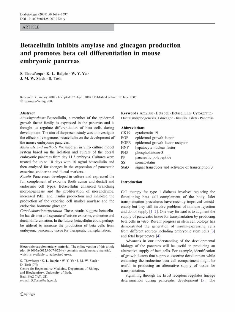
Differentiation of E11.5d embryonic pancreatic buds invitro We initially investigated the expression of endogenousbetacellulin in E11.5d developing pancreas. The gene encod-ing betacellulin was expressed in the epithelium but not themesenchyme (Fig. 1). The genes encoding ErbB1 andErbB4, the receptors for betacellulin, were expressed in boththe mesenchyme and the epithelium, suggesting that beta-cellulin could differentially affect both tissue types (Fig. 1).Pancreatic buds adhere to the fibronectin substrate within afew hours of isolation and gradually flatten out over theculture period. Mesenchymal cells spread rapidly out of theexplants to form a monolayer of cells surroundingthe epithelial clump in the centre. After 10 days of culturewith betacellulin, pancreatic buds had a thicker and a widerarea of mesenchyme in comparison with untreated buds. Wemeasured the total area occupied by the epithelium and themesenchyme using E-cadherin and smooth muscle actin astheir respective markers. The mesenchymal:epithelial ratiofor control and betacellulin-treated buds is shown in Table 1.The results indicate that there was more than a 1.5-foldincrease in the mesenchymal:epithelial ratio after betacellulintreatment.
Fig. 1 a, b The expression of the genes for betacellulin (Btc) and thereceptors ErbB1 (Erbb1, also known as Egfr) and ErbB4 (Erbb4) inthe epithelium and mesenchyme of E11.5d mouse dorsal pancreas.a Shows the β-actin equal loading control for all the samples and thepositive controls used. The — lane represents the water control(no cDNA). b Epithelium (panc epi.) and mesenchyme (panc mesen.)were separated as described in the “Materials and methods” section.Using PCR, we determined the expression of Btc, Erbb1 and Erbb4and the genes encoding CK19 (Ck19, also known as Krt19) andvimentin (Vim), markers of epithelium and mesenchyme, respectively,to demonstrate efficient separation of the two tissues. The results ofthe determination of Ck19 and Vim expression show that there is somecontamination of the epithelium with mesenchyme but there is littlecontamination of the mesenchyme with epithelium. Btc expression isseen preferentially in the pancreatic epithelium. Expression of Erbb1and Erbb4 is seen in both the pancreatic epithelium and mesenchyme,but at much higher levels in the epithelium. cDNA from adultpancreas and E11.5d embryonic brain were used as positive controls(+) for Btc and the ErbB genes, respectively, and cDNA from liver andskin were positive controls for the epithelium (Ck19) and mesenchyme(Vim), respectively
Table 1 Betacellulin regulates embryonic pancreatic development
Control Betacellulin n
Mesenchymal:epithelial ratio 13.20±1.01 21.73±1.12a 3Length of the entire ductalnetwork (×103 μm)
2.40±0.25 8.43±0.53b 4
Total area of amylase-positivecells (×103 μm2)
32.11±3.41 1.73±0.45b 4
Total area of insulin-positivecells (×103 μm2)
72.75±8.26 235.60±29.34a 6
Area of insulin-positive cells/no. of SS cells (μm2)
439.7±43.24 398.0±33.59c 4
Area of insulin-positive cells/no. of PP cells (μm2)
578.8±50.47 1,026.0±132.00a 4
Area of insulin-positive cells/no. of ghrelin cells (μm2)
3,490±215.80 1,937±224.50a 4
Total PH3-positive cell count 1,377±72.26 2,165±115.10a 4Maximum diameter of betacells (μm)
13.91±0.24 13.61±0.17c 50
Data are presented as the means±SEM.a p=0.0286 vs controlb p=0.0079 vs controlc Non-significant vs control
1690 Diabetologia (2007) 50:1688–1697
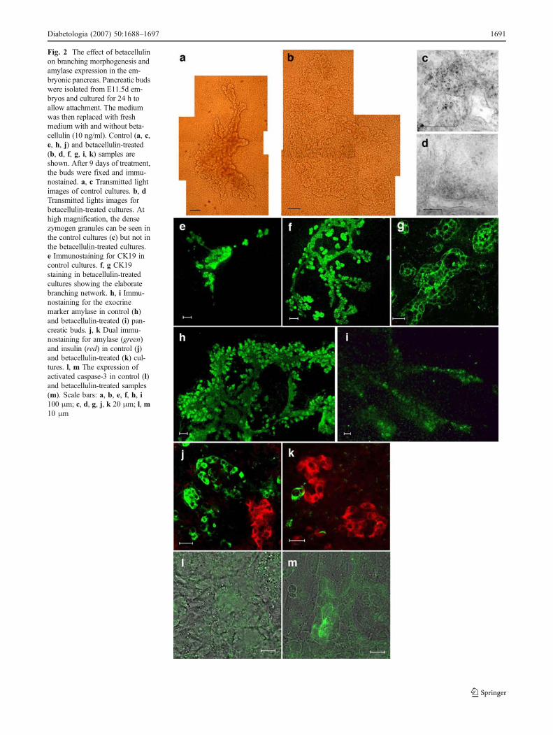
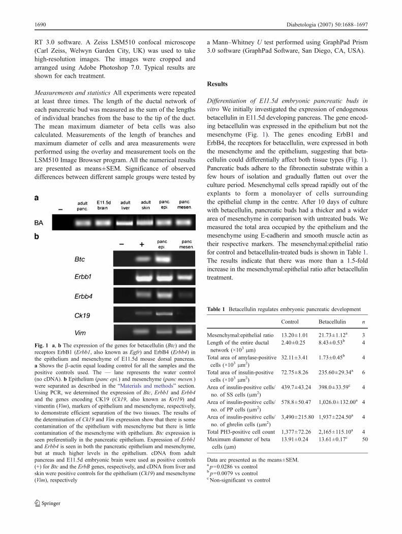
Fig. 2 The effect of betacellulinon branching morphogenesis andamylase expression in the em-bryonic pancreas. Pancreatic budswere isolated from E11.5d em-bryos and cultured for 24 h toallow attachment. The mediumwas then replaced with freshmedium with and without beta-cellulin (10 ng/ml). Control (a, c,e, h, j) and betacellulin-treated(b, d, f, g, i, k) samples areshown. After 9 days of treatment,the buds were fixed and immu-nostained. a, c Transmitted lightimages of control cultures. b, dTransmitted lights images forbetacellulin-treated cultures. Athigh magnification, the densezymogen granules can be seen inthe control cultures (c) but not inthe betacellulin-treated cultures.e Immunostaining for CK19 incontrol cultures. f, g CK19staining in betacellulin-treatedcultures showing the elaboratebranching network. h, i Immu-nostaining for the exocrinemarker amylase in control (h)and betacellulin-treated (i) pan-creatic buds. j, k Dual immu-nostaining for amylase (green)and insulin (red) in control (j)and betacellulin-treated (k) cul-tures. l, m The expression ofactivated caspase-3 in control (l)and betacellulin-treated samples(m). Scale bars: a, b, e, f, h, i100 μm; c, d, g, j, k 20 μm; l, m10 μm
Diabetologia (2007) 50:1688–1697 1691
Effect of betacellulin on branching morphogenesis SinceEGFR ligands can promote branching morphogenesis [5,15, 21], we investigated the effect of betacellulin on thebranching of the pancreatic ductal network. During thefirst few days of culture the epithelium becomes anextended branched structure, which can be observed undertransmitted light conditions (Fig. 2a). Addition of beta-cellulin increased branching and dilation of the incipientpancreatic ducts (Fig. 2b). CK19, a cytoskeletal protein, ispresent in the incipient ductal cells of the pancreas as wellas in developing acini [22]. To confirm the increase inbranching morphogenesis, we immunostained control andbetacellulin-treated cultures with an anti-CK19 antibody.Betacellulin promoted branching morphogenesis based onthe more extensive CK19 immunostaining (Fig. 2e–g).The differences in branching between the untreated andbetacellulin-treated buds were quantified by measuring thelength along the CK19-positive branches of the pancreaticductal network. The results indicate that there is a 3.5-foldincrease in branching induced by betacellulin (Table 1).This implies a possible role for betacellulin in proliferationof the incipient ducts. To test this idea, we co-stained forthe cell-proliferation marker phosphohistone-3 (PH3) withCK19 in control and betacellulin-treated samples. How-ever, no co-localisation of PH3 with CK19-positive cellswas observed at any of the four time-points examined(days 2, 3, 5 and 9), suggesting that proliferation of theductal cells may take place prior to their becoming CK19-positive (Fig. 3d,e). Instead, PH3-positive cells weremainly localised to the mesenchyme layer (Fig. 3f–h).Overall, betacellulin-treated samples contained increasednumbers of PH3-positive cells compared with controls(Table 1).
Betacel lu l in reduces the amylase pancreat icpopulation Amylase-expressing cells were present in thecontrol pancreatic organ cultures at day 7 and the numbers hadincreased further by day 10. The amylase-expressing cellswere distributed around the periphery of the branches andwere distinct from the endocrine cell populations (Fig. 2h,j).In contrast, the betacellulin-treated pancreatic tissue eithercompletely lacked or had reduced numbers of amylase-expressing cells (Fig. 2i,k). Under transmitted light, thezymogen granules, which store the exocrine enzymes, areclearly visible in the control (Fig. 2c) but not in thebetacellulin-treated (Fig. 2d). Overall, betacellulin treatmentreduced the number of amylase-positive cells (measured asthe total area covered by amylase-positive cells) by 18.5-fold (Table 1). Western blot analysis confirmed thisobservation and showed the virtual absence of the amylaseprotein in betacellulin-treated samples (see Fig. 4). Further-more, the pro-apoptotic protein caspase-3 was observed inacinar tissue of betacellulin-treated samples (Fig. 2l,m),
Fig. 3 Betacellulin regulates endocrine cell development. a–c Control(a, b) and betacellulin-treated (c) pancreatic buds after 9 days oftreatment, immunostained for insulin (red) and glucagon (blue). Insulin-positive cells are more abundant in betacellulin-treated culturescompared with the controls. Treatment with betacellulin also resultedin a marked reduction in the number of glucagon-positive cells (blue).d, e The increase in the number of beta cells in not due to an increase incell division since there was little or no co-localisation of theproliferation marker PH3 (green) with either insulin (red) or CK19(blue). f–h Instead PH3 (green) tended to co-localise with themesenchymal cells stained for smooth muscle actin (red) in bothcontrol (f, g) and betacellulin-treated tissue (h). i, j Staining for Pax6(green) and glucagon (red) shows the reduction in cells expressing Pax6in betacellulin-treated samples (j) compared with the controls (i). Scalebars: a, c 100 μm; d–f, h–j 20 μm; b, g 10 μm. Pax6 transcriptionfactor is expressed in progenitors for all pancreatic endocrine cells atsome point and is necessary for alpha cell differentiation
1692 Diabetologia (2007) 50:1688–1697

whereas it was much more difficult to find caspase-3-positive cells in control buds.
Betacellulin promotes beta cell differentiation We investi-gated the effect of betacellulin on insulin-expressing endo-crine cells. In both betacellulin-treated and untreatedsamples, after 10 days of culture, insulin-positive beta cellsout-numbered any other endocrine cell type. However, thedistribution and organisation of the beta cells were notice-ably different between treated and untreated samples. In theuntreated pancreatic buds, there were greater numbers ofbeta cells clumped together in large islet-like structures(Fig. 3a,b). In contrast, the betacellulin-treated buds hadsmaller clumps of beta cells that were dispersed throughoutthe organ culture (Fig. 3c). Additionally there were manyindividual cells or small islet-like cell clusters positive forinsulin in the inter-islet regions and in close proximity tothe ducts. Betacellulin induced an approximately threefoldincrease in the number of beta cells (measured as the totalarea covered by insulin-positive cells) with no significantchange in cell size (Table 1). This observation was alsoconfirmed by western blotting for insulin protein (Fig. 4).To test whether betacellulin increased the number of betacells by increasing the proliferation of beta cells we co-stained for insulin and the proliferation marker PH3.Betacellulin did not affect the proliferation of insulin-positive beta cells and we found only a few insulin-positivecells co-staining with PH3 (Fig. 3d,e).
Effects of betacellulin on alpha cell differentiation Incontrol samples, the classic islet morphology was preservedwith glucagon-expressing cells present in the periphery andthese cells outnumbered all other endocrine cell typesexcept beta cells by day 10 of culture (Fig. 3a,b). However,in the betacellulin-treated samples, the number of glucagon-positive cells was reduced to just a few cells in eachpancreas (Fig. 3c). Although glucagon protein was notdetectable on the western blot, presumably because of itssmall size, the amount of the 18 kDa pro-glucagon peptidefrom which glucagon is derived was much higher in theuntreated than betacellulin-treated pancreatic samples (Fig. 4).
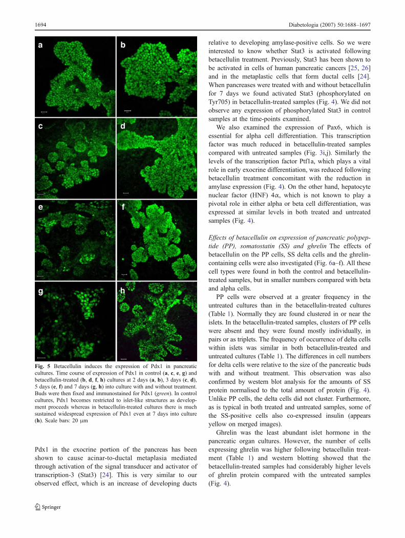
Effects of betacellulin on transcription factorexpression Since there was no evidence for changes ofproliferative rate underlying the observed effects of betacel-lulin on the epithelial cell types it is more likely that theeffects are due to an alteration in lineage determination ofprecursor cells. To examine this we determined the expres-sion of pancreatic transcription factors by immunohisto-chemistry. By day 2 of culture, both control and betacellulin-treated samples showed high levels of Pdx1 (Fig. 5). Pdx1 isa homeodomain protein that is expressed in the entirepancreatic anlagen at early stages of development. Normally,
Pdx1 expression declines during later embryonic stages inmost of the pancreas but remains selectively expressed in thebeta cells where it binds to and activates the insulinpromoter. By day 5 of our cultures, the expression of Pdx1was indeed limited to insulin-positive cells in the untreatedsamples (data not shown).
In comparison, Pdx1 expression in the betacellulin-treatedsamples was not strictly confined to such insulin-positivecell clusters. Pdx1-positive cells in betacellulin-treatedsamples outnumbered all of the differentiated endocrinecells put together and the expression was more ubiquitous,extending to cell populations that might be acinar or ductal innature. Pdx1 protein was also higher in betacellulin-treatedsamples compared with controls (Fig. 4). Pdx1 expression isnormally downregulated in non-beta cells during lateembryonic development [23] and prolonged expression of
Fig. 4 The effects of betacellulin (BTC) treatment on the levels ofpancreatic proteins. Protein was isolated from pancreatic buds culturedwith and without betacellulin for 7 days. Following BTC treatment,there was an increase in the levels of the pancreatic hormones insulinand ghrelin, the transcription factors Pdx1 and Sox9, and thephosphorylated form (Tyr705) of the signalling molecule Stat3(pStat3). The levels of the enzyme amylase, the immature form ofthe pancreatic hormone glucagon, and the exocrine promotingtranscription factor Ptf1a, were reduced. The levels of SS and HNF4αremained the same in both control (CTL) and BTC-treated samples.α-Tubulin was used as the loading control
Diabetologia (2007) 50:1688–1697 1693
Pdx1 in the exocrine portion of the pancreas has beenshown to cause acinar-to-ductal metaplasia mediatedthrough activation of the signal transducer and activator oftranscription-3 (Stat3) [24]. This is very similar to ourobserved effect, which is an increase of developing ducts
relative to developing amylase-positive cells. So we wereinterested to know whether Stat3 is activated followingbetacellulin treatment. Previously, Stat3 has been shown tobe activated in cells of human pancreatic cancers [25, 26]and in the metaplastic cells that form ductal cells [24].When pancreases were treated with and without betacellulinfor 7 days we found activated Stat3 (phosphorylated onTyr705) in betacellulin-treated samples (Fig. 4). We did notobserve any expression of phosphorylated Stat3 in controlsamples at the time-points examined.
We also examined the expression of Pax6, which isessential for alpha cell differentiation. This transcriptionfactor was much reduced in betacellulin-treated samplescompared with untreated samples (Fig. 3i,j). Similarly thelevels of the transcription factor Ptf1a, which plays a vitalrole in early exocrine differentiation, was reduced followingbetacellulin treatment concomitant with the reduction inamylase expression (Fig. 4). On the other hand, hepatocytenuclear factor (HNF) 4α, which is not known to play apivotal role in either alpha or beta cell differentiation, wasexpressed at similar levels in both treated and untreatedsamples (Fig. 4).
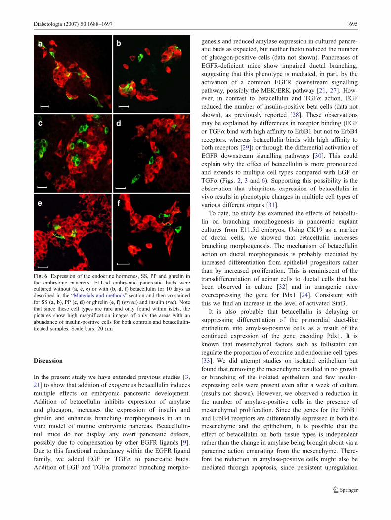
Effects of betacellulin on expression of pancreatic polypep-tide (PP), somatostatin (SS) and ghrelin The effects ofbetacellulin on the PP cells, SS delta cells and the ghrelin-containing cells were also investigated (Fig. 6a–f). All thesecell types were found in both the control and betacellulin-treated samples, but in smaller numbers compared with betaand alpha cells.
PP cells were observed at a greater frequency in theuntreated cultures than in the betacellulin-treated cultures(Table 1). Normally they are found clustered in or near theislets. In the betacellulin-treated samples, clusters of PP cellswere absent and they were found mostly individually, inpairs or as triplets. The frequency of occurrence of delta cellswithin islets was similar in both betacellulin-treated anduntreated cultures (Table 1). The differences in cell numbersfor delta cells were relative to the size of the pancreatic budswith and without treatment. This observation was alsoconfirmed by western blot analysis for the amounts of SSprotein normalised to the total amount of protein (Fig. 4).Unlike PP cells, the delta cells did not cluster. Furthermore,as is typical in both treated and untreated samples, some ofthe SS-positive cells also co-expressed insulin (appearsyellow on merged images).
Ghrelin was the least abundant islet hormone in thepancreatic organ cultures. However, the number of cellsexpressing ghrelin was higher following betacellulin treat-ment (Table 1) and western blotting showed that thebetacellulin-treated samples had considerably higher levelsof ghrelin protein compared with the untreated samples(Fig. 4).
Fig. 5 Betacellulin induces the expression of Pdx1 in pancreaticcultures. Time course of expression of Pdx1 in control (a, c, e, g) andbetacellulin-treated (b, d, f, h) cultures at 2 days (a, b), 3 days (c, d),5 days (e, f) and 7 days (g, h) into culture with and without treatment.Buds were then fixed and immunostained for Pdx1 (green). In controlcultures, Pdx1 becomes restricted to islet-like structures as develop-ment proceeds whereas in betacellulin-treated cultures there is muchsustained widespread expression of Pdx1 even at 7 days into culture(h). Scale bars: 20 μm
1694 Diabetologia (2007) 50:1688–1697
Discussion
In the present study we have extended previous studies [3,21] to show that addition of exogenous betacellulin inducesmultiple effects on embryonic pancreatic development.Addition of betacellulin inhibits expression of amylaseand glucagon, increases the expression of insulin andghrelin and enhances branching morphogenesis in an invitro model of murine embryonic pancreas. Betacellulin-null mice do not display any overt pancreatic defects,possibly due to compensation by other EGFR ligands [9].Due to this functional redundancy within the EGFR ligandfamily, we added EGF or TGFα to pancreatic buds.Addition of EGF and TGFα promoted branching morpho-
genesis and reduced amylase expression in cultured pancre-atic buds as expected, but neither factor reduced the numberof glucagon-positive cells (data not shown). Pancreases ofEGFR-deficient mice show impaired ductal branching,suggesting that this phenotype is mediated, in part, by theactivation of a common EGFR downstream signallingpathway, possibly the MEK/ERK pathway [21, 27]. How-ever, in contrast to betacellulin and TGFα action, EGFreduced the number of insulin-positive beta cells (data notshown), as previously reported [28]. These observationsmay be explained by differences in receptor binding (EGFor TGFα bind with high affinity to ErbB1 but not to ErbB4receptors, whereas betacellulin binds with high affinity toboth receptors [29]) or through the differential activation ofEGFR downstream signalling pathways [30]. This couldexplain why the effect of betacellulin is more pronouncedand extends to multiple cell types compared with EGF orTGFα (Figs. 2, 3 and 6). Supporting this possibility is theobservation that ubiquitous expression of betacellulin invivo results in phenotypic changes in multiple cell types ofvarious different organs [31].
To date, no study has examined the effects of betacellu-lin on branching morphogenesis in pancreatic explantcultures from E11.5d embryos. Using CK19 as a markerof ductal cells, we showed that betacellulin increasesbranching morphogenesis. The mechanism of betacellulinaction on ductal morphogenesis is probably mediated byincreased differentiation from epithelial progenitors ratherthan by increased proliferation. This is reminiscent of thetransdifferentiation of acinar cells to ductal cells that hasbeen observed in culture [32] and in transgenic miceoverexpressing the gene for Pdx1 [24]. Consistent withthis we find an increase in the level of activated Stat3.
It is also probable that betacellulin is delaying orsuppressing differentiation of the primordial duct-likeepithelium into amylase-positive cells as a result of thecontinued expression of the gene encoding Pdx1. It isknown that mesenchymal factors such as follistatin canregulate the proportion of exocrine and endocrine cell types[33]. We did attempt studies on isolated epithelium butfound that removing the mesenchyme resulted in no growthor branching of the isolated epithelium and few insulin-expressing cells were present even after a week of culture(results not shown). However, we observed a reduction inthe number of amylase-positive cells in the presence ofmesenchymal proliferation. Since the genes for the ErbB1and ErbB4 receptors are differentially expressed in both themesenchyme and the epithelium, it is possible that theeffect of betacellulin on both tissue types is independentrather than the change in amylase being brought about via aparacrine action emanating from the mesenchyme. There-fore the reduction in amylase-positive cells might also bemediated through apoptosis, since persistent upregulation
Fig. 6 Expression of the endocrine hormones, SS, PP and ghrelin inthe embryonic pancreas. E11.5d embryonic pancreatic buds werecultured without (a, c, e) or with (b, d, f) betacellulin for 10 days asdescribed in the “Materials and methods” section and then co-stainedfor SS (a, b), PP (c, d) or ghrelin (e, f) (green) and insulin (red). Notethat since these cell types are rare and only found within islets, thepictures show high magnification images of only the areas with anabundance of insulin-positive cells for both controls and betacellulin-treated samples. Scale bars: 20 μm
Diabetologia (2007) 50:1688–1697 1695

of Pdx1 has previously been shown to destroy pancreaticacinar cells by apoptosis [34]. It has also been observed thatacinar apoptosis is associated with ductal proliferation afterduct ligation [35]. We saw an upregulation of activatedcaspase-3, after betacellulin treatment and a concurrentdecrease in the level of the transcription factor Ptf1a, whichis necessary for early exocrine determination. The combineddata suggest that the effect of betacellulin on the exocrine cellsmight in fact act at two different levels. First, around the timewhen the exocrine and ductal fates are determined bypromoting progenitors towards a ductal fate, and second, byinducing apoptosis of already differentiated amylase-positivecells or promoting their transdifferentiation into ductal cellsmuch later on. One hypothesis to explain the pro-apoptoticaction of betacellulin compared with other receptor tyrosinekinase ligands (e.g. EGF) is that it is acting through a differentreceptor or post-receptor events [29].
In one previous study no change in the number ofamylase-expressing cells was seen after betacellulin treat-ment; however, the time of treatment was only half of thatused by ourselves [5].
We show by immunofluorescence microscopy andwestern blotting, that betacellulin enhances the number ofinsulin-positive beta cells and the overall levels of insulin inthe developing pancreas. Betacellulin is reported to regulatethe proportion of beta cells in other systems [5, 11, 14].Although our study agrees with previous observationsshowing an increase in the number of beta cells afterbetacellulin treatment [5], we show a more profound effect,which may again be due to the longer duration of thetreatment used. Three mechanisms might explain theincrease in the number of beta cells. The additional betacells could arise by: (1) the proliferation of existing betacells; (2) the differentiation of endocrine precursor cells intobeta cells rather than other endocrine cells; or (3) thetransdifferentiation of exocrine cell types into beta cells.
The first possibility can be ruled out because if the extrabeta cells did arise due to the proliferation of existing betacells we would observe larger islets and an increased numberof cells that show co-localisation for PH3 and insulin.
Huotari et al. [5] described the modulation of endocrinelineages by different growth factors. In the absence ofEGFR signalling, differentiation followed a default path-way towards alpha and PP cells. In the present study, thereduction in the number of alpha and PP cells in thepresence of betacellulin suggests that betacellulin might beinfluencing the fate choice made by the early endocrineprecursor cells and shifting the balance towards a beta cellphenotype. All pancreatic endocrine cell types initially arisefrom common multipotent precursor cells that express Pdx1but the continued expression of Pdx1 is only necessary forthe development of beta cells and not other endocrine celltypes [36]. The widespread late Pdx1 expression seen in the
betacellulin-treated pancreases might therefore bias thecommitment of endocrine precursors toward beta cells.Furthermore, the betacellulin-induced downregulation inexpression of other transcription factors such as the alphacell determinant Pax6 would ensure that the precursor cellsdo not differentiate down this lineage (Fig. 3). Cell lineagestudies have localised the endocrine progenitor cells closeto the primitive ducts of the fetal pancreas [37]. The Pdx1expression that is not restricted to the islets is seen in theseregions after betacellulin treatment (Fig. 5).
Ogata et al. [38] showed that betacellulin in combinationwith activin A can induce the differentiation of monolayercultures of rat neonatal ductal cells to beta cells. Three-dimensional pseudoislets can be formed by making sus-pensions of activin A- and betacellulin-treated cells andculturing with just betacellulin alone. The process wherebyendocrine tissue is generated from ductal epithelium istermed nesidioblastosis [39]. It has been reported in manyexperimental models and in clinical pathology [40]. Duringproliferation, duct cells have the potential to lose theirductal phenotype and revert back to multipotent cells [41].This is achieved by the ductal cells expressing Pdx1,differentiating into beta cells, then migrating away from theducts and clustering to form islets [35]. The possibility ofduct cell derived beta cells in this manner could explain theenhanced number of Pdx1-expressing cells in the ducts, thepresence of islet-like cell clusters and the insulin-positivecells found in isolation. So, betacellulin is probablyinfluencing beta cell formation both by increasing theproportion of endocrine cells and by increasing theproportion of endocrine precursors that become beta cells.
The present study shows that betacellulin can enhancebranching morphogenesis of the embryonic pancreaticbranching network and increase beta cell mass. We suggestthat these changes are largely brought about by prolongedubiquitous expression of Pdx1. Elevated Pdx1 expressionsuppresses early differentiation of the endodermal pancre-atic precursor tissue and enhances branching and formationof beta cells. This makes betacellulin a promising candidatefor stimulating beta cell regeneration in vivo, or increasing theproportion of beta cells in vitro in situations where beta celldifferentiation is being sought with a view to transplantation.
Acknowledgements The authors wish to thank E. B. Lane (Univer-sity of Dundee) for generously providing the anti-CK19 antibody. Thiswork was supported by a Universities UK Overseas ResearchStudentship and the Department of Biology and Biochemistry,University of Bath (S. Thowfeequ). Financial support was alsoprovided by the Biotechnology and Biological Sciences ResearchCouncil, Medical Research Council and the Wellcome Trust.
Duality of interest None of the authors has any financial interest inthis work.
1696 Diabetologia (2007) 50:1688–1697
References
1. Shapiro AM, Geng Hao E, Lakey JR, Finegood DT, Rajotte RV,Kneteman NM (2002) Defining optimal immunosuppression forislet transplantation based on reduced diabetogenicity in canineislet autografts. Transplantation 74:1522–1528
2. Naftanel MA, Harlan DM (2004) Pancreatic islet transplantation.PLoS Med 1:e58 (quiz e75)
3. Bonner-Weir S, Weir GC (2005) New sources of pancreatic beta-cells. Nat Biotechnol 23:857–861
4. Zalzman M, Anker-Kitai L, Efrat S (2005) Differentiation ofhuman liver-derived, insulin-producing cells toward the beta-cellphenotype. Diabetes 54:2568–2575
5. Huotari MA, Miettinen PJ, Palgi J et al (2002) ErbB signalingregulates lineage determination of developing pancreatic islet cellsin embryonic organ culture. Endocrinology 143:4437–4446
6. Dunbar AJ, Goddard C (2000) Structure–function and biologicalrole of betacellulin. Int J Biochem Cell Biol 32:805–815
7. Sasada R, Ono Y, Taniyama Y, Shing Y, Folkman J, Igarashi K(1993) Cloning and expression of cDNA encoding humanbetacellulin, a new member of the EGF family. Biochem BiophysRes Commun 190:1173–1179
8. Miyagawa J, Hanafusa O, Sasada R et al (1999) Immunohisto-chemical localization of betacellulin, a new member of the EGFfamily, in normal human pancreas and islet tumor cells. Endocr J46:755–764
9. Jackson LF, Qiu TH, Sunnarborg SW et al (2003) Defectivevalvulogenesis in HB-EGF and TACE-null mice is associatedwith aberrant BMP signaling. EMBO J 22:2704–2716
10. MashimaH, Ohnishi H,Wakabayashi K et al (1996) Betacellulin andactivin A coordinately convert amylase-secreting pancreatic AR42Jcells into insulin-secreting cells. J Clin Invest 97:1647–1654
11. Demeterco C, Beattie GM, Dib SA, Lopez AD, Hayek A (2000) Arole for activin A and betacellulin in human fetal pancreatic celldifferentiation and growth. J Clin Endocrinol Metab 85:3892–3897
12. Li L, Seno M, Yamada H, Kojima I (2003) Betacellulin improvesglucose metabolism by promoting conversion of intraislet precur-sor cells to beta-cells in streptozotocin-treated mice. Am J PhysiolEndocrinol Metab 285:E577–E583
13. Kojima H, Fujimiya M, Matsumura K et al (2003) NeuroD-betacellulin gene therapy induces islet neogenesis in the liver andreverses diabetes in mice. Nat Med 9:596–603
14. Li L, Seno M, Yamada H, Kojima I (2001) Promotion of beta-cellregeneration by betacellulin in ninety percent-pancreatectomizedrats. Endocrinology 142:5379–5385
15. Sakurai H, Tsukamoto T, Kjelsberg CA, Cantley LG, Nigam SK(1997) EGF receptor ligands are a large fraction of in vitrobranching morphogens secreted by embryonic kidney. Am JPhysiol 273:F463–F472
16. Kim SK, Hebrok M (2001) Intercellular signals regulatingpancreas development and function. Genes Dev 15:111–127
17. Percival AC, Slack JM (1999) Analysis of pancreatic developmentusing a cell lineage label. Exp Cell Res 247:123–132
18. Shen CN, Seckl JR, Slack JM, Tosh D (2003) Glucocorticoidssuppress beta-cell development and induce hepatic metaplasia inembryonic pancreas. Biochem J 375:41–50
19. Li WC, Horb ME, Tosh D, Slack JM (2005) In vitro trans-differentiation of hepatoma cells into functional pancreatic cells.Mech Dev 122:835–847
20. Jiang FX, Harrison LC (2005) Laminin-1 and epidermal growthfactor family members co-stimulate fetal pancreas cell prolifera-tion and colony formation. Differentiation 73:45–49
21. Miettinen PJ, Huotari M, Koivisto T et al (2000) Impairedmigration and delayed differentiation of pancreatic islet cells inmice lacking EGF-receptors. Development 127:2617–2627
22. Bouwens L, Braet F, Heimberg H (1995) Identification of ratpancreatic duct cells by their expression of cytokeratins 7, 19, and20 in vivo and after isolation and culture. J Histochem Cytochem43:245–253
23. Stoffers DA, Heller RS, Miller CP, Habener JF (1999) Develop-mental expression of the homeodomain protein IDX-1 in micetransgenic for an IDX-1 promoter/lacZ transcriptional reporter.Endocrinology 140:5374–5381
24. Miyatsuka T, Kaneto H, Shiraiwa T et al (2006) Persistentexpression of PDX-1 in the pancreas causes acinar-to-ductalmetaplasia through Stat3 activation. Genes Dev 20:1435–1440
25. Greten FR, Weber CK, Greten TF et al (2002) Stat3 and NF-kappaB activation prevents apoptosis in pancreatic carcinogenesis.Gastroenterology 123:2052–2063
26. Scholz A, Heinze S, Detjen KM et al (2003) Activated signaltransducer and activator of transcription 3 (STAT3) supports themalignant phenotype of human pancreatic cancer. Gastroenterol-ogy 125:891–905
27. Rescan C, Le Bras S, Lefebvre VH et al (2005) EGF-inducedproliferation of adult human pancreatic duct cells is mediated bythe MEK/ERK cascade. Lab Invest 85:65–74
28. Cras-Méneur C, Elghazi L, Czernichow P, Scharfmann R (2001)Epidermal growth factor increases undifferentiated pancreaticembryonic cells in vitro: a balance between proliferation anddifferentiation. Diabetes 50:1571–1579
29. Jones JT, Akita RW, Sliwkowski MX (1999) Binding specificitiesand affinities of EGF domains for ErbB receptors. FEBS Lett447:227–231
30. Saito T, Okada S, Ohshima K et al (2004) Differential activationof epidermal growth factor (EGF) receptor downstream signalingpathways by betacellulin and EGF. Endocrinology 145:4232–4243
31. Schneider MR, Dahlhoff M, Herbach N et al (2005) Betacellulinoverexpression in transgenic mice causes disproportionate growth,pulmonary hemorrhage syndrome, and complex eye pathology.Endocrinology 146:5237–5246
32. Rooman I, Heremans Y, Heimberg H, Bouwens L (2000)Modulation of rat pancreatic acinoductal transdifferentiation andexpression of PDX-1 in vitro. Diabetologia 43:907–914
33. Miralles F, Czernichow P, Scharfmann R (1998) Follistatinregulates the relative proportions of endocrine vs exocrine tissueduring pancreatic development. Development 125:1017–1024
34. Heller RS, Stoffers DA, Bock T et al (2001) Improved glucosetolerance and acinar dysmorphogenesis by targeted expression oftranscription factor PDX-1 to the exocrine pancreas. Diabetes50:1553–1561
35. Abe K, Watanabe S (1995) Apoptosis of mouse pancreatic acinarcells after duct ligation. Arch Histol Cytol 58:221–229
36. Jensen J (2004) Gene regulatory factors in pancreatic develop-ment. Dev Dyn 229:176–200
37. Ogata T, Li L, Yamada S et al (2004) Promotion of beta-celldifferentiation by conophylline in fetal and neonatal rat pancreas.Diabetes 53:2596–2602
38. Ogata T, Park KY, Seno M, Kojima I (2004) Reversal ofstreptozotocin-induced hyperglycemia by transplantation of pseu-doislets consisting of beta cells derived from ductal cells. Endocr J51:381–386
39. Laguesse E (1893) Sur la formation des îlots de Langerhans dansle pancréas. Comptes Rendus Séances Soc Biol Fil 45:819–820
40. Kerr-Conte J, Pattou F, Lecomte-Houcke M et al (1996) Ductalcyst formation in collagen-embedded adult human islet prepara-tions. A means to the reproduction of nesidioblastosis in vitro.Diabetes 45:1108–1114
41. Bonner-Weir S, Taneja M, Weir GC et al (2000) In vitrocultivation of human islets from expanded ductal tissue. ProcNatl Acad Sci U S A 97:7999–8004
Diabetologia (2007) 50:1688–1697 1697
Copyright © 2022 FDOKUMEN