Homogeneity and heterogeneity in amylase production by ...
17
University of Groningen Homogeneity and heterogeneity in amylase production by Bacillus subtilis under different growth conditions Ploss, Tina N.; Reilman, Ewoud; Monteferrante, Carmine G.; Denham, Emma L.; Piersma, Sjouke; Lingner, Anja; Vehmaanpera, Jari; Lorenz, Patrick; van Dijl, Jan Maarten Published in: Microbial Cell Factories DOI: 10.1186/s12934-016-0455-1 IMPORTANT NOTE: You are advised to consult the publisher's version (publisher's PDF) if you wish to cite from it. Please check the document version below. Document Version Publisher's PDF, also known as Version of record Publication date: 2016 Link to publication in University of Groningen/UMCG research database Citation for published version (APA): Ploss, T. N., Reilman, E., Monteferrante, C. G., Denham, E. L., Piersma, S., Lingner, A., Vehmaanpera, J., Lorenz, P., & van Dijl, J. M. (2016). Homogeneity and heterogeneity in amylase production by Bacillus subtilis under different growth conditions. Microbial Cell Factories, 15(57), [57]. https://doi.org/10.1186/s12934-016-0455-1 Copyright Other than for strictly personal use, it is not permitted to download or to forward/distribute the text or part of it without the consent of the author(s) and/or copyright holder(s), unless the work is under an open content license (like Creative Commons). The publication may also be distributed here under the terms of Article 25fa of the Dutch Copyright Act, indicated by the “Taverne” license. More information can be found on the University of Groningen website: https://www.rug.nl/library/open-access/self-archiving-pure/taverne- amendment. Take-down policy If you believe that this document breaches copyright please contact us providing details, and we will remove access to the work immediately and investigate your claim. Downloaded from the University of Groningen/UMCG research database (Pure): http://www.rug.nl/research/portal. For technical reasons the number of authors shown on this cover page is limited to 10 maximum.
-
Upload
khangminh22 -
Category
Documents
-
view
9 -
download
0
Transcript of Homogeneity and heterogeneity in amylase production by ...

University of Groningen
Homogeneity and heterogeneity in amylase production by Bacillus subtilis under differentgrowth conditionsPloss, Tina N.; Reilman, Ewoud; Monteferrante, Carmine G.; Denham, Emma L.; Piersma,Sjouke; Lingner, Anja; Vehmaanpera, Jari; Lorenz, Patrick; van Dijl, Jan MaartenPublished in:Microbial Cell Factories
DOI:10.1186/s12934-016-0455-1
IMPORTANT NOTE: You are advised to consult the publisher's version (publisher's PDF) if you wish to cite fromit. Please check the document version below.
Document VersionPublisher's PDF, also known as Version of record
Publication date:2016
Link to publication in University of Groningen/UMCG research database
Citation for published version (APA):Ploss, T. N., Reilman, E., Monteferrante, C. G., Denham, E. L., Piersma, S., Lingner, A., Vehmaanpera, J.,Lorenz, P., & van Dijl, J. M. (2016). Homogeneity and heterogeneity in amylase production by Bacillussubtilis under different growth conditions. Microbial Cell Factories, 15(57), [57].https://doi.org/10.1186/s12934-016-0455-1
CopyrightOther than for strictly personal use, it is not permitted to download or to forward/distribute the text or part of it without the consent of theauthor(s) and/or copyright holder(s), unless the work is under an open content license (like Creative Commons).
The publication may also be distributed here under the terms of Article 25fa of the Dutch Copyright Act, indicated by the “Taverne” license.More information can be found on the University of Groningen website: https://www.rug.nl/library/open-access/self-archiving-pure/taverne-amendment.
Take-down policyIf you believe that this document breaches copyright please contact us providing details, and we will remove access to the work immediatelyand investigate your claim.
Downloaded from the University of Groningen/UMCG research database (Pure): http://www.rug.nl/research/portal. For technical reasons thenumber of authors shown on this cover page is limited to 10 maximum.

Ploss et al. Microb Cell Fact (2016) 15:57 DOI 10.1186/s12934-016-0455-1
RESEARCH
Homogeneity and heterogeneity in amylase production by Bacillus subtilis under different growth conditionsTina N. Ploss1, Ewoud Reilman2, Carmine G. Monteferrante2,4, Emma L. Denham2,5, Sjouke Piersma2, Anja Lingner1,6, Jari Vehmaanperä3, Patrick Lorenz1 and Jan Maarten van Dijl2*
Abstract
Background: Bacillus subtilis is an important cell factory for the biotechnological industry due to its ability to secrete commercially relevant proteins in large amounts directly into the growth medium. However, hyper-secretion of proteins, such as α-amylases, leads to induction of the secretion stress-responsive CssR-CssS regulatory system, result-ing in up-regulation of the HtrA and HtrB proteases. These proteases degrade misfolded proteins secreted via the Sec pathway, resulting in a loss of product. The aim of this study was to investigate the secretion stress response in B. subtilis 168 cells overproducing the industrially relevant α-amylase AmyM from Geobacillus stearothermophilus, which was expressed from the strong promoter P(amyQ)-M.
Results: Here we show that activity of the htrB promoter as induced by overproduction of AmyM was “noisy”, which is indicative for heterogeneous activation of the secretion stress pathway. Plasmids were constructed to allow real-time analysis of P(amyQ)-M promoter activity and AmyM production by, respectively, transcriptional and out-of-frame translationally coupled fusions with gfpmut3. Our results show the emergence of distinct sub-populations of high- and low-level AmyM-producing cells, reflecting heterogeneity in the activity of P(amyQ)-M. This most likely explains the heterogeneous secretion stress response. Importantly, more homogenous cell populations with regard to P(amyQ)-M activity were observed for the B. subtilis mutant strain 168degUhy32, and the wild-type strain 168 under optimized growth conditions.
Conclusion: Expression heterogeneity of secretory proteins in B. subtilis can be suppressed by degU mutation and optimized growth conditions. Further, the out-of-frame translational fusion of a gene for a secreted target protein and gfp represents a versatile tool for real-time monitoring of protein production and opens novel avenues for Bacillus production strain improvement.
Keywords: Bacillus subtilis, Amylase, Heterogeneity, Secretion stress, Transcription, Translation
© 2016 Ploss et al. This article is distributed under the terms of the Creative Commons Attribution 4.0 International License (http://creativecommons.org/licenses/by/4.0/), which permits unrestricted use, distribution, and reproduction in any medium, provided you give appropriate credit to the original author(s) and the source, provide a link to the Creative Commons license, and indicate if changes were made. The Creative Commons Public Domain Dedication waiver (http://creativecommons.org/publicdomain/zero/1.0/) applies to the data made available in this article, unless otherwise stated.
BackgroundThe rod-shaped Gram-positive bacterium Bacillus sub-tilis has a long history of safe use as a production host in the biotechnological industry. It has been imple-mented for the synthesis of various different prod-ucts such as proteins, vitamins and antibiotics. Next to
B. licheniformis and B. amyloliquefaciens, B. subtilis has become one of the most well-established and relevant workhorses in biotechnology, especially for the produc-tion of secreted proteins like proteases and α-amylases [1–3]. Importantly, B. subtilis is free of endotoxins and considered suitable for the qualified presumption of safety (QPS) status of the European food safety author-ity. Accordingly, many B. subtilis products have received the generally regarded as safe (GRAS) status of the US Food and Drug Administration. In addition, high-qual-ity genomic sequences, as for B. subtilis 168 [4, 5], and
Open Access
Microbial Cell Factories
*Correspondence: [email protected] 2 Department of Medical Microbiology, University of Groningen, University Medical Center Groningen, Hanzeplein 1, 9700 RD Groningen, The NetherlandsFull list of author information is available at the end of the article
Page 2 of 16Ploss et al. Microb Cell Fact (2016) 15:57
well-established protocols for genetic modification [6–9] highly facilitate the construction of improved production hosts.
The ability of Bacillus species to secrete high amounts of proteins (up to 20–25 g/l) directly into the fermenta-tion broth is facilitated by its single-membrane physiol-ogy. The high secretion capacity of Bacillus offers clear advantages for downstream processing and final purifi-cation of the target protein [3]. The Sec pathway consti-tutes the main secretion pathway in B. subtilis with ~300 endogenous proteins appearing to be translocated through the cell membrane via this pathway [10–12]. Despite this relatively efficient protein translocation machinery, the secretion yield of most heterologous pro-teins expressed in Bacillus is usually lower than the afore-mentioned 20–25 g/l, imposing economic challenges to the industry. This problem, especially evident for proteins derived from organisms not closely related to Bacillus, can be attributed to different bottlenecks in the secre-tion process. These include poor membrane targeting, inefficient membrane translocation or cell wall passage, slow or incorrect folding of the Sec-dependent exopro-tein by PrsA and degradation by proteases [13, 14]. The Sec machinery exclusively transports polypeptides in an unfolded state. Consequently, while exiting the Sec chan-nel at the extra cytoplasmic side of the membrane, the transported proteins have to fold rapidly into their cor-rect structural conformation in order to become active and to achieve stability against proteolytic degradation. Misfolded proteins lead to a cellular stress response that generally results in either refolding or degradation of the affected proteins [15]. Protein secretion stress in B. subtilis is usually defined as the stress that induces the two-component regulatory system CssR-CssS [16]. High-level production of Sec-dependent secreted pro-teins, such as the α-amylase AmyQ from B. amylolique-faciens, leads to an accumulation of misfolded protein at the membrane-cell wall interface, resulting in the activa-tion of the response regulator CssR by phosphorylation [16]. This in turn activates the transcription of htrA and htrB encoding the membrane-bound proteases HtrA and HtrB, which are responsible for proteolytic cleavage and degradation of misfolded secreted proteins [17, 18]. Previously, it has been shown that the expression level of htrB correlates with the level of AmyQ production in B. subtilis [19]. However, studies dealing with other secre-tory proteins, such as lipase A of B. subtilis and human interleukin-3, showed that the intensity of the protein-secretion stress response only partly reflected the protein production levels [20]. This implies that induction of the secretion stress response largely depends on the nature of the secreted protein that is overproduced.
For industrial protein production, the question whether target gene expression is homogeneous or heterogeneous is highly relevant [21]. Clearly, to obtain the highest yields possible, homogeneous high-level target gene-expressing populations are most desirable. However, the expres-sion levels of individual genes in a bacterial population are often noisy or heterogeneous, and this applies also to B. subtilis [22–25]. The presence of low-expressing cells can thus affect the overall protein yield. In more extreme situations, the population can even be bimodal, in which case expression of the protein of interest depends on a particular sub-population [21, 26].
In the present study, we investigated the induction of the protein secretion stress response in B. subtilis 168 upon overproduction of AmyM, an industrially relevant α-amylase from Geobacillus stearothermophilus [27–29]. To assess the secretion stress response in detail, the tran-scriptional activity of the htrB promoter P(htrB) was analyzed using a promoter-gfp fusion. In particular, we investigated the correlation between a heterogeneous protein secretion stress response and expression hetero-geneity in B. subtilis cells producing AmyM where high-level amyM expression was directed by the P(amyQ)-M promoter. Our results show how a particular muta-tion in the transcriptional regulator degU, as well as the selected growth conditions impact on the heterogeneity of P(amyQ)-M activity and production of the α-amylase AmyM in B. subtilis.
MethodsPlasmids, primers bacterial strains and growth conditionsPlasmids and primers used and constructed in this study are described in Tables 1 and 2, respectively. The B. sub-tilis prototype strain 168 and derivatives were trans-formed as described previously [30]. B. subtilis DB104 was used for plasmid construction using standard tech-niques [31]. Strains used and constructed in this study are listed in Table 3. Lysogeny Broth (LB) was used to grow B. subtilis DB104, 168 and derivatives thereof. Live cell array (LCA) experiments were performed by grow-ing B. subtilis 168 and derivatives in LB, or EnPressoB medium (BioSilta; note that EnPressoB replaces the pre-vious EnBase Flo medium of the supplier) [32]. Media were supplemented with antibiotics as required in the following concentrations: 10 µg ml−1 kanamycin (Km), 3 µg ml−1 chloramphenicol (Cm), 5 µg ml−1 tetracycline (Tc) or spectinomycin (Sp) 100 µg ml−1.
Polymerase chain reaction (PCR)AccuPrime™ Pfx DNA Polymerase (Life Technologies) was used for DNA amplification by PCR following manu-facturer’s instructions.

Page 3 of 16Ploss et al. Microb Cell Fact (2016) 15:57
Construction of a transcriptional fusion of the P(amyQ)‑M promoter with gfpPlasmid pDAPamy‑gfpFor monitoring the promoter activity of P(amyQ)-M from B. amyloliquefaciens and heterogeneous or homo-geneous activity of P(amyQ)-M in B. subtilis 168, the high-copy plasmid pDAPamy-gfp was constructed by amplifying the high-copy number vector pDAM car-rying the P(amyQ)-M promoter and the gfpmut3 gene from plasmid pHB201 2.4 and introducing complemen-tary sequences to the 3´-ends with a Tm >50 °C. Plasmid pHB201 2.4 contains the gfpmut3 gene derived from vec-tor pBaSysBioII. gfpmut3 was amplified from the ATG start codon with primers gfp OV fw and gfp OV rv2 resulting in the insertion of a complementary sequence to the vector pDAM at the 3´-end. A truncated version of the vector pDAM was amplified using the primers Pamy OV rv and M gfp fw by adding sequences complemen-tary to the gfpmut3 gene. Amplified products were puri-fied using the Wizard SV gel purification Kit (Promega) and were fused by using the Gibson Assembly Kit (New England Biolabs). The product was directly used to trans-form competent cells of the intermediate host B. subtilis DB104.
Plasmid pDAPamy‑gfp tetThe kanamycin resistance gene (kan) in plasmid pDA-Pamy-gfp was replaced by the tetM gene from pKVM2. The complete vector pDAPamy-gfp, lacking the kan gene, was amplified using oligonucleotides KM OV fw2 and KM OV rv2 and inserting ends complementary to the
Table 1 Plasmids used and constructed in this study
Plasmids Description Source of reference
pKTH10 kan, amyQ; pUB110 [33]
pDAM kan, amyM AB Enzymes GmbH
pHB201 2.4 Cm, ermC, gfp AB Enzymes GmbH
pKVM2 tetM; E. coli-Bacillus shuttle vector
AB Enzymes GmbH
pBaSysBioII spec, amp, gfpmut3 [34]
pDAPamy-gfp kan; transcriptional P(amy)-gfp fusion
This study
pDAPamy-gfp tet tetM; transcriptional P(amy)-gfp fusion
This study
pDAamyM-gfp kan; out-of-frame translational amyM-gfp fusion
This study
Table 2 Primers used in this study
Primers Sequence (5´–3´)
P amy OV rv TTCTTCTCCTTTACGCATGTTTCCTCTCCCTCTCATTTTC
gfp OV fw ATGCGTAAAGGAGAAGAACTTTTCACTG
gfp OV rv2 CTTTTTTTGTCCATTTCTCTTATTTGTATAGTTCATCCATGCC
gfp OV fw 3 CAATGAGAAAAGGAGAAGAACTTTTCACTG
M gfp fw GAGAAATGGACAAAAAAAGCAAAGGGTTC
M gfp rv3 TTCTTCTCCTTTTCTCATTGTTGCCACGTAACAGTAATG
tetM OV fw TTAATACTAGTTCACTAAGTTATTTTATTGAACATATATCG
tetM OV rv TCTGAAAAGGGAATGAAAATTATTAATATTGGAGTTTTAGCTC
KM OV fw2 AATAAAATAACTTAGTGAACTAGTATTAATCTGTTCAGCAATC
KM OV rv2 TAATTTTCATTCCCTTTTCAGATAATTTTAGATTTGC
Table 3 Bacterial strains used and generated in this study
Bacterial strains Relevant properties Source of reference
B. subtilis strains
168 trpC2 UMCG, laboratory stock
168 HmB C5 trpC2 P(htrB)-gfp::spc [35]
168 ∆rok trpC2, ∆rok::cm UMCG, laboratory stock
168degUhy32 trpC2, degUhy32::kan UMCG, laboratory stock
DB104 His nprR2 nprE18 ∆aprA3 [36]
168 HmB C5::pKTH10 168 HmB C5 carrying plasmid pKTH10 This study
168 HmB C5::pDAM 168 HmB C5 carrying plasmid pDAM This study
168::pDAPamy-gfp 168 carrying plasmid pDAPamy-gfp This study
168::pDAPamy-gfp tet 168 carrying plasmid pDAPamy-gfp tet This study
168degUhy32::pDAPamy-gfp tet 168degUhy32 carrying plasmid pDAPamy-gfp tet This study
168∆rok::pDAPamy-gfp tet 168∆rok carrying plasmid pDAPamy-gfp tet This study
168::pDAamyM-gfp 168 carrying plasmid pDAamyM-gfp This study

Page 4 of 16Ploss et al. Microb Cell Fact (2016) 15:57
tetM gene. The tetM gene from pKVM2 was amplified by PCR with primers tetM OV fw and tetM OV rv. Ampli-fied products purified by gel extraction were fused by Gibson Assembly and used to transform competent cells of DB104.
Construction of translational fusions of the α‑amylase amyM gene with gfpFor real-time monitoring of translation levels of the secreted α-amylase AmyM from G. stearothermophi-lus, the gfpmut3 gene from pHB201 2.4 was fused to the 3´-end of amyM such that the TGA stop codon of amyM partially overlapped with the ATG start codon of gfpmut3. This resulted in the sequence ATG A, where the start codon is marked in italics, and the stop codon in bold. In this manner an out-of-frame translational fusion was created between the amyM and gfpmut3 genes. For this purpose, the complete expression vec-tor pDAM carrying amyM under the transcriptional control of P(amyQ)-M was amplified using oligonucleo-tides M gfp fw and M gfp rv3, and the gfpmut3 gene was amplified using gfp OV fw 3 and gfp OV rv2. Comple-mentary sequences were inserted at the 5´- and 3´-ends of the amplified fragments, which were fused by Gibson Assembly after purification via gel extraction. The result-ing amplicon was used to transform B. subtilis DB104, resulting in plasmid pDAamyM-gfp.
Cultivation of B. subtilis using the live cell array (LCA) systemB. subtilis strains were grown overnight in 100 µl LB medium supplemented with appropriate concentrations of antibiotics in 96-well plates at 37 °C under vigor-ous shaking at 1000 rpm in a Thermo Shaker L079-100 (Kisker). An over-day culture in 96-well plates was inocu-lated with the over-night culture at a 1/20 dilution and grown under the same conditions for 6 h. Cultivation of B. subtilis was carried out in a 96-well plate with flat bot-tom (Greiner bio-one, Cellstar) in a final volume of 100 µl of LB or EnPressoB medium (BioSilta), inoculated with 5 µl from the over-day culture. To avoid evaporation all plates were covered with a lid, incubated at 37 °C with constant shaking (fast mode: 1140 rpm) using a Biotek Synergy two multimode microplate reader. Fluorescence (excitation 485/20 mm, emission 528/20 mm) and opti-cal density (OD600) were measured at 10 min intervals. For light path length correction, the OD977 and OD900 were measured and were calculated for a sample length of 1 cm by (OD977–OD900)/0.18 as described previously [34].
GFP-expression levels were corrected for background fluorescence by subtracting the fluorescence values derived from the cultivation of the wild-type strain 168
(cultivated in six independent wells). Next, normalization of the fluorescence increase per time and growth rate was calculated using the following equation: (dGFP/dt/OD600).
Fluorescence microscopyBacillus cells carrying out-of-frame translational fusions of amyM and gfpmut3 were grown in 20 ml LB or in EnPressoB medium in 250 ml shake flasks under con-tinuous shaking at 200 rpm. At the chosen time points (5.5, 8, 24 and 32 h), cells were spotted on poly-l-lysin-coated microscopy slides. Fluorescence microscopy was performed with a Leica DM5500 B microscope equipped with a Leica EL6000 camera.
Time‑lapse microscopyTime-lapse fluorescence microscopy was performed with a Leica DM5500 B microscope equipped with a motor-ized stage and temperature-controlled incubation cham-ber set at 37 °C. Movies were recorded as described previously [34]. A preculture in 25 % LB medium was made from a LB over-night culture. After reaching an OD600 of ~0.2 cells were applied to 25 % LB agarose (1.5 %) slides, which were prepared as described previ-ously [37].
Flow cytometryB. subtilis strains were grown in 20 ml LB medium or EnPressoB medium in 250 ml shake flasks with baffles at 37 °C and shaking at 200 rpm. Samples were taken after 5.5, 8, 24 and 32 h of incubation and were measured via flow cytometry with a BD Accuri C6 (BD Biosciences) as described previously [25].
SDS‑PAGEB. subtilis strains were grown in 20 ml LB or in EnPres-soB medium in 250 ml shake flasks under continuous shaking (250 rpm, 37 °C). Growth medium fractions were separated from the cells by centrifugation (3500g, 15 min, 4 °C) after cultivation times of 5.5, 8, 24, and 32 h. Elec-trophoresis on 4–12 % Bis–Tris-NuPAGE gels (Invitro-gen) was performed as described by the manufacturer’s protocol.
ResultsActivation of the protein secretion stress response in B. subtilis cells hyper‑secreting the α‑amylase AmyMTo monitor the induction of a possible protein secretion stress response in B. subtilis 168 during overproduction of the α-amylase AmyM in comparison to the previ-ously investigated α-amylase AmyQ, the expression vec-tors pDAM (amyM) or pKTH10 (amyQ) were used to transform B. subtilis 168 Hm C5. AmyQ was used as a

Page 5 of 16Ploss et al. Microb Cell Fact (2016) 15:57
control, because it was previously shown that its high-level expression from plasmid pKTH10 [33] causes strong induction of the P(htrA/htrB) promoters [19]. Importantly, strain 168 Hm C5 contained a transcrip-tional fusion of the P(htrB) promoter with the gfpmut3 reporter gene [35]. To construct this fusion, the promoter region of htrB (~300 bp) plus an optimized ribosome-binding site (RBS) were fused directly upstream of the gfpmut3 gene, and the resulting construct was integrated into the genome by single cross-over recombination. Of note, the gfpmut3 gene codes for a GFP variant of high stability with an estimated half-life of ~10 h in B. subtilis that makes it a useful marker for promoter activity deter-mination [34]. Furthermore, to study secretion stress induction, all strains were grown in two different media, namely LB and EnPressoB. The EnPressoB medium per-mits increased volumetric productivity for recombinant proteins in small culture volumes (e.g. ~100 µl in 96-well plates). It is based on a soluble polysaccharide in the medium from which glucose is slowly released through the action of a specific endo-glucanase [32]. In the LCA setup, the resulting slow-release glucose feeding regime based on completely soluble medium components allows the online measurement of both OD600 and GFP fluo-rescence under conditions that mimic production scale conditions.
The growth profiles and P(htrB) promoter activities of B. subtilis 168 HmB C5 and transformants of this strain carrying plasmids pKTH10 or pDAM upon cultivation in LB or EnPressoB medium are shown in Fig. 1. As shown in Fig. 1a, the P(htrB)-gfp fusion was not expressed in B. subtilis 168 HmB C5 without the α-amylase-encoding plasmids. Consistent with previous findings [16, 19], the pKTH10-carrying strain showed very high P(htrB) pro-moter activities, indicating high induction of the secre-tion stress response by AmyQ production both in LB and EnPressoB medium (Fig. 1b). In fact, the fluores-cence intensity went outside the detection range upon growth in LB medium after ~260 min of cultivation and in EnPressoB medium after ~290 min. Although the pro-moter activity of P(htrB) in AmyQ-producing cells grown in LB medium may appear to be slightly higher than in cells grown in EnPressoB, the P(htrB) activities under both conditions are actually comparable when normal-ized to the OD600 (data not shown).
In contrast to AmyQ-producing cells, we found that cells producing AmyM displayed only a moderate stress response. In fact, the P(htrB) promoter activity in B. sub-tilis 168 Hm C5 with pDAM was ~10-fold lower than in B. subtilis 168 Hm C5 with pKTH10 (Fig. 1b, c). Further, slightly higher P(htrB) activities were measured in cells grown in EnPressoB than in cells grown in LB. Lastly, P(htrB) activity declined after 300 min of cultivation
in LB medium, whereas the P(htrB) activity increased continuously upon cultivation in EnPressoB. The latter observations suggest that AmyM production was some-what higher in EnPressoB than in LB cultures (Fig. 1c).
Heterogeneity of protein secretion stress in AmyM‑secreting B. subtilis cellsTo investigate the promoter P(htrB) activity with regard to heterogeneous or homogeneous activity in cells expressing AmyM, strain168 Hm C5::pDAM was ana-lyzed by time-lapse microscopy using strain 168 Hm C5 as a control. Interestingly, the activity of P(htrB) in the AmyM-producing strain was highly heterogeneous as reflected by the differential GFP fluorescence of indi-vidual cells (Fig. 2b). In fact, two sub-populations were detected with a minor population displaying very high P(htrB) activity and a majority of cells with low P(htrB) activity (Fig. 2b). Since the observed heterogeneity in P(htrB) activity suggested the presence of sub-popula-tions of high- and low-level AmyM-producing cells, the former suffering from high levels of secretion stress while the latter were only moderately stressed, we decided to further investigate the possible causes for the observed heterogeneity.
Heterogeneity and homogeneity of P(amyQ)‑M activity is growth condition‑dependentTranscription of the amyM gene on pDAM is directed by the highly active P(amyQ)-M promoter. Accordingly, a possible cause for the heterogeneous induction of pro-tein secretion stress in strain 168 Hm C5::pDAM was heterogeneity in P(amyQ)-M promoter activity, which would in turn result in heterogeneous production of AmyM. To evaluate whether promoter activity driving amyM expression was correlated with secretion stress, a transcriptional P(amyQ)-M-gfpmut3 fusion was con-structed and expressed from the same plasmid backbone that had been used to construct pDAM. The result-ing plasmid, which also carried a tetracyclin resistance marker, was named pDAPamy-gfp tet. To investigate the possible heterogeneous activity of P(amyQ)-M, B. sub-tilis 168::pDAPamy-gfp tet was grown in LB medium or EnPressoB, and the expression of GFP in individual cells was measured using flow cytometry. As shown in Fig. 3, GFP expression was markedly heterogeneous when the cells were cultivated in LB medium. In fact, a bimodal fluorescence distribution was observed in the exponential (5.5 h), early stationary (8 h) and stationary (24 h) growth phases (Fig. 3, left panel). Only at the lat-est time point (32 h), a mostly homogeneous GFP expres-sion was observed. These findings are consistent with the heterogeneous secretion stress response observed via time-lapse microscopy for cells grown on LB (Fig. 2b).

Page 6 of 16Ploss et al. Microb Cell Fact (2016) 15:57
Prom
oter
ac�
vity
dGF
P/dT
(UA 5
28 h
-1)
0
0.5
1
1.5
2
2.5
0
5000
10000
15000
20000
25000
30000
0 100 200 300 400
0
0.5
1
1.5
2
-5000
0
5000
10000
15000
20000
25000
0 100 200 300 400
OD
600
Cul�va�on �me [min]
LB a
0
0.5
1
1.5
2
-10000
-5000
0
5000
10000
15000
20000
25000
0 100 200 300 400
OD
600 EnPressoB
b
0
0.5
1
1.5
2
2.5
3
3.5
0
100000
200000
300000
400000
500000
600000
700000
0 100 200 300 400
LB
0
0.5
1
1.5
2
2.5
3
3.5
0
50000
100000
150000
200000
250000
300000
350000
0 100 200 300 400
EnPressoB
0
0.5
1
1.5
2
2.5
-5000
0
5000
10000
15000
20000
25000
0 100 200 300 400
c
LB EnPressoB
Prom
oter
ac�
vity
dGF
P/dT
(UA 5
28 h
-1)
Prom
oter
ac�
vity
dGF
P/dT
(UA 5
28 h
-1)
OD
600 O
D600
OD
600 O
D600
Cul�va�on �me [min]
Cul�va�on �me [min] Cul�va�on �me [min]
Cul�va�on �me [min] Cul�va�on �me [min]
Prom
oter
ac�
vity
dGF
P/dT
(UA 5
28 h
-1)
Prom
oter
ac�
vity
dGF
P/dT
(UA 5
28 h
-1)
Prom
oter
ac�
vity
dGF
P/dT
(UA 5
28 h
-1)
Fig. 1 P(htrB) promoter activity in AmyQ- and AmyM-secreting B. subtilis cells. Growth profiles (filled diamonds) and P(htrB) promoter activities (open triangles) of B. subtilis strain 168 Hm C5 (a) and transformants carrying plasmid pKTH10 (b) or pDAM (c) were monitored by the LCA system during growth in LB and EnPressoB medium over a cultivation time of 400 min. P(htrB) promoter activity in the pKTH10-carrying strain yielded fluorescence intensities outside the detection range after 260 min when cells were grown in LB medium, and after 290 min when cells were grown in EnPressoB medium
Page 7 of 16Ploss et al. Microb Cell Fact (2016) 15:57
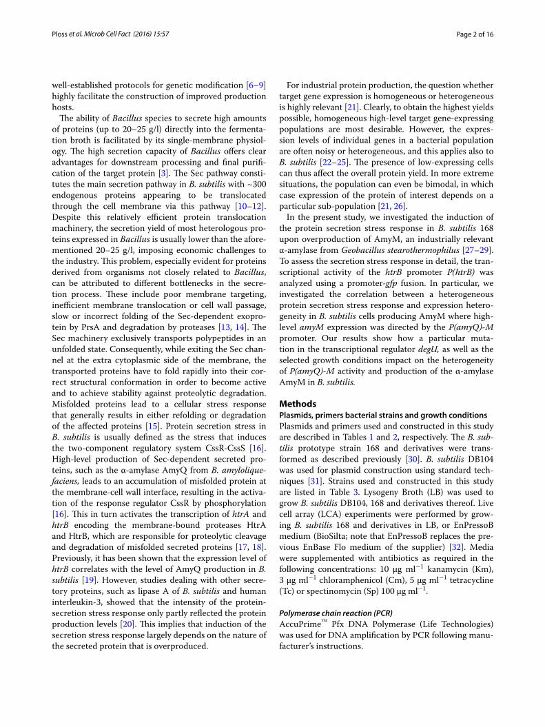
Interestingly, when cells were grown on EnPressoB (Fig. 3, right panel), a unimodal fluorescence distribution was already observed in the exponential growth phase (5.5 h) with most significant differences in fluorescence distribution after 8 and 24 h of cultivation when com-pared to LB medium. This showed that the growth con-ditions imposed by the EnPressoB medium resulted in a homogenous P(amyQ)-M promoter activity.
Homogeneous activity of the P(amyQ)‑M promoter depends on DegU‑P levels and growth conditionsThe two-component regulatory system DegS-DegU is known to regulate the synthesis of many secretory enzymes [38]. In addition, DegU is a master regulator for cell differentiation [39, 40]. The so-called degUhy32 mutation in the degU gene is known to increase the half-life of the phosphorylated form of DegU, leading to
Fig. 2 Heterogeneity of protein secretion stress in amyM-expressing cells. Time-lapse microscopy was performed of B. subtilis 168 Hm C5 (a) and 168 Hm C5 carrying plasmid pDAM coding for the secretory α-amylase AmyM (b). Strains were grown on 1.5 % agarose slides containing 25 % LB broth. Microscopy images are shown from cultures in the exponential, early stationary and stationary growth stages

Page 8 of 16Ploss et al. Microb Cell Fact (2016) 15:57
hyper-production of different enzymes [41, 42]. Further-more, it was previously demonstrated for the P(aprE) promoter that the DegU-P levels correlate with noisy transcription of aprE [43]. Similar to P(aprE), P(amyQ)-M contains a DegU-binding motif (http://dbtbs.hgc.jp/). Rok is another regulator that is known to modulate the expression of membrane-localized and secreted proteins [44]. We therefore decided to analyze the influence of DegU-P and Rok on P(amyQ)-M activity. For this pur-pose, the transcriptional P(amyQ)-M-gfpmut3 fusion was introduced in the B. subtilis strains 168degUhy32 and 168Δrok; the former strain carries the degUhy32 allele, while the rok gene was deleted from the latter strain. To measure the influence of these mutations on overall P(amyQ)-M activity, the respective transformants carry-ing pDAPamy-gfp tet were grown in LB for LCA analysis. As expected, the 168degUhy32 variant showed increased promoter activity of P(amyQ)-M (Fig. 4). In contrast, the 168Δrok strain showed similar promoter activity levels as the 168 wild-type (Fig. 4).
To investigate whether the increased P(amyQ)-M activity in cells bearing the degUhy32 allele might be due to reduced levels of expression heterogeneity, we performed a flow cytometric analysis of GFP expres-sion by individual cells grown in LB (Fig. 5a). Consistent with the LCA measurements in Fig. 4, the flow cytom-etry data showed a clear shift to higher GFP expression
levels for strain 168degUhy32 as compared to the wild-type strain 168. Specifically, the 168degUhy32 strain displayed ~4.5-fold higher GFP fluorescence during exponential growth (5.5 h) and ~2.5-fold higher after 8 h of cultivation (Fig. 5a, bottom). At the same time, the bimodal GFP fluorescence distribution observed in the wild-type strain after 8–24 h of growth was shifted to a unimodal GFP fluorescence distribution in the 168degUhy32 strain (Fig. 5a, top). This implies a more homogeneous P(amyQ)-M activity in the 168degUhy32 strain. In contrast, the 168Δrok strain showed no changes in the overall GFP expression levels and the distribution of GFP fluorescence over individual cells as compared to the wild-type strain 168 (Fig. 5a, top). These findings are consistent with the LCA data for the 168Δrok strain as shown in Fig. 4.
When grown on EnPressoB medium, the 168 wild-type strain showed a more homogeneous activity of P(amyQ)-M as represented by a unimodal GFP fluorescence dis-tribution when compared to cells grown in LB medium (Fig. 5b, top). This is consistent with the results shown in Fig. 3. Notably, also the 168degUhy32 and 168Δrok strains showed the same unimodal fluorescence distri-bution as the 168 wild-type strain when the cells were grown in EnPressoB (Fig. 5b, top). This implies that growth on EnPressoB leads to a homogeneous P(amyQ)-M promoter activity. The only significant difference
Fig. 3 Heterogeneous and homogeneous activity of P(amyQ)-M under different growth conditions. Flow cytometry histograms are shown for B. subtilis strain 168::pDAamy-gfp tet carrying transcriptional fusions of P(amyQ)-M-gfpmut3 grown in LB or EnPressoB medium. Samples were taken after 5.5 (black), 8 (red), 24 (blue) and 32 h (yellow) of cultivation time

Page 9 of 16Ploss et al. Microb Cell Fact (2016) 15:57
was that the GFP expression levels were higher in the 168degUhy32 strain during the exponential phase and in the late stationary growth phase compared to the wild-type and the 168Δrok strains. Specifically, the detected GFP expression levels in the 168degUhy32 strain were 3.5-fold higher after 5.5 h, and 2.5-fold higher after 32 h of cultivation, than in the wild-type strain (Fig. 5b, bot-tom). Together, the present analyses imply that both increased levels of phosphorylated DegU-P and the slow-release glucose feeding regime during cultivation on EnPressoB medium result in a more homogeneous P(amyQ)-M promoter activity.
Homogeneous and heterogeneous translation of AmyMA relevant question triggered by the observed hetero-geneity in the protein secretion stress response and the P(amyQ)-M promoter activity was whether this het-erogeneity could also be detected in the translation of AmyM, which would provide further evidence for AmyM production heterogeneity. For real-time moni-toring of the translation of AmyM, we designed an out-of-frame translational fusion between amyM and gfpmut3. This was necessary, because in-frame fusions between secretory proteins and GFP cannot be effec-tively translocated across the membrane via the Sec
pathway [45, 46]. The out-of-frame translational fusion was generated by creating an overlap between the stop codon of amyM and the start codon of gfpmut3 (Fig. 6a). In this setting, the translation of GFP is dependent on the efficient translation of AmyM, because there is no SD sequence upstream of the GFPmut3-coding region. Further, the expression of this out-of-frame translational amyM-gfpmut3 fusion was directed from P(amyQ)-M using the same plasmid backbone as in the above experiments. The resulting plasmid named pDAamyM-gfp is depicted in Fig. 6a. LCA analyses with B. subtilis 168::pDAamyM-gfp showed an increasing fluorescence intensity during cultivation, which confirmed the effective translation of GFP (Fig. 6b). This was further evidenced by fluorescence microscopy and flow cytom-etry, which also allowed us to monitor homogeneity or heterogeneity in the coupled transcription-transla-tion of the amyM-gfpmut3 fusion upon growth in LB or EnPressoB medium (Fig. 7a, b). The results show that the GFP fluorescence distribution amongst cells grown in the EnPressoB medium was more homogene-ous than that of cells grown in LB (Fig. 7, compare the lower panels in a, b). Moreover, the GFP fluorescence levels were ~2-fold higher upon growth in EnPressoB medium in comparison to growth in LB (Fig. 7c). To
-20000
-10000
0
10000
20000
30000
40000
50000
60000
70000
0 50 100 150 200 250 300 350 400
168::pDAPamy-gfp tet/LB
168degUhy32::pDAPamy-gfp tet/LB
168∆rok::pDAPamy-gfp tet/LB
Pro
mot
er a
ctiv
ity d
GFP
/dT
/OD
600
[UA 5
28h-1
UO
D60
0 -1
]
Cultivation time [min]
Fig. 4 Influence of degUhy32 and rok on P(amyQ)-M promoter activity. P(amyQ)-M promoter activities were determined by fluorescence intensity measurements using LCA for the B. subtilis 168 wild-type strain (diamond) and mutants 168degUhy32 (square) or 168∆rok (open circle) carrying plas-mid pDAPamy-gfp tet with the transcriptional P(amyQ)-M-gfpmut3 fusion. Strains were grown in LB medium over a cultivation period of 400 min

Page 10 of 16Ploss et al. Microb Cell Fact (2016) 15:57
LB medium
5,5 h 8 h 24 h 32 h
a
GFP
Expr
essi
on le
vel
Cul�va�on �me
0200000400000600000800000
100000012000001400000160000018000002000000
5.5h 8h 24h 32h
168::pDAPamy-gfp tet
168Δrok::pDAPamy-gfp tet
168degUHy32::pDAPamy-gfp tet
EnPressoB medium
5,5 h 8 h 24 h 32 h
b
GFP
Expr
essi
on le
vel
0200000400000600000800000
1000000120000014000001600000
5.5h 8h 24h 32h
168::pDAPamy-gfp tet
168Δrok::pDAPamy-gfp tet
168degUHy32::pDAPamy-gfp tet
Cul�va�on �meFig. 5 Influence of degUhy32 and rok mutations on heterogeneous activity of P(amyQ)-M under different growth conditions. GFP expression levels and flow cytometry histograms are shown for B. subtilis strains 168 (black), 168degUhy32 (blue) and 168Δrok (red) carrying plasmid pDAPamy-gfp tet with the transcriptional P(amyQ)-M-gfpmut3 fusion upon growth in LB (a) or EnPressoB (b) medium. Samples were taken after 5.5, 8, 24 and 32 h of cultivation

Page 11 of 16Ploss et al. Microb Cell Fact (2016) 15:57
pDAamyM-gfp6217 bps
NdeI (229)
DraIII (788)
AflII (1062)
BsmI (1781)
BspHI (2694) BssSI (3673) MluI (3821)
BmgBI (4185)
NaeI (5297)
SacII (5848)
gfp
repU
kan
P(amyQ)-M
amyM
tactgttacg tggcaacaat gagaaaagga gaagaacttt atgacaatgc accgttgtta ctcttttcct cttcttgaaa >.........amyM........>>i t v t w q q - >>........gfp..........>
m r k g e e l
a
b
-2000
0
2000
4000
6000
8000
10000
12000
14000
0 100 200 300 400 500
168
168::pDAamyM-gfp
Cultivation Time [min]
Rel
ativ
e Fl
uore
scen
ce In
tens
ity
dGFP
/dT
[UA
528
h-1]
Fig. 6 Out-of-frame translational amyM-gfpmut3 fusion: Plasmid map of pDAamyM-gfp and functional translation of GFP. The plasmid map of pDAamyM-gfp represents a high-copy plasmid for expression of α-amylases carrying a translationally coupled out-of-frame fusion of amyM and gfp-mut3. pDAamyM-gfp plasmid ORFs are indicated by thick colored arrows, with the direction of transcription and the identity of each ORF indicated. Relevant restriction sites are also indicated. Sequence details of the amyM-gfpmut3 fusion are shown in the box (a). LCA measurements with B. subtilis 168::pDAamyM-gfp show the functional translation of GFP. The increase of fluorescence intensity over time [dGFP/dT] was monitored using the LCA system with B. subtilis 168 and the 168 strain carrying plasmid pDAamyM-gfp during cultivation in EnPressoB medium (b)

Page 12 of 16Ploss et al. Microb Cell Fact (2016) 15:57
verify whether this higher GFP expression level reflects also the translation and subsequent secretion of AmyM into the culture broth, growth medium fractions were
analysed by SDS-PAGE. Indeed, the secretion of AmyM by the 168::pDAamyM-gfp strain grown in EnPressoB (Fig. 8b) started at an earlier time point and reached
Fig. 7 Fluorescence microscopy and flow cytometric analysis of GFP expression in B. subtilis 168::pDAamyM-gfp. Fluorescence microscopy and flow cytometry were performed with B. subtilis strain 168 carrying plasmid pDAamyM-gfp that encodes the out-of-frame translational amyM-gfpmut3 fusion upon cultivation in LB (a) or EnPressoB (b) medium. Cells were analysed by microscopy (a, b, top) and flow cytometry after 5.5 (black), 8 (red), 24 (blue), and 32 h (yellow), and histograms of GFP expression are shown (a, b, bottom). A comparison of GFP fluorescence levels detected upon growth in LB medium and EnPressoB is shown in c

Page 13 of 16Ploss et al. Microb Cell Fact (2016) 15:57
higher levels than when these cells were grown in LB (Fig. 8a). As expected, no AmyM was detectable in the growth medium of the wild-type strain 168 (Fig. 8). Together, these observations show that the homogene-ous or heterogeneous expression of amyM, as driven by the P(amyQ)-M promoter under different growth condi-tions, is reflected in the homogeneous or heterogeneous translation of GFP when using an out-of-frame transla-tional fusion between amyM and gfpmut3.
DiscussionThe present study was initially aimed at investigating the possible protein secretion stress response of B. sub-tilis 168 upon high-level expression of the heterologous α-amylase AmyM from G. stearothermopilus. Our results showed that AmyM production only led to a relatively low activation of the secretion stress-responsive P(htrB)
promoter, in contrast to the production of the α-amylase AmyQ from B. amyloliquefaciens, which caused strong P(htrB) activation in accordance with previously pub-lished data [16, 19]. Intriguingly, the induction of P(htrB) in AmyM producing cells was “noisy” when cells were grown in LB medium, suggesting the presence of sub-populations of low-level and high-level AmyM-produc-ing cells. Since low-level producing cells are unwanted in industrial fermentations, this finding was followed up by investigating the effects of a slow-release glucose feeding regime mimicking the industrial setting and par-ticular mutations in DegU and Rok, two major regula-tors of secretory protein production. This showed that slow-release glucose feeding and the degUhy32 mutation can both lead to homogeneous high-level production of AmyM in B. subtilis 168.
The pDAM plasmid that was used for AmyM produc-tion carries the amyM gene under the transcriptional control of the P(amyQ)-M promoter. This promoter is a slightly modified version of the amyQ promoter, which was developed for industrial-scale fermentations. Our present results show that this promoter is “noisy” when cells are cultured on LB medium. In turn this leads to noisy AmyM production which, most likely, explains the heterogeneous secretion stress response. To date, this phenomenon had not been described. Yet, it is a very useful observation because it implies that heterogeneous expression of a secreted target protein can be detected by assessment of heterogeneity in the secretion stress response with transcriptional P(htrB)-gfp fusions.
Gene expression heterogeneity is a general biological phenomenon that allows bacterial populations to rapidly adapt to changing environmental conditions [47]. As a resident of the soil and plant rhizosphere, B. subtilis is a master of adaptation and, accordingly, it is not surprising that it commits itself to differentiation into a multitude of cell types. These differentiation processes encompass motility, biofilm formation, development of genetic com-petence for DNA binding and uptake, sporulation, and also the production of secreted degradative enzymes [22–25, 48, 49]. The production of secretory enzymes in industrial-scale fermentations is aimed towards achiev-ing homogenous populations of highly productive cells, especially during stationary growth [21, 43, 50]. There-fore, promoters to be used in the high-level gene expres-sion for industrial purposes should ideally be strong and controllable. Such promoters can be classified as inducer-specific, growth phase-specific, stress-specific or auto-inducible [50]. This relates to the fact that strategies for large-scale high-yield fermentations for protein pro-duction are often based on a first stage where high cell densities are generated without stressing the cells, and a subsequent protein production phase during which the
a
AmyM
Strain 168 168::pDAM-gfp
94 kDa
67 kDa
43 kDa
30 kDa
20,1 kDa
14,4 kDa
Cul�va�on �me [h] 5.5 8 24 32 5.5 8 24 32
b
AmyM
Strain 168 168::pDAM-gfp
94 kDa
67 kDa
43 kDa
30 kDa
20,1 kDa
14,4 kDa
Cul�va�on �me [h] 5.5 8 24 32 5.5 8 24 32
Fig. 8 Production and secretion of AmyM in B. subtilis 168 carrying the out-of-frame translational amyM-gfpmut3 fusion. B. subtilis strains 168 and 168 carrying pDAamyM-gfp were grown in LB (a) or EnPres-soB medium (b). SDS PAGE was performed with culture supernatant samples after cultivation times of 5.5, 8, 24 and 32 h

Page 14 of 16Ploss et al. Microb Cell Fact (2016) 15:57
target gene is stably expressed at high-level over a long period of time and preferably in a homogeneous man-ner. Promoters satisfying these requirements are for example the Isopropyl β-d-1-thiogalactopyranoside (IPTG)-inducible P(spac) promoter or the xylose-induc-ible P(xynA) promoter. These promoters, however, have the disadvantage that the addition of IPTG or xylose during the fermentation process is relatively costly [2, 50]. Examples of growth phase-regulated promoters induced during the stationary phase are the P(amyE) and P(aprE) promoters of B. subtilis which, respectively, direct expression of the α-amylase AmyE and the serine protease AprE (i.e. subtilisin). Both promoters are highly threshold-dependent on specific transcriptional regula-tors [50, 51]. Our present studies with transcriptional gfp fusions indicate that this is also true for the P(amyQ)-M promoter, because the highest levels of GFP expression were detected during late growth stages, and because the degUhy32 mutation strongly enhanced GFP expres-sion. In this respect it is noteworthy that the high-yield production of native secreted enzymes of B. subtilis, such as proteases, α-amylase and levansucrase, is significantly dependent on the DegS-DegU two-component system [38]. Several mutations in degS and degU that lead to overproduction of proteases have been characterized pre-viously [52, 53]. One of these is the degUhy32 mutation, which increases the half-life of phosphorylated DegU [42]. Accordingly, our present results obtained with the degUhy32 strain suggest that high levels of phosphoryl-ated DegU lead to a more homogeneous and enhanced P(amyQ)-M activity. Consistent with the enhanced activ-ity of P(amyQ)-M in the degUhy32 mutant, this pro-moter contains a DegU binding motif (http://dbtbs.hgc.jp/). These findings are, thus, fully in line with previous studies, which showed that noisy activity of the DegU-dependent P(aprE) promoter is also correlated with the levels of phosphorylated DegU [43, 54].
Batch feeding of catabolite-repressing carbon sources is a well-known approach to reach high product levels for target proteins that are subject to carbon catabolite repression, such as the α-amylases of B. licheniformis and B. amyloliquefaciens [2]. To simulate the industrial carbon-limited conditions in a scaled-down setting, we employed the EnPressoB medium, since this medium simulates a continuous, rationally limited glucose feeding [32]. Our finding that this slow-release glucose feeding also leads to more homogeneous activity of P(amyQ)-M is unprecedented. While it is well known that medium optimization is crucial for high-yield protein production [55, 56], it was not yet known that this optimization may, at least partially, relate to the fact that a more homoge-neous population of high-producing cells is obtained. It will be interesting to verify in future studies whether
this shift to a more homogeneous expression pattern also applies to other proteins of commercial interest that are expressed from different promoters.
To monitor the homogeneous or heterogeneous trans-lation of amyM in B. subtilis, we employed an out-of-frame translationally coupled amyM-gfp fusion where the stop codon of amyM overlapped with the start codon of the gfp gene. This type of translational coupling occurs also naturally in B. subtilis 168, for example in the gerAA-gerAB-gerAC Operon [57]. In our amyM-gfp fusion, we avoided the presence of a spacer between the two genes and the presence of a RBS in the proximity of the gfp start codon as they are present in other gfp operon-like expres-sion constructs that were previously published [58, 59]. Importantly, this approach allowed us to monitor amyM translation by measuring the intracellular appearance of GFP fluorescence, without interfering with the secretion of the co-translated AmyM. Compared to the classical in-frame GFP fusion technology [59, 60], an additional advantage of out-of-frame translationally coupled GFP fusions is that they circumvent the frequently observed instability of large GFP fusion proteins. We conclude from the results of our studies that the transcriptional and out-of-frame translational gfp fusion constructs as described here represent a highly effective tool kit for monitoring potential bottlenecks in high-yield protein production by pinpointing potentially limiting steps in transcription, translation and/or protein secretion [61]. Additionally, the out-of-frame translational fusion of genes for secretory proteins with gfp may serve as a tool for assessing target protein degradation by the different proteases produced by B. subtilis [62]. Although, this potential problem was not specifically addressed in the present study, it is known that B. subtilis 168 secretes a cocktail of eight proteases (i.e. AprE, Bpr, Epr, Mpr, NprB, NprE, Vpr and WprA) that may degrade other cellular and secreted proteins [62–64]. In addition, secretion-stressed cells overproduce the quality control proteases HtrA and HtrB, which may add to the loss of product. Especially in those cases where low production levels are observed, out-of-frame translational gfp fusions are attractive tools to distinguish between production bot-tlenecks at the translational and post-translational levels. Eventually, such tools may even be implemented for the on-line monitoring of protein production during large-scale fermentation processes using state-of-the-art GFP sensors [65–67].
ConclusionIn the present study, we have employed different tran-scriptional and translational gfp fusions to assess the pro-duction of the α-amylase AmyM in B. subtilis in real time. Importantly, such fusions allowed us both to monitor the
Page 15 of 16Ploss et al. Microb Cell Fact (2016) 15:57
cellular secretion stress response, the expression of amyM and the homogeneity of the AmyM-producing cell popula-tion. We conclude that, together, the followed approaches represent a highly effective pipeline for optimizing tran-scription, translation and overall expression homogeneity during production strain and process development.
Availability of data and materialThe datasets supporting the conclusions of this article are included in the article.
Authors’ contributionsConception and design of the study: TNP and JMD. Acquisition of data: TNP, ER, CGM, SP and AL. Analysis and Interpretation of data: TNP, ER, CGM, ELD and JMD. Drafting the article: TNP and ER. All authors were involved in the writing of the manuscript and contributed intellectually. All authors read and approved the final manuscript.
Author details1 AB Enzymes GmbH, Feldbergstrasse 78, 64293 Darmstadt, Germany. 2 Department of Medical Microbiology, University of Groningen, University Medical Center Groningen, Hanzeplein 1, 9700 RD Groningen, The Nether-lands. 3 Roal Oy, Tykkimäentie 15b, 05200 Rajamäki, Finland. 4 Present Address: Department of Medical Microbiology and Infectious Diseases, Erasmus University Medical Center Rotterdam, Rotterdam, The Netherlands. 5 Present Address: Division of Biomedical Sciences, Warwick Medical School, University of Warwick, Coventry CV4 7AL, UK. 6 Present Address: c-LEcta GmbH, Perlick-straße 5, 04103 Leipzig, Germany.
AcknowledgementsThe authors thank Ruben Mars (UMCG) and Kim Langfelder (AB Enzymes GmbH) for helpful discussions, and Daniela Dollak, Evelyn Wallis and Yasar Kaya from AB Enzymes GmbH and Sauli Toikka and Jarno Kallio from Roal Oy for further excellent technical support.
Competing interestsTN. Ploss, A. Lingner and P. Lorenz are employees of AB Enzymes GmbH, and J. Vehmaanperä is an employee of Roal Oy. E. Reilman, C.G. Monteferrante, E.L. Denham, S. Piersma and J.M. van Dijl declare that they have no competing interests.
Received: 5 February 2016 Accepted: 22 March 2016
References 1. Westers L, Westers H, Quax WJ. Bacillus subtilis as cell factory for phar-
maceutical proteins: a biotechnological approach to optimize the host organism. Biochim Biophys Acta. 2004;1694(1–3):299–310.
2. Pohl S, Harwood CR. Heterologous Protein Secretion by Bacillus species from the cradle to the grave. Adv Appl Microbiol. 2010;73:1–25.
3. van Dijl JM, Hecker M. Bacillus subtilis: from soil bacterium to super-secret-ing cell factory. Microb Cell Fact. 2013;12:3. doi:10.1186/1475-2859-12-3.
4. Kunst F, Ogasawara N, Moszer I, Albertini AM, Alloni G, Azevedo V, Bertero MG, Bessieres P, Bolotin A, Borchert S, Boriss R, Boursier L, Brans A, Braun M, Brignell SC, Bron S, Brouillet S, Bruschi CV, Caldwell B, Capuano V, Carter NM, Choi SK, Codani JJ, Connerton A, Danchin A, et al. The com-plete genome sequence of the gram-positive bacterium Bacillus subtilis. Nature. 1997;390(6657):249–56.
5. Barbe V, Curveiller S, Kunst F, Lenoble P, Meurice G, Sekoska A, Vallenet D, Wang T, Moszer I, Medigue C, Danchin A. From a consortium sequence to a unified sequence: the Bacillus subtilis 168 reference genome a decade later. Microbiology. 2009;155:1758–75.
6. Spizizen J. Transformation of biochemically deficient strains of Bacillus subtilis by deoxyribonucleate. Proc Natl Acad Sci USA. 1958;44(10):1072–8.
7. Chang S, Cohen N. High frequency transformation of Bacillus subtilis protoplasts by plasmid DNA. Mol Gen Genet. 1979;168(1Zhan):111–5.
8. Vehmaanperä J, Steinborn G, Hofemeister J. Genetic manipulation of Bacillus amyloliquefaciens. J Biotechnol. 1991;19(2–3):221–40.
9. Zhang XZ, Zhang YH. Simple, fast and high-efficiency transformation system for directed evolution of cellulose in Bacillus subtilis. Microb Biotechnol. 2011;4(1):98–105.
10. Antelmann H, Tjalsma H, Voigt B, Ohlmeier S, Bron S, van Dijl JM, Hecker A. A proteomic view on genome-based signal peptide predictions. Genome Res. 2001;11(9):1484–502.
11. Tjalsma H, Antelmann H, Jongbloed JD, Braun PG, Darmon E, Dorenbos R, Dubois JY, Westers H, Zanen G, Quax WJ, Kuipers OP, Bron S, Hecker M, van Dijl JM. Proteomics of protein secretion by Bacillus subtilis: separating the “secrets” of the secretome. Microbiol Mol Biol Rev. 2004;68(2):207–33.
12. Yamane K, Bunai K, Kakeshita H. Protein traffic for secretion and related machinery of Bacillus subtilis. Biosci Biotechnol Biochem. 2004;68(10):2007–23.
13. Schumann W. Production of recombinant proteins in Bacillus subtilis. Adv Appl Microbiol. 2007;62:137–89.
14. Bolhuis A, Tjalsma H, Smith HE, de Jong A, Meima R, Venema G, Bron S, van Dijl JM. Evaluation of bottlenecks in the late stages of protein secre-tion in Bacillus subtilis. Appl Environ Microbiol. 1999;65(7):2934–41.
15. Sarvas M, Harwood CR, Bron S, van Dijl JM. Post-translocational folding of secretory proteins in Gram-positive bacteria. Biochim Biophys Acta. 2004;1694(1–3):311–27.
16. Hyyryläinen HL, Bolhuis A, Darmon E, Muukkonen L, Koski P, Vitikainen M, Sarvas M, Prágai Z, Bron S, van Dijl JM, Kontinen VP. A novel two-component regulatory system in Bacillus subtilis for the survival of severe secretion stress. Mol Microbiol. 2001;41(5):1159–72.
17. Darmon E, Noone D, Masson A, Bron S, Kuipers OP, Devine KM, van Dijl JM. A novel class of heat and secretion stress-responsive genes is con-trolled by the auto regulated CssRS two-component system of Bacillus subtilis. J Bacteriol. 2002;184(20):5661–71.
18. Antelmann H, Darmon E, Noone D, Veening JW, Westers H, Bron S, Kuipers OP, Devine KM, Hecker M, van Dijl JM. The extracellular proteome of Bacillus subtilis under secretion stress conditions. Mol Microbiol. 2003;49(1):143–56.
19. Westers H, Darmon E, Zanen G, Veening JW, Kuipers OP, Bron S, Quax WJ, van Dijl JM. The Bacillus secretion stress response is an indicator for α-amylase levels. Lett Appl Microbiol. 2004;39:65–73.
20. Westers H, Westers L, Darmon E, van Dijl JM, Quax WJ, Zanen G. The CssRS two-component regulatory system controls a general secretion stress response in Bacillus subtilis. FEBS J. 2006;273:3816–27.
21. Piersma S, Denham EL, Drulhe S, Tonk RHJ, Schwikowski B, van Dijl JM, Quant TLM. An open-source pipeline for visualization and quantification of gene expression heterogeneity in growing microbial cells. PLoS ONE. 2013;8(7):e68696.
22. Losick R, Desplan C. Stochasticity and cell fate. Science. 2008;320(5872):65–8.
23. Maamar H, Raj A, Dubnau D. Noise in gene expression determines cell fate in Bacillus subtilis. Science. 2007;317(5837):526–9.
24. Chastanet A, Vitkup D, Yuan GC, Norman TM, Liu JS, et al. Broadly het-erogeneous activation of the master regulator for sporulation in Bacillus subtilis. Proc Natl Acad Sci USA. 2010;107(18):8486–91.
25. Mars RA, Nicolas P, Ciccolini M, Reilman E, Reder A, Schaffer M, Mäder U, Völker U, van Dijl JM, Denham EL. Small regulatory RNA-induced growth rate heterogeneity of Bacillus subtilis. PLoS Genet. 2015;11(3):e1005046.
26. Veening JW, Smits WK, Kuipers OP. Bistability, epigenetics, and bet-hedging in bacteria. Annu Rev Microbiol. 2008;62:193–210.
27. Goesaert H, Leman P, Bijttebie A, Delcour JA. Antifirming effects of starch degrading enzymes in bread crumb. J Agri Food Chem. 2009;57:2346–55.
28. Derde LJ, Gomand SV, Courtin CM, Delcour JA. Characterisation of three starch degradading enzymes: thermostable β-amylase, maltotetaogenic and maltogenic α-amylases. Food Chem. 2012;135(2):713–21.
29. Van Steertegem B, Pareyt B, Brijs K, Delcour JA. Combined impact of Bacillus stearothermophilus maltogenic alpha-amylase and sur-factants on starch pasting and gelation properties. Food Chem. 2013;139(1–4):1113–20.
30. Anagnostopoulos C, Spizizen J. Requirements for transformation in Bacil-lus subtilis. J Bacteriol. 1961;81(5):741–6.

Page 16 of 16Ploss et al. Microb Cell Fact (2016) 15:57
31. Sambrook J, Fritsch EF, Maniatis T. Molecular Cloning: a laboratory manual. 2nd ed. Cold Spring Harbor: Cold Spring Harbor Laboratory; 1989.
32. Ukkonen K, Vasala A, Ojamo H, Neubauer P. High-yield production of biologically active recombinant protein in shake flask culture by combi-nation of enzyme-based glucose delivery and increased oxygen transfer. Micob Cell Fact. 2011;10:107. doi:10.1186/1475-2859-10-107.
33. Palva I. Molecular Cloning of α-amylase gene from Bacillus amyloliquefa-ciens and its expression in B. subtilis. Gene. 1982;19:81–7.
34. Botella E, Fogg M, Jules M, Piersma S, Doherty G, Hansen A, Denham EL, Chat L, Veiga P, Bailey K, Lewis PJ, van Dijl JM, Aymerich S, Wilkinson A, Devine KM. pBaSysBioII: an integrative plasmid generating gfp transcrip-tional fusions for high-throughput analysis of gene expression in Bacillus subtilis. Microbiology. 2010;156:1600–8.
35. Kouwen TRHM, Nielsen AK, Denham EL, Dubois J-YF, Dorenbos R, Ras-mussen MD, Quax WJ, Freudl R, van Dijl JM. Contributions of the pre- and pro-regions of Staphylococcus hyicus lipase to secretion of heterologous protein by Bacillus subtilis. Appl Environ Microbiol. 2010;76(3):219–25.
36. Kawamura F, Doi RH. Construction of a Bacillus subtilis double mutant deficient in extracellular alkaline and neutral proteases. J Bacteriol. 1984;160(1):442–4.
37. Veening JW, Stewart EJ, Berngruber TW, Taddei F, Kuipers OP, Hamoen LW. Bet-hedging and epigenetic inheritance in bacterial cell development. Proc Natl Acad Sci USA. 2008;105:4393–8.
38. Msadek T, Kunst F, Rapoport G. A signal transduction network in Bacillus subtilis includes the DegS/DegU and ComP/ComA two-component systems. In: Hoch JA, Silhavy TJ, editors. Two-Component Signal Transduc-tion. Washington: ASM press; 1995. p. 447–71.
39. Kobayashi K. Gradual activation of the response regulator DegU controls serial expression of genes for flagellum formation and biofilm formation in Bacillus subtilis. Mol Microbiol. 2007;66(2):395–409.
40. Verhamme DT, Kiley TB, Stanley-Wall NR. DegU co-ordinates multicellular behavior exhibited by Bacillus subtilis. Mol Microbiol. 2007;65(2):554–68.
41. Tjalsma H, Koetje EJ, Kiewiet R, Kuipers OP, Kolkman M, van deer Lan J, Daskin R, Ferrari E, Bron S. Engineering of quorum-sensing systems for improved production of alkaline protease by Bacillus subtilis. J Appl Microbiol. 2004;96(3):569–78.
42. Dahl MK, Msadek T, Kunst F, Rapoport G. The phosphorylation state of the DegU response regulator acts as a molecular switch allowing either degradative enzyme synthesis or expression of genetic competence in Bacillus subtilis. J Biol Chem. 1992;267(20):14509–14.
43. Veening JW, Igoshin OA, Eijlander RT, Nijland R, Hamoen LW, Kuipers O. Transient heterogeneity in extracellular protease production by Bacillus subtilis. Mol Sys Biol. 2008;4:184. doi:10.1038/msb.2008.18.
44. Albano M, Smits WK, Ho LTY, Kraigher B, Mandic-Mulec I, Kuipers OP, Dub-nau D. The Rok protein of Bacillus subtilis represses genes for cell surface and extracellular functions. J Bacteriol. 2005;187(6):2010–9.
45. van der Ploeg R, Monteferrante CG, Piersma S, Barnett JP, Kouwen TR, Robinson C, van Dijl JM. High-salinity growth conditions promote Tat-independent secretion of Tat substrates in Bacillus subtilis. Appl Environ Microbiol. 2012;78(21):7733–44.
46. Thomas JD, Daniel RA, Errington J, Robinson C. Export of active green fluorescent protein to the periplasm by the twin-arginine translocase (Tat) pathway in Escherichia coli. Mol Microbiol. 2001;39(1):47–53.
47. Raj A, van Oudenaarden A. Nature, nurture, or chance: stochastic gene expression and its consequences. Cell. 2008;135(2):216–26.
48. Lopez D, Vlamakis H, Kolter R. Generation of multiple cell types in Bacillus subtilis. FEMS Microbiol Rev. 2009;33(1):152–63.
49. Romero D. Bacterial determinants of the social behavior of Bacillus subtilis. Res Microbiol. 2013;164(7):788–98.
50. Schallmey M, Singh A, Ward OP. Development in the use of Bacillus spe-cies for industrial production. Can J Microbiol. 2004;50(1):1–17.
51. Valle F, Ferrari E. Subtilisin: a redundantly temporally regulated gene? In: Smith I, Slepecky RA, Setlow P, editors. Regulation of procaryotic develop-ment. Washington: American Society for Microbiology; 1989. p. 131–46.
52. Msadek T, Kunst F, Henner D, Klier A, Rapoport G, Dedonder R. Signal transduction pathway controlling synthesis of a class of degradative enzymes in Bacillus subtilis: expression of the regulatory genes and analy-sis of mutations in degS and degU. J Bacteriol. 1990;172(2):824–34.
53. Tanaka T, Kawata M, Mukai K. Altered phosphorylation of Bacillus subtilis DegU caused by single amino acid changes in DegS. J Bacteriol. 1991;173(17):5507–15.
54. Shimane K, Ogura M. Mutational analysis of the helix-turn-helix region of Bacillus subtilis response regulator DegU, and identification of cis-acting sequences for DegU in the aprE and comK promoters. J Biochem. 2004;136(3):387–97.
55. Raymond HM, Douglas CM, Christine M. Response surface methodology: process and product optimization using designed experiments. Wiley: Hoboken. ISBN 978-0-470-17446-3; 2009. p. 235–258.
56. Zhao W, Zheng J, Zhou HB. Hybrid on-line optimal control strategy for producing α-Amylase by Bacillus subtilis. Biosci Biotechnol Biochem. 2011;75(4):694–9.
57. Zuberi AR, Moir A, Feavers IM. The nucleotide sequence and gene organi-zation of the gerA spore germination operon of Bacillus subtilis 168. Gene. 1987;51(1):1–11.
58. Levin-Karp A, Barenholz U, Bareia T, Dayagi M, Zelcbuch L, Antonovsky N, Noor E, Milo R. Quantifying translational coupling in E. coli synthetic operons using RBS modulation and fluorescent reporters. ACS Synth Biol. 2013;2(6):327–36.
59. Delisa MP, Chae HJ, Weigand WA, Valdes JJ, Rao G, Bentley WE. Generic model control of induced protein expression in high cell density cultiva-tion of Escherichia coli using on-line GFP-fusion monitoring. Bioprocess Biosyst Eng. 2001;24(2):83–91.
60. Siepert E-M, Gartz E, Tur MK, Delbrück H, Barth S, Büchs J. Short-chain fluorescent tryptophan tags for on-line detection of functional recombi-nant proteins. BMC Biotechnol. 2012;12:65. doi:10.1186/1472-6750-12-65.
61. Li W, Zhou X, Lu P. Bottlenecks in the expression and secretion of heter-ologous proteins in Bacillus subtilis. Res Microbiol. 2004;155(8):605–10.
62. Krishnappa L, Dreisbach A, Otto A, Goosens VJ, Crananburgh RM, Har-wood CR, Becher D, van Dijl JM. Extracytoplasmic proteases determining the cleavage and release of secreted proteins, lipoproteins, and mem-brane proteins in Bacillus subtilis. J Proteome Res. 2013;12(9):4101–10.
63. Hoch JA, Losick R, Sonenshein AL. Bacillus subtilis and other gram positive bacteria: biochemistry physiology and molecular genetics. Washington: American Society for Microbiology; 1993.
64. Westers L, Westers H, Zanen G, Antelmann H, Hecker M, Noone D, Devine KM, van Dijl JM, Quax WJ. Genetic or chemical protease inhibition causes significant changes in the Bacillus subtilis exoproteome. Proteomics. 2008;8(13):2704–13.
65. Randers-Eichhorn L, Albano CR, Sipior J, Bentley WE, Rao G. On-line green fluorescent protein sensor with LED excitation. Biotechnol Bioeng. 1997;55(6):921–6.
66. Kostov Y, Albano CR, Rao G. All solid-state GFP sensor. Biotechnol Bioeng. 2000;70(4):473–7.
67. Suber G, Pötschacher F, Bayer K. Evaluation of GFP signal and its aptitude for novel on-line monitoring strategies of recombinant fermentation processes. J Biotechnol. 2000;108(2):115–25.