Manipulating insulin signaling to enhance mosquito reproduction
Upload
bakersfieldcollegeCategory
view
1download
0
ORIGINAL RESEARCH
Insulin and GLP-1 infusions demonstrate the onset ofadipose-specific insulin resistance in a large fastingmammal: potential glucogenic role for GLP-1Jose A. Viscarra1, Ruben Rodriguez1, Jose Pablo Vazquez-Medina1, Andrew Lee1, Michael S. Tift2,Stephen K. Tavoni3, Daniel E. Crocker3 & Rudy M. Ortiz1
1 School of Natural Sciences, University of California, Merced, California
2 Scripps Institution of Oceanography, University of California, San Diego, California
3 Department of Biology, Sonoma State University, Rohnert Park, California
Keywords
Adipose tissue, elephant seal, fatty acids,
GLP-1, glucose intolerance, insulin sensitivity.
Correspondence
Jose A. Viscarra, University of California
Merced, School of Natural Sciences, 5200 N.
Lake Rd., Merced, CA 95343.
Tel: (209)228-4016
Fax: (209)228-4060
E-mail: [email protected]
Funding Information
This work was supported by grants: NIH
NHLBI HL091767, NIH NHLBI HL091767-S1,
and NIH NHLBI K02HL103787.
Received: 6 May 2013; Revised: 10 June
2013; Accepted: 11 June 2013
doi: 10.1002/phy2.23
Physiol Rep, 1 (2), 2013, e00023, doi:
10.1002/phy2.23
Abstract
Prolonged food deprivation increases lipid oxidation and utilization, which
may contribute to the onset of the insulin resistance associated with fasting.
Because insulin resistance promotes the preservation of glucose and oxidation
of fat, it has been suggested to be an adaptive response to food deprivation.
However, fasting mammals exhibit hypoinsulinemia, suggesting that the insu-
lin resistance-like conditions they experience may actually result from reduced
pancreatic sensitivity to glucose/capacity to secrete insulin. To determine
whether fasting results in insulin resistance or in pancreatic dysfunction, we
infused early- and late-fasted seals (naturally adapted to prolonged fasting)
with insulin (0.065 U/kg), and a separate group of late-fasted seals with low
(10 pmol/L per kg) or high (100 pmol/L per kg) dosages of glucagon-like
peptide-1 (GLP-1) immediately following a glucose bolus (0.5 g/kg), and mea-
sured the systemic and cellular responses. Because GLP-1 facilitates glucose-
stimulated insulin secretion, these infusions provide a method to assess
pancreatic insulin-secreting capacity. Insulin infusions increased the phosphor-
ylation of insulin receptor and Akt in adipose and muscle of early- and late-
fasted seals; however, the timing of the signaling response was blunted in
adipose of late-fasted seals. Despite the dose-dependent increases in insulin
and increased glucose clearance (high dose), both GLP-1 dosages produced
increases in plasma cortisol and glucagon, which may have contributed to the
glucogenic role of GLP-1. Results suggest that fasting induces adipose-specific
insulin resistance in elephant seal pups, while maintaining skeletal muscle
insulin sensitivity, and therefore suggests that the onset of insulin resistance in
fasting mammals is an evolved response to cope with prolonged food
deprivation.
Introduction
Insulin resistance is a common consequence of fasting
(van der Crabben et al. 2008) and, although the exact
mechanisms by which it manifests are still unclear, it is
thought that its primary cause is the increased utilization
of lipids during food deprivation (Koves et al. 2005;
Samuel et al. 2010; Viscarra et al. 2012). Insulin resistance
has a negative connotation due to its association with
obesity and diabetes in humans, but it has been suggested
to be an adaptive response to food deprivation (Viscarra
et al. 2012; Tsatsoulis et al. 2013; Viscarra and Ortiz
2013). Because fasting mammals depend primarily on
lipids for energy, the decreased glucose uptake and utili-
zation resulting from impaired insulin signaling preserves
the limited carbohydrate substrates for tissues that do not
readily metabolize lipids (e.g., central nervous system
[CNS], red blood cells [RBC]). Additionally, as less glucose
ª 2013 The Authors. Physiological Reports published by Wiley Periodicals, Inc. on behalf of
the American Physiological Society and The Physiological Society.
This is an open access article under the terms of the Creative Commons Attribution License,
which permits use, distribution and reproduction in any medium, provided the original work is properly cited.
2013 | Vol. 1 | Iss. 2 | e00023Page 1
Physiological Reports ISSN 2051-817X

is used, less protein has to be broken down for gluconeo-
genesis, and thus lean tissue catabolism can be reduced
(Cherel et al. 1992).
Northern elephant seal pups (Mirounga angustirostris)
undergo a 2–3 month postweaning fast during which they
remain normothermic and metabolically active, while
relying primarily on the oxidation of stored lipids to meet
their caloric needs (Castellini et al. 1987; Adams and
Costa 1993). The postweaning fasts are characterized by
increased plasma free fatty acids (FFA), elevated plasma
glucose, and decreased cellular insulin signaling activity
(Castellini et al. 1987; Adams and Costa 1993;
Champagne et al. 2005; Viscarra et al. 2011b, 2012),
which collectively would constitute an insulin resistance
phenotype. Furthermore, we have previously reported sig-
nificant reductions in the concentrations of insulin-sensi-
tizing hormones, adiponectin and IGF-1, along with
significant increases in cortisol, which has been reported
to antagonize insulin activity (Barseghian et al. 1982;
Lambillotte et al. 1997; Viscarra et al. 2011a). Prolonged
fasting in pups is also characterized by decreased plasma
insulin and reduced glucose-stimulated insulin secretion
(GSIS) (Viscarra et al. 2011a) suggesting that this period
of absolute food deprivation is associated with impaired
pancreatic responsiveness. However, it remains unclear
whether the fasting-induced insulin resistance phenotype
in fasting-adapted mammals is the result of reduced
glucose tolerance by peripheral tissues, impaired/altered
pancreatic responsiveness, or both.
Therefore, the present study was designed to assess the
mechanisms contributing to the onset of an insulin resis-
tant-like condition induced by prolonged food depriva-
tion/starvation in mammals. Because elephant seals have
evolved robust physiological mechanisms that have
allowed them to naturally tolerate such protracted bouts
of fasting, they provide an ideal model to examine the
effects of fasting on insulin sensitivity. To address the
hypothesis that prolonged fasting induces insulin resis-
tance in elephant seal pups, we assessed insulin signaling
in skeletal muscle and adipose tissue in response to exog-
enous insulin. Furthermore, we infused late-fasted animals
with glucagon-like peptide 1 (GLP-1), a gastrointestinal
hormone that works synergistically with glucose to
enhance postprandial GSIS (MacDonald et al. 2002), to
determine whether seals experience reduced pancreatic
sensitivity to glucose or decreased capacity to secrete
insulin late in the fast.
Methods
All procedures were reviewed and approved by the Insti-
tutional Animal Care and Use Committees of both the
University of California Merced and Sonoma State
University. All work was realized under the National Mar-
ine Fisheries Service marine mammal permit #87-1743.
Animals
Seventeen northern elephant seal pups constituting four
different cohorts at A~no Nuevo State Reserve were stud-
ied at two postweaning periods: early (1–2 weeks post
weaning; n = 5) and late (6–7 weeks post weaning;
n = 12). Pups were weighed, sedated, and infused in the
field as previously described (Viscarra et al. 2011a,b).
Briefly, pups were sedated with 1 mg/kg Telazol (tileta-
mine/zolazepam HCl, Fort Dodge Labs, Ft Dodge, IA)
administered intramuscularly. Once immobilized, an 18
gauge, 3.5 inch spinal needle was inserted into the extra-
dural vein. Blood samples were obtained, and infusions
performed from this site. Continued immobilization was
maintained with ~100 mg bolus intravenous injections of
ketamine as needed.
Insulin and GLP-1 infusion protocols
Prior to each infusion protocol, preinfusion blood sam-
ples (i.v.) and tissue (adipose and muscle) biopsies were
collected as previously described (Viscarra et al. 2011a).
Because skeletal muscle was obtained opportunistically
(attached to adipose biopsy), we were able to obtain
samples from the insulin-infused animals but not the
GLP-1-infused animals. All biopsies were rinsed with ster-
ile saline, placed in cryovials, and immersed in liquid
nitrogen immediately after collection as previously
described (Viscarra et al. 2011a,b, 2012).
Insulin tolerance tests
To determine the effects of prolonged fasting on peripheral
insulin activity and function, 10 fasting seal pups (early
n = 5, late n = 5) were infused (i.v.) with a mass-specific
dose of insulin (0.065 U/kg) (Humulin; Eli Lilly, Indianap-
olis, IN). Following the bolus infusion, blood samples were
collected at 5, 10, 20, 30, 60, 90, and 120 min. Subsequent
adipose and opportunistic muscle biopsies were collected at
60 and 120 min. Procedures were terminated at 120 min
(instead of 150 min like in the GLP-GTT [glucose tolerance
test] and GTT) due to concerns over the safety of the
animals. Immediately following the collection of the
120 min samples, glucose was infused slowly to assist in
the restoration of preinfusion levels.
GLP-1 + glucose tolerance tests
GLP-1 is a gastrointestinal hormone that facilitates the
postprandial glucose-stimulated secretion of pancreatic
2013 | Vol. 1 | Iss. 2 | e00023Page 2
ª 2013 The Authors. Physiological Reports published by Wiley Periodicals, Inc. on behalf of
the American Physiological Society and The Physiological Society.
Adipose Insulin Resistance during Fasting J. A. Viscarra et al.

insulin (MacDonald et al. 2002), and thus, its infusion
should provide a method to differentiate between reduced
insulin production (pancreatic capacity) and pancreatic
glucose intolerance. We infused GLP-1 in the presence of
glucose (GTT) to allow us to differentiate between
reduced insulin production and glucose intolerance. This
experimental protocol was adopted because GLP-1 in the
presence of elevated glucose has the potential to provide
greater insight to GLP-1-mediated effects. Seven, late-
fasted seal pups were administered either a low (LDG;
10 pmol/kg; n = 3) or high (HDG; 100 pmol/kg; n = 4)
dose of GLP-1 (Sigma, St Louis, MO) bolus immediately
following a glucose bolus (0.5 g/kg) (i.v.) infused within
2 min. GLP1 + GTT manipulations were performed only
on late-fasted animals, because we and others have dem-
onstrated that the insulin resistance-like conditions
develop with fasting duration (Houser et al. 2007; Fowler
et al. 2008; Viscarra et al. 2011a,b, 2012). Following the
infusions, blood samples were collected at 10, 20, 30, 60,
90, 120, and 150 min. Subsequent adipose biopsies were
collected at 60 and 150 min.
Late-fasting GTT
To better assess and interpret the effects of GTT indepen-
dent of the GLP-1 doses, we present data from late-fasted
animals given the same dose of glucose (0.5 g/kg) (late
GTT; same aged animals as those studied here) from our
previous study (Viscarra et al. 2011a).
Sample preparation
Blood samples were centrifuged on site for 15 min at
3000g, and the plasma was transferred to cryo-vials,
frozen by immersion in liquid nitrogen, and stored at
�80°C. Adipose and skeletal muscle were homogenized
and the cytosolic- and membrane-bound protein
fractions separated as previously described (Viscarra et al.
2011a). Total protein content in both cytosolic and
membrane fractions was measured by the Bradford assay
(Bio-Rad Laboratories, Hercules, CA) and used to
normalize the loading of samples into gel wells (Viscarra
et al. 2011a).
Western blots
Twenty micrograms of total protein were resolved in
4–15% Tris-HCl SDS gradient gels. Proteins less than
100 kDa were electroblotted using the Bio-Rad Trans
Blot SD semi-dry cell onto 0.45 lm nitrocellulose mem-
branes. Proteins larger than 100 kDa were electroblotted
using the Bio-Rad Mini Protean Transfer apparatus onto
0.45 lm nitrocellulose membrane. Membranes were
blocked with 3% bovine serum albumin (BSA) in phos-
phate-buffered saline (PBS) containing 0.05% Tween 20
(PBS-T), and incubated overnight with primary antibod-
ies. Membranes were washed, incubated with horse
radish peroxidase (HRP)-conjugated secondary antibod-
ies (SCBT, Santa Cruz, CA), rewashed, and developed
by using the Pierce ECL Western Blotting Substrate
(Thermo Scientific, Waltham, MA). Blots were visualized
using a Kodak Image Station 440CF (Kodak Digital
Sciences, Rochester, NY) and quantified using Care-
Stream Molecular Imaging software. Because we have
previously suggested that increased lipid utilization con-
tributes to the onset of insulin resistance in late-fasted
seals (Viscarra et al. 2012), we examined the following
lipid handling proteins in adipose to assess the contri-
bution of lipid metabolism to insulin signaling in late-
fasted elephant seal pups: AMP kinase (AMPk; 65 kDa),
adipose triglyceride lipase (ATGL; 55 kDa), fatty acid
translocase (CD36; 53 kDa), fatty acid transport protein
1 (FATP1; 63 kDa), fatty acid synthase (FAS; 270 kDa),
hormone-sensitive lipase (HSL; 88 kDa), lipoprotein
lipase (LPL; 56 kDa), and phosphoenol pyruvate carboxy
kinase-c (PEPCK-c; 67 kDa). To assess the effect of the
infusions on insulin signaling we measured the phos-
phorylation and relative protein abundance of Akt
(56 kDa), and insulin receptor (IR; 100 kDa). Primary
antibodies were previously validated for use in elephant
seal tissues by preadsorption with a blocking peptide to
verify that the band being quantified was the band of
interest and by using cell lysates recommended by the
manufacturers as positive controls. In addition to con-
sistently loading the same amount of total protein per
well (25 lg), densitometry values were further normal-
ized by correcting for the densitometry values of actin
(Viscarra et al. 2011a).
Plasma analyses
The plasma concentrations of triglycerides (TAG; Cayman
Chemical, Ann Arbor, MI), glycerol (Cayman), glucose
(Cayman), and nonesterified fatty acids (NEFA; Wako
Chemicals; Richmond, VA) were measured with colorimet-
ric kits. Plasma insulin (porcine insulin, Millipore, Billeri-
ca, MA), glucagon (Millipore), and cortisol (Siemens,
Washington, D.C.) were measured using radioimmunoas-
say kits. Plasma adiponectin (canine adiponectin, Milli-
pore) was measured using an enzyme immunoassay kit.
GLP-1 (Millipore) was measured using a fluorometric
assay kit. All kits (with the exception of GLP-1) have been
previously validated for use with elephant seal plasma
(Ortiz et al. 2001, 2003; Viscarra et al. 2011a,b, 2012). The
GLP-1 assay was validated for use with elephant seal
plasma by performing linearity of dilution (limit of
ª 2013 The Authors. Physiological Reports published by Wiley Periodicals, Inc. on behalf ofthe American Physiological Society and The Physiological Society.
2013 | Vol. 1 | Iss. 2 | e00023Page 3
J. A. Viscarra et al. Adipose Insulin Resistance during Fasting

linearity: 2–90 pmol/L) as well as spike and recovery
assessments. All samples were analyzed in duplicate and
run in a single assay with intraassay percent coefficient of
variability of <10% for all assays.
Adipose lipid analyses
Adipose tissue was homogenized as recommended by the
manufacturer, and TAG and diacyl glycerol (DAG) were
measured using commercially available kits (TAG,
Cayman Chemicals; mouse DAG, CUSABIO, China).
Both kits were validated by performing linearity of dilu-
tion as well as spike and recovery assessments. All samples
were analyzed in duplicate and run in a single assay with
intraassay, percent coefficients of variability of <10% for
all assays.
Glucose clearance calculations
For the insulin infusions, the rate of glucose disappear-
ance (K) was calculated using the linear regression as the
negative slope of the natural log of glucose concentrations
from 0 min to 60 min postinfusion. For the GLP-1 infu-
sions, K was calculated assuming that equilibration of the
injected glucose with the total body pool was achieved by
20 min (Champagne et al. 2005; Fowler et al. 2008).
Thus, K was calculated using the linear regression as the
negative slope of glucose concentrations from 20 min to
150 min post infusion (Fowler et al. 2008; Viscarra et al.
2011a). This method was also used to estimate K from
previously reported mean values (Tura et al. 2001; Wu
et al. 2011; Harris and Apolzan 2012; Rodriguez et al.
2012) and compared to those calculated here in response
to the infusions.
Statistics
The baseline (or T0) measurements (plasma or tissue
protein content) of the early and late insulin infusions
were used to assess changes as a function of fasting
duration. Postinfusion measurements were also
compared between the different infusions (insulin or
GLP-1). Means (�SE) were compared by analysis of
variance (ANOVA) using a Fisher’s PLSD post hoc test.
Repeated measures ANOVA was used to determine
changes following the infusions. Adipose and skeletal
muscle target protein content was normalized by
expressing as percent change versus the early fast mean
value, and compared by ANOVA to determine changes
in response to the separate infusions. Changes were
considered significantly different at P < 0.05. Statistical
analyses were performed with StatView� software (SAS
Institute Inc., Cary, NC).
Results
Fasting reduces body mass and plasmaglucose, and increases plasma lipids andadipose DAG:TAG
Mean body mass of late-fasted pups was 27% lower than
early-fasted pups (Table 1). Mean plasma glucose
decreased 21% with fasting (Table 1). Mean plasma TAG
and NEFA increased 22% and 90%, respectively, with
fasting (Table 1). Mean plasma glycerol did not change
significantly with fasting; however, the NEFA to glycerol
ratio increased 50% with fasting (Table 1). Fasting did
not significantly change the absolute content of adipose
DAG or TAG; however, adipose DAG:TAG ratio increased
55% with fasting (Table 1).
Fasting is associated with an insulinresistance-like endocrine profile
Mean plasma adiponectin, GLP-1, and insulin decreased
23%, 71%, and 37%, respectively, with fasting (Table 1).
Mean plasma cortisol nearly doubled with fasting
(Table 1). Mean plasma glucagon did not change signifi-
cantly with fasting (Table 1).
Fasting alters adipose lipid-mobilizingproteins
Mean adipose ATGL relative protein abundance was 46%
higher in late- versus early-fasted animals (Fig. 1). Con-
versely, relative protein abundance of adipose fatty acid
Table 1. Mean (�SE) body mass, plasma glucose and lipids, adi-
pose diacylglyceride (DAG):triacylglyceride (TAG) ratio, and plasma
hormones at early (2 weeks) and late (7 weeks) fasting periods
from northern elephant seal pups.
Early (n = 5) Late (n = 5)
Body mass (kg) 127 � 1 93 � 41
Glucose (mmol/L) 9.6 � 0.4 7.5 � 0.41
Triglycerides (mmol/L) 0.88 � 0.09 1.07 � 0.141
Glycerol (mmol/L) 0.26 � 0.06 0.33 � 0.10
NEFA (mmol/L) 1.1 � 0.1 2.1 � 0.31
NEFA:Glycerol 4.2 � 0.3 6.3 � 0.41
Intraadipose DAG:TAG 0.45 � 0.06 0.70 � 0.041
Adiponectin (ng/mL) 80.8 � 3.7 65.1 � 3.61
Cortisol (nmol/L) 201 � 13 396 � 211
Glucagon (pmol/L) 11.6 � 1.6 12.9 � 0.9
Glucagon-like peptide-1 (pmol/L) 4.9 � 1.3 1.4 � 0.91
Insulin (lU/mL) 3.2 � 0.5 2.0 � 0.61
1Significant difference from early at P < 0.05.
2013 | Vol. 1 | Iss. 2 | e00023Page 4
ª 2013 The Authors. Physiological Reports published by Wiley Periodicals, Inc. on behalf of
the American Physiological Society and The Physiological Society.
Adipose Insulin Resistance during Fasting J. A. Viscarra et al.
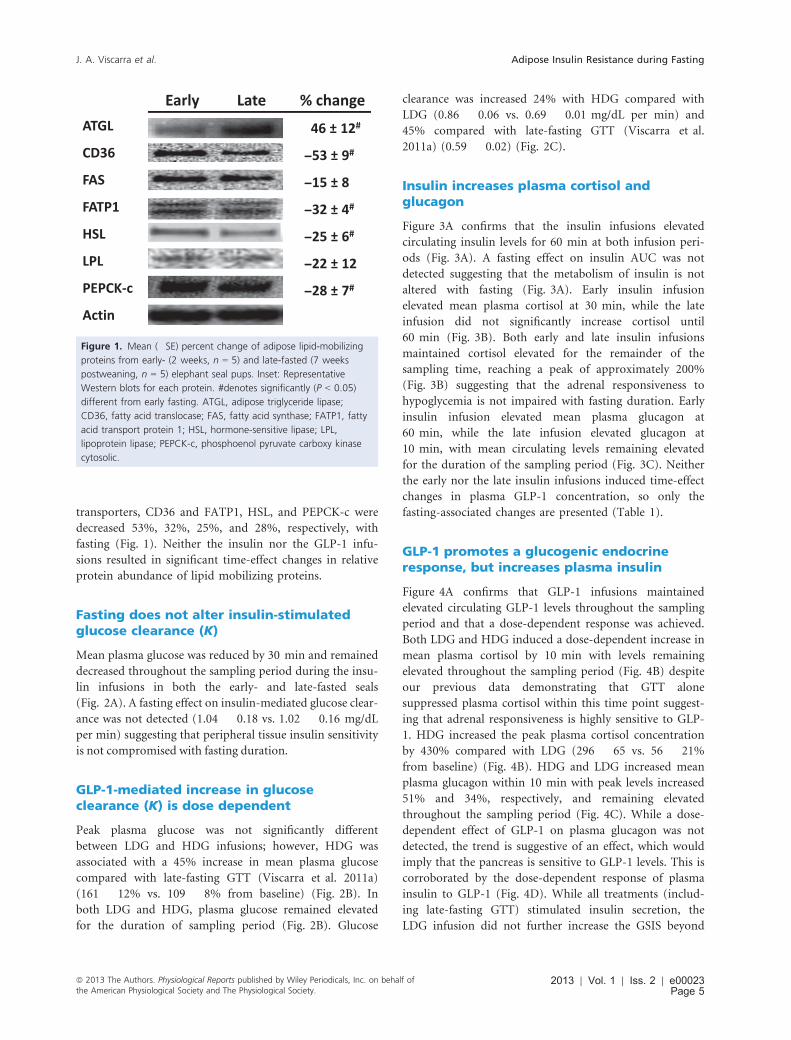
transporters, CD36 and FATP1, HSL, and PEPCK-c were
decreased 53%, 32%, 25%, and 28%, respectively, with
fasting (Fig. 1). Neither the insulin nor the GLP-1 infu-
sions resulted in significant time-effect changes in relative
protein abundance of lipid mobilizing proteins.
Fasting does not alter insulin-stimulatedglucose clearance (K)
Mean plasma glucose was reduced by 30 min and remained
decreased throughout the sampling period during the insu-
lin infusions in both the early- and late-fasted seals
(Fig. 2A). A fasting effect on insulin-mediated glucose clear-
ance was not detected (1.04 � 0.18 vs. 1.02 � 0.16 mg/dL
per min) suggesting that peripheral tissue insulin sensitivity
is not compromised with fasting duration.
GLP-1-mediated increase in glucoseclearance (K) is dose dependent
Peak plasma glucose was not significantly different
between LDG and HDG infusions; however, HDG was
associated with a 45% increase in mean plasma glucose
compared with late-fasting GTT (Viscarra et al. 2011a)
(161 � 12% vs. 109 � 8% from baseline) (Fig. 2B). In
both LDG and HDG, plasma glucose remained elevated
for the duration of sampling period (Fig. 2B). Glucose
clearance was increased 24% with HDG compared with
LDG (0.86 � 0.06 vs. 0.69 � 0.01 mg/dL per min) and
45% compared with late-fasting GTT (Viscarra et al.
2011a) (0.59 � 0.02) (Fig. 2C).
Insulin increases plasma cortisol andglucagon
Figure 3A confirms that the insulin infusions elevated
circulating insulin levels for 60 min at both infusion peri-
ods (Fig. 3A). A fasting effect on insulin AUC was not
detected suggesting that the metabolism of insulin is not
altered with fasting (Fig. 3A). Early insulin infusion
elevated mean plasma cortisol at 30 min, while the late
infusion did not significantly increase cortisol until
60 min (Fig. 3B). Both early and late insulin infusions
maintained cortisol elevated for the remainder of the
sampling time, reaching a peak of approximately 200%
(Fig. 3B) suggesting that the adrenal responsiveness to
hypoglycemia is not impaired with fasting duration. Early
insulin infusion elevated mean plasma glucagon at
60 min, while the late infusion elevated glucagon at
10 min, with mean circulating levels remaining elevated
for the duration of the sampling period (Fig. 3C). Neither
the early nor the late insulin infusions induced time-effect
changes in plasma GLP-1 concentration, so only the
fasting-associated changes are presented (Table 1).
GLP-1 promotes a glucogenic endocrineresponse, but increases plasma insulin
Figure 4A confirms that GLP-1 infusions maintained
elevated circulating GLP-1 levels throughout the sampling
period and that a dose-dependent response was achieved.
Both LDG and HDG induced a dose-dependent increase in
mean plasma cortisol by 10 min with levels remaining
elevated throughout the sampling period (Fig. 4B) despite
our previous data demonstrating that GTT alone
suppressed plasma cortisol within this time point suggest-
ing that adrenal responsiveness is highly sensitive to GLP-
1. HDG increased the peak plasma cortisol concentration
by 430% compared with LDG (296 � 65 vs. 56 � 21%
from baseline) (Fig. 4B). HDG and LDG increased mean
plasma glucagon within 10 min with peak levels increased
51% and 34%, respectively, and remaining elevated
throughout the sampling period (Fig. 4C). While a dose-
dependent effect of GLP-1 on plasma glucagon was not
detected, the trend is suggestive of an effect, which would
imply that the pancreas is sensitive to GLP-1 levels. This is
corroborated by the dose-dependent response of plasma
insulin to GLP-1 (Fig. 4D). While all treatments (includ-
ing late-fasting GTT) stimulated insulin secretion, the
LDG infusion did not further increase the GSIS beyond
Figure 1. Mean (�SE) percent change of adipose lipid-mobilizing
proteins from early- (2 weeks, n = 5) and late-fasted (7 weeks
postweaning, n = 5) elephant seal pups. Inset: Representative
Western blots for each protein. #denotes significantly (P < 0.05)
different from early fasting. ATGL, adipose triglyceride lipase;
CD36, fatty acid translocase; FAS, fatty acid synthase; FATP1, fatty
acid transport protein 1; HSL, hormone-sensitive lipase; LPL,
lipoprotein lipase; PEPCK-c, phosphoenol pyruvate carboxy kinase
cytosolic.
ª 2013 The Authors. Physiological Reports published by Wiley Periodicals, Inc. on behalf ofthe American Physiological Society and The Physiological Society.
2013 | Vol. 1 | Iss. 2 | e00023Page 5
J. A. Viscarra et al. Adipose Insulin Resistance during Fasting

the late-fasting GTT (Fig. 4D). However, HDG nearly
doubled the increase in mean plasma insulin compared
with LDG and late-fasting GTT (Viscarra et al. 2011a),
with this increase persisting throughout the sampling per-
iod (Fig. 4D).
Insulin and GLP-1 infusions decrease plasmalipids
Insulin
The early infusion decreased mean plasma TAG 30% at
60 min and returned to baseline levels at 120 min; how-
ever, the late infusion did not significantly change plasma
TAG (Fig. 5A). Early and late infusions increased mean
plasma glycerol 38% and 23%, respectively, at 120 min
(Fig. 5B). The early infusion decreased mean plasma
NEFA 65% at 30 min and 75% at 60 min before return-
ing to baseline levels at 120 min, while the late infusion
decreased levels 39% at 30 min, 44% at 60 min, and 42%
at 120 min (Fig. 5C).
GLP-1 + GTT
Neither LDG nor HDG significantly changed mean
plasma TAG (Fig. 5A). LDG decreased mean plasma glyc-
erol 42% at 60 min and 33% at 150 min, and HDG
decreased levels 28% at 60 min and 29% at 150 min
(Fig. 5B). LDG increased mean plasma NEFA 17% at
30 min with levels returning to baseline by 60 min, while
HDG decreased levels 26% at 30 min and 30% at
150 min (Fig. 5C).
Insulin infusion stimulates insulin signalingin adipose and muscle independent offasting duration
Fasting was associated with a 40% and 28% decrease in
insulin receptor phosphorylation in adipose and muscle,
respectively (Fig. 6A and B). The early insulin infusion
increased mean adipose insulin receptor phosphorylation
70% at 60 min and increased to 200% at 120 min
(Fig. 6A). The late insulin infusion increased adipose
A C
B
Figure 2. Mean (�SE) plasma glucose (A) in response to early (n = 5) and late insulin infusions (n = 5), (B) in response to low- (LDG; n = 3)
and high-dose (HDG; n = 4) glucagon-like peptide-1 (GLP-1) infusions plasma (GLP-1), and (C) the resulting glucose clearance rates (K) in
response to the exogenous infusions. #denotes significantly (P < 0.05) different from early fasting; *denotes significantly (P < 0.05) different
from baseline (T0); †denotes significantly (P < 0.05) different from late-fasting GTT; ‡denotes significantly (P < 0.05) different from LDG. Late
GTT glucose, and early and late glucose clearance values adapted from (Viscarra et al. 2011a).
2013 | Vol. 1 | Iss. 2 | e00023Page 6
ª 2013 The Authors. Physiological Reports published by Wiley Periodicals, Inc. on behalf of
the American Physiological Society and The Physiological Society.
Adipose Insulin Resistance during Fasting J. A. Viscarra et al.

insulin receptor phosphorylation 134% at 60 min and
192% at 120 min (Fig. 6A). The early insulin infusion
increased mean muscle insulin receptor phosphorylation
35% at 60 min and 72% at 120 min (Fig. 6B). The late
insulin infusion increased muscle receptor phosphoryla-
tion 45% at 60 min and 113% at 120 min (Fig. 6B).
Despite the fasting-associated difference in receptor phos-
phorylation (at T0), the magnitude of phosphorylation at
the postinfusion periods between early and late was not
different suggesting that fasting duration did not compro-
mise insulin-mediated receptor activation.
Fasting was not associated with significant changes in
either adipose or muscle Akt phosphorylation. The early
insulin infusion increased mean adipose Akt phosphoryla-
tion 33% at 60 min and 21% at 120 min (Fig. 6C). The
late insulin infusion increased adipose Akt phosphoryla-
tion 19% at 120 min (Fig. 6C). The early insulin infusion
increased mean muscle Akt phosphorylation 51% at
60 min and 47% at 120 min, while the late infusion
increased muscle Akt phosphorylation 26% at 60 min
and 48% at 120 min (Fig. 6D).
Fasting was associated with a near doubling of baseline
adipose AMPk phosphorylation (Fig. 6E). The early insu-
lin infusion increased mean adipose AMPk phosphoryla-
tion 30% at 120 min, while the late infusion increased
AMPk phosphorylation 88% at 60 min and 70% at
120 min (Fig. 6E).
High-dose GLP-1 stimulates adipose insulinsignaling
LDG did not significantly change the phosphorylation of
the insulin receptor or Akt (Fig. 7A and B). HDG
increased the phosphorylation of adipose insulin receptor
over twofold at 60 min and remained equally elevated at
120 min, while the phosphorylation of adipose Akt
increased 24% at 60 min (Fig. 7A and B).
Discussion
This study investigated the insulin sensitivity status of fast-
ing northern elephant seal pups by infusing different sets
A C
B
Figure 3. Mean (�SE) plasma (A) insulin, (B) cortisol, and (C) glucagon in response to insulin infusions in early- (2 weeks postweaning, n = 5)
and late-(7 weeks postweaning, n = 5) fasted elephant seal pups. *denotes significantly (P < 0.05) different from baseline (T0).
ª 2013 The Authors. Physiological Reports published by Wiley Periodicals, Inc. on behalf ofthe American Physiological Society and The Physiological Society.
2013 | Vol. 1 | Iss. 2 | e00023Page 7
J. A. Viscarra et al. Adipose Insulin Resistance during Fasting

of animals with either GLP-1 or insulin and measuring the
systemic and cellular (adipose tissue and skeletal muscle)
response. Results demonstrate the onset of adipose insulin
resistance in late-fasted elephant seal pups and suggest that
the phenomenon manifests through the impairment of
adipose insulin signaling due to the intracellular accumu-
lation of DAG. In addition, results show that skeletal mus-
cle remains sensitive to insulin in late-fasted elephant seal
pups and the observed reductions in basal skeletal muscle
insulin signaling are due primarily to decreased insulin
secretion associated with decreased pancreatic glucose tol-
erance. Taken together, these results provide some insight
as to how elephant seal pups are able to complete their
development while seemingly experiencing disorders asso-
ciated with metabolic deregulation and impaired lean tis-
sue development (dyslipidemia and insulin resistance)
(Pessin and Saltiel 2000; Montez et al. 2012). These results
emphasize the level of metabolic control that adipose tis-
sue possesses during the postweaning fast as it is able to
elicit an insulin resistant-like state at the systemic level
despite the maintenance of skeletal muscle insulin sensitiv-
ity. Additionally, results from the GLP-1 infusions suggest
that GLP-1 may play a glucogenic role during fasting in
addition to its role in facilitating GSIS.
Whole-body insulin resistance usually results from
decreased insulin sensitivity in skeletal muscle because it
is the main tissue responsible for insulin-stimulated
glucose uptake (Moller et al. 1996; DeFronzo and Tripa-
thy 2009). Therefore, maintenance of insulin sensitivity in
muscle and not in adipose with fasting duration is
surprising. As mentioned previously, the fasting-associ-
ated downregulation in basal insulin signaling in muscle
is likely due to the hypoinsulinemia observed in late-
fasted animals. This reduction in insulin secretion likely
facilitates the adaptation to an insulin resistance-like con-
dition in late-fasted seals. Nonetheless, skeletal muscle
remains sensitive to insulin as the phosphorylation of its
receptor and Akt increase with sampling time and is not
different between early- and late-fasted animals suggesting
that fasting does not compromise insulin signaling in
A C
DB
Figure 4. Mean (�SE) plasma (A) glucagon-like peptide-1 (GLP-1), (B) cortisol, (C) glucagon, and (D) insulin in response to low- (LDG, n = 3)
and high-dose (HDG, n = 4) GLP-1 infusions in late-fasted (7 weeks postweaning) elephant seal pups. *denotes significantly (P < 0.05) different
from baseline (T0); †denotes significantly (P < 0.05) different from late-fasting GTT; ‡denotes significantly (P < 0.05) different from LDG. Late
GTT insulin values adapted from (Viscarra et al. 2011a).
2013 | Vol. 1 | Iss. 2 | e00023Page 8
ª 2013 The Authors. Physiological Reports published by Wiley Periodicals, Inc. on behalf of
the American Physiological Society and The Physiological Society.
Adipose Insulin Resistance during Fasting J. A. Viscarra et al.

muscle. Furthermore, each infusion (LDG, HDG, ITT)
resulted in incremental increases in plasma insulin and a
corresponding increase in the rate of glucose clearance
suggesting that the increase in insulin signaling was func-
tional. In addition to allowing adipose to regulate the
availability of metabolic substrates in circulation,
decreased insulin secretion likely serves to decrease basal
glucose uptake by skeletal muscle, thereby preserving glu-
cose for glucose-dependent tissues and maintaining the
anabolic properties of insulin to facilitate the continued
development of the pups during their postweaning fast.
Comparing the values for glucose clearance (K) as a
function of insulin area under the curve (AUCinsulin) cal-
culated from the different infusions performed here with
those for other mammals (mice, rats, humans) (Fig. 8A)
illustrates that seal pups do not experience the typical
whole-body insulin resistance commonly associated with
fasting (DeFronzo and Tripathy 2009). The relationships
demonstrate that the curve for seals is shifted to the left,
which suggests that a given K is accomplished with a
smaller secretory burst of insulin (represented by
AUCinsulin) when compared with other mammals. Adult
female elephant seals show a similar response to a glu-
cose bolus (Fowler et al. 2008), suggesting that this phe-
notype is not exclusive to the developing pups. This
would then imply that, despite an insulin resistant-like
phenotype during fasting, these animals retain peripheral
insulin sensitivity to a greater extent than terrestrial
mammals.
Although adipose does not contribute to glucose
uptake to nearly the same extent as skeletal muscle (Ferre
et al. 1985), its functions as an endocrine organ (Ahima
2006) and its ability to regulate lipid availability (Viscarra
and Ortiz 2013) make it a potent regulator of insulin sen-
sitivity. Examination of adipose insulin signaling in fast-
ing animals demonstrated a blunted insulin signaling
response to an insulin infusion, consistent with the onset
of insulin resistance (Pessin and Saltiel 2000) in the late-
fasted seals. Similarly, the phosphorylation of insulin
receptor did not result in the phosphorylation of Akt in
adipose in response to the GLP-1 infusion in late-fasted
animals further suggesting that prolonged food depriva-
tion is associated with blunted insulin signaling in adi-
pose. Furthermore, the early insulin infusion decreased
A C
B
Figure 5. Mean (�SE) plasma (A) triglycerides, (B) glycerol, and (C) NEFA in fasting elephant seal pups in response to early (n = 5) and late
(n = 5) insulin infusions and low- (LDG, n = 3) and high-dose (HDG, n = 4) GLP-1 infusions. # denotes significantly (P < 0.05) different from
early fasting; *denotes significantly (P < 0.05) different from baseline (T0).
ª 2013 The Authors. Physiological Reports published by Wiley Periodicals, Inc. on behalf ofthe American Physiological Society and The Physiological Society.
2013 | Vol. 1 | Iss. 2 | e00023Page 9
J. A. Viscarra et al. Adipose Insulin Resistance during Fasting

plasma NEFA by more than 70%; however, the late insu-
lin and HDG infusions only reduced their concentrations
40% and 20%, respectively, suggesting that fasting is asso-
ciated with impaired insulin-mediated inhibition of lipol-
ysis. Adipose insulin resistance is usually due to
inflammation associated with obesity and the accumula-
tion of excess lipids (Kern et al. 2001; Boden and Shul-
man 2002; Xu et al. 2003), but is typically not detected
until it causes the loss of skeletal muscle insulin sensitivity
(DeFronzo and Tripathy 2009). Therefore, the presence of
adipose insulin resistance and a whole-body insulin resis-
tance-like phenotype, while muscle insulin sensitivity is
maintained in late-fasted seals is unique among mammals.
These evolved mechanisms likely allowed these animals to
adapt to prolonged food deprivation.
Similar to our previous findings (Viscarra et al. 2012),
the NEFA:glycerol ratio in late-fasted animals increased
50% suggesting that stored TAG were not being com-
pletely metabolized. We previously suggested that the
chronic activation of AMPk along with the increased rela-
tive protein abundance of adipose ATGL and reduced
HSL in late-fasted animals promotes a transition to par-
tial hydrolysis of stored triglycerides, resulting in the
accumulation of intracellular DAG (Viscarra et al. 2012;
Viscarra and Ortiz 2013). Additionally, the decreased rela-
tive protein abundance of fatty acid transporters, CD36
and FATP1, and the glyceroneogenic enzyme, PEPCK-c,
can maintain elevated plasma NEFA while actually lipoly-
sis decreases in late-fasted seals (Viscarra et al. 2012).
Although this was suggested as a mechanism by which
A
C D
E F
B
Figure 6. Mean (�SE) percent change of the phosphorylation of (A) adipose insulin receptor, (B) muscle insulin receptor, (C) adipose Akt, (D)
muscle Akt, and (E) adipose AMP kinase (AMPk) from early- (2 weeks postweaning, n = 5) and late-fasted (7 weeks postweaning, n = 5)
elephant seal pups in response to insulin infusions. (F) Representative Western blots for each protein from insulin-infused animals. #denotes
significantly (P < 0.05) different from early fasting; *denotes significantly (P < 0.05) different from baseline (T0).
2013 | Vol. 1 | Iss. 2 | e00023Page 10
ª 2013 The Authors. Physiological Reports published by Wiley Periodicals, Inc. on behalf of
the American Physiological Society and The Physiological Society.
Adipose Insulin Resistance during Fasting J. A. Viscarra et al.

fasting seals can reduce the futile cycling associated with
TAG metabolism, DAG accumulation may increase
inflammation and potentially impair insulin signaling
(Kennedy et al. 2009; Erion and Shulman 2010). There-
fore, the 55% increase in the DAG:TAG ratio in late-
fasted animals suggests that the DAG accumulation
contributes to the blunted adipose insulin signaling.
The LDG had little effect on plasma insulin, as plasma
insulin levels were similar to the levels induced by GTT
alone (Viscarra et al. 2011a). However, the HDG nearly
doubled the increase in plasma insulin secretion suggest-
ing that the pancreas in these fasting-adapted mammals is
sensitive to GLP-1 and the responsiveness is not impaired
with fasting duration. This indicates that the fasting-
induced insulin resistance and hypoinsulinemia in
adapted mammals is the consequence of impaired periph-
eral insulin signaling and not a result of pancreatic dys-
function. More interestingly, while the HDG-mediated
increase in plasma insulin was associated with an increase
in the rate of glucose clearance, both the LDG and HDG
increased plasma glucose compared with the GTT alone
(Xu et al. 2003) suggesting that GLP-1 induced glucogen-
ic mechanisms were sufficiently greater than insulin-
mediated glucose clearance. Activation of adrenal GLP-1
receptor has been reported to increase glucocorticoid
secretion (Ryan et al. 1998; Gil-Lozano et al. 2010),
resulting in a subsequent increase in endogenous glucose
production (Tirone and Brunicardi 2001). Therefore, the
observed GLP-1 dose-dependent increase in plasma corti-
sol and the increase in plasma glucagon are likely respon-
sible for the GLP-1-mediated glucogenic actions
(Fig. 8B). Though it has been previously reported that
cortisol has limited influence on glucose production in
fasting elephant seals (Champagne et al. 2005; Houser
et al. 2007), the observation of GLP-1-induced increase in
plasma glucose in the presence of elevated insulin suggests
that cortisol and glucagon may work synergistically to
regulate glucose production during fasting. This is further
demonstrated by the response to the insulin infusions, as
we observe a gradual increase in both plasma cortisol and
glucagon, likely in response to the rapid decrease in
plasma glucose.
In conclusion, the present study demonstrated that
prolonged food deprivation in the northern elephant seal
pup, a large, fasting-adapted mammal, is associated with
tissue-specific reductions in insulin sensitivity in which
muscle insulin signaling is maintained, but adipose signal-
ing is blunted. While late fasting is characterized by an
insulin resistant-like phenotype (i.e., elevated NEFA, rela-
tively high fasting blood glucose, etc.), the pancreas
remains sensitive to GLP-1 stimulation, suggesting that
the adaptation to prolonged food deprivation in these
large mammals is achieved by maintaining the integrity of
pancreatic function, unlike nonadapted mammals.
Increased ATGL relative protein abundance and chronic
AMPk activation promote partial hydrolysis of adipose
TAG that results in the accumulation of DAG in late-
fasted seals. Because DAG accumulation is associated with
insulin resistance in rodents (Xu et al. 2003; Kennedy
et al. 2009; Tsatsoulis et al. 2013), it is likely responsible
for the development of blunted adipose insulin signaling
in seals. The insulin resistance-like state likely assists in
the regulation of metabolic substrates (Viscarra and Ortiz
A
B
Figure 7. Mean (�SE) percent change of the phosphorylation of
adipose (A) insulin receptor and (B) Akt in response to low (LDG;
n = 3) and high-dose (HDG; n = 4) GLP-1 infusions in late-fasted
(7 weeks postweaning) elephant seal pups. Inset: Representative
Western blots for each protein from GLP-1 infused animals.
#denotes significantly (P < 0.05) different from early fasting;
*denotes significantly (P < 0.05) different from baseline (T0).
ª 2013 The Authors. Physiological Reports published by Wiley Periodicals, Inc. on behalf ofthe American Physiological Society and The Physiological Society.
2013 | Vol. 1 | Iss. 2 | e00023Page 11
J. A. Viscarra et al. Adipose Insulin Resistance during Fasting

2013) while permitting the continued development of
postweaned pups. Despite the increase in plasma insulin
and the associated increase in glucose clearance, the GLP-
1-mediated increase in plasma cortisol and glucagon was
sufficient to overcome these insulinogenic effects and
increase plasma glucose levels. Thus, cortisol and gluca-
gon maintain potent glucoregulatory capabilities during
fasting. This glucogenic response to GLP-1 infusion sug-
gests that GLP-1 may function as more than an insulin
secretagogue and may actually be involved in regulating
glucose production during fasting conditions via its
effects on cortisol and glucagon. Collectively, this data
provides insight to the endocrine mechanisms that regu-
late glucose and lipids during prolonged bouts of food
deprivation in large mammals.
Acknowledgments
We would like to thank A~no Nuevo State Reserve for
logistic support of this work and M. Thorwald, B. Marti-
nez, and J. G. Sonanez-Organis for their assistance in the
field. This work was supported by grants: NIH NHLBI
A
B
Figure 8. (A) Relationship between mean insulin area under the curve (AUCinsulin) and mean glucose clearance (K) from the present study
compared with values found in the literature for adult female elephant seals (S.GTT) (Fowler et al. 2008), Sprague-Dawley rats (R.GTT) (Harris
and Apolzan 2012), Long Evans Tokushima Otsuka rats (LETO.GTT) (Rodriguez et al. 2012), humans (H.GTT) (Tura et al. 2001), and mice
(M.ITT) (Wu et al. 2011). (B) Simplified schematic summarizing the effects of GLP-1 on the pancreas and adrenal glands with relation to their
glucoregulatory capabilities. Solid lines are representative of steps leading to increased blood glucose, dashed lines are representative of steps
leading to decreased blood glucose.
2013 | Vol. 1 | Iss. 2 | e00023Page 12
ª 2013 The Authors. Physiological Reports published by Wiley Periodicals, Inc. on behalf of
the American Physiological Society and The Physiological Society.
Adipose Insulin Resistance during Fasting J. A. Viscarra et al.

HL091767, NIH NHLBI HL091767-S1, and NIH NHLBI
K02HL103787.
Author Contributions
J. A. V., D. E. C., and R. M. O. designed the study. J. A.
V., R. R., J. P. V. M., A. L., M. S. T., and S. K. T. con-
ducted the infusions and participated in sample collec-
tion. J. A. V. drafted the manuscript. All authors revised
the manuscript and approved the submitted version.
Conflict of Interest
None declared.
References
Adams, S. H., and D. P. Costa. 1993. Water conservation and
protein metabolism in northern elephant seal pups during
the postweaning fast. J. Comp. Physiol. B 163:367–373.
Ahima, R. S. 2006. Adipose tissue as an endocrine organ.
Obesity 14:242S–249S.
Barseghian, G., R. Levine, and P. Epps. 1982. Direct effect of
cortisol and cortisone on insulin and glucagon secretion.
Endocrinology 111:1648–1651.
Boden, G., and G. I. Shulman. 2002. Free fatty acids in obesity
and type 2 diabetes: defining their role in the development
of insulin resistance and b-cell dysfunction. Eur. J. Clin.Invest. 32:14–23.
Castellini, M. A., D. P. Costa, and A. C. Huntley. 1987. Fatty
acid metabolism in fasting elephant seal pups. J. Comp.
Physiol. B 157:445–449.
Champagne, C. D., D. S. Houser, and D. E. Crocker. 2005.
Glucose production and substrate cycle activity in a fasting
adapted animal, the northern elephant seal. J. Exp. Biol.
208:859–868.
Cherel, Y., J.-P. Robin, A. Heitz, C. Calgari, and Y. Maho.
1992. Relationships between lipid availability and protein
utilization during prolonged fasting. J. Comp. Physiol. B
162:305–313.
van der Crabben, S. N., G. Allick, M. T. Ackermans, E. Endert,
J. A. Romijn, and H. P. Sauerwein. 2008. Prolonged fasting
induces peripheral insulin resistance, which is not
ameliorated by high-dose salicylate. J. Clin. Endocrinol.
Metab. 93:638–641.
DeFronzo, R. A., and D. Tripathy. 2009. Skeletal muscle
insulin resistance is the primary defect in type 2 diabetes.
Diabetes Care 32:S157–S163.
Erion, D., and G. I. Shulman. 2010. Diacylglycerol-mediated
insulin resistance. Nat. Med. 16:400–402.
Ferre, P., A. Leturque, A. F. Burnol, L. Penicaud, and
J. Girard. 1985. A method to quantify glucose utilization in
vivo in skeletal muscle and white adipose tissue of the
anaesthetized rat. Biochem. J. 228:103–110.
Fowler, M. A., C. D. Champagne, D. S. Houser, and
D. E. Crocker. 2008. Hormonal regulation of glucose
clearance in lactating northern elephant seals (Mirounga
angustirostris). J. Exp. Biol. 211:2943–2949.
Gil-Lozano, M., D. P�erez-Tilve, M. Alvarez-Crespo, A. Mart�ıs,
A. M. Fernandez, P. A. F. Catalina, et al. 2010. GLP-1
(7-36)-amide and exendin-4 stimulate the HPA axis in
rodents and humans. Endocrinology 151:2629–2640.
Harris, R. B. S., and J. W. Apolzan. 2012. Changes in glucose
tolerance and leptin responsiveness of rats offered a choice
of lard, sucrose, and chow. Am. J. Physiol. Regul. Integr.
Comp. Physiol. 302:R1327–R1339.
Houser, D. S., C. D. Champagne, and D. E. Crocker. 2007.
Lipolysis and glycerol gluconeogenesis in simultaneously
fasting and lactating northern elephant seals. Am. J. Physiol.
Regul. Integr. Comp. Physiol. 293:R2376–R2381.
Kennedy, A., K. Martinez, C.-C. Chuang, K. LaPoint, and
M. McIntosh. 2009. Saturated fatty acid-mediated
inflammation and insulin resistance in adipose tissue:
mechanisms of action and implications. J. Nutr. 139:1–4.
Kern, P. A., S. Ranganathan, C. Li, L. Wood, and
G. Ranganathan. 2001. Adipose tissue tumor necrosis factor
and interleukin-6 expression in human obesity and
insulin resistance. Am. J. Physiol. Endocrinol. Metab. 280:
E745–E751.
Koves, T. R., P. Li, J. An, T. Akimoto, D. Slentz, O. Ilkayeva,
et al. 2005. Peroxisome proliferator-activated receptor-cco-activator 1a-mediated metabolic remodeling of skeletal
myocytes mimics exercise training and reverses
lipid-induced mitochondrial inefficiency. J. Biol. Chem.
280:33588–33598.
Lambillotte, C., P. Gilon, and J. C. Henquin. 1997. Direct
glucocorticoid inhibition of insulin secretion. An in vitro
study of dexamethasone effects in mouse islets. J. Clin.
Invest. 99:414–423.
MacDonald, P. E., W. El-kholy, M. J. Riedel, A. M. F.
Salapatek, P. E. Light, and M. B. Wheeler. 2002. The
multiple actions of GLP-1 on the process of
glucose-stimulated insulin secretion. Diabetes 51:S434–S442.
Moller, D. E., P. Y. Chang, B. B. Yaspelkis, J. S. Flier, H.
Wallberg-Henriksson, and J. L. Ivy. 1996. Transgenic mice
with muscle-specific insulin resistance develop increased
adiposity, impaired glucose tolerance, and dyslipidemia.
Endocrinology 137:2397–2405.
Montez, P., J. P. V�azquez-Medina, R. Rodr�ıguez,
M. A. Thorwald, J. A. Viscarra, L. Lam, et al. 2012.
Angiotensin receptor blockade recovers hepatic UCP2
expression and aconitase and SDH activities and ameliorates
hepatic oxidative damage in insulin resistant rats.
Endocrinology 153:5746–5759.
Ortiz, R. M., C. E. Wade, and C. L. Ortiz. 2001. Effects of
prolonged fasting on plasma cortisol and TH in postweaned
northern elephant seal pups. Am. J. Physiol. Regul. Integr.
Comp. Physiol. 280:R790–R795.
ª 2013 The Authors. Physiological Reports published by Wiley Periodicals, Inc. on behalf ofthe American Physiological Society and The Physiological Society.
2013 | Vol. 1 | Iss. 2 | e00023Page 13
J. A. Viscarra et al. Adipose Insulin Resistance during Fasting

Ortiz, R., D. Noren, C. Ortiz, and F. Talamantes. 2003. GH
and ghrelin increase with fasting in a naturally adapted
species, the northern elephant seal (Mirounga angustirostris).
J. Endocrinol. 178:533–539.
Pessin, J. E., and A. R. Saltiel. 2000. Signaling pathways in
insulin action: molecular targets of insulin resistance.
J. Clin. Invest. 106:165–169.
Rodriguez, R., J. A. Viscarra, J. N. Minas, D. Nakano,
A. Nishiyama, and R. M. Ortiz. 2012. Angiotensin receptor
blockade increases pancreatic insulin secretion and decreases
glucose intolerance during glucose supplementation in a
model of metabolic syndrome. Endocrinology 153:1684–
1695.
Ryan, A. S., J. M. Egan, J. F. Habener, and D. Elahi. 1998.
Insulinotropic hormone glucagon-like peptide-1-(7–37)
appears not to augment insulin-mediated glucose uptake in
young men during euglycemia. J. Clin. Endocrinol. Metab.
83:2399–2404.
Samuel, V. T., K. F. Petersen, and G. I. Shulman. 2010.
Lipid-induced insulin resistance: unravelling the mechanism.
Lancet 375:2267–2277.
Tirone, T. A., and F. C. Brunicardi. 2001. Overview of glucose
regulation. World J. Surg. 25:461–467.
Tsatsoulis, A, M. D. Mantzaris, S. Bellou, and M. Andrikoula.
2013. Insulin resistance: an adaptive mechanism becomes
maladaptive in the current environment – an evolutionary
perspective. Metabolism 62:622–633.
Tura, A., B. Ludvik, J. J. Nolan, G. Pacini, and K.
Thomaseth. 2001. Insulin and C-peptide secretion and
kinetics in humans: direct and model-based measurements
during OGTT. Am. J. Physiol. Endocrinol. Metab. 281:
E966–E974.
Viscarra, J. A., and R. M. Ortiz. 2013. Cellular mechanisms
regulating fuel metabolism in mammals: role of adipose
tissue and lipids during prolonged food deprivation.
Metabolism 62:889–897.
Viscarra, J. A., C. D. Champagne, D. E. Crocker, and
R. M. Ortiz. 2011a. 5′ AMP-activated protein kinase activity
is increased in adipose of northern elephant seal pups
during prolonged fasting-induced insulin resistance.
J. Endocrinol. 209:317–325.
Viscarra, J. A., J. P. V�azquez-Medina, D. E. Crocker, and
R. M. Ortiz. 2011b. Glut4 is upregulated despite decreased
insulin signaling during prolonged fasting in northern
elephant seal pups. Am. J. Physiol. Regul. Integr. Comp.
Physiol. 300:R150–R154.
Viscarra, J. A., J. P. V�azquez-Medina, R. Rodriguez,
C. D. Champagne, S. H. Adams, D. E. Crocker, et al. 2012.
Decreased expression of adipose CD36 and FATP1 are
associated with increased plasma non-esterified fatty acids
during prolonged fasting in northern elephant seal pups
(Mirounga angustirostris). J. Exp. Biol. 215:2455–2464.
Wu, J., F. Zhang, M. Yan, D. Wu, Q. Yu, Y. Zhang, et al.
2011. WldS enhances insulin transcription and secretion via
a SIRT1-dependent pathway and improves glucose
homeostasis. Diabetes 60:3197–3207.
Xu, H., G. T. Barnes, Q. Yang, G. Tan, D. Yang, C. J. Chou,
et al. 2003. Chronic inflammation in fat plays a crucial role
in the development of obesity-related insulin resistance.
J. Clin. Invest. 112:1821–1830.
2013 | Vol. 1 | Iss. 2 | e00023Page 14
ª 2013 The Authors. Physiological Reports published by Wiley Periodicals, Inc. on behalf of
the American Physiological Society and The Physiological Society.
Adipose Insulin Resistance during Fasting J. A. Viscarra et al.
Copyright © 2022 FDOKUMEN