Internalization of ferromagnetic nanowires by different living cells
11
BioMed Central Page 1 of 11 (page number not for citation purposes) Journal of Nanobiotechnology Open Access Research Internalization of ferromagnetic nanowires by different living cells Adriele Prina-Mello*, Zhu Diao and John Michael David Coey Address: Centre for Research on Adaptive Nanostructures and Nanodevices (CRANN) and School of Physics. Trinity College Dublin, Dublin 2, Ireland Email: Adriele Prina-Mello* - [email protected]; Zhu Diao - [email protected]; John Michael David Coey - [email protected] * Corresponding author Abstract The ability of living cells, either adherent or suspended, to internalize nickel nanowires is demonstrated for MC3T3-E1, UMR106-tumour and Marrow-Stromal cells. Nanowires were produced by electrodeposition, 20 μm long and 200 nm in diameter. Cell separation and manipulation was achieved for the three cell types. Applied magnetic field successfully oriented the internalized nanowires but no clear anisotropy is induced on the adherent cells. Nanowires tend to bind to cytoplasm metalloproteins and trigger lysosome reorganization around the nucleus. This work demonstrates the applications of nanowires in adherent and suspended cells for cell separation and manipulation, and further explore into their role in nanobiotechnology. Background Ferromagnetic nanoparticles have found widespread uses in modern biology and medicine [1-3]. They are used as contrast agents for magnetic resonance imaging [4], local- ized radio-frequency heating [5] and applying mechanical stress in magnetic tweezers [6]. Applications include func- tionalized labeling [3] for separation [7], drug delivery [8], imaging and detection [9]. Most current cell labeling techniques use one of two approaches: (1) attaching mag- netic nanoparticles to the cell surface or (2) internalizing the nanoparticles by fluid phase endocytosis [10]. Studies of ferromagnetic nanowires are much less advanced. The advantages over the use of nanowires instead of nanoparticles are related to the favourable geo- metrical anisotropy, the increased surface to volume ratio and dipolar magnetic properties linked to the nanowire shape. Furthermore by using magnetic nanowires with large permanent magnetic moments it is possible to increase the range and effectiveness of such magnetic interactions to respond to the weak fields at a distance from external magnets. Techniques are well developed to produce these wires by electrodeposition into porous anodized alumina tem- plates in milligram quantities, and it is possible to tailor their length by changing the deposition time, and their diameter by choosing a suitable template [13,14,16]. The wire composition may be uniform or variable along the length of the wire by varying the conditions of elec- trodeposition. The ability to code information and spa- tially-varying functionality into the nanowires, as well as their shape anisotropy, means that they have a potentially greater range of useful applications than nanoparticles. Recently it has been shown by Reich and co-workers [7,11,12] that nanowires can be internalized by immortal- ized fibroblasts, and can be used in biotechnological applications. There, the optimization and yield of mag- netic nanowires was carried out as an alternative to mag- Published: 05 September 2006 Journal of Nanobiotechnology 2006, 4:9 doi:10.1186/1477-3155-4-9 Received: 21 June 2006 Accepted: 05 September 2006 This article is available from: http://www.jnanobiotechnology.com/content/4/1/9 © 2006 Prina-Mello et al; licensee BioMed Central Ltd. This is an Open Access article distributed under the terms of the Creative Commons Attribution License (http://creativecommons.org/licenses/by/2.0 ), which permits unrestricted use, distribution, and reproduction in any medium, provided the original work is properly cited.
Transcript of Internalization of ferromagnetic nanowires by different living cells

BioMed CentralJournal of Nanobiotechnology
ss
Open AcceResearchInternalization of ferromagnetic nanowires by different living cellsAdriele Prina-Mello*, Zhu Diao and John Michael David CoeyAddress: Centre for Research on Adaptive Nanostructures and Nanodevices (CRANN) and School of Physics. Trinity College Dublin, Dublin 2, Ireland
Email: Adriele Prina-Mello* - [email protected]; Zhu Diao - [email protected]; John Michael David Coey - [email protected]
* Corresponding author
AbstractThe ability of living cells, either adherent or suspended, to internalize nickel nanowires isdemonstrated for MC3T3-E1, UMR106-tumour and Marrow-Stromal cells. Nanowires wereproduced by electrodeposition, 20 μm long and 200 nm in diameter. Cell separation andmanipulation was achieved for the three cell types. Applied magnetic field successfully oriented theinternalized nanowires but no clear anisotropy is induced on the adherent cells. Nanowires tendto bind to cytoplasm metalloproteins and trigger lysosome reorganization around the nucleus. Thiswork demonstrates the applications of nanowires in adherent and suspended cells for cellseparation and manipulation, and further explore into their role in nanobiotechnology.
BackgroundFerromagnetic nanoparticles have found widespread usesin modern biology and medicine [1-3]. They are used ascontrast agents for magnetic resonance imaging [4], local-ized radio-frequency heating [5] and applying mechanicalstress in magnetic tweezers [6]. Applications include func-tionalized labeling [3] for separation [7], drug delivery[8], imaging and detection [9]. Most current cell labelingtechniques use one of two approaches: (1) attaching mag-netic nanoparticles to the cell surface or (2) internalizingthe nanoparticles by fluid phase endocytosis [10].
Studies of ferromagnetic nanowires are much lessadvanced. The advantages over the use of nanowiresinstead of nanoparticles are related to the favourable geo-metrical anisotropy, the increased surface to volume ratioand dipolar magnetic properties linked to the nanowireshape. Furthermore by using magnetic nanowires withlarge permanent magnetic moments it is possible toincrease the range and effectiveness of such magnetic
interactions to respond to the weak fields at a distancefrom external magnets.
Techniques are well developed to produce these wires byelectrodeposition into porous anodized alumina tem-plates in milligram quantities, and it is possible to tailortheir length by changing the deposition time, and theirdiameter by choosing a suitable template [13,14,16]. Thewire composition may be uniform or variable along thelength of the wire by varying the conditions of elec-trodeposition. The ability to code information and spa-tially-varying functionality into the nanowires, as well astheir shape anisotropy, means that they have a potentiallygreater range of useful applications than nanoparticles.
Recently it has been shown by Reich and co-workers[7,11,12] that nanowires can be internalized by immortal-ized fibroblasts, and can be used in biotechnologicalapplications. There, the optimization and yield of mag-netic nanowires was carried out as an alternative to mag-
Published: 05 September 2006
Journal of Nanobiotechnology 2006, 4:9 doi:10.1186/1477-3155-4-9
Received: 21 June 2006Accepted: 05 September 2006
This article is available from: http://www.jnanobiotechnology.com/content/4/1/9
© 2006 Prina-Mello et al; licensee BioMed Central Ltd.This is an Open Access article distributed under the terms of the Creative Commons Attribution License (http://creativecommons.org/licenses/by/2.0), which permits unrestricted use, distribution, and reproduction in any medium, provided the original work is properly cited.
Page 1 of 11(page number not for citation purposes)

Journal of Nanobiotechnology 2006, 4:9 http://www.jnanobiotechnology.com/content/4/1/9
netic particle separation system and as a cell sorting andpositioning system.
The orientation and manipulation of adherent cells byinternalized magnetic nanowires has not been extensivelyexploited [7]. Information in literature is restricted to justone cell type, NIH 3T3 mouse fibroblast cells [7,11,12].
Here we investigate a range of cells – undifferentiated, dif-ferentiated and tumour cells – and show how they canabsorb nickel nanowires in the adherent and suspendedstates. We investigate the possibility of alignment of thesecells in an external magnetic field, with a view towards thealignment of cells for preferential tissue regeneration andbone growth. Finally, we also speculate about the cellularinternalization process and activated cytoplasmic mecha-nism.
ResultsMagnetic properties of nanowiresThe X-ray diffraction pattern of the SEM imagednanowires (Fig. 1), showed a single Ni phase pattern (Fig.2) (powder diffraction, X-rays wavelength of CuKa (λ =1.5418 Å), 2-θ values in agreement with JCPDS No:04-0850). The room-temperature magnetization curve of thenickel nanowires is shown in Fig. 3. The saturation mag-netization is 50.7 Am2kg-1, which is significantly less thanthe value for bulk nickel (55.4 Am2kg-1). The EDAX anal-ysis indicated an O/Ni ratio of 1:15, hence it seems likelythat the surface of the wires is coated with a layer of nickeloxide which is approximately 3–4 nm thick, with a certain
amount of Al2O3. The absence of NiO and Al2O3 peaks inX-ray diffraction pattern can be explained by the amor-phous nature of the oxides. The magnetic moment, m of atypical wire is 1.6 × 10⁻¹³ A m2.
Magnetic separationsThe presence of the nanowires made it easy to separate thecells containing a wire from those which did not. The sep-aration experiment was carried out as described abovewith the MC3T3-E1 and UMR-106 cells. As a result, morethan 70 % and 60 % separation purity, defined as the cellscaptured with nanowires divided by the total number ofcaptured cells, was achieved for MC3T3-E1 cells and UMRcells respectively, which is consistent with the work byHultgren et. al. [7]. The separated cells were then replatedand cultured at physiological condition till confluency, upto 3 days, and then divided for further experimental sepa-ration. All the primary cells and cell lineages with internal-ised magnetic nanowires responded with a total cellsurvival higher than 95 % (live/death viability/cytotoxic-ity kit, Molecular Probes, USA) up to 5 days after separa-tion.
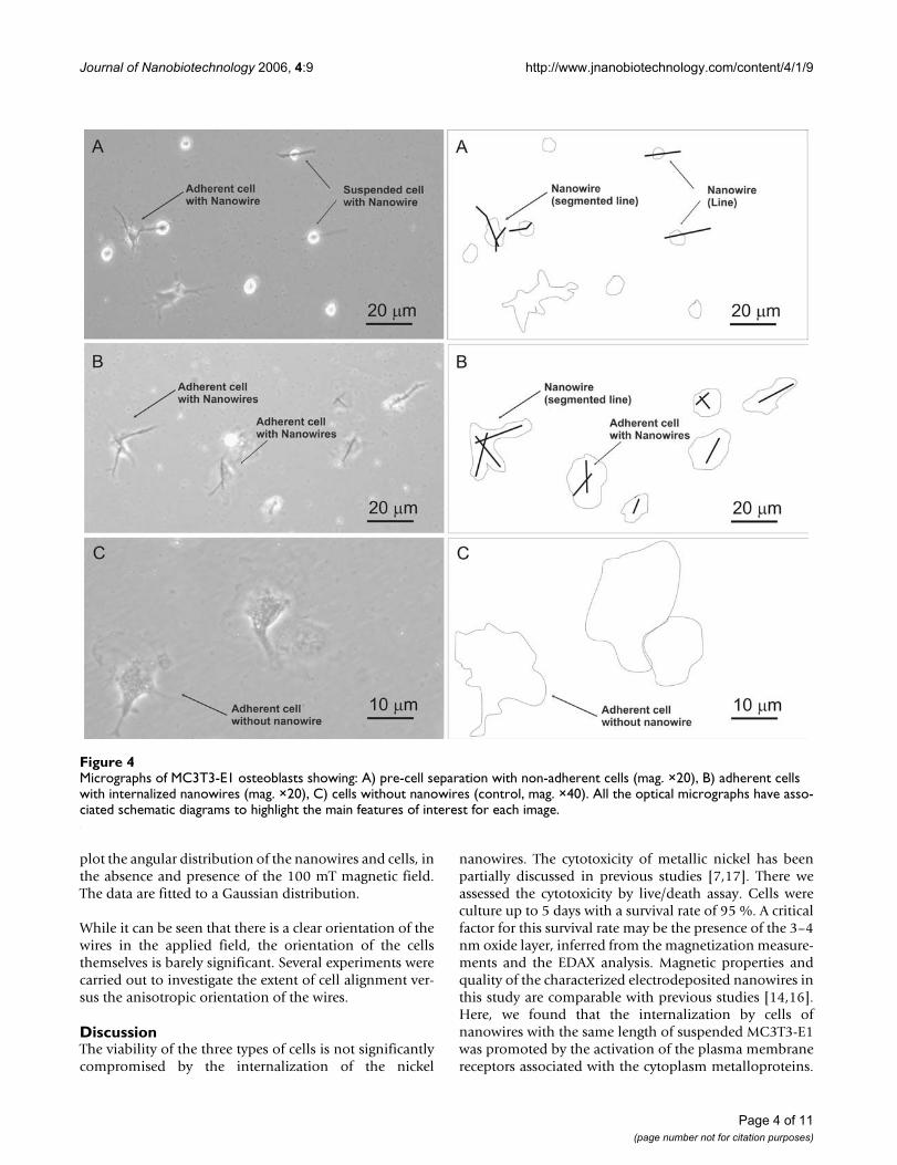
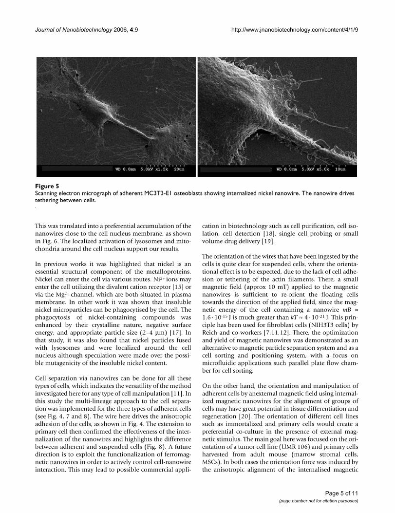
Nanowires in cell culture. MC3T3-E1 osteoblast cellsFrom the magnetically-separated cell-nanowire coloniessingle cell manipulation and imaging was achieved byscanning electron and fluorescent microscopy. Figure 4shows the osteoblast cell line with each cell containingone or more wires. An image taken with the scanning elec-tron microscope (Fig 5) shows a nanowire inside the cell.There, the left cell extends out towards the right cell and
Scanning electron micrograph of nickel nanowires after dissolving the alumina templateFigure 1Scanning electron micrograph of nickel nanowires after dissolving the alumina template.
Page 2 of 11(page number not for citation purposes)

Journal of Nanobiotechnology 2006, 4:9 http://www.jnanobiotechnology.com/content/4/1/9
uses the wire main axis as an alignment guidance. At thesame time, due to the difference in mechanical propertiesbetween nanowire and cell, the nanowire introduces ananisotropical stiffening contribution to the cellular inter-nal structures.
To investigate this mechanical re-arrangement in cell cyto-plasm evoked by nanowire internalization, fluorescentmicroscopy was used. To show this difference in cell activ-ity, MC3T3-E1 osteoblast single cell with and withoutinternalised nanowire were co-cultured and stained withliving mitochondria and lysosome fluorescent trackers.Figure 6 clearly show the difference in cell organelles dis-tribution. While the normal cell has a clear mitochondrialfluorescent staining due to normal cell activity but no lys-osome staining, the cell with a nanowire has a completelydifferent response.
A combined staining for lysosomes and mitochondrialocalized around the nanowire indicates that the internalorganelles inside the cytoplasm respond to the nanowire.In parallel to that localized mitochondrial staining alsoshows lamellipodia extensions due to cell tethering andre-alignment. This could be associated with a nanowire-induced cell stiffening response.
UMR106 osteosarcoma cellsWe showed that after separation and replating to conflu-ence half of the cells inside each field of view containwires and they are clearly distinguishable, Figure 7. Here,it was important to show that UMR106 cells have thesame size as the nanowires and that we succeeded in theinternalization process. This was achieved by the externalmagnetic field stimulation of the nanowires and subse-quent orientation of small colonies of UMR106, as shownin Fig. 7B,7C.
To extend this finding to a larger statistical population, 5batches, and 100 fields of view were analyzed for eachgroup studied. In total several thousand cells werecounted. The quantitative results achieved for the osteosa-rcoma cells were similar to those reported for the primaryMSCs.
Marrow stromal cellsMarrow Stromal (MS) cells were successfully separatedand isolated in small colonies to further investigate theinternal cell cytoskeleton re-arrangement. Small coloniesand single MS cells were stimulated and recorded byphase microscopy. Figure 8 shows both adherent cells andfloating cells with internalized nickel nanowires.
The MS cells were time lapse imaged for a total period of12-hours under phase contrast microscope, which was setso that there was a progressive heating of the culturemedium, which led successively to cell detachment fromthe substrate and subsequent programmed cell death [seeAdditional file 1]. The video shows floating cells ingestinga nanowire, as well as adherent cells containing ananowire detaching from the substrate, and eventuallyundergoing programmed cell death. The dead cells subse-quently release part of the cytoplasm and the internalisednanowire.
The alignment experiments of primary MS cells wereaimed to achieve orientation in an external magnetic field.This was achieved for MS cells in suspension wheremanipulation and cell-to-cell bridging was reported [seeAdditional file 2]. These were carried out in parallel to theUMR106 osteosarcoma cells. As mentioned for theUMR106, 5 batches and 100 fields of view were analyzedfor each group of study. In total, several thousand cellswere counted. The histograms shown in Figure 9 and 10,
Room temperature magnetization curve for nickel nanowires produced by electrodeposition in porous alumina templatesFigure 3Room temperature magnetization curve for nickel nanowires produced by electrodeposition in porous alumina templates.
-1000 -500 0 500 1000
-60
-40
-20
0
20
40
60
-2.00E-013
-1.50E-013
-1.00E-013
-5.00E-014
0.00E+000
5.00E-014
1.00E-013
1.50E-013
2.00E-013
Magnetization (
Am
2/k
g)
Applied field (mT)
Mom
ent/
nanow
ire (
Am
2)
X-ray diffraction pattern of the nickel nanowire powderFigure 2X-ray diffraction pattern of the nickel nanowire powder.
20 40 60 80 100
0
500
1000
1500
2000
(222)(311)
(220)
(200)
(111)
Cou
nts
2-theta
Page 3 of 11(page number not for citation purposes)
Journal of Nanobiotechnology 2006, 4:9 http://www.jnanobiotechnology.com/content/4/1/9
plot the angular distribution of the nanowires and cells, inthe absence and presence of the 100 mT magnetic field.The data are fitted to a Gaussian distribution.
While it can be seen that there is a clear orientation of thewires in the applied field, the orientation of the cellsthemselves is barely significant. Several experiments werecarried out to investigate the extent of cell alignment ver-sus the anisotropic orientation of the wires.
DiscussionThe viability of the three types of cells is not significantlycompromised by the internalization of the nickel
nanowires. The cytotoxicity of metallic nickel has beenpartially discussed in previous studies [7,17]. There weassessed the cytotoxicity by live/death assay. Cells wereculture up to 5 days with a survival rate of 95 %. A criticalfactor for this survival rate may be the presence of the 3–4nm oxide layer, inferred from the magnetization measure-ments and the EDAX analysis. Magnetic properties andquality of the characterized electrodeposited nanowires inthis study are comparable with previous studies [14,16].Here, we found that the internalization by cells ofnanowires with the same length of suspended MC3T3-E1was promoted by the activation of the plasma membranereceptors associated with the cytoplasm metalloproteins.
Micrographs of MC3T3-E1 osteoblasts showing: A) pre-cell separation with non-adherent cells (mag. ×20), B) adherent cells with internalized nanowires (mag. ×20), C) cells without nanowires (control, mag. ×40)Figure 4Micrographs of MC3T3-E1 osteoblasts showing: A) pre-cell separation with non-adherent cells (mag. ×20), B) adherent cells with internalized nanowires (mag. ×20), C) cells without nanowires (control, mag. ×40). All the optical micrographs have asso-ciated schematic diagrams to highlight the main features of interest for each image.
Page 4 of 11(page number not for citation purposes)
Journal of Nanobiotechnology 2006, 4:9 http://www.jnanobiotechnology.com/content/4/1/9
This was translated into a preferential accumulation of thenanowires close to the cell nucleus membrane, as shownin Fig. 6. The localized activation of lysosomes and mito-chondria around the cell nucleus support our results.
In previous works it was highlighted that nickel is anessential structural component of the metalloproteins.Nickel can enter the cell via various routes. Ni2+ ions mayenter the cell utilizing the divalent cation receptor [15] orvia the Mg2+ channel, which are both situated in plasmamembrane. In other work it was shown that insolublenickel microparticles can be phagocytised by the cell. Thephagocytosis of nickel-containing compounds wasenhanced by their crystalline nature, negative surfaceenergy, and appropriate particle size (2–4 μm) [17]. Inthat study, it was also found that nickel particles fusedwith lysosomes and were localized around the cellnucleus although speculation were made over the possi-ble mutagenicity of the insoluble nickel content.
Cell separation via nanowires can be done for all thesetypes of cells, which indicates the versatility of the methodinvestigated here for any type of cell manipulation [11]. Inthis study the multi-lineage approach to the cell separa-tion was implemented for the three types of adherent cells(see Fig. 4, 7 and 8). The wire here drives the anisotropicadhesion of the cells, as shown in Fig. 4. The extension toprimary cell then confirmed the effectiveness of the inter-nalization of the nanowires and highlights the differencebetween adherent and suspended cells (Fig. 8). A futuredirection is to exploit the functionalization of ferromag-netic nanowires in order to actively control cell-nanowireinteraction. This may lead to possible commercial appli-
cation in biotechnology such as cell purification, cell iso-lation, cell detection [18], single cell probing or smallvolume drug delivery [19].
The orientation of the wires that have been ingested by thecells is quite clear for suspended cells, where the orienta-tional effect is to be expected, due to the lack of cell adhe-sion or tethering of the actin filaments. There, a smallmagnetic field (approx 10 mT) applied to the magneticnanowires is sufficient to re-orient the floating cellstowards the direction of the applied field, since the mag-netic energy of the cell containing a nanowire mB ≈1.6·10-15 J is much greater than kT ≈ 4·10-21 J. This prin-ciple has been used for fibroblast cells (NIH3T3 cells) byReich and co-workers [7,11,12]. There, the optimizationand yield of magnetic nanowires was demonstrated as analternative to magnetic particle separation system and as acell sorting and positioning system, with a focus onmicrofluidic applications such parallel plate flow cham-ber for cell sorting.
On the other hand, the orientation and manipulation ofadherent cells by anexternal magnetic field using internal-ized magnetic nanowires for the alignment of groups ofcells may have great potential in tissue differentiation andregeneration [20]. The orientation of different cell linessuch as immortalized and primary cells would create apreferential co-culture in the presence of external mag-netic stimulus. The main goal here was focused on the ori-entation of a tumor cell line (UMR 106) and primary cellsharvested from adult mouse (marrow stromal cells,MSCs). In both cases the orientation force was induced bythe anisotropic alignment of the internalised magnetic
Scanning electron micrograph of adherent MC3T3-E1 osteoblasts showing internalized nickel nanowireFigure 5Scanning electron micrograph of adherent MC3T3-E1 osteoblasts showing internalized nickel nanowire. The nanowire drives tethering between cells.
Page 5 of 11(page number not for citation purposes)

Journal of Nanobiotechnology 2006, 4:9 http://www.jnanobiotechnology.com/content/4/1/9
nanowires. This was qualitatively investigated forUMR106 (Fig. 7) and quantitatively for MSC, as shown inFig 9 and 10.
The magnetic torque applied to each cell during the align-ment process is approximately mB ≈ 1.6·10-14 Nm. Theclear alignment of the nanowires indicates the resistanceof the rearrangement of the nanowires is smaller than thecell adhesion force, calculated for human bone marrowstromal cell (HBMSC) (EHBMSC = 3 kPa, cell diam. ≅ 130μm) [21].
This is shown in the two histograms reported in Fig. 9.However, in this study we found that there is little corre-lation between the cell morphology and the orientation ofthe nanowires, as by comparison of quantitative histo-grams in figure 9 and figure 10. It is probably either theconsequence of the cell-nanowire interaction due to theinternalization process or a considerably large mechano-chemical adhesion force exerted by the cells to the surface.
Therefore from the results of this study we can concludenot only that we have successfully achieved the internali-
Micrographs of two MC3T3-E1 osteoblast cells after overnight incubation with and without internalized nickel nanowires (mag. ×40)Figure 6Micrographs of two MC3T3-E1 osteoblast cells after overnight incubation with and without internalized nickel nanowires (mag. ×40). A) Phase contrast picture of both osteoblast adherent cells. (Internalized nanowire highlighted in red) B) Living cell stain-ing: double staining with Lyso-tracker (red) and Mito-tracker (green). The two cells show a completely different cytoplasm internal activity. C) Mitochondria only staining on both cells with localized activity of the cell with nanowires around lamellipo-dia and nanowires loci. D) Lysosomes only staining. Lysosomes are particularly localized around the bent nanowires.
Page 6 of 11(page number not for citation purposes)

Journal of Nanobiotechnology 2006, 4:9 http://www.jnanobiotechnology.com/content/4/1/9
zation of nanowires on different cell lines (such asMC3T3-E1 osteoblast, Marrow Stromal and UMR106osteosarcoma cells) but also that the interaction of thenickel nanowires with each cell cytoplasm leads to theinternal reorganization of those organelles affected by thenanowires, such as lysosomes and metalloproteins.
ConclusionMagnetic nanowires open new perspectives for the manip-ulation, identification and counting of many types of liv-ing cells. The viability of different cell types, here shown,is encouraging and opens a new window for the applica-tion of ferromagnetic nanowires. Therefore, integration ofdifferent magnetic materials inside cells open new pro-spective in tissue engineering and nanobiotechnology.
MethodsFabrication of nanowiresNickel nanowires were grown in alumina membranes(Whatman, UK). The membranes used were 20 mm indiameter and 60 μm thick, with 200 nm parallel-poresspaced by 300 nm. Firstly a gold electrode was depositedon the back of the membrane [13]. Then, nickel wasdeposited into the pores by electroplating from a NiSO4bath, at a potential of -0.9 to -1.0 V relative to an Ag, AgCl/KCl reference electrode [13]. The nickel wires were thenseparated from the membrane by dissolving the aluminamembrane in 3 M·l-1 NaOH [14]. The nanowires werethen characterized by scanning electron microscopy (Fig.1), X-ray diffraction (Fig. 2), X-ray fluorescence andvibrating-sample magnetometry (VSM).
Cell type and culture environmentIn this study three cell lines were used: primary cells fromadult rat marrow stromal cells (MSC), MC3T3-E1 osteob-lasts cell line and rat osteosarcoma cells (UMR106)(ATCC, USA). Every cell type used in this study was cul-tured under a different environment and culture medium,in line with the specific cell-culture requirements.
Primary MSCs were cultured and incubated at 37°C, 5 %CO2 and 95 % relative humidity, to confluency withDMEM supplemented with 2 % penicillin/streptomycin(Gibco, UK), 10 % foetal Bovine Serum (FBS; Gibco, UK),0.5 % l-Glutamine (Gibco, UK), 0.5 % Glutamax (Gibco,UK), and 1 % non-essential amino acids (Gibco, UK).
For the UMR 106 cell line, all samples were cultured andincubated as above with DMEM (30-2002 ATCC, USA)supplemented with 10 % foetal bovine serum, 100 U/mlpenicillin and 100 μg/ml streptomycin.
Whereas, MC3T3-E1 were cultured in alpha minimumessential medium (α-MEM: Sigma-Aldrich, UK) contain-ing 10 % foetal bovine serum (FBS; Gibco, UK), 100 U/ml
A) Confluent UMR-106 tumor cells: alignment experimentFigure 7A) Confluent UMR-106 tumor cells: alignment experiment. Cell with nanowires exposed to 0.1 T for 18 hrs and then cultured to confluent layer for 5 days. The nanowires in this case are about 10 μm long. B) Colony of UMR-106 cells con-taining nickel nanowires (bright field contrast micrograph, mag. ×40). C) Colony of UMR-106 cells containing nickel nanowires (phase contrast micrograph, mag. ×40). Diagram of cell structures and internalized wires have been high-lighted under the same z-focus level.
Page 7 of 11(page number not for citation purposes)

Journal of Nanobiotechnology 2006, 4:9 http://www.jnanobiotechnology.com/content/4/1/9
penicillin and 100 μm/ml streptomycin, 2 mM L-glutamine (Gibco, UK) and 0.5 % 100 mM sodium pyru-vate (Sigma-Aldrich, UK). The osteoblasts were kept in an
incubator at 37°C in a 95 % air 5 % CO2 environment.Subculture was routinely performed at ~80 % confluence,using 0.25 % trypsin.
Micrographs of marrow stromal cells with internalized nickel nanowires A) Colony adherent onto plastic dishes, B) Cell-cell interaction before magnetic alignment exposure, and C) MS cells floating in DMEM mediumFigure 8Micrographs of marrow stromal cells with internalized nickel nanowires A) Colony adherent onto plastic dishes, B) Cell-cell interaction before magnetic alignment exposure, and C) MS cells floating in DMEM medium. Phase contrast images (mag. ×40). All the optical micrographs have associated schematic diagrams to highlight the main features of interest for each image.
Page 8 of 11(page number not for citation purposes)

Journal of Nanobiotechnology 2006, 4:9 http://www.jnanobiotechnology.com/content/4/1/9
Internalization of wiresNickel nanowires were firstly washed 5 times in deionizedwater (Ω <= 30 M ohm) and rinsed in different PBS baths.The nanowires were then stabilized for 2 hours into thespecifics cell culture medium used for each cell line with adensity of 1011 particles/ml and then dispersed by 5 min-utes ultrasonic agitation immediately prior to addition.Cell medium-nanowire solution was then added at a finalconcentration of 106 particles/ml to adhering and sus-pended cells. Adhering cells were incubated for 30 min-
utes to promote the internalization of the wires and thenincubated overnight. Suspended cells were incubated for30 minutes and then exposed to magnetic field. After eachmagnetic exposure, cells on coverslips were subsequentlyfixed or stained as required for the imaging techniqueadopted.
Each experiment was repeated several times as part of alarger study to address the extent of nanowires internali-zation and cell yield.
Angular distributions of marrow stromal cells, with nanowire, orientation and cell morphology in zero field (left), and after alignment in a 100 mT applied field for 18 h (right)Figure 10Angular distributions of marrow stromal cells, with nanowire, orientation and cell morphology in zero field (left), and after alignment in a 100 mT applied field for 18 h (right).
Angular distributions of nickel nanowire orientation in marrow stromal cells in zero field (left), and after alignment in a 100 mT applied field for 18 h (right)Figure 9Angular distributions of nickel nanowire orientation in marrow stromal cells in zero field (left), and after alignment in a 100 mT applied field for 18 h (right).
Page 9 of 11(page number not for citation purposes)

Journal of Nanobiotechnology 2006, 4:9 http://www.jnanobiotechnology.com/content/4/1/9
Immunofluorescence staining and imagingTo characterize the ingestion of nanowires into the cellsseveral imaging and immunofluorescence techniqueswere used. These were optimized for each cell type toexamine the interaction between the cells and thenanowires. In this study all the three cell types were selec-tively stained for tracking active mitochondria and acidicorganelles in live cells to highlight the cell activity duringinternalization.
The cell staining and preparation was carried out by stain-ing live cells for cell tracking. In this study two cytoskele-ton markers were Myto- and Lyso-tracker (MolecularProbes, USA). In brief, after having prepared a 1 mM stocksolution of each tracker, a final concentration diluted inthe respective growth medium (described above) of 50nM for Myto-tracker and 50 nM for Lyso-tracker wasrespectively used to stain samples. Single and doublestaining on adherent and suspended live cell were carriedout for 30 min for Lyso-tracker and 3 min for Myto-tracker; samples were then washed with fresh warmmedium and imaging were taken. Subsequent, to liveimaging the stained cells were permeabilised and fixed in4.1 % paraformaldehyde in PBS for 30 min. These concen-trations were kept as low as possible in order to reducepossible overloading and artefacts.
Phase contrast, bright-field and time lapse imaging micro-scopy were carried out with the use of a Zeiss invertedmicroscope (Zeiss Axiovert 200 M, Germany) connectedto a cooled CCD camera (Zeiss Axiocam HRm, Germany).
SEM preparation and imagingSEM imaging was carried out on adherent MC3T3-E1 andMSC cells with internalized wires. All cell samples wereprepared by fixing each glass coverslip slide with 3 % Glu-taraldehyde in 0.1 M Sodium Cacodylate buffer (pH 7.2).The primary fixation was carried out for 1 hour at roomtemperature. Samples were then washed 6 times for 1hour with either 0.05 M Phosphate or 0.1 M Cacodylatebuffer to remove any un-reacted Glutaraldehyde from thesamples before rinse and dehydration was carried out. Thesamples were then quickly rinsed with 50 % ethanol;dehydrated with 10 %, 30 %, 50 %, 70 %, and 95 % eth-anol for 10 min each; and dehydrated with 100 % ethanoltwice for 15 min each. The samples were then gold sput-ter-coated for high resolution imaging.
Internalization process imagingThe ingestion of nanowires into the cells was examined bytime lapse contrast microscopy. It was possible to exam-ine the interaction of the cells with the nanowires by pro-gramming the Zeiss microscope to acquire an image every15 minutes for 18 hrs (as reported in the supplementalmaterial video). There, the light was programmed to heat
the medium so that both adherent and floating cells couldbe examined.
Magnetic separation and alignment of cell-nanowire populationThe cell separation set-up was made of two Nd2Fe14B per-manent magnets of 10 mm diameter and 15 mm length inantiparallel configuration at opposite sides of a 10 ml fal-con tube for cell suspension. The surface magnetic field ofthe magnets of 0.6 T was measured by hall sensor gaussm-eter (Hirst, UK). The two magnets fixed on either side ofthe tube together produced a magnetic field gradient up to100 T/m.
Cells cultured with nanowires were dissociated withwarmed trypsin plus EDTA4Na solution (Sigma-Aldrich,UK) for 5 min. From each full batch half was put into themagnetic separation set-up, and the other half wasreplated with 3 ml of fresh medium to estimate theamount of cell binding with nanowires pre-separation.
The magnetic separation was carried out for 5 minutes inorder to obtain the complete sedimentation of those cellswithout nanowires. These last, after separation wereremoved and plated in 3 ml fresh medium in a new Petridish. After having removed the magnets, the cells withnanowires were dispersed into 3 ml of fresh medium andreplated in a new Petri dish.
Complete cell adhesion was achieved in less then 1 h ofincubation at physiological conditions for all the batchesand then cell counting was carried out in 2–3 rounds tominimize systematic and operator errors. Images werecaptured using an Olympus BX41 microscope (Japan)with magnifications of 10×, 20×, and 40× connected to acooled CCD camera (Image II, USA). All images wereacquired and stored by using Analysis imaging software(Analysis, Germany).
For the nanowire and cell alignment test, a uniform mag-netic field of 100 mT was applied by a cylindrical Halbachpermanent magnet made of segments of Nd2Fe14B (Mag-netic Solutions, Ireland). The inner and outer radii of themagnet were 106 and 156 mm, respectively. Cells with Ninanowires were co-cultured overnight before the test. Theduration of the magnetic field exposure was chosen to be1 hr, 24 hrs or 48 hrs inside an incubator at physiologicalconditions (T = 37°C, CO2 = 5 % and RH = 95 %). Subse-quently, images were taken under phase contrast asdescribed above. The degree of alignment of the wires andcell morphology to the direction of the field was con-ducted by using Scion Image software (Scion Corpora-tion, USA). Both cell and nanowire counting were carriedout under blind conditions to reduce systematic errors.
Page 10 of 11(page number not for citation purposes)

Journal of Nanobiotechnology 2006, 4:9 http://www.jnanobiotechnology.com/content/4/1/9
Publish with BioMed Central and every scientist can read your work free of charge
"BioMed Central will be the most significant development for disseminating the results of biomedical research in our lifetime."
Sir Paul Nurse, Cancer Research UK
Your research papers will be:
available free of charge to the entire biomedical community
peer reviewed and published immediately upon acceptance
cited in PubMed and archived on PubMed Central
yours — you keep the copyright
Submit your manuscript here:http://www.biomedcentral.com/info/publishing_adv.asp
BioMedcentral
Competing interestsThe author(s) declare that they have no competing inter-ests.
Authors' contributionsAPM and ZD did most of the experiments and data analy-sis. APM and JMDC coordinated the experiments anddrafted the manuscript. All authors read and approved thefinal manuscript.
Additional material
AcknowledgementsThe authors thank L. McMahon, E. Kearney, and Dr. E. Byrne for their tech-nical assistance, and Dr. P. Maguire for her valuable discussions. The authors also thank Prof. P.J. Prendergast, Dr. V. Campbell and Dr. S. Jarvis for the use of their laboratories and facilities.
Science Foundation Ireland for the financial support, under the SFI CINSE and CRANN exploratory projects. Mr. Zhu Diao for the Ussher Fellowship from Trinity College Dublin, Ireland.
References1. Pankhurst QA, Connolly J, Jones SK, Dobson J: Applications of
magnetic nanoparticles in biomedicine. J Phys D Appl Phys 2003,36:R167-R181.
2. Tartaj P, Morales MP, Gonzalez-Carreno T, Veintemillas-Verdaguer S,Serna CJ: Advances in magnetic nanoparticles for biotechnol-ogy applications. J Magn Magn Mater 2005, 290:28-34.
3. Salata OV: Applications of nanoparticles in biology and medi-cine. J Nanobiotech 2004, 2(3):1-6.
4. Bulte JWM: Magnetic nanoparticles as markers for cellular MRimaging. J Magn Magn Mater 2005, 289:423-427.
5. Dutz S, Hergt R, Murbe J, Töpfer J, Müller R, Zeisberger M, AndräWand Bellemann ME: Magnetic nanoparticles for biomedicalheating applications. Z Phys Chem 2006, 220:145-151. available at:http://portal.isiknowledge.com
6. Walter N, Selhuber C, Kessler H, Spatz JP: Cellular unbindingforces of initial adhesion processes on nanopatterned sur-faces probed with magnetic tweezers. Nano lett 2006,6:398-402.
7. Hultgren A, Tanase M, Chen CS, Meyer GJ, Reich DH: Cell manip-ulation using magnetic nanowires. J Appl Phys 2003,93:7554-7556.
8. Saiyed ZM, Telang SD, Ramchand CN: Application of magnetictechniques in the field of drug discovery and biomedicine.BioMagn Res Technol 2003, 1(2):1-8. available at: http://www.biomagres.com/content/1/1/2
9. Harisinghani MG, Weissleder R: Sensitive, Noninvasive Detec-tion of Lymph Node Metastases. PLOS Medicine 2004,1(3):202-209.
10. Berry CC, Wells S, Charles S, Aitchison G, Curtis ASG: Cellresponse to dextran-derivatised iron oxide nanoparticlespost internalisation. Biomaterials 2004, 25:5405-5413.
11. Hultgren A, Tanase M, Felton EJ, Bhadriraju K, Salem AK, Chen CS,Reich DH: Optimization of Yield in Magnetic Cell SeparationsUsing Nickel Nanowires of Different Lengths. Biotechnol Prog2005, 21:509-515.
12. Tanase M, Felton EJ, Gray DS, Hultgren A, Chen CS, Reich DH:Assembly of multicellular constructs and microarrays ofcells using magnetic nanowires. Lab Chip 2005, 5:598-605.
13. Chaure NB, Stamenov P, Rhen FMF, Coey JMD: Oriented cobaltnanowires prepared by electrodeposition in a porous mem-brane. J Magn Magn Mater 2005, 290:1210-1213.
14. Chiriac H, Moga AE, Urse M, Óvári TA: Preparation and mag-netic properties of electrodeposited magnetic nanowires.Sensor Actuat A-Phys 2003, 106:348-351.
15. Pi M, Faber P, Ekema G, Jackson PD, Ting A, Wang N, Fontilla-PooleM, Mays RW, Brunden KR, Harrington JJ, Quarles LD: Identificationof a Novel Extracellular Cation-sensing G-proteincoupledReceptor. J Biol Chem 2005, 280:40201-40209.
16. Wu CG, Lin HL, Shau NL: Magnetic nanowires via templateelectrodeposition. J Sol State Elect 2006, 10:198-202.
17. Kasprzak KS, Sunderman FW Jr, Salnikow K: Nickel carcinogene-sis. Mutat Res 2003, 533:67-97.
18. Safarik I, Safarikova M: Magnetic techniques for the isolationand purification of proteins and peptides. BioMagn Res Technol2004, 2(1):7.
19. Vasir JK, Reddy MK, Labhasetwar VD: Nanosystems in drug tar-geting: Opportunities and challenges. Cur Nanosci 2005,1:47-64.
20. Wen X, Shi D, Zhang N: Applications of Nanotechnology in Tis-sue Engineering. In Handbook of Nanostructured Biomaterials andTheir Applications in Nanobiotechnology Edited by: Nalwa HS. AmericanScientific Publishers; 2005:1-22. available at: http://www.eng.uc.edu/~dshi/News/material.pdf
21. Simon A, Cohen-Bouhacina T, Porte MC, Aime JP, Amedee J, BareilleR, Baquey C: Characterization of Dynamic Cellular Adhesionof Osteoblasts Using Atomic Force Microscopy. Cytom Part A2003, 54A:36-47.
Additional File 1Cell-death induced by localized nanowire heat. Video showing the behav-ior of marrow stromal cells in the presence of nickel nanowires. The dura-tion of the video corresponds to a period of 18 hours. The medium, initially at 37°C is heated at a rate of around 2°C h-1 which leads to cell death.Click here for file[http://www.biomedcentral.com/content/supplementary/1477-3155-4-9-S1.avi]
Additional File 2Cell manipulation: bridging between cells. Video showing marrow stromal cell-to-cell bridging with nickel nanowire magnetic manipulation. The duration of the video corresponds to a period of 1 hour in which cells have been manipulated in cell culture medium at physiological conditions.Click here for file[http://www.biomedcentral.com/content/supplementary/1477-3155-4-9-S2.avi]
Page 11 of 11(page number not for citation purposes)