Glucagon-like peptide-1 as a treatment for chemotherapy-induced mucositis
11
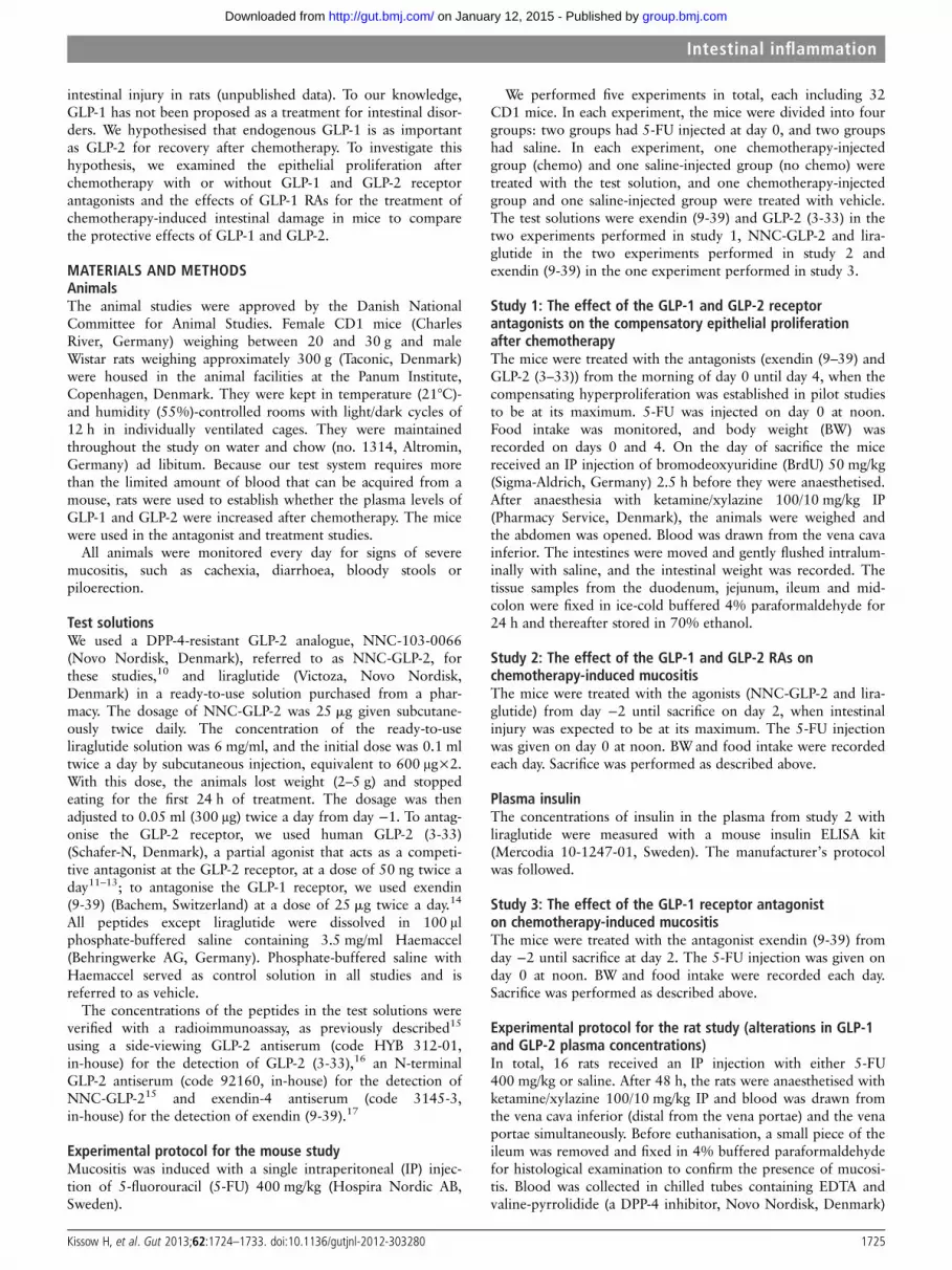
ORIGINAL ARTICLE Glucagon-like peptide-1 as a treatment for chemotherapy-induced mucositis Hannelouise Kissow, Bolette Hartmann, Jens Juul Holst, Steen Seier Poulsen Department of Biomedical Sciences and the Novo Nordisk Foundation Center for Basic Metabolic Research, University of Copenhagen, Copenhagen, Denmark Correspondence to Dr Hannelouise Kissow, Department of Biomedical Sciences and the Novo Nordisk Foundation Center for Basic Metabolic Research, Faculty of Health Science, University of Copenhagen, Blegdamsvej 3, Copenhagen DK-2200, Denmark; [email protected] Received 11 July 2012 Accepted 24 September 2012 Published Online First 20 October 2012 To cite: Kissow H, Hartmann B, Holst JJ, et al. Gut 2013;62:1724–1733. ABSTRACT Background Glucagon-like peptide-2 (GLP-2) has been suggested for the treatment of mucositis, but the peptide has also been shown to accentuate colonic dysplasia in carcinogen-treated mice. Recently, an effect on intestinal growth was discovered for glucagon-like peptide-1 (GLP-1), Objective To determine whether endogenous GLP-1 contributes to the healing processes and if exogenous GLP-1 has a potential role in treating mucositis. Methods Mice were injected with 5-fluorouracil (5-FU) or saline to induce mucositis and were then treated with GLP-1, GLP-2, GLP-2 (3-33), exendin (9-39) or vehicle. The mice were sacrificed 48 or 96 h after the 5-FU injections. The end points were intestinal weight, villus height, proliferation and histological scoring of mucositis severity. Rats were injected with 5-FU or saline, and after 48 h, blood was drawn and analysed for GLP-1 and GLP-2 concentration. Results GLP-1 and GLP-2 significantly prevented the loss of mucosal mass and villus height and significantly decreased the mucositis severity score in the duodenum and jejunum 48 h after chemotherapy. The effect was equivalent. Exendin (9-39) reduced the intestinal weight 96 h after chemotherapy. The GLP-1 levels in blood were increased more than 10-fold, and GLP-2 levels were increased sevenfold. Conclusions GLP-1 and GLP-2 were secreted after intestinal injury, and recovery was delayed after treatment with exendin (9-39), indicating an important role for the peptides in the protection of the intestine from injury. GLP-1 treatment ameliorated mucositis, which suggests that mucositis and other acute intestinal disorders might benefit from treatment with GLP-1 analogues. INTRODUCTION Chemotherapy-induced mucositis is a common complication of anticancer treatments, and may reduce the effectiveness of treatment because it often requires a dose reduction and impairs the quality of life of the patient. Glucagon-like peptide-2 (GLP-2) is a 33-amino acid peptide secreted by enteroendocrine L-cells located in the small and large intestines. 1 This peptide has been suggested for the treatment of chemotherapy- induced mucositis, 23 but the peptide has also been shown to accentuate colonic dysplasia in carcinogen- treated mice. 4–6 Glucagon-like peptide-1 (GLP-1) is secreted in parallel with GLP-2 from the L-cells. This secretion is stimulated by food intake, and GLP-1 participates in the maintenance of glucose homoeostasis by stimulating pancreatic β-cells to secrete insulin in a glucose-dependent manner. 7 Both GLP-1 and GLP-2 are rapidly degraded by the enzyme dipepti- dyl peptidase-4 (DPP-4). Because of the insulin- stimulating properties of GLP-1, several GLP-1 receptor agonists (GLP-1 RAs) (resistant to degrad- ation) are being and have been developed for the treatment of type 2 diabetes mellitus. Simonsen et al 8 investigated whether exendin-4, a GLP-1 RA, also has intestinal effects, and they unexpectedly found a trophic effect of GLP-1 in the rat small intestine. We reproduced this effect in mice 9 and also found increased GLP-1 secretion after Significance of this study What is already known about the subject? ▸ Glucagon-like peptide-1 (GLP-1) is an incretin hormone, and receptor agonists (RAs) have been developed for the treatment of type 2 diabetes mellitus. ▸ Gastrointestinal mucositis is a severe injury to the intestine with a significant affect on the health, economic outcome and quality of life of patients with cancer receiving chemotherapy. ▸ No treatment is available for this condition. ▸ The intestinal growth-factor glucagon-like peptide-2 (GLP-2) has previously been suggested as a treatment for mucositis. What are the new findings? ▸ The GLP-1 RA liraglutide can prevent severe mucositis in an experimental animal model. ▸ The treatment effect is comparable to the effect of GLP-2. ▸ GLP-1 and GLP-2 are both secreted in large amounts as a result of mucositis induction. ▸ The GLP-1 receptor antagonist exendin (9-39) prevented spontaneous healing after mucositis, suggesting the involvement of endogenous GLP-1 in its pathogenesis. How might it impact on clinical practice in the foreseeable future? ▸ These new findings may result in the further exploration of the possible role of GLP-1 in the pathogenesis of intestinal disorders and of GLP-1 RAs for the prevention of mucositis caused by chemotherapy in patients with cancer. 1724 Kissow H, et al. Gut 2013;62:1724–1733. doi:10.1136/gutjnl-2012-303280 Intestinal in fl ammation group.bmj.com on January 12, 2015 - Published by http://gut.bmj.com/ Downloaded from
-
Upload
independent -
Category
Documents
-
view
0 -
download
0
Transcript of Glucagon-like peptide-1 as a treatment for chemotherapy-induced mucositis

ORIGINAL ARTICLE
Glucagon-like peptide-1 as a treatment forchemotherapy-induced mucositisHannelouise Kissow, Bolette Hartmann, Jens Juul Holst, Steen Seier Poulsen
Department of BiomedicalSciences and the Novo NordiskFoundation Center for BasicMetabolic Research, Universityof Copenhagen, Copenhagen,Denmark
Correspondence toDr Hannelouise Kissow,Department of BiomedicalSciences and the Novo NordiskFoundation Center for BasicMetabolic Research, Faculty ofHealth Science, University ofCopenhagen, Blegdamsvej 3,Copenhagen DK-2200,Denmark; [email protected]
Received 11 July 2012Accepted 24 September 2012Published Online First20 October 2012
To cite: Kissow H,Hartmann B, Holst JJ, et al.Gut 2013;62:1724–1733.
ABSTRACTBackground Glucagon-like peptide-2 (GLP-2) has beensuggested for the treatment of mucositis, but thepeptide has also been shown to accentuate colonicdysplasia in carcinogen-treated mice. Recently, an effecton intestinal growth was discovered for glucagon-likepeptide-1 (GLP-1),Objective To determine whether endogenous GLP-1contributes to the healing processes and if exogenousGLP-1 has a potential role in treating mucositis.Methods Mice were injected with 5-fluorouracil (5-FU)or saline to induce mucositis and were then treated withGLP-1, GLP-2, GLP-2 (3-33), exendin (9-39) or vehicle.The mice were sacrificed 48 or 96 h after the 5-FUinjections. The end points were intestinal weight, villusheight, proliferation and histological scoring of mucositisseverity. Rats were injected with 5-FU or saline, andafter 48 h, blood was drawn and analysed for GLP-1and GLP-2 concentration.Results GLP-1 and GLP-2 significantly prevented theloss of mucosal mass and villus height and significantlydecreased the mucositis severity score in the duodenumand jejunum 48 h after chemotherapy. The effect wasequivalent. Exendin (9-39) reduced the intestinal weight96 h after chemotherapy. The GLP-1 levels in blood wereincreased more than 10-fold, and GLP-2 levels wereincreased sevenfold.Conclusions GLP-1 and GLP-2 were secreted afterintestinal injury, and recovery was delayed aftertreatment with exendin (9-39), indicating an importantrole for the peptides in the protection of the intestinefrom injury. GLP-1 treatment ameliorated mucositis,which suggests that mucositis and other acute intestinaldisorders might benefit from treatment with GLP-1analogues.
INTRODUCTIONChemotherapy-induced mucositis is a commoncomplication of anticancer treatments, and mayreduce the effectiveness of treatment because itoften requires a dose reduction and impairs thequality of life of the patient. Glucagon-likepeptide-2 (GLP-2) is a 33-amino acid peptidesecreted by enteroendocrine L-cells located in thesmall and large intestines.1 This peptide has beensuggested for the treatment of chemotherapy-induced mucositis,2 3 but the peptide has also beenshown to accentuate colonic dysplasia in carcinogen-treated mice.4–6
Glucagon-like peptide-1 (GLP-1) is secreted inparallel with GLP-2 from the L-cells. This secretionis stimulated by food intake, and GLP-1 participates
in the maintenance of glucose homoeostasis bystimulating pancreatic β-cells to secrete insulin in aglucose-dependent manner.7 Both GLP-1 andGLP-2 are rapidly degraded by the enzyme dipepti-dyl peptidase-4 (DPP-4). Because of the insulin-stimulating properties of GLP-1, several GLP-1receptor agonists (GLP-1 RAs) (resistant to degrad-ation) are being and have been developed for thetreatment of type 2 diabetes mellitus. Simonsenet al8 investigated whether exendin-4, a GLP-1 RA,also has intestinal effects, and they unexpectedlyfound a trophic effect of GLP-1 in the rat smallintestine. We reproduced this effect in mice9 andalso found increased GLP-1 secretion after
Significance of this study
What is already known about the subject?▸ Glucagon-like peptide-1 (GLP-1) is an incretin
hormone, and receptor agonists (RAs) havebeen developed for the treatment of type 2diabetes mellitus.
▸ Gastrointestinal mucositis is a severe injury tothe intestine with a significant affect on thehealth, economic outcome and quality of life ofpatients with cancer receiving chemotherapy.
▸ No treatment is available for this condition.▸ The intestinal growth-factor glucagon-like
peptide-2 (GLP-2) has previously beensuggested as a treatment for mucositis.
What are the new findings?▸ The GLP-1 RA liraglutide can prevent severe
mucositis in an experimental animal model.▸ The treatment effect is comparable to the effect
of GLP-2.▸ GLP-1 and GLP-2 are both secreted in large
amounts as a result of mucositis induction.▸ The GLP-1 receptor antagonist exendin (9-39)
prevented spontaneous healing after mucositis,suggesting the involvement of endogenousGLP-1 in its pathogenesis.
How might it impact on clinical practice inthe foreseeable future?▸ These new findings may result in the further
exploration of the possible role of GLP-1 in thepathogenesis of intestinal disorders and ofGLP-1 RAs for the prevention of mucositiscaused by chemotherapy in patients withcancer.
1724 Kissow H, et al. Gut 2013;62:1724–1733. doi:10.1136/gutjnl-2012-303280
Intestinal inflammation
group.bmj.com on January 12, 2015 - Published by http://gut.bmj.com/Downloaded from
intestinal injury in rats (unpublished data). To our knowledge,GLP-1 has not been proposed as a treatment for intestinal disor-ders. We hypothesised that endogenous GLP-1 is as importantas GLP-2 for recovery after chemotherapy. To investigate thishypothesis, we examined the epithelial proliferation afterchemotherapy with or without GLP-1 and GLP-2 receptorantagonists and the effects of GLP-1 RAs for the treatment ofchemotherapy-induced intestinal damage in mice to comparethe protective effects of GLP-1 and GLP-2.
MATERIALS AND METHODSAnimalsThe animal studies were approved by the Danish NationalCommittee for Animal Studies. Female CD1 mice (CharlesRiver, Germany) weighing between 20 and 30 g and maleWistar rats weighing approximately 300 g (Taconic, Denmark)were housed in the animal facilities at the Panum Institute,Copenhagen, Denmark. They were kept in temperature (21°C)-and humidity (55%)-controlled rooms with light/dark cycles of12 h in individually ventilated cages. They were maintainedthroughout the study on water and chow (no. 1314, Altromin,Germany) ad libitum. Because our test system requires morethan the limited amount of blood that can be acquired from amouse, rats were used to establish whether the plasma levels ofGLP-1 and GLP-2 were increased after chemotherapy. The micewere used in the antagonist and treatment studies.
All animals were monitored every day for signs of severemucositis, such as cachexia, diarrhoea, bloody stools orpiloerection.
Test solutionsWe used a DPP-4-resistant GLP-2 analogue, NNC-103-0066(Novo Nordisk, Denmark), referred to as NNC-GLP-2, forthese studies,10 and liraglutide (Victoza, Novo Nordisk,Denmark) in a ready-to-use solution purchased from a phar-macy. The dosage of NNC-GLP-2 was 25 mg given subcutane-ously twice daily. The concentration of the ready-to-useliraglutide solution was 6 mg/ml, and the initial dose was 0.1 mltwice a day by subcutaneous injection, equivalent to 600 μg×2.With this dose, the animals lost weight (2–5 g) and stoppedeating for the first 24 h of treatment. The dosage was thenadjusted to 0.05 ml (300 μg) twice a day from day −1. To antag-onise the GLP-2 receptor, we used human GLP-2 (3-33)(Schafer-N, Denmark), a partial agonist that acts as a competi-tive antagonist at the GLP-2 receptor, at a dose of 50 ng twice aday11–13; to antagonise the GLP-1 receptor, we used exendin(9-39) (Bachem, Switzerland) at a dose of 25 mg twice a day.14
All peptides except liraglutide were dissolved in 100 μlphosphate-buffered saline containing 3.5 mg/ml Haemaccel(Behringwerke AG, Germany). Phosphate-buffered saline withHaemaccel served as control solution in all studies and isreferred to as vehicle.
The concentrations of the peptides in the test solutions wereverified with a radioimmunoassay, as previously described15
using a side-viewing GLP-2 antiserum (code HYB 312-01,in-house) for the detection of GLP-2 (3-33),16 an N-terminalGLP-2 antiserum (code 92160, in-house) for the detection ofNNC-GLP-215 and exendin-4 antiserum (code 3145-3,in-house) for the detection of exendin (9-39).17
Experimental protocol for the mouse studyMucositis was induced with a single intraperitoneal (IP) injec-tion of 5-fluorouracil (5-FU) 400 mg/kg (Hospira Nordic AB,Sweden).
We performed five experiments in total, each including 32CD1 mice. In each experiment, the mice were divided into fourgroups: two groups had 5-FU injected at day 0, and two groupshad saline. In each experiment, one chemotherapy-injectedgroup (chemo) and one saline-injected group (no chemo) weretreated with the test solution, and one chemotherapy-injectedgroup and one saline-injected group were treated with vehicle.The test solutions were exendin (9-39) and GLP-2 (3-33) in thetwo experiments performed in study 1, NNC-GLP-2 and lira-glutide in the two experiments performed in study 2 andexendin (9-39) in the one experiment performed in study 3.
Study 1: The effect of the GLP-1 and GLP-2 receptorantagonists on the compensatory epithelial proliferationafter chemotherapyThe mice were treated with the antagonists (exendin (9–39) andGLP-2 (3–33)) from the morning of day 0 until day 4, when thecompensating hyperproliferation was established in pilot studiesto be at its maximum. 5-FU was injected on day 0 at noon.Food intake was monitored, and body weight (BW) wasrecorded on days 0 and 4. On the day of sacrifice the micereceived an IP injection of bromodeoxyuridine (BrdU) 50 mg/kg(Sigma-Aldrich, Germany) 2.5 h before they were anaesthetised.After anaesthesia with ketamine/xylazine 100/10 mg/kg IP(Pharmacy Service, Denmark), the animals were weighed andthe abdomen was opened. Blood was drawn from the vena cavainferior. The intestines were moved and gently flushed intralum-inally with saline, and the intestinal weight was recorded. Thetissue samples from the duodenum, jejunum, ileum and mid-colon were fixed in ice-cold buffered 4% paraformaldehyde for24 h and thereafter stored in 70% ethanol.
Study 2: The effect of the GLP-1 and GLP-2 RAs onchemotherapy-induced mucositisThe mice were treated with the agonists (NNC-GLP-2 and lira-glutide) from day −2 until sacrifice on day 2, when intestinalinjury was expected to be at its maximum. The 5-FU injectionwas given on day 0 at noon. BWand food intake were recordedeach day. Sacrifice was performed as described above.
Plasma insulinThe concentrations of insulin in the plasma from study 2 withliraglutide were measured with a mouse insulin ELISA kit(Mercodia 10-1247-01, Sweden). The manufacturer’s protocolwas followed.
Study 3: The effect of the GLP-1 receptor antagoniston chemotherapy-induced mucositisThe mice were treated with the antagonist exendin (9-39) fromday −2 until sacrifice at day 2. The 5-FU injection was given onday 0 at noon. BW and food intake were recorded each day.Sacrifice was performed as described above.
Experimental protocol for the rat study (alterations in GLP-1and GLP-2 plasma concentrations)In total, 16 rats received an IP injection with either 5-FU400 mg/kg or saline. After 48 h, the rats were anaesthetised withketamine/xylazine 100/10 mg/kg IP and blood was drawn fromthe vena cava inferior (distal from the vena portae) and the venaportae simultaneously. Before euthanisation, a small piece of theileum was removed and fixed in 4% buffered paraformaldehydefor histological examination to confirm the presence of mucosi-tis. Blood was collected in chilled tubes containing EDTA andvaline-pyrrolidide (a DPP-4 inhibitor, Novo Nordisk, Denmark)
Kissow H, et al. Gut 2013;62:1724–1733. doi:10.1136/gutjnl-2012-303280 1725
Intestinal inflammation
group.bmj.com on January 12, 2015 - Published by http://gut.bmj.com/Downloaded from

at final concentrations of 3.9 mmol/l and 0.01 mmol/l, respect-ively. The samples were centrifuged for 10 min at 2400×g and4°C, and the plasma was kept at −20°C until analysis. For thedetermination of GLP-1, the plasma was extracted with 70%ethanol before analysis. For the determination of GLP-2, theplasma was extracted with 75% ethanol before analysis.Hormone concentrations were determined by radioimmuno-assay as previously described, using an N-terminal specific anti-serum (code 92160, in-house) to measure active GLP-2 only15
and a side-viewing antiserum (code 2135, in-house) to measureall molecules that contain the mid-sequence of GLP-1.18
Histological sections and morphometric estimatesThe fixed tissue samples were dehydrated, embedded in paraffinand cut into 5 μm sections using a microtome. The sectionsfrom the mice from studies 2 and 3 that had received chemo-therapy were evaluated for the degree of mucositis using amethod described previously by Howarth et al.19 A total scorefor each region of the intestine (duodenum, jejunum and ileum)was obtained by rating 10 histological characteristics of mucosi-tis from 0 (normal) to 3 (severe). The criteria used were villusstunting/atrophy, villus fusion, disruption of enterocytes, cryptloss, architectural disruption of crypts, disruption or distortionof crypt cells, infiltration of inflammatory cells, dilatation oflymphatic vessels and capillaries and thickening or oedema ofthe submucosa or lamina muscularis. Sections from the rat studywere only evaluated to confirm the presence of mucositis.
To measure the area of the mucosal layer, the crypt depth andvillus height, transverse sections from the duodenum, jejunum,ileum and colon from each animal in all mouse studies werestained with haematoxylin/eosin and examined with a lightmicroscope connected to a camera (Zeiss Axioscope 2 plus,Brock & Michelsen, Denmark). Photomicrographs were ana-lysed using Image Pro 7.0 software (Media Cybernetics, USA).
To determine the proliferative activity, BrdU immunohisto-chemistry was performed on sections from the duodenum,jejunum, ileum and colon from each animal. Antigen retrievalwas performed by heating the sections for 15 min in a micro-wave oven (750 W) in citrate buffer pH 6.0. The sections werethen preincubated for 10 min in 2% bovine serum albumin, fol-lowed by overnight incubation with a monoclonal mouseanti-BrdU antibody Ab-4(Bu20a) (Thermo Scientific, USA)diluted 1 : 200.
To visualise the immunoreactions, the sections were incubatedfor 40 min with biotinylated horse anti-mouse IgG (VectorBA-2000 Vector Laboratories, USA) diluted 1:200, followed byStreptABComplex/horseradish peroxidase (Vectastain ABC kit,Vector Laboratories, USA), and finally developed with 3,3’-diaminobenzidine containing nickel ammonium sulphate(Sigma-Aldrich, Denmark) for 15 min. Proliferation was quanti-fied as the area of BrdU immunoreactive cells per crypt, mea-sured in 10 crypts for each section.
StatisticsAll results are shown as means and SE of the mean (SEM). Thecomparison between groups was performed with analysis ofvariance, followed by post hoc comparisons between the twochemo groups and the two no chemo groups with Bonferroni’stest to evaluate the influence of the test solutions in either thechemotherapy or saline-injected mice. Dunnett’s multiple com-parison test was used to compare all groups in each experimentwith the no chemo vehicle-treated group, using the latter as ahealthy untreated control. Student’s t test was used to comparethe plasma levels between two groups in the rat study. All
analyses were performed using GraphPad Prism V5.01, andprobability values p<0.05 were considered significant.
RESULTSStudy 1The mice given chemotherapy showed, as expected, a decreasein food intake and BW compared with the saline-injected mice.No differences in food intake were seen between the controlsand mice treated with exendin (9-39) or GLP-2 (3-33) (figure1A–D).
The mice receiving chemotherapy corresponded to a normalhealthy mouse (no chemo vehicle) when we measured the rela-tive small intestinal weight 4 days after chemotherapy (figure1E–H). Treatment with GLP-2 (3-33) had no influence on thisoutcome, but treatment with exendin (9-39) prevented the intes-tine from returning to its normal weight after the injury (3.1%of BW (±0.15) vs 3.6% (±0.2) of BW p<0.05). Surprisingly,exendin (9-39) treatment of the no chemo mice increased thesmall intestinal weight (4.0% of BW (±0.1) vs 3.6% of BW(±0.1) p<0.05), resulting in a large gap between the chemoand no chemo exendin (9-39)-treated mice. The relative colonweight increased after chemotherapy, but GLP-2 (3-33) andexendin (9-39) had no influence on this outcome (figure 1E–H).
The morphometric analysis of the cross-sectional area, villusheight and crypt depth of the duodenum, jejunum, ileum andcolon confirmed that there were no differences between GLP-2(3-33)-treated mice and vehicle-treated mice with or withoutchemotherapy (data not shown). The cross-sectional area wasstill decreased in the duodenum and jejunum after chemother-apy, and this result was significantly more pronounced in thejejunum in the exendin (9-39) group (table 1). Villus height wasdecreased and crypt depth was increased in all parts of the smallintestine after chemotherapy, and thus, the crypt:villus ratio wasincreased twofold. Treatment with either GLP-2 (3-33) orexendin (9-39) had no influence on these outcomes.
Morphometric analysis showed no difference in colonic tissuesamples.
The estimation of the area of BrdU-immunoreactive cells inthe small and large intestines showed a large increase in prolifer-ation in all investigated parts of the intestine 4 days after chemo-therapy. The proliferation was increased approximately fourfoldin the small intestine and sixfold in the colon. Treatment withthe antagonists GLP-2 (3-33) and exendin (9-39) did not influ-ence compensatory hyperproliferation after chemotherapy (datanot shown).
Study 2Treatment with NNC-GLP-2 did not influence the food intake,but treatment with NNC-GLP-2 protected the mice from BWloss after chemotherapy (1.6 g (±0.5) vs 0.4 g (±0.3) p<0.05)(figure 2D). The liraglutide-treated mice showed a markedlyreduced food intake and a reduced BW during the first 24 h,but decreasing the liraglutide dosage to 0.05 ml completely nor-malised both parameters (figure 2A,C). Treatment with either ofthe peptides abolished the considerable decrease in intestinalweight relative to BW observed after chemotherapy. This resultwas highly significant for both liraglutide (5.8% of BW (±0.1)vs 4.0% of BW (±0.1) p<0.001) and NNC-GLP-2 (4.4% ofBW (±0.1) vs 3.6% of BW (±0.1) p<0.01) (figure 2E–H).Insulin was measured in the liraglutide study, and there was nodifference between groups, either in the chemo (0.17 μg/l(±0.02) vs 0.18 μg/l (±0.02)) or the no-chemo groups(0.17 μg/l (±0.03) vs 0.22 μg/l (±0.01)).
1726 Kissow H, et al. Gut 2013;62:1724–1733. doi:10.1136/gutjnl-2012-303280
Intestinal inflammation
group.bmj.com on January 12, 2015 - Published by http://gut.bmj.com/Downloaded from

To investigate the effect of the different treatments on theintestinal mucosa, we measured the cross-sectional area of themucosa in different parts of the intestine. Both liraglutide andNNC-GLP-2 significantly prevented the chemotherapy-inducedreduction in the mucosal area in the duodenum (liraglutide
3.7 mm2 (±0.2) vs vehicle 2.7 mm2 (±0.2), p<0.01 andNNC-GLP-2 3.3 mm2 (±0.3) vs vehicle 2.4 mm2 (±0.2),p<0.01) and in the jejunum (liraglutide 2.6 mm2 (±0.2) vsvehicle 1.9 mm2 (±0.2), p<0.05 and NNC-GLP-2 2.5 mm2
(±0.1) vs vehicle 1.7 mm2 (±0.1), p<0.001). For both peptides,
Figure 1 Study 1: Food intake and body weight (BW) loss in mice after chemotherapy or saline injection, treated with the GLP-1 R antagonistexendin (9-39) (A,B) or the GLP-2 R antagonist GLP-2 (3-33) (C,D) for 4 days. Intestinal weight relative to BW after chemotherapy or saline injectiontreated with exendin (9-39) (E,F) or GLP-2 (3-33) (G,H) for 4 days. *p<0.05 compared with vehicle (analysis of variance (ANOVA) followed byBonferroni’s test). a=p<0.05 compared with no chemo vehicle (ANOVA followed by Dunnett’s multiple comparison test).
Kissow H, et al. Gut 2013;62:1724–1733. doi:10.1136/gutjnl-2012-303280 1727
Intestinal inflammation
group.bmj.com on January 12, 2015 - Published by http://gut.bmj.com/Downloaded from

the treatment resulted in a mucosal area comparable with thatof the healthy mice (figure 3).
The villus height was significantly decreased in all chemomice treated with vehicle, but after treatment with NNC-GLP-2or liraglutide, the villus height remained comparable to that ofthe healthy control mice in all parts of the intestine (figure 3).
Regarding the disease activity in the chemo mice treated withNNC-GLP-2, the mucositis scores were significantly lower inthe duodenum (8.71 (±0.68) vs 11.40 (±1.02), p<0.05) and inthe jejunum (10.88 (±0.72) vs 13.50 (±0.95), p<0.05). Similarresults were seen in the liraglutide-treated group (10.63 (±0.94)vs 13.50 (±0.87), p<0.05 in the duodenum and 14.00 (±0.95)vs 17.25 (±2.55), p<0.05 in the jejunum) (figure 4A).
The proliferative activity almost stopped in all chemo mice,and neither NNC-GLP-2 nor liraglutide prevented thisoutcome. However, we found that NNC-GLP-2 treatment inthe no chemo mice significantly increased the natural prolifer-ation rate (figure 4B).
Study 3The chemo mice showed reductions in BW, food intake, smallintestinal weight, cross-sectional area of the small intestine andvillus height compared with the no chemo mice, as seen instudy 2. Treatment with the GLP-1 R antagonist exendin (9-39)did not influence any of these parameters (data not shown).
Results of the rat study (alterations in GLP-1 and GLP-2plasma concentrations)The concentrations of GLP-1 and GLP-2 were measured inblood from the vena cava and vena portae 48 h after the chemo-therapy or saline injections. In the blood from the vena cava,the concentrations of both peptides were significantly increased(GLP-1 109 pmol/l (±11) vs 10 pmol/l (±2), p<0.001 andGLP-2 70 pmol/l (±8) vs 11 pmol/l (±1), p<0.001) (figure 5A).Similar results were seen in the blood from the vena portae(GLP-1 195 pmol/l (±25) vs 38 pmol/l (±2), p<0.001 andGLP-2 149 pmol/l (±11) vs 19 pmol/l (±2), p<0.001). Therewas a significant correlation between the GLP-1 and GLP-2 con-centrations in both the vena cava and vena portae. There was asignificant correlation between GLP-2 concentrations in thevena portae and GLP-2 concentrations in the vena cava, butwith approximately half of the amount in the vena cava. Thisresult was seen in both the chemo and no chemo rats, and theslope was almost the same for the two groups (figure 5C,D).There was no correlation between the GLP-1 concentrations inthe vena cava and in the vena portae (data not shown).
DISCUSSIONMucositis does not solely involve direct damage to the intestinalsurface epithelium, leading to cell death, atrophy and ulceration,but also involves connective tissue elements in the laminapropria.20 The process can be divided into five phases, startingwith the initiation phase, where the generation of reactiveoxygen species appears to be a primary event, leading to theprimary damage response with the upregulation and generationof messenger signals. Reactive oxygen species and chemotherapycause DNA damage and cell death directly, but at the same time,transcription factors activated by chemotherapy with NF-κBhave been suggested to play a key role in the genesis of mucosi-tis. As a consequence, proinflammatory cytokine genes are upre-gulated, leading to the third phase in which the signalling andamplification of a number of pathways lead to apoptosis. In thisphase, the tissue may still have a normal appearance. The nextphase, the ulceration phase, involves epithelial defects or ero-sions and carries a risk of bacterial infiltration. Finally, the tissueenters a healing phase with epithelial proliferation and differen-tiation.21 We found that both GLP-1 and GLP-2 are secreted inincreased amounts after chemotherapy. We measured thepeptide levels 48 h after 5-FU injection, and from simultaneoushistological analyses, we determined the intestinal tissue to besomewhere between the signalling/amplification phase and theulceration phase, which coincides with the increase in proin-flammatory cytokines observed before the severe histologicaldamage appears.22 The increased secretion of GLP-1 and GLP-2could be the endogenous response to mucosal damage, leadingto the initiation of the healing phase. The increased GLP-2levels are probably also responsible for the small increase in thecolon weight after chemotherapy.11
The correlation between the concentrations in the plasma ofthe two peptides was expected because the peptides are secretedin equimolar amounts from the L-cells. There also was a correl-ation between the GLP-2 concentrations in the vena portae andin the vena cava, with approximately half of the amount in thevena cava. The slope was almost the same for the chemo and nochemo rats, suggesting that the degradation rate was similar inthe two groups. We found no correlation between the GLP-1concentrations in the vena portae and in the vena cava and arelatively higher concentration of GLP-1 than of GLP-2. We
Table 1 Morphometric estimates of the intestine 4 days after asingle injection of 5-fluorouracil (400 mg/kg) or saline, treated witheither exendin (9-39) 25 μg twice a day or vehicle (cross-sectionalarea of the mucosa in mm2, villus height and crypt depth in μm)
GroupChemo exendin(9-39)
Chemovehicle
No chemoexendin (9-39)
No chemovehicle
Mean (SEM) Mean (SEM) Mean (SEM) Mean (SEM)
Mucosa (mm2)
Duodenum2.44 (0.20)* 2.40 (0.10)* 3.60 (0.11) 3.10 (0.21)
Jejunum 1.35 (0.09)*,** 1.85 (0.13)* 2.20 (0.11) 2.01 (0.11)Ileum 0.80 (0.04)* 0.91 (0.05) 1.07 (0.02) 1.00 (0.05)
Colon 1.03 (0.09) 1.50 (0.15) 1.57 (0.10) 1.58 (0.11)Villus height (mm)
Duodenum332 (19)* 413 (33)* 620 (16) 540 (28)
Jejunum 185 (14)* 214 (12)* 353 (7)* 294 (14)Ileum 122 (7)* 113 (5)* 177 (3)* 160 (4)
Crypt depth (mm)
Duodenum141 (8)* 148 (9)* 95 (3) 79 (4)
Jejunum 106 (7)* 97 (7)* 77 (2) 67 (2)Ileum 66 (4) 71 (7)* 59 (2) 55 (2)Colon 175 (9) 204 (19) 184 (10) 196 (10)
Crypt/villus ratio
Duodenum0.43 (0.02)* 0.37 (0.03)* 0.15 (0.01) 0.15 (0.01)
Jejunum 0.61 (0.10)* 0.47 (0.04)* 0.22 (0.01) 0.23 (0.01)Ileum 0.55 (0.05)* 0.63 (0.07)* 0.33 (0.02) 0.35 (0.01)
*Significantly (p<0.05) different from no chemo vehicle; **significantly (p<0.05)different from chemo vehicle.
1728 Kissow H, et al. Gut 2013;62:1724–1733. doi:10.1136/gutjnl-2012-303280
Intestinal inflammation
group.bmj.com on January 12, 2015 - Published by http://gut.bmj.com/Downloaded from

Figure 2 Study 2: Food intake and body weight (BW) loss in mice after chemotherapy or saline injection, treated with the GLP-1 R agonistliraglutide (A,B) or the GLP-2 R agonist NNC-GLP-2 (C,D) from two days before until two days after chemotherapy. Intestinal weight relative to BWafter chemotherapy or saline injection treated with liraglutide (E,F) or NNC-GLP-2 (G,H) from 2 days before until end of experiment 2 days afterchemotherapy. **p<0.01 and ***p<0.001 compared with vehicle (analysis of variance (ANOVA) followed by Bonferroni’s test). a=p<0.05 comparedwith no chemo vehicle (ANOVA followed by Dunnett’s multiple comparison test).
Kissow H, et al. Gut 2013;62:1724–1733. doi:10.1136/gutjnl-2012-303280 1729
Intestinal inflammation
group.bmj.com on January 12, 2015 - Published by http://gut.bmj.com/Downloaded from

used a processing-independent antibody to measure GLP-1,which may explain the difference and lack of correlation.
A number of studies have investigated the restorative and pro-tective effects of GLP-2 following damage to the intestinalmucosa. Exogenous GLP-2 has been shown to improve thebarrier function of the intestinal epithelium in healthy mice,23
have a protective effect on the murine small intestinal mucosaafter gamma-irradiation,24 protect the rat intestine from ischae-mia and reperfusion-induced injury25 26 and ameliorate experi-mentally induced inflammatory bowel diseases.27 28 To ourknowledge, these effects have never been examined for GLP-1,but given that GLP-1 has an intestinotrophic effect, it is tempt-ing to suggest that GLP-1 might also be involved in the intestinal
adaptation in a manner similar to that of GLP-2 and that it pos-sesses restorative and protective effects in the intestine.
In study 1, we aimed to elucidate the importance of endogen-ous GLP-1 and GLP-2 in the healing phase by antagonising thereceptors. The GLP-2 R antagonist GLP-2 (3-33) did not causeany differences in either the intestinal recovery or the prolifer-ation, which might be because endogenous GLP-2 is not neces-sarily involved in the healing process and that GLP-2 (3-33) is apartial agonist and that the receptor is thus not completelyantagonised. When antagonising the GLP-1 receptor withexendin (9-39), a known receptor antagonist,14 we found thatthe intestinal recovery stagnated and the intestinal weight in thechemo mice treated with exendin (9-39) was significantly lower
Figure 3 Study 2: Cross-sectional area of the mucosa layer and villus height in duodenum, jejunum and ileum in mice after chemotherapy or salineinjection. Mice were treated with the GLP-1 R agonist liraglutide or the GLP-2 R agonist NNC-GLP-2 from 2 days before until end of experiment 2 daysafter chemotherapy. *p<0.05, **p<0.01 and ***p<0.001 compared with vehicle (analysis of variance followed by Bonferroni’s test).
1730 Kissow H, et al. Gut 2013;62:1724–1733. doi:10.1136/gutjnl-2012-303280
Intestinal inflammation
group.bmj.com on January 12, 2015 - Published by http://gut.bmj.com/Downloaded from

than that of the vehicle-treated mice, suggesting that the restor-ation of the damaged intestine was delayed. This possibility wasconfirmed by morphometric analysis. This result is indirectproof that the GLP-1 RA is involved in the compensatory intes-tinal proliferation caused by injury.
Even though GLP-2 R activation has not been directly provedto be important in the pathogenesis of mucositis, several studies
have found a therapeutic effect of GLP-2 in experimentallyinduced mucositis. The protective effect of GLP-2 has beeninvestigated by Boushey et al,2 Yamazaki et al,29 Tavakkolizadehet al3 and Rasmussen et al,30 and data from their reports andour laboratory have convincingly shown an effect of the GLP-2analogue NNC-66-103 in the treatment of chemotherapy-induced mucositis in rats.10 Our study demonstrated that the
Figure 4 Study 2: Mucositis severity score in duodenum, jejunum and ileum in mice after chemotherapy injection. Mice were treated with theGLP-1 R agonist liraglutide or the GLP-2 R agonist NNC-GLP-2 from 2 days before until 2 days after chemotherapy (A). Area of bromodeoxyuridine(BrdU) immunoreactive cells per crypt as a measure of proliferation rate in mice after chemotherapy or saline injection treated with liraglutide orNNC-GLP-2 from 2 days before until end of experiment 2 days after chemotherapy. *p<0.05 and ***p<0.001 compared with vehicle (analysis ofvariance followed by Bonferroni’s test).
Kissow H, et al. Gut 2013;62:1724–1733. doi:10.1136/gutjnl-2012-303280 1731
Intestinal inflammation
group.bmj.com on January 12, 2015 - Published by http://gut.bmj.com/Downloaded from

Figure 5 Rat study: Plasma concentrations of GLP-1 and GLP-2 obtained from vena cava and vena portae in rats 48 h after chemotherapy or salineinjection (A). Correlations between plasma concentrations of GLP-1 and GLP-2 obtained from vena cava and vena portae (B). Correlation betweenplasma concentrations of GLP-2 obtained from vena cava or vena portae 48 h after saline injection (C) or chemotherapy injection (D). ***p<0.001compared with no chemo (Student’s t test).
1732 Kissow H, et al. Gut 2013;62:1724–1733. doi:10.1136/gutjnl-2012-303280
Intestinal inflammation
group.bmj.com on January 12, 2015 - Published by http://gut.bmj.com/Downloaded from

GLP-1 analogue liraglutide and the GLP-2 analogueNNC-103-0066 have comparable effects onchemotherapy-induced mucositis. In both cases, the treatmentssignificantly improved the same parameters—the small intestinalweight loss, reduction of the cross-sectional area of the mucosaand reduction in villus height. Most importantly, there was a sig-nificant reduction in the histological scoring of mucositis sever-ity after treatment with both analogues.
The mechanism of action of both peptides remains unknown,but because GLP-1 potentiates insulin-like growth factor-1receptor signalling,31 and growth factors have been associatedwith beneficial effects in experimental mucositis,32 this pathwaycould be involved in the described effect on mucositis. In clin-ical settings, only the keratinocyte growth factor analogue pali-fermin has shown an effect in the treatment of oral mucositis.33
We also hypothesised that the secretion of GLP-1 plays animportant role during the initial phase by targeting the receptorbefore and during the first days after the injury. We did not findany differences between the chemo mice treated with exendin(9-39) and those treated with vehicle in the acute state of muco-sitis, indicating that GLP-1 secretion and receptor targeting isimportant only during the healing phase.
GLP-2 analogues have been under development for the treat-ment of chemotherapy-induced mucositis, but Thulesen et al,5
Iakoubov et al4 and Trivedi et al6 have suggested that GLP-2 hastumour-promoting effects in carcinogen-treated animals.Recently, we tested whether liraglutide had the same effect asGLP-2 in the colon and found that it has a potent growth-stimulating effect on the healthy mouse intestine but, despitethat result, no stimulatory effect on colonic dysplasia.9 Thisexperiment suggests liraglutide as a new treatment forchemotherapy-induced mucositis and opens up the possibilitythat GLP-1 analogues might also be beneficial in other acuteintestinal disorders.
Acknowledgements The authors thank Heidi Paulsen and Lise Strange for theirtechnical support, and Lars Thim for providing NNC-GLP-2.
Contributors HK planned and performed the experimental studies and wrote themanuscript. BH participated in the experimental studies and analysed the plasmasamples. JJH and SSP supervised the experiment and participated in the writing ofthe article.
Competing interests SSP and BH declare no conflicts of interest. JJH received afee for speaking or consulting from Novo Nordisk, Merck, Glaxo, NovartisPharmaceuticals and Roche, and HK is the inventor of ‘GLP-1 RA as treatment forchemotherapy-induced mucositis’ for which Danish patent application no. PA 201270142 was filed on 23 March 2012.
Provenance and peer review Not commissioned; externally peer reviewed.
REFERENCES1 Drucker DJ. Biological actions and therapeutic potential of the glucagon-like
peptides. Gastroenterology 2002;122:531–44.2 Boushey RP, Yusta B, Drucker DJ. Glucagon-like peptide (GLP)-2 reduces
chemotherapy-associated mortality and enhances cell survival in cells expressing atransfected GLP-2 receptor. Cancer Res 2001;61:687–93.
3 Tavakkolizadeh A, Shen R, Abraham P, et al. Glucagonlike peptide 2 (glp-2)promotes intestinal recovery following chemotherapy-induced enteritis. Curr Surg2000;57:502.
4 Iakoubov R, Lauffer LM, Trivedi S, et al. Carcinogenic effects of exogenous andendogenous glucagon-like peptide-2 in azoxymethane-treated mice. Endocrinology2009;150:4033–43.
5 Thulesen J, Hartmann B, Hare KJ, et al. Glucagon-like peptide 2 (GLP-2) acceleratesthe growth of colonic neoplasms in mice. Gut 2004;53:1145–50.
6 Trivedi S, Wiber SC, El-Zimaity HM, et al. Glucagon-like peptide-2 increasesdysplasia in rodent models of colon cancer. Am J Physiol Gastrointest Liver Physiol2012;302:G840–9.
7 Holst JJ. The physiology of glucagon-like peptide 1. Physiol Rev 2007;87:1409–39.8 Simonsen L, Pilgaard S, Orskov C, et al. Exendin-4, but not dipeptidyl peptidase IV
inhibition, increases small intestinal mass in GK rats. Am J Physiol Gastrointest LiverPhysiol 2007;293:G288–95.
9 Kissow H, Hartmann B, Holst J, et al. Glucagon-like peptide-1 (GLP-1) receptoragonists or DPP-4 inhibition do not accelerate neoplasia in carcinogen treated mice.Regul Pept 2012;179:91–100.
10 Kissow H, Viby NE, Hartmann B, et al. Exogenous glucagon-like peptide-2 (GLP-2)prevents chemotherapy-induced mucositis in rat small intestine. Cancer ChemotherPharmacol 2012;70:39–48.
11 Thulesen J, Knudsen LB, Hartmann B, et al. The truncated metabolite GLP-2 (3-33)interacts with the GLP-2 receptor as a partial agonist. Regul Pept 2002;103:9–15.
12 Shin ED, Estall JL, Izzo A, et al. Mucosal adaptation to enteral nutrients isdependent on the physiologic actions of glucagon-like peptide-2 in mice.Gastroenterology 2005;128:1340–53.
13 Nelson DW, Murali SG, Liu X, et al. Insulin-like growth factor I and glucagon-likepeptide-2 responses to fasting followed by controlled or ad libitum refeeding in rats.Am J Physiol Regul Integr Comp Physiol 2008;294:R1175–84.
14 Goke R, Fehmann HC, Linn T, et al. Exendin-4 is a high potency agonist andtruncated exendin-(9-39)-amide an antagonist at the glucagon-like peptide1-(7-36)-amide receptor of insulin-secreting beta-cells. J Biol Chem1993;268:19650–5.
15 Hartmann B, Johnsen AH, Orskov C, et al. Structure, measurement, and secretion ofhuman glucagon-like peptide-2. Peptides 2000;21:73–80.
16 Ugleholdt R, Zhu X, Deacon CF, et al. Impaired intestinal proglucagon processing inmice lacking prohormone convertase 1. Endocrinology 2004;145:1349–55.
17 Kielgast U, Holst JJ, Madsbad S. Antidiabetic actions of endogenous and exogenousGLP-1 in type 1 diabetic patients with and without residual beta cell function.Diabetes 2011;60:1599–607.
18 Orskov C, Holst JJ, Poulsen SS, et al. Pancreatic and intestinal processing ofproglucagon in man. Diabetologia 1987;30:874–81.
19 Howarth GS, Francis GL, Cool JC, et al. Milk growth factors enriched from cheesewhey ameliorate intestinal damage by methotrexate when administered orally torats. J Nutr 1996;126:2519–30.
20 Sonis ST. A biological approach to mucositis. J Support Oncol 2004;2:21–32.21 Sonis ST. The pathobiology of mucositis. Nat Rev Cancer 2004;4:277–84.22 Logan RM, Stringer AM, Bowen JM, et al. Serum levels of NFkappaB and
pro-inflammatory cytokines following administration of mucotoxic drugs. Cancer BiolTher 2008;7:1139–45.
23 Benjamin MA, McKay DM, Yang PC, et al. Glucagon-like peptide-2 enhancesintestinal epithelial barrier function of both transcellular and paracellular pathwaysin the mouse. Gut 2000;47:112–19.
24 Booth C, Booth D, Williamson S, et al. Teduglutide ((Gly2)GLP-2) protects smallintestinal stem cells from radiation damage. Cell Prolif 2004;37:385–400.
25 Prasad R, Alavi K, Schwartz MZ. Glucagonlike peptide-2 analogue enhancesintestinal mucosal mass after ischemia and reperfusion. J Pediatr Surg2000;35:357–9.
26 Prasad R, Alavi K, Schwartz MZ. GLP-2alpha accelerates recovery of mucosalabsorptive function after intestinal ischemia/reperfusion. J Pediatr urg2001;36:570–2.
27 Boushey RP, Yusta B, Drucker DJ. Glucagon-like peptide 2 decreases mortality andreduces the severity of indomethacin-induced murine enteritis. Am J Physiol1999;277(5 Pt 1):E937–47.
28 Alavi K, Schwartz MZ, Palazzo JP, et al. Treatment of inflammatory bowel disease ina rodent model with the intestinal growth factor glucagon-like peptide-2. J PediatrSurg 2000;35:847–51.
29 Yamazaki K, Yasuda N, Inoue T, et al. The combination of metformin and adipeptidyl peptidase IV inhibitor prevents 5-fluorouracil-induced reduction of smallintestine weight. Eur J Pharmacol 2004;488:213–18.
30 Rasmussen AR, Viby NE, Hare KJ, et al. The intestinotrophic peptide, GLP-2,counteracts the gastrointestinal atrophy in mice induced by the epidermal growthfactor receptor inhibitor, erlotinib, and cisplatin. Dig Dis Sci 2010;55:2785–96.
31 Cornu M, Yang JY, Jaccard E, et al. Glucagon-like peptide-1 protects beta-cellsagainst apoptosis by increasing the activity of an IGF-2/IGF-1 receptor autocrineloop. Diabetes 2009;58:1816–25.
32 Keefe DM, Sonis ST, Bowen JM. Emerging drugs for chemotherapy-inducedmucositis. Expert Opin Emerging Drugs 2008;13:511–22.
33 Le QT, Kim HE, Schneider CJ, et al. Palifermin reduces severe mucositis in definitivechemoradiotherapy of locally advanced head and neck cancer: a randomized,Placebo-controlled study. J Clin Oncol 2011;29:2808–14.
Kissow H, et al. Gut 2013;62:1724–1733. doi:10.1136/gutjnl-2012-303280 1733
Intestinal inflammation
group.bmj.com on January 12, 2015 - Published by http://gut.bmj.com/Downloaded from

chemotherapy-induced mucositisGlucagon-like peptide-1 as a treatment for
PoulsenHannelouise Kissow, Bolette Hartmann, Jens Juul Holst and Steen Seier
doi: 10.1136/gutjnl-2012-3032802013 62: 1724-1733 originally published online October 20, 2012Gut
http://gut.bmj.com/content/62/12/1724Updated information and services can be found at:
These include:
References #BIBLhttp://gut.bmj.com/content/62/12/1724
This article cites 33 articles, 12 of which you can access for free at:
serviceEmail alerting
box at the top right corner of the online article. Receive free email alerts when new articles cite this article. Sign up in the
Notes
http://group.bmj.com/group/rights-licensing/permissionsTo request permissions go to:
http://journals.bmj.com/cgi/reprintformTo order reprints go to:
http://group.bmj.com/subscribe/To subscribe to BMJ go to:
group.bmj.com on January 12, 2015 - Published by http://gut.bmj.com/Downloaded from