Micro-light-emitting diodes with quantum dots ... - CityU Scholars
Upload
independentCategory
view
1download
0
ORIGINAL ARTICLE
Amelioration of oral mucositis pain by NASA near-infraredlight-emitting diodes in bone marrow transplant patients
Brian D. Hodgson & David M. Margolis & Donna E. Salzman & Dan Eastwood &
Sergey Tarima & Lisa D. Williams & Jane E. Sande & William P. Vaughan &
Harry T. Whelan
Received: 24 January 2011 /Accepted: 16 June 2011# Springer-Verlag 2011
AbstractPurpose This study seeks to investigate the use of extra-orally applied near-infrared phototherapy for the reductionof oral pain secondary to chemotherapy- and radiation
therapy-induced mucositis in adult and pediatric hemato-poietic stem cell transplant (HSCT) patients.Methods Eighty HSCT patients were divided into regular(R) and low (L) risk groups, then to experimental (E)
This paper was presented as an invited lecture at the Supportive Carein Cancer MASCC/ISOO 2010 International Symposium inVancouver, Canada on June 24–26, 2010.
B. D. Hodgson (*)Pediatric Dentistry, Marquette University School of Dentistry,1801 W. Wisconsin Ave., Rm. 326,Milwaukee, WI 53233, USAe-mail: [email protected]
B. D. HodgsonClinical Translational Science Institute,Medical College of Wisconsin,Wauwatosa, WI, USA
D. M. MargolisPediatric Oncology, Medical College of Wisconsin,8701 Watertown Plank Road,Wauwatosa, WI 53226, USAe-mail: [email protected]
D. M. MargolisBlood and Marrow Transplant, Children’s Hospital of Wisconsin,Wauwatosa, WI, USA
D. E. SalzmanEducation and Clinical Services BMT Program,University of Alabama at Birmingham,2000 6th Avenue South,Birmingham, AL 35233, USAe-mail: [email protected]
D. Eastwood : S. TarimaDivision of Biostatistics, Medical College of Wisconsin,8701 Watertown Plank Road,Wauwatosa, WI 53226, USAe-mail: [email protected]
S. Tarimae-mail: [email protected]
L. D. WilliamsBone Marrow Transplantation and Cell Therapy Program,University of Alabama at Birmingham,619 19th Street South,Birmingham, AL 35249, USAe-mail: [email protected]
J. E. SandeCenter for Cancer and Blood Disorders, Division of Blood andMarrow Transplant, Children’s National Medical Center,111 Michigan Avenue NW,Washington, DC 20010, USAe-mail: [email protected]
W. P. VaughanBMT Program, University of Alabama at Birmingham,2000 6th Avenue South,Birmingham, AL 35233, USAe-mail: [email protected]
H. T. WhelanNeurology, Medical College of Wisconsin,8701 Watertown Plank Road,Wauwatosa, WI 53226, USAe-mail: [email protected]
Support Care CancerDOI 10.1007/s00520-011-1223-8

and placebo (P) groups, resulting in four groups (ER, EL,PR, PL). Experimental subjects received 670 (±10)nmgallium-aluminum-arsinide light-emitting diode devicefor 80 s at ∼50 mW/cm2 energy density and powerexposure of 4 J/cm2. Placebo patients received the sameprocedures, but with a placebo phototherapy (identicaldevice but <5 mW/cm2 energy density). Patients receivedtheir respective light therapy once per day starting on theday of the HSCT (day 0) and continued through day +14.Blinded evaluators examined the patients three times perweek and scored their oral tissues and patient-reportedpain assessments at each evaluation utilizing the WHO,NCI-CTCAE, and OMAS scales.Results Analysis of the mean scores at each observationdemonstrate that the extra-oral application of phototherapyresulted in a significant reduction in patient-reported painbetween the ER and PR patients (p<0.05) at day +14 whengraded via the WHO criteria. The ER and EL patients wereimproved in almost all other categories and assessmentscales, but the differences were not statistically significant.Conclusion Phototherapy demonstrated a significant reduc-tion in patient-reported pain as measured by the WHOcriteria in this patient population included in this study.Improvement trends were noted in most other assessmentmeasurements.
Keywords Mucositis . Low-level laser therapy.
Photobiomodulation . Light therapy. Pain control
Introduction
Hematopoietic stem cell transplant (HSCT) therapy hasprogressed tremendously since the first transplant wasattempted in 1939 [1]. HSCT has become a standard ofcare for many diseases, and in some cases, the first choicetherapy rather than being a treatment of last resort [2].Complications suffered by these patients have been mini-mized by advances in infection control and pain manage-ment. However, mucositis throughout the gastrointestinaltract continues to be an extremely difficult complication tomanage [3]. Infections in ulcerated tissues are life threat-ening and require aggressive antibiotic therapy. Severemucositis compromises the patient’s ability to take oralmedications by mouth, causes significant pain, and inter-feres with speech [4]. This can have enormous consequen-ces in very young children, who sometimes experiencedevelopmental regression and long-standing feeding prob-lems following HSCT. Due to inability to obtain appropriatenutrition by mouth, parenteral feeding may be required butcannot fully replace the nutritional value of a healthy oraldiet. Furthermore, it may contribute to liver dysfunction,increasing morbidity and ultimately the overall success of
therapy [5]. Currently, Kepivance (Palifermin, KGFAmgenInc.) has been approved by the FDA for prevention ofmucositis in a subpopulation of HSCT patients.
Effective treatment or preventive regimen of oralmucositis (OM) would be a great advancement in HSCT.Severe OM can lead to reduction in the dosage and/orschedule of chemotherapy, which can ultimately reduce theefficacy of treatment. Many potential therapies have beenproposed including granulocyte–macrophage colony-stimulating factor [6], epidermal growth factor [7], kerati-nocyte growth factor [8], interleukin-11 [9], transforminggrowth factor-beta 3 [10], whey growth factor extract-A[11], ice (cryotherapy) [12], benzydamine [13], and low-power laser light therapy [14–16]. Whelan et al. demon-strated that near-infrared light generated by light-emittingdiodes (LEDs) at a 670-nm wavelength is also capable ofreducing the severity and duration of OM [17]. Whilenumerous low-power laser treatment reports indicate thatpatients tolerated the intra-oral application of the lighttherapy, this method of delivery requires adequate cooper-ation on the patient’s part, which can be difficult to achievein children. An effective extra-oral approach to this therapymay potentially allow the therapy to be delivered withminimal discomfort and improved patient cooperation.
The specific aim of this study was to demonstrate theeffectiveness of extra-orally applied near-infrared light(670 nm) generated by light-emitting diodes at reducingthe severity of OM pain in adult and pediatric patientsundergoing myeloablative therapy prior to HSCT rescue.It is difficult to predict the development and severity oforal OM in an individual patient, therefore, the patientsin this study will be dichotomized into regular or lowrisk groups depending on their HSCT preparation. Allpatients undergoing myeloablative therapy are at risk ofdeveloping OM, but there are populations of patients thatare statistically more likely to develop significant lesions.Allogeneic stem cell transplant (SCT) patients have ahigher risk of OM than autologous SCT patients [18],combined chemoradiation regimens result in higher OMrates than chemotherapy alone [19], and multiple drugchemotherapy regimens produce more OM than singledrug regimens [20].
Methods and materials
This was a randomized, double-blind, placebo-controlledstudy involving consecutively recruited patients who wereundergoing myeloablative therapy followed by autologous,matched related, or matched, unrelated donor HSCT rescue.The patients were randomized to either the control group(sham light treatment) or the experimental group (near-infrared LED light treatment).
Support Care Cancer
Recruitment and randomization
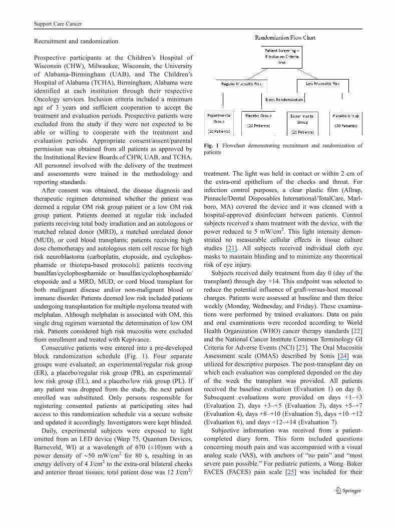
Prospective participants at the Children’s Hospital ofWisconsin (CHW), Milwaukee, Wisconsin, the Universityof Alabama-Birmingham (UAB), and The Children’sHospital of Alabama (TCHA), Birmingham, Alabama wereidentified at each institution through their respectiveOncology services. Inclusion criteria included a minimumage of 3 years and sufficient cooperation to accept thetreatment and evaluation periods. Prospective patients wereexcluded from the study if they were not expected to beable or willing to cooperate with the treatment andevaluation periods. Appropriate consent/assent/parentalpermission was obtained from all patients as approved bythe Institutional Review Boards of CHW, UAB, and TCHA.All personnel involved with the delivery of the treatmentand assessments were trained in the methodology andreporting standards.
After consent was obtained, the disease diagnosis andtherapeutic regimen determined whether the patient wasdeemed a regular OM risk group patient or a low OM riskgroup patient. Patients deemed at regular risk includedpatients receiving total body irradiation and an autologous ormatched related donor (MRD), a matched unrelated donor(MUD), or cord blood transplants; patients receiving highdose chemotherapy and autologous stem cell rescue for highrisk neuroblastoma (carboplatin, etoposide, and cyclophos-phamide or thiotepa-based protocols); patients receivingbusulfan/cyclophosphamide or busulfan/cyclophosphamide/etoposide and a MRD, MUD, or cord blood transplant forboth malignant disease and/or non-malignant blood orimmune disorder. Patients deemed low risk included patientsundergoing transplantation for multiple myeloma treated withmelphalan. Although melphalan is associated with OM, thissingle drug regimen warranted the determination of low OMrisk. Patients considered high risk mucositis were excludedfrom enrollment and treated with Kepivance.
Consecutive patients were entered into a pre-developedblock randomization schedule (Fig. 1). Four separategroups were evaluated; an experimental/regular risk group(ER), a placebo/regular risk group (PR), an experimental/low risk group (EL), and a placebo/low risk group (PL). Ifany patient was dropped from the study, the next patientenrolled was substituted. Only persons responsible forregistering consented patients at participating sites hadaccess to this randomization schedule via a secure websiteand updated it accordingly. Investigators were kept blinded.
Daily, experimental subjects were exposed to lightemitted from an LED device (Warp 75, Quantum Devices,Barneveld, WI) at a wavelength of 670 (±10)nm with apower density of ∼50 mW/cm2 for 80 s, resulting in anenergy delivery of 4 J/cm2 to the extra-oral bilateral cheeksand anterior throat tissues; total patient dose was 12 J/cm2/
treatment. The light was held in contact or within 2 cm ofthe extra-oral epithelium of the cheeks and throat. Forinfection control purposes, a clear plastic film (Allrap,Pinnacle/Dental Disposables International/TotalCare, Marl-boro, MA) covered the device and it was cleaned with ahospital-approved disinfectant between patients. Controlsubjects received a sham treatment with the device, with thepower reduced to 5 mW/cm2. This light intensity demon-strated no measurable cellular effects in tissue culturestudies [21]. All subjects received individual cloth eyemasks to maintain blinding and to minimize any theoreticalrisk of eye injury.
Subjects received daily treatment from day 0 (day of thetransplant) through day +14. This endpoint was selected toreduce the potential influence of graft-versus-host mucosalchanges. Patients were assessed at baseline and then thriceweekly (Monday, Wednesday, and Friday). These examina-tions were performed by trained evaluators. Data on painand oral examinations were recorded according to WorldHealth Organization (WHO) cancer therapy standards [22]and the National Cancer Institute Common Terminology GICriteria for Adverse Events (NCI) [23]. The Oral MucositisAssessment scale (OMAS) described by Sonis [24] wasutilized for descriptive purposes. The post-transplant day onwhich each evaluation was completed depended on the dayof the week the transplant was provided. All patientsreceived the baseline evaluation (Evaluation 1) on day 0.Subsequent evaluations were provided on days +1–+3(Evaluation 2), days +3–+5 (Evaluation 3), days +5–+7(Evaluation 4), days +8–+10 (Evaluation 5), days +10–+12(Evaluation 6), and days +12–+14 (Evaluation 7).
Subjective information was received from a patient-completed diary form. This form included questionsconcerning mouth pain and was accompanied with a visualanalog scale (VAS), with anchors of “no pain” and “mostsevere pain possible.” For pediatric patients, a Wong–BakerFACES (FACES) pain scale [25] was included for their
Fig. 1 Flowchart demonstrating recruitment and randomization ofpatients
Support Care Cancer
reporting of pain. These two scales were also used by thepatient to report the impact on swallowing, with the anchors“no trouble” and “unable to swallow anything (includingsaliva).” The patients also reported whether they could eatnormally; eat only soft, solid foods; consume only liquids;or could not tolerate any food or liquids.
Outcome measures
The primary outcome measure for analysis was the changein scores from baseline on the WHO Pain Assessment scalefor OM, documented at each evaluation. Secondaryoutcome measures also analyzed were incidence of erythe-ma and ulceration of oral tissues, and the duration oferythema and ulcerated tissues. All evaluators receivedtraining on the appearance of OM lesions and calibration onscoring the lesions in the OMAS scale.
Sample size and statistics
The study was powered for the primary comparisons betweenthe experimental and control groups based on 5% alpha and80% power. Based on a two-group independent design, thesample size was based on detecting a 25% decrease in themean pain score between the two groups. Due to the largevariation in pain perception between individuals, the standarddeviation is assumed to be no larger than 39%.With the abovementioned alpha and power, the sample size is estimated to beapproximately 40 patients per group, for a total of 80 patients.Chi-square tests were utilized to statistically compare theoutcomes between the two groups. p values less than 0.05were considered significant.
Blinding
All personnel directly involved with the delivery thephototherapy and evaluation of the patients were blindedas to the treatment arm. The devices constructed to deliverthe light included a switch that allowed the device todeliver both the sham and experimental treatments. Thepersonnel (trained nurses) delivering the phototherapy didnot know which switch position was the experimental orplacebo power density. The patients did not know to whichtreatment arm they were allocated and wore black cloth eyeshields which prevented them from seeing the switchposition on the light. Finally, the evaluators did not knowto which treatment arm the patients had been allocated.
Results
Between March 2007 and April 2009, 85 patients meetingthe inclusion criteria were consecutively recruited and
evaluated from the Oncology services of CHW (22patients), UAB (54 patients), and TCHA (11 patients). Fivepatients were withdrawn; four for admittance into anintensive care unit due to medical complications notassociated with the light therapy and one voluntarilywithdrew because of lack of perceived benefit.
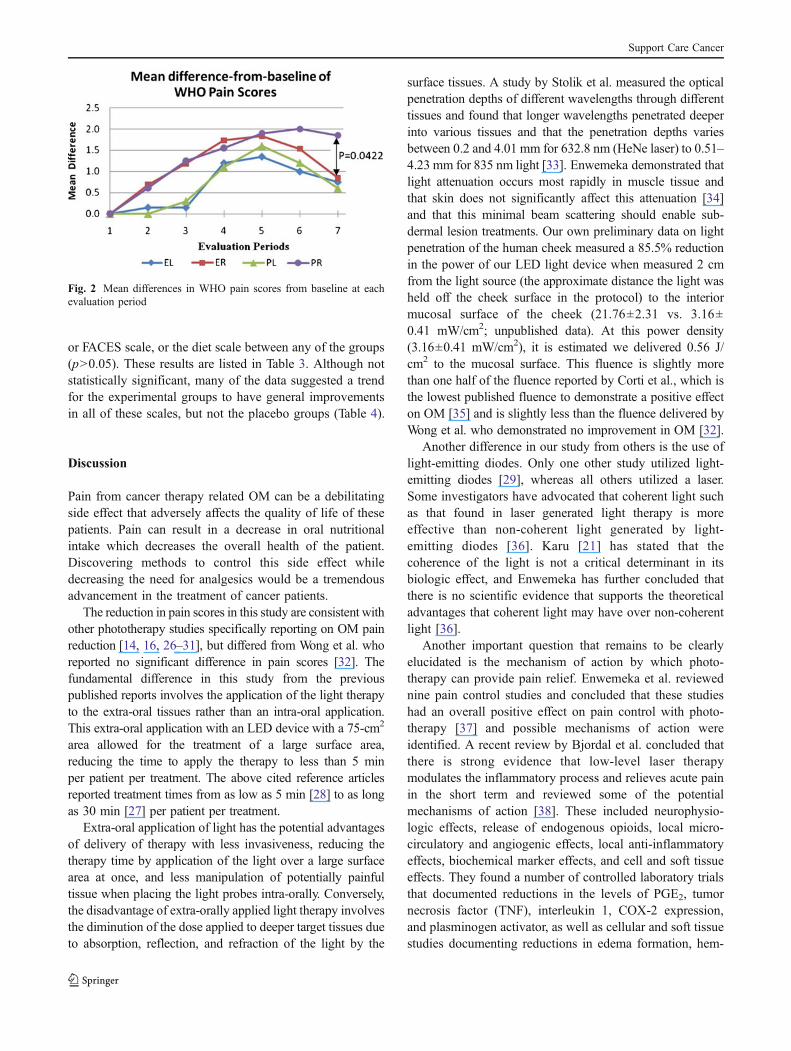
Of the final 80 patients, there were 44 males (55%) and36 females (45%). The mean age of the patients was37 years, with a range from 3 to 74. The sex and agedistribution of the patients in each grouping are summa-rized in Table 1 and the medical diagnosis and treatmentregimen are listed in Table 2. There was a statisticallysignificant difference in the ages only between the regularrisk and low risk groups (p<0.0001), but no difference insex or within the experimental or placebo groups. Note thatthe age difference in the low risk group was expected as thelow risk group was confined to patients receiving singleagent melphalan for the treatment of myeloma, a diseaseseen almost exclusively in adults.
WHO scales
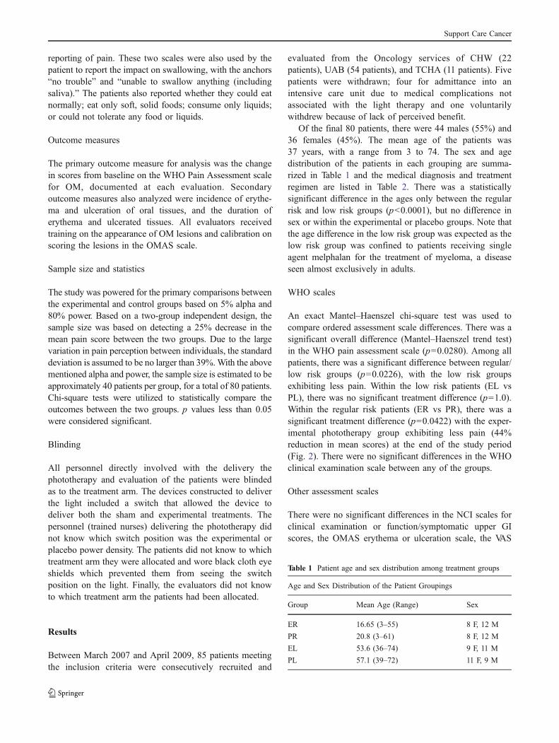
An exact Mantel–Haenszel chi-square test was used tocompare ordered assessment scale differences. There was asignificant overall difference (Mantel–Haenszel trend test)in the WHO pain assessment scale (p=0.0280). Among allpatients, there was a significant difference between regular/low risk groups (p=0.0226), with the low risk groupsexhibiting less pain. Within the low risk patients (EL vsPL), there was no significant treatment difference (p=1.0).Within the regular risk patients (ER vs PR), there was asignificant treatment difference (p=0.0422) with the exper-imental phototherapy group exhibiting less pain (44%reduction in mean scores) at the end of the study period(Fig. 2). There were no significant differences in the WHOclinical examination scale between any of the groups.
Other assessment scales
There were no significant differences in the NCI scales forclinical examination or function/symptomatic upper GIscores, the OMAS erythema or ulceration scale, the VAS
Table 1 Patient age and sex distribution among treatment groups
Age and Sex Distribution of the Patient Groupings
Group Mean Age (Range) Sex
ER 16.65 (3–55) 8 F, 12 M
PR 20.8 (3–61) 8 F, 12 M
EL 53.6 (36–74) 9 F, 11 M
PL 57.1 (39–72) 11 F, 9 M
Support Care Cancer
Table
2Patient
diagno
sisandtreatm
entregimen
bygrou
passign
ment
Patient
CancerDiagn
osis
Exp
erim
entalRegular
Risk
Placebo
Regular
Risk
Exp
erim
entalLow
Risk
Placebo
Low
Risk
Age
(yrs)
Diagn
osis
Con
ditio
ning
Regim
enAge
Diagn
osis
Con
ditio
ning
Regim
enAge
Diagn
osis
Con
ditio
ning
Regim
enAge
Diagn
osis
Con
ditio
ning
Regim
en
4AA
Cyclo,Bus,Meth
15SCA
Cyclo,Bus,Meth
74MM
Melph
alan
66MM
Melph
alan
3NBL
Mel,VP16
,Carbo
14CML
Cyclo,Bus,Meth
61MM
Melph
alan
72Amyloido
sis
Melph
alan
4AML
Thio,
Flu,TBIx3days
56TC
VP16
,Carbo
58MM
Melph
alan
54MM
Melph
alan
18NHL
BEAM
9ALL(B),HLH
Cyclo,Meth,
ARA-C,TBIx3days
50MM
Melph
alan
61MM
Melph
alan
47CLLSLL
Cyclo,Bus
3WAS
Cyclo,Bus,AT
G56
MM
Melph
alan
61MM
Melph
alan
12AML
Cyclo,Bus,Meth
39MDS
Bus,Flu,AT
G58
MM
Melph
alan
61MM
Melph
alan
55HD
Cyclo,Bus,VP16
16ALL
Cyclo,Meth,
ARA-C,TBIx3days
36MM
(poems)
Melph
alan
50MM
Melph
alan
28HD
Cyclo,Bus,VP16
12Osteopetrosis
Cyclo,Vin,CDPP
58Amyloido
sis
Melph
alan
53MM
Melph
alan
38PNT
Bus,Mel,Thio
61MDS
Cyclo,AT
G55
MM
Melph
alan
55MM
Melph
alan
17MES
Bus,Mel,PelvicRadiatio
n41
Lymph
oma
Cyclo,Bus,VP16
46MM
Melph
alan
69MM
Melph
alan
6AML
Cyclo,ARA-C.TBIx4
days
14AA
Cyclo,Meth,
ATG
58MM
Melph
alan
55MM
Melph
alan
10ALL
Cyclo,Flu,TBIx3days
5AML
Bus,Mel,Flu
54MM
Melph
alan
39MM
Melph
alan
6NBL
Mel,Carbo
,VP16
61NHL
Cyclo,Bus,VP16
43MM
Melph
alan
52MM
Melph
alan
14ALL
Cyclo,AT
G,TBIx3days
16ALL(H
)Cyclo,ARA-C,TBIx3days
43MM
Melph
alan
57MM
Melph
alan
10NBL
Mel,Carbo
,VP16
13ALL(T),AML
Cyclo,Flu,TBIx4days
47MM
Melph
alan
66MM
Melph
alan
11B-A
LL
Cyclo,Flu,TBIx3days
5B-A
LL®
Cyclo,Flu,TBIx4days
48MM
Melph
alan
63MM
Melph
alan
20ALL
Cyclo,Flu,TBIx3days
11AML
Cyclo,Bus,Meth
52MM
Melph
alan
46MM
Melph
alan
15Ewings
Bus,Mel,Thio
7NBL
Mel,VP16
,Carbo
51MM
Melph
alan
56MM
Melph
alan
3AA
Cyclo,Mel,AT
G11
AML
Cyclo,Bus,Meth
67MM
Melph
alan
52MM
Melph
alan
12AML
Cyclo,Flu,TBIx4days
7SCA
Cyclo,Bus,Meth,
ATG
57MM
Melph
alan
54MM
Melph
alan
Diagn
osis—AAaplastic
anem
ia,ALLacutelymph
ocytic
leuk
emia,AMLacutemyelogeno
usleuk
emia,CLLSL
Lchroniclymph
ocytic
leuk
emia/smallcelllymph
ocytic
leuk
emia,CMLchronic
myelogeno
usleuk
emia,E
wings
Ewings
sarcom
a,HDHod
gkin’sdisease,HLHhemop
hago
cytic
lymph
ohistio
cytosis,MDSmyelodisplasticsynd
rome,MM
multip
lemyeloma,NBLneurob
lastom
a,NHLno
n-Hod
gkin’slymph
oma,
POEMSpo
lyneurop
athy,organo
megaly,
endo
crinop
athy,mon
oclonalgammop
athy,or
mon
oclonalplasmaproliferativedisorder,skin
changes,
SCAsickle
cell
anem
ia.Con
ditio
ning
regimen—ARA-C
cytarabine,AT
Gantithy
mocyteglob
ulin,BEAM
bleomycin,cyclop
hosphamide,
adriam
ycin,metho
trexate,
BUSbu
sulphan,
Carbo
carbop
latin
,CDPP
cicplatin
,Cyclo
cyclop
hosphamide,
FLU
flud
arabine,
Mel
melph
alan,Methmetho
trexate,
TBItotalbo
dyirradiation,
Thiothiotepa,Vinvinb
lastine,
VP16
etop
oside
Support Care Cancer
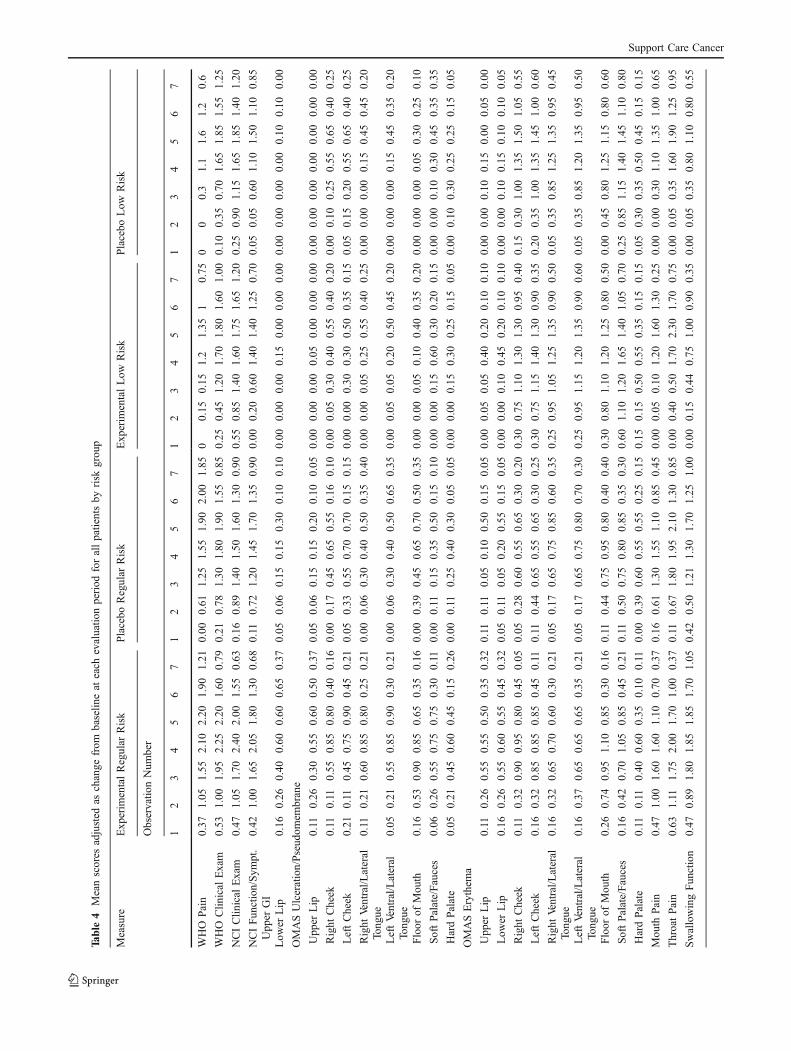
or FACES scale, or the diet scale between any of the groups(p>0.05). These results are listed in Table 3. Although notstatistically significant, many of the data suggested a trendfor the experimental groups to have general improvementsin all of these scales, but not the placebo groups (Table 4).
Discussion
Pain from cancer therapy related OM can be a debilitatingside effect that adversely affects the quality of life of thesepatients. Pain can result in a decrease in oral nutritionalintake which decreases the overall health of the patient.Discovering methods to control this side effect whiledecreasing the need for analgesics would be a tremendousadvancement in the treatment of cancer patients.
The reduction in pain scores in this study are consistent withother phototherapy studies specifically reporting on OM painreduction [14, 16, 26–31], but differed from Wong et al. whoreported no significant difference in pain scores [32]. Thefundamental difference in this study from the previouspublished reports involves the application of the light therapyto the extra-oral tissues rather than an intra-oral application.This extra-oral application with an LED device with a 75-cm2
area allowed for the treatment of a large surface area,reducing the time to apply the therapy to less than 5 minper patient per treatment. The above cited reference articlesreported treatment times from as low as 5 min [28] to as longas 30 min [27] per patient per treatment.
Extra-oral application of light has the potential advantagesof delivery of therapy with less invasiveness, reducing thetherapy time by application of the light over a large surfacearea at once, and less manipulation of potentially painfultissue when placing the light probes intra-orally. Conversely,the disadvantage of extra-orally applied light therapy involvesthe diminution of the dose applied to deeper target tissues dueto absorption, reflection, and refraction of the light by the
surface tissues. A study by Stolik et al. measured the opticalpenetration depths of different wavelengths through differenttissues and found that longer wavelengths penetrated deeperinto various tissues and that the penetration depths variesbetween 0.2 and 4.01 mm for 632.8 nm (HeNe laser) to 0.51–4.23 mm for 835 nm light [33]. Enwemeka demonstrated thatlight attenuation occurs most rapidly in muscle tissue andthat skin does not significantly affect this attenuation [34]and that this minimal beam scattering should enable sub-dermal lesion treatments. Our own preliminary data on lightpenetration of the human cheek measured a 85.5% reductionin the power of our LED light device when measured 2 cmfrom the light source (the approximate distance the light washeld off the cheek surface in the protocol) to the interiormucosal surface of the cheek (21.76±2.31 vs. 3.16±0.41 mW/cm2; unpublished data). At this power density(3.16±0.41 mW/cm2), it is estimated we delivered 0.56 J/cm2 to the mucosal surface. This fluence is slightly morethan one half of the fluence reported by Corti et al., which isthe lowest published fluence to demonstrate a positive effecton OM [35] and is slightly less than the fluence delivered byWong et al. who demonstrated no improvement in OM [32].
Another difference in our study from others is the use oflight-emitting diodes. Only one other study utilized light-emitting diodes [29], whereas all others utilized a laser.Some investigators have advocated that coherent light suchas that found in laser generated light therapy is moreeffective than non-coherent light generated by light-emitting diodes [36]. Karu [21] has stated that thecoherence of the light is not a critical determinant in itsbiologic effect, and Enwemeka has further concluded thatthere is no scientific evidence that supports the theoreticaladvantages that coherent light may have over non-coherentlight [36].
Another important question that remains to be clearlyelucidated is the mechanism of action by which photo-therapy can provide pain relief. Enwemeka et al. reviewednine pain control studies and concluded that these studieshad an overall positive effect on pain control with photo-therapy [37] and possible mechanisms of action wereidentified. A recent review by Bjordal et al. concluded thatthere is strong evidence that low-level laser therapymodulates the inflammatory process and relieves acute painin the short term and reviewed some of the potentialmechanisms of action [38]. These included neurophysio-logic effects, release of endogenous opioids, local micro-circulatory and angiogenic effects, local anti-inflammatoryeffects, biochemical marker effects, and cell and soft tissueeffects. They found a number of controlled laboratory trialsthat documented reductions in the levels of PGE2, tumornecrosis factor (TNF), interleukin 1, COX-2 expression,and plasminogen activator, as well as cellular and soft tissuestudies documenting reductions in edema formation, hem-
Fig. 2 Mean differences in WHO pain scores from baseline at eachevaluation period
Support Care Cancer
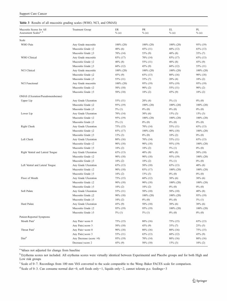
Table 3 Results of all mucositis grading scales (WHO, NCI, and OMAS)
Mucositis Scores for AllAssessment Scalesa, b
Treatment Group ER PR EL PL% (n) % (n) % (n) % (n)
Scale
WHO Pain Any Grade mucositis 100% (20) 100% (20) 100% (20) 95% (19)
Mucositis Grade ≤2 40% (6) 45% (11) 60% (12) 65% (13)
Mucositis Grade ≥3 70% (14) 55% (9) 40% (8) 35% (7)
WHO Clinical Any Grade mucositis 85% (17) 70% (14) 85% (17) 65% (13)
Mucositis Grade ≤2 40% (8) 55% (11) 40% (8) 45% (9)
Mucositis Grade ≥3 60% (12) 45% (9) 60% (12) 55% (11)
NCI Clinical Any Grade mucositis 100% (20) 100% (20) 100% (20) 100% (20)
Mucositis Grade ≤2 45% (9) 65% (13) 80% (16) 90% (18)
Mucositis Grade ≥3 55% (11) 35% (7) 20% (4) 10% (2)
NCI Functional Any Grade mucositis 100% (20) 95% (19) 95% (19) 95% (19)
Mucositis Grade ≤2 50% (10) 90% (2) 55% (11) 90% (2)
Mucositis Grade ≥3 50% (10) 10% (2) 45% (9) 10% (2)
OMAS (Ulceration/Pseudomemberane)
Upper Lip Any Grade Ulceration 55% (11) 20% (4) 5% (1) 0% (0)
Mucositis Grade ≤2 95% (19) 100% (20) 100% (20) 100% (20)
Mucositis Grade ≥3 5% (1) 0% (0) 0% (0) 0% (0)
Lower Lip Any Grade Ulceration 70% (14) 30% (6) 15% (3) 15% (3)
Mucositis Grade ≤2 95% (19) 100% (20) 100% (20) 100% (20)
Mucositis Grade ≥3 5% (1) 0% (0) 0% (0) 0% (0)
Right Cheek Any Grade Ulceration 75% (15) 70% (14) 55% (11) 65% (13)
Mucositis Grade ≤2 85% (17) 100% (20) 90% (18) 100% (20)
Mucositis Grade ≥3 15% (3) 0% (0) 10% (2) 0% (0)
Left Cheek Any Grade Ulceration 80% (16) 70% (14) 55% (11) 65% (13)
Mucositis Grade ≤2 90% (18) 90% (18) 95% (19) 100% (20)
Mucositis Grade ≥3 10% (2) 10% (2) 5% (1) 0% (0)
Right Ventral and Lateral Tongue Any Grade Ulceration 65% (13) 40% (8) 40% (8) 50% (10)
Mucositis Grade ≤2 90% (18) 90% (18) 95% (19) 100% (20)
Mucositis Grade ≥3 10% (2) 10% (2) 5% (1) 0% (0)
Left Ventral and Lateral Tongue Any Grade Ulceration 65% (13) 50% (10) 65% (13) 40% (8)
Mucositis Grade ≤2 90% (18) 85% (17) 100% (20) 100% (20)
Mucositis Grade ≥3 10% (2) 15% (3) 0% (0) 0% (0)
Floor of Mouth Any Grade Ulceration 75% (15) 60% (12) 30% (6) 30% (6)
Mucositis Grade ≤2 90% (18) 90% (18) 100% (20) 100% (20)
Mucositis Grade ≥3 10% (2) 10% (2) 0% (0) 0% (0)
Soft Palate Any Grade Ulceration 55% (11) 50% (10) 50% (10) 40% (8)
Mucositis Grade ≤2 90% (18) 100% (20) 100% (20) 95% (19)
Mucositis Grade ≥3 10% (2) 0% (0) 0% (0) 5% (1)
Hard Palate Any Grade Ulceration 45% (9) 50% (10) 30% (6) 30% (6)
Mucositis Grade ≤2 95% (19) 95% (19) 100% (20) 100% (20)
Mucositis Grade ≥3 5% (1) 5% (1) 0% (0) 0% (0)
Patient-Reported Symptoms
Mouth Painc Any Pain>score 0 75% (15) 80% (16) 75% (15) 65% (13)
Any Pain≥score 3 50% (10) 45% (9) 35% (7) 25% (5)
Throat Painc Any Pain>score 0 90% (18) 80% (16) 80% (16) 75% (15)
Any Pain≥score 3 55% (11) 65% (13) 60% (12) 45% (9)
Dietd Any Decrease (score >0) 95% (19) 70% (14) 80% (16) 80% (16)
Decrease≥score 2 45% (9) 50% (10) 15% (3) 10% (2)
a Values not adjusted for change from baselineb Erythema scores not included. All erythema scores were virtually identical between Experimental and Placebo groups and for both High andLow risk groupsc Scale of 0–7. Recordings from 100 mm VAS converted to the scale comparable to the Wong–Baker FACES scale for comparison.d Scale of 0–3. Can consume normal diet=0, soft foods only=1, liquids only=2, cannot tolerate p.o. feedings=3
Support Care Cancer
Table
4Meanscores
adjusted
aschange
from
baselin
eat
each
evaluatio
nperiod
forallpatientsby
risk
grou
p
Measure
Exp
erim
entalRegular
Risk
Placebo
Regular
Risk
Exp
erim
entalLow
Risk
Placebo
Low
Risk
Observatio
nNum
ber
12
34
56
71
23
45
67
12
34
56
71
23
45
67
WHO
Pain
0.37
1.05
1.55
2.10
2.20
1.90
1.21
0.00
0.61
1.25
1.55
1.90
2.00
1.85
00.15
0.15
1.2
1.35
10.75
00
0.3
1.1
1.6
1.2
0.6
WHO
Clin
ical
Exam
0.53
1.00
1.95
2.25
2.20
1.60
0.79
0.21
0.78
1.30
1.80
1.90
1.55
0.85
0.25
0.45
1.20
1.70
1.80
1.60
1.00
0.10
0.35
0.70
1.65
1.85
1.55
1.25
NCIClin
ical
Exam
0.47
1.05
1.70
2.40
2.00
1.55
0.63
0.16
0.89
1.40
1.50
1.60
1.30
0.90
0.55
0.85
1.40
1.60
1.75
1.65
1.20
0.25
0.90
1.15
1.65
1.85
1.40
1.20
NCIFun
ction/Sym
pt.
Upp
erGI
0.42
1.00
1.65
2.05
1.80
1.30
0.68
0.11
0.72
1.20
1.45
1.70
1.35
0.90
0.00
0.20
0.60
1.40
1.40
1.25
0.70
0.05
0.05
0.60
1.10
1.50
1.10
0.85
Low
erLip
0.16
0.26
0.40
0.60
0.60
0.65
0.37
0.05
0.06
0.15
0.15
0.30
0.10
0.10
0.00
0.00
0.00
0.15
0.00
0.00
0.00
0.00
0.00
0.00
0.00
0.10
0.10
0.00
OMASUlceration/Pseud
omem
brane
Upp
erLip
0.11
0.26
0.30
0.55
0.60
0.50
0.37
0.05
0.06
0.15
0.15
0.20
0.10
0.05
0.00
0.00
0.00
0.05
0.00
0.00
0.00
0.00
0.00
0.00
0.00
0.00
0.00
0.00
Right
Cheek
0.11
0.11
0.55
0.85
0.80
0.40
0.16
0.00
0.17
0.45
0.65
0.55
0.16
0.10
0.00
0.05
0.30
0.40
0.55
0.40
0.20
0.00
0.10
0.25
0.55
0.65
0.40
0.25
LeftCheek
0.21
0.11
0.45
0.75
0.90
0.45
0.21
0.05
0.33
0.55
0.70
0.70
0.15
0.15
0.00
0.00
0.30
0.30
0.50
0.35
0.15
0.05
0.15
0.20
0.55
0.65
0.40
0.25
Right
Ventral/L
ateral
Tong
ue0.11
0.21
0.60
0.85
0.80
0.25
0.21
0.00
0.06
0.30
0.40
0.50
0.35
0.40
0.00
0.00
0.05
0.25
0.55
0.40
0.25
0.00
0.00
0.00
0.15
0.45
0.45
0.20
LeftVentral/L
ateral
Tong
ue0.05
0.21
0.55
0.85
0.90
0.30
0.21
0.00
0.06
0.30
0.40
0.50
0.65
0.35
0.00
0.05
0.05
0.20
0.50
0.45
0.20
0.00
0.00
0.00
0.15
0.45
0.35
0.20
Floor
ofMou
th0.16
0.53
0.90
0.85
0.65
0.35
0.16
0.00
0.39
0.45
0.65
0.70
0.50
0.35
0.00
0.00
0.05
0.10
0.40
0.35
0.20
0.00
0.00
0.00
0.05
0.30
0.25
0.10
SoftPalate/Fauces
0.06
0.26
0.55
0.75
0.75
0.30
0.11
0.00
0.11
0.15
0.35
0.50
0.15
0.10
0.00
0.00
0.15
0.60
0.30
0.20
0.15
0.00
0.00
0.10
0.30
0.45
0.35
0.35
HardPalate
0.05
0.21
0.45
0.60
0.45
0.15
0.26
0.00
0.11
0.25
0.40
0.30
0.05
0.05
0.00
0.00
0.15
0.30
0.25
0.15
0.05
0.00
0.10
0.30
0.25
0.25
0.15
0.05
OMASErythem
a
Upp
erLip
0.11
0.26
0.55
0.55
0.50
0.35
0.32
0.11
0.11
0.05
0.10
0.50
0.15
0.05
0.00
0.05
0.05
0.40
0.20
0.10
0.10
0.00
0.00
0.10
0.15
0.00
0.05
0.00
Low
erLip
0.16
0.26
0.55
0.60
0.55
0.45
0.32
0.05
0.11
0.05
0.20
0.55
0.15
0.05
0.00
0.00
0.10
0.45
0.20
0.10
0.10
0.00
0.00
0.10
0.15
0.10
0.10
0.05
Right
Cheek
0.11
0.32
0.90
0.95
0.80
0.45
0.05
0.05
0.28
0.60
0.55
0.65
0.30
0.20
0.30
0.75
1.10
1.30
1.30
0.95
0.40
0.15
0.30
1.00
1.35
1.50
1.05
0.55
LeftCheek
0.16
0.32
0.85
0.85
0.85
0.45
0.11
0.11
0.44
0.65
0.55
0.65
0.30
0.25
0.30
0.75
1.15
1.40
1.30
0.90
0.35
0.20
0.35
1.00
1.35
1.45
1.00
0.60
Right
Ventral/L
ateral
Tong
ue0.16
0.32
0.65
0.70
0.60
0.30
0.21
0.05
0.17
0.65
0.75
0.85
0.60
0.35
0.25
0.95
1.05
1.25
1.35
0.90
0.50
0.05
0.35
0.85
1.25
1.35
0.95
0.45
LeftVentral/L
ateral
Tong
ue0.16
0.37
0.65
0.65
0.65
0.35
0.21
0.05
0.17
0.65
0.75
0.80
0.70
0.30
0.25
0.95
1.15
1.20
1.35
0.90
0.60
0.05
0.35
0.85
1.20
1.35
0.95
0.50
Floor
ofMou
th0.26
0.74
0.95
1.10
0.85
0.30
0.16
0.11
0.44
0.75
0.95
0.80
0.40
0.40
0.30
0.80
1.10
1.20
1.25
0.80
0.50
0.00
0.45
0.80
1.25
1.15
0.80
0.60
SoftPalate/Fauces
0.16
0.42
0.70
1.05
0.85
0.45
0.21
0.11
0.50
0.75
0.80
0.85
0.35
0.30
0.60
1.10
1.20
1.65
1.40
1.05
0.70
0.25
0.85
1.15
1.40
1.45
1.10
0.80
HardPalate
0.11
0.11
0.40
0.60
0.35
0.10
0.11
0.00
0.39
0.60
0.55
0.55
0.25
0.15
0.15
0.15
0.50
0.55
0.35
0.15
0.15
0.05
0.30
0.35
0.50
0.45
0.15
0.15
Mou
thPain
0.47
1.00
1.60
1.60
1.10
0.70
0.37
0.16
0.61
1.30
1.55
1.10
0.85
0.45
0.00
0.05
0.10
1.20
1.60
1.30
0.25
0.00
0.00
0.30
1.10
1.35
1.00
0.65
ThroatPain
0.63
1.11
1.75
2.00
1.70
1.00
0.37
0.11
0.67
1.80
1.95
2.10
1.30
0.85
0.00
0.40
0.50
1.70
2.30
1.70
0.75
0.00
0.05
0.35
1.60
1.90
1.25
0.95
SwallowingFun
ction
0.47
0.89
1.80
1.85
1.85
1.70
1.05
0.42
0.50
1.21
1.30
1.70
1.25
1.00
0.00
0.15
0.44
0.75
1.00
0.90
0.35
0.00
0.05
0.35
0.80
1.10
0.80
0.55
Support Care Cancer
orrhagic formation, neutrophil cell influx, cell apoptosis,and improvements in microcirculation. Cyclooxygenase-2is one of the enzymes that convert arachidonic acid intoPGE2, and PGE2 does not by itself cause pain, but results ina hyperalgesia state which does induce increased painperception [39]. Sonis et al. demonstrated in a hamstermodel that COX-2 expression paralleled mucositis severityand although it was not a primary cause of radiation injury,it did play an amplifying role [40]. Stimulation of epithelialcells, fibroblasts, and chondrocytes with interleukin 1 andTNF-α results in increased PGE2 production as well [41].Mizutani et al. demonstrated a reduction in serum PGE2
levels after phototherapy with an 830-nm GaAlAr laser at1 W [42]. Light therapy may cause a number of smallreductions in the amplification phase of these cytokinesduring the mucositis process, thereby reducing the hyper-algesia and pain perception.
This study also failed to show any significant decrease inthe other measures of OM utilized, which is contrary tomultiple published reports [14–17, 26–31, 35, 43, 44]. Thismay be due to the reduction of effect of the light caused bythe absorption of the power by more superficial non-targettissues resulting in inadequate light dosing of the targettissues. Given that the estimated fluence delivered to themucosal surface from the extra-orally applied phototherapywas only 0.56 J/cm2, this appears to be a reasonable critique.However, the review by Bjordal et al. states that light therapycan effectively radiate tissue that lies within 10–15 mm ofthe source [38], but these studies were of osteoarthritic andother musculoskeletal pain disorders and not mucositis.
Another reason for the lack of improvement in OMparameters may be associated with the timing of the photo-therapy. In this study, the phototherapy was started on day 0 ofthe transplant regimen. Several other OM studies [14–16, 26–29, 32, 43, 44] started the phototherapy prior to or with thestart of the myeloablation regimen, usually occurring 2–7 daysprior to the transplant, whereas others [17, 30] delivered thephototherapy on or after the day of the transplant orappearance of the OM. If the injury to the mucosa occurswith the initiation of the myeloablative therapy, and ifphototherapy works by reducing the amplification of theinflammatory process, starting the phototherapy at the initialadministration of the myeloablative therapy may have resultedin more favorable OM results.
The low risk patients were statistically significantly olderthan the regular risk patients, and this group did not haveany significant differences in their incidence of OM or painreporting. There may be several explanations for thesephenomena. Firstly, by definition, the degree of mucosalinjury from a single drug, melphalan, placed these patientsin this low risk group [20]. The multiple drug interactionsin the regular risk group can cause significantly more tissuedamage and produce greater amounts cytokines. Since the
low risk patients most likely had lower levels of these pro-inflammatory cytokines, the proposed interruption in cellsignal amplification caused by photobiomodulation wouldnot have as great an effect. Another potential explanation isthe decreased mitochondrial activities and increased dam-aged to mitochondrial DNA associated with human agingmay have resulted in less photobiostimulatory effects of themitochondria in this older population [45]. Further researchis needed to elucidate the effects of photobiostimulation onan aging population.
The extra-oral application of LED phototherapy in thisstudy was shown to have a statistically significant reductionin pain as reported by the WHO Pain Assessment scale forOM, but not for other mucositis scoring scales such as theNCI and OMAS scales. Much further research is neededthrough controlled trials to establish the appropriate timing,dose, power, and fluence of the phototherapy to determinethe optimum therapeutic parameters.
Acknowledgments This work was supported by the NationalAeronautics and Space Administration Grant # NNM05AB48C, theBleser Foundation Endowed Professorship and Chad BaumannNeurology Research Endowment at the Medical College of Wisconsin,and the Marquette University School of Dentistry Pediatric FacultyFund. Additionally, we would like to extend our warmest and sincerestappreciation to the nurses at the respective institutions who wereinstrumental in the completion of this study.
Conflict of interest There are no conflicts of interest to report.
References
1. Osgood EE, Riddle MC, Mathew TJ (1939) Aplastic anemiatreated with daily transfusions and intravenous marrow; casereport. Ann Intern Med 13:357
2. Long GD, Blume KG (1995) Allogenic and autologous marrowtransplantation. In: Beutler E, Lichtman MA, Coller BS, Kipps TJ(eds) Williams Hematology, 5th edn. McGraw Hill, New York, pp172–194
3. Peterson DE, Bensadoun RJ, Roila F (2009) Management of oraland gastrointestinal mucositis: ESMO clinical recommendations.Ann Oncol 20(Supp 4):iv174–iv177. doi:10.1093/annonc/mdq197
4. McGowan D (2008) Chemotherapy-induced oral dysfunction: aliterature review. Br J Nurs 17(22):1422–1426
5. Papadopoulou A, Williams MD, Darbyshire PJ, Booth IW (1998)Nutritional support in children undergoing bone marrow transplan-tation. Clin Nutr 17(2):57–63. doi:10.1016/S0261-5614(98)80306-3
6. Kannan V, Bapsy PP, Anantha N, Doval DC, Vaithianathan H,Banumathy G et al (1997) Efficacy and safety of granulocytemacrophage-colony stimulating factor (GM-CSF) on the frequen-cy and severity of radiation mucositis in patients with head andneck carcinoma. Int J Radiat Oncol Biol Phys 37(5):1005–1010
7. Sonis ST, Costa JW, Evitts SM, Lindquist LE, Nicolson M (1992)Effect of epidermal growth factor on ulcerative mucositis inhamsters that receive cancer chemotherapy. Oral Surg Oral MedOral Pathol 74:749–755. doi:10.1016/0030-4220(92)90402-C
8. Stiff PJ, Emmanouilides C, Bensinger WI, Gentile T, Blazar B, SheaTC, Lu J, Isitt J, Cesano A, Spielberger R (2006) Palifermin reducespatient-reported mouth and throat soreness and improves patient
Support Care Cancer
functioning in the hematopoietic stem-cell transplantation setting. JClin Oncol 24(33):5186–5193. doi:10.1200/JCO.2005.02.8340
9. Sonis ST, Van Vugt AG, McDonald J, Dotoli E, Schwertschlag U,Szklut P et al (1997) Mitigating effects of interleukin 11 onconsecutive courses of 5-fluorouracil-induced ulcerative mucositisin hamsters. Cytokine 9(8):605–612. doi:10.1006/cyto.1997.0208
10. Sonis ST, Van Vugt AG, Brien JPO, Muska AD, Bruskin AM,Rose A et al (1997) Transforming growth factor beta 3 mediatedmodulation of cell cycling and attenuation of 5-fluorouracilinduced oral mucositis. Oral Oncol 33(1):47–54. doi:10.1016/S0964-1955(96)00043-7
11. Clarke J, Butler R, Howarth G, Read L, Regester G (2002)Exposure of oral mucosa to bioactive milk factors reduces severityof chemotherapy-induced mucositis in the hamster. Oral Oncol38:478–485. doi:10.1016/S1368-8375(01)00107-5
12. Karagözoğlu S, Filiz Ulusoy M (2005) Chemotherapy: the effectof oral cryotherapy on the development of mucositis. J Clin Nurs14(6):754–765. doi:10.1111/j.1365-2702.2005.01128.x
13. Epstein JB, Silverman S Jr, Paggiarino DA, Crockett S, SchubertMM, Senzer NN, Lockhart PB, Gallagher MJ, Peterson DE,Leveque FG (2001) Benzydamine HCl for prophylaxis ofradiation-induced oral mucositis: results from a multicenter,randomized, double-blind, placebo-controlled clinical trial. Cancer92(4):875–885. doi:10.1002/1097-0142, (20010815)
14. Barasch A, Peterson DE, Tanzer JM, D’Ambrosio JA, Nuki K,Schubert MM, Franquin J-C, Clive J, Tutschka P (1995) Helium-neon laser effects on conditioning-induced oral mucositis in bonemarrow transplant patients. Cancer 76:2550–2556. doi:10.1002/1097-0142, (19951215)
15. Ciais G, Namer M, Schneider M, Demard F, Pourreau-SchneiderN, Martin PM, Soudry M, Franquin JC, Zattara H (1996) Lalaserthérapie dans la prevention et le traitement des mucites liées ála chimiothérapie anticancéreuse. Bull Cancer 79:183–191
16. Cowen D, Tardieu C, Schubert M, Peterson D, Resbeut M,Faucher C, Franquin J-C (1997) Low energy helium-neon laser inthe prevention of oral mucositis in patients undergoing bonemarrow transplant: results of a double blind randomized trial. Int JRadiation Oncology Biol Phys 38(4):697–703. doi:10.1016/S0360-3016(97)00076-X
17. Whelan HT, Connelly JF, Hodgson BD, Barbeau L, Post AC,Bullard G, Buchmann EV, Kane M, Whelan NT, Warwick A,Margolis D (2002) NASA light emitting diodes for the preventionof oral mucositis in pediatric bone marrow transplant patients. JC l i n La s e r Med Su rg 20 : 319–324 . do i : 10 . 1089 /104454702320901107
18. Sonis ST, Oster G, Fuchs H et al (2001) Oral mucositis and theclinical and economic outcomes of hematopoietic stem-celltransplantation. J Clin Oncol 19:2201–2205
19. Sonis ST (2004) Oral mucositis and cancer therapy. J SupportOncol 2(suppl 3):003–008
20. Lalla RV, Peterson DE, Brennan MT, Schubert MM (2008) Oraltoxicity. In: Perry MC (ed) The chemotherapy source book, 4thedn. Wolters Kluwer, Philadelphia, p 121
21. Karu TI (1987) Photobiological fundamentals of low-power lasertherapy. IEEE J Quantum Electronics QE 23(10):1703–1717
22. WHO (1979) Handbook for reporting results of cancer treatment.WHO Offset publication No. 48. World Health Organization,Geneva
23. Cancer Therapy Evaluation Program, Common TerminologyCriteria for Adverse Events, Version 3.0, DCTD, NCI, NIH,DHHS, March 31, 2003 (http://ctep.cancer.gov), Publish Date:August 9, 2006
24. Sonis ST, Eilers JP, Epstein JB, LeVeque FG, Liggett WH Jr,Mulagha MT, Peterson DE, Rose AH, Schubert MM, SpijkervetFK, Wittes JP (1999) Validation of a new scoring system for theassessment of clinical trial research of oral mucositis induced by
radiation or chemotherapy. Mucositis Study Group. Cancer 85(10):2103–2113. doi:10.1002/(SICI)1097-0142, (19990515)
25. Wong DL, Baker CM (1988) Pain in children: comparison ofassessment scales. Pediatr Nurs 14(1):9–17
26. Abramoff MMF, Lopes NNF, Lopes LAL, Dib LL, Guilherme A,Caran EM, Barreto AD, Lee MLM, Petrilli AS (2008) Low-levellaser therapy in the prevention and treatment of chemotherapy-induced oral mucositis in young patients. Photomed Laser Surg 26(4):393–400. doi:10.1089/pho.2007.2144
27. Arora H, Pai KM, Maiya A, Vidyasagar MS, Rajeev A (2008)Efficacy of He–Ne laser in the prevention and treatment ofradiotherapy-induced oral mucositis in oral cancer patients. OralSurg Oral Med Oral Pathol Oral Radiol Endod 105:180–186.doi:10.1016/j.tripleo.2007.07.043
28. Bensadoun RJ, Franquin JC, Ciais G, Darcourt V, Schubert MM,Viot M, Dejou J, Tardieu C, Benezery K, Nguyen TD, Laudoyer Y,Dassonville O, Poissonnet G, Vallicioni J, Thyss A, Hamdi M,Chauvel P, Demard F (1999) Low-energy He/Ne laser in theprevention of radiation-induced mucositis. A multicenter phase IIIrandomized study in patients with head and neck cancer. SupportCare Cancer 7:244–252. doi:10.1007/s005209900034
29. Lang-Bicudo L, Eduardo FDP, Eduardo CDP, Zezell DM (2008)LED phototherapy to prevent mucositis: a case report. PhotomedLaser Surg 26(6):609–613. doi:10.1089/pho.2007.2228
30. Nes AG, Posso MBS (2005) Patients with moderatechemotherapy-induced mucositis: pain therapy using low inten-sity lasers. Int Nurs Rev 52(1):68–72. doi:10.1111/j.1466-7657.2004.00401.x
31. Simoes A, Eduardo FP, Luiz AC, Campos L, Henrique P,Cristofaro M, Marques MM, Eduardo CP (2009) Laser photo-therapy as topical prophylaxis against head and neck cancerradiotherapy-induced oral mucositis: comparison between low andhigh/low power lasers. Lasers Surg Med 41:264–270.doi:10.1002/lsm.20758
32. Wong SF, Wilder-Smith P (2002) Pilot study of laser effects on oralmucositis in patients receiving chemotherapy. Cancer J 8:247–254
33. Stolik S, Delgado JA, Perez A, Anasagasti L (2000) Measurementof the penetration depths of red and near infrared light in humanex vivo tissues. J Photochem Photobiol B57:90–93. doi:10.1016/S1011-1344(00)00082-8
34. Enwemeka CS (2001) Attenuation and penetration of visible632.8 nm and invisible infra-red 904 nm light in soft tissues. LaserTher 13:95–101
35. Corti L, Chiarion-Sileni V, Aversa S, Ponzoni A, D’Arcais R,Pagnutti S, Fiore D, Sotti G (2006) Treatment of chemotherapy-induced oral mucositis with light emitting diode. Photomed LaserSurg 24(2):207–213. doi:10.1089/pho.2006.24.207
36. Enwemeka CS (2006) The place of coherence in light inducedtissue repair and pain modulation. Photomed Laser Surg 24(4):457. doi:10.1089/pho.2006.24.457
37. Enwemeka CS, Parker JC, Dowdy DS, Harkness EE, Sanford LE,Woodruff LD (2004) The efficacy of low-power lasers in tissuerepair and pain control: a meat-analysis study. Photomed LaserSurg 22(4):323–329. doi:10.1089/pho.2004.22.323
38. Bjordal JM, Johnson MI, Iversen V, Aimbire F, Lopes-MartinRAB (2006) Low-level laser therapy in acute pain: a systematicreview of possible mechanisms of action and clinical effects inrandomized placebo-controlled trials. Photomed Laser Surg 24(2):158–168. doi:10.1089/pho.2006.24.158
39. Griffiths RJ (1999) In Inflammation: Basic Principles and ClinicalCorrelates, 3rd edn. Lippincot Williams & Wilkins, Philadelphia,pp 349–360
40. Sonis ST, O’Donnell KE, Popat R, Bragdon C, Phelan S, Cocks D,Epstein JB (2004) The relationship betweenmucosal cyclooxygenase-2 (COX-2) expression and experimental radiation-induced mucositis.Oral Oncol 40:170–176. doi:10.1016/S1368-8375(03)00148-9
Support Care Cancer
41. Dayer J-M, Beutler B, Cerami A (1985) Cachectin/tumor necrosisfactor stimulates collagenase and prostaglandin E2 production byhuman synovial cells and dermal fibroblasts. J Exp Med162:2163–2168. doi:10.1084/jem.162.6.2163
42. Mizutani K, Musya Y, Wakae K, Kobayashi T, Tobe M, Taira K,Harada T (2004) A clinical study on serum prostaglandin E2 withlow-level laser therapy. Photomed Laser Surg 22(6):537–539.doi:10.1089/pho.2004.22.537
43. Genot-Klatersky MT, Klatersky J, Awada F, Awada A, Crombez P,Martinez MD, Jaivenois MF, Delmelle M, Vogt G, Meuleman N,Paesmans M (2008) The use of low-energy laser (LEL) for theprevention of chemotherapy- and/or radiotherapy-induced oral muco-
sitis in cancer patients: results from two prospective studies. SupportCare Cancer 16:1381–1387. doi:10.1007/s00520-008-0439-8
44. Schubert MM, Eduardo FP, Guthrie KA, Franquin J-C,Bensadoun R-JJ, Migliorati CA, Lloid CME, Eduardo CP,Walter N-F, Marques MM, Hamdi M (2007) A phase IIIrandomized double-blind placebo-controlled clinical trial todetermine the efficacy of low level laser therapy for theprevention of oral mucositis in patients undergoing hematopoi-etic cell transplantation. Support Care Cancer 15:1145–1154.doi:10.1007/s00520-007-0238-7
45. Larsson NG (2010) Somatic mitochondrial DNA mutations inmammalian aging. Ann Rev Biochem 79:683–706
Support Care Cancer
Copyright © 2022 FDOKUMEN