DRD2/CHRNA5 Interaction on Prefrontal Biology and Physiology during Working Memory
12
DRD2/CHRNA5 Interaction on Prefrontal Biology and Physiology during Working Memory Annabella Di Giorgio 1. , Ryan M. Smith 2 * . , Leonardo Fazio 3 , Enrico D’Ambrosio 3 , Barbara Gelao 3 , Aldo Tomasicchio 3 , Pierluigi Selvaggi 3 , Paolo Taurisano 3 , Tiziana Quarto 3,4 , Rita Masellis 3 , Antonio Rampino 3 , Grazia Caforio 3 , Teresa Popolizio 1 , Giuseppe Blasi 3 , Wolfgang Sadee 2 , Alessandro Bertolino 1,3,5 * 1 IRCCSS ‘‘Casa Sollievo della Sofferenza’’, San Giovanni Rotondo, Italy, 2 Department of Pharmacology, Center for Pharmacogenomics, The Ohio State University, Columbus, Ohio, United States of America, 3 Group of Psychiatric Neuroscience, Department of Basic Medical Science, Neuroscience and Sense Organs, Aldo Moro University, Bari, Italy, 4 Cognitive Brain Research Unit, Department of Behavioral Sciences, University of Helsinki, Helsinki, Finland, 5 pRED, NORD DTA, F. Hoffman-La Roche Ltd., Basel, Switzerland Abstract Background: Prefrontal behavior and activity in humans are heritable. Studies in animals demonstrate an interaction between dopamine D2 receptors and nicotinic acetylcholine receptors on prefrontal behavior but evidence in humans is weak. Therefore, we hypothesize that genetic variation regulating dopamine D2 and nicotinic acetylcholine receptor signaling impact prefrontal cortex activity and related cognition. To test this hypothesis in humans, we explored the interaction between functional genetic variants in the D2 receptor gene (DRD2, rs1076560) and in the nicotinic receptor a5 gene (CHRNA5, rs16969968) on both dorsolateral prefrontal cortex mediated behavior and physiology during working memory and on prefrontal gray matter volume. Methods: A large sample of healthy subjects was compared for genotypic differences for DRD2 rs1076560 (G.T) and CHNRA5 rs16969968 (G.A) on prefrontal phenotypes, including cognitive performance at the N-Back task, prefrontal physiology with BOLD fMRI during performance of the 2-Back working memory task, and prefrontal morphometry with structural MRI. Results: We found that DRD2 rs1076560 and CHNRA5 rs16969968 interact to modulate cognitive function, prefrontal physiology during working memory, and prefrontal gray matter volume. More specifically, CHRNA5-AA/DRD2-GT subjects had greater behavioral performance, more efficient prefrontal cortex activity at 2Back working memory task, and greater prefrontal gray matter volume than the other genotype groups. Conclusions: The present data extend previous studies in animals and enhance our understanding of dopamine and acetylcholine signaling in the human prefrontal cortex, demonstrating interactions elicited by working memory that are modulated by genetic variants in DRD2 and CHRNA5. Citation: Di Giorgio A, Smith RM, Fazio L, D’Ambrosio E, Gelao B, et al. (2014) DRD2/CHRNA5 Interaction on Prefrontal Biology and Physiology during Working Memory. PLoS ONE 9(5): e95997. doi:10.1371/journal.pone.0095997 Editor: Bart Rypma, University of Texas at Dallas, United States of America Received July 12, 2013; Accepted April 1, 2014; Published May 12, 2014 Copyright: ß 2014 Di Giorgio et al. This is an open-access article distributed under the terms of the Creative Commons Attribution License, which permits unrestricted use, distribution, and reproduction in any medium, provided the original author and source are credited. Funding: This study was in part supported by a grant from the US National Institutes of Health, General Medical Sciences, U01 GM092655. The funders had no role in study design, data collection and analysis, decision to publish, or preparation of the manuscript. No additional external funding received for this study. Competing Interests: The authors have declared that no competing interests exist. * E-mail: [email protected] (AB); [email protected] (RMS) . These authors contributed equally to this work. Introduction Working memory is a highly heritable complex cognitive trait [1–3], defined as the ability to keep information immediately available for a short period of time to solve a task that may be delayed; it is, therefore, a fundamental component of higher-level functions [4]. The prefrontal cortex has been identified as a key neocortical region supporting working memory [4,5]. Previous functional imaging studies in humans have demonstrated that working memory prefrontal activity is also heritable [6,7], suggesting the importance of prefrontal cortex function in generating and testing various neuroimaging intermediate pheno- types for complex genetic brain disorders. Several neurotransmitters [8] and related genetic variation [9– 11] modulate the physiology of prefrontal cortex and interact in determining neuronal response to cognitive stimuli. It is well known that dopamine critically modulates prefrontal neuronal signal-to-noise during working memory processes [12]. By differen- tially acting on dopamine D1 and D2 receptors, dopamine directly regulates firing of pyramidal neurons and of their GABA inhibitory surround within prefrontal cortex to focus prefrontal cortical resources to the task at hand [13,14]. Recent studies in animal models also implicate a specific role for prefrontal PLOS ONE | www.plosone.org 1 May 2014 | Volume 9 | Issue 5 | e95997
Transcript of DRD2/CHRNA5 Interaction on Prefrontal Biology and Physiology during Working Memory

DRD2/CHRNA5 Interaction on Prefrontal Biology andPhysiology during Working MemoryAnnabella Di Giorgio1., Ryan M. Smith2*., Leonardo Fazio3, Enrico D’Ambrosio3, Barbara Gelao3,
Aldo Tomasicchio3, Pierluigi Selvaggi3, Paolo Taurisano3, Tiziana Quarto3,4, Rita Masellis3,
Antonio Rampino3, Grazia Caforio3, Teresa Popolizio1, Giuseppe Blasi3, Wolfgang Sadee2,
Alessandro Bertolino1,3,5*
1 IRCCSS ‘‘Casa Sollievo della Sofferenza’’, San Giovanni Rotondo, Italy, 2 Department of Pharmacology, Center for Pharmacogenomics, The Ohio State University,
Columbus, Ohio, United States of America, 3 Group of Psychiatric Neuroscience, Department of Basic Medical Science, Neuroscience and Sense Organs, Aldo Moro
University, Bari, Italy, 4 Cognitive Brain Research Unit, Department of Behavioral Sciences, University of Helsinki, Helsinki, Finland, 5 pRED, NORD DTA, F. Hoffman-La Roche
Ltd., Basel, Switzerland
Abstract
Background: Prefrontal behavior and activity in humans are heritable. Studies in animals demonstrate an interactionbetween dopamine D2 receptors and nicotinic acetylcholine receptors on prefrontal behavior but evidence in humans isweak. Therefore, we hypothesize that genetic variation regulating dopamine D2 and nicotinic acetylcholine receptorsignaling impact prefrontal cortex activity and related cognition. To test this hypothesis in humans, we explored theinteraction between functional genetic variants in the D2 receptor gene (DRD2, rs1076560) and in the nicotinic receptor a5gene (CHRNA5, rs16969968) on both dorsolateral prefrontal cortex mediated behavior and physiology during workingmemory and on prefrontal gray matter volume.
Methods: A large sample of healthy subjects was compared for genotypic differences for DRD2 rs1076560 (G.T) andCHNRA5 rs16969968 (G.A) on prefrontal phenotypes, including cognitive performance at the N-Back task, prefrontalphysiology with BOLD fMRI during performance of the 2-Back working memory task, and prefrontal morphometry withstructural MRI.
Results: We found that DRD2 rs1076560 and CHNRA5 rs16969968 interact to modulate cognitive function, prefrontalphysiology during working memory, and prefrontal gray matter volume. More specifically, CHRNA5-AA/DRD2-GT subjectshad greater behavioral performance, more efficient prefrontal cortex activity at 2Back working memory task, and greaterprefrontal gray matter volume than the other genotype groups.
Conclusions: The present data extend previous studies in animals and enhance our understanding of dopamine andacetylcholine signaling in the human prefrontal cortex, demonstrating interactions elicited by working memory that aremodulated by genetic variants in DRD2 and CHRNA5.
Citation: Di Giorgio A, Smith RM, Fazio L, D’Ambrosio E, Gelao B, et al. (2014) DRD2/CHRNA5 Interaction on Prefrontal Biology and Physiology during WorkingMemory. PLoS ONE 9(5): e95997. doi:10.1371/journal.pone.0095997
Editor: Bart Rypma, University of Texas at Dallas, United States of America
Received July 12, 2013; Accepted April 1, 2014; Published May 12, 2014
Copyright: � 2014 Di Giorgio et al. This is an open-access article distributed under the terms of the Creative Commons Attribution License, which permitsunrestricted use, distribution, and reproduction in any medium, provided the original author and source are credited.
Funding: This study was in part supported by a grant from the US National Institutes of Health, General Medical Sciences, U01 GM092655. The funders had norole in study design, data collection and analysis, decision to publish, or preparation of the manuscript. No additional external funding received for this study.
Competing Interests: The authors have declared that no competing interests exist.
* E-mail: [email protected] (AB); [email protected] (RMS)
. These authors contributed equally to this work.
Introduction
Working memory is a highly heritable complex cognitive trait
[1–3], defined as the ability to keep information immediately
available for a short period of time to solve a task that may be
delayed; it is, therefore, a fundamental component of higher-level
functions [4]. The prefrontal cortex has been identified as a key
neocortical region supporting working memory [4,5]. Previous
functional imaging studies in humans have demonstrated that
working memory prefrontal activity is also heritable [6,7],
suggesting the importance of prefrontal cortex function in
generating and testing various neuroimaging intermediate pheno-
types for complex genetic brain disorders.
Several neurotransmitters [8] and related genetic variation [9–
11] modulate the physiology of prefrontal cortex and interact in
determining neuronal response to cognitive stimuli. It is well
known that dopamine critically modulates prefrontal neuronal
signal-to-noise during working memory processes [12]. By differen-
tially acting on dopamine D1 and D2 receptors, dopamine directly
regulates firing of pyramidal neurons and of their GABA
inhibitory surround within prefrontal cortex to focus prefrontal
cortical resources to the task at hand [13,14]. Recent studies in
animal models also implicate a specific role for prefrontal
PLOS ONE | www.plosone.org 1 May 2014 | Volume 9 | Issue 5 | e95997

acetylcholine [15–17]. For example, rhesus monkeys with selective
lesions of cholinergic input from the basal forebrain to the lateral
and orbital prefrontal cortex are unimpaired in tests of decision
making and episodic memory that also require intact prefrontal
cortex, but are severely impaired on a spatial working memory
task [16]. Pharmacological studies in animals and in humans
complement cholinergic lesion studies, specifically implicating
nicotinic acetylcholine receptors (nAChRs) in working memory
performance [18,19]. Systemic administration of high doses of
mecamylamine, a nicotinic receptor antagonist, as well as
intracranial infusion of the antagonist dihydro-b-erythroidine in
the frontal cortex of rats lead to significant working memory
performance deficits in the radial arm maze [20,21]. Conversely,
agonist-mediated activation of AchRs improves working memory
performance in rats [20,22], rabbits [23], non-human primates
[24], and abstinent smokers [25]. Moreover, transdermally-
administered nicotine in humans also improves performance in a
variety of recall tasks through non-selective stimulationof nAChRs
[26].
Neuronal nAChRs are pentameric ligand-gated channels,
distinguished on the basis of subunit stoichiometry (a2- a10, b2–
b4) [27]. a4b2-containing receptors are present on multiple cell
types in multiple layers (L) of the human prefrontal cortex [28],
where they modulate layer-specific activity of pyramidal neurons
[29]. Specifically, LII/III pyramidal neurons are inhibited by
nAChR stimulation, while LV and LVI pyramidal neurons are
prominently activated. a4b2 nAChRs incorporating the a5
accessory subunit (a4b2a5) are important players in the regulation
of prefrontal neuronal plasticity [30,31]. The a5 subunit is more
densely expressed on soma and axons of pyramidal neurons LVI of
the murine medial prefrontal cortex [29,32–35]. This subunit
substantially increases the conductance [31] and currents [36] of
a4b2-containing nAChRs, and drives developmental changes in
the morphology and activation of medial prefrontal cortex LVI
pyramidal neurons [37].
Because both dopaminergic and cholinergic systems modulate
pyramidal neuron firing in the prefrontal cortex, they likely
interact to shape prefrontal neuronal plasticity critical for
information processing. Pharmacological studies in rodents
support a potential interaction between these two neuromodula-
tors. Radial maze performance, a behavioral measure of working
memory in rats, is improved after application of a nAChR agonist
[38] but impaired by an antagonist [39]. Interestingly, the
detrimental effect of the nAChR antagonist can be reversed by a
dopamine D2 receptor agonist [40], while co-administration of a
D2 receptor and of a nAChR antagonist leads to an even stronger
impairment compared with the effect of each pharmacological
challenge alone [41]. The interaction is specific for dopamine D2
receptors as dopamine D1 agonists do not neutralize the
detrimental effect of nAChR antagonists [42]. However, studies
have yet to be performed in humans to evaluate this potential
interaction in terms of prefrontal physiology during executive and
cognitive control processes.
In the present study, we evaluated this interaction on prefrontal
cortical activity in humans, by exploiting known functional genetic
variants that have demonstrable effects on cortical dopamine and
acetylcholine signaling in vivo. Specifically, we investigated
dorsolateral prefrontal cortex (DLPFC) activity during working
memory in healthy subjects for interactions between single
nucleotide polymorphisms (SNPs) in genes encoding the D2
receptor (DRD2, rs1076560) and the nicotinic receptor a5
(CHRNA5, rs16969968). DRD2 is located on chromosome 11
and encodes two D2 isoforms, D2S (short) and D2L (long). D2L
receptors mainly mediate post-synaptic signaling, while D2S
receptors mainly serve as auto-receptors on pre-synaptic neurons
[43], even though they are also found on post-synaptic neurons
[44]. The minor allele (T) of DRD2 rs1076560 (G/T), located
within intron 6 of DRD2, is associated with reduced expression of
D2S in prefrontal cortex and striatum, and with altered activity of
the striato-thalamic-prefrontal pathway during working memory
in healthy subjects [45] and patients with schizophrenia [46].
DRD2 rs1076560 genotype also predicts putative steady-state
striatal dopamine as assessed with SPECT and its correlation with
prefrontal activity during performance of working memory, in that
subjects carrying the T allele have reduced striatal D2 signaling
and increased prefrontal activity during the 2-Back working
memory task [47]. Recently, the T allele has been associated with
risk for substance abuse related disorders including alcohol
dependence [48], cocaine abuse [49] and opioid addiction [50].
Moreover, other DRD2 variants have been associated with
nicotine dependence [51,52] and alcoholism [53,54]. CHRNA5,
encoding the a5 nicotinic accessory subunit, is located on
chromosome 15. The rs16969968 SNP within CHRNA5 changes
the encoded amino acid sequence from aspartic acid (G allele) to
asparagine (A allele) at position 398 (Asp398Asn) [55,56]. The A
allele resides almost exclusively on a haplotype associated with
reduced CHRNA5 mRNA expression in the brain [57,58].
Furthermore, it has been associated in vitro with lower agonist-
evoked intracellular calcium response of a4b2a5 nAChRs, lower
Ca2+ permeability and greater short-term desensitization com-
pared to the a5 ancestral allele (G) [55,59]. Moreover, the A allele
has also been associated with increased risk for lung cancer [55],
nicotine dependence and smoking behavior [60,61], as well as with
lower cognitive performance in healthy subjects [62]. More
recently, two studies have demonstrated that this allele is also
associated with increased susceptibility to schizophrenia and
bipolar disorders [63,64].
Altogether, these findings suggest the crucial functional
relevance of D2 and nAChR receptors as well as of genetic
variation in DRD2 and CHRNA5 for prefrontal physiology.
Furthermore, they implicate a complex and tight relationship
between D2 and nAChR signaling, and call for further investiga-
tion of the impact of related genetic interaction on brain function
[55]. Indeed, understanding the effect of genetic interactions on
brain function has immediate clinical potential in elucidating the
pathophysiology of complex neuropsychiatric disorders (i.e.
Alzheimer Disease, Parkinson Disease, Schizophrenia) and in
predicting therapeutic drug response [65].
Guided by the hypothesis that the DLPFC is especially
vulnerable to the combined effect of suboptimal dopaminergic
and cholinergic signaling, the aim of the present study was to
investigate in healthy subjects the effect of CHRNA5 rs16969968
and its interaction with DRD2 rs1076560 on prefrontal physiology
(as assessed with blood oxygenation level-dependent functional
magnetic resonance imaging, BOLD fMRI), and mediated
behavior during working memory. Furthermore, given compelling
evidence in animals that both dopamine and acetylcholine
signaling are involved in brain development and in ongoing local
synaptic plasticity [37,66], we also explored the potential effect of
these two polymorphisms and their interaction on prefrontal gray
matter volume.
Materials and Methods
ParticipantsHealthy Caucasian subjects from the region of Puglia, Italy,
were recruited for the study and were evaluated with the
Structured Clinical Interview for DSM-IV [67] to exclude any
DRD2/CHRNA5 and Prefrontal Cortex
PLOS ONE | www.plosone.org 2 May 2014 | Volume 9 | Issue 5 | e95997

psychiatric disorder. Further exclusion criteria were: history of
drug or alcohol abuse, active drug use in the past year, head
trauma with loss of consciousness, and any significant medical
condition revealed by clinical and magnetic resonance imaging.
Handedness (Edinburgh Inventory)[68], and total IQ (WAIS-R)
were also measured. The present study was approved by the local
Institutional Review Board (Comitato Etico Locale Indipendente
Azienda Ospedaliera ‘‘Ospedale Policlinico Consorziale’’ Bari).
After complete description of the protocol and procedures, written
informed consent was obtained by all participants, in accordance
with the Helsinki Declaration. All subjects were genotyped for
CHRNA5 rs16969968 and DRD2 rs1076560 and underwent one or
more of the procedures described below.
The study involved a total number of 460 healthy subjects, with
overlapping groups undergoing behavioral assessments, functional
MRI (fMRI), and structural MRI (sMRI). A sample of 387 subjects
(age, mean 6 SD: 26.667.8; 194 males) underwent working
memory behavioral assessment. A sample of 329 individuals (age:
27.167.8; 161 males) underwent fMRI during the N-Back
working memory task, and a group of 211 individuals (age:
26.567.4; 114 males) underwent sMRI for Voxel Based
Morphometry analysis. 166 subjects performed both fMRI and
sMRI, 274 subjects performed both fMRI and WM behavioral
assessment, while 173 subjects performed both sMRI and WM
behavioral assessment.
In order to exclude that nicotine consumption may have been a
confounding factor for our results, we also evaluated smoking
status. Smokers were defined as those who smoked for at least 1
year and were currently smoking [69]. Chronic exposure was
estimated in packs-year. All smokers were not allowed to tobacco
use at least for 2 hours before scanning. Non smokers were defined
by lifetime smoking of less than 20 cigarettes. Smoking status was
available for a total of N = 221 subjects. More specifically,
neuropsychological analyses were performed in a sample of
N = 205 subjects, 114 Non-Smokers and 91 Smokers (age, mean
6 SD: 26.4266.90; 98 males). fMRI analyses were performed in a
sample of N = 204 subjects, 137 Non-Smokers and 67 Smokers
(age, mean 6 SD: 26.4266.90; 93 males).
Genotype determination. DNA was extracted from whole
blood samples using standard procedures. CHRNA5 rs16969968
genotypes were determined by restriction fragment length
polymorphism methods, using primers tagged with a fluorophore
(forward 59-TAGAAACACATTGGAAGCTGCG-39 and reverse
59- AATTCTGGCCCTCAATCTATGCT-39). Taqa1 (from
New England Biolabs, Ipswich, MA, USA) was used to cut the
amplified gDNA ancestral allele, and the resultant fragment length
was resolved and analyzed on an ABI 3730 DNA analyzer (Life
Technologies). DRD2 rs1076560 genotypes were determined by
direct sequencing. Amplification of the 213 bp DNA fragment
containing the DRD2 rs1076560 polymorphism (G.T) was
performed using forward 59-GGCAGAACAGAAGTGGGGTA-
39 and reverse 59-GACAAGTTCCCAGGCATCAG-39 primers.
PCR was performed on 100 ng genomic DNA in a standard
25 mL volume, containing 0.2 mM primers, 100 mM dNTPs,
2.5 ml reaction Gold buffer (Applied Biosystems, Foster City, CA),
2 mM MgCl2 and 2.5 U Ampli Taq Gold Polymerase (Applied
Biosystems, Foster City, CA). Thermal cycler conditions were as
follows: initial denaturation step at 94uC for 12 min; 94uC for
30 sec, 62uC for 30 sec, 72uC for 45 sec for 35 cycles; final
elongation step at 72uC for 7 minutes. DRD2 rs1076560 PCR
products were sequenced in both directions using BigDye
Terminator chemistry and run on an ABI Prism 3130 DNA
sequencer (Applied Biosystems, Foster City, CA, USA). Sequences
were analyzed with SeqMan from Lasergene-DNASTAR package
(DNASTAR Inc., Madison, Wis.).
All alleles displayed Hardy-Weinberg equilibrium. Given the
low number of subjects homozygous for the DRD2-T minor allele,
we combined these individuals (when present) with heterozygous
subjects (GT) for further analyses, consistent with earlier studies
evaluating polymorphisms with low minor allele frequencies [70].
In each of the study cohorts included in the experiments, the x2
analysis demonstrated equal distribution of DRD2 genotypes in
CHRNA5 groups and vice versa (all x2,4.06; all p.0.13),
indicating that the genotype groups were not differentially
distributed in subpopulations.
N-Back Working Memory paradigm for behavioral
study. Briefly, ‘N-back’ refers to how far back in the sequence
of stimuli the subject had to recall. The stimuli consisted of
numbers (1–4) shown in random sequence and displayed at the
points of a diamond-shaped box. There was a visually paced
motor task which also served as a non-memory guided control
condition (0-Back) that simply required subjects to identify the
stimulus currently seen. In the working memory conditions, the
task required recollection of a stimulus seen one (1-Back) or two
stimuli (2-Back) previously while continuing to encode additionally
incoming stimuli. Performance data were recorded as the
percentage (%) of correct responses (accuracy) and as reaction
time (ms).
Statistical Analysis for demographics and behavioral
performance. One-way ANOVAs and x2 analyses were used
to compare demographic data across genotype groups. General
linear models with repeated measures for task conditions (1-Back
and 2-Back) and with predictors CHRNA5 rs16969968 and DRD2
rs1076560 were used to evaluate behavioral differences across
genotype groups. Fisher’s Least Significant Difference Test and t-
tests for dependent samples as appropriate were used for all post-hoc
analyses.
Imaging Data Acquisition and ProcessingFunctional and structural MRI were performed on a General
Electric (Milwaukee, WI) 3 Tesla scanner.
fMRI acquisition parameters. Each subject was scanned
using a gradient-echo echo planar imaging sequence (repetition
time, 2000 ms; echo time, 28 ms; 20 interleaved axial slices;
thickness, 4 mm; gap, 1 mm; voxel size, 3.7563.7563.75; flip
angle, 90u; field of view, 24 cm; matrix, 64664). We used a simple
block design in which each block consisted of eight alternating 0-
Back and 2-Back conditions (each lasting 30 s), obtained in 4 min
and 8 s, 120 whole-brain fMRI volumes. The first four scans at the
beginning of each time series were acquired to allow the signal to
reach a steady state and were not included in the final analysis.
fMRI image analysis. Preprocessing and statistical
analyses. Data processing and analysis were performed with
freely available Statistical Parametric Mapping software (SPM8;
Wellcome Trust Centre for Neuroimaging, London, UK, http://
www.fil.ion.ucl.ac.uk/spm). Images, for each subject, were re-
aligned to the first volume in the time series and movement
parameters were extracted to exclude subjects with excessive head
motion (.2 mm of translation, .2u rotation). Images were then
re-sampled to a 2 mm isotropic voxel size, spatially normalized
into a standard stereotactic space (Montreal Institute on Neurol-
ogy, MNI template) and smoothed using a 10 mm full-width half-
maximum isotropic Gaussian kernel to minimize noise and to
account for residual inter-subject differences. A box car model
convolved with the hemodynamic response function at each voxel
was modeled. In the first-level analysis, linear contrasts were
DRD2/CHRNA5 and Prefrontal Cortex
PLOS ONE | www.plosone.org 3 May 2014 | Volume 9 | Issue 5 | e95997
computed producing a t statistical map at each voxel for the 2-
Back condition, assuming the 0-Back condition as a baseline.
All the individual contrast images were entered in a second level
random effects analysis. A Factorial Analysis of Variance
(ANOVA) was then performed, with CHRNA5 rs16969968 and
DRD2 rs1076560 genotype as the between-subjects factors.
Because of our strong hypothesis about a4b2a5 nAChRs and
dopamine-D2 mediated modulation of dorsolateral prefrontal
neuronal plasticity, we used a statistical threshold of p,0.05, with
family-wise error (FWE) small-volume correction within a Region
of Interest (ROI) comprehensive of Brodmann’s areas 46 (BA46) as
defined by the Wake Forest University PickAtlas 1.04 (WFU_Pick-
Atlas) (http://www.fmri.wfubmc.edu/cms/software#PickAtlas).
Because we did not have a priori hypotheses regarding the activity
of brain regions outside of the ROI we used a statistical threshold
of p,0.05, FWE-corrected for these whole-brain comparisons.
Because no effects were detected with this threshold and for the
sake of completeness, we also report exploratory analyses at
p = 0.001 uncorrected, k = 10. Moreover, to further explore
differences between genotype groups, post-hoc analysis outside of
SPM8 was also performed on BOLD responses extracted from the
cluster showing the interaction using MarsBar (http://marsbar.
sourceforge.net/).
Finally, to evaluate the behavioral relevance of the interaction
between CHRNA5 rs16969968 and DRD2 rs1076560 genotypes on
DLPFC activity, we performed separate linear regression analyses
within SPM8 using as predictor behavioral accuracy (%) at 2-Back
working memory task both in the whole sample and within each
genotype group. Again, a statistical threshold of p,0.05, with
FWE small-volume correction within a ROI comprehensive of
BA46 as defined by the WFU_PickAtlas was applied. All fMRI
data are reported with reference to the MNI standard space within
SPM8.
sMRI acquisition parameters. Three-dimensional images
were acquired using a T1-weighted SPGR sequence (TE = min
full; flip angle, 6u; field of view, 250 mm; bandwidth, 31.25;
matrix, 2566256) with 124 1.3-mm-axial slices.
sMRI image analysis. Preprocessing and statistical
analyses. Voxel Brain Morphometry Analysis (VBM) of the
sMRI data was also performed using SPM8. The T1-weighted
scans were partitioned into different tissue classes- gray matter
(GM), white matter and non-brain voxels (cerebrospinal fluid,
skull) - based on separate tissue probability maps for each tissue
class using the ‘‘new segmentation’’ approach in SPM8 [71]. In
order to compare brains of different subjects, the resulting
segments were normalized to a population template generated
from the complete dataset using a diffeomorphic registration
algorithm [72]. This high-dimensional non-linear warping algo-
rithm selects conserved features, which are informative for
registration, thus minimizing structural variation among subjects
and providing optimal inter-subject registration. Subsequently, all
images were ‘‘modulated’’ by the Jacobian determinants from the
normalisation steps to preserve initial volumes. Thus, images were
smoothed by convolution with an isotropic Gaussian kernel of
8 mm full-width at half maximum.
We examined the SNP main effects and their interaction by
creating voxel-based, whole-brain, statistical parametric maps
using Gaussian random fields theory and the general linear model.
More specifically, we used a full factorial Analisys of Covariance
(ANCOVA) design with two level factors, DRD2 rs1076560 and
CHRNA5 rs16969968. The statistical model also included orthog-
onalized first- and second-order polynomials of age, gender and
total GM volume as ‘‘nuisance’’ variables, in order to control for
any independent effects on our findings and to ensure that the
analysis identified regionally specific ‘‘non-global’’ effects [73].
Because of our strong a priori hypothesis based on the effects of
CHRNA5 and DRD2 variants on mRNA levels in prefrontal cortex
[45,57] and consistent with the fMRI analyses, the ANCOVA was
masked with an ROI identified in BA46 using the WFU_Pick-
Atlas. Statistical non-stationary inference [74] was performed at
the cluster level at p,0.05 corrected within the ROI by using the
ns toolbox (http://fmri.wfubmc.edu/cms/NS-General) imple-
mented in SPM8, to avoid increased false-positive rate due to
the non-stationary structural images. Exploratory whole-brain
statistics outside the ROI was set at p = 0.001, uncorrected.
VBM results are reported with reference to the MNI standard
space within SPM8. To further examine differences between
genotype groups, post-hoc analysis outside of SPM8 was also
performed on gray matter volumes extracted from the cluster
showing a CHRNA5 rs16969968 by DRD2 rs1076560 interaction
using MarsBar.
Results
Demographics (6SD) and genetics of the samples included in
the experiments are reported in Table 1.
Association with Working Memory behavioralperformance
In the cognitive behavior sample (N = 387), genotype groups
were matched in terms of gender, age, handedness, and IQ (all p.
0.1). Repeated measures ANOVA on working memory load
accuracy indicated no significant effect of DRD2 rs1076560
(F1,381 = 0.18, p = 0.66); a main effect of CHRNA5 rs16969968
(F2,381 = 3.10, p = 0.046) and an interaction between DRD2
rs1076560 and CHRNA5 rs16969968 (F2,381 = 3.16, p = 0.044)
[Mean Squared Error (MSE): 1-Back = 94, 2-Back = 384] (Fig. 1).
More specifically, post hoc analysis with t-test for dependent samples
demonstrated a statistically significant drop in performance from
1-Back to 2-Back for all genotype groups (all p,0.001) with the
exception of CHRNA5 AA/DRD2 GT subjects (p = 0.09) (Fig. 1).
In other words, the interaction between the minor T allele of
rs1076560 and the minor A allele of rs16969968 was associated
with attenuated drop in performance which was instead observed
from 1-Back to 2-Back for all other genotypes. Repeated measures
ANOVA on working memory load reaction time indicated no
significant effect of DRD2 rs1076560 (F1,381 = 3.66, p = 0.07); no
significant effect of CHRNA5 rs16969968 (F2,381 = 1.04, p = 0.35),
and no interaction between DRD2 rs1076560 and CHRNA5
rs16969968 (F2,381 = 1.18, p = 0.307).
To test whether nicotine consumption may have confounded
these results, we also performed ANCOVA covarying for smoking
status in N = 205 subjects, including 114 Non-Smokers and 91
Smokers. Similar to the above analysis, ANCOVA demonstrated
an interaction between DRD2 rs1076560 and CHRNA5
rs16969968 on working memory load accuracy (F2,198 = 5.10;
p = 0.007; MSE 1-Back = 77.1, 2-Back = 327.4). More specifically,
post hoc analysis with t-test for dependent samples demonstrated a
statistically significant drop in performance from 1-Back to 2-Back
for all genotype groups (all p,0.0001) with the exception of
CHRNA5 AA/DRD2 GT subjects (p = 0.25). No significant
genotype effects or interactions on working memory load reaction
time were detected in this sample (all p.0.07).
Association with Working Memory DLPFC activitymeasured with fMRI
In the fMRI sample (N = 329), genotype groups were also
matched in terms of gender, age, handedness and IQ (all p.0.1).
DRD2/CHRNA5 and Prefrontal Cortex
PLOS ONE | www.plosone.org 4 May 2014 | Volume 9 | Issue 5 | e95997

No genotype effects or interaction were present on accuracy and
reaction time at the N-Back task in this sample (all p.0.1), thus
allowing us to compare brain responses in the absence of
behavioral differences. For behavioral performance see Table S1.
Effect of the Working Memory task. As expected from
previous studies with the N-Back task (Callicott et al.1999, 2000;
Bertolino et al. 2004, 2006), performance of the 2-Back working
memory condition was associated with activity in a distributed
network of brain regions including the prefrontal cortex, the
parietal cortex, the anterior cingulate, and the striatum bilaterally.
Genotype main effects and interaction during working
memory. No statistically significant main effect of CHRNA5
rs16969968 or DRD2 rs1076560 genotype in the DLPFC ROI was
found. On the other hand, ANOVA revealed a DRD2 by CHRNA5
interaction in left DLPFC (BA 46: x -44 y 30 z 24; K = 28;
corrected pFWE = 0.036; Fig. 2). Post hoc analysis of BOLD
response from this cluster indicated that within DRD2-GT
genotype, CHRNA5-GA subjects have greater prefrontal activity
compared with DRD2-GT CHRNA5- GG (p = 0.02) or -AA
subjects (p = 0.02) (Fig. 2b). No significant differences emerged in
Table 1. Demographics (6SD) and genetics of the samples included in the experiments performed.
Cognitive Behavior fMRI sMRI
N 387 329 211
Gender (M/F) 194/193 161/168 114/97
Age 26.6167.76 27.0667.76 26.4767.42
Handedness 0.7360.42 0.7760.36 0.6260.51
IQ 107.57612.48 109.81612.38 107.77612.62
N
CHRNA5 GG/DRD2 GG 122 99 72
CHRNA5 GG/DRD2 Tcarriers 28 21 15
CHRNA5 GA/DRD2 GG 143 117 76
CHRNA5 GA/DRD2 Tcarriers 42 45 23
CHRNA5 AA/DRD2 GG 42 36 28
CHRNA5 AA/DRD2 Tcarriers 10 11 7
doi:10.1371/journal.pone.0095997.t001
Figure 1. Interaction between Working Memory behavioral performance, DRD2 rs1076560 and CHNRA5 rs16969968 genotypes.Mean 6 Standard Errors correct responses (DRD2-GG, left panel; DRD2-Tcarriers, right panel) showing the interaction between the two genotypes.See text for statistics.doi:10.1371/journal.pone.0095997.g001
DRD2/CHRNA5 and Prefrontal Cortex
PLOS ONE | www.plosone.org 5 May 2014 | Volume 9 | Issue 5 | e95997

the context of DRD2-GG genotype. Results of the uncorrected
exploratory whole-brain analyses are reported in Table 2.
Again, to test whether nicotine consumption may have
confounded these results, we also performed ANCOVA covarying
for smoking status on BOLD responses identified in the above
analysis. This analysis included N = 204 subjects, of whom 137
were Non-Smokers and 67 Smokers. These analysis indeed
demonstrated an interaction between DRD2 by CHRNA5
(p = 0.008). Similar to the analysis in the whole sample, post hoc
analysis indicated within DRD2-GT genotype, CHRNA5-GA
subjects have greater prefrontal activity compared with DRD2-
GT CHRNA5- GG (p = 0.01) or -AA subjects (p = 0.007). No
significant differences emerged in the context of DRD2-GG
genotype. These results suggest that smoking status did not
significantly confound the identified interaction.
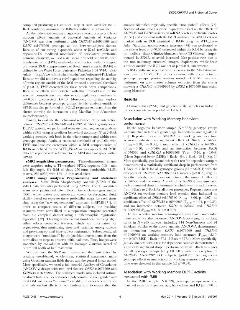
Relationship between DLPFC activity and behavioral
performance at 2-Back. Regression analysis in SPM8 demon-
strated a negative correlation between activity in DLPFC and
accuracy (%) at 2-Back in the CHRNA5-GA/DRD2-GT group (BA
46: x -56 y 26 z 30; K = 39; corrected pFWE = 0.02; Fig. 3). Also,
exploratory analyses which did not survive correction for multiple
comparisons suggested a negative correlation in the CHRNA5-AA/
DRD2-GT group (x -42 y 30 z 22; K = 15; p = 0.002 uncorrected),
and a positive correlation in the CHRNA5-GG/DRD2-GT group
(x -36 y 56 z 30; K = 20; p = 0.002 uncorrected) (See Fig. S1).
Association with DLPFC gray matter volume measuredwith sMRI
In the sMRI sample (211 subjects), genotype groups were also
matched in terms of gender, age, handedness and IQ (all p.0.1).
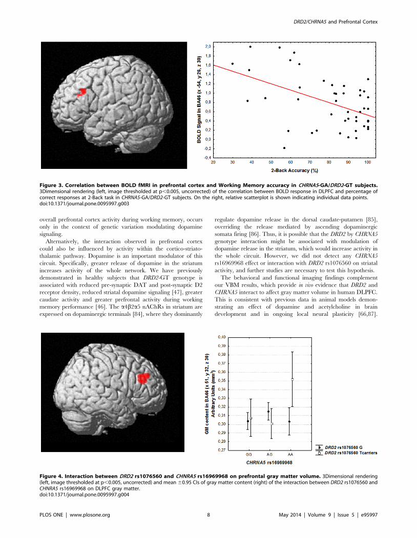
Genotype main effects and interaction. There was no
statistically significant main effect of CHRNA5 rs16969968 or of
DRD2 rs1076560 genotype in the DLPFC ROI. However, an
interaction between CHRNA5 and DRD2 genotypes was found in
right DLPFC (BA 46: x 51, y 32, z 30, k = 321, Z = 3.96, p = 0.006
cluster-level corrected; Fig. 4). Post hoc analysis of gray matter
volume extracted from the interaction cluster indicated that
CHRNA5-AA/DRD2-GT subjects have greater DLPFC gray
matter volume compared to all other genotype groups (all p,
0.02; Fig. 4).
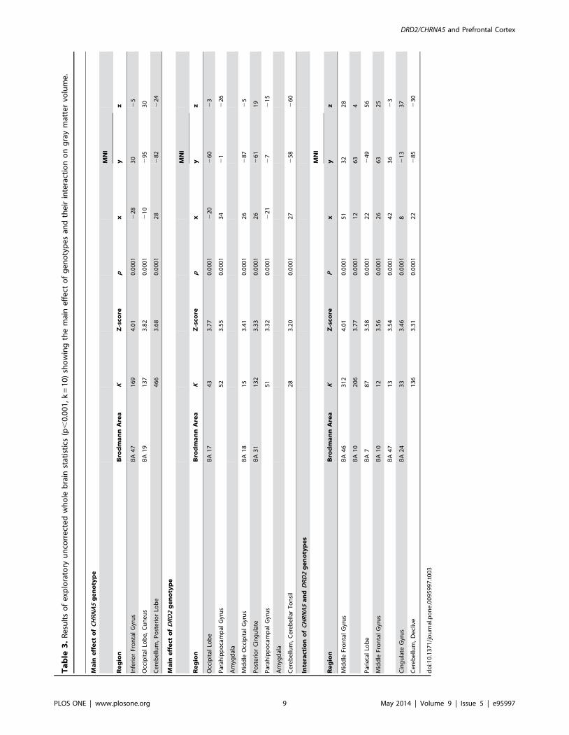
Results of the uncorrected exploratory whole-brain analyses are
reported in Table 3.
Discussion
The results of the present study demonstrate that variants in two
genes implicated in dopamine and acetylcholine signaling interact
to modulate the biology and physiology of the prefrontal cortex
during working memory. More specifically, the interaction
between CHNRA5 rs16969968 and DRD2 rs1076560 genotypes
differentially predicted cognitive behavior with increasing working
memory load, in that CHRNA5-AA/DRD2-GT subjects have
better behavioral performance. In addition, we found that the
effect of CHRNA5 rs16969968 in dorsolateral prefrontal activity at
2-Back is only evident in the context of DRD2 rs1076560
genotype, such that CHRNA5 demonstrates an inverted U shaped
prefrontal response in DRD2-GT subjects (see below).
As a further demonstration of the functional effects of these
polymorphisms, CHRNA5 rs16969968 and DRD2 rs1076560 also
interacted on gray matter volume of the dorsolateral prefrontal
cortex (DLPFC). Once again, the effect of CHNRA5 was mostly
evident in the context of DRD2-GT genotype.
Our behavioral findings during working memory as elicited by
the N-Back task are consistent with a previous report by Markett et
al. (2010) in healthy subjects (N = 101), showing an interaction
between a functional SNP in CHRNA4 (rs1044396) and a
haplotype block covering three SNPs in DRD2 (rs1800497,
rs6277, rs2283265) on working memory capacity [75]. As in our
sample, this effect only became apparent at greater working
memory load, suggesting that the CHRNA5 by DRD2 interaction
affects the efficiency by which relevant information is encoded
during the trial-wise updating of working memory items. Unlike all
other genotype groups, CHRNA5-AA/DRD2-GT subjects showed
no statistically significant reduction in behavioral performance
with increasing working memory load, leading to the speculation
that the prefrontal neuronal signal-to-noise affected by the geneti-
cally determined balance of cholinergic and dopaminergic
Figure 2. Interaction between DRD2 rs1076560 and CHNRA5 rs16969968 on prefrontal physiology at 2-Back. 3Dimensional rendering(left, image thresholded at p,0.005, uncorrected) and mean 60.95 CIs of BOLD response (right) of the interaction between DRD2 rs1076560 andCHNRA5 rs16969968 on working memory DLPFC activity.doi:10.1371/journal.pone.0095997.g002
DRD2/CHRNA5 and Prefrontal Cortex
PLOS ONE | www.plosone.org 6 May 2014 | Volume 9 | Issue 5 | e95997

signaling is increased in CHRNA5-AA/DRD2-GT subjects
allowing greater performance with increasing working memory
load. Moreover, this interaction is similar to the reciprocal rescue
of minor allele risk found in association with other biological
phenotypes [76]. Of note, our results suggest that the pattern of
genotype effect on working memory load accuracy was not
moderated by reaction time.
Our functional imaging data also indicate that the interaction
between CHRNA5 rs16969968 and DRD2 rs1076560 genotypes
differentially predicted the efficiency of the prefrontal cortex at 2-
Back condition. Earlier fMRI studies found that genetic variation
in dopamine signaling in the prefrontal cortex affects the efficiency
or signal-to-noise ratio of the physiological response during the N-
Back following an inverted U-shaped response function [77279].
In the present study, we found that only within DRD2- GT
genotype, CHRNA5 demonstrates an inverted U shaped prefrontal
response at 2-Back working memory task, suggesting that CHRNA5
rs16969968 further affects the signal-to-noise ratio of prefrontal
cortex in subjects with greater dopamine signaling (i.e. GT
subjects) [45247]. More specifically, within the context of DRD2-
GT genotype: CHRNA5-AA subjects are more efficient, because
the combination of behavioral data and imaging results suggests
that they have reduced activity for greater behavioral perfor-
mance; CHRNA5-AG subjects are less efficient, because they show
greater activity for reduced behavioral performance, which is also
consistent with the negative relationship between BOLD response
and behavioral accuracy; and CHRNA5-GG have reduced
engagement of prefrontal resources to the task at hand for reduced
behavioral performance, as also suggested by the positive
relationship between prefrontal activity and behavioral perfor-
mance.
Thus, the DRD2 GT subjects show an overdominance effect for
the CHRNA5 genotype with heterozygous revealing greater/more
inefficient prefrontal activity compared with individuals homozy-
gous for either allele. While this effect in genetics would be
regarded as a ‘‘heterozygous advantage’’, at the brain imaging
level it may actually reflect an inefficient prefrontal activity during
information processing. This finding is of particular interest since
central dopamine-acetylcholine imbalance in synaptic plasticity is
responsible for cognitive deficits in Parkinson disease [80] and
likely in psychosis, as suggested by heavy smoking in patients with
schizophrenia [81].
There may be several and complex molecular/neuronal
mechanisms in human DLPFC which are responsible for the
interaction we have measured in vivo with BOLD fMRI, and
further work on molecular and cellular models is warranted. Still,
previous work examining cortical anatomy and physiology allows
us to speculate on the biology underlying our observations. D2
receptors in prefrontal cortex are mainly found pre-synaptically on
dopamine terminals [82], modulating dopamine release and D2S
autoreceptors are relatively more abundant in the prefrontal
cortex compared to D2Ls [45]. Thus, DRD2-GT genotype
associated with reduced D2S may increase dopamine levels in
the prefrontal cortex, and in turn increase its activity. nAchRs
containing the a5 subunit in prefrontal cortex are mainly
expressed on soma and axon of LVI pyramidal neurons [29,83],
where they are responsible for strong activation of the neuronal
population of this layer [29]. Similarly to DRD2 genotype, the
CHRNA5-A allele which is associated with reduced total CHRNA5
mRNA expression in prefrontal cortex tissue and signaling in vitro,
may alter the neuronal activation of LVI pyramidal neurons.
However, as mentioned above, the effect of CHRNA5 rs16969968
is only manifest in the context of DRD2 rs1076560 GT genotype,
suggesting that the physiological relevance of this SNP, in terms of
Ta
ble
2.
Re
sult
so
fe
xplo
rato
ryu
nco
rre
cte
dw
ho
leb
rain
stat
isti
cs(p
,0
.00
1,
k=
10
)sh
ow
ing
the
mai
ne
ffe
cto
fD
RD
2an
dit
sin
tera
ctio
nw
ith
CH
RN
A5
on
bra
inp
hys
iolo
gy
at2
-B
ack
WM
Tas
k.
Ma
ine
ffe
cto
fD
RD
2g
en
oty
pe
MN
I
Re
gio
nB
rod
ma
nn
Are
ak
Z-s
core
px
yz
Mid
dle
Te
mp
ora
lG
yru
sB
A3
91
93
.59
0.0
00
14
02
54
22
Mid
dle
Fro
nta
lG
yru
sB
A1
01
53
.35
0.0
00
13
25
66
Inte
ract
ion
of
CH
RN
A5
an
dD
RD
2g
en
oty
pe
s
MN
I
Re
gio
nB
rod
ma
nn
Are
aK
Z-s
core
Px
yz
Infe
rio
rP
arie
tal
Lob
eB
A4
09
84
.17
0.0
00
12
36
24
65
0
Mid
dle
Fro
nta
lG
yru
sB
A1
02
93
.58
0.0
00
13
05
20
Mid
dle
Fro
nta
lG
yru
sB
A4
62
33
.58
0.0
00
12
46
30
28
Mid
dle
Fro
nta
lG
yru
sB
A6
21
3.5
10
.00
01
22
01
06
4
do
i:10
.13
71
/jo
urn
al.p
on
e.0
09
59
97
.t0
02
DRD2/CHRNA5 and Prefrontal Cortex
PLOS ONE | www.plosone.org 7 May 2014 | Volume 9 | Issue 5 | e95997
overall prefrontal cortex activity during working memory, occurs
only in the context of genetic variation modulating dopamine
signaling.
Alternatively, the interaction observed in prefrontal cortex
could also be influenced by activity within the cortico-striato-
thalamic pathway. Dopamine is an important modulator of this
circuit. Specifically, greater release of dopamine in the striatum
increases activity of the whole network. We have previously
demonstrated in healthy subjects that DRD2-GT genotype is
associated with reduced pre-synaptic DAT and post-synaptic D2
receptor density, reduced striatal dopamine signaling [47], greater
caudate activity and greater prefrontal activity during working
memory performance [46]. The a4b2a5 nAChRs in striatum are
expressed on dopaminergic terminals [84], where they dominantly
regulate dopamine release in the dorsal caudate-putamen [85],
overriding the release mediated by ascending dopaminergic
somata firing [86]. Thus, it is possible that the DRD2 by CHRNA5
genotype interaction might be associated with modulation of
dopamine release in the striatum, which would increase activity in
the whole circuit. However, we did not detect any CHRNA5
rs16969968 effect or interaction with DRD2 rs1076560 on striatal
activity, and further studies are necessary to test this hypothesis.
The behavioral and functional imaging findings complement
our VBM results, which provide in vivo evidence that DRD2 and
CHRNA5 interact to affect gray matter volume in human DLPFC.
This is consistent with previous data in animal models demon-
strating an effect of dopamine and acetylcholine in brain
development and in ongoing local neural plasticity [66,87].
Figure 3. Correlation between BOLD fMRI in prefrontal cortex and Working Memory accuracy in CHRNA5-GA/DRD2-GT subjects.3Dimensional rendering (left, image thresholded at p,0.005, uncorrected) of the correlation between BOLD response in DLPFC and percentage ofcorrect responses at 2-Back task in CHRNA5-GA/DRD2-GT subjects. On the right, relative scatterplot is shown indicating individual data points.doi:10.1371/journal.pone.0095997.g003
Figure 4. Interaction between DRD2 rs1076560 and CHNRA5 rs16969968 on prefrontal gray matter volume. 3Dimensional rendering(left, image thresholded at p,0.005, uncorrected) and mean 60.95 CIs of gray matter content (right) of the interaction between DRD2 rs1076560 andCHNRA5 rs16969968 on DLPFC gray matter.doi:10.1371/journal.pone.0095997.g004
DRD2/CHRNA5 and Prefrontal Cortex
PLOS ONE | www.plosone.org 8 May 2014 | Volume 9 | Issue 5 | e95997
Ta
ble
3.
Re
sult
so
fe
xplo
rato
ryu
nco
rre
cte
dw
ho
leb
rain
stat
isti
cs(p
,0
.00
1,
k=
10
)sh
ow
ing
the
mai
ne
ffe
cto
fg
en
oty
pe
san
dth
eir
inte
ract
ion
on
gra
ym
atte
rvo
lum
e.
Ma
ine
ffe
cto
fC
HR
NA
5g
en
oty
pe
MN
I
Re
gio
nB
rod
ma
nn
Are
aK
Z-s
core
px
yz
Infe
rio
rFr
on
tal
Gyr
us
BA
47
16
94
.01
0.0
00
12
28
30
25
Occ
ipit
alLo
be
,C
un
eu
sB
A1
91
37
3.8
20
.00
01
21
02
95
30
Ce
reb
ellu
m,
Po
ste
rio
rLo
be
46
63
.68
0.0
00
12
82
82
22
4
Ma
ine
ffe
cto
fD
RD
2g
en
oty
pe
MN
I
Re
gio
nB
rod
ma
nn
Are
aK
Z-s
core
px
yz
Occ
ipit
alLo
be
BA
17
43
3.7
70
.00
01
22
02
60
23
Par
ahip
po
cam
pal
Gyr
us
52
3.5
50
.00
01
34
21
22
6
Am
ygd
ala
Mid
dle
Occ
ipit
alG
yru
sB
A1
81
53
.41
0.0
00
12
62
87
25
Po
ste
rio
rC
ing
ula
teB
A3
11
32
3.3
30
.00
01
26
26
11
9
Par
ahip
po
cam
pal
Gyr
us
51
3.3
20
.00
01
22
12
72
15
Am
ygd
ala
Ce
reb
ellu
m,
Ce
reb
ella
rT
on
sil
28
3.2
00
.00
01
27
25
82
60
Inte
ract
ion
of
CH
RN
A5
an
dD
RD
2g
en
oty
pe
s
MN
I
Re
gio
nB
rod
ma
nn
Are
aK
Z-s
core
Px
yz
Mid
dle
Fro
nta
lG
yru
sB
A4
63
12
4.0
10
.00
01
51
32
28
BA
10
20
63
.77
0.0
00
11
26
34
Par
ieta
lLo
be
BA
78
73
.58
0.0
00
12
22
49
56
Mid
dle
Fro
nta
lG
yru
sB
A1
01
23
.56
0.0
00
12
66
32
5
BA
47
13
3.5
40
.00
01
42
36
23
Cin
gu
late
Gyr
us
BA
24
33
3.4
60
.00
01
82
13
37
Ce
reb
ellu
m,
De
cliv
e1
36
3.3
10
.00
01
22
28
52
30
do
i:10
.13
71
/jo
urn
al.p
on
e.0
09
59
97
.t0
03
DRD2/CHRNA5 and Prefrontal Cortex
PLOS ONE | www.plosone.org 9 May 2014 | Volume 9 | Issue 5 | e95997

Increasing synaptic dopamine in developing brains through
prenatal cocaine exposure leads to specific neurodevelopmental
alterations including abnormal dendritic growth and abnormal
arborization of pyramidal cells that persist postnatally [88].
Conversely, neonatal dopamine denervation in rat produces
permanent differential changes in prefrontal cortex dendritic
morphology, i.e. atrophy of proximal apical and basilar dendrites
[89]. Cholinergic inputs to the cortex also appear early during
brain development and are widespread in rat by the third week of
post-natal life [87,90], likely influencing the normal morphological
development of pyramidal neurons. Two elegant studies have
indicated that direct nicotinic stimulation can modulate growth or
retraction of neurites in cultured neurons [91,92]. More recently,
Bailey et al (2012) have demonstrated that a5 nAChRs underlie
the neurodevelopmental peak in the nicotinic excitation of murine
medial prefrontal cortex LVI neurons that occurs during the third
week of postnatal life, and that it is likely to influence a specific
ontogenetic retraction of apical dendrites in LVI pyramidal
neurons. However, it is possible that a5 nAChRs on dopaminergic
neurons [33,34] may also influence the morphology of prefrontal
cortex neurons.
Some potential limitations of the present study should be
addressed. First, the study includes assumptions that we expect
protein expression to correspond to mRNA levels, as we did not
directly measure dopamine and acetylcholine receptor levels in our
cohort, we can only speculate about the molecular mechanisms
relying on previous studies demonstrating functional consequences
of the chosen SNPs. Hence, it remains to be determined whether
and how genetic variation in dopaminergic and cholinergic
signaling to cortical signal-to-noise may directly affect differential
engagement of DLPFC at a cellular level, especially in humans.
Second, our neuropsychological findings are based on a relatively
small group size for the CHRNA5 AA/DRD2 GT genotype
(N = 10). Although computation of the maximal effect size d and
achieved power (respectively, 1.1 and 0.82) support their statistical
robustness, replication of our results is necessary. Furthermore,
Levene’s test indicates homogeneity of variance of behavioral
performance (difference between 2-Back and 1-Back accuracy)
across DRD2/CHRNA5 genotype groups (MS Effect = 106.33; MS
Error = 85.30; F = 1.24; p = 0.28) supporting that our results were
not influenced by unequal population variances. Another caveat of
our study is that our neuropsychological and fMRI findings could
be affected by tobacco use of subjects. Rs16969968 has been
associated with nicotine dependence [55], although it does not
alter per se sensitivity of a4b2a5 nAChRs to nicotine [59].
However, the additional analyses we performed including smoking
status as covariate allowed us to exclude it as confounding factor.
The present study advances our understanding of the in vivo
interactions between dopamine and acetylcholine signaling in the
prefrontal cortex, specifically through the DRD2 and CHRNA5
receptors. Our observations of these gene-gene interactions on
neurophysiology and cognition begin to build a more solid
foundation for explaining the neurobiology underlying complex
human behaviors and lend insight into disease susceptibility.
Furthermore, our results have relevant potential implications for
the therapeutic approach of various neurological and psychiatric
disorders in which altered cholinergic transmission potentially
contributes to cognitive deficits, such as those observed in
schizophrenia.
Supporting Information
Figure S1 Correlation between BOLD fMRI in prefron-tal cortex and Working Memory accuracy in CHRNA5-AA/DRD2-GT subjects (S1a) and in CHRNA5-GG/DRD2-GT subjects (S1b).
(DOCX)
Table S1 Behavioral data (mean ± SD) at the 2-Backtask for each genotype group.
(DOCX)
Acknowledgments
This study was in part supported by a grant from the US National
Institutes of Health, General Medical Sciences, U01 GM092655.
Author Contributions
Conceived and designed the experiments: AB ADG RMS WS. Performed
the experiments: RMS LF BG PT TQ AR GC TP. Analyzed the data:
ADG AB RMS AT LF EDA PS GB. Contributed reagents/materials/
analysis tools: RMS RM WS AB. Wrote the paper: ADG RMS AB WS.
References
1. Ando J, Ono Y, Wright MJ (2001) Genetic structure of spatial and verbal
working memory. Behav Genet 31: 615–624.
2. Luciano M, Wright M, Smith GA, Geffen GM, Geffen LB, et al. (2001) Geneticcovariance among measures of information processing speed, working memory,
and IQ. Behav Genet 31: 581–592.
3. Polderman TJ, Gosso MF, Posthuma D, Van Beijsterveldt TC, Heutink P, et al.
(2006) A longitudinal twin study on IQ, executive functioning, and attention
problems during childhood and early adolescence. Acta Neurol Belg 106: 191–207.
4. Goldman-Rakic PS (1995) Architecture of the prefrontal cortex and the centralexecutive. Ann N Y Acad Sci 769: 71–83.
5. Wang Y, Markram H, Goodman PH, Berger TK, Ma J, et al. (2006)
Heterogeneity in the pyramidal network of the medial prefrontal cortex. NatNeurosci 9: 534–542.
6. Blokland GA, McMahon KL, Thompson PM, Martin NG, de Zubicaray GI, et
al. (2011) Heritability of working memory brain activation. J Neurosci 31:10882–10890.
7. Callicott JH, Egan MF, Mattay VS, Bertolino A, Bone AD, et al. (2003)
Abnormal fMRI response of the dorsolateral prefrontal cortex in cognitivelyintact siblings of patients with schizophrenia. Am J Psychiatry 160: 709–719.
8. Robbins TW, Roberts AC (2007) Differential regulation of fronto-executivefunction by the monoamines and acetylcholine. Cereb Cortex 17 Suppl 1: i151–
160.
9. Blasi G, Napolitano F, Ursini G, Taurisano P, Romano R, et al. (2011) DRD2/AKT1 interaction on D2 c-AMP independent signaling, attentional processing,
and response to olanzapine treatment in schizophrenia. Proc Natl Acad Sci U S A
108: 1158–1163.
10. Papaleo F, Burdick MC, Callicott JH, Weinberger DR (2013) Epistatic
interaction between COMT and DTNBP1 modulates prefrontal function in
mice and in humans. Mol Psychiatry.
11. Tan HY, Chen Q, Sust S, Buckholtz JW, Meyers JD, et al. (2007) Epistasis
between catechol-O-methyltransferase and type II metabotropic glutamate
receptor 3 genes on working memory brain function. Proc Natl Acad Sci U S A
104: 12536–12541.
12. Tritsch NX, Sabatini BL (2012) Dopaminergic modulation of synaptic
transmission in cortex and striatum. Neuron 76: 33–50.
13. Wang M, Vijayraghavan S, Goldman-Rakic PS (2004) Selective D2 receptor
actions on the functional circuitry of working memory. Science 303: 853–856.
14. Seamans JK, Yang CR (2004) The principal features and mechanisms of
dopamine modulation in the prefrontal cortex. Prog Neurobiol 74: 1–58.
15. Zhou X, Qi XL, Douglas K, Palaninathan K, Kang HS, et al. (2011)
Cholinergic modulation of working memory activity in primate prefrontal
cortex. J Neurophysiol 106: 2180–2188.
16. Croxson PL, Kyriazis DA, Baxter MG (2011) Cholinergic modulation of a
specific memory function of prefrontal cortex. Nat Neurosci 14: 1510–1512.
17. Chudasama Y, Dalley JW, Nathwani F, Bouger P, Robbins TW (2004)
Cholinergic modulation of visual attention and working memory: dissociable
effects of basal forebrain 192-IgG-saporin lesions and intraprefrontal infusions of
scopolamine. Learn Mem 11: 78–86.
18. Granon S, Poucet B, Thinus-Blanc C, Changeux JP, Vidal C (1995) Nicotinic
and muscarinic receptors in the rat prefrontal cortex: differential roles in
working memory, response selection and effortful processing. Psychopharma-
cology (Berl) 119: 139–144.
DRD2/CHRNA5 and Prefrontal Cortex
PLOS ONE | www.plosone.org 10 May 2014 | Volume 9 | Issue 5 | e95997

19. Levin ED, McClernon FJ, Rezvani AH (2006) Nicotinic effects on cognitive
function: behavioral characterization, pharmacological specification, and
anatomic localization. Psychopharmacology (Berl) 184: 523–539.
20. Chan WK, Wong PT, Sheu FS (2007) Frontal cortical alpha7 and alpha4beta2
nicotinic acetylcholine receptors in working and reference memory. Neuro-
pharmacology 52: 1641–1649.
21. Levin ED, Simon BB (1998) Nicotinic acetylcholine involvement in cognitive
function in animals. Psychopharmacology (Berl) 138: 217–230.
22. Levin ED, Rose JE, Abood L (1995) Effects of nicotinic dimethylaminoethyl
esters on working memory performance of rats in the radial-arm maze.
Pharmacol Biochem Behav 51: 369–373.
23. Woodruff-Pak DS (2003) Mecamylamine reversal by nicotine and by a partial
alpha7 nicotinic acetylcholine receptor agonist (GTS-21) in rabbits tested with
delay eyeblink classical conditioning. Behav Brain Res 143: 159–167.
24. Spinelli S, Ballard T, Feldon J, Higgins GA, Pryce CR (2006) Enhancing effects
of nicotine and impairing effects of scopolamine on distinct aspects of
performance in computerized attention and working memory tasks in marmoset
monkeys. Neuropharmacology 51: 238–250.
25. Loughead J, Ray R, Wileyto EP, Ruparel K, Sanborn P, et al. (2010) Effects of
the alpha4beta2 partial agonist varenicline on brain activity and working
memory in abstinent smokers. Biol Psychiatry 67: 715–721.
26. Howe MN, Price IR (2001) Effects of transdermal nicotine on learning, memory,
verbal fluency, concentration, and general health in a healthy sample at risk for
dementia. Int Psychogeriatr 13: 465–475.
27. Gotti C, Zoli M, Clementi F (2006) Brain nicotinic acetylcholine receptors:
native subtypes and their relevance. Trends Pharmacol Sci 27: 482–491.
28. Sihver W, Gillberg PG, Nordberg A (1998) Laminar distribution of nicotinic
receptor subtypes in human cerebral cortex as determined by [3H](-)nicotine,
[3H]cytisine and [3H]epibatidine in vitro autoradiography. Neuroscience 85:
1121–1133.
29. Poorthuis RB, Bloem B, Schak B, Wester J, de Kock CP, et al. (2013) Layer-
specific modulation of the prefrontal cortex by nicotinic acetylcholine receptors.
Cereb Cortex 23: 148–161.
30. Gotti C, Clementi F, Fornari A, Gaimarri A, Guiducci S, et al. (2009) Structural
and functional diversity of native brain neuronal nicotinic receptors. Biochem
Pharmacol 78: 703–711.
31. Ramirez-Latorre J, Yu CR, Qu X, Perin F, Karlin A, et al. (1996) Functional
contributions of alpha5 subunit to neuronal acetylcholine receptor channels.
Nature 380: 347–351.
32. Marks MJ, Pauly JR, Gross SD, Deneris ES, Hermans-Borgmeyer I, et al. (1992)
Nicotine binding and nicotinic receptor subunit RNA after chronic nicotine
treatment. J Neurosci 12: 2765–2784.
33. Salas R, Orr-Urtreger A, Broide RS, Beaudet A, Paylor R, et al. (2003) The
nicotinic acetylcholine receptor subunit alpha 5 mediates short-term effects of
nicotine in vivo. Mol Pharmacol 63: 1059–1066.
34. Wada E, McKinnon D, Heinemann S, Patrick J, Swanson LW (1990) The
distribution of mRNA encoded by a new member of the neuronal nicotinic
acetylcholine receptor gene family (alpha 5) in the rat central nervous system.
Brain Res 526: 45–53.
35. Winzer-Serhan UH, Leslie FM (2005) Expression of alpha5 nicotinic
acetylcholine receptor subunit mRNA during hippocampal and cortical
development. J Comp Neurol 481: 19–30.
36. Bailey CD, De Biasi M, Fletcher PJ, Lambe EK (2010) The nicotinic
acetylcholine receptor alpha5 subunit plays a key role in attention circuitry
and accuracy. J Neurosci 30: 9241–9252.
37. Bailey CD, Alves NC, Nashmi R, De Biasi M, Lambe EK (2012) Nicotinic
alpha5 subunits drive developmental changes in the activation and morphology
of prefrontal cortex layer VI neurons. Biol Psychiatry 71: 120–128.
38. Levin ED, Rose JE (1991) Nicotinic and muscarinic interactions and choice
accuracy in the radial-arm maze. Brain Res Bull 27: 125–128.
39. Levin ED, Castonguay M, Ellison GD (1987) Effects of the nicotinic receptor
blocker mecamylamine on radial-arm maze performance in rats. Behav Neural
Biol 48: 206–212.
40. Levin ED, McGurk SR, South D, Butcher LL (1989) Effects of combined
muscarinic and nicotinic blockade on choice accuracy in the radial-arm maze.
Behav Neural Biol 51: 270–277.
41. McGurk SR, Levin ED, Butcher LL (1989) Radial-arm maze performance in
rats is impaired by a combination of nicotinic-cholinergic and D2 dopaminergic
antagonist drugs. Psychopharmacology (Berl) 99: 371–373.
42. Levin ED, McGurk SR, Rose JE, Butcher LL (1989) Reversal of a
mecamylamine-induced cognitive deficit with the D2 agonist, LY 171555.
Pharmacol Biochem Behav 33: 919–922.
43. Usiello A, Baik JH, Rouge-Pont F, Picetti R, Dierich A, et al. (2000) Distinct
functions of the two isoforms of dopamine D2 receptors. Nature 408: 199–203.
44. Centonze D, Gubellini P, Usiello A, Rossi S, Tscherter A, et al. (2004)
Differential contribution of dopamine D2S and D2L receptors in the modulation
of glutamate and GABA transmission in the striatum. Neuroscience 129: 157–
166.
45. Zhang Y, Bertolino A, Fazio L, Blasi G, Rampino A, et al. (2007)
Polymorphisms in human dopamine D2 receptor gene affect gene expression,
splicing, and neuronal activity during working memory. Proc Natl Acad Sci U S A
104: 20552–20557.
46. Bertolino A, Fazio L, Caforio G, Blasi G, Rampino A, et al. (2009) Functional
variants of the dopamine receptor D2 gene modulate prefronto-striatal
phenotypes in schizophrenia. Brain 132: 417–425.
47. Bertolino A, Taurisano P, Pisciotta NM, Blasi G, Fazio L, et al. (2010)
Genetically determined measures of striatal D2 signaling predict prefrontal
activity during working memory performance. PLoS One 5: e9348.
48. Sasabe T, Furukawa A, Matsusita S, Higuchi S, Ishiura S (2007) Association
analysis of the dopamine receptor D2 (DRD2) SNP rs1076560 in alcoholic
patients. Neurosci Lett 412: 139–142.
49. Moyer RA, Wang D, Papp AC, Smith RM, Duque L, et al. (2011) Intronic
polymorphisms affecting alternative splicing of human dopamine D2 receptor
are associated with cocaine abuse. Neuropsychopharmacology 36: 753–762.
50. Clarke TK, Weiss AR, Ferarro TN, Kampman KM, Dackis CA, et al. (2014)
The Dopamine Receptor D2 (DRD2) SNP rs1076560 is Associated with Opioid
Addiction. Ann Hum Genet 78: 33–39.
51. Voisey J, Swagell CD, Hughes IP, van Daal A, Noble EP, et al. (2012) A DRD2
and ANKK1 haplotype is associated with nicotine dependence. Psychiatry Res
196: 285–289.
52. Wei J, Chu C, Wang Y, Yang Y, Wang Q, et al. (2012) Association study of 45
candidate genes in nicotine dependence in Han Chinese. Addict Behav 37: 622–
626.
53. Connor JP, Young RM, Lawford BR, Saunders JB, Ritchie TL, et al. (2007)
Heavy nicotine and alcohol use in alcohol dependence is associated with D2
dopamine receptor (DRD2) polymorphism. Addict Behav 32: 310–319.
54. Wang TY, Lee SY, Chen SL, Huang SY, Chang YH, et al. (2013) Association
between DRD2, 5-HTTLPR, and ALDH2 genes and specific personality traits
in alcohol- and opiate-dependent patients. Behav Brain Res 250: 285–292.
55. Bierut LJ, Stitzel JA, Wang JC, Hinrichs AL, Grucza RA, et al. (2008) Variants
in nicotinic receptors and risk for nicotine dependence. Am J Psychiatry 165:
1163–1171.
56. Saccone SF, Hinrichs AL, Saccone NL, Chase GA, Konvicka K, et al. (2007)
Cholinergic nicotinic receptor genes implicated in a nicotine dependence
association study targeting 348 candidate genes with 3713 SNPs. Hum Mol
Genet 16: 36–49.
57. Smith RM, Alachkar H, Papp AC, Wang D, Mash DC, et al. (2011) Nicotinic
alpha5 receptor subunit mRNA expression is associated with distant 59 upstream
polymorphisms. Eur J Hum Genet 19: 76–83.
58. Wang JC, Spiegel N, Bertelsen S, Le N, McKenna N, et al. (2013) Cis-
Regulatory Variants Affect CHRNA5 mRNA Expression in Populations of
African and European Ancestry. PLoS One 8: e80204.
59. Kuryatov A, Berrettini W, Lindstrom J (2011) Acetylcholine receptor (AChR)
alpha5 subunit variant associated with risk for nicotine dependence and lung
cancer reduces (alpha4beta2)(2)alpha5 AChR function. Mol Pharmacol 79: 119–
125.
60. Bierut LJ, Madden PA, Breslau N, Johnson EO, Hatsukami D, et al. (2007)
Novel genes identified in a high-density genome wide association study for
nicotine dependence. Hum Mol Genet 16: 24–35.
61. Saccone NL, Saccone SF, Hinrichs AL, Stitzel JA, Duan W, et al. (2009)
Multiple distinct risk loci for nicotine dependence identified by dense coverage of
the complete family of nicotinic receptor subunit (CHRN) genes. Am J Med
Genet B Neuropsychiatr Genet 150B: 453–466.
62. Winterer G, Mittelstrass K, Giegling I, Lamina C, Fehr C, et al. (2010) Risk
gene variants for nicotine dependence in the CHRNA5-CHRNA3-CHRNB4
cluster are associated with cognitive performance. Am J Med
Genet B Neuropsychiatr Genet 153B: 1448–1458.
63. Hong LE, Yang X, Wonodi I, Hodgkinson CA, Goldman D, et al. (2011) A
CHRNA5 allele related to nicotine addiction and schizophrenia. Genes Brain
Behav 10: 530–535.
64. Jackson KJ, Fanous AH, Chen J, Kendler KS, Chen X (2013) Variants in the
15q25 gene cluster are associated with risk for schizophrenia and bipolar
disorder. Psychiatr Genet 23: 20–28.
65. Falcone M, Smith RM, Chenoweth MJ, Kumar Bhattacharjee A, Kelsoe JR, et
al. (2013) Neuroimaging in psychiatric pharmacogenetics research: the promise
and pitfalls. Neuropsychopharmacology 38: 2327–2337.
66. Lambe EK, Krimer LS, Goldman-Rakic PS (2000) Differential postnatal
development of catecholamine and serotonin inputs to identified neurons in
prefrontal cortex of rhesus monkey. J Neurosci 20: 8780–8787.
67. First M, Gibbon M, Spitzer R, Williams J (1996) Guide for the structured clinical
interview for DSM-IV axis I disorders-Research version. New York: Biometrics
Research.
68. Oldfield RC (1971) The assessment and analysis of handedness: the Edinburgh
inventory. Neuropsychologia 9: 97–113.
69. Hong LE, Hodgkinson CA, Yang Y, Sampath H, Ross TJ, et al. (2010) A
genetically modulated, intrinsic cingulate circuit supports human nicotine
addiction. Proc Natl Acad Sci U S A 107: 13509–13514.
70. Tan HY, Nicodemus KK, Chen Q, Li Z, Brooke JK, et al. (2008) Genetic
variation in AKT1 is linked to dopamine-associated prefrontal cortical structure
and function in humans. J Clin Invest 118: 2200–2208.
71. Ashburner J (2007) A fast diffeomorphic image registration algorithm. Neuro-
image 38: 95–113.
72. Ashburner J, Friston KJ (2005) Unified segmentation. Neuroimage 26: 839–851.
73. Ashburner J, Friston KJ (2000) Voxel-based morphometry—the methods.
Neuroimage 11: 805–821.
DRD2/CHRNA5 and Prefrontal Cortex
PLOS ONE | www.plosone.org 11 May 2014 | Volume 9 | Issue 5 | e95997
74. Hayasaka S, Phan KL, Liberzon I, Worsley KJ, Nichols TE (2004)
Nonstationary cluster-size inference with random field and permutationmethods. Neuroimage 22: 676–687.
75. Markett SA, Montag C, Reuter M (2010) The association between dopamine
DRD2 polymorphisms and working memory capacity is modulated by afunctional polymorphism on the nicotinic receptor gene CHRNA4. J Cogn
Neurosci 22: 1944–1954.76. Sullivan D, Pinsonneault JK, Papp AC, Zhu H, Lemeshow S, et al. (2013)
Dopamine transporter DAT and receptor DRD2 variants affect risk of lethal
cocaine abuse: a gene-gene-environment interaction. Transl Psychiatry 3: e222.77. Egan MF, Goldberg TE, Kolachana BS, Callicott JH, Mazzanti CM, et al.
(2001) Effect of COMT Val108/158 Met genotype on frontal lobe function andrisk for schizophrenia. Proc Natl Acad Sci U S A 98: 6917–6922.
78. Mattay VS, Goldberg TE, Fera F, Hariri AR, Tessitore A, et al. (2003) CatecholO-methyltransferase val158-met genotype and individual variation in the brain
response to amphetamine. Proc Natl Acad Sci U S A 100: 6186–6191.
79. Mattay VS, Tessitore A, Callicott JH, Bertolino A, Goldberg TE, et al. (2002)Dopaminergic modulation of cortical function in patients with Parkinson’s
disease. Ann Neurol 51: 156–164.80. Calabresi P, Picconi B, Parnetti L, Di Filippo M (2006) A convergent model for
cognitive dysfunctions in Parkinson’s disease: the critical dopamine-acetylcholine
synaptic balance. Lancet Neurol 5: 974–983.81. Kuehn BM (2006) Link between smoking and mental illness may lead to
treatments. JAMA 295: 483–484.82. Pickel VM, Chan J, Nirenberg MJ (2002) Region-specific targeting of dopamine
D2-receptors and somatodendritic vesicular monoamine transporter 2 (VMAT2)within ventral tegmental area subdivisions. Synapse 45: 113–124.
83. Kassam SM, Herman PM, Goodfellow NM, Alves NC, Lambe EK (2008)
Developmental excitation of corticothalamic neurons by nicotinic acetylcholinereceptors. J Neurosci 28: 8756–8764.
84. Zoli M, Moretti M, Zanardi A, McIntosh JM, Clementi F, et al. (2002)
Identification of the nicotinic receptor subtypes expressed on dopaminergic
terminals in the rat striatum. J Neurosci 22: 8785–8789.
85. Exley R, McIntosh JM, Marks MJ, Maskos U, Cragg SJ (2012) Striatal alpha5
nicotinic receptor subunit regulates dopamine transmission in dorsal striatum.
J Neurosci 32: 2352–2356.
86. Threlfell S, Lalic T, Platt NJ, Jennings KA, Deisseroth K, et al. (2012) Striatal
dopamine release is triggered by synchronized activity in cholinergic
interneurons. Neuron 75: 58–64.
87. Mechawar N, Descarries L (2001) The cholinergic innervation develops early
and rapidly in the rat cerebral cortex: a quantitative immunocytochemical study.
Neuroscience 108: 555–567.
88. Levitt P, Harvey JA, Friedman E, Simansky K, Murphy EH (1997) New
evidence for neurotransmitter influences on brain development. Trends
Neurosci 20: 269–274.
89. Sherren N, Pappas BA (2005) Selective acetylcholine and dopamine lesions in
neonatal rats produce distinct patterns of cortical dendritic atrophy in
adulthood. Neuroscience 136: 445–456.
90. Kristt DA (1979) Acetylcholinesterase-containing neurons of layer VIb in
immature neocortex: possible component of an early formed intrinsic cortical
circuit. Anat Embryol (Berl) 157: 217–226.
91. Lipton SA, Frosch MP, Phillips MD, Tauck DL, Aizenman E (1988) Nicotinic
antagonists enhance process outgrowth by rat retinal ganglion cells in culture.
Science 239: 1293–1296.
92. Pugh PC, Berg DK (1994) Neuronal acetylcholine receptors that bind alpha-
bungarotoxin mediate neurite retraction in a calcium-dependent manner.
J Neurosci 14: 889–896.
DRD2/CHRNA5 and Prefrontal Cortex
PLOS ONE | www.plosone.org 12 May 2014 | Volume 9 | Issue 5 | e95997