Disulfonated tetraphenyl chlorin (TPCS2a), a novel photosensitizer developed for clinical...
15
Photochemical & Photobiological Sciences Dynamic Article Links Cite this: Photochem. Photobiol. Sci., 2011, 10, 1637 www.rsc.org/pps PAPER Disulfonated tetraphenyl chlorin (TPCS 2a ), a novel photosensitizer developed for clinical utilization of photochemical internalization Kristian Berg,* a Solveig Nordstrand, b P˚ al Kristian Selbo, a Diem Thuy Thi Tran, a Even Angell-Petersen a and Anders Høgset c Received 26th April 2011, Accepted 15th June 2011 DOI: 10.1039/c1pp05128h Photochemical internalisation (PCI) is a novel technology for release of endocytosed macromolecules into the cytosol. The technology is based on the use of photosensitizers that locate in endocytic vesicles, and that upon activation by light induce a release of macromolecules from the endocytic vesicles. PCI has been shown to stimulate delivery of a large variety of macromolecules and other molecules that do not readily penetrate the plasma membrane. The preclinical evaluation of PCI has been performed with aluminum phthalocyanine disulfonate (AlPcS 2a ) as photosensitizer. AlPcS 2a , due to its large number of isomers potentially with batch-to-batch ratio variations, is not an optimal photosenstizer for clinical use. Disulfonated tetraphenyl chlorin (TPCS 2a ) has therefore been developed by di-imide reduction of disulfonated tetraphenyl porphine (TPPS 2a ). The synthesized TPCS 2a contains 3 isomers as shown by HPLC with low (<4%) inter-batch variation with respect to isomer formation, less than 0.5% (w/w) of the starting material TPPS 2a and absorbs light at 652 nm. As prerequisites for a PCI photosensitizer TPCS 2a was found to localize in intracellular granules assumed to be endocytic vesicles. In cells in culture TPCS 2a -PCI induced activation of gelonin as seen by enhanced cytotoxicity, increased transfection efficacy by an enhanced green fluorescence protein (EGFP)-encoding plasmid, induced gene silencing by siRNA towards EGFP and induced in a synergistic manner tumor growth delay by TPCS 2a -mediated PCI of bleomycin in CT26.CL25 carcinomas growing subcutaneously in athymic mice. TPCS 2a -PCI of bleomycin was found superior to meso-tetraphenyl chlorin-based photodynamic therapy (mTHPC-PDT) with respect to inhibition of tumor growth. The tumor growth delay by PCI of bleomycin was independent of the time of bleomycin administration between 3 h prior to light to immediately after light, while bleomycin administered 24 h prior to or 24 h after the light exposure induced suboptimal or only additive effects on tumor growth delay respectively. TPCS 2a -PDT and -PCI induced indistinguishably strong edema the first 3–4 days after TPCS 2a -administration and only weak erythema the first day after TPCS 2a administration. In contrast, mTHPC-PDT induced moderate edema the first 7 days after mTHPC administration, but strong erythema resulting in open wounds and escar formation the first 2–3 days after mTHPC administration. The pharmacokinetic properties of TPCS 2a were evaluated in athymic mice. The plasma pharmacokinetics was best fit to a 2-compartment model with half-lives of 0.78 and 36 hrs. TPCS 2a was found to be a clinically suitable PCI photosensitizer for photochemical activation of molecules that do not readily penetrate the cellular plasma membrane. Introduction The utilization of macromolecules in therapy of cancer and other diseases is becoming increasingly important. Recent advances in molecular biology and biotechnology have made it possible to improve targeting and design of cytotoxic agents, DNA complexes and other macromolecules for clinical applications. To achieve the a Department of radiation biology, Institute for Cancer research, The Norwegian Radium Hospital. Oslo University Hospital, Montebello, 0310, Oslo, Norway b Synthetica AS, Gaustadalleen 21, 0349, Oslo, Norway c PCI Biotech AS, Strandveien 55, 1366, Lysaker, Norway expected biological effect of these macromolecules in many cases internalisation to the cell cytosol is crucial. At an intracellular level, the most fundamental obstruction for cytosolic delivery of therapeutic macromolecule is the membrane-barrier of the endocytic vesicles. 1–3 Photochemical internalisation (PCI) is a novel technology for release of endocytosed macromolecules into the cytosol. 4–6 The technology is based on the use of photosensitizers located in endocytic vesicles that upon activation by light induce rupture of the endocytic vesicles and release the macromolecules into the cytosol in a functionally intact form. PCI has been shown to enhance the biological activity of a large variety of macromolecules and other molecules that do This journal is © The Royal Society of Chemistry and Owner Societies 2011 Photochem. Photobiol. Sci., 2011, 10, 1637–1651 | 1637 Downloaded on 31 January 2012 Published on 20 July 2011 on http://pubs.rsc.org | doi:10.1039/C1PP05128H View Online / Journal Homepage / Table of Contents for this issue
-
Upload
independent -
Category
Documents
-
view
2 -
download
0
Transcript of Disulfonated tetraphenyl chlorin (TPCS2a), a novel photosensitizer developed for clinical...

Photochemical &Photobiological Sciences
Dynamic Article Links
Cite this: Photochem. Photobiol. Sci., 2011, 10, 1637
www.rsc.org/pps PAPER
Disulfonated tetraphenyl chlorin (TPCS2a), a novel photosensitizer developedfor clinical utilization of photochemical internalization
Kristian Berg,*a Solveig Nordstrand,b Pal Kristian Selbo,a Diem Thuy Thi Tran,a Even Angell-Petersena andAnders Høgsetc
Received 26th April 2011, Accepted 15th June 2011DOI: 10.1039/c1pp05128h
Photochemical internalisation (PCI) is a novel technology for release of endocytosed macromoleculesinto the cytosol. The technology is based on the use of photosensitizers that locate in endocytic vesicles,and that upon activation by light induce a release of macromolecules from the endocytic vesicles. PCIhas been shown to stimulate delivery of a large variety of macromolecules and other molecules that donot readily penetrate the plasma membrane. The preclinical evaluation of PCI has been performed withaluminum phthalocyanine disulfonate (AlPcS2a) as photosensitizer. AlPcS2a, due to its large number ofisomers potentially with batch-to-batch ratio variations, is not an optimal photosenstizer for clinicaluse. Disulfonated tetraphenyl chlorin (TPCS2a) has therefore been developed by di-imide reduction ofdisulfonated tetraphenyl porphine (TPPS2a). The synthesized TPCS2a contains 3 isomers as shown byHPLC with low (<4%) inter-batch variation with respect to isomer formation, less than 0.5% (w/w) ofthe starting material TPPS2a and absorbs light at 652 nm. As prerequisites for a PCI photosensitizerTPCS2a was found to localize in intracellular granules assumed to be endocytic vesicles. In cells inculture TPCS2a-PCI induced activation of gelonin as seen by enhanced cytotoxicity, increasedtransfection efficacy by an enhanced green fluorescence protein (EGFP)-encoding plasmid, inducedgene silencing by siRNA towards EGFP and induced in a synergistic manner tumor growth delay byTPCS2a-mediated PCI of bleomycin in CT26.CL25 carcinomas growing subcutaneously in athymicmice. TPCS2a-PCI of bleomycin was found superior to meso-tetraphenyl chlorin-based photodynamictherapy (mTHPC-PDT) with respect to inhibition of tumor growth. The tumor growth delay by PCI ofbleomycin was independent of the time of bleomycin administration between 3 h prior to light toimmediately after light, while bleomycin administered 24 h prior to or 24 h after the light exposureinduced suboptimal or only additive effects on tumor growth delay respectively. TPCS2a-PDT and -PCIinduced indistinguishably strong edema the first 3–4 days after TPCS2a-administration and only weakerythema the first day after TPCS2a administration. In contrast, mTHPC-PDT induced moderate edemathe first 7 days after mTHPC administration, but strong erythema resulting in open wounds and escarformation the first 2–3 days after mTHPC administration. The pharmacokinetic properties of TPCS2a
were evaluated in athymic mice. The plasma pharmacokinetics was best fit to a 2-compartment modelwith half-lives of 0.78 and 36 hrs. TPCS2a was found to be a clinically suitable PCI photosensitizer forphotochemical activation of molecules that do not readily penetrate the cellular plasma membrane.
Introduction
The utilization of macromolecules in therapy of cancer and otherdiseases is becoming increasingly important. Recent advances inmolecular biology and biotechnology have made it possible toimprove targeting and design of cytotoxic agents, DNA complexesand other macromolecules for clinical applications. To achieve the
aDepartment of radiation biology, Institute for Cancer research, TheNorwegian Radium Hospital. Oslo University Hospital, Montebello, 0310,Oslo, NorwaybSynthetica AS, Gaustadalleen 21, 0349, Oslo, NorwaycPCI Biotech AS, Strandveien 55, 1366, Lysaker, Norway
expected biological effect of these macromolecules in many casesinternalisation to the cell cytosol is crucial. At an intracellularlevel, the most fundamental obstruction for cytosolic deliveryof therapeutic macromolecule is the membrane-barrier of theendocytic vesicles.1–3 Photochemical internalisation (PCI) is anovel technology for release of endocytosed macromoleculesinto the cytosol.4–6 The technology is based on the use ofphotosensitizers located in endocytic vesicles that upon activationby light induce rupture of the endocytic vesicles and release themacromolecules into the cytosol in a functionally intact form.PCI has been shown to enhance the biological activity of alarge variety of macromolecules and other molecules that do
This journal is © The Royal Society of Chemistry and Owner Societies 2011 Photochem. Photobiol. Sci., 2011, 10, 1637–1651 | 1637
Dow
nloa
ded
on 3
1 Ja
nuar
y 20
12Pu
blis
hed
on 2
0 Ju
ly 2
011
on h
ttp://
pubs
.rsc
.org
| do
i:10.
1039
/C1P
P051
28H
View Online / Journal Homepage / Table of Contents for this issue
not readily penetrate the plasma membrane, including type Iribosome-inactivating proteins (RIPs), gene-encoding plasmids,adenovirus, oligonucleotides and the chemotherapeutic agentbleomycin (BLM). PCI has also been shown to enhance thetreatment effect of targeted therapeutic macromolecules.7–10 Theresults indicate that PCI may have a variety of useful applicationsfor site-specific drug delivery, e.g. in gene therapy, vaccination andcancer treatment.
In order to achieve optimal photochemical drug deliverythe physico-chemical properties of the photosensitizer are ofoutermost importance.11 The photosensitizer should not be able topenetrate cellular membranes, but at the same time intercalate intosuch membranes. Thus, the photosensitizer should preferentiallybe amphiphilic with a hydrophilic area inhibiting penetrationthrough cellular membranes.12,13 Two sulfonate-groups, typicallywith very low pKa values, located structurally close togetherexert the required property of the hydrophilic area while a singlesulfonate-group appears insufficient.13,14 In all previous reports thetwo photosensitizers TPPS2a and AlPcS2a have been utilized forphotochemical drug delivery. TPPS2a with its porphyrin structureand thereby low absorption in the optical window optimal fortissue light penetration (600–800 nm) has been utilized for in vitrostudies, while AlPcS2a with strong absorption around 670 nm hasbeen used for in vivo documentation. The photosensitizer AlPcS2a
is useful for preclinical studies, but contains a large number ofisomers potentially with batch-to-batch ratio variations.15,16 Thebiological effect of the various isomers may vary substantiallyupon exposure to light and may cause batch-to-batch variationin clinical response.17 For clinical development of PCI a newchemical entity is needed which may fulfil the requirements withrespect to impurities, controllable and reproducible synthesis, etc..On this basis, the photosensitizer disulfonate tetraphenyl chlorin(TPCS2a) as presented here has been developed.18 In the presentpaper, TPCS2a and its activities as a photosensitizer to inducephotochemical drug delivery are for the first time presented. Theresults show that TPCS2a is able to photochemically activatedelivery of a protein toxin, enhance expression of a reportergene from a plasmid, induce siRNA-based gene silencing andactivate BLM to induce tumor growth delay in the CT26.CL25subcutaneously growing mouse carcinoma model. A phase I/IIdose-escalating clinical PCI-trial utilizing TPCS2a under the tradename Amphinex R© has been initiated (see www.pcibiotech.no,http://clinicaltrials.gov ID: NCT00993512).
Materials and methods
Materials
The WiDr human colon carcinoma cell line (Cat. no. CCL-218) and the mouse colon carcinoma cell line CT26.CL25(no. CRL-2639) transduced with the lacZ gene encoding beta-galactosidase were both purchased from ATCC R©. A375-GFP,a non-pigmented malignant skin melanoma stably expressinggreen fluorescence protein, was kindly provided by Prof. MichaelRosenblum, MD Anderson Cancer Centre, Houston, TX. MTT(3-[4,5-dimethylthiazol-2-yl]-2,5-diphenyltetrazolium bromide) isfrom Sigma (MO, USA; cat. no. M 2128), dissolved in PBSto a concentration of 5 mg ml-1, sterile filtered and stored at4 ◦C. The plasmid pEGFP-N1 was purchased from Clontech
Laboratories Inc. (CA, USA; Cat. No. 6085-1) and producedby ELIM Biopharmaceuticals, Inc. (CA, USA) (lot# 1002) anddelivered at a concentration of 2 mg ml-1 in sterile water. This stocksolution was aliquoted and kept at -20 ◦C. Poly-L-lysine HBr (MW15 000–30 000) is from Sigma (MO, USA; cat. no. P 7890). Poly-L-lysine HBr is dissolved and diluted in distilled water, sterilizedby filtration and stored at -20 ◦C. The EGFP siRNA (MWsiRNA =13 300 g mol-1) was purchased from Integrated DNA Technologies,Inc. (Leuven, Belgium). Polyethylenimine (PEI, branched, MW10.000) was purchased from Polysciences, Inc, USA. Bleomycin(Lundbeck, Copenhagen, DK) was provided as a powder, 15 IUper vial according to the U.S. standard (15 000 IU according to theEuropean Pharmacopoeia, 5.1) and was dissolved in 0.3 mL 0.9%NaCl. For in vivo use, further bleomycin dilutions (in 0.9% NaCl)were made to adjust the injection volume to100 ml in all the cases.Foscan (lot # 072304) was purchased from Biolitec, Germany.Foscan (meso-tetraphenyl porphine, mTHPC) was formulated ina water free mixture of 40% ethanol and 60% propylene glycol ata concentration of 4 mg ml-1. Disulfonated tetraphenylporphinewith the sulfonate groups on adjacent phenyl groups (TPPS2a) waspurchased from Porphyrin Products (Logan, UT).
Synthesis of disulfonated tetraphenyl chlorin (TPCS2a)
The photosensitizer TPCS2a was initially synthesized by photo-chemical reduction of TPPS2a, essentially as described by Hareland coworkers.19 A mixture of 950 ml benzene/triethyl amine(TEA) (18 : 7), 32 ml dimethyl sulfoxide (DMSO) and 18 ml TPPS2a
(from a solution of 1 mg ml-1 in DMSO) was made and saturatedwith nitrogen for 5 min in a 1 ml cuvette. The mixture was exposedto light from a 500 W high pressure xenon lamp fitted to a Bauschand Lomb grating monochromator. The cuvette was exposed to545 ± 15 nm light at 15 W m-2. The fluence rate was monitoredby means of a UDT 11 A photodetector with a 223 radiometricfilter. The absorption spectra were measured regularly by a Perkin–Elmer Lambda 15 UV/VIS spectrophotometer. The light pathwas 1 cm. For large scale production of TPCS2a (for cell studies)the mixture was made in a flask covered with plastic foil andcontinuously flushed with nitrogen during light exposure. The lightpath was kept smaller than 1 cm. The mixture was exposed to abank of TL¢/03 lamps and gently shaken during the exposure tolight. After irradiation the mixture was freeze dried and dissolvedin DMSO.
The photochemically formed photosensitizers from TPPS2a wasanalysed by HPLC by means of a Supelcosil LC-18-T column(4.6 ¥ 250 mm), Supelco, S.A., Gland, Switzerland), a fluorescencedetector (LDC fluoromonitor III) and an integrator (SpectraPhysics Data-jet) connected to a computer. Solvent A was amixture of methanol and water (30 : 70 by vol.) containing 1.5 mMphosphate, adjusted to pH 7.0. Solvent B was a mixture ofmethanol and water (95 : 5 by vol.) containing 1.5 mM phosphate,adjusted to pH 7.5. A 30 min linear gradient between 40 and 20%of solvent A was applied followed by 5 min linear gradient to100% of solvent B. The fluorescence was detected by excitation inthe wavelength region 330–400 nm. Scattered light was eliminatedfrom the fluorescence by means of a cut-off filter transmitting onlylight with wavelengths longer than 410 nm.
The photochemical reduction method for synthesis of TPCS2a,hereby described as pTPCS2a, was found suboptimal for
1638 | Photochem. Photobiol. Sci., 2011, 10, 1637–1651 This journal is © The Royal Society of Chemistry and Owner Societies 2011
Dow
nloa
ded
on 3
1 Ja
nuar
y 20
12Pu
blis
hed
on 2
0 Ju
ly 2
011
on h
ttp://
pubs
.rsc
.org
| do
i:10.
1039
/C1P
P051
28H
View Online
production of pure samples with a low number of isomers asdescribed in Results. Thus, a di-imide based process utilizingTPPS2a as starting material was selected, essentially as describedby Bonnett and co-workers.20
Sulfonation was carried out by dissolving tetraphenylporphin(25.0 g) in 1 L of H2SO4 and stirring for 4.5 h at room temperature.The reaction mixture was neutralized with 12 M NaOH andTPPS2a was purified by reversed phase chromatography. Theyield of TPPS2a was 30%. Reduction of TPPS2a was carriedout by dissolving TPPS2a (20 g) in 800 mL 3-picoline and260 mL H2O under a nitrogen atmosphere. Na2CO3 (43 g) wasadded and the solution was heated to 110 ◦C. A solution of p-toluenesulfonylhydrazide (76 g) in 300 mL 3-picoline was addedover a period of three hours. TPCS2a in the reaction mixture waspurified by reversed phase chromatography, and treated with o-chloranil in order to convert bacteriochlorin (TPBS2a) to TPCS2a.TPCS2a isolated as the di(monoethanolammonium) salt was finallypurified by reversed phase chromatography and recrystallisation.The final product was analysed by HPLC utilizing a SupelcosilLC-18 column (4.6 ¥ 250 mm, 5 mm particle size, 40 ◦C, flowrate: 0.25 ml min-1) with an isocratic mobile phase containing85% methanol with 0.5% sodium acetate. The photosensitizer wasdetected at 415 nm. The powder was dissolved in 10% CremophoreELP (BASF, Ludwigshafen, Germany) as described by BASF.The molar absorption coefficients of TPCS2a dissolved in 10%Cremophore ELP is at the Soret band, e420 = 108 400 M-1 cm-1,and the highest Q-band peak, e652 = 20 660 M-1 cm-1 as measureby means of a Shimadzu UV-2550 spectrophotometer, a cuvettewith 1 cm path length and based on MW 899.02.
The 1H NMR (proton nuclear magnetic resonance) spectrumof (MEA)2TPCS2a, corresponds to the assigned structure witha total number of protons equal to 36 in accordance with thegiven structure. The amine and hydroxy protons are not shownseparately in the deuterated NMR-solvent methanol.
The signals at d (chemical shift) 3.70 are assigned to the+NH4CH2CH2OH protons and the ones at d 2.95 to the+NH4CH2CH2OH protons. The aromatic protons and the imineand alkene protons appear at 8.60–8.75 (2H, m), 8.25–8.40 (2H,m), 8.05–8.25 (8H, m), at d 8.00–8.10 (2H, m), 7.70–7.95 (2H,m), 7.80–7.85 (2H, m) and 7.60–7.75 (6H, m). These signals havenot been individually assigned since the porphyrin structure is amixture of isomers. The methylene signals (designed CH2(A) andCH2(B) appear at d4.05–4.20.
In vitro studies
Cell cultivation. WiDr and CT26.CL25 cells were cultured inRPMI 1640 medium (Sigma, MO, USA) supplemented with 10%fetal calf serum (Life Technologies), 100 U ml-1 penicillin, 100 mgml-1 streptomycin and 2 mM glutamine, (all Sigma, MO, USA) at37 ◦C and 5% CO2 in a humid environment. The A375-GFP cellswere grown in DMEM containing G418 (1 mg ml-1, Gibco, BRL,Paisley, Scotland), 100 mg ml-1 streptomycin and 2 mM glutamine.
Treatment of the cells. WiDr cells (40 ¥ 104 cells per well, 0.5ml/well) added 0.2 mg ml-1 TPCS2a were seeded into 24-well plates(Nunc, Denmark) and incubated for 18 h (5% v/v CO2, 37 ◦C).The cells were then washed with culture medium before 1 mg ml-1
saporin (Sigma, MO, USA) was added and incubated for 4 h (37◦C, 5% v/v CO2) in serum-containing medium. The cells were then
washed once with medium, and after addition of 0.5 ml mediumthey were illuminated as described below. After one day of furtherincubation the cell survival was analysed by the protein synthesisassay.
The CT26.CL25 cells (40 ¥ 104 cells per well for the MTTassay, 80 ¥ 104 cells per well for transfection measurements) wereseeded into 24-well (MTT) or 12-well (transfection) plates (Nunc,Denmark) and incubated for 18 h (5% v/v CO2, 37 ◦C) with 0,2 mgml-1 TPCS2a. The cells were washed with medium, and incubatedfor 4 h (37 ◦C, 5% v/v CO2) in medium containing the plasmidcomplex. The cells were then washed once with medium, and afteraddition of fresh medium they were illuminated with differentlight doses. After further two days of incubation the expressionof EGFP (Enhanced Green Fluorescent Protein) was analysed byflow cytometry. In parallel samples cell survival was measured bythe MTT assay after two days of incubation.
The A375-GFP cells were seeded out at 4 ¥ 104 and 2 ¥104 cells per well in 24 well-plates (Nunc, Denmark) for GFPexpression analyses by flow cytometry and cell survival by theMTT assay, respectively. The cells were incubated with thesiRNA/PEI complex and/or 0.2 mg ml-1 TPCS2a for 18 h, washed3 times in drug-free medium and incubated in drug-free mediumfor 4 h before exposure to light. The cells were analysed for survivalor GFP-expression 3 days after the light exposure.
All the experiments were performed in triplicate.The cells were exposed to light from LumiSource R© (PCI
Biotech, Oslo, Norway). LumiSource R© is delivered with a bankof 4 light tubes (4 ¥ 18 W Osram L 18/67, Blue) emitting mainlyblue light with a peak wavelength of approximately 435 nm.
Preparation of plasmid/poly-L-lysine complexes.Plasmid/poly-L-lysine complexes with charge ratio 2.2 wereformed by gentle mixing of plasmid DNA and poly-L-lysinesolutions. 2.5 ml of DNA (stock solution 2 mg ml-1) was dilutedwith 47.5 ml water, and 6.92 ml poly-L-lysine (1 mg ml-1) was dilutedwith 40.08 ml water. After mixing, the solution was incubated atroom temperature for 30 min, then diluted with culture mediumto a final volume of 1 ml and added to the cells (1 ml per well).
Procedure for transfection of siRNA in A375-GFP cells. TheGFP siRNA had the following sequence:
5¢-pACCCUGAAGUUCAUCUGCACCACcg
3¢-ACUGGGACUUCAAGUAGACGUGGUGGC
where the underlined capital letters represent 2¢-O-methylribonucleotides, lower case letters are deoxyribonucleotidesand p is a phosphate residue.
Dried siRNA was dissolved in sterile water at a concentrationof 50 mM and stored at -20 ◦C. PEI was dissolved in sterile waterat 0.5 mM and stored at room temperature. siRNA and PEI werecomplexed at various N/P ratios and incubated for 30 min at roomtemperature and then further diluted in the cell growth medium.
Measurements of GFP expression. The cells were trypsinized in100 ml trypsin (Trypsin-EDTA, Sigma, MO, USA), resuspendedin 500 ml DMEM medium and filtered through a 50 mm meshnylon filter before analysis in a FACSCalibur flow cytometer(Becton Dickinson, CA, USA). EGFP was measured through a510–540 nm filter after excitation at 488 nm, and propidum iodide(Calbiochem Corporation, CA, USA) was measured through a
This journal is © The Royal Society of Chemistry and Owner Societies 2011 Photochem. Photobiol. Sci., 2011, 10, 1637–1651 | 1639
Dow
nloa
ded
on 3
1 Ja
nuar
y 20
12Pu
blis
hed
on 2
0 Ju
ly 2
011
on h
ttp://
pubs
.rsc
.org
| do
i:10.
1039
/C1P
P051
28H
View Online
655–667 nm filter after excitation at 488 nm. Propidum iodide(1 mg ml-1) was used to discriminate dead cells from viable cells,and pulse-processing was performed to discriminate cell doubletsfrom single cells. 10.000 events were collected for each sample,and the data were analyzed with CELLQuest Software (BectonDickinson, CA, USA).
Measurements of cell survival. Two methods were used formeasuring cell survival:
(1) Protein synthesis assay. The RPMI medium was replacedwith 0.5 ml leucin-free medium (MEM) containing 2 ml ml-1 [3H]-leucine. The cells were incubated for 60 min at 37 ◦C, the mediumaspirated off, 0.5 ml 5% trichloroacetic acid (TCA) added andthe samples left for 15 min at room temperature. Then, the TCAsolution was aspirated off and the cells washed once with 0.5 ml 5%TCA before 0.4 ml KOH was added to each well. Three hundredmicrolitres of each sample was transferred to the scintillationcounting vials, and 2 ml scintillation fluid added to each sample.The radioactivity in the samples was counted by using PackardTricarb 4550.
(2) MTT assay. Cell survival was measured by a methodbased on reduction of a water-soluble tetrazolium salt (MTT) to apurple, insoluble formazan product by mitochondrial dehydroge-nases present in living, metabolically active cells. 0.5 ml mediumcontaining 0.125 mg MTT was added to the cells, followed by at3–4 h incubation (37 ◦C, 5% v/v CO2). The resulting formazancrystals are dissolved by adding 300–500 ml isopropanol (Sigma,MO, USA) per dish. 100 ml of the solution is transferred to a 96wells plate which is read by a Multiskan EX microplate reader(Labsystems, Finland) with a 570 nm bandpass filter. Cell survivalis calculated as percent of controls (parallels with no light).
Cellular localization of TPCS2a in vitro by fluorescence mi-croscopy. The CT26.CL25 cells were seeded out in 10 cm2
wells (80 000 cells/well (Nunc, Roskilde Denmark)) 6 h prior toincubation with 0.2 mg ml-1 TPCS2a for 18 h. The cells were washed3 times and then chased for 4 h in drug free medium to removethe PS off the plasma membrane before microscopy. Fluorescencemicroscopy was carried out with a Zeiss Axioplan epifluorescenceand phase contrast microscope using a 63¥ magnification oilimmersion objective (Zeiss, Obercochen, Germany). The pictureswere taken with a cooled charge-coupled device (CCD) camera(Astromed 3200, Astromed, Cambridge, UK). The fluorescencewas recorded using a 395–440 nm band pass excitation filter, a460 nm dichroic beam splitter and a 610 nm long pass filter.
In vivo studies
Animals. Balb/c (nu/nu) nude female mice were bred at theanimal department at the Norwegian Radium Hospital. The micewere kept under specific pathogen-free conditions. Water and foodwas given ad libitum. All procedures involving mice were carriedout in agreement with the protocols approved by the animal carecommittee at the Norwegian Radium Hospital, under control bythe National Ethical Committee’s guidelines on animal welfare.The mice were on average 20–25 g (5–8 weeks old) at the start ofthe experiments. 10 animals were used per experimental group. TheCT26.CL25 tumors used in the present study were propagated byserial transplantation into the Balb/c (nu/nu) mice. The tumorswere minced to homogeneity by a scalpel and 10 ml of the solution
injected subcutaneously on the right hip of each mouse. The tumorsize was measured two or three times per week by measuring twoperpendicular diameters. Tumor volume was calculated using thefollowing formula:
V = (W 2 ¥ L)/2
where W is the width and L the length diameters of the tumorsmeasured.
Treatment. The stock solution of TPCS2a was diluted to1.25 mg ml-1 in PBS and 90–110 ml was injected intravenouslyin the tail vein (final dose 5 mg kg-1) when the tumors hadreached a volume of approximately 100–150 mm3. Seventy-two hours after the injection of TPCS2a 1500 U BLM (EURunits) in 100 ml was injected intraperitoneally. If not otherwisestated the tumors were illuminated 30 min after BLM injection.Alternatively, a mTHPC (Foscan) solution was diluted to 0.075 mgml-1 in ethanol/propyleneglycol 400/water (2/3/5) and 90–110 mlinjected intravenously in the tail vein (final dose 0.3 mg kg-1) whenthe tumors had reached a volume of approximately 100–150 mm3.Seventy-two hrs after the injection of mTHPC the tumors wereilluminated. The tumors were illuminated with a 652 nm diodelaser (Ceramoptec GmbH, Bonn, Germany), at an irradiance of50 mW cm-2. The animals were covered with aluminum foil exceptabove the tumor area where a hole in the foil with a diameter of2 mm larger than the tumor diameter had been made.
Pharmacokinetics. TPCS2a (in a total dose of 5 mg kg-1) wasinjected intravenously. At different time points after injectionof the photosensitizer the animals were euthanized by cervicaldislocation. Tissue samples were taken from skin, muscle, liver andthe CT26.CL25 tumor. Immediately after collection of the samplesthe tissues were rinsed twice with PBS, blotted dry on clean paper,weighed and stored at -70 ◦C until used for determination of totaldrug concentration by spectrofluorometric analyses.
(1) Chemical extraction of TPCS2a and quantitative analysis.The tissue samples were thawed and digested in 0.1 M NaOH (0.1 gwet tissue per 10 ml 0.1 M NaOH) for 4 h in a 50 ◦C water bathwith constant shaking. The resulting solutions were centrifuged at1600 ¥ g for 10 min, after which the drug levels in the supernatantwere measured by recording fluorescence emission (580–750 nm,peak 656 nm) spectra of the samples using a Perkin–Elmer LS-50B Spectrofluorimeter. The excitation wavelength was 420 nm.The absolute amounts of TPCS2a in tissues were calculated byestablishing separate standard curves in tissue extracts.
(2) Measurements of TPCS2a in blood samples. The animalswere anesthetized (Zoletil) and intracardial blood samples wereisolated. After centrifugation at 3000 ¥ g for 5 min the serumwas collected and kept frozen until spectroscopic measurement ofthe sensitizer concentration. The spectrofluorometric analyses ofTPCS2a were performed as described above. The absolute amountsof TPCS2a in serum were calculated by establishing separatestandard curves in serum extracts. The samples were diluted in PBSto an appropriate level for detection in the spectrofluorometer.
(3) Non-invasive measurement of TPCS2a skin fluorescence.TPCS2a fluorescence was measured using a bifurcated fiber opticprobe (OBIF600-SR/BX, OceanOptics, Dunedin, FL), connectedto a spectrometer (USB2000+, OceanOptics, Dunedin, FL) anda 407 nm diode laser (IQ1A-LD1788-F1, Power Technology,Little Rock, AR) delivering a 50 mJ pulse of excitation light to
1640 | Photochem. Photobiol. Sci., 2011, 10, 1637–1651 This journal is © The Royal Society of Chemistry and Owner Societies 2011
Dow
nloa
ded
on 3
1 Ja
nuar
y 20
12Pu
blis
hed
on 2
0 Ju
ly 2
011
on h
ttp://
pubs
.rsc
.org
| do
i:10.
1039
/C1P
P051
28H
View Online
a 7 mm2 sample area. Linear combinations of a TPCS2a standardspectrum and a fourth degree background function were fitted tothe recorded spectra to quantify the TPCS2a fluorescence.
Skin photosensitivity. The animals were treated with the pho-tosensitizers TPCS2a and mTHPC as well as BLM as describedabove. An area of about 1 cm2 on both hips of the mice wasilluminated with a 652 diode laser (Ceramoptec GmbH, Bonn,Germany) at an irradiance of 50 mW cm-2. The animals werecovered with aluminum foil except above the areas that wereilluminated. The mice were randomly allocated to the differentgroups (3 mice per group).
Throughout the study period, all animals were observed forpossible clinical signs resulting from the test compounds. At eachobservation point the illuminated area were photographed andthe degree of skin damage were graded according to the followingscale:
Erythema
Normal/No visible change 0Reddish 0.5Slight wound 1Wound with eschar formation 1.5Bleeding wound 2
Edema
Normal/No visible change 0Swollen in illuminated area 0.5Moderately swollen also outside illuminated area 1Swollen also outside illuminated area 1.5Strongly swollen in hind body 2
Statistical analysis of the data. The tumor growth data aresubjected to survival analysis, using the day when the tumorvolume supersedes the volume V crit = 1000 mm3 as the failuretime, and the duration of the experiment as the censoring time, ifthe tumor does not obtain this volume. Statistical differences intreatment response were evaluated by pairwise log-rank analysesin SPSS 16.0.
Because both the PDT and the BLM groups induced a delay intumor growth, documentation of synergism requires more detailedanalyses, as described in Berg et al.4 The documentation of synergyis based on a log-logistic survival function. Synergy is regarded asstatistically significant when the additional interaction variable, b3,is significantly different (P < 0.05) from the sum of the individualsurvival variables, b12 = b1 + b2.
Results
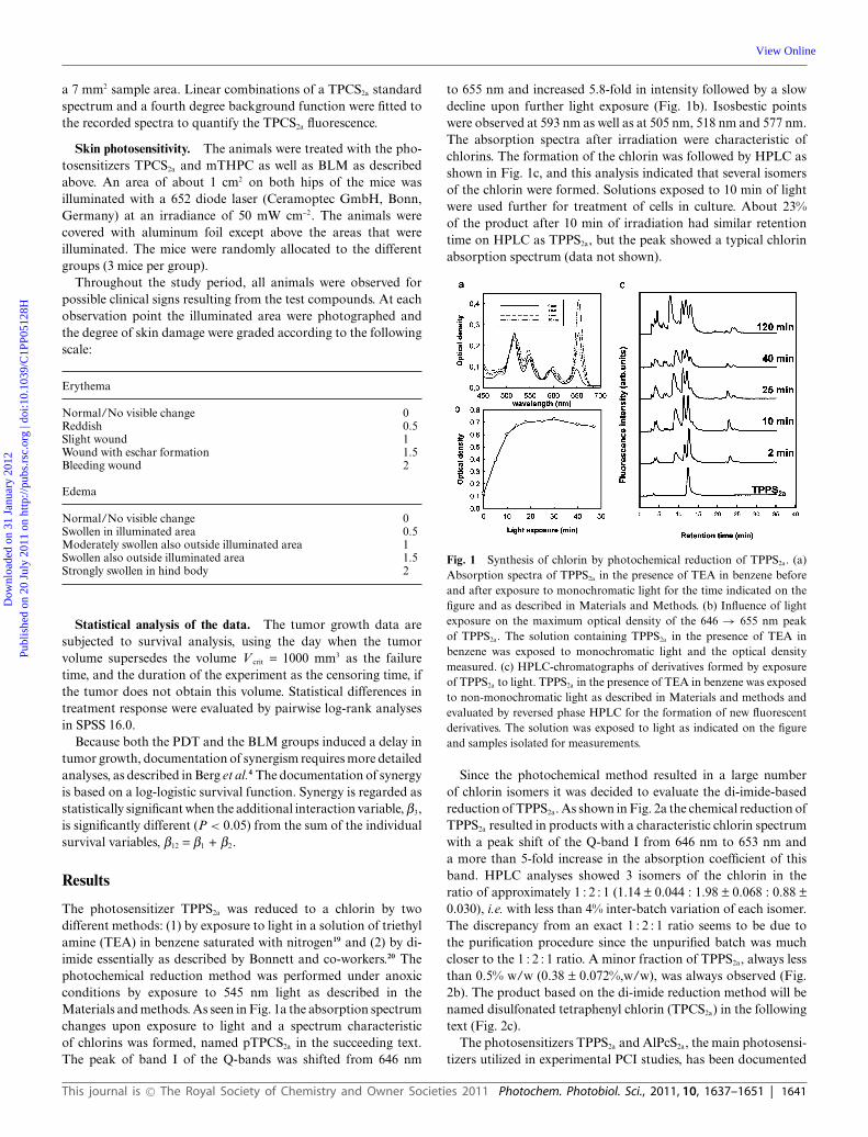
The photosensitizer TPPS2a was reduced to a chlorin by twodifferent methods: (1) by exposure to light in a solution of triethylamine (TEA) in benzene saturated with nitrogen19 and (2) by di-imide essentially as described by Bonnett and co-workers.20 Thephotochemical reduction method was performed under anoxicconditions by exposure to 545 nm light as described in theMaterials and methods. As seen in Fig. 1a the absorption spectrumchanges upon exposure to light and a spectrum characteristicof chlorins was formed, named pTPCS2a in the succeeding text.The peak of band I of the Q-bands was shifted from 646 nm
to 655 nm and increased 5.8-fold in intensity followed by a slowdecline upon further light exposure (Fig. 1b). Isosbestic pointswere observed at 593 nm as well as at 505 nm, 518 nm and 577 nm.The absorption spectra after irradiation were characteristic ofchlorins. The formation of the chlorin was followed by HPLC asshown in Fig. 1c, and this analysis indicated that several isomersof the chlorin were formed. Solutions exposed to 10 min of lightwere used further for treatment of cells in culture. About 23%of the product after 10 min of irradiation had similar retentiontime on HPLC as TPPS2a, but the peak showed a typical chlorinabsorption spectrum (data not shown).
Fig. 1 Synthesis of chlorin by photochemical reduction of TPPS2a. (a)Absorption spectra of TPPS2a in the presence of TEA in benzene beforeand after exposure to monochromatic light for the time indicated on thefigure and as described in Materials and Methods. (b) Influence of lightexposure on the maximum optical density of the 646 → 655 nm peakof TPPS2a. The solution containing TPPS2a in the presence of TEA inbenzene was exposed to monochromatic light and the optical densitymeasured. (c) HPLC-chromatographs of derivatives formed by exposureof TPPS2a to light. TPPS2a in the presence of TEA in benzene was exposedto non-monochromatic light as described in Materials and methods andevaluated by reversed phase HPLC for the formation of new fluorescentderivatives. The solution was exposed to light as indicated on the figureand samples isolated for measurements.
Since the photochemical method resulted in a large numberof chlorin isomers it was decided to evaluate the di-imide-basedreduction of TPPS2a. As shown in Fig. 2a the chemical reduction ofTPPS2a resulted in products with a characteristic chlorin spectrumwith a peak shift of the Q-band I from 646 nm to 653 nm anda more than 5-fold increase in the absorption coefficient of thisband. HPLC analyses showed 3 isomers of the chlorin in theratio of approximately 1 : 2 : 1 (1.14 ± 0.044 : 1.98 ± 0.068 : 0.88 ±0.030), i.e. with less than 4% inter-batch variation of each isomer.The discrepancy from an exact 1 : 2 : 1 ratio seems to be due tothe purification procedure since the unpurified batch was muchcloser to the 1 : 2 : 1 ratio. A minor fraction of TPPS2a, always lessthan 0.5% w/w (0.38 ± 0.072%,w/w), was always observed (Fig.2b). The product based on the di-imide reduction method will benamed disulfonated tetraphenyl chlorin (TPCS2a) in the followingtext (Fig. 2c).
The photosensitizers TPPS2a and AlPcS2a, the main photosensi-tizers utilized in experimental PCI studies, has been documented
This journal is © The Royal Society of Chemistry and Owner Societies 2011 Photochem. Photobiol. Sci., 2011, 10, 1637–1651 | 1641
Dow
nloa
ded
on 3
1 Ja
nuar
y 20
12Pu
blis
hed
on 2
0 Ju
ly 2
011
on h
ttp://
pubs
.rsc
.org
| do
i:10.
1039
/C1P
P051
28H
View Online
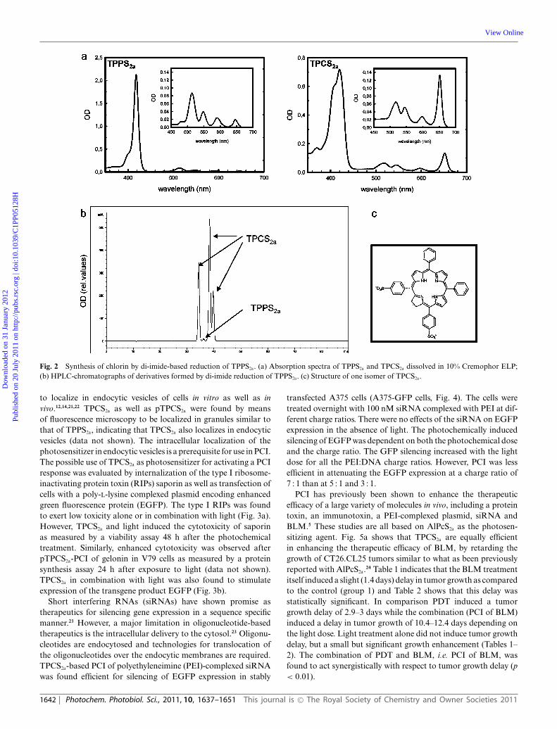
Fig. 2 Synthesis of chlorin by di-imide-based reduction of TPPS2a. (a) Absorption spectra of TPPS2a and TPCS2a dissolved in 10% Cremophor ELP;(b) HPLC-chromatographs of derivatives formed by di-imide reduction of TPPS2a. (c) Structure of one isomer of TPCS2a.
to localize in endocytic vesicles of cells in vitro as well as invivo.12,14,21,22 TPCS2a as well as pTPCS2a were found by meansof fluorescence microscopy to be localized in granules similar tothat of TPPS2a, indicating that TPCS2a also localizes in endocyticvesicles (data not shown). The intracellular localization of thephotosensitizer in endocytic vesicles is a prerequisite for use in PCI.The possible use of TPCS2a as photosensitizer for activating a PCIresponse was evaluated by internalization of the type I ribosome-inactivating protein toxin (RIPs) saporin as well as transfection ofcells with a poly-L-lysine complexed plasmid encoding enhancedgreen fluorescence protein (EGFP). The type I RIPs was foundto exert low toxicity alone or in combination with light (Fig. 3a).However, TPCS2a and light induced the cytotoxicity of saporinas measured by a viability assay 48 h after the photochemicaltreatment. Similarly, enhanced cytotoxicity was observed afterpTPCS2a-PCI of gelonin in V79 cells as measured by a proteinsynthesis assay 24 h after exposure to light (data not shown).TPCS2a in combination with light was also found to stimulateexpression of the transgene product EGFP (Fig. 3b).
Short interfering RNAs (siRNAs) have shown promise astherapeutics for silencing gene expression in a sequence specificmanner.23 However, a major limitation in oligonucleotide-basedtherapeutics is the intracellular delivery to the cytosol.23 Oligonu-cleotides are endocytosed and technologies for translocation ofthe oligonucleotides over the endocytic membranes are required.TPCS2a-based PCI of polyethyleneimine (PEI)-complexed siRNAwas found efficient for silencing of EGFP expression in stably
transfected A375 cells (A375-GFP cells, Fig. 4). The cells weretreated overnight with 100 nM siRNA complexed with PEI at dif-ferent charge ratios. There were no effects of the siRNA on EGFPexpression in the absence of light. The photochemically inducedsilencing of EGFP was dependent on both the photochemical doseand the charge ratio. The GFP silencing increased with the lightdose for all the PEI:DNA charge ratios. However, PCI was lessefficient in attenuating the EGFP expression at a charge ratio of7 : 1 than at 5 : 1 and 3 : 1.
PCI has previously been shown to enhance the therapeuticefficacy of a large variety of molecules in vivo, including a proteintoxin, an immunotoxin, a PEI-complexed plasmid, siRNA andBLM.5 These studies are all based on AlPcS2a as the photosen-sitizing agent. Fig. 5a shows that TPCS2a are equally efficientin enhancing the therapeutic efficacy of BLM, by retarding thegrowth of CT26.CL25 tumors similar to what as been previouslyreported with AlPcS2a.24 Table 1 indicates that the BLM treatmentitself induced a slight (1.4 days) delay in tumor growth as comparedto the control (group 1) and Table 2 shows that this delay wasstatistically significant. In comparison PDT induced a tumorgrowth delay of 2.9–3 days while the combination (PCI of BLM)induced a delay in tumor growth of 10.4–12.4 days depending onthe light dose. Light treatment alone did not induce tumor growthdelay, but a small but significant growth enhancement (Tables 1–2). The combination of PDT and BLM, i.e. PCI of BLM, wasfound to act synergistically with respect to tumor growth delay (p< 0.01).
1642 | Photochem. Photobiol. Sci., 2011, 10, 1637–1651 This journal is © The Royal Society of Chemistry and Owner Societies 2011
Dow
nloa
ded
on 3
1 Ja
nuar
y 20
12Pu
blis
hed
on 2
0 Ju
ly 2
011
on h
ttp://
pubs
.rsc
.org
| do
i:10.
1039
/C1P
P051
28H
View Online
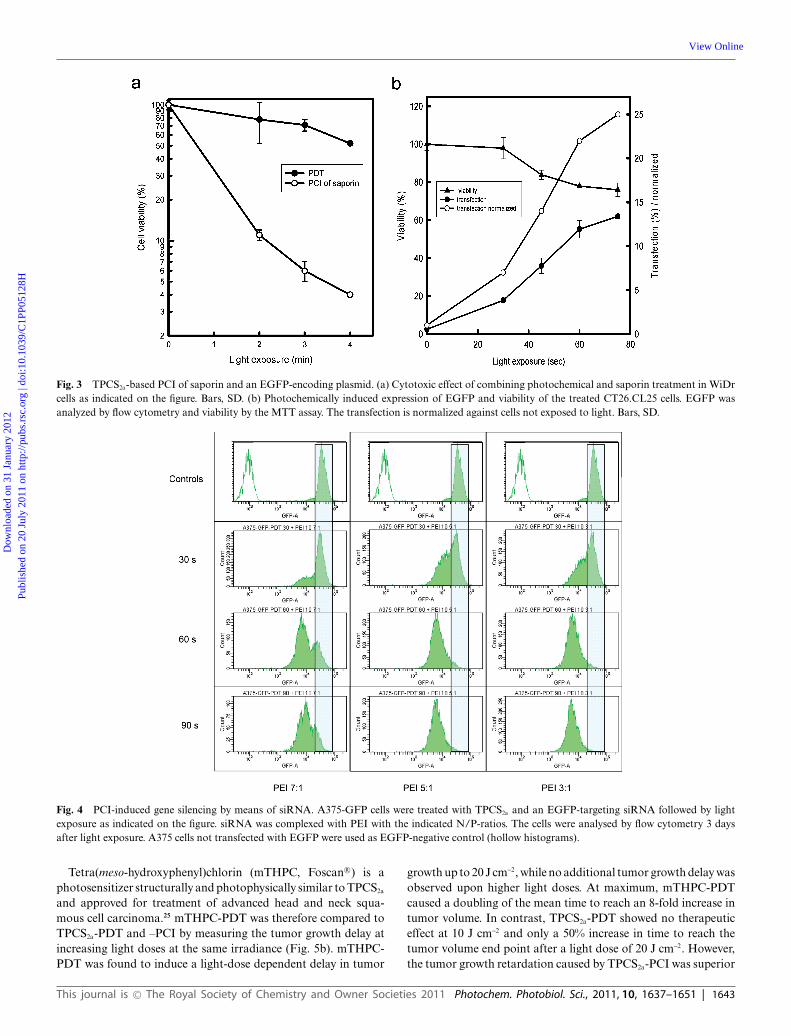
Fig. 3 TPCS2a-based PCI of saporin and an EGFP-encoding plasmid. (a) Cytotoxic effect of combining photochemical and saporin treatment in WiDrcells as indicated on the figure. Bars, SD. (b) Photochemically induced expression of EGFP and viability of the treated CT26.CL25 cells. EGFP wasanalyzed by flow cytometry and viability by the MTT assay. The transfection is normalized against cells not exposed to light. Bars, SD.
Fig. 4 PCI-induced gene silencing by means of siRNA. A375-GFP cells were treated with TPCS2a and an EGFP-targeting siRNA followed by lightexposure as indicated on the figure. siRNA was complexed with PEI with the indicated N/P-ratios. The cells were analysed by flow cytometry 3 daysafter light exposure. A375 cells not transfected with EGFP were used as EGFP-negative control (hollow histograms).
Tetra(meso-hydroxyphenyl)chlorin (mTHPC, Foscan R©) is aphotosensitizer structurally and photophysically similar to TPCS2a
and approved for treatment of advanced head and neck squa-mous cell carcinoma.25 mTHPC-PDT was therefore compared toTPCS2a-PDT and –PCI by measuring the tumor growth delay atincreasing light doses at the same irradiance (Fig. 5b). mTHPC-PDT was found to induce a light-dose dependent delay in tumor
growth up to 20 J cm-2, while no additional tumor growth delay wasobserved upon higher light doses. At maximum, mTHPC-PDTcaused a doubling of the mean time to reach an 8-fold increase intumor volume. In contrast, TPCS2a-PDT showed no therapeuticeffect at 10 J cm-2 and only a 50% increase in time to reach thetumor volume end point after a light dose of 20 J cm-2. However,the tumor growth retardation caused by TPCS2a-PCI was superior
This journal is © The Royal Society of Chemistry and Owner Societies 2011 Photochem. Photobiol. Sci., 2011, 10, 1637–1651 | 1643
Dow
nloa
ded
on 3
1 Ja
nuar
y 20
12Pu
blis
hed
on 2
0 Ju
ly 2
011
on h
ttp://
pubs
.rsc
.org
| do
i:10.
1039
/C1P
P051
28H
View Online
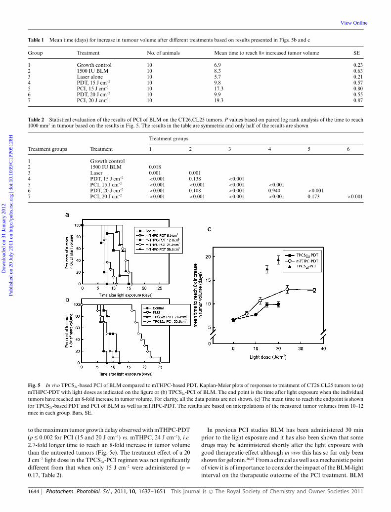
Table 1 Mean time (days) for increase in tumour volume after different treatments based on results presented in Figs. 5b and c
Group Treatment No. of animals Mean time to reach 8¥ increased tumor volume SE
1 Growth control 10 6.9 0.232 1500 IU BLM 10 8.3 0.633 Laser alone 10 5.7 0.214 PDT, 15 J cm-2 10 9.8 0.575 PCI, 15 J cm-2 10 17.3 0.806 PDT, 20 J cm-2 10 9.9 0.557 PCI, 20 J cm-2 10 19.3 0.87
Table 2 Statistical evaluation of the results of PCI of BLM on the CT26.CL25 tumors. P values based on paired log rank analysis of the time to reach1000 mm3 in tumour based on the results in Fig. 5. The results in the table are symmetric and only half of the results are shown
Treatment groups
Treatment groups Treatment 1 2 3 4 5 6
1 Growth control2 1500 IU BLM 0.0183 Laser 0.001 0.0014 PDT, 15 J cm-2 <0.001 0.138 <0.0015 PCI, 15 J cm-2 <0.001 <0.001 <0.001 <0.0016 PDT, 20 J cm-2 <0.001 0.108 <0.001 0.940 <0.0017 PCI, 20 J cm-2 <0.001 <0.001 <0.001 <0.001 0.173 <0.001
Fig. 5 In vivo TPCS2a-based PCI of BLM compared to mTHPC-based PDT. Kaplan-Meier plots of responses to treatment of CT26.CL25 tumors to (a)mTHPC-PDT with light doses as indicated on the figure or (b) TPCS2a-PCI of BLM. The end point is the time after light exposure when the individualtumors have reached an 8-fold increase in tumor volume. For clarity, all the data points are not shown. (c) The mean time to reach the endpoint is shownfor TPCS2a-based PDT and PCI of BLM as well as mTHPC-PDT. The results are based on interpolations of the measured tumor volumes from 10–12mice in each group. Bars, SE.
to the maximum tumor growth delay observed with mTHPC-PDT(p £ 0.002 for PCI (15 and 20 J cm-2) vs. mTHPC, 24 J cm-2), i.e.2.7-fold longer time to reach an 8-fold increase in tumor volumethan the untreated tumors (Fig. 5c). The treatment effect of a 20J cm-2 light dose in the TPCS2a-PCI regimen was not significantlydifferent from that when only 15 J cm-2 were administered (p =0.17, Table 2).
In previous PCI studies BLM has been administered 30 minprior to the light exposure and it has also been shown that somedrugs may be administered shortly after the light exposure withgood therapeutic effect although in vivo this has so far only beenshown for gelonin.26,27 From a clinical as well as a mechanistic pointof view it is of importance to consider the impact of the BLM-lightinterval on the therapeutic outcome of the PCI treatment. BLM
1644 | Photochem. Photobiol. Sci., 2011, 10, 1637–1651 This journal is © The Royal Society of Chemistry and Owner Societies 2011
Dow
nloa
ded
on 3
1 Ja
nuar
y 20
12Pu
blis
hed
on 2
0 Ju
ly 2
011
on h
ttp://
pubs
.rsc
.org
| do
i:10.
1039
/C1P
P051
28H
View Online
was therefore injected between 24 h prior to and 24 h after the lightexposure (Fig. 6). The results indicate that injection of BLM 30min to 3 h prior to light exposure as well as immediately after lightexposure caused no statistical difference in tumor growth delay (p =0.31–0.77), while injection of BLM 24 h prior to light resulted ina significantly smaller tumor growth delay (p = 0.023 compared toBLM 30 min prior to light). It should also be noted that injectionof BLM 24 h after light exposure caused only additive effectsin combination with the photochemical treatment insignificantlydifferent from BLM alone (p = 0.22) and significantly lower thanthe optimal PCI regimens (p £ 0.013).
Fig. 6 The impact of the BLM-light interval on the treatment effect ofPCI of BLM. The mean time of CT26.CL25 tumors to reach 1000 mm3
after treatments is presented as a function of the time between BLMandministration and light exposure. Light is administered at time zero andnegative time points illustrate BLM administration prior to light exposure.The mean time of CT26.CL25 tumors to reach 1000 mm3 is based oninterpolation of growth curves from 9–11 tumors in each group. The meantime for tumors treated with BLM, PDT and growth controls to reach theendo point is indicated on the figure.
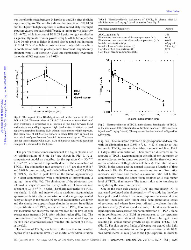
The pharmacokinetic measurements of TPCS2a in plasma afteri.v. administration of 5 mg kg-1 are shown in Fig. 7. A 2-compartment model as described by the equation C = 36e-0.88t
+ 5.9e-0.019t, was found to optimally describe the elimination ofTPCS2a. The elimination rate constants (1 h-1) are thus 0.88 h-1
and 0.019 h-1, respectively, and the half-lives 0.78 and 36 h (Table3). TPCS2a reached a peak level in the tumor approximately24 h after administration with a maximum of approximately 2ng mg-1 tissue (Fig. 8a). The elimination of the photosensitizerfollowed a single exponential decay with an elimination rateconstant of 0.013 h-1 (t1/2 = 52 h). The pharmacokinetics of TPCS2a
was similar in skin and muscle with a maximum accumulationapproximately 24 h after administration and a single exponentialdecay although in the muscle the level of accumulation was lowerand the elimination appears faster than in the tumor. In additionto quantification of TPCS2a in skin extracts, TPCS2a fluorescencewas measured non-invasively and normalized to the level of skinextract measurements 24 h after administration (Fig. 8a). Theresults indicate that the TPCS2a fluorescence is retained longer inthe skin than what was measured from the total content of TPCS2a
in the skin.The uptake of TPCS2a was faster to the liver than to the other
organs with a maximum level 6 h or shorter after administration
Table 3 Pharmacokinetic parameters of TPCS2a in plasma after i.v.administration of 5 mg kg-1 based on results from Fig. 7
Pharmacokinetic parameters Results
AUC0–• (mg h ml-1) 365Elimination rate constants of first compartment (h-1) 0.88Elimination rate constants of second compartment (h-1) 0.019Initial concentration (C0) 42 mg ml-1
Initial volume of distribution (V d) 99 ml kg-1
Half-life of first compartment (h) 0.78Half-life of second compartment (h) 36
Fig. 7 Pharmacokinetics of TPCS2ain the plasma. Semilog plot of TPCS2a
in the plasma of Balb/C (nu/nu) mice (without xenograft) after single i.v.injection of 5 mg kg-1 (n = 6). The regression line is calculated in SigmaPlot(R2 = 0.93).
(Fig. 8a). The elimination followed a single exponential decay ratewith an elimination rate (0.031 h-1, t1/2 = 22 h) similar to thatin muscle. TPCS2a was not detectable in muscle and liver 336 h(14 days) after administration. There were no differences in theamount of TPCS2a accumulating in the skin above the tumor ormuscle adjacent to the tumor compared to similar tissue locationson the contralateral thigh (data not shown). The ratio betweenTPCS2a in the tumor and the normal tissues as a function of timeis shown in Fig. 8b. The tumor : muscle and tumor : liver ratiosincreased with time and reached a maximum ratio 120 h afteradministration when the tumor tissue retained an 8-fold higherlevel of TPCS2a than muscle. The tumor : skin ratio was close tounity during the same time period.
One of the main side effects of PDT and presumably PCI isacute and prolonged skin photosensitivity.28 A study has thereforebeen performed to detect treatment responses to mouse skin inmice not inoculated with tumor cells. Semi-quantitative scalesof erythema and edema have been utilized to evaluate the skinphotosensitivity (Materials and methods). The skin reactions tolight exposure were assessed after administration of TPCS2a aloneor in combination with BLM in comparison to the responsescaused by administration of Foscan followed by light dosesresulting in similar tumor growth delays as after TPCS2a-PDT(Fig. 5). The skin on both flanks of the mice was treated with light1-14 days after administration of the photosensitizer while BLMwas administered 30 min prior to the light exposure. In order to
This journal is © The Royal Society of Chemistry and Owner Societies 2011 Photochem. Photobiol. Sci., 2011, 10, 1637–1651 | 1645
Dow
nloa
ded
on 3
1 Ja
nuar
y 20
12Pu
blis
hed
on 2
0 Ju
ly 2
011
on h
ttp://
pubs
.rsc
.org
| do
i:10.
1039
/C1P
P051
28H
View Online

Fig. 8 Concentration of TPCS2ain selected tissues of the Balb/C (nu/nu)mice with subcutaneously growing CT26.CL25 tumors. (a) Semilog plotsof TPCS2a (5 mg kg-1, i.v.) concentration in tumor, skin, muscle andliver. In addition, TPCS2a-fluorescence from dorsal skin was assessednon-invasively from 24 to 550 h after TPCS2a administration as indicatedon the figure. (b) Tumor : normal (skin, muscle and liver) tissue ratios ofTPCS2a at various times after administration (n = 6). Bars, SD.
follow the treatment procedure and mimic a clinical situation theskin reactions were assessed from one day after photosensitizeradministration, while PCI was performed from day 3 and skinreactions assessed the following days. TPCS2a-PDT resulted instrong edema when the skin was irradiated 1– 4 and 1–5 days afterexposure to 10 or 15 J cm-2 of 652 nm light, respectively (Fig. 9and 10). There were no or only moderate edema observed in micetreated with light 7 days or longer after TPCS2a administration.These observations are reflected in both the peak reaction observed1 day after treatment (Fig. 10) and as accumulated reactions byintegrating the edema reaction over time (Fig. 13). In contrast,mTHPC-PDT caused only a mild edema as compared to TPCS2a-PDT (Fig. 9, 12 and 13).
The erythema induced by TPCS2a-PDT was moderate bothafter 10 and 15 J cm-2 although 15 J cm-2 1 day after TPCS2a
administration caused a prolonged, but moderate erythema (Fig.9, 11 and 13). In contrast, mTHPC-PDT resulted in strongererythema when 6 J cm-2 light was administered 1 day after mTHPCinjection and for up to 4 days after mTHPC injected when followedby 12 J cm-2 (Fig. 12 and 13). The strongest response resulted in
Fig. 9 Photosensitivity of Balb/C (nu/nu) mice treated on the hip withTPCS2aor mTHPC. The photosensitizers (5 mg kg-1 TPCS2a or 0.3 mgkg-1 mTHPC) were systemically administered and the hips were exposedto light 24 h later (15 J cm-2 or 12 J cm-2 652 nm light for TPCS2a- ormTHPC-treated mice respectively). The treated areas of the mice werephotographed for 14 days after treatment as exemplified in the figure. Day0 is the day of light exposure.
Fig. 10 Skin edema score following light exposure of the skin of Balb/C(nu/nu) mice treated with TPCS2a alone or in combination with BLM.TPCS2a (5 mg kg-1) was administered on day 0 and the skin exposed to lightat various time intervals after TPCS2a administration: Day 1 (�), day 2 (�),day 3 (�), day 4 (�), day 5 (�), day 7 (�). BLM was administered 3 daysafter TPCS2a and the animals were exposed to light on the following daysas indicated on the figure. The skin edema assessment scale is described inMaterials and methods. Bars, SE (n = 3).
wounds that lasted for at least 14 days after Foscan administrationand resulted in a substantial accumulated reaction (Fig. 13).
The skin reactions observed after TPCS2a-PDT was comparedto TPCS2a-PDT in combination with BLM treatment mimickingthe PCI tumor treatment (Fig. 10, 11 and 13). However, althoughthe therapeutic effect when including BLM was significantlystronger the skin reactions did not increase concomitantly.
Discussion
Several hundred new photosensitizers have been developed thelast 20 years in a search for the optimal compounds for treatmentof various diseases with PDT. Out of all these new chemicalentities some has entered clinical trials. The most promisingor approved photosensitizers exert with few exceptions (e.g.Foscan) amphiphilic properties, but are able to penetrate cellularmembranes.13 This is made possible by including 1–2 carboxyl-groups on one side of the compound, leaving the rest of the
1646 | Photochem. Photobiol. Sci., 2011, 10, 1637–1651 This journal is © The Royal Society of Chemistry and Owner Societies 2011
Dow
nloa
ded
on 3
1 Ja
nuar
y 20
12Pu
blis
hed
on 2
0 Ju
ly 2
011
on h
ttp://
pubs
.rsc
.org
| do
i:10.
1039
/C1P
P051
28H
View Online
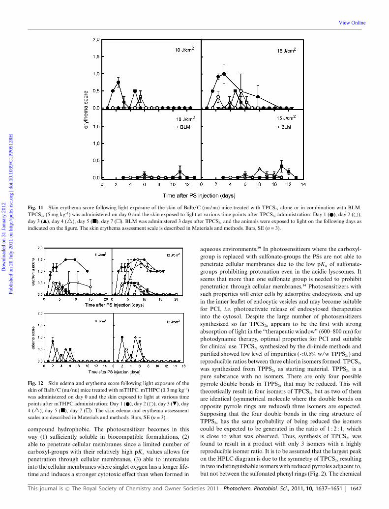
Fig. 11 Skin erythema score following light exposure of the skin of Balb/C (nu/nu) mice treated with TPCS2a alone or in combination with BLM.TPCS2a (5 mg kg-1) was administered on day 0 and the skin exposed to light at various time points after TPCS2a administration: Day 1 (�), day 2 (�),day 3 (�), day 4 (�), day 5 (�), day 7 (�). BLM was administered 3 days after TPCS2a and the animals were exposed to light on the following days asindicated on the figure. The skin erythema assessment scale is described in Materials and methods. Bars, SE (n = 3).
Fig. 12 Skin edema and erythema score following light exposure of theskin of Balb/C (nu/nu) mice treated with mTHPC. mTHPC (0.3 mg kg-1)was administered on day 0 and the skin exposed to light at various timepoints after mTHPC administration: Day 1 (�), day 2 (�), day 3 (�), day4 (�), day 5 (�), day 7 (�). The skin edema and erythema assessmentscales are described in Materials and methods. Bars, SE (n = 3).
compound hydrophobic. The photosensitizer becomes in thisway (1) sufficiently soluble in biocompatible formulations, (2)able to penetrate cellular membranes since a limited number ofcarboxyl-groups with their relatively high pKa values allows forpenetration through cellular membranes, (3) able to intercalateinto the cellular membranes where singlet oxygen has a longer life-time and induces a stronger cytotoxic effect than when formed in
aqueous environments.29 In photosensitizers where the carboxyl-group is replaced with sulfonate-groups the PSs are not able topenetrate cellular membranes due to the low pKa of sulfonate-groups prohibiting protonation even in the acidic lysosomes. Itseems that more than one sulfonate group is needed to prohibitpenetration through cellular membranes.14 Photosensitizers withsuch properties will enter cells by adsorptive endocytosis, end upin the inner leaflet of endocytic vesicles and may become suitablefor PCI, i.e. photoactivate release of endocytosed therapeuticsinto the cytosol. Despite the large number of photosensitizerssynthesized so far TPCS2a appears to be the first with strongabsorption of light in the “therapeutic window” (600–800 nm) forphotodynamic therapy, optimal properties for PCI and suitablefor clinical use. TPCS2a synthesized by the di-imide methods andpurified showed low level of impurities (<0.5% w/w TPPS2a) andreproducible ratios between three chlorin isomers formed. TPCS2a
was synthesized from TPPS2a as starting material. TPPS2a is apure substance with no isomers. There are only four possiblepyrrole double bonds in TPPS2a that may be reduced. This willtheoretically result in four isomers of TPCS2a but as two of themare identical (symmetrical molecule where the double bonds onopposite pyrrole rings are reduced) three isomers are expected.Supposing that the four double bonds in the ring structure ofTPPS2a has the same probability of being reduced the isomerscould be expected to be generated in the ratio of 1 : 2 : 1, whichis close to what was observed. Thus, synthesis of TPCS2a wasfound to result in a product with only 3 isomers with a highlyreproducible isomer ratio. It is to be assumed that the largest peakon the HPLC diagram is due to the symmetry of TPCS2a resultingin two indistinguishable isomers with reduced pyrroles adjacent to,but not between the sulfonated phenyl rings (Fig. 2). The chemical
This journal is © The Royal Society of Chemistry and Owner Societies 2011 Photochem. Photobiol. Sci., 2011, 10, 1637–1651 | 1647
Dow
nloa
ded
on 3
1 Ja
nuar
y 20
12Pu
blis
hed
on 2
0 Ju
ly 2
011
on h
ttp://
pubs
.rsc
.org
| do
i:10.
1039
/C1P
P051
28H
View Online

Fig. 13 Accumulated induction of erythema and edema after TPCS2a- or mTHPC-mediated PDT or PCI. The total skin reaction is estimated byintegrating the erythema and edema score over time, based on the results presented in Fig. 10–12. This is presented as the area under the curve (AUC) foreach treatment. The SigmaPlot software has been used for calculating AUC.
reduction of theTPPS2a tetrapyrrole structure resulted in a slightlyred-shifted and more than 5-fold increased light absorption ata wavelength (652 nm) acceptable for clinical utilization. Thepresent study shows that TPCS2a is able to exert photochemicalactivation of endocytosed therapeutics with promising preclinicalresponses. The photochemical reduction method was found toinduce a large number of isomers and therefore unsuitable forclinical utilization. AlPcS2a that has previously been utilized inthe preclinical studies of PCI is made by direct sulfonation ofAlPc to yield isomeric mixtures of sulfonated AlPc dependingon the reaction time followed by purification.30,31 With such aprocedure the relative content of the different isomers will varyfrom batch-to-batch and each isomer may vary substantially inbiological effect upon exposure to light.17,32 It is therefore ofimportance to ensure as little batch-to-batch variation in isomercomposition as possible. In case of TPCS2a the number of isomersis lower and the ratio between each isomer is reproducible andclose to the theoretical ratios. Furthermore, the photobleachingrate of TPCS2a is much higher that for AlPcS2a which may bebeneficial in degrading photosensitizer located in the skin. Thus,TPCS2a has several advantages when compared to AlPcS2a as aPCI photosensitizer for clinical use.
TPPS2a and AlPCS2a have been shown to be localized inendocytic vesicles, to enhance the cytotoxic effect of the ribosome-inactivating protein toxins gelonin and saporin and enhancetransfection of reporter genes in a large number of cell lines.5
TPCS2a was found to have similar properties as TPPS2a andAlPcS2a. The increased cytotoxicity by means of TPCS2a-basedPCI of saporin in WiDr cells was similar to that of TPPS2a inthe same cell line6 and TPCS2a-PCI increased the transfectionrate 20-fold at low photochemical doses, similar to what wasfound with TPPS2a and AlPcS2a upon poly-L-lysine/plasmid-basedtransfection.11 Much less has been performed on PCI of siRNAfor gene silencing although PCI has been shown to attenuate geneexpression of different genes (S100A4, EGFR and luciferase) bothin vitro and in vivo.33–35 Thus, green fluorescent protein (GFP)is strongly expressed in the A375-GFP cells and GFP-siRNAcomplexed to branched PEI was only able to attenuate GFPexpression when activated by PCI. The proton-sponge effect ofPEI facilitating the translocation of plasmids and oligonucleotidesfrom the endocytic vesicles to the cytosol usually increases with
increasing N/P charge ratio,36,37 resulting in increased transgeneexpression or oligonucleotide-based gene silencing. In contrast,PCI-induced gene silencing was less effective at the highestcharge ratio (7 : 1) than at 5 : 1 and 3 : 1. The reason for thesecontradicting results of the charge ratio in the absence andpresence of PCI is not clear, but has also been reported for PCI-based delivery of plasmids.38 The proposed “proton sponge” effectof PEI increases with increasing accumulation of PEI inside theendocytic vesicles.2 However, in combination with PCI PEI isneeded for protection against degradation and enhancement ofcellular uptake of the nucleotides, but not for the endosome-to-cytosol translocation. Thus, high doses of PEI appears not tobe beneficial for steps downstream of the cytosolic entry of thenucleotides in the pathways leading to gene silencing (siRNA) ortransgene expression (plasmids).
The ability of TPCS2a to induce PCI in vivo was evaluatedby using BLM as the drug to be photochemically activated.TPCS2a-PCI of BLM is also the treatment regimen utilized inthe ongoing PCI clinical trial (http://clinicaltrials.gov). The sameaggressive tumor model as used in this study, CT26.CL25, haspreviously been evaluated for PCI of BLM, but with AlPcS2a
as photosensitizer.24 Both treatments, AlPcS2a-PCI and TPCS2a-PCI, induced a strong and similar delay in tumor growth (12.4vs. 10,6 days, respectively). However, AlPcS2a-PDT induced amore pronounced delay (5.8 days) in tumor growth as comparedto TPCS2a-PDT (1.9 days). Thus, the enhanced tumor growthdelay introduced by combination of the photochemical and BLMtreatments (i.e. PCI) as compared to the sum of the individualtreatments appears stronger with TPCS2a than with AlPcS2a asphotosensitizer.
PCI is under development for delivery of targeted macromolecu-lar therapeutics aimed at generating a highly specific and efficientcancer treatment modality. However, targeted therapeutics thatmay benefit from activation by PCI is currently not approvedfor clinical use. Meanwhile, BLM has been found attractive asan alternative drug for PCI activation in the clinic. In order toevaluate potential clinical benefits of TPCS2a-PCI of BLM thistreatment regimen was compared to PDT based on mTHPC-PDT, a photosensitizer approved for treatment of head and neckcancers.39,40 mTHPC is a chlorin type photosensitizer as TPCS2a
and thus with similar light absorption and other photophysical
1648 | Photochem. Photobiol. Sci., 2011, 10, 1637–1651 This journal is © The Royal Society of Chemistry and Owner Societies 2011
Dow
nloa
ded
on 3
1 Ja
nuar
y 20
12Pu
blis
hed
on 2
0 Ju
ly 2
011
on h
ttp://
pubs
.rsc
.org
| do
i:10.
1039
/C1P
P051
28H
View Online
properties. Treatment of the subcutaneously growing CT26.CL25carcinoma with mTHPC-PDT and increasing doses of 652 nmlight showed a light-dose dependent delay in tumor growth upto 24 J cm-2, above which no further tumor growth delay wasobserved. A similar light dose-independence of tumor growthdelay after mTHPC-PDT has previously been reported.41 In arecent study of AlPcS2a-based PDT it was found that the tumorperiphery is relatively resistant to PDT42,43 and we have foundthat this is also the case with mTHPC-PDT by HE-stainingfor viability evaluation on tissue cut sections from CT26.CL25tumors (unpublished results). Thus, the lack of effect of light dosesabove 24 J cm-2 in the CT26.CL25 tumor model is apparentlydue to a rim of tumor cells exerting low sensitivity to PDT. Incontrast, with TPCS2a-PDT there was no effect on tumor growthwith light doses below 10 J cm-2 on tumor growth delay whilea dose-dependent tumor growth delay was observed at higherdoses. This implies that a threshold dose is required to inducea therapeutic effect with TPCS2a as photosensitizer, not seen withmTHPC-based PDT. The reason for this threshold dose is notfully understood, but previous reports indicate that photochemicalrelocation of TPPS2a from the lysosomes to the endoplasmicreticulum enhance the photocytotoxic effect of TPPS2a, a processthat requires a minimum dose of light.44,45 A threshold dose toinduce skin reactions may be clinically beneficial. When TPCS2a-PDT was combined with BLM a tumor growth delay strongerthan the maximum obtained by mTHPC-PDT was observed. Thisis in accordance with our previous studies showing that AlPcS2a-based PCI of BLM is able to target the tumor periphery, resistantto AlPcS2a-PDT. These results indicate that TPCS2a-based PCIof BLM is more effective in inducing local tumor control thanmTHPC-based PDT. The low sensitivity of the tumor rim toAlPcS2a- and mTHPC-PDT has also been observed utilizing otherphotosensitizers and may be a more general property of PDT ofsolid tumors.46–49
The half-life of BLM in the blood of humans after i.v. injectionis ~2 h and 70% of the BLM is excreted within the first 24h.50–52 In mice, the blood clearance time was shown to be muchshorter while the amount of BLM in the tumor was practicallyunchanged from shortly after i.v. injection and for the following24 h.51 The present results indicate that BLM may be injected inthe interval 3 h prior to light exposure to immediately after lightexposure without compromising the PCI-induced tumor growthdelay. The reduced effect of BLM injected 24 h prior to lightexposure may indicate partial intratumor degradation during thisdrug-light interval. This is in somewhat contrast to the reportedintratumorally stable level of BLM for at least 24 h. These studieswere however performed with radioactively labeled BLM. Label-ing of small molecules like BLM may in general influence bothpharmacokinetic properties and degradation rate and differencesin the pharmacokinetics of indium- and iodine-labeled BLMhave been reported.52 Labeling of small molecules may in generalinfluence their pharmacokinetic properties and degradation rate.Thus, the reduced therapeutic effect of PCI of BLM with a 24h BLM-light interval compared to a 3-to-0 h interval may betterreflect the intratumor level of biologically active BLM. The lackof synergism observed when BLM was administered 24 h afterthe light exposure is in accordance with the requirement forthe presence of photochemically ruptured endocytic vesicles afteror immediately before BLM administration. The results indicate
some flexibility in the BLM-light interval without perturbing thetherapeutic affect and may be of practical clinical importance.
The plasma pharmacokinetics of TPCS2a was best fitted to a twocompartment exponential decay model with a rapid redistributionphase (t1/2 = 0.78 h) and a slower elimination phase (t1/2 = 36 h). Ingeneral, photosensitizer pharmacokinetics has been shown to fit to2 and 3-compartment models. The plasma half-life of the secondphase for mTHPC was reported by Cramers and co-workers to be20.9 h in mice, based on a 2-compartment model.53 It has beensuggested that the plasma elimination of mTHPC may followa three compartment exponential decay model where the lastphase is very slow, about 4 days, which may be overlooked byending the studies too early.54 This is in accordance with the threecompartment exponential best fit of the mTHPC elimination inrats (t1/2, 3rd comp = 82,5 h55) and of Photofrin in mice (t1/2,3rd comp. = 220 h56). However, an attempt to fit the plasmapharmacokinetics results for TPCS2a to a three compartmentexponential decay model was not successful and at the end of theexperiments (336 h) the level of TPCS2a in the plasma approachedthe detection limit. A 3rd compartment can still not be excluded,but is most likely of little importance.
The initial volume of TPCS2a distribution of 99 ml kg-1 wassomewhat larger than the blood volume of mice (55–70 ml kg-1)and larger than that of mTHPC (32 ml kg-1,55) indicating a rapidextravasation of TPCS2a.
The peak accumulation of TPCS2a in skin, muscle and tumorwas reached about 24 h after administration, while in liver thepeak accumulation was at 6 h or shorter. The maximum level ofTPCS2a was approximately 2 mg g-1 skin and tumor tissue, 1 mgg-1 muscle tissue and 40 mg g-1 liver tissue. These ratios are similarto what has been found for mTHPC and Photofrin in mice.53,56
Photofrin, mTHPC and the liposomal formulation of mTHPC(Foslip) also reach a plateau in skin, tumor and muscles after24 h, but are very slowly eliminated from these tissues.53,56,57 Asimilar level of these photosensitizers was found in these tissueseven 96–168 h after administration. In contrast, elimination ofTPCS2a is initiated 24 h after administration and are reduced 2(tumor and skin) and 10 (muscle)-fold within the following 100h. The doubling time of this tumor model is approx. 2.5 days,indicating that the elimination of TPCS2a is slower from the skin. Itshould be pointed out that the elimination rate of the fluorescencefrom TPCS2a in the skin was slower than TPCS2a measured bythe chemical extraction method. The reason for this difference isnot clear, but may be due to measurements of only the superficialskin layer by the fluorescence method or by disaggregation ofaggregated (and therefore non-fluorescing) TPCS2a over time. Theelimination from liver appears similar within this time frame forTPCS2a and mTHPC, while the elimination of Photofrin is veryslow (25% of maximum level remaining in the liver 75 days afteradministration).
One of the main limitations of PDT is the skin photosen-sitivity that may last for several weeks after photosensitizeradministration.58 Light exposure of the skin of a sensitized patientor animal results in a dose-dependent erythema and edema thatmay be manifested as wounds and result in scar formation. Thephotosensitivity requires protection from bright light for 30–60days for Photofrin and somewhat shorter for Foscan. The skinreaction observed in the present study after mTHPC-PDT is there-fore in accordance with the literature although we distinguished
This journal is © The Royal Society of Chemistry and Owner Societies 2011 Photochem. Photobiol. Sci., 2011, 10, 1637–1651 | 1649
Dow
nloa
ded
on 3
1 Ja
nuar
y 20
12Pu
blis
hed
on 2
0 Ju
ly 2
011
on h
ttp://
pubs
.rsc
.org
| do
i:10.
1039
/C1P
P051
28H
View Online
between erythema and edema and found mTHPC-PDT to inducestrong erythema resulting in open wounds and scar formationsand little edema. In contrast, TPCS2a-PDT induced little erythemawith no wound formation, but a strong edema when light wasadministered shortly (1–4 days) after mTHPC administration. Thereason for such differences in response was surprising. The causeof this difference in skin response between TPCS2a- and mTHPC-PDT is not understood, but may intuitively be due to differencesin the distribution of these photosensitizers in the skin. However,the accumulation of both photosensitizers in the skin appearssimilar. PDT is reported to exert its therapeutic effect through bothtargeting the parenchyma cells and the vasculature in additionto immunological effects. Thus, mTHPC-PDT has been shownto target both the parenchyma cells as well as the vasculature.59
Similar investigations have so far not been performed with TPCS2a,but AlPcS2a which is similar with respect to physico-chemicalproperties as well as intracellular localization and efficacy as a PCIphotosensitizer has also been shown to target both the parenchymacells and the vasculature.42,43 However, the difference in responseto PDT in the skin indicates a relatively stronger vascular effectof TPCS2a-PDT than that of mTHPC-PDT. Further studies arehowever needed to reveal the mechanistic causes of this differencein skin response. Clinically, it may be easier to control edema thanerythema that may cause second-degree burn.60
As pointed out above the elimination rate of BLM is short bothin mice and humans. The level of circulating BLM is expected tobe very low one day after administration and BLM accumulated incells degraded by hydrolytic enzymes in the lysosomes. Thus, lightexposure of normal tissue, e.g. skin, of patients treated with PCI ofBLM one day or more after BLM administration may be expectedto cause a skin reaction mainly from the photosensitizer and lightand not potentiated by BLM. This is in accordance with thepresent study showing a lack of additional skin reactions by PCIof BLM as compared to TPCS2a-PDT and lack of increased tumorgrowth delay by BLM administered 24 h after the photochemicaltreatment. BLM may therefore enhance the therapeutic effect ofthe photochemical treatment within a small time-window whilethe skin photosensitivity is rapidly attenuated when BLM iseliminated. In this way PCI of BLM may be expected to causeonly transiently enhanced skin sensitivity which is a major benefitcompared to many of the PDT regimens utilized today.
In conclusion, the novel drug TPCS2a has been shown to be anefficient PCI photosensitizer by enhancing the therapeutic effectof the protein toxin saporin, increasing the transfection with areporter gene on a plasmid, increasing siRNA-mediated genesilencing and increasing in a synergistic manner the therapeuticefficacy of BLM in vivo. TPCS2a-PCI of BLM was found superiorto mTHPC-PDT in delaying growth of the rapidly growingsubcutaneous CT26.CL25 carcinoma tumor. TPCS2a has recentlybeen produced at clinical grade and is currently being tested in aphase I/II clinical trial with promising preliminary results. Thus,TPCS2a is a novel photosensitizer for clinical utilization of the PCIprinciple.
Acknowledgements
The authors are grateful to Marie-Therese Strand Larsen andBente Hovland for excellent technical assistance and Prof. ClaudeRimington for fruitful contributions.
References
1 R. J. Christie and D. W. Grainger, Design strategies to improve solublemacromolecular delivery constructs, Adv. Drug Delivery Rev., 2003, 55,421–437.
2 D. W. Pack, A. S. Hoffman, S. Pun and P. S. Stayton, Design anddevelopment of polymers for gene delivery, Nat. Rev. Drug Discovery,2005, 4, 581–593.
3 V. Russ and E. Wagner, Cell and tissue targeting of nucleic acids forcancer gene therapy, Pharm. Res., 2007, 24, 1047–1057.
4 K. Berg, M. Folini, L. Prasmickaite, P. K. Selbo, A. Bonsted,B. O. Engesaeter, N. Zaffaroni, A. Weyergang, A. Dietze, G. M.Maelandsmo, E. Wagner, O. J. Norum and A. Hogset, Photochemicalinternalization: a new tool for drug delivery, Curr. Pharm. Biotechnol.,2007, 8, 362–372.
5 P. K. Selbo, A. Weyergang, A. Hogset, O. J. Norum, M. B. Berstad,M. Vikdal and K. Berg, Photochemical internalization provides time-and space-controlled endolysosomal escape of therapeutic molecules,J. Controlled Release, 2010, 148, 2–12.
6 K. Berg, P. K. Selbo, L. Prasmickaite, T. E. Tjelle, K. Sandvig, J. Moan,G. Gaudernack, O. Fodstad, S. Kjolsrud, H. Anholt, G. H. Rodal,S. K. Rodal and A. Hogset, Photochemical internalization: a noveltechnology for delivery of macromolecules into cytosol, Cancer Res.,1999, 59, 1180–1183.
7 A. Weyergang, P. K. Selbo and K. Berg, Photochemically stimulateddrug delivery increases the cytotoxicity and specificity of EGF-saporin,J. Controlled Release, 2006, 111, 165–173.
8 W. L. Yip, A. Weyergang, K. Berg, H. H. Tonnesen and P. K. Selbo,Targeted delivery and enhanced cytotoxicity of cetuximab-saporin byphotochemical internalization in EGFR-positive cancer cells, Mol.Pharmaceutics, 2007, 4, 241–251.
9 P. K. Selbo, G. Sivam, O. Fodstad, K. Sandvig and K. Berg, Photochem-ical internalisation increases the cytotoxic effect of the immunotoxinMOC31-gelonin, Int. J. Cancer, 2000, 87, 853–859.
10 P. K. Selbo, M. G. Rosenblum, L. H. Cheung, W. Zhang and K.Berg, Multi-modality therapeutics with potent anti-tumor effects:photochemical internalization enhances delivery of the fusion toxinscFvMEL/rGel, PLoS One, 2009, 4, e6691.
11 L. Prasmickaite, A. Hogset and K. Berg, Evaluation of different pho-tosensitizers for use in photochemical gene transfection, Photochem.Photobiol., 2001, 73, 388–395.
12 K. Berg and J. Moan, Lysosomes as photochemical targets, Int. J.Cancer, 1994, 59, 814–822.
13 K. Berg, M. B. Berstad, L. Prasmickaite, A. Weyergang, P. K. Selbo,I. Hedfors and A. Hogset, Photochemical internalization: A new toofor gene and oligonucleotide delivery, Top. Curr. Chem., 2010, 296,251–281.
14 K. Berg, A. Western, J. C. Bommer and J. Moan, Intracellularlocalization of sulfonated meso-tetraphenylporphines in a humancarcinoma cell line, Photochem. Photobiol., 1990, 52, 481–487.
15 H. Ali, R. Langlois, J. R. Wagner, N. Brasseur, B. Paquette and J. E.van Lier, Biological activities of phthalocyanines–X. Syntheses, andanalyses of sulfonated phthalocyanines, Photochem. Photobiol., 1988,47, 713–717.
16 K. Berg, J. C. Bommer and J. Moan, Evaluation of sulfonated aluminumphthalocyanines for use in photochemotherapy. Cellular uptake studies,Cancer Lett., 1989, 44, 7–15.
17 N. Brasseur, H. Ali, R. Langlois and J. E. van Lier, Biological activitiesof phthalocyanines–VII. Photoinactivation, of V-79 Chinese hamstercells by selectively sulfonated gallium phthalocyanines, Photochem.Photobiol., 1987, 46, 739–744.
18 K. Berg, D. Tran, P. K. Selbo, and C. Rimington, Compound. PCTWO 03/020309. 2001.
19 Y. Harel, J. Manassen and H. Levanon, Photoreduction of por-phyrins to chlorins by tertiary amines in the visible spectral range.Optical and EPR studies, Photochem. Photobiol., 1976, 23, 337–341.
20 R. Bonnett, R. D. White, U. J. Winfield and M. C. Beren-baum, Hydroporphyrins of the meso-tetra(hydroxyphenyl)porphyrinseries as tumour photosensitizers, Biochem. J., 1989, 261, 277–280.
21 J. Moan, K. Berg, H. Anholt and K. Madslien, Sulfonated aluminiumphthalocyanines as sensitizers for photochemotherapy. Effects of smalllight doses on localization, dye fluorescence and photosensitivity inV79 cells, Int. J. Cancer, 1994, 58, 865–870.
1650 | Photochem. Photobiol. Sci., 2011, 10, 1637–1651 This journal is © The Royal Society of Chemistry and Owner Societies 2011
Dow
nloa
ded
on 3
1 Ja
nuar
y 20
12Pu
blis
hed
on 2
0 Ju
ly 2
011
on h
ttp://
pubs
.rsc
.org
| do
i:10.
1039
/C1P
P051
28H
View Online
22 P. K. Selbo, G. Sivam, O. Fodstad, K. Sandvig and K. Berg, In vivodocumentation of photochemical internalization, a novel approach tosite specific cancer therapy, Int. J. Cancer, 2001, 92, 761–766.
23 K. A. Whitehead, R. Langer and D. G. Anderson, Knocking downbarriers: advances in siRNA delivery, Nat. Rev. Drug Discovery, 2009,8, 129–138.
24 K. Berg, A. Dietze, O. Kaalhus and A. Hogset, Site-specific drugdelivery by photochemical internalization enhances the antitumoreffect of bleomycin, Clin. Cancer Res., 2005, 11, 8476–8485.
25 R. R. Allison, V. S. Bagnato and C. H. Sibata, Future of oncologicphotodynamic therapy, Future Oncol., 2010, 6, 929–940.
26 L. Prasmickaite, A. Hogset, P. K. Selbo, B. O. Engesaeter, M. Hellumand K. Berg, Photochemical disruption of endocytic vesicles beforedelivery of drugs: a new strategy for cancer therapy, Br. J. Cancer,2002, 86, 652–657.
27 K. Berg, A. Høgset, L. Prasmickaite, A. Weyergang, A. Bonsted, A.Dietze, P.-J. Lou, S. Bown, O.-J. Norum, H. M. T. Møllergard and P.K. Selbo, Photochemical internalization (PCI): A novel technology foractivation of endocytosed therapeutic agents, Med. Laser Appl., 2006,21, 239–250.
28 S. B. Brown, E. A. Brown and I. Walker, The present and future roleof photodynamic therapy in cancer treatment, Lancet Oncol., 2004, 5,497–508.
29 A. Lavi, H. Weitman, R. T. Holmes, K. M. Smith and B. Ehrenberg,The depth of porphyrin in a membrane and the membrane’s physicalproperties affect the photosensitizing efficiency, Biophys. J., 2002, 82,2101–2110.
30 K. Berg, J. C. Bommer and J. Moan, Evaluation of sulfonated aluminumphthalocyanines for use in photochemotherapy. Cellular uptake studies,Cancer Lett., 1989, 44, 7–15.
31 B. Paquette, H. Ali, R. Langlois and J. E. van Lier, Biological activ-ities of phthalocyanines–VIII. Cellular, distribution in V-79 Chinesehamster cells and phototoxicity of selectively sulfonated aluminumphthalocyanines, Photochem. Photobiol., 1988, 47, 215–220.
32 N. Brasseur, H. Ali, R. Langlois and J. E. van Lier, Biological activitiesof phthalocyanines–IX. Photosensitization, of V-79 Chinese hamstercells and EMT-6 mouse mammary tumor by selectively sulfonated zincphthalocyanines, Photochem. Photobiol., 1988, 47, 705–711.
33 S. Oliveira, A. Hogset, G. Storm and R. M. Schiffelers, Delivery ofsiRNA to the target cell cytoplasm: photochemical internalizationfacilitates endosomal escape and improves silencing efficiency, in vitroand in vivo, Curr. Pharm. Des., 2008, 14, 3686–3697.
34 S. L. Boe, A. S. Longva and E. Hovig, Cyclodextrin-containingpolymer delivery system for light-directed siRNA gene silencing,Oligonucleotides, 2010, 20, 175–182.
35 A. K. Varkouhi, T. Lammers, R. M. Schiffelers, M. J. van Steenbergen,W. E. Hennink and G. Storm, Gene silencing activity of siRNApolyplexes based on biodegradable polymers, Eur. J. Pharm. Biopharm.,2011, 77, 450–7.
36 Y. K. Oh, D. Suh, J. M. Kim, H. G. Choi, K. Shin and J. J. Ko,Polyethylenimine-mediated cellular uptake, nucleus trafficking andexpression of cytokine plasmid DNA, Gene Ther., 2002, 9, 1627–1632.
37 G. Creusat, A. S. Rinaldi, E. Weiss, R. Elbaghdadi, J. S. Remy,R. Mulherkar and G. Zuber, Proton sponge trick for pH-sensitivedisassembly of polyethylenimine-based siRNA delivery systems, Bio-conjugate Chem., 2010, 21, 994–1002.
38 L. Prasmickaite, A. Hogset, V. M. Olsen, O. Kaalhus, S. O. Mikalsenand K. Berg, Photochemically enhanced gene transfection increasesthe cytotoxicity of the herpes simplex virus thymidine kinase genecombined with ganciclovir, Cancer Gene Ther., 2004, 11, 514–523.
39 M. A. Biel, Photodynamic therapy of head and neck cancers, MethodsMol. Biol., 2010, 635, 281–293.
40 W. Jerjes, T. Upile, Z. Hamdoon, F. Nhembe, R. Bhandari, S. Mackay,P. Shah, C. A. Mosse, J. A. Brookes, S. Morley and C. Hopper,Ultrasound-guided photodynamic therapy for deep seated pathologies:prospective study, Lasers Surg. Med., 2009, 41, 612–621.
41 I. van G, H. Oppelaar, Y. G. Oussoren, V. van d and F. A. Stewart,Photosensitizing efficacy of MTHPC-PDT compared to photofrin-PDT in the RIF1 mouse tumour and normal skin, Int. J. Cancer, 2006,60, 388–394.
42 O. J. Norum, K. E. Giercksky and K. Berg, Photochemical internal-ization as an adjunct to marginal surgery in a human sarcoma model,Photochem. Photobiol. Sci., 2009, 8, 758–762.
43 O. J. Norum, J. V. Gaustad, E. ngell-Petersen, E. K. Rofstad, Q. Peng, K.E. Giercksky and K. Berg, Photochemical internalization of bleomycinis superior to photodynamic therapy due to the therapeutic effect in thetumor periphery, Photochem. Photobiol., 2009, 85, 740–749.
44 G. H. Rodal, S. K. Rodal, J. Moan and K. Berg, Liposome-bound Zn(II)-phthalocyanine. Mechanisms for cellular uptake and photosensiti-zation, J. Photochem. Photobiol., B, 1998, 45, 150–159.
45 K. Berg, K. Madslien, J. C. Bommer, R. Oftebro, J. W. Winkel-man and J. Moan, Light induced relocalization of sulfonated meso-tetraphenylporphines in NHIK 3025 cells and effects of dose fraction-ation, Photochem. Photobiol., 1991, 53, 203–210.
46 B. Chen, B. W. Pogue, J. M. Luna, R. L. Hardman, P. J. Hoopesand T. Hasan, Tumor vascular permeabilization by vascular-targetingphotosensitization: effects, mechanism, and therapeutic implications,Clin. Cancer Res., 2006, 12, 917–923.
47 B. Chen, C. Crane, C. He, D. Gondek, P. Agharkar, M. D. Savellano, P.J. Hoopes and B. W. Pogue, Disparity between prostate tumor interiorversus peripheral vasculature in response to verteporfin-mediatedvascular-targeting therapy, Int. J. Cancer, 2008, 123, 695–701.
48 C. D. Cole, J. K. Liu, X. Sheng, S. S. Chin, M. H. Schmidt, M. H. Weissand W. T. Couldwell, Hypericin-mediated photodynamic therapy ofpituitary tumors: preclinical study in a GH4C1 rat tumor model, J.Neuro-Oncol., 2008, 87, 255–261.
49 N. V. Koudinova, J. H. Pinthus, A. Brandis, O. Brenner, P. Bendel,J. Ramon, Z. Eshhar, A. Scherz and Y. Salomon, Photodynamictherapy with Pd-Bacteriopheophorbide (TOOKAD): successful in vivotreatment of human prostatic small cell carcinoma xenografts, Int. J.Cancer, 2003, 104, 782–789.
50 S. T. Crooke, R. L. Comis, L. H. Einhorn, J. E. Strong, A. Broughtonand A. W. Prestayko, Effects of variations in renal function on theclinical pharmacology of bleomycin administered as an iv bolus, CancerTreat. Rep., 1977, 61, 1631–1636.
51 M. M. Oken, S. T. Crooke, M. K. Elson, J. E. Strong and R. B. Shafer,Pharmacokinetics of bleomycin after im administration in man, CancerTreat. Rep., 1981, 65, 485–489.
52 G. L. DeNardo, K. A. Krohn and S. J. DeNardo, Comparison ofoncophilic radiopharmaceuticals, *I-fibrinogen, 67Ga-citrate, 111In-bleomycin, and *I-bleomycin in tumor-bearing mice, Cancer, 1977, 40,2923–2929.
53 P. Cramers, M. Ruevekamp, H. Oppelaar, O. Dalesio, P. Baas andF. A. Stewart, Foscan uptake and tissue distribution in relation tophotodynamic efficacy, Br. J. Cancer, 2003, 88, 283–290.
54 R. Whelpton, A. T. Michael-Titus, S. S. Basra and M. Grahn, Dis-tribution of temoporfin, a new photosensitizer for the photodynamictherapy of cancer, in a murine tumor model, Photochem. Photobiol.,1995, 61, 397–401.
55 H. J. Jones, D. I. Vernon and S. B. Brown, Photodynamic therapy effectof m-THPC (Foscan) in vivo: correlation with pharmacokinetics, Br. J.Cancer, 2003, 89, 398–404.
56 D. A. Bellnier, Y. K. Ho, R. K. Pandey, J. R. Missert and T. J.Dougherty, Distribution and elimination of Photofrin II in mice,Photochem. Photobiol., 1989, 50, 221–228.
57 H. P. Lassalle, D. Dumas, S. Grafe, M. A. D’Hallewin, F. Guilleminand L. Bezdetnaya, Correlation between in vivo pharmacokinetics,intratumoral distribution and photodynamic efficiency of liposomalmTHPC, J. Controlled Release, 2009, 134, 118–124.
58 S. B. Brown, E. A. Brown and I. Walker, The present and future roleof photodynamic therapy in cancer treatment, Lancet Oncol., 2004, 5,497–508.
59 S. Mitra, E. Maugain, L. Bolotine, F. Guillemin and T. H. Foster,Temporally and spatially heterogeneous distribution of mTHPC in amurine tumor observed by two-color confocal fluorescence imagingand spectroscopy in a whole-mount model, Photochem. Photobiol.,2005, 81, 1123–1130.
60 A. Radu, M. Zellweger, P. Grosjean and P. Monnier, Pulse oximeter asa cause of skin burn during photodynamic therapy, Endoscopy, 1999,31, 831–833.
This journal is © The Royal Society of Chemistry and Owner Societies 2011 Photochem. Photobiol. Sci., 2011, 10, 1637–1651 | 1651
Dow
nloa
ded
on 3
1 Ja
nuar
y 20
12Pu
blis
hed
on 2
0 Ju
ly 2
011
on h
ttp://
pubs
.rsc
.org
| do
i:10.
1039
/C1P
P051
28H
View Online