Distribution of tuberoinfundibular peptide of 39 residues and its receptor, parathyroid hormone 2...
34
Distribution of Tuberoinfundibular Peptide of 39 Residues and Its Receptor, Parathyroid Hormone 2 Receptor, in the Mouse Brain CATHERINE A. FABER 1 , ARPÁD DOBOLYI 2 , MARK SLEEMAN 3 , and TED B. USDIN 1,* 1 Section on Fundamental Neuroscience, National Institute of Mental Health, Bethesda, Maryland 20892 2 Neuromorphological and Neuroendocrinological Research Laboratory, Hungarian Academy of Sciences and Semmelweis University, Budapest, Hungary, 1094 3 Regeneron Pharmaceuticals Inc., Tarrytown, New York, 10591 Abstract Tuberoinfundibular peptide of 39 residues (TIP39) was identified as a potent parathyroid hormone 2 receptor (PTH2R) agonist. Existing anatomical data also support the suggestion that TIP39 is the PTH2R’s endogenous ligand, but a comprehensive comparison of TIP39 and PTH2R distributions has not been performed. In the present study, we compared the distributions of TIP39 and PTH2R on adjacent mouse brain sections. In addition, we determined the locations of PTH2R-expressing cell bodies by in situ hybridization histochemistry and by labeling β-galactosidase driven by the PTH2R promoter in knockin mice. An excellent correlation was found between the distributions of TIP39-containing fibers and PTH2R-containing cell bodies and fibers throughout the brain. TIP39 and the PTH2R are abundant in medial prefrontal, insular, and ectorhinal cortices, the lateral septal nucleus, the bed nucleus of the stria terminalis, the fundus striati, the amygdala, the ventral subiculum, the hypothalamus, midline and intralaminar thalamic nuclei, the medial geniculate body, the periaqueductal gray, the ventral tegmental area, the superior and inferior colliculi, the parabrachial nuclei, the locus coeruleus, subcoeruleus and periolivary areas, and the nucleus of the solitary tract. Furthermore, even the subregional distribution of TIP39- and PTH2R- immunoreactive fibers in these regions showed remarkable similarities, providing anatomical evidence that TIP39 may act on the PTH2R. Based on these observations and on previous pharmacological data, we propose that TIP39 is an endogenous ligand of the PTH2R and that they form a neuromodulator system, which is optimally positioned to regulate limbic, endocrine, and auditory brain functions. Indexing terms tuberoinfundibular peptide of 39 residues; parathyroid hormone 2 receptor; TIP39 and PTH2 receptor in situ hybridization and immunocytochemistry; neuropeptide; neuromodulator; neuroanatomical distribution; transgenic mouse; promoter driven expression The parathyroid hormone 2 receptor (PTH2R) was identified on the basis of its sequence homology to other polypeptide-recognizing receptors (Usdin et al., 1995). It is a seven transmembrane domain receptor, which belongs to the type II (or family B) class of G- * Correspondence to: Dr. Ted B. Usdin, Section on Fundamental Neuroscience, National Institute of Mental Health, 35 Convent Dr., Bethesda, MD 20892-4094. [email protected]. NIH Public Access Author Manuscript J Comp Neurol. Author manuscript; available in PMC 2010 August 18. Published in final edited form as: J Comp Neurol. 2007 June 1; 502(4): 563–583. doi:10.1002/cne.21330. NIH-PA Author Manuscript NIH-PA Author Manuscript NIH-PA Author Manuscript
Transcript of Distribution of tuberoinfundibular peptide of 39 residues and its receptor, parathyroid hormone 2...

Distribution of Tuberoinfundibular Peptide of 39 Residues andIts Receptor, Parathyroid Hormone 2 Receptor, in the MouseBrain
CATHERINE A. FABER1, ARPÁD DOBOLYI2, MARK SLEEMAN3, and TED B. USDIN1,*1 Section on Fundamental Neuroscience, National Institute of Mental Health, Bethesda, Maryland208922 Neuromorphological and Neuroendocrinological Research Laboratory, Hungarian Academy ofSciences and Semmelweis University, Budapest, Hungary, 10943 Regeneron Pharmaceuticals Inc., Tarrytown, New York, 10591
AbstractTuberoinfundibular peptide of 39 residues (TIP39) was identified as a potent parathyroid hormone2 receptor (PTH2R) agonist. Existing anatomical data also support the suggestion that TIP39 is thePTH2R’s endogenous ligand, but a comprehensive comparison of TIP39 and PTH2R distributionshas not been performed. In the present study, we compared the distributions of TIP39 and PTH2Ron adjacent mouse brain sections. In addition, we determined the locations of PTH2R-expressingcell bodies by in situ hybridization histochemistry and by labeling β-galactosidase driven by thePTH2R promoter in knockin mice. An excellent correlation was found between the distributionsof TIP39-containing fibers and PTH2R-containing cell bodies and fibers throughout the brain.TIP39 and the PTH2R are abundant in medial prefrontal, insular, and ectorhinal cortices, thelateral septal nucleus, the bed nucleus of the stria terminalis, the fundus striati, the amygdala, theventral subiculum, the hypothalamus, midline and intralaminar thalamic nuclei, the medialgeniculate body, the periaqueductal gray, the ventral tegmental area, the superior and inferiorcolliculi, the parabrachial nuclei, the locus coeruleus, subcoeruleus and periolivary areas, and thenucleus of the solitary tract. Furthermore, even the subregional distribution of TIP39- and PTH2R-immunoreactive fibers in these regions showed remarkable similarities, providing anatomicalevidence that TIP39 may act on the PTH2R. Based on these observations and on previouspharmacological data, we propose that TIP39 is an endogenous ligand of the PTH2R and that theyform a neuromodulator system, which is optimally positioned to regulate limbic, endocrine, andauditory brain functions.
Indexing termstuberoinfundibular peptide of 39 residues; parathyroid hormone 2 receptor; TIP39 and PTH2receptor in situ hybridization and immunocytochemistry; neuropeptide; neuromodulator;neuroanatomical distribution; transgenic mouse; promoter driven expression
The parathyroid hormone 2 receptor (PTH2R) was identified on the basis of its sequencehomology to other polypeptide-recognizing receptors (Usdin et al., 1995). It is a seventransmembrane domain receptor, which belongs to the type II (or family B) class of G-
*Correspondence to: Dr. Ted B. Usdin, Section on Fundamental Neuroscience, National Institute of Mental Health, 35 Convent Dr.,Bethesda, MD 20892-4094. [email protected].
NIH Public AccessAuthor ManuscriptJ Comp Neurol. Author manuscript; available in PMC 2010 August 18.
Published in final edited form as:J Comp Neurol. 2007 June 1; 502(4): 563–583. doi:10.1002/cne.21330.
NIH
-PA Author Manuscript
NIH
-PA Author Manuscript
NIH
-PA Author Manuscript

protein-coupled receptors (Harmar, 2001; Usdin et al., 2002). It has about 50% amino acidsequence similarity with the parathyroid hormone 1 receptor (PTH1R). Following up onpharmacological and distributional data suggesting that parathyroid hormone andparathyroid hormone-related peptide are not endogenous ligands of PTH2R,tuberoinfundibular peptide of 39 residues (TIP39) was purified from bovine hypothalamus(Usdin et al., 1999b) on the basis of its stimulation of cAMP formation in a PTH2R-expressing cell line. Mouse, rat, and human TIP39 were subsequently cloned (Dobolyi et al.,2002; John et al., 2002). Mouse and rat TIP39 sequences are identical, and share only 4 and6 amino acid residues with parathyroid hormone-related peptide and parathyroid hormone,respectively. TIP39 is a potent agonist and binds to the PTH2R with high affinity, providingpharmacological support for the suggestion that TIP39 is the PTH2R’s endogenous ligand.In contrast, TIP39 has low affinity and negligible agonism at the PTH1R (Usdin et al.,1999b; Usdin, 2000).
The expression and distribution of TIP39 has been investigated in detail in the rat (Dobolyiet al., 2003b). TIP39 neurons are restricted to two brain regions (Dobolyi et al., 2003b), thesubparafascicular/posterior intralaminar thalamic area, which extends caudolaterally fromthe periventricular gray of the thalamus to the medial geniculate body, and the medialparalemniscal nucleus at the midbrain-pons junction. In contrast to the restricted distributionof TIP39 cell bodies, amplification immunocytochemistry demonstrated a widespreaddistribution of TIP39 fibers in limbic, endocrine, and auditory brain regions (Dobolyi et al.,2003a; Wang et al., 2006b). The expression and distribution of PTH2R has also beenexamined in the rat (Wang et al., 2000). PTH2R-expressing cell bodies were found in avariety of brain areas (Wang et al., 2000). However, some discrepancies were reportedbetween PTH2R mRNA-expressing and PTH2R-immunoreactive (ir) cell bodies (Wang etal., 2000). Cells expressing PTH2R mRNA, but not immunoreactive cell bodies, wereobserved in the medial septum and some hypothalamic and brainstem areas. One possibleexplanation for these discrepancies is that it is generally difficult to detect PTH2R-ir in cellbodies. However, other possibilities cannot be excluded.
Related to the precise topographical localization of PTH2R is the question of spatialcorrespondence between TIP39 and the PTH2R. In order to act on the PTH2R, TIP39 has toreach the sites where it is located. The original mapping studies suggested some mismatchesbetween the localization of TIP39 and the PTH2R (Wang et al., 2000; Dobolyi et al.,2003b). Mismatches between the distributions of neuropeptides and their putative receptorshave been explained by the ability of peptides that have high affinity for their receptors to beeffective at very low concentration, and therefore act following relatively long diffusiondistances, as well as by the presence of peptide or receptor in some axonal or dendriticbranches in which they do not have functional relevance (Herkenham, 1987; Leng andLudwig, 2006). More precise comparison of TIP39 and PTH2R distributions would help todetermine the extent of mismatch and guide investigation of the explanation. In a recentstudy of the PTH2R in the rat hypothalamus we improved on the previous conventionalDAB immunolabeling of the PTH2R by using amplification immunocytochemistry (Dobolyiet al., 2006). This sensitive technique revealed previously undetected PTH2R-ir fibers(Dobolyi et al., 2006). These data suggest that amplification immunocytochemistry may alsobe a suitable technique to investigate the correlation of the localization of TP39 and thePTH2R in the mouse brain.
Initial functional studies implicate TIP39 in the modulation of some aspects of spinalnociceptive signaling (Dobolyi et al., 2002). Furthermore, c-fos activation associated withspecific sexual or maternal functions in brain areas expressing TIP39 suggests that TIP39neurons may be involved in reproductive regulation (Lin et al., 1998; Li et al., 1999;Holstege et al., 2003; Coolen et al., 2004) and the audiogenic stress response (Palkovits et
FABER et al. Page 2
J Comp Neurol. Author manuscript; available in PMC 2010 August 18.
NIH
-PA Author Manuscript
NIH
-PA Author Manuscript
NIH
-PA Author Manuscript
al., 2004). In addition, intracerebroventricular injection of TIP39 in rats produced effectsthat include the apparent modulation of an affective component of nociception (LaBuda andUsdin, 2004) and the regulation of the release of pituitary hormones (Ward et al., 2001;Sugimura et al., 2003; Usdin et al., 2003), as well as anxiolytic- and antidepressant-likeeffects (LaBuda et al., 2004). Nevertheless, investigation of the physiological functions ofTIP39 in the central nervous system has been limited by the lack of tools to antagonizeactions of endogenous TIP39. Future studies will use manipulation of TIP39 and PTH2Rexpression in transgenic mice. Knowledge of the distributions of TIP39 and the PTH2R inthe mouse are necessary for the design and interpretation of experiments using such mice.
To expand on the current description of TIP39 and the PTH2R in the brain and to provide afoundation for future functional studies in transgenic mice, our objectives in this study were:1) Description of the topographical localization of PTH2R-expressing cell bodies in thebrain of adult male and female mice. 2) Mapping of the distribution of PTH2R-ir fibers inadult male and female mouse brain. 3) To determine the distribution of TIP39 in adult maleand female mice in comparison to that of PTH2R. To address these questions we performedX-gal histochemical visualization of β-galactosidase in transgenic mice expressing β-galactosidase driven by the PTH2R promoter (PTH2R knockin mice). We also investigatedthe distribution of TIP39 and PTH2R-expressing cell bodies using in situ hybridizationhistochemistry in mouse brain serial sections. In addition, we compared adjacent sectionsimmunolabeled for TIP39 and the PTH2R using fluorescent amplificationimmunocytochemistry.
MATERIALS AND METHODSAnimals
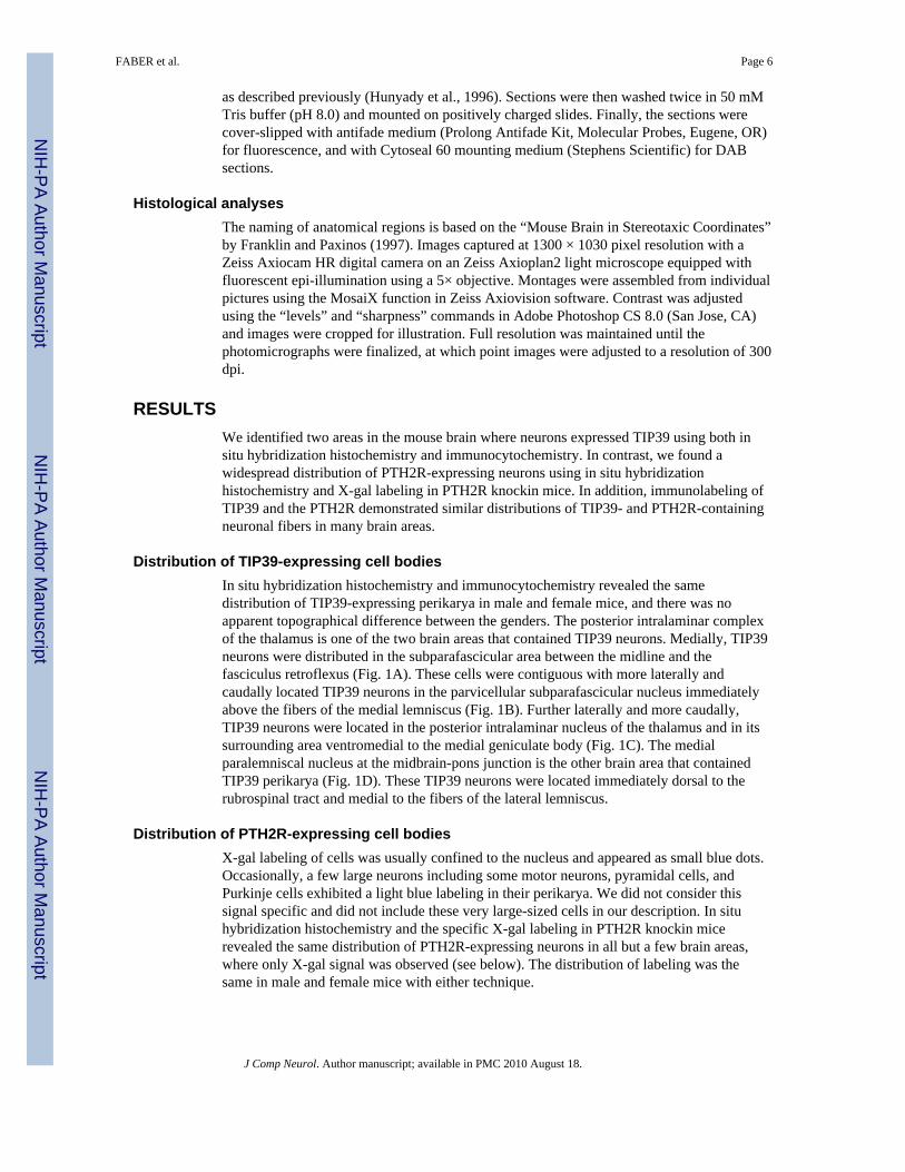
All procedures were performed according to approved National Institute of Mental Health(Bethesda, MD) animal care protocols and in accordance with the National Institutes ofHealth Guide for the Care and Use of Laboratory Animals. Experiments were performed onyoung adult (60–70 days old) mice (Taconic Farms, German-town, NY). All efforts weremade to minimize the number of animals used and their suffering. A total of 46 mice wereused for PTH2R in situ hybridization (two males and two females), X-gal histochemicalstaining (six PTH2R knockin males and three wildtype control males as well as six PTH2Rknockin females and three wildtype control females), PTH2R immunolabeling (six malesand six females), and TIP39 immunolabeling (six males and six females). Animals wereanesthetized with sodium pentobarbital (80 mg/kg i.p.) and then decapitated for in situhybridization or perfused for histochemistry and immunocytochemistry.
Knockin mice expressing β-galactosidase driven by the PTH2R promoterThe first protein coding exon of the mouse PTH2R gene was replaced by the bacterial lacZcoding sequence by homologous recombination in F1 (129Sv/Ev × c57Bl/6) hybrid mouseembryonic stem (ES) cells, as previously described (Valenzuela et al., 2003). Followingidentification of ES cells with appropriate recombination by the Regeneron group, the cellswere expanded and introduced into c57Bl/6 blastocysts in the NIMH transgenic mouse corefacility. Resulting mice were back-crossed with C57Bl/6 mice. Heterozygous animals wereused for the experiments in this study. Knockin mice were identified by polymerase chainreaction (PCR)-based genotyping on DNA from tail biopsies using primers for lacZ or oneprimer in the inserted sequence and one primer in the flanking gene sequence.
Primer sequences were: 5′-GCGCTGGTTGATTAGATACC, P2R_HDR2-B; 5′-GCTTCCTCGTGCTTTACGGTATC, Neo_3′b; and 5′-
FABER et al. Page 3
J Comp Neurol. Author manuscript; available in PMC 2010 August 18.
NIH
-PA Author Manuscript
NIH
-PA Author Manuscript
NIH
-PA Author Manuscript

GAGAGGCTGTTTGTAGAAGGCTGA, P2R_64264U, which produced bands of 260 base-pairs from the wildtype allele and 700 from the knockin allele.
X-gal labelingPTH2R knockin mice (six males and six females) as well as wild type control littermates(three males and three females) were used to visualize the β-galactosidase marker enzymehistochemically. Mice were anesthetized and perfused transcardially with 10 mL phosphate-buffered saline (PBS; pH 7.4) followed by 30 mL of cold 2% paraformaldehyde/0.2%glutaraldehyde mixture dissolved in PBS. Brains were removed, postfixed in 0.2%glutaraldehyde in PBS overnight, and coronal sections of 50 μm thickness were cut on avibrating microtome from bregma level of 3 mm to −8 mm. Sections were washed twice for10 minutes each in wash buffer (100 mM phosphate buffer at pH 7.4, 2 mM MgCl2, 0.01%Na-deoxycholate, and 0.02% octylphenol-ethylene oxide condensate [Nonidet P-40]). Thenthe sections were incubated in freshly made staining solution containing 5 mM potassiumferricyanide, 5 mM potassium ferrocyanide, and 1 mg/mL X-gal (5-bromo-4-chloro-3-indolyl-D-galactopyranoside) dissolved in wash buffer at room temperature in darknessovernight. Sections were then washed twice in 50 mM Tris buffer (pH 8.0), mounted onpositively charged slides, and cover-slipped with mounting medium (Cytoseal 60; StephensScientific, Kalamazoo, MI).
In situ hybridization histochemistryBrains were removed and quickly frozen on dry ice. Coronal sections (12 μm) were cutusing a cryostat from bregma level 3 mm to −8 mm, mounted on positively charged slides(SuperfrostPlus, Fisher Scientific, Pittsburgh, PA), dried, and stored at −80°C until use. Insitu hybridization protocols are described in detail on the Web(http://intramural.nimh.nih.gov/lcmr/snge/Protocols/ISHH/ISHH.html). [35S]UTP-labeledriboprobes were generated using a MAXIscript transcription kit (Ambion, Austin, TX) fromPCR-amplified fragments of the TIP39 cDNA subcloned into the vector pBluescript(Stratagene, La Jolla, CA). Antisense riboprobes were prepared using T7 RNA polymerase.A region of the rat TIP39 cDNA sequence corresponding to amino acids −55 to 37, whereamino acid 1 is the first residue of mature TIP39, was used to generate probes. We haveshown previously in rats that this antisense probe produces equivalent hybridization patternsto probes with nonoverlapping sequences corresponding to amino acids −55 to −18, and−17 to 37 (Dobolyi et al., 2003b). Similarly, a region of the rat PTH2R cDNA sequencecorresponding to bases 482–864 was used to generate probes. We have shown previouslythat this antisense probe produces equivalent hybridization patterns to probes with thenonoverlapping sequence corresponding to bases 1274–1828 (Wang et al., 2000). Followinghybridization and washes, slides were dipped in NTB2 nuclear track emulsion (EastmanKodak, Rochester, NY) and stored at 4°C for 3 weeks. Then the slides were developed andfixed with Kodak Dektol developer and Kodak fixer, respectively, counterstained withGiemsa, and coverslipped with mounting medium (Cytoseal 60; Stephens Scientific).
ImmunocytochemistryPTH2R antiserum—PTH2R was detected with an affinity-purified antiserum from arabbit immunized with the synthetic peptide RQIDSHVTLPGYVWSSSEQDC,corresponding to residues 480–500 of the rat PTH2R (GenBank Entry U55836), conjugatedto keyhole limpet hemocyanin. This antiserum (from rabbit #2710) has previously been usedand characterized (Usdin et al., 1999a; Wang et al., 2000) as summarized below. Thededuced rat and human receptor sequences differ at only 3 out of the 21 amino acids in thissequence, so we attempted to use cells expressing the cloned human receptor to examine thespecificity of the antiserum. Antiserum from two rabbits immunized with this peptide
FABER et al. Page 4
J Comp Neurol. Author manuscript; available in PMC 2010 August 18.
NIH
-PA Author Manuscript
NIH
-PA Author Manuscript
NIH
-PA Author Manuscript

produce strong immunolabeling of HEK293 cells stably expressing the human PTH2R.Preimmune serum does not label the PTH2R-expressing cells, and no labeling of either theparent HEK293 cells or HEK293 cells stably expressing the human PTH1R is detected.Similarly, there is intense labeling of 20–30% of COS-7 cells transfected with PTH2RcDNA but no labeling of cells in mock-transfected cultures. Several bands are labeled inWestern blots of PTH2R-enriched membranes, probably representing a combination ofmultiple glycosylation states and aggregation or oligomerization of the receptor. The highestmobility major band migrates with an apparent molecular weight of 84K, consistent with thesize seen following Western blotting of a C-terminal epitope labeled PTH2R, and labeling ofthe receptor with a radioactive photoaffinity ligand. Following digestion with PNGase F, themobility of the high mobility major band increases to an apparent molecular weight of 63K,consistent with the predicted size of the protein based on its cDNA sequence. No signal isseen in membranes prepared from the parent HEK293 cells or ones expressing PTH1R.Only 4 out of the 21 residues are the same as those in the human, mouse, or rat PTH1R andno significant labeling is detected in rat kidney tubules. Absorption of the antiserum with thepeptide used to generate it eliminates tissue labeling and specific staining is absent whenpreimmune serum is used to label tissue.
TIP39 antiserum—TIP39 was detected with an affinity-purified antiserum from a rabbitimmunized with mouse (m) TIP39 coupled to keyhole limpet hemocyanin by 1-ethyl-3-(3-dimethylaminopropyl)carbodiimide. This antiserum (pooled from rabbits #7250 and #7251)has previously been used and characterized (Dobolyi et al., 2002, 2003a,b) as summarizedbelow. The titer (50% maximum binding to immobilized peptide) of the affinity-purifiedanti-mTIP39 antiserum against mTIP39 was 3 ng/mL. Immunolabeling with the affinity-purified anti-mTIP39 was abolished by overnight preincubation at room temperature of aworking dilution of the antiserum with 1 μM synthetic mTIP39 (a sample treated identicallyexcept for omission of the peptide produced strong labeling). The anti-mTIP39 antiserumexhibited less than 1% crossreactivity with parathyroid hormone and no detectablecrossreactivity with other peptides tested including parathyroid hormone-related peptide,calcitonin, substance P, vasoactive intestinal peptide, glucagon, and calcitonin gene-relatedpeptide (Dobolyi et al., 2002). The anti-mTIP39 antiserum labels cell bodies in rat withexactly the same distribution as observed by in situ hybridization histochemistry with probesdirected against TIP39 mRNA (Dobolyi et al., 2003b). In addition, TIP39 immunolabelingdisappears from fibers following lesion of the distant TIP39 cell bodies (Dobolyi et al.,2003a).
Immunostaining protocol—Mice (12 males and 12 females) were anesthetized andperfused transcardially with 10 mL PBS followed by 30 mL ice-cold buffered (pH 7.4) 4%paraformaldehyde dissolved in PBS. The brains and spinal cords were removed, postfixed inbuffered 4% paraformaldehyde overnight, washed for at least 3 days with PBS (pH 7.4),embedded in 2% gelatin, and then 50-μm thick sections were cut with a vibrating microtomefrom bregma level of 3 mm to −8 mm in mice. Free-floating sections were then pretreatedwith 1% bovine serum albumin in PBS containing 0.5% Triton X-100 for 30 minutes atroom temperature. The sections were then placed in anti-TIP39 primary antiserum (1:3,000for tyramide amplification and 1:600 for DAB reaction) for 48 hours at room temperature oranti-PTH2 receptor primary antiserum (1:60,000 for tyramide amplification and 1:15,000 forDAB reaction) for 24 hours at room temperature as described previously (Wang et al., 2000;Dobolyi et al., 2003b). The sections were then incubated in biotinylated antirabbit secondaryantibody (1:600 dilution; Vector Laboratories, Burlingame, CA) for 2 hours followed byincubation in a solution containing avidin-biotin-peroxidase complex (1:150; VectorLaboratories) for 2 hours. The sections were then treated with 0.06% DAB or FITC-tyramide (1:20,000) and H2O2 in Tris hydrochloride buffer (0.1 M, pH 8.0) for 10 minutes
FABER et al. Page 5
J Comp Neurol. Author manuscript; available in PMC 2010 August 18.
NIH
-PA Author Manuscript
NIH
-PA Author Manuscript
NIH
-PA Author Manuscript
as described previously (Hunyady et al., 1996). Sections were then washed twice in 50 mMTris buffer (pH 8.0) and mounted on positively charged slides. Finally, the sections werecover-slipped with antifade medium (Prolong Antifade Kit, Molecular Probes, Eugene, OR)for fluorescence, and with Cytoseal 60 mounting medium (Stephens Scientific) for DABsections.
Histological analysesThe naming of anatomical regions is based on the “Mouse Brain in Stereotaxic Coordinates”by Franklin and Paxinos (1997). Images captured at 1300 × 1030 pixel resolution with aZeiss Axiocam HR digital camera on an Zeiss Axioplan2 light microscope equipped withfluorescent epi-illumination using a 5× objective. Montages were assembled from individualpictures using the MosaiX function in Zeiss Axiovision software. Contrast was adjustedusing the “levels” and “sharpness” commands in Adobe Photoshop CS 8.0 (San Jose, CA)and images were cropped for illustration. Full resolution was maintained until thephotomicrographs were finalized, at which point images were adjusted to a resolution of 300dpi.
RESULTSWe identified two areas in the mouse brain where neurons expressed TIP39 using both insitu hybridization histochemistry and immunocytochemistry. In contrast, we found awidespread distribution of PTH2R-expressing neurons using in situ hybridizationhistochemistry and X-gal labeling in PTH2R knockin mice. In addition, immunolabeling ofTIP39 and the PTH2R demonstrated similar distributions of TIP39- and PTH2R-containingneuronal fibers in many brain areas.
Distribution of TIP39-expressing cell bodiesIn situ hybridization histochemistry and immunocytochemistry revealed the samedistribution of TIP39-expressing perikarya in male and female mice, and there was noapparent topographical difference between the genders. The posterior intralaminar complexof the thalamus is one of the two brain areas that contained TIP39 neurons. Medially, TIP39neurons were distributed in the subparafascicular area between the midline and thefasciculus retroflexus (Fig. 1A). These cells were contiguous with more laterally andcaudally located TIP39 neurons in the parvicellular subparafascicular nucleus immediatelyabove the fibers of the medial lemniscus (Fig. 1B). Further laterally and more caudally,TIP39 neurons were located in the posterior intralaminar nucleus of the thalamus and in itssurrounding area ventromedial to the medial geniculate body (Fig. 1C). The medialparalemniscal nucleus at the midbrain-pons junction is the other brain area that containedTIP39 perikarya (Fig. 1D). These TIP39 neurons were located immediately dorsal to therubrospinal tract and medial to the fibers of the lateral lemniscus.
Distribution of PTH2R-expressing cell bodiesX-gal labeling of cells was usually confined to the nucleus and appeared as small blue dots.Occasionally, a few large neurons including some motor neurons, pyramidal cells, andPurkinje cells exhibited a light blue labeling in their perikarya. We did not consider thissignal specific and did not include these very large-sized cells in our description. In situhybridization histochemistry and the specific X-gal labeling in PTH2R knockin micerevealed the same distribution of PTH2R-expressing neurons in all but a few brain areas,where only X-gal signal was observed (see below). The distribution of labeling was thesame in male and female mice with either technique.
FABER et al. Page 6
J Comp Neurol. Author manuscript; available in PMC 2010 August 18.
NIH
-PA Author Manuscript
NIH
-PA Author Manuscript
NIH
-PA Author Manuscript

Cerebral cortex—PTH2R-expressing neurons were distributed throughout the cerebralcortex, with a density that varied somewhat between regions. The density of PTH2R-expressing neurons was the highest in the infra-limbic cortex, where they were found in allcortical layers. Some additional frontal cortical regions including the pre-limbic, anteriorcingulate, and insular cortices also exhibited a relatively higher density of PTH2R-expressing neurons. The density of PTH2R-expressing neurons decreased toward morecaudal levels of the cerebral cortex, except for an accumulation of PTH2R-expressingneurons in the superficial layers of the ectorhinal cortex. In addition, a higher density ofPTH2R-expressing neurons was found in layer 6b along the outer surface of the corpuscallosum throughout the cerebral cortex (Fig. 2A,C).
Limbic system—There were a few PTH2R-expressing neurons in the anterior olfactorynucleus and in the olfactory tubercle. In the septum, only the lateral nucleus containedPTH2R-expressing neurons. This region contained a very high density of PTH2R-expressingneurons, especially in its ventral part (Fig. 2D). More rostrally, the intermediate part of thelateral septal nucleus was also rich in PTH2R neurons. Most parts of the bed nucleus of thestria terminalis contained a moderate density of PTH2R-expressing neurons. In contrast, theposteromedial part of the medial subdivision of the bed nucleus of the stria terminaliscontained a very high density of PTH2R-expressing neurons (Fig. 3B). Many PTH2R-expressing neurons were present in the amygdala (Fig. 4C). The density of PTH2R-expressing neurons was high in the medial nucleus, especially in its posterodorsalsubdivision. A moderate density of neurons was found in the anterior amygdaloid area, thecentral and cortical amygdaloid nuclei, and the amygdala-hippocampal transitional zone. Alow density of scattered PTH2R-expressing neurons was present throughout thehippocampus, with a slightly higher density of PTH2R-expressing neurons in the dentategyrus and the subiculum.
In the basal ganglia, the claustrum and the dorsal endopiriform nucleus contained a fairlylarge number of PTH2R-expressing neurons (Fig. 4A,C), while there was a moderate densityof randomly distributed PTH2R-expressing neurons throughout the accumbens nucleus, thecaudate putamen (Fig. 2A,C), the ventral striatum, and the substantia innominata.
The thalamus was relatively poor in PTH2R-expressing neurons except for a few regions.The medial subdivision of the metathalamic medial geniculate body contained a very highdensity of PTH2R-expressing neurons, whereas its ventral subdivision contained a moderatedensity of PTH2R-expressing neurons. A high to moderate density of PTH2R-expressingneurons was detected in some midline and intralaminar thalamic nuclei including theparaventricular, centrolateral, paracentral, centromedian (Fig. 4D), and reuniens (Fig. 3C)thalamic nuclei. In addition, a moderate density of PTH2R-expressing neurons was presentin the epithalamic lateral habenular nucleus and the subthalamic zona incerta. Finally, X-gallabeling was present in the dorsal part of the lateral geniculate nucleus of PTH2R knockinmice, but in situ hybridization for the PTH2R did not indicate PTH2R-expressing neurons inthis brain area.
PTH2R-expressing neurons were abundant in many regions of the hypothalamus. In thepreoptic region, a high density of PTH2R-expressing neurons was present in the medialpreoptic nucleus, whereas a low density of PTH2R-expressing neurons was seen in otherparts of the medial preoptic area (Fig. 3A,B). In the anterior hypothalamic region a moderatedensity of PTH2R-expressing neurons was present in the paraventricular and periventricularnuclei, whereas the anterior hypothalamic nucleus contained a low density of PTH2R-expressing neurons (Fig. 3C). In the middle portion of the hypothalamus a high density ofPTH2R-expressing neurons was present in the arcuate nucleus, whereas a moderate densitywas observed in the dorsomedial and perifornical hypothalamic nuclei, and some parts of the
FABER et al. Page 7
J Comp Neurol. Author manuscript; available in PMC 2010 August 18.
NIH
-PA Author Manuscript
NIH
-PA Author Manuscript
NIH
-PA Author Manuscript

lateral hypothalamic area including the so-called far-lateral hypothalamus (Forel’s field)immediately next to the internal capsule (Fig. 3D). In the posterior hypothalamus a highdensity of PTH2R-expressing neurons was present in the medial subdivision of the superiormamillary nucleus (Fig. 5A,C), while its lateral subdivision (Fig. 5A,C), and the ventralpremamillary, and the tuberomamillary nuclei contained a moderate density of PTH2R-expressing neurons. In contrast, the medial and lateral nuclei of the mamillary body did notcontain PTH2R-expressing neurons.
There were relatively few PTH2R-expressing neurons in the lower brainstem andcerebellum. The midbrain contained a moderate density of PTH2R-expressing neurons inthe lateral interpeduncular and paranigral nuclei and in the medial raphe nucleus, and therewere a few scattered PTH2R-expressing neurons in the superior and inferior colliculi. In thepons there was a moderate to high density of PTH2R-expressing neurons in the sphenoidnucleus of the tegmental area (Fig. 6C), and in the nucleus of the trapezoid body (Fig. 6D).In addition, X-gal labeling was also present in the ventral cochlear nuclei (Fig. 6E). In themedulla the nucleus of the solitary tract contained a moderate density of PTH2R-expressingneurons and the spinal trigeminal nucleus contained a low density of PTH2R-expressingneurons. A few scattered PTH2R-expressing neurons were present in the cerebellar cortex,with a somewhat higher density in the superficial portion of the molecular layer.
TIP39- and PTH2R-containing neuronal networks and fibersWe observed the same distribution of TIP39-containing fibers in male and female mousebrains. Similarly, no difference was found in the distribution of PTH2R-containing fibersbetween males and females. Most strikingly, the distribution of TIP39-containing fibers wasvery similar to the distribution of PTH2R-containing fibers. In some brain regions, however,PTH2R-containing but not TIP39-containing fibers were observed.
The cerebral cortex, except for the medial prefrontal (Fig. 7A,B), the insular (Fig. 7C,D),and ectorhinal cortices, was largely devoid of TIP39- and PTH2R-containing fibers. In themedial prefrontal cortex the fibers were largely confined to the deep layers except within theinfralimbic cortex, where fibers were also observed in the superficial layers (Fig. 7A,B). Incontrast, in the insular (Fig. 7C,D) and ectorhinal cortices fibers were more abundant in thesuperficial layers. In addition, a few fine PTH2R-ir fibers were located along the outersurface of the corpus callosum, especially in the frontal cortices (Fig. 7B).
In the basal ganglia the core portion of the accumbens nucleus contained a moderate densityof PTH2R-ir fibers and fewer TIP39-ir fibers (Figs. 7A,B, 8). In contrast, the shell portion ofthe accumbens nucleus contained a moderate density of both TIP39-ir and PTH2R-ir fibers(Fig. 7A,B). In the bed nucleus of the stria terminalis the lateral subdivision contained a low,while the medial subdivision a moderate to high density of TIP39-ir and PTH2R-ir fibers. Inparticular, a very high density of both TIP39-ir and PTH2R-ir fibers was observed in theposteromedial part of the medial subdivision of the bed nucleus of the stria terminalis (Fig.9). The major part of the caudate-putamen was free of TIP39-ir and PTH2R-ir fibers (Fig. 8)except for a fiber bundle descending in its caudal and medial part from the stria terminalis,and its most caudal and ventral part, the fundus striati, which contained a moderately densenetwork of immunoreactive fibers (Fig. 11). The claustrum, the dorsal endopiriform nucleus,the ventral striatum, and the substantia innominata, including an area which overlays thenucleus basalis of Meynert, contained a low density of TIP39-ir and PTH2R-ir fibers.
Limbic system—There were a few TIP39-ir and PTH2R-ir fibers in the anterior olfactorynucleus and in the olfactory tubercle but not in the olfactory bulb. In the septum only thelateral nucleus contained TIP39-ir and PTH2R-ir fibers, but this region had a very highdensity of TIP39-containing fibers, especially in its intermediate and ventral parts (Fig. 8).
FABER et al. Page 8
J Comp Neurol. Author manuscript; available in PMC 2010 August 18.
NIH
-PA Author Manuscript
NIH
-PA Author Manuscript
NIH
-PA Author Manuscript

In this region some cells were surrounded with varicose fibers, and observation ofimmunostained sections at high magnification was necessary to determine that the cellbodies did not contain TIP39. Many TIP39-ir and PTH2R-ir fibers were present in theamygdala. The highest density of fibers was in the posterodorsal subdivision of the medialamygdaloid nucleus. The central nucleus (Fig. 11) and the amygdala-hippocampaltransitional zone (Fig. 12) contained a moderate density of TIP39-ir and PTH2R-ir fibers,although in the former the density of PTH2R-ir fibers was less than that of TIP39-ir fibers.There was a low density of fibers in the anterior amygdaloid area, the anterior subdivision ofthe medial amygdaloid nucleus, and in the basomedial and cortical amygdaloid nuclei (Fig.11), whereas the lateral and basolateral amygdaloid nuclei contained no TIP39-ir or PTH2R-ir fibers. Within the hippocampus only the ventral subiculum contained TIP39-ir andPTH2R-ir fibers (Fig. 12).
The thalamus was relatively poor in TIP39-ir and PTH2R-ir fibers except for a few regions.There was a high to moderate density of TIP39-ir fibers in the paraventricular and reuniens(Fig. 11A) thalamic nuclei. These two nuclei also contained a high density of PTH2R-irfibers (Fig. 11B), while other midline and intralaminar thalamic nuclei including thecentrolateral, paracentral, and centromedian thalamic nuclei had a low density of PTH2R-irfibers (Fig. 11B). Additional TIP39-ir and PTH2R-ir fibers were present in the lateralposterior and ethmoid thalamic nuclei and the lateral habenular nucleus. Apart from theseregions, TIP39-ir and PTH2R-ir fibers were also present in the vicinity of the TIP39-containing cell bodies in the subparafascicular area as well as in the area medial to themedial geniculate body (Fig. 12). From the latter region, TIP39-ir and PTH2R-ir fibers ranin the zona incerta between the subthalamic nucleus and the medial lemniscus to thehypothalamus as far medial as the paraventricular nucleus (Fig. 11). In addition, a moderatedensity of PTH2R-ir but not TIP39-ir fibers was found in the ventral part of the lateralgeniculate nucleus.
TIP39-ir and PTH2R-ir fibers were abundant in many regions of the hypothalamus. In thepreoptic region TIP39-ir and PTH2R-ir fibers had a high density in the medial preopticnucleus and a low density in other parts of the medial preoptic area and the lateral preopticarea (Fig. 9). In the anterior hypothalamus a high density of TIP39-ir and PTH2R-ir fiberswas present in the parvicellular subdivisions of the paraventricular nucleus and in theperiparaventricular zone (Fig. 10). The latter was particularly conspicuous on the side of themagnocellular subdivisions of the paraventricular nucleus, which lack of TIP39-ir andPTH2R-ir fibers (Fig. 10). A moderate density of TIP39-ir and PTH2R-ir fibers were presentin the periventricular nucleus and the anterior hypothalamic nucleus, whereas only a fewimmunoreactive fibers were seen in the lateral hypothalamic area and the supraopticnucleus, and no immunoreactive fibers were present in the suprachiasmatic nucleus (Fig.10). In the middle part of the hypothalamus, the ventrolateral subdivision of theventromedial nucleus, and the dorsomedial and arcuate nuclei contained a high density ofTIP39-ir and PTH2R-ir fibers (Fig. 11). A moderate density of TIP39-ir and PTH2R-irfibers was present in the perifornical nucleus and some parts of the lateral hypothalamic areaincluding the so-called far-lateral hypothalamus (Forel’s fields) immediately next to theinternal capsule (Fig. 11), but the immunolabeled fibers were sparse in most parts of thelateral hypothalamic area. In addition, a very dense accumulation of TIP39-containing fiberswas seen along both the dorsal and ventral surfaces of the internal capsule. Some of thefibers from the dorsal bundle seemed to penetrate into the internal capsule. The ventralbundle, which may correspond to the supraoptic decussations, is located between theinternal capsule and the optic tract (Fig. 11). Medially, fibers enter the lateral hypothalamusand cross over at the caudal edge of the optic chiasm (Fig. 11). In addition to theoverwhelming similarities between the distributions of the TIP39-ir and PTH2R-ir fibers andfiber terminals in this region of the hypothalamus, there were also some differences. A high
FABER et al. Page 9
J Comp Neurol. Author manuscript; available in PMC 2010 August 18.
NIH
-PA Author Manuscript
NIH
-PA Author Manuscript
NIH
-PA Author Manuscript

density of PTH2R-ir fibers but only a few TIP39-ir fibers were present in the medianeminence. In addition, there were no TIP39-ir fibers in the dorsomedial and centralsubdivisions of the ventromedial nucleus of the hypothalamus, whereas a low density ofPTH2R-ir fibers was visible here. In the posterior hypothalamus the highest density ofTIP39-ir and PTH2R-ir fibers was observed in the ventral premamillary, arcuate, and thetuberomamillary nuclei. In addition, TIP39-ir and PTH2R-ir fibers were also abundant in theposterior hypothalamic nucleus, the dorsal premamillary, supramamillary, and lateralmamillary nuclei. In contrast, the medial mamillary nuclei contained no labeled fibers.
In the midbrain a high density PTH2R-ir fibers and a somewhat lower density of TIP39-irfibers were present in the periaqueductal gray, especially its lateral and dorsal subdivisions,and the ventral tegmental area, while the deep layers of the superior colliculus contained amoderate density of PTH2R-ir fibers and a low density of TIP39-ir fibers (Fig. 12). Inaddition, there was a very dense accumulation of PTH2R-ir fibers but no TIP39-ir fibers inthe dorsal subdivision of the interpeduncular nucleus (Fig. 12). In contrast, a high density ofboth TIP39-ir and PTH2R-ir fibers was present in the deep mesencephalic nucleus (Fig. 12),the subbrachial nucleus, the intercollicular nucleus, and the external and dorsal cortices ofthe inferior colliculus, while the immunoreactive fibers were scarce in the central nucleus ofthe inferior colliculus. Fine immunoreactive fibers cross over in the commissures of thesuperior and inferior colliculi. TIP39-ir and PTH2R-ir fibers were also found at high densityin areas near the regions of the TIP39-containing cell bodies in the medial paralemniscalnucleus. Some immunoreactive fibers could also be seen in the pretectal area, the anteriorpretectal nucleus, the retrorubral field, the dorsal and median raphe nuclei, the cuneiformnucleus, and the dorsal nucleus of the lateral lemniscus. In the substantia nigra only the parslateralis contained immunoreactive fibers. The red nucleus and the superficial layers of thesuperior colliculus were also largely devoid of immunoreactive fibers.
In the pons PTH2R-ir fibers were abundant in the pedunculopontine tegmental nucleus, theparabrachial nuclei, the locus coeruleus and Barrington nucleus, the subcoeruleus area, thenucleus of the trapezoid body, the dorsal periolivary nucleus, the A5 noradrenergic cellgroup, and the raphe magnus nucleus (Fig. 13). While the distribution of TIP39-ir fibers wasvery similar in these regions, their density was considerably less in some of these regions,including the parabrachial nuclei, the nucleus of the trapezoid body, and the A5noradrenergic cell group (Fig. 13). A lower density of TIP39-ir and PTH2R-ir fibers wasalso observed in the supragenual nucleus, the Kölliker-Fuse nucleus, and the superior olive.Fine TIP39-ir and PTH2R-ir fibers could be seen to cross the midline at the ventral edge ofthe tegmentum.
There was no labeling for TIP39 and only a few scattered PTH2R-ir fibers were seen in thecerebellum. In the medulla a high density of PTH2R-ir fibers was present in the marginallayer of the spinal trigeminal nucleus and in the nucleus of the solitary tract. In contrast,there were only a few TIP39-ir fibers in the spinal trigeminal nucleus while the density ofTIP39-ir fibers in the nucleus of the solitary tract was low to moderate. Other medullaryregions that contained a low to moderate density of both TIP39-ir and PTH2R-ir fibersinclude the vestibular nuclei, the paragigantocellular nucleus, the lateral and intermediatereticular nuclei, the dorsal medullary reticular nucleus, and the cuneate nucleus.
A summary of the distribution of TIP39 and the PTH2R is shown schematically in Figure14. As demonstrated by the schematic panels the topographical distribution of TIP39-irfibers was, in general, very similar to that of the PTH2R-ir fibers. In addition, many brainregions that contained PTH2R-ir fibers also contained PTH2R-expressing neurons that had avery similar distribution.
FABER et al. Page 10
J Comp Neurol. Author manuscript; available in PMC 2010 August 18.
NIH
-PA Author Manuscript
NIH
-PA Author Manuscript
NIH
-PA Author Manuscript

DISCUSSIONWe first compare the distributions of TIP39-ir and PTH2R-containing cell bodies and fibersobtained using different methods. Then we discuss evidence for the existence of a TIP39-PTH2R neuromodulator system. Finally, we propose potential functions of the TIP39-PTH2R neuromodulator system based on its location in the mouse brain.
Methodological considerationsIn situ hybridization histochemistry directly visualizes the presence of RNA that iscomplementary to the sequence of the probe, while X-gal labeling indicates the presence ofβ-galactosidase activity, which appears in our knockin mice in cells in which the PTH2Rpromoter is active. The two different methods provided the same distribution of PTH2R-expressing cells in almost all regions, throughout many parts of the brain, which confirmstheir specificity. However, there were two brain regions, the dorsal part of the lateralgeniculate nucleus in the thalamus and the ventral cochlear nuclei, which exhibited a strongX-gal signal without apparent in situ hybridization signal. No X-gal signal was observed inthese regions in control material, so we suggest that it results either from ectopic expressionof the transgene, because of insertion of foreign sequences, or mismatch between thesensitivities of the two techniques. Since PTH2R-ir fibers were not detected in these regions,we believe that the former is a more likely explanation.
The distribution of PTH2R-ir cell bodies previously described in rat (Wang et al., 2000) is,in general, similar to the distribution of PTH2R-expressing cells we report for mouse in thisstudy. However, in this study we observed that the immunolabeling of cell bodies in mice isnot reliable. Using the same staining protocol as in rats, we found many fewer and muchmore lightly labeled cell bodies, if any at all, in most brain regions in mice. Theimmunolabeling of cell bodies did not improve using an amplificationimmunocytochemistry staining protocol. We believe that the transport of PTH2R protein outof cell bodies and into neuronal fibers may be faster in mice than in rats. Therefore, we didnot use immunocytochemistry for the description of the distribution of PTH2R-expressingcell bodies. In contrast, the immunolabeling of TIP39-containing cell bodies was satisfactoryas demonstrated by the same distribution of the TIP39 in situ hybridization signal and theTIP39-ir cell bodies.
The distribution of nerve fibers and terminals was described based on an FITC-tyramineamplification immunostaining protocol. Using this protocol the labeling was comparable tothe labeling of fibers in rats using the same technique. However, in rats the distribution ofPTH2R-ir fibers has not been described previously using amplificationimmunocytochemistry except for the hypothalamus (Dobolyi et al., 2006). Since the labelingof fibers by FITC-tyramine amplification immunostaining is more intense than by thetraditional DAB technique, we were able to analyze PTH2R-ir fibers throughout the brain inmuch more detail than in previous studies (Wang et al., 2000).
Comparison of the distributions of TIP39 and the PTH2R between mice and ratsThe distribution of TIP39-expressing cell bodies determined in this study in mice was thesame as that previously reported in rats (Dobolyi et al., 2003b). The distribution of TIP39-irfibers in mice was almost the same as that in rats. Only two notable differences weredetected by labeling for TIP39 and scanning a comprehensive set of mouse and rat brainsections. The density of TIP39-ir fibers was high in the medial preoptic nucleus and theventrolateral subdivision of the ventromedial hypothalamic nucleus in mice while onlymoderate in rats.
FABER et al. Page 11
J Comp Neurol. Author manuscript; available in PMC 2010 August 18.
NIH
-PA Author Manuscript
NIH
-PA Author Manuscript
NIH
-PA Author Manuscript

The distribution of PTH2R-expressing cell bodies in mice was similar to that previouslyreported in rats (Wang et al., 2000) in most brain regions, including the cerebral cortex, thecaudate putamen, the claustrum and endopiriform nucleus, the lateral septum, thehippocampus, the amygdala, and many thalamic, hypothalamic, and brainstem regions, andthe cerebellum. There were, however, brain regions where marked differences were found inthe density of PTH2R-expressing neurons between mice and rats. Most particularly, a strongin situ hybridization signal but no more than a few immunoreactive cell bodies were foundin the medial septal, the ventromedial hypothalamic, the parabrachial, and the spinaltrigeminal nuclei in rats (Wang et al., 2000). None of these regions contained a significantnumber of PTH2R-expressing cells in mice. In addition, both in situ hybridizationhistochemistry and immunocytochemistry revealed a high density of PTH2R-expressingneurons in the periventricular hypothalamic nucleus in rat, while this area contained only amoderate number of PTH2R-expressing neurons in mice. In contrast, there are brain regionsthat were abundant in PTH2R-expressing neurons in mice but do not contain a large numberof PTH2R-expressing neurons in rat, including the posteromedial part of the medial divisionof the bed nucleus of the stria terminalis, the medial geniculate body, and the nucleus of thetrapezoid body.
The distribution of PTH2R-ir fibers is also very similar between mice and rats in most brainregions including the septum, many basal ganglia nuclei, the hippocampus, the amygdala,the midline thalamic nuclei, and many hypothalamic and brainstem areas. The few notabledifferences when comparing our current data in mice to previously published data in rats(Wang et al., 2000) were a high to moderate density of PTH2R-ir fibers in the infralimbicand ectorhinal cortices, the ventral subiculum, the posteromedial part of the medialsubdivision of the bed nucleus of the stria terminalis, the fundus striati, the zona incerta, theposterior intralaminar thalamic nuclei, the periaqueductal gray, and ventral tegmental area,and the medial parabrachial nuclei in mice and a lower density of PTH2R-ir fibers in thesebrain regions of rats (Wang et al., 2000). In turn, there are brain regions, where PTH2R-irfibers were reported in rat but were not observed in mice, including the suprachiasmaticnucleus, the pars compacta and pars reticularis of the substantia nigra, the superficial layersof the superior colliculus, and the inferior olive (Wang et al., 2000). We believe that some ofthese differences derive from the immunostaining method used and do not represent realspecies differences. As discussed above, amplification immunocytochemistry and nottraditional DAB immunolabeling was used in the present study.
Comparison of the distribution of TIP39 to that of the PTH2R provides anatomical evidencefor a TIP39-PTH2R neuromodulator system
The localization of cell bodies that express TIP39 and those that express the PTH2R areprofoundly different. TIP39 expression is confined to the subparafascicular area-posteriorintralamine thalmic complex and the medial paraleminiscal nucleus, while the PTH2R isexpressed in many brain regions. In contrast to the profoundly different localization ofTIP39-ir and PTH2R-expressing cell bodies, the distributions of TIP39-ir and PTH2R-irfibers are markedly similar. TIP39-ir and PTH2R-ir fibers are present in the same nuclei andareas throughout the brain except for a few areas where PTH2R-ir but not TIP39-ir fibers areabundant including the median eminence, the interpeduncular, and the spinal trigeminalnuclei. Furthermore, not only are TIP39-ir and PTH2R-ir fibers present in the same brainregions but their subregional distributions also show remarkable similarities. In fact, the twodistributions are indistinguishable in most brain nuclei and areas. This finding suggests thatTIP39 is available to act on the PTH2R in these brain regions and furthermore that it mayact in a fairly traditional manner, with its actions focused on adjacent fibers. Together withthe strong in vitro pharmacological evidence that TIP39 is a potent and high-affinity ligandfor PTH2R, our anatomical data suggest that TIP39 is the endogenous ligand of the PTH2R.
FABER et al. Page 12
J Comp Neurol. Author manuscript; available in PMC 2010 August 18.
NIH
-PA Author Manuscript
NIH
-PA Author Manuscript
NIH
-PA Author Manuscript

Furthermore, we propose that TIP39 and the PTH2R form a neuromodulator system in manybrain regions. TIP39-ir fibers are distant axons of the TIP39-expressing perikarya in thesubparafascicular-ir posterior intralaminar complex and in the medial paralemniscal nuclei,which disappear following the destruction of their cell bodies (Dobolyi et al., 2003a).PTH2R-ir fibers, however, are often localized in the vicinity of PTH2R-expressing neurons.Therefore, they may represent either axons or dendrites on which TIP39 could act via thePTH2R. The finding that in a few areas PTH2R’s but not TIP39 were present could beexplained by the appearance of TIP39 in these areas under specific physiological conditions,although a nonfunctional expression of the PTH2R, or the existence of another ligand for thePTH2R in these areas cannot be excluded. It is possible that circulating TIP39 acts onPTH2R’s in the median eminence, but a source of such TIP39 has not been observed.
Functional implications of the distribution of the TIP39-PTH2R neuromodulator systemOn the basis of neuroanatomical and pharmacological observations, we proposed above thatTIP39 and PTH2R constitute a TIP39-PTH2R neuromodulator system in the central nervoussystem. The distribution of the TIP39-PTH2R neuromodulator system suggests itsinvolvement in limbic functions. Many of these functions involve several brain centers. Herewe mention some functions in which the role of the TIP39-PTH2R neuromodulator systemis supported by its localization in several of the involved brain centers and/or by initialfunctional studies.
The role of the TIP39-PTH2R neuromodulator system in male sexual behaviors is supportedby its presence in several areas that are activated in males following mating (Sachs andMeisel, 1988; Coolen et al., 1997; Veening and Coolen, 1998; Holstege et al., 2003)including the medial preoptic nucleus, the posteromedial part of the medial subdivision ofthe bed nucleus of the stria terminalis, the subparafascicular area, and the posterodorsalsubdivision of the medial amygdaloid nuclei. Indeed, TIP39 neurons in the subparafasciculararea have been recently shown to exhibit c-fos activation following male sexual behavior inrats (Wang et al., 2006a). Additional brain regions have also been suggested to be parts of aninterconnected network of reproductive brain centers and play a role in female sexualbehaviors, lactation, and female maternal behaviors including the medial prefrontal cortex,the lateral septum, the medial preoptic, periventricular hypothalamic, arcuate andpremamillary nuclei, the amygdalo-hippocampal transitional zone, the ventral tegmentalarea, and the periaqueductal gray (Gorski, 1985; Sachs and Meisel, 1988; Lonstein andStern, 1997; Numan and Sheehan, 1997; Pfaus and Heeb, 1997; Li et al., 1998, 1999;Veening and Coolen, 1998; Simerly, 2002; Holstege et al., 2003; Numan and Insel, 2003;Hasen and Gammie, 2005). The TIP39-PTH2R neuromodulator system is abundant in thesebrain regions, suggesting a role in reproductive behaviors.
Another potential function of the TIP39-PTH2R neuromodulator system is involvement inthe processing of nociceptive and vegetative sensory input to the limbic system, which issuggested by its presence in brain areas that may participate in such information processing(Benarroch, 2006) including the nucleus of the solitary tract, the parabrachial nuclei, themidline thalamic nuclei, the paraventricular hypothalamic nucleus, the insular andinfralimbic cortices. This suggestion is also supported by our previous finding thatintracerebroventricular injection of TIP39 decreases pain-related affective behavior (LaBudaand Usdin, 2004).
Possibly related to limbic functions of the TIP39-PTH2R neuromodulator system is itsoptimal position to influence the hypothalamo-pituitary axis through its presence in thepreoptic area, the arcuate, periventricular and paraventricular hypothalamic nuclei. Indeed,in vitro evidence suggests that TIP39 may influence the release of luteinizing hormone,
FABER et al. Page 13
J Comp Neurol. Author manuscript; available in PMC 2010 August 18.
NIH
-PA Author Manuscript
NIH
-PA Author Manuscript
NIH
-PA Author Manuscript

growth hormone, and adrenocorticotropin from the pituitary (Ward et al., 2001; Usdin et al.,2003).
The TIP39-PTH2R neuromodulator system may also be involved in the processing ofauditory information because it is present in several auditory brain centers including theectorhinal cortex, the medial geniculate body and surrounding auditory thalamic areas, thedeep layers of the superior colliculus, the external cortex of the inferior colliculus, thenucleus of the trapezoid body, and possibly the ventral cochlear nuclei. In the higherauditory centers the TIP39-PTH2R neuromodulator system is typically located innontonotopically organized regions. Therefore, we propose that the TIP39-PTH2Rneuromodulator system may modulate auditory processes that do not require tonotopicinformation such as acoustic fear-conditioning or the loud noise-induced stress response.Such potential functions of the TIP39-PTH2R neuromodulator system are supported by theloud noise-induced c-fos activation in posterior intralaminar and medial paralemniscalTIP39 neurons (Palkovits et al., 2004).
In conclusion, the present article first described the precise distribution of PTH2R-expressing cell bodies by using knockin mice expressing β-galactosidase driven by thePTH2R promoter and comparing the location of β-galactosidase-positive cells to the locationof PTH2R mRNA-containing cells visualized by in situ hybridization histochemistry. Inaddition, a very good correlation was found between the distributions of TIP39-containingfibers and PTH2R-containing cell bodies and fibers throughout the brain. Furthermore, eventhe subregional distribution of TIP39-ir and PTH2R-ir fibers showed remarkablesimilarities, providing anatomical evidence that TIP39 may act on the PTH2R. Based on thisfinding and on previous pharmacological evidence, we propose that TIP39 is an endogenousligand of the PTH2R and that they form a neuromodulator system, which is optimallypositioned to regulate limbic, endocrine, and auditory brain functions.
AcknowledgmentsWe thank Andrew Murphy, David Valenzuela, and George Yancopolos for help with the generation of the targetedES cells, Miklós Palkovits for critically reading the article, Charles R. Gerfen for initial instructions in the scanningprocedure, Timothy Reardon for technical support, and Jim Pickel and the NIMH transgenic core facility forestablishing the PTH2R knockin line.
Grant sponsor: National Institute of Mental Health Intramural Research Program.
Abbreviations
A5 A5 noradrenergic cell group
ac Anterior commissure
Acc Accumbens nucleus
AH Anterior hypothalamic nucleus
AHi Amygdalo-hippocampal transitional area
Arc Arcuate nucleus
BST Bed nucleus of the stria terminalis
BSTMPM BST, medial subdivision, posteromedial part
cc Corpus callosum
Ce Cerebellum
FABER et al. Page 14
J Comp Neurol. Author manuscript; available in PMC 2010 August 18.
NIH
-PA Author Manuscript
NIH
-PA Author Manuscript
NIH
-PA Author Manuscript

CeA Central amygdaloid nucleus
CoA Cortical amygdaloid nucleus
CL Centrolateral thalamic nucleus
Cl Claustrum
CM Centromedian thalamic nucleus
CP Caudate putamen
Cx Cerebral cortex
DEn Dorsal endopiriform nucleus
DM Dorsomedial hypothalamic nucleus
DP Dorsal peduncular cortex
DpMe Deep mesencephalic nucleus
DR Dorsal raphe nucleus
Ect Ectorhinal cortex
f Fornix
fr Fasciculus retroflexus
FS Fundus striati
H Hippocampus
ic Internal capsule
IL Infralimbic cortex
Ins Insular cortex
IP Interpeduncular nucleus
LC Locus coeruleus
LDTg Laterodorsal tegmental nucleus
LH Lateral hypothalamic area
ll Lateral lemniscus
LPO Lateral preoptic area
LS Lateral septal nucleus
LSi Lateral septal nucleus, intermediate subdivision
LSv Lateral septal nucleus, ventral subdivision
LV Lateral ventricle
m Midline
MeA Medial amygdaloid nucleus
MG Medial geniculate body
MGD Medial geniculate nucleus, dorsal subdivision
MGM Medial geniculate nucleus, medial subdivision
MGV Medial geniculate nucleus, ventral subdivision
FABER et al. Page 15
J Comp Neurol. Author manuscript; available in PMC 2010 August 18.
NIH
-PA Author Manuscript
NIH
-PA Author Manuscript
NIH
-PA Author Manuscript

ml Medial lemniscus
MPN Medial preoptic nucleus
MRe Intramamillary recess of the third ventricle
MS Medial septum
mSPF Medial subparafascicular area
mt Mamillothalamic tract
ot Optic tract
PAG Periaqueductal gray
PB Parabrachial nuclei
PC Paracentral thalamic nucleus
pc Posterior commissure
Pe Periventricular hypothalamic nucleus
Pir Piriform cortex
PIL Posterior intralaminar nucleus of the thalamus
POA Periolivary area
PTH2R Parathyroid hormone 2 receptor
PVN Paraventricular hypothalamic nucleus
PVT Paraventricular thalamic nucleus
py Pyramidal tract
Re Reuniens thalamic nucleus
RMg Raphe magnus nucleus
rs Rubrospinal tract
S Subiculum
SC Superior colliculus
SCh Suprachiasmatic nucleus
scp Superior cerebellar peduncle
sm Stria medullaris
SPF Subparafascicular area
SPFp Parvicellular subparafascicular nucleus
Sph Sphenoid nucleus
SubC Subcoeruleus area
SuMM Supramamillary nucleus, medial subdivision
TIP39 Tuberoinfundibular peptide of 39 residues
Tz Nucleus of the trapezoid body
VMH Ventromedial hypothalamic nucleus
VTA Ventral tegmental area
FABER et al. Page 16
J Comp Neurol. Author manuscript; available in PMC 2010 August 18.
NIH
-PA Author Manuscript
NIH
-PA Author Manuscript
NIH
-PA Author Manuscript

ZI Zona incerta
3V Third ventricle
4V Fourth ventricle
7n Root of the facial nerve
LITERATURE CITEDBenarroch EE. Pain-autonomic interactions. Neurol Sci 2006;27(Suppl 2):S130–133. [PubMed:
16688616]Coolen LM, Peters HJ, Veening JG. Distribution of fos immunoreactivity following mating versus
anogenital investigation in the male rat brain. Neuroscience 1997;77:1151–1161. [PubMed:9130794]
Coolen LM, Allard J, Truitt WA, McKenna KE. Central regulation of ejaculation. Physiol Behav2004;83:203–215. [PubMed: 15488540]
Dobolyi A, Ueda H, Uchida H, Palkovits M, Usdin TB. Anatomical and physiological evidence forinvolvement of tuberoinfundibular peptide of 39 residues in nociception. Proc Natl Acad Sci U S A2002;99:1651–1656. [PubMed: 11818570]
Dobolyi A, Palkovits M, Bodnar I, Usdin TB. Neurons containing tuberoinfundibular peptide of 39residues project to limbic, endocrine, auditory and spinal areas in rat. Neuroscience 2003a;122:1093–1105. [PubMed: 14643775]
Dobolyi A, Palkovits M, Usdin TB. Expression and distribution of tuberoinfundibular peptide of 39residues in the rat central nervous system. J Comp Neurol 2003b;455:547–566. [PubMed:12508326]
Dobolyi A, Irwin S, Wang J, Usdin TB. The distribution and neurochemistry of the parathyroidhormone 2 receptor in the rat hypothalamus. Neurochem Res 2006;31:227–236. [PubMed:16570212]
Franklin, KBJ.; Paxinos, G. The mouse brain in stereotaxic coordinates. San Diego: Academic Press;1997.
Gorski RA. Sexual dimorphisms of the brain. J Anim Sci 1985;61(Suppl 3):38–61. [PubMed:3908433]
Harmar AJ. Family-B G-protein-coupled receptors. Genome Biol 2001;2:3013.Hasen NS, Gammie SC. Differential fos activation in virgin and lactating mice in response to an
intruder. Physiol Behav 2005;84:681–695. [PubMed: 15885244]Herkenham M. Mismatches between neurotransmitter and receptor localizations in brain: observations
and implications. Neuroscience 1987;23:1–38. [PubMed: 2891080]Holstege G, Georgiadis JR, Paans AM, Meiners LC, van der Graaf FH, Reinders AA. Brain activation
during human male ejaculation. J Neurosci 2003;23:9185–9193. [PubMed: 14534252]Hunyady B, Krempels K, Harta G, Mezey E. Immunohistochemical signal amplification by catalyzed
reporter deposition and its application in double immunostaining. J Histochem Cytochem1996;44:1353–1362. [PubMed: 8985127]
John MR, Arai M, Rubin DA, Jonsson KB, Juppner H. Identification and characterization of themurine and human gene encoding the tuberoinfundibular peptide of 39 residues. Endocrinology2002;143:1047–1057. [PubMed: 11861531]
LaBuda CJ, Usdin TB. Tuberoinfundibular peptide of 39 residues decreases pain-related affectivebehavior. Neuroreport 2004;15:1779–1782. [PubMed: 15257146]
LaBuda CJ, Dobolyi A, Usdin TB. Tuberoinfundibular peptide of 39 residues produces anxiolytic andantidepressant actions. Neuroreport 2004;15:881–885. [PubMed: 15073536]
Leng G, Ludwig M. Jacques Benoit lecture. Information processing in the hypothalamus: peptides andanalogue computation. J Neuroendocrinol 2006;18:379–392. [PubMed: 16684129]
FABER et al. Page 17
J Comp Neurol. Author manuscript; available in PMC 2010 August 18.
NIH
-PA Author Manuscript
NIH
-PA Author Manuscript
NIH
-PA Author Manuscript

Li C, Chen P, Smith MS. Neural populations in the rat forebrain and brainstem activated by thesuckling stimulus as demonstrated by c-fos expression. Neuroscience 1999;94:117–129. [PubMed:10613502]
Lin SH, Miyata S, Matsunaga W, Kawarabayashi T, Nakashima T, Kiyohara T. Metabolic mapping ofthe brain in pregnant, parturient and lactating rats using fos immunohistochemistry. Brain Res1998;787:226–236. [PubMed: 9518626]
Lonstein JS, Stern JM. Role of the midbrain periaqueductal gray in maternal nurturance andaggression: C-fos and electrolytic lesion studies in lactating rats. J Neurosci 1997;17:3364–3378.[PubMed: 9113892]
Numan, M.; Insel, TR. The neurobiology of parental behavior. New York: Springer; 2003.Numan M, Sheehan TP. Neuroanatomical circuitry for mammalian maternal behavior. Ann N Y Acad
Sci 1997;807:101–125. [PubMed: 9071346]Palkovits M, Dobolyi A, Helfferich F, Usdin TB. Localization and chemical characterization of the
audiogenic stress pathway. Ann N Y Acad Sci 2004;1018:16–24. [PubMed: 15240348]Pfaus JG, Heeb MM. Implications of immediate-early gene induction in the brain following sexual
stimulation of female and male rodents. Brain Res Bull 1997;44:397–407. [PubMed: 9370204]Sachs, BD.; Meisel, RL. The physiology of male sexual behavior. In: Knobil, E.; Meill, J., editors. The
physiology of reproduction. New York: Raven Press; 1988.Simerly RB. Wired for reproduction: Organization and development of sexually dimorphic circuits in
the mammalian forebrain. Annu Rev Neurosci 2002;25:507–536. [PubMed: 12052919]Sugimura Y, Murase T, Ishizaki S, Tachikawa K, Arima H, Miura Y, Usdin TB, Oiso Y. Centrally
administered tuberoinfundibular peptide of 39 residues inhibits arginine vasopressin release inconscious rats. Endocrinology 2003;144:2791–2796. [PubMed: 12810532]
Usdin TB. The PTH2 receptor and TIP39: a new peptide-receptor system. Trends Pharmacol Sci2000;21:128–130. [PubMed: 10740285]
Usdin TB, Gruber C, Bonner TI. Identification and functional expression of a receptor selectivelyrecognizing parathyroid hormone, the PTH2 receptor. J Biol Chem 1995;270:15455–15458.[PubMed: 7797535]
Usdin TB, Hilton J, Vertesi T, Harta G, Segre G, Mezey E. Distribution of the parathyroid hormone 2receptor in rat: immunolocalization reveals expression by several endocrine cells. Endocrinology1999a;140:3363–3371. [PubMed: 10385434]
Usdin TB, Hoare SR, Wang T, Mezey E, Kowalak JA. TIP39: a new neuropeptide and PTH2-receptoragonist from hypothalamus. Nat Neurosci 1999b;2:941–943. [PubMed: 10526330]
Usdin TB, Bonner TI, Hoare SR. The parathyroid hormone 2 (PTH2) receptor. Receptors Channels2002;8:211–218. [PubMed: 12529938]
Usdin TB, Dobolyi A, Ueda H, Palkovits M. Emerging functions for tuberoinfundibular peptide of 39residues. Trends Endocrinol Metab 2003;14:14–19. [PubMed: 12475607]
Valenzuela DM, Murphy AJ, Frendewey D, Gale NW, Economides AN, Auerbach W, PoueymirouWT, Adams NC, Rojas J, Yasenchak J, Chernomorsky R, Boucher M, Elsasser AL, Esau L, ZhengJ, Griffiths JA, Wang X, Su H, Xue Y, Dominguez MG, Noguera I, Torres R, Macdonald LE,Stewart AF, DeChiara TM, Yancopoulos GD. High-throughput engineering of the mouse genomecoupled with high-resolution expression analysis. Nat Biotechnol 2003;21:652–659. [PubMed:12730667]
Veening JG, Coolen LM. Neural activation following sexual behavior in the male and female rat brain.Behav Brain Res 1998;92:181–193. [PubMed: 9638960]
Wang T, Palkovits M, Rusnak M, Mezey E, Usdin TB. Distribution of parathyroid hormone-2receptor-like immunoreactivity and messenger rna in the rat nervous system. Neuroscience2000;100:629–649. [PubMed: 11098126]
Wang J, Coolen LM, Brown JL, Usdin TB. Neurons containing tuberoinfundibular peptide of 39residues are activated following male sexual behavior. Neuropeptides 2006a;40:403–408.[PubMed: 17056109]
Wang J, Palkovits M, Usdin TB, Dobolyi A. Forebrain projections of tuberoinfundibular peptide of 39residues (TIP39)-containing subparafascicular neurons. Neuroscience 2006b;138:1245–1263.[PubMed: 16458435]
FABER et al. Page 18
J Comp Neurol. Author manuscript; available in PMC 2010 August 18.
NIH
-PA Author Manuscript
NIH
-PA Author Manuscript
NIH
-PA Author Manuscript

Ward HL, Small CJ, Murphy KG, Kennedy AR, Ghatei MA, Bloom SR. The actions oftuberoinfundibular peptide on the hypothalamo-pituitary axes. Endocrinology 2001;142:3451–3456. AQ—AQ1: No scale bar in figure. [PubMed: 11459790]
FABER et al. Page 19
J Comp Neurol. Author manuscript; available in PMC 2010 August 18.
NIH
-PA Author Manuscript
NIH
-PA Author Manuscript
NIH
-PA Author Manuscript

Fig. 1.TIP39-containing neurons in the mouse brain. A1–3: TIP39+ neurons in the medialsubparafascicular area at the thalamus-midbrain junction. B1–3: TIP39+ neurons continueinto the parvicellular subparafascicular nucleus. C1–3: TIP39+ neurons continue furtherlaterally in the posterior intralaminar nucleus of the thalamus and surrounding areas medialto the medial geniculate body. D1–3: A separate group of TIP39+ neurons in the medialparalemniscal nucleus at the midbrain-pons junction. A1, B1, C1, D1: TIP39 mRNAexpression is demonstrated in darkfield following in situ hybridization histochemistry. A2,B2, C2, D2: TIP39 immunoreactivity is demonstrated by following fluorescent amplificationimmunocytochemistry. TIP39+ cell bodies and TIP39+ fibers are visible in the same areas.A3, B3, C3, D3: Schematic diagrams of brain regions containing TIP39+ cells. Arrows pointto the location of brain regions that contain TIP39+ neurons. Scale bar = 1 mm.
FABER et al. Page 20
J Comp Neurol. Author manuscript; available in PMC 2010 August 18.
NIH
-PA Author Manuscript
NIH
-PA Author Manuscript
NIH
-PA Author Manuscript

Fig. 2.PTH2R-expressing neurons in the septal region of the mouse brain. A: Darkfield image ofan in situ hybridization section demonstrates the location of PTH2R mRNA in the cerebralcortex, the caudate putamen, and the septum. B: Schematic diagram of the areacorresponding to A. C: Scattered PTH2R+ neurons are demonstrated by X-galhistochemistry in the cerebral cortex and the caudate putamen of a PTH2R knockin mouse.In addition, arrowheads indicate PTH2R+ neurons lined up in the innermost layer of thecerebral cortex along the corpus callosum. The photomicrograph corresponds to the areaframed in A. D: X-gal histochemistry in the septal region of a PTH2R knockin mouse. Thephotomicrograph corresponds to the area framed in A. PTH2R+ neurons are abundant in theventral subdivision as well as in the ventral part of the intermediate subdivision of the lateralseptal nucleus. Scale bars = 1 mm in A; 300 μm in C; 500 μm in D.
FABER et al. Page 21
J Comp Neurol. Author manuscript; available in PMC 2010 August 18.
NIH
-PA Author Manuscript
NIH
-PA Author Manuscript
NIH
-PA Author Manuscript

Fig. 3.PTH2R-expressing neurons in preoptic and hypothalamic regions of the mouse braindemonstrated by X-gal histochemistry in PTH2R knockin mice. A: PTH2R+ neurons in themedial preoptic nucleus. B: PTH2R+ neurons are particularly abundant in the posteromedialpart of the medial subdivision of the bed nucleus of the stria terminalis (arrows). C:PTH2R+ neurons in the paraventricular and periventricular hypothalamic nuclei. PTH2R+
neurons are also present in the reuniens thalamic nucleus. D: The density of PTH2R+
neurons is very high in the arcuate nucleus and high in the lateral hypothalamic area, theperifornical, and dorsomedial hypothalamic nuclei. Scale bars = 300 μm.
FABER et al. Page 22
J Comp Neurol. Author manuscript; available in PMC 2010 August 18.
NIH
-PA Author Manuscript
NIH
-PA Author Manuscript
NIH
-PA Author Manuscript

Fig. 4.PTH2R-expressing neurons in the amygdala and dience-phalic regions of the mouse brain.A: Darkfield image of an in situ hybridization section demonstrates the location of PTH2RmRNA in the amygdala, the midline thalamic nuclei, and the tuberal region of thehypothalamus. B: Schematic diagram of the area corresponding to A. C: PTH2R+ neuronsare demonstrated by X-gal histochemistry in the amygdala of a PTH2R knockin mouse. Thelabeling is intense in the central, medial, and cortical amygdaloid nuclei as well as in thedorsal endopiriform nucleus. The photomicrograph corresponds to the area framed in A. D:X-gal histochemical labeling in the midline thalamic nuclei of a PTH2R knockin mouse.The photomicrograph corresponds to the area framed in A. PTH2R+ neurons are abundant inthe paraventricular, centromedian, centrolateral, and paracentral thalamic nuclei. Thehabenula also contains PTH2R+ neurons. Scale bars = 1 mm in A; 500 μm in C; 400 μm inD.
FABER et al. Page 23
J Comp Neurol. Author manuscript; available in PMC 2010 August 18.
NIH
-PA Author Manuscript
NIH
-PA Author Manuscript
NIH
-PA Author Manuscript

Fig. 5.PTH2R-expressing neurons at the level of the diencephalon–midbrain junction in the mousebrain. A: PTH2R mRNA is shown in a darkfield photomicrograph of an in situ hybridizationsection. PTH2R+ neurons are abundant in the medial geniculate body, the supramamillarynucleus, the dorsal endopiriform nucleus, the amygdalo-hippocampal transitional zone, andthe ventral subiculum. Scattered PTH2R+ neurons are also present in the cerebral cortex, thehippocampus, and the superior colliculus. B, C: PTH2R+ neurons are demonstrated by X-galhistochemistry in a PTH2R knockin mouse in areas that correspond to the areas framed in A.In the medial geniculate body (B) the labeling is particularly intense in the medialsubdivision of the medial geniculate nucleus. A relatively high density of PTH2R+ neuronsis also present in the ventral subdivision of the medial geniculate nucleus and in theposterior intralaminar thalamic nucleus. Within the supramamillary nucleus (C) the labelingis particularly intense in the medial subdivision. Scale bars = 1 mm in A; 400 μm in B; 500μm in C.
FABER et al. Page 24
J Comp Neurol. Author manuscript; available in PMC 2010 August 18.
NIH
-PA Author Manuscript
NIH
-PA Author Manuscript
NIH
-PA Author Manuscript

Fig. 6.PTH2R-expressing neurons at the level of the pons in the mouse brain. A: A schematicdiagram shows framed areas corresponding to the photomicrographs of X-gal labeledsections of PTH2R knockin mice. PTH2R+ neurons are present in the sphenoid nucleus (B),and the nucleus of the trapezoid body (C). Scale bar = 300 μm.
FABER et al. Page 25
J Comp Neurol. Author manuscript; available in PMC 2010 August 18.
NIH
-PA Author Manuscript
NIH
-PA Author Manuscript
NIH
-PA Author Manuscript

Fig. 7.Comparison of TIP39 and PTH2R immunolabeling in the cerebral cortex. A: TIP39+ fibersin the medial prefrontal cortex. B: PTH2R+ fibers in the medial prefrontal cortex. BothTIP39+ and PTH2R+ fibers are present in the superficial as well as the deep layers of theinfralimbic cortex, while other medial prefrontal cortical regions contain immunoreactivefibers only in deep layers. TIP39+ and PTH2R+ fibers are also abundant in the shell part ofthe accumbens nucleus. C: TIP39+ fibers in the claustrum and insular cortex. D:PTH2R+fibers in the claustrum and insular cortex. Both TIP39+ and PTH2R+fibers arepresent in the ventral part of the insular cortex but not in the piriform cortex. Medially,however, immunoreactive fibers are present in the claustrum as well as in the dorsalendopiriform nucleus. Scale bars = 500 μm in B; 300 μm in D.
FABER et al. Page 26
J Comp Neurol. Author manuscript; available in PMC 2010 August 18.
NIH
-PA Author Manuscript
NIH
-PA Author Manuscript
NIH
-PA Author Manuscript

Fig. 8.Comparison of TIP39 and PTH2R immunolabeling in the septal region. TIP39-ir (A) as wellas PTH2R-ir (B) is abundant in two different layers of the intermediate and ventralsubdivisions of the lateral septal nucleus. Analysis using high-magnification objectivesdemonstrated that the immunore-activities here are present in fibers and fiber terminals andnot in cell bodies. Scale bar = 500 μm.
FABER et al. Page 27
J Comp Neurol. Author manuscript; available in PMC 2010 August 18.
NIH
-PA Author Manuscript
NIH
-PA Author Manuscript
NIH
-PA Author Manuscript

Fig. 9.Comparison of TIP39 and PTH2R immunolabeling in the preoptic region of male andfemale mice. The distribution of TIP39+ fibers in the male (A) looks exactly the same as thedistribution of TIP39+ fibers in the female (B). Similarly, the distribution of PTH2R+ fibersin the male (C) looks exactly the same as the distribution of PTH2R+ fibers in the female(D). In addition, the distribution of TIP39+ fibers is very similar to the distribution of thePTH2R-ir. Both immunoreactivities are intense in the bed nucleus of the stria terminalis,particularly in the posteromedial part of its medial subdivision, and in the medial preopticnucleus. Scale bar = 500 μm.
FABER et al. Page 28
J Comp Neurol. Author manuscript; available in PMC 2010 August 18.
NIH
-PA Author Manuscript
NIH
-PA Author Manuscript
NIH
-PA Author Manuscript

Fig. 10.Comparison of TIP39 and PTH2R immunolabeling in the anterior hypothalamus. TIP3+ (A)as well as PTH2R+ fibers (B) are abundant in the parvicellular subdivisions of theparaventricular hypothalamic nucleus, in the periparaventricular zone, and in the zonaincerta. A moderate density of immunoreactive fibers is present in the anterior hypothalamicnucleus, while the density of immunoreactive fibers is low in the magnocellular subdivisionsof the paraventricular hypothalamic nucleus and in the lateral hypothalamic area. Noimmunoreactivity is visible in the suprachiasmatic nucleus. Scale bar = 400 μm.
FABER et al. Page 29
J Comp Neurol. Author manuscript; available in PMC 2010 August 18.
NIH
-PA Author Manuscript
NIH
-PA Author Manuscript
NIH
-PA Author Manuscript

Fig. 11.Comparison of TIP39 and PTH2R immunolabeling in the amygdala and diencephalicregions of the mouse brain. TIP39-ir (A) as well as PTH2R-ir (B) is abundant at this level ofthe hypothalamus including the paraventricular and periventricular hypothalamic nuclei andthe arcuate nucleus. A somewhat lower density of fibers is present in the lateralhypothalamic area. Other diencephalic structures in the figure where the density of TIP39+
as well as PTH2R+ fibers are high include the zona incerta and the paraventricular andreuniens thalamic nuclei. A low intensity of PTH2R-ir is seen in additional midline andintralaminar thalamic nuclei including the centromedian, paracentral, and centrolateralthalamic nuclei. In the amygdaloid region TIP39+ and PTH2R+ fibers have the highest
FABER et al. Page 30
J Comp Neurol. Author manuscript; available in PMC 2010 August 18.
NIH
-PA Author Manuscript
NIH
-PA Author Manuscript
NIH
-PA Author Manuscript

density in the central amygdaloid nucleus. A lower density of fibers is also present in otheramygdaloid nuclei as well as in the fundus striati. Scale bar = 1 mm.
FABER et al. Page 31
J Comp Neurol. Author manuscript; available in PMC 2010 August 18.
NIH
-PA Author Manuscript
NIH
-PA Author Manuscript
NIH
-PA Author Manuscript

Fig. 12.Comparison of TIP39 and PTH2R immunolabeling at the level of the rostral midbrain.TIP39-ir (A) as well as PTH2R-ir (B) is abundant at this level in the periaqueductal gray, theposterior intralaminar thalamic nuclei, the deep mesencephalic nucleus, the ventraltegmental area, the ectorhinal cortex, the amygdalo-hippocampal transitional area, and theventral subiculum. A lower density of immunoreactive fibers is present in the deep layers ofthe superior colliculus. Scale bar = 1 mm.
FABER et al. Page 32
J Comp Neurol. Author manuscript; available in PMC 2010 August 18.
NIH
-PA Author Manuscript
NIH
-PA Author Manuscript
NIH
-PA Author Manuscript

Fig. 13.Comparison of TIP39 and PTH2R immunolabeling in the pons. The distribution of TIP39-ir(A) is very similar to that of PTH2R-ir (B). Both TIP39+ and PTH2R+ fibers are present inthe parabrachial nuclei, the locus coeruleus, the subcoeruleus area, and the periolivary area.However, the density of fibers is considerably higher for the PTH2R, especially in theparabrachial nuclei. In addition, PTH2R immunolabeling but not TIP39 immunolabeling canbe observed in the cerebellum, the tegmentum, and the nucleus of the trapezoid body. Scalebar = 1 mm.
FABER et al. Page 33
J Comp Neurol. Author manuscript; available in PMC 2010 August 18.
NIH
-PA Author Manuscript
NIH
-PA Author Manuscript
NIH
-PA Author Manuscript

Fig. 14.Schematic diagrams demonstrate the distribution of TIP39 and the PTH2R in the mousebrain. The diagrams are modifications from a mouse stereotaxic atlas (Franklin and Paxinos,1997). The left side of each panel shows the location of TIP39 and the right side of eachpanel shows the location of the PTH2R. Dots represent fibers and fiber terminals, whilesquares represent cell bodies.
FABER et al. Page 34
J Comp Neurol. Author manuscript; available in PMC 2010 August 18.
NIH
-PA Author Manuscript
NIH
-PA Author Manuscript
NIH
-PA Author Manuscript