Inhibitory processes in covert orienting in patients with Alzheimer's disease
Upload
independentCategory
view
0download
0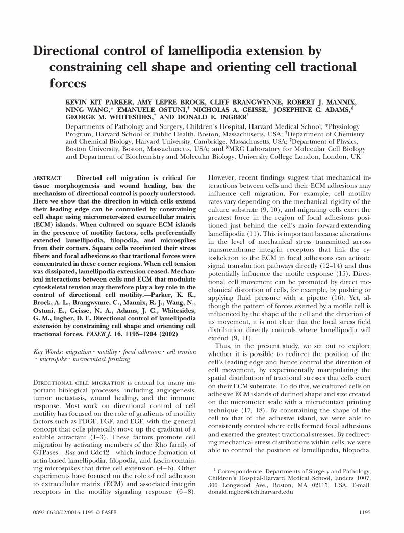
Directional control of lamellipodia extension byconstraining cell shape and orienting cell tractionalforces
KEVIN KIT PARKER, AMY LEPRE BROCK, CLIFF BRANGWYNNE, ROBERT J. MANNIX,NING WANG,* EMANUELE OSTUNI,† NICHOLAS A. GEISSE,‡ JOSEPHINE C. ADAMS,§
GEORGE M. WHITESIDES,† AND DONALD E. INGBER1
Departments of Pathology and Surgery, Children’s Hospital, Harvard Medical School; *PhysiologyProgram, Harvard School of Public Health, Boston, Massachusetts, USA; †Department of Chemistryand Chemical Biology, Harvard University, Cambridge, Massachusetts, USA; ‡Department of Physics,Boston University, Boston, Massachusetts, USA; and §MRC Laboratory for Molecular Cell Biologyand Department of Biochemistry and Molecular Biology, University College London, London, UK
ABSTRACT Directed cell migration is critical fortissue morphogenesis and wound healing, but themechanism of directional control is poorly understood.Here we show that the direction in which cells extendtheir leading edge can be controlled by constrainingcell shape using micrometer-sized extracellular matrix(ECM) islands. When cultured on square ECM islandsin the presence of motility factors, cells preferentiallyextended lamellipodia, filopodia, and microspikesfrom their corners. Square cells reoriented their stressfibers and focal adhesions so that tractional forces wereconcentrated in these corner regions. When cell tensionwas dissipated, lamellipodia extension ceased. Mechan-ical interactions between cells and ECM that modulatecytoskeletal tension may therefore play a key role in thecontrol of directional cell motility.—Parker, K. K.,Brock, A. L., Brangwynne, C., Mannix, R. J., Wang, N.,Ostuni, E., Geisse, N. A., Adams, J. C., Whitesides,G. M., Ingber, D. E. Directional control of lamellipodiaextension by constraining cell shape and orienting celltractional forces. FASEB J. 16, 1195–1204 (2002)
Key Words: migration � motility � focal adhesion � cell tension� microspike � microcontact printing
Directional cell migration is critical for many im-portant biological processes, including angiogenesis,tumor metastasis, wound healing, and the immuneresponse. Most work on directional control of cellmotility has focused on the role of gradients of motilityfactors such as PDGF, FGF, and EGF, with the generalconcept that cells physically move up the gradient of asoluble attractant (1–3). These factors promote cellmigration by activating members of the Rho family ofGTPases—Rac and Cdc42—which induce formation ofactin-based lamellipodia, filopodia, and fascin-contain-ing microspikes that drive cell extension (4–6). Otherexperiments have focused on the role of cell adhesionto extracellular matrix (ECM) and associated integrinreceptors in the motility signaling response (6–8).
However, recent findings suggest that mechanical in-teractions between cells and their ECM adhesions mayinfluence cell migration. For example, cell motilityrates vary depending on the mechanical rigidity of theculture substrate (9, 10), and migrating cells exert thegreatest force in the region of focal adhesions posi-tioned just behind the cell’s main forward-extendinglamellipodia (11). This is important because alterationsin the level of mechanical stress transmitted acrosstransmembrane integrin receptors that link the cy-toskeleton to the ECM in focal adhesions can activatesignal transduction pathways directly (12–14) and thuspotentially influence the motile response (15). Direc-tional cell movement can be promoted by direct me-chanical distortion of cells, for example, by pushing orapplying fluid pressure with a pipette (16). Yet, al-though the pattern of forces exerted by a motile cell isinfluenced by the shape of the cell and the direction ofits movement, it is not clear that the local stress fielddistribution directly controls where lamellipodia willextend (9, 11).
Thus, in the present study, we set out to explorewhether it is possible to redirect the position of thecell’s leading edge and hence control the direction ofcell movement, by experimentally manipulating thespatial distribution of tractional stresses that cells exerton their ECM substrate. To do this, we cultured cells onadhesive ECM islands of defined shape and size createdon the micrometer scale with a microcontact printingtechnique (17, 18). By constraining the shape of thecell to that of the adhesive island, we were able toconsistently control where cells formed focal adhesionsand exerted the greatest tractional stresses. By redirect-ing mechanical stress distributions within cells, we wereable to control the position of lamellipodia, filopodia,
1 Correspondence: Departments of Surgery and Pathology,Children’s Hospital-Harvard Medical School, Enders 1007,300 Longwood Ave., Boston, MA 02115, USA. E-mail:[email protected]
11950892-6638/02/0016-1195 © FASEB

and microspikes that drive forward motion. Theseresults, combined with the finding that lamellipodiaextension abruptly ceased when cytoskeletal tensiongeneration was inhibited, suggest that mechanical in-teractions between the cytoskeleton and ECM withinthe focal adhesion may help to determine the directionin which cells move.
MATERIALS AND METHODS
Experimental system
Micropatterned substrates containing square or circular ad-hesive islands were created on glass slides by microcontactprinting (18) and coated with high-density (50 �g/mL)fibronectin (17) or 50 nM thrombospondin-1 (19), as de-scribed. Bovine capillary endothelial cells and mouse NIH3T3 fibroblasts were allowed to grow to confluence in serum-containing medium, as described (17, 20), then were serum-deprived for 1 to 2 days before use in experiments. Subcon-fluent mouse skeletal C2C12 myoblasts were maintained asdescribed (19). The quiescent cells were then trypsinized andplated sparsely (3�103 cells/cm2) on the micropatternedsubstrates to ensure that individual islands were seeded withsingle cells. Studies of the microscope were carried out inexperimental bicarbonate-free minimum essential medium(MEM) containing Hank’s balanced salts lacking phenol redand bicarbonate (Sigma Chemical Co., St. Louis, MO),MEM amino acids (Sigma), MEM vitamins (Sigma), 2 mMl-glutamine, 1 mM sodium pyruvate, 20 mM N-2-hydroxyeth-ylpiperazine-N�-2-ethanesulfonic acid, d-glucose (1 g/L), hy-drocortisone (1 �g/mL), and 1% bovine serum albumin(BSA; Intergen Co., Purchase, NY; Cohn fraction V, pH 7.0).This medium was supplemented with 20 �g/mL high-densitylipoprotein (Bionetics Research, Rockville, MD) and 5�g/mL transferrin (Collaborative Research, Lexington, MA)for studies with endothelial cells. Studies with fibroblasts usedthe same medium with high glucose (5 g/L). Lamellipodiaextension was synchronously activated in NIH 3T3s by addi-tion of human PDGF-BB (5 ng/mL) and in capillary endo-thelial cells by addition of basic FGF (5 ng/mL) or 10% calfserum. Microspike formation in myoblasts was activated byplating on thrombospondin-1 for 1 h. F-actin, vinculin, fi-bronectin, and DNA (nuclei) were visualized in paraformal-dehyde-fixed cells using fluoresceinated-conjugated phalloi-din (300 ng/mL), rabbit anti-fibronectin antibody, mouseanti-vinculin antibody, and DAPI staining (all from Sigma),respectively. Fascin was visualized in methanol-fixed cells asdescribed (19). In some experiments, fibroblasts were trans-fected with Rac1 linked to green fluorescent protein (GFP;21; kindly provided by N. Hotchin and R. Horwitz) to bettervisualize lamellipodia or the control plasmid pGFP usingEffectene transfection reagent (Qiagen, Chatsworth, CA);lamellipodia staining was observed only in cells containingRac-GFP. Twenty-four hours later, the transfected cells wereplated on fibronectin-coated adhesive islands and cultured inserum-free medium overnight. The medium was replacedwith experimental bicarbonate-free medium for analysis bytime-lapse digital microscopy. BDM (2,3-butanedione 2-mon-oxime; Sigma) was added at a concentration (5 mM), whichhas been shown not to significantly alter intracellular calciumconcentration (22).
Morphological studies
Living cells were visualized with a Hamamatsu CCD cameraon a Nikon Diaphot 300 inverted microscope equipped with
phase contrast optics and epifluorescence illumination. Tem-perature was controlled by a stage mount (Micro VideoInstruments, Avon, MA) equipped with a temperature con-troller (Omega technologies Co., Stamford, CT). The totalprojected area of lamellipodia per cell was quantitated usingthe computerized image acquisition and analysis tools of IPLab Spectrum and RatioPlus software (Scanalytics, Fairfax,VA). Images of fibroblasts on 30 � 30 �m islands and stainedwith fluoresceinated phalloidin were projected over images inwhich the square limits of the fibronectin-coated islands werevisualized using anti-fibronectin antibodies and a rhodami-nated secondary. All cells that covered the entire 900 �m2
surface of each island were included in this analysis whetheror not they appeared to respond to PDGF stimulation. Anycell projection that extended over the nonadhesive regionsurrounding the square and stained positively for fluorescein-ated phalloidin was considered a lamellipodium, provided itwas greater than 1 �m2 in area (to account for registrationerror) and had a pixel intensity greater than background. Todetermine relative changes in lamellipodia length in differentregions of the cell, corners were defined as parts of the squareperimeter within 6 �m from the intersection of the two sides;sides were defined as the 18 �m interval between these cornerregions. To control for bias in morphometric calculations dueto the geometry of the orthogonal corner regions relative tothe linear sides and the large range of lamellipodial morphol-ogy, the normalized lamellipodia length was determined bytransforming the total lamellipodia area measured in eachregion into a similar shaped region (corner or side) com-posed of a lamellipodium that extended equally from allpoints along its perimeter. Total cumulative data were pre-sented by overlaying 20 to 40 images of fibroblasts culturedon 30 � 30 �m square fibronectin islands and stained withfluoresceinated phalloidin. The pixel occupancy at eachposition relative to the fibronectin island was determinedusing IP Lab software and the pixel distribution was colorcoded for frequency. Immunofluorescence microscopy wascarried out using the epifluorescence optics of the NikonDiaphot microscope. Atomic Force Microscopy was carriedout with a Dimension 3000 atomic force microscope (DigitalInstruments, Santa Barbara, CA) attached to a NanoscopeIIIa Controller using a modification of a published technique(23). For these studies, serum-stimulated NIH 3T3 fibroblastswere trypsinized and cultured on micropatterned substratesfor 6 h in defined medium without growth factors beforeexperimental analysis. Deflection and height data were col-lected at 512 � 512 pixels over scan sizes of 67 �m; imageswere flattened to a zero or first order polynomial fit.
Traction force microscopy
Traction force microscopy (9, 24) was adapted for use withcells cultured on micropatterned adhesive islands, as de-scribed (25). Flexible polyacrylamide gels (0.25% bis and 2%acrylamide; �70 �m thick; Young’s modulus�1300 Pa) werefabricated containing 0.2 �m diameter fluorescent beads(Molecular Probes, Eugene, OR). Polydimethylsiloxane(PDMS) membranes containing square holes the same sizeand shape as the desired adhesive islands were created usingmicrofabrication techniques (26). ECM-coated adhesive is-lands of desired size and shape were created on the surface ofthe gel by chemically conjugating type I collagen (0.2 mg/mL) to its surface through the holes in the PDMS membrane.After removal of the PDMS membrane, serum-free mediumcontaining 1% BSA was added for 30 min to block proteinbinding on the newly exposed, uncoated surface of thepolyacrylamide gel. Human airway smooth muscle cells iso-lated and cultured as described (27) were trypsined andplated on the micropatterned gels at high-density (100,000
1196 Vol. 16 August 2002 PARKER ET AL.The FASEB Journal
cells per membrane); nonadherent cells were removed after2 h by changing the medium. The displacement field of thegel beneath individual adherent cells was determined fromimages of the same region of the gel taken at different timesbefore or after experimental intervention and the tractionfield was calculated from the displacement field.
RESULTS
Directional extension of lamellipodia
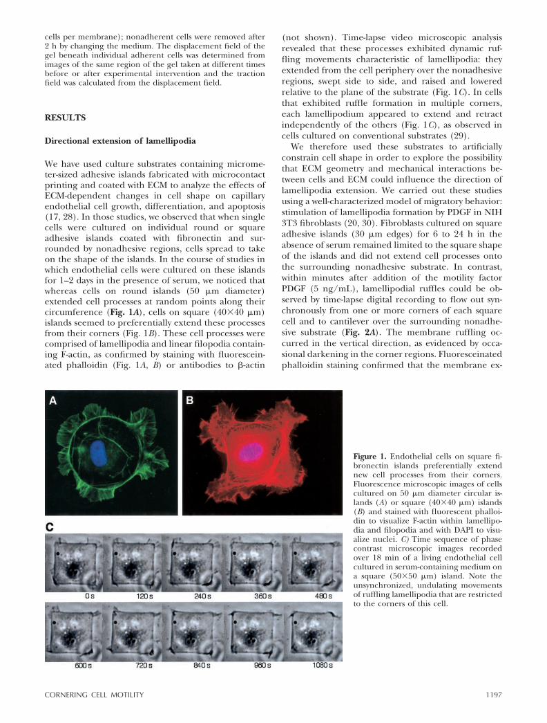
We have used culture substrates containing microme-ter-sized adhesive islands fabricated with microcontactprinting and coated with ECM to analyze the effects ofECM-dependent changes in cell shape on capillaryendothelial cell growth, differentiation, and apoptosis(17, 28). In those studies, we observed that when singlecells were cultured on individual round or squareadhesive islands coated with fibronectin and sur-rounded by nonadhesive regions, cells spread to takeon the shape of the islands. In the course of studies inwhich endothelial cells were cultured on these islandsfor 1–2 days in the presence of serum, we noticed thatwhereas cells on round islands (50 �m diameter)extended cell processes at random points along theircircumference (Fig. 1A), cells on square (40�40 �m)islands seemed to preferentially extend these processesfrom their corners (Fig. 1B). These cell processes werecomprised of lamellipodia and linear filopodia contain-ing F-actin, as confirmed by staining with fluorescein-ated phalloidin (Fig. 1A, B) or antibodies to �-actin
(not shown). Time-lapse video microscopic analysisrevealed that these processes exhibited dynamic ruf-fling movements characteristic of lamellipodia: theyextended from the cell periphery over the nonadhesiveregions, swept side to side, and raised and loweredrelative to the plane of the substrate (Fig. 1C). In cellsthat exhibited ruffle formation in multiple corners,each lamellipodium appeared to extend and retractindependently of the others (Fig. 1C), as observed incells cultured on conventional substrates (29).
We therefore used these substrates to artificiallyconstrain cell shape in order to explore the possibilitythat ECM geometry and mechanical interactions be-tween cells and ECM could influence the direction oflamellipodia extension. We carried out these studiesusing a well-characterized model of migratory behavior:stimulation of lamellipodia formation by PDGF in NIH3T3 fibroblasts (20, 30). Fibroblasts cultured on squareadhesive islands (30 �m edges) for 6 to 24 h in theabsence of serum remained limited to the square shapeof the islands and did not extend cell processes ontothe surrounding nonadhesive substrate. In contrast,within minutes after addition of the motility factorPDGF (5 ng/mL), lamellipodial ruffles could be ob-served by time-lapse digital recording to flow out syn-chronously from one or more corners of each squarecell and to cantilever over the surrounding nonadhe-sive substrate (Fig. 2A). The membrane ruffling oc-curred in the vertical direction, as evidenced by occa-sional darkening in the corner regions. Fluoresceinatedphalloidin staining confirmed that the membrane ex-
Figure 1. Endothelial cells on square fi-bronectin islands preferentially extendnew cell processes from their corners.Fluorescence microscopic images of cellscultured on 50 �m diameter circular is-lands (A) or square (40�40 �m) islands(B) and stained with fluorescent phalloi-din to visualize F-actin within lamellipo-dia and filopodia and with DAPI to visu-alize nuclei. C) Time sequence of phasecontrast microscopic images recordedover 18 min of a living endothelial cellcultured in serum-containing medium ona square (50�50 �m) island. Note theunsynchronized, undulating movementsof ruffling lamellipodia that are restrictedto the corners of this cell.
1197CORNERING CELL MOTILITY

tensions that formed primarily in the corners of squarefibroblasts when stimulated with PDGF were rich inF-actin and that cell processes often extended simulta-neously from multiple corner regions of the same cell(Fig. 2B). A similar propensity for directional lamel-lipodia extension in corners was observed whenquiescent, serum-deprived capillary endothelial cellscultured on square fibronectin-coated islands werestimulated with a different motile agonist, FGF (Fig.2C). Mouse skeletal C2C12 myoblasts cells that havebeen shown to move by extending fascin-containingmicrospikes when plated on thrombospondin-1 (6, 19)preferentially extended microspikes from their cornerswhen cultured on square adhesive islands coated withthis ECM protein (Fig. 2D). Thus, cell processes formedin the corners of square cells regardless of the cell type,molecular nature of the ECM coating, or the morphol-ogy of the cell extension.
Quantitation of changes in cell shape using comput-erized image analysis demonstrated that PDGF doubledthe total projected area of lamellipodia formed by eachcell within 5 min of addition and increased it by almost10-fold at 30 min (Fig. 3A). When the projectionlengths of all lamellipodia were quantified within mul-tiple cells using image analysis, the average lamellipo-dia length doubled in both the corner and side regionsof the square cells within 5 min after PDGF addition(Fig. 3B). However, after 30 min the mean lamellipodialength was �60% higher in the corners compared tothe sides (1.61�0.15 �m vs. 1.05�0.14 �m; P�0.007).This preference for cell processes to form in the cornerregions was observed when the images of multiple cellsrecorded at 0, 5, and 30 min after PDGF stimulationwere registered at the corners and overlaid. When the
distribution of their pixel occupancy was color codedfor frequency, the propensity for lamellipodia exten-sion from the corner regions again was not detectableat 5 min, but was clear by 30 min (Fig. 3C), even thoughF-actin staining had already appeared to concentratewithin some of the corners of these square cells at earlytimes (Fig. 3D).
Moreover, when lamellipodia movement was ana-lyzed at higher resolution by transfecting fibroblastswith Rac linked to enhanced GFP to visualize newprocess formation, waves of lamellipodia formationcould be seen to initiate precisely from the corners ofthe square cells and to propagate back over the cellsurface and along its sides (Fig. 4A). Given these results,the relative increase in lamellipodial length in cornersrelative to sides measured in fixed cells (Fig. 3B) may bean underestimate because waves that propagated fromthe corners but were present in the side regions or onthe apical surface of the cell would be scored asside-based lamellipodia or not scored at all in thatquantitative analysis. In any case, these data clearlyindicate that the position in which cells initiate forma-tion of lamellipodia, and hence the direction in whichthey move, can be significantly influenced by the ge-ometry of the ECM substrate or the shape of the cell.
Link between cell tension and directional extension
The finding that various cells preferentially extendednew cell processes from their corners when constrainedto square adhesive islands raised the question of con-trol. How could altering ECM geometry or cell shapeinfluence the direction in which cells move? One
Figure 2. Synchronized extensionof lamellipodia, filopodia, and mi-crospikes from cells on squareECM islands in response to stimu-lation with motility factors. A) Timesequence of phase contrast micro-scopic images recorded over 18min of a living fibroblast after stim-ulation with PDGF in serum-freemedium. Note that new lamellipo-dia simultaneously extend frommultiple corners of this singlesquare cell. Fluorescence images ofa fibroblast (B), endothelial cell(C), and skeletal C2C12 myoblast(C) cultured on square adhesiveislands coated with fibronectin (B,C) or thrombospondin-1 (D) andstained with fluoresceinated phal-loidin (B, C) or anti-fascin antibod-ies (D). A, B) 30 � 30 �m island; C,D) 40 � 40 �m islands. Note thatnewly formed lamellipodia, filopo-dia, and fascin microspikes all ex-tend preferentially from the cor-ners of these cells.
1198 Vol. 16 August 2002 PARKER ET AL.The FASEB Journal

possibility is that altering cell shape somehow causedmotility receptors and associated signaling complexesto preferentially distribute to the corner regions of thesquare cells before addition of the motile stimulus(PDGF). However, immunolocalization studies withanti-PDGF receptor antibodies revealed a homoge-neous distribution of these motility receptors over thecell surface (not shown). Thus, the localized lamellipo-dia extension we observed in square cells did notappear to be biased due to the distribution of PDGFreceptors.
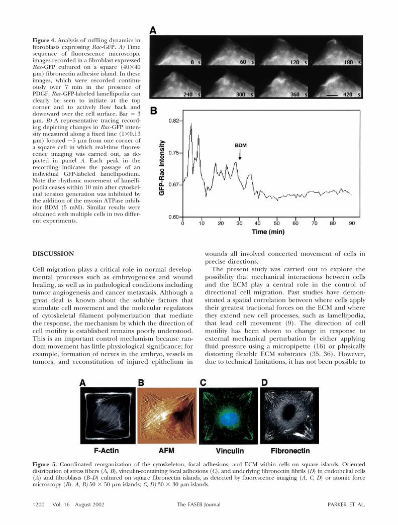
One clue to the mechanism of direction controlcame from analysis of cytoskeletal structure within cellscultured on square, fibronectin-coated adhesive is-lands. When square endothelial cells were analyzedusing fluoresceinated phalloidin, actin-containing stressfibers were observed to preferentially align diagonallywithin these square cells (Fig. 5A) and similar results
were obtained with cells cultured on square islands withedges 30, 40, or 50 �m in length. Similar diagonalalignment of stress fibers could be detected in fibroblastsusing atomic force microscopy (Fig. 5B) This highlyoriented distribution of actin bundles was consistent withthe finding that the focal adhesion protein, vinculin,became concentrated within adhesion plaques that local-ized within the corners regions of the square cells wherethe actin stress fibers terminated at the cell base andexhibited similar diagonal orientation (Fig. 5C). More-over, the fibroblasts accumulated elongated fibronectinfibrils in a diagonal pattern directly beneath the vinculin-containing focal adhesions (Fig. 5D).
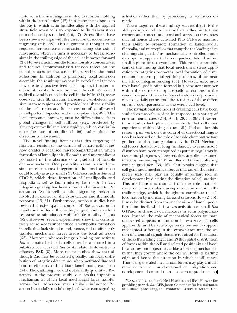
Past studies have shown that cells preferentially trans-fer mechanical stresses across their surface throughtransmembrane integrin receptors that mechanicallycouple ECM proteins, such as fibronectin, to the actincytoskeleton (31) and that vinculin contributes signifi-cantly to this response (32). Fibronectin fibril assemblyis promoted by mechanical tension exerted by contrac-tile cells (33, 34). Thus, these results suggested thatphysical constraint of cells within square islands maycause them to focus cytoskeleton-based tractionalforces in their corners. To explore this possibility, thespatial distribution of cell tractional forces exerted bycells cultured on square adhesive islands was analyzedand quantitated using a modified form of traction forcemicroscopy (24, 25). Microcontact printing was used tocreate square, collagen-coated adhesive islands on thesurface of flexible polyacrylamide gels in which small(0.2 �m) fluorescent beads were embedded. By usingthe beads as fiducial markers, displacements andstresses could be visualized and quantitated within theECM beneath individual square cells.
When human airway smooth muscle cells were cul-tured on these square islands, cell processes were againobserved to preferentially extend from the corners ofthe cells (Fig. 6, left). Traction force analysis confirmedthat the greatest bead displacements (Fig. 6, middle)and associated tractional stresses (Fig. 6, right) weresimilarly observed in the corners of the cells near wherefocal adhesions formed (Fig. 5C), just micrometersaway from where new cell processes initiated.
These results suggested that tensional forces gener-ated within the cytoskeleton could actively contributeto control of the direction of cell migration. In supportof this hypothesis, we found that the waves of lamelli-podia formation that initiated in the corners of squarefibroblasts transfected with Rac-GFP and cultured onfibronectin islands (Fig. 4A) ceased abruptly withinminutes after we inhibited actomyosin-based tensiongeneration using the myosin inhibitor BDM (Fig. 4B).Lamellipodia formation and movement ceased aftertension generation was suppressed using the morespecific ROCK inhibitor Y27632 or by chelating intra-cellular calcium using BAPTA-AM (not shown). Thus,cytoskeleton-based tractional forces appear to play akey role in control of lamellipodia extension.
Figure 3. Quantitative analysis of spatial control of lamellipo-dia extension. Time-dependent effects of PDGF stimulationon mean lamellipodia area (A) and normalized lamellipodialength (B) in fibroblasts cultured on square (30�30 �m)fibronectin islands. The average length of lamellipodia thatextended from corners (filled circles) vs. sides (open circles)was determined by computerized image analysis as describedin Materials and Methods; error bars indicated se. C) Visual-ization of cumulative data obtained by overlaying images from20 to 40 cells recorded at 0, 5, and 30 min after PDGFaddition (left to right) and pseudo coloring the data forfrequency of pixel occupancy at each position, as indicated inthe scale bar at the left. Yellow indicates the median positionof the cell edge for the entire population studied. D) Fluo-rescence images of F-actin staining within three differentrepresentative fibroblasts recorded at 0, 5, and 30 min afterPDGF addition (left to right). Note the presence of increasedF-actin staining in some of the corner regions at 5 min eventhough preferential extension of lamellipodia from cornerscould not be detected at this time.
1199CORNERING CELL MOTILITY
DISCUSSION
Cell migration plays a critical role in normal develop-mental processes such as embryogenesis and woundhealing, as well as in pathological conditions includingtumor angiogenesis and cancer metastasis. Although agreat deal is known about the soluble factors thatstimulate cell movement and the molecular regulatorsof cytoskeletal filament polymerization that mediatethe response, the mechanism by which the direction ofcell motility is established remains poorly understood.This is an important control mechanism because ran-dom movement has little physiological significance; forexample, formation of nerves in the embryo, vessels intumors, and reconstitution of injured epithelium in
wounds all involved concerted movement of cells inprecise directions.
The present study was carried out to explore thepossibility that mechanical interactions between cellsand the ECM play a central role in the control ofdirectional cell migration. Past studies have demon-strated a spatial correlation between where cells applytheir greatest tractional forces on the ECM and wherethey extend new cell processes, such as lamellipodia,that lead cell movement (9). The direction of cellmotility has been shown to change in response toexternal mechanical perturbation by either applyingfluid pressure using a micropipette (16) or physicallydistorting flexible ECM substrates (35, 36). However,due to technical limitations, it has not been possible to
Figure 5. Coordinated reorganization of the cytoskeleton, focal adhesions, and ECM within cells on square islands. Orienteddistribution of stress fibers (A, B), vinculin-containing focal adhesions (C), and underlying fibronectin fibrils (D) in endothelial cells(A) and fibroblasts (B–D) cultured on square fibronectin islands, as detected by fluorescence imaging (A, C, D) or atomic forcemicroscopy (B). A, B) 50 � 50 �m islands; C, D) 30 � 30 �m islands.
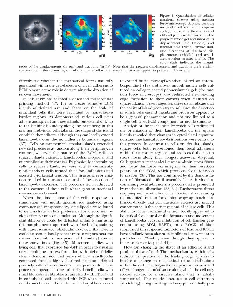
Figure 4. Analysis of ruffling dynamics infibroblasts expressing Rac -GFP. A) Timesequence of fluorescence microscopicimages recorded in a fibroblast expressedRac -GFP cultured on a square (40�40�m) fibronectin adhesive island. In theseimages, which were recorded continu-ously over 7 min in the presence ofPDGF, Rac -GFP-labeled lamellipodia canclearly be seen to initiate at the topcorner and to actively flow back anddownward over the cell surface. Bar � 3�m. B) A representative tracing record-ing depicting changes in Rac -GFP inten-sity measured along a fixed line (1�0.13�m) located �5 �m from one corner ofa square cell in which real-time fluores-cence imaging was carried out, as de-picted in panel A. Each peak in therecording indicates the passage of anindividual GFP-labeled lamellipodium.Note the rhythmic movement of lamelli-podia ceases within 10 min after cytoskel-etal tension generation was inhibited bythe addition of the myosin ATPase inhib-itor BDM (5 mM). Similar results wereobtained with multiple cells in two differ-ent experiments.
1200 Vol. 16 August 2002 PARKER ET AL.The FASEB Journal
directly test whether the mechanical forces naturallygenerated within the cytoskeleton of a cell adherent toECM play an active role in determining the direction ofits own movement.
In this study, we adapted a described microcontactprinting method (17, 18) to create adhesive ECMislands of defined size and shape on the scale ofindividual cells that were separated by nonadhesivebarrier regions. As demonstrated, various cell typesadhere and spread on these islands, but extend only upto the limiting boundary along the periphery; in thismanner, individual cells take on the shape of the islandon which they adhere, although they can locally extendlamellipodia over the nonadhesive boundary regions(37). Cells on symmetrical circular islands extendednew cell processes at random along their periphery. Incontrast, whatever the nature of the ECM, cells onsquare islands extended lamellipodia, filopodia, andmicrospikes at their corners. By physically constrainingcells to square islands, we were able to consistentlyreorient where cells formed their focal adhesions andexerted cytoskeletal tension. This structural reorienta-tion resulted in consistent control of the direction oflamellipodia extension: cell processes were redirectedto the corners of these cells where greatest tractionalstresses were observed.
When the time course of the cells’ response tostimulation with motile agonists was analyzed usingcomputerized morphometry, lamellipodia were foundto demonstrate a clear preference for the corner re-gions after 30 min of stimulation. Although no signifi-cant difference could be detected within 5 min usingthis morphometric approach with fixed cells, stainingwith fluoresceinated phalloidin revealed that F-actincould be seen to locally concentrate in regions near thecorners (i.e., within the square cell boundary) even atthese early times (Fig. 3D). Moreover, studies withliving cells that expressed Rac -GFP in order to visualizenew membrane process formation with higher fidelityclearly demonstrated that pulses of new lamellipodiagenerated from a highly localized position orientedprecisely within the corners of the square cells. Theseprocesses appeared to be primarily lamellipodia withsmall filopodia in fibroblasts stimulated with PDGF andin endothelial cells activated with FGF when culturedon fibronectin-coated islands. Skeletal myoblasts shown
to extend fascin microspikes when plated on throm-bospondin-1 (19) and airway smooth muscle cells cul-tured on collagen-coated polyacrylamide gels (for trac-tion force microscopy) also redirected new leadingedge formation to their corners when cultured onsquare islands. Taken together, these data indicate thatthe ability of island geometry to influence the directionin which cells extend membrane processes appears tobe a general phenomenon and not one limited to asingle cell type, ECM component, or motile stimulus.
Analysis of the mechanism by which cells redirectedthe orientation of their lamellipodia on the squareislands revealed that changes in cytoskeletal organiza-tion and mechanical force distributions were central tothis process. In contrast to cells on circular islands,square cells both repositioned their focal adhesionswithin their corner regions and redirected their actinstress fibers along their longest axis—the diagonal.Cells generate mechanical tension within stress fibersand focus this force via integrins on their insertionpoints on the ECM, which promotes focal adhesionformation (38). This was confirmed by the demonstra-tion of fibronectin fibril assembly beneath vinculin-containing focal adhesions, a process that is promotedby mechanical distortion (33, 34). Furthermore, directmapping and quantitation of cell tractional forces usingthe modified traction force microscopy approach con-firmed directly that cell tractional stresses are indeedconcentrated in the corner regions of square cells. Thisability to focus mechanical tension locally appeared tobe critical for control of the formation and movementof lamellipodia because inhibition of cell tension gen-eration using BDM, BAPTA, or a ROCK inhibitorsuppressed this response. Inhibitors of Rho and ROCKhave similarly been shown to inhibit cell movement inpast studies (39–41), even though they appear toincrease Rac activity (42–44).
How can changing the shape of an adhesive islandproduce these effects? The mechanism by which cellsredirect the position of the leading edge appears toinvolve a change in mechanical stress distributionswithin the cell. The diagonal of a square adhesive islandoffers a longer axis of advance along which the cell mayspread relative to a circular island that is radiallysymmetrical. This relative increase in cell distortion(stretching) along the diagonal may preferentially pro-
Figure 6. Quantitation of cellulartractional stresses using tractionforce microscopy. A phase contrastimage of a cell cultured on a squarecollagen-coated adhesive island(40�40 �m) created on a flexiblepolyacrylamide gel and maps of itsdisplacement field (middle) andtraction field (right). Arrows indi-cate directions of the bead dis-placements (middle) and associ-ated traction stresses (right). Thecolor scale indicates the magni-
tudes of the displacements (in �m) and tractions (in Pa). Note that the greatest displacement and tractions preferentiallyconcentrate in the corner regions of the square cell where new cell processes appear to preferentially extend.
1201CORNERING CELL MOTILITY
mote actin filament alignment due to tension moldingwithin the actin lattice (45) in a manner analogous tothe way in which actin bundles align with the appliedstress field when cells are exposed to fluid shear stressor mechanically stretched (46, 47). Stress fibers havebeen shown to align with the direction of movement inmigrating cells (48). This alignment is thought to berequired for isometric contraction along the axis ofmovement, which in turn is necessary to break adhe-sions in the trailing edge of the cell as it moves forward(2). However, actin bundle formation also concentratesand focuses actomyosin-based tension forces on theinsertion sites of the stress fibers within the focaladhesions. In addition to promoting focal adhesionassembly, the resulting increase in cytoskeletal tensionmay create a positive feedback loop that further in-creases stress fiber formation inside the cell (45) as wellas fibril assembly outside the cell in the ECM (34), as weobserved with fibronectin. Increased cytoskeletal ten-sion in these regions could provide local shape stabilityof the cell necessary for extension of cantileveredlamellipodia, filopodia, and microspikes (45, 49). Thislocal response, however, must be differentiated fromglobal changes in cell stiffness (e.g., produced byaltering extracellular matrix rigidity), which can influ-ence the rate of motility (9, 50) rather than thedirection of movement.
The novel finding here is that this targeting ofisometric tension to the corners of square cells some-how creates a localized microcompartment in whichformation of lamellipodia, filopodia, and microspikes ispromoted in the absence of a gradient of solublechemoattractants. One possibility is that localized ten-sion transfer across integrins in the focal adhesioncould locally activate small Rho GTPases such as Rac andCDC42, which drive formation of lamellipodia andfilopodia as well as fascin microspikes (4–6). In fact,integrin signaling has been shown to be linked to Racactivation (8) as well as other signaling moleculesinvolved in control of the cytoskeleton and the motileresponse (15, 51). Furthermore, previous studies haverevealed precise spatial control of Rac activation inmembrane ruffles at the leading edge of motile cells inresponse to stimulation with soluble motility factors(52). However, recent experiments show that constitu-tively active Rac cannot induce lamellipodia formationin cells that lack vinculin and, hence, fail to efficientlytransfer mechanical forces across the focal adhesion(53). Moreover, whereas integrin binding can activateRac in unattached cells, cells must be anchored to asubstrate for activated Rac to stimulate its downstreameffector, PAK (8). More recent studies show that al-though Rac may be activated globally, the local distri-bution of integrins determines where activated Rac willbind to effectors and facilitate lamellipodia extension(54). Thus, although we did not directly quantitate Racactivity in the present study, our results support amechanism in which local mechanical force transferacross focal adhesions may similarly influence Racaction by spatially modulating its downstream signaling
activities rather than by promoting its activation di-rectly.
Taken together, these findings suggest that it is theability of square cells to localize focal adhesions to theircorners and concentrate tensional stresses at these sitesthat influence where activated Rho GTPases manifesttheir ability to promote formation of lamellipodia,filopodia, and microspikes that comprise the leading edgeof a migrating cell. This mechanically controlled motil-ity response appears to be compartmentalized withinsmall regions of the cytoplasm. This result is reminis-cent of the finding that local mechanical stress appli-cation to integrins promotes local formation of a mi-crocompartment specialized for protein synthesis nearthe site of integrin binding (55). However, since mul-tiple lamellipodia often formed in a consistent mannerwithin the corners of square cells, alterations in theoverall shape of the cell or cytoskeleton may provide away to spatially orchestrate the activities of these differ-ent microcompartments at the whole cell level.
The navigational methods of crawling cells have beenstudied extensively in vitro in response to a variety ofenvironmental cues (2–4, 9–11, 20, 30, 36). However,these studies lack physical constraints that cells mayexperience within living tissues (21). Perhaps for thisreason, past work on the control of directional migra-tion has focused on the role of soluble chemoattractantgradients and contact guidance by the ECM. Mechani-cal forces that act over long (millimeter to centimeter)distances have been recognized as pattern regulators intissue morphogenesis, however, they are often assumedto act by reorienting ECM bundles and thereby alteringcontact guidance (35, 36). Our results suggest thatcell-generated mechanical forces that act on the micro-meter scale may play an equally important role indevelopment by dictating the direction of cell motion.This mechanism is distinct from the role that cellcontractile forces play during retraction of the cell’strailing edge, which is thought to contribute to celllocomotion by increasing forward cytosolic flow (2, 15).It may be distinct from the mechanism of lamellipodiaformation itself, which involves activation of small RhoGTPases and associated increases in actin polymeriza-tion. Instead, the role of mechanical forces we haveuncovered appears to function in two ways: 1) cellsapparently must be able to generate tension to supportmechanical stiffening in the cytoskeleton and the ac-tion of chemical signals that are required for formationof the cell’s leading edge, and 2) the spatial distributionof forces within the cell and related positioning of basalfocal adhesions appear to act like a steering mechanismin that they govern where the cell will form its leadingedge and hence the direction in which it will move.Thus, cell-generated mechanical forces may play a muchmore central role in directional cell migration anddevelopmental control than has been appreciated.
We would like to thank Neil Hotchin and Rick Horwitz forproviding us with Rac -GFP, Jason Comander for his assistancewith image processing, the Photonics Center at Boston Uni-
1202 Vol. 16 August 2002 PARKER ET AL.The FASEB Journal
versity for the use of their atomic force microscope, and theHarvard Materials Research Science and Engineering Center(MRSEC) for use of their microfabrication facilities. Thiswork was supported by National Institutes of Health grantsCA-45548 (D.E.I.) and GM30367 (G.M.W.), NASA grantNAG2–1509 (N.W.), and Wellcome Trust Senior Fellowship038284 (J.C.A.).
REFERENCES
1. Banyard, J., and Zetter, B. R. (1998–99). The role of cell motilityin prostate cancer. Cancer Metastasis Rev. 17, 449–458
2. Lauffenburger, D. A., and Horwitz, A. F. (1996) Cell migration:a physically integrated molecular process. Cell 84, 359–369
3. Parent, C. A., and Devreotes, P. N. (1999) A cell’s sense ofdirection. Science 284, 765–769
4. Ridley, A. J., Paterson, H. F., Johnston, C. L., Diekmann, D., andHall, A. (1992) The small GTP-binding protein rac regulatesgrowth factor-induced membrane ruffling. Cell 70, 401–410
5. Nobes, C. D., and Hall, A. (1999) Rho GTPases control polarity,protrusion, and adhesion during cell movement. J. Cell Biol. 144,1235–1244
6. Adams, J. C., and Schwartz, M. A. (2000) Stimulation of fascinspikes by thrombospondin-1 is mediated by the GTPases Racand Cdc42. J. Cell Biol. 150, 807–822
7. Holly, S. P., Larson, M. K., and Parise, L. V. (2000) Multipleroles of integrins in cell motility. Exp. Cell Res. 261, 69–74
8. del Pozo, M. A., Price, L. S., Alderson, N. B., Ren, X. D, andSchwartz, M. A. (2000) Adhesion to the extracellular matrixregulates the coupling of the small GTPase Rac to its effectorPAK. EMBO J. 19, 2008–2014
9. Pelham, R. J., Jr., and Wang, Y.-L. (1997) Cell locomotion andfocal adhesions are regulated by substrate flexibility. Proc. Natl.Acad. Sci. USA 94, 13661–13665
10. Lo, C. M., Wang, H. B., Dembo, M., and Wang, Y.-L. (2000) Cellmovement is guided by the rigidity of the substrate. Biophys. J.79, 144–152
11. Pelham, R. J., Jr., and Wang, Y.-L. (1999) High resolutiondetection of mechanical forces exerted by locomoting fibro-blasts on the substrate. Mol. Biol. Cell 10, 935–945
12. Bershadsky, A., Chausovsky, A., Becker, E., Lyubimova, A., andGeiger, B. (1996) Involvement of microtubules in the control ofadhesion-dependent signal transduction. Curr. Biol. 6, 1279–1289
13. Choquet, D., Felsenfeld, D. P., and Sheetz, M. P. (1997)Extracellular matrix rigidity causes strengthening of integrin-cytoskeleton linkage. Cell 88, 39–48
14. Meyer, C. J., Alenghat, F. J., Rim, P., Fong, J. H., Fabry, B., andIngber, D. E. (2000) Mechanical control of cyclic AMP signal-ling and gene transcription through integrins. Nat. Cell Biol. 2,666–668
15. Sheetz, M. P., Felsenfeld, D. P., and Galbraith, C. G. (1998) Cellmigration: regulation of force on extracellular-matrix-integrincomplexes. Trends Cell Biol. 8, 51–54
16. Verkhovsky, A. B., Svitkina, T. M., and Borisy, G. G. (1999)Self-polarization and directional motility of cytoplasm. Curr.Biol. 9, 11–20
17. Chen, C. S., Mrksich, M., Huang, S., Whitesides, G. M., andIngber, D. E. (1997) Geometric control of cell life and death.Science 276, 1425–1428
18. Chen, C. S., Ostuni, E., Whitesides, G. M., and Ingber, D. E.(2000) Using self-assembled monolayers to pattern ECM pro-teins and cells on substrates. Methods Mol. Biol. 139, 209–219
19. Adams, J. C. (1997) Characterization of cell-matrix adhesionrequirements for the formation of fascin microspikes. Mol. Biol.Cell 8, 2345–2363
20. Yu, J., Moon, A., and Kim, H. R. (2001) Both platelet-derivedgrowth factor receptor (PDGFR)-alpha and PDGFR-beta pro-mote murine fibroblast cell migration. Biochem. Biophys. Res.Commun. 282, 697–700
21. Knight, B., Laukaitis, C., Akhtar, N., Hotchin, N. A., Edlund, M.,and Horwitz, A. F. (2000) Visualizing muscle cell migration insitu. Curr. Biol.10, 576–585
22. Blanchard, E. M., Smith, G. L., Allen, D. G., and Alpert, N. R.(1990) The effects of 2,3-butanedione monoxime on initialheat, tension and aequorin light output of ferret papillarymuscles. Pfluegers Arch. 416, 219–221
23. Goldmann, W. H., and Ezzell, R. M. (1996) Viscoelasticity inwild-type and vinculin-deficient (5.51) mouse F9 embryoniccarcinoma cells examined by atomic force microscopy andrheology. Exp. Cell Res. 226, 234–237
24. Dembo, M., and Yang, Y.-L. (1999) Stresses at the cell-to-substrate interface during locomotion of fibroblasts. Biophys. J.76, 2307–2316
25. Wang, N., Ostuni, E., Whitesides, G. M., and Ingber, D. E.(2002) Micropatterning tractional forces in living cells. CellMotil. Cytoskelet. In press
26. Ostuni, E., Kane, R., Chen, C. S., Ingber, D. E., and Whitesides,G. M. (2000) Patterning mammalian cells using elastomericmembranes. Langmuir 16, 7811–7819
27. Panettieri, R. A., Murray, R. K., DePalo, L. R., Yadvish, R. A., andKotlikoff, M. I. (1989) A human airway smooth muscle cell linethat retains physiological responsiveness. Am. J. Physiol. 256,C329–C335
28. Dike, L. E., Chen, C. S., Mrksich, M., Tien, J., Whitesides, G. M.,and Ingber, D. E. (1999) Geometric control of switchingbetween growth, apoptosis, and differentiation during angio-genesis using micropatterned substrates. In Vitro Cell Dev. Biol.Anim. 35, 441–448
29. Abercrombie, M., Heaysman, J. E. M., and Pegrum, S. M. (1970)The locomotion of fibroblasts in culture. I. Movements of theleading edge. Exp. Cell Res. 59, 393–398
30. Grotendorst, G. R. (1984) Alteration of the chemotactic re-sponse of NIH/3T3 cells to PDGF by growth factors, transfor-mation, and tumor promoters. Cell 36, 279–285
31. Wang, N., Butler, J. P., and Ingber, D. E. (1993) Mechanotrans-duction across the cell surface and through the cytoskeleton.Science 260, 1124–1127
32. Ezzell, R. M., Goldmann, W. H., Wang, N., Parasharama, N., andIngber, D. E. (1997) Vinculin promotes cell spreading bymechanically coupling integrins to the cytoskeleton. Exp. CellRes. 231, 14–26
33. Halliday, N. L., and Tomasek, J. J. (1995) Mechanical propertiesof the extracellular matrix influence fibronectin fibril assemblyin vitro. Exp. Cell Res. 217, 109–117
34. Schwarzbauer, J. E., and Sechler, J. L. (1999) Fibronectinfibrillogenesis: a paradigm for extracellular matrix assembly.Curr. Opin. Cell Biol. 11, 622–627
35. Oster, G. F., Murray, J. D., and Harris, A. K. (1983) Mechanicalaspects of mesenchymal morphogenesis. J. Embryol. Exp. Morphol.78, 83–125
36. Nakatsuji, N., and Johnson, K. E. (1984) Experimental manip-ulation of a contact guidance system in amphibian gastrulationby mechanical tension. Nature (London) 307, 453–455
37. Bailly, M., Yan, L., Whitesides, G. M., Condelis, J. S., and Segall,J. E. (1998) Regulation of protrusion shape and adhesion to thesubstratum during chemotactic responses of mammalian carci-noma cells. Exp. Cell Res. 241, 285–299
38. Chrzanowska-Wodnicka, M., and Burridge, K. (1996) Rho-stimulated contractility drives the formation of stress fibers andfocal adhesions. J. Cell Biol. 133, 1403–1415
39. Itoh, K., Yoshioka, K., Akedo, H., Uehata, M., Ishizaki, T., andNarumiya, S. (1999) An essential role for Rho-associated kinasein the transcellular invasion of tumor cell. Nat. Med. 5, 221–225
40. Kishi, H., Bao, J., and Kohama, K. (2000) Inhibitory effects ofML-9, wortmannin, and Y-27632 on the chemotaxis of vascularsmooth muscle cells in response to platelet-derived growthfactor-BB. J. Biochem. 128, 719–722
41. Bourguignon, L. Y. W., Zhu, H., Shao, L., Zhu, D., and Chen, Y.(1999) Rho-kinase promotes CD44v3,8–10-ankyrin interactionand tumor cell migration in metastatic breast cancer cells. CellMotil. Cytoskeleton 43, 269–287
42. Rottner, K., Hall, A., and Small, J. V. (1999) Interplay betweenRac and Rho in the control of substrate contact dynamics. Curr.Biol. 9, 640–648
43. Moorman, J. P., Luu, D., Wickham, J., Bobak, D. A., and Hahn,C. S. (1999) A balance of signaling by Rho family small GTPasesRhoA, Rac1 and Cdc42 coordinates cytoskeletal morphologybut not cell survival. Oncogene 18, 47–57
1203CORNERING CELL MOTILITY
44. Cox, E. A., Sastry, S. K., and Huttenlocher, A. (2001) Integrin-mediated adhesion regulates cell polarity and membrane protru-sion through the Rho family of GTPases. Mol. Biol. Cell 12, 265–277
45. Ingber, D. E. (1993) Cellular tensegrity: defining new rules ofbiological design that govern the cytoskeleton. J. Cell Sci. 104,613–627
46. Girard, P. R., and Nerem, R. M. (1995) Shear stress modulatesendothelial cell morphology and F-actin organization throughthe regulation of focal adhesion-associated proteins. J. Cell.Physiol. 163, 179–193
47. Smith, P. G., Garcia, R., and Kogerman, L. (1997) Strainreorganizes focal adhesions and cytoskeleton in cultured airwaysmooth muscle cells. Exp. Cell Res. 232, 127–136
48. Wang, Y. L. (1984) Reorganization of actin filament bundles inliving fibroblasts. J. Cell Biol. 99, 1478–1485
49. Wang, N., Naruse, K., Stamenovic, D., Fredberg, J. J., Mijailovich,S. M., Tolic-Norrelykke, I. M., Polte, T., Mannix, R., and Ingber,D. E. (2001) Mechanical behavior in living cells consistent with thetensegrity model. Proc. Natl. Acad. Sci. USA 98, 7765–7770
50. DiMilla, P. A., Barbee, K., and Lauffenburger, D. A. (1991)Mathematical model for the effects of adhesion and mechanicson cell migration speed. Biophys. J. 60, 15–37
51. Schwartz, M. A., and Shattil, S. J. (2000) Signaling networkslinking integrins and rho family GTPases. Trends Biochem. Sci. 25,388–391
52. Kraynov, V. S., Chamberlain, C., Bokoch, G. M., Schwartz, M. A.,Slabaugh, S., and Hahn, K. M. (2000) Localized Rac activationdynamics visualized in living cells. Science 290, 333–337
53. Goldmann, W. H., and Ingber, D. E. (2002) Intact vinculinprotein is required for control of cell shape, cell mechanics, andrac-dependent lamellipodia formation. Biochem. Biophys. Res.Commun. 290, 749–755
54. Del Pozo, M. A., Kiosses, W. B., Alderson, N. B., Meller, N.,Hahn, K. M., and Schwartz, M. A. (2002) Integrins regulateGTP-Rac localized effector interactions through dissociation ofRho-GDI. Nat Cell Biol. 4, 232–239
55. Chicurel, M. E., Singer, R. H., Meyer, C. J., and Ingber, D. E.(1998) Integrin binding and mechanical tension induce move-ment of mRNA and ribosomes to focal adhesions. Nature(London) 392, 730–733
Received for publication January 15, 2002.Revised for publication April 1, 2002.
1204 Vol. 16 August 2002 PARKER ET AL.The FASEB Journal
Copyright © 2022 FDOKUMEN