Is the conformational flexibility of piperazine derivatives important to inhibit HIV-1 replication?
Upload
independentCategory
view
3download
0
Nano-Stenciled RGD-Gold Patterns That Inhibit FocalContact Maturation Induce Lamellipodia Formation inFibroblastsRoman Lutz1¤, Kristopher Pataky2, Neha Gadhari3, Mattia Marelli2, Juergen Brugger2, Matthias
Chiquet3*
1 Friedrich Miescher Institute for Biomedical Research, Basel, Switzerland, 2 Microsystems Laboratory, Ecole Polytechnique Federale de Lausanne, Lausanne, Switzerland,
3 Department of Orthodontics and Dentofacial Orthopedics, University of Bern, Bern, Switzerland
Abstract
Cultured fibroblasts adhere to extracellular substrates by means of cell-matrix adhesions that are assembled in a hierarchicalway, thereby gaining in protein complexity and size. Here we asked how restricting the size of cell-matrix adhesions affectscell morphology and behavior. Using a nanostencil technique, culture substrates were patterned with gold squares of awidth and spacing between 250 nm and 2 mm. The gold was functionalized with RGD peptide as ligand for cellularintegrins, and mouse embryo fibroblasts were plated. Limiting the length of cell-matrix adhesions to 500 nm or lessdisturbed the maturation of vinculin-positive focal complexes into focal contacts and fibrillar adhesions, as indicated bypoor recruitment of a5-integrin. We found that on sub-micrometer patterns, fibroblasts spread extensively, but did notpolarize. Instead, they formed excessive numbers of lamellipodia and a fine actin meshwork without stress fibers. Moreover,these cells showed aberrant fibronectin fibrillogenesis, and their speed of directed migration was reduced significantlycompared to fibroblasts on 2 mm square patterns. Interference with RhoA/ROCK signaling eliminated the pattern-dependent differences in cell morphology. Our results indicate that manipulating the maturation of cell-matrix adhesions bynanopatterned surfaces allows to influence morphology, actin dynamics, migration and ECM assembly of adheringfibroblasts.
Citation: Lutz R, Pataky K, Gadhari N, Marelli M, Brugger J, et al. (2011) Nano-Stenciled RGD-Gold Patterns That Inhibit Focal Contact Maturation InduceLamellipodia Formation in Fibroblasts. PLoS ONE 6(9): e25459. doi:10.1371/journal.pone.0025459
Editor: Erik H. J. Danen, Leiden University, The Netherlands
Received February 28, 2011; Accepted September 5, 2011; Published September 27, 2011
Copyright: � 2011 Lutz et al. This is an open-access article distributed under the terms of the Creative Commons Attribution License, which permits unrestricteduse, distribution, and reproduction in any medium, provided the original author and source are credited.
Funding: This work was funded by grant CR2312_125290/1 from the Swiss National Science Foundation (http://www.snf.ch/E/Pages/default.aspx) and by theNovartis Research Foundation (http://www.fmi.ch/About/). The funders had no role in study design, data collection and analysis, decision to publish, orpreparation of the manuscript.
Competing Interests: The authors have declared that no competing interests exist.
* E-mail: [email protected]
¤ Current address: CLS Behring AG, Bern, Switzerland
Introduction
Cell adhesion to extracellular matrix (ECM) is mediated mainly
via integrins, heterodimeric cell surface receptors that play key
roles in transmembrane signaling processes and thereby regulate
cell behavior and fate [1]. One important group of integrins
interacts with Arg-Gly-Asp (RGD) peptide motifs that are pre-
sented by many ECM ligands; depending on their subunit
composition, however, these receptors bind to individual RGD-
containing ECM proteins with different affinities. For example, the
vitronectin receptor integrin-avb3 recognizes RGD in various
contexts (also in short peptides), whereas integrin-a5b1 very
specifically interacts with fibronectin. Intracellularly, integrins are
linked to the actin cytoskeleton by means of specialized adaptor
proteins such as talin and vinculin, which form a dense and
heterogeneous protein network [2]. Interaction partners also
involve protein kinases like Src and FAK that build a platform for
early steps of signaling. Since cell-matrix adhesions are the sites
where forces are transmitted from the ECM to the cytoskeleton
and back, they are critical for transducing mechanical stimuli, such
as substrate rigidity or changes in substrate strain, into chemical
signals [1]. Among the signaling cascades that are activated by
mechanical stimuli is the RhoA/ROCK pathway [3,4] that
promotes actin stress fiber formation [5,6].
Attachment and spreading of cells on an ECM substrate occurs
in several steps that depend on integrin-mediated sensing of local
substrate features, a process which in turn controls the formation
and maturation of cell-matrix adhesions. Shortly after first contact
of a cell with its substrate, Rac1, a Rho-family GTPase, stimulates
the assembly of a fine meshwork of actin filaments at cell borders
and the protrusion of lamellipodia [7,8]. A lamellipodium
represents the ‘‘leading edge’’ of a moving or spreading cell and
is the birthplace of cell-matrix adhesions called focal complexes.
These small, dot-like adhesions are less than 1 mm2 in area and
characterized by the colocalization of avb3-integrin, paxillin, talin,
vinculin, FAK and phosphotyrosine [9,10,11]. Focal complexes
are highly ephemeral and often disassemble rapidly. Alternatively,
if at this site the ECM substrate is adhesive (i.e. of high integrin
ligand density) and mechanically stable, focal complexes can
mature into focal contacts by growing in size and recruiting
additional proteins like zyxin, tensin, and a5b1-integrin. This
maturation depends on actin and myosin-II induced cellular
PLoS ONE | www.plosone.org 1 September 2011 | Volume 6 | Issue 9 | e25459
traction and can be arrested by inhibitors of the RhoA/ROCK
pathway [10,12]. Mature focal contacts are usually found at the tip
of actin stress fibers; they are 1–2 mm wide, several mm long, and
can persist for many minutes at the same location. Further traction
by means of the actin cytoskeleton pulls out a5b1-integrin and
tensin from focal contacts, initiating the formation of fibrillar
adhesions that are tens of mm long [13]. During this process,
secreted pericellular fibronectin is bound to a5b1-integrin and
stretched, and exposes self-assembly sites that lead to the formation
of fibronectin fibrils [14]. Thus, on most substrates the different
types of cell-matrix adhesions are assembled in a hierarchical way,
growing in size during the process.
Much has been learned about the mechanisms of integrin-
dependent cell-ECM interactions by the use of micro- and
nanopatterened adhesive substrates that were engineered in recent
years [15,16,17]. The most widely used method is microcontact
printing, whereby patterns of adhesive ECM proteins (e.g.
fibronectin) are applied onto a passivated (non-adhesive) back-
ground surface. Cell-sized ECM islands of various shapes were
designed to impose onto cells a defined surface area and form; this
allowed to study how cell shape controls cell survival [18], division
[19], and differentiaton [20]. On the other hand, ECM stripe or
dot patterns in the micro- and nanometer range were engineered
that forced cells to establish cell-matrix contacts only at locations
defined by the print design. With these techniques, essential
physical parameters of ECM substrates could be explored that are
required for focal contact formation [16], cell spreading [21], cell
polarity and cell motility [22].
In the present study, we used a different technique to produce
substrate patterns in the sub-micrometer range. By a nanostencil
procedure [23], gold squares were applied to glass surfaces,
functionalized with RGD-thiol peptide, and the intervening spaces
passivated with poly(L-lysine)-graft-poly(ethylene glycol) (PLL-g-
PEG). Because of the almost covalent nature of the Au-S bond,
organic thiol compounds assemble into monolayers on gold
surfaces [24], and it is thus generally assumed that the amount of
coupled RGD-thiol is proportional to the gold surface area [16].
RGD rather than fibronectin was used here as a ligand, in order to
render the initial cell adhesion dependent on integrin-avb3. Cells
that attach to a pure fibronectin substrate immediately engage
integrin-a5b1; this leads to activation of RhoA [25] and the
distinction between different types of cell-matrix adhesions
becomes blurred [6]. For the same reason, medium depleted of
exogenous fibronectin was used to plate cells on the nanopatterns.
The smallest RGD-gold pattern consisted of squares 2506250 nm
(0.06 mm2) in size, spaced 250 nm apart. For the larger pattern,
the respective dimensions were 500, 1000, and 2000 nm. On
purpose, for all patterns used in these experiments the coverage of
substrate area with integrin ligand (i.e. RGD-coupled gold) was set
to 25%, which is far in excess to that reported to be required for
full cell spreading [21]. We used this new method with the aim to
restrict the size of cell matrix adhesions, and hence to inhibit the
maturation of small, newly formed focal complexes into larger,
a5b1-integrin-positive focal contacts. Similar nanopatterns to
control focal adhesion assembly have been produced before by
electron beam lithography [26]; however, downstream effects on
cell behavior were not explored in this study. Here, we asked
specifically how manipulating the size and spacing of cell-matrix
adhesions affected cell shape and motility. We found that cells
were able to attach and spread on sub-micrometer RDG-gold
patterns via small focal complexes, but that the formation of focal
and fibrillar adhesions was inhibited. The nanostructure of cell-
matrix adhesions in turn affected lamellipodia formation, the actin
cytoskeleton, fibronectin fibrillogenesis, and directed cell migra-
tion. Our results suggest that distinct signals arise from cell-matrix
adhesions during different stages of their maturation.
Results
Design of the RGD-gold patternsIn order to restrict the growth of cell-matrix adhesions during
cell adhesion to a defined size, gold square patterns in the
micrometer and sub-micrometer range were applied to glass
surfaces with a nanostencil technique [23]. Before seeding
fibroblasts, Cys-containing RGD peptide was bound to the gold
squares to provide restricted and integrin-specific cell attachment,
and the area surrounding the gold squares was passivated with
poly-L-lysine covalently grafted to polyehthylene glycol (PLL-g-
PEG; [27]). Four different patterns with gold squares ranging from
2506250 nm2, 5006500 nm2, 161 mm2 to 262 mm2 were
prepared (Fig. 1). To ensure that cells on all patterns had the
same areal density of adhesive ligand to attach, the patterns were
designed in a way that the surface area of RGD-coupled gold was
fixed to 25% of the total substrate area. This was achieved by
spacing the gold squares at a distance corresponding to their side
length (Fig. 1A). Control experiments showed that gold surfaces
were functionally saturated with RGD peptide under the coating
conditions used (see Materials and Methods; Figure S1). Thus, the
only difference between the four patterns used is the distribution,
not the amount or density, of adhesive RGD-gold ligand.
Scanning electron microscope images of the patterns are shown
in Fig. 1B. For simplicity, the four different substrates are called
250 nm, 500 nm, 1000 nm and 2000 nm patterns in the following
paragraphs. For positive and negative controls, cells were plated
on plain (i.e. unpatterned) RGD-gold or on plain PLL-g-PEG
passivated glass surfaces, respectively.
Restriction of cell-matrix adhesions to RGD-coupled goldareas
Mouse embryo fibroblasts (MEF) were plated on the RGD-
coupled and PLL-g-PEG-passivated gold patterns in medium
containing 3% fibronectin-depleted FCS (to reduce nonspecific
adhesion). Cells were allowed to attach and spread for 4 hours,
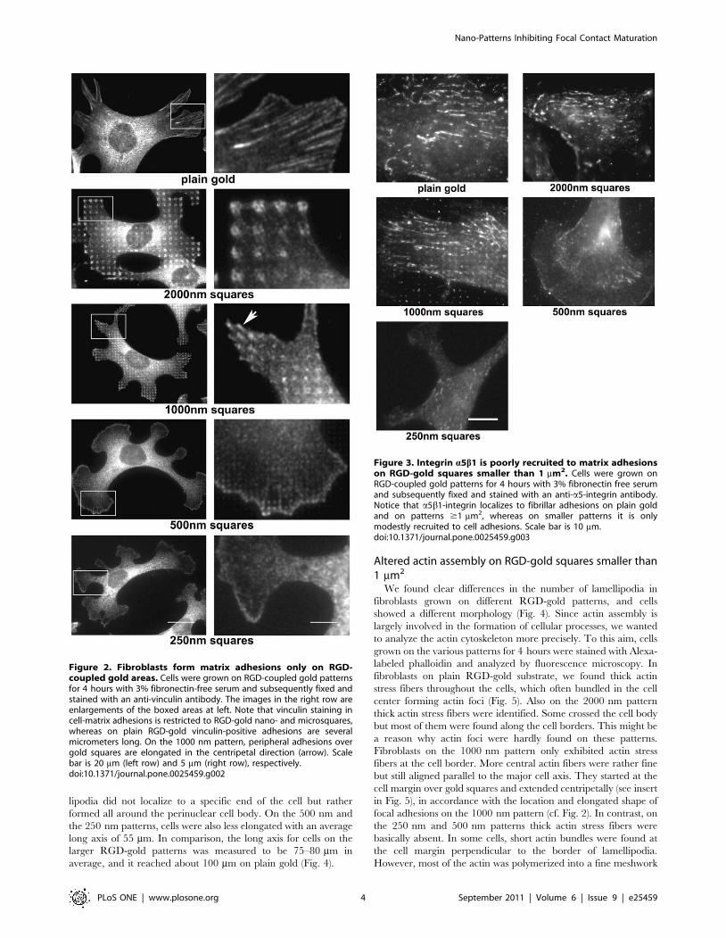
subsequently fixed and stained for vinculin (Fig. 2). Some
cytoplasmic staining in the perinuclear area was seen on all
substrates. On plain RGD-coupled gold surface, specific staining
for vinculin was additionally found in elongated, up to 10 mm long
cell-matrix adhesions primarily at the cell border, with radial
orientation towards the cell center. Such a distribution is
indistinguishable from what is observed for the same cells on
tissue culture plastic (not shown; see eg. [6]). On the 2000 nm and
1000 nm patterns, anti-vinculin labeled cell-matrix adhesions were
located over the RGD-coupled gold squares. More intense staining
was usually detected for adhesion sites in the cell periphery;
however, they were also present in more central regions. On the
2000 nm patterns, often two vinculin-positive, elongated foci could
be discerned on individual RGD-gold squares. In most cases, they
ran either parallel or diagonal to the sides of the squares, and their
orientation was roughly centripetal with respect to the cell nucleus.
On the 1000 nm patterns, a single cell-matrix adhesion practically
filled the area of each RGD-gold square. Interestingly, however,
many peripheral adhesions exhibited a vinculin-positive short
‘‘tail’’ that extended over the margin of the respective 1 mm2
square, pointing again towards the cell center. The directionality
of cell-matrix adhesions on both the 2000 nm and 1000 nm
patterns indicated that they were subjected to centripetal
cytoskeletal force by actin contractility (see below; Fig. 5), a
typical feature of classical focal adhesions [1]. In contrast, on the
Nano-Patterns Inhibiting Focal Contact Maturation
PLoS ONE | www.plosone.org 2 September 2011 | Volume 6 | Issue 9 | e25459

500 nm patterns vinculin was assembled in dot-like, non-polarized
cell-matrix adhesions that were most prominent at the periphery of
large lamellipodia, and again matched the underlying RGD-gold
squares (Fig. 2). A similar observation was made for cells on the
250 nm RGD-gold dots although this pattern is at the limit of
resolution for light microscopy. Thus on all patterns, vinculin-
positive cell-matrix adhesions were clearly positioned over areas
coated with RGD-coupled gold.
In the absence of gold or without coupling of RGD, cells did
neither attach nor spread on the passivated substrate (not shown).
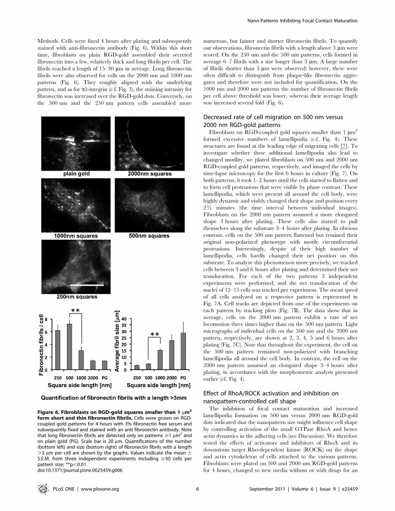
Poor recruitment of a5b1-integrin to matrix adhesions onRGD-gold squares smaller than 1 mm2
Integrin-a5b1 is largely absent from focal complexes of
spreading cells [2], but during their maturation is recruited into
early focal adhesions [28] where it binds secreted fibronectin [25].
Shortly later, a5b1-integrin is pulled out of early focal adhesions to
form fibrillar adhesions, leaving late focal adhesions behind with a
low content of this integrin [13]. To analyze the localization of
a5b1-integrin in cell-matrix adhesions, we plated fibroblasts on the
various RGD-gold patterns for 4 hours and stained them with
anti-a5-integrin antibody (Fig. 3). On plain RGD-gold (as on tissue
culture plastic; not shown), cells exhibited many a5-integrin-
positive fibrillar adhesions several mm in length, many of them
starting at the cell periphery and oriented in the long axis of the
cell. A similar distribution was observed on the 2000 nm and
1000 nm patterns: The cells bridged neighboring RGD-gold
squares with a5-integrin positive fibrillar adhesions, and hence
overcame the restriction given by pattern. However, staining for
a5-integrin was often brighter over the RGD-gold dots (Fig. 3), i.e.
at sites of vinculin-positive focal adhesions (cf. Fig. 2). In contrast,
on RGD-coupled gold patterns with dot size smaller than 1 mm2,
staining for a5-integrin in cell-matrix adhesions was clearly
diminished, especially in peripheral regions of the cells (Fig. 3).
A small amount of this integrin subunit was still found more
centrally in short (1–3 mm long) fibrillar structures, which however
did not appear to correlate with the underlying RGD-gold pattern.
These data indicated that the vinculin-positive, a5-integrin-
negative structures detected in the cell periphery on 250 nm and
500 nm patterns (Fig. 2) can indeed be classified as focal
complexes that failed to be converted into focal and fibrillar
adhesions.
Enhanced lamellipodia formation on RGD-gold squaressmaller than 1 mm2
When plating fibroblasts on the various RGD-gold patterns, we
found clear differences in cell morphology already by visual
inspection with a phase contrast microscope. To analyze cell-shape
more quantitatively, we fixed cells after 4 hours, co-stained them
for vinculin and actin, and viewed them by fluorescence
microscopy. On plain RGD-gold as well as on the 2000 nm and
the 1000 nm patterns, cells were well spread 4 hours after plating
(Fig. 4). Fibroblasts appeared elongated with usually several (3–7)
spiky processes and few lamellipodia (less than one per cell in
average). However, on the 500 nm and the 250 nm patterns the
cell morphology changed drastically. Cells again were well spread,
but assumed a phenotype with an average of 4–5 extended
lamellipodia per cell and almost no spike-like processes. Lamel-
Figure 1. RGD-gold patterns produced by the nanostencil technique. (A) Design of the patterns. Gold squares of the indicated sizes wereapplied to glass surfaces (see Materials and Methods). Unit cells of the four individual patterns are shown in blue. Note that by keeping the width ofthe squares the same as their lateral spacing, the patterns were designed such that the surface of RGD-coupled gold was fixed to 25% of the totalarea. After manufacturing the patterns, RGD peptide was coupled to the gold squares to provide restricted and integrin-specific cell attachment, andintervening spaces were passivated with PLL-g-PEG. (B) SEM micrographs of patterns of gold squares (5006500 nm2, 100061000 nm2 and200062000 nm2) on glass coverslips. The 2506250 nm2 squares could not be imaged due to electron charging effects.doi:10.1371/journal.pone.0025459.g001
Nano-Patterns Inhibiting Focal Contact Maturation
PLoS ONE | www.plosone.org 3 September 2011 | Volume 6 | Issue 9 | e25459
lipodia did not localize to a specific end of the cell but rather
formed all around the perinuclear cell body. On the 500 nm and
the 250 nm patterns, cells were also less elongated with an average
long axis of 55 mm. In comparison, the long axis for cells on the
larger RGD-gold patterns was measured to be 75–80 mm in
average, and it reached about 100 mm on plain gold (Fig. 4).
Altered actin assembly on RGD-gold squares smaller than1 mm2
We found clear differences in the number of lamellipodia in
fibroblasts grown on different RGD-gold patterns, and cells
showed a different morphology (Fig. 4). Since actin assembly is
largely involved in the formation of cellular processes, we wanted
to analyze the actin cytoskeleton more precisely. To this aim, cells
grown on the various patterns for 4 hours were stained with Alexa-
labeled phalloidin and analyzed by fluorescence microscopy. In
fibroblasts on plain RGD-gold substrate, we found thick actin
stress fibers throughout the cells, which often bundled in the cell
center forming actin foci (Fig. 5). Also on the 2000 nm pattern
thick actin stress fibers were identified. Some crossed the cell body
but most of them were found along the cell borders. This might be
a reason why actin foci were hardly found on these patterns.
Fibroblasts on the 1000 nm pattern only exhibited actin stress
fibers at the cell border. More central actin fibers were rather fine
but still aligned parallel to the major cell axis. They started at the
cell margin over gold squares and extended centripetally (see insert
in Fig. 5), in accordance with the location and elongated shape of
focal adhesions on the 1000 nm pattern (cf. Fig. 2). In contrast, on
the 250 nm and 500 nm patterns thick actin stress fibers were
basically absent. In some cells, short actin bundles were found at
the cell margin perpendicular to the border of lamellipodia.
However, most of the actin was polymerized into a fine meshwork
Figure 2. Fibroblasts form matrix adhesions only on RGD-coupled gold areas. Cells were grown on RGD-coupled gold patternsfor 4 hours with 3% fibronectin-free serum and subsequently fixed andstained with an anti-vinculin antibody. The images in the right row areenlargements of the boxed areas at left. Note that vinculin staining incell-matrix adhesions is restricted to RGD-gold nano- and microsquares,whereas on plain RGD-gold vinculin-positive adhesions are severalmicrometers long. On the 1000 nm pattern, peripheral adhesions overgold squares are elongated in the centripetal direction (arrow). Scalebar is 20 mm (left row) and 5 mm (right row), respectively.doi:10.1371/journal.pone.0025459.g002
Figure 3. Integrin a5b1 is poorly recruited to matrix adhesionson RGD-gold squares smaller than 1 mm2. Cells were grown onRGD-coupled gold patterns for 4 hours with 3% fibronectin free serumand subsequently fixed and stained with an anti-a5-integrin antibody.Notice that a5b1-integrin localizes to fibrillar adhesions on plain goldand on patterns $1 mm2, whereas on smaller patterns it is onlymodestly recruited to cell adhesions. Scale bar is 10 mm.doi:10.1371/journal.pone.0025459.g003
Nano-Patterns Inhibiting Focal Contact Maturation
PLoS ONE | www.plosone.org 4 September 2011 | Volume 6 | Issue 9 | e25459

in lamellipodia and also in the cell body. At the rear end of
protruding lamellipodia, the orientation of the meshwork was
parallel to the cell border. To quantify our observations, images
from three independent experiments with a total of around 50 cells
per pattern were evaluated. The threshold of the 8-bit pictures was
set to a grey value of 150, and remaining visible structures with a
fibrous shape were counted as actin stress fibers. The number of
stress fibers above threshold increased from almost zero on the
250 nm and 500 nm patterns to 2–3 on the 1000 nm and
2000 nm patterns, and up to 5 in average on plain RGD-gold
substrate (Fig. 5, lower panel). The mean length of these structures
gradually increased from 10 mm on the 250 nm pattern to 40 mm
on plain RGD-gold.
Altered fibronectin fibrillogenesis on RGD-gold squaressmaller than 1 mm2
Fibronectin fibrillogenesis by fibroblasts depends on actomyosin
contractility [29]. Since cells on the 250 nm and 500 nm pattern
assembled actin into a fine meshwork rather than stress fibers, we
asked whether these cells are still able to assemble fibronectin into
fibrils. Therefore we cultured cells on RGD-coupled gold patterns
in fibronectin-depleted medium as described in Materials and
Figure 4. Fibroblasts on RGD-gold squares smaller than 1 mm2
show altered morphology. Cells were grown on RGD-coupled goldpatterns for 4 hours with 3% fibronectin-free serum and subsequentlyfixed and stained with Alexa-labeled phalloidin (red) and anti-vinculinantibody (green). Scale bar is 50 mm. Note the high number oflamellipodia on the 250 nm and 500 nm patterns, which is quantified inthe graph (bottom left). In addition, the maximal cell span was reducedon the smaller compared to the larger patterns (bottom right).PG = Plain gold. Values indicate the mean 6 S.E.M. from threeindependent experiments including $30 cells per pattern size;**p,0.01.doi:10.1371/journal.pone.0025459.g004
Figure 5. Fibroblasts on RGD- gold squares smaller than 1 mm2
do not form actin stress fibers. Cells were grown on RGD-coupledgold patterns for 4 hours with 3% fibronectin free serum andsubsequently fixed and stained with Alexa-labeled phalloidin. Theinsert at the upper right of the cells on the 1000 nm pattern is anenlargement of the boxed area. Scale bar is 50 mm. For quantification ofstress fibers, a fluorescence intensity threshold was set to distinguishstress fibers from remaining actin structures. Note the increasednumber (bottom left) and length (bottom right) of actin stress fibers onpatterns $1 mm2 and on plain gold (PG). Values indicate the mean 6S.E.M. from three independent experiments including $30 cells perpattern size; **p,0.01, *p,0.05, #p = 0.056.doi:10.1371/journal.pone.0025459.g005
Nano-Patterns Inhibiting Focal Contact Maturation
PLoS ONE | www.plosone.org 5 September 2011 | Volume 6 | Issue 9 | e25459
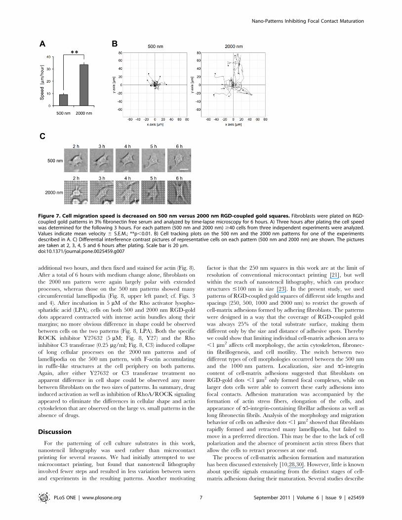
Methods. Cells were fixed 4 hours after plating and subsequently
stained with anti-fibronectin antibody (Fig. 6). Within this short
time, fibroblasts on plain RGD-gold assembled their secreted
fibronectin into a few, relatively thick and long fibrils per cell. The
fibrils reached a length of 15–30 mm in average. Long fibronectin
fibrils were also observed for cells on the 2000 nm and 1000 nm
patterns (Fig. 6). They roughly aligned with the underlying
pattern, and as for a5-integrin (c.f. Fig. 3), the staining intensity for
fibronectin was increased over the RGD-gold dots. Conversely, on
the 500 nm and the 250 nm pattern cells assembled more
numerous, but fainter and shorter fibronectin fibrils. To quantify
our observations, fibronectin fibrils with a length above 3 mm were
scored. On the 250 nm and the 500 nm patterns, cells formed in
average 6–7 fibrils with a size longer than 3 mm. A large number
of fibrils shorter than 3 mm were observed; however, these were
often difficult to distinguish from plaque-like fibronectin aggre-
gates and therefore were not included for quantification. On the
1000 nm and 2000 nm patterns the number of fibronectin fibrils
per cell above threshold was lower, whereas their average length
was increased several fold (Fig. 6).
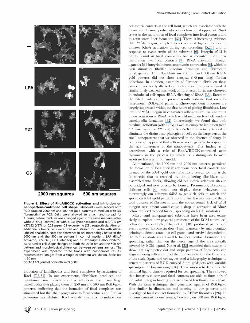
Decreased rate of cell migration on 500 nm versus2000 nm RGD-gold patterns
Fibroblasts on RGD-coupled gold squares smaller than 1 mm2
formed excessive numbers of lamellipodia (c.f. Fig. 4). These
structures are found at the leading edge of migrating cells [7]. To
investigate whether these additional lamellipodia also lead to
changed motility, we plated fibroblasts on 500 nm and 2000 nm
RGD-coupled gold patterns, respectively, and imaged the cells by
time-lapse microscopy for the first 6 hours in culture (Fig. 7). On
both patterns, it took 1–2 hours until the cells started to flatten and
to form cell protrusions that were visible by phase contrast. These
lamellipodia, which were present all around the cell body, were
highly dynamic and visibly changed their shape and position every
2K minutes (the time interval between individual images).
Fibroblasts on the 2000 nm pattern assumed a more elongated
shape 3 hours after plating. These cells also started to pull
themselves along the substrate 3–4 hours after plating. In obvious
contrast, cells on the 500 nm pattern flattened but retained their
original non-polarized phenotype with motile circumferential
protrusions. Interestingly, despite of their high number of
lamellipodia, cells hardly changed their net position on this
substrate. To analyze this phenomenon more precisely, we tracked
cells between 3 and 6 hours after plating and determined their net
translocation. For each of the two patterns 3 independent
experiments were performed, and the net translocation of the
nuclei of 12–15 cells was tracked per experiment. The mean speed
of all cells analyzed on a respective pattern is represented in
Fig. 7A. Cell tracks are depicted from one of the experiments on
each pattern by tracking plots (Fig. 7B). The data show that in
average, cells on the 2000 nm pattern exhibit a rate of net
locomotion three times higher than on the 500 nm pattern. Light
micrographs of individual cells on the 500 nm and the 2000 nm
pattern, respectively, are shown at 2, 3, 4, 5 and 6 hours after
plating (Fig. 7C). Note that throughout the experiment, the cell on
the 500 nm pattern remained non-polarized with branching
lamellipodia all around the cell body. In contrast, the cell on the
2000 nm pattern assumed an elongated shape 3–4 hours after
plating, in accordance with the morphometric analysis presented
earlier (cf. Fig. 4).
Effect of RhoA/ROCK activation and inhibition onnanopattern-controlled cell shape
The inhibition of focal contact maturation and increased
lamellipodia formation on 500 nm versus 2000 nm RGD-gold
dots indicated that the nanopattern size might influence cell shape
by controlling activation of the small GTPase RhoA and hence
actin dynamics in the adhering cells (see Discussion). We therefore
tested the effects of activators and inhibitors of RhoA and its
downstram target Rho-dependent kinase (ROCK) on the shape
and actin cytoskeleton of cells attached to the various patterns.
Fibroblasts were plated on 500 and 2000 nm RGD-gold patterns
for 4 hours, changed to new media without or with drugs for an
Figure 6. Fibroblasts on RGD-gold squares smaller than 1 mm2
form short and thin fibronectin fibrils. Cells were grown on RGD-coupled gold patterns for 4 hours with 3% fibronectin free serum andsubsequently fixed and stained with an anti fibronectin antibody. Notethat long fibronectin fibrils are detected only on patterns $1 mm2 andon plain gold (PG). Scale bar is 20 mm. Quantifications of the number(bottom left) and size (bottom right) of fibronectin fibrils with a length.3 mm per cell are shown by the graphs. Values indicate the mean 6S.E.M. from three independent experiments including $30 cells perpattern size; **p,0.01.doi:10.1371/journal.pone.0025459.g006
Nano-Patterns Inhibiting Focal Contact Maturation
PLoS ONE | www.plosone.org 6 September 2011 | Volume 6 | Issue 9 | e25459
additional two hours, and then fixed and stained for actin (Fig. 8).
After a total of 6 hours with medium change alone, fibroblasts on
the 2000 nm pattern were again largely polar with extended
processes, whereas those on the 500 nm patterns showed many
circumferential lamellipodia (Fig. 8, upper left panel; cf. Figs. 3
and 4). After incubation in 5 mM of the Rho activator lysopho-
sphatidic acid (LPA), cells on both 500 and 2000 nm RGD-gold
dots appeared contracted with intense actin bundles along their
margins; no more obvious difference in shape could be observed
between cells on the two patterns (Fig. 8, LPA). Both the specific
ROCK inhibitor Y27632 (5 mM; Fig. 8, Y27) and the Rho
inhibitor C3 transferase (0.25 mg/ml; Fig. 8, C3) induced collapse
of long cellular processes on the 2000 nm patterns and of
lamellipodia on the 500 nm pattern, with F-actin accumulating
in ruffle-like structures at the cell periphery on both patterns.
Again, after either Y27632 or C3 transferase treatment no
apparent difference in cell shape could be observed any more
between fibroblasts on the two sizes of patterns. In summary, drug
induced activation as well as inhibition of RhoA/ROCK signaling
appeared to eliminate the differences in cellular shape and actin
cytoskeleton that are observed on the large vs. small patterns in the
absence of drugs.
Discussion
For the patterning of cell culture substrates in this work,
nanostencil lithography was used rather than microcontact
printing for several reasons. We had initially attempted to use
microcontact printing, but found that nanostencil lithography
involved fewer steps and resulted in less variation between users
and experiments in the resulting patterns. Another motivating
factor is that the 250 nm squares in this work are at the limit of
resolution of conventional microcontact printing [21], but well
within the reach of nanostencil lithography, which can produce
structures #100 nm in size [23]. In the present study, we used
patterns of RGD-coupled gold squares of different side lengths and
spacings (250, 500, 1000 and 2000 nm) to restrict the growth of
cell-matrix adhesions formed by adhering fibroblasts. The patterns
were designed in a way that the coverage of RGD-coupled gold
was always 25% of the total substrate surface, making them
different only by the size and distance of adhesive spots. Thereby
we could show that limiting individual cell-matrix adhesion area to
,1 mm2 affects cell morphology, the actin cytoskeleton, fibronec-
tin fibrillogenesis, and cell motility. The switch between two
different types of cell morphologies occurred between the 500 nm
and the 1000 nm pattern. Localization, size and a5-integrin
content of cell-matrix adhesions suggested that fibroblasts on
RGD-gold dots ,1 mm2 only formed focal complexes, while on
larger dots cells were able to convert these early adhesions into
focal contacts. Adhesion maturation was accompanied by the
formation of actin stress fibers, elongation of the cells, and
appearance of a5-integrin-containing fibrillar adhesions as well as
long fibronectin fibrils. Analysis of the morphology and migration
behavior of cells on adhesive dots ,1 mm2 showed that fibroblasts
rapidly formed and retracted many lamellipodia, but failed to
move in a preferred direction. This may be due to the lack of cell
polarization and the absence of prominent actin stress fibers that
allow the cells to retract processes at one end.
The process of cell-matrix adhesion formation and maturation
has been discussed extensively [10,28,30]. However, little is known
about specific signals emanating from the distinct stages of cell-
matrix adhesions during their maturation. Several studies describe
Figure 7. Cell migration speed is decreased on 500 nm versus 2000 nm RGD-coupled gold squares. Fibroblasts were plated on RGD-coupled gold patterns in 3% fibronectin free serum and analyzed by time-lapse microscopy for 6 hours. A) Three hours after plating the cell speedwas determined for the following 3 hours. For each pattern (500 nm and 2000 nm) $40 cells from three independent experiments were analyzed.Values indicate mean velocity 6 S.E.M.; **p,0.01. B) Cell tracking plots on the 500 nm and the 2000 nm patterns for one of the experimentsdescribed in A. C) Differential interference contrast pictures of representative cells on each pattern (500 nm and 2000 nm) are shown. The picturesare taken at 2, 3, 4, 5 and 6 hours after plating. Scale bar is 20 mm.doi:10.1371/journal.pone.0025459.g007
Nano-Patterns Inhibiting Focal Contact Maturation
PLoS ONE | www.plosone.org 7 September 2011 | Volume 6 | Issue 9 | e25459
induction of lamellipodia and focal complexes by activation of
Rac1 [7,8,31]. In our experiments, fibroblasts produced and
maintained small vinculin-positive adhesions and many large
lamellipodia after plating them on 250 nm and 500 nm RGD-gold
patterns, indicating that the formation of focal complexes was
stimulated but that their progression to focal contacts and fibrillar
adhesions was inhibited. Rac1 was demonstrated to induce new
cell-matrix contacts at the cell front, which are associated with the
formation of lamellipodia, whereas its functional opponent RhoA
serves in the maturation of focal complexes into focal contacts and
in actin stress fiber formation [32]. There is increasing evidence
that a5b1-integrin, coupled to its secreted ligand fibronectin,
initiates RhoA activation during cell spreading [5,25] and in
response to cyclic strain of the substrate [6]. Integrin a5b1 is
hardly found in focal complexes but is recruited upon their
maturation into focal contacts [9]. RhoA activation through
ligated a5b1-integrin induces actomyosin contraction [6], which in
turn stimulates fibrillar adhesion formation and fibronectin
fibrillogenesis [13]. Fibroblasts on 250 nm and 500 nm RGD-
gold patterns did not show classical (.5 mm long) fibrillar
adhesions. In addition, assembly of fibronectin fibrils on these
patterns was clearly affected as only fine short fibrils were found. A
similar finely weaved meshwork of fibronectin fibrils was observed
in endothelial cells upon siRNA silencing of RhoA [33]. Based on
the cited evidence, our present results indicate that on sub-
micrometer RGD-gold patterns, RhoA-dependent processes are
largely suppressed within the first hours of plating fibroblasts. Low
levels of a5b1-integrin in cell-matrix adhesions are likely to result
in low activation of RhoA, which would maintain Rac1-dependent
lamellipodia formation [32]. Interestingly, we found that both
maximal activation (with LPA) as well as complete inhibition (with
C3 exoenzyme or Y27632) of RhoA/ROCK activity tended to
eliminate the distinct morphologies of cells on the large versus the
small nanopatterns that we observed in the absence of drugs. In
both cases, it appeared that cells were no longer able to respond to
the size differences of the nanopatterns. This finding is in
accordance with a role of RhoA/ROCK-controlled actin
dynamics in the process by which cells distinguish between
substrate features in our model.
As mentioned, the 1000 nm and 2000 nm patterns permitted
the formation of long fibrillar adhesions once focal contacts had
formed on the RGD-gold dots. The likely reason for this is the
fibronectin that is secreted by the adhering fibroblasts and
assembled into fibrils, allowing old cell-matrix adhesion sites to
be bridged and new ones to be formed. Presumably, fibronectin
deficient cells [6] would not display these behaviors, but
interestingly our attempts failed to get such cells to attach and
spread on RGD-gold patterns (not shown). It seems possible that a
total absence of fibronectin and the consequential lack of a5b1
integrin recruitment would cause a drop in activation of RhoA
below the level needed for cell spreading on RGD substrates.
Micro- and nanopatterned substrates have been used exten-
sively to explore how physical parameters of the ECM control cell
behavior. For example, Chen et al. [18] produced patterns of
evenly spaced fibronectin dots (3 mm diameter) by micro-contact
printing to demonstrate that cell growth and survival depended on
the total substrate area available for focal contact formation and
spreading, rather than on the percentage of the area actually
covered by ECM ligand. Xia et al. [22] extended these studies to
show that asymmetric dot and stripe patterns of fibronectin can
align adhering cells and direct their movements. On the lower end
of the scale, Spatz and colleagues used a lithography technique to
generate patterns of RGD-coupled 8 nm gold dots with variable
spacings in the low nm range [16]. Their aim was to determine the
minimal ligand density required for cell spreading. They showed
that integrins cluster and focal contacts are able to form only if
individual integrin binding sites are spaced less than 70 nm apart.
With the same technique, they generated squares of RGD-gold
dots similar in dimensions and spacing to our patterns, and
investigated focal contact formation by REF52 fibroblasts [26]. In
obvious contrast to our results, however, on 500 nm RGD-gold
Figure 8. Effect of RhoA/ROCK activation and inhibition onnanopattern-controlled cell shape. Fibroblasts were seeded ontoRGD-coupled 2000 nm and 500 nm gold patterns in medium with 3%fibronectin-free FCS. Cells were allowed to attach and spread for4 hours, before medium was changed against the same medium eitherwithout drug (control) or with 5 mM lysophospatitic acid (LPA), 5 mMY27632 (Y27), or 0.25 mg/ml C3 exoenzyme (C3), respectively. After anadditional 2 hours, cells were fixed and stained for F-actin with Alexa-labeled phalloidin. Note the difference in cell morphology between the2000 nm and the 500 nm pattern in control medium. LPA (RhoAactivator), Y27632 (ROCK inhibitor) and C3 exoenzyme (Rho inhibitor)cause similar cell shape changes on both the 2000 nm and the 500 nmpattern, and morphological differences between patterns are lost. Theexperiment was repeated three times with comparable results;representative images from a single experiment are shown. Scale baris 50 mm.doi:10.1371/journal.pone.0025459.g008
Nano-Patterns Inhibiting Focal Contact Maturation
PLoS ONE | www.plosone.org 8 September 2011 | Volume 6 | Issue 9 | e25459
dot squares spaced 500 nm apart these cells formed extended
paxillin-positive focal contacts that bridged the gaps between
patches. Thus, patterns in the sub-micrometer range did not
restrict the growth of cell-matrix adhesions in their case. This is
probably due to the different cell types used: REF52 cells are
considerably larger and spread more extensively than immortal-
ized mouse embryo fibroblasts which resemble primary cells [34].
In another study directly relevant to our present, Lehnert et al.
[21] used micro-contact printing to produce patterns of fibronectin
squares with side lengths from 0.3–3 mm (,0.1–10 mm2) and
center-to-center spacing from 1–30 mm. They found that a pattern
of 0.25 mm2 fibronectin dots with 5 mm spacing (1% coverage of
the substrate with ligand) was about the limit for B16 melanoma
cells to establish focal contacts, to spread, and to generate actin
stress fibers. These authors observed cell spreading and small cell-
matrix adhesions even on 0.1 mm2 fibronectin dots when the
spacing was reduced to 1 mm; however, they did not investigate
cell motility and the actin cytoskeleton under these conditions. In
the present study, we tested similar patterns engineered with the
nanostencil technique. We however kept the area covered with
ligand constant at 25% and used RGD peptide instead of
fibronectin, in order to reduce integrin-a5b1 and RhoA activation
during initial attachment of cells [5]. Our present results show that
for mouse embryo fibroblasts, adhesive RGD-gold patterns in the
sub-micrometer range inhibit the maturation of focal complexes to
focal contacts as well as stress fiber assembly, but still promote
extensive lamellipodia formation and cell spreading. This indicates
a difference in the balance between Rac1 and RhoA activation in
cells on the various patterns, which at present is difficult to
demonstrate directly due to the low number of cells available in
our experiments. Nevertheless, our findings demonstrate the
potential of the nanostencil patterning technique for manipulating
cell-matrix adhesion sites and hence the shape and motility of cells
adhering to engineered surfaces. RhoA-regulated cell shape has
been reported to control the differentiation of mesenchymal stem
cells towards the adipocyte and osteoblast lineage, respectively
[20]. Thus in the future, the nanostencil method may offer new
possibilities to control more precisely the interaction of mesen-
chymal cells with implant surfaces, and to influence their
differentiation around the implant.
Materials and Methods
Nanostencil techniqueNanostencil lithography is essentially a shadow-masking tech-
nique in which a material is added or removed through nanoscale
apertures in a thin membrane to create corresponding nanopat-
terns on a substrate in conformal contact. In this case, Au
nanopatterns were evaporated onto glass coverslips.
Precise details on nanostencil fabrication can be found in [35]
but we describe the major steps here. Fabrication begins by
depositing a 200 nm thick silicon nitride film on both faces of a
polished Si wafer. The nanoapertures are patterned into a resist on
the first face by electron beam lithography, and transferred into
the nitride film by reactive ion etching. Large (0.560.5 or
161 mm) ‘windows’ are patterned onto the second face using
photolithography and reactive ion etching. The exposed Si at the
‘window’ in the second face is etched through the wafer using a
combination of reactive ion and wet chemical etching, until it
meets the nanopatterned nitride film. The result is a 200 nm thick
nanopatterned nitride membrane supported in a sturdy silicon
frame. The wafer is cleaved into chips to facilitate stencil handling.
Patterning occurred in the following steps. The glass coverslips
were affixed to a large glass wafer using polyimide tape. Next, the
stencils were affixed to the coverslips using polyimide tape. The
assembly of wafers, coverslips, and nanostencils was cleaned in an
oxygen plasma chamber (30 s, 500 W) to ensure proper metal film
adhesion to the coverslips (Tepla 300, PVA Tepla AG, Germany).
Finally, the assembly was placed in a LAB 600 electron beam
evaporator (Leybold Optics GmbH, Germany) for metallization.
First, a 5 nm thick Ti layer was deposited to ensure the Au would
stick to the coverslip. Next, 40 nm of Au was deposited to serve as
the cell adhesive pattern. Following the evaporation the nanos-
tencils were removed from the coverslips, and the coverslips were
removed from the glass wafer and stored for use.
RGD-coupling of gold patterned coverslipsPatterned coverslips were cleaned in a UV-ozone photoreactor
(PR-100, UVP, Upland, CA) for 30 minutes. Each coverslip was
then placed in a m-dish (35 mm diameter, high) from Ibidi (Vitaris,
Baar, Switzerland) and subsequently coated with a solution of the
RGD peptide Ac-Gly-Cys-Gly-Arg-Gly-Asp-Ser-Pro-Gly-NH2
(Polypeptide, Boulogne, France) at 3 mg/ml in water for 24 hours.
Patterns were then washed 3 times with PBS and passivated with
PLL(20)-g[3.5]-PEG(2) (SuSoS AG, Dubendorf, Switzerland) in
water at 0.2 mg/ml for 2 hours. PLL-g-PEG solution was
extensively washed away with PBS and the PBS was then again
changed against Dulbecco’s modified Eagle medium (D-MEM;
Seromed, Basel, Switzerland).
Plating of cellsA clonal mouse embryonic kidney fibroblast cell line immor-
talized by stable transfection with SV40 large T antigen [36] was
obtained from Dr. Reinhard Fassler (Max Planck Institute for
Biochemistry, Martinsried, Germany). Cells were maintained at
37uC with 6% CO2 in Dulbecco’s modified Eagle medium (D-
MEM; Seromed, Basel, Switzerland) containing 10% fetal calf
serum (FCS; Gibco/Invitrogen, Basel, Switzerland). Instead of a
commercial established fibroblast line e.g. NIH-3T3, we chose
these cells for our experiments because they are not transformed
and resemble primary fibroblasts in their gene expression pattern
[34] and integrin profile [6]. Cells were harvested with trypsin-
EDTA (Gibco/Invitrogen, Basel, Switzerland), resuspended in D-
MEM containing 3% fibronectin-depleted FCS [6], and seeded
onto the nanopatterned substrates (40’000 cells per 35 mm dish).
The medium was depleted of fibronectin to reduce deposition of
this adhesive glycoprotein to the passivated areas. Dishes were kept
at 37uC and 6% CO2 for 4 hours. In certain experiments, medium
was changed to D-MEM/3% fibronectin-free FCS containing
5 mM lysophophatidic acid (LPA, Sigma), 5 mM Y27632 (Y27;
Calbiochem), or 0.25 mg/ml bacterial recombinant C3 transferase
(C3; Cytoskeleton), respectively, and cells were incubated for an
additional 2 hours. Optimal drug concentrations for these cells
had been determined in previously published experiments [3,6].
Cells were then fixed with 4% paraformaldehyde for 10 minutes,
followed by washing with PBS.
Control experiments for RGD functionalizationIn control experiments (Figure S1), gold-patterned coverslips
were coated with 3 mg/ml of a peptide in which the RGD
sequence was changed to RDG (Ac-Gly-Cys-Gly-Tyr-Gly-Arg-
Asp-Gly-Ser-Pro-Gly-NH2; synthesized by J. Patterson in the lab
of J. A. Hubbell, Ecole Polytechnique Federale de Lausanne).
Additionally, patterns were functionalized as above with active
RGD and control RDG peptide mixed in different ratios (1:1, 1:4
and 1:9), keeping the total concentration of 3 mg/ml constant.
After passivation with PLL-g-PEG, cells were plated as above
before fixation. We observed that fibroblasts spread fully on gold
Nano-Patterns Inhibiting Focal Contact Maturation
PLoS ONE | www.plosone.org 9 September 2011 | Volume 6 | Issue 9 | e25459

patterns coupled with pure RGD as well as with mixtures of
RGD:control peptide of 1:1 and 1:4. On patterns coated with 1:9
mixtures, cells spread partially but did not polarize, whereas on
plain control peptide they attached but did not spread (Fig. S1).
These results showed that with pure RGD peptide and up to a 1:4
dilution with control peptide, gold patterns were functionally
saturated with RGD ligand for cell spreading.
Immunofluorescence and phalloidin stainingPatterned cover slips with attached, fixed cells were permeabi-
lized at room temperature for 30 minutes in PBS solution
containing 3% BSA and 0.1% Triton X-100 (Sigma, Buchs,
Switzerland). Cells were then stained for 1 hour at room
temperature with one or several of the following reagents in PBS
containing 3% BSA and 0.1% Triton X-100: anti-fibronectin
antiserum [37] diluted 1:300; anti-a5-integrin diluted 1:500 (BD
Pharmingen, Basel, Switzerland); anti-vinculin diluted 1:1000
(Sigma, Buchs, Switzerland); Alexa 546-labeled phalloidin at
1 mg/ml (Sigma, Buchs, Switzerland). After staining, cells were
washed three times with PBS/0.1% Triton X-100 and incubated
for 1 hour with one or several of the following secondary
antibodies: Alexa 488-labeled goat anti rabbit IgG; Alexa 488-
labeled goat anti mouse IgG; Alexa 488-labeled goat anti rat IgG;
Alexa 633-labeled goat anti rabbit (all from Sigma, Buchs,
Switzerland) diluted 1:1000 in PBS/3% BSA/0.1% Triton X-
100. Cells were again washed three times with PBS/0.1% Triton-
X100 and mounted in Prolong Gold antifade reagent (Invitrogen,
Basel, Switzerland). Slides were examined with a Zeiss Z1 or a
Olympus BX51 fluorescence microscope equipped with 206/0.8
and 406/0.75 objectives and filter cubes for Alexa 488 and Alexa
546. Numbers of lamellipodia, cell span, number and length of
actin and fibronectin fibers per cell were determined from
fluorescence images of cells on the various patterns. At least 30
cells from three independent experiments were measured per
pattern. Data were evaluated by ANOVA to test the null
hypothesis, and the significance of differences between 500 nm
and 1000 nm patterns was determined by unpaired Student’s t-
test.
Live Imaging MicroscopyFor live imaging microscopy, fibroblasts were seeded into Ibidi
m-dishes (Vitaris, Baar, Switzerland) onto RGD-coupled gold
patterns as described above. Immediately after seeding, cells were
observed with a Zeiss inverted microscope for the following
6 hours. Time-lapse phase contrast images were collected every
2K minutes. Pictures were processed and analyzed with the
ImageJ program and the manual tracking plug-in. For quantifi-
cation of cell velocity, $40 cells per pattern were measured, and
the significance of the difference between 2000 nm and 500 nm
patterns was determined by unpaired Student’s t-test.
Supporting Information
Figure S1 Control experiment for RGD functionaliza-tion of gold patterns. 1000 nm gold patterns were coupled
with either RGD peptide alone (pure RGD), control peptide alone
(pure RDG control), or mixtures of RGD:control peptide of 1:1,
1:4 and 1:9, respectively (total peptide concentration 3 mg/ml in
all cases). Fibroblasts were plated, fixed and stained for F-actin as
described in Materials and Methods. Note that cells spread fully on
the coated gold patterns up to a RGD:control peptide ratio of 1:4.
The experiment was repeated three times with comparable results;
representative images from a single experiment are shown. Scale
bar is 50 mm.
(TIF)
Acknowledgments
We thank Reinhard Fassler for providing the mouse embryo fibroblast cell
line used here. We acknowledge Jennifer Patterson (laboratory of J. A.
Hubbell, EPF Lausanne) for the synthesis of the RDG control peptide, and
thank Matthias Lutolf for providing the peptide. R.L. and M.C. are
grateful to Ruth Chiquet-Ehrismann for her continuing support and
hospitality while working in her lab at FMI.
Author Contributions
Conceived and designed the experiments: RL NG JB MC. Performed the
experiments: RL KP NG MM. Analyzed the data: RL NG MC.
Contributed reagents/materials/analysis tools: RL KP NG MM JB MC.
Wrote the paper: RL MC. Nanopattern design: JB.Nanopattern
manufacturing: KP MM. Critical reading of the manuscript: KP JB NG.
References
1. Geiger B, Spatz JP, Bershadsky AD (2009) Environmental sensing through focal
adhesions. Nat Rev Mol Cell Biol 10: 21–33.
2. Zamir E, Geiger B (2001) Molecular complexity and dynamics of cell-matrix
adhesions. J Cell Sci 114: 3583–3590.
3. Sarasa-Renedo A, Tunc-Civelek V, Chiquet M (2006) Role of RhoA/ROCK-
dependent actin contractility in the induction of tenascin-C by cyclic tensile
strain. Exp Cell Res 312: 1361–1370.
4. Smith PG, Roy C, Zhang YN, Chauduri S (2003) Mechanical stress increases RhoA
activation in airway smooth muscle cells. Am J Respir Cell Mol Biol 28: 436–442.
5. Danen EH, Sonneveld P, Brakebusch C, Fassler R, Sonnenberg A (2002) The
fibronectin-binding integrins alpha5beta1 and alphavbeta3 differentially mod-
ulate RhoA-GTP loading, organization of cell matrix adhesions, and fibronectin
fibrillogenesis. J Cell Biol 159: 1071–1086.
6. Lutz R, Sakai T, Chiquet M (2010) Pericellular fibronectin is required for RhoA-
dependent responses to cyclic strain in fibroblasts. J Cell Sci 123: 1511–1521.
7. Hall A (1998) Rho GTPases and the actin cytoskeleton. Science 279: 509–514.
8. Nobes CD, Hall A (1995) Rho, rac, and cdc42 GTPases regulate the assembly of
multimolecular focal complexes associated with actin stress fibers, lamellipodia,
and filopodia. Cell 81: 53–62.
9. Harjanto D, Zaman MH (2010) Matrix mechanics and receptor-ligand
interactions in cell adhesion. Org Biomol Chem 8: 299–304.
10. Wolfenson H, Henis YI, Geiger B, Bershadsky AD (2009) The heel and toe of
the cell’s foot: a multifaceted approach for understanding the structure and
dynamics of focal adhesions. Cell Motil Cytoskeleton 66: 1017–1029.
11. Geiger B, Bershadsky A, Pankov R, Yamada KM (2001) Transmembrane
crosstalk between the extracellular matrix–cytoskeleton crosstalk. Nat Rev Mol
Cell Biol 2: 793–805.
12. Alexandrova AY, Arnold K, Schaub S, Vasiliev JM, Meister JJ, et al. (2008)
Comparative dynamics of retrograde actin flow and focal adhesions: formation
of nascent adhesions triggers transition from fast to slow flow. PLoS One 3:
e3234.
13. Zamir E, Katz M, Posen Y, Erez N, Yamada KM, et al. (2000) Dynamics and
segregation of cell-matrix adhesions in cultured fibroblasts. Nat Cell Biol 2:
191–196.
14. Mao Y, Schwarzbauer JE (2005) Fibronectin fibrillogenesis, a cell-mediated
matrix assembly process. Matrix Biol 24: 389–399.
15. Chen CS, Mrksich M, Huang S, Whitesides GM, Ingber DE (1998)
Micropatterned surfaces for control of cell shape, position, and function.
Biotechnol Prog 14: 356–363.
16. Arnold M, Cavalcanti-Adam EA, Glass R, Blummel J, Eck W, et al. (2004)
Activation of integrin function by nanopatterned adhesive interfaces. Chem-
physchem 5: 383–388.
17. Gallant ND, Charest JL, King WP, Garcia AJ (2007) Micro- and nano-patterned
substrates to manipulate cell adhesion. J Nanosci Nanotechnol 7: 803–807.
18. Chen CS, Mrksich M, Huang S, Whitesides GM, Ingber DE (1997) Geometric
control of cell life and death. Science 276: 1425–1428.
19. Thery M, Piel M (2009) Adhesive micropatterns for cells: a microcontact
printing protocol. Cold Spring Harb Protoc 2009: pdb prot5255.
20. McBeath R, Pirone DM, Nelson CM, Bhadriraju K, Chen CS (2004) Cell shape,
cytoskeletal tension, and RhoA regulate stem cell lineage commitment. Dev Cell
6: 483–495.
21. Lehnert D, Wehrle-Haller B, David C, Weiland U, Ballestrem C, et al. (2004)
Cell behaviour on micropatterned substrata: limits of extracellular matrix
geometry for spreading and adhesion. J Cell Sci 117: 41–52.
Nano-Patterns Inhibiting Focal Contact Maturation
PLoS ONE | www.plosone.org 10 September 2011 | Volume 6 | Issue 9 | e25459
22. Xia N, Thodeti CK, Hunt TP, Xu Q, Ho M, et al. (2008) Directional control of
cell motility through focal adhesion positioning and spatial control of Rac
activation. FASEB J 22: 1649–1659.
23. Vazquez-Mena O, Villanueva G, Savu V, Sidler K, van den Boogaart MA, et al.
(2008) Metallic nanowires by full wafer stencil lithography. Nano Lett 8:
3675–3682.
24. Love JC, Estroff LA, Kriebel JK, Nuzzo RG, Whitesides GM (2005) Self-
assembled monolayers of thiolates on metals as a form of nanotechnology. Chem
Rev 105: 1103–1169.
25. Huveneers S, Truong H, Fassler R, Sonnenberg A, Danen EH (2008) Binding of
soluble fibronectin to integrin alpha5 beta1 - link to focal adhesion redistribution
and contractile shape. J Cell Sci 121: 2452–2462.
26. Arnold M, Schwieder M, Blummel J, Cavalcanti-Adam EA, Lopez-Garcia M,
et al. (2009) Cell interactions with hierarchically structured nano-patterned
adhesive surfaces. Soft Matter 5: 72–77.
27. Csucs G, Michel R, Lussi JW, Textor M, Danuser G (2003) Microcontact
printing of novel co-polymers in combination with proteins for cell-biological
applications. Biomaterials 24: 1713–1720.
28. Zaidel-Bar R, Cohen M, Addadi L, Geiger B (2004) Hierarchical assembly of
cell-matrix adhesion complexes. Biochem Soc Trans 32: 416–420.
29. Wierzbicka-Patynowski I, Schwarzbauer JE (2003) The ins and outs of
fibronectin matrix assembly. J Cell Sci 116: 3269–3276.
30. Katz BZ, Zamir E, Bershadsky A, Kam Z, Yamada KM, et al. (2000) Physical
state of the extracellular matrix regulates the structure and molecularcomposition of cell-matrix adhesions. Mol Biol Cell 11: 1047–1060.
31. Machesky LM, Hall A (1997) Role of actin polymerization and adhesion to
extracellular matrix in Rac- and Rho-induced cytoskeletal reorganization. J CellBiol 138: 913–926.
32. Rottner K, Hall A, Small JV (1999) Interplay between Rac and Rho in thecontrol of substrate contact dynamics. Curr Biol 9: 640–648.
33. Fernandez-Sauze S, Grall D, Cseh B, Van Obberghen-Schilling E (2009)
Regulation of fibronectin matrix assembly and capillary morphogenesis inendothelial cells by Rho family GTPases. Exp Cell Res 315: 2092–2104.
34. May T, Mueller PP, Weich H, Froese N, Deutsch U, et al. (2005) Establishmentof murine cell lines by constitutive and conditional immortalization. J Biotechnol
120: 99–110.35. Kim GM, van den Boogaart MA, Brugger J (2003) Fabrication and application
of a full wafer size micro/nanostencil for multiple length-scale surface
patterning. Microelectronic Engineering 67: 609–614.36. Graness A, Giehl K, Goppelt-Struebe M (2006) Differential involvement of the
integrin-linked kinase (ILK) in RhoA-dependent rearrangement of F-actin fibersand induction of connective tissue growth factor (CTGF). Cell Signal 18:
433–440.
37. Wehrle B, Chiquet M (1990) Tenascin is accumulated along developingperipheral nerves and allows neurite outgrowth in vitro. Development 110:
401–415.
Nano-Patterns Inhibiting Focal Contact Maturation
PLoS ONE | www.plosone.org 11 September 2011 | Volume 6 | Issue 9 | e25459
Copyright © 2022 FDOKUMEN