Direct MR Arthrography of the Hip with Leg Traction: Feasibility for Assessing Articular Cartilage
7
1124 AJR:190, April 2008 the loaded hyaline cartilage of the femur and acetabulum, acetabular fossa, and teres liga- ment [1, 9]. The labrum separates the two compartments. Arthroscopic evaluation of the hip is a two-step procedure: flexion without traction for evaluation of the peripheral com- partment and extension with traction for evalu- ation of the central compartment [10, 11]. Dis- traction and distention are used to visualize cartilage in the central compartment, including the more central part of the labrum [2, 12, 13]. Manual traction during radiography has been used to make a diastasis between the femoral head and the acetabulum [14, 15]. Continuous leg traction has been used to im- prove visualization of acetabular labral tears during MR arthrography after IV rather than articular administration of a contrast agent. To the best of our knowledge [13, 16], no pre- vious studies have explored the potential ad- vantage of combining intraarticular admin- istration of a contrast agent and leg traction. The purpose of our study was to determine the feasibility of leg traction combined with MR arthrography and the effect of the tech- nique on visualization of cartilage surfaces. Subjects and Methods Informed consent was obtained from each pa- tient, and the study was approved by the insti- tutional review boards at two hospitals. The study group consisted of 48 patients consecutively referred from December 2005 through December 2006 for hip MR arthrography for the evaluation of groin pain. Prospective MR arthrographic examinations were performed after application of leg traction while the subjects were on the MRI Direct MR Arthrography of the Hip with Leg Traction: Feasibility for Assessing Articular Cartilage Eva Llopis 1 Luis Cerezal 2 Ara Kassarjian 3 Victoria Higueras 1 Ernesto Fernandez 4 Llopis E, Cerezal L, Kassarjian A, Higueras V, Fernandez E 1 Department of Radiology, Hospital de la Ribera, Carretera de Corbera km1, Alzira, Valencia, 46600, Spain. Address correspondence to E. Llopis. 2 Instituto Radiologico Cantabro, Clinica Mompia, Santander, Spain. 3 Department of Radiology, Division of Musculoskeletal Radiology, Massachusetts General Hospital, Boston, MA. 4 Department of Orthopedics, Hospital de la Ribera, Valencia, Spain. Musculoskeletal Imaging • Technical Innovation AJR 2008; 190:1124–1128 0361–803X/08/1904–1124 © American Roentgen Ray Society T he hip joint is a substantial chal- lenge to radiologists for a variety of reasons. The critical struc- tures, mainly the acetabular la- brum and the femoral and acetabular cartilage, are small, requiring high-resolution imaging for adequate depiction of normal and patho- logic anatomic features. Chondral lesions are an important source of hip joint pain, and the extent or thickness of the cartilage injury is the most decisive predictor of surgical out- come [1, 2]. The morphologic characteristics of the hip joint—the ball-in-socket configu- ration with permanent contact between the articular surfaces and small intraarticular volume, the strong articular capsule, and the tightness of the ligaments (especially the il- iofemoral ligament) and surrounding mus- cles—make separation between the femoral and acetabular cartilage difficult. Moreover, the limited value of surface coils due to the deep position of the cartilage within the body adversely affects image quality. MR arthrography of the hip has been shown accurate for evaluating the acetabular labrum and peripheral compartment. However, the ac- curacy in assessing lesions of the central carti- lage is only moderate [3, 4]. The cartilage of the acetabulum and femoral head often cannot be seen as distinct entities despite the use of an intraarticular contrast agent, and therefore small lesions can be difficult to visualize [3, 5–8]. At arthroscopy, a distinction is made be- tween the easily accessible peripheral compart- ment, which comprises unloaded femoral car- tilage, femoral neck, and synovial folds, and the tight central compartment, which includes Keywords: cartilage, hip, MR arthrography, traction DOI:10.2214/AJR.07.2559 Received May 13, 2007; accepted after revision October 12, 2007. OBJECTIVE. Hip arthrography is an accurate diagnostic method for evaluation of the peripheral compartment, but its depiction of cartilage lesions is moderate. The purpose of this study was to add leg traction to MR arthrography of the hip to test its effect on visualization of cartilage surfaces. CONCLUSION. Hip MR arthrography with leg traction is a technically feasible and safe procedure that improves visualization of the femoral and acetabular cartilage surfaces. Llopis et al. Hip MR Arthrography Musculoskeletal Imaging Technical Innovation Downloaded from www.ajronline.org by Sociedad Espanola De Radiologia Medica on 09/30/13 from IP address 176.31.224.175. Copyright ARRS. For personal use only; all rights reserved
-
Upload
independent -
Category
Documents
-
view
2 -
download
0
Transcript of Direct MR Arthrography of the Hip with Leg Traction: Feasibility for Assessing Articular Cartilage

1124 AJR:190, April 2008
the loaded hyaline cartilage of the femur and acetabulum, acetabular fossa, and teres liga-ment [1, 9]. The labrum separates the two compartments. Arthroscopic evaluation of the hip is a two-step procedure: flexion without traction for evaluation of the peripheral com-partment and extension with traction for evalu-ation of the central compartment [10, 11]. Dis-traction and distention are used to visualize cartilage in the central compartment, including the more central part of the labrum [2, 12, 13].
Manual traction during radiography has been used to make a diastasis between the femoral head and the acetabulum [14, 15]. Continuous leg traction has been used to im-prove visualization of acetabular labral tears during MR arthrography after IV rather than articular administration of a contrast agent. To the best of our knowledge [13, 16], no pre-vious studies have explored the potential ad-vantage of combining intraarticular admin-istration of a contrast agent and leg traction. The purpose of our study was to determine the feasibility of leg traction combined with MR arthrography and the effect of the tech-nique on visualization of cartilage surfaces.
Subjects and MethodsInformed consent was obtained from each pa-
tient, and the study was approved by the insti-tutional review boards at two hospitals. The study group consisted of 48 patients consecutively referred from December 2005 through December 2006 for hip MR arthrography for the evaluation of groin pain. Prospective MR arthrographic examinations were performed after application of leg traction while the subjects were on the MRI
Direct MR Arthrography of the Hip with Leg Traction: Feasibility for Assessing Articular Cartilage
Eva Llopis1 Luis Cerezal2 Ara Kassarjian3 Victoria Higueras1 Ernesto Fernandez4
Llopis E, Cerezal L, Kassarjian A, Higueras V, Fernandez E
1Department of Radiology, Hospital de la Ribera, Carretera de Corbera km1, Alzira, Valencia, 46600, Spain. Address correspondence to E. Llopis.
2Instituto Radiologico Cantabro, Clinica Mompia, Santander, Spain.
3Department of Radiology, Division of Musculoskeletal Radiology, Massachusetts General Hospital, Boston, MA.
4Department of Orthopedics, Hospital de la Ribera, Valencia, Spain.
Musculoskeleta l Imaging • Technica l Innovat ion
AJR 2008; 190:1124–1128
0361–803X/08/1904–1124
© American Roentgen Ray Society
The hip joint is a substantial chal-lenge to radiologists for a variety of reasons. The critical struc-tures, mainly the acetabular la-
brum and the femoral and acetabular cartilage, are small, requiring high-resolution imaging for adequate depiction of normal and patho-logic anatomic features. Chondral lesions are an important source of hip joint pain, and the extent or thickness of the cartilage injury is the most decisive predictor of surgical out-come [1, 2]. The morphologic characteristics of the hip joint—the ball-in-socket configu-ration with permanent contact between the articular surfaces and small intraarticular volume, the strong articular capsule, and the tightness of the ligaments (especially the il-iofemoral ligament) and surrounding mus-cles—make separation between the femoral and acetabular cartilage difficult. Moreover, the limited value of surface coils due to the deep position of the cartilage within the body adversely affects image quality.
MR arthrography of the hip has been shown accurate for evaluating the acetabular labrum and peripheral compartment. However, the ac-curacy in assessing lesions of the central carti-lage is only moderate [3, 4]. The cartilage of the acetabulum and femoral head often cannot be seen as distinct entities despite the use of an intraarticular contrast agent, and therefore small lesions can be difficult to visualize [3, 5–8]. At arthroscopy, a distinction is made be-tween the easily accessible peripheral compart-ment, which comprises unloaded femoral car-tilage, femoral neck, and synovial folds, and the tight central compartment, which includes
Keywords: cartilage, hip, MR arthrography, traction
DOI:10.2214/AJR.07.2559
Received May 13, 2007; accepted after revision October 12, 2007.
ObjeCTive. Hip arthrography is an accurate diagnostic method for evaluation of the peripheral compartment, but its depiction of cartilage lesions is moderate. The purpose of this study was to add leg traction to MR arthrography of the hip to test its effect on visualization of cartilage surfaces.
COnCLuSiOn. Hip MR arthrography with leg traction is a technically feasible and safe procedure that improves visualization of the femoral and acetabular cartilage surfaces.
Llopis et al.Hip MR Arthrography
Musculoskeletal ImagingTechnical Innovation
Dow
nloa
ded
from
ww
w.a
jron
line.
org
by S
ocie
dad
Esp
anol
a D
e R
adio
logi
a M
edic
a on
09/
30/1
3 fr
om I
P ad
dres
s 17
6.31
.224
.175
. Cop
yrig
ht A
RR
S. F
or p
erso
nal u
se o
nly;
all
righ
ts r
eser
ved
AJR:190, April 2008 1125
Hip MR Arthrography
table. Exclusion criteria were previous surgery, inadequate hip distention due to leakage from the joint, and pain related to injection. Two patients in the study group underwent bilateral hip MR arthrography, so 50 MR arthrographic exami-nations of the hip were performed. All patients had undergone conventional bilateral hip MRI.
The records of 10 aged-matched patients who had undergone conventional MR arthrography for evaluation of groin pain before December 2005 were retrieved from our database. The imaging protocol was the same as for the 48 patients except for use of the manual traction and load device. The 10 patients made up the control group. The characteristics of the control group were similar to those of the study group. The mean age of the control group was 35 years (range, 21–46 years).
Arthrography was performed by one of two musculoskeletal radiologists, who had 5 and 7 years of experience in MR arthrography of the hip. The only difference in procedure between the study and control groups was that traction was not applied to the control group. The patients were placed supine on the fluoroscopic table. The lower extremity was held in neutral or slight internal rotation with the toes taped together to bring the femoral neck into the coronal plane. The skin was prepared in the usual sterile manner, disinfected with iodine solution, and covered with sterile drapes. Arthrography was performed with a 22-gauge needle in 47 hips and a 20-gauge needle in three hips. A 22-gauge needle was used for the control group. Local anesthesia with 4 mL of 2% lidocaine (Lidocaina, Braun) was injected at the skin entrance site. An oblique approach from the intertrochanteric line toward the femoral neck
junction was used. The intraarticular position of the needle tip was checked with 1–2 mL of iodinated contrast material (amido trizoate acid, Trazograf, Juste). A mean of 15 mL (range, 10–18 mL) of standard dilute 0.01-mmol gadopentetate dimeglumine (Omniscan, GE Healthcare) solution was injected. The solution is made with 12 mL of 0.9% saline solution, 4 mL of lidocaine, and 4 mL of iodine. The cocktail was injected under fluoroscopic guidance until a change in resistance, pain, or leakage occurred. The patients then were transferred to the MRI suite.
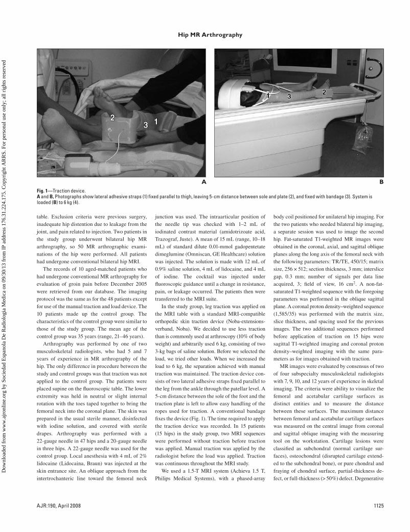
In the study group, leg traction was applied on the MRI table with a standard MRI-compatible orthopedic skin traction device (Noba-extension s-verband, Noba). We decided to use less traction than is commonly used at arthroscopy (10% of body weight) and arbitrarily used 6 kg, consisting of two 3-kg bags of saline solution. Before we selected the load, we tried other loads. When we increased the load to 6 kg, the separation achieved with manual traction was maintained. The traction device con-sists of two lateral adhesive straps fixed parallel to the leg from the ankle through the patellar level. A 5-cm distance between the sole of the foot and the traction plate is left to allow easy handling of the ropes used for traction. A conventional bandage fixes the device (Fig. 1). The time required to apply the traction device was recorded. In 15 patients (15 hips) in the study group, two MRI sequences were performed without traction before traction was applied. Manual traction was applied by the radiologist before the load was applied. Traction was continuous throughout the MRI study.
We used a 1.5-T MRI system (Achieva 1.5 T, Philips Medical Systems), with a phased-array
body coil positioned for unilateral hip imaging. For the two patients who needed bilateral hip imaging, a separate session was used to image the second hip. Fat-saturated T1-weighted MR images were obtained in the coronal, axial, and sagittal oblique planes along the long axis of the femoral neck with the following parameters: TR/TE, 450/15; matrix size, 256 × 512; section thickness, 3 mm; interslice gap, 0.3 mm; number of signals per data line acquired, 3; field of view, 16 cm2. A non-fat-saturated T1-weighted sequence with the foregoing parameters was performed in the oblique sagittal plane. A coronal proton density–weighted sequence (1,585/35) was performed with the matrix size, slice thickness, and spacing used for the previous images. The two additional sequences performed before application of traction on 15 hips were sagittal T1-weighted imaging and coronal proton density–weighted imaging with the same para-meters as for images obtained with traction.
MR images were evaluated by consensus of two of four subspecialty musculoskeletal radio logists with 7, 9, 10, and 12 years of experience in skeletal imaging. The criteria were ability to visualize the femoral and acetabular cartilage surfaces as distinct entities and to measure the distance between these surfaces. The maximum distance between femoral and acetabular cartilage surfaces was measured on the central image from coronal and sagittal oblique imaging with the measuring tool on the workstation. Cartilage lesions were classified as subchondral (normal cartilage sur-faces), osteo chondral (disrupted carti lage extend-ed to the sub chondral bone), or pure chondral and fraying of chondral surface, partial-thickness de-fect, or full-thickness (> 50%) defect. De generative
AFig. 1—Traction device.A and B, Photographs show lateral adhesive straps (1) fixed parallel to thigh, leaving 5-cm distance between sole and plate (2), and fixed with bandage (3). System is loaded (B) to 6 kg (4).
b
Dow
nloa
ded
from
ww
w.a
jron
line.
org
by S
ocie
dad
Esp
anol
a D
e R
adio
logi
a M
edic
a on
09/
30/1
3 fr
om I
P ad
dres
s 17
6.31
.224
.175
. Cop
yrig
ht A
RR
S. F
or p
erso
nal u
se o
nly;
all
righ
ts r
eser
ved

1126 AJR:190, April 2008
Llopis et al.
changes seen as bony osteo phytes and joint space narrowing were documented.
All patients in the study group were instructed to inform the radiologist if they experienced any discomfort during application of traction or during MRI data acquisition. Pain was graded with a semiquantitative scale: none, mild, moderate, and severe. All patients were given the option to request discontinuation of traction at any point during the examination. After the procedure, all patients in the study group were questioned regarding presence of discomfort, pain, and neurologic symptoms involving the leg to which traction was applied. For patients with symptoms, the questions were repeated during a telephone conversation 48 hours after MRI.
ResultsThe mean age of the study group was 36
years (range, 20–49 years), and 28 patients were men. Intraarticular injection of contrast material was achieved in all cases. The inter-val between arthrography and MRI was less than 20 minutes in all cases. Three patients were excluded because of inadequate intraar-ticular distention with the contrast agent. Two hips in the study group exhibited small normal communication between the hip joint and the iliopsoas bursa.
The average time it took to place the leg traction device on the MRI table was 4 min-utes (range, 3–8 minutes). We detected no complications related to the procedure. None
of the patients requested termination of trac-tion or reported pain or neurologic symptoms during or immediately after the examina-tion. The leg traction device was well toler-ated by all patients. Five patients had mild problems related to arthrography that re-solved within 48 hours without the need for intervention or medication.
The mean cartilage surface separation without traction in the 10 patients in the control group was 0.2 mm (range, 0–0.6 mm) in both the coronal and oblique sagittal planes. In two of these patients, the observ-ers were able to differentiate femoral from acetabular cartilage.
With traction, in all patients in the study group except three who had early degenera-tive changes, the femoral and acetabular carti-lage surfaces were seen as separate structures with contrast agent separating the two surfac-es. The mean separation of the cartilage sur-faces with traction was 1.7 mm (range, 0.6–3.8) mm. The mean distance in the coro-nal plane was 1.4 mm and in the oblique sagit-tal plane was 1.8 mm (Figs. 2 and 3).
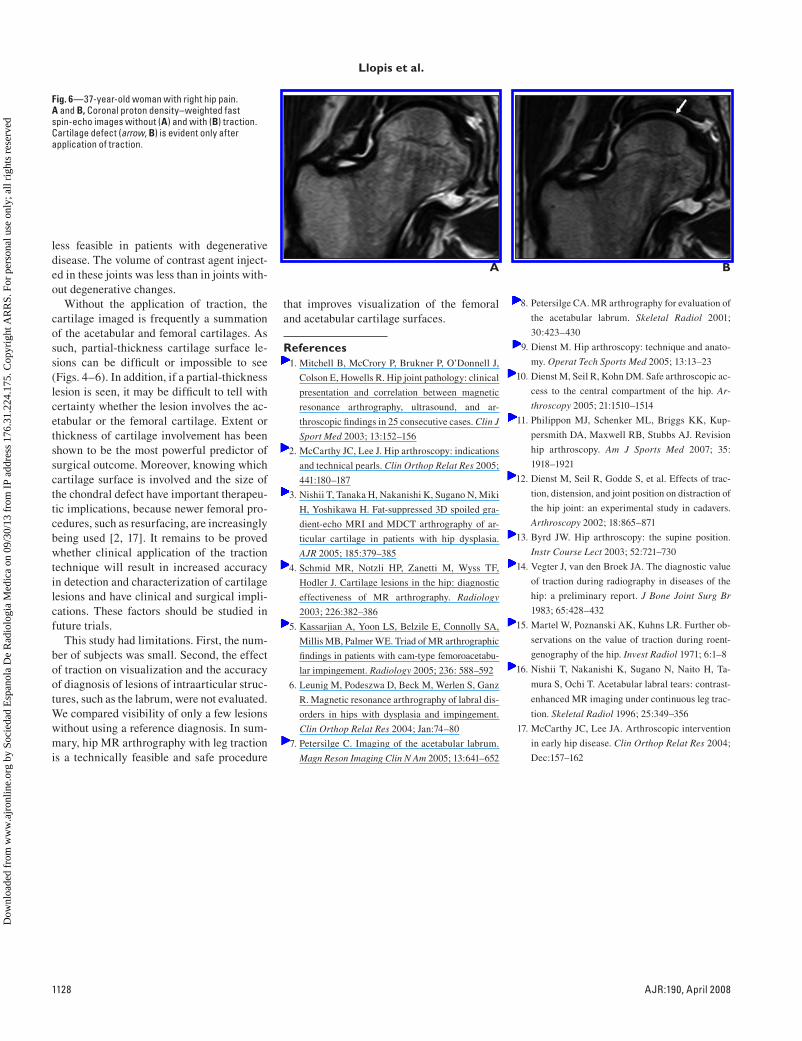
In 15 hips (15 patients) imaged without and with traction, the mean distance between fem-oral and acetabular cartilages was 0.2 mm (range, not measurable to 0.4 mm) before trac-tion. The distance increased an average of 1.5 mm (range, 0.75–3.8 mm) with traction (Figs. 4–6). In three hips in the study group, despite adequate intraarticular hip distention, insuffi-cient separation of the joint cartilage was ob-tained for clear depiction of the two cartilage surfaces. Traction in these three patients was insufficient to achieve separation between the femoral and acetabular cartilages. These three patients had degenerative changes secondary to cam-type femoroacetabular impingement.
Maximal separation between the cartilage surfaces was typically along the superior
A
C
Fig. 2—23-year-old male tennis player. MR arthrograms with and without traction show how well cartilage surfaces are depicted with traction.A and B, Coronal proton density–weighted fast spin-echo MR arthrogram without traction (A) and oblique sagittal fat-suppressed T1-weighted fast spin-echo image (B) readily show labral degeneration and tear, but femoral and acetabular cartilages are not evident as separate structures.C and D, Traction proton density–weighted fast spin-echo (C) and fat-suppressed T1-weighted (D) images corresponding to A and B show separation (arrow) between cartilage surfaces, which allows assessment of cartilage defects. Labrum tear (arrowheads, C) is evident.
b
D
Dow
nloa
ded
from
ww
w.a
jron
line.
org
by S
ocie
dad
Esp
anol
a D
e R
adio
logi
a M
edic
a on
09/
30/1
3 fr
om I
P ad
dres
s 17
6.31
.224
.175
. Cop
yrig
ht A
RR
S. F
or p
erso
nal u
se o
nly;
all
righ
ts r
eser
ved

AJR:190, April 2008 1127
Hip MR Arthrography
portion of the hip joint and was better depicted in the oblique sagittal plane. In the study group, 20 patients had normal cartilage surfaces. Ex-cluding the three patients with degenerative changes in whom traction was insufficient, 25 patients were found to have cartilage injuries. Femoral cartilage fraying was present in 17 pa-tients, femoral pure chondral defects (three par-tial thickness, two full thickness) in five, femo-ral osteochondral lesions in three, femoral sub-chondral lesion in three, acetabular cartilage fraying in nine, acetabular pure chondral lesion (two partial thickness and four full-thickness defect) in six, and acetabular osteochondral le-sion in eight patients. In the 15 patients imaged without and with traction, two pure femoral chondral lesions (one full thickness, one partial thickness) were seen only after traction. Eight possible lesions seen without traction were bet-ter characterized with traction. Of these eight, imaging with traction showed two instances of normal cartilage surfaces, five instances of fem-oral cartilage fraying, one instance of femoral subchondral lesion, and one instance of acetab-ular cartilage fraying.
DiscussionThis study showed the potential advantage
of applying manual traction followed by gen-tle leg traction during MR arthrography of the hip. Such traction produces enough space for the intraarticular contrast agent to enter the tight central compartment. This combi-nation of contrast agent and additional space allows visualization of the cartilage surfaces as distinct entities. The study also showed
that limited traction is well tolerated and can be applied in a short time without special-ized equipment. The arbitrarily chosen 6 kg of traction was well within the traction force used during arthroscopy, and no adverse ef-fects such as transient neuropraxia occurred. Only five patients mentioned a temporary in-crease in discomfort in the hip. There seems to be room for optimization; for instance, we used the same traction for men and women and independently of patient weight.
For 15 hips, images without and with trac-tion were compared. The findings in these cases showed that traction and the ensuing mean increase in distance between femur and acetabulum of 1.5 mm make a difference. Identification of femoral and acetabular carti-lages as distinct structures was possible only on images obtained during traction (Figs. 2 and 3). Results of comparison of the study group with the control group, who underwent only conventional MR arthrography, further support the value of traction. The mean dis-tance between femoral and acetabular carti-lages was not measurable in the control group and was an average of 1.7 mm in the study group. Also in this comparison, identification of the femoral and acetabular cartilages as distinct structures was the benefit of traction, facilitating characterization of cartilage le-sions and increasing diagnostic confidence.
Distraction and the possibility of identify-ing the cartilaginous surfaces of the femur and acetabulum as separate structures were
A
Fig. 3—16-year-old female runner with unilateral left hip pain.A and B, Oblique sagittal T1-weighted MR images without (A) and with (B) traction show marked distention of deep central compartment hip that allows differentiation of articular femoral and acetabular cartilages as separate structures.
b
Fig. 4—28-year-old man with right hip pain. Oblique sagittal T1-weighted image MR arthrogram shows large subchondral lesion without involvement of articular cartilage (arrow).
Fig. 5—40-year-old man with decreased internal rotation of left hip. Oblique sagittal T1-weighted MR arthrogram shows large osteochondral anterosuperior lesion extending to cartilage and small cartilage flap (arrow).
Dow
nloa
ded
from
ww
w.a
jron
line.
org
by S
ocie
dad
Esp
anol
a D
e R
adio
logi
a M
edic
a on
09/
30/1
3 fr
om I
P ad
dres
s 17
6.31
.224
.175
. Cop
yrig
ht A
RR
S. F
or p
erso
nal u
se o
nly;
all
righ
ts r
eser
ved
1128 AJR:190, April 2008
Llopis et al.
less feasible in patients with degenerative disease. The volume of contrast agent inject-ed in these joints was less than in joints with-out degenerative changes.
Without the application of traction, the cartilage imaged is frequently a summation of the acetabular and femoral cartilages. As such, partial-thickness cartilage surface le-sions can be difficult or impossible to see (Figs. 4–6). In addition, if a partial-thickness lesion is seen, it may be difficult to tell with certainty whether the lesion involves the ac-etabular or the femoral cartilage. Extent or thickness of cartilage involvement has been shown to be the most powerful predictor of surgical outcome. Moreover, knowing which cartilage surface is involved and the size of the chondral defect have important therapeu-tic implications, because newer femoral pro-cedures, such as resurfacing, are increasingly being used [2, 17]. It remains to be proved whether clinical application of the traction technique will result in increased accuracy in detection and characterization of cartilage lesions and have clinical and surgical impli-cations. These factors should be studied in future trials.
This study had limitations. First, the num-ber of subjects was small. Second, the effect of traction on visualization and the accuracy of diagnosis of lesions of intraarticular struc-tures, such as the labrum, were not evaluated. We compared visibility of only a few lesions without using a reference diagnosis. In sum-mary, hip MR arthrography with leg traction is a technically feasible and safe procedure
that improves visualization of the femoral and acetabular cartilage surfaces.
References 1. Mitchell B, McCrory P, Brukner P, O’Donnell J,
Colson E, Howells R. Hip joint pathology: clinical
presentation and correlation between magnetic
resonance arthrography, ultrasound, and ar-
throscopic findings in 25 consecutive cases. Clin J
Sport Med 2003; 13:152–156
2. McCarthy JC, Lee J. Hip arthroscopy: indications
and technical pearls. Clin Orthop Relat Res 2005;
441:180–187
3. Nishii T, Tanaka H, Nakanishi K, Sugano N, Miki
H, Yoshikawa H. Fat-suppressed 3D spoiled gra-
dient-echo MRI and MDCT arthrography of ar-
ticular cartilage in patients with hip dysplasia.
AJR 2005; 185:379–385
4. Schmid MR, Notzli HP, Zanetti M, Wyss TF,
Hodler J. Cartilage lesions in the hip: diagnostic
effectiveness of MR arthrography. Radiology
2003; 226:382–386
5. Kassarjian A, Yoon LS, Belzile E, Connolly SA,
Millis MB, Palmer WE. Triad of MR arthrographic
findings in patients with cam-type femoroacetabu-
lar impingement. Radiology 2005; 236: 588–592
6. Leunig M, Podeszwa D, Beck M, Werlen S, Ganz
R. Magnetic resonance arthrography of labral dis-
orders in hips with dysplasia and impingement.
Clin Orthop Relat Res 2004; Jan:74–80
7. Petersilge C. Imaging of the acetabular labrum.
Magn Reson Imaging Clin N Am 2005; 13:641–652
8. Petersilge CA. MR arthrography for evaluation of
the acetabular labrum. Skeletal Radiol 2001;
30:423–430
9. Dienst M. Hip arthroscopy: technique and anato-
my. Operat Tech Sports Med 2005; 13:13–23
10. Dienst M, Seil R, Kohn DM. Safe arthroscopic ac-
cess to the central compartment of the hip. Ar-
throscopy 2005; 21:1510–1514
11. Philippon MJ, Schenker ML, Briggs KK, Kup-
persmith DA, Maxwell RB, Stubbs AJ. Revision
hip arthroscopy. Am J Sports Med 2007; 35:
1918–1921
12. Dienst M, Seil R, Godde S, et al. Effects of trac-
tion, distension, and joint position on distraction of
the hip joint: an experimental study in cadavers.
Arthroscopy 2002; 18:865–871
13. Byrd JW. Hip arthroscopy: the supine position.
Instr Course Lect 2003; 52:721–730
14. Vegter J, van den Broek JA. The diagnostic value
of traction during radiography in diseases of the
hip: a preliminary report. J Bone Joint Surg Br
1983; 65:428–432
15. Martel W, Poznanski AK, Kuhns LR. Further ob-
servations on the value of traction during roent-
genography of the hip. Invest Radiol 1971; 6:1–8
16. Nishii T, Nakanishi K, Sugano N, Naito H, Ta-
mura S, Ochi T. Acetabular labral tears: contrast-
enhanced MR imaging under continuous leg trac-
tion. Skeletal Radiol 1996; 25:349–356
17. McCarthy JC, Lee JA. Arthroscopic intervention
in early hip disease. Clin Orthop Relat Res 2004;
Dec:157–162
A
Fig. 6—37-year-old woman with right hip pain.A and B, Coronal proton density–weighted fast spin-echo images without (A) and with (B) traction. Cartilage defect (arrow, B) is evident only after application of traction.
b
Dow
nloa
ded
from
ww
w.a
jron
line.
org
by S
ocie
dad
Esp
anol
a D
e R
adio
logi
a M
edic
a on
09/
30/1
3 fr
om I
P ad
dres
s 17
6.31
.224
.175
. Cop
yrig
ht A
RR
S. F
or p
erso
nal u
se o
nly;
all
righ
ts r
eser
ved

This article has been cited by:
1. Suzanne M. Shepherd, Eric Y. Chang, Julie L. Rutledge, Brady Huang, Debra Trudell, Donald L. Resnick. 2013. Cartilageassessment of the metacarpophalangeal joints: cadaveric study with magnetic resonance arthrography and finger traction. ClinicalImaging 37:4, 718-722. [CrossRef]
2. Milena Cerny, Romain Marlois, Nicolas Theumann, Christof Bollmann, Laurent Wehrli, Delphine Richarme, Reto Meuli,Fabio Becce. 2013. 3-T direct MR arthrography of the wrist: Value of finger trap distraction to assess intrinsic ligament andtriangular fibrocartilage complex tears. European Journal of Radiology . [CrossRef]
3. Luis S. Beltran, Zehava S. Rosenberg, Jason D. Mayo, Maria Diaz De Tuesta, Olga Martin, Luis P. Neto, Jenny T. Bencardino.2013. Imaging Evaluation of Developmental Hip Dysplasia in the Young Adult. American Journal of Roentgenology 200:5,1077-1088. [Abstract] [Full Text] [PDF] [PDF Plus]
4. Fabio Becce, Delphine Richarme, Patrick Omoumi, Ali Djahangiri, Alain Farron, Reto Meuli, Nicolas Theumann. 2013. DirectMR arthrography of the shoulder under axial traction: Feasibility study to evaluate the superior labrum-biceps tendon complexand articular cartilage. Journal of Magnetic Resonance Imaging 37:5, 1228-1233. [CrossRef]
5. Daniel Hendry, Eric England, Keith Kenter, Robert D. Wissman. 2013. Femoral Acetabular Impingement. Seminars inRoentgenology 48:2, 158-166. [CrossRef]
6. Corinne R. Henak. 2013. Subject-Specific Analysis of Joint Contact Mechanics: Application to the Study of Osteoarthritis andSurgical Planning. Journal of Biomechanical Engineering 135:2, 021003. [CrossRef]
7. Eva Llopis, Victoria Higueras, Maria Vaño, José R. Altónaga. 2012. Anatomic and radiographic evaluation of the hip. EuropeanJournal of Radiology 81:12, 3727-3736. [CrossRef]
8. Luis Cerezal, Javier Arnaiz, Ana Canga, Tatiana Piedra, José R. Altónaga, Ricardo Munafo, Luis Pérez-Carro. 2012. Emergingtopics on the hip: Ligamentum teres and hip microinstability. European Journal of Radiology 81:12, 3745-3754. [CrossRef]
9. Philip Robinson. 2012. Conventional 3-T MRI and 1.5-T MR Arthrography of Femoroacetabular Impingement. AmericanJournal of Roentgenology 199:3, 509-515. [Abstract] [Full Text] [PDF] [PDF Plus]
10. S. Tamura, T. Nishii, T. Shiomi, Y. Yamazaki, K. Murase, H. Yoshikawa, N. Sugano. 2012. Three-dimensional patterns ofearly acetabular cartilage damage in hip dysplasia; a high-resolutional CT arthrography study. Osteoarthritis and Cartilage 20:7,646-652. [CrossRef]
11. Marietta Garmer, Dietrich Grönemeyer. 2011. Magnetic Resonance–Guided Interventions of Large and Small Joints. Topics inMagnetic Resonance Imaging 22:4, 153-169. [CrossRef]
12. Daniel Guntern, Fabio Becce, Delphine Richarme, Nuno S Palhais, Reto Meuli, Nicolas Theumann. 2011. Direct magneticresonance arthrography of the wrist with axial traction: A feasibility study to assess joint cartilage. Journal of Magnetic ResonanceImaging 34:1, 239-244. [CrossRef]
13. Donna G. Blankenbaker, Michael J. Tuite. 2011. MR Imaging of Early Hip Joint Degeneration. Magnetic Resonance ImagingClinics of North America 19:2, 365-378. [CrossRef]
14. Donna G. Blankenbaker, Arthur A. De Smet. 2010. Hip Injuries in Athletes. Radiologic Clinics of North America 48:6,1155-1178. [CrossRef]
15. Pamela Brian, Stephanie Bernard, Donald Flemming. 2010. Femoroacetabular Impingement: Screening and Definitive Imaging.Seminars in Roentgenology 45:4, 228-237. [CrossRef]
16. Inger Mechlenburg, Jens R. Nyengaard, John Gelineck, Kjeld Soballe, Anders Troelsen. 2010. Cartilage Thickness in the HipMeasured by MRI and Stereology Before and After Periacetabular Osteotomy. Clinical Orthopaedics and Related Research®468:7, 1884-1890. [CrossRef]
17. Sébastien Aubry, Danny Bélanger, Caroline Giguère, Martin Lavigne. 2010. Magnetic resonance arthrography of the hip:technique and spectrum of findings in younger patients. Insights into Imaging 1:2, 72-82. [CrossRef]
18. N. S. Palhais, D. Guntern, A. Kagel, M. Wettstein, E. Mouhsine, N. Theumann. 2009. Direct magnetic resonance arthrographyof the knee: utility of axial traction. European Radiology 19:9, 2225-2231. [CrossRef]
19. Patrick Omoumi, Gustavo A. Mercier, Frédéric Lecouvet, Paolo Simoni, Bruno C. Vande Berg. 2009. CT Arthrography, MRArthrography, PET, and Scintigraphy in Osteoarthritis. Radiologic Clinics of North America 47:4, 595-615. [CrossRef]
20. Usha Chundru, Geoffrey M. Riley, Lynne S. Steinbach. 2009. Magnetic Resonance Arthrography. Radiologic Clinics of NorthAmerica 47:3, 471-494. [CrossRef]D
ownl
oade
d fr
om w
ww
.ajr
onlin
e.or
g by
Soc
ieda
d E
span
ola
De
Rad
iolo
gia
Med
ica
on 0
9/30
/13
from
IP
addr
ess
176.
31.2
24.1
75. C
opyr
ight
AR
RS.
For
per
sona
l use
onl
y; a
ll ri
ghts
res
erve
d

21. B.J. Manaster. 2009. Direct MR Arthrography of the Hip with Leg Traction: Feasibility for Assessing Articular Cartilage.Yearbook of Diagnostic Radiology 2009, 72-73. [CrossRef]
22. Eva LLopis, Luis Cerezal, Ara Kassarjian. 2008. Reply. American Journal of Roentgenology 191:5, W207-W207. [Citation] [FullText] [PDF] [PDF Plus]
23. Michael Wettstein, Daniel Guntern, Nicolas Theumann. 2008. Direct MR Arthrography of the Hip with Leg Traction:Feasibility for Assessing Articular Cartilage. American Journal of Roentgenology 191:5, W206-W206. [Citation] [Full Text][PDF] [PDF Plus]
Dow
nloa
ded
from
ww
w.a
jron
line.
org
by S
ocie
dad
Esp
anol
a D
e R
adio
logi
a M
edic
a on
09/
30/1
3 fr
om I
P ad
dres
s 17
6.31
.224
.175
. Cop
yrig
ht A
RR
S. F
or p
erso
nal u
se o
nly;
all
righ
ts r
eser
ved