MANAGEMENT OF PATIENTS WITH VENOUS LEG ULCERS
68
CHALLENGES AND CURRENT BEST PRACTICE MANAGEMENT OF PATIENTS WITH VENOUS LEG ULCERS A JOINT DOCUMENT
-
Upload
khangminh22 -
Category
Documents
-
view
4 -
download
0
Transcript of MANAGEMENT OF PATIENTS WITH VENOUS LEG ULCERS

CHALLENGES AND CURRENT BEST PRACTICE
MANAGEMENT OF PATIENTS WITH VENOUS LEG ULCERS
A JOINT DOCUMENT

S 2 � J O U R N A L � O F �WO U N D � C A R E � � VO L 2 5 N O 6 E W M A D O C U M E N T 2 0 1 6
© EWMA 2016
All rights reserved. No reproduction, transmission or copying of this publication is allowed without written permission. No part of this publication may be reproduced, stored in a retrieval system, or transmitted in any form or by any means, mechanical, electronic, photocopying, recording, or otherwise, without the prior written permission of the European Wound Management Association (EWMA) or in accordance with the relevant copyright legislation.
Although the editor, MA Healthcare Ltd. and EWMA have taken great care to ensure accuracy, neither MA Healthcare Ltd. nor EWMA will be liable for any errors of omission or inaccuracies in this publication.
Published on behalf of EWMA by MA Healthcare Ltd.Publisher: Anthony Kerr Editor: Rachel Webb Designer: Milly McCulloch Published by: MA Healthcare Ltd, St Jude’s Church, Dulwich Road, London, SE24 0PB, UKTel: +44 (0)20 7738 5454 Email: [email protected] Web: www.markallengroup.com
Peter J Franks1 (Editor), PhD, Professor of Health Sciences and Director
Judith Barker2 (Co editor), RN, NP, STN, BHlth Sci (Nurs); MN(NP)
Mark Collier3, Nurse Consultant - Tissue Viability
Georgina Gethin4, PhD, Pg Dip Wound Healing, RGN, FFNMRCSI. A/Head of School
Emily Haesler5 PhD, BN, P Grad Dip Adv Nurs (Gerontics), Consultant researcher
Arkadiusz Jawien6 MD, PhD, Professor, Head of Department
Severin Laeuchli7, Priv.-Doz. Dr. med., Chief of Dermatologic Surgery, President of the European Wound Management Association (EWMA)
Giovanni Mosti8, MD, Head of Angiology Department
Sebastian Probst9, DClinPrac, RN, Professor of Wound Care
Carolina Weller10, PhD, GCHE Med (Research), BN, NHMRC Public Health Fellow, Senior Research Fellow
1. Centre for Research & Implementation of Clinical Practice, 128 Hill House, 210 Upper Richmond Road, London SW15 6NP, United Kingdom
2. Wounds Australia
3. United Lincolnshire Hospitals NHS Trust (ULHT), c/o Pilgrim Hospital, Sibsey Road, Boston, Lincoln-shire, PE21 9QS, United Kingdom.
4. School of Nursing and Midwifery, NUI Galway, Ireland
5. Wound Management and Healing Node, Curtin University, Perth, Australia & Academic Unit of General Practice, Australian National University, Canberra, Australia (Visiting Fellow)
6. Department of Vascular Surgery and Angiology, Collegium Medicum, University of Nicolaus Copernicus, Bydgoszcz, Poland
7. University Hospital Zürich, Department of Dermatology, Gloriastrasse 31, CH-8091 Zürich, Switzerland
8. Barbantini Clinic, Via del Calcio n.2, Lucca, Italy
9. School of Health, University of Applied Sciences Western Switzerland, HES-SO Genève, Avenue de Champel 47, CH-1206 Geneva, Switzerland
10. Department of Epidemiology and Preventive Medicine, School of Public Health and Preventive Medi-cine, Monash University, 99 Commercial Road, Melbourne VIC 3004, Australia
Editorial support and coordination: Julie Bjerregaard, EWMA Secretariat
Corresponding authors:
Editor, Peter J Franks, [email protected]
Co-editor, Judith Barker, [email protected]
The document is supported by an unrestricted grant from: Activa Healthcare,, BSN Medical, Lohmann & Rauscher, Urgo and Welcare.
This article should be referenced as: Franks P, Barker J, Collier M, et al. Management of Patients with
Venous Leg Ulcer: Challenges and Current Best Practice , J Wound Care, 25; 6, Supple.

J O U R N A L � O F �WO U N D � C A R E � � VO L 2 5 N O 6 E W M A D O C U M E N T 2 0 1 6 S 3
ContentsAbbreviations 4
1. Introduction 61.1 Background 61.2 Document focus and aims 71.3 Target population 7
2. Methodology 82.1 Guideline consensus 82.2 Literature search 8
3. Overview and comparison of available guidelines 103.1 Identifying and comparing guidelines 103.2 Guideline comparison—results 103.3 Key points/summary of findings 14
4. Clinical adherence to guidelines—barriers and facilitators 164.1 Introduction 164.2 The health-care system/organisation—the payer and
provider perspective 174.2.1 Reimbursement of patients and health-care
organisations 174.2.2 Pursuing cost effective care 184.2.3 Ehealth as a facilitator for implementation/
integrated care 194.2.4 Management support 204.3 Health-care professionals—barriers and facilitators 204.4 Patient-related barriers and facilitators 224.5 Conclusion 23
5. Current best practice leg ulcer management–clinical practice statements 245.1 Introduction 245.2 Differential diagnosis and assessment 245.2.1 Key characteristics of different aetiologies—how to
differentiate 24 Venous leg ulcers 24 Arterial and mixed ulcers 25 Atypical ulcers 265.2.2 Patient assessment and vascular assessment 27 Responsibility for assessing the patient 27 Patient assessment 27 Medical, surgical and leg ulcer history 27 Vascular assessment 28 Biochemical investigations 30 Mobility and functional status 30 Pain assessment 30 Psychosocial status, cognitive status and quality of life 305.2.3 Local ulcer assessment 31 How to assess the leg and ulcer 31 Microbiology and histopathology 32 When to refer to a specialist? 335.2.4 Clinical practice statements: 335.3 Treatment delivery 345.3.1 Non-invasive treatments 345.3.1.1 Compression therapy 34 Selection of devices for compression therapy 34 Role of elastic stockings 35 Intermittent pneumatic compression 35 Compression therapy—mixed ulcers 35
5.3.1.2 Clinical practice statements 365.3.1.3 The role of dressings in venous leg ulcer management 37 Management of surrounding skin 38 Clinical infection 38 Maintenance debridement 385.3.1.4 Clinical practice statements 385.3.2 Invasive treatments 395.3.2.1 Primary types of invasive treatments in venous leg ulcer
management 405.3.2.2 Selecting between invasive treatments 415.3.2.3 Clinical practice statements 415.4 Referral structures 435.4.1 Managing patients with venous leg ulcers between primary
and secondary health-care settings 435.4.2 The multi-disciplinary team in venous leg ulcer management 445.4.3 Clinical practice statements 455.5 Secondary prevention 475.5.1 Need for services/education in place to monitor
patients with a healed venous leg ulcer 475.5.2 A venous leg ulcer has healed—what next? 48 How to reduce the risk of recurrence of a venous leg ulcer 48 Type of service 48 Patient assessment 48 The appropriate compression hosiery 49 The benefits of a daily skin care programme 49 The benefit of exercise and leg elevation 49 Patient wellbeing 49 How frequent and for how long to monitor the patient 49 Surgical options to prevent ulcer recurrence 495.5.3 Clinical practice statements 505.6 Monitoring outcome 505.6.1 Relevant endpoints in venous leg ulcer studies 515.6.2 Patient-centred outcomes 525.6.3 Clinical practice statements 53
6. Conclusion 55References 57Appendix 1: literature search strategy—guideline implementation 65Search 1: general facilitators or barriers for implementation 65Search 2: specific on guidelines on chronic wounds 65Search 3: specific on leg ulcer guidelines 65Appendix 2: literature search strategy—venous leg ulcer management 67Search 1: definition 67Search 2: assessment and diagnosis 67Search 3: treatment delivery / management 67Search 4: monitoring outcomes 67Search 5: referral structures 68Search 6: secondary prevention 68Search 7: patients’ perspective 68Search 8: organisation 68Search 9: health economics 68Appendix 3: diagnosis and assessment of atypical leg ulcers 69

S 4 � J O U R N A L � O F �WO U N D � C A R E � � VO L 2 5 N O 6 E W M A D O C U M E N T 2 0 1 6
Abbreviations
• AAWC: Association for the Advancement of
Wound Care
• ABI: Ankle Brachial Index
• ABPI: Ankle Brachial Pressure Index
• ANA: Anti-nuclear antibodies
• ANCA: Anti-neutrophil cytoplasm antibodies
• ASVAL: Ambulatory selective varicose vein
ablation under local anaesthesia
• AVCD: Self Adjustable Velcro Compression Devices
• AVF: The American Venous Forum
• AVVQ : Aberdeen Varicose Vein Questionnaire
• BMI: Body Mass Index
• CEAP Classification: Clinical class (C), etiology
(E), anatomical distribution of reflux (A) and
obstruction in the superficial, deep and perforating
veins, and the underlying pathophysiology (P)
• CHIVA: Ambulatory conservative haemodynamic
management of varicose veins
• CIVIQ: Chronic Venous Insufficiency
Questionnaire
• CoI: Conflict of Interest
• CPG: Clinical Practice Guideline
• CRP: C-reactive protein
• CVI: Chronic Venous Insufficiency
• CVD: chronic venous disease
• CWIS: Cardiff Wound Impact Schedule
• CXVUQ: Charing Cross Venous Ulceration
Questionnaire
• EDF: European Dermatology Forum
• ESR: Erythrocyte sedimentation rate
• ESVS: European Society for Vascular Surgery
• EU: European Union
• EVLT: Endovenous laser therapy
• EWMA: European Wound Management
Association
• FRS: FACES Pain Rating Scale
• FPS: Functional Pain Scale
• GP: General Practitioner
• HCP: Health care professional
• HYTILU: Hypertensive ischemic leg ulcers
(Martorell’s ulcers)
• ICT: Information and Communication Technology
J O U R N A L � O F �WO U N D � C A R E � � VO L 2 5 N O 6 E W M A D O C U M E N T 2 0 1 6 S 5
• LFT: Liver function tests
• LU: Leg Ulcer
• MMPs: Matrix metalloproteinases
• MPQ : McGill Pain Questionnaire
• MUST: Malnutrition Universal Screening Tool
• MD: Medical Doctor
• NHG: Dutch College of General Practitioners
• NRS: Nutrition Risk Screening
• PAOD: Peripheral Arterial Occlusive Disease
• PN: Practice Nurse
• QoL: Quality of Life
• RCT: Randomised Clinical Trial
• RF/RhF: Rheumatoid Factors
• RFA: Radiofrequency ablation
• SEPS: Subfascial endoscopic perforator surgery
• SIGN: Scottish Intercollegiate Guidelines
Network
• SVS: The Society for Vascular Surgery
• TIME: Tissue management, Control of infection
and inflammation, Moisture imbalance,
• Advancement of the epithelial edge of the
wound
• UK: United Kingdom
• US: United States (of America)
• VAS: Visual Analogue Scale
• VEINES-QOL: Venous Insufficiency
Epidemiological and Economic Study
• VLU: Venous Leg Ulcer

S 6 � J O U R N A L � O F �WO U N D � C A R E � � VO L 2 5 N O 6 E W M A D O C U M E N T 2 0 1 6
1. Introduction
1.1 BackgroundIt is well documented that the prevalence of
venous leg ulcers (VLUs) is increasing, coinciding
with an ageing population. Accurate global
prevalence of VLUs is difficult to estimate due
to the range of methodologies used in studies
and accuracy of reporting.1 Venous ulceration
is the most common type of leg ulceration
and a significant clinical problem, affecting
approximately 1% of the population and 3%
of people over 80 years of age2 in westernised
countries. Moreover, the global prevalence of VLUs
is predicted to escalate dramatically, as people are
living longer, often with multiple comorbidities.
Recent figures on the prevalence of VLUs is based
on a small number of studies and conducted
in Western countries and the evidence is weak.
However it is estimated that 93% of VLUs will
heal in 12 months, and 7% remain unhealed
after five years.3 Furthermore, the recurrence rate
within 3 months after wound closure is as high as
70%.4–6 Thus, cost-effective adjunct evidence-based
treatment strategies and services are needed to help
prevent these ulcers, facilitate healing when they
occur and prevent recurrence.
The impact of a VLU represents social, personal,
financial and psychological costs to the individual
and further economic drain to the health-
care system. With this brings the challenge
of providing a standardised leg ulcer service
which delivers evidence-based treatment for the
patient and their ulcer. It is recognised there are
variations in practice and barriers preventing
the implementation of best practice. There are
patients not receiving appropriate and timely
treatment in the initial development of VLU’s,
effective management of their VLU and preventing
recurrence once the VLU has healed.
Health-care professionals (HCPs) and organisations
must have confidence in the development process
of clinical practice guidelines and have ownership
of these guidelines to ensure those of the highest
quality guide their practice. These systematic
judgments can assist in policy development,
decision making, improve communication, reduce
errors and improve patient outcomes.
There is an abundance of studies and guidelines
that are available and regularly updated,
however, there is still variation in the quality
of the services offered to patients with a VLU.
There are also variations in the evidence and
some recommendations contradict each other
which can cause confusion and be a barrier to
implementation.7 The difference in health-care
organisational structures, management support
and the responsibility of VLU management can
vary in different countries, often causing confusion
and a barrier to seeking treatment. These factors
further complicate the guideline implementation
process which is generally known to be a challenge
with many diseases.8
The expert working committee responsible for
this document agree there is an urgent need to
improve leg ulcer management, to identify barriers
to implementation and provide facilitators to
assist in the development of a leg ulcer service that

J O U R N A L � O F �WO U N D � C A R E � � VO L 2 5 N O 6 E W M A D O C U M E N T 2 0 1 6 S 7
enhances the patient journey in the healing of
these debilitating ulcers.
1.2 Document focus and aimThe European Wound Management Association
(EWMA) and Wounds Australia have developed
this document, aiming to highlight some of the
barriers and facilitators related to implementation
of VLU guidelines as well as provide clinical
practice statements to overcome these and ‘fill
the gaps’ currently not covered by the majority of
available guidelines.
The expert working committee responsible for
this document is composed of HCPs with different
professional backgrounds and nationalities,
to cover all aspects of VLU management and
develop a document that takes the organisational
differences across countries into consideration.
The document focus is leg ulcers of a venous
origin. The authors of this document alert HCPs to
the importance of a correct diagnosis of the type
of ulcer being treated. Other types of leg ulcers are
described to assist the HCP in determining arterial,
mixed aetiology and atypical ulcers and when to
consider referral.
Thus, the aim of this document is twofold:
• To identify barriers and facilitators in the
implementation of best practice in the
management of a VLU
• To provide clinical practice statements
addressing key aspects to consider when
developing an evidence-based leg ulcer service
that enhances the patient journey
1.3 Target populationThis document is intended for use by health-
care organisations and HCPs involved in the
management of VLUs, in health care settings in
metropolitan, rural and remote areas worldwide.
This information could also be used as an
education resource for consumers and for use
by policy makers and organisations wishing to
develop an evidence-based leg ulcer service.

S 8 � J O U R N A L � O F �WO U N D � C A R E � � VO L 2 5 N O 6 E W M A D O C U M E N T 2 0 1 6
2. Methodology
2.1 Guideline consensusThis document presents comprehensive review
of the assessment, diagnosis, management
and prevention of VLUs within the
international health-care context, based on the
recommendations reviewed from eight clinical
practice guidelines and the opinion of the Expert
Working Committee. It is designed to provide
information to assist in the development of an
evidenced-based leg ulcer service that assist HCP
and health-care organisations overcome barriers
and facilitate decision making.
Guidelines were identified via a search in
the following databases: National Guideline
Clearing House, Cinahl, Embase and Medline.
A combination of the following terms was used:
lower limb ulcer, VLU, varicose ulcer, venous
insufficiency, varicose eczema, wound, ulcer,
guideline, clinical guideline. The first search was
performed in April 2015. However, guidelines
published/updated later were evaluated for
inclusion until September 2015.
The focus of this work is the synthesis of clinical
practice guidelines and thus our database search
was limited specifically to documents which use the
word ‘guideline’ in the title. Additional inclusion
and exclusion criteria are outlined in Table 1.
2.2 Literature searchTwo literature searches were carried out to identify
additional relevant background literature for
document sections 4 and 5:
1. Literature Search strategy - guideline
implementation
Search question:
• Identification of generally applicable, potential
barriers to and facilitators for guideline imple-
mentation (general, wound and VLU related)
2. Literature Search strategy – VLU management
Search questions:
• To identify recent evidence on the strategies
used in clinical practice to define/classify, assess
and diagnose, treat/manage leg ulcers, monitor
outcome of leg ulcer management, refer patients
and prevent leg ulcer recurrence
• To identify recent evidence on leg ulcer
prevalence and incidence
• To identify recent evidence on patient
Inclusion criteria
Must explicitly state they are a guidelineGuideline must include the management of venous leg ulcerationPublished/updated in 2010–2015Available in English language
Exclusion criteria
Consensus or expert opinion documents
Table�1.�Guideline�inclusion�criteria

J O U R N A L � O F �WO U N D � C A R E � � VO L 2 5 N O 6 E W M A D O C U M E N T 2 0 1 6 S 9
perspectives on leg ulcer management, as well as
the health economic aspects and organisation of
leg ulcer management
The identified literature was used to supplement
the evidence from the reviewed VLU guidelines.
The search strategies are further outlined in
Appendixes 1 and 2.
A systematic review of the identified literature is
outside the scope of this document.

S 1 0 � J O U R N A L � O F �WO U N D � C A R E � � VO L 2 5 N O 6 E W M A D O C U M E N T 2 0 1 6
3. Overview and comparison of available guidelines
3.1 Identifying and comparing guidelinesThe definition of the term guideline is explicit
and states: CPGs (‘guidelines’) are systematically
developed statements to assist practitioner and
patient decisions about appropriate health care for
specific clinical circumstances.8
Of 17 documents identified nine were excluded.
Reasons for exclusion included: being consensus
only (n=2); an older version of a current guideline
(n=2); for compression therapy only and not
limited to VLU (n=1); for management of wounds
without specific reference to management of VLU
(n=3); for varicose veins (n=1).
The inclusion criteria were met by eight guidelines
(Table 1). A data extraction grid based on the themes
of the AGREE II framework for appraisal of CPGs9
was developed. There were nine review group
members, working independently, who entered
data into the data extraction grid. The findings were
discussed by the group and consensus achieved on
the final content of this review.
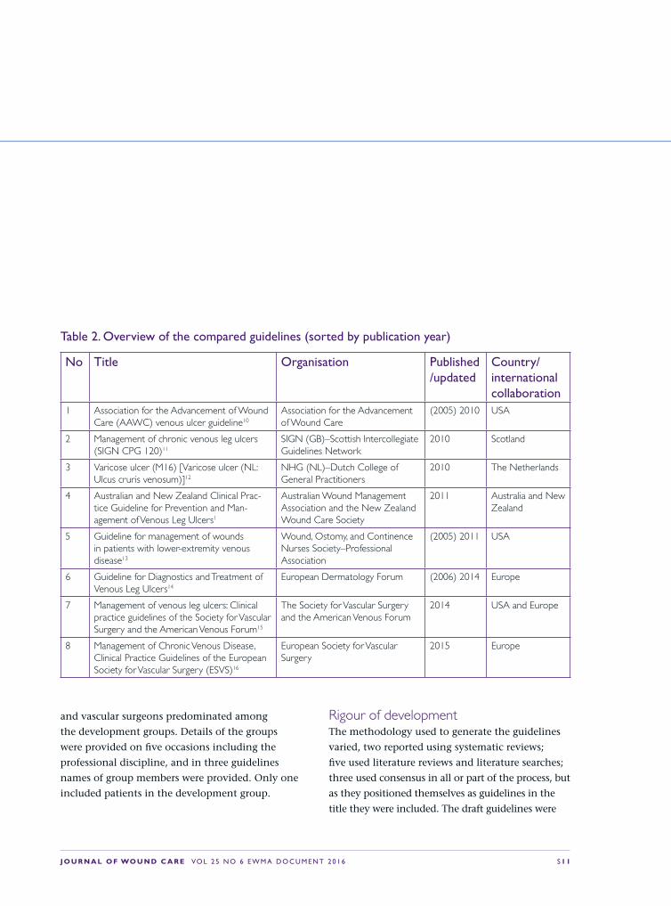
Of the eight guidelines identified, all were
published between 2010-2015; there were three
from 2010; one from 2011; three from 2014
and one from 2015. There were two updates of
previous versions.
The source of guidelines by country included one
joint document from Australia and New Zealand;
one joint document from the USA and Europe;
two solely from groups in the USA; one each from
Scotland and the Netherlands.
3.2 Guideline comparison–resultsThe following details were extracted:
Scope and purposeAll guidelines explicitly stated they were for the
management of patients/clients with a VLU.
One guideline was targeted specifically for use by
dermatologists, one for general practitioners (GPs)
only and the remainder were for all HCPs involved
in the management of patients with chronic
venous disease (CVD).
There was only one guideline which introduced
health questions as a means of developing
recommendations. Of the remainder, specific
objectives were not stated but they did indicate the
purpose was for the management of VLU. Surgical
management was excluded by three guidelines.
Stakeholder involvementThere were two unidisciplinary guidelines and the
remainder multidisciplinary. Vascular physicians
J O U R N A L � O F �WO U N D � C A R E � � VO L 2 5 N O 6 E W M A D O C U M E N T 2 0 1 6 S 1 1
No Title Organisation Published /updated
Country/international collaboration
1 Association for the Advancement of Wound Care (AAWC) venous ulcer guideline10
Association for the Advancement of Wound Care
(2005) 2010 USA
2 Management of chronic venous leg ulcers (SIGN CPG 120)11
SIGN (GB)–Scottish Intercollegiate Guidelines Network
2010 Scotland
3 Varicose ulcer (M16) [Varicose ulcer (NL: Ulcus cruris venosum)]12
NHG (NL)–Dutch College of General Practitioners
2010 The Netherlands
4 Australian and New Zealand Clinical Prac-tice Guideline for Prevention and Man-agement of Venous Leg Ulcers1
Australian Wound Management Association and the New Zealand Wound Care Society
2011 Australia and New Zealand
5 Guideline for management of wounds in patients with lower-extremity venous disease13
Wound, Ostomy, and Continence Nurses Society–Professional Association
(2005) 2011 USA
6 Guideline for Diagnostics and Treatment of Venous Leg Ulcers14
European Dermatology Forum (2006) 2014 Europe
7 Management of venous leg ulcers: Clinical practice guidelines of the Society for Vascular Surgery and the American Venous Forum15
The Society for Vascular Surgery and the American Venous Forum
2014 USA and Europe
8 Management of Chronic Venous Disease, Clinical Practice Guidelines of the European Society for Vascular Surgery (ESVS)16
European Society for Vascular Surgery
2015 Europe
Table 2. Overview of the compared guidelines (sorted by publication year)
and vascular surgeons predominated among
the development groups. Details of the groups
were provided on five occasions including the
professional discipline, and in three guidelines
names of group members were provided. Only one
included patients in the development group.
Rigour of developmentThe methodology used to generate the guidelines
varied, two reported using systematic reviews;
five used literature reviews and literature searches;
three used consensus in all or part of the process, but
as they positioned themselves as guidelines in the
title they were included. The draft guidelines were

S 1 2 � J O U R N A L � O F �WO U N D � C A R E � � VO L 2 5 N O 6 E W M A D O C U M E N T 2 0 1 6
opened for public consultation, as such patients
would have had an opportunity to comment.
Four opened the document for professional
comments and consultation and one was peer-
reviewed by four professionals. Cultural and
diversity review by non-medical cultural groups
was completed for one.
The Grading of Recommendations, Assessments,
Development and Evaluations (GRADE) system
(http://www.gradeworkinggroup.org/) was used by
four guidelines, providing the strength of evidence
to support the recommendations.
Clarity of presentationRecommendations were generally explicitly stated.
ApplicabilityA specific implementation plan was developed by
one guideline, one made recommendations to
support implementation and of the remainder,
no details were provided on how to implement or
disseminate. None of the guidelines included an
audit tool but one included an assessment tool.
Editorial independenceDeclarations of conflicts of interest (CoI) of
development group members were documented in
four guidelines. The remainder did not provide any
details of CoI.
The result of the guideline recommendations
(content) comparison is summarised in Table 3.
Assessment and referralsPatient assessment The following factors have been recommended to be included when assessing the patient
presenting with lower limb ulceration or with a venous leg ulcer (VLU):
Clinical history [5 guidelines]; leg ulcer history [2 guidelines]; physical examination [1 guideline]; varicose veins either present, or having a history of, or surgery for [2 guidelines]. Five recommended that persons performing the assessment should be trained in that assessment and should have a knowledge of anatomy and physiology.
Specific comorbidities to be recorded or taken account of included: peripheral vascular disease [1 guideline]; diabetes [2 guidelines]; deep vein thrombosis (DVT) [2 guidelines]; hypertension [2 guidelines]; obesity/body mass index (BMI) [3 guidelines]; trauma [1 guideline]; malnutrition [1 guideline]. Four guidelines did not refer to comorbidities.
Patient referral Three guidelines did not make any recommendations about referral of patients. Two recommended that a multidisciplinary team approach is required. Timing and reasons for referral forward included: if the ulcer had not reduced by 25% in 4 weeks or failed to heal in 12 weeks [1 guideline]; if there is a lack of tendency to heal by 4 weeks [1 guideline]; if there is a lack of tendency to heal by 8 weeks [1 guideline]; doubts about aetiology or atypical ulcer presentation [3 guidelines]; ankle to brachial pressure index (ABPI) <0.8 [1 guideline]; where chronic venous insufficiency (CVI) is complicated by lymphoedema [1 guideline].
Leg assessment The use of the clinical signs, aetiological cause, anatomical distribution, pathophysiological dysfunction (CEAP) classification score was referred to in only one guideline. Factors to be included in assessment of the limb included: varicose veins [2 guidelines], atrophie blanche [2 guidelines], oedema [2 guidelines], joint mobility [2 guidelines], hemosiderin deposits [1 guideline], lipodermatosclerosis [1 guideline], vascular dermatitis [1 guideline].
Table 3. Guideline content summary

J O U R N A L � O F �WO U N D � C A R E � � VO L 2 5 N O 6 E W M A D O C U M E N T 2 0 1 6 S 1 3
InvestigationsABPI One guideline did not refer to the use of ABPI, the remainder recommended its use as part
of the assessment process. Four guidelines recommended that persons trained in performing ABPI should complete this, with one stating it should be performed in a vascular lab. The remaining two did not state who should perform this.
Pulse oximetry Five guidelines did not refer to this investigation. The remaining three stated it was not necessary in routine practice but may be used in conjunction with other tests.
Assessing the ulcer Two guidelines did not provide recommendations on assessing the ulcer. Of those that did, four recommend measuring ulcer size and repeating this serially, although the frequency of repeat measurements was not stated [4 guidelines].
Biopsy It was recommended that biopsies should be performed on atypical ulcers [4 guidelines]; non-healing ulcers [2 guidelines]; ulcers not healing at 4-6 weeks [1 guideline]; and ulcers not healing at 12 weeks [1 guideline].
Bacteriological swabs Two guidelines made no recommendation. Five stated that routine swabs are not indicated; six stated swabs should be taken when there are signs of infection and one recommended swabs prior to surgery.
Management of eczema
Three guidelines made no recommendations. Two recommended the use of zinc bandages or zinc based ointments; three recommended patch testing and three recommended topical steroid therapy if indicated.
Reassessment Seven guidelines made no recommendation about reassessment. The one that did recommended that patients are reassessed at 12 weeks if no progress was evident, then reassessment should be completed at 12 weekly intervals. If the ulcer remained unhealed then a biopsy should be performed [1 guideline].
Ulcer managementCleansing Water of sound (safe) quality was recommended for routine cleansing in four [4 guidelines]
and a non-irritating, neutral, non-toxic solution was recommended by three [3 guidelines].
Debridement Two guidelines did not make recommendations. All methods of debridement were suggested, with two making it explicit that surgical and sharp debridement is performed by persons trained in such procedures. Only one guideline recommended that debridement is performed at the initial assessment and periodically thereafter, none of the others made recommendations on frequency.
Wound dressings One guideline did not refer to dressings at all. The remainder recommend that non-adherent dressings are suited to most cases and thereafter according to patient need.
Topical antimicrobials Three guidelines did not make reference to the use of topical antimicrobial agents. Of the remainder, it was recommended they should not be used in routine care or when there were no signs of infection [3 guidelines]. In addition, it was recommended that topical agents can be used when there is local infection and in addition to culture guided systemic antibiotic therapy.
Peri-wound area Five guidelines recommend the use of moisturising agents in the peri-wound area.
Compression therapy The decision to apply compression is based on holistic assessment which includes ABPI.
In addressing which patients should be offered compression therapy based on the recording of the ABPI the following was recommended: when ABPI 0.8-1.2 [1 guideline]; ABPI >0.8 [3 guidelines]; ABPI >0.9 [1 guideline]; ABPI > 0.5 [1 guideline] the latter recommended a reduced level of compression. Three guidelines did not make any recommendation.
Hosiery None of the guidelines recommend hosiery for management of active open ulcers as a first line of treatment. One recommended that once the ulcer has healed, bandages should be applied for two weeks, followed by hosiery [1 guideline]. Hosiery should be replaced every 12 months [1 guideline].

S 1 4 � J O U R N A L � O F �WO U N D � C A R E � � VO L 2 5 N O 6 E W M A D O C U M E N T 2 0 1 6
Systemic therapies Pentoxyfilline was recommended if there were no contraindications to its use [3 guidelines].
Antibiotics should be used only in the presence of confirmed infection [1 guideline].
Analgesia may be required and the use of eutectic mixture of local anesthetics cream for debridement was recommended [2 guidelines]. However while acknowledging that pain may be an issue, no clear recommendations were made for pain management or how pain should be assessed.
Surgery Five guidelines addressed the issue of surgery in the management of VLU. Of these, it was recommended that all patients with a VLU should see a vascular surgeon and be considered for surgery [2 guidelines], in patients with VLU C6, ablation of the incompetent veins in addition to compression to improve ulcer healing [1 guideline], in patients with VLU C6 and incompetent superficial veins that have axial reflux directed to the bed of the ulcer ablation of the incompetent veins in addition to standard compressive therapy to prevent recurrence was recommended [1 guideline], surgical treatment of isolated insufficiency of the superficial system may promote healing and reduce recurrence rate [1 guideline].
Other aspects of managementCosts While costs were acknowledged by four guidelines, no recommendations were made with
regards to routine collection of data to assess costs.
Patient education This was alluded to in four guidelines. These recommended education of the patient on the following factors: cause of the ulcer [2 guidelines], use of compression [3 guidelines], mobility and exercise [2 guidelines].
For each item listed below the number of
guidelines making this recommendation or
including this item is presented in brackets.
3.3 Key points/summary of findingsIdeally guidelines need to contain evidence-based
practice recommendations that provide a clear
description of desired performance and specific
advice about what to do in which situation and
which factors should be taken into account.
However, only two of the reviewed guidelines used
the GRADE classification system.
Many frameworks are readily available to guide
the development of CPGs to support the rigour
of the development process and strength of
recommendations (www.Sign.org, http://www.g-
i-n.net). Therefore one could reasonably expect
that documents using the term ‘guideline’
would meet the requirements as outlined in
these frameworks. However, review of the eight
guidelines here shows considerable variation
in the development process and strength of
recommendations. Nonetheless some key points
have emerged:
• All patients presenting with lower limb
ulceration must have a comprehensive
assessment including assessment of systemic,
regional and wound factors and this assessment
must be completed by clinicians educated
and trained in this assessment. There are no
recommendations on the nature or extent of this
training and education.
• All patients must have an ankle brachial
pressure index (ABPI) completed as part of the
assessment process and before commencement
of compression therapy. There is no consensus
among these guidelines on the minimum ABPI
value that is required prior to commencement
of compression. There is no consensus on the
frequency of repeat ABPI measurement with
only one recommending re-measurement after
12 weeks.

J O U R N A L � O F �WO U N D � C A R E � � VO L 2 5 N O 6 E W M A D O C U M E N T 2 0 1 6 S 1 5
• The use of compression therapy in the form of
inelastic material (bandages or Velcro devices)
is recommended for the management of
venous leg ulceration. Compression hosiery is
recommended for healed ulcers. While hosiery
may be used for active ulcers they are not
recommended as the first line of treatment.
• There is no consensus on when patients should
be referred forward. However as routine wound
measurement is advocated and the milestone of
4-weeks post initiation of treatment is referred
to in four guidelines, this could be considered
as a time to reflect on healing progress and
review of the treatment plan. Biopsy of the
wound is recommended for atypical ulcers or
those that are not responding to therapy.
• Widespread agreement exists that routine
bacteriological swabs are not indicated, routine
antimicrobial therapy is not indicated.
• Simple non-adherent dressings are suited for the
majority of wounds.
• Pain should be assessed and managed, but
specific guidance on how this is achieved was
not evident.
• There was scant reference made to patient
quality of life, patient wellbeing, patient
education and costs.
• It is well recognised that individual patients and
carers can play a proactive role in self-care ulcer
management including amongst other things
changing of dressings and compression bandages/
hosiery/wraps. The HCP should support the
patient to enhance self-care activities.

S 1 6 � J O U R N A L � O F �WO U N D � C A R E � � VO L 2 5 N O 6 E W M A D O C U M E N T 2 0 1 6
4. Clinical adherence to guidelines—barriers and facilitators
4.1 IntroductionEvidence-based CPGs are designed to improve
quality of care and reduce practice variation by
providing graded recommendations based on
the best available evidence. They are intended
as instruments of knowledge transfer to support
decision-making by physicians, other health
professionals and patients in clinical practice.
Efficient and effective guidelines, which are
thoroughly implemented, impact patient safety
and quality by increasing the consistency of
behaviour and replacing idiosyncratic behaviours
with best practices.17
Difficulties arise when introducing evidence and
guidelines into routine practice. Many are not
used after dissemination and implementation
activities frequently produce only moderate
improvement in patient management.18–20
Many approaches have been published offering
potential solutions of barriers to guideline
implementation, mostly in areas other than
wound care. Substantial evidence suggests that
behaviour change is possible, but this change
generally requires comprehensive approaches at
different levels (doctor, team practice, hospital,
and health system environment), tailored to
specific settings and target groups. Plans for
change should be based on characteristics of
the evidence or guideline itself and barriers and
facilitators to change. In general, evidence shows
that no one approach for transferring evidence to
practice is superior in all situations.21,22
A systematic review of the effectiveness and
costs of different guideline development,
dissemination and implementation strategies
reported on a four-step approach, consisting of
guiding questions, to direct the choice of the most
appropriate components of an implementation
intervention23–25
1. Who needs to do what, differently?
2. Which barriers and enablers need to be
addressed?
3. Which intervention components (behaviour
change techniques and mode(s) of delivery)
could overcome the modifiable barriers and
enhance the enablers?
4. How can behaviour change be measured and
understood?
In the following chapters we will outline
potential barriers and facilitators for clinical
practice guideline implementation related to the
various players. Some of these are specific for leg
ulcer management.

J O U R N A L � O F �WO U N D � C A R E � � VO L 2 5 N O 6 E W M A D O C U M E N T 2 0 1 6 S 1 7
4.2 The health-care system/organisation—the payer and provider perspectiveVarious factors defined by structures of the health-
care systems as well as traditions and structures
defined by specific health-care organisations may
influence an organisation’s ability to successfully
adapt leg ulcer management to guideline
recommendations.
These may facilitate implementation, or work as
barriers to implementation, depending on the
actions and preferences they support. In both
cases, guideline implementation planning is likely
to benefit from taking these into consideration.
4.2.1 Reimbursement of patients and health-care organisationsReimbursement for wound care products is
frequently cited as the reason for failure to change
practice. Much of this will depend on who pays for
care. For instance, if the patient is required to buy
their own bandages and dressings this will have a
major impact on what is available according to their
financial situation. The health-care system may also
be unable to afford best practice treatments.26
In a comprehensive health system, inequalities
of this nature are less likely to occur but may
occur as a consequence of other issues, such as
the care providers’ knowledge and understanding
of when and where different products should
be used. Efficiencies in leg ulcer services can be
a trade-off between increased costs of bandages
with reductions in nurse time to treat patients.
As an example, until the changes in Drug Tariff
(list of treatments available to be prescribed
compiled by the UK National Health Service),
this additional cost of bandages had to be borne
by the community nursing service. In a study of
service development nurses acknowledged that
while compression bandages were expensive they
could be cost-effective due to the improvements in
healing.27 This was sometimes an area of conflict
between the nurses and GP and health trust
managers who held the finances. Much of this was
resolved upon the addition of multi-layer compres-
sion on the Drug Tariff in the UK. Reimbursement
for products and services can therefore facilitate
implementation, whereas restrictions on these can
lead to failure to change practice.
While limited access to products may prevent
the adoption of recommendations on treatment,
the health system may also impact on the
implementation of guidelines. Payment by
Diagnostic Related Groups (DRGs) will provide
resources on the basis of the condition and the

S 1 8 � J O U R N A L � O F �WO U N D � C A R E � � VO L 2 5 N O 6 E W M A D O C U M E N T 2 0 1 6
expected cost of care. This may or may not provide
all the care needs that patients may require to
provide an effective management protocol. GPs
and hospital doctors may also be paid according
to the number of patient visits. This may have a
positive influence, or may limit patient contacts
according to the contract they have with the
funding agency (government health provider or
insurance agency).
4.2.2 Pursuing cost-effective careImplementing guidelines does not necessarily
require evidence of cost-effectiveness, but the
increasing need to reduce health-care costs may
lead to recommendations supported by evidence
of cost-effectiveness being more likely to be
successfully implemented. In VLU management
there is some evidence on effectiveness but little
evidence on the relative cost-effectiveness of
different interventions. The comparison of VLU
guidelines showed that recommendations for
routine collection of cost data is not included in
the guidelines.
Cost-effectiveness examines the relationship
between costs of care and outcomes of treatment.
Cost effectiveness can be defined as:
Incremental cost per additional outcome =
Cost of treatment 1−Cost treatment 2
Outcome 1−Outcome 2
For venous leg ulceration the outcome is routinely
the number of ulcers healed or alternatively the
ulcer-free weeks following healing. The latter is
usually preferred as this can include a further
period of healing that may occur following a
recurrence of the original ulcer.
Having defined the outcome, one must develop
a system that captures the appropriate costs of
care. This may include health professionals’
costs, dressings and bandages used together with
adjunctive therapies and on-costs associated
with the care of these patients. It is important to
consider that the cost-effectiveness relates only to
those treatments or systems being tested. A blanket
statement of cost-effectiveness is meaningless
without an understanding of what has been tested,
and particularly what has not been tested. As an
example of this, one might undertake a study
of three products. Product A may be more cost-
effective than product B but less cost effective
than product C. It would not be appropriate to call
product A cost effective without the proviso that it
is in relation to product B. The plethora of dressing
and bandage systems makes the statement that any
of these are ‘cost-effective’ should thus be treated
with caution.
A brief outline of the current evidence on the cost
effectiveness of dressings and bandage systems are
provided below:
The key rationale for all health-care organisations
is to provide the best care for patients within
the financial constraints of the organisation; to
provide a cost-effective service. Thus, the level of
care will be dependent on the resources available
to it.
A potential barrier to implementation of a CPG
may be the misinterpretation of health economic
data in relation to the costs of care provision.
While the costs of dressings and bandages and
other medical devices are clear for all to see,
what is still frequently forgotten or ignored is
the cost of delivering the care through staffing.2
High cost products may appear more expensive
to use but may reduce the time and frequency of
visits made by the health care professional. Any
changes that are undertaken to improve practice
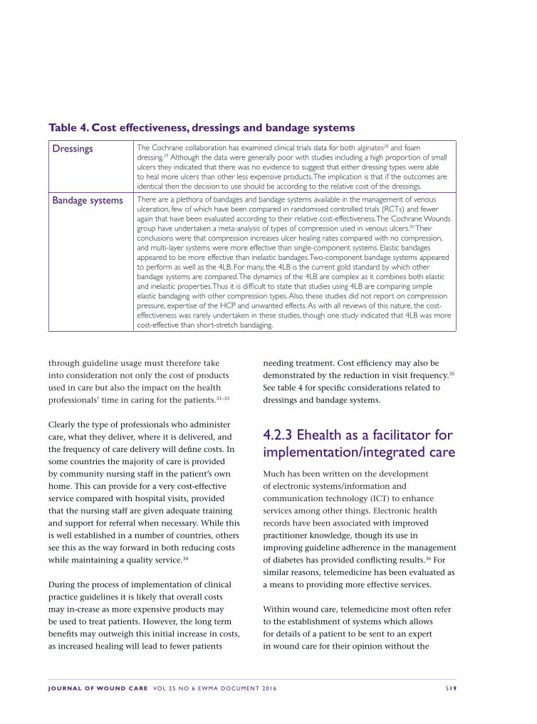
J O U R N A L � O F �WO U N D � C A R E � � VO L 2 5 N O 6 E W M A D O C U M E N T 2 0 1 6 S 1 9
Dressings The Cochrane collaboration has examined clinical trials data for both alginates28 and foam dressing.29 Although the data were generally poor with studies including a high proportion of small ulcers they indicated that there was no evidence to suggest that either dressing types were able to heal more ulcers than other less expensive products. The implication is that if the outcomes are identical then the decision to use should be according to the relative cost of the dressings.
Bandage systems There are a plethora of bandages and bandage systems available in the management of venous ulceration, few of which have been compared in randomised controlled trials (RCTs) and fewer again that have been evaluated according to their relative cost-effectiveness. The Cochrane Wounds group have undertaken a meta-analysis of types of compression used in venous ulcers.30 Their conclusions were that compression increases ulcer healing rates compared with no compression, and multi-layer systems were more effective than single-component systems. Elastic bandages appeared to be more effective than inelastic bandages. Two-component bandage systems appeared to perform as well as the 4LB. For many, the 4LB is the current gold standard by which other bandage systems are compared. The dynamics of the 4LB are complex as it combines both elastic and inelastic properties. Thus it is difficult to state that studies using 4LB are comparing simple elastic bandaging with other compression types. Also, these studies did not report on compression pressure, expertise of the HCP and unwanted effects. As with all reviews of this nature, the cost-effectiveness was rarely undertaken in these studies, though one study indicated that 4LB was more cost-effective than short-stretch bandaging.
Table�4.�Cost�effectiveness,�dressings�and�bandage�systems
through guideline usage must therefore take
into consideration not only the cost of products
used in care but also the impact on the health
professionals’ time in caring for the patients.31–33
Clearly the type of professionals who administer
care, what they deliver, where it is delivered, and
the frequency of care delivery will define costs. In
some countries the majority of care is provided
by community nursing staff in the patient’s own
home. This can provide for a very cost-effective
service compared with hospital visits, provided
that the nursing staff are given adequate training
and support for referral when necessary. While this
is well established in a number of countries, others
see this as the way forward in both reducing costs
while maintaining a quality service.34
During the process of implementation of clinical
practice guidelines it is likely that overall costs
may in-crease as more expensive products may
be used to treat patients. However, the long term
benefits may outweigh this initial increase in costs,
as increased healing will lead to fewer patients
needing treatment. Cost efficiency may also be
demonstrated by the reduction in visit frequency.35
See table 4 for specific considerations related to
dressings and bandage systems.
4.2.3 Ehealth as a facilitator for implementation/integrated care Much has been written on the development
of electronic systems/information and
communication technology (ICT) to enhance
services among other things. Electronic health
records have been associated with improved
practitioner knowledge, though its use in
improving guideline adherence in the management
of diabetes has provided conflicting results.36 For
similar reasons, telemedicine has been evaluated as
a means to providing more effective services.
Within wound care, telemedicine most often refer
to the establishment of systems which allows
for details of a patient to be sent to an expert
in wound care for their opinion without the

S 2 0 � J O U R N A L � O F �WO U N D � C A R E � � VO L 2 5 N O 6 E W M A D O C U M E N T 2 0 1 6
need of a face-to-face meeting. In most of the
established telemedicine services implemented
in wound care, the patient information is sent
by community care nurses to hospital-based
wound experts. In areas where specialised HCPs
may not be available, for example rural areas,
telemedicine may thus offer an opportunity to
provide specialised assistance for assessment,
diagnosis and treatment of a VLU patient.37
Patient information is, in most cases, entered into
a patient profile and stored in online databases.
Thus, telemedicine services may provide a good
opportunity to support the use of specific patient
records by all the involved health-care providers.
Telemedicine has also been described as a way to
increase the knowledge and involvement of the
patient in his or her disease and treatment.38,39
Thus, these services have an integrated potential
to enhance the knowledge about leg ulcer care of
patients and private care givers, as well as non-
specialised nurses and GPs in primary care. By
serving this educational purpose, telemedicine may
be a valuable tool to support guideline-driven care
in hospitals as well as community care settings.40
Additional services aiming for more independent
involvement of the patients are on their way
to the market may, in the future, further
develop the opportunities related to supporting
implementation of guidelines via telemedicine
services.
Several studies have indicated positive outcome
of telemedicine in wound care, with regards to
providing a good structure of care and the services
have in general been received well by patients and
health care professionals.37 Only a few of these
focus on leg ulcer care.41
An overview of the available evidence as well
as considerations about general benefits and
challenges related to use of telemedicine in wound
care (e.g. leg ulcer care) is provided in a EWMA
Document published in 2015.37
4.2.4 Management supportThe importance of management support for
change is well established,42 and may constitute a
barrier as well as a facilitator for implementation.
Clearly successful implementation support from
the most senior management can help those
undertaking change. Previous studies have shown
that management behaviours have important
impact on how nurses use research.27,43 A
systematic review identified lack of support from
managers and other staff to be one of the greatest
barriers to the ability for nurses to use research.44
Lack of high-level support from management will
cause difficulties in accessing additional resources
that may be required for successful change in
wound management practices.
As part of the management process it is essential
to ensure the availability of suitably trained staff,
and of a critical mass to allow the implementation
process to flourish. There is need for a skill mix
to allow for appropriate delegation of particular
duties. Referral routes need to be established to
ensure that patients are seen by the appropriate
professional allowing for a seamless service
between the community and acute sectors.
4.3 Health-care professionals— barriers and facilitatorsIn daily clinical practice, HCPs have a large
responsibility for the provision of guideline-
driven care. However, it is well documented that
the main responsibility for LU management is
placed with different groups of HCPs in different
countries (and perhaps also with local variations).
An Australian cross-sectional study reported
that nurses worked in collaboration with GPs to
J O U R N A L � O F �WO U N D � C A R E � � VO L 2 5 N O 6 E W M A D O C U M E N T 2 0 1 6 S 2 1
determine the treatment plans.45 This is in contrast
to a study which surveyed US family physicians46
where treatment and management of VLU patients
is undertaken primarily by the physician. A UK
survey reported that 71% of practice nurses (PNs)
reported being solely responsible for determining
the patient’s VLU treatment plan47 and an
Australian study of GPs in 2006 reported that
nursing assistance for leg ulceration management
was an integral part of general practice.48 In
addition, our results from the review of existing
guidelines show that two were unidisciplinary in
their approach, thus mitigating against a team
approach to care.
HPCs also work in diverse settings, have different
levels of expertise and may work very differently.
Some workplace solutions in one organisation may
not be directly transferable or applicable to another
health-care environment or patient group.
Depending on the structure of diagnosis and
treatment of VLUs and the groups of HCPs with
primary responsibility for the various aspects of
management, barriers related to the HCP role
may include:
• HCPs may experience that their practice
environment is not understood and reflected in
the guidelines. Thus, when the potential adopters
seek the best fit between evidence and their
clinical practice setting this may lead to lacking
implementation of the evidence-based guidelines49
• Implementation of guidelines requires both access
and knowledge. Varying levels of knowledge
among the HCPs involved in VLU management
have been reported50,51 and may constitute a barrier
for implementation. If we use compression therapy
as the example; becoming familiar with the many
different types of bandages, contraindications
of application, adverse effects, and monitoring
require improved education and improved training
in wound care to lead to better wound care
outcomes for patients.52,53 Although randomised
controlled trials (RCTs) and published systematic
reviews in wound care inform evidence-based
decisions about the use of multicomponent
compression therapy as best practice treatment for
people with VLUs, there are still examples of lack
of compression application by some community
and PNs54,55
• Even when HCPs know and accept guideline
recommendations about what needs to be done,
with high workloads they may forget or neglect
to do it.56,57 Clinicians increasingly experience
excessive workloads, inadequate practice
organisational support and financial pressures/
lacking resources58
Guideline implementation from the bedside may
benefit from addressing these barriers.
With regards to methods to facilitate guideline
implementation within a health-care organisation/
service, the following activities have been
demonstrated to be effective:
• Addressing the demand versus ability to change
practice (The size of changes required should
be compared with available resources and
collaboration)59,60
• Developing dissemination strategies that serve
to increase relevancy to everyday practice
(focus on implementation in context),59,49
ensuring a clear professional motivation
to implement guidelines, demonstrated by
the influence of individual perception of
the guidelines and personal commitment to
improved practice59
• Incorporating local CPGs in professional
training, and linking guideline adherence to key
performance indicators60

S 2 2 � J O U R N A L � O F �WO U N D � C A R E � � VO L 2 5 N O 6 E W M A D O C U M E N T 2 0 1 6
• Developing a collaborative, cooperative,
democratic environment that involves all
stakeholder groups including the patient59,61–64
• Using technology to facilitate CPG accessibility.60
These facilitators may obviously have varying
relevance and/or effects, depending on local
situations. The list above is intended to
provide areas to consider when planning an
implementation programme addressing the role of
frontline HCPs.
Other facilitators are related to the content of the
CPGs and include:
• Expanding guidelines to incorporate detailed
educational content49
• Updating the guidelines regularly and keeping
the content simple with specific sections for
allied health workers.60
Finally, addressing general challenges related
to supporting standardised VLU management
may, in time, have a positive effect on CPG
implementation. For example, efforts could be
made to decrease wound care product confusion
by developing standardised product naming and
improve the quality of wound-care research to
increase nurses’ confidence in the evidence.49
4.4 Patient-related barriers and facilitators We have dedicated this final section to
considerations about the role of the patient in CPG
implementation. Guideline implementation may
benefit from taking the patient role and opinion
into consideration, as this may influence the
general outcome.
CPGs link clinical practice to underlying evidence
and aim to improve the quality of care. What
is not clear is if guidelines take into account
what patients want and value. Clinical practice
guidelines all agree that adherence to compression
improves healing rates for people with VLUs. There
is little evidence about patient-related barriers
to guideline recommendations such as patient
adherence to compression therapy.
One potential reason could be that guidelines
do not take patient preferences into account
and may not include published evidence about
patient perspectives in the process of guideline
formulation. Our review of guidelines found that
only one included patients in the development
process. As described in the previous section,
clinicians may not implement guidelines because
they perceive a direct conflict between considering
patient preferences and applying guideline
recommendations. Clinical practice variations,
influenced by factors that are extrinsic to the
patient, such as costs of compression, occur among
clinicians, hospitals, and health-care systems.
These variations in practice do not serve the best
interests of patients. Patients may not understand
key facts that are critical to making decisions and,
despite the interest of patients to participate in
decisions, clinicians are often unaware of patient
preferences and weigh the risks and benefits based
on CPGs differently to patients.
Limited research has evaluated reasons for non-
adherence to VLU treatment. However, the
following potential influencing factors have
been identified for LU patients, in particular for
compression, as well as more generally:65,66
• Competing claims and advice from clinicians
• Adverse effects or fear of the recommended
treatment

J O U R N A L � O F �WO U N D � C A R E � � VO L 2 5 N O 6 E W M A D O C U M E N T 2 0 1 6 S 2 3
• Lack of funding, for example to pay for
compression treatments
• Psychosocial influences
• Interpersonal relationships. For example patient
trust in the nurse as central to treatment
adherence. Adherence has been reported to be
more likely when nurses provided care beyond
patients’ expectations, such as understanding
patient preferences and attending to pain.67
There is also a paucity of clinical trials that
have investigated which interventions promote
adherence to compression therapy for venous
ulcers.68 Some potential approaches to support
patient adherence have been investigated, but
none of these revealed a real benefit over usual care
in terms of healing rates, prevention of recurrence
of VLUs, or quality of life.69,70 The small number
of participants may, however, have hidden a real
benefit. These tested approaches included an
investigation of:
• Socialisation and support as a method to
improve adherence to compression69
• Leg exercises and walking via counselling and
behaviour modification as a method supporting
improved adherence to compression70
• The relevance of patient education.71
The paucity of rigorous process and impact
evaluations limits current understanding of how
best to improve patient involvement in guideline
development and implementation.72 CPGs are
mainly developed to inform health professionals’
decisions rather than foster patient involvement
in decision making. The question is how to
adapt clinical practice guidelines in such a way
that both the professionals’ perspective as care
provider and the patients’ preferences are equal in
the decision-making process.73 Including patients
in the guideline development process is the first
important step to ensure patient perspectives
inform future guideline process.72,74
4.5 ConclusionIt is an issue that many of the available guidelines
for VLU management as well as other disease areas
are not effectively integrated into clinical practice.
Therefore, action is required to improve the
strategies related to CPG implementation.
Could it be as simple as change in behaviour at
different levels (doctor, nurse, team practice, and
environment), tailored to specific settings and
target groups? In general, evidence demonstrates
that no singular approach in CPG uptake is
superior in all situations. Characteristics of research
evidence may affect whether it is accepted and
used in clinical practice. Some research findings
are more easily adopted, however change is rarely
easy if the innovation requires complex changes in
clinical practice or improved collaboration between
disciplines or changes in the organisation of care.75
With regards to VLU guideline implementation,
studies are needed to identify specific enablers and
barriers to adherence to clinical practice guidelines
for the management of people with VLU.

S 2 4 � J O U R N A L � O F �WO U N D � C A R E � � VO L 2 5 N O 6 E W M A D O C U M E N T 2 0 1 6
5. Current best practice leg ulcer management—clinical practice statements
5.1 IntroductionThis section aims to provide an overview of the
required basis for high-quality service provision,
with a focus on the ‘good patient journey’. This
section is organised in 5 chapters focusing on key
elements of the VLU patient’s journey:
• Differential diagnosis and assessment
• Treatment delivery: invasive and non-invasive
• Monitoring outcome
• Referral structures
• Secondary prevention.
All sub chapters will be finalised with a set of
number of key clinical practice statements, which
refer back to the comparison of evidence-based
VLU guidelines (Table 2). Disagreements between
recommendations in the available guidelines are
only highlighted in case these affect the overall
agreement between the guidelines, which include
a recommendation on a specific aspect of VLU
management.
5.2 Differential diagnosis and assessmentWhile there are a number of definitions that are
available to describe leg ulceration, it is generally
held that they are a defect in the dermis located on
the lower leg. LUs are not a disease entity per se, but
rather a symptom of an underlying disease. Vascular
diseases are the most common problem leading to
skin ulcerations on the lower legs. However, there is
a large variety of infectious diseases, immunological
diseases, physical factors, skin tumours and other
skin diseases that lead to skin ulcerations, many of
which manifest themselves mostly on the lower legs
(Table 5 and Table 16 Appendix 3).76–80 The treatment
approaches to these different disease entities vary
greatly.80 Every LU must therefore be assessed to
identify the underlying disease. The success of any
LU treatment will be higher if it is aimed primarily
at the underlying disease and not only at correcting
local factors that impair wound healing. However,
the aetiological assessment and classification score
was not described in the majority of the guidelines
reviewed for this document.
5.2.1 Key characteristics of different aetiologies—how to differentiateVenous leg ulcersThe majority of LUs are seen in the context of
chronic venous insufficiency (CVI). This type
of ulcer, VLUs, are the focus of this document
and make up about 50–60% of all LUs.80,81 CVI

J O U R N A L � O F �WO U N D � C A R E � � VO L 2 5 N O 6 E W M A D O C U M E N T 2 0 1 6 S 2 5
can either be caused by a primary varicosis or by
post-thrombotic syndrome. Each of these causes
are responsible for about half of all VLUs.82,83 Both
lead to a venous hypertension, which in turn leads
to microvascular changes such as elongation of
capillaries, micro-thrombosis, fibrin cuffs around
vessels and leukocyte leakage.84 VLUs are usually
located on the medial aspect of the lower leg and
around the medial ankle. However, a minority
are caused by an isolated varicosis of the lesser
saphenous vein or a congenital aplasia of venous
valves, and are located on the lateral or dorsal
aspect of the foot, respectively.77 Diagnosis of
CVI is based on clinical characteristics; there
are the skin changes that are caused by chronic
venous hypertension: oedema, visible capillaries
around the ankle (corona phlebectatica), trophic
skin changes such as hyperpigmentation caused
by hemosiderin deposits, atrophie blanche,
induration of the skin and underlying tissue
(dermatoliposclerosis) and stasis eczema.
Apparative diagnostic procedures are mostly used
to confirm venous hypertension and to exclude
concomitant arterial or other disease.
Arterial and mixed ulcersPeripheral arterial occlusive disease (PAOD) can
be an underlying disease or a contributing factor
leading to lower leg ulcerations.85 Arterial disease
always has to be regarded in the clinical context
of generalised arteriosclerosis and often occurs in
combination with other manifestations, such as
coronary heart disease or cerebrovascular disease.
While peripheral necrosis of the toes is the typical
presentation of PAOD stage IV, there are a number
of LUs that are caused solely by arterial occlusion
or in combination with venous insufficiency
(mixed ulcers). These LUs are not represented in
the commonly used La Fontaine classification
of PAOD, some authors call this a ‘complicated
stage II’. Arterial ulcers are typically located on
the lateral or ventral aspect of the lower leg or on
the dorsum of the foot. They tend to be deep and
sharply demarcated with irregular borders.
Arterial impairment occurs in 15–20 % of venous
ulcers.86 Mixed venous-arterial ulcers usually
combine clinical characteristics of CVI and of
arterial ulcers. They can be located in the medical
or lateral aspects of the leg and circumferential
extension is not rare.87
A frequently under-recognised cause of LUs related
to arterial ulcers is microvascular occlusion in
hypertensive ischemic leg ulcers (HYTILU or
Martorell’s ulcers).81 These ulcers occur in persons
with marked arterial hypertension, arterial
examinations are usually normal. Most of these
ulcers are very painful and located on the lateral
lower leg or over the shin. The ulcer surroundings
are highly inflammatory. Due to their clinical

S 2 6 � J O U R N A L � O F �WO U N D � C A R E � � VO L 2 5 N O 6 E W M A D O C U M E N T 2 0 1 6
appearance, they are often misdiagnosed as
pyoderma gangrenosum. The diagnosis of these
ulcers requires a large, deep biopsy that includes
some of the ulcer base but also at least 1cm of
surrounding skin and underlying soft tissue to
show the arteriosclerosis.
Arterial assessment is essential for all LUs as the
clinical characteristics are not sufficient to rule
out arterial disease and arterial occlusion requires
special treatment. Furthermore, arterial disease can
complicate many other underlying diseases of LUs
and its treatment speeds up healing of these ulcers
of combined aetiologies.88
A summary of aspects of the differential diagnosis of
the primary types of LUs can be found in Table 5.
Atypical ulcersApproximately 10–20% of all LUs are caused
by other, miscellaneous causes.77 These causes
are often referred to as ‘atypical ulcers’, they
Underlying disease Clinical characteristics History AssessmentVascular Chronic venous
insufficiency (CVI) (50%)Ulcer location: retromalleolar, mainly medial.
Surroundings: oedema, hyperpigmentation, purpura, atrophie blanche. Stasis eczema /allerg. Contact dermatitis, dermatoliposclerosis
Thrombosis, Varicosis, heavy legs, oedema
Doppler-/ Duplex- Sonography
Arterial (10%) Lateral and ventral aspect of leg, dorsum of foot
Surrounding skin: atrophic, shiny, hair loss
Cardiovascular risk factors, intermittent claudication
Palpation peripheral pulses, ABI, Duplex-Sonography, Angiography
Mixed venous-arterial (20%)
Other aetiologies (20%), See Appendix 3
Medial and lateral, signs of CVI, ABI<0.8
Cf venous and arterial Cf venous and arterial
Table�5.�Differential�diagnosis�and�assessment�of�venous,�arterial�and�mixed�leg�ulcers76,77,88–90
are summarised in Appendix 3, Table 16. They
include infectious ulcer causes, different forms of
vasculitis, ulcerating skin diseases such as pyoderma
gangraenosum, haematological and microvascular
disorders, physical causes and ulcerating skin
tumours.76 Many of these ulcer causes can be
recognised due to their clinical characteristics, for
example palpable purpura in the surrounding skin
which is typical for vasculitis, highly inflammatory
borders in pyoderma gangraenosum or tissue growth
resembling hypergranulation in ulcerating skin
tumours. Infectious diseases as cause of a LU require
microbiological examination, often a skin biopsy is
necessary to provide the deep tissue sample needed
for this. Vasculitic ulcers, some skin diseases and
all skin tumours need histological assessment of a
skin biopsy to make the diagnosis. Ulcerating skin
tumours are the cause of up to 3% of all LU, and
they are frequently misdiagnosed as LUs of other
aetiologies.89 Therefore, biopsy is recommended in
all ulcers with atypical appearance and/or no healing
tendency after six months of treatment.

J O U R N A L � O F �WO U N D � C A R E � � VO L 2 5 N O 6 E W M A D O C U M E N T 2 0 1 6 S 2 7
5.2.2 Patient assessment and vascular assessmentResponsibility for assessing the patientHCPs should meet the qualification, registration
and/or licensing requirements of their geographic
region before undertaking a role in assessing patients
with LUs. Whether HCPs other than medical doctors
have the right to diagnose and prescribe varies across
countries (See chapter 5.4 ‘Referral structures’).
The HCP conducting patient assessment should
have the appropriate anatomical and physiological
knowledge. Assessment of venous ulcers is
complex, and post-basic education and training
is recommended. HCPs should have appropriate
training in the use of diagnostic equipment (for
example performing an ABPI). Although there is a
paucity of literature on the effectiveness training, the
available research and consensus opinion suggests
that patient outcomes are superior when a HCP with
specific training in venous ulcer assessment and
management is engaged in the patient’s care.1
Patient assessmentComprehensive clinical assessment should
include:1,11,10
• Medical and surgical history in the context of a
VLU, including assessment of comorbidities
• LU history
• Vascular assessment
• Biochemical investigations
• Mobility and functional status
• Pain history
• Psychosocial status, cognitive status and quality
of life (QoL).
• Physical examination including examination
of the leg and ulcer, including microbiological
investigation when applicable.
Medical, surgical and leg ulcer historyA demographic and clinical background indicative
of a LU with venous origin include those factors
presented in Table 6. Evaluation of these factors is
essential in diagnosing an ulcer of vascular origin
and identifying risk factors for delayed healing
and/or ulcer recurrence that require address in the
patient’s treatment plan.1,10
Comorbidities can influence management of venous
disease and require concurrent management1,11
Patients should receive screening for, and
investigation of, the conditions in Table 7, along
with other comorbidities relevant to the patient’s
presenting signs and symptoms and past history.
A nutritional screening should be undertaken by
the HCP performing the comprehensive patient
assessment.91 It is recommended that HCPs use
of a valid and reliable nutrition screening tool
appropriate to the patient demographics that
includes, but may not be limited to, factors
such as weight/body mass index (BMI), recent
food and fluid intake, hair and skin changes,
Table�6.�Clinical�factors�associated�with�venous�leg�ulcers1,10
• Venous disease including post-thrombotic syndrome, venous insufficiency (superficial or deep), deep vein thrombosis, phlebitis or varicose veins, previous ulcer diagnosed as being of vascular origin
• History of vigorous exercise or occupation/lifestyle with prolonged standing or sitting
• Chest pain, haemoptysis or pulmonary embolism• Surgery or trauma of the affected leg• Family history of venous leg ulceration • Multiple pregnancies• Obesity • Increasing age >50 years• Duration of the ulcer

S 2 8 � J O U R N A L � O F �WO U N D � C A R E � � VO L 2 5 N O 6 E W M A D O C U M E N T 2 0 1 6
appetite, and weight history (including any
recent, unintentional weight loss).1 No nutritional
screening tools have been validated specifically
for use in screening patients with VLUs, however
there are a range of screening tools available (see
Table 8 for examples), many of which are validated
for patient groups applicable to people with
venous disease. Patients who are screened at risk of
malnutrition should be referred to a dietician for a
comprehensive nutritional assessment.91
Taking a comprehensive LU history provides a
clinical picture that provides diagnostic indicators
to the ulcer aetiology and realistic expectations
of the healing trajectory. History should include
duration of the ulcer, any previous ulcers, time
spent without ulcers, effectiveness of strategies
• Peripheral arterial disease• Rheumatoid arthritis• Vasculitis • Diabetes mellitus• Previous history of skin cancer • Under-nutrition• Obesity (BMI ≥30kg/m2)• Impaired mobility
Table�7.�Comorbidities�and�conditions�that�may�influence�the�treatment�of�vascular�disease�and�venous�leg�ulcers1,11,91
used in managing and the time taken to heal any
previous ulcers.1
Vascular assessmentVascular assessment must be undertaken to
determine underlying aetiology of the ulcer (venous,
arterial or mixed) and to determine severity of
disease. Adequate arterial perfusion is essential for
wound healing and patients with insufficient arterial
supply require revascularisation before compression
therapy or surgical wound repair.15
Inspection of the leg is an important component
of a vascular assessment. Pale or bluish colouring,
cool temperature, decreased hair growth,
hypertrophied nails and muscle atrophy are
indicators for arterial disease. Examination of the
leg appearance, including level and characteristics
of oedema (for example pitting), skin presentation
(see 5.2.3 How to assess the leg and ulcer) and
visibility of varicose veins are also used in
identifying and classifying venous disease.
The patient’s clinical symptoms may also provide
indications of active venous disease (Table 9).10,11,15
A range of clinical investigations (Table 10) can
be used to confirm disease aetiology and diagnose
anatomical and pathophysiological involvement
when venous disease is identified. Clinical
investigations are also used to determine severity
of vascular disease and determine prognosis with
respect to ulcer healing.10,11,1,15 In most cases,
clinical examination, venous duplex ultrasound Screening Tool Patient GroupShort Nutritional Assessment Questionnaire (SNAQ)
Patients in hospital
SNAQRC Patients in residential care
SNAQ65+ Patients aged ≥65 years
Nutrition Risk Screening (NRS-2002)
Mini Nutritional Assessment Patients aged ≥65 years
Malnutrition Universal Screening Tool (MUST)
Table�8.�Examples�of�commonly�used�nutritional�screening�tools91
• Leg ache and pain• Tightness• Skin irritation• Feeling of heaviness• Muscle cramps• Tiredness of the legs
Table�9.�Clinical�symptoms�indicative�of�venous�disease10,15

J O U R N A L � O F �WO U N D � C A R E � � VO L 2 5 N O 6 E W M A D O C U M E N T 2 0 1 6 S 2 9
Investigation PurposeAnkle brachial pulse index (ABPI) Provides estimate of central systolic blood pressure and presence and severity
of arterial disease. In patients with incompressible arteries due to calcification (for example patients with diabetes or renal disease), a toe brachial pressure index (TBPI) may be more reliable.
ABPI <0.8 is suggestive of arterial disease and requires investigation by a specialist.
Venous duplex ultrasound Ultrasonography technique that identifies blood flow, patterns of venous obstruction (for example superficial versus deep vein involvement), and venous reflux.
Photoplethysmography (PPG) Used to measure venous refill time and investigate deficiency of the calf muscle pump function. Venous refill time >20 seconds is indicative of venous insufficiency and potential delay in ulcer healing.
Computed tomography venography (CTV) Used to increase diagnostic accuracy and suggested for patients with suspected thrombosis or non-thrombotic venous obstruction.
Pulse oximetry A secondary diagnostic tool to measure level of oxygenation of the blood and assess arterial disease.
Transcutaneous oxygen tension (TCPO2) Used to determine arterial aetiology and identify ulcers that have potential for delayed ulcer healing.
Skin perfusion pressure Used to determine extent of venous disease and potential for delayed ulcer healing.
Blood pressure measured in both arms Indication of a range of cardiovascular diseases.
Table�10.�Investigations�used�in�vascular�assessment1,10,11,15
Table�11.�Basic�CEAP�Classification�System1,15
Clinical Classification
Description
C0 No visible or palpable signs of venous disease
C1 Telangiectasies or reticular veins
C2 Varicose veins
C3 Presence of oedema
C4a Eczema or pigmentation
C4b Lipodermatosclerosis or atrophie blanche
C5 Evidence of a healed venous leg ulcer
C6 Active venous leg ulcer symptoms
Aetiology Classification
Description
Ec Congenital
Ep Primary
Es Secondary (post-thrombotic)
En No venous aetiology
Anatomic Classification
Description
As Superficial veins
Ap Perforator veins
Ad Deep veins
An No identified venous location
Pathophysiologic Classification
Description
Pr Reflux
Po Obstruction
Pr,o Reflux and obstruction
Pn No venous pathophysiology identified

S 3 0 � J O U R N A L � O F �WO U N D � C A R E � � VO L 2 5 N O 6 E W M A D O C U M E N T 2 0 1 6
and ABPI is enough to make a diagnosis or clarify
whether further examination is needed.
The findings from the vascular assessment must be
used to categorise the clinical severity of venous
disease and expected response to treatment
using the internationally recognised CEAP
(clinical, aetiology, anatomy, pathophysiology)
Classification System (Table 11).1,10,15
Biochemical investigationsBiochemical investigations should be undertaken
to investigate the venous disease and comorbidities.
Laboratory investigations may include:1
• Blood glucose level (BGL) and/or haemoglobin
A1c (HbA1c)
• Haemoglobin (Hb)
• Urea and electrolytes
• Serum albumin
• Lipids
• Rheumatoid factor (RhF)
• Auto antibodies
• White blood cell count
• Erythrocyte sedimentation rate (ESR)
• C-reactive protein (CRP)
• Liver function tests (LFT).
Mobility and functional statusFunctional status, particularly mobility level,
should be assessed as part of the diagnostic process,
as well as to ensure that the VLU management plan
developed for the patient is feasible to implement.
Lower leg joint mobility is a component in calf
muscle pump function, which assists in venous
return in a healthy venous system. Calf pump
muscle function haemodynamic performance
is related to the strength of calf muscles and
the mobility of the ankle joint.11 No significant
differences are noted in time spent exercising
between people with and without venous
ulceration92 however, it is important to assess and
understand the patient’s physical abilities (for
example flexibility) in order to develop a feasible
treatment plan to which the patient can adhere
(for example ability to elevate legs, ability toput on
and take off compression stockings).
Pain assessmentA pain assessment should be conducted using
a pain tool that is reliable and valid (Table 12).
Select an appropriate tool based on the patient’s
demographics and comorbidities (for example
dementia). A pain assessment should include:1
• Location of ulcer-related pain
• Severity of the pain
• Quality/characteristics of the pain
• Frequency of the pain and when it occurs (for
example at dressing changes, background pain)
• Any triggers and effective relievers
• Impact of the pain on the patient’s quality of life
and functional ability.
Psychosocial status, cognitive status and quality of lifeComprehensive patient assessment includes
evaluating the patient’s cognitive ability
(for example, using the mini-mental state
examination), social support networks and overall
QoL and screening for mental health problems.

J O U R N A L � O F �WO U N D � C A R E � � VO L 2 5 N O 6 E W M A D O C U M E N T 2 0 1 6 S 3 1
If mental health conditions are suspected after
screening, the patient should be referred to a HCP
with experience in assessing and managing mental
health. A range of disease-specific QoL tools (Table
13) have been shown to have high sensitivity
when used to assess people with venous disease.1,15
5.2.3 Local ulcer assessmentHow to assess the leg and ulcer In general, VLUs tend to be shallow and irregular
in shape, often occurring in the lower third of the
leg (pre-tibial, anterior to medial malleolus).1,11
A comprehensive assessment of the leg and ulcer
should be made on initial presentation and at
frequent intervals to guide ongoing management.1
Ulcer assessment should include measurement of
the ulcer size,1,11 by measuring the length, width
and depth with a disposable ruler. This will record
the progress of wound healing over time. Where
resources are available, computerised calculation,
digital photography or wound tracing of the ulcer
area should be attended at frequent intervals.
The type of exudate should include a description of
the colour, consistency and amount. For example
serous (yellow fluid), haemoserous (blood and
serous fluid) sanguineous (old blood) or purulent
(green fluid).
Screening Tool Patient groupVisual Analogue Scale (VAS) Adults
Wong-Baker FACES Pain Rating Scale (FRS)
Appropriate for adults and people with dementia
McGill Pain Questionnaire (MPQ)
Appropriate for adults and people with dementia
Functional Pain Scale (FPS) Adults
Table�12.�Examples�of�commonly�used�pain�assessment�tools93
Recording the amount of exudate as accurately
as possible. Is the exudate minimal or soaked
through the dressing? The presence of any odour
can indicate infection. Loss of an excessive amount
of exudate can cause maceration of surrounding
skin and requires monitoring with the appropriate
dressing to manage the exudate. It can also cause
electrolyte imbalance.96
The appearance of the ulcer bed should describe
the tissue in the wound bed. For example, black
or necrotic tissue (dead eschar), yellow or sloughy
tissue (old fibrin), green or infected tissue (clinical
signs of infection exist), red or granulating (healthy
tissue), hypergranulating (over granulating
or proud flesh), pink or epithelisation (new
epithelium evident).
The condition of the ulcer edges should be assessed
for raised or rolled edges (any undermining),
changes in colour (red, purple, white) or evidence
of contracting or epithelisation, (healing).96 Raised
or rolled edges can delay healing and be a sign of
hypergranulation or malignancy.96 Colour changes
can indicate decreased tissue perfusion, redness
or erythema indicating infection or a purple/
Generic QoL assessment tools36 Item Short Form Health Survey (SF-36)
Cardiff Wound Impact Schedule (CWIS)
Venous disease-specific QoL assessment toolsChronic Venous Insufficiency Questionnaire (CIVIQ)
Venous Insufficiency Epidemiological and Economic Study (VEINES-QOL)
Aberdeen Varicose Vein Questionnaire (AVVQ)
Charing Cross Venous Ulceration Questionnaire (CXVUQ)
Wound-QoL
Table�13.�Valid�and�reliable�quality�of�life�(QoL)�tools�for�populations�with�venous�disease1,15,94,95

S 3 2 � J O U R N A L � O F �WO U N D � C A R E � � VO L 2 5 N O 6 E W M A D O C U M E N T 2 0 1 6
blue colour indicating malignancy, pyoderma
gangrenosum or vasculitis.96 Any abnormalities
should be further investigated and referred to a
trained HCP.
Inspection of the peri-ulcer area and surrounding
skin should be assessed for dryness, scaly,
maceration, erythema, puritis, cellulitis, oedema,
contact dermatitis or venous eczema.96 Pedal and
leg pulses are palpated.
There are many associated changes in the leg as a
result of CVI. The changes are described in Table 14.
Microbiology and histopathology When should you take a bacterial swab? All leg
ulcers are contaminated with microorganisms.
Bacteria exist on the skin as natural flora and
migrate to a wound, however, bacterial swabs
do not need to be taken unless clinical signs of
infection are present.1,11,97
Signs and symptoms of a wound infection in a
chronic ulcer may be subtle96 and include one or
more of the following:
• New, or increased wound-related pain96
• Delayed healing96
• Friable, hypergranulating tissue
• Increased heat
• Increased exudate
• Change to green/purulent exudate
• Increase odour from wound.
• Increased white cell count
• Pyrexia
Haemosiderin deposit Red cells leak out in the tissue causing reddish brown staining of the skin
Dilated and torturous veins As hypertension increases over time the larger veins become affected and visible through the skin
Lipodermatosclerosis The limb becomes hard and woody to touch as a result of malnourished tissue and fibrosis
Atrophie blanche A vascular or white skin scarring as a result of thrombosis and obliteration of capillaries in the deeper dermis—can be very painful often appears in areas where there is hyper-pigmentation or lipodermatosclerosis
Eczema Malnourished skin becoming dry and flaky
Hyperkeratosis A build-up of dry skin
Hypersensitivity The skin can become very sensitive and many substances can cause irritation and allergic responses
Ankle flare Venous congestion—tiny capillaries become swollen and are visible through the skin
Altered shape of lower leg Inverted champagne bottle
Oedema Capillaries swell and fluid leaks into the tissues
Evidence of healed ulcers Scar tissue present
Hair Present on the limb
Table�14.�Associated�changes�in�the�leg�as�a�result�of�CVI1,10,11
J O U R N A L � O F �WO U N D � C A R E � � VO L 2 5 N O 6 E W M A D O C U M E N T 2 0 1 6 S 3 3
• Increase erythema or cellulitis
• Malaise
• Increased swelling/localised
• Oedema of lower limb.
Investigations may include:1
• Bacterial wound swab or biopsy for
bacteriological analysis
• Wound biopsy if malignancy or other aetiology
such as vasculitis is suspected
• Wound biopsy for patients with a non-healing or
atypical LU.
When to refer to a specialist? If the aetiology of the ulcer on initial presentation
is unable to be determined by the HCP currently
responsible for assessment and management of
the ulcer, referral to a HCP trained and competent
in the assessment and management of VLUs
is required. Patients with a non-healing or
atypical leg ulcer should be referred for further
investigations, including consideration of biopsy.1
Referral pathways are described in Chapter 5.4
5.2.4 Clinical practice statements• Statement5.2.a:Allpatientspresentingwith
lowerlegulcerationmustreceiveacomprehensive
assessment.
Comments: This must include medical/
surgical history; vascular assessment; laboratory
investigations; LU history and symptoms; pain;
mobility and function; psychosocial status;
QoL and examination of the leg and ulcer.1 A
comprehensive clinical assessment and treatment
plan must be developed and documented.
Basic assessment before initiation of treatment
should include clinical assessment of the ulcer
and leg as well as ruling out arterial disease by
performing ABI measurements.
• Statement5.2.b:Patientassessmentmustbe
conductedbyaHCPwithappropriateclinical
knowledgeandskillswhohastherequired
qualifications,registrationandlicensingforthe
healthsysteminwhichtheypractice1,11
• Statement5.2.c:Followingacomprehensive
assessment,arecognisedclassificationsystem(for
exampletheCEAPClassificationSystem)shouldbe
usedtoclassifytheextentofvenousdisease
• Statement5.2.d:Apatientmustbereassessedifthe
ulcerdoesnothealontheexpectedtrajectoryorwhen
thepatient’sclinicalorsocialstatuschanges.
Comments: Further assessment to exclude other
underlying diseases must be performed after
three months or if there is cause for concern
before this.
Patients with a non-healing or atypical LU must
be referred to a HCP trained and competent in the
management of LUs for further assessment and
consideration of biopsy.1
• Statement5.2.e:Bacterialswabsshouldnotbe
takenroutinelyunlessclinicalsignsofinfectionare
present.1,11,97
For information about level of evidence available
to support these statements, we refer to the
following guidelines:
• Association for the Advancement of Wound Care

S 3 4 � J O U R N A L � O F �WO U N D � C A R E � � VO L 2 5 N O 6 E W M A D O C U M E N T 2 0 1 6
(AAWC) Venous Ulcer Guideline, A.f.t.A.o.W.C.
(AAWC), Editor. 201010
• Management of chronic VLUs. A national
clinical guideline, S.I.G.N. (SIGN), Editor. 2010,
SIGN Publication: Edinburgh, Scotland11
• Australian and New Zealand Clinical Practice
Guideline for Prevention and Management of
VLUs1
5.3 Treatment deliveryFrom the comparison of VLU CPGs, it becomes
clear that consistency across guidelines is lacking
with regards to the various treatment options.
There are few contradictions in the available
guidelines, but significant variations in the
available information about what, how and when
to perform various therapies. In this section
we provide key clinical practice statements
concerning the available non-invasive and
invasive treatment options.
5.3.1 Non-invasive treatments5.3.1.1 Compression therapy Compression therapy is based on the simple
concept of applying an external pressure to
the limb, which is able to improve venous
haemodynamics98–101 control oedema,102–106
reduce inflammatory mediators,107–110 improve
microcirculation,111,112 improve arterial
inflow,110,113–115 and improve the lymphatic
drainage.116–122 All the reported effects are
extremely beneficial in promoting ulcer healing
and the first conclusion of the recent Cochrane
review providing the most complete overview
of randomised controlled compression trials in
VLUs states that ‘compression increases ulcer
healing rates compared with no compression’30
confirming data reported in previous studies.123–125
Selection of devices for compression therapyCompression therapy may be applied by means of
different devices: elastic or inelastic or short-stretch
bandages, elastic stockings or elastic kits, adjustable
Velcro compression devices, pneumatic pumps.
Which kind of compression should be used in
VLUs treatment is still debated.
Inelastic materials or short-stretch
multicomponent bandages that do not give way
to the expanding muscle during walking are able
to produce great differences between resting and
working pressure and high pressure peaks. Such
bandages are both comfortable at rest and more
effective in improving venous haemodynamics
in standing position and during muscle exercise
compared to elastic bandages or compression
stockings.99,100,126,127 These materials give way to the
muscle expansion and exert a sustained pressure
that is similar in supine and standing position and
during work without any pressure peaks.
Multicomponent bandages are more effective than
single component bandages in achieving ulcer
healing, perhaps due to the ability to maintain
pressure and stiffness. They are cost-effective as
well as effective in reducing healing time, thereby
shortening the treatment period.30,128–133
As VLUs are ultimately due to the impaired
venous haemodynamics leading to ambulatory
venous hypertension, the compression devices
which support improvement of the venous
haemodynamics should theoretically be more
effective in promoting ulcer healing.
If we restrict our observation to guidelines and
studies reporting the compression pressure, it is
clear that when correctly applied to exert a strong
to very strong pressure, inelastic bandages are very
effective in achieving ulcer healing.1,11,14,134 It is also
J O U R N A L � O F �WO U N D � C A R E � � VO L 2 5 N O 6 E W M A D O C U M E N T 2 0 1 6 S 3 5
clear that the stronger the pressure the higher the
healing rate135–138 which favours inelastic materials
that are able to achieve a very strong pressure.
This is also, even if indirectly, in favour of high
stiffness which is the main physical characteristic
of inelastic, short-stretch materials.
Short-stretch multicomponent bandages require
skill, competent and trained staff to be properly
applied.12,13 Self-Adjustable Velcro Compression
Devices (AVCD) may not be as stiff as short-
stretch bandages but may represent an effective
alternative even if we have just one report on their
effectiveness on ulcer healing. In addition, they
may aid self management with related significant
costs savings.139
Role of elastic stockingsIn the most recent meta-analysis comparing the
effectiveness of elastic stockings and inelastic
bandages in promoting VLU healing,140 the
claimed superiority of stockings can mainly
be explained by the fact that in most analysed
papers, good stockings have been compared with
poor bandages. In addition, almost all the studies
included in this meta-analysis are burdened with
the flaws previously reported and in some cases
with erroneous interpretations of included trials,
making the conclusion hard to accept.
It must be noted that in all the included studies
not a single elastic stocking but an elastic kit made
up of two stockings exerting a pressure ≥40mmHg
was compared with inelastic bandages.
Even if we do not believe that elastic kits are more
effective than inelastic bandages in ulcer healing,
we need to highlight that they were able to promote
ulcer healing in an average of 64.2% of patients in
three months (four months just in one study).
We may conclude that elastic kits exerting
a pressure ≥40mmHg may be used in ulcer
treatment, especially in small ulcers, and by
caregivers without the adequate expertise to apply
a good bandage.
Intermittent pneumatic compressionWe do not have any comparative study between
intermittent pneumatic compression (IPC)
and sustained compression in promoting ulcer
healing. When compared with no compression,
IPC is able to increase the VLU healing rate.141,142
It may improve ulcer healing rate when added to
standard compression.143
Compression therapy—mixed ulcersIn 15–20% of VLU an arterial impairment co-
exists:86,144 they are named mixed ulcers. Due to
high prevalence of arterial disease in patients with
leg ulcers, a simple but accurate screening for
arterial disease is mandatory in order to choose
the best compression modality if the arterial
impairment is light or moderate, or to immediately
refer the patients to a vascular specialist for
revascularisation procedure, avoiding compression
therapy as first step. It must be noted that if the
treating HCP is not trained and competent in the
assessment and management of mixed ulcers, the
patient must be referred to the appropriate service.
Arterial impairment is assessed by measuring
the ratio between the ankle and the brachial
pressure ABPI and is >0.95 in normal subjects.145
Compression therapy is often contraindicated
in mixed ulcers and considered an exclusion
criteria in enrolling patients in many VLU
healing studies when ABPI is <0.8.146 Despite
these recommended restrictions, compression
therapy is used in mixed ulcers with modified,
reduced compression pressure provided the ABPI is
higher than 0.6.87,144,146–149 Sustained compression
pressure is contraindicated in chronic, severe,
critical limb ischaemia.150 Compression in mixed
ulcers does not reduce distal pressure measured
at toe level.151 It increases the arterial peri-wound

S 3 6 � J O U R N A L � O F �WO U N D � C A R E � � VO L 2 5 N O 6 E W M A D O C U M E N T 2 0 1 6
flow and arterial flow distal to the bandage115 and
improves the impaired venous haemodynamics.115
Compression therapy may increase the healing rate
in mixed ulcers.152
5.3.1.2 Clinical practice statements• Statement5.3.a:Compressiontherapyis
recommendedovernocompressioninpatientswitha
VLUtopromotehealing.1,10–16
Comment: we have a great number of studies
comparing compression with no compression
therapy and confirming that VLUs heal more
quickly with compression therapy.30,123–125
• Statement5.3.b:InpatientswithaVLUstrong
compressionpressureoverlowcompressionpressure
isrecommendedtoincreasehealing.1,11,13,14
Comment: there is evidence that a strong
compression (higher that 40mmHg) is more
effective than a Iow compression pressure
(≤20mmHg) in promoting ulcer healing.30,135–138
Compression should be applied by means of a
multicomponent system, which increases pressure
and stiffness rather than single component
bandages.128–133 Adjustable Velcro compression
devices or elastic kits may be considered effective
alternatives especially when trained personnel are
unavailable.10,137,139
• Statement5.3.c:InpatientswithVLUswesuggest
usingIPCwhenothercompressionoptionsare
notavailableorcannotbeused.Whenpossible
wesuggestusingIPCinadditiontostandard
compression14,15,153
Comment: there is evidence that compared with
no compression, IPC is able to increase the VLU
healing rate.141,142 There is also limited evidence
that IPC might improve healing of venous ulcers
when used in addition to standard compression.143
• Statement5.3.d:InpatientswithVLUsandarterial
impairment(mixedulcers)wesuggestapplyinga
modifiedcompressioninpatientswithlesssevere
arterialdisease:ABPI>0.5orabsoluteanklepressure
>60mmHg.15Thisshouldonlybeappliedbya
trainedHCPinmixedulcermanagementandwhere
thepatientcanbemonitored.Wehaveenough
datathatinpatientswitharterialimpairment
compressionmaybeappliedwithreducedpressure
providedarterialimpairmentisnotsevere.87,144,146–149
Whenarterialimpairmentismoderate(ABPI>0.5)
amodified,reducedcompressionpressuredoesnot
impedethearterialinflow115,151andmayfavourulcer
healing.152Compressionmustbeavoidedinsevere,
critical,limbischaemia.15,150
• Statement5.3.e:InpatientswithahealedVLU,
compressiontherapyisrecommendedtodecreasethe
riskofulcerrecurrence.15
Comment: even if available trials have some
flaws, the evidence regarding the effectiveness
of compression by stockings in ulcer recurrence
prevention is strong. Some evidence is in favour
of the strongest possible compression, which
seems directly related to the effectiveness in
ulcer recurrence prevention.154–156 A recent
paper underlines the adherence of the patients
wearing elastic stockings, which seems even more
important than pressure itself.157
For information about level of evidence available
to support these statements, we refer to the
following guideline:
• Management of VLU: Clinical practice guidelines
of the Society for Vascular Surgery and the
American Venous Forum15

J O U R N A L � O F �WO U N D � C A R E � � VO L 2 5 N O 6 E W M A D O C U M E N T 2 0 1 6 S 3 7
5.3.1.3 The role of dressings in venous leg ulcer managementLocal dressings applied to VLUs are one of the
treatments to prepare the wound bed to ‘ensure
formation of good quality granulation tissue
leading to complete wound closure, either
naturally or through skin products or grafting
procedures.158 Modern dressings produce and
maintain a moist microclimate on the ulcer-
dressing interface and claim to be beneficial in
ulcer treatment in conjunction with compression
therapy. In particular they promote autolytic
debridement,159–161 control exudate,28,29,162,163
manage wound infection,163–168 reduce pain,168–170
and are cost-effective.169–180
Despite all the positive effects of new and
advanced dressings, a number of published
papers does not report any advantage on ulcer
healing time when compared with traditional and
advanced dressings applied under compression
therapy. Almost all of them are included in the
Cochrane review on venous ulcer dressings in 2006
and reviewed in 2014.181
Nevertheless, a careful reading of these studies
reveals some limits of clinical RCTs and a number
of serious methodological flaws: The initial ulcer
size was not reported in some studies; when
reported, the ulcer size was usually small (lower
than 10 cm2) and the randomisation scheme
was adequately described in only 6 out of 24
studies.179,182–186 The sample size was often not
powered for statistical significance, and a blinded
outcome assessment was rarely carried out. In
addition, many exclusion criteria made the ulcer
patients highly selected.28,29
Summarising, all the studies involved patients
with small venous ulcers and often without
other concomitant or complicating conditions.
Unfortunately we do not have one single study
assessing the effectiveness of modern wound
dressings in patients with large ulcers, infected or
covered with fibrin slough, with comorbidities like
arterial disease or rheumatic diseases: all of them
well known factors making such ulcers difficult-
to-heal187 and where dressings could prove to be
effective in increasing the healing rate.
It can therefore be argued that the extensive
conclusion of the reported meta-analysis:181
‘The type of dressing applied with compression
therapy did not demonstrably influence ulcer
healing’ should be restricted to: ‘The type of
dressing applied with compression therapy did
not demonstrably influence healing of small
venous ulcers which are not complicated by
comorbidities’.
In addition, the effectiveness in pain and infection
control results in an improvement of patients QoL
and reduction of resource consumption related to
global care management (including pain killers,
antibiotics, swab costs, hospital admission), and
should be taken into consideration. This may lead
to a reduction in the number of biopsy cultures
needed to monitor infection, as well as the use of
antibiotics and analgesic agents.188 Furthermore,
costs savings with advanced dressings have been
documented, even with healing rates equal to
those achieved with conventional dressings.189
Finally, there is some evidence that some modern
dressings and procedures that modulate MMPs may
be effective in improving healing rates.190,191
All these factors support the targeted use of
modern dressing in VLU management.
Thus, the HCP should select an appropriate
dressing based on the following factors:1,192–196
• Ulcer size and location, wound bed and tissue
characteristics

S 3 8 � J O U R N A L � O F �WO U N D � C A R E � � VO L 2 5 N O 6 E W M A D O C U M E N T 2 0 1 6
• Wear time
• The specific ulcer stage (inflammatory,
granulating, in re-epithelisation phase)
• Amount and type of exudate
• Level of bacteria and/or topical infection
• Presence of pain and odour
• Assessment of peri wound and surrounding skin
• Patient tolerance and preference
• Ease of application and removal
• Cost and availability.
Ulcer dressings must be correctly applied according
to the manufacturer’s instructions.
Management of surrounding skinThe surrounding skin of a patient with a VLU
requires attention and care. The skin can become
red, infected, macerated or dry. This can be
related to infection (local or cellulitis), venous
eczema, hypersensitivity (contact dermatitis) or
maceration from exudate. The HCP can consider
using topical barrier preparations to reduce
erythema and maceration from VLUs. The dressing
can be reviewed for possible hypersensitivity to
the product. A topical barrier preparation can
be applied to the surrounding skin to protect it
from the exudate. Venous eczema can be treated
with short term topical steroids, zinc impregnated
bandages, or other dermatological preparations.1
Clinical InfectionWound infection delays the normal healing
process. A comprehensive assessment of the
patient and their VLU is required to determine the
severity of the infection and appropriate treatment
implemented. Antimicrobial therapy such as silver,
honey and cadexomer iodine dressings can be
prescribed when a VLU exhibits signs of infection.
Maintenance debridementMaintenance debridement has been proposed as
a therapeutic intervention to address the problem
of chronic wounds characterised by an adequate
wound bed but absent or slow healing.197 If the
inflammation is not controlled, the excess of
inflammatory mediators favours the breakdown
of the new epithelialisation tissues and of the
endogenous proteins biologically active, as the
growth factors and the cytokines. Also the peri-
wound skin, often already compromised by
previous skin alterations (lipodermatosclerosis,
atrophie blanche, hyperpigmentation, dry, scaling
and atrophic skin and venous stasis dermatitis)
may be further damaged in these conditions and
lead to an increase of ulcer size.
In conclusion, ‘maintenance debridement’ can be
useful for the wound bed and peri-wound skin in
order to guarantee improvement of the biological
microenvironment and increase the healing
chance for ulcers which do not heal despite an
adequate wound bed.
The clinical practice statements provided below are
based on the available evidence referred to in the
reviewed guidelines or supporting literature.
5.3.1.4 Clinical practice statements• Statement5.3.f:Nospecificdressingproductis
superiorforreducinghealingtimesinVLUs.1Simple
non-adherentdressingsarerecommendedinthe
managementofVLUs.11Thisappliestothemajority
ofsmallandnoncomplicatedVLUs.
Dressings are selected based on assessment of the

J O U R N A L � O F �WO U N D � C A R E � � VO L 2 5 N O 6 E W M A D O C U M E N T 2 0 1 6 S 3 9
stage of the ulcer bed, cost, access to dressing and
patient and HCP preference.1,13,15
Comment: If the VLU is exudating heavily, select
a dressing that has a high absorptive capacity that
can also protect the peri-wound from maceration.
• Statement5.3.g:Concerningmanagementofthe
surroundingskin,theHCPcanconsiderusing
topicalbarrierpreparationstoreduceerythema
andmacerationfromVLUs.Venouseczemacan
betreatedwithshort-termtopicalsteroids,zinc
impregnatedbandages,orotherdermatological
preparations1,11
• Statement5.3.h:Concerninguseofwounddressings
inthecaseofclinicalinfection,acom-prehensive
assessmentofthepatientandtheirVLUisrequired
todeterminetheseverityoftheinfectionand
appropriatetreatmentimplemented.Antimicrobial
therapysuchassilver,honeyandcadexomeriodine
dressingscanbeprescribedwhenaVLUexhibits
signsofinfection.1,13–15
Comment: The use of topical antimicrobials should
not be used in the standard care of VLUs with no
clinical signs of infection (1, 13-15).
• Statement5.3.i:Regardingwounddressingsandcost
saving,thestandardcareoftreatingVLUsreduces
thecostofulcermanagement.1,13
Comment: we have sufficient evidence to support
that ulcer dressings are effective in exudate
management, in controlling ulcer infection and in
allowing cost savings.28,29,160–180
• Statement5.3.J:Ulcerscharacterisedbyanadequate
woundbedbutabsentorslowhealingmayneeda
maintenancedebridementofwoundbedandperi-
woundskin.197
For information about level of evidence available
to support these statements, we refer to the
following guidelines:
• Association for the Advancement of Wound
Care (AAWC) venous ulcer guideline, Malvern,
Pennsylvania: Association for the Advancement
of Wound Care (AAWC).December 201010
• Management of chronic venous leg ulcers. A
national clinical guideline, S.I.G.N. (SIGN), Editor.
2010, SIGN Publication: Edinburgh, Scotland11
• Australian and New Zealand Clinical Practice
Guideline for Prevention and Management of
Venous Leg Ulcers1
• Guideline for management of wounds in
patients with lower-extremity venous disease,
O. Mount Laurel (NJ): Wound, and Continence
Nurses Society (WOCN), Editor. 201113
• Management of venous leg ulcer: Clinical
practice guidelines of the Society for Vascular
Surgery and the American Venous Forum15
5.3.2 Invasive treatmentsMultifactorial pathogenesis, differences in the
anatomical distribution of venous pathology,
as well as the vast variety of different surgical
and endovascular procedures available make it
difficult to provide clear and generally acceptable
recommendations on how to perform invasive
treatments of VLU.198
This is why a recent guideline for operative/
endovascular management of VLU categorise these
anatomically as 1. Superficial, 2. Perforator and 3.
Deep-iliocaval and/or infrainguinal venous disease
to cover all possible treatments and relate them to
clinical situations (Fig 1).15,153 Nevertheless, quality
of the evidence available from the published papers
for invasive treatment is low (Primarily level C).

S 4 0 � J O U R N A L � O F �WO U N D � C A R E � � VO L 2 5 N O 6 E W M A D O C U M E N T 2 0 1 6
5.3.2.1 Primary types of invasive treatments in venous leg ulcers management
Local treatments:
• Debridement: Refers to deeply removing adherent,
dead or contaminated tissue from a wound
(such as necrotic material, eschar, devitalised
tissue, serocrusts, infected tissue, hyperkeratosis,
slough, pus, haematomas, foreign bodies, debris,
bone fragments or any other type of bioburden)
with the objective to promote wound healing.
Debridement options are available today, include
mechanical, autolytic dressings, larvae therapy and
various debridement technologies.199 Debridement
is an important part of the TIME strategy for
treatment of chronic wounds: Tissue debridement,
control of Infection and inflammation, Moisture
imbalance, and advancement of the epithelial
Edge of the wound.200
• Shave therapy: A local surgical technique, based
on sharp removal of scar tissue by ‘shaving’ it
with dermatome. The ulcer should be shaved
layer by layer until reaching a healthy looking
tissue and capillary bleeding. In longstanding
VLUs when chronic inflammatory process
leads to fascial scarring and thickening the
fasciectomy, shave therapy is needed followed
by skin grafting. The shave therapy must be
carried out by a surgeon in standard operating
conditions under local or even general
anaesthesia in an inpatient setting. To date
there is no single RCT to assess efficacy of this
treatment although there are some retrospective
studies reaching a healing rate of 80% at 12
weeks in treated patients. Interestingly, the
recurrence rate was significantly reduced to
about 25% after 2.5 years of follow-up.201 This
technique is popular in German speaking
countries, France and Poland.
Systemic treatments, related to the venous bed:
• Venous stripping: An operative treatment
under local or general anaesthesia to remove
the whole length of the vein. It usually deals
with insufficient great saphenous vein (GSV) or
small saphenous vein (SSV) because of reflux.
The surgery requires incision in the groin, high
ligation of GSV at the sapheno-femoral junction,
insertion of a plastic or metal stripper into the
vein and removal of attached vein to the stripper
downwards usually in eversion fashion.202
• Endovenous Laser Therapy (EVLT): A minimally
invasive, ultrasound-guided technique for
treating varicose veins by means of laser energy.
Under tumescent local anaesthesia catheter
bearing laser fibre is inserted into GSV or small
SSV respectively to the level of sapheno-femoral
junction or sapheno-popliteal junction. Then,
the laser fibre with carefully applied energy
is slowly withdrawn, causing obliteration of
saphenous trunk. The EVLT technique can also
be used to close perforating veins.203,204
• Radiofrequency Ablation (RFA): A technique
and method similar to EVLT but instead of
laser fibre a radiofrequency catheter is used
and radioenergy is applied under the same
circumstances.205
• Foam sclerotherapy: A technique where
foamed sclerosant is injected under ultrasound
guidance into GSV, SSV, perforating vein or even
smaller veins located under the venous ulcer to
obliterate them. This non-surgical technique
has in recent years revolutionised treatment of
venous reflux and varicose veins.206
• Subfascial endoscopic perforator surgery (SEPS):
A minimally invasive operative technique used
to treat VLU caused by incompetent perforator
veins. SEPS represents a minimally invasive

J O U R N A L � O F �WO U N D � C A R E � � VO L 2 5 N O 6 E W M A D O C U M E N T 2 0 1 6 S 4 1
alternative to the Linton procedure which
originally involved a long medial calf incision
to expose all posterior, medial and paramedical
perforators. Using endoscopic techniques, the
perforating veins are clipped or divided by
endoscopic scissors. The procedure can be done
in the hospital or outpatient setting by general
or vascular surgeon under local anaesthesia.
To date there is ongoing debate concerning
the general efficacy of perforator ligation in
the surgical management of advanced chronic
venous insufficiency and venous ulceration.207,208
• Venous stenting of deep iliocaval and/or
infrainguinal veins: The introduction of minimally
invasive venous stenting using Venography and
Intravenous Ultrasonography (IVUS) provides
the ability to treat the ‘obstructive’ component
of the VLU. The stenting procedure requires
femoral vein puncture in the groin or popliteal
vein puncture behind the knee with respect to
localised obstruction of the vein. A guide wire is
then passed up high into the normal caval vein,
crossing the narrowings or obstructions of the
femoral or iliac veins making a way to insert the
balloon. By inflating the balloon, the diameter
of the vein increases and then safe deployment
of the stent is possible. The stent must cover the
entire area of diseased vein to provide a long
standing effect of endovascular treatment. Stents
usually keep the vein open, improving patient’s
symptoms of leg swelling and leading to faster
ulcer healing. In follow up, instent stenosis can
occur and the rate of restenosis is about 5% for
patients with extrinsic compression syndrome and
over 10% in a case when obstruction of the vein
was because of prior venous thrombosis.209
The main objectives for operative/endovascular
treatment for VLUs are:
• To accelerate ulcer healing
• To prevent ulcer reoccurrence.
5.3.2.2 Selecting between invasive treatmentsRecently published SVS/AVF American guidelines
on management of VLUs and its revised version of
International Union of Phlebology (UIP) present
17 recommendations related to the invasive
treatment of venous bed in patients with VLU.15,153
Of the recommendations four related to superficial
venous reflux and VLU, 4 are related to perforator
venous reflux and VLU, and the remaining 9
cover deep venous obstruction/reflux and VLU.
All of them except one have a C level of evidence
demonstrating that in fact there is a lack of
properly conducted RCTs in this field.15
Only prevention of VLU recurrence after surgical
treatment and compression therapy reached higher
level of recommendation, means 1B. The ESCHAR
study (The Effect of Surgery and Compression
on Healing and Recurrence) illustrated that there
is no significant difference in healing time and
healing rate between superficial venous surgery
plus compression and compression alone. However,
the 12-month recurrence rate in the study was
considerably lower for patients treated with surgery.
The ESCHAR study emphasised that 85% of the
patients with VLUs would benefit from surgery.210
Fig 1 illustrates what to consider when selecting
an invasive treatment of VLU, taking the current
evidence base and current guidelines into
consideration.15,153
5.3.2.3 Clinical practice statements• Statement5.3.k:Toimproveulcerhealinginpatients
withVLUandincompetentsuperficialveins,

S 4 2 � J O U R N A L � O F �WO U N D � C A R E � � VO L 2 5 N O 6 E W M A D O C U M E N T 2 0 1 6
surgery(highligation/stripping)oralternativelyany
newablationtechniquesshould be suggested in
addition to standard compression therapy.15,153
Comment: Traditional surgery has a slightly higher
level of evidence than new ablative techniques,
probably because they have not been sufficiently
studied for this purpose.211,212
• Statement5.3.l:Topreventulcerrecurrencein
patientswithactiveorhealedVLUandin-competent
superficialveins,thesurgery(highligation/stripping)
ofincompetentveinsinadditiontostandard
compressiontherapyisrecommended.15,16,153
• Statement5.3.m:Topreventulcerrecurrencein
patientswithactiveorhealedVLUsandincompetent
superficialveins,ablationtechniqueinadditionto
standardcompressiontherapyissuggested.11,14,15,153
Comment: Open surgery for prevention of ulcer
recurrence when superficial veins are involved is
the only well documented treatment.210,212,213
New ablation techniques still require more studies
so this is why the evidence of using them is much
lower.203,204
• Statement5.3.n:Toimproveulcerhealingand
preventrecurrenceinpatientswithaVLUand
incompetentsuperficialveinswithpathologic
perforatingveinsandwithorwithoutdeepvenous
Operative/endovascular�treatment�of�VLU
Local Systemic
• Venous stripping and high ligation
• CHIVA, ASVAL• Endovenous ablation
techniques• FOAM Sclerotherapy• Techniques under
investigations: steam ablatio, pharmacomechanical and cyanocrylate ablation
• SEPS• Foam sclerotherapy
• Stenting• Venous bypasses• Valve reconstruction
Shave�therapy Superficial�venous�disease
Perforator�venous�disease
Deep-illocaval�and/or�infrainguinal-venous�disease
Figure abbreviations: CHIVA–Ambulatory conservative haemodynamic management of varicose veins; ASVAL–Ambulatory selective varicose vein–ablation under local anaesthesia; SEPS–Subfascial endoscopic perforator surgery; EVLT–Endovenous laser therapy; RFA–Radiofrequency ablation
Fig�1.�Selecting�between�operative/endovascular�venous�leg�ulcer�(VLU)�treatments
J O U R N A L � O F �WO U N D � C A R E � � VO L 2 5 N O 6 E W M A D O C U M E N T 2 0 1 6 S 4 3
disease,surgeryorablationofsuperficialand
perforatingveinsissuggestedinadditiontostandard
compressiontherapy.15,153
Comment: Every treatment of perforating veins
is controversial, because of lack of well-designed
RCTs and uncertainties whether abolition of axial
reflux or closure of insufficient perforator is more
beneficial for improving healing of LU.214–216
• Statement5.3.o:Toimproveulcerhealingand
topreventrecurrenceinpatientswithactiveor
healedVLUandisolatedpathologicperforating
veins,surgeryoralternativeablationtechniqueof
perforatingveinsissuggestedincaseoffailureof
standardcompressiontherapy.15,153
• Statement5.3.p:Toclosethepathologicperforator
veinsinpatientswithVLU,percutaneoustechniques,
whichdonotneedincisionsintheareasof
compromisedskinarerecommendedoveropen
venousperforatorsurgery.15,153
Comment: Avoidance of any incision within a
region of compromised skin is crucial. This is
why the minimally invasive techniques, from
a ultrasound-guided foam sclerotherapy to
SEPS should be taken into consideration when
treatment is planned.153
• Statement5.3.q:Inpatientswithinfrainguinal
deepvenousrefluxandactiveorhealedVLUthe
recommendationisagainstdeepveinligationofthe
femoralorpoplitealveinsasaroutinetreatment.15,153
Comment: This is an old surgical procedure which
fortunately currently is rarely performed.217,218
• Statement5.3.r:Toimproveulcerhealingandto
preventrecurrenceinpatientswithtotalocclusionor
severestenosisofinferiorvenacavaand/oriliacveins,
venousangioplastyandstentingisrecommendedin
additiontocompressiontherapy.15,153
• Statement5.3.s:Nospecificdebridementmethod
hasbeendocumentedtobeoptimalfortreatment
ofVLUs.13
Comment: The most commonly used methods
of debridement are surgical (sharp), conservative
sharp, autolytic, larval, enzymatic and mechanical.
Surgical debridement is rapid, although it requires
either general or local anaesthetic and can be
painful. Conservative sharp debridement is the
removal of loose avascular tissue without pain or
bleeding.1
• Statement5.3.t:Mechanicaldebridementmethods,
suchasultrasound,high-pressureirrigationorwetto
drydressings,maybeusefulforreducingnon-viable
tissue,bacterialburdenandinflammation.1
For information about level of evidence available
to support these statements, we refer to the
following guidelines:
• Management of venous leg ulcer: Clinical
practice guidelines of the Society for Vascular
Sur-gery and the American Venous Forum15
• Australian and New Zealand Clinical Practice
Guideline for Prevention and Management of
Venous Leg Ulcers.1
5.4 Referral structures5.4.1 Managing patients with venous leg ulcers between primary and secondary health-care settingsInternationally there has been a move to manage
more patients with chronic conditions in the home
or community care setting.34 This has come about
due to the ever-changing population demographics
(an ever increasing elderly population) and
pressure on health-care resources, in particular
the cost of funding hospitals and keeping acute
beds ‘open for business’. With the predicted

S 4 4 � J O U R N A L � O F �WO U N D � C A R E � � VO L 2 5 N O 6 E W M A D O C U M E N T 2 0 1 6
increase in the numbers of individuals with
chronic conditions such as leg ulceration, this will
inevitably mean that there will be a corresponding
increase in the prevalence and incidence of
wounds into the future.
With this in mind, it is therefore evident that if
patients are moving constantly between primary
and secondary health-care settings that all HCPs
involved in that patient’s care should have a good
understanding of each other’s roles in order to
optimise the care offered to and outcomes achieved
for the patient with a chronic leg ulcer. This
constitutes a basic requirement for establishing or
maintaining effective referral structures.
5.4.2 The multi-disciplinary team in venous leg ulcer management‘Multi-disciplinary’ can be defined as, ‘a group of
health-care workers who are members of different
disciplines (professions), such as psychiatrists and
social workers, each providing specific services to
the patient. The team members independently
treat various issues a patient may have, focusing on
the issues in which they specialise.219
The essence of the multi-disciplinary team
approach in wound management is that the
team is interdependent and team members share
responsibility and are accountable for attaining the
desired results. However, a team may be defined
within the literature and within an individual
country setting, there is substantial evidence that
when individual professionals come together with
a shared goal that is patient focused, enhanced
clinical outcomes can be achieved.220
The roles and competencies required for all
members of the multi-disciplinary team are very
important and need to be fully understood by all
HCPs involved in the management of patients with
leg ulceration.
Treatment pathways vary greatly from country to
country. In many countries, all medical treatments
must be done under the supervision of a medical
doctor and only medical doctors have the right
to prescribe. In other countries, specialised nurses
coordinate treatments and have extended practice
that includes the right to prescribe, refer and
order investigations. In addition, there is a lack of
specialised HCPs in the rural areas, which means
that specialist referral may not be possible.1
The example provided in Fig 2 illustrates the
patient referral structures in the UK, and the
HCP profiles and health-care organisations that
contribute to the management of patients with a
VLU. It also illustrates that the patient’s journey
through the system may have different starting
points, depending on local structures and patient
situations. This may complicate the establishment
of clear cut referral structures for individuals.
An outline of the roles and responsibilities of
members of the multidisciplinary team in VLU
management, as presented in Fig 2, can be found
in Table 15.
The referral pathways described above with the
example of the UK differ from the situation in
many other European countries. For example, in
German speaking countries (Germany, Switzerland,
Austria), the competency to perform most of the
diagnostic procedures and the right to prescribe
lies with medical doctors. Most patients with a
LU would therefore be followed by a primary care
physician, in most cases an independent GP, who
could delegate some basic diagnostic procedures to
community-based nursing services or wound care
specialist nurses. After the initial appearance of an
ulcer, the GP would refer the patient to a specialist
for further assessment. This would usually be

J O U R N A L � O F �WO U N D � C A R E � � VO L 2 5 N O 6 E W M A D O C U M E N T 2 0 1 6 S 4 5
Primary�health�care Secondary�health�care
Vascular surgeon
Dermatologist Vascular laboratory
Specialist clinic e.g. complex wound
Ward based care under auspices of vascular team/vascular nurse or tissue viability nurse specialist teams
Commercial health care companies/orthotists providing support and materials for on-going patient management, e.g. compression bandages/hosiery in all health care settings.
Patient
Leg ulcer clinic/leg club
Patient’s private carers
District nurse/tissue viability nurse
Podiatrist
Social worker, dietician, etc
Practice nurse
General practitioner (GP)
performed by a vascular surgeon, an angiologist
or a dermatologist for vascular assessment, or
in the case of prolonged healing failure, to a
dermatologist for further diagnostic work to
exclude other differential diagnoses.
The treatment (compression treatment or local
wound care) is prescribed by the GP or one of the
specialists consulted, and can be carried out by
practice staff of the GP, community-based nurses
or wound care specialist nurses at wound care
centres. Even though patients are increasingly
treated in ambulatory settings most countries, due
to the increasing need to cut costs, patients may
still be hospitalised for their treatment if they have
hard-to-heal wounds or need surgical intervention.
Networks between hospitals and community-
based nursing services or wound care centres are
often established to ensure the continued care of
patients after their discharge from the hospital.
To sum up, generally applicable referral structures
for VLU patients cannot be defined across different
national or even regional and local settings. It
should also be highlighted that the CPGs currently
available includes few recommendations about
general referral patterns, and these are generally
not supported by high level evidence.1 Given the
general shift towards management of patients
within home or community care settings there
is an increasing need to ensure that home and
community care staff have sufficient education
to evaluate when specialist referral is needed and
understands the roles and responsibilities of the
multi-disciplinary team members.34
5.4.3 Clinical practice statements• Statement5.4.a:LUmanagementmustbe
undertakenbytrainedorspecialisedHCP.1,11–13
Comment: However, individual patients and
carers can play a proactive role in self-care ulcer
management including among other things
changing of dressings and compression bandages/
Fig�2.�Example�of�a�patient�referral�pathway�between�primary�and�secondary�health�care�for�a�patient�with�a�chronic�leg�ulcer�in�the�UK

S 4 6 � J O U R N A L � O F �WO U N D � C A R E � � VO L 2 5 N O 6 E W M A D O C U M E N T 2 0 1 6
HCP/stakeholder Tasks in VLU managementThe practice nurse (registered nurse) (RN)
Assess, screen, treat and educate all ages of patients and members of the community.
Work within the GP practices to provide nursing and medical care221 with both a preventative and management focus.
Often the first health-care professional to be aware of a patient with a leg ulcer.
May not have all of the desired competencies for the long-term management of patients with venous leg ulcers (VLUs).
The general practitioner (GP)
Employed by the relevant Health Service (NHS) as independent contractors to work within local communities.
Typical responsibilities include patient consultations at home and within the surgery, physical examinations, diagnoses and treatment of illnesses/ailments, minor surgery, health education, practice management and administration, liaison with other health-care professionals and/or hospitals.222
The dermatologist Physician specialising in treating conditions of the skin, hair and nails. Often receives the first secondary care referral from the GP in primary care.
Trained in the assessment and treatment of venous disease and can take also care of other underlying diseases, leading to skin ulcerations and accompanying skin problems.
The vascular surgeon Specialising in the diagnosis and management of patients with a variety of conditions that affect the patient’s venous or arterial circulation. Can prescribe or undertake a variety of investigations in order to confirm a suspected diagnosis, such as venous/arterial LU, as well as perform a variety of non-invasive or invasive surgical procedures to correct identified circulatory deficiency.
The Specialist Nurse: Vascular Nurse Specialist (VNS) / Tissue Viability Nurse (TVN)
The VNS act as a central member of the vascular team.
They see and support patients in hospital and in the out-patient clinics and helps to coordinate care for patients and provides a simple means of communication between the hospital services and the community.223
The TVN promote and ensure ‘evidence-based and cost-effective care of all patients managed within an acute care and outpatient clinic setting. They help to coordinate care for patients and provide a simple means of communication between the hospital services and the community.
The vascular laboratory technician
Schedule and prepare patients for investigations and assist in the delivery of prescribed treatments by assisting with the application of modalities such as ultrasound and x-ray. Work in settings such as fixed or mobile laboratories, doctors’ offices and specialist clinics. Not independently involved in the treatment of the patient.
The district nurse (DN) Should be involved in ‘The planning, provision and evaluation of appropriate programmes of nursing care’, particularly for people discharged from hospital and patients with complex needs; long-term conditions, those who have a disability, are frail or at the end of their life.224
Clinical competencies in VLU care: the application of compression therapy—bandages and hosiery.
Podiatrist Podiatrists provide preventative care, diagnosis and treatment of a wide range of problems affecting the feet, ankle and lower legs. Their specialist skills focus on managing infections, ailments, defects and injuries of the foot and lower leg, as well as treating foot and nail conditions related to other major health disorders (for example diabetes/leg ulceration).
They also provide preventative care and advice on improving mobility, independence and the quality of life for their patients.Podiatrists are also known as chiropodists and this profession exists primarily in the Anglo-Saxon countries.
Table�15.�Members�of�the�multi-disciplinary�team�responsible�for�VLU�management�(UK�example)

J O U R N A L � O F �WO U N D � C A R E � � VO L 2 5 N O 6 E W M A D O C U M E N T 2 0 1 6 S 4 7
The patient Patients are expected to seek medical assistance for the diagnosis and on-going management of their leg ulcers (LU), and then report any changes/on-going issues, for example pain/comfort/change in exudate levels/associated odour related to the planned management of their ulceration. In addition they will be encouraged to be concordant with the agreed management plan and the rationale underpinning the professional expectation.
Concordance has been defined as ‘a new way’ to define the process of successful planning and delivery of health care based on partnership, which has three essential elements:• The patient has knowledge to participate as a partner• Consultations involve patients as partners; Patients are supported during their treatment (adapted from Medicines Partnership, www.medicines-partnership.org)225
• Patients can play a proactive role in self-care ulcer management including e.g. changing of dressings and compression bandages/hosiery/wraps (In collaboration with the health-care professional responsible for the ulcer management)
Patient carers All patient carers should be fully involved in the care process in an informed manner (as highlighted above for patients) and should be aware of any on-going management decisions that have/are being made to optimise the patient with a leg ulcers outcomes—both clinical and psychological. They should also be aware of any potential issues that may arise and have a clear referral pathway agreed that is relevant to the care set-ting in which they are currently operating. Patient carers may also play an active role in changing dressing and compression bandages/hosiery/wraps.
hosiery/wraps. The HCP should support the patient
to enhance self-care activities.
• Statement5.4.b:SpecialisedLUclinicsare
recommendedastheoptimalservicefortreatmentof
VLUinthecommunity(primarycare)setting11
• Statement5.4.c:Inruralareas,wherespecialised
HCPsmaynotbeavailable,telemedicinecanoffer
anopportunitytoprovidespecialisedassistance
forassessment,diagnosisandtreatmentofaVLU
patient37
5.5 Secondary prevention5.5.1 Need for services/education in place to monitor patients with a healed venous leg ulcerTo prevent VLU reoccurrence it is important
to support VLU patients to acquire skills and
knowledge through trained HCPs about preventive
and therapeutic interventions. The literature
demonstrates that patients with a VLU do not
have enough knowledge about pathophysiology of
VLU to conduct effective self-management.226–229
The guideline comparison in this document
highlighted that 4 of the 8 guidelines included
recommendations for patient education, but that
the types of secondary prevention actions that
were recommended varied across the guidelines.
There are only a few studies demonstrating the
effectiveness of education interventions with
VLU patients that are improving the skills and
knowledge regarding aetiology/pathophysiology
and adherence. After the wound closure most
of the patients are left on their own and do not
receive any aftercare. Hence, they are responsible
for the care of their legs. To prevent a recurrence,
education has to take place before completion
of the treatment. The content of the education
should allow effective self-management. There is
little published evidence available that describes
education programmes/services.
Studies demonstrate that the recurrence rate can
be lowered as much as nine times by delivering
information leaflets.230,231 O’Brien et al.232 present in
their qualitative study with 10 patients a patient-
centred 12 week-intervention programme. This

S 4 8 � J O U R N A L � O F �WO U N D � C A R E � � VO L 2 5 N O 6 E W M A D O C U M E N T 2 0 1 6
programme consists firstly of information material
about leg elevation to heart level and movement.
Secondly, six follow–up telephone calls. The results
demonstrate how VLU patients learn to understand
the connection between their wound and the
adherence. A RCT illustrated this by delivering
a patient information brochure that showed the
yearly recurrence rate could be lowered from 36%
to 4% (Log-Rang-Test = 8.28, p=0.004). 231
5.5.2 A venous leg ulcer has healed—what next? The risk with every healed VLU, will it recur or a
new area of ulceration develop? Once a VLU has
healed, ongoing management is essential and the
focus and effort is on preventing recurrence. CVI is
a causative risk factor, it is a chronic condition; a
lifelong commitment to preventing recurrence and
its associated implications requiring an individual
to be active in their management with support
from the health-care system.1,233
A recurrence of a VLU is a burden which
challenges individuals and health-care providers;
it can represent social, personal, financial and
psychological costs to the individual and further
economic drain to the health-care systems which
support them.
How to reduce the risk of recurrence of a venous leg ulcersTo reduce the risk of recurrence of VLUs, the
evidence recommends the continued use of
compression therapy.1 In most countries, patients
have to cover the cost of compression hosiery. The
cost does vary and this can cause financial difficulty
for some patients. It is one of the significant
barriers to adherence to wearing compression
hosiery and the patient is disadvantaged as they
have a high risk of recurrence and the associated
implications that this brings.
It is well documented that delivering effective
interventions with a collaborative approach will
prevent VLU recurrence and promote patient
wellbeing and independence. The need for services
with the aim to implement maintenance strategies
is crucial and can be provided in the home setting,
a clinic or ambulatory care setting, with virtual
technology such as eHealth in partnership with the
clinician, patient, family and caregivers.
Type of serviceA service with trained HCPs11 that provides
education, support and prevention strategies to the
patient in the home setting, specialised clinics or
via eHealth or telemedicine. For example, online
video calling, apps and smart phone support.
Education through patient information booklets/
brochures, apps, DVDs, and online tutorial
webinars can be readily available and offered in
multiple languages and for the visually impaired.1,
11,234 An individualised management care plan can
be developed in collaboration with the patient and
carer/family.
Patient assessmentThe patient should be assessed for suitability
and strength of compression, identifying any
peripheral artery disease and functional ability
to apply and remove compression hosiery. The
patient and their home should be assessed and the
necessary support services implemented.
The appropriate compression hosiery The patient requires lifelong medical grade
compression hosiery providing 18–40mmHg to
reduce the long-term effects of venous disease.1,11
To determine the strength of compression,
peripheral arterial disease needs to be determined
by performing a comprehensive clinical
assessment of the patient and the leg. The choice
of compression hosiery is influenced by several
factors, such as the preference of the individual
and HCP, cost, and shape of limb.234 Accurate

J O U R N A L � O F �WO U N D � C A R E � � VO L 2 5 N O 6 E W M A D O C U M E N T 2 0 1 6 S 4 9
measurement and the appropriate class of
compression are essential to ensure the stocking
provides adequate compression and is worn safely,
with-out risk of injury to the skin.234 To avoid any
development of peripheral oedema, compression
hosiery is usually applied first thing in the
morning after a shower or upon getting out of bed,
and removed before going to bed at night.234
Stocking aids or donners may be used to assist the
patient or caregiver with application and removal of
compression hosiery.234 When the person is applying
their own stocking, these appliances may assist those
with limited strength, reduced manual dexterity
and who are unable to reach their feet.234 For the
professional care giver, stocking aids may reduce
the physical effort required during application and
removal, and prevent injury. This is an important
factor for consideration when protecting the
occupational health and safety of the caregiver.234
The patient should consider replacing compression
hosiery every six to twelve months and/or per
manufacturer’s recommendation.1
The benefits of a daily skin care programmeThe benefit of a daily skin care programme
promotes the health of legs and reduces the
risk of VLU recurrence.1,11 An effective skin care
programme is essential to promote the normal
skin pH to prevent and/or manage dry, irritated
skin.1 Skin cleansers and moisturisers should be
applied at least daily. For very dry and scaly skin,
such as varicose eczema, an oil or emollient-based
moisturiser is more effective than a cream or lotion
and helps to maintain skin integrity. Zinc-based
creams and bandages and short-term steroid
cream/ointments can be applied to the skin to treat
varicose eczema.1
The benefit of exercise and leg elevation Exercise and movement benefit the patient and
enhance calf muscle pump.1,11,235 Progressive
resistance exercisers have been shown to promote
calf muscle function.1,235 Patients who are unable to
ambulate or have limited mobility can be educated
on the benefits of progressive resistance exercise.
The benefits of leg elevation have been well
documented. Elevation of the limbs when sitting
and avoidance of standing for prolonged periods
assists in controlling lower leg oedema.1,11
Patient wellbeing Support groups such as ‘The Leg Clubs’ can promote
acceptance and adherence with practices that help
maintain skin integrity and provide long-term
psychosocial support and improve patient wellbeing.
How frequent and for how long to monitor the patient CVI is a lifelong medical condition and requires
commitment to prevention strategies and is
a permanent lifestyle change for the patient.
Each patient requires different levels of support.
Consider monitoring the patient for six to twelve
months after the VLU has healed.
Surgical options to prevent ulcer recurrenceFor information about the surgical options to
prevent ulcer recurrence, see chapter 5.3.3 on
invasive treatment options.
5.5.3 Clinical practice statements• Statement5.5.a:WhenaVLUhashealed,the
patientrequireslifelongmedicalgradecompression
hosieryproviding18–40mmHgtoreducethelong
termeffectsofvenousdisease1,11
• Statement5.5.b:Thepatientmustbeassessed
byatrainedHCPforsuitabilityandstrengthof
compression1,11

S 5 0 � J O U R N A L � O F �WO U N D � C A R E � � VO L 2 5 N O 6 E W M A D O C U M E N T 2 0 1 6
• Statement5.5.c:Thepatientshouldconsider
replacingcompressionhosieryeverysixtotwelve
monthsand/orpermanufacturer’srecommendation1
• Statement5.5.d:Thebenefitofadailyskincare
programmepromotesthehealthoflegsandreduces
theriskofVLUrecurrence1,11
• Statement5.5.e:Exerciseandmovementhasa
positivebenefitforthepatientandenhancescalf
musclepump.1,235Progressiveresistanceexercisehas
beenshowntopromotecalfmusclefunction
• Statement5.5.f:Elevationofthelimbswhensitting
andavoidanceofstandingforprolongedperiods
assistsincontrollinglowerlegoedema1,11
• Statement5.5.g:Considermonitoringthepatientfor
sixtotwelvemonthsaftertheVLUhashealed
For information about level of evidence available
to support these statements, we refer to the
following guideline:
• Australian and New Zealand Clinical Practice
Guideline for Prevention and Management of
Venous Leg Ulcers1
5.6 Monitoring outcomeTo finalise this section focusing on clinical practice
in VLU management, we have chosen to focus on a
discussion concerning relevant outcome measures
in chronic wounds and VLU management. This
discussion feeds into the evaluation of the evidence
base available to support recommendations for VLU
management, which is presented in most of the
evaluated guidelines (See Chapter 3).
The approach to treating a chronic or delayed
healing wound such as a VLU has evolved greatly
during the last 15 years.236 A wide range of
approaches and products are available for treating
chronic wounds, but it is widely acknowledged
that many of them lack high-level evidence
which robustly demonstrates their benefits.
‘Evidence-based practice’ specifically refers to
clinical decision making that is based on the best
available evidence, with practitioners reviewing
information from powerful data, instead of relying
on single observations or customs.237 However, the
extended definition by Sackett (1996) may be more
relevant in the wound sector today. This proposes
that evidence-based medicine is not restricted to
randomised trials and meta-analyses, but involves
the exploration of all types of best external
evidence.238
5.6.1 Relevant endpoints in venous leg ulcer studiesAn endpoint is defined as the objective of an
evaluation or study.237 Study outcomes are more
convincing when they apply to a single or small
number of clearly defined objectives.
These objectives should include:
• A precise statement of the degree of benefit
expected from the intervention, and its duration
• Clear statements on the time frame of the study
(especially in relation to how quickly the benefits
might start)
• A definition of the patients for whom the benefit
is sought237
Wide variations in endpoints of trials of VLU have
been reported together with a lack of endpoints
related to QoL or patient identified endpoints.239
All studies on VLUs must include compression
as part of standard care. However, epidemiologi-
cal data suggest that ulcers that are the result

J O U R N A L � O F �WO U N D � C A R E � � VO L 2 5 N O 6 E W M A D O C U M E N T 2 0 1 6 S 5 1
of varying degrees of arterial disease and other
confounding factors are increasingly being
presented. To date, only limited data is available
on the natural outcome of arterial and mixed
aetiology leg ulcers.237
Wound healing-related outcomes (wound closure,
reduction rate and healing time)237 which are
relevant to the assessment of improvement for
patients with leg ulceration may include:
Wound closure defined as ‘total epithelialisation
without discharge’ should be confirmed by
an independent source such as photography.
Definitions of ‘healing’ as a clinical outcome have
been debated for some time. However, recent
recommendations from the FDA support the view
that complete closure of a chronic wound is the
most clinically meaningful endpoint.
Reduction rate should be confirmed by tracing and
consider ‘reduction rate error’. 237 Currently, there
is an on-going debate over the usefulness of using
reduction in wound area as a primary outcome
as the ‘clinical benefit of incremental wound size
changes has not been fully established’. However,
some studies have shown that reduction in wound
area within a specified time frame can indicate
the potential to achieve complete healing in the
future.237
Use of wound healing time as an outcome
measure has received increasing interest due to
its importance from the clinical perspective and
with regard to resource use and economic costs.
For most studies reporting wound healing time,
the major concern is that it is only reported
for the minority of patients who have healed
within a specific observation period, generally of
4–12 weeks.237
Changes in wound condition should also be noted.
Due to the introduction of more targeted treatment
strategies that focus on specific aspects of symptom
management, rather than aiming for complete
healing, it is important to ensure that the chosen
outcomes reflect the modality under investigation.
These endpoints may include exudate level,
necrosis/slough, odour, and fibrous/fibrotic tissue.
It should also be considered that a range of issues
involving wound dressings have a significant
impact on activities of daily living, particularly
mobility. Bulky bandages, compression devices
and different types of foot off-loading can
interfere with daily living to such an extent that
concordance with treatment is jeopardised, while
frequent dressing changes can result in a life
dominated by clinical appointments.
In recent years, health-related quality of life
(HRQoL) has become a more routinely accepted
outcome in health-care studies. As an outcome,
HRQoL can be measured using three different
approaches:
• Generic measures
• Condition-specific measures
• Utility measures.
In each case, it is important that assessments are
made using tools with established psychometrics
as this will ensure that they are valid, reliable,
sensitive to change and can discriminate between
health states.
HRQoL has been defined as, ‘Personal health
status’. HRQoL usually refers to aspects of our lives
that are dominated or significantly influenced by
our mental or physical wellbeing’.236
Finally, in order to maximise the value of
investments in future clinical research, all studies

S 5 2 � J O U R N A L � O F �WO U N D � C A R E � � VO L 2 5 N O 6 E W M A D O C U M E N T 2 0 1 6
should be designed to address the relative cost-
effectiveness of the alternatives being tested from
the outset, as well as their efficacy (safety) and
clinical effectiveness,237 especially in an era of
resource constrained health-care provision.
5.6.2 Patient-centred outcomesHCPs have a variety of treatment options at their
disposal. This range of therapeutic options come
with a variety of caveats for patients, including
potential pain, discomfort, inconvenience,
expense/unavailability and burden (time and
social) to both the patient and their caregivers.
Despite this, patients with chronic wounds do
not always feel engaged in the decisions made
regarding their care, and patient concerns are often
not aligned with the concerns of their health-care
providers (See Chapter 4.4. Patient related barriers
and facilitators).
It has therefore been suggested that research
that involves patient-centred outcomes will help
patients and their caregivers to communicate more
productively and make better informed choices
about their health care.
Traditionally, patient-reported outcomes have
been defined as the patient’s assessment of
how they function or feel with respect to their
health or associated health care. Patient-reported
outcome metrics provide a patient’s perspective on
treatment benefit, allows for direct measurement
of treatment benefit beyond survival, disease, and
physiological markers. They are often the outcomes
of greatest importance to patients. Reports from
patients may include the signs and symptoms
reported in diaries, the evaluation of sensations
and symptoms, reports of behaviours and abilities,
general perceptions or feelings of wellbeing, and
reports of satisfaction with treatment, general or
HRQoL, and adherence to treatments.236
Patient-reported outcome metrics can complement
traditional clinical study outcomes data, and they
can be particularly valuable when more objective
measures of disease outcomes are challenging to
obtain, are long-term, or otherwise unavailable.
It is important to remember that patient-reported
outcomes are not always patient-centred outcomes,
and researchers need to ensure that a patient-
reported outcome questionnaires focus on
outcomes that are important to the patient, by
keeping the following questions in mind:
• Are patient-reported outcome measures
meaningful to patients?
• Do they capture patient experiences?
• Are the important outcomes measurable?
• Do questions reflect what patients think and
feel about their experiences, i.e. beyond simply
reporting symptoms and side effects?
• Are questions clear and concise?
• Is the length of the questionnaire appropriate?
• Is the time at which patient-reported outcomes
are captured appropriate?
Patient-reported outcomes are developed by
capturing information directly from patients
through interviews, self-completed questionnaires,
focus groups, diaries, and other data collection
tools. They can also be collected using condition-
specific95,240 or generic instruments.
To be patient-centred, it is important to be
proactive in obtaining information directly from
patients and their caregivers in order to understand
what is truly important to them. Patient advisory
groups and focus groups can be helpful. It is also
critical to account for the diversity of patients

J O U R N A L � O F �WO U N D � C A R E � � VO L 2 5 N O 6 E W M A D O C U M E N T 2 0 1 6 S 5 3
and the impact of cultural factors on patient-
centred outcome variables. To be meaningful,
measurements of patient-centred outcomes
should be fully integrated into point-of-care
communication, quality improvement initiatives,
and research efforts.
Items that have been top-ranked for impact on
life by patients with chronic wounds include:
impact on family, wound drainage, and lack of
participation in social activities. The lower ranked
items, representing those of least concern to the six
patients with chronic wounds, include: difficulty
with bandaging, sleep disturbance, and odour. The
highest ranked wound-specific item was associated
with the statement: ‘I was confident my wound
would heal,’ but the next highest ranking score was
associated with the worrying about a ‘recurrence of
the wound’.227
5.6.3 Clinical practice statements• Statement5.6.a:EWMApublishedstudy
recommendationsforclinicalinvestigationsinLUs
andwoundcarein2014.Thesemayproviderelevant
guidanceforfutureVLUstudies.241

S 5 4 � J O U R N A L � O F �WO U N D � C A R E � � VO L 2 5 N O 6 E W M A D O C U M E N T 2 0 1 6
6. Conclusion
It is well established that VLU prevalence is on the
increase, more often in older adults, which will
escalate the cost to the patient and health-care
organisations in the coming decades. More than
ever there is a substantial need for international
consensus on prevention and management
strategies of these chronic wounds, which is cost
effective, with positive outcomes for the patient.
There is a need for a multidisciplinary team
approach of all health-care professionals across
different health sectors to work collaboratively
in the future to reduce the development and
recurrence of these wounds.
This document presents a comprehensive review
of the assessment, diagnosis, management and
prevention of VLUs from 8 CPGs (published
2010–2015) and compares the recommendations
provided in these guidelines. While there is little
evidence of contradictions between the guidelines,
the differences are largely related to omissions and/
or exclusions of information. The differences may
in part be a reflection of the target audience for
each document, with greater emphasis on sections
that relate directly to the practitioners who will use
them and the way that evidence has been collected.
‘Clinical Practice Statements’ to assist HCPs and
organisations guide practice have been developed on
the basis of the review of guidelines undertaken.
It is clear that the development of a guideline
does not change practice per se but is the start
of a change process. This document examines
the barriers and facilitators for implementation
of guidelines. While there is some evidence
from implementing LU guidelines, information
has also been used from other diseases and
conditions to provide a more rounded approach to
implementation strategies.
It has been identified there are variations
in practice and barriers preventing the
implementation of best practice from the HCP,
patient and organisation perspective. HCPs work
in varied and sometimes challenging settings, have
different levels of expertise, skill set and knowledge
and may work very differently. Some workplace
processes in one organisation may not be directly
transferable or applicable to another health-care
environment or patient group.
For the HCP, becoming skilled and competent with
the many different wound products, assessment
skills, adverse effects and potential complications,
and ongoing monitoring, requires education
and training in wound care to lead to better VLU
management. Incorporating CPGs in professional
training and expanding guidelines to incorporate
detailed educational and competency skills, in the
clinical environment with senior management
support, is a strategy for putting ‘evidence into
practice”’.
While it is not possible to prove cost-effectiveness
of guideline implementation, it would be expected
that patient and clinical outcomes would improve
when using the best available evidence. Along
with clinical outcomes such as improved ulcer
healing should come greater efficiency in resource
allocation as the number of patients reduces with

J O U R N A L � O F �WO U N D � C A R E � � VO L 2 5 N O 6 E W M A D O C U M E N T 2 0 1 6 S 5 5
health resources used more appropriately in those
whose ulcers remain.
There has been a move in recent years to manage
patients with a VLU at home in the community
setting. This has occurred due to the changing
population demographics, increasing elderly
population and increased pressure on health-
care resources, in particular the cost of funding
hospitals. With access to specialised LU services,
where trained HCPs may not be available, for
example rural areas, telemedicine may offer an
opportunity to provide specialised assistance for
patients who are not able to access services.
EWMA and Wounds Australia as expert bodies
can lead the way in providing education and
evidence-based publications on VLU management
and ensure this chronic, debilitating, often
slow-healing wound is kept on the agenda as an
international health priority.

S 5 6 � J O U R N A L � O F �WO U N D � C A R E � � VO L 2 5 N O 6 E W M A D O C U M E N T 2 0 1 6
References1�Australian and New Zealand Clinical Practice Guideline for Prevention and Management of Venous Leg Ulcers. The Australian Wound Management Association Inc and the New Zealand Wound Care Society Inc. 2011.
2�Posnett, J., Gottrup, F., Lundgren, H., Saal, G. The resource impact of wounds on health-care providers in Europe. J Wound Care 2009;18: 4,154–61.
3�Harrisson, MB., Graham, ID., Friedberg, E. et al. Assessing the population with leg and foot ulcers. Can Nurse. 2001; 97: 2, 18–23.
4�Abbade, LP., Lastoria, S., de Almeida Rollo, H., Stolf, H.O. A sociodemographic, clinical study of patients with venous ulcer. Int J Dermatol 2005; 44: 12, 989-992.
5�McDaniel, HB., Marston, WA., Farber, MA. et al. Recurrence of chronic venous ulcers on the basis of clinical, etiologic, anatomic, and pathophysiologic criteria and air plethysmography. J Vasc Surg 2002; 35: 4, 723–728.
6�Finlayson, K., Wu, ML., Edwards, HE. Identifying risk factors and protective factors for venous leg ulcer recurrence using a theoretical approach: A longitudinal study. Int J Nurs Stud 2015; 52: 6,1042–1045.
7�Scott, IA., Glasziou, P.P. Improving the effectiveness of clinical medicine: the need for better science. Med J Aust 2012; 196: 5, 304-8.
8�Woolf, SH., Grol, R., Hutchinson, A., Eccles, M., Grimshaw, J. Clinical guidelines: potential benefits, limitations, and harms of clinical guidelines. BMJ 1999; 318: 7182, 527-30.
9�Brouwers, MC., Kho, ME., Browman, GP. et al. AGREE II: advancing guideline development, reporting and evaluation in health care. CMAJ. 2010 182: 18, :E839–E842.
10�Association for the Advancement of Wound Care (AAWC) Venous Ulcer Guideline. Malvern, Pennsylvania: Association for the Advancement of Wound Care (AAWC) December 2010 (Accessible at http://aawconline.org/professional-resources/resources/).
11�Management of chronic venous leg ulcers. A national clinical guideline. SIGN Publication. 2010.
12�Van Hof N BF, Apeldoorn L, De Nooijer, HJ VDV, Van Rijn-van Kortenhof NMM. NHG Guideline Venous Ulcers. Dutch College of General Practitioners (NHG), Huisarts & Wetenschap, . 2010;53((6)):321-33.
13�Wound O, and Continence Nurses Society (WOCN). Guideline for management of wounds in patients with lower-extremity venous disease. Mount Laurel (NJ): Wound, Ostomy, and Continence Nurses Society (WOCN). 2011 (Jun 1. 58 p. (WOCN clinical practice guideline series; no. 4).).
14�Neumann M C-TA, Jünger M, Mosti G, Munte K, Partsch H, Rabe E, Ramelet AA, Streit M. Evidence Based (S3) Guidelines for Diagnostics and Treatment of Venous Leg Ulcers. European Dermatology Forum. 2014;EDF guidelines leg ulcers / version 4.0.
15�O’Donnell, T.F., Passman, M.A., Marston, W.A. et al. Management of venous leg ulcers: Clinical practice guidelines of the Society for Vascular Surgery® and the American Venous Forum. Journal of Vascular Surgery 2014; 60: 2, 3S–59S.
16�Wittens, C., Davies, A.H., Bækgaard, N. et al. Editor’s Choice —
Management of Chronic Venous Disease: Clinical Practice Guidelines of the European Society for Vascular Surgery (ESVS). Eur J Vasc Endovasc Surg 2015; 49: 6, 678–737.
17.�Keiffer, MR. Utilization of clinical practice guidelines: barriers and facilitators. Nurs Clin North Am 2015; 50: 2, 327–345.
18�Grol, R. Has guideline development gone astray? Yes. BMJ 2010; 340: c306.
19�Grol, R., Buchan, H. Clinical guidelines: what can we do to increase their use? Med J Aust 2006; 185: 6, 301–302.
20�Solberg, L.I., Brekke, M.L., Fazio, C.J. et al. Lessons from experienced guideline implementers: attend to many factors and use multiple strategies. Jt Comm J Qual Improv 2000; 26: 4, 171-88.
21�Grol, R., Grimshaw, J. From best evidence to best practice: effective implementation of change in patients’ care Lancet. 2003; 362: 9391,1225–1230.
22�Muche-Borowski, C., Nothacker, M., Kopp, I. [Implementation of clinical practice guidelines: how can we close the evidence-practice gap?]. Bundesgesundheitsblatt Gesundheitsforschung Gesundheitsschutz. 2015 58: 1, 32–37.
23�Grimshaw, JM., Thomas, RE., MacLennan, G. et al. Effectiveness and efficiency of guideline dissemination and implementation strategies. Health Technol Assess 2004; 8: 6, iii-iv, 1–72.
24�Grol, R., Grimshaw, J. Evidence-based implementation of evidence-based medicine. Jt Comm J Qual Improv 1999; 25: 10, 503–513.
25.�French, S.D., Green, S.E., O’Connor, D.A. et al. Developing theory-informed behaviour change interventions to implement evidence into practice: a systematic approach using the Theoretical Domains Framework. Implement Sci 2012; 7: 38.
26�Thompson, C., McCaughan, D., Cullum, N. et al. Barriers to evidence-based practice in primary care nursing--why viewing decision-making as context is helpful. J Adv Nurs 2005; 524, 432–444.
27�Marshall, JL., Mead, P., Jones, K. et al. The implementation of venous leg ulcer guidelines: process analysis of the intervention used in a multi-centre, pragmatic, randomized, controlled trial. J Clin Nurs 2001; 10: 6, 758–766.
28�O’Meara, S., Martyn-St James, M., Adderley, UJ. Alginate dressings for venous leg ulcers. Cochrane Database Syst Rev 2015; 8: CD010182.
29�O’Meara, S., Martyn-St James, M. Foam dressings for venous leg ulcers. Cochrane Database of Systematic Reviews. 2013; 5: CD009907.pub2.
30�O’Meara, S., Cullum, N,. Nelson, EA., Dumville, JC. Compression for venous leg ulcers. Cochrane Database of Systematic Reviews. 2012; 1: CD000265.
31�Vowden, K., Vowden, P., Posnett., J. The resource costs of wound care in Bradford and Airedale primary care trust in the UK. J Wound Care 2009;18: 3, 93–4, 6-8, 100 passim.
32.�Hurd, T., Posnett, J. Point prevalence of wounds in a sample of acute hospitals in Canada. Int Wound J 2009; 6: 4, 287–93.
33.�Gottrup, F., Henneberg, E., Trangbæk. et al. J. Point prevalence of wounds and cost impact in the acute and community setting in Denmark. J Wound Care 2013; 22: 8, 413–422

J O U R N A L � O F �WO U N D � C A R E � � VO L 2 5 N O 6 E W M A D O C U M E N T 2 0 1 6 S 5 7
34�Probst, S., Gethin, G. Home Care – Wound Care. EWMA Journal. 2014; 14: 2, 84.
35�Moffatt, C.J., Franks, P.J. Implementation of a leg ulcer strategy. Br J Dermatol 2004; 151: 4, 857-67
36�Perria, C., Mandolini, D., Guerrera, C. et al. Implementing a guideline for the treatment of type 2 diabetics: results of a cluster-randomized controlled trial (C-RCT). BMC Health Serv Res. 2007; 7: 79.
37�Moore Z et al. eHealth in wound care–overview and key issues to consider before implementation. J Wound Care 2015; 24: 5, S1–S44.
38�Dobke, M.K., Bhavsar, D., Gosman, A. et al. Pilot trial of telemedicine as a decision aid for patients with chronic wounds. Telemedicine journal and e-health. The official journal of the American Telemedicine Association. 2008; 14: 3, 245–249.
39�Dobke, M.K., Bhavsar, D., Herrera, F. Do telemedicine wound care specialist consults meet the needs of the referring physician? A survey of primary care providers. Int J Telemed Appl 2011: 2011. doi:10.1155/2011/321376.
40�Ameen, J., Coll, A.M., Peters, M. Impact of tele-advice on community nurses’ knowledge of venous leg ulcer care. J Adv Nurs 2005; 50: 6, 583–594.
41�Hofmann-Wellenhof, R., Salmhofer, W., Binder, B. et al. Feasibility and acceptance of telemedicine for wound care in patients with chronic leg ulcers. J Telemed Telecare 2006; 12: 5 Suppl 1: 15–17.
42�Gifford, W.A., Davies, B., Graham, I.D. et al. A mixed methods pilot study with a cluster randomized control trial to evaluate the impact of a leadership intervention on guideline implementation in home care nursing. Implement Sci 2008; 3: 1, 51.
43�Graham, I.D., Logan, J. Innovations in knowledge transfer and continuity of care. Can J Nurs Res 2004; 36: 2, :89–103.
44�Hutchinson, A.M., Johnston, L. Beyond the BARRIERS Scale: commonly reported barriers to research use. J Nurs Adm 2006; 36: 4,189–99.
45�Weller, C., Evans, S. Venous leg ulcer management in general practice--practice nurses and evidence based guidelines. Aust Fam Physician 2012; 41: 5, 331–337.
46.�McGuckin, M., Kerstein, M.D. Venous leg ulcers and the family physician. Adv Wound Care 1998; 11: 7, 344–346.
47�Ertl, P. How do you make your treatment decision? Prof Nurse 1992; 7: 8, 543–552.
48�Sadler, G.M., Russell, G.M., Boldy, D.P., Stacey, M.C. General practitioners’ experiences of managing patients with chronic leg ulceration. Med J Aust 2006; 185: 2, 78–81
49�Lloyd-Vossen, J. Implementing wound care guidelines: observations and recommendations from the bedside. Ostomy Wound Manage 2009; 1; 55: 6, 50–55.
50�Weller, C., Evans, S. Venous leg ulcer management in general practice: Practice nurses and evidence based guidelines. Aust Fam Physician 2012; 41: 5, 331–337.
51.�Ylonen, M., Stolt, M., Leino-Kilpi, H., Suhonen, R. Nurses’ knowledge about venous leg ulcer care: a literature review.Int Nurs Rev 2014; 61: 2, 194–202.
52.�Gottrup, F. Optimizing wound treatment through health care structuring and professional education. Wound Repair Regen 2004; 12: 2,
129–133.
53�Spring, B. Health decision making: lynchpin of evidence-based practice. Medical Decision Making 2008; 28: 6, 866–874.
54�Newall, N. M.C., Lewin, G., Kapp, S. et al. Nurses’ experiences of participating in a randomised controlled trial (RCT) in the community. Wound Practice and Research. 2009; 17, 1.
55�Annells, M., O’Neill, J., Flowers, C. Compression bandaging for venous leg ulcers: the essentialness of a willing patient. J Clin Nurs 2008; 17: 3, 350–359.
56�Sox, H.C., Helfand, M., Grimshaw, J. et al. Comparative effectiveness research: Challenges for medical journals. Cochrane Database Syst Rev 2010; 8: ED000003.
57�Glasziou, P,. Haynes, B. The paths from research to improved health outcomes. ACP Journal Club. 2005; 142: 2, A8ºA10.
58�Clark, M. Barriers to the implementation of clinical guidelines. Journal Tissue Vability. 2003; 2: 62-4, 6, 8 passim.
59�Miao, M., Power, E., O’Halloran, R. Factors affecting speech pathologists’ implementation of stroke management guidelines: a thematic analysis. Disabil Rehabil. 2015; 37: 8, 674–685.
60�Lee, P.Y., Liew, S.M., Abdullah, A. et al. Healthcare professionals’ and policy makers’ views on implementing a clinical practice guideline of hypertension management: a qualitative study. PLoS One. 2015; 10: 5, e0126191.
61�Adderley, U., Thompson, C. A study of the factors influencing how frequently district nurses re-apply compression bandaging. J Wound Care. 2007; 16: 5, 217–221.
62�Atallah, A.N. The Cochrane Collaboration: shared evidence for improving decision-making in human health. Sao Paulo Med J 1999; 117: 5, 183–184.
63�Avorn, J., Fischer, M. ‘Bench to behavior’: translating comparative effectiveness research into improved clinical practice. Health Affairs 2010; 29: 10, 1891–1900.
64�Grol, R.P., Bosch, M.C., Hulscher, M.E. et al. Planning and studying improvement in patient care: the use of theoretical perspectives. Milbank Quarterly 2007; 85: 1, 93–138.
65�Van Hecke, A., Grypdonck. M., Defloor. T. A review of why patients with leg ulcers do not adhere to treatment. J Clin Nurs 2009; 18: 3, 337–349.
66�Van Hecke, A., Grypdonck, M., Beele, H. et al. Adherence to leg ulcer lifestyle advice: qualitative and quantitative outcomes associated with a nurse-led intervention. J Clin Nurs 2011; 20: 3–4, 429-443.
67�Van Hecke, A., Verhaeghe, S., Grypdonck, M. et al Processes underlying adherence to leg ulcer treatment: a qualitative field study. International J Nurs Studies 2011; 48: 2, 145–155.
68�Weller, C.D., Buchbinder, R., Johnston, R.V. Interventions for helping people adhere to compression treatments for venous leg ulceration. Cochrane Database Syst Rev 2013; 9: CD008378.
69�Edwards, H., Courtney, M., Finlayson, K. et al. randomised controlled trial of a community nursing intervention: improved quality of life and healing for clients with chronic leg ulcers. J Clin Nurs 2009; 18: 11, 1541-1549.
70�Heinen, M., Borm, G., van der Vleuten, C. et al. The Lively Legs self-management programme increased physical activity and reduced wound days in leg ulcer patients: Results from a randomized controlled trial. Int J

S 5 8 � J O U R N A L � O F �WO U N D � C A R E � � VO L 2 5 N O 6 E W M A D O C U M E N T 2 0 1 6
Nurs Studies 2012; 49: 2, 151–161.
71�Pearsall, E.A., Meghji, Z., Pitzul, K.B. et al. A qualitative study to understand the barriers and enablers in implementing an enhanced recovery after surgery program. Ann Surg 2015; 261: 1, 92–96.
72�Nilsen, E.S., Myrhaug, H.T., Johansen, M. et al. Methods of consumer involvement in developing healthcare policy and research, clinical practice guidelines and patient information material. Cochrane Database Syst Rev 2006; 3: CD004563.
73�van der Weijden, T., Legare, F., Boivin, A. et al. How to integrate individual patient values and preferences in clinical practice guidelines? A research protocol. Implement Sci 2010; 5: 10.
74�Krahn, M., Naglie, G. The next step in guideline development: incorporating patient preferences. JAMA 2008; 300: 4, 436–438.
75�Burgers, J.S., Grol, R.P., Zaat, J.O. et al. Characteristics of effective clinical guidelines for general practice. Br J General Practice 2003; 53: 486, 15–19.
76�Dissemond, J., Korber, A., Grabbe, S. [Differential diagnoses in leg ulcers]. J Dtsch Dermatol Ges 2006; 4: 8, 627–634.
77�Hafner, J., Ramelet, A.A., Schmeller, W., Brunner ,U.V. Management of leg ulcers. Curr Probl Dermatol 1999; 27: 4–7.
78�Korber, A., Klode, J., Al-Benna, S. et al. Etiology of chronic leg ulcers in 31,619 patients in Germany analyzed by an expert survey. J Dtsch Dermatol Ges. 2011; 9: 2, 116–121.
79�Korber, A., Schadendorf, D., Dissemond, J. [Causes of leg ulcers. Analysis of the data from a dermatologic wound care center]. Hautarzt. 2009; 60: 6, 483–488.
80�Lauchli, S., Bayard, I., Hafner, J. et al. [Healing times and the need for hospitalization for leg ulcers of different etiologies]. Hautarzt 2013; 64: 12, 917–922.
81�Hafner, J., Nobbe, S., Partsch, H. et al. Martorell hypertensive ischemic leg ulcer : a model of ischemic subcutaneous arteriolosclerosis. Arch Dermatol 2010; 146: 9, 961–968.
82�Eberhardt, R.T., Raffetto, J.D. Chronic venous insufficiency. Circulation 2014; 130: 4, 333–346.
83�Hafner, J., Mayer, D., Amann, B. et al. [Chronic venous insufficiency in postthrombotic syndrome and varicose veins]. Praxis (Bern 1994) 2010; 99: 20, 1195–1202.
84�Junger, M., Steins, A., Hahn, M., Hafner, H.M. Microcirculatory dysfunction in chronic venous insufficiency (CVI). Microcirculation 2000; 7: S3–S12.
85�Hafner, J., Schaad, I., Schneider, E., Seifert B, Burg G, Cassina PC. Leg ulcers in peripheral arterial disease (arterial leg ulcers): impaired wound healing above the threshold of chronic critical limb ischemia. J Am Acad Dermatol 2000; 43: 6, 1001–1008.
86�Humphreys, M., Stewart, A., Gohel, M. et al. Management of mixed arterial and venous leg ulcers. Br J Surg 2007; 94: 9, 1104–1147.
87�Humphreys, M.L., Stewart, A.H., Gohel, M.S. et al. Management of mixed arterial and venous leg ulcers. Br J Surg 2007; 94: 9, 1104–1147.
88�Hafner, J., Schneider, E., Burg, G., Cassina, PC. Management of leg ulcers in patients with rheumatoid arthritis or systemic sclerosis: the importance of concomitant arterial and venous disease. J Vasc Surg 2000; 32: 2, 322–329.
89�Gil, T., Pistunovich, Y., Kulikovsky, M. et al. A prospective case-control study of non-healing wounds of the lower limbs—the value of biopsies for
ulcerating carcinoma. J Eur Acad Dermatol Venereol 2015; 29: 2, 337–345.
90�Chia, H.Y., Tang, M.B. Chronic leg ulcers in adult patients with rheumatological diseases—a 7-year retrospective review. Int Wound J 2014; 11: 6, 601–604.
91�Mueller, C., Compher, C., Ellen, D.M. ASPEN clinical guidelines nutrition screening, assessment, and intervention in adults. JPEN J Parenter Enteral Nut 2011; 35: 1, 16–24.
92�Clarke-Moloney, M., Godfrey, A., O’Connor, V. et al. Mobility in patients with venous leg ulceration. Eur J Vasc Endovasc Surg 2007; 33: 4, 488–493.
93�Australian Wound Management Association. Pan Pacific clinical practice guideline for the prevention and management of pressure injury. Cambridge Media, 2012. Available at: http://bit.ly/1h1WutS (accessed May 2016).
94�Vasquez, M.A., Munschauer, C.E. Venous Clinical Severity Score and quality-of-life assessment tools: application to vein practice. Phlebology 2008; 23: 6, 259–275.
95�Blome, C., Baade, K., Debus, E.S. et al. The ‘Wound-QoL’: a short questionnaire measuring quality of life in patients with chronic wounds based on three established disease-specific instruments. Wound Repair Regen 2014; 22: 4, 504–514.
96�Carville, K. Wound care manual. 6th ed. Perth: Silver Chain Nursing Association; 2012.
97�Australian Wound Management Association Inc. Standards for wound management. (2nd edn): AWMA; 2010.
98�O’Donnell, T.F., Rosenthal, D.A., Callow, A.D., Ledig, B.L. Effect of elastic compression on venous hemodynamics in postphlebitic limbs. JAMA 1979; 242: 25, 2766–2768.
99�Partsch H. Improving the venous pumping function in chronic venous insufficiency by compression as dependent on pressure and material. VASA Zeitschrift für Gefässkrankheiten 1984; 13: 1, 58.
100�Partsch, B., Mayer, W., Partsch, H. Improvement of Ambulatory Venous Hypertension by Narrowing of the Femoral Vein in Congenital Absence of Venous Valves. Phlebology 1992; 7: 101–104.
101�Partsch, H., Menzinger, G., Borst-Krafek, B., Groiss. E. Does thigh compression improve venous hemodynamics in chronic venous insufficiency? J Vasc Surg 2002; 36: 5, 948–952.
102�Mosti, G., Cavezzi, A., Partsch, H. et al Adjustable Velcro® Compression Devices are More Effective than Inelastic Bandages in Reducing Venous Edema in the Initial Treatment Phase: A Randomized Controlled Trial. Eur J Vasc Endovasc Surg 2015; 50: 3, 368–374.
103�Mosti, G., Partsch, H. Bandages or double stockings for the initial therapy of venous oedema? A randomized, controlled pilot study. Eur J Vasc Endovasc Surg 2013; 46:1, 142–148.
104�Mosti, G., Picerni, P., Partsch, H. Compression stockings with moderate pressure are able to reduce chronic leg oedema. Phlebology 2012; 27: 6, 289–296.
105�Damstra, R.J., Partsch. H. Compression therapy in breast cancer-related lymphedema: A randomized, controlled comparative study of relation between volume and interface pressure changes. J Vasc Surg 2009; 49: 5, 1256–1263.
106�Partsch, H., Damstra, R., Mosti, G. Dose finding for an optimal compression pressure to reduce chronic edema of the extremities. Int

J O U R N A L � O F �WO U N D � C A R E � � VO L 2 5 N O 6 E W M A D O C U M E N T 2 0 1 6 S 5 9
Angiol 2011; 30: 6, 527–533.
107�Beidler, S.K., Douillet, C.D., Berndt, D.F. et al. Inflammatory cytokine levels in chronic venous insufficiency ulcer tissue before and after compression therapy. J Vascular Surg 2009; 49: 4, 1013–1020.
108�Beidler, S.K., Douillet, C.D., Berndt, D.F. et al. Multiplexed analysis of matrix metalloproteinases in leg ulcer tissue of patients with chronic venous insufficiency before and after compression therapy. Wound Repair Regen 2008; 16: 5, 642–648.
109�Murphy, M., Joyce, W., Condron, C., Bouchier-Hayes, D. A reduction in serum cytokine levels parallels healing of venous ulcers in patients undergoing compression therapy. Eur J Vasc Endovasc Surg 2002; 23: 4, 349–352.
110�Chen, A., Frangos, S., Kilaru, S., Sumpio, B. Intermittent pneumatic compression devices–physiological mechanisms of action. Eur J Vasc Endovasc Surg 2001; 21: 5, 383–392.
111�Luetolf, O., Bull, R., Bates, D., Mortimer, P. Capillary underperfusion in chronic venous insufficiency: a cause for leg ulceration? Br J Dermatol 1993; 128: 3, 249–254.
112�Fagrell, B., Bollinger, A. Clinical Capillaroscopy. Hogrefe & Huber, 1990.
113�Mayrovitz, H., Larsen, P. Effects of compression bandaging on leg pulsatile blood flow. Clinical Physiology 1997; 17: 1, 105–117.
114�Mayrovitz, H. Compression-induced pulsatile blood flow changes in human legs. Clin Physiol 1998; 18: 2, 117–124.
115�Mosti, G., Iabichella, M.L., Partsch, H. Compression therapy in mixed ulcers increases venous output and arterial perfusion. J Vasc Surg 2012; 55: 1, 122–128.
116�Kriederman, B., Myloyde, T., Bernas, M. et al. Limb volume reduction after physical treatment by compression and/or massage in a rodent model of peripheral lymphedema. Lymphology 2002; 35: 1, 23–27.
117�Franzeck, U., Spiegel, I., Fischer, M. et al. Combined physical therapy for lymphedema evaluated by fluorescence microlymphography and lymph capillary pressure measurements. J Vasc Res 1997;34(4):306-11.
118�Olszewski, W.L. Lymph stasis: pathophysiology, diagnosis, and treatment: CRC Press, 1991.
119�Földi, E., Sauerwald. A., Hennig, B. Effect of complex decongestive physiotherapy on gene expression for the inflammatory response in peripheral lymphedema. Lymphology 2000; 33: 1, 19–23.
120�Leduc, A., Bastin, R., Bourgeois, P. Lymphatic reabsorption of proteins and pressotherapies. Progress in Lymphology XI Amsterdam: Excerpta Medica 1988: 591–92.
121�Partsch, H., Mostbeck, A., Leitner G. Experimentelle Untersuchungen zur Wirkung einer Druckwellenmassage (Lymphapress) beim Lymphödem. Phlebol Proktol 1980; 9: 65–66.
122�Miranda, Jr F., Perez, MdC., Castiglioni, M. et al. Effect of sequential intermittent pneumatic compression on both leg lymphedema volume and on lymph transport as semi-quantitatively evaluated by lymphoscintigraphy. Lymphology 2001; 34: 3, 135–141.
123�Kikta, M.J., Schuler, J.J., Meyer, J.P. et al. A prospective, randomized trial of Unna’s boots versus hydroactive dressing in the treatment of venous statis ulcers. J Vascular Surg 1988; 7: 3, 478–486.
124�Rubin, J.R., Alexander, J., Plecha, E.J., Marman, C. Unna’s boot Vs polyurethane foam dressings for the treatment of venous ulceration: A
randomized prospective study. Arch Surg 1990; 125: 4, 489–490.
125�Cordts, P.R., Hanrahan, L.M., Rodriguez, A.A. et al A prospective, randomized trial of Unna’s boot versus Duoderm CGF hydroactive dressing plus compression in the management of venous leg ulcers. J Vascular Surg 1992; 15: 3 480–486.
126�Mosti, G., Mattaliano, V., Partsch, H. Inelastic compression increases venous ejection fraction more than elastic bandages in patients with superficial venous reflux. Phlebology 2008; 23: 6, 287–294.
127�Partsch, H., Menzinger, G., Mostbeck, A. Inelastic leg compression is more effective to reduce deep venous refluxes than elastic bandages. Dermatol Surg 1999; 259: 695–700.
128�Nelson EA, Prescott RJ, Harper DR, Gibson B, Brown D, Ruckley CV. A factorial, randomized trial of pentoxifylline or placebo, four-layer or single-layer compression, and knitted viscose or hydrocolloid dressings for venous ulcers. J Vascular Surg 2007;45(1):134-41.
129.�Nelson, E., Harper, D., Ruckley, C. et al. A randomised trial of a single layer and multilayer bandaging in the treatment of chronic venous ulceration. Phlebology 1995; 2: suppl 1, 852.
130�Kralj, B., Kosicek, M. eds. Randomised comparative trial of single-layer and multi-layer bandages in the treatment of venous leg ulcer. Proceedings of the 6th European Conference on Advances in Wound Management; 1996.
131�Taylor, A., Taylor. R., Marcuson, R. Prospective comparison of healing rates and therapy costs for conventional and four-layer high-compression bandaging treatments of venous leg ulcers. Phlebology 1998; 13: 1, 20–24.
132�Morrell, C.J., Walters, S.J., Dixon, S. et al. Cost effectiveness of community leg ulcer clinics: randomised controlled trial. BMJ 1998; 316: 7143, 1487.
133�O’Brien, J., Grace, P., Perry, I. et al. Randomized clinical trial and economic analysis of four‐layer compression bandaging for venous ulcers. Br J Surg 2003; 90: 7, 794–798.
134�Mosti, G., Crespi, A., Mattaliano, V. Comparison Between a New, Two-component Compression System With Zinc Paste Bandages for Leg Ulcer Healing: A Prospective, Multicenter, Randomized, Controlled Trial Monitoring Sub-bandage Pressures. Wounds: a Compendium of Clinical Research and Practice. 2011; 23: 5, 126–134.
135�Milic, D.J., Zivic, S.S., Bogdanovic, D.C. et al. A randomized trial of the Tubulcus multilayer bandaging system in the treatment of extensive venous ulcers. J Vascular Surg 2007; 46: 4, 750–755.
136�Milic DJ, Zivic SS, Bogdanovic DC, Jovanovic MM, Jankovic RJ, Milosevic ZD, et al. The influence of different sub-bandage pressure values on venous leg ulcers healing when treated with compression therapy. J Vascular Surg 2010;51(3):655-61.
137�Brizzio, E., Amsler, F., Lun, B., Blättler, W. Comparison of low-strength compression stockings with bandages for the treatment of recalcitrant venous ulcers. J Vascular Surg 2010; 51: 2, 410–416.
138�Wong, I.K., Andriessen, A., Charles, H. et al. Randomized controlled trial comparing treatment outcome of two compression bandaging systems and standard care without compression in patients with venous leg ulcers. J Eur Acad Dermatol Venereol 2012; 26: 1, 102–110.
139�Blecken, S.R., Villavicencio, J.L., Kao, T.C. Comparison of elastic versus nonelastic compression in bilateral venous ulcers: a randomized trial. J Vascular Surg 2005; 42: 6, 1150–1155.

S 6 0 � J O U R N A L � O F �WO U N D � C A R E � � VO L 2 5 N O 6 E W M A D O C U M E N T 2 0 1 6
140�Amsler, F., Willenberg, T, Blättler. W. In search of optimal compression therapy for venous leg ulcers: a meta-analysis of studies comparing divers bandages with specifically designed stockings. J Vascular Surg 2009; 50: 3, 668–674.
141�Nelson, E.A., Mani, R, Thomas. K., Vowden, K. Intermittent pneumatic compression for treating venous leg ulcers. The Cochrane Library 2011; CD001899.
142�McCulloch, J.M., Marler, K.C., Neal, M.B., Phifer, T.J. Intermittent pneumatic compression improves venous ulcer healing. Adv Wound Care 1994; 7: 4, 22–29.
143�Kalodiki, E. Use of intermittent pneumatic compression in the treatment of venous ulcers. 2007.
144�Ghauri, A., Nyamekye, I., Grabs, A. et al. The diagnosis and management of mixed arterial/venous leg ulcers in community-based clinics. Eur J Vasc Endovasc Surg 1998; 16: 4, 350–355.
145�Feigelson, H.S., Criqui, M.H., Fronek, A., et al. Screening for peripheral arterial disease: the sensitivity, specificity, and predictive value of noninvasive tests in a defined population. Am J Epidemiol 1994; 140: 6, 526–534.
146�Adam, D., Naik, J., Hartshorne, T. et al. The diagnosis and management of 689 chronic leg ulcers in a single-visit assessment clinic. Eur J Vasc Endovasc Surg 2003; 25: 5, 462–468.
147�Clinical Practice Guidelines. The nursing management of patients with venous leg ulcers. Royal College of Nursing. 2006.
148�Treiman, G.S., Copland, S., McNamara, R.M. et al. Factors influencing ulcer healing in patients with combined arterial and venous insufficiency. J Vascular Surg 2001; 33: 6, 1158–1164.
149�Georgopoulos, S., Kouvelos, G., Koutsoumpelis, A. et al. The effect of revascularization procedures on healing of mixed arterial and venous leg ulcers. Int Angiol 2013; 32: 4, 368–374.
150�Norgren, L., Hiatt, W.R., Dormandy, Ja. et al. Inter-society consensus for the management of peripheral arterial disease (TASC II). Eur J Vasc Endovasc Surg 2007; 33: 1, S1–S75.
151�Top, S., Arveschoug, A., Fogh, K. Do short-stretch bandages affect distal blood pressure in patients with mixed aetiology leg ulcers? J Wound Care 2009; 18: 10, 439–442.
152�Marston, W.A., Davies, S.W., Armstrong, B. et al. Natural history of limbs with arterial insufficiency and chronic ulceration treated without revascularization. J Vascular Surg 2006; 44: 1, 108–114. e1.
153�Mosti G, De Maeseneer M, Cavezzi A. et al. Society for Vascular Surgery (SVS) and American Venous Forum (AVF) guidelines on management of venous leg ulcers: the International Union of Phlebology (UIP) point of view. Int Angiol 2015.
154�Nelson, E.A., Harper, D.R., Prescott, R.J. et al. Prevention of recurrence of venous ulceration: randomized controlled trial of class 2 and class 3 elastic compression. J Vascular Surg 2006; 44: 4, 803–888.
155�Vandongen, Y., Stacey, M. Graduated compression elastic stockings reduce lipodermatosclerosis and ulcer recurrence. Phlebology 2000; 15: 1, 33–37.
156�Nelson, E.A., Bell‐Syer, S.E. Compression for preventing recurrence of venous ulcers. The Cochrane Database Syst Rev. 2014.
157�Clarke‐Moloney, M., Keane, N. et al. Randomised controlled trial
comparing European standard class 1 to class 2 compression stockings for ulcer recurrence and patient compliance. Int Wound J 2014; 11: 4, 404–408.
158�Schultz, G.S., Sibbald, R.G., Falanga, V. et al. Wound bed preparation: a systematic approach to wound management. Wound repair and regeneration. 2003; 11: S1, S1–S28.
159�Baranoski, S., Ayello, E.A. Wound care essentials: Practice principles: Lippincott Williams & Wilkins, 2004.
160�Ayello, E.A., Cuddigan, J.E. Debridement: controlling the necrotic/cellular burden. Adv Skin Wound Care 2004; 17: 2, 76–78.
161�Baharestani, M. The clinical relevance of debridement. The clinical relevance of debridement Berlin, Heidelberg: Springer-Verlag 1999; 3: 23–80.
162�Cutting, K., Panca, M., Guest, J. Clinical and cost-effectiveness of absorbent dressings in the treatment of highly exuding VLUs. J Wound Care 2013; 22: 3, 109–118.
163�Karlsmark, T., Agerslev, R., Bendz, S. et al. Clinical performance of a new silver dressing, Contreet Foam, for chronic exuding venous leg ulcers. J Wound Care 2003; 12: 9, 351–354.
164�O’Meara, S., Al-Kurdi, D., Ologun, Y. et al. Antibiotics and antiseptics for venous leg ulcers. Cochrane Database of Systematic Reviews. 2014; CD003557.pub5. Available at: http://bit.ly/1Okg5HJ (accessed May 2016).
165�White, R.J., Cooper, R., Kingsley, A. Wound colonization and infection: the role of topical antimicrobials. Br J Nurs 2001; 10: 9, 563–578.
166�Leaper, D.J., Schultz, G., Carville, K. et al. Extending the TIME concept: what have we learned in the past 10 years?*. Int Wound J 2012; 9: s2, 1–19.
167�Smith, J., Dore, C., Charlett, A., Lewis, J. A randomized trial of biofilm dressing for venous leg ulcers. Phlebology 1992; 7: 3, 108–113.
168�Skog, E., Arnesjö, B., Troeng, T. et al. A randomized trial comparing cadexomer iodine and standard treatment in the out‐patient management of chronic venous ulcers. Br J Dermatol 1983; 109: 1, 77–83.
169�Briggs, M., Nelson, E.A., Martyn-St James, M. Topical agents or dressings for pain in venous leg ulcers. Cochrane Database of Systematic Reviews 2012; 11: CD001177.
170�Gottrup, F., Jørgensen, B., Karlsmark, T. et al. Reducing wound pain in venous leg ulcers with Biatain Ibu: a randomized, controlled double-blind clinical investigation on the performance and safety. Wound Repair Regen 2008; 16: 5, 615–625.
171�Capillas, P.R., Cabré, A.V., Gil, C.A., Gaitano, G.A. [Comparison of the effectiveness and cost of treatment with humid environment as compared to traditional cure. Clinical trial on primary care patients with venous leg ulcers and pressure ulcers]. Revista de enfermeria (Barcelona, Spain). 2000; 23: 1, 17–24.
172�Guest J.F., Ruiz, F.J., Mihai, A., Lehman, A. Cost effectiveness of using carboxymethylcellulose dressing compared with gauze in the management of exuding venous leg ulcers in Germany and the USA. Curr Med Res Opin 2005; 21: 1, 81–92.
173�Silverstein, P., Heimbach, D., Meites, H. et al. An open, parallel, randomized, comparative, multicenter study to evaluate the cost-effectiveness, performance, tolerance, and safety of a silver-containing soft silicone foam dressing (intervention) vs silver sulfadiazine cream. J Burn Care Res 2011; 32: 6, 617–626.

J O U R N A L � O F �WO U N D � C A R E � � VO L 2 5 N O 6 E W M A D O C U M E N T 2 0 1 6 S 6 1
174�Payne, W.G., Posnett, J., Alvarez, O. et al. A prospective, randomized clinical trial to assess the cost-effectiveness of a modern foam dressing versus a traditional saline gauze dressing in the treatment of stage II pressure ulcers. Ostomy Wound Manage 2009; 55: 2, 50–55.
175�Williams, C. Using alginate dressings: a cost-effective option. Community Nurse 1998; 4: 1, 43–44.
176�Schulze, H. Clinical evaluation of TIELLE* Plus dressing in the management of exuding chronic wounds. Br J Community Nurs 2003; 8: Sup5, S18–S22.
177�Capasso, V.A., Munro, B.H. The cost and efficacy of two wound treatments. AORN J 2003; 77: 5, 984–1004.
178�Arnold, T.E., Stanley, J.C., Fellows, E.P. et al. Prospective, multicenter study of managing lower extremity venous ulcers. Ann Vasc Surg 1994; 8: 4, 356–362.
179�Callam, M., Harper, D., Dale, J. et al. Lothian and Forth Valley Leg Ulcer Healing Trial, part 1: elastic versus non-elastic bandaging in the treatment of chronic leg ulceration. Phlebology 1992; 7: 4, 136–141.
180�Lindholm, C. Results of cost-efficacy aspects in wound care trials. 4th Annual Meeting of the European Tissue Repair Society. 1994.
181�Palfreyman, S.J., Nelson, E.A., Lochiel, R., Michaels, J.A. Dressings for healing venous leg ulcers. Cochrane Database of Systematic Reviews [Internet]. 2014; (5). Available at: http://bit.ly/1TzGxP1.
182�Blair, S., Backhouse, C., Wright, D. et al Do dressings influence the healing of chronic venous ulcers? Phlebology 1988; 3: 2, 129–134.
183�Groenwald, J. Comparative effects of a hydrocolloid dressing and conventional treatment on the healing of venous stasis ulcers. An Environment for Healing: the Role of Occlusion. Royal Society of Medicine International Congress and Symposium. 1984;88.
184�Nelson, E., Ruckley, C., Harper, D. et al. A randomised trial of a knitted viscose dressing and a hydrocolloid dressing in the treatment of chronic venous ulceration. Phlebology 1995; 1: 913–914.
185�Pessenhofer, H., Stangl, M. The effect of a two-layered polyurethane foam wound dressing on the healing of venous leg ulcers. Journal of tissue viability. 1992; 2: 2, 57–61.
186�Meredith, K., Gray, E.. Dressed to heal. J District Nurs 1988; 7: 3, 8–10.
187�Margolis, D.J., Berlin, J.A., Strom, B.L. Risk factors associated with the failure of a venous leg ulcer to heal. Arch Dermatol 1999; 135: 8, 920–926.
188�Harding, K., Jones, V., Price, P. Topical treatment: which dressing to choose. Diabetes/metabolism research and reviews. 2000; 16: S1, S47–S50.
189�O’Donnell, T.F., Lau, J. A systematic review of randomized controlled trials of wound dressings for chronic venous ulcer. J Vascular Surg 2006; 44: 5, 1118–1125.
190�O’Donnell TF, Jr., Lau J. A systematic review of randomized controlled trials of wound dressings for chronic venous ulcer. J Vasc Surg. 2006 Nov;44(5):1118-25. PubMed PMID: 17098555. Epub 2006/11/14. eng.
191�Schmutz, J.L., Meaume, S., Fays, S. et al. Evaluation of the nano-oligosaccharide factor lipido-colloid matrix in the local management of venous leg ulcers: results of a randomised, controlled trial. Int Wound J. 2008; 5: 2, 172–182.
192�Jones, V., Grey, J.E., Harding, K.G. Wound dressings. BMJ 2006; 332: 7544, 777–780.
193�Morin, R.J., Tomaselli, N.L. Interactive dressings and topical agents. Clinics in Plastic Surgery 2007; 34: 4, 643–658.
194�Powers, J.G., Morton, L.M., Phillips, T.J. Dressings for chronic wounds. Dermatologic Therapy 2013; 26: 3, 197–206.
195�Adkins, C.L. Wound care dressings and choices for care of wounds in the home. Home Healthcare Now 2013; 31: 5, 259–267.
196�Greco, A., Magnoni, C. Functional classification of wound dressings. Acta Vulnol 2014.
197�Falanga, V., Brem, H., Ennis, W.J.et al Maintenance debridement in the treatment of difficult-to-heal chronic wounds. Recommendations of an expert panel. Ostomy Wound Manage 2008; Suppl: 2–13; quiz 4–5.
198�Nicolaides, A. Investigation of chronic venous insufficiency a consensus statement. Circulation 2000; 102: 20, e126–e63.
199�Strohal, R. et. al. EWMA Document: Debridement. J Wound Care 2013; 22: Suppl. 1, S1–S52.
200�Werdin, F., Tennenhaus, M., Schaller, H.E., Rennekampff, H.O. Evidence-based management strategies for treatment of chronic wounds. Eplasty 2009; 9: e19.
201�Schmeller, W., Gaber, Y. Surgical Removal of Ulcer and Lipodermatosclerosis Followed by Split-Skin Grafting (Shave Therapy) Yields Good Long-term Results inNon-healing’Venous Leg Ulcer. ACTA DERMATOVENEREOLOGICA-STOCKHOLM 2000; 80: 4, 267–271.
202�Winterborn, R., Earnshaw, J. Crossectomy and great saphenous vein stripping. J Cardiovascular Surg 2006; 47: 1, 19.
203�Samuel, N., Carradice, D., Wallace ,T. et al. Endovenous thermal ablation for healing venous ulcers and preventing recurrence. The Cochrane Library. 2013.
204�Marston, W.A. Efficacy of endovenous ablation of the saphenous veins for prevention and healing of venous ulcers. J Vasc Surg 2015; 3: 1, 113–116.
205�Goodyear, S.J., Nyamekye, I.K. Radiofrequency ablation of varicose veins: Best practice techniques and evidence. Phlebology 2015; 30: 2 suppl, 9–17.
206�Cavezzi, A., Tessari, L. Foam sclerotherapy techniques: different gases and methods of preparation, catheter versus direct injection. Phlebology 2009; 24: 6, 247–251.
207�O’Donnell, T. The role of perforators in chronic venous insufficiency. Phlebology 2010; 25: 1, 3–10.
208�Nelzen, O., Fransson, I. Early results from a randomized trial of saphenous surgery with or without subfascial endoscopic perforator surgery in patients with a venous ulcer. Br J Surg 2011; 98: 4, 495–500.
209�Neglén, P. Stenting is the “Method-of-Choice” to treat iliofemoral venous outflow obstruction. J Endovascular Therapy 2009; 16: 4, 492–493.
210�Barwell, J.R., Davies, C.E., Deacon, J. et al. Comparison of surgery and compression with compression alone in chronic venous ulceration (ESCHAR): randomised controlled trial. Lancet 2004; 335.
211�Scriven, J., Hartshorne, T., Thrush, A. et al. Role of saphenous vein surgery in the treatment of venous ulceration. Br J Surg 1998; 85: 6, 781–784.
212�Howard, D., Howard, A., Kothari, A. et al. The role of superficial venous surgery in the management of venous ulcers: a systematic review.

S 6 2 � J O U R N A L � O F �WO U N D � C A R E � � VO L 2 5 N O 6 E W M A D O C U M E N T 2 0 1 6
European J Vasc Endovasc Surg 2008; 36: 4, 458–465.
213�van Gent, W.B., Hop, W.C., van Praag, M.C. et al. Conservative versus surgical treatment of venous leg ulcers: a prospective, randomized, multicenter trial. J Vascular Surg 2006; 44: 3, 563–571.
214�Jeanneret, C., Fischer, R., Chandler, J.G. et al. Great saphenous vein stripping with liberal use of subfascial endoscopic perforator vein surgery (SEPS). Annals Vasc Surg 2003; 17: 5, 539–549.
215�Stuart, W.P., Lee, A.J., Allan, P.L. et al. Most incompetent calf perforating veins are found in association with superficial venous reflux. J Vascular Surg 2001; 34: 5, 774–778.
216�Stuart, W.P., Adam, D.J., Allan, P.L. et al. Saphenous surgery does not correct perforator incompetence in the presence of deep venous reflux. J Vascular Surg 1998; 28: 5, 834–838.
217�Lindhagen, A., Hallböök, T. Venous function in the leg 20 years after ligation and partial resection of the popliteal vein. Acta Chirurgica Scandinavica 1981; 148: 2, 131–134.
218�Bauer, G. The etiology of leg ulcers and their treatment by resection of the popliteal vein. J Int Chir 1948; 8: 937–937.
219.�Health Service Executive, (2013).
220�Moore, Z., Butcher, G., Corbett, L. et al.Managing Wounds as a Team. J Wound Care. 2014; 23: 5 Suppl, S1–S38.
221�National Career Service; 2016. Available from: https://nationalcareersservice.direct.gov.uk/advice/planning/jobprofiles/Pages/practicenurse.aspx#sthash.gp4E9qpt.dpuf.
222�United Kingdom: Target Job; [cited 2016]. Available from: https://targetjobs.co.uk/careers-advice/job-descriptions/279307-doctor-general-practitioner-gp-job-description.
223�The Vascular Nurse Specialist. 2011.
224�Response of the Royal College of Nursing (RCN) to the World Health Organisation in Europe’s technical briefing, ‘Strengthening Nursing and Midwifery’. 2014.
225.�Understanding patient concordance in the management of leg ulcers. Nursing Times. 2004.
226�Finlayson, K., Edwards, H., Courtney, M. The impact of psychosocial factors on adherence to compression therapy to prevent recurrence of venous leg ulcers. J Clin Nurs 2010; 19: 9–10, 1289–1297.
227�Heinen, M.M., van Achterberg, T., op Reimer, W.S., et al. Venous leg ulcer patients: a review of the literature on lifestyle and pain-related interventions. J Clin Nurs. 2004 Mar;13(3):355–366.
228�Persoon, A., Heinen, M.M., van der Vleuten, C.J. et al. Leg ulcers: a review of their impact on daily life. J Clin Nurs 2004; 13: 3, 341–354.
229�Van Hecke, A., Verhaeghe, S., Grypdonck, M. et al. Processes underlying adherence to leg ulcer treatment: a qualitative field study. Int J Nurs Studies 2011; 48: 2, 145–155.
230�Bobridge, A., Sandison, S., Paterson. et al. A pilot study of the development and implementation of a ‘best practice’ patient information booklet for patients with chronic venous insufficiency. Phlebology 2011; 26: 8, 338–343.
231�Brooks, J., Ersser, S.J., Lloyd, A., Ryan, T.J. Nurse-led education sets out to improve patient concordance and prevent recurrence of leg ulcers. J Wound Care 2004; 13; 3, 111–116.
232�O’Brien, J.A., Finlayson, K.J., Kerr, G., Edwards, H.E. Testing the
effectiveness of a self-efficacy based exercise intervention for adults
with venous leg ulcers: protocol of a randomised controlled trial. BMC
dermatology. 2014; 14: 16.
233�Vowden, K.R, Vowden, P. Preventing venous ulcer recurrence: a review.
Int Wound Journal 2006; 3: 1, 11–21.
234�Kapp, S. Sayers, V. Preventing venous leg ulcer recurrence: a review.
2008.
235�Jull, A., Mitchell, N., Aroll, J. et al. Factors influencing concordance with
compression stockings after venous leg ulcer healing. J Wound Care 2004;
13: 3, 90–92.
236�Patient–Centred Outcomes in Wound Care (2013).
237�Gottrup, F., Apelqvist, J., Price, P. Outcomes in controlled and
comparative studies on non-healing wounds: recommendations. Journal of
wound care. 2010;19(6):239.
238.�Sackett DL, Rosenberg WM, Gray J, Haynes RB, Richardson WS.
Evidence based medicine: what it is and what it isn’t. BMJ: British Medical
Journal. 1996;312(7023):71.
239�Gethin, G., Killeen, F, Devane D. Heterogeneity of wound outcome
measures in RCTs of treatments for VLUs: a systematic review. J Wound
Care. 2015 May;24(5):211-2, 4, 6 passim.
240�Price, P., Harding, K. Cardiff Wound Impact Schedule: the development
of a condition-specific questionnaire to assess health-related quality of
life in patients with chronic wounds of the lower limb. Int Wound J. 2004;
1:1, 10–7.
241�Price, P., Abel, M. Study recommendations for clinical investigations in
leg ulcers and wound care. J Wound Care 2014; 23: 5, S1-S36.

J O U R N A L � O F �WO U N D � C A R E � � VO L 2 5 N O 6 E W M A D O C U M E N T 2 0 1 6 S 6 3
Appendix 1: Literature search strategy—guideline implementation
The search was divided in to three separate
searches.
DatabasesAll searches were performed in the following
databases: Cinahl, Embase, Cochrane, Medline
Exclusion criteria applied to all three searches: Intervention specific guidelines, RCTs/studies
of specific interventions (not general guideline
implementation), evaluation of specific guidelines,
implementation in developing countries/health-
care systems outside Europe, Canada, USA,
Australia
Search 1: General facilitators or barriers for implementationSearch question: Identification of generally applicable, potential
barriers to and facilitators for guideline
implementation
Search terms: Clinical guideline [AND] Implementation
[AND] Barriers [OR] facilitators [OR] health care
professional [OR] clinician [OR] patient [OR] costs
[OR] budget [OR] training [OR] resources [OR]
service redesign [OR] organisation [OR] attitude
[OR] perception [OR] skills [OR] awareness [OR]
accessible [OR] adherence
Period: 2010–2015
Search 2: Specific on guidelines on chronic woundsSearch question: Identification of generally applicable, potential
barriers to and facilitators for guideline
implementation, focusing on wound management
Search terms: Clinical guideline [AND] Implementation
[AND] Barriers [OR] facilitators [OR] health care
professional [OR] clinician [OR] patient [OR] costs
[OR] budget [OR] training [OR] resources [OR]
service redesign [OR] organisation [OR] attitude
[OR] perception [OR] skills [OR] awareness [OR]
accessible [OR] adherence [AND] wound
Period: 2005–2015
Search 3: Specific on leg ulcer guidelinesSearch question: Identification of generally applicable, potential

S 6 4 � J O U R N A L � O F �WO U N D � C A R E � � VO L 2 5 N O 6 E W M A D O C U M E N T 2 0 1 6
barriers to and facilitators for guideline
implementation, focusing on VLU management
Search terms: Clinical guideline [AND] Implementation
[AND] Barriers [OR] facilitators [OR] health care
professional [OR] clinician [OR] patient [OR] costs
[OR] budget [OR] training [OR] resources [OR]
service redesign [OR] organisation [OR] attitude
[OR] perception [OR] skills [OR] awareness [OR]
accessible [OR] adherence [AND] leg ulcer [OR]
venous leg ulcer [OR] lower limb ulcer [OR]
varicose ulcer [OR] venous insufficiency [OR]
varicose eczema
Period: 2005–2015

J O U R N A L � O F �WO U N D � C A R E � � VO L 2 5 N O 6 E W M A D O C U M E N T 2 0 1 6 S 6 5
Appendix 2: Literature search strategy—venous leg ulcer management
The search was divided in to nine separate
searches.
DatabasesAll searches were performed in the following
databases: Cinahl, Embase, Cochrane, Medline
Period: 2005–2015
Search questions: 1. To identify recent evidence on the strategies
used in clinical practice to define/classify, assess
and diag-nose, treat/manage leg ulcers, monitor
outcome of leg ulcer management, refer patients
and prevent leg ulcer recurrence
2. To identify recent evidence on leg ulcer
prevalence and incidence
3. To identify recent evidence on patient
perspectives on leg ulcer management, as well as
the health economic aspects and organisation of
leg ulcer management
Search 1: DefinitionSearch terms: Leg ulcer [OR] Venous ulcer
[OR] Varicose ulcer [OR] Venous insufficiency
[OR] Varicose eczema [AND] Definition [OR]
Classification [OR] Etiology [OR] Prevalence [OR]
Incidence
Search 2: assessment and diagnosisSearch terms: Leg ulcer [OR] Venous ulcer [OR]
Varicose ulcer [OR] Venous insufficiency [OR]
Varicose eczema [AND] Diagnosis [OR] Diagnosis,
Differential [OR] Assessment [OR] Sensitivity [OR]
Specificity [OR] Predictive
Search 3: Treatment delivery / managementSearch limited to include systematic reviews
only. Additional literature was identified via the
evaluated guidelines.
Search terms: Leg ulcer [OR] Venous ulcer
[OR] Varicose ulcer [OR] Venous insufficiency
[OR] Varicose eczema [AND] Dressings [OR]
Compression [OR] Debridement [OR] Hydration
[OR] Surgery [OR] Oxygen [OR] Ultrasound [OR]
Negative pressure [OR] Nutrition [OR] Drug
therapy [OR] Wound bed preparation [OR] Wound
cleansing
Search 4: Monitoring outcomesSearch terms: Leg ulcer [OR] Venous ulcer [OR]
Varicose ulcer [OR] Venous insufficiency [OR]
Varicose eczema [AND] Method [AND] Healing

S 6 6 � J O U R N A L � O F �WO U N D � C A R E � � VO L 2 5 N O 6 E W M A D O C U M E N T 2 0 1 6
[OR] Ulcer free period [OR] Quality of life [OR]
Cost effectiveness
Search 5: Referral structuresSearch terms: Leg ulcer [OR] Venous [OR] Varicose
ulcer [OR] Venous insufficiency [OR] Varicose
eczema [AND] Referral criteria
Search 6: Secondary preventionSearch terms: Leg ulcer [OR] Venous ulcer [OR]
Varicose ulcer [OR] Venous insufficiency [OR]
Varicose eczema [OR] Varicose veins [OR] Venous
hypertension [AND] Prevention [AND] Surgery
[OR] Compression [OR] Skin care [OR] Exercise
[OR] Activity
Search 7: Patients’ PerspectiveSearch terms: Leg ulcer [OR] Venous ulcer [OR]
Varicose ulcer [OR] Venous insufficiency [OR]
Varicose eczema [AND] Patient [AND] Compliance
[OR] Concordance [OR] Adherence
Search 8: OrganisationSearch terms: Leg ulcer [OR] Venous ulcer
[OR] Varicose ulcer [OR] Venous insufficiency
[OR] Varicose eczema [AND] organization and
administration [AND] Competence, clinical [AND]
Nursing treatment [OR] Medical treatment [OR]
Nursing education [OR] Medical education
Search 9: Health EconomicsSearch terms: Leg ulcer [OR] Venous ulcer [OR]
Varicose ulcer [OR] Venous insufficiency [OR]
Varicose eczema [AND] Cost

J O U R N A L � O F �WO U N D � C A R E � � VO L 2 5 N O 6 E W M A D O C U M E N T 2 0 1 6 S 6 7
Appendix 3: Diagnosis and assessment of atypical leg ulcers
Underlying disease Clinical characteristics
History assessment
Vasculitis Small vessel: leukozytoclastic (infection tumour /drug induced/autoimmune disease such as rheumatoid arthritis, Lupus Erythematodes, Sclerodermia, Sjögren’s Syndrome)
Middle and large vessel (Polyarteritis nodosa, Nodular Vasculitis, Wegener Granulomatosis)
Dorsum of foot, pretibial, calf
Sharply demarcated, punched-out appearance, deep, multiple, confluence, necrotic
Surrounding skin: palpable purpura
Autoimmune disease, medication, infection, tumour
Vasculitis Serology: RF, ANA, ANCA, C3, C4, anti-dsDNS, Cryoglobulins
Biopsy
Rule out infection / tumour
Neuropathic Diabetes mellitus, Tabes dorsalis, Poliomyelitis, peripheral nerve lesions
Weight bearing areas
Sharply demarcated, punched-out appearance, deep, sometimes pus (Osteomyelitis)
Surrounding skin: thick callus, Anaesthesia, Hyperesthesia.
Polyneuropathy (Diabetes, alcohol, renal insufficiency, vitamin deficiency, bor-relia, lepra, drugs)
Reflexes, sensibility. HbA1c, Creatinin, Vitamin B12. Probe to bone (Osteomyelitis)
Metabolic Diabetes mellitus Bullosis diabeticorum, arteriopathy / Microangiopathy, delayed healing of ulcers of other aetiologies
Diabetes
Calciphylaxis Palpable calcium deposits, necrotic ulcers
Renal insufficiency Ca++, Phosphate
Gout, Cholesterol emboli
Gout
Table�16.�Differential�diagnosis�and�assessment�of�atypical�leg�ulcers

S 6 8 � J O U R N A L � O F �WO U N D � C A R E � � VO L 2 5 N O 6 E W M A D O C U M E N T 2 0 1 6
Haematologic Erythrocytes: sickle cell anaemia, Sphärozytosis, Thalassemia, Polycythemia
Leukocytes: Leukaemia
Dysproteinemias: Cryoglobulinemia, cold agglutinins, Macroglobulinemia
Blood analysis, RF, Cryogloulins
Trauma Pressure, cold, post-actinic, burn, artefact
History of trauma, psychiatric disorder
Neoplasia Epithelial tumours (Basal cell carcinoma, squamous cell carcinoma), Sarkoma, Lymphoproliferative, Metastasis
Primary ulcerating tumour on healthy skin, secondary in long-standing venous ulcer or scar
History of skin cancer, sun exposure
Biopsy
Infectious Bacteria, Mycobacteria, Spirochetes, deep Mycosen, Protozoa often mixed infections (tropical ulcers)
Trips to tropical countries, drug use, immuno-suppression
Microbiology from swab or biopsy
Panniculitis Alpha-1-Antitrypsin deficiency, Pancreatic fat tissue necrosis
Biopsy
Ulcerating skin diseases
Necrobiosis lipoidica Atrophic plaque with nodular borders
Diabetes mellitus HbA1c, Biopsy
Pyoderma gangraenosum
Highly inflammatory edge, purulent ulcer base
Cave Pathergy-Phenomenon
Inflammatory bowel disease, RA, MDS
Diagnosis by exclusion!
Necrobiotic Xanthogranuloma
Genetic diseases Sickle cell anaemia, Klinefelter Syndrome
Resembling venous ulcers
Drug induced Topical and systemic, e.g. Hydroxyurea