Tbx1 is required for second heart field proliferation in zebrafish
1
DiGeorge syndrome gene tbx1 functions through wnt11r to regulate heart looping
and differentiation
Priya Choudhrya,*
, Nikolaus Tredeb,*
a : Huntsman Cancer Institute, b: Department of Pediatrics, University of Utah, Salt Lake
City, UT 84112, USA
* Please send correspondence to either [email protected] or
Corresponding author address: Dr Nikolaus Trede
Investigator, The Huntsman Cancer Institute
University of Utah
2000, Circle of Hope
Salt Lake City, UT 84112, USA
Office: 1-801-585-0199
FAX: 1-801-581-8547
SHORT TITLE : tbx1 regulates heart looping and differentiation via wnt11r
KEYWORDS : tbx1, wnt11r, alcama, heart looping, DiGeorge syndrome
GRANT INFORMATION : This work was supported in part by R01 HD047863-01 and
by Award Number P30CA042014 from the National Cancer Institute.
ABSTRACT
DiGeorge syndrome (DGS) is the most common microdeletion syndrome, and is
characterized by congenital cardiac, craniofacial and immune system abnormalities. The
cardiac defects in DGS patients include conotruncal and ventricular septal defects.
Although the etiology of DGS is critically regulated by TBX1 gene, the molecular
pathways underpinning TBX1’s role in heart development are not fully understood. In this
study, we characterized heart defects and downstream signaling in the zebrafish tbx1-/-
mutant, which has craniofacial and immune defects similar to DGS patients. We show
that tbx1-/-
mutants have defective heart looping, morphology and function. Defective
heart looping is accompanied by failure of cardiomyocytes to differentiate normally and
failure to change shape from isotropic to anisotropic morphology in the outer curvatures
of the heart. This is the first demonstration of tbx1’s role in regulating heart looping,
cardiomyocyte shape and differentiation, and may explain how Tbx1 regulates
conotruncal development in humans. Next we elucidated tbx1’s molecular signaling
pathway guided by the cardiac phenotype of tbx1-/-
mutants. We show for the first time
that wnt11r (wnt11 related), a member of the non-canonical Wnt pathway, and its
downstream effector gene alcama (activated leukocyte cell adhesion molecule a) regulate
heart looping and differentiation similar to tbx1. Expression of both wnt11r and alcama
are downregulated in tbx1-/-
mutants. In addition, both wnt11r -/-
mutants and alcama
morphants have heart looping and differentiation defects similar to tbx1-/-
mutants.
Strikingly, heart looping and differentiation in tbx1-/-
mutants can be partially rescued by
*Manuscript no trackClick here to download Manuscript: Revised Manuscript Choudhry and Trede no track.doc
2
ectopic expression of wnt11r or alcama, supporting a model whereby heart looping and
differentiation are regulated by tbx1 in a linear pathway through wnt11r and alcama. This
is the first study linking tbx1 and non-canonical Wnt signaling and extends our
understanding of DGS and heart development.
INTRODUCTION
DiGeorge syndrome (DGS) is the most common microdeletion syndrome occurring in
1/4000 live births [1]. Approximately 75-80% of patients have congenital heart disease
with conotruncal defects (Tetralogy of Fallot, aortic arch defects, and truncus arteriosus)
and ventricular septal defects. Other defects include thymic hypoplasia, palate defects
and thyroid and parathyroid abnormalities. Cardiac defects are the leading cause of
mortality in DGS, but the underlying molecular pathobiology is not well understood.
Most DGS patients have a 3Mb or a nested 1.5 Mb deletion of chromosome 22q11.2 that
includes the TBX1 gene. Genetically engineered mouse mutants have led to the
identification of Tbx1 as the gene responsible for cardiovascular and thymic defects
[2,3,4]. Several patients without the chromosomal deletion harbor frame-shift and
missense mutations in TBX1, strongly suggesting that DGS is caused by
haploinsufficiency of TBX1 [5,6]. TBX1 is a member of a group of transcription factors
that are characterized by the presence of a T-box, a highly conserved DNA-binding
region that also has a conserved interaction domain for other transcription factors [7]. T-
box genes mediate transcriptional activation and/or repression and are important for
embryonic development and differentiation of all three germ layers [8,9,10]. In addition,
T-box genes are extremely dose-sensitive and often act in a combinatorial or hierarchical
fashion [7].
Dose-sensitivity of mouse Tbx1 gene has been tested using various hypomorphic and null
alleles and transgenic models [3,11,12]. While Tbx1+/- mice have a milder phenotype,
Tbx1-/- mice have a severe phenotype with single cardiac outflow tract (OFT), aortic and
pharyngeal arch defects, thymus and parathyroid gland aplasia [2]. Tissue-specific
knockdown of Tbx1 in the pharyngeal endoderm or mesoderm recapitulates the cardiac
defects observed in Tbx1-/- mice and DGS patients, making it difficult to study the role of
Tbx1 separately in different tissues [13,14]. Previous studies have identified several
genes, including Fgf10, as part of the Tbx1 pathway [12]. However, validation and
detailed description of their roles downstream of Tbx1 during development is lacking.
While non-canonical Wnt signaling has no known link to Tbx1, it inhibits -catenin
signaling and promotes its own signaling through protein kinase C and c-jun terminal
kinase during normal heart morphogenesis [15,16,17,18]. Wnt11, a non-canonical Wnt
member, is required for heart specification and can induce expression of cardiac genes in
Xenopus explants and non-cardiac cells from humans and mouse [15,19,20,21,22,23,24].
Furthermore, mutations in Wnt11 cause cardiac OFT defects such as truncus arteriosus,
similar to those observed in Tbx1-/- mutants [25]. Knockdown of wnt11r, an ortholog of
Wnt11 present in Xenopus, resulted in heart morphology defects and cardia bifida in some
3
cases [15]. In spite of the similarity in phenotypes caused by loss of Tbx1 and Wnt11
signaling, no link between these pathways has been established to date.
The pattern of heart development is conserved through evolution. Development begins
with specification of the bilateral Primary Heart Fields (PHF) in lateral plate mesoderm.
Subsequent migration towards the midline results in the cardiac crescent and later the
linear heart tube, at which point the heart starts beating. The tube then undergoes
asymmetric looping and morphogenetic movements to result in the multi-chambered
heart. This complex process involves ballooning of the chamber walls, addition of
cardiomyocytes derived from the Secondary Heart Field (SHF) at the arterial pole and
formation of septa and valves. Formation of heart chambers is accompanied by
differential gene expression within the heart [18,26]. Cell tracing experiments in mice
and chick have established that heart myocardium derives from PHF, while SHF
contributes to the OFT [27]. Interestingly, previous studies suggest that Tbx1 is expressed
in SHF and that Tbx1 positively regulates SHF cell proliferation and contribution to the
muscle layer of OFT [12,28,29].
This vertebrate pattern of heart development is largely conserved in zebrafish. Similar to
mice and chick, zebrafish PHF cells migrate towards the midline to form a linear heart
tube [30]. Moreover, presence of SHF and its contribution to OFT has been recently
confirmed in zebrafish [31,32]. Zebrafish have a simple two-chambered heart with a
single inflow and outflow tract and looping results in an S-shaped heart instead of the 4-
chambered heart with multiple outlets in higher vertebrates [30]. The OFT retains the
bulbous arteriosus (BA), an accessory chamber equivalent to the conus arteriosus of
amphibians and reptiles. This structure has been replaced by the pulmonary and aortic
trunk in mammals and birds [33,34]. The simpler structure of the heart combined with
transparency of embryos and availability of a tbx1 mutant, make zebrafish the ideal
system to study cardiac defects associated with DGS. Here we use the zebrafish tbx1-/-
mutant [35] to study cardiac defects associated with DGS and identify tbx1 target genes.
We show that tbx1 is required for normal heart looping and differentiation of
myocardium derived from the PHF and SHF. In addition we provide evidence that tbx1
mediates its function at least in part via wnt11r, and one of its downstream mediators,
alcama.
MATERIALS AND METHODS
Fish stocks and maintenance
Fish were maintained at 28.5 oC under standard conditions [36] and were staged as
described [37]. The vgotm208
(tbx1-/-
) line and wnt11r fh224/+
(wnt11r -/-
) mutant lines were
a kind gift from Dr Tatjana Piotrowski (Stowers Institute of Medical Research, Kansas
City, MO) and Dr. Cecilia Moens (Fred Hutchinson Cancer Center, Seattle, WA).
Homozygous mutants were obtained by inbreeding of heterozygous carriers.
Tg(cmlc2:EGFP) fish that express green fluorescent protein (GFP) in the nuclei of
cardiomyocytes under the control of cardiac myocyte light chain promoter, were a gift
from Dr Joseph Yost (Department of Neurobiology, University of Utah)[38].
4
Identification and genotyping of mutants
tbx1-/-
mutants have an A-to-T missense mutation [35]. Mutant embryos were identified
by high resolution melting analysis [39]. DNA was extracted from the tails of stained
embryos and PCR was conducted using the primers 5’-
CACAACTGAAAATCGCCAGCAATC-3’ (forward, 0.2 M), 5’-
AATATGGTAAAACTCACCAGTCCT -3’ (reverse, 1 M) and 5’-
TTTACCAAAGGCTTCAGAGACTGTAATCCC -3’ (unlabelled probe complementary
to reverse strand, 1 M). After PCR, the samples were heated to 94 °C for 2 minutes and
then cooled to 10 °C before melting.
LCGreen dye (Idaho Technology) was included in
the PCR mix and melting analysis was done on LightScannerTM
(Idaho Technology). The
wnt11r-/-
mutant was generated by tilling, and has a G-to-T nonsense mutation that
generates a stop codon at amino acid 94. wnt11r -/-
mutants were identified in a similar
fashion using the primers 5’- GGTCTGCCAAAAGACCTTCACAG-3’ (forward,
0.2M), 5’- TTGGAAATAAATGCGTTTAGACACGGTT-3’ (reverse, 1M) and 5’-
TGTTCCCCTATTGATGGACCGAAACTCCT-3’ (unlabelled probe complementary to
reverse strand, 1 M). All the identified WT and mutants were included in the analysis.
Cloning and RNA transcription
The tbx1 gene cloned in pCS2+, and wnt11r gene cloned in pCMVSport6.1, were
obtained from ZIRC (Zebrafish International Resource Center). To make sense RNA for
injection, these plasmids were cut with NotI and XhoI respectively, and in vitro
transcribed from Sp6 promoter using mMessage Machine kit (Ambion). RNA for alcama
was made as previously described [40].
Morpholino antisense oligonucleotide and RNA injections
Morpholino and RNA were dissolved in molecular biology grade water and pressure
injected into 1-4 cell zebrafish embryos. For RNA rescue experiments, 26 pg of tbx1,
wnt11r or alcama RNA was injected per embryo. The previously described translation
blocking morpholino for alcama was used at 1.1 ng per embryo [40].
Tissue labeling procedures
Whole mount RNA in situ hybridization (ISH) with digoxigenin was performed as
previously described [41]. The plasmids for versican and eln2 were a gift from Dr Joseph
Yost (Department of Neurobiology, University of Utah) and for amhc and vmhc from Dr
Dean Li (Department of Human Genetics, University of Utah). Alcama protein was
stained using Zn-5 antibody from ZIRC at 1:500 dilution. A goat anti-mouse secondary
antibody conjugated with Alexa 555 (Invitrogen) was used for fluorescence labeling of
cell boundaries. For non-fluorescence detection, secondary antibody conjugated to
alkaline phosphatase (Bio-Rad) was used with NBT-BCIP (Roche). The Isl-1 antibody
was a gift from Dr Richard Dorsky (Department of Neurobiology and Anatomy,
5
University of Utah) and was used at 1:100 dilution. Labeling of bulbous arteriosus with
DAF-2DA (VWR) was done as previously described [42].
Imaging and Quantification
Head-on photographs of 48 hours post fertilization (hpf) larvae were taken on Nikon
SMZ1000. Dissected hearts and ISH embryos were photographed on a Nikon Y-IDP
microscope at 20x zoom using Spot software. Confocal images of dissected hearts were
taken on Olympus FV1000 microscope at 40x zoom. Images of all larvae from the same
experiment were taken using the same settings and exposure. Cell numbers were counted
using Imaris software. Measurements for circularity, length and volume were done using
ImageJ and angles were measured on Adobe Photoshop. Live embryos were mounted in
1% low melt agarose and movies of the beating heart were taken with the Nikon Y-IDP
microscope.
Morphometric measurements
Head-on pictures of cmlc2 stained 48hpf embryos were used for measuring looping
angles. To quantitate the looping angle, we measured the angle between the longitudinal
axes of ventricle and atrium as shown in schematic Fig. S1D. The ventricular axis was
drawn by connecting the midpoint at the dorsal end of the ventricle (m, where it meets
the OFT) and the midpoint at the widest part of the ventricle in a parallel plane to the
dorsal end (n). Similarly the atrial axis was determined by joining the midpoint at the
ventral end of the atrium (p, where it meets the IFT) and the midpoint at the widest part
of the atrium in a parallel plane to the ventral end (q). The length of the chambers was
determined by measuring along the longitudinal axes (m-n-o for the ventricle, and p-q-r
for the atrium) with the end point at the bisection of the arc formed by the boundary of
cmlc2 staining near the AVC (o and r, respectively).
The BA is shaped like a circle sliced horizontally at dorsal and ventral ends. Width of the
BA was measured (red bi-directional arrow, Fig. 4A, D) at the widest part of the small
circular chamber (diameter of the circle). The length was measured from the ventral end
of the cut where it meets the ventricle, to the dorsal end where it continues as an artery
(black bi-directional arrow, Fig. 4A, D).
Analysis of Cardiac performance
Movies of beating hearts were imaged as above and analyzed as follows. Heart rate was
calculated by counting the number of sequential contractions. The widths of
ventricles/atria at diastole and systole were measured. The width was measured at the
widest part along the anterior-posterior axis. Since the chambers are prolate spheroids,
width measurement along the anterior-posterior axes in lateral view is equivalent to left-
right measurement in a head-on view. Shortening fractions were calculated as width
[(diastole – systole) / diastole]. Stroke volume was determined as previously described
[43]. The perimeter of the ventricle during diastole and systole was outlined on ImageJ
and analyzed with a “fit-to-ellipse” algorithm, giving the major and minor axes. Volume
6
was calculated using V = 4/3 * * a * b2, where “a” is the major axis and “b” the minor
axis. The stroke volume was obtained by subtracting the volume at systole from volume
at diastole.
Paraffin embedding and sectioning
72 hpf larvae were fixed in 4% paraformaldehyde for 2 days, dehydrated in ethanol series
and transferred directly to xylene. They were allowed to equilibrate for 90 min and then
placed in paraffin. Embryos were embedded in disposable plastic molds and cooled
before sectioning at 5 m on a Leica RM2155 microtome. Glass slides were heated to
60oC overnight, placed in xylenes for 5 min, rehydrated through an
ethanol series to water, stained with 0.1% toluidine blue, and coverslipped using Cytoseal
60 (Fisher Scientific).
Ethics statement
This work has been approved by the University of Utah IACUC (#11-07006). All the fish
are housed at the campus fish facility where, all laboratory personnel are trained for the
maintenance and care of zebrafish. I am trained and certified for handling zebrafish.
RESULTS
tbx1 is required for normal cardiac looping in zebrafish
In zebrafish, tbx1 expression initiates between 12-14 hpf in the lateral plate mesoderm
containing the cardiac progenitors [35], and is maintained in the myocardium as the heart
develops (Fig. S1A-C). By 48 hpf, expression is stronger in the ventricle as compared to
the atrium and particularly pronounced in the atrioventricular canal (AVC) (Fig. S1C).
We investigated whether tbx1 is necessary for correct cardiac morphogenesis in
zebrafish, similar to humans and mice. To that end, we visualized cardiomyocytes in the
tbx1-/-
null mutant [35] using cmlc2 in situ hybridization (ISH). The cardiac progenitors
are correctly specified at 14 somites, migrate towards the midline to fuse at 21 somites
and form the linear heart tube at 24 hpf in tbx1-/-
mutants. However, jogging of ventricle
(30-36 hpf) and looping of the heart (36-48 hpf), is defective in tbx1-/-
mutants resulting
in a straight heart (Fig. 1B) rather than the WT S-shaped heart (Fig. 1A). The straight
heart phenotype was variable between larvae, so we measured the angle between the
longitudinal axes of ventricle and atrium to quantify the defect (see Experimental
Procedures and Fig. S1D). There is a statistically significant decrease in the angle
between WT (22o) and tbx1
-/- mutants (10
o) (Fig. 1C). While the atrioventricular angle
and left-right positioning of the heart is affected in tbx1-/-
mutants, the antero-posterior
positioning of the heart is unaffected. Our data demonstrate that tbx1 is necessary for
heart looping.
7
tbx1 regulates cardiomyocyte shape
Cardiomyocyte shape changes are a major contributor to normal heart looping [44,45].
During the process of looping, the cells transition from small and rounded (isotropic)
morphology in the linear heart tube (24-28 hpf) to flattened and elongated anisotropic cell
shapes in the outer curvature at the expanded chamber stage (48-58 hpf) [46]. We
investigated the possibility that tbx1 regulates the shape of cardiomyocytes, hence
affecting heart looping. To that end, we stained 48 hpf Tg(cmlc2:EGFP) larvae with
Alcama antibody to visualize the cell boundary, and analyze cell shape. Larvae were co-
stained with DAPI to visualize the nuclei, and hearts were dissected and imaged using a
confocal microscope (Fig. 2A, B). In WT siblings, the cells in the outer curvature are
elongated with their longitudinal axes pointing towards the AVC (white outline), while
cells in the inner curvature continue to be small and rounded (yellow outline) (Fig. 2A).
However, tbx1-/-
mutant cells in the outer and inner curvatures retain the small and
rounded morphology at 48 hpf (Fig. 2B). We quantified this defect by calculating the
circularity of 10 cells from the inner and outer chambers of 7 mutant and WT embryos.
Cells from tbx1-/-
mutants in the outer curvature have 1.2-1.3 fold higher circularity (are
rounder) as compared to WT siblings (Fig. 2C), while cells in the inner curvature are
equally round between the mutant and WT (Fig. 2D). In summary, our data suggest that
tbx1 is required for regulation of cardiomyocyte shape, and heart looping defect in tbx1-/-
mutants.
tbx1 regulates cardiac morphology and cardiomyocyte number
In addition to the looping defect, tbx1-/-
mutants have other cardiac morphological
defects. Mutant ventricles are significantly shorter at 48 hpf (Fig. S1E), while atrial
length is similar between mutants and WT. The ventricle at its widest point is
significantly wider in the mutant when compared to WT at 48 hpf, (Fig. S1F) and 72 hpf
(data not shown). Atrium width differences between mutants and WT are significant at 72
hpf (Fig. S1G), but not at 48 hpf (data not shown). In addition, sections of the heart at 72
hpf, reveal that mutant hearts have thinner ventricular walls and larger intercellular
spaces (Fig. 1D).
The smaller ventricle size in tbx1-/-
mutants may be due either to fewer or to smaller
cardiomyocytes. To distinguish between these possibilities, we stained tbx1-/-
;
Tg(cmcl2:EGFP) larvae with DAPI to count total cell numbers in hearts (Fig. S2A,B).
Total cardiomyocyte number was unchanged at 33 hpf, but was significantly decreased
1.3 fold at 48 hpf (post-looping and morphological defects) (Fig. S2C). This correlates
with the lower cell density and larger intercellular spaces observed in sections of tbx1-/-
mutant hearts (Fig. 1D). Our data suggests that tbx1-/-
mutants have proper specification
and differentiation of cardiomyocytes during early stages marked by cmlc2 expression
(Fig. 1A), but have decreased number of cardiomyocytes during later stages (after 33 hpf,
Fig. S2C).
8
The late decrease in cardiomyocyte number observed in tbx1-/-
mutants may be caused by
decrease in proliferation of the PHF cells. We tested this possibility by analyzing
proliferation of cardiomyocytes using phosphohistone 3 (PH3) staining to determine the
percentage of cells undergoing mitosis (Fig. S2A, B). Analysis of PH3 staining reveals
that the mean number of proliferating cells per heart is significantly reduced 1.6 times at
33 hpf (15 in WT siblings versus 9 in tbx1-/-
mutants, Fig. S2D). However, the low
percentage of cardiomyocytes undergoing mitosis (5-8%) combined with the physiologic
decrease in cell proliferation from 24 to 48 hpf [47], suggests that decreased proliferation
in tbx1-/-
mutants might be insufficient to drive the dramatic difference in cardiomyocyte
number at 48 hpf (235 in WT siblings versus 194 in tbx1-/-
mutants).
An alternative or additional explanation of reduced cardiomyocyte number in tbx1-/-
mutants is reduced contribution of SHF-derived cells. Recently several groups have
reported the existence of a SHF in zebrafish that contributes cardiomyocytes to the
arterial pole of the heart tube between 24 and 48 hpf [31,32,48]. During the course of our
analysis, Hami et al. reported that tbx1-/-
mutants have reduced incorporation of dye-
labeled cells from the pericardial wall to the arterial pole of the heart. Thus the reduced
cardiomyocyte number in tbx1-/-
mutants could result from a combination of decrease in
PHF cell proliferation and decrease in incorporation of SHF cells. Taken together, these
data suggest that tbx1 regulates proliferation of PHF cells and incorporation of SHF cells
into the developing heart. Whether SHF cell incorporation plays a role in heart looping is
an intriguing possibility and will be a part of future investigations.
tbx1 expression is necessary for regional differentiation of heart
At 48 hpf the heart becomes differentiated to form the various regions: inflow tract (IFT),
atrium, atrioventricular canal (AVC), ventricle and OFT. In zebrafish, heart looping
defects are highly correlated with defects in regional differentiation of the heart,
suggesting that these regional changes in expression pattern are important for the looping
process [46,49,50,51,52,53]. We performed ISH analysis in tbx1-/-
mutants at 52 hpf to
evaluate if regional differentiation occurs normally. The ventricular marker vmhc and
atrial marker amhc are expressed normally in tbx1-/-
mutants, suggesting that atrial and
ventricular specification occurs normally in tbx1-/-
mutants (Fig. S3).
We next analyzed expression of markers specific to different regions of the myocardium.
At 52 hpf, bone morphogenetic protein 4 (bmp4) is expressed in the IFT, AVC and OFT
in WT siblings (Fig. 3A), but expression is absent in tbx1-/-
mutants (Fig. 3E). Similarly,
WT siblings express versican and notch1b in the AVC myocardium and endocardium,
respectively (Fig. 3B, C). However, tbx1-/-
mutants fail to express these markers in the
AVC at this stage (Fig. 3F, G). Next, we analyzed atrial natriuretic factor (anf), whose
expression is restricted to the outer curvature of the ventricle and atrium in WT siblings
(Fig. 3D). However, in tbx1-/-
mutants, anf expression is expanded to the inner curvature
in the ventricle and to a lesser degree in the atrium. In order to determine if these defects
are heart specific, we examined versican expression in the otoliths. tbx1-/-
mutant otoliths
express versican (inset Fig. 3G), indicating that tbx1 is required for regional expression
of these markers specifically in the heart.
9
The abovementioned markers are expressed broadly along the linear heart tube at 30 hpf,
but become restricted to specific regions by 48 hpf. These markers are expressed
normally in the tbx1-/-
mutants at 30 hpf (data not shown), indicating that the
differentiation defect arises later in development, concomitant with the heart looping and
cardiomyocyte shape defects. The differentiation defect may be an effect or a cause of the
looping defect. In addition, penetrance of these defects is not uniform in tbx1-/-
mutant
larvae (Fig. 3M-P). Alterations in notch1b expression showed the highest penetrance and
we will henceforth use it as a marker for regional differentiation. Failure of tbx1-/-
mutants to restrict anf expression, taken together with absence of bmp4, notch1b, and anf,
suggests that tbx1 is necessary for regional differentiation of the heart.
tbx1 is necessary for normal OFT formation and differentiation
DGS patients and Tbx1-/- mutant mice have defects in OFT such as persistent truncus
arteriosus and interrupted aortic arch. Hence we studied the OFT in the zebrafish tbx1-/-
mutants. The zebrafish ventricle ends in a constriction leading to the BA, an accessory
chamber present in lower vertebrates that is composed of smooth muscle. The BA is
shaped like a circle sliced horizontally at the dorsal and ventral ends, where it meets the
artery and ventricle, respectively (Fig. 4A, D). While BA width is unaffected (Fig. 4 G),
its length is reduced in tbx1-/-
mutants versus WT siblings (Fig. 4H).
In addition to morphology, we analyzed the tbx1-/-
mutant for proper differentiation of the
BA. To that end we used the fluorescent nitric oxide sensor DAF-2DA, a marker for
smooth muscle [42]. Our analysis revealed that BA smooth muscle in tbx1-/-
mutants is
defective as evidenced by negative staining with DAF-2DA (Fig. 4B, E). tbx1-/-
mutants
also fail to express the BA-specific marker tropoelastin2 (eln2) at 72 hpf (Fig. 4F). Taken
together, our findings suggest that in the absence of tbx1 signal, the BA is
underdeveloped and undifferentiated. In keeping with our data, a recent report
demonstrated that tbx1-/-
mutants have reduced incorporation of SHF cells at the arterial
pole [32], which may contribute to decreased size of BA.
tbx1-/-
mutants have defects in cardiac performance
To determine how the heart looping and differentiation defects in tbx1-/-
mutants affected
cardiac function, we analyzed movies of beating hearts in live embryos at 31, 48 and 72
hpf. Cardiac contractility was assessed using the ventricular and atrial shortening
fractions. Mean ventricular shortening fraction was unaffected at 48 hpf but was
significantly lower at 72 hpf in tbx1-/-
mutants (0.160 + 0.012) compared to WT (0.197 +
0.014) (Fig. S4A). In contrast, mean atrial shortening fraction was unaffected at 48 hpf
and 72 hpf (Fig. S4B). As shortening fraction is a function of width, the unchanged atrial
contractility was in keeping with the weaker morphological defect observed in tbx1-/-
mutant atria (Fig. S1E, G). Considering the decreased ventricular contractility in tbx1-/-
mutants, we next assessed the cardiac output as stroke volume of the ventricle. While the
stroke volume was 10-20% lower in tbx1-/-
mutants at both 48 and 72 hpf, this difference
is not statistically significant (p-value > 0.05) (Fig. S4C). The unchanged stroke volume

10
suggests that in spite of decrease in ventricular length, the mutant heart probably
compensates by increased ventricular width to maintain cardiac output.
In addition to contractility and stroke volume, another important factor to measure for
cardiac performance is the heart rate. Heart rate was lower in tbx1-/-
mutants at both 48
and 72 hpf. At 48 hpf the mean heart rate decreased to 75% in tbx1-/-
mutants (108 + 2
beats per minute) as compared to WT siblings (143 + 2 beats per minute). At 72 hpf the
mutant heart rate (167 + 2) partially recovered to 86% of WT (195 + 4) (Fig. S4D). This
is in direct contrast to DGS patients where the heart beats faster to compensate for OFT
obstructions. Since zebrafish do not have any obstruction in the BA, they probably do not
need this compensatory mechanism. In conclusion, the ventricle in tbx1-/-
zebrafish
mutants has defects in contractility and heart rate, while stroke volume is unaffected.
wnt11r regulates heart looping and regional differentiation similar to tbx1
While several genes have been implicated in the tbx1 pathway, detailed characterization
of the tbx1 pathway regulating DGS phenotype is lacking. We sought to utilize the
zebrafish heart looping defect to identify new genes that may function downstream of
tbx1 during this process. To that end we utilized a candidate gene approach, and analyzed
all genes known to regulate heart looping and whose misregulation cause cardiac diseases
in humans. Interestingly, T-Box proteins frequently interact and several among them,
such as tbx2a, tbx3b [50], tbx5 [53], and tbx20 [54], have been implicated in zebrafish
heart looping. Furthermore, mutations in TBX5 and TBX20 are associated with congenital
heart disease in humans. To test whether these genes function downstream of tbx1 to
regulate zebrafish heart development, we analyzed the expression of these tbx genes in
the tbx1-/-
mutant. By ISH analysis, all these genes are more highly expressed in the
ventricle as compared to the atrium in WT embryos at 52 hpf. However, these genes are
unaffected in the tbx1-/-
mutants (Fig. S5A-D, F-I), suggesting that they are not regulated
by tbx1.
Another important gene that regulates heart development and looping is Wnt11. In spite
of similarity in cardiac phenotype of Tbx1-/- and Wnt11-/- mouse mutants, no link
between these genes has been established to date. While zebrafish wnt11r is homologous
to Xenopus wnt11r, and Xenopus wnt11r has been implicated in heart looping [15], the
role of zebrafish wnt11r in heart development and looping has not been studied to date.
We sought to test whether wnt11 may function in the tbx1 pathway using zebrafish
mutants. Hence we first studied heart development in the zebrafish wnt11r -/-
mutant [55].
cmcl2 ISH revealed that the wnt11r -/-
mutants also have straight hearts, similar to tbx1-/-
mutants (Fig. 5A). Quantification of angle between the ventricular and atrial axes
revealed that the looping angle is significantly reduced to 10o in wnt11r
-/- mutants from
19o in WT siblings.
Next, we analyzed the regional differentiation in wnt11r-/- hearts. Similar to tbx1-/-
mutants (Fig. 3), wnt11r -/-
mutants have unchanged ventricular and atrial differentiation
(data not shown). However, similar to tbx1-/- mutants (Fig. 3), wnt11r -/-
mutants have
down-regulated bmp4, versican and notch1b (Fig. 5C). Additionally, expression of anf is
11
expanded to the inner curvature especially in the ventricle (Fig. 5C). Staining with DAF-
2DA and eln2 ISH reveal that wnt11r -/-
mutants do not have BA defects (Fig. S6),
suggesting that the heart looping and regional differentiation defects are independent of
BA defects. Together, the heart looping and regional differentiation defects observed in
wnt11r -/-
mutants indicate that wnt11r may function downstream of tbx1 during these
processes.
tbx1 and wnt11r function in the same pathway to regulate heart looping and regional
differentiation
The expression of wnt11r in zebrafish heart development in zebrafish is unknown. Our
ISH analysis revealed that wnt11r is first expressed in cardiac mesoderm just prior to
fusion (21 somites, Fig. 6A) and after tbx1 expression in the heart begins. wnt11r
expression is maintained in cardiomyocytes in both the atrium and the ventricle as the
heart forms and loops (Fig. 6B). Over time, wnt11r expression weakens throughout the
heart, although it is still present as late as 48 hpf (Fig. 6C). The expression of wnt11r in
the heart correlates with the straight heart defect in wnt11r -/-
mutants. Furthermore, the
initiation of wnt11r expression after the onset of tbx1 expression in cardiac progenitors
supports the possibility that that tbx1 initiates heart expression of wnt11r.
To assess whether tbx1 regulates wnt11r during heart development we analyzed wnt11r
expression in tbx1 knockdowns. If tbx1 activates wnt11r expression, we would expect a
decrease in wnt11r signal in the absence of tbx1. Indeed, we observed that wnt11r
expression is down-regulated in tbx1-/-
mutants (Fig. S5E, J and 6D, E). This result was
corroborated by quantitative RT-PCR, which indicates that wnt11r RNA is down-
regulated by 37% in hearts of tbx1-/-
mutants (not shown). In contrast, tbx1 expression is
unchanged in wnt11r -/-
mutants as compared to WT siblings (Fig. 7A, C). The defective
heart looping and regional differentiation defects of wnt11r -/-
mutants, the timing and
location of wnt11r expression with respect to tbx1 expression, and the reduction of
wnt11r in tbx1-/-
mutant larvae all support our hypothesis that tbx1 regulates heart looping
and regional differentiation through wnt11r.
To further explore this hypothesis we conducted non-allelic non-complementation assays.
tbx1+/-
and wnt11r+/-
heterozygotes have unaffected cardiac looping and regional
differentiation (Fig. 6F, G). We crossed tbx1+/-
and wnt11r +/-
mutants and analyzed the
double heterozygous larvae. Our analysis reveals that tbx1+/-
;wnt11r +/-
larvae have
incomplete looping and fail to express notch1b in significantly more larvae as compared
to tbx1 +/-
or wnt11r +/-
larvae (Fig. 6F, G). The penetrance of defects in tbx1+/-
;wnt11r +/-
is slightly lower than tbx1-/-
or wnt11r-/-
mutants, but the resulting phenotype is the same.
Thus the double heterozygote carrying recessive mutations in tbx1 and wnt11r exhibits a
phenotype similar to homozygous mutant of either gene, while single heterozygotes do
not. These double-heterozygote analyses indicate a case of non-allelic non-
complementation and suggest that tbx1 and wnt11r function in the same pathway to
regulate heart looping and regional differentiation.
12
tbx1 regulates heart looping and regional differentiation via wnt11r and alcama in a
linear pathway
Previous work identified Xenopus alcama as a downstream target of wnt11r during
differentiation of cardiomyocytes and heart looping [16]. This led us to propose tbx1
regulates heart looping and differentiation by activating wnt11r, which in turn activates
alcama. We tested our hypothesis by analyzing Alcama expression during heart
development. alcama expression starts in the heart progenitors at 21 somites and
continues on until 4 dpf [56,57]. Similar to tbx1 expression, Alcama protein expression is
stronger in the ventricle than atrium and is prominent in the AVC in WT embryos (Fig.
7B, E). However in both tbx1-/-
and wnt11r-/-
mutants, Alcama expression is strongly
down-regulated in ventricles and weakly down-regulated in atria at 48 hpf (Fig. 7D, F),
supporting our hypothesis that alcama is downstream of tbx1 and wnt11r.
To test the specific function of alcama during zebrafish heart morphogenesis, we injected
a morpholino directed against alcama [40] into zebrafish at the 1-cell stage and assessed
heart looping and regional differentiation at 48 hpf. alcama morphants have defective
heart looping of varying severity (Fig. 7H), a finding that may be explained by dosage
differences in morpholino injections. In addition to looping defects, alcama morphants
have defects in regional differentiation of the heart as assessed by notch1b ISH (Fig. 7J).
Furthermore, in accordance with our hypothesis that alcama functions downstream of
tbx1 and wnt11r, their expression is unaffected in alcama morphants (data not shown).
Hence, we have shown that Alcama is down-regulated in tbx1-/-
and wnt11r -/-
mutants,
and that alcama is needed for normal heart looping and differentiation.
We further tested whether wnt11r and alcama function downstream of tbx1 by rescue
experiments. If cardiac defects in tbx1-/-
mutants are caused specifically by suppression of
the wnt11r-alcama pathway, ectopic expression of wnt11r or alcama should rescue
cardiac defects in tbx1-/-
mutants. We injected wnt11r or alcama RNA into 1-cell stage
tbx1-/-
mutants at concentrations that do not produce a phenotype in WT embryos (see
methods). Injected mutants were assessed for heart looping (cmlc2 ISH, Fig. 8B) at 52
hpf. tbx1 RNA was injected as a positive control for rescue. Injection of either wnt11r
RNA or alcama RNA increased the angle between the ventricular and atrial axes to
nearly WT levels (18o in RNA injected tbx1
-/- mutants versus 9
o in uninjected tbx1
-/-
mutants and 23o in WT) (Fig. 8C). This data demonstrates that wnt11r and alcam do
indeed function downstream of tbx1 to regulate heart looping in zebrafish. Similarly,
mutants were assessed for regional differentiation defects (notch1b ISH, Fig. 8E)
following injection of wnt11r or alcama RNA. Injection of either RNA decreased the
percentage of larvae with absent notch1b staining (Fig. 8F). Partial rescue of tbx1-/-
mutants by RNA injection substantiates our model whereby tbx1, wnt11r and alcama
function in a linear pathway regulating zebrafish cardiac development. Partial rescue can
be explained by differences in dosage and timing of expression, degradation of RNA by
late stages, or other downstream targets of tbx1 that remain unexplored to date. These
data taken together with our expression analysis support our hypothesis whereby tbx1
13
activates wnt11r, which then activates alcama to regulate heart looping and
differentiation (Fig. 8G).
DISCUSSION
tbx1 is required for heart formation, cardiomyocyte shape and differentiation
Tbx1 has been identified as the gene critical for regulating DGS etiology However, the
molecular and cellular defects caused by loss of tbx1 have not been well characterized.
Using the zebrafish tbx1-/-
mutant, we have demonstrated that similar to mice, zebrafish
tbx1 is required for normal heart development. Zebrafish tbx1-/-
mutants have improperly
looped hearts, undifferentiated BA and other morphological defects. These defects may
be a milder presentation of the conotruncal defects and ventricular septal defects in DGS
patients and mouse Tbx1-/- mutants. Indeed, genes regulating zebrafish heart looping
such as Bmp4, Tbx2, Tbx3, Tbx5, Tbx20, and Wnt11r, are associated with conotruncal
defects in humans or mice, but not looping defects [58,59,60,61,62].
Furthermore, data indicates that OFT defects result from a failure of OFT myocardial
wall to rotate [63], suggesting that myocardial wall rotation might be the final stage of the
looping process that is affected in mouse mutants and DGS patients. This would explain
the intersection of genes involved in the two processes. Hence, improper looping and BA
morphology in zebrafish may be a milder presentation of the conotruncal defects in DGS
patients. Our data reveals a new role for tbx1 in regulating heart looping thereby
contributing to the conotruncal and septal development of the heart. This may provide a
new mechanism by which loss of Tbx1 contributes to conotruncal cardiac defects in
DGS.
The looping defect, taken together with the weaker ventricular contractions and slower
heart rate in tbx1-/-
mutants, suggests that tbx1 is required for proper heart formation.
Despite the defects in heart formation, the early stages of cardiogenesis proceed normally
in tbx1 mutants. Atrial and ventricular fates are assigned normally in the tbx1-/-
mutant as
indicated by normal cmcl2, vmhc and amhc ISH staining (Fig. 1, S3), and normal
antibody staining for S46 and MF20 (data not shown). Given the expression of tbx1 in
cardiac mesodermal progenitors from the fusion stage (Fig. S1A), this finding was
surprising. However, it is consistent with mouse data [12] and suggests that tbx1 is not
required for heart specification and early development.
While cells are specified properly our data show that cell shape is disrupted in tbx1
mutants. In tbx1-/-
mutants cells fail to change from isotropic to anisotropic shape in the
outer curvature. Other zebrafish mutants/morphants with looping defects share the
observed defects in cell shape [46,50,52]. This cell shape defect combined with reduction
in cell number may contribute to looping defects by changing cell-cell interaction.
Indeed, cross-sections of tbx1-/-
mutant hearts reveal an increase in extracellular spaces
14
compared to WT siblings. This observation is important, as increased extracellular spaces
have been observed in wnt11r and alcama morphants in Xenopus [15,16]. Decreased cell
adhesion due to down-regulation of the adhesion molecule alcama, may be causing the
increase in extracellular spaces in tbx1-/-
mutants. Alternatively, the increased
extracellular spaces may lead to altered cell polarity or may preclude changes in cell
shape or differentiation. These data indicate that tbx1 is required for induction of
morphological, proliferative and shape changes in cardiomyocytes.
An additional factor contributing to the heart defects observed in tbx1-/-
is a loss of tissue
differentiation. Cardiomyocytes fail to differentiate into the OFT and AVC as indicated
by down regulation of bmp4, versican and notch1b. In addition mis-expression of anf
also corroborates this conclusion. Absent expression of bmp4, versican and notch1b in
tbx1-/-
mutants is in striking contrast to other looping mutants, where these markers fail to
be restricted to their respective domains [46,49,50,52]. Hence we propose that tbx1
regulation of heart looping is distinct from genes such as tbx2a, tbx3b and nkx2.5, which
also regulate this process. An unresolved question is whether differentiation of the heart
regions contributes to looping or vice versa.
tbx1 regulates differentiation of the SHF and BA
DGS patients and Tbx1-/- mice have OFT defects such as persistent truncus arteriosus
[2]. Moreover, Tbx1 is expressed in the mouse SHF and regulates SHF contribution to the
OFT [12]. The zebrafish SHF has been recently described [31,32] but tbx1 expression in
SHF cells has not been described so far. However, similarly to data in mouse, zebrafish
tbx1-/-
mutants have a smaller BA, indicating decreased contribution of SHF to the
arterial pole of the heart. In addition, absent eln2 expression and DAF-2DA staining
indicates that smooth muscle in the BA is undifferentiated. In addition to the BA, the
ventricle in tbx1-/-
mutants is also small and dysmorphic, with a severe reduction in total
cardiomyocyte number. While our investigation of the BA defect in tbx1-/-
mutants was
ongoing, absence of tbx1 was reported to reduce differentiation and incorporation of
SHF-derived cells to the arterial pole of the heart [32]. These results are complementary
to our data, indicating that tbx1 is required for SHF cell incorporation into the BA and
subsequent differentiation of smooth muscle cells.
Reduced incorporation of cardiomyocytes from the SHF in zebrafish tbx1-/-
mutant hearts
suggests that the processes involved in heart development are conserved through
evolution. Furthermore, the BA defects in zebrafish are consistent with conotruncal and
septal defects observed in DGS patients and Tbx1-/- mice. However, as opposed to
humans and mice, zebrafish do not develop ventricular hypertrophy as a compensatory
mechanism to outflow tract defects. The defects in looping, differentiation, and heart rate
emerge at 48 hpf, before the first detectable time point for outflow tract defect (72 hpf),
suggesting that the ventricular defect is not secondary to the outflow tract defect.
While Tbx1’s role in regulating proliferation and contribution of SHF cells has been
studied [12], our data reveals a role for tbx1 in regulating heart looping. We postulate that
the conotruncal and septal defects in DGS may be a more severe manifestation of the
15
defects in SHF cell incorporation and the defects in morphological movements of looping
during development.
wnt11r functions downstream of tbx1
Although Wnt11 signaling has been shown to be important for specification of cardiac
fate and Wnt11-/- mutants have OFT defects [19,20,25], Tbx1 and Wnt11 signaling
pathways have not been linked to date. Unlike mouse and chicken embryos, Xenopus and
zebrafish embryos do not express wnt11 in the developing heart [15]. wnt11r, a second
wnt11 gene in Xenopus and zebrafish, with high homology to human and chicken Wnt11,
mediates non-canonical Wnt signaling and is necessary for normal heart morphogenesis.
In Xenopus, wnt11r starts to be expressed just prior to fusion of cardiac progenitors and
subsequently continues to be expressed in the heart tissue. It was later discovered that
alcama regulates cardiac looping and functions downstream of wnt11r in Xenopus [16].
The role of wnt11r in zebrafish heart morphogenesis has not been previously
characterized. We show that similarly to Xenopus, zebrafish wnt11r is first expressed at
21somites just prior to cardiac progenitor fusion and continues to be expressed as the
heart develops. Our data shows that wnt11r -/-
mutants have similar looping and regional
differentiation defects as observed in tbx1-/-
mutants, and that wnt11r is down-regulated
in tbx1-/-
mutant hearts. Furthermore, our non-allelic non-complementation assay
indicates that tbx1 and wnt11r function in the same pathway. Knockdown of alcama, a
gene demonstrated to be downstream of wnt11r, also presents with looping and regional
differentiation defects. Importantly, we were able to rescue the looping and
differentiation defects in tbx1-/-
mutants by injection of wnt11r and/or alcama RNA. All
these data lead us to our working model that tbx1 regulates wnt11r, wnt11r regulates
alcama, which in turn regulates heart looping and differentiation (Fig. 8G). Elucidation
of new players in the pathway may help in developing future therapies for patients with
DGS and other cardiac defects.
CONCLUSIONS
In summary, this study demonstrates that zebrafish tbx1-/-
mutants have defects in heart
looping and function. This is the first demonstration that tbx1 regulates differentiation
and shape of cardiac cells derived from PHF. In addition, we describe tbx1’s role in
regulating total cardiomyocyte number via PHF cell proliferation that may be
compounded by lack of contribution of SHF cells in tbx1-/-
mutants. Importantly, we have
identified wnt11r and alcama as novel mediators of the tbx1 pathway. We show for the
first time that in zebrafish wnt11r-/-
mutants and alcama morphants have heart looping
and differentiation defects similar to tbx1-/-
mutants, and our expression and non-
complementation assay confirms that wnt11r and tbx1 function in the same pathway.
Moreover, these defects can be rescued by over-expression of wnt11r and/or alcama in
tbx1-/-
mutants, suggesting that they function downstream of tbx1. Our data support a
model whereby tbx1 regulates heart looping and differentiation via wnt11r and alcama.
These findings are an important contribution to our understanding of tbx1 signaling and
heart development.

16
ACKNOWLEDGMENTS
The authors wish to thank Sarah Hutchinson, Tatjana Piotrowski and Joseph Yost for
intellectual contributions. We wish to thank Tatjana Piotrowski for vgotm208
(tbx1-/-
)
mutant zebrafish and the Yost lab for Tg(cmlc2:EGFP) fish. The wnt11r fh224/+
mutant
line was obtained from the Moens lab TILLING project, which is supported by NIH
grant HG002995. Several plasmids for ISH probes were kind gifts from Sheila Samson
and Josh Wythe. This work was supported in part by R01 HD047863-01 and by Award
Number P30CA042014 from the National Cancer Institute. The content is solely the
responsibility of the authors and does not necessarily represent the official views of the
National Cancer Institute or the National Institutes of Health.
REFERENCES
1. Oskarsdottir S, Vujic M, Fasth A (2004) Incidence and prevalence of the 22q11
deletion syndrome: a population-based study in Western Sweden. Arch Dis Child
89: 148-151.
2. Jerome LA, Papaioannou VE (2001) DiGeorge syndrome phenotype in mice mutant
for the T-box gene, Tbx1. Nat Genet 27: 286-291.
3. Lindsay EA, Vitelli F, Su H, Morishima M, Huynh T, et al. (2001) Tbx1
haploinsufficieny in the DiGeorge syndrome region causes aortic arch defects in
mice. Nature 410: 97-101.
4. Merscher S, Funke B, Epstein JA, Heyer J, Puech A, et al. (2001) TBX1 is responsible
for cardiovascular defects in velo-cardio-facial/DiGeorge syndrome. Cell 104:
619-629.
5. Gong W, Gottlieb S, Collins J, Blescia A, Dietz H, et al. (2001) Mutation analysis of
TBX1 in non-deleted patients with features of DGS/VCFS or isolated
cardiovascular defects. J Med Genet 38: E45.
6. Yagi H, Furutani Y, Hamada H, Sasaki T, Asakawa S, et al. (2003) Role of TBX1 in
human del22q11.2 syndrome. Lancet 362: 1366-1373.
7. Greulich F, Rudat C, Kispert A (2011) Mechanisms of T-box gene function in the
developing heart. Cardiovasc Res.
8. Smith J (1997) Brachyury and the T-box genes. Curr Opin Genet Dev 7: 474-480.
9. Smith J (1999) T-box genes: what they do and how they do it. Trends Genet 15: 154-
158.
10. Wilson V, Conlon FL (2002) The T-box family. Genome Biol 3: REVIEWS3008.
17
11. Liao J, Kochilas L, Nowotschin S, Arnold JS, Aggarwal VS, et al. (2004) Full
spectrum of malformations in velo-cardio-facial syndrome/DiGeorge syndrome
mouse models by altering Tbx1 dosage. Hum Mol Genet 13: 1577-1585.
12. Xu H, Morishima M, Wylie JN, Schwartz RJ, Bruneau BG, et al. (2004) Tbx1 has a
dual role in the morphogenesis of the cardiac outflow tract. Development 131:
3217-3227.
13. Arnold JS, Werling U, Braunstein EM, Liao J, Nowotschin S, et al. (2006)
Inactivation of Tbx1 in the pharyngeal endoderm results in 22q11DS
malformations. Development 133: 977-987.
14. Zhang Z, Huynh T, Baldini A (2006) Mesodermal expression of Tbx1 is necessary
and sufficient for pharyngeal arch and cardiac outflow tract development.
Development 133: 3587-3595.
15. Garriock RJ, D'Agostino SL, Pilcher KC, Krieg PA (2005) Wnt11-R, a protein
closely related to mammalian Wnt11, is required for heart morphogenesis in
Xenopus. Dev Biol 279: 179-192.
16. Gessert S, Maurus D, Brade T, Walther P, Pandur P, et al. (2008) DM-
GRASP/ALCAM/CD166 is required for cardiac morphogenesis and maintenance
of cardiac identity in first heart field derived cells. Dev Biol 321: 150-161.
17. Matsui T, Raya A, Kawakami Y, Callol-Massot C, Capdevila J, et al. (2005)
Noncanonical Wnt signaling regulates midline convergence of organ primordia
during zebrafish development. Genes Dev 19: 164-175.
18. Brade T, Manner J, Kuhl M (2006) The role of Wnt signalling in cardiac development
and tissue remodelling in the mature heart. Cardiovasc Res 72: 198-209.
19. Eisenberg CA, Eisenberg LM (1999) WNT11 promotes cardiac tissue formation of
early mesoderm. Dev Dyn 216: 45-58.
20. Pandur P, Lasche M, Eisenberg LM, Kuhl M (2002) Wnt-11 activation of a non-
canonical Wnt signalling pathway is required for cardiogenesis. Nature 418: 636-
641.
21. Schneider VA, Mercola M (2001) Wnt antagonism initiates cardiogenesis in Xenopus
laevis. Genes Dev 15: 304-315.
22. Belema Bedada F, Technau A, Ebelt H, Schulze M, Braun T (2005) Activation of
myogenic differentiation pathways in adult bone marrow-derived stem cells. Mol
Cell Biol 25: 9509-9519.
23. Flaherty MP, Abdel-Latif A, Li Q, Hunt G, Ranjan S, et al. (2008) Noncanonical
Wnt11 signaling is sufficient to induce cardiomyogenic differentiation in
unfractionated bone marrow mononuclear cells. Circulation 117: 2241-2252.
18
24. Ueno S, Weidinger G, Osugi T, Kohn AD, Golob JL, et al. (2007) Biphasic role for
Wnt/beta-catenin signaling in cardiac specification in zebrafish and embryonic
stem cells. Proc Natl Acad Sci U S A 104: 9685-9690.
25. Zhou W, Lin L, Majumdar A, Li X, Zhang X, et al. (2007) Modulation of
morphogenesis by noncanonical Wnt signaling requires ATF/CREB family-
mediated transcriptional activation of TGFbeta2. Nat Genet 39: 1225-1234.
26. Brand T (2003) Heart development: molecular insights into cardiac specification and
early morphogenesis. Dev Biol 258: 1-19.
27. Buckingham M, Meilhac S, Zaffran S (2005) Building the mammalian heart from two
sources of myocardial cells. Nat Rev Genet 6: 826-835.
28. Chen L, Fulcoli FG, Tang S, Baldini A (2009) Tbx1 regulates proliferation and
differentiation of multipotent heart progenitors. Circ Res 105: 842-851.
29. Liao J, Aggarwal VS, Nowotschin S, Bondarev A, Lipner S, et al. (2008)
Identification of downstream genetic pathways of Tbx1 in the second heart field.
Dev Biol 316: 524-537.
30. Yelon D (2001) Cardiac patterning and morphogenesis in zebrafish. Dev Dyn 222:
552-563.
31. Zhou Y, Cashman TJ, Nevis KR, Obregon P, Carney SA, et al. (2011) Latent TGF-
beta binding protein 3 identifies a second heart field in zebrafish. Nature 474:
645-648.
32. Hami D, Grimes AC, Tsai HJ, Kirby ML (2011) Zebrafish cardiac development
requires a conserved secondary heart field. Development 138: 2389-2398.
33. Kardong KV (2002) Vertebrates: Comparative Anatomy, Function, Evolution.:
McGraw Hill.
34. Martini FH, Timmons MJ, Tallitsch B (2008) The Cardiovascular System: The Heart.
Human Anatomy 4th ed: Benjamin Cummings Pub Co.
35. Piotrowski T, Ahn DG, Schilling TF, Nair S, Ruvinsky I, et al. (2003) The zebrafish
van gogh mutation disrupts tbx1, which is involved in the DiGeorge deletion
syndrome in humans. Development 130: 5043-5052.
36. Westerfield M (2000) The zebrafish book. A guide for the laboratory use of zebrafish
(Danio rerio). Eugene, OR: University of Oregon Press, Eugene.
37. Kimmel CB, Ballard WW, Kimmel SR, Ullmann B, Schilling TF (1995) Stages of
embryonic development of the zebrafish. Dev Dyn 203: 253-310.
19
38. Huang CJ, Tu CT, Hsiao CD, Hsieh FJ, Tsai HJ (2003) Germ-line transmission of a
myocardium-specific GFP transgene reveals critical regulatory elements in the
cardiac myosin light chain 2 promoter of zebrafish. Dev Dyn 228: 30-40.
39. Zhou L, Wang L, Palais R, Pryor R, Wittwer CT (2005) High-resolution DNA
melting analysis for simultaneous mutation scanning and genotyping in solution.
Clin Chem 51: 1770-1777.
40. Choudhry P, Joshi D, Funke B, Trede N (2010) Alcama mediates Edn1 signaling
during zebrafish cartilage morphogenesis. Dev Biol 349: 483-493.
41. Miller CT, Schilling TF, Lee K, Parker J, Kimmel CB (2000) sucker encodes a
zebrafish Endothelin-1 required for ventral pharyngeal arch development.
Development 127: 3815-3828.
42. Grimes AC, Stadt HA, Shepherd IT, Kirby ML (2006) Solving an enigma: arterial
pole development in the zebrafish heart. Dev Biol 290: 265-276.
43. Fritsche R, Schwerte T, Pelster B (2000) Nitric oxide and vascular reactivity in
developing zebrafish, Danio rerio. Am J Physiol Regul Integr Comp Physiol 279:
R2200-2207.
44. Manasek FJ (1981) Determinants of heart shape in early embryos. Fed Proc 40: 2011-
2016.
45. Taber LA (2006) Biophysical mechanisms of cardiac looping. Int J Dev Biol 50: 323-
332.
46. Auman HJ, Coleman H, Riley HE, Olale F, Tsai HJ, et al. (2007) Functional
modulation of cardiac form through regionally confined cell shape changes. PLoS
Biol 5: e53.
47. de Pater E, Clijsters L, Marques SR, Lin YF, Garavito-Aguilar ZV, et al. (2009)
Distinct phases of cardiomyocyte differentiation regulate growth of the zebrafish
heart. Development 136: 1633-1641.
48. Lazic S, Scott IC (2011) Mef2cb regulates late myocardial cell addition from a
second heart field-like population of progenitors in zebrafish. Dev Biol 354: 123-
133.
49. Tu CT, Yang TC, Tsai HJ (2009) Nkx2.7 and Nkx2.5 function redundantly and are
required for cardiac morphogenesis of zebrafish embryos. PLoS One 4: e4249.
50. Ribeiro I, Kawakami Y, Buscher D, Raya A, Rodriguez-Leon J, et al. (2007) Tbx2
and Tbx3 regulate the dynamics of cell proliferation during heart remodeling.
PLoS ONE 2: e398.

20
51. Chi NC, Shaw RM, De Val S, Kang G, Jan LY, et al. (2008) Foxn4 directly regulates
tbx2b expression and atrioventricular canal formation. Genes Dev 22: 734-739.
52. Qu X, Jia H, Garrity DM, Tompkins K, Batts L, et al. (2008) Ndrg4 is required for
normal myocyte proliferation during early cardiac development in zebrafish. Dev
Biol 317: 486-496.
53. Garrity DM, Childs S, Fishman MC (2002) The heartstrings mutation in zebrafish
causes heart/fin Tbx5 deficiency syndrome. Development 129: 4635-4645.
54. Brown DD, Martz SN, Binder O, Goetz SC, Price BM, et al. (2005) Tbx5 and Tbx20
act synergistically to control vertebrate heart morphogenesis. Development 132:
553-563.
55. Banerjee S, Gordon L, Berti C, Donn T, Moens CB, et al. (2011) A novel role for
unplugged/MuSK and non-canonical Wnt signaling during segmental neural crest
cell migration. . Development.
56. Beis D, Bartman T, Jin SW, Scott IC, D'Amico LA, et al. (2005) Genetic and cellular
analyses of zebrafish atrioventricular cushion and valve development.
Development 132: 4193-4204.
57. Rohr S, Otten C, Abdelilah-Seyfried S (2008) Asymmetric involution of the
myocardial field drives heart tube formation in zebrafish. Circ Res 102: e12-19.
58. Takeuchi JK, Ohgi M, Koshiba-Takeuchi K, Shiratori H, Sakaki I, et al. (2003) Tbx5
specifies the left/right ventricles and ventricular septum position during
cardiogenesis. Development 130: 5953-5964.
59. Takeuchi JK, Mileikovskaia M, Koshiba-Takeuchi K, Heidt AB, Mori AD, et al.
(2005) Tbx20 dose-dependently regulates transcription factor networks required
for mouse heart and motoneuron development. Development 132: 2463-2474.
60. Bakker ML, Boukens BJ, Mommersteeg MT, Brons JF, Wakker V, et al. (2008)
Transcription factor Tbx3 is required for the specification of the atrioventricular
conduction system. Circ Res 102: 1340-1349.
61. Harrelson Z, Kelly RG, Goldin SN, Gibson-Brown JJ, Bollag RJ, et al. (2004) Tbx2
is essential for patterning the atrioventricular canal and for morphogenesis of the
outflow tract during heart development. Development 131: 5041-5052.
62. McCulley DJ, Kang JO, Martin JF, Black BL (2008) BMP4 is required in the anterior
heart field and its derivatives for endocardial cushion remodeling, outflow tract
septation, and semilunar valve development. Dev Dyn 237: 3200-3209.
63. Bajolle F, Zaffran S, Kelly RG, Hadchouel J, Bonnet D, et al. (2006) Rotation of the
myocardial wall of the outflow tract is implicated in the normal positioning of the
great arteries. Circ Res 98: 421-428.
21
FIGURE LEGENDS
Fig. 1. tbx1 expression and heart looping defects in tbx1-/-
embryos.
(A) cmlc2 ISH in tbx1-/-
mutants and WT siblings at specification (14 somites), fusion (21
somites), linear heart tube (24 hpf), jogging (30-36 hpf) stages. (B) By 48hpf the heart
has finished looping in WT forming an acute angle between the atrial and ventricular
axes (green lines), while in tbx1-/-
mutants the axes are nearly parallel. (C) Comparison of
atrio-ventricular axis angles between wild-type (left, green) and tbx1-/-
mutants (right,
red). N indicates the total number of embryos represented in the plot. (D) 5 micron
sections of the hearts from 72 hpf embryos, showing wider ventricle and atrium and
thinner heart walls in tbx1-/-
mutant as compared to WT sibling. Arrows indicate the
AVC; a, atrium; v, ventricle. Scale bar: 50 m.
Fig. 2. Cell shape defects in tbx1-/-
mutants.
(A, B) Confocal projections of hearts from Tg(cmlc2:EGFP) embryos at 48 hpf stained
with Alcama antibody (red) to demarcate cell boundaries. Cells in the outer and inner
chambers are outlined in white and yellow respectively. The insets in E and F show a
magnified view of the outer curvature cell outlined in white. The plot in C shows that
cells in the outer chamber of tbx1-/-
mutants are rounder (circularity tending towards 1) as
compared to WT. (D) Cell shape is unchanged in the inner chamber of tbx1-/-
mutants.
Each bar in plots C and D represents data collected from 70 cells in 7 different embryos.
Arrows point to the AVC; v, ventricle; a, atrium. Scale bars: 25 m.
Fig. 3. Regional differentiation defects in tbx1-/-
mutants.
48 hpf embryos stained for region-specific markers (A-H) and their schematic
representation (I-L). Red lines indicate myocardium, grey lines indicate endocardium and
blue lines indicate gene expression. bmp4 (OFT, AVC and IFT), notch1b (AVC
endocardium) and versican (AVC myocardium) expression is down-regulated in tbx1-/-
mutants (E-G) as compared to WT siblings (A-C). The insets in C and G show versican
expression in the ear as a control for ISH. anf expression is localized to the outer parts of
the chamber myocardium in WT (D), but is broadly expressed in the ventricle and atrium
in tbx1-/-
mutants (H). Arrows point to the AVC in all panels; v, ventricle; a, atrium. The
plots in M-P show the penetrance of the phenotype as percentage embryos with
absent/mis-localized expression of the respective gene. N depicts the total number of
embryos represented in the plot. Scale bars: 50 m.
Fig. 4. Bulbous arteriosus defects in tbx1-/-
embryos.
Dissected hearts from 72 hpf embryos showing the BA region (A, D) outlined in white,
with the bidirectional arrowheads in black and red showing the length and width of BA,
respectively. (B, E) The corresponding images showing staining for Alcama antibody
(red) and DAF-2DA (green). (C, F) tbx1-/-
mutants at the same stage have absent eln2
expression. Black arrows indicate the BA. (G) Quantification of BA width in WT and
22
tbx1-/- mutants, while (H) demonstrates that BA length is reduced in tbx1-/-
mutants; * p-
value = 0.0045. N = 13 for all measurements. Scale bars: 25 m.
Fig. 5. Heart looping and regional differentiation defects in wnt11r -/-
mutants.
(A) cmlc2 ISH showing the looping defect in wnt11r -/-
mutants. (B) Atrio-ventricular
axis angle in wild-type (left, green) and wnt11r -/-
mutants (right, blue). N indicates the
total number of embryos from 3 experiments. (C) Defective regional differentiation of
heart in wnt11r -/-
mutants, similar to those seen in tbx1-/-
mutants. Scale bars: 50 m.
Fig. 6 wnt11r and tbx1 cooperate to regulate heart looping and differentiation.
(A-C) ISH showing wnt11r expression in the fusing heart fields (21 somites, A), linear
heart tube (24 hpf, B) and fully looped heart (48 hpf, C) in WT embryos. (D, E)
Dissected hearts from WT (D) versus tbx1-/-
mutant (E) embryos stained for wnt11r
expression at 48 hpf. The heart boundaries are indicated by dotted lines. Arrows point to
the AVC. Scale bars: 50 m. (F, G) Quantification of penetrance of looping and regional
differentiation defects in double heterozygotes as compared to single heterozygotes of
tbx1 and wnt11r. WT is shown as negative control, and tbx1-/-
and wnt11r -/-
mutants as
positive control for looping and regional differentiation defects. N depicts the total
number of embryos from 3 experiments.
Fig. 7. tbx1 and wnt11r regulate Alcama levels, and alcama regulates heart looping and
differentiation.
(A, C) ISH analysis on 48 hpf embryos showing that wnt11r -/-
mutants have unaffected
tbx1 expression. (B, D, E, F) Antibody staining showing that wnt11r -/-
(D) and tbx1-/-
(F)
mutants have down-regulated Alcama expression when compared to WT siblings (B, E).
cmlc2 ISH on 48hpf embryos showing the various looping phenotypes observed in
alcama morphants (H) and proper looping in an uninjected larva (G). notch1b is
expressed in the AVC endocardium in an uninjected larva at 48 hpf (I), but is down-
regulated in alcama morphants (J). Scale bars: 50 m.
Fig. 8. tbx1 functions through wnt11r to regulate heart looping and regional
differentiation.
ISH for cmlc2 (A, B) and notch1b (D, E) showing rescue of looping (B) and regional
differentiation defect (E) at 48hpf after injection of 26 pg wnt11r RNA at the 1-cell stage.
Plots in C and F quantify the rescue of looping and regional differentiation defect after
the indicated injections. WT siblings are represented in green and uninjected tbx1-/-
mutants in pink, with injected mutants in dark pink or red. Scale bars: 50 m. N indicates
the total number of embryos from 3 experiments. (G) Outline of the working model,
where tbx1 regulates wnt11r; wnt11r regulates alcama, which in turn regulates heart
looping and regional differentiation. tbx1 regulates migration/differentiation of SHF cells
by an independent mechanism.
Fig S1. tbx1 expression and heart shape defect of tbx1-/-
1arvae
ISH for tbx1 in WT embryos showing expression in the fusing heart fields (21 somites,
A), linear heart tube (24 hpf, B) and the looped heart (48 hpf, C). A and B are dorsal
views and C is a head-on view. Arrowheads in A and B point towards pharyngeal
23
pouches and the cardiac cells are outlined in A-C. (D) Schematic of head-on view of 48
hpf heart. Arrow indicates the AVC; a, atrium; v, ventricle. Dotted red lines indicate the
widths at the ends and at the widest part of the ventricle and atrium. The longitudinal
axes (shown in green) were drawn by joining the midpoints m and n in ventricle and
midpoints p and q in atrium. mo and pr were measured for length of ventricle and atrium,
respectively (see Materials for details). (E) Measurement of lengths of the ventricle and
atrium in WT and tbx1-/-
mutants. (F, G) Measurement of the widths of the ventricles at
48 hpf and atriums at 72 hpf in tbx1-/-
mutants and WT siblings in the expanded and
contracted states. WT siblings are represented in green and tbx1-/-
mutants in pink. N
indicates the total number of embryos represented in the plot. Scale bars: 50 m.
Fig. S2. Proliferation defects in tbx1-/-
mutants.
(A, B) Confocal projections of hearts from cmlc2:gfp (all heart cells are green) embryos
at 33hpf stained with phospho-histone 3 antibody (proliferating cells are red). The insets
in A and B show a magnified view of a co-labeled cell (yellow). (C, D) Plots showing the
total (C) and proliferating (D) number of cells in WT and tbx1-/-
mutants. N is the total
number of embryos. Arrows point to the AVC; v, ventricle; a, atrium. Scale bar: 25 m.
Fig. S3. Chambers are specified correctly in tbx1-/-
mutants.
ISH analysis at 48 hpf for vmhc (A, C) and amhc (B, D) shows that the chambers are
correctly specified in tbx1-/-
mutants. The dotted lines indicate the heart boundary. Scale
bars: 50 m.
Fig. S4. tbx1-/-
embryos have defects in heart performance.
(A) Measurement of ventricular contractility as shortening fraction [(width at diastole –
width at systole)/ width at diastole] shows no difference between WT and tbx1-/-
embryos.
(B) Determination of atrium contractility between WT and tbx1-/-
embryos. The stroke
volume (ventricular volume [diastole – systole]) plotted in C is unaffected in tbx1-/-
embryos, but the heart rate is decreased, shown in D. N = 19 in all experiments. WT
siblings are represented in green and tbx1-/-
mutants in pink. p-values are reported under
each measurement.
Fig S5. Candidate gene analysis for tbx1-/-
mutants.
Whole mount lateral views (A, B, F, G) and head-on views (C-E, H-J) of 48 hpf embryos
stained for known genes that affect heart looping. Expression of tbx2a (A, F), tbx3b (B,
G), tbx5 (C, H) and tbx20 (D, I) was unaffected between WT siblings and tbx1-/-
mutants.
(E, J) Dissected hearts from 48 hpf embryos show down-regulated expression of wnt11r
in the mutant (J) versus WT (E). The heart boundary is shown by the red dotted line.
Arrows point to the AVC. Scale bars: 50 m.
Fig. S6 Unaffected BA in wnt11r -/-
mutants.
(A, B) Lateral and ventral views of 72 hpf larva showing normal eln2 and DAF-2DA
staining respectively in wnt11r -/-
mutants. Scale bars: 50 m.
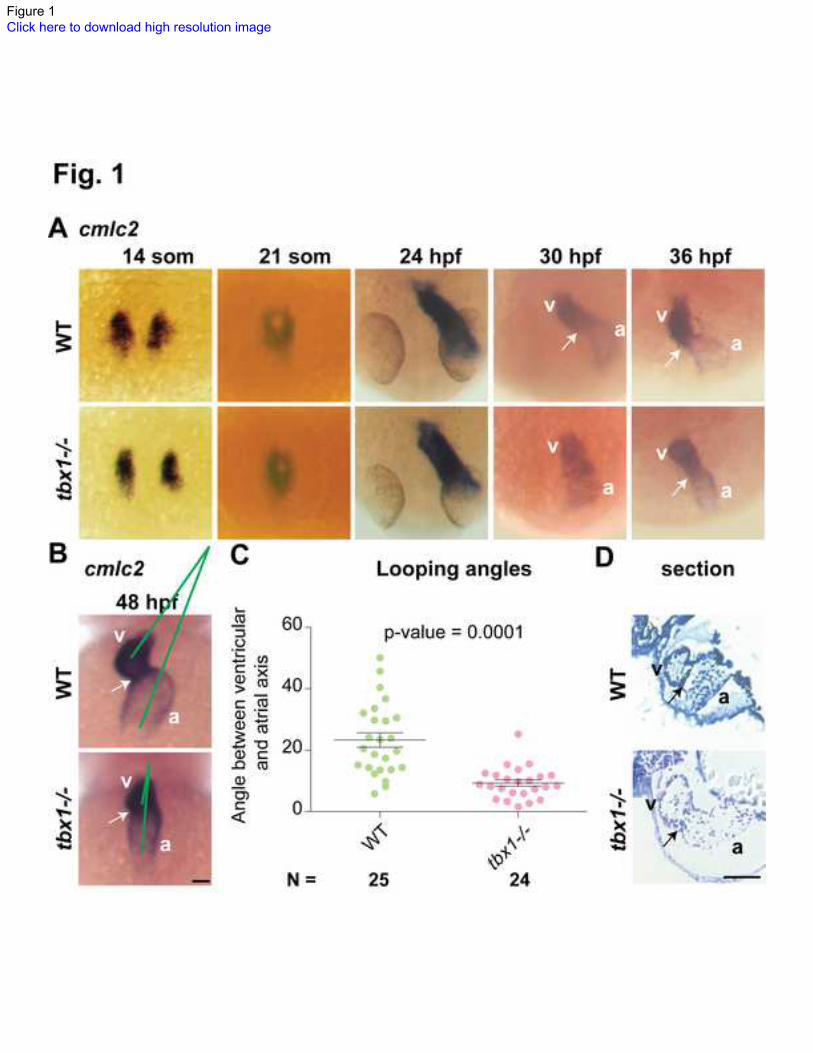
Figure 1Click here to download high resolution image
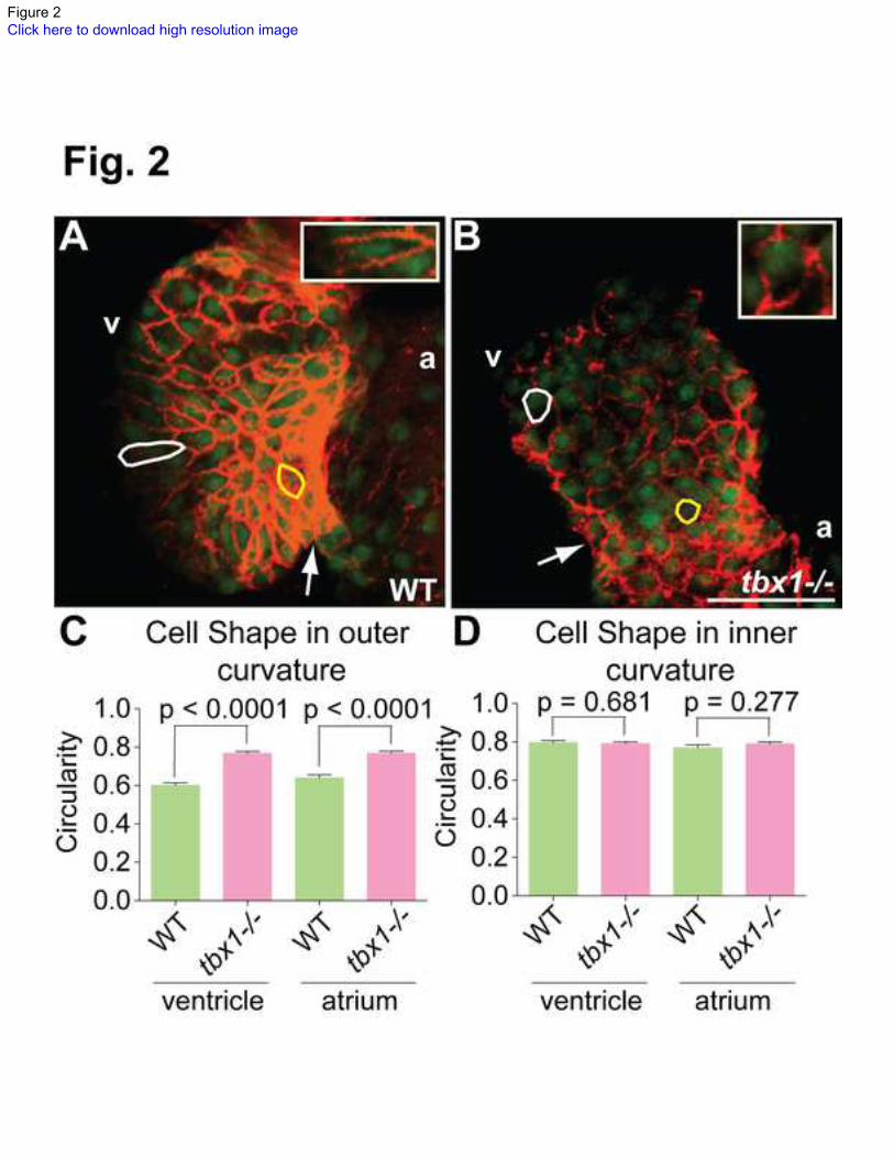
Figure 2Click here to download high resolution image
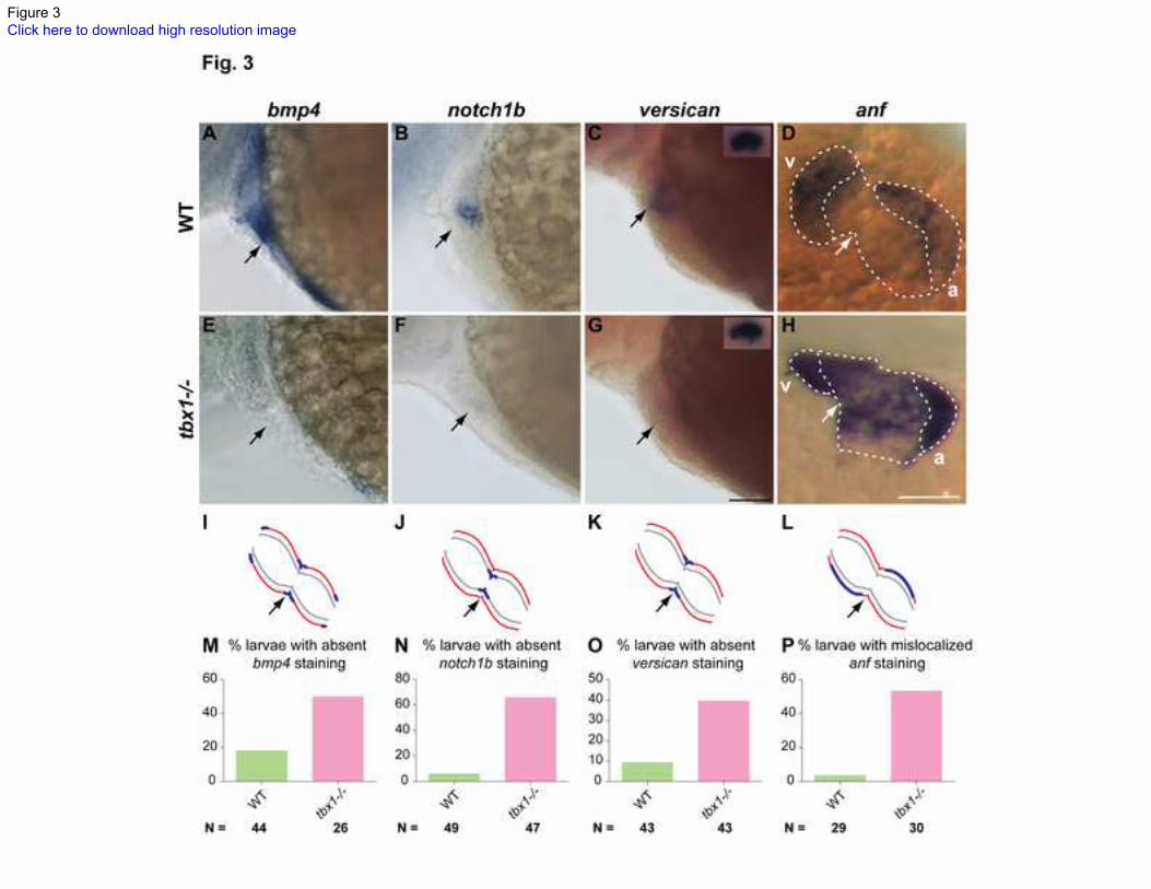
Figure 3Click here to download high resolution image
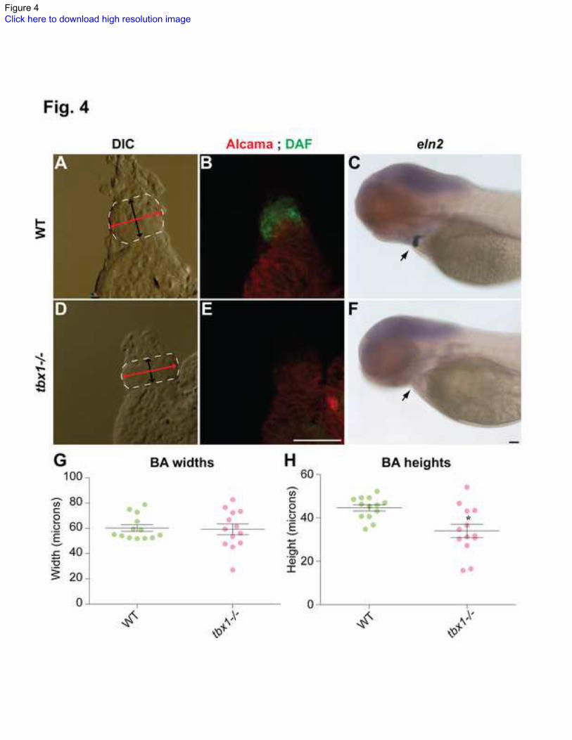
Figure 4Click here to download high resolution image
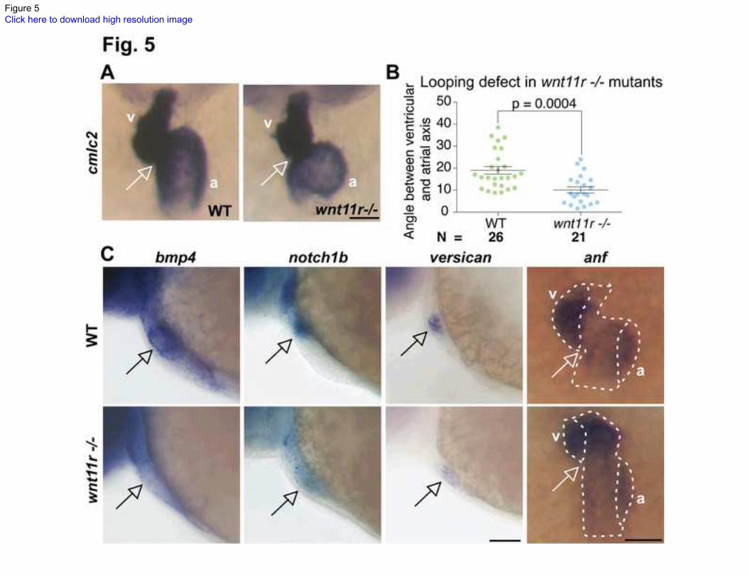
Figure 5Click here to download high resolution image
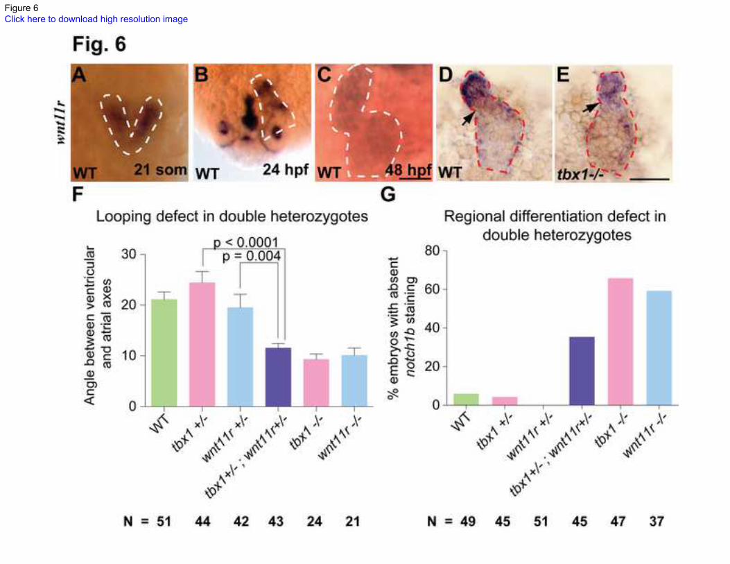
Figure 6Click here to download high resolution image
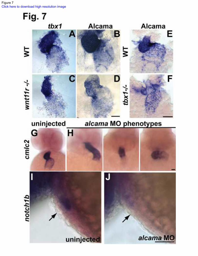
Figure 7Click here to download high resolution image
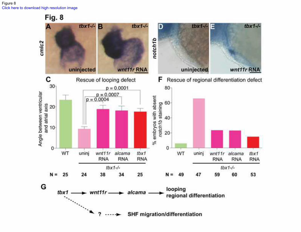
Figure 8Click here to download high resolution image

Supporting InformationClick here to download Supporting Information: Supporting Information-1.docx

Supplemental Figure 1Click here to download Supporting Information: FigS1.tif

Supplemental Figure 2Click here to download Supporting Information: FigS2.tif

Supplemental Figure 3Click here to download Supporting Information: FigS3.tif

Supplemental Figure 4Click here to download Supporting Information: FigS4.tif

Supplemental Figure 5Click here to download Supporting Information: FigS5.tif

Supplemental Figure 6Click here to download Supporting Information: FigS6.tif
Copyright © 2022 FDOKUMEN