Review of 54 patients with complete DiGeorge anomaly enrolled in protocols for thymus...
10
TRANSPLANTATION Review of 54 patients with complete DiGeorge anomaly enrolled in protocols for thymus transplantation: outcome of 44 consecutive transplants M. Louise Markert, 1,2,7 Blythe H. Devlin, 1 Marilyn J. Alexieff, 1 Jie Li, 1 Elizabeth A. McCarthy, 1 Stephanie E. Gupton, 1 Ivan K. Chinn, 1 Laura P. Hale, 5,7 Thomas B. Kepler, 3,4 Min He, 3,4 Marcella Sarzotti, 2 Michael A. Skinner, 6 Henry E. Rice, 6 and Jeffrey C. Hoehner 6 1 Department of Pediatrics, Duke University Medical Center, Durham, NC; 2 Department of Immunology, Duke University Medical Center, Durham, NC; 3 Center for Computational Immunology, Duke University Medical Center, Durham, NC; 4 Department of Biostatistics and Bioinformatics, Duke University Medical Center, Durham, NC; 5 Department of Pathology, Duke University Medical Center, Durham, NC; 6 Department of Surgery, Duke University Medical Center, Durham, NC; 7 Human Vaccine Institute, Duke University Medical Center, Durham, NC The purpose of this study was to charac- terize a large group of infants with com- plete DiGeorge anomaly and to evaluate the ability of thymus transplantation to reconstitute immune function in these infants. DiGeorge anomaly is character- ized by varying defects of the heart, thy- mus, and parathyroid glands. Complete DiGeorge anomaly refers to the subgroup that is athymic (< 1%). The characteris- tics of 54 subjects at presentation and results from 44 consecutive thymus trans- plantations are reported. Remarkably, only 52% had 22q11 hemizygosity and only 57% had congenital heart disease requir- ing surgery. Thirty-one percent devel- oped an atypical phenotype with rash and lymphadenopathy. To date, 33 of 44 sub- jects who received a transplant survive (75%) with post-transplantation follow-up as long as 13 years. All deaths occurred within 12 months of transplantation. All 25 subjects who were tested 1 year after transplantation had developed polyclonal T-cell repertoires and proliferative re- sponses to mitogens. Adverse events de- veloping after transplantation included hypothyroidism in 5 subjects and enteri- tis in 1 subject. In summary, diagnosis of complete DiGeorge anomaly is challeng- ing because of the variability of presenta- tion. Thymus transplantation was well tolerated and resulted in stable immunore- constitution in these infants. (Blood. 2007; 109:4539-4547) © 2007 by The American Society of Hematology Introduction DiGeorge anomaly is characterized by defects in the thymus and parathyroid glands as a result of malformation of the third and fourth pharyngeal pouches. 1 A broad spectrum of abnormalities has been reported in this condition. 2,3 Congenital heart disease, espe- cially truncus arteriosus and interrupted aortic arch type B, results from defects in the fourth pharyngeal arch. No thymus is present in less than 1% of patients with DiGeorge anomaly. 4-7 The term “complete DiGeorge anomaly” is used to describe the population of infants with DiGeorge anomaly who are athymic. The diagnosis of athymia is based on profound suppression of the numbers of recent thymic emigrants in the blood. 8-10 The diagnosis of athymia cannot be made based on the gross appearance at cardiac surgery, by the absence of thymus on a chest radiograph, or by computerized tomogram because the thymus may be in an ectopic location or may be very small. Infants with complete DiGeorge anomaly have various genetic and syndromic associations, 8,9 including 22q11 hemizygosity, 11 CHARGE association (coloboma, heart defect, choanal atresia, growth or developmental retardation, genital hypoplasia, and ear anomalies, including deafness) 12-15 and diabetic embryopathy. 16,17 Infants with complete DiGeorge anomaly present with either a “typical” or “atypical” phenotype. Infants with typical complete DiGeorge anomaly have very low T-cell numbers and do not have a rash. At some point after birth, infants with typical complete DiGeorge anomaly may develop oligoclonal T-cell populations associated with rash and lymphadenopathy. 18 These patients are described as having atypical complete DiGeorge anomaly. The oligoclonal T cells infiltrate the skin and other organs such as the liver. 9,18,19 The T-cell numbers in peripheral blood can be low, normal, or elevated, with low or normal T-cell proliferative responses to the mitogen phytohemagglutinin. The presentation of infants with atypical complete DiGeorge anomaly may be confused with severe atopic dermatitis 20,21 ; Omenn syndrome in RAG1, RAG2 deficiency or Artemis deficiency 22 ; maternal T-cell engraft- ment in patients with severe combined immunodeficiency; or maternal T-cell engraftment in patients with complete DiGeorge anomaly. 23-25 The diagnosis of atypical complete DiGeorge anomaly hinges on the infant having features of DiGeorge anomaly, profoundly depressed numbers of naive T cells, and absence of maternal T cells. The distinction between typical and atypical complete DiGeorge anomaly is important with respect to thymus transplantation because the T cells in atypical complete DiGeorge anomaly can reject transplants. Our group has investigated transplantation of allogeneic postna- tal cultured thymus as a therapy for infants with complete DiGeorge anomaly. 8,9,26-29 We report results for 54 infants enrolled Submitted October 4, 2006; accepted January 21, 2007. Prepublished online as Blood First Edition Paper, February 6, 2007; DOI 10.1182/blood-2006-10- 048652. The publication costs of this article were defrayed in part by page charge payment. Therefore, and solely to indicate this fact, this article is hereby marked ‘‘advertisement’’ in accordance with 18 USC section 1734. © 2007 by The American Society of Hematology 4539 BLOOD, 15 MAY 2007 VOLUME 109, NUMBER 10 For personal use only. on April 8, 2016. by guest www.bloodjournal.org From
-
Upload
utsouthwestern -
Category
Documents
-
view
1 -
download
0
Transcript of Review of 54 patients with complete DiGeorge anomaly enrolled in protocols for thymus...
TRANSPLANTATION
Review of 54 patients with complete DiGeorge anomaly enrolled in protocolsfor thymus transplantation: outcome of 44 consecutive transplantsM. Louise Markert,1,2,7 Blythe H. Devlin,1 Marilyn J. Alexieff,1 Jie Li,1 Elizabeth A. McCarthy,1 Stephanie E. Gupton,1
Ivan K. Chinn,1 Laura P. Hale,5,7 Thomas B. Kepler,3,4 Min He,3,4 Marcella Sarzotti,2 Michael A. Skinner,6 Henry E. Rice,6
and Jeffrey C. Hoehner6
1Department of Pediatrics, Duke University Medical Center, Durham, NC; 2Department of Immunology, Duke University Medical Center, Durham, NC; 3Center forComputational Immunology, Duke University Medical Center, Durham, NC; 4Department of Biostatistics and Bioinformatics, Duke University Medical Center,Durham, NC; 5Department of Pathology, Duke University Medical Center, Durham, NC; 6Department of Surgery, Duke University Medical Center, Durham, NC;7Human Vaccine Institute, Duke University Medical Center, Durham, NC
The purpose of this study was to charac-terize a large group of infants with com-plete DiGeorge anomaly and to evaluatethe ability of thymus transplantation toreconstitute immune function in theseinfants. DiGeorge anomaly is character-ized by varying defects of the heart, thy-mus, and parathyroid glands. CompleteDiGeorge anomaly refers to the subgroupthat is athymic (< 1%). The characteris-tics of 54 subjects at presentation andresults from 44 consecutive thymus trans-plantations are reported. Remarkably, only
52% had 22q11 hemizygosity and only57% had congenital heart disease requir-ing surgery. Thirty-one percent devel-oped an atypical phenotype with rash andlymphadenopathy. To date, 33 of 44 sub-jects who received a transplant survive(75%) with post-transplantation follow-upas long as 13 years. All deaths occurredwithin 12 months of transplantation. All25 subjects who were tested 1 year aftertransplantation had developed polyclonalT-cell repertoires and proliferative re-sponses to mitogens. Adverse events de-
veloping after transplantation includedhypothyroidism in 5 subjects and enteri-tis in 1 subject. In summary, diagnosis ofcomplete DiGeorge anomaly is challeng-ing because of the variability of presenta-tion. Thymus transplantation was welltolerated and resulted in stable immunore-constitution in these infants. (Blood. 2007;109:4539-4547)
© 2007 by The American Society of Hematology
Introduction
DiGeorge anomaly is characterized by defects in the thymus andparathyroid glands as a result of malformation of the third andfourth pharyngeal pouches.1 A broad spectrum of abnormalities hasbeen reported in this condition.2,3 Congenital heart disease, espe-cially truncus arteriosus and interrupted aortic arch type B, resultsfrom defects in the fourth pharyngeal arch. No thymus is present inless than 1% of patients with DiGeorge anomaly.4-7 The term“complete DiGeorge anomaly” is used to describe the population ofinfants with DiGeorge anomaly who are athymic. The diagnosis ofathymia is based on profound suppression of the numbers of recentthymic emigrants in the blood.8-10 The diagnosis of athymia cannotbe made based on the gross appearance at cardiac surgery, by theabsence of thymus on a chest radiograph, or by computerizedtomogram because the thymus may be in an ectopic location ormay be very small. Infants with complete DiGeorge anomaly havevarious genetic and syndromic associations,8,9 including 22q11hemizygosity,11 CHARGE association (coloboma, heart defect,choanal atresia, growth or developmental retardation, genitalhypoplasia, and ear anomalies, including deafness)12-15 and diabeticembryopathy.16,17
Infants with complete DiGeorge anomaly present with either a“typical” or “atypical” phenotype. Infants with typical completeDiGeorge anomaly have very low T-cell numbers and do not have a
rash. At some point after birth, infants with typical completeDiGeorge anomaly may develop oligoclonal T-cell populationsassociated with rash and lymphadenopathy.18 These patients aredescribed as having atypical complete DiGeorge anomaly. Theoligoclonal T cells infiltrate the skin and other organs such as theliver.9,18,19 The T-cell numbers in peripheral blood can be low,normal, or elevated, with low or normal T-cell proliferativeresponses to the mitogen phytohemagglutinin. The presentation ofinfants with atypical complete DiGeorge anomaly may be confusedwith severe atopic dermatitis20,21; Omenn syndrome in RAG1,RAG2 deficiency or Artemis deficiency22; maternal T-cell engraft-ment in patients with severe combined immunodeficiency; ormaternal T-cell engraftment in patients with complete DiGeorgeanomaly.23-25 The diagnosis of atypical complete DiGeorge anomalyhinges on the infant having features of DiGeorge anomaly,profoundly depressed numbers of naive T cells, and absence ofmaternal T cells. The distinction between typical and atypicalcomplete DiGeorge anomaly is important with respect to thymustransplantation because the T cells in atypical complete DiGeorgeanomaly can reject transplants.
Our group has investigated transplantation of allogeneic postna-tal cultured thymus as a therapy for infants with completeDiGeorge anomaly.8,9,26-29 We report results for 54 infants enrolled
Submitted October 4, 2006; accepted January 21, 2007. Prepublished onlineas Blood First Edition Paper, February 6, 2007; DOI 10.1182/blood-2006-10-048652.
The publication costs of this article were defrayed in part by page charge
payment. Therefore, and solely to indicate this fact, this article is herebymarked ‘‘advertisement’’ in accordance with 18 USC section 1734.
© 2007 by The American Society of Hematology
4539BLOOD, 15 MAY 2007 � VOLUME 109, NUMBER 10
For personal use only.on April 8, 2016. by guest www.bloodjournal.orgFrom

in thymus transplantation protocols. From 1993 through November2006, 44 of the infants were treated with allogeneic postnatalcultured thymus transplantation. Twenty-six subjects who receiveda transplant are more than 1 year after transplantation. Our resultssupport both the safety of thymus transplantation and efficacyreflected in a pronounced improvement in T-cell function and ratesof infection.
Patients, materials, and methods
Subject inclusion criteria
All subjects were enrolled in protocols approved by the Duke InstitutionalReview Board. A total of 54 subjects with complete DiGeorge anomalywere enrolled from 1993 through November 2006; 44 received a transplant.Informed consent was obtained from the parents of all enrolled subjects, inaccordance with the Declaration of Helsinki. Preliminary data werereported previously on 19 of these subjects.8,9,18
The subjects in this report were enrolled in 1 of 6 protocols. Three phase1 protocols were descriptive only. Two are closed to enrollment, the thirdcontinues to investigate optimal immunosuppression regimens. Two otherphase 1 protocols have as primary end points (1) naive T-cell counts andT-cell receptor repertoire variability at 1 year (looking for a dose effect) and(2) calcium supplementation after parathyroid transplantation. Both areopen at this time. The final protocol is phase 2 and has the primary end pointof survival.
To be considered athymic, subjects had either fewer than 50 naive Tcells/mm3 or their naive T cells comprised less than 5% of total T cells byflow cytometry. Naive T cells were defined as those coexpressing CD45RAand CD62L.10 For the diagnosis of DiGeorge anomaly, subjects had at leastone of the following: hypoparathyroidism, congenital heart disease,CHARGE association, or 22q11 hemizygosity. One person who met thesecriteria had ectodermal dysplasia with absence of T cells and B cells atbirth. The T cells isolated from subjects with atypical complete DiGeorgeanomaly were shown to be genetically host-derived prior to transplantation.This was done by a clinical molecular laboratory using multiplex polymer-ase chain reaction amplification for microsatellite markers with a sensitivityof 2%.
Thymus donor and tissue processing
Processing of the thymus tissue was done as described9,30,31 under anInvestigational New Drug (IND) application with the Food and DrugAdministration (FDA). In brief, thymus tissue was obtained as discardedtissue from pediatric cardiac surgery after informed consent of the donor’sparents. All thymus donors were younger than 7 months. The donors and themothers of the donors were screened for infectious diseases per FDAguidelines.32 In addition the presence of EBV and CMV was assessedbecause these viruses have resulted in lymphoproliferative disorders ininfants treated in the past at other centers.33,34 As has been reported in thepast,8,9 HLA matching was not required for transplantation.
Immunosuppression
Whether immunosuppression was used in the 44 subjects who received atransplant depended on the level of T-cell function of the recipient prior totransplantation. Twenty-two subjects with typical complete DiGeorgeanomaly did not receive immunosuppression. They had fewer than 5000counts per minute (cpm) of T-cell proliferative response to phytohemagglu-tinin (PHA) or less than a 20-fold response to PHA over background. Of theremaining 22 subjects, 8 had typical complete DiGeorge anomaly andreceived rabbit antithymocyte globulin. The usual dose was 2 mg/kg/d for 3doses before transplantation. Methylprednisolone, 2 mg/kg, was given priorto the first dose of rabbit antithymocyte globulin followed by 0.5 mg/kgevery 6 hours intravenously until 24 hours after finishing the rabbitantithymocyte globulin. These 8 subjects had a T-cell proliferative responseto PHA of more than 5000 cpm or greater than 20-fold over background (3
subjects), enrolled in a protocol to receive parathyroid transplants (4subjects), or had preexisting graft-versus-host disease (GVHD, 1 subject).This latter subject was also treated with cyclophosphamide and cyclospor-ine. The 14 subjects with atypical complete DiGeorge anomaly had variedproliferative responses to PHA, ranging from very low to more than100 000 cpm. They received one of several immunosuppressive treatments.Deoxycoformycin was used to immunosuppress the first subject withatypical complete DiGeorge anomaly prior to transplantation. Four subjectswith atypical complete DiGeorge received rabbit antithymocyte globulin(with concurrent steroids) before transplantation with no additional beforeor after transplantation immunosuppression. Nine additional subjects withatypical complete DiGeorge anomaly received rabbit antithymocyte globu-lin before transplantation plus various combinations of cyclosporine ortacrolimus (7 subjects), steroids (1 subject), and mycophenylate mofetil anddaclizumab (1 subject who received cyclosporine and steroids as well)before and after transplantation.
Thymus transplantation
After 12 to 21 days in culture,8,9 the thymus slices were transplanted into theinfant’s quadriceps in an open procedure under general anesthesia asdescribed.27
Immune phenotype and function
Standard flow cytometry was done on whole blood.9 Percentages of positivecells were recorded, and absolute numbers were calculated based on theabsolute lymphocyte count. Standard tests of T-cell function were per-formed,9 including proliferative responses to PHA (3 concentrations, withcells harvested on days 3 and 4), concanavalin A (3 concentrations, withcells harvested on days 4 and 5), soluble and immobilized CD3 (cellsharvested on day 4), and tetanus antigen and Candida skin test antigen (4concentrations of each antigen, cells harvested on day 6).
Spectratyping to assess T-cell receptor diversity was performed asdescribed, usually on isolated CD4 T cells.8,9 All panels were inspected toensure that they met acceptable technical criteria in terms of sufficientheight of the peaks above background and absence of overloading.Technically adequate panels were analyzed via computation of theirKullback-Leibler divergences (DKL).35 The DKL generically measures thedivergence of 2 probability functions; in this case, it measures thedivergence of each spectratype profile against the corresponding referenceprofile based on the average of 7 healthy adult volunteers. The normal rangewas determined by trimming the top and bottom 2.5% from the historicalDKLs observed in healthy adult volunteers. The higher the DKL, the greaterthe divergence from the normal profile and the more oligoclonal thedistribution of peaks.
Immunohistochemistry of graft biopsy
Biopsies of the thymus allograft were conducted at 2 to 3 months aftertransplantation if the medical condition of the subject was stable.8,28 Frozenand paraffin-embedded sections were reacted with a panel of antibodies,including cytokeratin (clones AE1/AE3; Dako, Carpinteria, CA) CD3(polyclonal; Dako), CD1a (clone 010; Beckman Coulter, Marseille, France),and Ki-67 (clone mib-1; Beckman Coulter).8,28 Histologic evidence ofthymopoiesis in biopsies was defined as the presence of a lacy pattern ofcytokeratin-positive thymic epithelial cells and the presence ofCD3�CD1a�Ki-67� cells. The latter is the phenotype of cortical thymocytes.
Surveillance for adverse events
Infections were defined per the Blood and Marrow Transplant ClinicalTrials Network.36 Any ongoing bacterial pulmonary infection or viralinfection before transplantation was counted as a single infection becausethese infections usually persist until T cells develop. Except for the first 12subjects, all recipients were screened prior to transplantation for thepresence of Epstein Barr virus (EBV), cytomegalovirus (CMV), respiratorysyncytial virus (RSV), parainfluenza virus, adenovirus, and enterovirus.Screening for HIV-1, human herpes virus type 6 (HHV6), and West Nilevirus was added in the past 2 years. Stool, nasopharyngeal, and urine viral
4540 MARKERT et al BLOOD, 15 MAY 2007 � VOLUME 109, NUMBER 10
For personal use only.on April 8, 2016. by guest www.bloodjournal.orgFrom
cultures were repeated after the initial screening based on clinical symp-toms. Since 2001, all subjects have been screened for thyroid disease.
Results
Subject characteristics
During the study period from 1993 to November 2006, 54 infantswere enrolled as subjects in thymus transplantation protocols. Twosubjects were withdrawn by their parents prior to transfer to DukeUniversity Medical Center (DUMC) because of the severity ofother anomalies in the infants. Fifty-one were admitted to DUMCand one is awaiting transfer to DUMC. Three infants werewithdrawn after admission (without transplantation), one by theparents because of the severity of other anomalies and 2 by theinvestigator because the infants were not medically stable enoughfor surgery. Three infants died of infection and one from respiratoryarrest prior to transplantation. Forty-four infants underwenttransplantation.
Characteristics of the entire group of 54 enrolled subjects and ofthe 44 subjects who received a transplant were similar and areshown in Table 1. Athymia was confirmed in all subjects by thepresence of very low naive T-cell numbers or percentages. Theinitial absolute lymphocyte count was greater than the 10thpercentile in 13% (7 of 54) of the subjects. Thirty-one percent (17of 54) of the subjects presented with the atypical phenotype withrash and lymphadenopathy associated with oligoclonal T cells. Theatypical subjects comprised 36% (10 of 28) of subjects with 22q11hemizygosity as compared with 27% (7 of 26) normal at 22q11.Unexpectedly, the rate of conotruncal congenital heart disease wasrelatively low at 31% overall (Table 1) compared with approxi-mately 60% in the literature.2,4
Survival
Survival of the entire group of 54 subjects is 64% (35 of 54).Currently, 75% of subjects who received a transplant (33 of 44)survive (Figure 1).
Of the 11 deaths after transplantation, none of the subjects diedsecondary to thymus transplantation. Two were due to RSV, 2 toCMV, 1 from CMV and GVHD from unirradiated blood transfu-sions given prior to transfer to Duke, 1 from a complication ofcalcium therapy, and 5, all in subjects with tracheostomies or onventilators, from sepsis or progressive bronchopulmonary dyspla-sia. Although no subjects have died after 12 months after theirtransplantation, 2 of the infants have had more morbidity thanothers. One subject had a stroke secondary to sickle cell disease 2years after transplantation and is significantly disabled. The infantwith ectodermal dysplasia has had life-threatening problems withthrombosis and infections before and after thymus transplantation.Although T cells developed that respond to mitogens, they do notrespond to antigens. These morbid events may yet lead to the deathof some subjects later than 12 months after transplantation.
We examined the effect of 5 factors on survival: 22q11hemizygosity, presentation with atypical complete DiGeorgeanomaly, use of immunosuppression, age at transplantation, andexperience of the medical and surgical transplantation team inmanaging these medically fragile subjects. We evaluated survivalof the 54 subjects who were consented with the intent to treat andthe 44 subjects who received a transplant. These analyses weredone post hoc and do not have statistical power. Of the 54 enrolledsubjects, we did not observe significant differences in survival ratesbetween the 57% (16 of 28) of subjects with 22q11 hemizygosity
who survive and the 73% (19 of 26) of subjects without 22q11hemizygosity who survive (P � .347 by a chi-square goodness-of-fit test). Ten (59%) of 17 subjects with atypical DiGeorgesurvive versus 25 (68%) of 37 typical subjects (P � .75). Seven-teen (77%) of 22 subjects who received a transplant withoutimmunosuppression survive compared with 16 (73%) of 22subjects who received a transplant with immunosuppression(P � .99). We did not find significant differences in survival ratesfor these comparisons. Regarding the effect of age, of the 22subjects who received a transplant younger than the median age of4.25 months, 14 survive (64%), whereas 19 (86%) of 22 subjectswho received a transplant at the median age of 4.25 months or oldersurvive. This difference likely relates to comorbid conditionsleading to death of some children in the first few months of life. Afifth factor evaluated was whether improvements in medical care
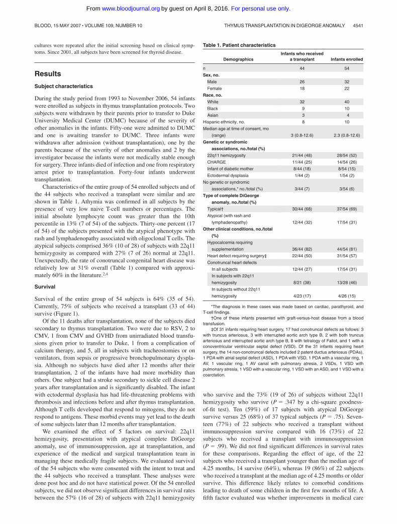
Table 1. Patient characteristics
DemographicsInfants who received
a transplant Infants enrolled
n 44 54
Sex, no.
Male 26 32
Female 18 22
Race, no.
White 32 40
Black 9 10
Asian 3 4
Hispanic ethnicity, no. 8 10
Median age at time of consent, mo
(range) 3 (0.8-12.6) 2.3 (0.8-12.6)
Genetic or syndromic
associations, no./total (%)
22q11 hemizygosity 21/44 (48) 28/54 (52)
CHARGE 11/44 (25) 14/54 (26)
Infant of diabetic mother 8/44 (18) 8/54 (15)
Ectodermal dysplasia 1/44 (2) 1/54 (2)
No genetic or syndromic
associations,* no./total (%) 3/44 (7) 3/54 (6)
Type of complete DiGeorge
anomaly, no./total (%)
Typical† 30/44 (68) 37/54 (69)
Atypical (with rash and
lymphadenopathy) 12/44 (32) 17/54 (31)
Other clinical conditions, no./total
(%)
Hypocalcemia requiring
supplementation 36/44 (82) 44/54 (81)
Heart defect requiring surgery‡ 22/44 (50) 31/54 (57)
Conotruncal heart defects
In all subjects 12/44 (27) 17/54 (31)
In subjects with 22q11
hemizygosity 8/21 (38) 13/28 (46)
In subjects without 22q11
hemizygosity 4/23 (17) 4/26 (15)
*The diagnosis in these cases was made based on cardiac, parathyroid, andT-cell findings.
†One of these infants presented with graft-versus-host disease from a bloodtransfusion.
‡Of 31 infants requiring heart surgery, 17 had conotruncal defects as follows: 3with truncus arteriosus, 3 with interrupted aortic arch type B, 2 with both truncusarteriosus and interrupted aortic arch type B, 8 with tetralogy of Fallot, and 1 with aconoventricular ventricular septal defect (VSD). Of the 31 infants requiring heartsurgery, the 14 non-conotruncal defects included 2 patent ductus arteriosus (PDAs),1 PDA with atrial septal defect (ASD), 1 PDA with VSD, 1 PDA with a vascular ring, 1AV, 1 vascular ring, 1 AV canal with pulmonary atresia, 2 VSDs, 1 VSD withpulmonary atresia, 1 VSD with a vascular ring, 1 VSD with an ASD, and 1 VSD with acoarctation.
THYMUS TRANSPLANTATION IN DIGEORGE ANOMALY 4541BLOOD, 15 MAY 2007 � VOLUME 109, NUMBER 10
For personal use only.on April 8, 2016. by guest www.bloodjournal.orgFrom

led to improved survival. Of the initial 27 subjects, 16 (59%)survive. Of the subsequent 27 subjects, 19 survive (70%). It islikely that 2 additional subjects consented in the past year may die(one with significant morbidity, one who did not receive atransplant and withdrawn from the protocol), so these percentageswill likely be similar. The similar survival for the 2 groups ofsubjects suggests that experience in medical and surgical manage-ment of these subjects did not improve survival. Infants withcomplete DiGeorge anomaly continue to die of cardiac complica-tions and infections (if T cells have not been reconstituted). Thesurvival curve shows that the death rate only levels off after T-cellfunction develops after approximately 9 months.
T lymphocyte phenotype, numbers, and function
Testing of peripheral blood samples showed that phenotypicallynormal T cells could be detected in the blood between 3 and 6months after thymus transplantation. Specifically, the subjectsdeveloped more CD4 than CD8 single-positive T cells and had lowlevels of double negative (CD4�CD8�) T cells. The CD4 TCRrepertoire normalized (see data under “Spectratyping”). Circulatingnaive T cells could be detected by 6 months.
Twenty-two typical subjects received a transplant withoutimmunosuppression of whom 17 survive. Fourteen of these are past1 year after transplantation. The immune results for all 14 typicalsubjects who were transplanted without immunosuppression andare more than 1 year from transplantation are shown in Figure2A-D. (The 3 subjects who are under 1 year after transplantationare not shown. The other 5 subjects died.). Seven of these 14subjects have previously been reported.8
In the group of 22 subjects who received a transplant and did notreceive immunosuppression, 4 developed early T-cell amplifica-tions (Figure 2A-C).
Two of these subjects developed a mild rash immediately priorto transplantation. One of these 2 subjects progressed to a fullatypical phenotype with circulating CD3�CD4�CD8� oligoclonalT cells. Two different subjects with typical complete DiGeorgeanomaly developed transient oligoclonal T-cell proliferations asso-ciated with rash and lymphadenopathy within 2 months of transplan-tation. When naive T cells developed at 5 to 7 months aftertransplantation, the rash and lymphadenopathy spontaneouslyresolved and the oligoclonal T cells dramatically decreased innumber. For 1 of the 2 latter subjects, the prominent oligoclonal Tcells were CD3�CD4�CD8�. Normalization of the CD4/CD8 ratiooccurred shortly before the appearance of naive T cells. We do notthink that the development of oligoclonal T cells in these subjectswas secondary to transplantation, but instead that the developmentwas equivalent to development of the atypical phenotype in aninfant with typical complete DiGeorge anomaly.
Of 22 subjects who received immunosuppression, 16 survive.The immune results for the subset of 12 subjects who are more than1 year from transplantation are shown in Figure 2D-F,H. (The 4subjects who are under 1 year after transplantation are not shown.The other 6 died.) T-cell numbers level off slightly below the 10thpercentile for age. Seven of these infants had the atypical pheno-type. Two of those had predominant CD3�CD4�CD8� T-cellclones. The CD4/CD8 ratio in these subjects normalized coincidentwith disappearance of rash and lymphadenopathy and appearanceof naive T cells around 4 to 5 months after transplantation. Seven ofthese 22 subjects have been reported previously.9 Similar resultsare seen in the groups with and without immunosuppression.
Of the 26 subjects 1 year after transplantation, 21 have had atleast 2 immune studies completed after year 1. In comparing 13subjects without immunosuppression and 8 subjects with immuno-suppression, the CD3, CD4, and CD8 mean numbers for the first 2studies after 1 year are slightly higher in the group with immunosup-pression (CD3, 935/mm3 versus 804/mm3; CD4, 631/mm3 versus554/mm3; and CD8, 215/mm3 versus 159/mm3). Naive CD4 andCD8 numbers also remain slightly below the 10th percentile (notshown) and are nearly identical in the 2 groups. T-cell proliferativeresponses to the mitogen PHA became normal by 6 to 12 months.The most recent mean proliferative responses after year 1 weresomewhat higher in the subjects who received immunosuppression(221 702 cpm versus 177 515 cpm). Although not shown, antigen-specific T-cell proliferative responses have been documented ineach of these subjects except the one who has ectodermaldysplasia. In summary, although the T-cell numbers are belownormal, the T-cell proliferative function is robust.
Spectratyping
TCRBV repertoire diversity is assessed by spectratyping. TheKullback-Leibler divergence (DKL) statistic is used to assessquantitatively the diversity of the TCRBV repertoire as comparedwith normal diversity.34 (The higher the DKL, the more therepertoire diverges from a normal diverse repertoire.) When T cellsfirst emerge from the thymus after thymus transplantation, they arequite oligoclonal with high DKL values. The TCRBV repertoirerapidly diversifies and the DKL drops to the normal range. Prior totransplantation, subjects with atypical complete DiGeorge anomalyhave high numbers of oligoclonal T cells (with high DKL values).The DKL drops (indicating less divergence from a normal reper-toire), entering the normal range by 9 months after transplantation
Figure 1. Kaplan-Meyer plot of survival of recipients of a thymus transplant. (A)Survival of 35 of 54 subjects who were enrolled. (B) Survival of 33 of 44 subjects whoreceived a transplant.
4542 MARKERT et al BLOOD, 15 MAY 2007 � VOLUME 109, NUMBER 10
For personal use only.on April 8, 2016. by guest www.bloodjournal.orgFrom

in both typical and atypical complete DiGeorge anomaly subjects(Figure 3A-B). The current CD4 spectratyping data for one subjectwho is now 7 years after transplantation is shown as an example oflong-term maintenance of T-cell diversity (Figure 3C).
Thymus biopsy results
Biopsies of the thymus grafts were done in 30 of the 44 subjectswho underwent transplantation. Biopsies were not performed ininfants with pulmonary infections or unstable heart disease. Thegraft (keratin-positive material) was identified in 25 of the 30biopsies. In the other 5 biopsies, only fat or muscle was obtained.Finding only fat or muscle does not rule out engraftment becausethe transplanted tissue may not have been sampled. Examples ofbiopsies showing thymopoiesis have been published.9,27-29 Thymo-poiesis was found in 23 of the 25 biopsies in which keratin-positiveepithelial cells were identified. Two biopsies detected viableepithelium but no thymopoiesis. One of these 2 subjects hadGVHD and CMV infection from unirradiated blood transfusionsbefore transplantation. The second subject had atypical completeDiGeorge anomaly and was suppressed before transplantation withrabbit anti–human thymocyte globulin. The oligoclonal T cellsincreased dramatically by day 13 after transplantation at whichtime cyclosporine and steroids were initiated. This subject eventu-ally developed naive T cells with a normal TCRBV repertoire;however, the percentage of naive T cells is lower than in mostsubjects. Because of the findings in that one subject and to protectthe thymus graft from oligoclonal T cells after transplantation,
subsequent atypical subjects have been started on cyclosporinebefore transplantation and are maintained on this treatment untilnaive T cells develop.
B-cell function
All subjects received intravenous immunoglobulin (IVIG) infu-sions until 2 years after transplantation. After the IVIG wasstopped, antibody responses to tetanus toxoid and pneumococcalimmunizations were checked (Tables 2-3).
Fourteen subjects immunized after stopping IVIG have devel-oped antigen-specific antibodies. Thirteen have normal responsesto pneumococcal carbohydrate antigens in unconjugated vaccines.A fourteenth subject was immunized with a conjugated pneumococ-cal vaccine and made a specific response. Isohemagglutinins havedeveloped in some subjects. After 2 years, most subjects stop IVIGtherapy. The persistence of protective titers is a topic of currentinvestigation in our laboratory.
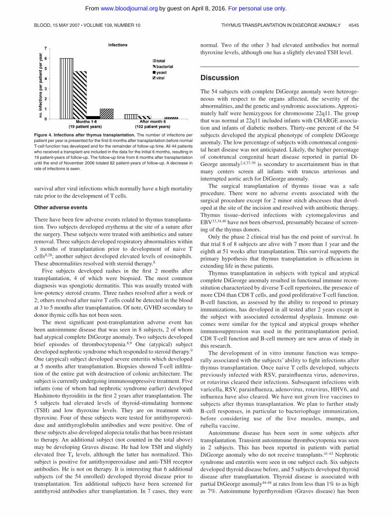
Infections
Figure 4 shows the number of infections per subject normalized peryear for the initial 6 months after transplantation when T cells havenot yet developed versus the infections after the first 6 months. Allsubjects are included, although some died in the first 6 months. Amarked fall in infection frequency is seen even if only the survivingsubjects are analyzed (data not shown). The most commoninfections in the first 6 months were bacterial line infections
Figure 2. T-cell development as determined by T-cell subset antibody staining and T-cell proliferative responses to mitogens for subjects past 1 year after thymustransplantation. Fourteen subjects who did not receive immunosuppression (A-C,G) are compared with 12 subjects who did receive immunosuppression (D-F,H); (A,D) CD3,(B,E) CD4, (C,F) CD8, (G,H) PHA responses. Each subject is represented by a separate line. The x-axis is years after transplantation. The y-axis for panels A to F is cells/mm3.The y-axis for panels G and H is counts per minute. Note the T-cell amplifications seen in panels A to C for 4 subjects who developed the atypical form of DiGeorge anomalyshortly after transplantation.
THYMUS TRANSPLANTATION IN DIGEORGE ANOMALY 4543BLOOD, 15 MAY 2007 � VOLUME 109, NUMBER 10
For personal use only.on April 8, 2016. by guest www.bloodjournal.orgFrom

representing 40% (44 of 113) of the total infections recorded. Noneof these infections was related to the transplantation procedure. Theother infections in the first 6 months included 20 bacterial lunginfections, 19 bacterial urine infections, 19 viral infections, 6episodes of diarrhea associated with Clostridium difficile, and 5yeast infections. The viral infections that were clinically the mostproblematic in the first 6 months after transplantation were RSV (4cases with 2 deaths), CMV (3 cases with 2 deaths), and parainflu-enza virus (2 cases).
After 6 months, there were relatively few line infections, likelybecause most central lines were removed after immune function
developed. After 6 months, viral infections had similar courses toviral infections in healthy children. All preexisting viral infectionscleared after circulating naive T cells developed. Nine subjects had15 documented viral infections. Eight of the 9 subjects haveantigen-specific T-cell responses (and B-cell antigen-specific re-sponses documented for those who are not on IVIG). The ninth isthe subject with ectodermal dysplasia. She never developedantigen-specific T-cell responses and has had serious post-transplantation viral infections. We acknowledge a significantunderreporting of viral infections in the follow-up period. Thiscould be due to lack of surveillance cultures and lack of reportingby the pediatrician. Taken as a whole, it appears that the flatteningof the survival curve (Figure 1) after 6 months is secondary to
Figure 3. CD4 TCRBV repertoire diversity as assessed by spectratyping. (A) DKL values for 16 subjects with typical complete DiGeorge anomaly. (B) DKL values for 7subjects with atypical complete DiGeorge anomaly. (C) CD4 spectratyping profiles for subject DIG005 at 7 years after transplantation. The Jurkat control panel is in the lowerright. The DKL value for this spectratype is 0.11.
Table 2. Immunoglobulin evaluations in children more than 2 yearsafter transplantation (n � 21)
Normal for agen* Low, n High, n
IgG levels for infants off IVIG† 13 1 1
IgA levels 15 2 4
IgM levels 17 4 0
IgE levels‡ 15 3 2
All values listed were obtained at least 1 year from transplantation and most arewithin 1 year of the present time. Eighteen infants were off IVIG. Of the 3 on IVIG, twosubjects have not been taken off IVIG and have not been tested for antibodyformation by the local physician. The patient with ectodermal dysplasia remains onIVIG because this patient does not have T-cell proliferative responses to tetanustoxoid.
*The years after transplantation are used instead of years of life for the Ig levels. Thus,normal values in mg/dL are as follows: IgA, 15 to 108 for 2 to 3 years, 21 to 144 for 4 to 5years, 22 to 141 for 6 years, 38 to 240 for 7 years, 32 to 248 for 8 to 10 years, and 40 to 277for 11 to 12 years; IgM is 43 to 202 for 2 to 3 years, 31 to 163 for 4 to 5 years, 43 to 177 for 6years, 37 to 151 for 7 years, 42 to 185 for 8 to 10 years, and 42 to 174 for 11 to 12 years.Thenormal values for IgE in IU/mLare as follows: 0-126 for 2 years, 3-135 for 3 years, 1-219 for4 years, 2-158 for 5-7 years, 2-524 for 7-10 years, and 4-437 for 10-15 years. Note: “low” isbelow these levels and “high” is above these levels.
†IgG levels are not included for 6 patients. This includes the 3 patients on IVIG.Three others have not had IgG levels obtained since stopping IVIG.
‡Not done after year 1 in one patient.
Table 3. Specific antibodies in children more than 2 yearsafter transplantation
n
Tetanus antibodies*
Normal 12
Low 1
Pneumococcal antibodies†
Response to 3 or more serotypes 9
Responses to 1-2 serotypes 4
No significant responses 0
Isohemagglutinins‡
Normal for age 4
Low 9
Negative 4
All values listed were obtained at least 1 year from transplantation and most arewithin 1 year of the present time.
*Tetanus antibodies have not been tested in 8 subjects.†Pneumococcal titers to multiple serotypes (unconjugated) were assessed in 13
subjects. One additional subject (not included in the table) also made antibodies inresponse to a conjugated pneumococcal vaccine.
‡Isohemagglutinins have not been tested in 4 subjects.
4544 MARKERT et al BLOOD, 15 MAY 2007 � VOLUME 109, NUMBER 10
For personal use only.on April 8, 2016. by guest www.bloodjournal.orgFrom
survival after viral infections which normally have a high mortalityrate prior to the development of T cells.
Other adverse events
There have been few adverse events related to thymus transplanta-tion. Two subjects developed erythema at the site of a suture afterthe surgery. These subjects were treated with antibiotics and sutureremoval. Three subjects developed respiratory abnormalities within3 months of transplantation prior to development of naive Tcells8,28; another subject developed elevated levels of eosinophils.These abnormalities resolved with steroid therapy.8
Five subjects developed rashes in the first 2 months aftertransplantation, 4 of which were biopsied. The most commondiagnosis was spongiotic dermatitis. This was usually treated withlow-potency steroid creams. Three rashes resolved after a week or2; others resolved after naive T cells could be detected in the bloodat 3 to 5 months after transplantation. Of note, GVHD secondary todonor thymic cells has not been seen.
The most significant post-transplantation adverse event hasbeen autoimmune disease that was seen in 8 subjects, 2 of whomhad atypical complete DiGeorge anomaly. Two subjects developedbrief episodes of thrombocytopenia.8,9 One (atypical) subjectdeveloped nephrotic syndrome which responded to steroid therapy.9
One (atypical) subject developed severe enteritis which developedat 5 months after transplantation. Biopsies showed T-cell infiltra-tion of the entire gut with destruction of colonic architecture. Thesubject is currently undergoing immunosuppressive treatment. Fiveinfants (one of whom had nephrotic syndrome earlier) developedHashimoto thyroiditis in the first 2 years after transplantation. The5 subjects had elevated levels of thyroid-stimulating hormone(TSH) and low thyroxine levels. They are on treatment withthyroxine. Four of these subjects were tested for antithyroperoxi-dase and antithyroglobulin antibodies and were positive. One ofthese subjects also developed alopecia totalis that has been resistantto therapy. An additional subject (not counted in the total above)may be developing Graves disease. He had low TSH and slightlyelevated free T4 levels, although the latter has normalized. Thissubject is positive for antithyroperoxidase and anti-TSH receptorantibodies. He is not on therapy. It is interesting that 6 additionalsubjects (of the 54 enrolled) developed thyroid disease prior totransplantation. Ten additional subjects have been screened forantithyroid antibodies after transplantation. In 7 cases, they were
normal. Two of the other 3 had elevated antibodies but normalthyroxine levels, although one has a slightly elevated TSH level.
Discussion
The 54 subjects with complete DiGeorge anomaly were heteroge-neous with respect to the organs affected, the severity of theabnormalities, and the genetic and syndromic associations. Approxi-mately half were hemizygous for chromosome 22q11. The groupthat was normal at 22q11 included infants with CHARGE associa-tion and infants of diabetic mothers. Thirty-one percent of the 54subjects developed the atypical phenotype of complete DiGeorgeanomaly. The low percentage of subjects with conotruncal congeni-tal heart disease was not anticipated. Likely, the higher percentageof conotruncal congenital heart disease reported in partial Di-George anomaly2,4,37-39 is secondary to ascertainment bias in thatmany centers screen all infants with truncus arteriosus andinterrupted aortic arch for DiGeorge anomaly.
The surgical transplantation of thymus tissue was a safeprocedure. There were no adverse events associated with thesurgical procedure except for 2 minor stitch abscesses that devel-oped at the site of the incision and resolved with antibiotic therapy.Thymus tissue–derived infections with cytomegalovirus andEBV33,34,40 have not been observed, presumably because of screen-ing of the thymus donors.
Only the phase 2 clinical trial has the end point of survival. Inthat trial 8 of 8 subjects are alive with 7 more than 1 year and theeighth at 51 weeks after transplantation. This survival supports theprimary hypothesis that thymus transplantation is efficacious inextending life in these patients.
Thymus transplantation in subjects with typical and atypicalcomplete DiGeorge anomaly resulted in functional immune recon-stitution characterized by diverse T-cell repertoires, the presence ofmore CD4 than CD8 T cells, and good proliferative T-cell function.B-cell function, as assessed by the ability to respond to primaryimmunizations, has developed in all tested after 2 years except inthe subject with associated ectodermal dysplasia. Immune out-comes were similar for the typical and atypical groups whetherimmunosuppression was used in the peritransplantation period.CD8 T-cell function and B-cell memory are new areas of study inthis research.
The development of in vitro immune function was tempo-rally associated with the subjects’ ability to fight infections afterthymus transplantation. Once naive T cells developed, subjectspreviously infected with RSV, parainfluenza virus, adenovirus,or rotavirus cleared their infections. Subsequent infections withvaricella, RSV, parainfluenza, adenovirus, rotavirus, HHV6, andinfluenza have also cleared. We have not given live vaccines tosubjects after thymus transplantation. We plan to further studyB-cell responses, in particular to bacteriophage immunization,before considering use of the live measles, mumps, andrubella vaccine.
Autoimmune disease has been seen in some subjects aftertransplantation. Transient autoimmune thrombocytopenia was seenin 2 subjects. This has been reported in patients with partialDiGeorge anomaly who do not receive transplants.41-43 Nephroticsyndrome and enteritis were seen in one subject each. Six subjectsdeveloped thyroid disease before, and 5 subjects developed thyroiddisease after transplantation. Thyroid disease is associated withpartial DiGeorge anomaly44-48 at rates from less than 1% to as highas 7%. Autoimmune hyperthyroidism (Graves disease) has been
Figure 4. Infections after thymus transplantation. The number of infections perpatient per year is presented for the first 6 months after transplantation before normalT-cell function has developed and for the remainder of follow-up time. All 44 patientswho received a transplant are included in the data for the initial 6 months, resulting in19 patient-years of follow-up. The follow-up time from 6 months after transplantationuntil the end of November 2006 totaled 82 patient-years of follow-up. A decrease inrate of infections is seen.
THYMUS TRANSPLANTATION IN DIGEORGE ANOMALY 4545BLOOD, 15 MAY 2007 � VOLUME 109, NUMBER 10
For personal use only.on April 8, 2016. by guest www.bloodjournal.orgFrom

described in partial DiGeorge anomaly.45,47 Hashimoto thyroiditishas also been reported in partial DiGeorge anomaly.46 Antithyro-globulin and antithyroperoxidase antibodies have been found inboth conditions.45-48 We continue to monitor the subjects closely forevidence of thyroid disease.
The outcome of subjects regarding survival, adverse events, andimmune function was similar whether immunosuppression wasused. Use of immunosuppression was based on the number andfunction of T cells present prior to transplantation. Only the mostrecent 8 atypical subjects receiving immunosuppression have beenon post-transplantation cyclosporine, tacrolimus, and/or steroids.By 1 year, all developed naive T cells, proliferative responses tomitogens and antigens, and a normal TCRBV repertoire. The use ofcyclosporine before and after transplantation has not only pre-vented graft rejection in subjects with atypical complete DiGeorgeanomaly, but it has also improved the clinical condition of theinfants who had severe rashes and failure to thrive associated withthe oligoclonal T-cell proliferations.
This group of 54 infants is the largest series of infants withcomplete DiGeorge anomaly ever reported. We report 44 consecu-tive subjects treated with thymus transplantation with follow-up aslong as 13 years. There were no transplantation-related deaths and75% survive. It is gratifying that the initial promising immunelaboratory results after thymus transplantation published in 199928
have translated into long-term good clinical functioning of thechildren for up to 13 years (median follow-up, 3 years 10 months)from transplantation. It is important to reemphasize that only halfof patients with complete DiGeorge anomaly are 22q11 hemizy-gous. Accurate and prompt diagnosis becomes more important asthymus transplantation continues to be developed as a promising,life-saving therapy.
Acknowledgments
We thank Dr Page Anderson for identifying which subjects hadconotruncal heart defects. We thank Drs J.M. Cook and S. Langdon
of the Duke Comprehensive Cancer Center flow cytometry andsequencing facilities; Drs J. Jaggers and A. Lodge for acquiringthymus tissue; Drs Rebecca Buckley, Wesley Burks, Laurie Lee,Joseph Roberts, and Larry Williams in caring for the subjects;members of the protocol data and safety monitoring board (DrsR. Bollinger, R. H. Buckley, J. Dawson, B. F. Haynes, D.Howell, J. Kurtzberg, E. W. St Clair, P. Szabolcs) for theirsuggestions; Drs G. D. Sempowski and L. Cowell for reviewingthe manuscript; and Dr Yi-Ju Li for statistical assistance. Wethank the referring physicians for sending blood samples anddata to us.
This work was supported by the National Institutes of Health(grants R01-AI47040, R01-AI 54843, R21 AI 60967, P30-AI51445[Duke Center for Translational Research]), NCRR (clinical re-search grant M03-RR30), and FDA (grant FD-R-002606).
M.L.M., L.P.H., and T.B.K. are members of the Duke Compre-hensive Cancer Center.
Authorship
Contribution: M.L.M. designed research, performed research,contributed new reagents or tools, collected data, analyzed data,and wrote the paper; B.H.D. designed research, performed re-search, collected data, analyzed data, and contributed to writing thepaper; M.J.A. performed research and analyzed data; E.A.M. andS.E.G. performed research and collected data; I.K.C. performedresearch and analyzed data; L.P.H. analyzed data; T.B.K. contrib-uted new tools and analyzed data; M.H. contributed new analyticaltools. M.S. performed research, collected data, and analyzed data;J.L., M.A.S., H.E.R., and J.C.H. performed research.
Conflict-of-interest disclosure: The authors declare no compet-ing financial interests.
Correspondence: M. Louise Markert, Box 3068, DUMC,Durham, NC 27710; e-mail: [email protected].
References
1. Thomas RA, Landing BH, Wells TR. Embryologicand other developmental considerations of thirty-eight possible variants of the DiGeorge anomaly.Am J Med Genet Suppl. 1987;3:43-66.
2. Conley ME, Beckwith JB, Mancer JF, Tenckhoff L.The spectrum of the DiGeorge syndrome. J Pedi-atr. 1979;94:883-890.
3. Hong R. The DiGeorge anomaly. ImmunodeficRev. 1991;3:1-14.
4. Muller W, Peter HH, Wilken M, et al. The Di-George syndrome, I: clinical evaluation andcourse of partial and complete forms of the syn-drome. Eur J Pediatr. 1988;147:496-502.
5. Ryan AK, Goodship JA, Wilson DI, et al. Spec-trum of clinical features associated with interstitialchromosome 22q11 deletions: a European col-laborative study. J Med Genet. 1997;34:798-804.
6. Markert ML, Hummell DS, Rosenblatt HM, et al.Complete DiGeorge syndrome: persistence ofprofound immunodeficiency. J Pediatr. 1998;132:15-21.
7. Bastian J, Law S, Vogler L, et al. Prediction ofpersistent immunodeficiency in the DiGeorgeanomaly. J Pediatr. 1989;115:391-396.
8. Markert ML, Sarzotti M, Ozaki DA, et al. Thymustransplantation in complete DiGeorge syndrome:immunologic and safety evaluations in 12 pa-tients. Blood. 2003;102:1121-1130.
9. Markert ML, Alexieff MJ, Li J, et al. Postnatal thy-
mus transplantation with immunosuppression astreatment for DiGeorge syndrome. Blood. 2004;104:2574-2581.
10. Picker LJ, Treer JR, Ferguson-Darnell B, CollinsPA, Buck D, Terstappen LW. Control of lympho-cyte recirculation in man, I: differential regulationof the peripheral lymph node homing receptorL-selectin on T cells during the virgin to memorycell transition. J Immunol. 1993;150:1105-1121.
11. Driscoll DA, Budarf ML, Emanuel BS. A geneticetiology for DiGeorge syndrome: consistent dele-tions and microdeletions of 22q11. Am J HumGenet. 1992;50:924-933.
12. Blake KD, Davenport SL, Hall BD, et al. CHARGEassociation: an update and review for the primarypediatrician. Clin Pediatr (Phila). 1998;37:159-173.
13. Lin AE, Chin AJ, Devine W, Park SC, Zackai E.The pattern of cardiovascular malformation in theCHARGE association. Am J Dis Child. 1987;141:1010-1013.
14. Pagon RA, Graham JM Jr, Zonana J, Yong SL.Coloboma, congenital heart disease, and choanalatresia with multiple anomalies: CHARGE asso-ciation. J Pediatr. 1981;99:223-227.
15. de Lonlay-Debeney P, Cormier-Daire V, Amiel J,et al. Features of DiGeorge syndrome andCHARGE association in five patients. J MedGenet. 1997;34:986-989.
16. Gosseye S, Golaire MC, Verellen G, Van Lierde
M, Claus D. Association of bilateral renal agen-esis and Di George syndrome in an infant of adiabetic mother. Helv Paediatr Acta.1982;37:471-474.
17. Wang R, Martinez-Frias ML, Graham JM Jr. In-fants of diabetic mothers are at increased risk forthe oculo-auriculo-vertebral sequence: a case-based and case-control approach. J Pediatr.2002;141:611-617.
18. Markert ML, Alexieff MJ, Li J, et al. Complete Di-George syndrome: development of rash, lymph-adenopathy, and oligoclonal T cells in 5 cases. JAllergy Clin Immunol. 2004;113:734-741.
19. Collard HR, Boeck A, McLaughlin TM, et al. Pos-sible extrathymic development of nonfunctional Tcells in a patient with complete DiGeorge syn-drome. Clin Immunol. 1999;91:156-162.
20. Archer E, Chuang TY, and Hong R. Severe ec-zema in a patient with DiGeorge’s syndrome. Cu-tis. 1990;45:455-459.
21. Pirovano S, Mazzolari E, Pasic S, Albertini A,Notarangelo LD, Imberti L. Impaired thymic out-put and restricted T-cell repertoire in two infantswith immunodeficiency and early-onset general-ized dermatitis. Immunol Lett. 2003;86:93-97.
22. Junker AK, Chan KW, Massing BG. Clinical andimmune recovery from Omenn syndrome afterbone marrow transplantation. J Pediatr. 1989;114:596-600.
23. Etzioni A, Pollack S. Autoimmune phenomena in
4546 MARKERT et al BLOOD, 15 MAY 2007 � VOLUME 109, NUMBER 10
For personal use only.on April 8, 2016. by guest www.bloodjournal.orgFrom

DiGeorge syndrome [letter]. Isr J Med Sci. 1994;30:853.
24. Denianke KS, Frieden IJ, Cowan MJ, WilliamsML, McCalmont TH. Cutaneous manifestations ofmaternal engraftment in patients with severecombined immunodeficiency: a clinicopathologicstudy. Bone Marrow Transplant. 2001;28:227-233.
25. Ocejo-Vinyals JG, Lozano MJ, Sanchez-VelascoP, Escribano de Diego J, Paz-Miguel JE, Leyva-Cobian F. An unusual concurrence of graft versushost disease caused by engraftment of maternallymphocytes with DiGeorge anomaly. Arch DisChild; 2000;83:165-169.
26. Davis CM, McLaughlin TM, Watson TJ, et al. Nor-malization of the peripheral blood T cell receptorV beta repertoire after cultured postnatal humanthymic transplantation in DiGeorge syndrome.J Clin Immunol. 1997;17:167-175.
27. Rice HE, Skinner MA, Mahaffey SM, et al. Thymictransplantation for complete DiGeorge syndrome:medical and surgical considerations. J PediatrSurg. 2004;39:1607-1615.
28. Markert ML, Boeck A, Hale LP, et al.. Transplan-tation of thymus tissue in complete DiGeorgesyndrome. N Engl J Med. 1999;341:1180-1189.
29. Markert ML, Kostyu DD, Ward FE, et al. Success-ful formation of a chimeric human thymus allo-graft following transplantation of cultured postna-tal human thymus. J Immunol. 1997;158:998-1005.
30. Hong R, Moore AL. Organ culture for thymustransplantation. Transplantation. 1996;61:444-448.
31. 2006. Title 21-Food and Drugs; Chapter I-Foodand Drug Administration; Department of Healthand Human; Subchapter L-Regulations UnderCertain Other Acts Administered by the Food andDrug Administration, Vol. Part 1270 Human Tis-
sue Intended for Transplantation. Code of Fed-eral Regulations.
32 2006. Title 21-Food and Drugs; Chapter I-Foodand Drug Administration; Department of Healthand Human; Subchapter L-Regulations UnderCertain Other Acts Administered by the Food andDrug Administration, Vol. Part 1271 Human Cells,Tissues, and Cellular and Tissue-based Products.Code of Federal Regulations.
33. Dimery IW, Lee JS, Blick M, Pearson G, SpitzerG, Hong WK. Association of the Epstein-Barr vi-rus with lymphoepithelioma of the thymus. Can-cer. 1988;61:2475-2480.
34. Dictor M, Fasth A, Olling S. Abnormal B-cell prolif-eration associated with combined immunodefi-ciency, cytomegalovirus, and cultured thymusgrafts. Am J Clin Pathol. 1984;82:487-490.
35. Kepler TB, He M, Tomfohr JK, Devlin BH, SarzottiM, Markert ML. Statistical analysis of antigen re-ceptor spectratype data. Bioinformatics. 2005;21:3394-3400.
36. Definitions of Infection Severity. Blood and Mar-row Transplant, Definitions of Infection Severity.Clinical Trials Network. https://web.emmes.com/study/bmt/definitions/Definitions_of_Inf_Severi-ty.pdf. Accessed November 21, 2006.
37. Van Mierop LH, Kutsche LM. Cardiovascularanomalies in DiGeorge syndrome and impor-tance of neural crest as a possible pathogeneticfactor. Am J Cardiol. 1986;58:133-137.
38. Kornfeld SJ, Zeffren B, Christodoulou CS, DayNK, Cawkwell G, Good RA. DiGeorge anomaly: acomparative study of the clinical and immunologiccharacteristics of patients positive and negativeby fluorescence in situ hybridization. J AllergyClin Immunol. 2000;105:983-987.
39. Barrett DJ, Ammann AJ, Wara DW, Cowan MJ,Fisher TJ, Stiehm ER. Clinical and immunologic
spectrum of the DiGeorge syndrome. J Clin LabImmunol 1981;6:1-6.
40. Borzy MS, Hong R, Horowitz SD, et al. Fatal lym-phoma after transplantation of cultured thymus inchildren with combined immunodeficiency dis-ease. N Engl J Med. 1979;301:565-568.
41. Levy A, Michel G, Lemerrer M, Philip N. Idiopathicthrombocytopenic purpura in two mothers of chil-dren with DiGeorge sequence: a new componentmanifestation of deletion 22q11? Am J MedGenet. 1997;69:356-359.
42. DePiero AD, Lourie EM, Berman BW, Robin NH,Zinn AB, Hostoffer RW. Recurrent immune cyto-penias in two patients with DiGeorge/velocardio-facial syndrome. J Pediatr. 1997;131:484-486.
43. Duke SG, McGuirt WF Jr, Jewett T, Fasano MB.Velocardiofacial syndrome: incidence of immunecytopenias. Arch Otolaryngol Head Neck Surg.2000;126:1141-1145.
44. Ham Pong AJ, Cavallo A, Holman GH, GoldmanAS. DiGeorge syndrome: long-term survival com-plicated by Graves disease. J Pediatr. 1985;106:619-620.
45. Brown JJ, Datta V, Browning MJ, Swift PG.Graves’ disease in DiGeorge syndrome: patientreport with a review of endocrine autoimmunityassociated with 22q11.2 deletion. J Pediatr Endo-crinol Metab. 2004;17:1575-1579.
46. Choi JH, Shin YL, Kim GH, et al. Endocrine mani-festations of chromosome 22q11.2 microdeletionsyndrome. Horm Res. 2005;63:294-299.
47. Kawame H, Adachi M, Tachibana K, et al. Graves’disease in patients with 22q11.2 deletion. J Pedi-atr. 2001;139:892-895.
48. Scuccimarri R, Rodd C. Thyroid abnormalities asa feature of DiGeorge syndrome: a patient reportand review of the literature. J Pediatr EndocrinolMetab. 1998;11:273-276.
THYMUS TRANSPLANTATION IN DIGEORGE ANOMALY 4547BLOOD, 15 MAY 2007 � VOLUME 109, NUMBER 10
For personal use only.on April 8, 2016. by guest www.bloodjournal.orgFrom

online February 6, 2007 originally publisheddoi:10.1182/blood-2006-10-048652
2007 109: 4539-4547
Skinner, Henry E. Rice and Jeffrey C. HoehnerGupton, Ivan K. Chinn, Laura P. Hale, Thomas B. Kepler, Min He, Marcella Sarzotti, Michael A. M. Louise Markert, Blythe H. Devlin, Marilyn J. Alexieff, Jie Li, Elizabeth A. McCarthy, Stephanie E. transplantsprotocols for thymus transplantation: outcome of 44 consecutive Review of 54 patients with complete DiGeorge anomaly enrolled in
http://www.bloodjournal.org/content/109/10/4539.full.htmlUpdated information and services can be found at:
(2133 articles)Transplantation (5375 articles)Immunobiology
(4282 articles)Clinical Trials and Observations Articles on similar topics can be found in the following Blood collections
http://www.bloodjournal.org/site/misc/rights.xhtml#repub_requestsInformation about reproducing this article in parts or in its entirety may be found online at:
http://www.bloodjournal.org/site/misc/rights.xhtml#reprintsInformation about ordering reprints may be found online at:
http://www.bloodjournal.org/site/subscriptions/index.xhtmlInformation about subscriptions and ASH membership may be found online at:
Copyright 2011 by The American Society of Hematology; all rights reserved.of Hematology, 2021 L St, NW, Suite 900, Washington DC 20036.Blood (print ISSN 0006-4971, online ISSN 1528-0020), is published weekly by the American Society
For personal use only.on April 8, 2016. by guest www.bloodjournal.orgFrom