Differential var gene transcription in Plasmodium falciparum isolates from patients with cerebral...
17
Differential var gene transcription in Plasmodium falciparum isolates from patients with cerebral malaria compared to hyperparasitaemia Helen M. Kyriacou a , Graham N. Stone b , Richard J. Challis b , Ahmed Raza a , Kirsten E. Lyke c , Mahamadou A. Thera d , Abdoulaye K. Koné d , Ogobara K. Doumbo d , Christopher V. Plowe c , and J. Alexandra Rowe a,⁎ a Institute of Immunology and Infection Research, School of Biological Sciences, University of Edinburgh, Edinburgh EH9 3JT, UK. b Institute of Evolutionary Biology, School of Biological Sciences, University of Edinburgh, Edinburgh EH9 3JT, UK. c University of Maryland School of Medicine, Baltimore, MD 21201, USA. d Malaria Research and Training Centre, Faculty of Medicine, Pharmacy and Dentistry, University of Bamako, Bamako, BP 1805, Mali. Abstract The Plasmodium falciparum variant erythrocyte surface antigens known as PfEMP1, encoded by the var gene family, are thought to play a crucial role in malaria pathogenesis because they mediate adhesion to host cells and immuno-modulation. Var genes have been divided into three major groups (A, B and C) and two intermediate groups (B/A and B/C) on the basis of their genomic location and upstream sequence. We analysed expressed sequence tags of the var gene DBLα domain to investigate var gene transcription in relation to disease severity in Malian children. We found that P. falciparum isolates from children with cerebral malaria (unrousable coma) predominantly transcribe var genes with DBLα1-like domains that are characteristic of Group A or B/A var genes. In contrast, isolates from children with equally high parasite burdens but no symptoms or signs of severe malaria (hyperparasitaemia patients) predominantly transcribe var genes with DBLα0-like domains that are characteristic of the B and C-related var gene groups. These results suggest that var genes with DBLα1-like domains (Group A or B/A) may be implicated in the pathogenesis of cerebral malaria, while var genes with DBLα0-like domains promote less virulent malaria infections. Abbreviations DBL, Duffy binding like; PfEMP1, P. falciparum erythrocyte membrane protein one © 2006 Elsevier B.V. This document may be redistributed and reused, subject to certain conditions. ⁎Corresponding address: Institute of Immunology and Infection Research, School of Biological Sciences, University of Edinburgh, West Mains Road, Edinburgh EH9 3JT, UK. Tel.: +44 131 650 5492; fax: +44 131 650 6564. [email protected]. This document was posted here by permission of the publisher. At the time of deposit, it included all changes made during peer review, copyediting, and publishing. The U.S. National Library of Medicine is responsible for all links within the document and for incorporating any publisher-supplied amendments or retractions issued subsequently. The published journal article, guaranteed to be such by Elsevier, is available for free, on ScienceDirect. Sponsored document from Molecular and Biochemical Parasitology Published as: Mol Biochem Parasitol. 2006 December ; 150(2-4): 211–218. Sponsored Document Sponsored Document Sponsored Document
-
Upload
independent -
Category
Documents
-
view
2 -
download
0
Transcript of Differential var gene transcription in Plasmodium falciparum isolates from patients with cerebral...

Differential var gene transcription in Plasmodium falciparumisolates from patients with cerebral malaria compared tohyperparasitaemia
Helen M. Kyriacoua, Graham N. Stoneb, Richard J. Challisb, Ahmed Razaa, Kirsten E.Lykec, Mahamadou A. Therad, Abdoulaye K. Konéd, Ogobara K. Doumbod, Christopher V.Plowec, and J. Alexandra Rowea,⁎aInstitute of Immunology and Infection Research, School of Biological Sciences, University ofEdinburgh, Edinburgh EH9 3JT, UK.bInstitute of Evolutionary Biology, School of Biological Sciences, University of Edinburgh, EdinburghEH9 3JT, UK.cUniversity of Maryland School of Medicine, Baltimore, MD 21201, USA.dMalaria Research and Training Centre, Faculty of Medicine, Pharmacy and Dentistry, Universityof Bamako, Bamako, BP 1805, Mali.
AbstractThe Plasmodium falciparum variant erythrocyte surface antigens known as PfEMP1, encoded by thevar gene family, are thought to play a crucial role in malaria pathogenesis because they mediateadhesion to host cells and immuno-modulation. Var genes have been divided into three major groups(A, B and C) and two intermediate groups (B/A and B/C) on the basis of their genomic location andupstream sequence. We analysed expressed sequence tags of the var gene DBLα domain toinvestigate var gene transcription in relation to disease severity in Malian children. We found thatP. falciparum isolates from children with cerebral malaria (unrousable coma) predominantlytranscribe var genes with DBLα1-like domains that are characteristic of Group A or B/A var genes.In contrast, isolates from children with equally high parasite burdens but no symptoms or signs ofsevere malaria (hyperparasitaemia patients) predominantly transcribe var genes with DBLα0-likedomains that are characteristic of the B and C-related var gene groups. These results suggest thatvar genes with DBLα1-like domains (Group A or B/A) may be implicated in the pathogenesis ofcerebral malaria, while var genes with DBLα0-like domains promote less virulent malaria infections.
AbbreviationsDBL, Duffy binding like; PfEMP1, P. falciparum erythrocyte membrane protein one
© 2006 Elsevier B.V.This document may be redistributed and reused, subject to certain conditions.
⁎Corresponding address: Institute of Immunology and Infection Research, School of Biological Sciences, University of Edinburgh, WestMains Road, Edinburgh EH9 3JT, UK. Tel.: +44 131 650 5492; fax: +44 131 650 6564. [email protected] document was posted here by permission of the publisher. At the time of deposit, it included all changes made during peer review,copyediting, and publishing. The U.S. National Library of Medicine is responsible for all links within the document and for incorporatingany publisher-supplied amendments or retractions issued subsequently. The published journal article, guaranteed to be such by Elsevier,is available for free, on ScienceDirect.
Sponsored document fromMolecular and BiochemicalParasitology
Published as: Mol Biochem Parasitol. 2006 December ; 150(2-4): 211–218.
Sponsored Docum
ent Sponsored D
ocument
Sponsored Docum
ent
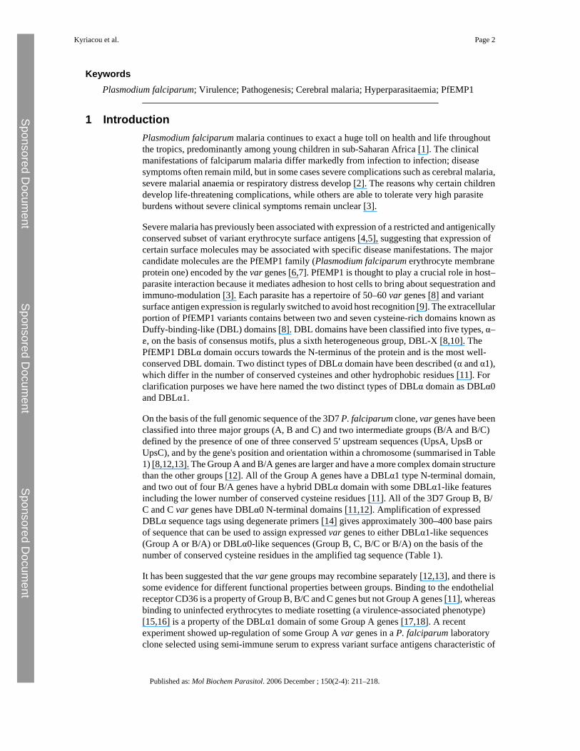
KeywordsPlasmodium falciparum; Virulence; Pathogenesis; Cerebral malaria; Hyperparasitaemia; PfEMP1
1 IntroductionPlasmodium falciparum malaria continues to exact a huge toll on health and life throughoutthe tropics, predominantly among young children in sub-Saharan Africa [1]. The clinicalmanifestations of falciparum malaria differ markedly from infection to infection; diseasesymptoms often remain mild, but in some cases severe complications such as cerebral malaria,severe malarial anaemia or respiratory distress develop [2]. The reasons why certain childrendevelop life-threatening complications, while others are able to tolerate very high parasiteburdens without severe clinical symptoms remain unclear [3].
Severe malaria has previously been associated with expression of a restricted and antigenicallyconserved subset of variant erythrocyte surface antigens [4,5], suggesting that expression ofcertain surface molecules may be associated with specific disease manifestations. The majorcandidate molecules are the PfEMP1 family (Plasmodium falciparum erythrocyte membraneprotein one) encoded by the var genes [6,7]. PfEMP1 is thought to play a crucial role in host–parasite interaction because it mediates adhesion to host cells to bring about sequestration andimmuno-modulation [3]. Each parasite has a repertoire of 50–60 var genes [8] and variantsurface antigen expression is regularly switched to avoid host recognition [9]. The extracellularportion of PfEMP1 variants contains between two and seven cysteine-rich domains known asDuffy-binding-like (DBL) domains [8]. DBL domains have been classified into five types, α–ɛ, on the basis of consensus motifs, plus a sixth heterogeneous group, DBL-X [8,10]. ThePfEMP1 DBLα domain occurs towards the N-terminus of the protein and is the most well-conserved DBL domain. Two distinct types of DBLα domain have been described (α and α1),which differ in the number of conserved cysteines and other hydrophobic residues [11]. Forclarification purposes we have here named the two distinct types of DBLα domain as DBLα0and DBLα1.
On the basis of the full genomic sequence of the 3D7 P. falciparum clone, var genes have beenclassified into three major groups (A, B and C) and two intermediate groups (B/A and B/C)defined by the presence of one of three conserved 5′ upstream sequences (UpsA, UpsB orUpsC), and by the gene's position and orientation within a chromosome (summarised in Table1) [8,12,13]. The Group A and B/A genes are larger and have a more complex domain structurethan the other groups [12]. All of the Group A genes have a DBLα1 type N-terminal domain,and two out of four B/A genes have a hybrid DBLα domain with some DBLα1-like featuresincluding the lower number of conserved cysteine residues [11]. All of the 3D7 Group B, B/C and C var genes have DBLα0 N-terminal domains [11,12]. Amplification of expressedDBLα sequence tags using degenerate primers [14] gives approximately 300–400 base pairsof sequence that can be used to assign expressed var genes to either DBLα1-like sequences(Group A or B/A) or DBLα0-like sequences (Group B, C, B/C or B/A) on the basis of thenumber of conserved cysteine residues in the amplified tag sequence (Table 1).
It has been suggested that the var gene groups may recombine separately [12,13], and there issome evidence for different functional properties between groups. Binding to the endothelialreceptor CD36 is a property of Group B, B/C and C genes but not Group A genes [11], whereasbinding to uninfected erythrocytes to mediate rosetting (a virulence-associated phenotype)[15,16] is a property of the DBLα1 domain of some Group A genes [17,18]. A recentexperiment showed up-regulation of some Group A var genes in a P. falciparum laboratoryclone selected using semi-immune serum to express variant surface antigens characteristic of
Kyriacou et al. Page 2
Published as: Mol Biochem Parasitol. 2006 December ; 150(2-4): 211–218.
Sponsored Docum
ent Sponsored D
ocument
Sponsored Docum
ent
severe malaria [19]. Taken together these studies raise the hypothesis that parasite virulenceand clinical disease severity in malaria patients could be related to transcription of particularvar gene groups. Recent studies on var gene transcription and malaria severity in clinicalisolates have, however, reached conflicting results [20–23]. This may be because severemalaria encompasses a variety of clinical syndromes (e.g. cerebral malaria, severe anaemia,respiratory distress, prostration) [2], and these syndromes may have different underlyingpathogenic mechanisms [3]. Significant associations between var gene groups and clinicaldisease may be masked unless strictly defined clinical groups are assessed along with suitablecontrol groups [24,25].
We aimed to investigate the hypothesis that parasite virulence and clinical disease severity inAfrican children are associated with the transcription of distinct subsets of P. falciparum vargenes. We examined P. falciparum isolates from patients with one of the commonest and mostvirulent forms of severe malaria, cerebral malaria (unrousable coma), compared to isolatesfrom controls with equally high parasite burdens but non-virulent disease (hyperparasitaemiapatients). Our data show that P. falciparum isolates from cerebral malaria patients aresignificantly more likely to transcribe var genes with DBLα1-like domains characteristic ofGroup A and B/A var genes than isolates from patients with non-severe hyperparasitaemia,which predominantly transcribe var genes with DBLα0-like domains.
2 Materials and methods2.1 Sample collection and RNA extraction
The samples were collected as part of the Bandiagara Malaria Project case-control study ofsevere malaria that has been described in detail previously [26,27]. Blood samples werecollected from children with malaria after informed consent from parents or guardians, and allprotocols received institutional review board approval. Blood samples were suspended inglycerolyte after lymphocyte separation via density centrifugation and frozen to −70 °C. Ablood spot from each isolate was made on 3 MM Whatman paper for genomic DNA extraction.Frozen samples were shipped to Edinburgh where they were thawed by standard methods andcultured for 8–24 h until the parasites were a mixture of late ring stage and early pigmented-trophozoite stage (morphologically equivalent to approximately 16–20 h post-invasion in alaboratory strain). This was done in order to study parasite stages in which the full lengthvar mRNA is present [28], while avoiding the early ring stage, when some authors claimmultiple non-specific var transcription occurs [29]. The cultured cells were solubilised in Trizoland stored at −70 °C. RNA was extracted as described [28].
2.2 DNA extraction and genotypingDNA was extracted from blood spots on filter paper using chelex-100 extraction [30]. Theminimum number of genotypes per isolate was estimated by genotyping PCR with primers toMSP1 and MSP2 [31].
2.3 cDNA preparationRNA was treated for 30 min at room temperature with 1.5 units DNAase (Gibco) to removeany contaminating genomic (g) DNA. cDNA was prepared using Superscript First StrandSynthesis System (Invitrogen) with random hexamers according to manufacturers instructions.
2.4 RT-PCRReverse-transcriptase (RT)-PCR was used to amplify a region of 300–400 bp of the varDBLα domain using unbiased degenerate primers αAF′ and αBR [14,21]. These primers arecapable of amplifying the entire var gene repertoire of 3D7 apart from the strain-transcending
Kyriacou et al. Page 3
Published as: Mol Biochem Parasitol. 2006 December ; 150(2-4): 211–218.
Sponsored Docum
ent Sponsored D
ocument
Sponsored Docum
ent
var2CSA gene implicated in malaria in pregnancy [32] and the type 3 var genes that are ofunknown function. Amplification conditions were as described [14] with a hot start (95 °C,5 min) followed by 35 cycles of 95 °C, 20 s; 42 °C, 20 s; 60 °C, 1 min. Samples without RTwere used in all reactions to exclude gDNA contamination.
2.5 PCR for upstream regionsUpstream PCRs were carried out on gDNA using upstream primers UpsA (5′-TAT TYH ATKTAT TAY ATT TGT TGT A) UpsB (5′-GTT AGA ACA TTT AAA ATT ATA) and UpsC(5′-AVA GAW ATA TGR TAG ATA YAG), based on sequences from 3D7, and a gene-specific DBLα primer for each isolate. Amplification conditions were 35 cycles of 94 °C, 5 s;46 °C, 15 s; 60 °C, 2 min. The upstream sequence of the predominant var gene from nineisolates was studied. The remaining upstream sequences were not studied due to lack of parasitematerial.
2.6 Cloning and sequencingPCR and RT-PCR products were run on agarose gels and extracted (Gel Extraction Kit,QIAGEN) then cloned (TA cloning kit, Invitrogen), and used to transform One Shot TOP10Fcompetent cells (Invitrogen). Transformed cells were grown overnight and individual whitecolonies were selected for culture. Plasmids were extracted from overnight cultures using aminiprep kit (QIAGEN), and were sequenced using BigDye terminator reaction mix (AppliedBiosciences).
2.7 Sequence analysisSequences were analysed using Lasergene software (DNASTAR Inc). Contigs were createdwith a minimum percentage match of 95% to classify sequences for each isolate. Amino acidsequences were aligned using MUSCLE [33] and the alignment converted into a nucleotidealignment with Tranalign (http://bioweb.pasteur.fr/docs/EMBOSS/tranalign.html)
2.8 Phylogenetic networkA phylogenetic network was generated rather than a traditional phylogenetic tree becausevar genes are subject to recombination. The network allows visualisation of ambiguous andconflicting signals in the dataset due to recombination or other factors [34]. Splits computedfrom the data are represented as parallel edges rather than single branches and the networkprovides an implicit representation of evolutionary history [34]. The phylogenetic network wasgenerated in SplitsTree 4.4 [34] using the Neighbor-Net distances transformation [35] andequal angle splits transformation [36].
3 ResultsTwenty-six P. falciparum field isolates were obtained from a vaccine trial site in Mali withintense seasonal transmission of P. falciparum (up to 20–60 infected bites per person per monthat the peak of the July–December transmission season [37]). Blood was obtained from childrenwith cerebral malaria (unrousable coma with a Blantyre coma score ≤2 and no other detectablecause of coma), non-severe hyperparasitaemia (>500, 000 parasites per microlitre of blood butwith no symptoms or signs of severe disease) or uncomplicated malaria (fever and P.falciparum parasitaemia with no evidence of severe malaria or hyperparasitaemia).Hyperparasitaemia is generally considered to be an indication of severe malaria using WorldHealth Organisation criteria [38]. However, African children with hyperparasitaemia and noother symptoms or signs of severe disease have a very low mortality rate, andhyperparasitaemia in sub-Saharan Africa can thus be considered as a form of non-severemalaria [2,26].
Kyriacou et al. Page 4
Published as: Mol Biochem Parasitol. 2006 December ; 150(2-4): 211–218.
Sponsored Docum
ent Sponsored D
ocument
Sponsored Docum
ent
There were no significant differences in patient age across the three clinical categories (Table2). The cerebral malaria and hyperparasitaemia patients did not differ significantly inparasitaemia, but the uncomplicated malaria patients had significantly lower parasitaemias thanthe other two groups (Table 2). We therefore considered the hyperparasitaemia isolates toprovide a better control group for the cerebral malaria isolates than the uncomplicated malariaisolates, because the major difference between the cerebral and hyperparasitaemia isolates isin parasite virulence and disease severity rather than parasite burden. Parasite rosette frequencywas significantly associated with disease severity, as expected in isolates from Africa (Table2) [15,16]. Multiple genotypes were present in most isolates (range 1–5), and there was nosignificant difference in the number of genotypes between the three clinical categories (Table2).
The transcribed DBLα var gene sequences from each isolate were amplified by reversetranscriptase-PCR using unbiased degenerate primers [14]. The RT-PCR products were clonedand 14–19 recombinant plasmids with var gene inserts were sequenced per isolate. Identicalsequences from the same isolate were defined as isolate sequence a, b, c etc. in decreasingorder of abundance (GenBank accession numbers DQ367086–DQ367226). The number ofdistinct DBLα var gene sequences detected per isolate varied between 1 and 14, and almostevery isolate showed a predominant gene (Fig. 1). Identical or almost identical sequences fromdifferent isolates were rare, indicating minimal overlap in var gene repertoires between isolates(see Supplementary material for further details). There was no significant difference in thenumber of distinct DBLα sequences per isolate detected in each clinical category (cerebralmalaria: median 5, range 1–9; hyperparasitaemia: median 6.5, range 2–14; uncomplicatedmalaria: median 4, range 2–10, P = 0.72, Kruskall–Wallis test). Further experiments to validatethe reproducibility of these data are shown in the Supplementary material (Fig. S1).
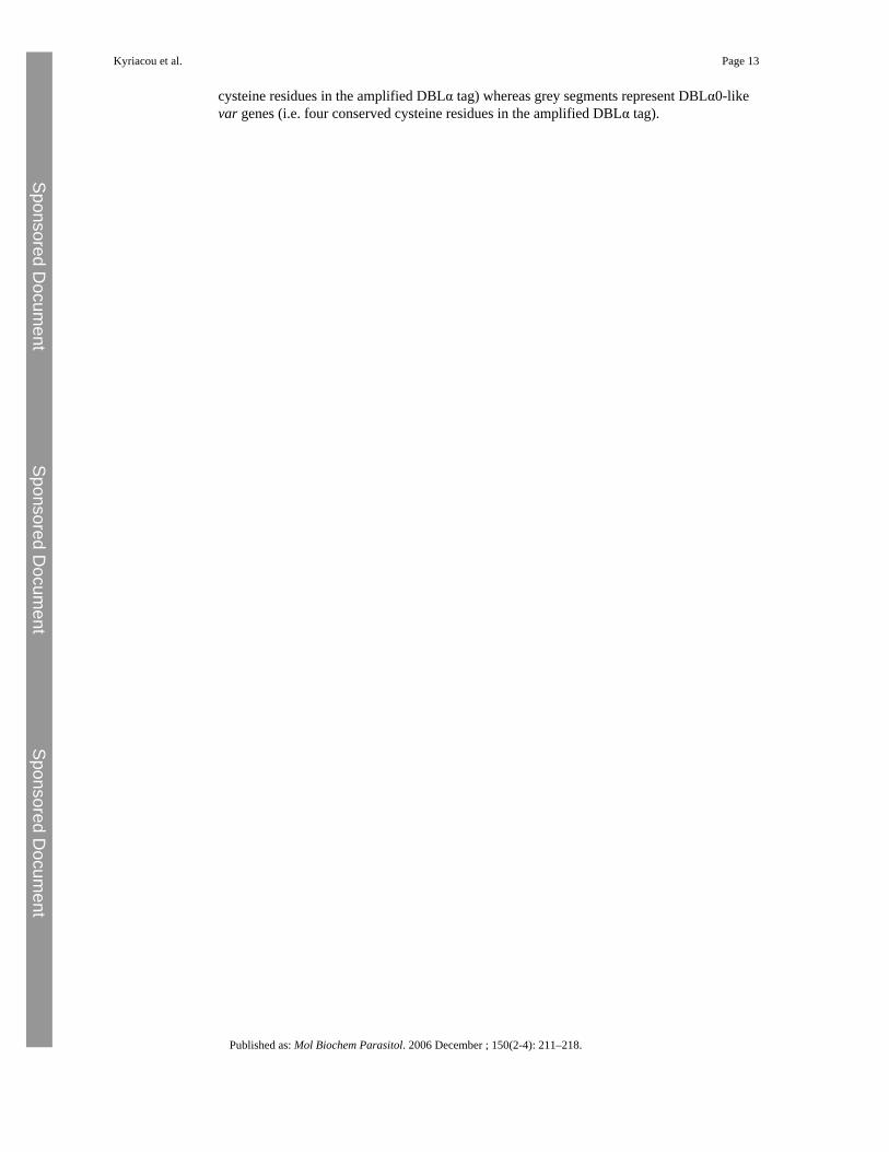
There was a highly significant difference between clinical categories in the percentage ofvar genes amplified that were DBLα1-like. 80.1% of the var gene sequences amplified fromthe cerebral malaria isolates were DBLα1-like, compared to only 25.7% of the sequences fromthe hyperparasitaemia isolates and 40.5% of the sequences from the uncomplicated malariapatients (P < 0.001, Chi-square test, Fig. 1). If only the predominant gene from each isolatewas examined, a marked difference remained between the cerebral malaria andhyperparasitaemia isolates (P = 0.013, Fishers Exact test, Fig. 2), but not between any othergroup (uncomplicated versus hyperparasitaemia, P = 0.294; uncomplicated versus cerebral,P = 0.131, Fishers Exact test). There was a significant positive correlation between theproportion of DBLα1-like sequences and the rosette frequency (Rho = 0.520, P = 0.009,Spearman Rank correlation).
The contrast between the var genes transcribed by the isolates from cerebral malaria patientscompared to hyperparasitaemia patients was also demonstrated by mapping clinical categoriesacross a phylogenetic network of DBLα sequence tags (Fig. 3). A phylogenetic network wasused rather than a traditional phylogenetic tree because var genes are subject to recombination,a process that can generate conflicting signals in tree-building and can be a significant sourceof error [34]. For reference, a selection of DBLα sequence tags from the three major GroupsA, B and C from the fully sequenced P. falciparum laboratory clone 3D7 [8] were included,plus four 3D7 genes with intermediate characteristics (B/A or B/C). In the phylogeneticnetwork the sequences fell into two major clades (Fig. 3). All of the sequences to the left ofthe dotted line are DBLα0-like (i.e. they contain four cysteine residues in the expressedsequence tag), whereas all of the sequences to the right of the dotted line are DBLα1-like(containing two cysteine residues in the expressed sequence tag) (Fig. 3). A phylogeneticnetwork containing the full repertoire of 3D7 var gene sequence tags plus the Malian sequenceswas also generated and gave the same separation into two distinct clades of DBLα0-like andDBLα1-like sequences (data not shown due to the complexity of the figure. This is available
Kyriacou et al. Page 5
Published as: Mol Biochem Parasitol. 2006 December ; 150(2-4): 211–218.
Sponsored Docum
ent Sponsored D
ocument
Sponsored Docum
ent

from the authors on request). Sequences from the cerebral malaria patients (red in Fig. 3) aresignificantly concentrated into the “DBLα1-like clade” (χ2 = 12.72 on 1 d.f., P < 0.001), whilesequences from the hyperparasitaemia patients (blue) are significantly concentrated in a“DBLα0-like clade” (χ2 = 5.6 on 1 d.f., P < 0.025). Sequences from the uncomplicated malariapatients (green) showed no bias in distribution between the two clades (Fig. 3).
Further separation of the DBLα expressed sequence tags into the six sequence groups recentlyproposed by Bull et al. [21] did not reveal any further differences between the three diseasecategories (see Supplementary material, Fig. S2). Investigation of the var gene upstreamregions for the predominant var gene from nine isolates showed that four field isolate var geneswith DBLα0-like domains had UpsB or UpsC upstream regions, whereas four out of five vargenes with DBLα1-like domains had UpsA-type upstream regions and the fifth (Hyp4A) hadan UpsB sequence linked to a hybrid DBLα0/α1 domain and was therefore a Group B/A vargene (Fig. 3).
4 DiscussionThe major finding of this study is that P. falciparum isolates from Malian children with cerebralmalaria (a virulent form of disease) predominantly transcribe var genes with DBLα1-likedomains that are characteristic of Group A or B/A var genes, whereas isolates from patientswith hyperparasitaemia (a non-virulent form of disease) predominantly transcribe var geneswith DBLα0-like domains. These results provide the first firm evidence from P. falciparumclinical isolates to support the hypothesis put forward previously that Group A var genes (whichall have DBLα1 domains) could be implicated in the pathogenesis of severe malaria in Africanchildren [19].
Previous studies on var gene transcription in clinical isolates have given conflicting results. Arole for var genes with DBLα1-like domains in the multi-organ failure type of severe malariathat occurs in adults in low malaria transmission settings was suggested by a study of 10Brazilian isolates [20]. A study in Papua New Guinea, however, showed upregulation of GroupB var genes in clinical disease (severe and uncomplicated) compared to asymptomaticinfections [22]. The “severe disease” patients in the Papua New Guinea study had a variety ofdifferent clinical syndromes, and strictly defined cerebral malaria (Blantyre coma score ≤2)was not included. A recent study from Tanzania also investigated a mixed group of severemalaria patients and found increased abundance of both Groups A and B var genes in severecompared to uncomplicated and asymptomatic malaria patients [23]. Analysis of a sub-groupof seven patients with neurological alterations (Blantyre coma score ≤3, therefore this groupdoes not fulfill a strict definition of cerebral malaria) did suggest a trend towards increasedabundance of Group A var genes, but this was not statistically significant [23]. A study ofsequence diversity in Kenyan field isolates did not find any association between var gene groupand disease manifestation, however only six isolates from assorted severe malaria patients werestudied, only one of whom had cerebral malaria [21]. As pointed out previously, severe malariais a heterogeneous group of syndromes that may have different underlying pathogenicmechanisms, therefore strict definitions of disease are essential in studies addressing malariapathogenesis [24]. Appropriate controls groups with equivalent parasite burdens are alsorequired [24,25], and the study reported here is the first to investigate var gene transcriptionusing isolates from non-severe hyperparasitaemia patients as a control group rather thanuncomplicated malaria patients with low parasite burdens. Although it remains possible thatthere are genuine geographical differences in the relationship between var gene transcriptionand disease severity, we suggest that the reason why a clear pattern emerged from the currentstudy, even though the sample numbers are not large, is because a strictly defined severe diseasesyndrome (cerebral malaria) was investigated and a non-severe malaria control group withequivalent parasite burdens (hyperparasitaemia) was used.
Kyriacou et al. Page 6
Published as: Mol Biochem Parasitol. 2006 December ; 150(2-4): 211–218.
Sponsored Docum
ent Sponsored D
ocument
Sponsored Docum
ent
We also investigated var gene transcription in parasite isolates from children withuncomplicated malaria and lower parasite burdens that are equivalent to the control groupsused in previous studies [20–23]. Isolates from these patients showed a mixed pattern of vargene transcription with four out of nine isolates transcribing mostly DBLα1-like var genes,while five out of nine isolates transcribed mostly DBLα0-like var genes (Figs. 1 and 3 and Fig.S2). This is consistent with the idea that non-hyperparasitaemic uncomplicated malariarepresents an earlier stage in the natural history or pathogenesis pathway of a malaria infection.At this stage, the host–parasite process has progressed beyond asymptomatic infection andproduced symptoms, but it may yet diverge along several possible pathways ranging fromspontaneous resolution to hyperparasitaemia, cerebral malaria, other severe syndromes, anddeath. Our data support the hypothesis that parasites expressing DBLα1-like var genesinfluence the direction of the disease process toward cerebral malaria, presumably increasingthe predilection for parasites to sequester and cause microvascular obstruction in the brain.Other factors are likely to influence this process including host genotype [39], and it is possiblethat parasite isolates transcribing “cerebral malaria-type” DBα1-like var genes may fail tocause severe disease in some children because of host genetic polymorphisms that can modifyparasite virulence phenotypes [40].
This study and previous similar investigations [20–23] use peripheral blood samples to examineP. falciparum var gene transcription profiles in clinical isolates. One unusual feature of malariainfection is that only immature ring stage parasites are found in the peripheral blood, whereasthe mature blood stage parasites (pigmented trophozoites and schizonts) are sequestered in themicrovasculature [3]. Therefore it is a major concern in studies of this type whether the parasitesin the peripheral blood adequately represent the sequestered population. Ideally studies aimedat examining the relationship between var gene transcription and particular disease syndromeswould examine sequestered parasites, however, these are only accessible in post-mortemsamples making such studies technically and ethically difficult. Two recent studies fromMalawi suggest that the dominant parasite genotypes of sequestered parasites are usually thesame as those in the peripheral blood [41], and that parasite genotypes in cerebral malariapatients are homogeneously distributed throughout the body (i.e. distinct genotypes are notsequestered in the brain compared to elsewhere in the body or the peripheral blood) [42].Although these studies do not shed light upon the question of whether the var gene profile ofperipheral blood parasites reflects that of sequestered ones, they are consistent with thepossibility that the peripheral blood population could adequately reflect the sequesteredparasite mass. Any difference in var gene transcription between peripheral and sequesteredparasite populations would be likely to confound a clear picture of the relationship betweenvar genes and disease syndromes. The fact that such a clear pattern of var gene transcriptionin isolates from cerebral malaria patients compared to hyperparasitaemia patients did emergefrom this study suggests that such a confounding effect is not a major problem.
Can the differing virulence-associations of the DBLα1-like var genes compared to theDBLα0-like var genes shown in this study be explained by the known functions of the differentvar gene groups? The only adhesion phenotype currently ascribed to the DBLα1 domain ofsome Group A or B/A var genes is binding to complement receptor 1 on uninfected erythrocytesto form rosettes ([17,18] and J.A. Rowe unpublished data). The field isolate data reported herealong with two previous studies [21,22] supports a link between the transcription of var geneswith DBLα1-like domains and high rosette frequencies. Rosetting is a parasite virulence-associated phenotype [15,16] that is thought to contribute to malaria pathogenesis by enhancingsequestration and microvascular obstruction [43]. The function of rosetting is unknown, but itmay be an immune evasion mechanism that promotes parasite growth and survival leading torapid high parasitaemia in vivo [44]. Whether var genes containing DBLα1-like domainsmediate other binding phenotypes, in addition to rosetting, that promote virulence is unknown,and is an important question requiring further research.
Kyriacou et al. Page 7
Published as: Mol Biochem Parasitol. 2006 December ; 150(2-4): 211–218.
Sponsored Docum
ent Sponsored D
ocument
Sponsored Docum
ent
Var genes encoding PfEMP1 variants with DBLα0 domains (i.e. Group B, C, B/C and someB/A var genes), on the other hand, have been shown to bind to the endothelial receptor CD36[11], and this interaction is believed to lead to the sequestration of infected erythrocytes in non-vital tissues [45] (away from brain endothelium, which does not express CD36 [46]). Ourfinding that var genes with DBLα0-like domains are predominantly transcribed in non-virulentinfections is consistent with this hypothesis. In addition, binding of infected erythrocytes toCD36 on dendritic cells leads to inhibition of dendritic cell maturation and impairment of hostimmune-responsiveness [47]. This immuno-modulatory effect of Group B and C var genescould contribute to the lower virulence of malaria in the hyperparasitaemia patients if, as hasbeen suggested previously, immuno-pathology contributes to the pathogenesis of severemalaria [48]. Parasites from hyperparasitaemia patients have also been shown to have lowerin vitro multiplication rates than parasites from patients in other disease categories [49], whichcould also contribute to the lower virulence of these infections.
In conclusion, we have shown that var genes with DBLα1-like domains (characteristic ofGroup A and some B/A var genes) are predominantly transcribed in P. falciparum isolatesfrom Malian cerebral malaria patients, whereas var genes with DBLα0-like domains arepredominantly transcribed in isolates from patients with a non-virulent form of disease(hyperparasitaemia). These findings suggest fundamental differences in the roles played by thedifferent var genes groups in host–parasite interaction that could be related to the contrastingcytoadhesive and immuno-modulatory functions of the var gene groups. Further research isrequired to examine the disease-associations, functions and diversity of var gene subsets indifferent geographical areas, and we emphasise the importance of strict clinical definitions andappropriate control groups in future work.
Appendix A Supplementary dataRefer to Web version on PubMed Central for supplementary material.
Appendix A Supplementary dataRefer to Web version on PubMed Central for supplementary material.
AcknowledgmentsWe are grateful to Sue Kyes, Andrew Read and Brian Charles worth for comments on the manuscript and to theBandiagara Malaria Project Team and the patients and their guardians for participation in the study. We are also gratefulto Martin Jones for bioinformatics advice. This work was supported by the Wellcome Trust (Ph.D. studentship toH.M.K. and Senior Research Fellowship to J.A.R., grant number 067431), the NERC (research grants to G.N.S., anda Ph.D. studentship to R.J.C.), the National Institutes of Health (contract no. N01-AI-85346) and the FogartyInternational Center (training grant no. D43TW001589).
References[1]. Breman J.G. The ears of the hippopotamus: manifestations, determinants, and estimates of the malaria
burden. Am J Trop Med Hyg 2001;64:1–11. [PubMed: 11425172][2]. Marsh K. Forster D. Waruiru C. Indicators of life-threatening malaria in African children. New Eng
J Med 1995;332:1399–1404. [PubMed: 7723795][3]. Miller L.H. Baruch D.I. Marsh K. Doumbo O.K. The pathogenic basis of malaria. Nature
2002;415:673–679. [PubMed: 11832955][4]. Bull P.C. Kortok M. Kai O. Plasmodium falciparum-infected erythrocytes: agglutination by diverse
Kenyan plasma is associated with severe disease and young host age. J Infect Dis 2000;182:252–259. [PubMed: 10882604]
Kyriacou et al. Page 8
Published as: Mol Biochem Parasitol. 2006 December ; 150(2-4): 211–218.
Sponsored Docum
ent Sponsored D
ocument
Sponsored Docum
ent
[5]. Nielsen M.A. Staalsoe T. Kurtzhals J.A. Plasmodium falciparum variant surface antigen expressionvaries between isolates causing severe and nonsevere malaria and is modified by acquired immunity.J Immunol 2002;168:3444–3450. [PubMed: 11907103]
[6]. Smith J.D. Chitnis C.E. Craig A.G. Switches in expression of Plasmodium falciparum var genescorrelate with changes in antigenic and cytoadherent phenotypes of infected erythrocytes. Cell1995;82:101–110. [PubMed: 7606775]
[7]. Su X.-Z. Heatwole V.M. Wertheimer S.P. A large and diverse family gene family (var) encodes 200–350kDa proteins implicated in the antigenic variation and cytoadherence of Plasmodiumfalciparum-infected erythocytes. Cell 1995;82:89–99. [PubMed: 7606788]
[8]. Gardner M.J. Hall N. Fung E. Genome sequence of the human malaria parasite Plasmodiumfalciparum. Nature 2002;419:498–511. [PubMed: 12368864]
[9]. Kyes S. Horrocks P. Newbold C. Antigenic variation at the infected red cell surface in malaria. AnnuRev Microbiol 2001;55:673–707. [PubMed: 11544371]
[10]. Smith J.D. Subramanian G. Gamain B. Baruch D.I. Miller L.H. Classification of adhesive domainsin the Plasmodium falciparum erythrocyte membrane protein 1 family. Mol Biochem Parasitol2000;10:293–310. [PubMed: 11071284]
[11]. Robinson B.A. Welch T.L. Smith J.D. Widespread functional specialization of Plasmodiumfalciparum erythrocyte membrane protein 1 family members to bind CD36 analysed across aparasite genome. Mol Microbiol 2003;47:1265–1278. [PubMed: 12603733]
[12]. Lavstsen T. Salanti A. Jensen A.T. Arnot D.E. Theander T.G. Sub-grouping of Plasmodiumfalciparum 3D7 var genes based on sequence analysis of coding and non-coding regions. Malar J2003;2:27. [PubMed: 14565852]
[13]. Kraemer S.M. Smith J.D. Evidence for the importance of genetic structuring to the structural andfunctional specialization of the Plasmodium falciparum var gene family. Mol Microbiol2003;50:1527–1538. [PubMed: 14651636]
[14]. Taylor H.M. Kyes S.A. Harris D. Kriek N. Newbold C.I. A study of var gene transcription in vitrousing universal var gene primers. Mol Biochem Parasitol 2000;105:13–23. [PubMed: 10613695]
[15]. Carlson J. Helmby H. Hill A.V. Brewster D. Greenwood B.M. Wahlgren M. Human cerebralmalaria: association with erythrocyte rosetting and lack of anti-rosetting antibodies. Lancet1990;336:1457–1460. [PubMed: 1979090]
[16]. Rowe A. Obeiro J. Newbold C.I. Marsh K. Plasmodium falciparum rosetting is associated withmalaria severity in Kenya. Infect Immun 1995;63:2323–2326. [PubMed: 7768616]
[17]. Rowe J.A. Moulds J.M. Newbold C.I. Miller L.H. P. falciparum rosetting mediated by a parasite-variant erythrocyte membrane protein and complement-receptor 1. Nature 1997;388:292–295.[PubMed: 9230440]
[18]. Russell C. Mercereau-Puijalon O. Le Scanf C. Steward M. Arnot D.E. Further definition of PfEMP-1DBL-1alpha domains mediating rosetting adhesion of Plasmodium falciparum. Mol BiochemParasitol 2005;144:109–113. [PubMed: 16122820]
[19]. Jensen A.T. Magistrado P. Sharp S. Plasmodium falciparum associated with severe childhoodmalaria preferentially expresses PfEMP1 encoded by group A var genes. J Exp Med2004;199:1179–1190. [PubMed: 15123742]
[20]. Kirchgatter K. Portillo Hdel A. Association of severe noncerebral Plasmodium falciparum malariain Brazil with expressed PfEMP1 DBL1 alpha sequences lacking cysteine residues. Mol Med2002;8:16–23. [PubMed: 11984002]
[21]. Bull P.C. Berriman M. Kyes S. Plasmodium falciparum variant surface antigen expression patternsduring malaria. PLoS Pathog 2005;l:e26. [PubMed: 16304608]
[22]. Kaestli M. Cockburn I.A. Cortes A. Baea K. Rowe J.A. Beck H.P. Virulence of malaria is associatedwith differential expression of Plasmodium falciparum var gene subgroups in a case-control study.J Infect Dis 2006;193:1567–1574. [PubMed: 16652286]
[23]. Rottmann M. Lavstsen T. Mugasa J.P. Differential expression of var gene groups is associated withmorbidity caused by Plasmodium falciparum infection in Tanzanian children. Infect Immun2006;74:3904–3911. [PubMed: 16790763]
[24]. Kurtzhals J.A. Goka B.Q. Akanmori B.D. Hviid L. The importance of strict patient definition instudies of malaria pathogenesis. Trends Parasitol 2001;17:313–314. [PubMed: 11446355]
Kyriacou et al. Page 9
Published as: Mol Biochem Parasitol. 2006 December ; 150(2-4): 211–218.
Sponsored Docum
ent Sponsored D
ocument
Sponsored Docum
ent
[25]. Nacher M. Singhasivanon P. Gay F. Silachamroon U. Looareesuwan S. Case-control studies onhost factors in severe malaria. Trends Parasitol 2001;17:253–254. [PubMed: 11378013]
[26]. Lyke K.E. Diallo D.A. Dicko A. Association of intraleukocytic Plasmodium falciparum malariapigment with disease severity, clinical manifestations, and prognosis in severe malaria. Am J TropMed Hyg 2003;69:253–259. [PubMed: 14628940]
[27]. Lyke K.E. Burges R. Cissoko Y. Serum levels of the proinflammatory cytokines interleukin-1 beta(IL-lbeta), IL-6, IL-8, IL-10, tumor necrosis factor alpha, and IL-12(p70) in Malian children withsevere Plasmodium falciparum malaria and matched uncomplicated malaria or healthy controls.Infect Immun 2004;72:5630–5637. [PubMed: 15385460]
[28]. Kyes S. Pinches R. Newbold C. A simple RNA analysis method shows var and rif multigene familyexpression patterns in Plasmodium falciparum. Mol Biochem Parasitol 2000;105:311–315.[PubMed: 10693754]
[29]. Scherf A. Hernandez-Rivas R. Buffet P. Antigenic variation in malaria: in situ switching, relaxedand mutually exclusive transcription of var genes during intra-erythrocytic development inPlasmodium falciparum. EMBO J 1998;17:5418–5426. [PubMed: 9736619]
[30]. Plowe C.V. Djimde A. Bouare M. Doumbo O. Wellems T.E. Pyrimethamine and proguanilresistance-conferring mutations in Plasmodium falciparum dihydrofolate reductase: polymerasechain reaction methods for surveillance in Africa. Am J Trop Med Hyg 1995;52:565–568. [PubMed:7611566]
[31]. Ranford-Cartwright L.C. Balfe P. Carter R. Walliker D. Frequency of cross-fertilization in thehuman malaria parasite Plasmodium falciparum. Parasitology 1993;107:11–18. [PubMed:8355994]
[32]. Salanti A. Dahlback M. Turner L. Evidence for the involvement of VAR2CSA in pregnancy-associated malaria. J Exp Med 2004;200:1197–1203. [PubMed: 15520249]
[33]. Edgar R.C. MUSCLE: multiple sequence alignment with high accuracy and high throughput.Nucleic Acids Res 2004;32:1792–1797. [PubMed: 15034147]
[34]. Huson D.H. Bryant D. Application of phylogenetic networks in evolutionary studies. Mol Biol Evol2006;23:254–267. [PubMed: 16221896]
[35]. Bryant D. Moulton V. Neighbor-net: an agglomerative method for the construction of phylogeneticnetworks. Mol Biol Evol 2004;21:255–265. [PubMed: 14660700]
[36]. Dress A.W.M. Huson D.H. Constructing splits graphs. IEEE Trans Comput Biol Bioinform2004;1:109–115.
[37]. Lyke K.E. Dicko A. Kone A. Incidence of severe Plasmodium falciparum malaria as a primaryendpoint for vaccine efficacy trials in Bandiagara, Mali. Vaccine 2004;22:3169–3174. [PubMed:15297070]
[38]. World Health Organization, Communicable Diseases Cluster. Severe falciparum malaria. Trans RSoc Trop Med Hyg 2000;94(Suppl. 1):S1–90.
[39]. Mackinnon M.J. Mwangi T.W. Snow R.W. Marsh K. Williams T.N. Heritability of malaria in Africa.PLoS Med 2005;2:e340. [PubMed: 16259530]
[40]. Cockburn I.A. Mackinnon M.J. O’Donnell A. A human complement receptor 1 polymorphism thatreduces Plasmodium falciparum rosetting confers protection against severe malaria. Proc Natl AcadSci USA 2004;101:272–277. [PubMed: 14694201]
[41]. Dembo E.G. Phiri H.T. Montgomery J. Molyneux M.E. Rogerson S.J. Are Plasmodium falciparumparasites present in peripheral blood genetically the same as those sequestered in the tissues? AmJ Trop Med Hyg 2006;74:730–732. [PubMed: 16687670]
[42]. Montgomery J. Milner D.A. Tse M.T. Genetic analysis of circulating and sequestered populationsof Plasmodium falciparum in fatal pediatric malaria. J Infect Dis 2006;194:115–122. [PubMed:16741890]
[43]. Kaul D.K. Roth E.F.J. Nagel R.L. Howard R.J. Handunnetti S.M. Rosetting of Plasmodiumfalciparum-infected red blood cells with uninfected red blood cells enhances microvascularobstruction under flow conditions. Blood 1991;78:812–819. [PubMed: 1859893]
[44]. Rowe J.A. Obiero J. Marsh K. Raza A. Positive correlation between rosetting and parasitemia inPlasmodium falciparum clinical isolates. Am J Trop Med Hyg 2002;66:458–460. [PubMed:12201576]
Kyriacou et al. Page 10
Published as: Mol Biochem Parasitol. 2006 December ; 150(2-4): 211–218.
Sponsored Docum
ent Sponsored D
ocument
Sponsored Docum
ent
[45]. Serghides L. Smith T.G. Patel S.N. Kain K.C. CD36 and malaria: friends or foes? Trends Parasitol2003;19:461–469. [PubMed: 14519584]
[46]. Turner G.D.H. Morrison H. Jones M. An immunohistochemical study of the pathology of fatalmalaria. Evidence for widespread endothelial activation and a potential role for intercellularadhesion molecule-1 in cerebral sequestration. Am J Pathol 1994;145:1057–1069. [PubMed:7526692]
[47]. Urban B.C. Ferguson D.J. Pain A. Plasmodium falciparum-infected erythrocytes modulate thematuration of dendritic cells. Nature 1999;400:73–77. [PubMed: 10403251]
[48]. Artavanis-Tsakonas K. Tongren J.E. Riley E.M. The war between the malaria parasite and theimmune system: immunity, immunoregulation and immunopathology. Clin Exp Immunol2003;133:145–152. [PubMed: 12869017]
[49]. Deans A.M. Lyke K.E. Thera M.A. Low multiplication rates of African Plasmodium falciparumisolates and lack of association of multiplication rate and red blood cell selectivity with malariavirulence. Am J Trop Med Hyg 2006;74:554–563. [PubMed: 16606983]
Kyriacou et al. Page 11
Published as: Mol Biochem Parasitol. 2006 December ; 150(2-4): 211–218.
Sponsored Docum
ent Sponsored D
ocument
Sponsored Docum
ent
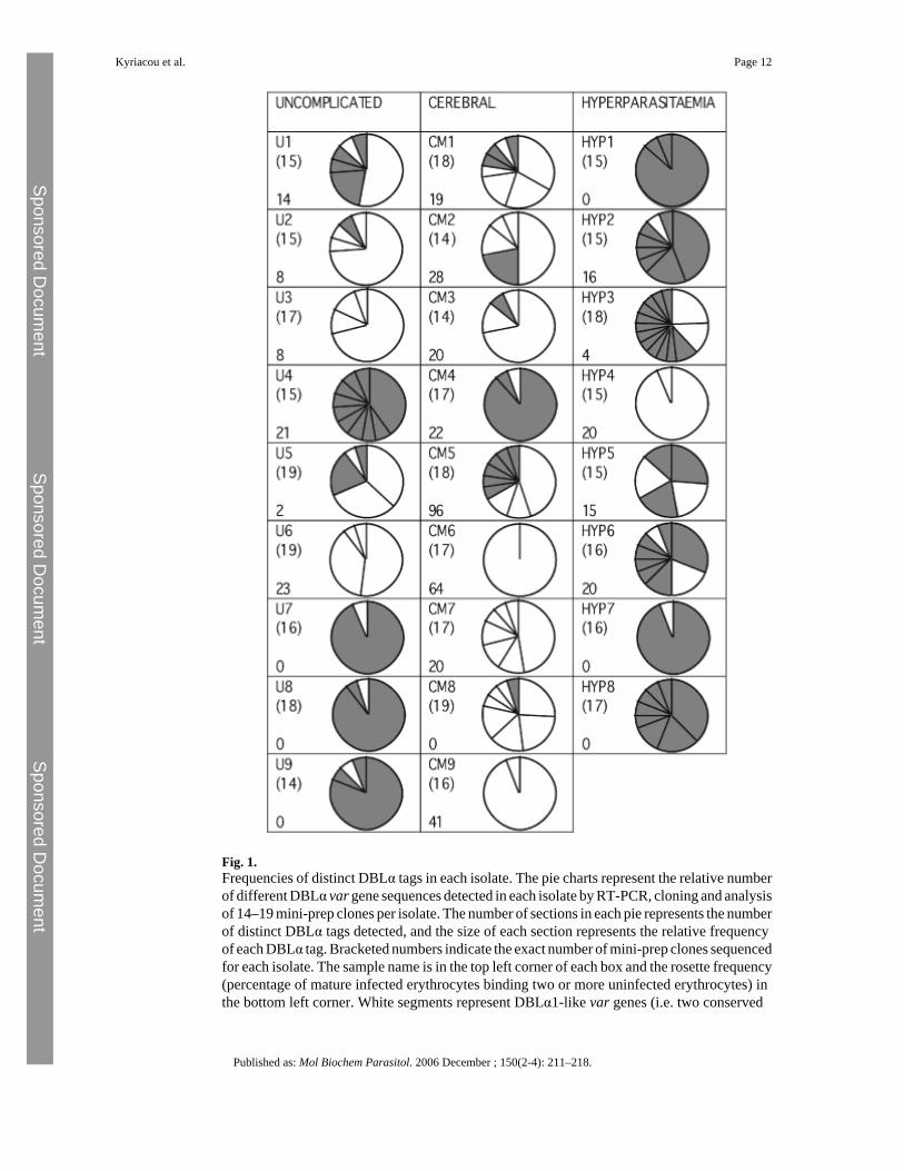
Fig. 1.Frequencies of distinct DBLα tags in each isolate. The pie charts represent the relative numberof different DBLα var gene sequences detected in each isolate by RT-PCR, cloning and analysisof 14–19 mini-prep clones per isolate. The number of sections in each pie represents the numberof distinct DBLα tags detected, and the size of each section represents the relative frequencyof each DBLα tag. Bracketed numbers indicate the exact number of mini-prep clones sequencedfor each isolate. The sample name is in the top left corner of each box and the rosette frequency(percentage of mature infected erythrocytes binding two or more uninfected erythrocytes) inthe bottom left corner. White segments represent DBLα1-like var genes (i.e. two conserved
Kyriacou et al. Page 12
Published as: Mol Biochem Parasitol. 2006 December ; 150(2-4): 211–218.
Sponsored Docum
ent Sponsored D
ocument
Sponsored Docum
ent
cysteine residues in the amplified DBLα tag) whereas grey segments represent DBLα0-likevar genes (i.e. four conserved cysteine residues in the amplified DBLα tag).
Kyriacou et al. Page 13
Published as: Mol Biochem Parasitol. 2006 December ; 150(2-4): 211–218.
Sponsored Docum
ent Sponsored D
ocument
Sponsored Docum
ent
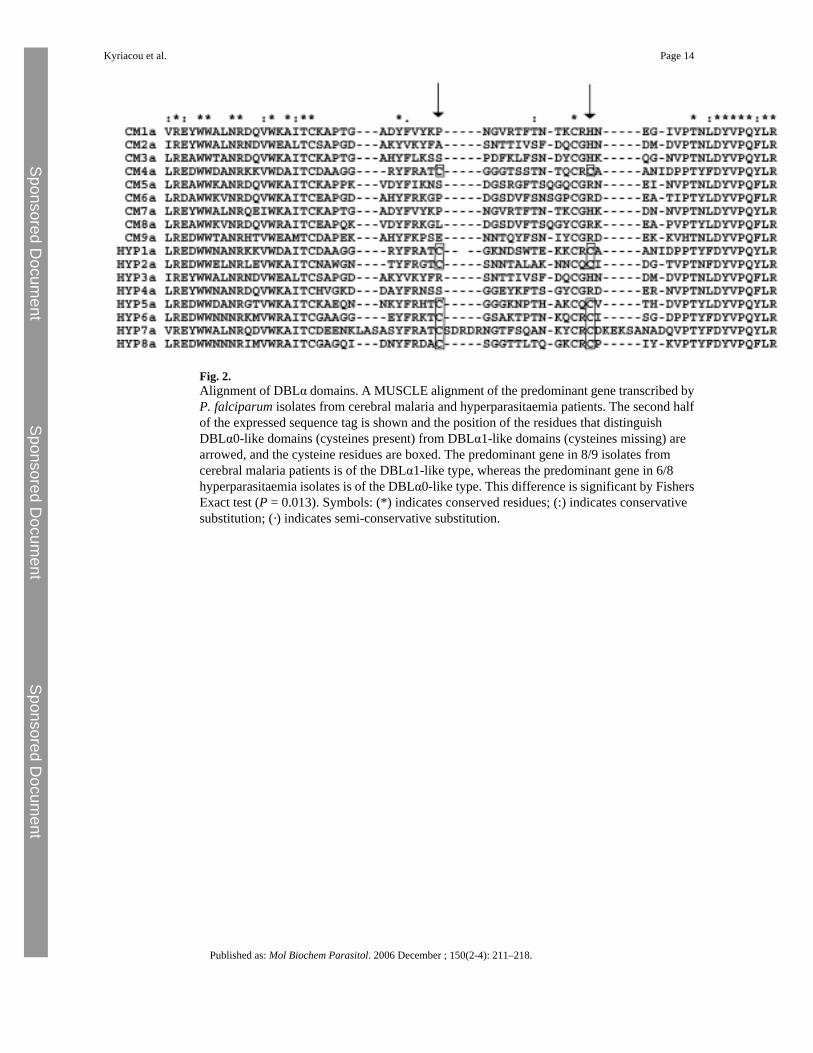
Fig. 2.Alignment of DBLα domains. A MUSCLE alignment of the predominant gene transcribed byP. falciparum isolates from cerebral malaria and hyperparasitaemia patients. The second halfof the expressed sequence tag is shown and the position of the residues that distinguishDBLα0-like domains (cysteines present) from DBLα1-like domains (cysteines missing) arearrowed, and the cysteine residues are boxed. The predominant gene in 8/9 isolates fromcerebral malaria patients is of the DBLα1-like type, whereas the predominant gene in 6/8hyperparasitaemia isolates is of the DBLα0-like type. This difference is significant by FishersExact test (P = 0.013). Symbols: (*) indicates conserved residues; (:) indicates conservativesubstitution; (·) indicates semi-conservative substitution.
Kyriacou et al. Page 14
Published as: Mol Biochem Parasitol. 2006 December ; 150(2-4): 211–218.
Sponsored Docum
ent Sponsored D
ocument
Sponsored Docum
ent
Fig. 3.Phylogenetic network showing relationships between var gene DBLα sequence tagstranscribed by P. falciparum isolates, generated using Neighbour-Net [35]. Sequencestranscribed by isolates from African children with cerebral malaria (CM, red),hyperparasitaemia (HYP, blue), and uncomplicated malaria (U, green) are compared to aselection of var genes from the laboratory clone 3D7. For the 3D7 genes, the gene name ispreceded by A, B, C, BA or BC to indicate the group to which the gene belongs. The sequencesfall into two major clades, with the DBLα0-like sequences to the left of the dotted line and theDBLα1-like sequences to the right. For the predominant gene from nine of the Malian isolates,the var gene upstream region was determined by PCR and is indicated as a boxed letter.
Kyriacou et al. Page 15
Published as: Mol Biochem Parasitol. 2006 December ; 150(2-4): 211–218.
Sponsored Docum
ent Sponsored D
ocument
Sponsored Docum
ent
Sponsored Docum
ent Sponsored D
ocument
Sponsored Docum
ent
Kyriacou et al. Page 16
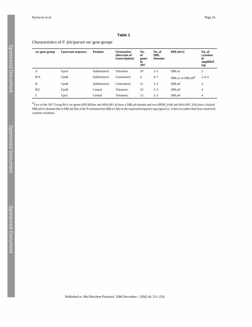
Table 1
Characteristics of P. falciparum var gene groups
var gene group Upstream sequence Position Orientation(direction oftranscription)
No.ofgenesin3D7
No. ofDBLdomains
DBLα0/α1 No. ofcysteinesinamplifiedtag
A UpsA Subtelomeric Telomeric 10 2–5 DBLα1 2
B/A UpsB Subtelomeric Centromeric 4 4–7 DBLα1 or DBLα0a 2 or 4
B UpsB Subtelomeric Centromeric 21 2–3 DBLα0 4
B/C UpsB Central Telomeric 10 2–3 DBLα0 4
C UpsC Central Telomeric 13 2–3 DBLα0 4
aTwo of the 3D7 Group B/A var genes (PFL0020w and MAL6P1.4) have a DBLα0 domain and two (PF08_0140 and MAL6P1.316) have a hybrid
DBLα0/α1 domain that is DBLα0-like at the N-terminus but DBLα1-like in the expressed sequence tag region (i.e. it has two rather than four conservedcysteine residues).
Published as: Mol Biochem Parasitol. 2006 December ; 150(2-4): 211–218.
Sponsored Docum
ent Sponsored D
ocument
Sponsored Docum
ent
Kyriacou et al. Page 17
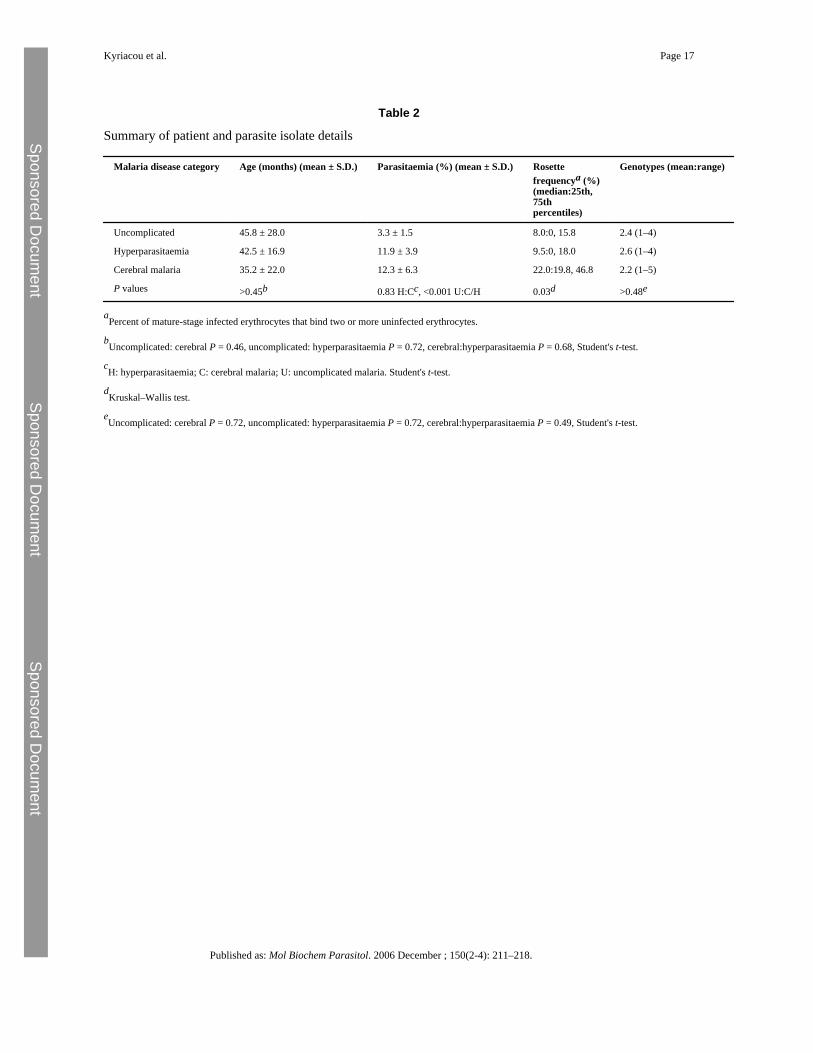
Table 2
Summary of patient and parasite isolate details
Malaria disease category Age (months) (mean ± S.D.) Parasitaemia (%) (mean ± S.D.) Rosettefrequencya (%)(median:25th,75thpercentiles)
Genotypes (mean:range)
Uncomplicated 45.8 ± 28.0 3.3 ± 1.5 8.0:0, 15.8 2.4 (1–4)
Hyperparasitaemia 42.5 ± 16.9 11.9 ± 3.9 9.5:0, 18.0 2.6 (1–4)
Cerebral malaria 35.2 ± 22.0 12.3 ± 6.3 22.0:19.8, 46.8 2.2 (1–5)
P values >0.45b 0.83 H:Cc, <0.001 U:C/H 0.03d >0.48e
aPercent of mature-stage infected erythrocytes that bind two or more uninfected erythrocytes.
bUncomplicated: cerebral P = 0.46, uncomplicated: hyperparasitaemia P = 0.72, cerebral:hyperparasitaemia P = 0.68, Student's t-test.
cH: hyperparasitaemia; C: cerebral malaria; U: uncomplicated malaria. Student's t-test.
dKruskal–Wallis test.
eUncomplicated: cerebral P = 0.72, uncomplicated: hyperparasitaemia P = 0.72, cerebral:hyperparasitaemia P = 0.49, Student's t-test.
Published as: Mol Biochem Parasitol. 2006 December ; 150(2-4): 211–218.