Differential Regulation of Estrogen Receptor Turnover and Transactivation by Mdm2 and...
10
2007;67:5513-5521. Cancer Res Vanessa Duong, Nathalie Boulle, Sylvain Daujat, et al. Transactivation by Mdm2 and Stress-Inducing Agents Turnover and α Differential Regulation of Estrogen Receptor Updated version http://cancerres.aacrjournals.org/content/67/11/5513 Access the most recent version of this article at: Cited Articles http://cancerres.aacrjournals.org/content/67/11/5513.full.html#ref-list-1 This article cites by 57 articles, 31 of which you can access for free at: Citing articles http://cancerres.aacrjournals.org/content/67/11/5513.full.html#related-urls This article has been cited by 8 HighWire-hosted articles. Access the articles at: E-mail alerts related to this article or journal. Sign up to receive free email-alerts Subscriptions Reprints and . [email protected] Department at To order reprints of this article or to subscribe to the journal, contact the AACR Publications Permissions . [email protected] Department at To request permission to re-use all or part of this article, contact the AACR Publications Research. on June 1, 2013. © 2007 American Association for Cancer cancerres.aacrjournals.org Downloaded from
Transcript of Differential Regulation of Estrogen Receptor Turnover and Transactivation by Mdm2 and...

2007;67:5513-5521. Cancer Res Vanessa Duong, Nathalie Boulle, Sylvain Daujat, et al. Transactivation by Mdm2 and Stress-Inducing Agents
Turnover andαDifferential Regulation of Estrogen Receptor
Updated version
http://cancerres.aacrjournals.org/content/67/11/5513
Access the most recent version of this article at:
Cited Articles
http://cancerres.aacrjournals.org/content/67/11/5513.full.html#ref-list-1
This article cites by 57 articles, 31 of which you can access for free at:
Citing articles
http://cancerres.aacrjournals.org/content/67/11/5513.full.html#related-urls
This article has been cited by 8 HighWire-hosted articles. Access the articles at:
E-mail alerts related to this article or journal.Sign up to receive free email-alerts
Subscriptions
Reprints and
To order reprints of this article or to subscribe to the journal, contact the AACR Publications
Permissions
To request permission to re-use all or part of this article, contact the AACR Publications
Research. on June 1, 2013. © 2007 American Association for Cancercancerres.aacrjournals.org Downloaded from

Differential Regulation of Estrogen Receptor A Turnover and
Transactivation by Mdm2 and Stress-Inducing Agents
Vanessa Duong,1,2
Nathalie Boulle,1,2
Sylvain Daujat,3Jerome Chauvet,
1,2
Sandrine Bonnet,1,2
Henry Neel,3and Vincent Cavailles
1,2
1Institut National de la Sante et de la Recherche Medicale U540; 2Universite Montpellier I; and 3Institut de GenetiqueMoleculaire de Montpellier, Montpellier, France
Abstract
In mammalian cells, the level of estrogen receptor A (ERA) israpidly decreased upon estrogen treatment, and this regula-tion involves proteasome degradation. Using differentapproaches, we showed that the Mdm2 oncogenic ubiquitin-ligase directly interacts with ERA in a ternary complex withp53 and is involved in the regulation of ERA turnover (bothin the absence or presence of estrogens). Several lines ofevidence indicated that this effect of Mdm2 required itsubiquitin-ligase activity and involved the ubiquitin/protea-some pathway. Moreover, in MCF-7 human breast cancer cells,various p53-inducing agents (such as UV irradiation) ortreatment with RITA (which inhibits the interaction of p53with Mdm2) stabilized ERA and abolished its 17B-estradiol–dependent turnover. Interestingly, our data indicated thatligand-dependent receptor turnover was not required forefficient transactivation. Altogether, our results indicate thatthe Mdm2 oncoprotein and stress-inducing agents complexlyand differentially regulate ERA stability and transcriptionalactivity in human cancer cells. [Cancer Res 2007;67(11):5513–21]
Introduction
Estrogens are key regulators of cell differentiation andproliferation, and these hormones play important roles in femalereproduction physiology and tissue homeostasis. They exert theirbiological action via specific receptors (ERa and ERh), which aremembers of a superfamily of hormone nuclear receptors, actingas gene regulatory transcription factors (1). Upon ligand binding,ERs regulate gene expression through the binding to their cognateestrogen response elements (ERE) or via protein-protein inter-actions with transcription factors such as activator protein-1 orspecificity protein-1. In the presence of hormone, ERs undergo amajor conformational change allowing the recruitment of tran-scriptional cofactor complexes, which in turn engage the basaltranscription machinery and/or act to locally modify chromatinstructure and subsequently stimulate expression of estrogen-responsive genes.
Liganded nuclear receptors recruit various types of enzymaticactivities that participate in gene expression regulation (2).Previous studies have suggested that ubiquitin-conjugating
enzymes or ubiquitin-protein ligases, such as UbcH5/UbcH7 (3, 4),RPF1/RSP5 (5), or E6-AP (6), interact with members of the nuclearreceptor superfamily and modulate their transactivation functions.Similarly, ATPase subunits of the proteasome complex, such asTRIP1/SUG1 (7) or TBP1 (8), also bind nuclear receptors andmodulate their functions.
More than 30 years ago, Jensen et al. have shown that 17h-estradiol (E2) treatment significantly reduces ERa levels in theuterus of ovariectomized rats (9). More recently, several studieshave shown that binding of E2 to ERa significantly decreases itsstability. This shorter half-life in the presence of hormone seemsto implicate the ubiquitin/proteasome pathway because ERa hasbeen shown to be ubiquitinated (10), and the ligand-dependentdown-regulation is blocked by proteasome inhibitors (3, 11, 12).Interestingly, although partial antiestrogens, such as tamoxifen,also increase ERa accumulation, pure antihormones, such as theICI 182,780 compound, strongly decrease receptor stability (10, 13).Several reports have suggested that the proteasome may controlnot only ERa protein levels but also hormone-dependent trans-cription (14). This effect is associated with the immobilization ofERa on the nuclear matrix as determined by fluorescence recoveryafter photobleaching (15). Although several candidates have beenproposed to account for the E2-dependent regulation of ERaexpression, the exact molecular mechanisms still remain unraveled.
The Mdm2 oncogene is overexpressed in a wide variety ofhuman cancers (16), and its role in tumorigenesis is linked to itsability to act as an E3 ubiquitin-ligase (17), which mediates theubiquitination and proteasome-dependent degradation of severalgrowth regulatory proteins including p53 (18, 19). Interestingly, ithas been previously suggested that Mdm2 could directly interactwith ERa (20, 21), and a positive effect of Mdm2 overexpression onERa activity has also been reported (20).
In the present study, we show that the Mdm2 oncoproteinis involved in both ligand-dependent and ligand-independentdecrease of ERa stability. Our data indicate that Mdm2 regulatesERa expression as a ternary complex with p53, and in support ofthis observation, we show that various stress-inducing agents(which stabilize p53) block E2-dependent regulation of ERastability in MCF-7 human breast cancer cells. Finally, this studyprovides several lines of evidence showing that the E2-dependentturnover of the receptor is not necessary for ERE-mediatedtransactivation.
Materials and Methods
Plasmids and reagents. The ERa expression vectors (wild type and
deletion mutants) were given by P. Chambon [Institut de Genetique et deBiologie Moleculaire et Cellulaire (IGBMC), Strasbourg, France]. The GST-
AF2wt vectors (22), the plasmids encoding Gal-GRIP1 and ER-VP16 (23),
and the ERE-hGlob-Luc and 17M5hGlob-Luc (24) reporter constructs were
Note: V. Duong was a recipient of fellowships from the French Minister of Researchand the Association pour la Recherche sur le Cancer.
Current address for S. Daujat: Max Planck Institute for Immunology, 79108Freiburg, Germany.
Requests for reprints: Vincent Cavailles, Institut National de la Sante et de laRecherche Medicale U540, 60 rue de Navacelles, Montpellier, F-34090 France. Phone:33-4-67-61-24-05; Fax: 33-4-67-61-37-87; E-mail: [email protected].
I2007 American Association for Cancer Research.doi:10.1158/0008-5472.CAN-07-0967
www.aacrjournals.org 5513 Cancer Res 2007; 67: (11). June 1, 2007
Research Article
Research. on June 1, 2013. © 2007 American Association for Cancercancerres.aacrjournals.org Downloaded from

described elsewhere. The GST-p53 and the pcDNA3 plasmids encoding full-length p53 (25) were obtained from U. Hibner [Institut de Genetique
Moleculaire de Montpellier (IGMM), Montpellier, France]. The pXJ-Mdm2
vector was obtained from B. Wasylyk (IGBMC), and the pcMI-Hdm2
contained the Mdm2 cDNA (BamHI/EcoRI fragment) subcloned into thepCMI1 vector (26). The pcMI-Hdm2DRING was obtained by inserting into
the pCMI1 vector a PCR-amplified BamHI/EcoRI fragment corresponding
to amino acids 1 to 435. The GST-Mdm2 was constructed by cloning a
XhoI/NotI fragment of the human Mdm2 cDNA into the pGEX4T3 vector(Amersham Biosciences). The Gal-Mdm2 was generated by introducing a
blunted NcoI/BglII fragment from pXJMdm2 into the SmaI site of the
pSG424 plasmid (27). The RITA compound (NSC652287) was obtained from
Dr. R.J. Schultz (NIH, Developmental Therapeutics Program, Drug Synthesisand Chemistry Branch, Rockville, MD).
Cell culture. Monolayer cell cultures (MCF-7, HeLa, and BG1) were
grown, respectively, in Ham’s F-12/Dulbecco’s modified Eagle’s medium(1:1, F12/DMEM) or DMEM alone supplemented with 10% FCS (Invitrogen)
and antibiotics. Before hormonal treatments, cells were stripped of endo-
genous estrogens as previously described (28). When indicated, cells were
irradiated using a Stratalinker UV cross-linker (model 1800) fromStratagene. The MELN cell line derived from MCF-7 cells stably transfected
with the ERE-hGlob-Luc-SVneo plasmid (29). Mouse embryo fibroblasts
(MEF) null for both p53 and Mdm2 (MEFdKO; ref. 30) were obtained from
G. Lozano (Houston, TX) and cultured in F12/DMEM.Transient transfection and luciferase assays. Cells were plated in six-
well plates (105 per well) 24 h before DNA transfection (4 Ag of total DNA)
by the calcium phosphate method using CMV-hGal expression vector as an
internal control. Luciferase values from transient transfection werenormalized by the h-galactosidase activities as described (28), and all data
were expressed as mean F SD.
Western blot analysis. Whole-cell extracts were prepared in high-salt
lysis buffer (HSB) containing 500 mmol/L NaCl, 50 mmol/L Tris (pH 8), 1%NP40, 1 mmol/L DTT, and proteases inhibitors (Roche Diagnostics).
Proteins were quantified using the Bradford assay (Bio-Rad Laboratories),
and 30 Ag were usually loaded on SDS-PAGE and transferred to poly-vinylidene difluoride membrane. Blots were saturated in TBST buffer
[50 mmol/L Tris (pH 7.5), 150 mmol/L NaCl, 0.1% Tween 20 (v/v), 5% nonfat
dehydrated milk (w/v)]; incubated with specific primary antibodies for ERa,
Figure 1. Interaction of ERa with p53 and Mdm2. A, GSTpull-down assays were carried out as described inMaterials and Methods using bacterially expressed GST,GST-Mdm2, or GST-LBD proteins to retain 35S-labeled ERaor Mdm2 in the presence of vehicle (C ), E2 (10�6 mol/L),or 4-hydroxytamoxifen (OHT ; 10�6 mol/L). B, schematicrepresentation of the ERa mutants. In vitro translatedfull-length ERa or various deletion mutants were analyzedfor their interaction with the GST-Mdm2 fusion protein.Inputs, 10% of the material used in the assay (as in A ).C, ERa and Mdm2 expression vectors were transfected intop53/Mdm2+/+ U2OS cells (lanes 1 and 2 ) or p53/Mdm2�/�
MEFs (MEFdKO; lanes 3 and 4). MCF-7 cells weretransfected with p53 and Mdm2 expression vectors(lanes 5–7 ). Whole-cell extracts were subjected toimmunoprecipitation as described in Materials and Methodsusing anti-Mdm2 antibody (lanes 2 and 4), anti-ERa(lane 6), or with an irrelevant antibody (lane 7). Westernblotting using either anti-Mdm2, anti-ERa, or anti-p53antibody was done as indicated. Whole-cell extracts(lanes 1, 3 , and 5 ). Inputs, 10% (left ) and 6% or 18%(respectively, for ERa and for p53/Mdm2; right ).D, mammalian two-hybrid assay was carried out usingE2-treated MCF-7 cells transfected with the 17M5-Lucreporter together with expressing vectors for Gal4,Gal-GRIP1, or Gal-Mdm2 with VP16 or ER-VP16, in thepresence or not of p53. Luciferase activity was expressedas % control in presence of Gal4. Columns, mean of threevalues; bars, SD. *, P < 0.05 (using Student’s t test).
Cancer Research
Cancer Res 2007; 67: (11). June 1, 2007 5514 www.aacrjournals.org
Research. on June 1, 2013. © 2007 American Association for Cancercancerres.aacrjournals.org Downloaded from
p53, or ubiquitin (Tebu), Mdm2 (clone 2A10; gift from J. Piette, IGMM),
pS2 (clone p2802; gift from M.C. Rio, IGBMC), or actin (Sigma); and probedwith the appropriate secondary antibody (Sigma). Detection was done using
the Chemiluminescence Reagent Plus kit (Perkin-Elmer Life Science).
Immunoprecipitation experiments. Transfected MCF-7 or HeLa cells
were lysed either in HSB for ubiquitination detection or in NETN buffer forcoimmunoprecipitation. Immunoprecipitations were carried out using anti-
ERa or anti-Mdm2 antibody (clone 4B11; gift from J. Piette) and protein
G+ agarose beads (Tebu). Beads were washed four times in lysis buffer andboiled in 2� SDS sample buffer.
GST pull-down assays. In vitro translation and glutathione S-transferase
(GST) pull-down assays were done as previously described (28). Protein
interactions were analyzed by SDS-PAGE followed by quantification using aPhosphorimager (Fujix BAS1000).
Apoptosis assay and quantitative PCR. These assays have been
described elsewhere (31).
Results
ERA is involved in a ternary complex with p53 and Mdm2.To better characterize protein-protein interactions between Mdm2and ERa, we first did GST pull-down experiments. As shown inFig. 1A (left), we observed a ligand-independent interaction ofin vitro translated ERa with Mdm2 expressed as a fusion proteinwith GST, whereas no detectable interaction was obtained withGST alone. The binding of Mdm2 to ERa was mediated by theligand-binding domain (LBD) of the receptor, as shown by therecruitment of in vitro expressed Mdm2 by the GST-LBD protein,which was also unaffected by E2 (right). Moreover, the use of aGST-LBD mutated in the conserved AF2 activation domainindicated that this interaction did not require an intact AF2transactivation domain (data not shown). The use of ERa deletionmutants (shown in Fig. 1B, left) confirmed that both the DBD(HE11 mutant) and the NH2-terminal region (HE19 mutant) werenot necessary for the binding to Mdm2 (Fig. 1B, right). By contrast,
our data confirmed that the COOH-terminal LBD of ERa wasrequired for the in vitro interaction because the HE15 mutantpoorly associated with GST-Mdm2. Altogether, these resultsshowed a direct ligand- and AF-2–independent interaction ofERa with Mdm2.
To confirm that these interactions also occurred in intact cells,we first set up coimmunoprecipitation experiments. As illustratedin Fig. 1C , ERa was found associated with immunoprecipitatedMdm2, as determined by probing the Western blot with a poly-clonal antibody specific for the receptor (lanes 1 and 2). Inter-estingly, when the same experiment was carried out in MEFdKOembryonic fibroblasts (32) derived from p53�/� and Mdm2�/�
mice (lanes 3 and 4), ERa was not coprecipitated with Mdm2,suggesting that p53 could be required for the interaction betweenERa and Mdm2. Indeed, the formation of a ternary complex amongERa, p53, and Mdm2 was further shown by coimmunoprecipitationof both p53 and Mdm2 with endogenous ERa from MCF-7 cells(Fig. 1C, right).
Moreover, the fact that ERa could be engaged in a ternarycomplex with p53 and Mdm2 was also confirmed using a modifiedmammalian two-hybrid system (Fig. 1D). An expression plasmidcoding for Mdm2 fused to the Gal4 DBD (Gal-Mdm2) wascotransfected in MCF-7 cells together with a Gal4-responsivereporter plasmid and an expression vector coding for ERa fused tothe VP16 activation domain or for VP16 alone. In our experimentalconditions, a slight but significant increase in luciferase activity(>2-fold versus 7-fold with Gal-GRIP used as a positive control)was obtained when ER-VP16 was coexpressed with Gal-Mdm2(compared with the activity obtained with VP16 alone). Interest-ingly, when a p53 expression plasmid was cotransfected withGal-Mdm2 and ER-VP16, we observed a significant increase inluciferase activity (>8-fold), suggesting that p53 indeed stabilizethe interaction between ERa and Mdm2. Altogether, these results
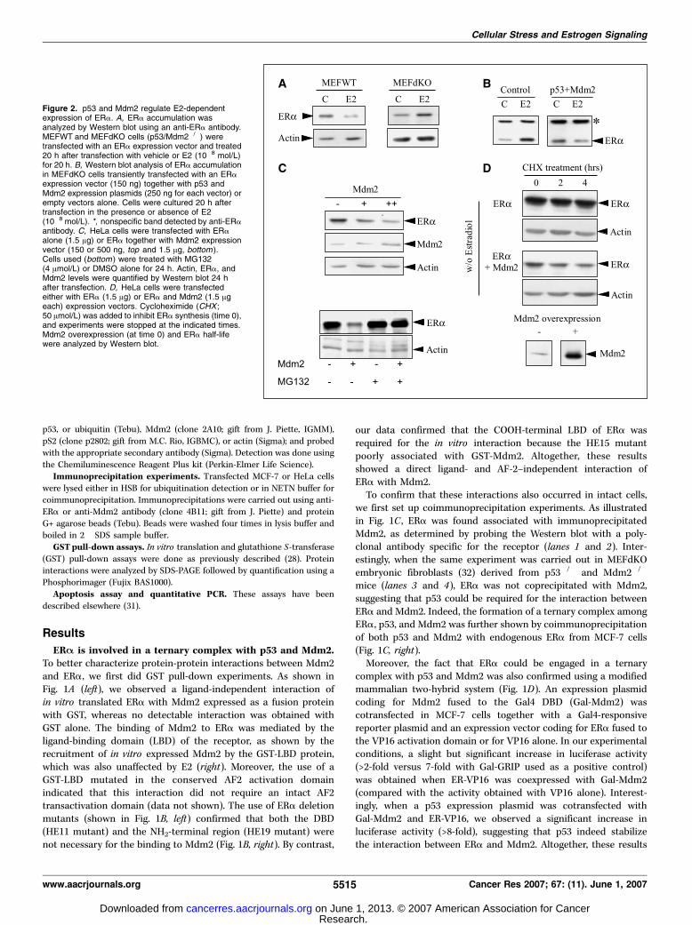
Figure 2. p53 and Mdm2 regulate E2-dependentexpression of ERa. A, ERa accumulation wasanalyzed by Western blot using an anti-ERa antibody.MEFWT and MEFdKO cells (p53/Mdm2�/�) weretransfected with an ERa expression vector and treated20 h after transfection with vehicle or E2 (10�8 mol/L)for 20 h. B, Western blot analysis of ERa accumulationin MEFdKO cells transiently transfected with an ERaexpression vector (150 ng) together with p53 andMdm2 expression plasmids (250 ng for each vector) orempty vectors alone. Cells were cultured 20 h aftertransfection in the presence or absence of E2(10�8 mol/L). *, nonspecific band detected by anti-ERaantibody. C, HeLa cells were transfected with ERaalone (1.5 Ag) or ERa together with Mdm2 expressionvector (150 or 500 ng, top and 1.5 Ag, bottom ).Cells used (bottom ) were treated with MG132(4 Amol/L) or DMSO alone for 24 h. Actin, ERa, andMdm2 levels were quantified by Western blot 24 hafter transfection. D, HeLa cells were transfectedeither with ERa (1.5 Ag) or ERa and Mdm2 (1.5 Ageach) expression vectors. Cycloheximide (CHX ;50 Amol/L) was added to inhibit ERa synthesis (time 0),and experiments were stopped at the indicated times.Mdm2 overexpression (at time 0) and ERa half-lifewere analyzed by Western blot.
Cellular Stress and Estrogen Signaling
www.aacrjournals.org 5515 Cancer Res 2007; 67: (11). June 1, 2007
Research. on June 1, 2013. © 2007 American Association for Cancercancerres.aacrjournals.org Downloaded from

suggest that ERa, p53, and Mdm2 coexist within the same proteincomplex in intact cells.p53 and Mdm2 are required for ligand-dependent ERA
turnover. To evaluate the role of Mdm2 and p53 in a ligand-dependent down-regulation of ERa, we first used the MEFdKOmodel (32). As shown in Fig. 2A , in this p53/Mdm2�/� background,estrogen treatment did not decrease ERa accumulation (asobserved in parallel in p53/Mdm2+/+ wild-type MEFs) but insteadslightly increased receptor levels. To show that the expression ofp53 and Mdm2 was required for the E2-dependent inhibition ofERa accumulation, we transiently transfected the expressionvectors for both p53 and Mdm2 together with the expressionvector for ERa in p53/Mdm2�/� MEFs (Fig. 2B). In this condition,we not only abolished the positive effect of E2 on ERa accu-mulation that we observed in control p53/Mdm2�/� cells, but wealso restored the negative hormonal regulation of ERa expression.Mdm2 regulates ligand-independent expression of ERA.
Because some of the data shown in Fig. 1 suggested that Mdm2could also bind ERa in an E2-independent manner, we investigatedthe effect of Mdm2 overexpression on ERa accumulation in theabsence of ligand. As shown in Fig. 2C (top), when increasingamounts of Mdm2 expression vectors were transiently cotrans-fected with ERa in untreated HeLa cells, the accumulation ofthe receptor was significantly decreased. This effect involved theubiquitin/proteasome pathway because the Mdm2-dependentdown-regulation of ERa was completely relieved by the proteasome
inhibitor MG132 (Fig. 2C, bottom). To emphasize the importance ofMdm2 in ligand-independent turnover of ERa, we measured theeffect of Mdm2 overexpression on the apparent stability of the ERaprotein by chase experiment in HeLa cells using cycloheximide asa protein synthesis inhibitor. As shown in Fig. 2D , we noticed asignificant decrease in ERa apparent stability in the presence ofMdm2. This effect of Mdm2 on ERa stability was also observed inthe presence of E2 (data not shown), thus emphasizing the role ofMdm2 in the E2-dependent and E2-independent posttranslationalregulation of ERa expression.Mechanisms involved in Mdm2 regulation of ERA stability.
To further decipher the molecular mechanisms involved in ERadegradation by Mdm2, we first used a mutant of Mdm2(Mdm2DRING) deleted in the COOH-terminal part of the protein,which contains the RING domain required for its ubiquitin-ligaseactivity (33). As shown in Fig. 3A , this mutant still interacted withERa in GST pull-down experiment. Interestingly, overexpression ofthe Mdm2DRING mutant did not decrease ERa accumulationcompared with the effect of its wild-type counterpart (Fig. 3B),suggesting that the E3 ubiquitin-ligase activity of Mdm2 is directlyinvolved in ERa degradation. The implication of the ubiquitin/proteasome pathway was further shown in ubiquitination assays.As shown in Fig. 3C , after immunoprecipitation of ERa, slowlymigrating forms of the receptor were detected both using anti-ERa(bottom) or anti-ubiquitin (top) antibodies, thus confirming that thelevel of ERa ubiquitination increased upon Mdm2 overexpression.
Figure 3. Mechanisms of Mdm2-dependentdegradation of ERa. A, a GST pull-down experimentwas done to determine the in vitro interaction between35S-labeled Mdm2 or Mdm2DRING (deleted of theCOOH-terminal region) with the GST-AF2 fusionprotein that contains the ligand-binding domain ofERa. B, HeLa cells were transfected either with theERa expression plasmid alone (1.5 Ag) or togetherwith Mdm2 or Mdm2DRING expression vector(1.5 Ag). The accumulation of ERa and Mdm2 proteins(wild type or Mdm2DRING) was analyzed byWestern blot. C, ERa expression vector (1.5 Ag)was transfected into HeLa cells with or without Mdm2(1.5 Ag). The ubiquitination status of ERa was thendetermined by immunoprecipitation of ERa fromwhole-cell extracts (as described in Materials andMethods) followed by Western blotting usinganti-ubiquitin antibody (top ) or with an ERa antibody(bottom ). D, HeLa cells were transiently transfectedwith ERa expression vector alone (500 ng; lanes 1, 2,4 , and 5 ) or together with the Mdm2 expressionvector (1 Ag; lanes 3 and 6). Cells were treated with E2(10�8 mol/L, lanes 2 and 5) or with vehicle alone(lanes 1, 3, 4 , and 6 ). The expression of ERa andMdm2 was analyzed by immunoblotting. The sameexperiment was done using the wild-type receptor orthe HE11 ERa mutant (deleted of the DBD).
Cancer Research
Cancer Res 2007; 67: (11). June 1, 2007 5516 www.aacrjournals.org
Research. on June 1, 2013. © 2007 American Association for Cancercancerres.aacrjournals.org Downloaded from

Finally, in an attempt to compare the E2-dependent turnover ofthe receptor with the ligand-independent degradation observedupon Mdm2 overexpression, we used a mutant of ERa deleted inthe central DNA-binding domain involved in the binding of p53(see Fig. 1B).4 This ERa-DDBD mutant (HE11) was no longerdegraded upon E2 stimulation (Fig. 3D, lane 5) in support withthe role of this domain in recruiting p53 and consistent with theformation of a ternary complex among the receptor, p53, andMdm2 (Fig. 1). By contrast, the ligand-independent increase in ERaturnover observed upon Mdm2 overexpression was comparable forthe wild-type protein and the DBD-deleted mutant (Fig. 3D, lane 3for ERa wild type and lane 6 for HE11). Altogether, these datasupport the hypothesis that different mechanisms might beinvolved in the regulation of ERa turnover upon E2 treatment orMdm2 overexpression.Stress-inducing agents block ligand-dependent turnover of
ERA. Previous studies reported that in MCF-7 human breastcancer cells, the E2-dependent decrease of ERa accumulation wasabolished by the MG132 proteasome inhibitor (3, 11, 12). Resultsshown in Fig. 4A confirmed that MG132 was able to reverse theeffect of various ligands, such as E2, estrone (E1), estriol (E3), ordiethylstilbestrol, on ERa levels in MCF-7 cells. Interestingly,proteasome blockade is a cellular stress that increased accumu-lation of p53 (Fig. 4A , compare right and left) and, consequently,induced apoptosis, as shown by quantification of cytoplasmicnucleosomes (Fig. 4B).
Based on our results concerning the role of Mdm2 in theregulation of ERa turnover, we thought that the effect of MG132
could be linked (at least in part) to its ability to dissociate Mdm2from p53 and/or ERa. We therefore analyzed the effect of otherstress-inducing agents (genotoxic or non-genotoxic) for their abilityto regulate E2-dependent turnover. As shown in Fig. 4B , both UVirradiation and inhibition of transcription (actinomycin D treat-ment) increased significantly programmed cell death in MCF-7cells. In all conditions, the accumulation of p53 was significantlyincreased to levels comparable with those obtained after MG132treatment and, very interestingly, both treatments that increasep53 levels concomitantly suppressed the hormone-dependentdown-regulation of ERa (Fig. 4C).
We then characterized the effect of UV irradiation on theregulation of ERa accumulation. We first showed that the blockadeof E2-dependent turnover after UV exposition was rapid sinceobserved 4 h after irradiation and still detectable 24 h later (datanot shown). We then investigated the effect of UV irradiation onthe response to antiestrogens, which have also been shown toregulate ERa expression. Indeed, as previously reported, incubationwith the partial antagonist 4-hydroxytamoxifen increased thelevel of ERa, whereas pure antiestrogens, such as the ICI 182,780molecule, negatively regulated the accumulation of the receptor(10). As shown in Fig. 4D , UV irradiation did not affect thestabilization of ERa upon 4-hydroxytamoxifen treatment. Veryinterestingly, it did not antagonize the effect of ICI 182,780, whereasit completely reversed the agonist-dependent decrease of ERalevels. This suggested that degradation of ERa by pure anti-estrogens involved different mechanisms than those required forhormone-dependent degradation.Concomitant stabilization of ERA and p53 by stress-
inducing agents. To confirm that UV irradiation modulated thestability of the ERa protein, we did chase experiments withcycloheximide. As shown in Fig. 5A and B , the stability of the4 Our unpublished results.
Figure 4. Effect of p53-inducing agents on ERasignaling. A, MCF-7 cells were treated for 20 h with10�7 mol/L E1, E2, E3, or diethylstilbestrol (DES ) in theabsence (control) or presence of MG132 (4 Amol/L).ERa and p53 protein levels were analyzed by Westernblot. B, MCF-7 cells were cultured with MG132(MG ; 4 Amol/L), actinomycin D (ActD ; 10 nmol/L), orexposed to UV (150 J/m2) for 20 h, and apoptosis wasmeasured by cytoplasmic nucleosomes quantification.Values were normalized by DNA quantification assay(measurement of DNA with DABA). C, MCF-7 cellswere irradiated (UV), treated with MG132 (4 Amol/L),actinomycin D (10 nmol/L), or untreated (control) inthe presence of vehicle or E2 (10�8 mol/L). Extractswere prepared and Western blotted with antibodies forERa and p53. D, MCF-7 cells were irradiated by UV ornot (control) and cultured in the presence of vehicle,E2 (10�8 mol/L), 4-hydroxytamoxifen (10�8 mol/L),or ICI 182,780 (ICI ; 10�8 mol/L) for 20 h. ERa and p53accumulation were analyzed by Western blot.
Cellular Stress and Estrogen Signaling
www.aacrjournals.org 5517 Cancer Res 2007; 67: (11). June 1, 2007
Research. on June 1, 2013. © 2007 American Association for Cancercancerres.aacrjournals.org Downloaded from

receptor in the absence of de novo protein synthesis decreasedupon E2 treatment, and UV irradiation completely inhibited thehormone-dependent degradation. As expected, upon UV treatment,p53 seemed very stable, and its accumulation was stronglyincreased (Fig. 5A). Altogether, these data suggest that stress-inducing agents induce a co-stabilization of p53 and ERa in MCF-7breast cancer cells. Our hypothesis was that disruption of the p53/Mdm2/ERa complex upon cellular stress was at the basis of theloss of E2-dependent turnover of the receptor.
To further emphasize this point, we used the NSC652287compound, also known as RITA (which stands for reactivation of
p53 and induction of tumor cell apoptosis) and recently describedas an inhibitor of the p53/Mdm2 interaction (34). As shown inFig. 5C , treatment of MCF-7 cells with RITA at 1 Amol/L signi-ficantly abolished the ligand-dependent decrease in ERa accumu-lation. This effect was detectable 4 h after the beginning of thetreatment and very interestingly, it correlated again with theincrease in p53 accumulation. As a control of UV irradiation andRITA treatment, we checked by quantitative reverse transcription-PCR that no up-regulation of the ERa mRNA was associated withthe blockade of the E2-dependent decrease observed at the proteinlevel (data not shown). Combined, these data therefore suggest thatdissociation of the p53/Mdm2/ERa complex upon cellular stressleads to a co-stabilization of p53 and ERa proteins.Dissociation between ligand-dependent ERA expression and
transactivation. Because previous studies proposed that the E2-dependent decrease of ERa accumulation was required for trans-criptional activity of the receptor (14), we analyzed whether thesetwo variables were indeed linked in our hands. We first investigatedthe ability of transiently transfected ERa to increase the trans-cription of a reporter gene in MEFs expressing or not p53/Mdm2.As shown in Fig. 6A , we found that ERa strongly activated trans-cription in cells where E2 up-regulated its accumulation (Control).Very interestingly, we found that overexpression of p53/Mdm2only slightly modify ERa transactivation although it completelyinversed the effect of E2 on its accumulation (see Fig. 2B).
We then analyzed the effect of UV irradiation on endogenousERa activity. To this aim, we used MELN cells that are MCF-7 cellsstably transfected with an E2-regulated luciferase reporter gene,allowing easy monitoring of endogenous ERa activity (24). MELNcells were treated with E2 4 h after irradiation by UV (i.e., whenE2-dependent turnover of ERa was no longer detectable; see Fig. 4).As shown in Fig. 6B , the transcriptional activity of ERa wassignificantly increased both in the absence of ligand or in thepresence of E2. In all conditions, the induction by E2 wasdetectable, confirming that E2-dependent turnover was not aprerequisite for transactivation even on endogenous ERa. More-over, the same results were obtained on the endogenous E2-regulated pS2 gene (Fig. 6C), thus confirming the dissociation ofstress-inducing agents on the accumulation of ERa and on itstransactivation.
Finally, we screened several ERa-expressing human cancer celllines for the negative E2-dependent regulation of ERa accumula-tion. As shown in Fig. 6D , we found that BG1 ovarian cancer cellsexhibited a strong positive regulation of ERa levels in response toE2 treatment, contrasting with the negative regulation observed inMCF-7 breast cancer cells (see Figs. 4 and 5). Of interest, when weanalyzed the transactivation properties of ERa in these two celllines, we found that at least on the reporter that we used intransient transfection, the respective endogenous receptors wereable to transactivate to similar extent (Fig. 6D). Altogether, ourresults strongly suggest that the effect of E2 on ERa turnover couldbe dissociated from its effect on transcriptional activation at leaston some target promoters.
Discussion
Modulation of ERa levels is a critical variable in determining thehormonal response of breast cancer cell proliferation. The controlof ERa expression is under a complex regulation that takes placeboth at the transcriptional and posttranscriptional levels. Previousdata suggested that ERa was down-regulated in the presence of E2
Figure 5. Effect of stress-inducing agents on ERa stability. A, the steady-statelevel of ERa was measured by pulse-chase assay with cycloheximide. MCF-7cells were irradiated with UV (bottom ) or not (top ) and treated concomitantly withE2 (10�8 mol/L) for 4 h. Cycloheximide was then added (time 0) during 2, 4,or 6 h. ERa stability was measured by Western blot. B, quantification of theexperiment in (A). The intensity of the bands corresponding to ERa levels wasdetermined by PCBAS imaging. Values were normalized by quantifying actinexpression on the same blot. Results are expressed as % control (time 0).C, kinetics of ERa and p53 accumulation by Western blot in stably transfectedMELN cells treated or not with E2 (10�8 mol/L) in presence or not of RITA(1 Amol/L) during 2, 4, or 8 h.
Cancer Research
Cancer Res 2007; 67: (11). June 1, 2007 5518 www.aacrjournals.org
Research. on June 1, 2013. © 2007 American Association for Cancercancerres.aacrjournals.org Downloaded from

through a proteasome-dependent mechanism (3, 11, 12). In thisstudy, we have investigated the mechanisms regulating thehormone-induced ERa turnover, and several lines of evidenceindicate that Mdm2, an oncogenic ubiquitin-ligase, plays animportant role in the regulation of ERa accumulation by E2.
First, using coimmunoprecipitation and a modified two-hybridassay, we show that Mdm2 interacted with ERa in a ternarycomplex with p53. The direct in vitro interaction between thereceptor and both Mdm2 and p53 was also shown using GSTpull-down assays. Our data suggested that Mdm2 binding involvedthe LBD of ERa but did not require the ligand-dependent AF2interface. Interestingly, p53 interacted with a different region ofthe receptor (i.e., the central region; data not shown), thus sup-porting the formation of a ternary complex.
The use of p53/Mdm2�/� cells showed that the two proteinswere required for the E2-dependent down-regulation of ERa.Moreover, we showed that the COOH-terminal region of Mdm2,which encompassed the E3 activity, was required for thedegradation of ERa and that overexpression of Mdm2 increasedERa ubiquitination. Previous studies highlighted the role of theMdm2 ubiquitin-ligase in the degradation of steroid hormonenuclear receptors. In the case of the glucocorticoid receptor (GR),Sengupta and Wasylyk showed that disruption of the p53/Mdm2interaction prevented ubiquitination of GR and that the ligand-dependent trimeric complex between GR, p53 and Mdm2 enhancedproteasomal degradation of the receptor (35). Moreover, the E3ligase activity of Mdm2 was also necessary for the ubiquitinationand degradation of the androgen receptor (36).
From the data presented in our study, we propose that p53 andMdm2 are involved in the ligand-dependent degradation of ERa.Interestingly, in support of our data, it has been recently shown, bychromatin immunoprecipitation, that Mdm2 was recruited on the
ERa-regulated pS2 promoter upon E2 stimulation (37, 38).Moreover, both Mdm2 and p53 expression levels are increased byE2 in MCF-7 breast cancer cells (39). This positive regulation couldbe of importance in the E2-dependent down-regulation of thereceptor. Obviously, other factors associated with p53 or Mdm2could be involved in such a degradation complex. A strongcandidate is the coactivator AIB1, which has been shown tointeract with p53 (40) and to be required for the E2-dependentturnover of ERa (41). The involvement of AIB1 could explain whythe AF2-mutated ERa is no longer degraded upon hormonestimulation.5 Interestingly, in breast cancer, AIB1 amplificationcorrelates with that of Mdm2 (42), and AIB1 expression is regulatedby ubiquitination (43).
Our results dealing with the effect of stress-inducing agents onERa expression clearly indicated that the increased turnover ofthe receptor in the presence of the pure antiestrogen ICI 182,780involves different mechanisms than those implicated in the E2-dependent degradation. Indeed, the effect of ICI182,780 on ERalevels were not abolished in UV-irradiated MCF-7 cells (Fig. 4D)and in MEFdKO (data not shown). Similar dissociation betweenthe effects of E2 and ICI 182,780 were previously reported (44), andother pathways involving, for example, the NEDD8 ubiquitin-likeprotein (45) or CSN5/Jab1 (46) could account for the degradation inthe presence of pure antihormones.
Previous data have highlighted the existence of a ligand-independent degradation of ERa and showed the role of CHIP(COOH terminus of Hsc70-interacting protein) in the ubiquitina-tion of misfolded ERa (47). More recently, E6-AP has also been
Figure 6. E2-dependent transactivation ofERa does not require receptor turnover.A, MEFdKO cells were transiently transfectedeither with the 17Eh-Luc reporter plasmid(300 ng) and ERa (100 ng) alone or togetherwith p53 and Mdm2 expression vectors (sametransfection experiment as for the Western blotanalysis in Fig. 2B). Cells were treated withvehicle or E2 (10�8 mol/L), and the luciferaseactivity was quantified as described inMaterials and Methods. Results wereexpressed relative to control in presence ofERa alone (% control). Columns, mean ofthree values; bars, SD. B, MCF-7 cells stablytransfected with ERE-hGlob-Luc (MELN cells)were treated with vehicle (control) or E2(10�8 mol/L) for 20 h, 4 h after irradiation (UV)or without being irradiated (C ). Luciferaseactivity was measured as described inMaterials and Methods. Columns, meanrelative activity (% E2 without irradiation) ofthree values; bars, SD. C, MCF-7 cells wereUV irradiated (150 J/m2) or not (control) in thepresence (E2, 10�8 mol/L) or in the absence(C ) of E2 for 20 h. pS2, p53, and actin proteinlevels were analyzed by Western blot. D, left,BG1 ovarian cancer cells were treated or not(C ) for 20 h with E2 (10�8 mol/L), and ERaprotein levels were analyzed by Western blot.Right, MCF-7 and BG1 cells were transientlytransfected with ERE-hGlob-Luc, treated withvehicle or E2 (10�8 mol/L), 4-hydroxytamoxifen(10�8 mol/L) or ICI 182,780 (10�8 mol/L) for20 h, and the luciferase activity was quantifiedand expressed as in (A ).
5 V. Duong, unpublished data.
Cellular Stress and Estrogen Signaling
www.aacrjournals.org 5519 Cancer Res 2007; 67: (11). June 1, 2007
Research. on June 1, 2013. © 2007 American Association for Cancercancerres.aacrjournals.org Downloaded from

shown to be involved in a calmodulin-dependent degradationpathway of ERa (48). Our data suggest that, when overexpressed,Mdm2 is also involved in the ligand-independent turnover ofERa. In these conditions, several evidences indicate that p53 couldbe dispensable for the effect of Mdm2, and this could reflect thefact that, upon overexpression of Mdm2, the binding equilibriumbetween ERa and Mdm2 could be strongly displaced towardscomplex formation, thus avoiding the requirement for p53 tostabilize the interaction. Finally, although we show that Mdm2increased ERa ubiquitination, further work will be required todefine which particular lysine residues are modified by Mdm2 inthe presence or absence of E2.
The present work also highlights the regulation of ERaexpression by cellular stress. We show that several stress-inducingagents that stabilize p53 also increase ERa levels and block itsE2-dependent down-regulation. The stabilization of p53 upontreatment with stress-inducing agents results from its dissociationfrom Mdm2 due to posttranslational modifications such as phos-phorylation (49). It is interesting to note that phosphorylation ofnuclear receptors has also been linked to their turnover (36, 50, 51).Concerning ERa, it has very recently been reported that S118 is anessential determinant of ERa degradation (38), and a previouswork has shown that extracellular signal-regulated kinase 7enhances the destruction of the receptor in a ligand-independentmanner (52). By contrast, another report suggests that in MCF-7cells, inhibition of mitogen-activated protein kinase results in anincreased degradation of ERa (53). Altogether, these data suggestthat phosphorylation is directly or indirectly involved in ERaturnover. Interestingly, we found that the degradation observedupon Mdm2 overexpression was no longer detected when wetested ERa mutated on the phosphorylated Ser118 residues (datanot shown).
Finally, although it has been suggested that the E2-dependentturnover of ERa was required for efficient transactivation of thereceptor (14), the data presented herein do not support such aconclusion. We first show that in p53/Mdm2�/� cells where E2treatment led to an increase in ERa accumulation, a stronghormone-dependent transactivation was observed. Interestingly,the same result was obtained with endogenous ERa in BG1 ovariancancer cells. In addition, in MCF-7 cells, the blockade of ERa
E2-dependent degradation upon UV irradiation (or MG132; datanot shown) did not abolish the transcriptional response to E2cells both on a stably transfected ERE-containing reporter geneand on the well-known pS2 target gene. Altogether, the presentwork dissociates the effect of E2 on ERa turnover from the effectof hormone on transactivation, thus supporting previous studiesdone using MG132, which suggested that the two events are notlinked (54).
In conclusion, we propose that within a same protein complex,ERa and p53 are both targeted by the Mdm2 E3 ubiquitin ligase;consequently, the stability of ERa and p53 is concomitantlyincreased in response to stress-inducing agents. This studytherefore emphasizes the relevance of protein-protein interactionsamong nuclear receptors, the Mdm2 oncogene, and the p53 tumorsuppressor. Several physiologic and pathologic consequences ofthese interactions have been proposed for the glucocorticoidreceptor (55). Our data showing the involvement of the Mdm2oncoprotein in the regulation of ligand-dependent and ligand-independent ERa expression are in accordance with a recent studythat reported that Mdm2 protein expression is a negative prog-nostic marker in breast carcinoma whose expression showed anegative correlation with ERa (56). Such an inverse associationmight seem surprising because this receptor is believed to mediateproliferative signaling of estrogens. However, our laboratory hasshown that in the absence of ligand, ERa also exerts anti-invasiveactivity in breast cancer cell lines (57), and this effect couldtherefore be lowered in tumor cells overexpressing Mdm2.Altogether, the present work highlights the role of Mdm2 onhormone signaling and will be at the basis of future investigationsto decipher its importance in various physiopathologic situations.
Acknowledgments
Received 7/28/2006; revised 3/13/2007; accepted 3/22/2007.Grant support: Institut National de la Sante et de la Recherche Medicale,
University of Montpellier I, Ligue Nationale contre le Cancer, and Association pour laRecherche sur le Cancer.
The costs of publication of this article were defrayed in part by the payment of pagecharges. This article must therefore be hereby marked advertisement in accordancewith 18 U.S.C. Section 1734 solely to indicate this fact.
We thank P. Chambon and R.J. Schultz for plasmids and reagents and Jean-MarcVanacker, Stephan Jalaguier, Patrick Augereau, Jean-Claude Nicolas, and Audrey Castetfor critical reading of the manuscript.
References1. Nilsson S, Makela S, Treuter E, et al. Mechanisms of
estrogen action. Physiol Rev 2001;81:1535–65.2. McKenna NJ, Xu J, Nawaz Z, et al. Nuclear receptor
coactivators: multiple enzymes, multiple complexes,multiple functions. J Steroid Biochem Mol Biol 1999;69:3–12.3. Nawaz Z, Lonard DM, Dennis AP, Smith CL, O’Malley
BW. Proteasome-dependent degradation of the humanestrogen receptor. Proc Natl Acad Sci U S A 1999;96:1858–62.4. Perissi V, Aggarwal A, Glass CK, Rose DW, Rosenfeld
MG. A corepressor/coactivator exchange complex re-quired for transcriptional activation by nuclear recep-tors and other regulated transcription factors. Cell 2004;116:511–26.5. Imhof MO, McDonnell DP. Yeast RSP5 and its
human homolog hRPF1 potentiate hormone-depen-dent activation of transcription by human progester-one and glucocorticoid receptors. Mol Cell Biol 1996;16:2594–605.6. Nawaz Z, Lonard DM, Smith CL, et al. The Angelman
syndrome-associated protein, E6-AP, is a coactivator for
the nuclear hormone receptor superfamily. Mol Cell Biol1999;19:1182–9.7. Lee JW, Ryan F, Swaffield JC, Johnston SA, Moore DD.
Interaction of thyroid-hormone receptor with a con-served transcriptional mediator. Nature 1995;374:91–4.8. Ishizuka T, Satoh T, Monden T, et al. Human
immunodeficiency virus type 1 Tat binding protein-1 isa transcriptional coactivator specific for TR. MolEndocrinol 2001;15:1329–43.9. Jensen EV, De Sombre ER. Oestrogen-receptor inter-
action in target tissues. Biochem J 1969;115:28–9P.10. Wijayaratne AL, McDonnell DP. The human estrogen
receptor-alpha is a ubiquitinated protein whose stabilityis affected differentially by agonists, antagonists, andselective estrogen receptor modulators. J Biol Chem2001;276:35684–92.11. Alarid ET, Bakopoulos N, Solodin N. Proteasome-
mediated proteolysis of estrogen receptor: a novelcomponent in autologous down-regulation. Mol Endo-crinol 1999;13:1522–34.12. El Khissiin A, Leclercq G. Implication of proteasome
in estrogen receptor degradation. FEBS Lett 1999;448:160–6.13. Dauvois S, Danielian PS, White R, Parker MG.
Antiestrogen ICI 164,384 reduces cellular estrogenreceptor content by increasing its turnover. Proc NatlAcad Sci U S A 1992;89:4037–41.14. Lonard DM, Nawaz Z, Smith CL, O’Malley BW.
The 26S proteasome is required for estrogen receptor-alpha and coactivator turnover and for efficientestrogen receptor-alpha transactivation. Mol Cell 2000;5:939–48.15. Stenoien DL, Patel K, Mancini MG, et al. FRAP
reveals that mobility of oestrogen receptor-alpha isligand- and proteasome-dependent. Nat Cell Biol 2001;3:15–23.16. Momand J, Jung D, Wilczynski S, Niland J. The MDM2
gene amplification database. Nucleic Acids Res 1998;26:3453–9.17. Honda R, Tanaka H, Yasuda H. Oncoprotein MDM2 is
a ubiquitin ligase E3 for tumor suppressor p53. FEBSLett 1997;420:25–7.18. Haupt Y, Maya R, Kazaz A, Oren M. Mdm2 promotes
the rapid degradation of p53. Nature 1997;387:296–9.19. Michael D, Oren M. The p53-2 module and the
ubiquitin system. Semin Cancer Biol 2003;13:49–58.20. Saji S, Okumura N, Eguchi H, et al. MDM2 enhances
the function of estrogen receptor alpha in human breast
Cancer Research
Cancer Res 2007; 67: (11). June 1, 2007 5520 www.aacrjournals.org
Research. on June 1, 2013. © 2007 American Association for Cancercancerres.aacrjournals.org Downloaded from

cancer cells. Biochem Biophys Res Commun 2001;281:259–65.21. Liu G, Schwartz JA, Brooks SC. Estrogen receptor
protects p53 from deactivation by human doubleminute-2. Cancer Res 2000;60:1810–4.22. Cavailles V, Dauvois S, Danielian PS, Parker MG.
Interaction of proteins with transcriptionally activeestrogen receptors. Proc Natl Acad Sci U S A 1994;91:10009–13.23. Teyssier C, Belguise K, Galtier F, Cavailles V, Chalbos
D. Receptor-interacting protein 140 binds c-Jun andinhibits estradiol-induced activator protein-1 activity byreversing glucocorticoid receptor-interacting protein 1effect. Mol Endocrinol 2003;17:287–99.24. Balaguer P, Francois F, Comunale F, et al. Reporter
cell lines to study the estrogenic effects of xenoestro-gens. Sci Total Environ 1999;233:47–56.25. Lassus P, Roux P, Zugasti O, et al. Extinction of rac1
and Cdc42Hs signalling defines a novel p53-dependentapoptotic pathway. Oncogene 2000;19:2377–85.26. Neel H, Weil D, Giansante C, Dautry F. In vivo
cooperation between introns during pre-mRNA process-ing. Genes Dev 1993;7:2194–205.27. Sadowski I, Ptashne M. A vector for expressing
GAL4(1-147) fusions in mammalian cells. Nucleic AcidsRes 1989;17:7539.28. Castet A, Boulahtouf A, Versini G, et al. Multiple
domains of the receptor-interacting protein 140 con-tribute to transcription inhibition. Nucleic Acids Res2004;32:1957–66.29. Balaguer P, Boussioux AM, Demirpence E, Nicolas JC.
Reporter cell lines are useful tools for monitoringbiological activity of nuclear receptor ligands. Lumines-cence 2001;16:153–8.30. McMasters KM, Montes de Oca LR, Pena JR, Lozano
G. mdm2 deletion does not alter growth characteristicsof p53-deficient embryo fibroblasts. Oncogene 1996;13:1731–6.31. Duong V, Licznar A, Margueron R, et al. ERalpha and
ERbeta expression and transcriptional activity aredifferentially regulated by HDAC inhibitors. Oncogene2006;25:1799–806.32. Montes de Oca LR, Wagner DS, Lozano G. Rescue of
early embryonic lethality in mdm2-deficient mice bydeletion of p53. Nature 1995;378:203–6.33. Honda R, Yasuda H. Activity of MDM2, a
ubiquitin ligase, toward p53 or itself is dependenton the RING finger domain of the ligase. Oncogene2000;19:1473–6.
34. Issaeva N, Bozko P, Enge M, et al. Small moleculeRITA binds to p53, blocks p53-HDM-2 interaction andactivates p53 function in tumors. Nat Med 2004;10:1321–8.35. Sengupta S, Wasylyk B. Ligand-dependent interaction
of the glucocorticoid receptor with p53 enhances theirdegradation by Hdm2. Genes Dev 2001;15:2367–80.36. Lin HK, Wang L, Hu YC, Altuwaijri S, Chang C.
Phosphorylation-dependent ubiquitylation and degra-dation of androgen receptor by Akt require Mdm2 E3ligase. EMBO J 2002;21:4037–48.37. Reid G, Hubner MR, Metivier R, et al. Cyclic,
proteasome-mediated turnover of unliganded andliganded ERalpha on responsive promoters is an integralfeature of estrogen signaling. Mol Cell 2003;11:695–707.38. Valley CC, Metivier R, Solodin NM, et al. Differential
regulation of estrogen-inducible proteolysis and tran-scription by the estrogen receptor alpha N terminus.Mol Cell Biol 2005;25:5417–28.39. Kinyamu HK, Archer TK. Estrogen receptor-depen-
dent proteasomal degradation of the glucocorticoidreceptor is coupled to an increase in mdm2 proteinexpression. Mol Cell Biol 2003;23:5867–81.40. Lee SK, Kim HJ, Kim JW, Lee JW. Steroid receptor
coactivator-1 and its family members differentiallyregulate transactivation by the tumor suppressorprotein p53. Mol Endocrinol 1999;13:1924–33.41. Shao W, Keeton EK, McDonnell DP, Brown M.
Coactivator AIB1 links estrogen receptor transcriptionalactivity and stability. Proc Natl Acad Sci U S A 2004;101:11599–604.42. Bautista S, Valles H, Walker RL, et al. In breast cancer,
amplification of the steroid receptor coactivator geneAIB1 is correlated with estrogen and progesteronereceptor positivity. Clin Cancer Res 1998;4:2925–9.43. Yan F, Gao X, Lonard DM, Nawaz Z. Specific
ubiquitin-conjugating enzymes promote degradation ofspecific nuclear receptor coactivators. Mol Endocrinol2003;17:1315–31.44. Alarid ET, Preisler-Mashek MT, Solodin NM. Thyroid
hormone is an inhibitor of estrogen-induced degrada-tion of estrogen receptor-alpha protein: estrogen-dependent proteolysis is not essential for receptortransactivation function in the pituitary. Endocrinology2003;144:3469–76.45. Fan M, Bigsby RM, Nephew KP. The NEDD8 pathway
is required for proteasome-mediated degradation ofhuman estrogen receptor (ER)-alpha and essential forthe antiproliferative activity of ICI 182,780 in ERalpha-
positive breast cancer cells. Mol Endocrinol 2003;17:356–65.46. Callige M, Kieffer I, Richard-Foy H. CSN5/Jab1 is
involved in ligand-dependent degradation of estrogenreceptor {alpha} by the proteasome. Mol Cell Biol 2005;25:4349–58.47. Tateishi Y, Kawabe Y, Chiba T, et al. Ligand-
dependent switching of ubiquitin-proteasome pathwaysfor estrogen receptor. EMBO J 2004;23:4813–23.48. Li L, Li Z, Howley PM, Sacks DB. E6AP and
calmodulin reciprocally regulate estrogen receptorstability. J Biol Chem 2006;281:1978–85.49. Meek DW. p53 Induction: phosphorylation sites
cooperate in regulating. Cancer Biol Ther 2002;1:284–6.50. Lange CA, Shen T, Horwitz KB. Phosphorylation of
human progesterone receptors at serine-294 by mito-gen-activated protein kinase signals their degradationby the 26S proteasome. Proc Natl Acad Sci U S A 2000;97:1032–7.51. Gianni M, Bauer A, Garattini E, Chambon P,
Rochette-Egly C. Phosphorylation by p38MAPK andrecruitment of SUG-1 are required for RA-induced RARgamma degradation and transactivation. EMBO J 2002;21:3760–9.52. Henrich LM, Smith JA, Kitt D, et al. Extracellular
signal-regulated kinase 7, a regulator of hormone-dependent estrogen receptor destruction. Mol Cell Biol2003;23:5979–88.53. Marsaud V, Gougelet A, Maillard S, Renoir JM.
Various phosphorylation pathways, depending on ago-nist and antagonist binding to endogenous estrogenreceptor alpha (ERalpha), differentially affect ERalphaextractability, proteasome-mediated stability, and tran-scriptional activity in human breast cancer cells. MolEndocrinol 2003;17:2013–27.54. Fan M, Nakshatri H, Nephew KP. Inhibiting protea-
somal proteolysis sustains estrogen receptor-alphaactivation. Mol Endocrinol 2004;18:2603–15.55. Sengupta S, Wasylyk B. Physiological and patholog-
ical consequences of the interactions of the p53 tumorsuppressor with the glucocorticoid, androgen, andestrogen receptors. Ann N Y Acad Sci 2004;1024:54–71.56. Turbin DA, Cheang MC, Bajdik CD, et al. MDM2
protein expression is a negative prognostic marker inbreast carcinoma. Mod Pathol 2006;19:69–74.57. Platet N, Cunat S, Chalbos D, Rochefort H, Garcia M.
Unliganded and liganded estrogen receptors protectagainst cancer invasion via different mechanisms. MolEndocrinol 2000;14:999–1009.
Cellular Stress and Estrogen Signaling
www.aacrjournals.org 5521 Cancer Res 2007; 67: (11). June 1, 2007
Research. on June 1, 2013. © 2007 American Association for Cancercancerres.aacrjournals.org Downloaded from