Ovulation-Inducing Fertility Treatments and the Risk of Breast ...
143
1 Ovulation-Inducing Fertility Treatments and the Risk of Breast Cancer Adriano Petrangelo Department of Medicine Division of Experimental Medicine McGill University Montréal, Québec, Canada April 2019 A thesis submitted to McGill University in partial fulfillment of the requirements of the degree of Master of Science in Experimental Medicine Copyright© Adriano Petrangelo, 2019
-
Upload
khangminh22 -
Category
Documents
-
view
0 -
download
0
Transcript of Ovulation-Inducing Fertility Treatments and the Risk of Breast ...

1
Ovulation-Inducing Fertility Treatments and the Risk of Breast Cancer
Adriano Petrangelo
Department of Medicine
Division of Experimental Medicine
McGill University
Montréal, Québec, Canada
April 2019
A thesis submitted to McGill University in partial fulfillment of the requirements of the
degree of Master of Science in Experimental Medicine
Copyright© Adriano Petrangelo, 2019

2
Table of Contents Abstract ........................................................................................................................................... 6
Résumé ............................................................................................................................................ 8
Contribution of Authors ................................................................................................................ 10
List of Abbreviations .................................................................................................................... 11
List of Tables and Figures............................................................................................................. 13
Chapter 1: Introduction ................................................................................................................. 16
Chapter 2: Hormonal Etiologies of Breast Cancer, Infertility, and Ovarian-Stimulating Fertility
Treatments..................................................................................................................................... 18
2.1: Breast Cancer: An Overview ............................................................................................. 18
2.2 Hormonal Etiologies of Breast Cancer................................................................................ 20
2.3 – The Role of Estrogen in the Development of Breast Cancer ........................................... 21
2.3.1 – Role of Reproductive Hormones in Normal Breast Development and Functioning . 21
2.3.2 Estrogen and Breast Cancer: Mechanism of Action ..................................................... 22
2.3.3 Estrogen and Breast Cancer: Observational Evidence ................................................. 25
Table 2.1: Hormonal Risk Factors for Breast Cancer According to the Collaborative
Group on Hormonal Factors in Breast Cancer ................................................................... 26
2.4: Infertility - Prevalence and Trends ..................................................................................... 28
2.5: Principles of Fertility Treatments....................................................................................... 30
2.5.1: Counselling and Investigation for Fertility Issues - Primary Care .............................. 30
2.5.2: Counselling and Investigation for Fertility Issues – Specialized Care ........................ 31
2.5.3: Treatment of Infertility – Specialized Care ................................................................. 34
Table 2.2: Treatment of Infertility According to Underlying Cause ................................. 35
2.6: Overview of Ovarian-Stimulating Fertility Treatments ..................................................... 38
2.6.1: Clomiphene.................................................................................................................. 38
2.6.2: Gonadotropins ............................................................................................................. 39
2.6.3: Hormone Levels during Ovarian Stimulation ............................................................. 44
2.7: Objectives and Rationale ................................................................................................... 46
Chapter 3: Systematic Review and Meta-Analysis ....................................................................... 49
Abstract ..................................................................................................................................... 51
3.1: Materials and Methods ....................................................................................................... 52
3.1.1: Criteria of Studies Selected ......................................................................................... 52
3.1.2 Exposures Assessed ...................................................................................................... 53

3
3.1.3: Objectives .................................................................................................................... 53
3.1.4: Search and Selection Methods ..................................................................................... 54
3.1.5: Data Extraction ............................................................................................................ 55
3.1.6 Statistical Analysis ....................................................................................................... 55
3.2: Results ................................................................................................................................ 56
3.2.1 Studies Identified .......................................................................................................... 58
3.2.2 Breast Cancer Risk Associated with Clomiphene Use ................................................. 59
3.2.3 Breast Cancer Risk Associated with Gonadotropin Use .............................................. 59
3.2.4: Breast Cancer Risk Associated with Clomiphene and Gonadotropin Use .................. 60
3.2.5: Subgroup Analysis: Length of Follow-Up Time ......................................................... 60
3.3: Discussion .......................................................................................................................... 62
Figure 3.1: Flow Chart – Study Selection .......................................................................... 67
Table 3.1: Characteristics of Included Cohort Studies ...................................................... 68
Table 3.2: Characteristics of the Included Case-Control Studies ...................................... 72
Figure 3.2: Forest plot and pooled RR – All cohort studies examining the association
between clomiphene and breast cancer included in the analysis ....................................... 73
Figure 3.3: Forest plot and pooled RR – All case-control studies examining the
association between clomiphene and breast cancer included in the analysis .................... 73
Figure 3.4: Forest plot and pooled RR – All cohort studies examining the association
between gonadotropins and breast cancer included in the analysis ................................... 74
Figure 3.5: Forest plot and pooled RR – All case-control studies examining the
association between gonadotropins and breast cancer included in the analysis ................ 74
Figure 3.6: Forest plot and pooled RR – All cohort studies examining the association
between clomiphene plus gonadotropins and breast cancer included in the analysis ........ 75
Figure 3.7: Forest plot and pooled RR – Cohort studies which conducted a stratified
analysis on follow-up time, measures of effect for subjects followed less than 10 years . 76
Figure 3.8: Forest plot and pooled RR – Cohort studies which conducted a stratified
analysis on follow-up time, measures of effect for subjects followed between 10 to 19
years ................................................................................................................................... 76
Figure 3.9: Forest plot and pooled RR – Cohort studies which conducted a stratified
analysis on follow-up time, measures of effect for subjects followed more than 20 years 77
Chapter 4: Case-Control Study ..................................................................................................... 78
Abstract ..................................................................................................................................... 80
4.1 Materials and Methods ........................................................................................................ 81
4.1.1: Study Population and Design ...................................................................................... 82

4
4.1.2: Case and Control Selection ......................................................................................... 83
4.1.3: Exposure Assessment .................................................................................................. 83
4.1.4: Statistical Analysis ...................................................................................................... 84
4.2: Results ................................................................................................................................ 87
4.4: Discussion .......................................................................................................................... 88
Table 4.1: Demographic, Clinical, and Lifestyle Characteristics of Breast Cancer Cases
and Matched Controls ........................................................................................................ 95
Table 4.2: Overall Association Between Ovarian-Stimulating Fertility Treatments and
Breast Cancer ..................................................................................................................... 97
Table 4.3: Association between Clomiphene and Breast Cancer - Stratified by Number of
Clomiphene Pills Prescribed .............................................................................................. 98
Table 4.4: Association Between Clomiphene and Breast Cancer – Stratified by Diagnoses
of PCOS ............................................................................................................................. 99
Chapter 5: General Discussion, Conclusions and Frameworks for Future Research ................. 100
5.1: General Discussion........................................................................................................... 100
5.2: Conclusion........................................................................................................................ 105
5.3: Framework for Future Research....................................................................................... 106
Appendix ..................................................................................................................................... 108
Search algorithm used to search the EMBED and MEDLINE databases ............................... 108
Exposure Codes ....................................................................................................................... 109
Table A.1: Codes used to identify exposure to clomiphene within the CPRD ................... 109
Table A.2: Codes used to identify exposure to IVF within the CPRD ................................ 109
Outcome Codes ....................................................................................................................... 110
Table A.3: Codes used to identify diagnoses of breast cancer within the CPRD ................ 110
Covariate Codes ...................................................................................................................... 111
A.4: BMI .............................................................................................................................. 111
Table A.4: BMI categories ............................................................................................... 111
Table A.5: Codes used to evaluate smoking status within the CPRD ................................. 111
Table A.6: Codes used to evaluate alcohol use within the CPRD ....................................... 112
Table A.7: Codes used to evaluate parity status within the CPRD ..................................... 112
Table A.8: Codes used to evaluate hormonal contraceptive use within the CPRD............. 118
Table A.9: Codes used to evaluate usage of hormone replacement therapy within the CPRD
............................................................................................................................................. 121
Table A.10: Codes used to identify oophorectomies within the CPRD .............................. 125

5
Table A.11: Codes used to identify diagnoses of polycystic ovary syndrome within the
CPRD ................................................................................................................................... 125
References…………………………………………………………………………………........126

6
Abstract Background and Objectives: The interplay between reproductive hormones and breast cancer
is well established. The safety of fertility treatments which stimulate follicular development has
been questioned as these therapies augment endogenous estrogen to supraphysiologic levels.
Two prominent methods of ovarian stimulation are clomiphene and exogenous gonadotropins.
The main goal of this manuscript was to evaluate the association between ovarian stimulation
and breast cancer. First, a systematic review and meta-analysis of the existing literature on this
topic was conducted. Second, an observational study using a case-control design was performed
using the Clinical Practice Research Datalink (CPRD).
Materials and Methods: The systematic review of the literature involved searching the
MEDLINE and EMBED databases to identify observational studies examining the relationship
of interest. Effect measures for studies compliant with inclusion and exclusion criteria were
extracted. The selected studies were stratified based on their study design and the type(s) of
exposure assessed. Pooling of study data was conducted in accordance with the DerSimonian and
Laird method. The case-control study involved identifying cases of breast cancer recorded in the
CPRD between 1995 and 2015. Each case of breast cancer was matched to 10 controls based on
age, general practice, and length of follow-up within the CPRD. Risk ratios were computed using
multivariate logistic regression. The regression model was adjusted for body mass index,
smoking, alcohol use, parity, hormonal contraceptive use, hormone replacement therapy use, and
oophorectomy. Exposure to either clomiphene or in vitro fertilization (IVF) was assessed for
both case and control subjects. The primary analysis characterized exposure on an “ever/never”
basis further stratifying for the age of breast cancer diagnosis. Two secondary analyses were
performed. The first involved evaluating the dose-response relationship between clomiphene and

7
breast cancer. The second involved examining whether the results of the primary analysis were
confounded by the underlying effects of infertility. This involved stratifying the study population
on previous diagnoses of polycystic ovary syndrome (PCOS).
Results: The meta-analysis included 17 cohort studies and 4 case-control studies. Pooled RRs
found no association between ovarian stimulation and breast cancer, regardless of the study
design or the type of exposure. A secondary analysis stratifying on subject follow-up time
showed that breast cancer risk in patients having undergone ovarian stimulation did not increase
even with extended periods of follow-up. The case-control study showed conflicting results.
Exposure to clomiphene (RR 1.32, 95% CI [1.23-1.42]) and to IVF (RR 1.55, 95% CI [1.42-
1.69]) was associated with an increased risk of breast cancer, following adjustment. After
stratification for age of diagnosis, IVF remained significantly associated with both pre- and post-
menopausal malignancies whereas clomiphene remained solely associated with pre-menopausal
breast cancers. The association between clomiphene and breast cancer was not significant in the
population of women previously diagnosed with PCOS (1.22 [0.99-1.50]), indicating the results
of the primary analysis may have been confounded by the underlying effects of infertility.
Conclusion: Presently, there is no definitive link between ovarian stimulation and breast cancer.
While the results of the case-control study indicated the association was significant, the strong
possibility that infertility and its associated conditions confounded results suggests that these
findings should be interpreted with caution. Furthermore, the existing literature refutes this
association. Nevertheless, continued monitoring of this relationship is warranted given its
biological plausibility and the increasing use of these treatments across the developed world.

8
Résumé Contexte et objectifs : Il existe une relation bien établie entre le taux d’hormones reproductrices
et le cancer du sein. L’innocuité des traitements de fertilité qui stimulent le développement
folliculaire au sein des ovaires, engendrant une hausse supra-physiologique des taux
d’œstrogènes endogènes, est remise en cause. L’administration de clomifène et de
gonadotrophines exogènes en sont les deux formes de traitement les plus rependues. L’objectif
principal de cette recherche était d’évaluer le lien entre ces traitements de fertilité et le cancer du
sein. Dans un premier temps, une étude systématique et une méta-analyse des études sur ce sujet
ont été menées. Dans un second temps, une étude observationnelle se basant sur une conception
d’étude cas-témoins a été effectuée à l’aide du Clinical Practice Research Datalink (CPRD).
Documentations et méthodes : Les bases de données MEDLINE et EMBED ont été explorées
afin d’identifier les études observationnelles traitant du sujet. Les résultats des études conformes
aux critères d’inclusion et d’exclusion ont été soutirés. Les études sélectionnées ont été stratifiées
en se basant sur le modèle d’étude et le type d’exposition évalués. Le regroupement des données
des études a été mené conformément à la méthode de DerSimonian et Laird. L’étude cas-témoins
a nécessité l’identification des cas de cancer du sein enregistrés dans le CPRD entre 1995 et
2015. Dix cas-témoins ont été appariés à chaque cas de cancer du sein en fonction de
l’ancienneté et de la durée du suivi au sein du CPRD. Les taux de risques ont été calculés en
utilisant une régression logistique multivariée, prenant en compte l’indice de masse corporelle, le
tabagisme, la consommation d’alcool, la parité, l’utilisation de contraceptifs hormonaux, la
thérapie de substitution hormonale, et l’ovariectomie. Les cas et les témoins ont été évalués en
fonction de leur exposition soit au clomifène soit à la FIV. L’analyse primaire a caractérisé
l’exposition sur la base du critère « a été exposé/n’a jamais été exposé », la stratifiant ensuite en
fonction de l’ancienneté du diagnostic du cancer du sein. Deux analyses secondaires ont été

9
réalisées. La première a consisté à apprécier la relation dose-réponse entre le clomifène et le
cancer du sein. La deuxième a consisté à évaluer si les résultats de l’analyse primaire ont pu être
faussés par les effets de l’infertilité sous-jacente en stratifiant la population étudiée sur des
diagnostics de syndrome des ovaires polykystiques (SOPK).
Résultats : La méta-analyse a inclus 17 études de cohortes et 4 cas-témoins. Le regroupement
des RR n’a montré aucune corrélation entre stimulation ovarienne et cancer du sein, quels que
soient le modèle d’étude et le type d’exposition évalués. Une analyse secondaire après
stratification sur la durée du suivi n’a révélé aucun lien significatif. Les études cas-témoins ont
démontré des résultats conflictuels. L’exposition au clomifène (RR 1.32, 95% IC [1.23-1.42]) ou
à la FIV (RR 1.55, 95% IC [1.42-1.69]) était associée à un risque accru de cancer du sein après
ajustement. Après une stratification par ancienneté du diagnostic, la FIV est restée
significativement associée aux tumeurs malignes aussi bien post- que pré- ménopause tandis que
le clomifène est resté associé seulement aux cancers du sein postménopause. Le lien entre
clomifène et cancer du sein n’était pas significatif dans la population des femmes préalablement
diagnostiquées avec un SOPK (1.22 [0.99-1.50]), indiquant que les résultats de l’analyse
primaire ont pu être faussés par les effets sous-jacents à l’infertilité.
Conclusion : Actuellement, il n’existe pas de lien établi entre la stimulation ovarienne et le
cancer du sein. Les résultats de l’étude cas-témoins ont démontré une association possible
significative, cependant la forte possibilité que les effets de l’infertilité aient confondu les
résultats nécessite d’interpréter ces résultats avec prudence. En outre, la littérature existante
réfute généralement cette association. Néanmoins, une surveillance continue de ce lien s’impose
compte tenu de sa plausibilité biologique et de la popularité croissante de ces traitements.

10
Contribution of Authors
Adriano Petrangelo is the primary author of this thesis under the supervision and guidance of Dr.
Haim A. Abenhaim.
The study concept and design with planned by both Adriano Petrangelo and Dr. Haim A.
Abenhaim, along with the advice of members of the thesis committee, which included Dr.
Laurent Azoulay and Dr. Togas Tulandi.
Adriano Petrangelo conducted all analyses for the meta-analysis. The manuscript for the
systematic review and meta-analysis was drafted by Adriano Petrangelo.
Analyses for the case-control study were performed through the joint effort of Adriano
Petrangelo and Nicholas Czuzoj-Shulman. Adriano Petrangelo drafted the manuscript to this
study. Adriano Petrangelo also drafted this study’s protocol, which was reviewed and approved
by the Independent Scientific Advisory Committee, the regulatory body granting access to the
Clinical Practice Research Datalink.
All other sections of the thesis, including the Literature Review, Discussion, Conclusion, and
Frameworks for Future Research were authored by Adriano Petrangelo.
The translation of this work’s abstract was performed by Adriano Petrangelo with editorial help
from Louise Bouchart.
Dr. Haim A. Abenhaim, Dr. Andrea Spence, and Dr. Jacques Balayla reviewed and provided
suggestions for all aspects of the thesis.

11
List of Abbreviations
AMH - Anti-Mullerian Hormone
ART – Assisted Reproductive Technology
ASIR – Age-standardized incidence rate
BMI – Body mass index
cAMP – Cyclic AMP
CPRD – Clinical Practice Research Datalink
EO – Early-onset
ER – Estrogen receptor
FSH – Follicle stimulating hormone
GnRH – Gonadotropin-releasing hormone
HH – Hypogonadotropic hypogonadism
HPG – Hypothalamic-pituitary-gonadal
HR – Hazard ratio
HRT – Hormone replacement therapy
ISAC – Independent Scientific Advisory Committee
IVF – In vitro fertilization
LH – Luteinizing hormone

12
LO – Late-onset
MCMC – Markov chain Monte Carlo
NHS – National Health Service
NICE – National Institute for Health and Care Excellence
OHSS – Ovarian hyperstimulation syndrome
OR – Odds ratio
PCOS – Polycystic ovary syndrome
POP – Progestin-only pill
PR – Progesterone receptor
RR – Risk ratio
SERM – Selective estrogen receptor modulator
SES – Socioeconomic status
SIR – Standardized incidence ratio
TEB – Terminal end bud
UK – United Kingdom
WHO – World Health Organization

13
List of Tables and Figures
Chapter 2
Table 2.1: Hormonal Risk Factors for Breast Cancer According to the Collaborative Group on
Hormonal Factors in Breast Cancer
Table 2.2: Treatment of Infertility According to Underlying Cause
Chapter 3
Figure 3.1: Flow Chart – Study Selection
Figure 3.2: Forest plot and pooled RR – All cohort studies examining the association between
clomiphene and breast cancer included in the analysis
Figure 3.3: Forest plot and pooled RR – All case-control studies examining the association
between clomiphene and breast cancer included in the analysis
Figure 3.4: Forest plot and pooled RR – All cohort studies examining the association between
gonadotropins and breast cancer included in the analysis
Figure 3.5: Forest plot and pooled RR – All case-control studies examining the association
between gonadotropins and breast cancer included in the analysis
Figure 3.6: Forest plot and pooled RR – All cohort studies examining the association between
clomiphene plus gonadotropins and breast cancer included in the analysis

14
Figure 3.7: Forest plot and pooled RR – Cohort studies which conducted a stratified analysis on
follow-up time, measures of effect for subjects followed less than 10 years
Figure 3.8: Forest plot and pooled RR – Cohort studies which conducted a stratified analysis on
follow-up time, measures of effect for subjects followed between 10 to 19 years
Figure 3.9: Forest plot and pooled RR – Cohort studies which conducted a stratified analysis on
follow-up time, measures of effect for subjects followed more than 20 years
Table 3.1: Characteristics of the Included Cohort Studies
Table 3.2: Characteristics of the Included Case-Control Studies
Chapter 4
Table 4.1: Demographic, Clinical, and Lifestyle Characteristics of Breast Cancer Cases and
Matched Controls
Table 4.2: Overall Association Ovarian-Stimulating Fertility Treatments and Breast Cancer,
Further Stratified by Age of Diagnosis
Table 4.3: Association between Clomiphene and Breast Cancer - Stratified by Number of
Clomiphene Pills Prescribed
Table 4.4: Association Between Clomiphene and Breast Cancer – Stratified by Diagnoses of
PCOS

15
Appendix
Table A.1: Codes used to identify exposure to clomiphene within the CPRD
Table A.2: Codes used to identify exposure to IVF within the CPRD
Table A.3: Codes used to identify diagnoses of breast cancer within the CPRD
Table A.4: BMI categories
Table A.5: Codes used to evaluate smoking status within the CPRD
Table A.6: Codes used to evaluate alcohol use within the CPRD
Table A.7: Codes used to evaluate parity status within the CPRD
Table A.8: Codes used to evaluate hormonal contraceptive use within the CPRD
Table A.9: Codes used to evaluate usage of hormone replacement therapy within the CPRD
Table A.10: Codes used to identify oophorectomies within the CPRD
Table A.11: Codes used to identify diagnoses of polycystic ovary syndrome within the CPRD
16
Chapter 1: Introduction
Breast cancer is the most commonly diagnosed form of cancer in women, worldwide
(Ferlay et al., 2015). Efforts by legislative, medical, and charitable communities have aimed to
curtail the disease’s mortality rate through the development and implementation of novel
treatment regimens and comprehensive screening programs. As a result, recent decades have
seen a decrease in breast cancer-related mortality rates across the developed world (WHO
International Agency for Research on Cancer, 2014). Nevertheless, age-standardized incidence
rates (ASIR) for breast cancer remain significantly elevated in developed nations when compared
to less developed regions of the world (Ferlay et al., 2015). While a sizeable portion of this
disparity may be attributed to superior detection rates in developed nations, hormonal factors
also play a role (Colditz & Bohlke, 2015). Understanding these hormonal factors is critical for
identifying targets for preventative efforts.
There exists a strong interplay between a woman’s longitudinal hormonal profile and
their risk of developing breast cancer. This has led to debate regarding the safety of ovarian-
stimulating fertility treatments. These therapies involve the administration of drugs which
promote follicular growth and development within the ovaries. Drugs such as clomiphene and
exogenous gonadotropins are extremely useful in the context of treating infertility as they
regulate and amplify the ovulatory cycle. They are used in a variety of clinical situations, such as
in treating women with ovulatory abnormalities, or in the context of assisted reproductive
technologies such as in vitro fertilisation (IVF). The concern regarding these treatments stems
from the effect they have on a female’s hormonal profile during therapy. During ovarian
stimulation, serum estrogen increases to supraphysiologic levels.

17
The main objective of this thesis is to evaluate the potential association between ovarian-
stimulating fertility treatments and breast cancer. Evaluating this relationship is an important
public health question as these treatments are becoming more common across the developed
world. The association of interest will be explored in four ways. First, the hormonal etiologies of
breast cancer and the mechanisms through which estrogen contributes to mammary
tumorigenesis will be reviewed. Second, the framework for ovarian stimulation in the context of
treating infertility will be outlined, with a focus on how these therapies alter the hormonal
profiles of treated patients. Third, a systematic review of the existing literature on this topic will
be presented. This review will include the results of a meta-analysis of observational studies
examining this association of interest. Finally, a novel case-control study examining this
relationship will be presented and evaluated in the context of the existing literature.

18
Chapter 2: Hormonal Etiologies of Breast Cancer, Infertility, and
Ovarian-Stimulating Fertility Treatments
2.1: Breast Cancer: An Overview
Breast cancer is the most frequently diagnosed form of cancer in women worldwide, with
approximately 1.7 million new cases diagnosed each year (Ferlay et al., 2015). In Canada,
approximately 26,000 women are diagnosed with breast cancer annually (Canadian Cancer
Society’s Advisory Committee on Cancer & Statistics, 2017). In recent decades, important
medical developments have led to improved breast cancer detection and treatment. For instance,
more robust screening regimens have been implemented to detect breast malignancies at earlier
stages of their progression (Coldman et al., 2014; Marmot et al., 2013). The discovery that breast
cancer is not a homogeneous disease but should rather be subclassified into histological and
molecular subtypes has spurred the development of targeted treatment strategies (Malhotra,
Zhao, Band, & Band, 2010). Identifying and characterizing causative risk factors for breast
cancer, such as the discovery of the BRCA1 and BRCA2 genes, or the association between
hormone replacement therapy (HRT) and breast cancer, has aided in identifying high-risk
populations which should be screened and treated appropriately (Balmana, Diez, Castiglione, &
Group, 2009; Collaborative Group on Hormonal Factors in Breast Cancer, 1997). As a result,
breast cancer’s mortality rate has decreased across the developed world. For example, in Canada,
the annual age-standardized mortality rate for this disease decreased from 41.7 deaths in 100,000
in 1988, to 23.2 deaths in 100,000 in 2017, a decrease of 44% (Canadian Cancer Society’s
Advisory Committee on Cancer & Statistics, 2017). The Canadian Cancer Society estimates that
32,000 breast cancer-related deaths in this time interval were prevented.
19
Unlike breast cancer-related mortality rates, the incidence of this disease in the Canadian
population has remained relatively stable throughout the past three decades. The nationwide
annual ASIR has fluctuated in the vicinity of 130 diagnoses per 100,000 women (Canadian
Cancer Society’s Advisory Committee on Cancer & Statistics, 2017). This is almost four times
higher than the ASIR of developing nations, which according to the World Health Organization
(WHO), is 27.3 in 100,000 (Jemal et al., 2011). This discrepancy may be explained, in part, by
ASIRs in the developing world being significantly underestimated. Many cases of breast cancer
remain undiagnosed as the cancer surveillance programs in these regions are simply not as robust
as those in developed countries (Althuis, Dozier, Anderson, Devesa, & Brinton, 2005).
Nevertheless, other factors are also involved in explaining this discrepancy. Numerous
reproductive factors affect breast cancer risk. Age of menarche, age of menopause, age of first
birth, and parity have all been identified as factors affecting a female’s lifetime risk of
developing a breast malignancy (Collaborative Group on Hormonal Factors in Breast, 2012;
MacMahon et al., 1970). These factors alter a woman’s longitudinal hormonal profile, which in
turn affects her risk of developing breast cancer. Additionally, obesity, hormone replacement
therapy (HRT), and hormonal contraceptive use also have an effect on reproductive hormone
levels (Basen-Engquist & Chang, 2011; Collaborative Group on Hormonal Factors in Breast,
1996; Collaborative Group on Hormonal Factors in Breast Cancer, 1997; Morch et al., 2017).
Women in developed regions are statistically more likely to be of lower parity, to give
birth later in life, to be obese, and are more likely to use, or have used, hormonal contraceptives
and HRT (Bray, McCarron, & Parkin, 2004). Furthermore, in the developed world, girls are
entering puberty at much younger ages, a phenomenon that has been, in part, attributed to

20
environmental contaminants which act as endocrine disruptors (Karapanou & Papadimitriou,
2010). These factors all contribute to breast cancer’s elevated ASIR in developed nations.
2.2 Hormonal Etiologies of Breast Cancer
The interplay between endogenous reproductive hormone levels and breast cancer has
been discussed in scientific literature for almost two centuries. In 1824, the British surgeon Sir
Astley Cooper published his Practices and Principles of Surgery in which he outlined his
experiences diagnosing and treating patients with breast malignancies (Cooper & Tyrrell, 1824).
Notably, he remarked “the symptoms [of breast cancer] are augmented at the approach of
menstruation and decline as the period is passing (Cooper & Tyrrell, 1824).” He postulated that
physiological fluctuations occurring over the menstrual period interact with the tumour,
increasing its size, and aggravating symptoms. Additionally, he noted that the disease appeared
to occur more frequently in women who had never given birth. At the time, the understanding of
the human endocrine system was relatively rudimentary, and these observations could not be
explained mechanistically, but would be evidenced in due time. The German physician Albert
Schinzinger published his own findings 65 years following the work of Cooper, in 1889. He
reported that cases of breast cancer in younger females were much more aggressive than those
occurring in women of post-menopausal ages. Dr. Schinzinger suggested these younger women
undergo oophorectomy to treat their disease (Schinziger A, 1889). This work influenced British
physician G.T. Beatson, who published a case report in The Lancet detailing an oophorectomy
performed on a woman with pre-menopausal breast cancer. The procedure resulted in a
significant improvement in his patient’s condition (Beatson GT, 1896). A decade later, Lett et al.
published an observational study supporting this form of therapy for pre-menopausal breast
malignancies. The study reported 24 of 99 pre-menopausal women with breast cancer

21
experienced a marked improvement in their disease status following ovarian ablation (Lett,
1905). For a period at the start of the 20th century, oophorectomy, or ovarian ablation by
radiotherapy, was the standard of care for severe cases of pre-menopausal breast cancer.
At this point in time, it was understood that ‘secretions’ from the ovary influence a
significant portion of breast cancers. The major player in these ‘secretions’ is, of course,
estrogen, a steroid hormone characterized in 1929 by Butenandt and Doisy (Tata, 2005). Their
findings formed the basis for research into breast cancer treatments which inhibit the effects of
estrogen in breast tissue. For example, tamoxifen, a selective estrogen receptor modulator
(SERM), acts as a competitive antagonist for estrogen receptors present in breast tissue. This
drug has become the standard of care for both pre- and post-menopausal breast cancers
expressing the estrogen receptor (ER). Other important forms of therapy for ER+ breast cancers
are aromatase inhibitors such as letrozole and anastrozole. These drugs inhibit the functioning of
the aromatase protein, the key catalyzing enzyme in the conversion of androgens to estrogens.
Estrogen also plays a role in the initiation and progression phases of breast tumorigenesis. The
following sections will explore the mechanisms through which this hormone modulates breast
cancer risk and will provide an overview of studies which have explored this association
observationally.
2.3 – The Role of Estrogen in the Development of Breast Cancer
2.3.1 – Role of Reproductive Hormones in Normal Breast Development and Functioning
The structure of the mature mammary gland is similar to that of most exocrine glands
found in the human body, that is, a series of secretion-producing lobules connected by a ductal
system directing secretions towards larger exocrine ducts. The mammary gland is unique in that

22
the most critical points in its development occur postnatally. In fact, at birth, the mammary gland
is a rudimentary and dormant exocrine structure. The structure, known as the breast pad, is
composed of premature lactiferous ducts and their terminal end buds (TEBs), the precursor to
milk-secreting lobules (Javed & Lteif, 2013). Under normal circumstances, the breast pad will
remain non-functional and indolent until puberty.
The onset of puberty commences the most critical period of breast development. With
first menarche comes elevated estrogen and progesterone levels produced by the developing
follicles and the corpus luteum. Ductal epithelial cells express the estrogen receptor (ER) α
isoform. Upon activation, ER-α mediates downstream signalling cascades promoting cellular
proliferation (Macias & Hinck, 2012). In breast tissue, ER-α signalling cascades promote
lactiferous duct elongation and bifurcation into subsidiaries (Macias & Hinck, 2012). Under the
influence of progesterone, the TEBs located at the distal ends of these lactiferous ducts begin
their transformation into secretory acini (H. J. Lee et al., 2013). The transformation of the TEBs
into secretory acini is primarily mediated by progesterone receptor (PR)-B activity through the
activation of downstream Wnt and RANKL signalling pathways responsible for cellular
differentiation and morphogenesis (H. J. Lee et al., 2013). Final maturation and differentiation of
the breast occurs during pregnancy, as the effects of prolactin are needed for “final lactogenic
differentiation (Macias & Hinck, 2012).”
2.3.2 Estrogen and Breast Cancer: Mechanism of Action
The development of breast cancer is a complex, multi-step process occurring over the
span of many years. This tumorigenic process involves the accumulation of changes to cellular
signalling and genetic expression which transforms normal cells into ones exhibiting abnormal

23
proliferation, growth regulation, and survival. This section will focus on the role of estrogen in
the development of breast cancers of epithelial origin, as over 95% of breast cancers arise from
the breast tissue’s epithelial cells (Makki, 2015).
As mentioned in the previous section, estrogen promotes the proliferation of lobular and
ductal epithelial cells within the mammary gland. Upon the binding of this steroid hormone to
cytoplasmic estrogen receptors, the complex undergoes a conformational change and dimerizes.
The dimers localize to the nucleus where they bind to a series of regulatory regions in DNA
known as estrogen response elements. Upon binding, the complexes interact with transcription
factors, coactivators, and corepressors to alter DNA transcription. Transcriptional activity of
genes responsible for cell growth, proliferation, and the suppression of apoptosis is upregulated
(Gompel et al., 2000; Pike, Spicer, Dahmoush, & Press, 1993). Key targets for this process are
the CDK4, Cyclin D1, and c-Myc genes (Dalvai & Bystricky, 2010). Estrogens also exert effects
on cellular proliferation which are not transcriptionally regulated. For example, estrogen-
mediated signalling activates a group of kinases known as the mitogen-activated protein kinases
(MAPs). Estrogen signalling also increases the intracellular levels of the second messenger
cyclic AMP (cAMP). MAP kinase activity and increased levels of intracellular cAMP promotes
cellular proliferation and the inhibition of apoptosis (Yager & Davidson, 2006).
An elevated rate of cellular proliferation is a key factor in the initiation of many solid
cancers. As the rate of cell division increases, the effectiveness of DNA repair mechanisms
decreases, leading to an increase in the frequency of errors during DNA replication. For
example, nondisjunction events resulting in mitotic recombination occur more frequently (Yue,
Yager, Wang, Jupe, & Santen, 2013). Mutations in the DNA may subsequently commence a
cascade of events resulting in cellular transformation towards malignancy. The issue of genomic
24
instability is compounded by estrogen signalling having an inhibitory effect on apoptosis. This
prevents cells which would normally undergo cell death due to an accumulation of mutations
from doing so (Zhivotovsky & Kroemer, 2004).
The metabolism of estrogen is also postulated to contribute to mammary carcinogenesis.
In breast tissue, estrogen is metabolized by cytochrome P-450 enzymes into catechols, namely 2-
hydroxycatechol estrogen and 4-hydroxycatechol estrogen (Samavat & Kurzer, 2015; Yager &
Davidson, 2006). These catechols are further metabolized into quinones, which form unstable
bonds to adenine and guanine. These quinones act as DNA adducts and cause errors in DNA
replication. The catechols may also enter reduction-oxidization cycling. By-products of the
reduction of these catechols are reactive oxygen species, which cause significant damage to
DNA (Yager & Davidson, 2006).
Estrogen signalling also influences breast cancer progression. This is due the hormone’s
effects on cellular proliferation, evasion of apoptosis, promotion of genome instability, and the
deregulation of cell-cell interactions promoting the invasive and metastatic capabilities of pre-
malignant cells. The effects of estrogen are postulated to stimulate the advancement of pre-
malignant breast lesions, such as ductal hyperplasia and ductal carcinoma in situ (DCIS), into
more invasive stages of the disease. For example, estrogens promote cytoskeletal remodelling
and inhibit cell-to-cell interactions in pre-malignant cells. The activated estrogen receptor
increases the activity of the RhoA GTPases Cdc42 and Rac. These proteins work to increase the
remodelling rate of the actin cytoskeleton (Azios et al., 2007). Cytoskeletal remodelling
promotes the invasive and migratory potential of transformed cells. Estrogen receptor signalling
also downregulates E-cadherin expression, impairing a cell’s ability to maintain its epithelial
directionality (Platet, Cathiard, Gleizes, & Garcia, 2004). The hormone also acts as a promoter of

25
tumour angiogenesis, a key step in the pre-malignant to malignant transition. In endothelial cells,
vascular endothelial factor receptors are upregulated by ER signalling, facilitating vascular
angiogenesis within masses of transformed cells (Losordo & Isner, 2001).
2.3.3 Estrogen and Breast Cancer: Observational Evidence
Numerous models have been developed aiming to quantify a patient’s lifetime risk of
breast cancer. One such example is the Tyrer-Cuzick model (Tyrer, Duffy, & Cuzick, 2004),
which is used for patient counselling and identifying candidates for more vigilant screening
protocols. This model has been externally validated and shown to possess sufficient sensitivity
and specificity in quantifying breast cancer risk (Amir, Freedman, Seruga, & Evans, 2010). The
algorithm synthesizes a series of genetic, demographic, and clinical variables which impact
breast cancer risk. Additionally, it includes a series of reproductive factors such as age of
menarche, age of menopause, age of first birth, parity, HRT use, and hormonal contraceptive use.
The values of these variables are integrated to determine the patient’s lifetime risk of breast
cancer.
Observational studies have provided much of the evidence supporting the inclusion of
these reproductive factors in risk assessment models for breast cancer. The highest quality
evidence comes from a series of meta-analyses conducted by the Collaborative Group on
Hormonal Factors in Breast Cancer. This multi-centre collaboration aimed to compile and
analyze the body of literature examining the association between hormonal factors and breast
cancer. A summary of their findings is presented in Table 2.1.

26
Table 2.1: Hormonal Risk Factors for Breast Cancer According to the Collaborative Group on Hormonal
Factors in Breast Cancer
Hormonal Factor Impact on Breast Cancer Risk According to the Collaborative Group on
Hormonal Factors in Breast Cancer
Age of menarche
Breast cancer risk increases by a factor of 1.050 (95% CI [1.044-1.057], p <
0.0001) for every year a woman is younger at first menarche (baseline mean
age of 13.1 years) (Collaborative Group on Hormonal Factors in Breast,
2012).
Age of menopause
Breast cancer risk increases by a factor of 1.029 (95% CI [1.025-1.032], p <
0.0001) for every year a woman is older at menopause (baseline mean age of
49.3 years) (Collaborative Group on Hormonal Factors in Breast, 2012).
HRT use
Breast cancer risk in current users of HRT, or those who ceased using HRT 1
to 4 years prior, is increased by a factor of 1.023 (95% CI [1.011-1.036], p =
0.0002) for every year of use. The relative risk in women using HRT for more
than 5 years is 1.35 (95% CI [1.21-1.49], p < 0.0001). The risk subsides 5
years following cessation of use regardless of the duration of use
(Collaborative Group on Hormonal Factors in Breast Cancer, 1997).
Hormonal contraceptive use
Increased relative risk of breast cancer in current users (RR 1.24, 95% CI
[1.15-1.33], p < 0.00001).
Increased relative risk of breast cancer 1 to 4 years after cessation of use (RR
1.16, 95% CI [1.08-1.23], p = 0.00001).
Increased relative risk of breast cancer 5 to 9 years after cessation of use (RR
1.07, 95% CI [1.02-1.13], p= 0.009).
No increased relative risk of breast cancer 10 or more years following
cessation of use (Collaborative Group on Hormonal Factors in Breast, 1996).

27
Obesity
Women with a BMI between 25.0 and 27.4 kg/m2 had a relative risk of 1.45
(95% CI [1.08-1.95]), compared with the reference group (BMI of less than
22.5 kg/m2).
Women with a BMI between 27.5 and 29.9 kg/m2 had a RR 1.62 (95% CI
[1.17-2.24]), compared with the reference group.
Women with a BMI greater than 30 kg/m2 had a RR 1.36 (95% CI [1.00-
1.85]), compared with the reference group (Key et al., 2003).

28
The classification of elevated BMI levels as a ‘hormonal factor’ is in-part due to white
adipose tissue’s endocrine function. Adipocytes express the aromatase enzyme. Adipose tissue is
therefore one of the most significant sources of peripheral estrogen production (Nelson & Bulun,
2001). As a result, higher volumes of adipose tissue in the hypodermal layer surrounding the
mammary gland will lead to higher local concentrations of estrogen. Additionally, adrenal
activity is often elevated in overweight and obese women, leading to higher serum
concentrations of androgens, the precursor to estrogens (Pasquali, Vicennati, Cacciari, &
Pagotto, 2006).
Common to the above factors is they increase the total lifetime exposure of breast tissue
to estrogen. Many forms of solid cancers exhibit a log-linear increase in risk with age. As Pike et
al. reported, this model does not adequately quantify a patient’s risk of developing breast cancer
over their lifetime. For the model to more accurately reflect breast cancer risk, it must be
adjusted for total lifetime exposure to estrogen (Pike, Krailo, Henderson, Casagrande, & Hoel,
1983).
2.4: Infertility - Prevalence and Trends
The World Health Organization (WHO) defines infertility as “the failure to achieve a
clinical pregnancy after 12 months or more of regular unprotected sexual intercourse [by
individuals of reproductive age] (Zegers-Hochschild et al., 2009).” A systematic analysis of 277
international surveys concluded that infertility affects over 48.5 million women worldwide
(Mascarenhas, Flaxman, Boerma, Vanderpoel, & Stevens, 2012). In developed nations, infertility
rates are reportedly on the rise. The 2009-2010 Canadian Community Health Survey found that
approximately 500,000 couples of ages 16 to 44 experienced a failure to become pregnant within

29
12 months of unprotected intercourse, a prevalence of 16% among all couples (Bushnik, Cook,
Yuzpe, Tough, & Collins, 2012). Comparable surveys from 1992 and 1982 reported prevalence
estimates of 8.5% (Dulberg CS, 1993) and 5.4% (Balakrishnan TR, 1993), respectively. Similar
trends have been documented both in the United Kingdom (UK) (Bhattacharya et al., 2009) and
the United States (Martin JA, 2017).
The increasing prevalence of infertility in the developed world can be attributed to a
multitude of sociological factors. Women are pursuing university-level degrees and full-time
careers at unprecedented rates. In 2013, Statistics Canada reported 82.1% of women participated
in the workforce, an increase from 60% in the early 1980s (Morissette, 2017). The percentage of
women with a university-level degree doubled from 15% to 30% within the same period. As a
result, women are increasingly delaying childbirth. Historical data from 1976 shows the average
maternal age of first birth, in Canada, was approximately 24 years old. In 2011, the mean age of
first childbirth had risen to 28.5 years of age (Dion P, 2014). Advanced maternal age is among
the most important causes of infertility. As a woman’s ovaries age, the quality of her oocytes
declines appreciably, affecting fecundity (Klein & Sauer, 2001).
The prevalence of obesity in women of reproductive age is also at historically high levels.
The United States National Centre for Health Statistics reported that in 2014, 34.4% of women
aged 20 to 39 were clinically obese, defined as a BMI of over 30 kg/m2 (Ogden CL, 2015). A
similar nationwide survey conducted in 2002 reported 23.0% of women aged 20 to 44 had a BMI
greater than 30 kg/m2 (Vahratian, 2009). The mechanism through which obesity affects fecundity
is complex and multifactorial. Observational studies have shown that obese women are at an
increased risk of experiencing disruptions in their hypothalamic-pituitary-gonadal (HPG) axis.
For example, a study by Jain et al. showed obese women were significantly more likely to

30
experience impaired luteinizing hormone (LH) pulsatility, a risk factor for ovulatory dysfunction
(Jain et al., 2007). Obesity is also recognized as a major risk factor for hyperinsulinism and
insulin resistance. These conditions are associated with polycystic ovary syndrome (PCOS), an
important risk factor for infertility (Dag & Dilbaz, 2015).
2.5: Principles of Fertility Treatments
The overview of the principles of infertility evaluation and treatment detailed in this
review are primarily based on the National Institute for Health and Care Excellence (NICE)
framework (National Collaborating Centre for Women’s and Children’s Health (UK), 2013).
NICE guidelines are funded by the UK’s Department of Health and serve to standardize
evidence-based care. This guideline was chosen as the information used to conduct the case-
control study, presented in Chapter 4, was extracted from the Clinical Practice Research Datalink
(CPRD). This database compiles medical records for patients treated by primary care practices
operating in the UK.
2.5.1: Counselling and Investigation for Fertility Issues - Primary Care
Couples with difficulty conceiving and who seek counselling from a primary care
physician will generally undergo a medical history review, physical examination, and assessment
of lifestyle factors which may affect fecundity. Following the results of this evaluation, the
physician, in conjunction with the patient(s), will orient further treatment. The physician may
recommend that the couple seek care from secondary or tertiary care centres should the root
cause of the infertility be readily diagnosable and require specialized forms of therapy.
Immediate referral is also indicated if the female is of advanced age, as delaying specialized
evaluation and treatment diminishes the probability of favourable outcomes. Additionally,

31
immediate referral is indicated in cases where the couple is unable to have unprotected sexual
intercourse, such as in cases where one of the partners is infected with the human
immunodeficiency virus.
Should immediate referral not be warranted, the couple will generally be counselled to
continue unprotected intercourse while enacting lifestyle adjustments to optimize the odds of
conception. Factors such as smoking, alcohol use, recreational drug use, occupational exposure
to certain chemicals, and a BMI of less than 19 or greater than 30 decrease fecundity. The couple
should also be encouraged to monitor the female’s ovulatory cycle and perform sexual
intercourse during the period surrounding ovulation to maximize the chances of conception.
Should expectant management be unsuccessful, referral to a clinic specialized in treating fertility
is warranted.
2.5.2: Counselling and Investigation for Fertility Issues – Specialized Care
Investigations at fertility clinics are concerned with diagnosing the root cause of the
fertility issue in order to orient treatment. For females, further investigations can be classified
into three main lines of testing: ovulation monitoring, ovarian reserve assessment, and imaging
of the uterine cavity and fallopian tubes.
To assess for ovulatory function, the patient is asked to describe both the regularity and
frequency of her menstrual cycles. Ovulation monitoring may also be considered to evaluate
whether the patient is regularly ovulatory. This form of monitoring is most commonly performed
by measuring serum progesterone during the mid-luteal phase of the ovulatory cycle. Generally,
a serum progesterone level of less than 5ng/mL during the mid-luteal phase indicates
anovulation. Females reporting irregular menstrual cycling are at a higher risk of having an

32
ovulatory disorder. Women who report normal menstrual cycling are most likely ovulatory, yet
serum progesterone testing may still be offered to confirm ovulation.
Assessing a patient’s ovarian reserve is important as a poor ovarian reserve can be
indicative of either poor oocyte quality or oocyte quantity. Ovarian reserve testing is especially
important in women over the age of 35 as oocyte quality deteriorates with age. There are three
common forms of testing to assess the ovarian reserve: serum FSH measurement, serum Anti-
Mullerian Hormone (AMH) measurement, and antral follicle count. FSH levels should be
measured on day two or three of the menstrual cycle. The levels of this hormone early in the
cycle correlate with the number of developing follicles within the ovaries. Should the number of
developing antral follicles be low, serum estrogen levels will also be low. As a result, serum FSH
levels will be elevated as estrogen-mediated inhibition of FSH release by the anterior pituitary
will be decreased. AMH is a glycoprotein expressed by preantral and early antral follicles. Its
levels in serum are therefore indicative of the quantity and quality of the primordial follicle pool.
AMH levels may be measured at any time point of the menstrual cycle as its serum concentration
remains relatively constant in this time period (La Marca et al., 2009). Low AMH levels are
indicative of a poor ovarian reserve. Finally, the ovarian antral follicle count is typically
quantified by transvaginal ultrasound of the ovaries. Imaging should ideally be performed in the
early follicular phase of the cycle. In summary, FSH levels of more than 8.9 IU/L, AMH levels
of more than 5.4 pmol/L, or an antral follicle count of less than 4 are indicative of a poor ovarian
reserve. Low FSH levels may also be indicative of a poorly functioning HPG axis.
Should these two lines of non-invasive investigations indicate the infertility is due to
ovulatory dysfunction, the results from ovulation tracking and ovarian reserve testing are
synthesized to classify the disorder according to WHO guidelines.

33
WHO Group I ovulation disorders: This group includes patients suffering from hypothalamic
pituitary failure, a condition known as hypogonadotropic hypogonadism (HH). The typical
clinical presentation for patients in this group is amenorrhea, estrogen deficiency, and low levels
of FSH due to a poorly functioning HPG axis.
WHO Group II ovulation disorders: The most common cause of Group II ovulation dysfunction
is PCOS. The most common clinical features of PCOS is amenorrhea or oligomenorrhea,
hyperandrogenism, and polycystic ovary morphology with an antral follicle count of greater than
12 seen on transvaginal ultrasound. Obesity is the most important risk factor for PCOS. In this
condition, developing follicles will typically experience an arrest in the early antral stages and
will not progress in their development.
WHO Group III ovulation disorders: This group includes patients experiencing, or having
experienced, ovarian failure. Generally, these patients present with amenorrhea accompanied by
high FSH levels, low AMH levels, and decreased antral follicle count.
Should no ovulatory dysfunction be found, additional testing should be performed to
determine if abnormalities or conditions of the reproductive system are causing the fertility
issues. Generally, these lines of testing are only performed if ovulatory factors are ruled out as
they are quite invasive and costly, however, they may be performed more readily in cases where
the patient presents with certain conditions or a medical history indicative of uterine or tubal
dysfunction. The preeminent method of imaging the uterine anatomy and assessing the patency
of the fallopian tubes is via hysterosalpingography (HSG). HSG may reveal findings such as
tubal occlusions, uterine synechiae, and uterine fibroids which may be causing the fertility
issues.

34
Endometriosis is also a common cause of infertility. This condition is characterized by
the outgrowth of endometrial tissue from the uterine cavity. Most commonly, the endometrial
tissue infiltrates the fallopian tubes, ovaries, and peritoneal cavity. Depending on its severity,
endometriosis may impede normal reproductive functioning and affect fecundity. Approximately
30% to 50% of women suffering from endometriosis are infertile (Bulletti, Coccia, Battistoni, &
Borini, 2010).
Assessment of male-factor infertility includes similar lines of testing to that of female-
factor infertility. Physical examination, endocrine testing, imaging of the male reproductive
system, and semen analyses are common investigative tools. Male-factor infertility generally
manifests itself in the form of low semen volume, low sperm concentration within semen, poor
sperm motility, or abnormal sperm morphology.
Finally, it should be noted that a sizeable portion of infertility cases, 15% to 30%, have
an unidentifiable root cause following the normal lines of investigation (Quaas & Dokras, 2008).
2.5.3: Treatment of Infertility – Specialized Care
Treatment of infertility is oriented according to the underlying cause. In certain
situations, expectant management may be attempted before more invasive or costly forms of
therapy are considered. For example, PCOS in obese women may, in part, be caused by insulin
resistance. In such cases, the patient will be advised to lead a healthier lifestyle which may lead
to a resolution of the condition. For women with WHO Type I ovulatory disorders, the decreased
activity of the pituitary gland may be caused by an extremely low BMI or an excessive amount
of strenuous exercise. A summary of the treatment protocols according to underlying cause is
outlined in Table 2..

35
Table 2.2: Treatment of Infertility According to Underlying Cause
Cause of
Infertility
Treatment
Ovulation Disorders
WHO Group I
ovulation disorders
Pituitary insufficiency should be addressed through gonadotropin replacement
therapies to mimic normal pituitary function. The gonadotropin formulations
used should have both FSH and LH activity. Human chorionic gonadotropin
(hCG) may be administered to mimic the LH surge and trigger ovulation. IVF
may be offered should gonadotropin-based therapies prove unsuccessful.
WHO Group II
ovulation disorders
Should expectant management in women with Group II ovulation disorders
prove unsuccessful or not be indicated, ovarian stimulation with clomiphene is
the first line of therapy. The drug is administered on day 2 of the cycle for a
period of 5 days, with a starting dose of 50 mg per day. This dose may be
increased depending on the patient’s response. Clomiphene may also be
supplemented with metformin.
In certain cases, clomiphene is unsuccessful in normalizing the ovulatory cycle,
even at higher doses. Should this occur, gonadotropin-based stimulation
protocols may be attempted. IVF may be offered if these efforts fail, or if IVF is
indicated.
WHO Group III
ovulation disorders
No forms of ovarian stimulation should be attempted. A common course of
treatment is IVF with donor oocytes.

36
Tubal-factor
Management of tubal-factor infertility is dependent on the severity, location and
cause of the tubal abnormality. Tubal surgery may be opted for in cases where
surgery offers a good prognosis. For example, in cases where the fallopian tubes
are obstructed proximally, salpingography with tubal catheterization may be
indicated. Salpingectomy may be offered should the tubal occlusion be the
result of a hydrosalpinx. Should the tubal disease be severe, IVF may be
offered. Generally, treatment is oriented according to prognosis, cost, and
patient input.
Uterine Abnormality
Surgical correction of the abnormality may be necessary should the uterine
abnormality be directly impeding embryo implantation within the endometrium.
This may be true in cases of uterine fibroids, adhesions, or endometrial polyps.
For example, women presenting with amenorrhea caused by intrauterine
adhesions may be offered hysteroscopic adhesiolysis to restore menstruation. In
cases of severe malformations, surgical correction may be improbable, and a
gestational carrier will be needed.
Endometriosis
Treatment for women suffering from endometriosis depends on the stage of the
disease. Staging may be performed in accordance with American Society of
Reproductive Medicine guidelines (American Society for Reproductive
Medicine, 1997). In mild cases, conception through sexual intercourse or
intrauterine insemination (IUI) may be still be possible. In severe cases, surgery
may be considered to resect the outgrowths of endometrial tissue. IVF may also
be considered.

37
Male-factor
Treatment for male-factor infertility is outside of the scope of this review,
however, in cases of severe deficits in semen quality and quantity,
intracytoplasmic sperm injection (ICSI) may be necessary.
Unexplained
Ovarian stimulation treatments should not be offered in cases of unexplained
infertility. Instead, the couple should undergo expectant management while
continuing attempts at conception for a period of 12 to 24 months. Should the
issues persist, IVF may be considered.

38
2.6: Overview of Ovarian-Stimulating Fertility Treatments
The purpose of this section is to explore ovarian-stimulating fertility treatments in greater
detail. This includes clomiphene therapy, gonadotropin therapy, IVF, and ICSI. The stimulation
protocols for IVF are identical to those of ICSI. Therefore, any mention of IVF in the following
section should be treated as a proxy for ICSI.
2.6.1: Clomiphene
Clomiphene is classified as a selective estrogen receptor modulator (SERM). SERMs are
structurally similar to estrogens, owing to their stilbene cores, allowing them to cross cell
membranes and bind to cytoplasmic estrogen receptors. SERMs may act as either estrogen
receptor agonists or antagonists depending on the target tissue. Site-specific activity is regulated
by the interaction between the SERMs and coactivators or corepressors involved in the
transcription of estrogen-response genes as these interactions are cell-type specific (Feng &
O'Malley, 2014).
Clomiphene’s utility in ovarian stimulation is due to its mechanism of action at the level
of the hypothalamus, anterior pituitary, and the ovaries. In the hypothalamus, clomiphene binds
to estrogen receptors and acts as an antagonist, thereby inhibiting estrogen receptor signalling.
This decreases estrogen-mediated inhibition of gonadotropin-releasing hormone (GnRH) release
by the hypothalamus. As a result, GnRH is released from the hypothalamus into the anterior
pituitary in a pulsatile fashion (Adashi, 1984). Within the anterior pituitary and in the ovaries,
clomiphene acts as an ER agonist. In the anterior pituitary, clomiphene sensitizes gonadotropin-
secreting cells to GnRH, mimicking estrogen-mediated sensitization of the anterior pituitary to
the effects of GnRH (Adashi, Hsueh, Bambino, & Yen, 1981). In the ovaries, clomiphene has

39
been shown to sensitize granulosa cells within the developing follicles to the effects of FSH,
thereby promoting follicular growth and aromatase activity within these cells (Schwartz,
Brezinski, & Laufer, 1993). In summary, clomiphene is directly involved in processes taking
place in the hypothalamus, anterior pituitary, and ovaries to initiate follicular recruitment,
growth, and development.
Clomiphene also affects other estrogen-sensitive parts of the body such as the vagina,
cervix, and uterus. In these tissues, clomiphene acts as an ER antagonist. As such, these organs
respond to clomiphene treatment in a manner similar to their response during periods of low
estrogen levels. For example, in clomiphene-initiated ovulatory cycles, the uterine volume does
not increase as it would during unstimulated cycles, and the typical endometrial thickening seen
during normal cycles is reduced in magnitude (Eden et al., 1989). Furthermore, a meta-analysis
has shown that when clomiphene doses exceed 100 mg per day, the production of cervical mucus
is significantly reduced (Roumen, 1997).
Generally, clomiphene administration commences on day 2 of the menstrual cycle and is
continued for a period of 5 days. Response to treatment may be monitored by transvaginal
ultrasound of the ovaries. In cases where the timing of ovulation is important, such as cases
where intrauterine insemination (IUI) is being used, ovulation may be triggered with the use of
human chorionic gonadotropin (hCG).
2.6.2: Gonadotropins
Gonadotropin-based therapies may either be used as standalone treatments or as a part of
procedures such as IVF. For the purposes of this review, the use of gonadotropins as standalone
treatments will be discussed first, followed by a discussion about their role in IVF. As standalone

40
treatments, the goal is similar to that of clomiphene-based treatments, that is, the normalization
of the ovulatory cycle. Unlike clomiphene, which stimulates the release of endogenous
gonadotropins from the anterior pituitary, this form of treatment involves the administration of
exogenous gonadotropins to directly raise their serum levels.
Gonadotropin preparations may either be urinary or recombinant. The urinary version is
commonly referred to as human menopausal gonadotropin (hMG). hMG is produced by
purifying FSH and LH from the urine of postmenopausal women, as postmenopausal women
typically have high circulating levels of these hormones. Certain urinary preparations contain
both FSH and LH (Pergonal, Humegon), while other versions have eliminated most of the LH
from the preparation (Bravelle, Metrodin). Most recombinant preparations contain either solely
FSH (Gonal-F, Follistim), or solely LH (Luveris). In most cases, FSH-only preparations are
adequate for restoring the normal ovulatory cycle. Randomized control trials have demonstrated
that in normo-gonadotropic women, FSH-only preparations had similar effectiveness compared
to preparations containing both FSH and LH (Weiss et al., 2015). However, in certain cases,
preparations containing both FSH and LH must be used. This is essential for stimulating
follicular development in patients with impaired LH release from the anterior pituitary, such as
those who suffer from HH (Gardner, Weissman, Howles, & Shoham, 2018).
As a standalone treatment, gonadotropin administration typically commences on day 2 or
3 of the menstrual cycle. Gonadotropin dosages are determined on a case by case basis. When
they are used as a standalone treatment, where the goal of treatment is mono-follicular
development, the therapeutic window may be relatively small. Low dosages may fail to
effectively stimulate follicular development. High dosages may cause multi-follicular
development, and subsequently, multiple pregnancy. High dosages also increase the risk of
41
ovarian hyperstimulation syndrome (OHSS) (Fiedler & Ezcurra, 2012). Regardless of the initial
dosage, follicular development should be closely monitored through transvaginal ultrasound to
assess the patient’s response to treatment. Dosages may then be adjusted depending on the
results of monitoring.
Gonadotropin-based stimulation protocols are also used in treatments such as IVF. IVF is
typically reserved for cases where previous attempts at ovarian stimulation using clomiphene or
gonadotropins have proved unsuccessful, or when IVF is indicated. Cases where IVF is indicated
include, but are not limited to, tubal pathologies which cannot be resolved routinely through
surgery, unexplained infertility lasting longer than 2 years, endometriosis, and severe cases of
male-factor infertility (Gardner et al., 2018).
Ovarian stimulation for IVF differs from the forms of ovarian stimulation discussed
previously as it aims to elicit multi-follicular development. The clomiphene-based and
gonadotropin-based protocols discussed previously aimed to elicit mono-follicular development
to prevent multiple pregnancy after sexual intercourse or IUI. Multi-follicular development
allows for the retrieval of multiple follicles which can then be inseminated and incubated in
vitro. The embryos are incubated until they are ready for transfer into the woman’s uterus, which
may be anywhere from the 2-cell embryonic stage to the expanded blastocyst stage. Embryos are
evaluated and graded in accordance with multiple criteria which have been identified as having
an impact on outcome (Cutting et al., 2008). A selected number of embryos will then be
transferred. This number is determined on a case-by-case basis, by seeking to balance successful
pregnancy with the risk of multiple pregnancy (Joint SOGC-CFAS, 2008).
Ovarian stimulation protocols for IVF typically require the suppression of the normal
ovulatory cycle. Without this suppression, cycle cancellation can occur in up to 20% of cases due

42
to a premature endogenous LH surge prior to follicle retrieval (Gardner et al., 2018). Two main
protocols are used to achieve this suppression and stimulate follicular development for IVF. The
first involves the use of a GnRH agonist, otherwise known as the long protocol. The second
involves the use of a GnRH antagonist, otherwise known as the short protocol.
Long protocol for IVF: Intervention begins one week prior to the desired start of the ovulatory
cycle, on day 21 of the preceding ovulatory cycle. GnRH is administered daily for 14 days. In
response to GnRH administration, FSH and LH are released from the anterior pituitary.
Prolonged elevated levels of GnRH cause a downregulation in the number of GnRH receptors
expressed in the anterior pituitary, thereby desensitizing the gland to endogenous GnRH.
Additionally, estrogen and progesterone are produced by the ovaries in response to the
gonadotropin surge at the beginning of GnRH administration. These hormones exert a negative
feedback at the level of the hypothalamus, thereby inhibiting endogenous GnRH production. On
day 2 of the current ovulatory cycle, exogenous gonadotropins are administered to stimulate
follicular growth and development.
Short protocol for IVF: The principle of the short protocol is similar to that of the long protocol
and only differs in the manner in which pituitary suppression is achieved. Here, a GnRH
antagonist is administered in conjunction with the commencement of gonadotropin
administration. Endogenous GnRH signaling at the level of the anterior pituitary is suppressed,
inhibiting the endogenous ovulatory cycle.

43
To stimulate multi-follicular development, elevated levels of FSH are needed to override
normal processes of follicular selection which would result in the development of a singular
dominant follicle (Gardner et al., 2018). Dosages are determined on a case-by-case basis and can
be adjusted based on the follicular response to stimulation. Once the follicles are sufficiently
mature, hCG is used to stimulate final oocyte maturation and its release from the ovary.

44
2.6.3: Hormone Levels during Ovarian Stimulation
At the beginning of the ovulatory cycle, serum estrogen and progesterone levels decline
as the corpus luteum produced by the previous ovulatory cycle atrophies. This leads to pulsatile
GnRH release from the hypothalamus. GnRH stimulates the release of FSH from the anterior
pituitary. FSH is responsible for the recruitment and development of a cohort of antral follicles
within the ovaries. This is mediated by FSH receptors expressed in the granulosa cells of these
antral follicles. In response to FSH, granulosa cells start to synthesize estrogen. Rising estrogen
levels exert a negative feedback on FSH release from the anterior pituitary. As the level of
circulating FSH declines, follicular selection occurs. One follicle from the cohort, termed the
‘dominant follicle’, will continue its growth and development in an FSH-independent manner
until its ovulation. The rest of the cohort, under normal circumstances, will atrophy.
The cohort of developing follicles are hubs for estrogen production as their granulosa
cells express the aromatase enzyme. The ‘dominant follicle’ also produces a substantial amount
of estrogen throughout its growth. Consequentially, serum estrogen levels reach their peak in the
days preceding ovulation. In the normal ovulatory cycle, the serum concentration of this estrogen
peak ranges between 300 to 600 pg/mL (Reed & Carr, 2000).
During clomiphene-stimulated cycles, serum estrogen levels are elevated. A study by
Reed et al. reported the mean peak serum estrogen level in women receiving clomiphene-based
ovarian-stimulation was 1228 pg/mL, approximately a five-fold increase from levels during a
normal ovulatory cycle (Reed et al., 2015). Unfortunately, this study did not report the precise
dosage of clomiphene used. Another study corroborated these findings, reporting a mean peak
estrogen level of 1150 pg/mL in women receiving 150 mg of clomiphene per day for a 5-day
period at the start of the cycle (Hunlich, Trotnow, Mulz, & Kniewald, 1984).

45
The increased levels of estrogen seen in clomiphene-stimulated cycles can be attributed
to two main factors. Firstly, aromatase activity within granulosa cells of the developing follicle is
increased. Aromatase synthesis and activity within granulosa cells is directly dependent on FSH-
mediated signaling (Parakh et al., 2006; Steinkampf, Mendelson, & Simpson, 1987). Clomiphene
indirectly promotes the release of FSH from the anterior pituitary due to clomiphene-mediated
sensitization of gonadotropin-releasing cells in the anterior pituitary to the effects of GnRH
(Adashi et al., 1981). Additionally, clomiphene sensitizes granulosa cells to the effects of FSH.
This is evidenced by a study by Zhuang et al. where granulosa cells were cultured, in vitro, with
FSH. Granulosa cells cultured with both clomiphene and FSH produced higher amounts of
estrogen than cells cultured solely with FSH (Zhuang, Adashi, & Hsuch, 1982). Secondly,
clomiphene-stimulated cycles are more likely to result in multi-follicular development, in part
due to the augmented levels of FSH seen during these cycles (Coughlan, Fitzgerald, Milne, &
Wingfield, 2010). Multi-follicular development occurs in approximately 17% of clomiphene-
stimulated cycles (Amer, Smith, Mahran, Fox, & Fakis, 2017). As stated previously, follicular
selection occurs during the time period of decreasing FSH levels in the follicular phase of the
menstrual cycle. The timing of this decline is crucial. Should FSH concentrations remain
elevated for a longer than normal period of time, multi-follicular development may occur,
resulting in the development of more than one ‘dominant follicle’ (Baerwald, Adams, & Pierson,
2012).
Gonadotropin-based stimulation protocols also raise serum estrogen to supraphysiologic
levels. In treatments such as IVF, exogenous FSH is administered at the beginning of the
follicular phase to stimulate antral follicle growth and development. FSH administration
continues throughout the period of follicular selection to promote the development of multiple

46
pre-ovulatory follicles (Gardner et al., 2018). These follicles may then be collected for selection
and insemination. Ou et al. studied over 3500 IVF and ICSI cycles that used either the long or
short protocol for ovarian stimulation (Ou, Xing, Li, Xu, & Zhou, 2015). They concluded that in
women undergoing the long protocol, 8 to 13 follicles were retrieved per stimulated cycle, with
pre-ovulatory estrogen levels ranging between 1935 and 2821 pg/mL. For women undergoing
the short protocol, 5 to 10 follicles were collected, and pre-ovulatory estrogen levels ranged
between 1782-2791 pg/mL. The estrogen levels reported in this study are 5 to 15 times higher
than the pre-ovulatory estrogen levels seen in women ovulating naturally. Another study by Joo
et al. did not stratify their analysis on the basis of protocol type but reported an average of 10.1
oocytes were collected, per cycle, in women undergoing IVF. The average pre-ovulatory serum
estrogen concentration was 3700 pg/mL (Joo et al., 2010).
2.7: Objectives and Rationale
The etiology of a significant portion of breast cancers has a hormonal component.
Ovarian-stimulating fertility treatments raise serum estrogen significantly in comparison to the
natural ovulatory cycle, raising questions regarding the impact of these treatments on breast
cancer development. This issue has become increasingly relevant as the prevalence of infertility,
and the prevalence of ovarian-stimulating fertility treatments, increases across the developed
world. The total number of IVF cycles performed in fertility clinics annually has been on the rise
in Canada, the UK, and the United States since the start of the 1990s. A Canadian nationwide
database collecting information on the use of assisted reproductive technologies (ART), named
the Canadian ART Register, reported that in the year 2000, 19 fertility clinics operating across
the country performed a total of 4,685 IVF cycles (Gunby, Daya, Fertility, & Andrology, 2005).

47
By 2016, the number of cycles had tripled to 15,344, and the number of second and tertiary
centres providing specialized fertility treatments had nearly doubled to 34 (Canadian Assisted
Reproductive Technologies Registry Plus, 2017). In the UK the number of IVF cycles performed
nationwide rose from approximately 7,000 cycles in 1991, to over 60,000 cycles in 2014 (Human
Fertilisation and Embryology Authority, 2016). In the United States, IVF-related information has
been compiled by the Center for Disease Control since 2005, when an estimated 130,000 cycles
of IVF were performed nationwide. In 2014, that number had increased to approximately
170,000 (Centers for Disease Control and Prevention: American Society for Reproductive
Medicine and the Society for Assisted Reproductive Technology, 2016).
A substantial number of Canadian provinces have approved, or are deliberating,
legislation to include IVF in provincial healthcare coverage, which further emphasizes the need
to understand the risks associated with these therapies. Québec was the first province to provide
public funding of IVF as of August 2010 (Tulandi, King, & Zelkowitz, 2013). Ontario followed
suit by including IVF in its provincial health care plan in late 2015 (Gotz & Jones, 2017).
Manitoba and New Brunswick also provide financial assistance in the form of tax credits
(Motluk, 2016). Recently, lobbying groups have formed in Saskatchewan, British Columbia,
Alberta, and Nova Scotia to support the addition of IVF to the provincial health care coverage.
The trend towards attributing public funds to cover IVF, in conjunction with rising infertility
rates in Canada, will undoubtedly increase the use of IVF in the country, underscoring the need
to understand the risks involved with this procedure.

48
The relationship between ovarian-stimulating fertility treatments and breast cancer will
be further evaluated in three ways:
1) A systematic review of the current literature. Observational studies examining the
association between ovarian stimulation and breast cancer will be reviewed and their
results will be compiled into a meta-analysis. The results and a discussion of this analysis
will be presented in Chapter 3.
2) A case-control study using patient records compiled in the UK’s Clinical Practice
Research Datalink (CPRD). The results from this case-control study will be presented in
Chapter 4.
3) A qualitative evaluation of these two studies in the context of the literature, in addition to
further research perspectives for future studies analyzing this relationship, presented in
Chapter 5.

49
Chapter 3: Systematic Review and Meta-Analysis
The following chapter includes a manuscript detailing the methods and results from a
systematic review and meta-analysis of the published literature exploring the association
between ovarian-stimulating fertility treatments and breast cancer. To limit redundancy with the
previous chapters, the introduction and a section on the rationale were removed as these sections
contained similar information to that presented in Chapters 2 and 3.

50
Ovarian-Stimulating Fertility Treatments and the Risk of Breast Cancer: A Systematic Review
and Meta-Analysis
Adriano Petrangelo, BSc1
Haim A. Abenhaim, MD, MPH 1, 2
Montréal, Québec, Canada
1 Department of Obstetrics and Gynecology, Jewish General Hospital, McGill University,
Montréal, Québec, Canada
2 Centre for Clinical Epidemiology and Community Studies, Jewish General Hospital, Montréal,
Québec, Canada
No funding was received for this study.
Authors have no conflicts of interest to report.
Corresponding Author:
Haim A. Abenhaim, MD, MPH, FRCSC
Jewish General Hospital, Obstetrics & Gynecology, McGill University
Pavilion H, Room 325
5790 Cote-Des-Neiges Road,
Montréal, Québec, H3S 1Y9
Tel: 514-340-8222 x5488
Fax: 514-340-7941
Email: [email protected]

51
Abstract
Background and Importance: The association between reproductive hormone levels and breast
cancer has been an area of interest for quite some time lending credence to the hypothesis that
fertility drugs, which stimulate follicular growth and development, may also modify breast
cancer risk. During treatment, the serum estrogen levels of patients rise to supraphysiologic
levels. Treatments such as IVF, which primarily makes use of exogenous gonadotropins, and
clomiphene have been the subject of numerous observational studies examining the association
between these treatments and breast cancer. We sought to review the literature and aggregate
eligible observational studies into a meta-analysis to determine if a quantitative synthesis of
these studies supported any potential association.
Methods: A literature search of the EMBED and MEDLINE databases was performed to
identify both cohort and case-control studies examining the association between ovarian-
stimulating fertility treatments and breast cancer. A random effects meta-analysis was performed
using the DerSimonian and Laird pooling model to calculate a pooled measure of effect. Studies
were weighted according to the inverse-variance weighting method.
Results: Our analysis included 17 cohort studies and 4 case-control studies. No significant
association was found between breast cancer and clomiphene use, gonadotropin use, or a
combination of the two. A subgroup analysis stratified subjects on the duration of follow-up
time. Ovarian-stimulating fertility treatments were not associated with and increased risk breast
cancer in all strata of subject follow-up time.

52
3.1: Materials and methods
3.1.1: Criteria of studies selected
All observational studies examining the association between ovarian-stimulating fertility
treatments and breast cancer were considered for potential inclusion in this review. Only studies
published after January 1st, 1995 were considered for inclusion to ensure studies possessed
sufficient follow-up time of their study population. IVF became noticeably popular in the mid-to-
late 1980s, shortly after the first IVF treatment was performed in 1978. This cut-off date ensured
studies possessed the possibility of having a follow-up time of at least 10 years. All qualitative
reviews, meta-analyses, case series, and case reports were excluded. Abstracts for which a full
text was unavailable were excluded. A study must have included a minimum of 10 cases of
breast cancer in the exposed cohort to ensure their statistical analysis had sufficient power. Only
studies which provided a clear representation of their source population were considered for
inclusion. Studies must have had a unique study population which had not been used in any other
included study. Therefore, for studies which published two or more analyses using the same, or
overlapping, study populations, the latest study was included, and the previous analyses were
excluded. There were no criteria for control subjects, however, the study must have clearly stated
the population from which controls were selected. Studies must have been available in the
English language. Finally, a study must have clearly stated its exposure(s) of interest and
provided sufficient details regarding the types of drugs included in their exposure definition.

53
3.1.2 Exposures assessed
Ovarian-stimulating treatments were broken down into three main categories: clomiphene
use, gonadotropin use, and clomiphene used in conjunction with gonadotropins. Concerning the
gonadotropins, measures of effect evaluating the relationship between human chorionic
gonadotropin (hCG) and breast cancer were excluded. In fertility treatments, hCG is used to
mimic the LH surge which occurs in the middle of the normal ovulatory cycle to stimulate final
oocyte maturation and trigger ovulation. Therefore, hCG itself does not directly increase the
levels of circulating estrogen. Studies such as Bernstein et al.’s, which had hCG as its exposure
of interest, were excluded (Bernstein, Hanisch, Sullivan-Halley, & Ross, 1995). Another
example was the study by Burkman et al., which reported a measure of effect for both hMG and
hCG (Burkman et al., 2003). In this case, the measure of effect for hCG was excluded, whereas
hMG’s measure of effect was included. Studies with IVF as an exposure of interest were grouped
into the gonadotropin group. van den Belt-Dusebout et al.’s study reports that some subjects who
underwent IVF had clomiphene included in their stimulation protocol, however, the study
reported that this was the case for only a small percentage (<10%) of the exposed population
(van den Belt-Dusebout et al., 2016). Therefore, it was treated as a gonadotropin-only study.
3.1.3: Objectives
The primary objective of this study was to conduct a systematic review of the literature
and identify all studies examining the association between ovarian-stimulating drugs and the risk
of breast cancer. There is considerable interest in this topic due to the effect these treatments
have on serum estrogen levels. With the rising prevalence of infertility in the developed world,
these fertility treatments have become more common. A multitude of studies have been

54
published examining this relationship, yet there is considerable inter-study heterogeneity in
multiple aspects such as design, exposure definition, and source population which makes
interpretation difficult. This analysis aims to synthesize these findings.
A secondary objective of our study was to evaluate whether subject follow-up time
affected the relationship of interest. Breast cancer is a disease with a substantially long latency
period. The period from tumor initiation due to a carcinogenic insult to its progression into a
clinically detectable mass is substantial, sometimes spanning over a decade (Olsson, Baldetorp,
Ferno, & Perfekt, 2003; P. D. Terry, Miller, & Rohan, 2002; Wanebo, Johnson, Sato, &
Thorslund, 1968). To appropriately observe whether ovarian-stimulating fertility treatments may
be associated with breast cancer initiation or progression in its early stages, extended periods of
follow-up may be required.
3.1.4: Search and selection methods
We carried out a comprehensive search of studies published between January 1st, 1995
and January 1st, 2017. The algorithm of terms used to conduct the literature search can be found
in the appendix. This algorithm was used to search both the EMBASE and MEDLINE databases.
Additionally, while meta-analyses were excluded from our study, the reference lists from two
such studies (Sergentanis et al., 2014; Zreik et al., 2010) were reviewed to identify any studies
which were unaccounted for in the initial search. All titles, abstracts, and full texts were
extracted into the reference manager Endnote X7. Duplicates were removed. Study titles and
abstracts were screened for relevancy to the primary objective. The full texts of studies selected
for further review were read and evaluated in accordance with the inclusion and exclusion
criteria.

55
3.1.5: Data extraction
The following variables were extracted from the included studies: the nation in which the
study was conducted, study design, study population size, enrollment period, type(s) of
exposures assessed, type of control group used, mean follow-up time for the study population,
covariates adjusted for in the analysis, and the reported adjusted measure(s) of effect along with
their corresponding 95% confidence interval(s). For cohort studies reporting multiple distinct
types of effect measures, for example, providing both a relative risk, hazard ratio, or odds ratio as
well as a standardized incidence ratio (SIR), the unstandardized effect measure was preferred.
3.1.6 Statistical Analysis
All odds ratios (ORs), standard incidence ratios (SIRs), and hazard ratios (HRs) were
considered to be estimates of the risk ratio (RR). Due to the relative rarity of breast cancer in the
general population, ORs, SIRs and HRs are reliable estimators of the RR (McNutt, Wu, Xue, &
Hafner, 2003; Spruance, Reid, Grace, & Samore, 2004). This allowed for the pooling of a single
type of effect measure.
Following stratification on the basis of exposure type and study design, risk ratios and
their 95% confidence intervals were plotted with the use of forest plots. Considerable
heterogeneity existed between studies in terms of study period, type of control group used, and
covariates adjusted for. Therefore, the use of a random effects meta-analysis was deemed to be
more appropriate as it was not possible to assume all included studies were functionally identical
(Borenstein, 2009). Studies were assigned weights using the inverse-variance weighting method
(C. H. Lee, Cook, Lee, & Han, 2016). In this method, the weight designated to each study is
inversely proportional to the study’s standard error. As standard error is inversely related to the

56
size of a study’s population, studies including a larger number of subjects were assigned greater
weights. Pooling was performed in accordance with DerSimonian and Laird method
(DerSimonian & Laird, 1986). The z-statistic for overall effect was employed to test whether the
overall measure of effect indicated a significant association. Variation across studies was
assessed with the use of the I2 statistic. This statistic measures the proportion of between-study
variation attributable to inter-study heterogeneity (Higgins & Thompson, 2002). I2 values were
interpreted as follows: values below 25% indicated minimal heterogeneity, values between 25%
and 75% indicated moderate heterogeneity, and values above 75% indicated high heterogeneity
(Huedo-Medina, Sanchez-Meca, Marin-Martinez, & Botella, 2006).
A secondary analysis was performed to examine the effect of follow-up time on the
relationship of interest. Studies which provided a stratified analysis based upon subject follow-up
time were included in this portion of the analysis. Measures of effect were grouped into those for
subjects followed less than 10 years, between 10 and 19 years, and more than 20 years. We chose
to ignore exposure type in this secondary analysis as there were too few studies which stratified
on follow-up time for this to be feasible. All analyses were performed with the usage of the
Review Manager software program (Review Manager (RevMan) [Computer program]. Version
5.3. Copenhagen: The Nordic Cochrane Centre).
3.2: Results
The search of both the MEDLINE and EMBASE databases resulted in 1,224 unique
studies considered for inclusion. The titles and abstracts of all 1,224 studies were screened for
potential relevance to our study’s objective. 1,084 studies were excluded following title and
abstract review. A common reason for exclusion was that the study dealt with the safety of

57
fertility treatments in women undergoing treatment for existing breast cancer or in women with a
history of breast cancer. Full-text analysis of the remaining 140 studies was necessary to evaluate
their compliance with the inclusion and exclusion criteria. Of these, 70 studies were removed as
they were deemed to be irrelevant to our study’s objective, and 32 were removed as they were
qualitative reviews, meta-analyses, case reports, case series, and in vitro studies. Five studies
were excluded due to issues with their exposure type, definition, or assessment. Bernstein et al.
was excluded as its sole exposure of interest was hCG (Bernstein et al., 1995). Petro-Nustas et al.
was excluded as it classified its exposure of interest as ‘fertility drugs’ and did not elaborate on
what types of medications was included in the exposure definition (Petro-Nustas, Norton, & al-
Masarweh, 2002). Similarly, Ricci et al. and Braga et al. identified the exposed population as
‘women having undergone fertility drug use’ and did not elaborate on the type of fertility drugs
included in the exposure definition (Braga et al., 1996; Ricci, Parazzini, Negri, Marsico, & La
Vecchia, 1999). Doyle et al. was excluded as it defined its exposed population as women being
treated for infertility at a fertility clinic (Doyle, Maconochie, Beral, Swerdlow, & Tan, 2002).
The authors did not stratify their analysis on the type of fertility treatment used nor could they
confirm all subjects included in the exposed population underwent some form of ovarian-
stimulating fertility treatment. One study, Kotsopoulos et al., was excluded due to its cohort
criteria. This study restricted the cohort to women with a BRCA 1 or BRCA 2 mutation
(Kotsopoulos et al., 2008). Finally, 4 studies were excluded due to issues regarding their
outcome of interest. Katz et al. was excluded as it was concerned with covariates which may
influence the relationship between IVF and breast cancer (Katz et al., 2008). Siegelmann-Danieli
et al. was excluded as it was concerned with the prognostic features of breast cancers diagnosed
following fertility treatments (Siegelmann-Danieli et al., 2003). Similarly, Turkoz et al. was

58
excluded as it was concerned with the association between IVF and molecular subtypes of breast
cancer (Turkoz et al., 2013). Finally, Kessous et al. was excluded as it simply stated the results of
their study qualitatively but did not provide any measures of effect for the relationship between
IVF and breast cancer (Kessous, Davidson, Meirovitz, Sergienko, & Sheiner, 2016).
3.2.1 Studies Identified
A total of 28 observational studies were identified following inspection of their full-texts.
Seven of these studies had study populations overlapping with the study populations of other
identified studies (Brinton et al., 2004; Kallen, Finnstrom, Nygren, Otterblad Olausson, &
Wennerholm, 2005; Kristiansson, Bjor, & Wramsby, 2007; Lerner-Geva et al., 2003; Modan et
al., 1998; Potashnik et al., 1999; Venn et al., 1995). In these cases, the latest study,
chronologically, was included and the earlier one was excluded. Seventeen cohort studies
(Brinton et al., 2014; Brinton et al., 2013; Calderon-Margalit et al., 2009; Dor et al., 2002; dos
Santos Silva et al., 2009; Gauthier, Paoletti, Clavel-Chapelon, & group, 2004; Kallen et al.,
2011; Lerner-Geva et al., 2006; Lerner-Geva et al., 2012; Orgeas et al., 2009; Pappo et al., 2008;
Reigstad et al., 2015; Rossing, Daling, Weiss, Moore, & Self, 1996; Stewart et al., 2012; K. L.
Terry, Willett, Rich-Edwards, & Michels, 2006; van den Belt-Dusebout et al., 2016; Venn,
Watson, Bruinsma, Giles, & Healy, 1999) and four case-control studies (Burkman et al., 2003;
Fei, Deroo, Sandler, & Weinberg, 2012; Jensen, Sharif, Svare, Frederiksen, & Kjaer, 2007; Yli-
Kuha, Gissler, Klemetti, Luoto, & Hemminki, 2012) were included in the analysis. These studies
were conducted in a diverse array of nations, including the Netherlands, Norway, the United
States, Israel, Finland, Sweden, Australia, the United Kingdom, France, and Denmark.

59
3.2.2 Breast Cancer Risk Associated with Clomiphene Use
Ten cohort studies (Brinton et al., 2014; Brinton et al., 2013; Calderon-Margalit et al.,
2009; dos Santos Silva et al., 2009; Gauthier et al., 2004; Lerner-Geva et al., 2006; Lerner-Geva
et al., 2012; Orgeas et al., 2009; Rossing et al., 1996; K. L. Terry et al., 2006) and three case-
control studies (Burkman et al., 2003; Fei et al., 2012; Jensen et al., 2007) examined the
association between exposure to clomiphene and breast cancer. A total of 1,115 cases of breast
cancer across both types of study design were exposed to clomiphene. Although it examined the
relationship between clomiphene and breast cancer, Venn et al. was excluded from the analysis
as it reported fewer than 10 cases of breast cancer in the exposed cohort (Venn et al., 1999).
Forest plots for the cohort and case-control studies are found in Figures 3.2 and 3.3, respectively.
Only one cohort study, Lerner-Geva et al., reported a statistically significant association for this
relationship (Lerner-Geva et al., 2006). No case-control study reported a significant association.
The random-effects pooled RR for the cohort studies was 1.06, 95% CI [0.93-1.20], z-test
for overall effect was non-significant (p = 0.38). The pooled RR for the case-control studies was
0.97, 95% CI [0.82-1.15], z-test for overall effect also non-significant (p = 0.73). The I2 value for
heterogeneity between the cohort studies was 38%, indicating moderate heterogeneity, whereas
the I2 value for the case-control studies indicated low heterogeneity, I2 = 11%.
3.2.3 Breast Cancer Risk Associated with Gonadotropin Use
Nine cohort studies (Brinton et al., 2014; Brinton et al., 2013; Dor et al., 2002; Gauthier
et al., 2004; Kallen et al., 2011; Pappo et al., 2008; Reigstad et al., 2015; Stewart et al., 2012;
Venn et al., 1999) and four case-control studies (Burkman et al., 2003; Fei et al., 2012; Jensen et
al., 2007; Yli-Kuha et al., 2012) examined the association between gonadotropin use and breast

60
cancer. One cohort study reported this relationship as significant (Reigstad et al., 2015). No case-
control study reported a significant association. Forest plots for these studies, stratified by study
design, are displayed in Figures 3.4 and 3.5.
The pooled risk ratio for the cohort studies was 1.03, 95% CI [0.94-1.14], z-test was non-
significant (p = 0.52). The pooled risk ratio for the case-control studies was 1.18, 95% CI [0.94-
1.49], z-test for overall effect was non-significant (p = 0.16). The I2 value for heterogeneity
between the cohort studies was 52%, indicating moderate heterogeneity, whereas the I2 value for
the case-control group showed minimal heterogeneity, I2 = 0%.
3.2.4: Breast Cancer Risk Associated with Clomiphene and Gonadotropins
Six cohort studies examined the association between breast cancer and the usage of both
clomiphene and gonadotropins (Brinton et al., 2014; Dor et al., 2002; dos Santos Silva et al.,
2009; Lerner-Geva et al., 2006; Lerner-Geva et al., 2012; Orgeas et al., 2009). A forest plot of
the results from these studies is found in Figure 3.6. None of the cohort studies reported a
statistically significant association. Only one case-control study studied this relationship and
reported an OR of 0.73, 95% CI [0.43-1.24] (Fei et al., 2012). Therefore, a pooled analysis for
studies with a case-control design was not performed.
The pooled risk ratio for the cohort studies was 1.11, 95% CI [0.93-1.31], z-test was non-
significant (p = 0.25). The I2 value was 0%, indicating minimal inter-study heterogeneity.
3.2.5: Subgroup Analysis: Length of Follow-Up Time
All cohort studies were screened for subgroup analyses stratifying for length of subject
follow-up time. Four studies (Brinton et al., 2004; dos Santos Silva et al., 2009; Reigstad et al.,
2015; van den Belt-Dusebout et al., 2016) performed such analyses. The study by Brinton et al.
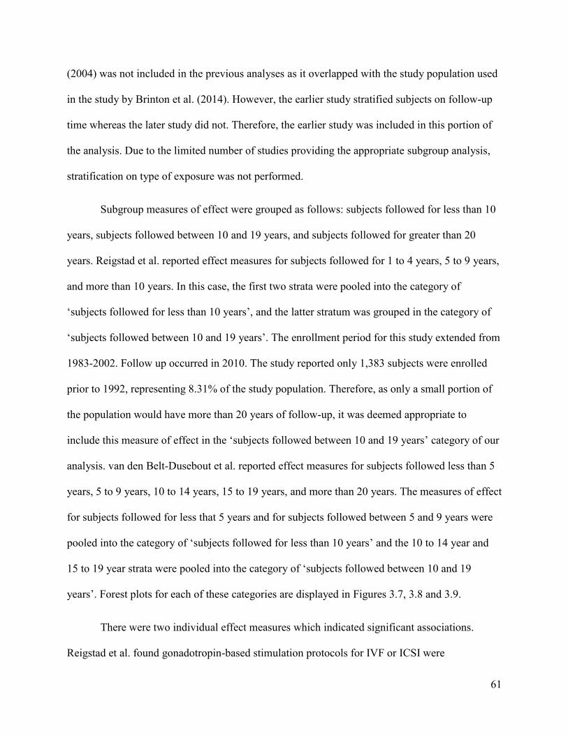
61
(2004) was not included in the previous analyses as it overlapped with the study population used
in the study by Brinton et al. (2014). However, the earlier study stratified subjects on follow-up
time whereas the later study did not. Therefore, the earlier study was included in this portion of
the analysis. Due to the limited number of studies providing the appropriate subgroup analysis,
stratification on type of exposure was not performed.
Subgroup measures of effect were grouped as follows: subjects followed for less than 10
years, subjects followed between 10 and 19 years, and subjects followed for greater than 20
years. Reigstad et al. reported effect measures for subjects followed for 1 to 4 years, 5 to 9 years,
and more than 10 years. In this case, the first two strata were pooled into the category of
‘subjects followed for less than 10 years’, and the latter stratum was grouped in the category of
‘subjects followed between 10 and 19 years’. The enrollment period for this study extended from
1983-2002. Follow up occurred in 2010. The study reported only 1,383 subjects were enrolled
prior to 1992, representing 8.31% of the study population. Therefore, as only a small portion of
the population would have more than 20 years of follow-up, it was deemed appropriate to
include this measure of effect in the ‘subjects followed between 10 and 19 years’ category of our
analysis. van den Belt-Dusebout et al. reported effect measures for subjects followed less than 5
years, 5 to 9 years, 10 to 14 years, 15 to 19 years, and more than 20 years. The measures of effect
for subjects followed for less that 5 years and for subjects followed between 5 and 9 years were
pooled into the category of ‘subjects followed for less than 10 years’ and the 10 to 14 year and
15 to 19 year strata were pooled into the category of ‘subjects followed between 10 and 19
years’. Forest plots for each of these categories are displayed in Figures 3.7, 3.8 and 3.9.
There were two individual effect measures which indicated significant associations.
Reigstad et al. found gonadotropin-based stimulation protocols for IVF or ICSI were

62
significantly associated with breast cancer in women followed for more than 10 years, RR 1.35,
95% CI [1.07-1.71] (Reigstad et al., 2015). dos Santos Silva et al. reported a significant
association between clomiphene exposure and breast cancer in women who were followed for
more than 20 years, RR 1.65, 95% CI [1.10-2.48] (dos Santos Silva et al., 2009).
The pooled RR for subjects followed less than 10 years was 0.98, 95% CI [0.82-1.17], z-
test was non-significant (p = 0.84). The I2 measure indicated moderate levels of heterogeneity, I2
= 34%. The pooled RR for subjects followed between 10 and 19 years was 1.08, 95% CI [0.97-
1.21]. The z-test for this category was non-significant (p = 0.17). There was low to moderate
inter-study heterogeneity, I2 = 27%. Finally, the pooled RR for subjects followed for more than
20 years was 1.27, 95% CI [0.91-1.78]. The z-test for this category was non-significant (p =
0.15). Inter-study heterogeneity was high, I2 = 64%.
3.3: Discussion
The meta-analysis failed to find an association between ovarian-stimulating fertility
treatments and breast cancer, regardless of type of exposure(s) assessed or the study design.
These results are consistent with the findings of two previous meta-analyses synthesizing the
literature on this topic (Sergentanis et al., 2014; Zreik et al., 2010). The study by Sergentanis et
al. identified IVF as its exposure of interest and the study by Zreik et al. included both
clomiphene and gonadotropins as its exposures of interest. Both analyses concluded that there
was no evidence of an association between ovarian-stimulating fertility treatments and breast
cancer.
The secondary analysis indicated that ovarian stimulation has not been shown to be
associated with breast cancer regardless of the length of subject follow-up time. There was some

63
concern raised in qualitative reviews on this topic stating that definitive conclusions regarding
the safety of ovarian stimulation in the context of breast cancer risk cannot be made as previous
observational studies had limited follow-up time (Brinton, Moghissi, Scoccia, Westhoff, &
Lamb, 2005; Practice Committee of the American Society for Reproductive Medicine, 2016;
Stewart & Hart, 2015). Breast cancer is a disease with a relatively long latency period.
Estimating the length of this latency period is challenging as tracking tumor development in
human subjects poses methodological and ethical issues. The latency period is defined as the
time between the onset of the disease to the time where the cancer has grown to a clinically
detectable mass. To date, the literature on the topic has failed to definitely quantify the latency
period of breast cancer, however there is some evidence supporting the notion that this period is
significant. The current observational evidence supporting a long latency period comes from a
study which followed women exposed to significant quantities of radiation (Wanebo et al.,
1968). The study by Wanebo et al. included 31 women diagnosed with breast cancer after being
exposed to the radiation emitted by the nuclear explosions occurring in Hiroshima and Nagasaki
in 1945. The study reported the average latency period between exposure to radiation and
diagnosis with breast cancer was 15.4 years (Wanebo et al., 1968). It should be noted that it is
impossible to conclude that cancers which originate due to a radiation insult progress in the same
fashion as those rooted in a hormonal etiology. Nevertheless, it is prudent to study whether
lengthened periods of follow-up would reveal a significant association as shorter periods of
follow-up may be insufficient in capturing the true effects of the exposure.
There were only two studies included in this meta-analysis which reported significant
associations between ovarian-stimulating fertility treatments and breast cancer. Reigstad et al.
reported a significant association between gonadotropin use and breast cancer, and Lerner-Geva

64
et al. reported a significant association between clomiphene use and breast cancer (Lerner-Geva
et al., 2006; Reigstad et al., 2015). It is important to note that these studies utilized general
population controls. Infertile women are inherently at an increased risk of developing breast
cancer for multiple reasons. First, they are more likely to remain nulliparous, have a lower total
parity, and have a later age of first birth than the general population, both of which have been
shown to be risk factors for breast cancer (Lambe et al., 1996). Additionally, on average, they are
expected to have fewer total pregnancies. High total parity has been shown to be protective
against breast cancer (Ursin et al., 2004). Finally, many of the lifestyle factors which have an
impact on fertility such as alcohol use, smoking, recreational drug use, and a high BMI are also
risk factors for breast cancer.
The study by Reigstad et al. utilized a Norwegian birth registry as a source of data. Their
entire study population was therefore parous, and age of first birth and total parity were included
as covariates in their statistical model. However, lifestyle variables associated with infertility
were not included in the model, and the authors correctly identified this as a limitation of their
study. This highlights one of the major methodological challenges in attempting to evaluate this
relationship of interest. Infertility, and its associated conditions, is an important confounder when
evaluating the relationship between ovarian stimulation and breast cancer. Gathering the
necessary information to appropriately control for the effects of infertility requires substantial
amounts of information frequently not readily available to researchers. Consequently, many
studies have opted to restrict their study populations to infertile subjects. There were 7 cohort
studies (Brinton et al., 2014; Brinton et al., 2013; dos Santos Silva et al., 2009; Rossing et al.,
1996; Stewart et al., 2012; van den Belt-Dusebout et al., 2016; Venn et al., 1999) and 1 case-
control study (Jensen et al., 2007) which did so. None of these studies found the existence of a

65
significant association between ovarian stimulation and breast cancer. Studies also had
limitations pertaining to the methods in which they gathered information. These limitations will
be discussed in further detail in Chapter 4.
The current body of literatures disputes the postulated association between ovarian-
stimulating fertility treatments and breast cancer. While these treatments do indeed raise serum
estrogen levels to supraphysiologic levels, they do so only transiently. There is no evidence
supporting any sort of long-term impact on the hormone levels of future, natural, ovulatory
cycles. Therefore, it is entirely possible that estrogen levels are increased for too transient a
period to have any noticeable impact on breast cancer risk. Consequently, perhaps undergoing a
greater number of stimulated cycles may increase a woman’s risk of breast cancer as this would
increase the time period where serum estrogen levels are supraphysiologic. A total of 9 cohort
studies (Brinton et al., 2014; Brinton et al., 2013; dos Santos Silva et al., 2009; Orgeas et al.,
2009; Pappo et al., 2008; Rossing et al., 1996; K. L. Terry et al., 2006; van den Belt-Dusebout et
al., 2016; Venn et al., 1999) and 2 case-control studies conducted subgroup analyses analyzing a
potential dose-response relationship (Burkman et al., 2003; Jensen et al., 2007). Unfortunately,
there was considerable inter-study heterogeneity pertaining to stratification on the basis of
number of cycles, and pooling these studies was not feasible. Only three studies found an
association between breast cancer risk and number of ovarian stimulation cycles performed.
Orgeas et al. (2009) found the risk of breast cancer was significantly elevated in women who
underwent more than 4 cycles of clomiphene treatment (SIR 1.90, 95% CI [1.08-3.35]) (Orgeas
et al., 2009). Burkman et al. reported a significant association in subjects undergoing more than 4
cycles of ovarian stimulation using hMG (RR 2.1, 95% CI [1.0-4.4]) (Burkman et al., 2003). It
should be noted that both these analyses were limited in terms of number of exposed subjects, as

66
evidenced by their relatively large standard error for their measure of effect, and their results
should be interpreted with caution. Finally, Brinton et al. reported the risk of breast cancer was
significantly elevated in subjects undergoing more than 12 cycles of clomiphene therapy (HR
1.45, 95% CI [1.02-2.05]). The remaining studies found no evidence of a dose-response
relationship. While it was not assessed quantitatively, evidence supporting any sort of increase in
breast cancer risk with increasing number of stimulated cycles is limited.

67
Figure 3.1: Flow Chart – Study Selection

68
Table 3.1: Characteristics of Included Cohort Studies
Study, year Location Cohort Size,
description
Enrollment
Period, End
of Follow-up
Follow-
up
Time
Exposures
Assessed
Types of
Controls
Measure
of Effect
Covariates Adjusted for
in Model
Van den Belt
Dusebout,
2016
Netherlands
25,108 women
undergoing
fertility
investigations
1980-1995,
follow-up
through 2013
Median
of 21
years
Gonadotropins
Provided
analysis with
both general
population
controls and
infertile
population
controls
HR Age, age of first birth,
number of births
Reigstad,
2015 Norway
806,834 women
having given birth
1984-2010,
follow-up
through 2010
Mean of
16 years Gonadotropins
Unexposed
women from
the cohort
HR Age, calendar period of
follow-up, region, parity
Brinton, 2014 United
States
9,892 women
registered at
reproductive
endocrinology
clinics
1965-1988,
follow-up
through 2010
Mean of
28.9
years
Clomiphene
Gonadotropins
Unexposed
women from
the cohort
HR
Study site, calendar year
of infertility
investigation, parity
Brinton, 2013 Israel
87,418 women
registered with
fertility issues
After 1994
(end of
enrolment
period not
stated),
Mean of
8.1
years
Clomiphene
Gonadotropins
Unexposed
women from
the cohort
HR
Age at entry, BMI,
smoking, parity at exit,
SES

69
follow-up
through 2011
Stewart, 2012 Australia
21,025 women
having undergone
investigation or
treatment for
infertility
1983-2002,
follow-up
through
August 2010
Mean of
16 years Gonadotropins
Unexposed
women from
the cohort
HR Age, age at first birth,
multiple births
Lerner-Geva,
2012 Israel
2,431 women
undergoing
infertility
investigations
1964-1974,
follow-up
through 2005
Mean of
34 years
Clomiphene
Gonadotropins
General
population SIR
Sex, age, continent of
birth
Kallen, 2011 Sweden
1,388,371 giving
birth during the
study period
1982-2006,
end of
follow-up not
published
Mean of
6.2
years
Gonadotropins General
population OR
Smoking, age at delivery,
year of delivery
Calderon-
Margalit, 2009 Israel
15,392 women
giving birth in the
study period
1964-1976,
follow-up
through 2004
Mean of
27.6
years
Clomiphene
Unexposed
women from
the cohort
HR
Age, socioeconomic
status, maternal country
of birth, BMI
Orgeas, 2009 Sweden
1,135 women
treated for
infertility at
fertility clinics
1961-1976,
follow-up
through 2004
Mean of
32.1
years
Clomiphene General
population SIR
Age at end of follow-up,
age of first birth, parity,
calendar period of
diagnosis
dos Santos
Silva, 2009
United
Kingdom
7,355 women
seeking treatment
for ovulatory
disorders
1963-1999,
follow-up
through 2005
Mean of
21.4
years
Clomiphene
Gonadotropins
Unexposed
women from
the cohort
RR Age, calendar period

70
Pappo, 2008 Israel
3,375 women
who underwent
IVF
1986-2003,
follow-up
through 2004
Mean of
8.1
years
Gonadotropins General
population SIR Age, continent of birth
Lerner-Geva,
2006 Israel
5,788 women
attending fertility
clinics
1964-1984,
follow-up
through 1996
Mean of
20.9
years
Clomiphene
Gonadotropins
General
population SIR
Age, place of origin, type
of infertility
Terry, 2006 United
States
116,671 women
aged 25 to 42
1989, follow-
up through
2001
Mean of
10.9
years
Clomiphene
Unexposed
women from
the cohort
HR
Age, height, current BMI,
BMI at 18 years old,
family history of breast
cancer, history of benign
breast disease, age of
menarche, parity, oral
contraceptive use,
physical activity
Gauthier,
2004 France
92,555 women
enrolled in a
national health
insurance
database
1990-1992,
follow-up
through June
2000
Mean of
9.7
years
Clomiphene
Gonadotropins
Unexposed
women from
the cohort
RR
Education, smoking,
BMI, family history
breast cancer, age of
menarche, menopausal
status, parity, age of first
pregnancy
Dor, 2002 Israel
5,026 women
treated for
infertility at
fertility clinics
1981-1992,
follow up
through 1996
Mean of
3.63
years
Clomiphene
Gonadotropins
General
population SIR Age, sex, place of birth
Venn, 1999 Australia
29,700 women
registered at
fertility clinics
Prior to 1994,
follow-up
through 1996
Mean of
8.5
years
Gonadotropins
Unexposed
women from
the cohort
SIR Age, calendar period,
national region

71
Rossing, 1996 United
States
3,837 women
seeking fertility
counselling
1974-1986,
follow-up
through 1991
Mean of
11.32
years
Clomiphene
Unexposed
women from
the cohort
RR
Age, weight at cohort
entry, calendar year at
entry

72
Table 3.2: Characteristics of the Included Case-Control Studies
Study, year Location Enrollment
Period Exposures Assessed
Number of
Cases,
Number of
Controls
Type of
Controls
Measure
of Effect
Covariates Adjusted for in
Model
Yli-Kuha, 2012 Finland 1996-2004 Gonadotropins 55 cases
60 controls
General
population OR
Age, geographic location,
socioeconomic status,
marital status
Fei, 2012 United
States 2008-2010
Clomiphene
Gonadotropins
1,669 cases
1,422
controls
Sisters of cases OR
Birth order of sisters,
menopausal status at index,
age of first birth
Jensen, 2007 Denmark 1965-1998
Clomiphene
Gonadotropins
331 cases
1,226
controls
Infertile
controls RR
Age, parity, number of
births
Burkman, 2003 United
States 1994-1998
Clomiphene
Gonadotropins
4,566 cases
4,676
controls
General
population OR Age, ethnicity, study site

73
Figure 3.2: Forest plot and pooled RR – All cohort studies examining the association between clomiphene and breast
cancer included in the analysis
Figure 3.3: Forest plot and pooled RR – All case-control studies examining the association between clomiphene and
breast cancer included in the analysis
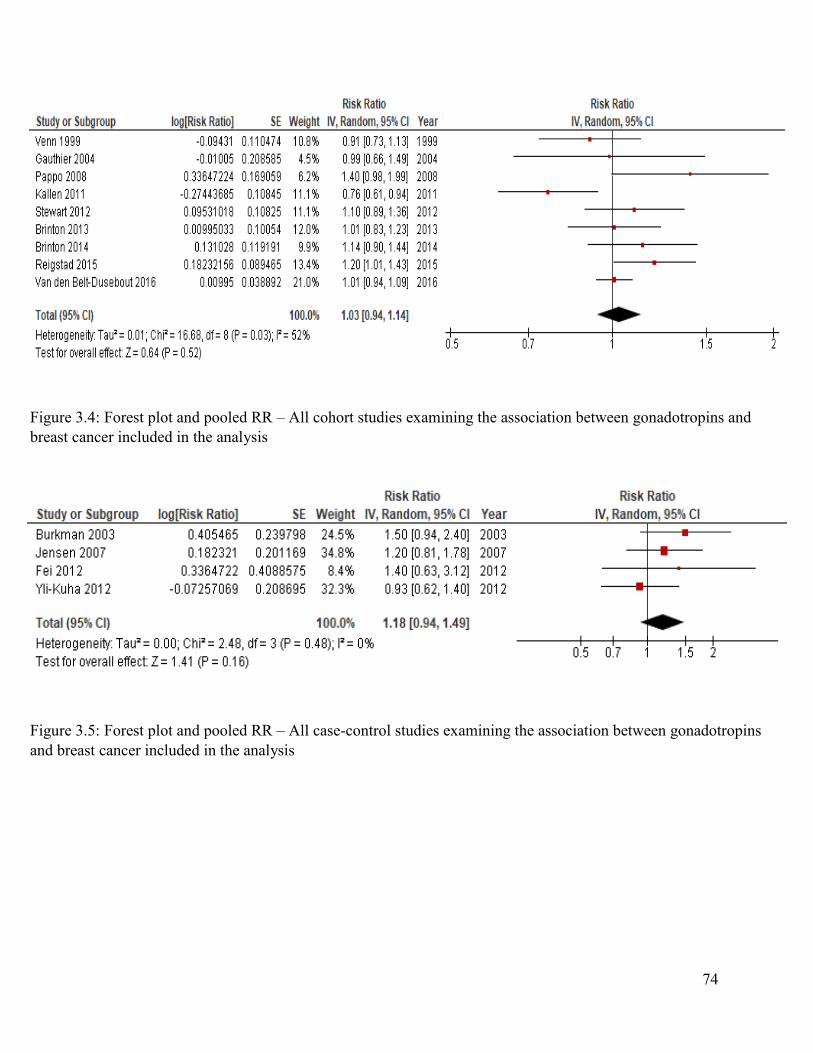
74
Figure 3.4: Forest plot and pooled RR – All cohort studies examining the association between gonadotropins and
breast cancer included in the analysis
Figure 3.5: Forest plot and pooled RR – All case-control studies examining the association between gonadotropins
and breast cancer included in the analysis

75
Figure 3.6: Forest plot and pooled RR – All cohort studies examining the association between clomiphene plus
gonadotropins and breast cancer included in the analysis

76
Figure 3.7: Forest plot and pooled RR – Cohort studies which conducted a stratified analysis on follow-up time,
measures of effect for subjects followed less than 10 years
Figure 3.8: Forest plot and pooled RR – Cohort studies which conducted a stratified analysis on follow-up time,
measures of effect for subjects followed between 10 to 19 years

77
Figure 3.9: Forest plot and pooled RR – Cohort studies which conducted a stratified analysis on follow-up time,
measures of effect for subjects followed more than 20 years

78
Chapter 4: Case-Control Study
A case-control study was performed to examine the association between ovarian
stimulation and breast cancer utilizing the Clinical Practice Research Datalink (CPRD). The
protocol for this study was approved by the Independent Scientific Advisory Committee (ISAC)
in March of 2017 (protocol number: 17_043). The following chapter contains a manuscript
detailing the materials, methods, and results of this study, as well as a discussion of the results.
To limit redundancy with previous chapters the introduction of this manuscript was removed as it
contained similar information to that presented in Chapters 1 and 2.
79
Ovarian-Stimulating Fertility Treatments and the Long-Term Risk of Breast Cancer: A Case-
Control Study Using the Clinical Practice Research Datalink (CPRD)
Adriano Petrangelo, BSc1
Nicholas Czuzoj-Shulman, MMA2
Haim A. Abenhaim, MD, MPH 1, 2
Montréal, Québec, Canada
1 Department of Obstetrics and Gynecology, Jewish General Hospital, McGill University,
Montréal, Québec, Canada
2 Centre for Clinical Epidemiology and Community Studies, Jewish General Hospital, Montréal,
Québec, Canada
No funding was received for this study.
Authors have no conflicts of interest to report.
Corresponding Author:
Haim A. Abenhaim, MD, MPH, FRCSC
Jewish General Hospital, Obstetrics & Gynecology, McGill University
Pavilion H, Room 325
5790 Cote-Des-Neiges Road,
Montréal, Québec, H3S 1Y9
Tel: 514-340-8222 x5488
Fax: 514-340-7941
Email: [email protected]

80
Abstract Introduction: The interplay between reproductive hormone levels and breast cancer is well
established. Ovarian-stimulating fertility treatments raise serum estrogen levels transiently over
the course of therapy, which has raised questions regarding their safety.
Methods: A population-based case-control study was performed using the Clinical Practice
Research Datalink. Cases of breast cancer diagnosed between 1995 and 2013 were matched to 10
controls based on age, general practice, and follow-up time. Cases and controls were assessed for
exposure to clomiphene or IVF via their medical records. Risk ratios and their 95% confidence
intervals were computed using multivariate logistic regression. The model was adjusted for BMI,
smoking, alcohol use, parity, hormonal contraceptive use, hormone replacement therapy, and
oophorectomy.
Results: The study population consisted of 27,026 cases of breast cancer and their controls.
1,717 subjects had a history of clomiphene exposure and 1,137 subjects had a history of IVF.
Exposure to clomiphene was significantly associated with breast cancer, RR 1.32, 95% CI [1.23-
1.42]. Exposure to IVF was also associated with breast cancer, RR 1.55, 95% CI [1.42-1.69].
The stratified analysis indicated clomiphene was associated with pre-menopausal cancers, RR
1.58 95% CI [1.45-1.72] but not post-menopausal malignancies, RR 0.91 95% CI [0.80-1.03].
IVF was associated with both pre- and post-menopausal cancers. A secondary analysis stratified
the study population on PCOS status. Among women with diagnoses of PCOS, clomiphene use
was not associated with an increased risk of breast cancer, RR 1.22, 95% CI [0.99-1.50].
Conclusion: Cases of breast cancer were significantly more likely to have underwent
clomiphene or IVF treatment. However, underlying infertility and its associated conditions may
have confounded this relationship and these results should be interpreted with caution.
81
4.1 Materials and Methods
We performed a population-based case-control study using the Clinical Practice Research
Datalink (CPRD). The CPRD is a primary care database collecting information from over 674
general practices in the United Kingdom, including medical records for over 11.3 million
patients (Herrett et al., 2015). As of June 2015, there were an estimated 4.4 million patients
registered in the database, representing approximately 7% of the UK’s population (Herrett et al.,
2015). Demographics of the CPRD’s population have been compared to information collected by
the UK’s 2011 census. The population captured within the CPRD was deemed to be broadly
representative of the general population in terms of age, sex, and ethnicity (Mathur et al., 2014).
The CPRD is maintained by the UK’s Department of Health in association with the National
Institute for Health Research. These regulatory bodies have instituted quality assurance and
compliance regulations to ensure accuracy of the recorded information. General practices are
individually evaluated and approved. Quality assurance for medical records is performed in
accordance with the Quality and Outcomes Framework, a guideline outlining the reporting
standards for general practitioners in the UK (Sutcliffe D, 2012). The use of the CPRD is
advantageous in epidemiological studies, particularly those evaluating etiological causes of
cancer, due to its longitudinal nature. Data collection occurs prospectively, thereby reducing the
risk of recall bias. Additionally, as breast cancer is a disease with a relatively long latency
period, extended follow-up times are generally preferred in studies evaluating its causal risk
factors (Wanebo et al., 1968). The CPRD includes patient consultation records from 1987
onwards, with active patients having a median follow-up time of 9.4 years (Herrett et al., 2015).
A protocol for this study was submitted to the Independent Scientific Advisory
Committee (ISAC) for review. The ISAC is an advisory body evaluating the feasibility,

82
ethicality, confidentiality, quality, and value of studies vying to use CPRD information. The
protocol was approved by the Committee in March of 2017 (Protocol number 17_043).
4.1.1: Study Population and Design
The source population from which both cases and controls were selected was defined as
all females with records present in the CPRD between January 1st, 1995 and January 1st, 2015. A
subject entered the source population at the occurrence of the latest of the following events:
registration with an up-to-standard general practice as of January 1st, 1995, calendar date of the
subject’s 18th birthday, or date of first registration with an up-to-standard general practice. Up-to-
standard practices are designated as such following quality assurance reviews performed by the
regulatory bodies maintaining the CPRD (Reeves et al., 2014). Subjects were excluded from the
source population if they were not from an up-to-standard general practice, had less than one
year of medical history recorded in the CPRD, or were diagnosed with any form of cancer prior
to a first diagnosis of breast cancer. The latter exclusion criterion was used to ensure all cases of
breast cancer were not re-occurrences of a prior breast malignancy or a metastasis of any other
malignancy. The source population was restricted to females born between January 1st, 1945 and
January 1st, 1995. The left censoring date ensured no female in the source population would be
older than the age of 50 as of January 1st, 1995. As a result, all subjects entering the source
population on January 1st, 1995 had the possibility of being exposed to fertility treatments and
having these treatments captured within the CPRD. The right censoring date ensured no female
in the source population will be younger than 18 years of age as of January 1st, 2013. The two-
year disparity between the right censoring date and the end of the study period allowed for a
minimum of two years of follow-up following the subject’s 18th birthday.

83
4.1.2: Case and Control Selection
The entire source population was followed until a first diagnosis of breast cancer, death
from any cause, end of registration with the general practice, or end of the study period,
whichever occurred first. Diagnoses of breast cancer were identified with the use of a validated
computer algorithm used in previous studies performed with CPRD records (Opatrny,
Dell'Aniello, Assouline, & Suissa, 2008). The list of medical and read codes used in outcome
identification is found in the appendix.
For each case of breast cancer identified, 10 controls were selected. Control subjects
were matched with their respective cases based on year of birth, general practice, and year of
registration in the general practice. Thus, cases and controls were matched on age, general
practice, and follow-up time. All controls were alive, never diagnosed with breast cancer, and
registered with an up-to-standard general practice when matched to their corresponding case.
The index date for cases was used as the index date for their matched controls. All baseline
variables were evaluated as of the index date for cases and controls. The choice of 10 controls
per case allowed the analysis to be manageable, with minimal loss of precision (Breslow NE,
1987).
4.1.3: Exposure Assessment
All cases and controls were evaluated for exposure to either clomiphene or IVF. Medical
and read code lists were generated by searching the CPRD Code Browser with the relevant
search terms. A list of codes used to identify cases of exposure is found in the appendix. As the
CPRD contains primary care records, evaluation of the types and dosages of the drugs used in
IVF treatments was not possible, as this information is restricted to the records of secondary or
tertiary centres. Evaluating IVF exposure on an ‘ever/never’ basis was still feasible as IVF

84
treatments are still likely to be recorded in a patient’s primary care file, either due to the patient
being referred to a fertility clinic by the primary care physician, or the patient reporting these
treatments to their primary care physician. Conversely, it is not uncommon for clomiphene to be
prescribed to patients by primary care physicians in the UK. NICE guidelines published in 2004
and updated in 2013 recommend physicians “offer ultrasound monitoring during at least the first
cycle of treatment to ensure [patients] are taking a dose that minimises the risk of multiple
pregnancy” (National Collaborating Centre for Women’s and Children’s Health, 2013). These
guidelines would certainly dissuade some primary care physicians from providing clomiphene
treatment. Some authors argue ultrasound monitoring for clomiphene treatment is not necessary
as ultrasound monitoring has not been shown to consistently decrease the risk of multiple
pregnancy following clomiphene use (Cahill & Wardle, 2002). Studies have shown that some
physicians in the UK still prescribe clomiphene in the primary care setting (Wilkes, Chinn,
Murdoch, & Rubin, 2009). It should be noted, however, that a sizeable portion of females
undergoing clomiphene treatment would do so in specialized fertility clinics. The use of primary
care data to evaluate exposure to either clomiphene or IVF is therefore an important limitation of
our study.
4.1.4: Statistical Analysis
First, descriptive statistics were used to summarize the characteristics of cases and their
matched controls. Second, multivariate logistic regression was used to estimate RRs and their
95% confidence intervals. As we matched cases and controls on follow-up time, odds ratios
(ORs) computed by the regression model were reliable estimators of the RRs. The regression
model was adjusted for a series of covariates considered to be potential confounders of the
relationship between ovarian-stimulating fertility treatments and breast cancer. These variables
85
were body mass index (BMI), smoking, alcohol use, parity, hormonal contraceptives use,
hormone replacement therapy (HRT), and oophorectomy. BMI values were stratified into four
categories in accordance with WHO definitions, that is, Underweight (BMI <18.50 kg/m2),
Normal (BMI 18.50-24.99 kg/m2), Overweight (BMI 25.00-29.99 kg/m2), and Obese (BMI
≥30.00 kg/m2) (Nuttall, 2015). Smoking status was stratified into never use, current light use,
current moderate use, current heavy use, and ex-heavy use. Alcohol use was stratified into
similar categories. Both hormonal contraceptive use and HRT use were evaluated on an
‘ever/never’ basis. Hormonal contraceptives included combined oral contraceptives, progestin-
only pills (POPs), and implantable progestin devices. POPs and implantable progestin devices
were included due to the evidence presented in the paper by Morch et al. which found a
significant association between these medications and breast cancer (Morch et al., 2017). Finally,
oophorectomy was evaluated as a binary, ‘yes/no’, variable. Instances of oophorectomy must
have occurred prior to a subject’s index date. Lists of CPRD read codes for these variables can
be found in the appendix.
Our primary analysis consisted of evaluating clomiphene and IVF exposure on an
‘ever/never’ basis. Additionally, cases of breast cancer were stratified on age of diagnosis, that
is, whether the diagnosis occurred before the age of 50 or after the age of 50. This approximates
whether the breast cancer was pre-menopausal or post-menopausal.
Two secondary analyses were performed. The first aimed to determine the possible
relationship between the number of clomiphene pills prescribed and the risk of breast cancer.
This analysis evaluated the potential dose-response relationship between clomiphene usage and
breast cancer. Secondly, as we were unable to adjust for underlying infertility and some of its
associated conditions such as total parity and age of first birth, we aimed to evaluate the potential

86
confounding effects of these variables in a different manner. This involved stratifying the study
population on previous diagnoses of polycystic ovary syndrome (PCOS). Subjects were
identified as having PCOS if their clinical records indicated they had been diagnosed with this
syndrome prior to their date of exposure to either clomiphene or IVF. The list of read codes used
to identify subjects diagnosed with PCOS can be found in the appendix of this document.
In order to address the issue of missing data, we used multiple imputation methods that
have been shown to produce valid estimates of effect when the rate of missing data is less than
60% (Barzi & Woodward, 2004). Multiple imputation of missing values was performed using
the SAS function PROC MI on all covariate model variables to produce the values for
imputation. Similar to a study by Delaney et al. which successfully used this technique in a study
using the CPRD, we used mixed chain imputation with 1000 burn-in iterations and the Markov
chain Monte Carlo (MCMC) method in which a Markov chain long enough for the distribution
of the elements to stabilize to a common, stationary distribution (Delaney et al., 2007). By
repeatedly simulating steps of the chain, draws from the distribution of interest are also
simulated. In keeping with the Delaney et al. methodology, time-plots and auto-regression plots
were assessed to determine whether imputation was successful and that there was minimal auto-
regression between iterations of the MCMC algorithm. The results for each imputation were
generated using conditional logistic regression and then combined using PROC MIANALYZE
procedure in SAS. Finally, 10 imputed datasets for this study were produced to ensure that our
effect estimates were not overly inaccurate due to Monte Carlo variability (Schafer, 1999). All
analyses were done with the statistical software package SAS Institute Inc. Enterprise Guide 6.1,
Copyright ©2013. SAS Institute Inc. SAS and all other SAS Institute Inc. product or service
names are registered trademarks or trademarks of SAS Institute Inc., Cary, NC, USA.

87
4.2: Results
The study population consisted of 27,026 cases of breast cancer and their corresponding
270,260 matched controls. Covariate characteristics for the study population, including
demographic, lifestyle, and clinical variables, are displayed in Table 4.1. Breast cancer cases
were more likely to be underweight (BMI of less than 18.5 kg/m2), to have been prescribed
hormonal contraceptives, to have been prescribed HRT, and be parous. Cases and controls were
similar in terms of smoking status, alcohol use, and rates of previous oophorectomy.
The results from the primary analysis are presented in Table 4.2. In total, 1,717 subjects
were exposed to clomiphene and 1,137 underwent IVF treatment. Following adjustment for the
relevant covariates, exposure to clomiphene was significantly associated with breast cancer, RR
1.32, 95% CI [1.23-1.42]. Similarly, exposure to IVF was significantly associated with breast
cancer, RR 1.55, 95% CI [1.42-1.69]. Following stratification based on age of breast cancer
diagnosis, exposure to clomiphene remained significantly associated to pre-menopausal breast
cancers, RR 1.58, 95% CI [1.45-1.72], but not post-menopausal breast malignancies, RR 0.91,
95% CI [0.80-1.03]. IVF remained significantly associated to both pre-menopausal and post-
menopausal breast cancers with RRs of 1.61, 95% CI [1.44-1.79] and 1.43, 95% CI [1.25-1.64],
respectively.
Table 4.3 presents the results of the first secondary analysis exploring the dose-response
relationship between clomiphene and breast cancer. Clomiphene remained associated with breast
cancer regardless of the number of pills prescribed, except in subjects who were prescribed
between 46 and 60 pills, RR 1.13, 95% CI [0.93-1.38]. No apparent trend existed. The second
secondary analysis stratified cases and controls based on prior diagnoses of PCOS. The results of
this analysis are displayed in Table 4.4. A total of 2,264 women were diagnosed with PCOS, 203

88
of which were prescribed clomiphene. In women without PCOS, clomiphene exposure remained
significantly associated with breast cancer, RR 1.24, 95% CI [1.16-1.33]. In women who were
diagnosed with PCOS, clomiphene use was not significantly associated with an increased risk of
breast cancer, RR 1.22, 95% CI [0.99-1.50].
4.4: Discussion
Determining whether ovarian-stimulating fertility treatments are linked with breast
cancer is clinically important. Evidence supporting this relationship would potentially modify
treatment protocols for future patients and would justify more robust screening regimens for
patients having already undergone ovarian stimulation. Numerous observational studies have
explored this relationship. The results of these studies have been synthesized in the meta-analysis
presented in Chapter 3. Overall, the current evidence on this topic refutes any relationship
between ovarian stimulation and breast cancer, however, many previous studies suffered from
certain limitations which we aimed to improve upon.
Firstly, several previous observational studies utilized multiple, distinct databases to
compile information. This requires the use of data linkage. For example, studies such as the ones
by van den Belt-Dusebout et al., Brinton et al., and Stewart et al. ascertained exposure
information from the medical records of fertility clinics (Brinton et al., 2014; Stewart et al.,
2012; van den Belt-Dusebout et al., 2016). At follow-up, subjects were linked to national cancer
registries to determine their outcome status. Data linkage is a common tool used in
epidemiological studies, however, it is susceptible to bias. When databases are linked, errors may
occur. These errors are often due to missing data, leading to missed matches, or erroneous patient
identifiers in one of the linked databases, leading to false matches (Harron, Wade, Gilbert,
Muller-Pebody, & Goldstein, 2014). Linkage bias stems from missed or erroneous matching

89
occurring more often when linking subjects with certain characteristics. Bohensky et al.
conducted a systematic review of the literature quantitatively evaluating linkage bias, mainly by
comparing the characteristics of correctly matched and falsely matched records (Bohensky et al.,
2010). Of the 13 studies included in the analysis, 11 if them identified instances of bias arising
from data linkage. These biases were primarily the result of disparities in overall health, age,
geographical location, ethnicity, time period of enrollment, and socioeconomic status between
matched and unmatched, or erroneously-matched, subjects. In our study, exposure, outcome, and
covariate information was extracted from a single database which effectively eliminated the risk
of bias arising from data linkage. A second major source of bias in previous studies arose from
the methods of gathering covariate information. Cancer registries and fertility clinic records
often do not record the necessary information required for appropriate control of confounding.
For example, these databases are less likely to include information pertaining to smoking status,
alcohol use, HRT use, and hormonal contraceptive use when compared to primary care records.
Therefore, studies would either fail to include these covariates in their model or gather
information on these variables using patient-filled questionnaires at the time of subject follow-
up. This was the case for studies such as van den Belt-Dusebout et al., Burkman et al., and
Gauthier et al (Burkman et al., 2003; Gauthier et al., 2004; van den Belt-Dusebout et al., 2016).
Gathering covariate information at the time of follow-up causes this information to be
susceptible to recall bias. Recall bias, in this context, stems from subjects being more likely to
report variables associated with breast cancer should they have been diagnosed with breast
cancer in the past. For example, subjects with a history breast cancer would be more likely to
report past HRT use compared with those not diagnosed with breast cancer. Following
adjustment, this would dilute the association, if any, between the exposure of interest and breast
cancer. Our study used information collected by primary care physicians in accordance with

90
rigorous quality assurance standards, which somewhat increases the validity of the information
used to evaluate exposure, outcome, and covariate variables. Information was collected
longitudinally, thereby eliminating the possibility of recall bias affecting results. Finally, with
27,026 cases of breast cancer included in the analysis, 211 of whom were exposed to clomiphene
and 152 exposed to IVF, our study was substantially larger and more statistically powerful than
prior case-control studies. The next-largest case-control study was the one conducted by
Burkman et al., which included 61 cases of breast cancer exposed to gonadotropins and 111
cases exposed to clomiphene (Burkman et al., 2003).
Our study had important limitations which warrant further discussion. One such
limitation was our inability to control for the effects of infertility and some of its associated
factors in the regression model. It is postulated that women who experience fertility issues are
inherently at a greater risk of developing breast cancer. Typically, infertile women are more
likely to remain nulliparous and give birth later in life in comparison to women with normal
reproductive functioning. In cases where they succeed in conceiving, they are likely to have, on
average, a lower total parity compared to women without fertility issues. Nulliparity and an older
age of first birth are both considered risk factors for developing breast cancer (Lambe et al.,
1996). A lower total parity is also associated with an increased risk of breast cancer (National
Cancer, 2005). Factors such as alcohol use, smoking, and an elevated BMI are associated with
reduced fecundity. Breast cancer is also strongly linked with alcohol use (McDonald, Goyal, &
Terry, 2013), smoking (Jones, Schoemaker, Wright, Ashworth, & Swerdlow, 2017) and obesity
(Ligibel, 2011), making these factors important confounding variables. In our primary analysis,
we attempted to control for the effects of alcohol use, smoking, BMI, and parity, however, we
were unable to adjust for age of first birth and total number of pregnancies, which was another
important limitation of our study.

91
In ideal conditions, the entire study population would have been restricted to women with
a history of fertility issues as to negate the confounding effects of infertility. This was not
feasible as codes for infertility within the CPRD are not standardized and have not been
externally validated. Instead, we decided to perform a secondary analysis which stratified the
study population on a variable which has been validated and used in previous observational
studies using the CPRD, PCOS (Rees, Jenkins-Jones, & Morgan, 2016). This syndrome is a
common cause of infertility and is strongly associated with breast cancer risk factors such as
obesity (Sam, 2007). Women suffering from PCOS are also be more likely to be nulliparous
(Mikola, Hiilesmaa, Halttunen, Suhonen, & Tiitinen, 2001). Within the group of women who
were diagnosed with PCOS, the association between clomiphene and breast cancer was not
significant. In other words, within a population of women affected by fertility issues, clomiphene
was no longer significantly associated with breast cancer. This finding puts into question the
whether the significant association observed in the primary analysis was due to a causal
relationship between ovarian stimulation and breast cancer as it is possible that these results were
confounded by the underlying infertility.
Another limitation of our study was the use of primary care data to evaluate exposure
information. Firstly, IVF is performed in secondary and tertiary care centres specialized in
providing these treatments. Thus, there is a strong possibility that a significant numbers of
exposure events were missed as exposure information was extracted from primary care records.
IVF exposure would only be present in the CPRD if the primary care physician referred their
patient to a specialty clinic, or the patient self-reported their treatment to their doctor. However,
we cannot assume that the misclassification of IVF exposure data was biased in any way. The
evaluation of exposure to IVF would be biased if it was more likely to be reported in women
with breast cancer than in control subjects. In this case, information on IVF exposure could not

92
be biased as information contained in the CPRD is collected longitudinally. The use of primary
care data also limited our study by preventing an analysis of the dose-response relationship
between number of IVF cycles and breast cancer. Information the number of IVF cycles given to
each subject would be contained in the records of the fertility clinics these women attended.
Additionally, while it is not uncommon for clomiphene treatment to be prescribed by primary
care physicians, regional guidelines for the UK suggest patient response to the first cycle of
clomiphene treatment be monitored by transvaginal ultrasound (National Collaborating Centre
for Women’s and Children’s Health (UK), 2013). This type of monitoring is often performed in
specialized care centres. Therefore, it is likely that a sizeable portion of clomiphene exposures
were misclassified. However, similar to the case of IVF, it is unlikely that this misclassification
was biased in any way.
Determining whether the observed association between ovarian stimulation and breast
cancer is a causal relationship is of important clinical significance. According to the Bradford-
Hill considerations for causality, evidence of a dose-response relationship is important for stating
a relationship is causal (Hofler, 2005). This was analyzed in the first of the secondary analyses.
No apparent trend was seen between the number of clomiphene pills prescribed and the risk of
breast cancer. This disputes any sort of dose-response relationship between clomiphene and
breast cancer. There is a plausible alternate explanation for this finding. Information on the
number of clomiphene pills administered to each patient was extracted from the primary care
clinic’s prescription records. Stratification was therefore performed based on the number of
clomiphene pills prescribed by the subject’s primary care physician. It is possible that a sizeable
portion of the population was not administered the entire dose of clomiphene prescribed. This
may be especially true for subjects prescribed a large quantity of pills. As a result, the association
between clomiphene and breast cancer may have been diluted in the higher-dosage strata. This

93
may have effectively masked a potential dose-response relationship. Nevertheless, these results
cannot support a dose-response relationship between clomiphene and breast cancer, which
further limits our ability to conclude the existence of a causal relationship between ovarian
stimulation and breast cancer.
The possibility that surveillance bias affected results should also be emphasized.
Surveillance bias is a form of non-differential bias caused by a group of subjects being clinically
monitored more closely than the reference group (Hemminki et al., 2017). In this case, women
who require fertility treatments to conceive are be more likely to have co-morbid conditions
which were direct or indirect causes of their infertility. In turn, these conditions would cause a
subject to require more frequent, and more extensive, medical visits. Conditions such as
endocrine dysfunction, endometriosis, PCOS, and sexually transmitted infections may cause a
woman to visit their physician more frequently than the general population. Additionally, several
lifestyle factors such as alcohol use, smoking, and obesity are associated with other co-
morbidities which would necessitate frequent clinical care. Should infertile women visit their
physician more often than the general population, breast cancer may be preferentially detected in
this group of women, which would bias the association between ovarian-stimulating fertility
treatments and breast cancer towards significance.
The disparity is socioeconomic status between the exposed and reference groups may
also contribute to surveillance bias. In the UK, IVF treatments are subsidized by the National
Health Service (NHS), however, a comprehensive evaluation of each patient is performed to
evaluate whether the service will be covered in full. Variables such as age, comorbidities
affecting prognosis, and previous births are considered when deciding coverage. Approximately
40% of IVF cycles performed in the UK are fully funded by the NHS. The remaining cycles are
privately funded, either partially or fully (Human Fertilisation and Embryology Authority, 2016).

94
The cost of an IVF cycle may be prohibitively expensive to couples of lower socioeconomic
statuses. Therefore, it may be assumed that a sizeable portion of women undergoing IVF are of
moderate to high socioeconomic status. Although the UK has a single-payer health care system,
low socioeconomic status is still considered to be an important barrier to accessing regular
medical care. Observational studies performed in countries utilizing a single-payer health care
system consistently conclude that low socioeconomic status is associated with a decreased
utilization of clinical services (Filc, Davidovich, Novack, & Balicer, 2014; Olah, Gaisano, &
Hwang, 2013). Women of low socioeconomic status have also been shown to be less likely to
undergo regular mammographic screening in single-payer systems (Hanson, Montgomery,
Bakker, & Conlon, 2009). As it may be assumed that a sizeable portion of women undergoing
IVF are of moderate to high socioeconomic status, breast cancers may be preferentially detected
in this group, which would further bias results towards significance.
In summary, the results of our study contradict many of the previous analyses examining
this relationship, as both clomiphene and IVF was related to an increased risk of breast cancer,
even after adjustment for several important confounders. However, we are hesitant to promote
the presence of a causal association between ovarian-stimulating fertility treatments and breast
cancer. The results our secondary analyses indicate there is a possibility that the results from our
primary analysis may have been confounded by the effects of infertility. These results may also
have been affected by surveillance bias and confounding due to socioeconomic status.
Additionally, a clear dose-response relationship between clomiphene and breast cancer could not
be established which further puts into question the existence of a causal relationship between
ovarian stimulation and breast cancer.

95
Table 4.1: Demographic, Clinical, and Lifestyle Characteristics of Breast Cancer Cases and Matched
Controls
Characteristics Breast Cancer
n =27,026 (%)
Controls
n = 270,260
Age (years)
<35 629 (2.33) 6,290 (2.33)
35-49 8,776 (32.47) 87,760 (32.47)
50-64 15,826 (58.56) 158,260 (58.56)
65+ 1,795 (6.64) 17,950 (6.64)
BMI (at index)
<18.50 3,062 (19.23) 51,970 (11.33)
18.50-24.99 10,534 (37.65) 3,062 (38.98)
25.00-29.99 7,378 (24.33) 65,473 (27.3)
30.00+ 6,052 (18.90) 51,074 (22.39)
Smoking
Non-smoker 17,584 (57.27) 154,770 (65.06)
Light-moderate smoker 2,107 (6.82) 18,438 (7.8)
Heavy smoker 710 (2.44) 6,592 (2.63)
Ex-heavy smoker 235 (0.63) 1,694 (0.87)
Missing 6,390 (32.84) 88,766 (23.64)
Alcohol
Non-drinker 4,310 (16.17) 43,690 (15.95)
Light-moderate drinker 10,122 (31.25) 84,457 (37.45)
Heavy drinker 269 (0.82) 2,212 (1.00)
Ex-heavy drinker 8 (0.04) 117 (0.03)
Missing 12,317 (51.72) 139,784 (45.57)
Parity
Parous 11,473 (42.45) 93,390 (34.59)
Nulliparous/Missing 15,553 (57.55) 176,870 (65.41)

96
Hormonal Contraceptive Use
Ever 7,469 (27.64) 60,454 (22.37)
Never 19,557 (72.36) 209,806 (77.63)
Hormone Replacement Therapy
Use
Ever 4,551 (16.84) 39,897 (14.76)
Never 22,475 (83.16) 230,363 (85.24)
Oophorectomy
Yes 69 (0.26) 616 (0.23)
No 26,957 (99.74) 269,644 (99.77)

97
Table 4.2: Overall Association Between Ovarian-Stimulating Fertility Treatments and Breast Cancer
Variable Cases of
breast
cancer
(n = 27,026)
Controls
(n = 270,260)
Crude RR (95% CI) Adjusted RRa (95%
CI)
Adjusted
p-value
Overall
Clomiphene
(n = 1,717)
211 1,506 1.41 (1.32-1.51) 1.32 (1.23-1.42) <0.0001
IVF
(n = 1,137)
152 985 1.55 (1.43-1.68) 1.55 (1.42-1.69) <0.0001
Age of breast cancer
diagnosis - Clomiphene
Diagnosis before 50 154 944 1.65 (1.52-1.79) 1.58 (1.45-1.72) <0.0001
Diagnosis after 50 57 562 1.01 (0.90-1.14) 0.91 (0.80-1.03) 0.1400
Age of breast cancer
diagnosis - IVF
Diagnosis before 50 95 585 1.63 (1.47-1.81) 1.61 (1.44-1.79) <0.0001
Diagnosis after 50 57 400 1.43 (1.26-1.62) 1.43 (1.25-1.64) <0.0001
a Adjusted for BMI, smoking, alcohol use, parity, hormonal contraceptive use, HRT use and oophorectomy

98
Table 4.3: Association between Clomiphene and Breast Cancer - Stratified by Number of Clomiphene Pills
Prescribed
Variable Cases of
breast
cancer
Controls Crude RR (95% CI) Adjusted RRa (95 % CI) Adjusted
p-value
Number of Pills
1-15 54 398 1.36 (1.19-1.55) 1.22 (1.07-1.39) 0.0027
16-30 63 462 1.37 (1.21-1.54) 1.27(1.13-1.44) <0.0001
31-45 26 134 1.94 (1.58-2.40) 1.71 (1.39-2.11) <0.0001
46-60 22 175 1.26 (1.03-1.54) 1.13 (0.93-1.38) 0.2214
61+ 46 335 1.38 (1.20-1.58) 1.22 (1.06-1.41) 0.0052
a Adjusted for BMI, smoking, alcohol use, parity, hormonal contraceptive use, HRT use and oophorectomy

99
Table 4.4: Association Between Clomiphene and Breast Cancer – Stratified by Diagnoses of PCOS
Variable Cases of
breast
cancer
Controls Crude RR (95%
CI)
Adjusted RRa
(95% CI)
Adjusted
p-value
PCOS status Clomiphene
No (n = 295,022) Exposed 180 1,334 1.36 (1.26-1.45) 1.24 (1.16-1.33) <0.0001
Unexposed 26,576 266,932
Yes (n = 2,264) Clomiphene
Exposed 31 172 1.37 (1.13-1.67) 1.22 (0.99-1.50) 0.0605
Unexposed 239 1,822
a Adjusted for BMI, smoking, alcohol use, parity, hormonal contraceptive use, HRT use and oophorectomy

100
Chapter 5: General Discussion, Conclusions and Frameworks for
Future Research
5.1: General Discussion
The results of the case-control study were unexpected in the context of the existing
literature on this subject. As discussed in the previous section, the possibility that the case-
control study’s results were confounded by the underlying effects of infertility is significant. Its
results, therefore, should be interpreted with caution. Nevertheless, it is important to explore
lines of evidence which support the postulated association between ovarian stimulation and
breast cancer.
A study by Lundberg et al. compiled a cohort of 43,313 women 40 to 69 years old who
were enrolled in a mammographic screening program (Lundberg et al., 2016). All members of
the study population were never diagnosed with a previous breast cancer nor were they
undergoing further testing for a suspected breast malignancy. Subjects completed questionnaires
assessing for a history of infertility and a history of ovarian-stimulating fertility treatments.
Those with a history of infertility were stratified into three categories based on the type of
previous fertility treatment they received: ovarian stimulation with clomiphene or low-dosage
gonadotropin cycling, ovarian stimulation with high-dose gonadotropins for IVF, and no
previous ovarian stimulation. Breast density was quantitatively assessed by a software program
which computed measures of absolute breast density by integrating the percent density of each
pixel of the mammogram over total breast volume (Garcia et al., 2017). There were 1,576
women with a history high-dose gonadotropin stimulation for IVF. On average, this group had
3.12 cm3, 95% CI [2.22-4.02] higher absolute breast density volume compared to women with a
history of infertility with no previous treatment. Those with a history of ovarian stimulation with

101
clomiphene or low-dose gonadotropins did not have a significantly higher than average breast
density when compared to the same reference group. The statistical model was adjusted for
important confounders such as age at menarche, HRT use, BMI, smoking history, alcohol use,
and parity.
Sprague et al. conducted a similar study utilizing a cohort of 1,009 subjects undergoing
mammography screening regimens with no history of breast cancer (Sprague et al., 2008).
Subjects were assessed for exposure to either clomiphene or gonadotropins through pharmacy
records. No stratification based on type of drug used, or drug dosage, was performed. Breast
density was qualitatively evaluated by board-certified radiology specialists. Mammograms were
classified as being of either low or high density. Overall, this study found no association between
ovarian-stimulating therapies and increased breast density, OR 1.06, 95% CI [0.63-1.77],
however, the authors reported an increasing trend between breast density and time since
exposure. Each additional month following drug dispensal was associated with a 13%, 95% CI
[1%-27%] increase in the risk of a subject being classified as having dense breast tissue. Subjects
whose mammogram was performed more than a year following drug dispensal were significantly
more likely to be classified as having high breast density than those whose mammogram
occurred within a year of drug dispensal. Although the study found no overall association, this
could be due to the short follow-up time of the study. Only exposures occurring 2 years prior to
mammography were considered. Additionally, exposures were assessed through pharmacy
records. Therefore, the date of drug dispensal would not correspond with the date of actual
exposure. A lag period, of undetermined length, existed between drug dispensal and drug
administration. The authors of this study suggested that if the follow-up period was lengthened,
the overall association may have approached or entered significance.

102
Increased breast density is a validated risk factor for breast cancer. A multi-centre case-
control study published in the New England Journal of Medicine reported that the risk of breast
cancer in women with dense breast tissue is significantly elevated for up to 8 years following
initial mammography (Boyd et al., 2007). The National Cancer Institute presently classifies
dense breast tissue as an independent risk factor for breast cancer (National Cancer Institute,
2018). Breast density is representative of the composition of mammary tissue. Breast tissue of
greater density is associated with greater epithelial cell proliferation, greater collagen levels, and
greater volume of glandular structures, which includes the lactiferous ducts and lobules. In
simpler terms, increased breast density is representative of an increased volume of the breast
composed of epithelial and stromal tissue (Clemons & Goss, 2001). These findings are
compatible with the differing radiography characteristics of tissues composing the mammary
gland. On mammography, adipose tissue is relatively translucent whereas glandular and
connective tissue appear to be of increased radiographic density (Lu et al., 2012). As presented in
Chapter 2, estrogen possesses well-characterized effects on glandular proliferation. Breast
density has been shown to be related with hormonal factors such age of first birth, total parity,
and HRT use (Clemons & Goss, 2001; El-Bastawissi, White, Mandelson, & Taplin, 2000; Hou et
al., 2013). As a result, it may be postulated that the supraphysiologic estrogen levels seen during
ovarian stimulation contribute to increased breast density through its effects on epithelial
proliferation and glandular expansion.
Another result from the case-control study worthy of further discussion is the finding that
clomiphene use was only associated with pre-menopausal breast cancers, whereas IVF was
associated with both pre and post-menopausal malignancies. For the sake of simplicity, pre-
menopausal malignancies, that is, those diagnosed between 18 to 49 years of age, will be referred

103
to as early-onset (EO). Post-menopausal malignancies, or those diagnosed after 50 years of age,
will be referred to as late-onset (LO).
Breast cancer incidence rates in the general population follow a bimodal pattern.
Incidence rates peak both at the perimenopausal period and at 70 years of age, “representing the
central tendencies for early-onset and late-onset breast cancers (Anderson, Rosenberg, Prat,
Perou, & Sherman, 2014).” The majority of EO and LO breast cancers are distinct in their
incidence rates and tumor characteristics. According to the Surveillance, Epidemiology and End
Results’ Cancer Statistics Review, a quarter (25.3%) of breast cancers diagnosed in the United
States are EO, with the remaining three quarters being LO (Howlader N, 2017). EO breast
cancers are typically more invasive, have a faster growth rate, and are more likely to be resistant
to hormonal therapies such as tamoxifen as they are less likely to express the estrogen receptor
than their LO counterparts (Anders, Johnson, Litton, Phillips, & Bleyer, 2009; Benz, 2008).
The distinct differences between EO and LO breast malignancies suggest that they have
differing etiologies. The initiation stages of EO tumours occur earlier in a woman’s life, and they
are strongly related to hereditary genetic factors. For example, women with at least one first-
degree relative with breast cancer are also at an increased risk of developing EO breast cancer
(Lynch, Watson, Conway, Fitzsimmons, & Lynch, 1988). The average age of diagnosis in
women with a BRCA1 mutation is also relatively young, with estimates ranging from 40-45
years of age (Brose et al., 2002; Eerola et al., 2005). Conversely, the initial transformation stages
of LO tumours occur later in life, during the mid-reproductive years (Benz, 2008).
The relationship between hormonal factors and age of breast cancer diagnosis is not well
characterized. As discussed previously, estrogen-receptor signalling may act to promote both the
initiation and progression phases of breast tumorigenesis. Therefore, an increase in the exposure
of breast tissue to estrogen can theoretically play a role in both EO and LO breast cancer

104
development. This would occur as the hormone can promote the progression of EO tumours or
promote the initiation and progression of LO tumours.
When studied observationally, the association between hormonal factors and age of
breast cancer diagnosis is less clear. Certain hormonal factors such as age of menarche, age of
first birth, and parity are associated with both EO and LO cancers (Dartois et al., 2016).
Conversely, increased BMI has been shown to be more strongly associated with LO
malignancies (Lauby-Secretan et al., 2016). Combined oral contraceptive use is associated with
only a transient increase in breast cancer risk during their use, and for up to ten years following
the cessation of use (Collaborative Group on Hormonal Factors in Breast, 1996). For women
using HRT, breast cancer risk is transiently increased during its use and for a 5-year period
following cessation of use. (Collaborative Group on Hormonal Factors in Breast Cancer, 1997).
The nebulous relationship between hormonal factors and the age of breast cancer
diagnosis makes the interpretation of our results difficult. A possible explanation may lie in three
main concepts: ovarian stimulation is only offered to patients of reproductive ages, serum
estrogen levels are increased only transiently during treatment, and serum estrogen levels during
ovarian stimulation for IVF are significantly higher than estrogen concentrations during ovarian
stimulation using clomiphene. When a woman of reproductive age undergoes ovarian
stimulation, her risk of breast cancer may be transiently increased as the increased serum
estrogen levels may promote the progression of pre-existing breast lesions. This phenomenon
would be similar to the transient increase in breast cancer risk seen with oral contraceptives use
and HRT use (Collaborative Group on Hormonal Factors in Breast, 1996; Collaborative Group
on Hormonal Factors in Breast Cancer, 1997). As ovarian stimulation is only offered to women
of reproductive ages, a transient increase in breast cancer risk would lead to an increase in the
incidence of EO breast malignancies. An explanation as to why IVF is associated with LO breast

105
cancers whereas clomiphene is not may be due to the difference in peak estrogen levels seen
between the two treatments. As discussed in Chapter 2, ovarian stimulation for IVF results in
serum estrogen concentrations which are 2-3 times higher than the levels seen during ovarian
stimulation using clomiphene. In a purely speculative capacity, it is possible that the estrogen
levels seen during IVF are high enough to have a genotoxic effect on breast tissue which would
fundamentally alter its composition. To recall, Lundberg et al.’s study found prior ovarian
stimulation for IVF was significantly associated with increased breast density whereas
stimulation protocols using clomiphene or low-dose gonadotropins were not (Lundberg et al.,
2016). The estrogen levels caused by IVF may act as an initiator of breast tumorigenesis which
would alter breast cancer risk over a longer period of time.
5.2: Conclusion
The safety of ovarian-stimulating fertility treatments is an important clinical question.
This association was explored in depth by discussing its biological plausibility, overviewing the
current observational evidence on the topic, and conducting a novel study which aimed to
improve upon many of the limitations affecting previous analyses. For example, our study used
longitudinal, validated, prospectively-collected information to evaluate exposure, outcome, and
covariate variables. The study also had high statistical power owing to its large study population.
Nevertheless, the possibility that its results were confounded by the underlying effects of
infertility in addition to the possibility that surveillance bias skewed results necessitates the need
to interpret our study with caution. Furthermore, the current literature on this topic generally
refutes the notion that these fertility treatments are associated with an increased risk of breast
cancer. While this relationship has a strong biological plausibility, in addition to certain lines of
evidence linking ovarian stimulation to an increased breast density, there is no definitive

106
observational evidence of a causal link between ovarian-stimulating fertility treatments and
breast cancer. Nevertheless, it is imperative that the safety of these fertility treatments continues
to be monitored as they are becoming increasingly prevalent. Additionally, it should be
emphasized that while, presently, there is no definitive link between ovarian stimulation and
breast cancer, the population of women suffering from fertility-related issues may be inherently
at an increased risk of developing breast cancer. Health care professionals should be aware of
this phenomenon and offer appropriate guidance and monitoring to this group of patients.
5.3: Framework for Future Research
Monitoring the relationship between ovarian-stimulating fertility treatments and breast
cancer is challenging. Firstly, there are several important variables which may confound this
relationship of interest. Women affected by infertility are at an inherently higher risk of
developing breast cancer as they are more likely to be of lower parity and have a later age of first
birth. Additionally, infertility is commonly associated with comorbid conditions and lifestyle
factors which predispose a woman to developing breast cancer, such as increased BMI, alcohol
us, and a history of tobacco use. Women undergoing treatments such as IVF may also, on
average, be of higher socioeconomic status, which is associated with increased health-seeking
behaviour. Breast cancer may therefore be preferentially detected in this group of women which
contributes to surveillance bias. Secondly, many previous studies have relied on retrospectively
collected data to assess exposure. They additionally often performed database linkages.
Together, these factors have caused many previous studies to be susceptible to both recall and
linkage biases. It is strongly suggested that future research initiatives ensure a robust control of
confounding due to the underlying effects of infertility and its associated conditions. Ideally, the
study population should be restricted to women affected by infertility. Further adjustment for

107
socioeconomic status should be strongly considered. The use of prospectively collected data
compiled in a single longitudinal database to assess exposure, outcome, and covariate
information is also strongly recommended.

108
Appendix
Search algorithm used to search the EMBED and MEDLINE databases
1. breast cancer.mp.
2. breast carcinoma.mp.
3. breast neoplasm.mp.
4. breast tumor.mp.
5. ductal carcinoma*.mp.
6. lobular carcinoma*.mp.
7. tubullary carcinoma*.mp.
8. medullary carcinoma*.mp.
9. papillary carcinoma*.mp.
10. cribiform carcinoma*.mp.
11. Paget’s disease*.mp.
12. mammary tum*.mp.
13. mammary cancer*.mp.
14. 1/or 2/or 3/or 4/or 5/or 6/or 7/or 8/or 9/or 10/or 11/or 12/or 13
15. ovulation ind*.mp.
16. ovulation stim*.mp.
17. ovulatory stim*.mp.
18. ovarian stim*.mp.
19. controlled ovarian stim*.mp.
20. fertility treat*.mp.
21. infertility treat*.mp.
22. fertility drug* .mp.
23. clomifene.mp.
24. clomifene citrate.mp.
25. clomiph*.mp.
26. fertility promoting agent.mp.
27. fertilization in vitro.mp.
28. IVF*.mp.
29. ICSI*.mp.
30. gonadotropin*.mp.
31. human menopausal gonadotropin.mp.
32. human chorionic gonadotropin.mp.
33. 15/or 16/or 17/or 18/or 19/or 20/or 21/or 22/or 23/or 24/or 25/or 26/or 27/or 28/or
29/or 30/or 31/or 32
34. 35, restricted to Jan 1, 1995 to Jan 1, 2017, article, full-text available, English language
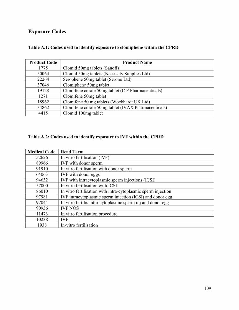
109
Exposure Codes
Table A.1: Codes used to identify exposure to clomiphene within the CPRD
Product Code Product Name
1775 Clomid 50mg tablets (Sanofi)
50064 Clomid 50mg tablets (Necessity Supplies Ltd)
22264 Serophene 50mg tablet (Serono Ltd)
37046 Clomiphene 50mg tablet
19128 Clomifene citrate 50mg tablet (C P Pharmaceuticals)
1271 Clomifene 50mg tablet
18962 Clomifene 50 mg tablets (Wockhardt UK Ltd)
34862 Clomifene citrate 50mg tablet (IVAX Pharmaceuticals)
4415 Clomid 100mg tablet
Table A.2: Codes used to identify exposure to IVF within the CPRD
Medical Code Read Term
52626 In vitro fertilisation (IVF)
89966 IVF with donor sperm
91910 In vitro fertilisation with donor sperm
64063 IVF with donor eggs
94632 IVF with intracytoplasmic sperm injections (ICSI)
57000 In vitro fertilisation with ICSI
86010 In vitro fertilisation with intra-cytoplasmic sperm injection
97981 IVF intracytoplasmic sperm injection (ICSI) and donor egg
97044 In vitro fertilis intra-cytoplasmic sperm inj and donor egg
90936 IVF NOS
11473 In vitro fertilisation procedure
10238 IVF
1938 In-vitro fertilisation

110
Outcome Codes
Table A.3: Codes used to identify diagnoses of breast cancer within the CPRD
Medical Code Read Term
348 Ca female breast
3968 Malignant neoplasm of female breast
20685 Malignant neoplasm of axillary tail of female breast
23380 Malignant neoplasm of nipple of female breast
23399 Malignant neoplasm of upper-outer quadrant of female breast
26853 Malignant neoplasm of nipple and areola of female breast
29826 Malignant neoplasm of upper-inner quadrant of female breast
31546 Malignant neoplasm of central part of female breast
38475 Malignant neoplasm of other site of female breast NOS
42070 Malignant neoplasm of lower-outer quadrant of female breast
45222 Malignant neoplasm of lower-inner quadrant of female breast
56715 Malignant neoplasm of other site of female breast
59831 Malignant neoplasm of nipple or areola of female breast NOS
64686 Malignant neoplasm of areola of female breast
95057 Malignant neoplasm of ectopic site of female breast

111
Covariate Codes
A.4: BMI
Information on subject weight (in kilograms) and height (in metres) were extracted. BMI was
calculated using the formula: BMI = weight/(height2). The weight and height values inputted
closest to the index date were used.
Subjects were categorized into 4 categories (underweight, normal weight, overweight, obese)
according to the WHO’s standards ("World Health Organization Global Database on Body Mass
Index ").
Table A.4: BMI categories
BMI Value Category
<18.50 kg/m2 Underweight
18.50-24.99 kg/m2 Normal Weight
25.00-29.99 kg/m2 Overweight
≥30.00 kg/m2 Obese
Table A.5: Codes used to evaluate smoking status within the CPRD
Medical Code Read Term
33, 60, 11788 Non-smoker
1878, 12944, 12958, 12961 Light-moderate smoker
3568, 1822 Heavy smoker
12956, 12959 Ex-heavy smoker
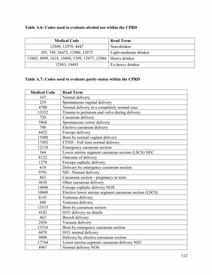
112
Table A.6: Codes used to evaluate alcohol use within the CPRD
Medical Code Read Term
12949, 12970, 4447 Non-drinker
385, 749, 26472, 12980, 12972
12975, 322, 12985
Light-moderate drinker
12982, 8999, 1618, 19494, 1399, 12977, 12984 Heavy drinker
12983, 19493 Ex-heavy drinker
Table A.7: Codes used to evaluate parity status within the CPRD
Medical Code Read Term
167 Normal delivery
239 Spontaneous vaginal delivery
4780 Normal delivery in a completely normal case
13352 Trauma to perineum and vulva during delivery
720 Caesarean delivery
5868 Spontaneous vertex delivery
740 Elective caesarean delivery
6452 Forceps delivery
15480 Born by normal vaginal delivery
17492 FTND - Full term normal delivery
12118 Emergency caesarean section
364 Lower uterine segment caesarean section (LSCS) NEC
8122 Outcome of delivery
1279 Forceps cephalic delivery
618 Delivery by emergency caesarean section
9791 ND - Normal delivery
863 Caesarean section - pregnancy at term
4638 Other caesarean delivery
14686 Forceps cephalic delivery NOS
10049 Elective lower uterine segment caesarean section (LSCS)
6141 Ventouse delivery
688 Ventouse delivery
13315 Born by caesarean section
4182 H/O: delivery no details
463 Breech delivery
2458 Vacuum delivery
13316 Born by emergency caesarean section
6078 H/O: normal delivery
9800 Delivery by elective caesarean section
17744 Lower uterine segment caesarean delivery NEC
8967 Normal delivery NOS

113
43563 [X]Delivery
6619 H/O: caesarean section
24039 H/O: full term delivery
15361 Born by elective caesarean section
9383 Normal delivery
14727 Born by forceps delivery
3085 Elective caesarean delivery NOS
15675 Born by ventouse delivery
3108 Episiotomy to facilitate delivery
11530 Vaginal delivery
14892 Vacuum delivery NOS
9668 Elective lower uterine segment caesarean delivery
19600 Second degree perineal tear during delivery
25681 SVD - Spontaneous vaginal delivery
12014 [V]Outcome of delivery
58721 Normal delivery in completely normal case NOS
25223 Vacuum extractor delivery
12052 Delivery normal
9160 Spontaneous vertex delivery
11829 H/O: premature delivery
11194 Spontaneous vaginal delivery
20425 Neville - Barnes forceps delivery
11532 Breech extraction delivery
7670 Caesarean delivery NOS
5213 Low forceps cephalic delivery
3365 Premature delivery
19356 Delivery - sex of baby
5522 Complications of labour and delivery NOS
35190 Other caesarean delivery NOS
19599 First degree perineal tear during delivery
11650 Third degree perineal tear during delivery
7916 Delivered by caesarean section - pregnancy at term
15828 Born by breech delivery
5866 Induction and delivery operations
40710 Forceps delivery NOS
30274 Low forceps delivery
15167 Early onset of delivery
17684 Kielland forceps delivery
23394 Mid forceps cephalic delivery NEC
23994 Assisted breech delivery
15256 Unspecified perineal laceration during delivery
2943 Assisted breech delivery
21554 Kielland forceps cephalic delivery with rotation
28726 Water birth delivery
31743 Normal delivery but ante- or post- natal conditions present

114
7377 [X]Infection of caesarean section wound following delivery
19609 Vaginal tear during delivery
33477 Caesarean delivery - delivered
42275 Mid-cavity forceps delivery
49446 Vacuum extractor delivery - delivered
50675 Forceps delivery - delivered
43959 Outcome of delivery NOS
28100 Caesarean wound disruption
53937 Forceps delivery unspecified
28903 Delivered by low forceps delivery
29885 Complications occurring during labour and delivery
26712 Labial tear during delivery
19605 Second degree perineal tear during delivery NOS
17078 H/O: previous forceps delivery
34411 Delivered by mid-cavity forceps delivery
52875 Caesarean delivery unspecified
19602 Second degree perineal tear during delivery, unspecified
5721 Urinary tract infection following delivery
33773 Other specified normal delivery
22491 Spontaneous breech delivery
4786 Multiple delivery, all by caesarean section
5033 High forceps cephalic delivery NEC
37699 Vacuum extractor delivery NOS
20738 Manually assisted vaginal delivery
33480 Other specified forceps cephalic delivery
25157 Early onset of delivery - delivered
29070 Spontaneous breech delivery
19608 First degree perineal tear during delivery NOS
19603 First degree perineal tear during delivery, unspecified
15926 Other specified other breech delivery
20002 High forceps cephalic delivery with rotation
33378 Other breech delivery
36037 [X]Vaginitis following delivery
17704 Simpson's forceps delivery
32590 Delivery problem
33915 Multiple delivery
29155 Caesarean hysterectomy
56310 Delivery observations
40876 Third degree perineal tear during delivery NOS
31954 Number of caesarean sections
39576 Fourth degree perineal tear during delivery
37054 Second degree perineal tear during delivery - delivered
18287 Normal delivery of placenta
19604 Third degree perineal tear during delivery, unspecified
50293 Induction and delivery operations NOS

115
43760 Other method of delivery NOS
15514 Upper uterine segment caesarean delivery NEC
47546 Other specified elective caesarean delivery
37878 Multiple birth delivery
40706 Unspecified perineal laceration during delivery NOS
32380 Vulval delivery trauma
39168 Third degree perineal tear during delivery - delivered
40744 Abnormal delivery
15073 Other specified other caesarean delivery
31280 Vulval tear during delivery
83535 [X]Other single spontaneous delivery
29872 History of past delivery
34628 Failed forceps delivery
31254 First degree perineal tear during delivery - delivered
54591 Breech extraction delivery NOS
42324 Vulval/perineal trauma during delivery NOS
55543 Other operations to facilitate delivery
34399 [X]Cervicitis following delivery
44235 Vaginal delivery following previous caesarean section
44494 Caesarean wound disruption NOS
43918 Fourchette tear during delivery
47508 Other methods of delivery
57789 Delivery by combination of forceps and vacuum extractor
36137 Early onset of delivery NOS
36703 Vulval and perineal haematoma during delivery
50847 Caesarean delivery following previous Caesarean delivery
31780 Spontaneous breech delivery
9625 Labour and delivery complicated by fetal heart rate anomaly
64464 Other breech delivery NOS
61259 Elective upper uterine segment caesarean delivery
27731 Vulval and perineal haematoma during delivery
60953 Precipitate delivery
58844 Vacuum extractor delivery unspecified
48230 Deliveries by spontaneous breech delivery
69920 Complications of labour and delivery NOS
61212 Other vulval and perineal trauma during delivery
33884 Multiple delivery, all spontaneous
53572 Other specified complications of labour or delivery
61077 Mid forceps cephalic delivery with rotation
52151 [X]Labour+delivery complications/other evidence of fetal distress
34102 Incomplete placenta at delivery
60538 Multiple delivery, all by forceps and vacuum extractor
19607 Unspecified perineal laceration during delivery, unspecified
28861 Delivery by caesarean hysterectomy
47157 Vulval/perineal trauma during delivery NOS

116
23866 Symphysiotomy to facilitate delivery
34333 Incomplete delivery of placenta
57652 Fourth degree perineal tear during delivery NOS
59941 Early onset of delivery unspecified
61578 Caesarean wound disruption unspecified
72022 Fourth degree perineal tear during delivery, unspecified
86399 Other specified vacuum delivery
40875 Second degree perineal tear during delivery with p/n prob
44472 Other complications of labour and delivery
48742 Face delivery
47863 [X]Other single delivery by caesarean section
51736 Low vacuum delivery
66074 Other complications of labour and delivery with p/n problem
67052 Unspecified perineal laceration during delivery - delivered
104873 Premature labour and delivery
50236 Other specified induction or delivery operations
62862 Third degree perineal tear during delivery with p/n problem
67333 [V]Unspecified delivery outcome
24243 Labour and delivery complications by meconium in amniotic fluid
49485 Labour+delivery complicated by biochem evidence/fetal stress
104872 Premature labour with premature delivery
62406 Delayed delivery second twin unspecified
62508 [V]Examination immediately after delivery
96089 Rapid rate of delivery
57957 Other specified other method of delivery
61226 [V]Other specified outcome of delivery
72021 Fourth degree perineal tear during delivery - delivered
73048 Other specified breech extraction delivery
27963 Delayed delivery of second twin, triplet etc.
66219 [X]Other infection of genital tract following delivery
69971 Slow rate of delivery
95780 Complete placenta at delivery
37245 Vulval and perineal haematoma during delivery, unspecified
50924 First degree perineal tear during delivery with p/n problem
66315 [X]Other specified assisted single delivery
69780 [X]Other and unspecified forceps delivery
30805 Other vulval/perineal trauma during delivery, unspecified
39573 Umbilical cord not around baby's neck at delivery
55124 Other complications of labour and delivery NOS
62318 Trial of vacuum delivery
65940 Trial of forceps delivery
30806 Other vulval/perineal trauma during delivery NOS
47036 Problem of pelvis for delivery
54623 Caesarean wound disruption with postnatal complication
57945 Vacuum delivery before full dilation of cervix

117
60256 Brow delivery
60924 No progress with delivery
66501 Complications of labour and delivery NOS
66843 Unspecified perineal laceration during delivery + p/n prob
67489 Vulval/perineal trauma during delivery NOS unspec
72513 Extraperitoneal caesarean section
93273 Complications of labour and delivery NOS - delivered
100675 [X]Assisted single delivery, unspecified
61597 Fourth degree perineal tear during delivery with p/n problem
70242 Barton forceps cephalic delivery with rotation
70615 Vulval and perineal haematoma during delivery NOS
71057 Vulval and perineal haematoma during delivery - delivered
90497 Complications of labour and delivery NOS with p/n problem
99608 Other operation to facilitate delivery NOS
100597 Other complications of labour and delivery - delivered
105032 Premature labour without delivery
22498 Rate of delivery
42572 Delayed delivery second twin - delivered
42885 Observation of pattern of delivery
57488 Vulval/perineal trauma during delivery NOS - delivered
65331 Other vulval/perineal trauma during delivery + p/n problem
67410 [X]Other genitourinary tract infections following delivery
68752 Normal rate of delivery
71862 Delayed delivery second twin with antenatal problem
90885 [X]Labour+delivery complicated by other cord entanglement
95858 Other vulval/perineal trauma during delivery- delivered
98059 [X]Other manipulation-assisted delivery
98749 Incision of cervix to facilitate delivery
99361 Vulval and perineal haematoma during delivery + p/n problem
101229 Observation of delivery push in labour
103319 [X]Complications of labour and delivery
105276 Breech extraction delivery with version
105419 High vacuum delivery
106001 Destructive operation for delivery
106113 [X]Multiple delivery, unspecified
106808 Vulval/perineal trauma during delivery NOS with p/n problem
40729 Gravida 1
54604 Gravida 2
25612 Gravida 3
54572 Gravida 4
54597 Gravida 5
58003 Gravida 6
61894 Gravida 7
46527 Primigravida
86753 Gravida 8

118
94244 Gravida 9
63196 Multigravida
8776 [X] Stillbirth
23330 GP unit birth
1825 Twin birth
6184 [V]Normal pregnancy
5709 Pregnancy care
29662 Multiparous
Table A.8: Codes used to evaluate hormonal contraceptive use within the CPRD
Product Codes Product Term
65068 Yacella 0.03mg/3mg tablets (Morningside Healthcare Ltd)
65312 Eloine 0.02mg/3mg tablets (Bayer Plc)
68515 Teragezza 2000microgram/35microgram tablets (Morningside Healthcare
Ltd) 65475 Generic Femodene ED tablets
37073 Sunya 20/75 tablets (Stragen UK Ltd)
44229 Millinette 20microgram/75microgram tablets (Consilient Health Ltd
2265 Etynodiol 500microgram tablets
61088 Generic Logynon tablets
8176 Ethinylestradiol 20microgram / Norethisterone acetate 1mg tablets
3436 Ortho-novin 1/50 Tablet (Janssen-Cilag Ltd)
15987 Ethinylestradiol with norethisterone - triphasic 7 x 35+500mcg; 7 x
35+750mcg; 7 x 35mcg+1mg Tablet 18569 Gestodene with ethinylestradiol 75microgramwith20microgram Tablet
65786 Ethinylestradiol 30microgram / Desogestrel 150microgram tablets (AM
Distributions (Yorkshire) Ltd) 14670 Ethinylestradiol with norethisterone - biphasic 7 x 35mcg+500mcg; 14 x
35mcg+1mg 63725 Dretine 0.03mg/3mg tablets (Teva UK Ltd)
13209 Etonogestrel 68mg implant
81w03 Conova 30 Tablet (Pharmacia Ltd)
54527 Cerelle 75microgram tablets (Consilient Health Ltd)
45059 Ethinylestradiol with levonorgestrel 30micrograms + 50micrograms Tablet
2061 Norethisterone 350microgram tablets
3693 Triadene tablets (Bayer Plc)
36829 Katya 30/75 tablets (Stragen UK Ltd)
19131 Ethinylestradiol with gestodene 30micrograms+75micrograms Tablet
57181 Lizinna 250microgram/35microgram tablets (Morningside Healthcare Ltd)
23211 Desogestrel with ethinylestradiol 150micrograms with 30micrograms tablets
239 Micronor 350microgram tablets (Janssen-Cilag Ltd)
47132 Co-cyprindiol 2mg+35microgram Tablet (Sandoz Ltd)
1720 Levonorgestrel 30microgram tablets
49214 Cilest 35microgram/250microgram tablets (Mawdsley-Brooks & Company
Ltd) 40650 Ethinylestradiol with norethisterone 35micrograms + 750micrograms Tablet

119
61190 Co-cyprindiol 2000microgram/35microgram tablets (A A H Pharmaceuticals
Ltd 41 Microgynon 30 tablets (Bayer Plc)
16624 Levonorgestrel 228mg Implant
44046 Rigevidon tablets (Consilient Health Ltd)
14459 Gynovlar 21 Tablet
11910 Ethinylestradiol 20microgram / Gestodene 75microgram tablets
5862 Norinyl-1 tablets (Pfizer Ltd) Norethisterone/Mestranol
14601 Ethinylestradiol 35microgram / Norgestimate 250microgram tablets
68352 Aidulan 30microgram/75microgram tablets (Lupin (Europe) Ltd)
4964 Femodette tablets (Bayer Plc) Gestodene/Ethinylestradiol
25124 Acnocin 2000microgram/35microgram tablets (Sandoz Ltd)
1601 Trinovum tablets
29499 Ethinylestradiol 33.9micrograms/24hours / Norelgestromin
203micrograms/24hours transdermal patches 4608 Dianette tablets (Mylan Ltd) Cyproterone acetate/Ethinylestradiol 2mg +
35microgram
2856 Norimin 1mg/35microgram tablets (Pfizer Ltd)
Norethisterone/Ethinylestradiol 8482 Ethinylestradiol 35microgram / Norethisterone 1mg tablets
59503 Lucette 0.03mg/3mg tablets (Consilient Health Ltd)
44278 TriRegol tablets (Consilient Health Ltd)
936 Femodene tablets (Bayer Plc)
1613 Microval 30microgram tablets (Wyeth Pharmaceuticals) Levonorgestrel
30microgram 56483 Generic Tri-Minulet tablets
56103 Cerelle 75microgram tablets (Consilient Health Ltd)
62968 Maexeni 150microgram/30microgram tablets (Lupin (Europe) Ltd)
44336 Gedarel 30microgram/150microgram tablets (Consilient Health Ltd)
443 Desogestrel with ethinylestradiol 150micrograms with 20micrograms tablets
977 Minulet tablets (Wyeth Pharmaceuticals)
6431 Co-cyprindiol 2000microgram/35microgram tablets
47057 Yasminelle 3mg+20microgram Tablet (Bayer Plc)
31528 Ethinylestradiol with norethisterone - triphasic 7x35+500mcg;
9x35mcg+1mg; 5x35+500mcg 10201 Desogestrel 75microgram tablets
53201 Dianette tablets (Lexon (UK) Ltd)
44994 Millinette 30microgram/75microgram tablets (Consilient Health Ltd)
3472 Trinovum ed ED tablets (Janssen-Cilag Ltd)
9592 Implanon 68mg implant (Organon Laboratories Ltd)
64918 Aidulan 20microgram/75microgram tablets (Lupin (Europe) Ltd)
56732 Aizea 75microgram tablets (Besins Healthcare (UK) Ltd)
13248 Ethinylestradiol 30microgram / Desogestrel 150microgram tablets
7814 Ethinylestradiol 35microgram / Norethisterone 500microgram tablets
43003 Levest 150/30 tablets (Morningside Healthcare Ltd)
14977 Ethinylestradiol with gestodene - triphasic 6 x 30+50mcg; 5 x 40+70mcg; 10
x 30+100mcg Tablet 1062 Ovranette 150microgram/30microgram tablets (Pfizer Ltd)
1378 Mercilon 150microgram/20microgram tablets (Merck Sharp & Dohme Ltd)
12631 Ethinylestradiol with levonorgestrel and placebo 30micrograms +
150micrograms Tablet 6596 Norelgestromin with ethinylestradiol 203micrograms +
33.9micrograms/24hours Transdermal patch

120
61890 Desogestrel 75microgram tablets (A A H Pharmaceuticals Ltd)
1352 Loestrin 30 tablets (Galen Ltd)
25263 Norgestimate with ethinylestradiol 250micrograms + 35micrograms Tablet
18823 Ethinylestradiol 30microgram / Norethisterone acetate 1.5mg tablets
5986 Cerazette 75microgram tablets (Merck Sharp & Dohme Ltd)
1354 Brevinor 500microgram/35microgram tablets (Pfizer Ltd)
15886 Ethinylestradiol with levonorgestrel - triphasic 6x30+50mcg; 5x40+75mcg;
10x30+125mcg Tablet 52443 Microgynon 30 tablets (Mawdsley-Brooks & Company Ltd)
44196 Nexplanon 68mg implant (Merck Sharp & Dohme Ltd)
19551 Controvlar Tablet (Schering Health Care Ltd)
697 Yasmin tablets (Bayer Plc)
38500 Co-cyprindiol 2000microgram/35microgram tablets (Fannin UK Ltd)
7776 Ethinylestradiol 30microgram / Gestodene 75microgram tablets
6686 Ethinylestradiol 30microgram / Levonorgestrel 150microgram tablets
58642 Cimizt 30microgram/150microgram tablets
55819 Nacrez 75microgram tablets (Teva UK Ltd)
45557 Levest 150/30 tablets (Actavis UK Ltd)
2026 Logynon ED tablets (Bayer Plc)
42510 Ethinylestradiol with levonorgestrel Tablet
57264 Estradiol 1.5mg / Nomegestrol 2.5mg tablets
60739 Yaz tablets (Imported (United States))
44457 Gedarel 20microgram/150microgram tablets (Consilient Health Ltd)
59414 Desogestrel 75microgram tablets (Actavis UK Ltd)
47281 Elevin 150microgram/30microgram tablets (MedRx Licences Ltd)
2769 Cyproterone acetate with ethinylestradiol 2mg with 35micrograms tablets
6166 Evra transdermal patches (Janssen-Cilag Ltd)
67248 Dianette tablets (Waymade Healthcare Plc)
65711 Daylette 0.02mg/3mg tablets (Consilient Health Ltd)
2084 Ovran 30 Tablet (Wyeth Pharmaceuticals)
68841 Ethinylestradiol 30microgram / Drospirenone 3mg tablets (Colorama
Pharmaceuticals Ltd) 2553 Norgeston 30microgram tablets (Bayer Plc)
66154 Cilique 250microgram/35microgram tablets (Consilient Health Ltd)
1466 Femulen 500microgram tablets (Pfizer Ltd)
935 Marvelon tablets (Merck Sharp & Dohme Ltd)
67318 Evra transdermal patches (Dowelhurst Ltd)
40618 Estradiol valerate (estradiol valerate with dienogest) tablets
63258 Munalea 150microgram/30microgram tablets (Lupin (Europe) Ltd)
21343 Minovlar ed Tablet (Schering Health Care Ltd)
2354 Ovysmen 500microgram/35microgram tablets (Janssen-Cilag Ltd)
1988 Binovum tablets (Janssen-Cilag Ltd)
125 Dianette tablets (Bayer Plc) Cyproterone acetate/Ethinylestradiol 2mg +
35microgram 21733 Gestodene with ethinylestradiol 75microgramwith30microgram Tablet
23218 Ethinylestradiol with cyproterone acetate 35microgram with 2mg tablets
2819 Norplant 228mg Implant (Hoechst Marion Roussel)
61465 Feanolla 75microgram tablets (Lupin (Europe) Ltd)

121
56539 Zoely 2.5mg/1.5mg tablets (Merck Sharp & Dohme Ltd)
67274 Dianette tablets (Sigma Pharmaceuticals Plc) Cyproterone
acetate/Ethinylestradiol 2mg + 35microgram 5576 Synphase tablets (Pfizer Ltd)
16110 Ethinylestradiol 20microgram / Desogestrel 150microgram tablets
6716 Ethinylestradiol 30microgram / Drospirenone 3mg tablets
3471 Femodene ED tablets (Bayer Plc)
58485 Zelleta 75microgram tablets (Morningside Healthcare Ltd)
17756 Mestranol 50microgram / Norethisterone 1mg tablets
4917 Microgynon 30 ED tablets (Bayer Plc)
9119 Minilyn Tablet (Organon Laboratories Ltd)
1427 Loestrin 20 tablets (Galen Ltd)
65005 Ethinylestradiol 20microgram / Drospirenone 3mg tablets
1071 Cilest 35microgram/250microgram tablets (Janssen-Cilag Ltd)
978 Logynon tablets (Bayer Plc)
Table A.9: Codes used to evaluate usage of hormone replacement therapy within the CPRD
Product code Product name
206 Estraderm TTS 100 patches (Novartis Pharmaceuticals UK Ltd)
986 Estraderm TTS 50 patches (Novartis Pharmaceuticals UK Ltd)
988 Estraderm TTS 25 patches (Novartis Pharmaceuticals UK Ltd)
1488 Evorel -50 50microgram/24hr (3.2mg/unit) Transdermal patch (Janssen-Cilag
Ltd) 1489 Evorel -75 75microgram/24 hr(4.8mg/unit) Transdermal patch (Janssen-Cilag
Ltd) 1797 Oestrogel 0.06% Gel (Aventis Pharma)
2140 FemSeven 50 patches (Teva UK Ltd)
2141 Evorel -25 25microgram/24hr (1.6mg/unit) Transdermal patch (Janssen-Cilag
Ltd) 2397 Evorel -100 100microgram/24 hr (6.4mg/unit) Transdermal patch (Janssen-
Cilag Ltd) 2977 Estradiol 50mg implant
3040 Estraderm MX 50 patches (Novartis Pharmaceuticals UK Ltd)
3387 Estraderm MX 25 patches (Novartis Pharmaceuticals UK Ltd)
3433 Sandrena 0.10% Gel (Organon Laboratories Ltd)
4328 Elleste Solo 1mg tablets (Meda Pharmaceuticals Ltd)
4466 Progynova TS 50microgram/24hr (3.8mg/unit) Transdermal patch (Schering
Health Care Ltd) 4467 Progynova TS 100microgram/24 hr (7.6mg/unit) Transdermal patch
(Schering Health Care Ltd) 4511 Estradiol 100mg implant
4721 Estradiol 75micrograms/24hr twice weekly patch
4882 Estraderm MX 100 patches (Novartis Pharmaceuticals UK Ltd)
4909 Elleste Solo 2mg tablets (Meda Pharmaceuticals Ltd)
4956 Estradiol 1mg tablets
4977 Estraderm MX 75 patches (Novartis Pharmaceuticals UK Ltd)
5005 Estradiol 50micrograms/24hr twice weekly patch
5100 Estradiol 25micrograms/24hr twice weekly patch
5292 Fematrix 40 patches (Abbott Healthcare Products Ltd)
122
5343 Estradiol 25micrograms/24hr twice weekly patch
5755 Elleste Solo MX 40 transdermal patches (Meda Pharmaceuticals Ltd)
5759 Estradiol 100micrograms/24hr twice weekly patch
6041 Estradiol 0.06% gel
6059 Estradiol 100micrograms/24hr twice weekly patch
6082 FemSeven 75 patches (Teva UK Ltd)
6177 FemSeven 100 patches (Teva UK Ltd)
6563 Estradiol 75micrograms/24hr once weekly patch
6601 Estradiol 25micrograms/24hr twice weekly patch
6622 Zumenon 2mg tablets (Abbott Healthcare Products Ltd)
6793 Estradiol 40micrograms/24hours transdermal patches
7242 Estradiol 0.1% gel
7381 Estradiol 2mg tablets
7388 Zumenon 1mg tablets (Abbott Healthcare Products Ltd)
8837 Estradiol 25mg implant
9047 Fematrix 80 patches (Abbott Healthcare Products Ltd)
9268 Estradiol 50micrograms/24hr twice weekly patch
9649 Elleste Solo MX 80 transdermal patches (Meda Pharmaceuticals Ltd)
9901 Progynova TS 100micrograms/24hours transdermal patches (Bayer Plc)
10052 Progynova TS 50micrograms/24hours transdermal patches (Bayer Plc)
10076 Estradot 75micrograms/24hours patches (Novartis Pharmaceuticals UK Ltd)
10096 Estradot 100micrograms/24hours patches (Novartis Pharmaceuticals UK Ltd)
10126 Estradot 25micrograms/24hours patches (Novartis Pharmaceuticals UK Ltd)
10180 Estradot 50micrograms/24hours patches (Novartis Pharmaceuticals UK Ltd)
10946 Menorest 50 patches (Novartis Pharmaceuticals UK Ltd)
10967 Menorest 37.5 patches (Novartis Pharmaceuticals UK Ltd)
11375 Estradiol 50micrograms/24hr once weekly patch
11430 Estradiol 25micrograms/24hr once weekly patch
11672 Estradiol 25micrograms/24hr twice weekly patch
11882 Estradiol 50micrograms/24hr twice weekly patch
12773 Estradiol 100micrograms/24hr twice weekly patch
13582 Menorest 75 patches (Novartis Pharmaceuticals UK Ltd)
14234 Estradiol 37.5micrograms/24hours transdermal patches
14580 Dermestril 25 patches (ProStrakan Ltd)
14792 Estradiol 50micrograms/24hr twice weekly patch
15100 Estradiol 37.5micrograms/24hr twice weekly patch
15194 Dermestril 100 patches (ProStrakan Ltd)
15298 Estradiol 25micrograms/24hr twice weekly patch
15328 Estradiol 25micrograms/24hr twice weekly patch
15869 Dermestril 50 patches (ProStrakan Ltd)
15880 Dermestril - Septem 75 patches (ProStrakan Ltd)
16437 Estradiol 100micrograms/24hr once weekly patch
18009 Estradiol 50micrograms/24hr once weekly patch
18218 Estradiol 100micrograms/24hr twice weekly patch
18383 Estradiol 80micrograms/24hours transdermal patches

123
18437 Estradiol 75micrograms/24hr twice weekly patch
18502 Estradiol 75micrograms/24hr twice weekly patch
18600 Estradiol 75micrograms/24hr twice weekly patch
18901 Estradiol 50micrograms/24hr twice weekly patch
19135 Estradiol 50mg implant (Merck Sharp & Dohme Ltd)
19145 Estradiol 50micrograms/24hr twice weekly patch
19487 Estradot 37.5micrograms/24hours patches (Novartis Pharmaceuticals UK
Ltd) 20135 Estradiol 75micrograms/24hr twice weekly patch
20155 Estradiol 50micrograms/24hr once weekly patch
20888 Adgyn Estro 2mg tablets (ProStrakan Ltd)
20895 Dermestril - Septem 50 patches (ProStrakan Ltd)
21757 Estradiol 100mg implant (Organon Laboratories Ltd)
22335 Estradiol 100micrograms/24hr once weekly patch
26049 Estradiol 100micrograms/24hr twice weekly patch
32930 Dermestril septem 25microgram/24hr Transdermal patch (Strakan Ltd)
35091 Oestrogel Pump-Pack 0.06% gel (Marlborough Pharmaceuticals Ltd)
35742 Estradiol 100micrograms/24hr once weekly patch
35958 Bedol 2mg tablets (ReSource Medical UK Ltd)
37033 Estradiol 75micrograms/24hours transdermal patches
37037 Estradiol 100micrograms/24hours transdermal patches
37692 Estradiol 25micrograms/24hours transdermal patches
37697 Estradiol 50micrograms/24hours transdermal patches
38932 Evorel 50 patches (Janssen-Cilag Ltd)
38935 Evorel 25 patches (Janssen-Cilag Ltd)
38940 Evorel 75 patches (Janssen-Cilag Ltd)
38965 Evorel 100 patches (Janssen-Cilag Ltd)
42515 Estradiol 40micrograms/24 hourspatch
49694 Estraderm TTS 25 patches (Doncaster Pharmaceuticals Ltd)
51201 Estradot 50micrograms/24hours patches (Necessity Supplies Ltd)
51978 Estraderm MX 25 patches (Stephar (U.K.) Ltd)
52174 Estradot 50micrograms/24hours patches (Sigma Pharmaceuticals Plc)
52480 Estraderm MX 25 patches (Lexon (UK) Ltd)
52813 Nuvelle TS Phase I patches (Bayer Plc)
54425 Oestrogel Pump-Pack 0.06% gel (Doncaster Pharmaceuticals Ltd)
56468 Oestrogel Pump-Pack 0.06% gel (Waymade Healthcare Plc)
8465 Ovestin 1mg tablets (Organon Laboratories Ltd)
10593 Estriol 1mg tablets
18311 Estriol 250micrograms tablets
774 Premarin 1.25mg tablets (Pfizer Ltd)
1331 Premarin 0.625mg tablets (Pfizer Ltd)
2433 Premarin 2.5mg tablets (Wyeth Pharmaceuticals)
35198 Premarin 0.3mg tablets (Pfizer Ltd)
3423 Conjugated oestrogens 625microgram tablets
3960 Conjugated oestrogens 1.25mg tablets
35718 Conjugated oestrogens 300microgram tablets

124
12138 Conjugated oestrogens 2.5mg tablets
37004 Progesterone micronised 100mg capsules
36623 Progesterone micronised 200mg capsules
5569 Pro-gest Cream (Higher Nature Ltd)
33162 Phyto progesterone 1.5% cream
33501 Phyto progesterone cream
41349 Phyto progesterone 3% cream
6235 Pro-Juven cream (Imported (United States))
15984 Pro-Juven 1.5% cream (Imported (United States))
21079 Gestone 10mg/ml Injection (Ferring Pharmaceuticals Ltd)
10761 Progesterone 25mg/1ml solution for injection ampoules
11167 Gestone 25mg/1ml solution for injection ampoules (Ferring Pharmaceuticals
Ltd) 35502 Progesterone 50mg/1ml solution for injection ampoules
35547 Gestone 50mg/1ml solution for injection ampoules (Nordic Pharma Ltd)
36056 Progesterone 100mg/2ml solution for injection ampoules
37004 Progesterone micronised 100mg capsules
3982 Medroxyprogesterone acetate contraceptive 150mg/ml Injection
19391 Depo-provera 500mg (150mg/ml) Injection (Pharmacia Ltd)
3892 Depo-provera 50mg/ml Injection (Pharmacia Ltd)
13811 Medroxyprogesterone acetate 500mg (150mg/ml) Injection
24432 Depo-provera oncology 500mg (150mg/ml) Injection (Pharmacia Ltd)
3445 Medroxyprogesterone acetate contraceptive 50mg/ml Injection
24839 Provera 80mg/ml Liquid (Pharmacia Ltd)
13623 Medroxyprogesterone acetate 80mg/ml Oral suspension
967 Depo-Provera 150mg/1ml suspension for injection vials (Pfizer Ltd)
35001 Depo-Provera 150mg/1ml suspension for injection pre-filled syringes (Pfizer
Ltd) 35075 Medroxyprogesterone 150mg/1ml suspension for injection pre-filled syringes
18059 Medroxyprogesterone 500mg/2.5ml suspension for injection vials
27815 Farlutal 500 suspension for injection 2.5ml vials (Pfizer Ltd)
49518 Depo-Provera 150mg/1ml suspension for injection pre-filled syringes
(Doncaster Pharmaceuticals Ltd) 49666 Depo-Provera 150mg/1ml suspension for injection pre-filled syringes
(Mawdsley-Brooks & Company Ltd) 51351 Depo-Provera 150mg/1ml suspension for injection pre-filled syringes
(Necessity Supplies Ltd) 57037 Sayana Press 104mg/0.65 ml suspension for injection pre-filled disposable
devices (Pfizer Ltd) 3174 Medroxyprogesterone 10mg tablets
2420 Provera 10mg tablets (Pfizer Ltd)
2421 Medroxyprogesterone 5mg tablets
1266 Provera 5mg tablets (Pfizer Ltd)
2549 Medroxyprogesterone 100mg tablets
3884 Provera 100mg tablets (Pfizer Ltd)
2882 Medroxyprogesterone 200mg tablets
9051 Provera 200mg tablets (Pfizer Ltd)
8750 Provera 400mg tablets (Pfizer Ltd)
11214 Medroxyprogesterone 2.5mg tablets
9058 Provera 2.5mg tablets (Pfizer Ltd)
8761 Medroxyprogesterone 400mg tablets

125
12230 Medroxyprogesterone 500mg tablets
11439 Farlutal 100 tablets (Pfizer Ltd)
17293 Improvera 1.5mg+10mg Tablet (Pharmacia Ltd)
9078 Farlutal 500 tablets (Pfizer Ltd)
36409 Climanor 5mg tablets (ReSource Medical UK Ltd)
15544 Medroxyprogesterone 250mg tablets
23783 Farlutal 250 tablets (Pfizer Ltd)
30671 Adgyn Medro 5mg tablets (ProStrakan Ltd)
49841 Provera 10mg tablets (Waymade Healthcare Plc)
32248 Piperazine oestrone sulphate 1.5mg with medroxyprogesterone 10mg tablet
339 Premique 0.625mg/5mg tablets (Pfizer Ltd)
6492 Premique Low Dose 0.3mg/1.5mg modified-release tablets (Pfizer Ltd)
6861 Indivina 1mg/5mg tablets (Orion Pharma (UK) Ltd)
7175 Indivina 1mg/2.5mg tablets (Orion Pharma (UK) Ltd)
9671 Indivina 2mg/5mg tablets (Orion Pharma (UK) Ltd)
11859 Conjugated oestrogens 625microgram / Medroxyprogesterone 5mg tablets
11934 Conjugated oestrogens 300microgram / Medroxyprogesterone 1.5mg
modified-release tablets 16367 Estradiol valerate 1mg / Medroxyprogesterone 2.5mg tablets
16392 Estradiol valerate 2mg / Medroxyprogesterone 5mg tablets
19432 Estradiol valerate 1mg / Medroxyprogesterone 5mg tablets
52101 Premique 0.625mg/5mg tablets (Lexon (UK) Ltd)
34174 Conjugated oestrogens equine with medroxyprogesterone acetate
625micrograms with 10mg tablets 19429 Conjugat oestrogen equi and (conjugat oestrogen equi with
medroxyprogesterone acetate 625 micrograms with (625 microgram with 10
mg)
9235 Norgestrel and conjugated oestrogens (equine) 150micrograms + 1.25mg
Tablet 9224 Norgestrel and conjugated oestrogens (equine) 150micrograms +
625micrograms Tablet 22741 Conjugated oestrogens 625microgram tablets and Norgestrel 150microgram
tablets 12970 Conjugated oestrogens 1.25mg tablets and Norgestrel 150microgram tablets
7917 CONJUGATED OESTROGENS 625/NORGESTREL 500 MCG TAB
Table A.10: Codes used to identify oophorectomies within the CPRD
Medical
Code
Read Term
46003 Oophorectomy of remaining solitary ovary
1454 Oophorectomy NEC
Table A11: Codes used to identify diagnoses of polycystic ovary syndrome within the
CPRD
Read Code Read Term
C165.00 Polycystic ovary syndrome
C164.12 Stein-Leventhal syndrome
C164.00 Polycystic ovaries
126
References
Adashi, E. Y. (1984). Clomiphene citrate: mechanism(s) and site(s) of action--a hypothesis
revisited. Fertil Steril, 42(3), 331-344.
Adashi, E. Y., Hsueh, A. J., Bambino, T. H., & Yen, S. S. (1981). Disparate effect of clomiphene
and tamoxifen on pituitary gonadotropin release in vitro. Am J Physiol, 240(2), E125-
130. doi:10.1152/ajpendo.1981.240.2.E125
Althuis, M. D., Dozier, J. M., Anderson, W. F., Devesa, S. S., & Brinton, L. A. (2005). Global
trends in breast cancer incidence and mortality 1973-1997. Int J Epidemiol, 34(2), 405-
412. doi:10.1093/ije/dyh414
Amer, S. A., Smith, J., Mahran, A., Fox, P., & Fakis, A. (2017). Double-blind randomized
controlled trial of letrozole versus clomiphene citrate in subfertile women with polycystic
ovarian syndrome. Hum Reprod, 32(8), 1631-1638. doi:10.1093/humrep/dex227
American Society for Reproductive Medicine. (1997). Revised American Society for
Reproductive Medicine classification of endometriosis: 1996. Fertil Steril, 67(5), 817-
821.
Amir, E., Freedman, O. C., Seruga, B., & Evans, D. G. (2010). Assessing women at high risk of
breast cancer: a review of risk assessment models. J Natl Cancer Inst, 102(10), 680-691.
doi:10.1093/jnci/djq088
Anders, C. K., Johnson, R., Litton, J., Phillips, M., & Bleyer, A. (2009). Breast cancer before age
40 years. Semin Oncol, 36(3), 237-249. doi:10.1053/j.seminoncol.2009.03.001
Anderson, W. F., Rosenberg, P. S., Prat, A., Perou, C. M., & Sherman, M. E. (2014). How many
etiological subtypes of breast cancer: two, three, four, or more? J Natl Cancer Inst,
106(8). doi:10.1093/jnci/dju165
Azios, N. G., Krishnamoorthy, L., Harris, M., Cubano, L. A., Cammer, M., & Dharmawardhane,
S. F. (2007). Estrogen and resveratrol regulate Rac and Cdc42 signaling to the actin
cytoskeleton of metastatic breast cancer cells. Neoplasia, 9(2), 147-158.
Baerwald, A. R., Adams, G. P., & Pierson, R. A. (2012). Ovarian antral folliculogenesis during
the human menstrual cycle: a review. Hum Reprod Update, 18(1), 73-91.
doi:10.1093/humupd/dmr039

127
Balakrishnan TR, F. R. (1993). Infertility among Canadians: an analysis of data from the
Canadian Fertility Survey (1984) and General Social Survey (1990). Retrieved from
Ottawa:
Balmana, J., Diez, O., Castiglione, M., & Group, E. G. W. (2009). BRCA in breast cancer:
ESMO clinical recommendations. Ann Oncol, 20 Suppl 4, 19-20.
doi:10.1093/annonc/mdp116
Barzi, F., & Woodward, M. (2004). Imputations of missing values in practice: results from
imputations of serum cholesterol in 28 cohort studies. Am J Epidemiol, 160(1), 34-45.
doi:10.1093/aje/kwh175
Basen-Engquist, K., & Chang, M. (2011). Obesity and cancer risk: recent review and evidence.
Curr Oncol Rep, 13(1), 71-76. doi:10.1007/s11912-010-0139-7
Beatson GT. (1896). On the treatment of inoperable cases of carcinoma of the mamma:
suggestions for a new method of treatment with illustrative cases. Lancet ii, 104-107.
Benz, C. C. (2008). Impact of aging on the biology of breast cancer. Crit Rev Oncol Hematol,
66(1), 65-74. doi:10.1016/j.critrevonc.2007.09.001
Bernstein, L., Hanisch, R., Sullivan-Halley, J., & Ross, R. K. (1995). Treatment with human
chorionic gonadotropin and risk of breast cancer. Cancer Epidemiol Biomarkers Prev,
4(5), 437-440.
Bhattacharya, S., Porter, M., Amalraj, E., Templeton, A., Hamilton, M., Lee, A. J., & Kurinczuk,
J. J. (2009). The epidemiology of infertility in the North East of Scotland. Hum Reprod,
24(12), 3096-3107. doi:10.1093/humrep/dep287
Bohensky, M. A., Jolley, D., Sundararajan, V., Evans, S., Pilcher, D. V., Scott, I., & Brand, C. A.
(2010). Data linkage: a powerful research tool with potential problems. BMC Health Serv
Res, 10, 346. doi:10.1186/1472-6963-10-346
Borenstein, M. (2009). Introduction to meta-analysis. Chichester, U.K.: John Wiley & Sons.
Boyd, N. F., Guo, H., Martin, L. J., Sun, L., Stone, J., Fishell, E., . . . Yaffe, M. J. (2007).
Mammographic density and the risk and detection of breast cancer. N Engl J Med,
356(3), 227-236. doi:10.1056/NEJMoa062790
Braga, C., Negri, E., La Vecchia, C., Parazzini, F., Dal Maso, L., & Franceschi, S. (1996).
Fertility treatment and risk of breast cancer. Hum Reprod, 11(2), 300-303.
Bray, F., McCarron, P., & Parkin, D. M. (2004). The changing global patterns of female breast
cancer incidence and mortality. Breast Cancer Res, 6(6), 229-239. doi:10.1186/bcr932
128
Breslow NE, D. N. (1987). Statistical Methods in Cancer Research. Oxford: Oxford University
Press.
Brinton, L. A., Moghissi, K. S., Scoccia, B., Westhoff, C. L., & Lamb, E. J. (2005). Ovulation
induction and cancer risk. Fertil Steril, 83(2), 261-274; quiz 525-266.
doi:10.1016/j.fertnstert.2004.09.016
Brinton, L. A., Scoccia, B., Moghissi, K. S., Westhoff, C. L., Althuis, M. D., Mabie, J. E., &
Lamb, E. J. (2004). Breast cancer risk associated with ovulation-stimulating drugs. Hum
Reprod, 19(9), 2005-2013. doi:10.1093/humrep/deh371
Brinton, L. A., Scoccia, B., Moghissi, K. S., Westhoff, C. L., Niwa, S., Ruggieri, D., . . . Lamb,
E. J. (2014). Long-term relationship of ovulation-stimulating drugs to breast cancer risk.
Cancer Epidemiol Biomarkers Prev, 23(4), 584-593. doi:10.1158/1055-9965.EPI-13-
0996
Brinton, L. A., Trabert, B., Shalev, V., Lunenfeld, E., Sella, T., & Chodick, G. (2013). In vitro
fertilization and risk of breast and gynecologic cancers: a retrospective cohort study
within the Israeli Maccabi Healthcare Services. Fertil Steril, 99(5), 1189-1196.
doi:10.1016/j.fertnstert.2012.12.029
Brose, M. S., Rebbeck, T. R., Calzone, K. A., Stopfer, J. E., Nathanson, K. L., & Weber, B. L.
(2002). Cancer risk estimates for BRCA1 mutation carriers identified in a risk evaluation
program. J Natl Cancer Inst, 94(18), 1365-1372.
Bulletti, C., Coccia, M. E., Battistoni, S., & Borini, A. (2010). Endometriosis and infertility. J
Assist Reprod Genet, 27(8), 441-447. doi:10.1007/s10815-010-9436-1
Burkman, R. T., Tang, M. T., Malone, K. E., Marchbanks, P. A., McDonald, J. A., Folger, S. G.,
. . . Spirtas, R. (2003). Infertility drugs and the risk of breast cancer: findings from the
National Institute of Child Health and Human Development Women's Contraceptive and
Reproductive Experiences Study. Fertil Steril, 79(4), 844-851.
Bushnik, T., Cook, J. L., Yuzpe, A. A., Tough, S., & Collins, J. (2012). Estimating the
prevalence of infertility in Canada. Hum Reprod, 27(3), 738-746.
doi:10.1093/humrep/der465
Cahill, D. J., & Wardle, P. G. (2002). Management of infertility. BMJ, 325(7354), 28-32.
Calderon-Margalit, R., Friedlander, Y., Yanetz, R., Kleinhaus, K., Perrin, M. C., Manor, O., . . .
Paltiel, O. (2009). Cancer risk after exposure to treatments for ovulation induction. Am J
Epidemiol, 169(3), 365-375. doi:10.1093/aje/kwn318

129
Canadian Assisted Reproductive Technologies Registry (CARTR) Plus. (2017). Preliminary
treatment cycle data for 2016. Paper presented at the Better Outcomes Registry &
Network Ontario., Ottawa, Ontario.
Canadian Cancer Society’s Advisory Committee on Cancer, & Statistics. (2017). Canadian
Cancer Statistics 2017. Retrieved from Toronto, ON: cancer.ca/Canadian-
CancerStatistics-2017-EN.pdf
Centers for Disease Control and Prevention: American Society for Reproductive Medicine and
the Society for Assisted Reproductive Technology. (2016). 2014 Assisted Reproductive
Technology National Summary Report. . Retrieved from Atlanta (GA):
Clemons, M., & Goss, P. (2001). Estrogen and the risk of breast cancer. N Engl J Med, 344(4),
276-285. doi:10.1056/NEJM200101253440407
Colditz, G. A., & Bohlke, K. (2015). Preventing breast cancer now by acting on what we already
know. Npj Breast Cancer, 1, 15009. doi:10.1038/npjbcancer.2015.9
Coldman, A., Phillips, N., Wilson, C., Decker, K., Chiarelli, A. M., Brisson, J., . . . Ahmad, R.
(2014). Pan-Canadian study of mammography screening and mortality from breast
cancer. J Natl Cancer Inst, 106(11). doi:10.1093/jnci/dju261
Collaborative Group on Hormonal Factors in Breast, C. (1996). Breast cancer and hormonal
contraceptives: collaborative reanalysis of individual data on 53 297 women with breast
cancer and 100 239 women without breast cancer from 54 epidemiological studies.
Lancet, 347(9017), 1713-1727.
Collaborative Group on Hormonal Factors in Breast, C. (2012). Menarche, menopause, and
breast cancer risk: individual participant meta-analysis, including 118 964 women with
breast cancer from 117 epidemiological studies. Lancet Oncol, 13(11), 1141-1151.
doi:10.1016/S1470-2045(12)70425-4
Collaborative Group on Hormonal Factors in Breast Cancer. (1997). Breast cancer and hormone
replacement therapy: collaborative reanalysis of data from 51 epidemiological studies of
52,705 women with breast cancer and 108,411 women without breast cancer. . Lancet,
350(9084), 1047-1059.
Cooper, A., & Tyrrell, F. (1824). The lectures of Sir Astley Cooper ... on the principles and
practice of surgery. London,: T. and G. Underwood.

130
Coughlan, C., Fitzgerald, J., Milne, P., & Wingfield, M. (2010). Is it safe to prescribe
clomiphene citrate without ultrasound monitoring facilities? J Obstet Gynaecol, 30(4),
393-396. doi:10.3109/01443611003646280
Cutting, R., Morroll, D., Roberts, S. A., Pickering, S., Rutherford, A., Bfs, & Ace. (2008).
Elective single embryo transfer: guidelines for practice British Fertility Society and
Association of Clinical Embryologists. Hum Fertil (Camb), 11(3), 131-146.
doi:10.1080/14647270802302629
Dag, Z. O., & Dilbaz, B. (2015). Impact of obesity on infertility in women. J Turk Ger Gynecol
Assoc, 16(2), 111-117. doi:10.5152/jtgga.2015.15232
Dalvai, M., & Bystricky, K. (2010). Cell cycle and anti-estrogen effects synergize to regulate cell
proliferation and ER target gene expression. PLoS One, 5(6), e11011.
doi:10.1371/journal.pone.0011011
Dartois, L., Fagherazzi, G., Baglietto, L., Boutron-Ruault, M. C., Delaloge, S., Mesrine, S., &
Clavel-Chapelon, F. (2016). Proportion of premenopausal and postmenopausal breast
cancers attributable to known risk factors: Estimates from the E3N-EPIC cohort. Int J
Cancer, 138(10), 2415-2427. doi:10.1002/ijc.29987
Delaney, J. A., Daskalopoulou, S. S., Brophy, J. M., Steele, R. J., Opatrny, L., & Suissa, S.
(2007). Lifestyle variables and the risk of myocardial infarction in the general practice
research database. BMC Cardiovasc Disord, 7, 38. doi:10.1186/1471-2261-7-38
DerSimonian, R., & Laird, N. (1986). Meta-analysis in clinical trials. Control Clin Trials, 7(3),
177-188.
Dion P, B. N. (2014). Population Projections for Canada (2013 to 2063), Provinces and
Territories (2013 to 2038): Technical Report on Methodology and Assumptions. (91-620-
X ). Ottawa Retrieved from http://www.statcan.gc.ca/pub/91-620-x/2014001/chap03-
eng.htm
Dor, J., Lerner-Geva, L., Rabinovici, J., Chetrit, A., Levran, D., Lunenfeld, B., . . . Modan, B.
(2002). Cancer incidence in a cohort of infertile women who underwent in vitro
fertilization. Fertil Steril, 77(2), 324-327.
dos Santos Silva, S., Wark, P. A., McCormack, V. A., Mayer, D., Overton, C., Little, V., . . .
MacLean, A. B. (2009). Ovulation-stimulation drugs and cancer risks: a long-term
follow-up of a British cohort. Br J Cancer, 100(11), 1824-1831.
doi:10.1038/sj.bjc.6605086

131
Doyle, P., Maconochie, N., Beral, V., Swerdlow, A. J., & Tan, S. L. (2002). Cancer incidence
following treatment for infertility at a clinic in the UK. Hum Reprod, 17(8), 2209-2213.
Dulberg CS, S. T. (1993). The prevalence of infertility in Canada, 1991–1992: analysis of three
national surveys. Retrieved from Ottawa:
Eden, J. A., Place, J., Carter, G. D., Jones, J., Alaghband-Zadeh, J., & Pawson, M. E. (1989). The
effect of clomiphene citrate on follicular phase increase in endometrial thickness and
uterine volume. Obstet Gynecol, 73(2), 187-190.
Eerola, H., Heikkila, P., Tamminen, A., Aittomaki, K., Blomqvist, C., & Nevanlinna, H. (2005).
Relationship of patients' age to histopathological features of breast tumours in BRCA1
and BRCA2 and mutation-negative breast cancer families. Breast Cancer Res, 7(4),
R465-469. doi:10.1186/bcr1025
El-Bastawissi, A. Y., White, E., Mandelson, M. T., & Taplin, S. H. (2000). Reproductive and
hormonal factors associated with mammographic breast density by age (United States).
Cancer Causes Control, 11(10), 955-963.
Fei, C., Deroo, L. A., Sandler, D. P., & Weinberg, C. R. (2012). Fertility drugs and young-onset
breast cancer: results from the Two Sister Study. J Natl Cancer Inst, 104(13), 1021-1027.
doi:10.1093/jnci/djs255
Feng, Q., & O'Malley, B. W. (2014). Nuclear receptor modulation--role of coregulators in
selective estrogen receptor modulator (SERM) actions. Steroids, 90, 39-43.
doi:10.1016/j.steroids.2014.06.008
Ferlay, J., Soerjomataram, I., Dikshit, R., Eser, S., Mathers, C., Rebelo, M., . . . Bray, F. (2015).
Cancer incidence and mortality worldwide: sources, methods and major patterns in
GLOBOCAN 2012. Int J Cancer, 136(5), E359-386. doi:10.1002/ijc.29210
Fiedler, K., & Ezcurra, D. (2012). Predicting and preventing ovarian hyperstimulation syndrome
(OHSS): the need for individualized not standardized treatment. Reprod Biol Endocrinol,
10, 32. doi:10.1186/1477-7827-10-32
Filc, D., Davidovich, N., Novack, L., & Balicer, R. D. (2014). Is socioeconomic status associated
with utilization of health care services in a single-payer universal health care system? Int
J Equity Health, 13, 115. doi:10.1186/s12939-014-0115-1
Garcia, E., Diaz, O., Marti, R., Diez, Y., Gubern-Merida, A., Sentis, M., . . . Oliver, A. (2017).
Local breast density assessment using reacquired mammographic images. Eur J Radiol,
93, 121-127. doi:10.1016/j.ejrad.2017.05.033

132
Gardner, D. K., Weissman, A., Howles, C. M., & Shoham, Z. (2018). Textbook of assisted
reproductive techniques (Fifth edition. ed.). Boca Raton, FL: CRC Press.
Gauthier, E., Paoletti, X., Clavel-Chapelon, F., & group, E. N. (2004). Breast cancer risk
associated with being treated for infertility: results from the French E3N cohort study.
Hum Reprod, 19(10), 2216-2221. doi:10.1093/humrep/deh422
Gompel, A., Somai, S., Chaouat, M., Kazem, A., Kloosterboer, H. J., Beusman, I., . . . Rostene,
W. (2000). Hormonal regulation of apoptosis in breast cells and tissues. Steroids, 65(10-
11), 593-598.
Gotz, T., & Jones, C. (2017). Prioritization of Patients for Publicly Funded IVF in Ontario: A
Survey of Fertility Centres. J Obstet Gynaecol Can, 39(3), 138-144.
doi:10.1016/j.jogc.2016.11.011
Gunby, J., Daya, S., Fertility, I. V. F. D. G. o. t. C., & Andrology, S. (2005). Assisted
reproductive technologies (ART) in Canada: 2001 results from the Canadian ART
Register. Fertil Steril, 84(3), 590-599. doi:10.1016/j.fertnstert.2005.03.037
Hanson, K., Montgomery, P., Bakker, D., & Conlon, M. (2009). Factors influencing
mammography participation in Canada: an integrative review of the literature. Curr
Oncol, 16(5), 65-75.
Harron, K., Wade, A., Gilbert, R., Muller-Pebody, B., & Goldstein, H. (2014). Evaluating bias
due to data linkage error in electronic healthcare records. BMC Med Res Methodol, 14,
36. doi:10.1186/1471-2288-14-36
Hemminki, K., Hemminki, O., Forsti, A., Sundquist, K., Sundquist, J., & Li, X. (2017).
Surveillance Bias in Cancer Risk After Unrelated Medical Conditions: Example
Urolithiasis. Sci Rep, 7(1), 8073. doi:10.1038/s41598-017-08839-5
Herrett, E., Gallagher, A. M., Bhaskaran, K., Forbes, H., Mathur, R., van Staa, T., & Smeeth, L.
(2015). Data Resource Profile: Clinical Practice Research Datalink (CPRD). Int J
Epidemiol, 44(3), 827-836. doi:10.1093/ije/dyv098
Higgins, J. P., & Thompson, S. G. (2002). Quantifying heterogeneity in a meta-analysis. Stat
Med, 21(11), 1539-1558. doi:10.1002/sim.1186
Hofler, M. (2005). The Bradford Hill considerations on causality: a counterfactual perspective.
Emerg Themes Epidemiol, 2, 11. doi:10.1186/1742-7622-2-11

133
Hou, N., Hong, S., Wang, W., Olopade, O. I., Dignam, J. J., & Huo, D. (2013). Hormone
replacement therapy and breast cancer: heterogeneous risks by race, weight, and breast
density. J Natl Cancer Inst, 105(18), 1365-1372. doi:10.1093/jnci/djt207
Howlader N, N. A., Krapcho M, Miller D, Bishop K, Kosary CL, Yu M, Ruhl J, Tatalovich Z,
Mariotto A, Lewis DR, Chen HS, Feuer EJ, Cronin KA (eds), . (2017). SEER Cancer
Statistics Review, 1975-2014. Retrieved from Bethesda, MD:
https://seer.cancer.gov/csr/1975_2014/, based on November 2016 SEER data submission
Huedo-Medina, T. B., Sanchez-Meca, J., Marin-Martinez, F., & Botella, J. (2006). Assessing
heterogeneity in meta-analysis: Q statistic or I2 index? Psychol Methods, 11(2), 193-206.
doi:10.1037/1082-989X.11.2.193
Human Fertilisation and Embryology Authority. (2016). Fertility treatment 2014 - Trends and
figures. Retrieved from United Kingdom: https://www.hfea.gov.uk/.../hfea-fertility-
treatment-2014-2016-trends-and-figures.pdf.
Hunlich, T., Trotnow, S., Mulz, D., & Kniewald, T. (1984). Hormonal and ultrasonic
characteristics of 23 clomiphene citrate stimulated cycles resulting in pregnancies after in
vitro fertilization and embryo transfer. Arch Gynecol, 236(1), 19-33.
Jain, A., Polotsky, A. J., Rochester, D., Berga, S. L., Loucks, T., Zeitlian, G., . . . Santoro, N.
(2007). Pulsatile luteinizing hormone amplitude and progesterone metabolite excretion
are reduced in obese women. J Clin Endocrinol Metab, 92(7), 2468-2473.
doi:10.1210/jc.2006-2274
Javed, A., & Lteif, A. (2013). Development of the human breast. Semin Plast Surg, 27(1), 5-12.
doi:10.1055/s-0033-1343989
Jemal, A., Bray, F., Center, M. M., Ferlay, J., Ward, E., & Forman, D. (2011). Global cancer
statistics. CA Cancer J Clin, 61(2), 69-90. doi:10.3322/caac.20107
Jensen, A., Sharif, H., Svare, E. I., Frederiksen, K., & Kjaer, S. K. (2007). Risk of breast cancer
after exposure to fertility drugs: results from a large Danish cohort study. Cancer
Epidemiol Biomarkers Prev, 16(7), 1400-1407. doi:10.1158/1055-9965.EPI-07-0075
Joint SOGC-CFAS. (2008). Guidelines for the number of embryos to transfer following in vitro
fertilization No. 182, September 2006. Int J Gynaecol Obstet, 102(2), 203-216.
Jones, M. E., Schoemaker, M. J., Wright, L. B., Ashworth, A., & Swerdlow, A. J. (2017).
Smoking and risk of breast cancer in the Generations Study cohort. Breast Cancer Res,
19(1), 118. doi:10.1186/s13058-017-0908-4
134
Joo, B. S., Park, S. H., An, B. M., Kim, K. S., Moon, S. E., & Moon, H. S. (2010). Serum
estradiol levels during controlled ovarian hyperstimulation influence the pregnancy
outcome of in vitro fertilization in a concentration-dependent manner. Fertil Steril, 93(2),
442-446. doi:10.1016/j.fertnstert.2009.02.066
Kallen, B., Finnstrom, O., Lindam, A., Nilsson, E., Nygren, K. G., & Olausson, P. O. (2011).
Malignancies among women who gave birth after in vitro fertilization. Hum Reprod,
26(1), 253-258. doi:10.1093/humrep/deq307
Kallen, B., Finnstrom, O., Nygren, K. G., Otterblad Olausson, P., & Wennerholm, U. B. (2005).
In vitro fertilisation in Sweden: obstetric characteristics, maternal morbidity and
mortality. BJOG, 112(11), 1529-1535. doi:10.1111/j.1471-0528.2005.00745.x
Karapanou, O., & Papadimitriou, A. (2010). Determinants of menarche. Reprod Biol Endocrinol,
8, 115. doi:10.1186/1477-7827-8-115
Katz, D., Paltiel, O., Peretz, T., Revel, A., Sharon, N., Maly, B., . . . Allweis, T. (2008).
Beginning IVF treatments after age 30 increases the risk of breast cancer: results of a
case-control study. Breast J, 14(6), 517-522. doi:10.1111/j.1524-4741.2008.00641.x
Kessous, R., Davidson, E., Meirovitz, M., Sergienko, R., & Sheiner, E. (2016). The risk of
female malignancies after fertility treatments: a cohort study with 25-year follow-up. J
Cancer Res Clin Oncol, 142(1), 287-293. doi:10.1007/s00432-015-2035-x
Key, T. J., Appleby, P. N., Reeves, G. K., Roddam, A., Dorgan, J. F., Longcope, C., . . .
Endogenous Hormones Breast Cancer Collaborative, G. (2003). Body mass index, serum
sex hormones, and breast cancer risk in postmenopausal women. J Natl Cancer Inst,
95(16), 1218-1226.
Klein, J., & Sauer, M. V. (2001). Assessing fertility in women of advanced reproductive age. Am
J Obstet Gynecol, 185(3), 758-770. doi:10.1067/mob.2001.114689
Kotsopoulos, J., Librach, C. L., Lubinski, J., Gronwald, J., Kim-Sing, C., Ghadirian, P., . . .
Hereditary Breast Cancer Clinical Study, G. (2008). Infertility, treatment of infertility,
and the risk of breast cancer among women with BRCA1 and BRCA2 mutations: a case-
control study. Cancer Causes Control, 19(10), 1111-1119. doi:10.1007/s10552-008-
9175-0
Kristiansson, P., Bjor, O., & Wramsby, H. (2007). Tumour incidence in Swedish women who
gave birth following IVF treatment. Human Reproduction, 22(2), 421-426.

135
La Marca, A., Broekmans, F. J., Volpe, A., Fauser, B. C., Macklon, N. S., & Table, E. S. I. G. f.
R. E.-.-A. R. (2009). Anti-Mullerian hormone (AMH): what do we still need to know?
Hum Reprod, 24(9), 2264-2275. doi:10.1093/humrep/dep210
Lambe, M., Hsieh, C. C., Chan, H. W., Ekbom, A., Trichopoulos, D., & Adami, H. O. (1996).
Parity, age at first and last birth, and risk of breast cancer: a population-based study in
Sweden. Breast Cancer Res Treat, 38(3), 305-311.
Lauby-Secretan, B., Scoccianti, C., Loomis, D., Grosse, Y., Bianchini, F., Straif, K., &
International Agency for Research on Cancer Handbook Working, G. (2016). Body
Fatness and Cancer--Viewpoint of the IARC Working Group. N Engl J Med, 375(8), 794-
798. doi:10.1056/NEJMsr1606602
Lee, C. H., Cook, S., Lee, J. S., & Han, B. (2016). Comparison of Two Meta-Analysis Methods:
Inverse-Variance-Weighted Average and Weighted Sum of Z-Scores. Genomics Inform,
14(4), 173-180. doi:10.5808/GI.2016.14.4.173
Lee, H. J., Gallego-Ortega, D., Ledger, A., Schramek, D., Joshi, P., Szwarc, M. M., . . .
Ormandy, C. J. (2013). Progesterone drives mammary secretory differentiation via
RankL-mediated induction of Elf5 in luminal progenitor cells. Development, 140(7),
1397-1401. doi:10.1242/dev.088948
Lerner-Geva, L., Geva, E., Lessing, J. B., Chetrit, A., Modan, B., & Amit, A. (2003). The
possible association between in vitro fertilization treatments and cancer development. Int
J Gynecol Cancer, 13(1), 23-27.
Lerner-Geva, L., Keinan-Boker, L., Blumstein, T., Boyko, V., Olmar, L., Mashiach, S., . . .
Lunenfeld, B. (2006). Infertility, ovulation induction treatments and the incidence of
breast cancer--a historical prospective cohort of Israeli women. Breast Cancer Res Treat,
100(2), 201-212. doi:10.1007/s10549-006-9238-4
Lerner-Geva, L., Rabinovici, J., Olmer, L., Blumstein, T., Mashiach, S., & Lunenfeld, B. (2012).
Are infertility treatments a potential risk factor for cancer development? Perspective of
30 years of follow-up. Gynecol Endocrinol, 28(10), 809-814.
doi:10.3109/09513590.2012.671391
Lett, H. (1905). An Analysis of Ninety-nine Cases of Inoperable Carcinoma of the Breast treated
by Oophorectomy. Med Chir Trans, 88, 147-189.
Ligibel, J. (2011). Obesity and breast cancer. Oncology (Williston Park), 25(11), 994-1000.

136
Losordo, D. W., & Isner, J. M. (2001). Estrogen and angiogenesis: A review. Arterioscler
Thromb Vasc Biol, 21(1), 6-12.
Lu, L. J., Nishino, T. K., Johnson, R. F., Nayeem, F., Brunder, D. G., Ju, H., . . . Khamapirad, T.
(2012). Comparison of breast tissue measurements using magnetic resonance imaging,
digital mammography and a mathematical algorithm. Phys Med Biol, 57(21), 6903-6927.
doi:10.1088/0031-9155/57/21/6903
Lundberg, F. E., Johansson, A. L., Rodriguez-Wallberg, K., Brand, J. S., Czene, K., Hall, P., &
Iliadou, A. N. (2016). Association of infertility and fertility treatment with
mammographic density in a large screening-based cohort of women: a cross-sectional
study. Breast Cancer Res, 18(1), 36. doi:10.1186/s13058-016-0693-5
Lynch, H. T., Watson, P., Conway, T., Fitzsimmons, M. L., & Lynch, J. (1988). Breast cancer
family history as a risk factor for early onset breast cancer. Breast Cancer Res Treat,
11(3), 263-267.
Macias, H., & Hinck, L. (2012). Mammary gland development. Wiley Interdiscip Rev Dev Biol,
1(4), 533-557. doi:10.1002/wdev.35
MacMahon, B., Cole, P., Lin, T. M., Lowe, C. R., Mirra, A. P., Ravnihar, B., . . . Yuasa, S.
(1970). Age at first birth and breast cancer risk. Bull World Health Organ, 43(2), 209-
221.
Makki, J. (2015). Diversity of Breast Carcinoma: Histological Subtypes and Clinical Relevance.
Clin Med Insights Pathol, 8, 23-31. doi:10.4137/CPath.S31563
Malhotra, G. K., Zhao, X., Band, H., & Band, V. (2010). Histological, molecular and functional
subtypes of breast cancers. Cancer Biol Ther, 10(10), 955-960.
Marmot, M. G., Altman, D. G., Cameron, D. A., Dewar, J. A., Thompson, S. G., & Wilcox, M.
(2013). The benefits and harms of breast cancer screening: an independent review. Br J
Cancer, 108(11), 2205-2240. doi:10.1038/bjc.2013.177
Martin JA, H. B., Osterman MJK, et al. . (2017). Births: Final Data for 2015. National vital
statistics report. Retrieved from Hyattsville, MD:
Mascarenhas, M. N., Flaxman, S. R., Boerma, T., Vanderpoel, S., & Stevens, G. A. (2012).
National, regional, and global trends in infertility prevalence since 1990: a systematic
analysis of 277 health surveys. PLoS Med, 9(12), e1001356.
doi:10.1371/journal.pmed.1001356

137
Mathur, R., Bhaskaran, K., Chaturvedi, N., Leon, D. A., vanStaa, T., Grundy, E., & Smeeth, L.
(2014). Completeness and usability of ethnicity data in UK-based primary care and
hospital databases. J Public Health (Oxf), 36(4), 684-692. doi:10.1093/pubmed/fdt116
McDonald, J. A., Goyal, A., & Terry, M. B. (2013). Alcohol Intake and Breast Cancer Risk:
Weighing the Overall Evidence. Curr Breast Cancer Rep, 5(3). doi:10.1007/s12609-013-
0114-z
McNutt, L. A., Wu, C., Xue, X., & Hafner, J. P. (2003). Estimating the relative risk in cohort
studies and clinical trials of common outcomes. Am J Epidemiol, 157(10), 940-943.
Mikola, M., Hiilesmaa, V., Halttunen, M., Suhonen, L., & Tiitinen, A. (2001). Obstetric outcome
in women with polycystic ovarian syndrome. Hum Reprod, 16(2), 226-229.
Modan, B., Ron, E., Lerner-Geva, L., Blumstein, T., Menczer, J., Rabinovici, J., . . . Lunenfeld,
B. (1998). Cancer incidence in a cohort of infertile women. Am J Epidemiol, 147(11),
1038-1042.
Morch, L. S., Skovlund, C. W., Hannaford, P. C., Iversen, L., Fielding, S., & Lidegaard, O.
(2017). Contemporary Hormonal Contraception and the Risk of Breast Cancer. N Engl J
Med, 377(23), 2228-2239. doi:10.1056/NEJMoa1700732
Morissette, R. (2017). The surge of women in the workforce. Canadian Megatrends. Retrieved
from http://www.statcan.gc.ca/pub/11-630-x/11-630-x2015009-eng.htm
Motluk, A. (2016). Ontario funds one cycle of IVF--while supplies last. CMAJ, 188(2), E32.
doi:10.1503/cmaj.109-5210
National Cancer, I. (2005). Summary report: early reproductive events and breast cancer
workshop. Issues Law Med, 21(2), 161-165.
National Cancer Institute. (2018). Breast Cancer Prevention (PDQ®)–Health Professional
Version. Retrieved from https://www.cancer.gov/types/breast/hp/breast-prevention-pdq
National Collaborating Centre for Women’s and Children’s Health. (2013). Fertility: Assessment
and Treatment for People with Fertility Problems: Ovulation disorders. Retrieved from
https://www.ncbi.nlm.nih.gov/books/NBK327781/
National Collaborating Centre for Women’s and Children’s Health (UK). (2013). Fertility:
Assessment and Treatment for People with Fertility Problems. In NICE Clinical
Guidelines, 8, Ovulation disorders. Retrieved from
https://www.ncbi.nlm.nih.gov/books/NBK327781/

138
Nelson, L. R., & Bulun, S. E. (2001). Estrogen production and action. J Am Acad Dermatol, 45(3
Suppl), S116-124.
Nuttall, F. Q. (2015). Body Mass Index: Obesity, BMI, and Health: A Critical Review. Nutr
Today, 50(3), 117-128. doi:10.1097/NT.0000000000000092
Ogden CL, C. M., Fryar CD, Flegal KM. (2015). Prevalence of obesity among adults and youth:
United States, 2011–2014. Retrieved from Hyattsville, MD:
Olah, M. E., Gaisano, G., & Hwang, S. W. (2013). The effect of socioeconomic status on access
to primary care: an audit study. CMAJ, 185(6), E263-269. doi:10.1503/cmaj.121383
Olsson, H., Baldetorp, B., Ferno, M., & Perfekt, R. (2003). Relation between the rate of tumour
cell proliferation and latency time in radiation associated breast cancer. BMC Cancer, 3,
11.
Opatrny, L., Dell'Aniello, S., Assouline, S., & Suissa, S. (2008). Hormone replacement therapy
use and variations in the risk of breast cancer. BJOG, 115(2), 169-175; discussion 175.
doi:10.1111/j.1471-0528.2007.01520.x
Orgeas, C. C., Sanner, K., Hall, P., Conner, P., Holte, J., Nilsson, S. J., . . . Czene, K. (2009).
Breast cancer incidence after hormonal infertility treatment in Sweden: a cohort study.
Am J Obstet Gynecol, 200(1), 72 e71-77. doi:10.1016/j.ajog.2008.08.066
Ou, J., Xing, W., Li, Y., Xu, Y., & Zhou, C. (2015). Short versus Long Gonadotropin-Releasing
Hormone Analogue Suppression Protocols in IVF/ICSI Cycles in Patients of Various Age
Ranges. PLoS One, 10(7), e0133887. doi:10.1371/journal.pone.0133887
Pappo, I., Lerner-Geva, L., Halevy, A., Olmer, L., Friedler, S., Raziel, A., . . . Ron-El, R. (2008).
The possible association between IVF and breast cancer incidence. Ann Surg Oncol,
15(4), 1048-1055. doi:10.1245/s10434-007-9800-2
Parakh, T. N., Hernandez, J. A., Grammer, J. C., Weck, J., Hunzicker-Dunn, M., Zeleznik, A. J.,
& Nilson, J. H. (2006). Follicle-stimulating hormone/cAMP regulation of aromatase gene
expression requires beta-catenin. Proc Natl Acad Sci U S A, 103(33), 12435-12440.
doi:10.1073/pnas.0603006103
Pasquali, R., Vicennati, V., Cacciari, M., & Pagotto, U. (2006). The hypothalamic-pituitary-
adrenal axis activity in obesity and the metabolic syndrome. Ann N Y Acad Sci, 1083,
111-128. doi:10.1196/annals.1367.009
Petro-Nustas, W., Norton, M. E., & al-Masarweh, I. (2002). Risk factors for breast cancer in
Jordanian women. J Nurs Scholarsh, 34(1), 19-25.

139
Pike, M. C., Krailo, M. D., Henderson, B. E., Casagrande, J. T., & Hoel, D. G. (1983).
'Hormonal' risk factors, 'breast tissue age' and the age-incidence of breast cancer. Nature,
303(5920), 767-770.
Pike, M. C., Spicer, D. V., Dahmoush, L., & Press, M. F. (1993). Estrogens, progestogens,
normal breast cell proliferation, and breast cancer risk. Epidemiol Rev, 15(1), 17-35.
Platet, N., Cathiard, A. M., Gleizes, M., & Garcia, M. (2004). Estrogens and their receptors in
breast cancer progression: a dual role in cancer proliferation and invasion. Crit Rev Oncol
Hematol, 51(1), 55-67. doi:10.1016/j.critrevonc.2004.02.001
Potashnik, G., Lerner-Geva, L., Genkin, L., Chetrit, A., Lunenfeld, E., & Porath, A. (1999).
Fertility drugs and the risk of breast and ovarian cancers: Results of a long-term follow-
up study. Fertility and Sterility, 71(5), 853-859.
Practice Committee of the American Society for Reproductive Medicine. (2016). Fertility drugs
and cancer: a guideline. Fertil Steril, 106(7), 1617-1626.
doi:10.1016/j.fertnstert.2016.08.035
Quaas, A., & Dokras, A. (2008). Diagnosis and treatment of unexplained infertility. Rev Obstet
Gynecol, 1(2), 69-76.
Reed, B. G., & Carr, B. R. (2000). The Normal Menstrual Cycle and the Control of Ovulation. In
L. J. De Groot, G. Chrousos, K. Dungan, K. R. Feingold, A. Grossman, J. M. Hershman,
C. Koch, M. Korbonits, R. McLachlan, M. New, J. Purnell, R. Rebar, F. Singer, & A.
Vinik (Eds.), Endotext. South Dartmouth (MA).
Reed, B. G., Ezzati, M., Babayev, S. N., Libby, V., Carr, B., & Bukulmez, O. (2015). Use of
clomiphene citrate in minimal ovarian stimulation cycles negatively impacts endometrial
thickness, independent of peak estradiol levels: an argument for a freeze-all approach.
Fertility and Sterility, 104(3), e170. doi:10.1016/j.fertnstert.2015.07.526
Rees, D. A., Jenkins-Jones, S., & Morgan, C. L. (2016). Contemporary Reproductive Outcomes
for Patients With Polycystic Ovary Syndrome: A Retrospective Observational Study. J
Clin Endocrinol Metab, 101(4), 1664-1672. doi:10.1210/jc.2015-2682
Reeves, D., Springate, D. A., Ashcroft, D. M., Ryan, R., Doran, T., Morris, R., . . .
Kontopantelis, E. (2014). Can analyses of electronic patient records be independently and
externally validated? The effect of statins on the mortality of patients with ischaemic
heart disease: a cohort study with nested case-control analysis. BMJ Open, 4(4), e004952.
doi:10.1136/bmjopen-2014-004952

140
Reigstad, M. M., Larsen, I. K., Myklebust, T. A., Robsahm, T. E., Oldereid, N. B., Omland, A.
K., . . . Storeng, R. (2015). Cancer risk among parous women following assisted
reproductive technology. Hum Reprod, 30(8), 1952-1963. doi:10.1093/humrep/dev124
Review Manager (RevMan) [Computer program]. Version 5.3. Copenhagen: The Nordic
Cochrane Centre, T. C. C., 2014, . In.
Ricci, E., Parazzini, F., Negri, E., Marsico, S., & La Vecchia, C. (1999). Fertility drugs and the
risk of breast cancer. Hum Reprod, 14(6), 1653-1655.
Rossing, M. A., Daling, J. R., Weiss, N. S., Moore, D. E., & Self, S. G. (1996). Risk of breast
cancer in a cohort of infertile women. Gynecol Oncol, 60(1), 3-7.
Roumen, F. J. (1997). [Decreased quality of cervix mucus under the influence of clomiphene: a
meta-analysis]. Ned Tijdschr Geneeskd, 141(49), 2401-2405.
Sam, S. (2007). Obesity and Polycystic Ovary Syndrome. Obes Manag, 3(2), 69-73.
doi:10.1089/obe.2007.0019
Samavat, H., & Kurzer, M. S. (2015). Estrogen metabolism and breast cancer. Cancer Lett,
356(2 Pt A), 231-243. doi:10.1016/j.canlet.2014.04.018
Schafer, J. L. (1999). Multiple imputation: a primer. Stat Methods Med Res, 8(1), 3-15.
doi:10.1177/096228029900800102
Schinziger A. (1889). Uber Carcinoma Mammae. Verhandlungen der Gesellschaft fuer
Orthopaedische Chirurgie, 18(28
).
Schwartz, L. B., Brezinski, A., & Laufer, N. (1993). The effect of clomiphene citrate isomers on
human granulosa-lutein cells in culture. Gynecol Endocrinol, 7(4), 229-233.
Sergentanis, T. N., Diamantaras, A. A., Perlepe, C., Kanavidis, P., Skalkidou, A., & Petridou, E.
T. (2014). IVF and breast cancer: a systematic review and meta-analysis. Hum Reprod
Update, 20(1), 106-123. doi:10.1093/humupd/dmt034
Siegelmann-Danieli, N., Tamir, A., Zohar, H., Papa, M. Z., Chetver, L. L., Gallimidi, Z., . . .
Kuten, A. (2003). Breast cancer in women with recent exposure to fertility medications is
associated with poor prognostic features. Annals of Surgical Oncology, 10(9), 1031-1038.
Sprague, B. L., Trentham-Dietz, A., Terry, M. B., Nichols, H. B., Bersch, A. J., & Buist, D. S.
(2008). Fertility drug use and mammographic breast density in a mammography

141
screening cohort of premenopausal women. Cancer Epidemiol Biomarkers Prev, 17(11),
3128-3133. doi:10.1158/1055-9965.EPI-08-0503
Spruance, S. L., Reid, J. E., Grace, M., & Samore, M. (2004). Hazard ratio in clinical trials.
Antimicrob Agents Chemother, 48(8), 2787-2792. doi:10.1128/AAC.48.8.2787-
2792.2004
Steinkampf, M. P., Mendelson, C. R., & Simpson, E. R. (1987). Regulation by follicle-
stimulating hormone of the synthesis of aromatase cytochrome P-450 in human granulosa
cells. Mol Endocrinol, 1(7), 465-471. doi:10.1210/mend-1-7-465
Stewart, L. M., & Hart, R. (2015). Long-term cancer risks in women after treatment with IVF: do
we have any answers yet? Womens Health (Lond), 11(1), 7-10. doi:10.2217/whe.14.58
Stewart, L. M., Holman, C. D., Hart, R., Bulsara, M. K., Preen, D. B., & Finn, J. C. (2012). In
vitro fertilization and breast cancer: is there cause for concern? Fertil Steril, 98(2), 334-
340. doi:10.1016/j.fertnstert.2012.04.019
Sutcliffe D, L. H., Hutton J, Stokes T. (2012). NICE and the quality and outcomes framework
(QOF) 2009–2011. Quality in Primary Care 20(1), 47-55.
Tata, J. R. (2005). One hundred years of hormones. EMBO Rep, 6(6), 490-496.
doi:10.1038/sj.embor.7400444
Terry, K. L., Willett, W. C., Rich-Edwards, J. W., & Michels, K. B. (2006). A prospective study
of infertility due to ovulatory disorders, ovulation induction, and incidence of breast
cancer. Arch Intern Med, 166(22), 2484-2489. doi:10.1001/archinte.166.22.2484
Terry, P. D., Miller, A. B., & Rohan, T. E. (2002). Cigarette smoking and breast cancer risk: a
long latency period? Int J Cancer, 100(6), 723-728. doi:10.1002/ijc.10536
Tulandi, T., King, L., & Zelkowitz, P. (2013). Public funding of and access to in vitro
fertilization. N Engl J Med, 368(20), 1948-1949. doi:10.1056/NEJMc1213687
Turkoz, F. P., Solak, M., Petekkaya, I., Keskin, O., Kertmen, N., Sarici, F., . . . Altundag, K.
(2013). Association between common risk factors and molecular subtypes in breast
cancer patients. Breast, 22(3), 344-350. doi:10.1016/j.breast.2012.08.005
Tyrer, J., Duffy, S. W., & Cuzick, J. (2004). A breast cancer prediction model incorporating
familial and personal risk factors. Stat Med, 23(7), 1111-1130. doi:10.1002/sim.1668
Ursin, G., Bernstein, L., Wang, Y., Lord, S. J., Deapen, D., Liff, J. M., . . . Spirtas, R. (2004).
Reproductive factors and risk of breast carcinoma in a study of white and African-
American women. Cancer, 101(2), 353-362. doi:10.1002/cncr.20373

142
Vahratian, A. (2009). Prevalence of overweight and obesity among women of childbearing age:
results from the 2002 National Survey of Family Growth. Matern Child Health J, 13(2),
268-273. doi:10.1007/s10995-008-0340-6
van den Belt-Dusebout, A. W., Spaan, M., Lambalk, C. B., Kortman, M., Laven, J. S., van
Santbrink, E. J., . . . van Leeuwen, F. E. (2016). Ovarian Stimulation for In Vitro
Fertilization and Long-term Risk of Breast Cancer. JAMA, 316(3), 300-312.
doi:10.1001/jama.2016.9389
Venn, A., Watson, L., Bruinsma, F., Giles, G., & Healy, D. (1999). Risk of cancer after use of
fertility drugs with in-vitro fertilisation. Lancet, 354(9190), 1586-1590.
doi:10.1016/S0140-6736(99)05203-4
Venn, A., Watson, L., Lumley, J., Giles, G., King, C., & Healy, D. (1995). Breast and ovarian
cancer incidence after infertility and in vitro fertilisation. Lancet, 346(8981), 995-1000.
Wanebo, C. K., Johnson, K. G., Sato, K., & Thorslund, T. W. (1968). Breast cancer after
exposure to the atomic bombings of Hiroshima and Nagasaki. N Engl J Med, 279(13),
667-671. doi:10.1056/NEJM196809262791301
Weiss, N. S., Nahuis, M., Bayram, N., Mol, B. W., Van der Veen, F., & van Wely, M. (2015).
Gonadotrophins for ovulation induction in women with polycystic ovarian syndrome.
Cochrane Database Syst Rev(9), CD010290. doi:10.1002/14651858.CD010290.pub2
WHO International Agency for Research on Cancer. (2014). Breast Cancer: Estimated
Incidence, Mortality and Prevalence Worldwide in 2012. Retrieved from
http://globocan.iarc.fr/Pages/fact_sheets_cancer.aspx
Wilkes, S., Chinn, D. J., Murdoch, A., & Rubin, G. (2009). Epidemiology and management of
infertility: a population-based study in UK primary care. Fam Pract, 26(4), 269-274.
doi:10.1093/fampra/cmp029
World Health Organization Global Database on Body Mass Index Retrieved from
http://apps.who.int/bmi/index.jsp?introPage=intro_3.html.
Yager, J. D., & Davidson, N. E. (2006). Estrogen carcinogenesis in breast cancer. N Engl J Med,
354(3), 270-282. doi:10.1056/NEJMra050776
Yli-Kuha, A. N., Gissler, M., Klemetti, R., Luoto, R., & Hemminki, E. (2012). Cancer morbidity
in a cohort of 9175 Finnish women treated for infertility. Hum Reprod, 27(4), 1149-1155.
doi:10.1093/humrep/des031

143
Yue, W., Yager, J. D., Wang, J. P., Jupe, E. R., & Santen, R. J. (2013). Estrogen receptor-
dependent and independent mechanisms of breast cancer carcinogenesis. Steroids, 78(2),
161-170. doi:10.1016/j.steroids.2012.11.001
Zegers-Hochschild, F., Adamson, G. D., de Mouzon, J., Ishihara, O., Mansour, R., Nygren, K., . .
. World Health, O. (2009). The International Committee for Monitoring Assisted
Reproductive Technology (ICMART) and the World Health Organization (WHO)
Revised Glossary on ART Terminology, 2009. Hum Reprod, 24(11), 2683-2687.
doi:10.1093/humrep/dep343
Zhivotovsky, B., & Kroemer, G. (2004). Apoptosis and genomic instability. Nat Rev Mol Cell
Biol, 5(9), 752-762. doi:10.1038/nrm1443
Zhuang, L. Z., Adashi, E. Y., & Hsuch, A. J. (1982). Direct enhancement of gonadotropin-
stimulated ovarian estrogen biosynthesis by estrogen and clomiphene citrate.
Endocrinology, 110(6), 2219-2221. doi:10.1210/endo-110-6-2219
Zreik, T. G., Mazloom, A., Chen, Y., Vannucci, M., Pinnix, C. C., Fulton, S., . . . Dabaja, B.
(2010). Fertility drugs and the risk of breast cancer: a meta-analysis and review. Breast
Cancer Res Treat, 124(1), 13-26. doi:10.1007/s10549-010-1140-4