Differential expression of 5HT-1A, ?1b adrenergic, CRF-R1, and CRF-R2 receptor mRNA in serotonergic,...
27
Differential Expression of 5HT-1A, α 1b Adrenergic, CRF-R1, and CRF-R2 Receptor mRNA in Serotonergic, γ-Aminobutyric Acidergic, and Catecholaminergic Cells of the Rat Dorsal Raphe Nucleus Heidi E.W. Day 1,* , Benjamin N. Greenwood 2 , Sayamwong E. Hammack 1,3 , Linda R. Watkins 1 , Monika Fleshner 2 , Steven F. Maier 1 , and Serge Campeau 1 1Department of Psychology and Center for Neuroscience, University of Colorado, Boulder, Colorado 80309 2Department of Integrative Physiology and Center for Neuroscience, University of Colorado, Boulder, Colorado 80309 3Department of Psychiatry, Emory University, Atlanta, Georgia 30322 Abstract The dorsal raphe nucleus (DR) has a topographic neuroanatomy consistent with the idea that different parts of this nucleus subserve different functions. Here we use dual in situ hybridization to describe the rostral-caudal neurochemical distribution of three major cell groups, serotonin (5- hydroxytryptamine; 5-HT), γ-aminobutyric acid (GABA), and catecholamine, and their relative colocalization with each other and mRNA encoding four different receptor subtypes that have been described to influence DR responses, namely, 5HT-1A, α 1b adrenergic (α 1b ADR), and corticotropin- releasing factor type 1 (CRF-R1) and 2 (CRF-R2) receptors. Serotonergic and GABAergic neurons were distributed throughout the rostral-caudal extent of the DR, whereas catecholaminergic neurons were generally restricted to the rostral half of the nucleus. These phenotypes essentially represent distinct cell populations, because the neurochemical markers were rarely colocalized. Both 5HT-1A and α 1b ADR mRNA were highly expressed throughout the DR, and the vast majority of serotonergic neurons expressed both receptors. A smaller percentage of GABAergic neurons also expressed 5HT-1A or α 1b ADR mRNA. Very few catecholaminergic cells expressed either 5HT-1A or α 1b ADR mRNA. CRF-R1 mRNA was detected only at very low levels within the DR, and quantitative colocalization studies were not technically feasible. CRF-R2 mRNA was mainly expressed at the middle and caudal levels of the DR. At midlevels, CRF-R2 mRNA was expressed exclusively in serotonin neurons, whereas, at caudal levels, approximately half the CRF-R2 mRNA was expressed in GABAergic neurons. The differential distribution of distinct neurochemical phenotypes lends support to the idea of functional differentiation of the DR. Indexing terms GABA; tyrosine hydroxylase; serotonin; corticotropin; dual in situ hybridization The dorsal raphe nucleus (DR) contains the highest concentration of serotonin neurons in the brain and has extensive ascending projections that innervate most forebrain structures (Steinbusch, 1981). These projection neurons have been shown to regulate a wide variety of physiological responses and behaviors, including sleep–wake states, feeding, nociception, *Correspondence to: Heidi E.W. Day, Department of Psychology and Center for Neuroscience, University of Colorado, Muenzinger Building, Rm. D244, Boulder, CO 80309-0345. E-mail: [email protected]. NIH Public Access Author Manuscript J Comp Neurol. Author manuscript; available in PMC 2008 June 18. Published in final edited form as: J Comp Neurol. 2004 June 28; 474(3): 364–378. NIH-PA Author Manuscript NIH-PA Author Manuscript NIH-PA Author Manuscript
Transcript of Differential expression of 5HT-1A, ?1b adrenergic, CRF-R1, and CRF-R2 receptor mRNA in serotonergic,...

Differential Expression of 5HT-1A, α1b Adrenergic, CRF-R1, andCRF-R2 Receptor mRNA in Serotonergic, γ-AminobutyricAcidergic, and Catecholaminergic Cells of the Rat Dorsal RapheNucleus
Heidi E.W. Day1,*, Benjamin N. Greenwood2, Sayamwong E. Hammack1,3, Linda R.Watkins1, Monika Fleshner2, Steven F. Maier1, and Serge Campeau1
1Department of Psychology and Center for Neuroscience, University of Colorado, Boulder, Colorado 80309
2Department of Integrative Physiology and Center for Neuroscience, University of Colorado, Boulder,Colorado 80309
3Department of Psychiatry, Emory University, Atlanta, Georgia 30322
AbstractThe dorsal raphe nucleus (DR) has a topographic neuroanatomy consistent with the idea that differentparts of this nucleus subserve different functions. Here we use dual in situ hybridization to describethe rostral-caudal neurochemical distribution of three major cell groups, serotonin (5-hydroxytryptamine; 5-HT), γ-aminobutyric acid (GABA), and catecholamine, and their relativecolocalization with each other and mRNA encoding four different receptor subtypes that have beendescribed to influence DR responses, namely, 5HT-1A, α1b adrenergic (α1b ADR), and corticotropin-releasing factor type 1 (CRF-R1) and 2 (CRF-R2) receptors. Serotonergic and GABAergic neuronswere distributed throughout the rostral-caudal extent of the DR, whereas catecholaminergic neuronswere generally restricted to the rostral half of the nucleus. These phenotypes essentially representdistinct cell populations, because the neurochemical markers were rarely colocalized. Both 5HT-1Aand α1b ADR mRNA were highly expressed throughout the DR, and the vast majority of serotonergicneurons expressed both receptors. A smaller percentage of GABAergic neurons also expressed5HT-1A or α1b ADR mRNA. Very few catecholaminergic cells expressed either 5HT-1A or α1bADR mRNA. CRF-R1 mRNA was detected only at very low levels within the DR, and quantitativecolocalization studies were not technically feasible. CRF-R2 mRNA was mainly expressed at themiddle and caudal levels of the DR. At midlevels, CRF-R2 mRNA was expressed exclusively inserotonin neurons, whereas, at caudal levels, approximately half the CRF-R2 mRNA was expressedin GABAergic neurons. The differential distribution of distinct neurochemical phenotypes lendssupport to the idea of functional differentiation of the DR.
Indexing termsGABA; tyrosine hydroxylase; serotonin; corticotropin; dual in situ hybridization
The dorsal raphe nucleus (DR) contains the highest concentration of serotonin neurons in thebrain and has extensive ascending projections that innervate most forebrain structures(Steinbusch, 1981). These projection neurons have been shown to regulate a wide variety ofphysiological responses and behaviors, including sleep–wake states, feeding, nociception,
*Correspondence to: Heidi E.W. Day, Department of Psychology and Center for Neuroscience, University of Colorado, MuenzingerBuilding, Rm. D244, Boulder, CO 80309-0345. E-mail: [email protected].
NIH Public AccessAuthor ManuscriptJ Comp Neurol. Author manuscript; available in PMC 2008 June 18.
Published in final edited form as:J Comp Neurol. 2004 June 28; 474(3): 364–378.
NIH
-PA Author Manuscript
NIH
-PA Author Manuscript
NIH
-PA Author Manuscript

neuroendocrine responses, and motor activities (Jacobs and Azmitia, 1992). In addition,increasing evidence has implicated serotonin in affective conditions, such as depression andanxiety (Kahn et al., 1988; Graeff et al., 1996).
However, the DR is not a homogeneous structure, and serotonin neurons have been classifiedinto subpopulations according to their electrophysiological properties in behaving animals. Forexample, the activity of type I neurons that make up the majority of serotonergic neurons iscorrelated with the degree of behavioral arousal and motor activity (Jacobs and Fornal,1999). In contrast, the firing rate of the small group of type II serotonergic neurons, found atthe caudal interface of the DR and median raphe nucleus, does not correlate with changes inarousal or activity (Rasmussen et al., 1984). Although substantial numbers of DR neuronscontain serotonin, many other neurotransmitters are also present, including γ-aminobutyricacid (GABA), dopamine, glutamate, and corticotropin-releasing factor (CRF), and thedistribution of cellular neurochemical phenotypes varies across the rostral-caudal extent of thenucleus (Gamrani et al., 1979; Stratford and Wirtshafter, 1990; Jacobs and Azmitia, 1992;Commons et al., 2003). In addition, both afferent and efferent projections exhibit a distincttopographic arrangement (Kohler and Steinbusch, 1982; Van Bockstaele et al., 1993; Peyronet al., 1996, 1998). Together these observations are consistent with the idea that distinct regionsof the DR may subserve different functions.
Experimental support for this idea has recently been obtained with the animal model of learnedhelplessness. In this model, animals that experience uncontrollable shock exhibit a behavioralprofile different from that of animals that have control over and can escape an equivalent shock,including an interference with subsequent escape behavior (Overmier and Seligman, 1967;Irwin et al., 1980), increased conditioned fear responses (Osborne et al., 1975), and increasedanxiety (Short and Maier, 1993). Converging evidence suggests that the expression of learnedhelplessness following an uncontrollable stressor requires activation of the serotonergicneurons of the DR, specifically, in the caudal half of the nucleus, and interventions that reducethat activation prevent the development and/or expression of learned helplessness behaviors(Maier et al., 1993, 1994, 1995; Maswood et al., 1998; Grahn et al., 1999; Greenwood et al.,2003). Administration of CRF into the caudal, but not rostral, DR impeded escape behaviorand increased conditioned fear in a manner similar to uncontrollable stress (Hammack et al.,2002). Furthermore, the behavioral consequences of exposure to uncontrollable shock wereprevented by administration of a CRF receptor antagonist into the DR before exposure to theshock (Hammack et al., 2002). Use of more selective agonists and antagonists revealed thatthese effects were due to activation of the CRF-R2 receptor (Hammack et al., 2003). Consistentwith these effects is the observation that CRF increases serotonergic activity in a subpopulationof cells in the caudal DR (Lowry et al., 2000). In contrast, the activity of serotonergic cells inthe rostral to mid-DR is reportedly inhibited by CRF (Kirby et al., 2000).
Despite the growing evidence for rostral-caudal and dorsal-ventral variations in DR responses,relatively little is known about the relative distribution and colocalization patterns ofneurotransmitters and receptors in the DR. The present dual in situ hybridization study wastherefore carried out to assess whether there was a rostral-caudal difference in the distributionof mRNA encoding the CRF receptors (types 1 and 2) in the DR and to determine whether theyare expressed in serotonergic, GABAergic, or catecholaminergic neurons. In addition, thedistribution and colocalization pattern of the α1b adrenergic receptor (α1b ADR) mRNA wasdetermined, because noradrenergic afferents to the DR exert a tonic excitatory effect onserotonergic neurons of the DR through an α1 ADR mechanism (Baraban and Aghajanian,1980a,b, 1981; Yoshimura et al., 1985), intra-DR administration of a selective α1 ADRantagonist prevents the behavioral consequences of uncontrollable stress exposure (Grahn etal., 2002), and mRNA for the α1b subtype is highly expressed in the rat DR (Day et al.,1997). Finally, the colocalization pattern of serotonin 1A (5HT-1A) receptor mRNA was
Day et al. Page 2
J Comp Neurol. Author manuscript; available in PMC 2008 June 18.
NIH
-PA Author Manuscript
NIH
-PA Author Manuscript
NIH
-PA Author Manuscript

determined, because this autoreceptor plays a vital role in the direct inhibition of serotonergicactivity (Sprouse and Aghajanian, 1987), because its activation can prevent and reverse theenhancement of fear conditioning and interference with escape produced by exposure toinescapable shock (Maier et al., 1995), and because its expression on nonserotonergic neuronshas been reported recently (Kirby et al., 2003). The distribution of mRNA encoding thesereceptors in serotonergic, GABAergic, and catecholaminergic neurons at four rostral-caudallevels, and dorsal, ventral, and lateral divisions of the DR is described. The differentialdistribution patterns observed provide additional evidence that the DR has a functionaltopographic organization.
Materials and MethodsAnimals
Adult male Sprague Dawley rats (Harlan, Indianapolis, IN), weighing approximately 300 gwere used (N = 15). Rats were housed in groups of three or four under standard laboratoryconditions, with free access to food and water. Rats were maintained on a 12-hour:12-hourlight: dark cycle, with lights on at 08:00. Animals were allowed to acclimate to these conditionsfor at least 1 week before they were killed by rapid decapitation, between 1 and 3 hours afterlights on, as approved by The University of Colorado at Boulder Institutional Animal Care andUse Committee, in conformation with NIH guidelines.
Dual in situ hybridizationThe brains were removed and frozen in isopentane at −30°C to −40°C and stored at −80°C.Sections of 10 μm were cut on a cryostat (Leica model 1850) through the rostral-caudal extentof the DR. Tissue was thaw mounted onto polylysine-coated slides, air dried, and stored at−80°C until processing for in situ hybridization. The method for dual in situ hybridization hasbeen described previously (Day et al., 1999).
cRNA probes complementary to the following rat mRNAs were used: serotonin transporter(5HT-T), was used as a marker for serotonin neurons (courtesy of Dr. Stanley J. Watson,University of Michigan; 490 mer: 828–1,318); glutamic acid decarboxylase isoforms 65 and67 (GAD 65/67) were used concurrently as a marker for GABAergic neurons [courtesy of Dr.N.J. Killataratne, University of California, Los Angeles; GAD-65 (230 mer): 1,736–1,966;GAD-67 (210 mer): 1,836–2,046]; tyrosine hydroxylase (TH) was used as a marker forcatecholaminergic neurons (courtesy of Dr. Stanley J. Watson, University of Michigan; 300mer); CRF-R1 (the first probe used was courtesy of Dr. A. Seasholtz, University of Michigan;367 mer: 1–367; a second, full-length probe (1.3 kb) was subsequently used, courtesy of Dr.P.E. Sawchenko, Salk Institute); CRF-R2 (courtesy of Dr. Robert Thompson, University ofMichigan; 461 mer); α1b ADR (courtesy of Dr. Stanley J. Watson, University of Michigan;766 mer: 144–910); and 5HT-1A receptor (courtesy of Dr. Stanley J. Watson, University ofMichigan; 910 mer: 333–1,243). Control experiments using “sense” probes or pretreatment oftissue with 200 μg/ml RNase A, 37°C for 1 hour, prior to hybridization indicated that thelabeling observed with the “antisense” probes was specific (data not shown). For the full-lengthCRF-R1 probe, an additional in situ hybridization experiment was performed as follows.Sections were treated under standard conditions or adjacent sections were deproteinated withproteinase K (PK), 1 μg/ml, for 10 minutes at 37°C, to aid in probe penetration. In thisexperiment, additional sections (with or without PK pretreatment) were run for dual in situhybridization with digoxigenin 5HT-T (see below).
The distribution of each mRNA was determined by using a radioactive probe, labeled with[35S]UTP (Amersham Biosciences Corp., Piscataway, NJ) or [35S]UTP and -CTP for CRF-R1and -R2, which were less abundant. For dual in situ hybridization, digoxigenin (DIG)-UTP-
Day et al. Page 3
J Comp Neurol. Author manuscript; available in PMC 2008 June 18.
NIH
-PA Author Manuscript
NIH
-PA Author Manuscript
NIH
-PA Author Manuscript

labeled probes (Roche Diagnostics Corp, Indianapolis, IN; 5HT-T, GAD65/67, or TH) wereused in combination with 35S-labeled probes (5HT-T, GAD65/67, TH, 5HT-1A, α1b ADR,CRF-R1, or CRF-R2). To visualize labeled cells, after exposure to X-ray film, sections weredipped in liquid emulsion (Ilford KD-5; Polysciences Inc., Warrington, PA) and stored in light-tight boxes for 1 week (5HT-T), 3 weeks (GAD65/67; TH), 6 weeks (5HT-1A; α1b ADR), or3 months (CRF-R1, CRF-R2). The relatively lengthy exposure times were used becausesections were developed on ice (D-19; Kodak, Rochester, NY; diluted 1:1 with distilled water)to prevent an increase in nonradioactive background, and this results in a lower silver grainsignal. Sections were then dehydrated and coverslipped in a xylene-based mounting medium(Permount; Fisher Scientific, Pittsburgh, PA). To increase the chances of observing CRF-R1mRNA, the sections that were processed in the additional in situ hybridization experiment(singly labeled or dually labeled with DIG-5HT-T, with or without PK pretreatment) weredeveloped at room temperature after a 2-month exposure.
Data analysisThe cellular distribution was determined with a Nikon Eclipse 800 microscope. Nonradioactiveprobes (5HT-T, GAD65/67, or TH) were visualized under brightfield as a blue/purpleprecipitate, whereas the radioactive probes (5HT-T, GAD65/67, TH, 5HT-1A, α1b ADR, CRF-R1, or CRF-R2) were visualized under darkfield by silver grain distribution. Sections throughthe DR (100-μm intervals) were analyzed to determine the extent of colocalization. Schematicdiagrams of the DR are shown in Figure 1. The DR was divided into four separate levels: rostral(approximately −7.2 to −7.4 mm bregma), midrostral (approximately −7.6 to −7.8 mmbregma), midcaudal (approximately −8.0 to −8.2 mm bregma), and caudal (approximately −8.4to −8.6 mm bregma). These levels were largely determined by the distribution of the 5HT-TmRNA, which was determined for each animal studied, and the white matter distribution. Nissl-stained sections were of limited used, probably because the tissue was fresh frozen rather thanperfused. For the rostral and caudal levels, sections were further divided into dorsal and ventralparts; for the midrostral and midcaudal levels, sections were divided into dorsal, ventral, andlateral wings (Fig. 2). At least two sections per animal per level were analyzed. For eachcombination of probes, sections from three to five animals were analyzed. Cell profile countswere determined at ×20, with the aid of an eyepiece grid centered over each area. It should benoted that cell counts do not represent the absolute number of cells within a particular DR area.Rather, cells were counted to obtain an approximation of the relative degree of colocalizationof two mRNAs. Although the relative abundance of each probe across areas was not quantifiedabsolutely, a qualitative account is given in Results, and the cell counts given in Table 1 providean approximation of the relative abundance.
Digital photographyImages in black-and-white mode were captured from the Nikon Eclipse 800 microscope witha Spot RT digital camera (Diagnostic Instruments, Sterling Heights, MI). Separate images ofdarkfield and brightfield were captured for dual in situ hybridization studies. Photographicmontages were created using Adobe Photoshop 5.5 for PC. For all figures, brightness andcontrast were adjusted to best represent the data. For Figures 3 and 4 (low magnificationdarkfield), the individual figures were adjusted for “input levels” that adjusted the brightnessof the midtone gray levels without altering shadows and highlights. This helped to compensatefor a somewhat uneven darkfield illumination at this low magnification. For dual in situhybridization (see Figs. 5–9), darkfield images were layered over brightfield images with a50% opacity to allow both images to be seen concurrently. In addition, the darkfield imageunderwent a single “sharpen” filtering to try to provide more contrast between the silver grainsand the underlying DIG-labeled cells.
Day et al. Page 4
J Comp Neurol. Author manuscript; available in PMC 2008 June 18.
NIH
-PA Author Manuscript
NIH
-PA Author Manuscript
NIH
-PA Author Manuscript

ResultsTechnical note
It should be noted that the nonradioactive in situ hybridization technique is generally lesssensitive than the radioactive technique, so the digoxigenin-labeled cell population tends to beunderrepresented. Ideally, both combinations of radioactive and nonradioactive probes wouldbe analyzed, but the abundance of several of the receptor probes was not sufficient to obtainreliable labeling with the nonradioactive in situ hybridization technique. However, dual in situhybridization of the following probe combinations in the DR revealed that, for the phenotypemarkers for serotonin (5HT-T), GABA (GAD65/67), and catecholamine (TH) neurons, theDIG-labeled probes detected the vast majority of the cells detected by the more sensitiveradioactive method. Specifically, for [35S]5HT-T with DIG-5HT-T, 100% of cells labeled with[35S]5HT-T were also positive for DIG-5HT-T. For [35S]GAD65/67 with DIG-GAD65/67,98.7% of cells labeled with [35S]GAD65/67 also were labeled with DIG-GAD65/67. For[35S]TH with DIG-TH, 95.5% of cells labeled with [35S]TH were also labeled with DIG-TH.Hence we are confident that the data reported reflect the majority of the cell population thatcan be detected with an in situ hybridization method.
General distribution patterns5HT-T mRNA was abundant throughout the rostral-caudal extent of the DR, in all subdivisionsof the nucleus (Fig. 3A–D). In the dorsal and ventral aspects of the nucleus, the labeling wascentered along the midline, and there was relatively less dense expression in the lateral wings.In contrast, GAD65/67 mRNA expression, which was also abundant throughout the DR, tendedto flank the dorsal and ventral 5HT-T mRNA expression, with few cells in the vicinity of themidline, so that the distribution was mainly just lateral to the midline (Figs. 3E–H, 5A). Evenin the ventral DR, which has a high density of closely packed serotonergic cells, it is clear thatsignificant numbers of GABAergic cells are present in the immediate lateral region. Inparticular, a high density of GABAergic cells was present in the lateral wings. Overall, fewercells expressed TH (Fig. 3I–L), and these cells were most abundant at the rostral level, withexpression tapering off over the midrostral and midcaudal levels, so that virtually no TH-positive cells were observed at the caudal level.
Both 5HT-1A (Fig. 4A–D) and α1b ADR (Fig. 4E–H) receptor mRNAs were relatively highlyexpressed at all levels and in all subdivisions of the DR. In contrast, the CRF receptors showeda relatively discrete profile. Expression of CRF-R2 mRNA (Fig. 4I–L) was lower than that of5HT-1A or α1b ADR mRNA. Very few cells expressing CRF-R2 mRNA were observed at therostral level. The density increased over the midrostral level, was maximal at the midcaudallevel, and was still relatively high at the caudal level. At the midrostral level of the DR, asignificant number of cells expressing CRF-R2 mRNA in the ventral subdivision wereobserved in the interfascicular part of the nucleus. At the midcaudal and caudal levels,expression became more diffuse, with scattered cells also observed in the immediatesurrounding region, for example, along the edge of the aqueduct (Aq). These additional cellswere not included in the cell counts. Although it has been reported that CRF-R1 is present inthe DR, under these experimental conditions, the 367 mer probe detected very few cells in theDR, except for those interspersed in the lateral wings, mostly at the midcaudal level (data notshown). Although these cells were clearly within the confines of the DR, as defined by thedistribution of the 5HT-T, we believe that they are most likely part of the rostral extension ofthe lateral dorsal tegmental nucleus (LDTg). This is because 1) they appeared to be a part of acontinuous collection of cells that clearly were part of the LDTg at more caudal levels and 2)they were not colocalized with 5HT-T, GAD65/67, or TH mRNA (data not shown). Becauseof the discrepancy between these data and immunohistochemical data [which has shownabundant CRF-R1 protein in cell bodies of the DR (Chen et al., 2000; Lowry et al., 2002;
Day et al. Page 5
J Comp Neurol. Author manuscript; available in PMC 2008 June 18.
NIH
-PA Author Manuscript
NIH
-PA Author Manuscript
NIH
-PA Author Manuscript

Roche et al., 2003)], we subsequently used a probe directed against the full length of the CRF-R1 receptor that has been reported to detect low levels of CRF-R1 mRNA in the DR (Van Pettet al., 2000). This probe was used under standard hybridization conditions or with an additionalpretreatment with PK (see Materials and Methods). This latter probe was more sensitive thanthe 367 mer probe in that some additional cells were detected scattered throughout the DR;however, expression levels of CRF-R1 mRNA remained low in the DR (Fig. 5A–E).Pretreatment with PK did not increase the signal-to-noise ratio; in fact, the signal was slightlypoorer in these sections compared with the standard conditions (data not shown).
Colocalization studiesBy using reciprocal combinations of DIG- and 35S-labeled 5HT-T, GAD65/67, and TH cRNAprobes, it was determined that serotonin, GABA, and catecholamines are very rarelycolocalized at any rostral-caudal level or subdivision of the DR. Overall, colocalization wasless than 0.5% for each combination of probes (Fig. 6). Thus, markers for each cell phenotypeessentially define a distinct cell population of the DR.
As expected, 5HT-1A receptor mRNA was expressed on virtually all identified serotoninneurons in the DR, regardless of rostrocaudal level or subdivision (Table 1, Fig. 7A). Inaddition, a small percentage (3.8–17.8%) of GABAergic neurons expressed the 5HT-1Areceptor (Table 1, Fig. 7B). A clear difference in subdivision colocalization was not apparent,although the rostral level and the lateral wings tended to exhibit slightly higher levels ofcolocalization of 5HT-1A receptor with GAD65/67 mRNA (Table 1). 5HT-1A receptor mRNAwas rarely colocalized with TH mRNA (Table 1).
Virtually all serotonin neurons of the DR also expressed α1b ADR mRNA, regardless ofrostrocaudal level or subdivision (Table 1, Fig. 8A). A moderate number (∼40%; Table 1, Fig.8B) of GABA neurons also expressed α1b ADR mRNA, with the rate of colocalization tendingto decline at caudal levels (∼20%; Table 1). A very small percentage of TH-positive cells alsoexpressed α1b ADR mRNA, and these were localized mainly ventrally in the rostral half of thenucleus (Table 1).
Dual in situ hybridization with a probe directed against the full-length CRF-R1 receptor with5HT-T mRNA suggested that both a few 5HT-T mRNA-containing cells and a few non-5HT-T mRNA-containing cells expressed CRF-R1 mRNA (Fig. 5F). However, expression levelsof CRF-R1 mRNA were so low that we were unable to perform a quantitative analysis withany confidence. No further attempts were made to colocalize CRF-R1 mRNA with GAD65/67or TH mRNAs. CRF-R2 mRNA, however, showed a distinct rostral-caudal pattern ofcolocalization. At rostral and midrostral levels, expression of CRF-R2 mRNA was exclusivelyconfined to the serotonin cells of the DR (Table 1, Fig. 9A,B). However, at increasingly morecaudal levels, the CRF-R2 receptor was increasingly expressed in GABAergic cells, withapproximately 50% of CRF-R2 mRNA expressed in GABAergic cells at the caudal level (Table1, Fig. 9C). In addition, there was substantial colocalization of CRF-R2 and GAD65/67mRNAs ventral to the DR at midcaudal to caudal levels. The double-labeled cells formed abilateral strip on either side of the midline and are probably part of the paramedian raphenucleus, also known as the lateral part of the superior central nucleus (Swanson, 1992). Finally,clusters of cells doubly labeled for CRF-R2 and GAD65/67 mRNAs were observedimmediately lateral to the ventral DR at caudal levels, which appeared to be part of the dorsaltegmental nucleus, pericentral region (DTgP). No colocalization of CRF-R2 mRNA and THmRNA was observed (Table 1).
Day et al. Page 6
J Comp Neurol. Author manuscript; available in PMC 2008 June 18.
NIH
-PA Author Manuscript
NIH
-PA Author Manuscript
NIH
-PA Author Manuscript

DiscussionGeneral distribution patterns: serotonin, GABA, catecholamines
The general distribution patterns reported here are largely in agreement with previous studies.The widespread distribution of serotonergic neurons, throughout all subdivisions and at alllevels, has been widely reported (Steinbusch, 1981; Steinbusch et al., 1981; Kirifides et al.,2001; Serrats et al., 2003). In contrast to that found in primates, in which most serotonergicneurons are found in the ventrolateral part (Hornung and Fritschy, 1988; McLaughlin et al.,1996; Charara and Parent, 1998), the distribution of serotonergic neurons in rats is centeredalong the midline of the DR. Early immunohistochemical studies had suggested thatGABAergic and serotonergic neurons were intermingled throughout the DR (Nanopoulos etal., 1982; Belin et al., 1983). However, more recent immunohistochemical studies havesuggested that GABA is expressed in the periphery of the nucleus (Ford et al., 1995; Stampand Semba, 1995). These latter studies are in better agreement with the present datademonstrating that GABAergic neurons mostly flank the midline serotonergic distribution indorsal and ventral regions, whereas these two subtypes of neurons are more evenly distributedwithin the lateral wings. Several studies have shown both TH mRNA and TH-likeimmunoreactivity in the rat DR (Descarries et al., 1986; Seroogy et al., 1989; Stratford andWirtshafter, 1990; Kirifides et al., 2001). The pattern of TH mRNA is similar to that seenpreviously for protein (Kirifides et al., 2001), with a largely medial and rostral distribution.However, we were able to detect some cells that expressed TH mRNA in the lateral wings,albeit relatively few, in contrast to that reported for the protein (Kirifides et al., 2001).
General distribution patterns: 5HT-1A, α1b, CRF-R1, and CRF-R2 receptorsAs expected, 5HT-1A and α1b ADR mRNAs were relatively abundant throughout the rostral-caudal extent and in all subdivisions of the DR (Wright et al., 1995; Day et al., 1997). Incontrast, by using a 367 mer probe, we were unable to detect CRF-R1 mRNA reliably withinthe boundaries of the DR. The use of a probe against the full-length CRF-R1 mRNA was moresensitive and did detect some CRF-R1-positive cells scattered throughout the DR. The relativelack of CRF-R1 mRNA in the DR is somewhat surprising. For example, intra-DRadministration of CRF in anesthetized rats has been reported to have inhibitory effects on DRneurons, at least at lower doses, and a selective CRF-R1 antagonist blocked these effects(Kirby et al., 2000). Immunohistochemical studies have demonstrated a substantial level ofCRF-R1-like immunoreactivity in the DR of both the mouse (Chen et al., 2000) and the rat(Lowry et al., 2002; Roche et al., 2003). In contrast, in situ hybridization studies have beenunable to detect CRF-R1 mRNA in mouse (Van Pett et al., 2000) and at best demonstrated lowCRF-R1 mRNA levels in rat, even with a full-length cRNA probe (this study; Bonaz and Rivest,1998; Van Pett et al., 2000). It is not clear at the present time why there is such a largediscrepancy between mRNA and protein levels, but it does not appear to be due solely to adifference in cell body vs. terminal distribution, because immunohistochemical studies havedetected numerous CRF-R1-positive cell bodies within the DR. Although appropriate controlswere performed in the immunohistochemical studies, it is possible that the antibodies used inthe immunohistochemical studies are cross-reacting to some extent with CRF-R2. There is thepossibility that the mRNA pool is very stable with a very high translation rate that may resultin the apparent mismatch between mRNA and protein levels. Finally, the possibility of a thirdCRF receptor that is also recognized by the antibodies used in the immunohistochemical studiescannot be ruled out. The possibility of a third CRF receptor has been raised previously withrespect to other brain areas that are highly responsive to CRF but do not express significantlevels of either CRF-R1 or CRF-R2 mRNA, such as the central nucleus of the amygdala(Chalmers et al., 1995; Bittencourt and Sawchenko, 2000; Van Pett et al., 2000).
Day et al. Page 7
J Comp Neurol. Author manuscript; available in PMC 2008 June 18.
NIH
-PA Author Manuscript
NIH
-PA Author Manuscript
NIH
-PA Author Manuscript

CRF-R2 mRNA showed the greatest rostral-caudal differential in distribution, and the data areconsistent with a topographic functional organization of the CRF system. The effect of CRFon DR activity has been the subject of increased attention recently. Aside from the inhibitoryeffect of CRF on serotonergic neurons described above, in vitro electrophysiological evidencehas shown that CRF has a mainly excitatory effect on a subpopulation of serotonergic neurons,situated predominantly in the ventral and interfascicular regions of the caudal half of the DR(Lowry et al., 2000). Prior isolation housing and 5 days of repeated restraint stress enhancedthe effects of CRF on serotonergic neurons in this region, suggesting that these neurons maybe important in the modulation of stress responses (Lowry et al., 2000). The facts that 1) onlya subset of DR neurons was responsive to CRF, 2) these neurons predominated in a relativelydiscrete region within the DR, and 3) the responsiveness of these neurons changed afterexposure to stress suggested that the DR may have a more topographic functional organizationthan was previously thought (Rueter et al., 1997; Jacobs and Fornal, 1999). From the patternof mRNA distribution, it seems likely that the excitatory effect on the caudal serotonergicneurons observed by Lowry and colleagues (2000) was mediated by CRF type 2 receptors.These data are consistent with behavioral studies that have also suggested a topographicfunctional organization for CRF responsiveness, with CRF mimicking the effect of inescapableshock on escape behavior and fear conditioning when injected into the caudal half of the DR,but not the rostral half (Hammack et al., 2002). This effect appears to be mediated by CRFtype 2 receptors, and intra-DR administration of a specific CRF-R2 antagonist blocks thebehavioral consequences of inescapable shock (Hammack et al., 2003).
Colocalization studies: serotonin, GABA, and catecholaminesThe data presented in the current study demonstrate that serotonin and GABA are rarelycoexpressed in the same neuron in any subdivision of the rat DR. Although early studies hadsuggested significant colocalization of GABA and serotonin in the DR (Nanopoulos et al.,1982; Belin et al., 1983; Harandi et al., 1987), our data are in good agreement with a morerecent immunohistochemical study that showed virtually no colocalization of theseneurotransmitters in the rat DR (Stamp and Semba, 1995). This is consistent with the idea thatintrinsic GABAergic neurons form local circuits to inhibit serotonergic neuron activity (Liu etal., 2000). The lack of colocalization of TH mRNA with serotonin transporter mRNA is alsoin good agreement with immunohistochemical data (Kirifides et al., 2001). The present datafurther demonstrate that GABA and catecholamines rarely coexist in the rat DR. To ourknowledge, this has not been shown directly before, although similar results have been obtainedin medullary catecholaminergic regions (Stornetta and Guyenet, 1999). Hence, it appears that,for the most part, serotonergic, GABAergic, and catecholaminergic neurons form discreteneuronal populations in the rat DR.
Colocalization studies: 5HT-1A, α1b, CRF-R1, and CRF-R2 receptors in identified neuronsThe widespread expression of 5HT-1A receptor mRNA on virtually every serotonergic neuronis consistent with its role as an inhibitory autoreceptor (Sprouse and Aghajanian, 1987;Stamford et al., 2000). Recent electrophysiological and immunohistochemical evidence hasindicated that the 5HT-1A receptor is expressed on nonserotonergic neurons also (Kirby et al.,2003). Data presented here are consistent with this idea and demonstrate that one populationof nonserotonergic neurons that expresses 5HT-1A receptor mRNA is GABAergic.Approximately 100% of 5HT-1A receptor mRNA was colocalized in either serotonin- orGABAergic cells, so it is likely that the GABAergic expression represents most, if not all, thenonserotonergic 5HT-1A receptor colocalization. The general pattern of nonserotonergic5HT-1A receptor distribution was somewhat similar between the studies, with the highestnumber of cells observed in the lateral wings. However, the extent to which the 5HT-1Areceptor is colocalized in serotonin vs. nonserotonin cells is different between the studies.Specifically, we found that 5HT-1A receptor mRNA was expressed in virtually every
Day et al. Page 8
J Comp Neurol. Author manuscript; available in PMC 2008 June 18.
NIH
-PA Author Manuscript
NIH
-PA Author Manuscript
NIH
-PA Author Manuscript

serotonergic neuron, irrespective of subdivision, whereas Kirby and colleagues (2003) founda significant number of serotonergic neurons that did not express the 5HT-1A receptor,particularly in the lateral wings and caudal dorsal subdivision. Similarly, greater numbers (upto 40–50%, depending on the subdivision) of 5HT-1A receptors were expressed onnonserotonergic neurons (Kirby et al., 2003) than were found in the current study (up to 10–15%, depending on the subdivision). The reasons for these differences are not clear at presentbut they may simply reflect a difference in sensitivity between immunohistochemical and insitu hybridization techniques. Despite the relatively low colocalization rate, the presence of5HT-1A receptors on GABAergic as well as serotonergic neurons of the DR should beconsidered when defining a role for this receptor as purely an autoreceptor for DR serotonergicneurons.
Noradrenergic afferents to the DR exert a tonic excitatory effect on serotonergic neurons ofthe DR through an α1 ADR mechanism (Baraban and Aghajanian, 1980a,b, 1981; Yoshimuraet al., 1985). The presence of α1b ADR mRNA on virtually every serotonergic neuronthroughout the DR makes this receptor subtype a good candidate for this response. However,the presence of α1b ADR mRNA on a significant proportion of GABAergic DR cells suggeststhat GABAergic neurons may also be activated by noradrenergic input. It is not clear at presentwhether this input results in tonic excitation, as has recently been suggested (Kirby et al.,2003), but it raises the possibility that, under some circumstances, activation of the α1b ADRsubtype in the DR could actually result in inhibition of serotonergic neurons through localGABAergic circuits.
Dual in situ hybridization with a full-length probe for CRF-R1 mRNA and a DIG-labeled probefor 5HT-T mRNA suggested that both a few 5HT-T mRNA-containing cells and a fewnon-5HT-T mRNA-containing cells expressed CRF-R1 mRNA. However, because of the lowlevels of this receptor mRNA within individual cells, a quantitative analysis could not beperformed with any confidence, and no further attempts were made to colocalize CRF-R1mRNA with GAD65/67 or TH mRNAs. As was discussed above, previousimmunohistochemical studies have detected much higher levels of CRF-R1 protein in the DR,which has been shown to be colocalized with serotonin (Lowry et al., 2002) or with GABA(Roche et al., 2003).
The colocalization pattern of CRF-R2 mRNA showed the highest topographic variation, beingexpressed exclusively in serotonergic cells at rostral to midrostral levels but becomingincreasingly expressed in GABAergic cells at increasingly caudal levels of the DR. In addition,clusters of cells doubly labeled for CRF-R2 and GAD65/67 mRNA were observed in theimmediate vicinity of the caudal DR in the DTgP and also widely in the paramedian raphe inthe caudal half of the nucleus. To our knowledge, CRF-R2 mRNA expression has not beenobserved in the DTgP previously (Van Pett et al., 2000), and colocalization of CRF-R2 witha marker for cholinergic neurons would be useful in confirming this observation. The functionalsignificance of a shift in colocalization patterns at more caudal levels is not clear, and this canmake interpretation of data obtained from intra-DR injection studies more difficult. Forexample, Hammack and colleagues (2002, 2003) have found the greatest effects of CRF andurocortin II (a selective CRF-R2 agonist) in producing interference with escape behavior andpotentiation of fear conditioning to occur with injection in the caudal half of the DR. Injectionof specific CRF-R2 antagonists into the same region also inhibits the interference of escapebehavior and potentiation of fear conditioning following exposure to uncontrollable stress(Hammack et al., 2003). CRF-R2 receptors are highly expressed in this region, and most areexpressed in serotonergic neurons. This is consistent with the idea that activation of CRF-R2receptors is important in mediating the changes in serotonergic function in the DR that arethought to be critical for producing the behavioral consequences of uncontrollable stress(learned helplessness). The present data suggest that this may be a direct effect mediated via
Day et al. Page 9
J Comp Neurol. Author manuscript; available in PMC 2008 June 18.
NIH
-PA Author Manuscript
NIH
-PA Author Manuscript
NIH
-PA Author Manuscript

CRF-R2 receptors on serotonergic DR neurons. However, the fact that some effectiveinjections were centered at caudal levels, where an increasing number of neurons are doublylabeled for GAD65/67 and CRF-R2, suggests that a role for CRF-R2 and GABA in thesebehavioral effects cannot be excluded. Overall, the data presented here provide further evidencefor the neurochemical heterogeneity of the rat DR and lend support to the idea that the DR hasa functional topographic organization.
Acknowledgements
The authors thank Dr. Jose Amat and Scott Nebel for their assistance at the microscope.
Grant sponsor: National Institute of Mental Health; Grant number: MHO65327 (S.C.); Grant number: MHO50479(S.F.M.).
Literature CitedBaraban JM, Aghajanian GK. Suppression of firing activity of 5-HT neurons in the dorsal raphe by alpha-
adrenoceptor antagonists. Neuropharmacology 1980a;19:355–363. [PubMed: 6104308]Baraban JM, Aghajanian GK. Suppression of serotonergic neuronal firing by alpha-adrenoceptor
antagonists: evidence against GABA mediation. Eur J Pharmacol 1980b;66:287–294. [PubMed:6106551]
Baraban JM, Aghajanian GK. Noradrenergic innervation of serotonergic neurons in the dorsal raphe:demonstration by electron microscopic autoradiography. Brain Res 1981;204:1–11. [PubMed:6166350]
Belin MF, Nanopoulos D, Didier M, Aguera M, Steinbusch H, Verhofstad A, Maitre M, Pujol JF.Immunohistochemical evidence for the presence of gamma-aminobutyric acid and serotonin in onenerve cell. A study on the raphe nuclei of the rat using antibodies to glutamate decarboxylase andserotonin. Brain Res 1983;275:329–339. [PubMed: 6354359]
Bittencourt JC, Sawchenko PE. Do centrally administered neuropeptides access cognate receptors?: ananalysis in the central corticotropin-releasing factor system. J Neurosci 2000;20:1142–1156. [PubMed:10648719]
Bonaz B, Rivest S. Effect of a chronic stress on CRF neuronal activity and expression of its type 1 receptorin the rat brain. Am J Physiol 1998;275:R1438–1449. [PubMed: 9791059]
Chalmers DT, Lovenberg TW, De Souza EB. Localization of novel corticotropin-releasing factor receptor(CRF2) mRNA expression to specific subcortical nuclei in rat brain: comparison with CRF1 receptormRNA expression. J Neurosci 1995;15:6340–6350. [PubMed: 7472399]
Charara A, Parent A. Chemoarchitecture of the primate dorsal raphe nucleus. J Chem Neuroanat1998;15:111–127. [PubMed: 9719363]
Chen Y, Brunson KL, Muller MB, Cariaga W, Baram TZ. Immunocytochemical distribution ofcorticotropin-releasing hormone receptor type-1 [CRF(1)]-like immunoreactivity in the mouse brain:light microscopy analysis using an antibody directed against the C-terminus. J Comp Neurol2000;420:305–323. [PubMed: 10754504]
Commons KG, Connolley KR, Valentino RJ. A neurochemically distinct dorsal raphe-limbic circuit witha potential role in affective disorders. Neuropsychopharmacology 2003;28:206–215. [PubMed:12589373]
Day HE, Campeau S, Watson SJ Jr, Akil H. Distribution of alpha 1a-, alpha 1b- and alpha 1d-adrenergicreceptor mRNA in the rat brain and spinal cord. J Chem Neuroanat 1997;13:115–139. [PubMed:9285356]
Day HE, Curran EJ, Watson SJ Jr, Akil H. Distinct neurochemical populations in the rat central nucleusof the amygdala and bed nucleus of the stria terminalis: evidence for their selective activation byinterleukin-1beta. J Comp Neurol 1999;413:113–128. [PubMed: 10464374]
Descarries L, Berthelet F, Garcia S, Beaudet A. Dopaminergic projection from nucleus raphe dorsalis toneostriatum in the rat. J Comp Neurol 1986;249:511–520. 484–485. [PubMed: 2427554]
Day et al. Page 10
J Comp Neurol. Author manuscript; available in PMC 2008 June 18.
NIH
-PA Author Manuscript
NIH
-PA Author Manuscript
NIH
-PA Author Manuscript

Ford B, Holmes CJ, Mainville L, Jones BE. GABAergic neurons in the rat pontomesencephalictegmentum: codistribution with cholinergic and other tegmental neurons projecting to the posteriorlateral hypothalamus. J Comp Neurol 1995;363:177–196. [PubMed: 8642069]
Gamrani H, Calas A, Belin MF, Aguera M, Pujol JF. High resolution radioautographic identification of[3H]GABA labeled neurons in the rat nucleus raphe dorsalis. Neurosci Lett 1979;15:43–48.[PubMed: 530515]
Graeff FG, Guimaraes FS, De Andrade TG, Deakin JF. Role of 5-HT in stress, anxiety, and depression.Pharmacol Biochem Behav 1996;54:129–141. [PubMed: 8728550]
Grahn RE, Will MJ, Hammack SE, Maswood S, McQueen MB, Watkins LR, Maier SF. Activation ofserotonin-immunoreactive cells in the dorsal raphe nucleus in rats exposed to an uncontrollablestressor. Brain Res 1999;826:35–43. [PubMed: 10216194]
Grahn RE, Hammack SE, Will MJ, O'Connor KA, Deak T, Sparks PD, Watkins LR, Maier SF. Blockadeof alpha1 adrenoreceptors in the dorsal raphe nucleus prevents enhanced conditioned fear andimpaired escape performance following uncontrollable stressor exposure in rats. Behav Brain Res2002;134:387–392. [PubMed: 12191825]
Greenwood BN, Foley TE, Day HE, Campisi J, Hammack SH, Campeau S, Maier SF, Fleshner M.Freewheel running prevents learned helplessness/behavioral depression: role of dorsal rapheserotonergic neurons. J Neurosci 2003;23:2889–2898. [PubMed: 12684476]
Hammack SE, Richey KJ, Schmid MJ, LoPresti ML, Watkins LR, Maier SF. The role of corticotropin-releasing hormone in the dorsal raphe nucleus in mediating the behavioral consequences ofuncontrollable stress. J Neurosci 2002;22:1020–1026. [PubMed: 11826130]
Hammack SE, Schmid MJ, LoPresti ML, Der-Avakian A, Pellymounter MA, Foster AC, Watkins LR,Maier SF. Corticotropin releasing hormone type 2 receptors in the dorsal raphe nucleus mediate thebehavioral consequences of uncontrollable stress. J Neurosci 2003;23:1019–1025. [PubMed:12574432]
Harandi M, Aguera M, Gamrani H, Didier M, Maitre M, Calas A, Belin MF. Gamma-aminobutyric acidand 5-hydroxytryptamine inter-relationship in the rat nucleus raphe dorsalis: combination ofradioautographic and immunocytochemical techniques at light and electron microscopy levels.Neuroscience 1987;21:237–251. [PubMed: 3299140]
Hornung JP, Fritschy JM. Serotoninergic system in the brainstem of the marmoset: a combinedimmunocytochemical and three-dimensional reconstruction study. J Comp Neurol 1988;270:471–487. [PubMed: 3131390]
Irwin J, Suissa A, Anisman H. Differential effects of inescapable shock on escape performance anddiscrimination learning in a water escape task. J Exp Psychol Anim Behav Process 1980;6:21–40.[PubMed: 7373225]
Jacobs BL, Azmitia EC. Structure and function of the brain serotonin system. Physiol Rev 1992;72:165–229. [PubMed: 1731370]
Jacobs BL, Fornal CA. Activity of serotonergic neurons in behaving animals. Neuropsychopharmacology1999;21:9S–15S. [PubMed: 10432483]
Kahn RS, van Praag HM, Wetzler S, Asnis GM, Barr G. Serotonin and anxiety revisited. Biol Psychiatry1988;23:189–208. [PubMed: 3275471]
Kirby LG, Rice KC, Valentino RJ. Effects of corticotropin-releasing factor on neuronal activity in theserotonergic dorsal raphe nucleus. Neuropsychopharmacology 2000;22:148–162. [PubMed:10649828]
Kirby LG, Pernar L, Valentino RJ, Beck SG. Distinguishing characteristics of serotonin and non-serotonin-containing cells in the dorsal raphe nucleus: electrophysiological andimmunohistochemical studies. Neuroscience 2003;116:669–683. [PubMed: 12573710]
Kirifides ML, Simpson KL, Lin RC, Waterhouse BD. Topographic organization and neurochemicalidentity of dorsal raphe neurons that project to the trigeminal somatosensory pathway in the rat. JComp Neurol 2001;435:325–340. [PubMed: 11406815]
Kohler C, Steinbusch H. Identification of serotonin and non-serotonin-containing neurons of the midbrainraphe projecting to the entorhinal area and the hippocampal formation. A combinedimmunohistochemical and fluorescent retrograde tracing study in the rat brain. Neuroscience1982;7:951–975. [PubMed: 7048127]
Day et al. Page 11
J Comp Neurol. Author manuscript; available in PMC 2008 June 18.
NIH
-PA Author Manuscript
NIH
-PA Author Manuscript
NIH
-PA Author Manuscript

Liu R, Jolas T, Aghajanian G. Serotonin 5-HT(2) receptors activate local GABA inhibitory inputs toserotonergic neurons of the dorsal raphe nucleus. Brain Res 2000;873:34–45. [PubMed: 10915808]
Lowry CA, Rodda JE, Lightman SL, Ingram CD. Corticotropin-releasing factor increases in vitro firingrates of serotonergic neurons in the rat dorsal raphe nucleus: evidence for activation of atopographically organized mesolimbocortical serotonergic system. J Neurosci 2000;20:7728–7736.[PubMed: 11027235]
Lowry, CA.; Johnson, PL.; Hathway, NJA.; Lightman, SL. 2002 Abstract Viewer/Itinerary Planner.Washington, DC: Society for Neuroscience; 2002. Distribution of corticotropin-releasing factorreceptor 1 (CRFR1)- and CRFR2-immunoreactivity in limbic and caudal brainstem raphe nuclei.
Maier SF, Grahn RE, Kalman BA, Sutton LC, Wiertelak EP, Watkins LR. The role of the amygdala anddorsal raphe nucleus in mediating the behavioral consequences of inescapable shock. Behav Neurosci1993;107:377–388. [PubMed: 8484901]
Maier SF, Kalman BA, Grahn RE. Chlordiazepoxide microinjected into the region of the dorsal raphenucleus eliminates the interference with escape responding produced by inescapable shock whetheradministered before inescapable shock or escape testing. Behav Neurosci 1994;108:121–130.[PubMed: 8192838]
Maier SF, Grahn RE, Watkins LR. 8-OH-DPAT microinjected in the region of the dorsal raphe nucleusblocks and reverses the enhancement of fear conditioning and interference with escape produced byexposure to inescapable shock. Behav Neurosci 1995;109:404–412. [PubMed: 7662151]
Maswood S, Barter JE, Watkins LR, Maier SF. Exposure to inescapable but not escapable shock increasesextracellular levels of 5-HT in the dorsal raphe nucleus of the rat. Brain Res 1998;783:115–120.[PubMed: 9479059]
McLaughlin DP, Little KY, Lopez JF, Watson SJ. Expression of serotonin transporter mRNA in humanbrainstem raphe nuclei. Neuropsychopharmacology 1996;15:523–529. [PubMed: 8914126]
Nanopoulos D, Belin MF, Maitre M, Vincendon G, Pujol JF. Immunocytochemical evidence for theexistence of GABAergic neurons in the nucleus raphe dorsalis. Possible existence of neuronscontaining serotonin and GABA. Brain Res 1982;232:375–389. [PubMed: 7188029]
Osborne FH, Mattingly BA, Redmon WK, Osborne JS. Factors affecting the measurement of classicallyconditioned fear in rats following exposure to escapable vs. inescapable signaled shock. J Exp PsycholAnim Behav Process 1975;1:364–373. [PubMed: 45811]
Overmier JB, Seligman ME. Effects of inescapable shock upon subsequent escape and avoidanceresponding. J Comp Physiol Psychol 1967;63:28–33. [PubMed: 6029715]
Paxinos, G.; Watson, C. The rat brain in stereotaxic coordinates. 4th. New York: Academic Press; 1998.Peyron C, Luppi PH, Fort P, Rampon C, Jouvet M. Lower brainstem catecholamine afferents to the rat
dorsal raphe nucleus. J Comp Neurol 1996;364:402–413. [PubMed: 8820873]Peyron C, Petit JM, Rampon C, Jouvet M, Luppi PH. Forebrain afferents to the rat dorsal raphe nucleus
demonstrated by retrograde and anterograde tracing methods. Neuroscience 1998;82:443–468.[PubMed: 9466453]
Rasmussen K, Heym J, Jacobs BL. Activity of serotonin-containing neurons in nucleus centralis superiorof freely moving cats. Exp Neurol 1984;83:302–317. [PubMed: 6692870]
Roche M, Commons KG, Peoples A, Valentino RJ. Circuitry underlying regulation of the serotonergicsystem by swim stress. J Neurosci 2003;23:970–977. [PubMed: 12574426]
Rueter LE, Fornal CA, Jacobs BL. A critical review of 5-HT brain microdialysis and behavior. RevNeurosci 1997;8:117–137. [PubMed: 9344182]
Seroogy K, Schalling M, Brene S, Dagerlind A, Chai SY, Hökfelt T, Persson H, Brownstein M, Huan R,Dixon J, Filer D, Schlessinger D, Goldstein M. Cholecystokinin and tyrosine hydroxylase messengerRNAs in neurons of rat mesencephalon: peptide/monoamine coexistence studies using in situhybridization combined with immunocytochemistry. Exp Brain Res 1989;74:149–162. [PubMed:2564343]
Serrats J, Artigas F, Mengod G, Cortes R. GABAB receptor mRNA in the raphe nuclei: co-expressionwith serotonin transporter and glutamic acid decarboxylase. J Neurochem 2003;84:743–752.[PubMed: 12562519]
Short KR, Maier SF. Stressor controllability, social interaction, and benzodiazepine systems. PharmacolBiochem Behav 1993;45:827–835. [PubMed: 8415822]
Day et al. Page 12
J Comp Neurol. Author manuscript; available in PMC 2008 June 18.
NIH
-PA Author Manuscript
NIH
-PA Author Manuscript
NIH
-PA Author Manuscript

Sprouse JS, Aghajanian GK. Electrophysiological responses of serotoninergic dorsal raphe neurons to5-HT1A and 5-HT1B agonists. Synapse 1987;1:3–9. [PubMed: 3505364]
Stamford JA, Davidson C, McLaughlin DP, Hopwood SE. Control of dorsal raphe 5-HT function bymultiple 5-HT(1) autoreceptors: parallel purposes or pointless plurality? Trends Neurosci2000;23:459–465. [PubMed: 11006462]
Stamp JA, Semba K. Extent of colocalization of serotonin and GABA in the neurons of the rat raphenuclei. Brain Res 1995;677:39–49. [PubMed: 7606468]
Steinbusch HW. Distribution of serotonin-immunoreactivity in the central nervous system of the rat-cellbodies and terminals. Neuroscience 1981;6:557–618. [PubMed: 7017455]
Steinbusch HW, Nieuwenhuys R, Verhofstad AA, Van der Kooy D. The nucleus raphe dorsalis of therat and its projection upon the caudatoputamen. A combined cytoarchitectonic,immunohistochemical and retrograde transport study. J Physiol 1981;77:157–174.
Stornetta RL, Guyenet PG. Distribution of glutamic acid decarboxylase mRNA-containing neurons inrat medulla projecting to thoracic spinal cord in relation to monoaminergic brainstem neurons. JComp Neurol 1999;407:367–380. [PubMed: 10320217]
Stratford TR, Wirtshafter D. Ascending dopaminergic projections from the dorsal raphe nucleus in therat. Brain Res 1990;511:173–176. [PubMed: 1970510]
Swanson, LW. Brain maps: structure of the rat brain. Amsterdam: Elsevier; 1992.Van Bockstaele EJ, Biswas A, Pickel VM. Topography of serotonin neurons in the dorsal raphe nucleus
that send axon collaterals to the rat prefrontal cortex and nucleus accumbens. Brain Res1993;624:188–198. [PubMed: 8252391]
Van Pett K, Viau V, Bittencourt JC, Chan RK, Li HY, Arias C, Prins GS, Perrin M, Vale W, SawchenkoPE. Distribution of mRNAs encoding CRF receptors in brain and pituitary of rat and mouse. J CompNeurol 2000;428:191–212. [PubMed: 11064361]
Wright DE, Seroogy KB, Lundgren KH, Davis BM, Jennes L. Comparative localization of serotonin1A,1C, and 2 receptor subtype mRNAs in rat brain. J Comp Neurol 1995;351:357–373. [PubMed:7706547]
Yoshimura M, Higashi H, Nishi S. Noradrenaline mediates slow excitatory synaptic potentials in ratdorsal raphe neurons in vitro. Neurosci Lett 1985;61:305–310. [PubMed: 3001598]
Day et al. Page 13
J Comp Neurol. Author manuscript; available in PMC 2008 June 18.
NIH
-PA Author Manuscript
NIH
-PA Author Manuscript
NIH
-PA Author Manuscript

Fig. 1.Schematic coronal diagrams showing the rat dorsal raphe nucleus from rostral through caudallevels (A–H). Numbers refer to the distance from bregma. Colocalization cell counts weretaken from sections at 200-μm intervals, with counts from two sections averaged for each offour levels: rostral (approximately −7.2 mm and −7.4 mm), midrostral (approximately −7.6mm and −7.8 mm), midcaudal (approximately −8.0 and −8.2 mm), and caudal (approximately−8.4 mm and −8.6 mm). 3, Oculomotor nucleus; 4, trochlear nucleus; A8, A8 dopamine cells;Aq, aqueduct (Sylvius); Atg, anterior tegmental nucleus; CLi, caudal linear nucleus of theraphe; DLPAG, dorsolateral PAG; DR, dorsal raphe; DRC, DR caudal; DRD, DR dorsal; DRI,DR interfascicular; DRV, DR ventral; DRVL, DR ventrolateral; DTgP, dorsal tegmentalnucleus, pericentral; LDTg, laterodorsal tegmental nucleus; LPAG, lateral PAG; mlf, mediallongitudinal fasciculus; MnR, median raphe nucleus; Pa4, paratrochlear nucleus; PAG,periaqueductal gray; PMnR, paramedian raphe nucleus; PnO, pontine reticular nucleus, oralpart; PnR, pontine raphe nucleus; scp, superior cerebellar peduncle; SPTg, subpedenculartegmental nucleus; ts, tectospinal tract; VLPAG, ventrolateral PAG; VTg, ventral tegmentalnucleus; xscp, decussation of the scp. Adapted, with permission, from Paxinos and Watson(1998).
Day et al. Page 14
J Comp Neurol. Author manuscript; available in PMC 2008 June 18.
NIH
-PA Author Manuscript
NIH
-PA Author Manuscript
NIH
-PA Author Manuscript

Fig. 2.Darkfield digital photomicrographs of serotonin transporter mRNA to illustrate theapproximate areas used for microscopic cellular analysis (for each probe combination), atrostral (A; approximately −7.2 to −7.4 mm relative to Bregma), midrostral (B; Mid-R,approximately −7.6 to −7.8 mm relative to Bregma), midcaudal (C; Mid-C, approximately−8.0 to −8.2 mm relative to Bregma), and caudal (D; approximately −8.4 mm to −8.6 mmrelative to Bregma) levels. D, dorsal; V, ventral; L, lateral. Scale bar = 500 μm.
Day et al. Page 15
J Comp Neurol. Author manuscript; available in PMC 2008 June 18.
NIH
-PA Author Manuscript
NIH
-PA Author Manuscript
NIH
-PA Author Manuscript

Fig. 3.Darkfield digital photomicrographs showing the distribution of serotonin transporter (5HT-T;A–D); glutamic acid decarboxylase isoforms 65 and 67, used as a marker for γ-aminobutyricacid (GAD65/67; E–H); and tyrosine hydroxylase, used as a marker for catecholamines (TH;I–L), mRNAs in the rat DR. The following levels are shown: rostral (approximately −7.2 to−7.4 mm relative to bregma; A,E,I), midrostral (Mid-R, approximately −7.6 to −7.8 mmrelative to bregma; B,F,J), midcaudal (Mid-C, approximately −8.0 to −8.2 mm relative tobregma; C,G,K), and caudal (approximately −8.4 mm to −8.6 mm relative to bregma; D,H,L).Aq, aqueduct; mlf, medial longitudinal fasciculus; PAG, periaqueductal gray; scp, superiorcerebellar peduncle; xscp, decussation of superior cerebellar peduncle. Scale bar = 500 μm.
Day et al. Page 16
J Comp Neurol. Author manuscript; available in PMC 2008 June 18.
NIH
-PA Author Manuscript
NIH
-PA Author Manuscript
NIH
-PA Author Manuscript
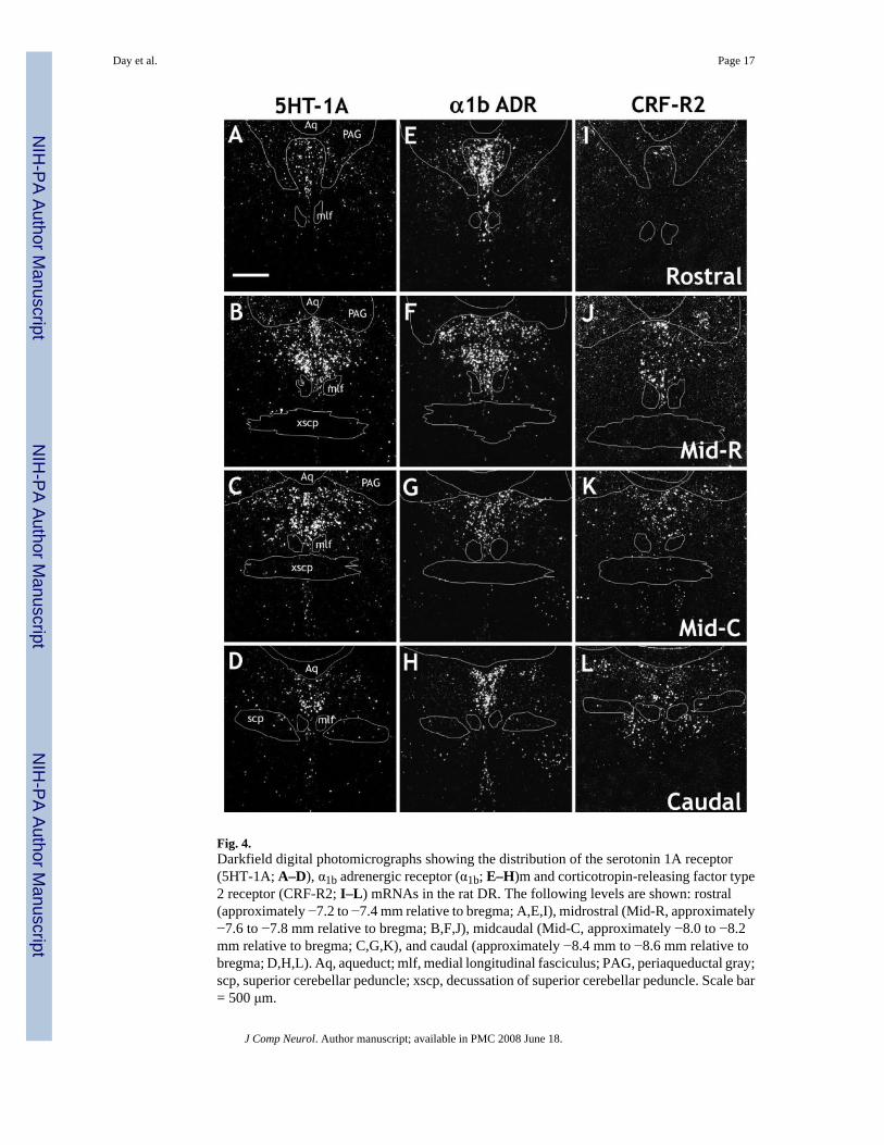
Fig. 4.Darkfield digital photomicrographs showing the distribution of the serotonin 1A receptor(5HT-1A; A–D), α1b adrenergic receptor (α1b; E–H)m and corticotropin-releasing factor type2 receptor (CRF-R2; I–L) mRNAs in the rat DR. The following levels are shown: rostral(approximately −7.2 to −7.4 mm relative to bregma; A,E,I), midrostral (Mid-R, approximately−7.6 to −7.8 mm relative to bregma; B,F,J), midcaudal (Mid-C, approximately −8.0 to −8.2mm relative to bregma; C,G,K), and caudal (approximately −8.4 mm to −8.6 mm relative tobregma; D,H,L). Aq, aqueduct; mlf, medial longitudinal fasciculus; PAG, periaqueductal gray;scp, superior cerebellar peduncle; xscp, decussation of superior cerebellar peduncle. Scale bar= 500 μm.
Day et al. Page 17
J Comp Neurol. Author manuscript; available in PMC 2008 June 18.
NIH
-PA Author Manuscript
NIH
-PA Author Manuscript
NIH
-PA Author Manuscript

Fig. 5.Expression of CRF-R1 mRNA, determined using a full-length riboprobe, in the rat DR. X-rayimages at rostral (A), midrostral (B), midcaudal (C), and caudal (D) levels. Althoughsignificant expression of CRF-R1 mRNA was detected in several brain regions, including theLDTg, very little CRF-R1 mRNA was detected in the DR. E: Darkfield photomicrograph takenat a midcaudal level, midline, to show the relatively low levels of CRF-R1 mRNA in the DR.F shows DIG-5HT-T with [35S]CRF-R1 at a rostral level, ventral part. DIG-labeled cells werephotographed under brightfield illumination (dark gray cells), whereas 35S-labeled cells werephotographed under darkfield illumination (clusters of white grains). The photographs werecombined in Adobe Photoshop (see Materials and Methods). Black arrows denote double-
Day et al. Page 18
J Comp Neurol. Author manuscript; available in PMC 2008 June 18.
NIH
-PA Author Manuscript
NIH
-PA Author Manuscript
NIH
-PA Author Manuscript

labeled cells, and white arrows denote single-labeled cells. Although levels of CRF-R1 mRNAexpression were not high enough to perform a systematic analysis, it appears that CRF-R1mRNA is expressed at low levels in both a few 5HT-T and non-5HT-T mRNA containing cells.LDTg, lateral dorsal tegmental nucleus; Aq, aqueduct; mlf, medial longitudinal fasciculus.Scale bars = 400 μm in E; 100 μm in F.
Day et al. Page 19
J Comp Neurol. Author manuscript; available in PMC 2008 June 18.
NIH
-PA Author Manuscript
NIH
-PA Author Manuscript
NIH
-PA Author Manuscript

Fig. 6.Digital photomicrographs showing that serotonin, GABA, and catecholamines are rarelyexpressed in the same cell in the rat DR. DIG-labeled cells were photographed under brightfieldillumination (dark gray cells), whereas 35S-labeled cells were photographed under darkfieldillumination (clusters of white grains). The photographs were combined in Adobe Photoshop(see Materials and Methods). A,B: Show DIG-5HT-T (serotonin transporter, used as a markerfor serotonergic cells) and [35S]GAD65/67 (glutamic acid decarboxylase, isoforms 65 and 67,used as a marker for GABAergic cells) at a midrostral level. Note that, for the dorsal and ventralDR, GABAergic cells largely flank the midline distribution of serotonergic cells (A), whereas,in the lateral wings, the distribution is more intermingled (B). C: Shows DIG-5HT-T and[35S]TH (used as a marker for catecholamines) at a rostral level, ventral part. D: Shows DIG-GAD65/67 and [35S]TH at a rostral level, dorsal part. Scale bar = 100 μm in D (applies to B–D); 200 μm for A.
Day et al. Page 20
J Comp Neurol. Author manuscript; available in PMC 2008 June 18.
NIH
-PA Author Manuscript
NIH
-PA Author Manuscript
NIH
-PA Author Manuscript

Fig. 7.Digital photomicrographs showing the relative colocalization of serotonin 1A receptor(5HT-1A) mRNA with mRNA markers for serotonin (A) or GABA (B). DIG-labeled cellswere photographed under brightfield illumination (dark gray cells), whereas 35S-labeled cellswere photographed under darkfield illumination (clusters of white grains). The photographswere combined in Adobe Photoshop (see Materials and Methods). Black arrows denote double-labeled cells, and white arrows denote single-labeled cells. A: Shows DIG-5HT-T (serotonintransporter, used as a marker for serotonergic cells) with [35S]5HT-1A at a midrostral level,ventral part. Note the extensive colocalization, although a few cells expressing 5HT-1A mRNAdid not express 5HT-T mRNA. B: Shows DIG-GAD65/67 (glutamic acid decarboxylase,isoforms 65 and 67, used as a marker for GABAergic cells) with [35S]5HT-1A at a midrostrallevel, dorsal/lateral part. A few cells expressed both GAD65/67 and 5HT-1A mRNAs. Scalebar = 100 μm.
Day et al. Page 21
J Comp Neurol. Author manuscript; available in PMC 2008 June 18.
NIH
-PA Author Manuscript
NIH
-PA Author Manuscript
NIH
-PA Author Manuscript

Fig. 8.Digital photomicrographs showing the relative colocalization of α1b adrenergic receptor(α1b) mRNA with mRNA markers for serotonin (A) or GABA (B). DIG-labeled cells werephotographed under brightfield illumination (dark gray cells), whereas 35S-labeled cells werephotographed under darkfield illumination (clusters of white grains). The photographs werecombined in Adobe Photoshop (see Materials and Methods). Black arrows denote double-labeled cells, and white arrows denote single-labeled cells. A: Shows DIG-5HT-T (serotonintransporter, used as a marker for serotonergic cells) with [35S]α1b at a midrostral level, dorsalpart. Note the extensive colocalization, although a number of cells expressing α1b mRNA didnot express 5HT-T mRNA. B: Shows DIG-GAD65/67 (glutamic acid decarboxylase, isoforms65 and 67, used as a marker for GABAergic cells) with [35S]α1b at a midrostral level, dorsal/lateral part. Although many cells were singly labeled, a number of cells expressed bothGAD65/67 and α1b mRNAs. Scale bar = 100 μm.
Day et al. Page 22
J Comp Neurol. Author manuscript; available in PMC 2008 June 18.
NIH
-PA Author Manuscript
NIH
-PA Author Manuscript
NIH
-PA Author Manuscript

Fig. 9.Digital photomicrographs showing the relative colocalization of corticotropin-releasing factortype 2 receptor (CRF-R2) mRNA with mRNA markers for serotonin or GABA. DIG-labeledcells were photographed under brightfield illumination (dark gray cells), whereas 35S-labeledcells were photographed under darkfield illumination (clusters of white grains). Thephotographs were combined in Adobe Photoshop (see Materials and Methods). Black arrowsdenote double-labeled cells, and white arrows denote single-labeled cells. A: Shows DIG-5HT-T with [35S]CRF-R2 midcaudal, dorsal part. Note the extensive colocalization, althoughseveral cells that express 5HT-T mRNA do not express CRF-R2 mRNA. B: Shows DIG-GAD65/67 (glutamic acid decarboxylase, isoforms 65 and 67, used as a marker for GABAergiccells) with [35S]CRF-R2 at a midrostral level, lateral part. Note that there is no colocalizationat this level. C: Shows DIG-GAD65/67 (glutamic acid decarboxylase, isoforms 65 and 67,used as a marker for GABAergic cells) with [35S]CRF-R2 at a midcaudal level, ventral part.
Day et al. Page 23
J Comp Neurol. Author manuscript; available in PMC 2008 June 18.
NIH
-PA Author Manuscript
NIH
-PA Author Manuscript
NIH
-PA Author Manuscript

Note that there is significant colocalization at this level, although many cells remain singlylabeled. For orientation, the single-labeled CRF-R2 cells, white grains, denoted by whitearrows on the right of the image are approximately at the midline of the ventral DR. Scale bar= 100 μm.
Day et al. Page 24
J Comp Neurol. Author manuscript; available in PMC 2008 June 18.
NIH
-PA Author Manuscript
NIH
-PA Author Manuscript
NIH
-PA Author Manuscript

NIH
-PA Author Manuscript
NIH
-PA Author Manuscript
NIH
-PA Author Manuscript
Day et al. Page 25Ta
ble
1Ex
pres
sion
of 5
HT-
1A, α
1b A
dren
ergi
c, an
d C
RF-
R2
Rec
epto
r mR
NA
in S
erot
oner
gic,
GA
BA
ergi
c, an
d C
atec
hola
min
ergi
c Cel
ls o
f the
Rat
DR
, Det
erm
ined
by
Dua
l In
Situ
Hyb
ridiz
atio
n1
No.
DIG
onl
yN
o. 35
S on
lyN
o. d
oubl
esPe
rcen
tage
dou
ble
w.r
.t. D
igPe
rcen
tage
dou
ble
w.r
.t. 35
S
DIG
-5H
T-T
+ 35
S-5H
T-1A
(n =
4)
R
ostra
l: do
rsal
1.1
± 0.
34.
9 ±
0.4
44.4
± 5
.597
.4 ±
0.7
89.9
± 0
.6
Ros
tral:
vent
ral
0.6
± 0.
26.
4 ±
0.8
44.4
± 6
.098
.5 ±
0.5
86.6
± 3
.0
Mid
-R: d
orsa
l0.
4 ±
0.2
2.0
± 0.
465
.8 ±
2.6
99.5
± 0
.397
.1 ±
0.5
M
id-R
: ven
tral
0.3
± 0.
11.
8 ±
0.5
83.1
± 2
.799
.7 ±
0.2
97.9
± 0
.6
Mid
-R: l
ater
al0.
4 ±
0.2
4.0
± 0.
838
.3 ±
1.6
99.1
± 0
.490
.4 ±
2.1
M
id-C
: dor
sal
0.6
± 0.
41.
0 ±
0.2
72.4
± 4
.799
.2 ±
0.5
98.6
± 0
.4
Mid
-C: v
entra
l0.
5 ±
0.2
0.4
± 0.
182
.6 ±
8.9
99.3
± 0
.399
.5 ±
0.2
M
id-C
: lat
eral
0.1
± 0.
12.
4 ±
0.5
34.3
± 4
.199
.8 ±
0.2
93.0
± 2
.2
Cau
dal:
dors
al0.
6 ±
0.5
2.9
± 0.
648
.5 ±
1.7
98.7
± 1
.094
.4 ±
1.2
C
auda
l: ve
ntra
l0.
5 ±
0.2
1.5
± 0.
536
.0 ±
2.4
98.7
± 0
.596
.2 ±
0.9
DIG
-GA
D65
/67
+ 35
S-5H
T-1A
(n =
4)
R
ostra
l: do
rsal
37.4
± 3
.045
.8 ±
8.1
7.5
± 1.
116
.9 ±
2.8
14.5
± 1
.5
Ros
tral:
vent
ral
29.3
± 3
.637
.4 ±
2.8
6.1
± 0.
517
.8 ±
1.9
14.1
± 0
.7
Mid
-R: d
orsa
l22
.5 ±
2.4
61.3
± 5
.52.
3 ±
0.5
9.7
± 2.
93.
5 ±
0.7
M
id-R
: ven
tral
19.0
± 2
.573
.1 ±
10.
91.
1 ±
0.3
5.9
± 2.
01.
4 ±
0.2
M
id-R
: lat
eral
57.7
± 6
.240
.6 ±
2.5
4.8
± 0.
38.
0 ±
1.2
10.6
± 0
.8
Mid
-C: d
orsa
l19
.4 ±
1.9
74.1
± 9
.10.
9 ±
0.6
4.0
± 2.
41.
0 ±
0.5
M
id-C
: ven
tral
15.3
± 1
.471
.8 ±
7.9
0.6
± 0.
23.
8 ±
1.3
0.8
± 0.
3
Mid
-C: l
ater
al57
.5 ±
7.7
37.9
± 3
.03.
8 ±
0.8
6.0
± 0.
69.
4 ±
2.6
C
auda
l: do
rsal
26.3
± 3
.435
.8 ±
5.8
2.1
± 0.
47.
6 ±
1.4
5.6
± 0.
7
Cau
dal:
vent
ral
31.4
± 4
.922
.6 ±
1.7
1.6
± 0.
25.
5 ±
1.5
6.6
± 0.
8D
IG-T
H +
35S-
5HT-
1A (n
= 4
)
Ros
tral:
dors
al16
.5 ±
2.9
31.0
± 3
.50.
5 ±
0.2
2.8
± 1.
21.
7 ±
0.9
R
ostra
l: ve
ntra
l13
.3 ±
3.9
27.6
± 3
.00.
1 ±
0.1
1.5
± 1.
50.
5 ±
0.5
M
id-R
: dor
sal
8.8
± 2.
736
.1 ±
3.8
0.0
± 0.
00.
0 ±
0.0
0.0
± 0.
0
Mid
-R: v
entra
l4.
6 ±
3.1
50.0
± 7
.50.
0 ±
0.0
0.0
± 0.
00.
0 ±
0.0
M
id-R
: lat
eral
2.1
± 0.
625
.7 ±
2.6
0.0
± 0.
00.
0 ±
0.0
0.0
± 0.
0
Mid
-C: d
orsa
l2.
5 ±
0.3
32.0
± 4
.80.
3 ±
0.1
12.2
± 5
.41.
0 ±
0.4
M
id-C
: ven
tral
0.0
± 0.
038
.8 ±
6.6
0.0
± 0.
00.
0 ±
0.0
0.0
± 0.
0
Mid
-C: l
ater
al0.
8 ±
0.1
22.7
± 2
.80.
0 ±
0.0
0.0
± 0.
00.
0 ±
0.0
C
auda
l: do
rsal
0.4
± 0.
219
.1 ±
1.9
0.0
± 0.
00.
0 ±
0.0
0.0
± 0.
0
Cau
dal:
vent
ral
0.0
± 0.
024
.8 ±
2.9
0.0
± 0.
00.
0 ±
0.0
0.0
± 0.
0D
IG-5
HT-
T +
35S-α 1
b (n
= 4)
R
ostra
l: do
rsal
0.7
± 0.
214
.2 ±
3.2
53.3
± 4
.698
.8 ±
0.2
79.1
± 4
.1
Ros
tral:
vent
ral
0.3
± 0.
212
.5 ±
3.3
31.3
± 9
.999
.2 ±
0.4
68.7
± 7
.1
Mid
-R: d
orsa
l0.
6 ±
0.3
22.8
± 4
.284
.9 ±
4.2
99.3
± 0
.378
.9 ±
3.8
M
id-R
: ven
tral
0.5
± 0.
56.
5 ±
0.8
82.9
± 1
2.0
99.5
± 0
.592
.3 ±
1.6
M
id-R
: lat
eral
0.3
± 0.
116
.8 ±
3.4
26.8
± 2
.299
.1 ±
0.6
60.6
± 7
.2
Mid
-C: d
orsa
l1.
3 ±
0.3
12.3
± 2
.077
.1 ±
4.6
98.4
± 0
.386
.6 ±
1.5
M
id-C
: ven
tral
1.5
± 0.
64.
9 ±
0.9
84.0
± 1
1.6
98.3
± 0
.693
.9 ±
1.7
M
id-C
: lat
eral
0.6
± 0.
48.
8 ±
1.4
38.5
± 5
.598
.5 ±
0.8
81.3
± 1
.7
Cau
dal:
dors
al0.
5 ±
0.3
7.5
± 1.
160
.3 ±
3.4
99.2
± 0
.488
.9 ±
1.4
C
auda
l: ve
ntra
l0.
9 ±
0.5
5.4
± 1.
355
.8 ±
7.5
98.1
± 1
.290
.9 ±
2.2
DIG
-GA
D65
/67
+ 35
S-α 1
b (n
= 4)
R
ostra
l: do
rsal
19.2
± 5
.361
.2 ±
11.
111
.7 ±
2.3
38.8
± 1
.916
.9 ±
4.0
R
ostra
l: ve
ntra
l11
.0 ±
2.1
36.3
± 1
0.5
12.5
± 1
.853
.4 ±
7.0
27.5
± 4
.2
Mid
-R: d
orsa
l8.
0 ±
0.8
79.1
± 5
.88.
0 ±
0.9
49.9
± 2
.39.
4 ±
1.4
M
id-R
: ven
tral
11.1
± 1
.583
.5 ±
9.7
7.9
± 0.
542
.1 ±
3.8
9.0
± 1.
2
Mid
-R: l
ater
al31
.1 ±
4.2
33.5
± 1
.222
.2 ±
3.1
41.6
± 1
.039
.4 ±
2.9
J Comp Neurol. Author manuscript; available in PMC 2008 June 18.

NIH
-PA Author Manuscript
NIH
-PA Author Manuscript
NIH
-PA Author Manuscript
Day et al. Page 26N
o. D
IG o
nly
No.
35S
only
No.
dou
bles
Perc
enta
ge d
oubl
ew
.r.t.
Dig
Perc
enta
ge d
oubl
ew
.r.t.
35S
M
id-C
: dor
sal
8.6
± 0.
470
.1 ±
5.2
7.3
± 1.
144
.8 ±
4.5
9.6
± 1.
9
Mid
-C: v
entra
l8.
6 ±
0.9
75.8
± 6
.84.
5 ±
1.3
32.8
± 7
.65.
6 ±
1.7
M
id-C
: lat
eral
33.8
± 4
.237
.7 ±
2.9
13.1
± 1
.328
.3 ±
2.4
25.9
± 2
.3
Cau
dal:
dors
al25
.5 ±
6.7
51.1
± 2
.93.
8 ±
1.1
16.9
± 7
.76.
7 ±
1.5
C
auda
l: ve
ntra
l22
.1 ±
7.7
39.5
± 3
.33.
1 ±
0.7
17.8
± 7
.77.
5 ±
1.6
DIG
-TH
+ 35
S-α 1
b (n
= 4)
R
ostra
l: do
rsal
12.1
± 2
.236
.4 ±
6.0
0.9
± 0.
65.
2 ±
3.0
2.5
± 1.
4
Ros
tral:
vent
ral
7.3
± 1.
934
.3 ±
8.2
2.1
± 0.
723
.0 ±
3.3
6.7
± 1.
7
Mid
-R: d
orsa
l7.
6 ±
2.7
48.1
± 3
.30.
5 ±
0.3
7.3
± 5.
20.
9 ±
0.5
M
id-R
: ven
tral
4.1
± 2.
547
.4 ±
3.0
0.9
± 0.
420
.4 ±
10.
71.
8 ±
0.8
M
id-R
: lat
eral
1.9
± 0.
627
.1 ±
1.5
0.0
± 0.
00.
0 ±
0.0
0.0
± 0.
0
Mid
-C: d
orsa
l1.
3 ±
0.5
41.8
± 5
.40.
0 ±
0.0
0.0
± 0.
00.
0 ±
0.0
M
id-C
: ven
tral
0.0
± 0.
032
.9 ±
2.8
0.0
± 0.
00.
0 ±
0.0
0.0
± 0.
0
Mid
-C: l
ater
al0.
1 ±
0.1
20.0
± 3
.30.
0 ±
0.0
0.0
± 0.
00.
0 ±
0.0
C
auda
l: do
rsal
0.1
± 0.
120
.3 ±
1.0
0.0
± 0.
00.
0 ±
0.0
0.0
± 0.
0
Cau
dal:
vent
ral
0.0
± 0.
015
.4 ±
0.9
0.0
± 0.
00.
0 ±
0.0
0.0
± 0.
0D
IG-5
HT-
T +
35S-
CR
F-R
2 (n
= 4
)
Ros
tral:
dors
al40
.8 ±
5.8
0.0
± 0.
01.
6 ±
0.4
3.7
± 0.
510
0.0
± 0.
0
Ros
tral:
vent
ral
37.4
± 7
.10.
0 ±
0.0
0.8
± 0.
31.
6 ±
0.6
100.
0 ±
0.0
M
id-R
: dor
sal
56.1
± 6
.90.
0 ±
0.0
5.6
± 1.
79.
0 ±
2.0
100.
0 ±
0.0
M
id-R
: ven
tral
85.0
± 2
.90.
0 ±
0.0
9.3
± 1.
39.
8 ±
1.2
100.
0 ±
0.0
M
id-R
: lat
eral
34.9
± 4
.80.
0 ±
0.0
1.7
± 0.
34.
8 ±
0.8
100.
0 ±
0.0
M
id-C
: dor
sal
49.0
± 4
.10.
0 ±
0.0
14.3
± 1
.922
.7 ±
3.1
100.
0 ±
0.0
M
id-C
: ven
tral
57.5
± 6
.20.
3 ±
0.1
6.5
± 1.
010
.3 ±
1.2
95.5
± 2
.6
Mid
-C: l
ater
al35
.4 ±
6.2
0.4
± 0.
22.
3 ±
0.5
6.9
± 2.
585
.9 ±
6.5
C
auda
l: do
rsal
30.1
± 8
.45.
4 ±
1.0
4.9
± 1.
416
.9 ±
6.5
46.0
± 6
.9
Cau
dal:
vent
ral
35.9
± 3
.85.
5 ±
1.5
1.6
± 0.
54.
4 ±
1.4
24.7
± 1
0.0
DIG
-GA
D65
/67
+ 35
S-C
RF-
R2
(n =
5)
R
ostra
l: do
rsal
26.9
± 3
.01.
2 ±
0.4
0.0
± 0.
00.
0 ±
0.0
0.0
± 0.
0
Ros
tral:
vent
ral
33.3
± 5
.80.
8 ±
0.4
0.0
± 0.
00.
0 ±
0.0
0.0
± 0.
0
Mid
-R: d
orsa
l13
.9 ±
1.6
7.4
± 1.
80.
0 ±
0.0
0.0
± 0.
00.
0 ±
0.0
M
id-R
: ven
tral
16.6
± 2
.39.
3 ±
3.5
0.0
± 0.
00.
0 ±
0.0
0.0
± 0.
0
Mid
-R: l
ater
al41
.0 ±
2.3
3.6
± 1.
20.
0 ±
0.0
0.0
± 0.
00.
0 ±
0.0
M
id-C
: dor
sal
14.5
± 3
.816
.8 ±
2.1
0.0
± 0.
00.
0 ±
0.0
0.0
± 0.
0
Mid
-C: v
entra
l16
.7 ±
2.8
12.0
± 1
.40.
5 ±
0.5
1.7
± 1.
72.
9 ±
2.9
M
id-C
: lat
eral
55.9
± 7
.63.
8 ±
0.7
0.7
± 0.
50.
8 ±
0.6
11.6
± 9
.1
Cau
dal:
dors
al40
.1 ±
8.6
5.3
± 1.
05.
7 ±
1.4
12.4
± 1
.051
.2 ±
8.2
C
auda
l: ve
ntra
l29
.4 ±
2.4
4.1
± 1.
16.
4 ±
1.1
17.9
± 2
.966
.2 ±
8.7
DIG
-TH
+ 35
S-C
RF-
R2
(n =
4)
R
ostra
l: do
rsal
18.5
± 0
.92.
5 ±
1.1
0.0
± 0.
00.
0 ±
0.0
0.0
± 0.
0
Ros
tral:
vent
ral
15.4
± 3
.21.
9 ±
0.6
0.0
± 0.
00.
0 ±
0.0
0.0
± 0.
0
Mid
-R: d
orsa
l16
.9 ±
2.7
10.4
± 3
.20.
0 ±
0.0
0.0
± 0.
00.
0 ±
0.0
M
id-R
: ven
tral
6.5
± 2.
610
.5 ±
2.3
0.0
± 0.
00.
0 ±
0.0
0.0
± 0.
0
Mid
-R: l
ater
al3.
8 ±
0.3
3.8
± 1.
20.
0 ±
0.0
0.0
± 0.
00.
0 ±
0.0
M
id-C
: dor
sal
5.8
± 1.
819
.3 ±
3.1
0.0
± 0.
00.
0 ±
0.0
0.0
± 0.
0
Mid
-C: v
entra
l0.
0 ±
0.0
17.8
± 3
.90.
0 ±
0.0
0.0
± 0.
00.
0 ±
0.0
M
id-C
: lat
eral
0.7
± 0.
34.
7 ±
1.1
0.0
± 0.
00.
0 ±
0.0
0.0
± 0.
0
Cau
dal:
dors
al0.
1 ±
0.1
10.5
± 0
.90.
0 ±
0.0
0.0
± 0.
00.
0 ±
0.0
C
auda
l: ve
ntra
l0.
0 ±
0.0
10.8
± 2
.70.
0 ±
0.0
0.0
± 0.
00.
0 ±
0.0
1 Dua
l in
situ
hyb
ridiz
atio
n w
as u
sed
to d
eter
min
e th
e di
strib
utio
n an
d re
lativ
e co
loca
lizat
ion
of 5
HT-
1A (s
erot
onin
1A
rece
ptor
), α 1
b (α
1B a
dren
ergi
c re
cept
or) a
nd C
RF-
R2
(cor
ticot
ropi
n re
leas
ing
fact
or re
cept
or ty
pe 2
) mR
NA
in se
roto
nerg
ic (e
xpre
ssin
g se
roto
nin
trans
porte
r (5H
T-T)
mR
NA
), G
AB
Aer
gic
(exp
ress
ing
glut
amic
aci
d de
carb
oxyl
ase
isof
orm
s 65
or 6
7 (G
AD
65/6
7) m
RN
A) o
rca
tech
olam
iner
gic
(exp
ress
ing
tyro
sine
hyd
roxy
lase
(TH
) mR
NA
) cel
ls th
roug
h th
e ro
stra
l-cau
dal e
xten
t of t
he ra
t dor
sal r
aphe
nuc
leus
. Cel
ls w
ere
defin
ed a
s bei
ng la
bele
d fo
r the
dig
oxig
enin
pro
be
J Comp Neurol. Author manuscript; available in PMC 2008 June 18.

NIH
-PA Author Manuscript
NIH
-PA Author Manuscript
NIH
-PA Author Manuscript
Day et al. Page 27on
ly (D
ig),
radi
oact
ive
prob
e on
ly (3
5 S) o
r bot
h pr
obes
(dou
ble)
. Num
bers
in th
e fir
st 3
col
umns
refe
r to
the
aver
age
num
ber o
f cel
ls c
ount
ed p
er se
ctio
n an
alyz
ed. A
t lea
st 2
sect
ions
wer
e an
alyz
ed p
erra
t for
eac
h re
gion
, and
the
data
ave
rage
d to
obt
ain
a si
ngle
val
ue fo
r eac
h an
imal
. The
se si
ngle
val
ues w
ere
then
ave
rage
d. “
n” re
fers
to th
e nu
mbe
r of a
nim
als a
naly
zed
(not
the
num
ber o
f sec
tions
).N
ote:
Cel
ls w
ere
coun
ted
with
the
aid
of a
grid
in o
rder
to c
alcu
late
the
perc
enta
ge o
f dou
ble-
labe
led
cells
, and
do
not r
epre
sent
abs
olut
e nu
mbe
rs o
f cel
ls in
a g
iven
regi
on. H
owev
er, s
ince
the
grid
use
dw
as id
entic
al b
etw
een
coun
ts, t
he n
umbe
rs g
ive a
n ap
prox
imat
ion
for r
elat
ive c
ell c
ount
s for
each
pro
be. T
he p
erce
ntag
e col
ocal
izat
ion
was
calc
ulat
ed se
para
tely
for e
ach
anim
al an
d th
en av
erag
ed ac
ross
anim
als.
Hen
ce th
e nu
mbe
rs in
the
last
2 c
olum
ns h
ave
not b
een
deriv
ed d
irect
ly fr
om th
e nu
mbe
rs in
the
first
3 c
olum
ns. w
.r.t.—
with
resp
ect t
o
J Comp Neurol. Author manuscript; available in PMC 2008 June 18.