Catecholaminergic connectivity to the inner ear, central auditory, and vocal motor circuitry in the...
41
Catecholaminergic Connectivity to the Inner Ear, Central Auditory, and Vocal Motor Circuitry in the Plainfin Midshipman Fish Porichthys notatus Paul M. Forlano, 1,2,3,4 * Spencer D. Kim, 1 Zuzanna M. Krzyminska, 1 and Joseph A. Sisneros 4,5,6 1 Department of Biology, Brooklyn College, City University of New York, Brooklyn, New York 11210 2 Programs in Neuroscience, Ecology, Evolutionary Biology and Behavior, and Behavioral and Cognitive Neuroscience, The Graduate Center, City University of New York, New York, New York 10016 3 Aquatic Research and Environmental Assessment Center, Brooklyn College, Brooklyn, New York 11210 4 Marine Biological Laboratory, Woods Hole, MA 02543 5 Departments of Psychology and Biology, University of Washington, Seattle, WA 98195 6 Virginia Merrill Bloedel Hearing Research Center, Seattle, WA 98195 ABSTRACT Although the neuroanatomical distribution of catecholami- nergic (CA) neurons has been well documented across all vertebrate classes, few studies have examined CA connec- tivity to physiologically and anatomically identified neural circuitry that controls behavior. The goal of this study was to characterize CA distribution in the brain and inner ear of the plainfin midshipman fish (Porichthys notatus) with particular emphasis on their relationship with anatomically labeled circuitry that both produces and encodes social acoustic signals in this species. Neurobiotin labeling of the main auditory end organ, the saccule, combined with tyro- sine hydroxylase immunofluorescence (TH-ir) revealed a strong CA innervation of both the peripheral and central auditory system. Diencephalic TH-ir neurons in the peri- ventricular posterior tuberculum, known to be dopaminer- gic, send ascending projections to the ventral telencephalon and prominent descending projections to vocal–acoustic integration sites, notably the hindbrain octavolateralis efferent nucleus, as well as onto the base of hair cells in the saccule via nerve VIII. Neurobiotin back- fills of the vocal nerve in combination with TH-ir revealed CA terminals on all components of the vocal pattern gen- erator, which appears to largely originate from local TH-ir neurons but may include input from diencephalic projec- tions as well. This study provides strong neuroanatomical evidence that catecholamines are important modulators of both auditory and vocal circuitry and acoustic-driven social behavior in midshipman fish. This demonstration of TH-ir terminals in the main end organ of hearing in a nonmam- malian vertebrate suggests a conserved and important anatomical and functional role for dopamine in normal audition. J. Comp. Neurol. 000:000–000, 2014. V C 2014 Wiley Periodicals, Inc. INDEXING TERMS: dopaminergic neurons; noradrenergic neurons; posterior tuberculum; saccule; vocal pattern gen- erator; octavolateralis efferent nucleus Catecholamines (CAs) are a highly conserved group of neurochemicals that are known to function as impor- tant modulators of motivation, reward, arousal, sensory and motor systems, and reproduction (Berridge, 2008; Hurley et al., 2004; Joshua et al., 2009; Riters, 2012; Salamone and Correa, 2012). The neuroanatomical dis- tribution of catecholaminergic (CA) neuronal groups, which include dopamine (DA) and noradrenaline (NA), is well characterized across all major vertebrate taxa, and, with a few exceptions, is largely conserved. Like other vertebrates, teleosts exhibit brainstem CA cell groups Additional Supporting Information may be found in the online version of this article. Grant sponsor: National Institutes of Health; Grant number: SC2DA034996 (to P.M.F.); Grant sponsor: The Professional Staff Con- gress/The City University of New York (PSC-CUNY); Grant number: 65650-00 43 (to P.M.F.); Grant sponsor: Leonard and Claire Tow Travel Award (to P.M.F.); Grant sponsor: Whitman Investigator Faculty Research Fellowships from the Marine Biological Laboratory, Woods, Hole, MA (where the study was partly conducted) (to P.M.F. and J.A.S.). *CORRESPONDENCE TO: Paul M. Forlano, Department of Biology, Brooklyn College, City University of New York, 2900 Bedford Ave, Brook- lyn, NY 11210. E-mail: [email protected]. Received January 16, 2014; Revised March 25, 2014; Accepted March 28, 2014. DOI 10.1002/cne.23596 Published online April 8, 2014 in Wiley Online Library (wileyonlinelibrary.com) V C 2014 Wiley Periodicals, Inc. The Journal of Comparative Neurology | Research in Systems Neuroscience 00:00–00 (2014) 1 RESEARCH ARTICLE
Transcript of Catecholaminergic connectivity to the inner ear, central auditory, and vocal motor circuitry in the...

Catecholaminergic Connectivity to the Inner Ear,Central Auditory, and Vocal Motor Circuitry in thePlainfin Midshipman Fish Porichthys notatus
Paul M. Forlano,1,2,3,4* Spencer D. Kim,1 Zuzanna M. Krzyminska,1 and Joseph A. Sisneros4,5,6
1Department of Biology, Brooklyn College, City University of New York, Brooklyn, New York 112102Programs in Neuroscience, Ecology, Evolutionary Biology and Behavior, and Behavioral and Cognitive Neuroscience, The Graduate
Center, City University of New York, New York, New York 100163Aquatic Research and Environmental Assessment Center, Brooklyn College, Brooklyn, New York 112104Marine Biological Laboratory, Woods Hole, MA 025435Departments of Psychology and Biology, University of Washington, Seattle, WA 981956Virginia Merrill Bloedel Hearing Research Center, Seattle, WA 98195
ABSTRACTAlthough the neuroanatomical distribution of catecholami-
nergic (CA) neurons has been well documented across all
vertebrate classes, few studies have examined CA connec-
tivity to physiologically and anatomically identified neural
circuitry that controls behavior. The goal of this study was
to characterize CA distribution in the brain and inner ear
of the plainfin midshipman fish (Porichthys notatus) with
particular emphasis on their relationship with anatomically
labeled circuitry that both produces and encodes social
acoustic signals in this species. Neurobiotin labeling of the
main auditory end organ, the saccule, combined with tyro-
sine hydroxylase immunofluorescence (TH-ir) revealed a
strong CA innervation of both the peripheral and central
auditory system. Diencephalic TH-ir neurons in the peri-
ventricular posterior tuberculum, known to be dopaminer-
gic, send ascending projections to the ventral
telencephalon and prominent descending projections to
vocal–acoustic integration sites, notably the hindbrain
octavolateralis efferent nucleus, as well as onto the base
of hair cells in the saccule via nerve VIII. Neurobiotin back-
fills of the vocal nerve in combination with TH-ir revealed
CA terminals on all components of the vocal pattern gen-
erator, which appears to largely originate from local TH-ir
neurons but may include input from diencephalic projec-
tions as well. This study provides strong neuroanatomical
evidence that catecholamines are important modulators of
both auditory and vocal circuitry and acoustic-driven social
behavior in midshipman fish. This demonstration of TH-ir
terminals in the main end organ of hearing in a nonmam-
malian vertebrate suggests a conserved and important
anatomical and functional role for dopamine in normal
audition. J. Comp. Neurol. 000:000–000, 2014.
VC 2014 Wiley Periodicals, Inc.
INDEXING TERMS: dopaminergic neurons; noradrenergic neurons; posterior tuberculum; saccule; vocal pattern gen-
erator; octavolateralis efferent nucleus
Catecholamines (CAs) are a highly conserved group
of neurochemicals that are known to function as impor-
tant modulators of motivation, reward, arousal, sensory
and motor systems, and reproduction (Berridge, 2008;
Hurley et al., 2004; Joshua et al., 2009; Riters, 2012;
Salamone and Correa, 2012). The neuroanatomical dis-
tribution of catecholaminergic (CA) neuronal groups,
which include dopamine (DA) and noradrenaline (NA), is
well characterized across all major vertebrate taxa, and,
with a few exceptions, is largely conserved. Like other
vertebrates, teleosts exhibit brainstem CA cell groups
Additional Supporting Information may be found in the online versionof this article.
Grant sponsor: National Institutes of Health; Grant number:SC2DA034996 (to P.M.F.); Grant sponsor: The Professional Staff Con-gress/The City University of New York (PSC-CUNY); Grant number:65650-00 43 (to P.M.F.); Grant sponsor: Leonard and Claire Tow TravelAward (to P.M.F.); Grant sponsor: Whitman Investigator Faculty ResearchFellowships from the Marine Biological Laboratory, Woods, Hole, MA(where the study was partly conducted) (to P.M.F. and J.A.S.).
*CORRESPONDENCE TO: Paul M. Forlano, Department of Biology,Brooklyn College, City University of New York, 2900 Bedford Ave, Brook-lyn, NY 11210. E-mail: [email protected].
Received January 16, 2014; Revised March 25, 2014;Accepted March 28, 2014.DOI 10.1002/cne.23596Published online April 8, 2014 in Wiley Online Library(wileyonlinelibrary.com)VC 2014 Wiley Periodicals, Inc.
The Journal of Comparative Neurology | Research in Systems Neuroscience 00:00–00 (2014) 1
RESEARCH ARTICLE

in the area postrema (AP), the vagal lobe (and associ-
ated areas), and the noradrenergic (NAergic) locus
coeruleus, as well as dopaminergic (DAergic) cell
groups in the hypothalamus, preoptic area, thalamus,
subpallium, and olfactory bulb but lack the midbrain
ventral tegmental (VTA)/substantia nigra (SN) DA popu-
lations found in both tetrapods and elasmobranch
fishes that form the well-characterized ascending meso-
limbic/nigrostriatal pathways involved in motivation and
reward-related behaviors (for review, see Carrera et al.,
2012; O’Connell and Hofmann, 2011; Smeets and Gon-
zalez, 2000; Yamamoto and Vernier, 2011). A subpopu-
lation of DA neurons in the diencephalic periventricular
posterior tuberculum (TPp) of teleosts sends ascending
projections to the ventral telencephalon and therefore
was originally proposed to be homologous to VTA/SN
of tetrapods (Rink and Wullimann, 2001, 2002a; O’Con-
nell and Hofmann, 2011, 2012); however, recent stud-
ies using genetic manipulations strongly suggest this
group is homologous to diencephalic A11 DA neurons
that are dependent on the transcription factor orthope-
dia (otp), and local subpallial DA neurons may function
to supply DA to proposed striatal homologs (Kasten-
huber et al., 2010; Lohr et al., 2009; Ryu et al., 2006;
Schweitzer et al., 2012; Tay et al., 2011).
Regardless of homology, the widespread projection
patterns (largely descending but also ascending) of
these TPp neurons position them to be important inte-
grative neuromodulators of sensory and motor function,
cognition, and behavior (Ma, 2003; Schweitzer et al.,
2012; Tay et al., 2011). Teleosts exhibit CA neuronal
populations and innervation patterns in nuclei with pro-
posed homologies to those found in tetrapods where
they are known to modulate motivated social and repro-
ductive behavior (Goodson and Kingsbury, 2013; O’Con-
nell and Hofmann, 2011; Petersen et al., 2013).
However, aside from DA neurons in the anterior pre-
optic area that are well documented to regulate the
hypothalamic–pituitary–gonadal (HPG) axis in teleosts
(Dufour et al., 2005, 2010; Kah et al., 1987; Peter and
Fryer, 1983), the functions of specific CA cell groups in
teleosts are largely unknown. However, recent studies
in teleosts have begun to characterize cFos induction
of CA nuclei to social challenge stimuli (O’Connell
et al., 2013; Petersen et al., 2013). Furthermore, there
are few examples (McLean and Fetcho, 2004b) of CA
connectivity to physiologically and anatomically identi-
fied neural circuitry that controls behavior in teleosts.
The plainfin midshipman fish, Porichthys notatus, is a
well-studied model system for understanding neural and
hormonal mechanisms underlying vocal–acoustic com-
munication in vertebrates (Bass and McKibben, 2003;
Forlano and Bass, 2011), and thus is an ideal model to
investigate the structure/function of CAs in relation to
well-delineated vocal and auditory circuits, which may
in turn provide important insights into the evolution of
CAs in auditory-driven social behavior (Petersen et al.,
2013). During the summer months in the intertidal zone
off northern California and the Pacific Northwest, type I
male midshipman excavate and defend nests under
rocks, and court females at night by producing a long-
duration (>1 minute) advertisement call via rapid con-
traction of sound-generating musculature on the sides
of the swimbladder (Bass and McKibben, 2003; Cohen
and Winn, 1967). Females localize males by sound,
spawn once, and return offshore, whereas type I males
continue to court additional females and alone care for
offspring (Bass, 1996; Bass and McKibben, 2003). Type
II males do not court females but rather sneak or satel-
lite spawn in competition with type I males (Bass,
1996; Brantley and Bass, 1994).
The descending vocal motor system in midshipman
has been physiologically and anatomically well charac-
terized and consists of forebrain nuclei in the preoptic
area and anterior hypothalamus with connections to the
midbrain periaqueductal gray, paratoral and midbrain
isthmal nuclei, which in turn connect to the vocal pat-
tern generator (VPG) in the hindbrain spinal cord (Bass
et al., 1994; Chagnaud et al., 2011; Goodson and Bass,
2002; Kittelberger and Bass, 2013; Kittelberger et al.,
2006). The VPG consists of vocal pre-pacemaker neu-
rons (VPP) that receive input from midbrain vocal cen-
ters and hindbrain auditory nuclei, and innervate vocal
pacemaker neurons (VPN) and the central paired vocal
motor nucleus (VMN) whose axons exit the central
nervous system (CNS) as occipital nerve roots to inner-
vate vocal musculature (Bass and Baker, 1990; Bass
et al., 1994; Chagnaud et al., 2011; Goodson and Bass,
2002). Importantly, the VPG sets the temporal pattern
of natural calls, and this is encoded separately by VPP
for duration and VPN for frequency (Chagnaud et al.,
2011, 2012). Furthermore, the VPG of midshipman is
derived from the same compartment of the CNS that
patterns vocalizations in tetrapods (Bass and Baker,
1997; Bass et al., 2008).
The auditory system in midshipman has also been
well characterized and, like other teleosts, contains pri-
mary afferents from the saccule, the main end organ of
hearing, which synapse onto first-order medullary neu-
rons comprising the descending octaval nucleus and its
subdivisions and secondary octaval populations, both of
which project to the midbrain torus semicircularis,
which in turn projects to several diencephalic nuclei
including the anterior tuberal hypothalamus and central
posterior thalamus, which relays information to pallial
and subpallial nuclei (Bass et al., 2000; Bass et al.,
P.M. Forlano et al.
2 The Journal of Comparative Neurology |Research in Systems Neuroscience

1994; Goodson and Bass, 2002; McCormick, 1999,
2011). A hindbrain octavolateral efferent nucleus (OE)
receives input from the vocal motor system and proj-
ects to the inner ear end organs and lateral line system
(Bass et al., 1994, 2000; Chagnaud et al., 2011; Chag-
naud and Bass, 2013; Weeg et al., 2005). Additionally,
the auditory system in midshipman is interconnected to
vocal nuclei in the forebrain, midbrain, and hindbrain
(Bass et al., 2000, 2001a; Goodson and Bass, 2002;
Kittelberger et al., 2006).
The goal of this study was to characterize CA distribu-
tion in the midshipman brain with particular emphasis on
its relationship to identified circuitry that both produces
and encodes social acoustic signals. We tested the
hypothesis that CA neurons directly innervate the periph-
eral and central auditory system and VPG by transneuro-
nal backfill of the saccular branch of nerve VIII and vocal
occipital nerve roots, respectively, combined with immu-
nofluorescence (-ir) for tyrosine hydroxylase (TH), the
rate-limiting enzyme for catecholamine synthesis. Data
contained in the present study have been partly reported
in abstract form (Forlano et al., 2012).
MATERIALS AND METHODS
AnimalsFish were hand collected from intertidal nests in Tom-
ales Bay, CA or Hood Canal, WA in the summer reproduc-
tive period or captured by otter trawl off Edmonds, WA in
Neuroanatomical Abbreviations
ac anterior commissureAP area postremaAT anterior tuberal nucleusC cerebellumCA cerebral aqueductcc cerebellar crestCg granular layer of the corpus of the cerebellumCm molecular layer of the corpus of the cerebellumCP central posterior nucleus of the thalamusCPc compact division of the central posterior nucleusCPd diffuse division of the central posterior nucleusd dendrites of octavolateralis efferent nucleusD area dorsalis of the telencephalonDc central zone of DDf diffuse nucleus of the hypothalamusDl lateral zone of DDm medial zone of DDm-p posterior zone of DmDOdl/dl dorsolateral division of descending octaval nucleusDOdm/dm dorsomedial division of descending octaval nucleusDOi/i intermediate division of descending octaval nucleusDOri/ri rostral intermediate division of descending octaval nucleusDp posterior zone of DDPo dorsal posterior nucleus of the thalamusea efferent axons of octavolateralis efferent nucleusEB efferent bundle tract of octavolateralis efferent nucleusEG eminentia granularisG nucleus glomerulosusHa habenulaHC hair cell layerHc central periventricular hypothalamusHd dorsal periventricular hypothalamusHoC horizontal commissureHv ventral periventricular hypothalamusiaf internal arcuate fiber tractIII third ventricleIL inferior lobe of the hypothalamusIN intermediate nucleus of the hypothalamusIP isthmal paraventricular nucleusIs midbrain isthmal nucleus (not nucleus isthmi)IV fourth ventricleLH lateral hypothalamusLC locus coeruleusLL central tract of the lateral line nervesll lateral lemniscusMED cell plate of medial octavolateralis nucleusMFB medial forebrain bundleMG magnocellular octaval nucleusmlct medial longitudinal catecholaminergic tractMLF medial longitudinal fasiculusnIII oculomotor nucleusnll nucleus of the lateral lemniscusNIn nucleus interpeduncularisOB olfactory bulboc occipital nervesOE octavolateralis efferent nucleusOEc caudal division of OE
OEd-c caudal division of OE dendritic fieldOEd-r rostral division of OE dendritic fieldOEr rostral division of OEON optic nerveOT optic tractPAG periaqueductal grayPe periventricular cell layer of the torus semicircularisPGl lateral division of nucleus preglomerulosusPGm medial division of nucleus preglomerulosusPHT preoptico-hypophysial tractPit pituitaryPL paralemniscal midbrain tegmentumPM magnocellular preoptic nucleusPMg gigantocellular division of PMPoC posterior commissurePPa anterior parvocellular preoptic nucleusPPp posterior parvocellular preoptic nucleusPPv nucleus pretectalis periventricularis pars ventralisPTN posterior tuberal nucleusPTT paratoral tegmentumPVO paraventricular organRF reticular formationSC support cell layerSCN suprachiasmatic nucleusSD saccus dorsalisSE sensory epithelium of the sacculeSOv ventral division of secondary octaval nucleusSR superior rapheSU supramedullary neuronsSV saccus vasculosusT telencephalonTeg tegmentumTe midbrain tectumTL torus longitudinalisTPp periventricular nucleus of the posterior tuberculumTS torus semicircularisTSd deep layer of torus semicircularisv ventricleV area ventralis of the telencephalonVc central nucleus of VVd dorsal nucleus of VVde descending tract of the trigeminal nerveVg granular layer of the valvulaVIII eighth nerveVL ventrolateral nucleus of the thalamusVm trigeminal motor nucleusVM ventromedial nucleus of the thalamusVMN vocal motor nucleusVp postcommissural nucleus of VVPN vocal pacemaker nucleusVPP vocal prepacemaker nucleusVs supracommissural nucleus of VvT ventral tuberal hypothalamusVT ventral tegmental nucleusVv ventral nucleus of VXL vagal lobeXm vagal motor nucleus
Catecholamines in auditory and vocal circuity
The Journal of Comparative Neurology | Research in Systems Neuroscience 3

Puget Sound in the winter nonreproductive season
(December). Fish were either shipped to the Aquatic
Research and Environmental Assessment Center (AREAC)
at City University of New York (CUNY) Brooklyn College,
separated by sexual morphotype, and maintained in recir-
culating saltwater aquaria or shipped to the Marine Biolog-
ical Laboratory (MBL) in Woods Hole, MA and maintained
in flow-through seawater tables. All experimental proce-
dures performed in this study were approved by the Ani-
mal Care and Use Committee of CUNY Brooklyn College
and the MBL. A total of 42 fish were used in this study,
including type I males, type II males, and females. Only
type I males were used for vocal nerve backfill experi-
ments (see below). Analyses regarding differences in sex
and/or season are beyond the scope of the current study
and will be reported elsewhere.
Neuroanatomical tract tracingSaccular end organ fillsProcedures were conducted as previously reported to
delineate hindbrain auditory nuclei (Bass et al., 1994,
2000; Sisneros et al., 2002). Type I (12.5–15 cm stand-
ard length [SL]) and type II (8–9.5 cm) males were
anesthetized in a 0.025% benzocaine seawater bath,
and the saccule was exposed by dorsal craniotomy (Fig.
1). Neurobiotin crystals (Vector, Burlingame, CA) were
applied either to the saccular nerve branch near the
sensory macula (n 5 5) or directly on the saccular
macula (n 5 2) via a minutian pin. Neurobiotin was
applied bilaterally on two fish and unilaterally on the
others. The exposed inner ear was covered with paraf-
ilm and glued with Vetbond (3M, St. Paul, MN) for a
water-tight seal. Survival times ranged between 1 and 2
days until sacrifice by transcardial perfusion (below).
Vocal nerve backfillsProcedures were conducted as previously reported
whereby application of neurobiotin or biocytin to a sin-
gle vocal (sonic) nerve will delineate the bilateral vocal
pattern generator in the hindbrain–spinal cord (Bass
et al., 1994, 1996; Knapp et al., 1999). Type I males (n
5 6) ranging from 11.6 to 15.6 cm SL were used.
Based on gonadosomatic index and vocal muscle
appearance, 2/6 were in reproductive condition, and
4/6 were in a nonreproductive state (Sisneros et al.,
2009). Fish were anesthetized as above, and the left
swimbladder muscle was exposed by a ventral incision
of the body wall. The vocal nerve was then exposed by
separating the medial muscle from the swimbladder
wall, and neurobiotin crystals were applied to the dried
cut end of the nerve. The body wall was sutured shut
and sealed with Vetbond. Postsurgery survival time was
6 days until sacrifice by transcardial perfusion (below).
ImmunohistochemistryMethods for fixation and immunohistochemistry have
been described elsewhere (Petersen et al., 2013). Ani-
mals were deeply anesthetized in 0.025% benzocaine in
seawater, transcardially perfused with ice-cold teleost
Ringer’s solution followed by 4% paraformaldehyde in 0.1
M phosphate buffer (PB; pH 7.2). Brains and saccules
were removed, postfixed for 1 hour, rinsed, and stored
in PB. Brains were cryoprotected in 30% sucrose in PB
for 24 to 48 hours at 4�C, embedded in Cryo-Gel (Instru-
Medics, Ann Arbor, MI) and sectioned at 25 lm (brain)
or 15–20 lm (saccular epithelium) in the transverse,
sagittal, or horizontal plane on a Leica CM1850 cryostat.
Sections were collected onto gelatin-subbed microscope
slides and then stored at 220�C or 280�C.
For immunolabeling, slides were brought to room
temperature (at which all subsequent steps were car-
ried out), washed 2X 10 minutes in 0.1 M phosphate-
buffered saline (PBS), and then blocked in 0.1 M PBS
1 5% normal donkey serum (NDS; Jackson ImmunoRe-
search, West Grove, PA) or 5% NDS 1 5% bovine serum
albumin (BSA; Sigma, St. Louis, MO) 1 0.3% Triton X-
100 (PBST) for 1 hour. Primary antibodies were diluted
in PBST, applied onto tissue sections, and incubated for
16 hours in a humidified chamber. The primary antibod-
ies and dilutions were as follows: mouse anti-tyrosine
hydroxylase (TH; 1:1,000; Millipore/Chemicon MAB318,
RRID: AB_2201528, Temecula, CA), sheep anti-TH
Figure 1. Dorsal view of exposed brain and inner ear of a mid-
shipman indicating levels (rostral–caudal) of transverse sections
for the brain atlas of tyrosine hydroxylase immunoreactivity
shown in Figure 2. Abbreviations: C, cerebellum; M, midbrain; OB,
olfactory bulb; oc, occipital nerve roots; SE, saccular epithelium
of the inner ear; T, telencephalon; VIII, eighth nerve. Modified
from Forlano et al., 2010. Scale bar 5 1.5 mm.
P.M. Forlano et al.
4 The Journal of Comparative Neurology |Research in Systems Neuroscience

(1:3,000; Millipore/Chemicon AB1542, RRID:
AB_90755), and mouse anti-hair cell HCS-1 (1:1,000,
RRID: AB_10804296, gift of J. Corwin) (Table 1). After
primary antibody incubation, slides were washed 5X 10
minutes in PBS 1 0.5% donkey serum (PBS-DS).
For chromagen visualization (TH-ir atlas, Fig. 2 only),
secondary biotinylated anti-mouse antibody (Vector)
was diluted 1:200 in PBST, applied to sections, and
incubated for 2 hours. The slides were washed 4X 10
minutes in PBS-DS. Vectastain ABC solution (Vector)
was diluted 1:200 in PBS-DS and then applied to the
sections, after which they were incubated for 1 hour.
The slides were washed in PB 3X 10 minutes and then
incubated in 0.1% diaminobenzidine (DAB) in 0.1 M PB
with 0.3% hydrogen peroxide for 2 minutes to visualize
the labeling. After additional washes in PB, the slides
were then Nissl-stained (0.5% Cresyl Violet in distilled
water), dehydrated, and coverslipped with Eukitt (Sigma).
For fluorescence visualization, slides were incubated
with Alexa Fluor–conjugated secondary antibodies
diluted in PBST for 2 hours (1:200 anti-mouse Alexa
Fluor 488; anti-sheep Alexa Fluor 680; Invitrogen, Carls-
bad, CA). For tract-tracing studies, streptavidin Alexa
Fluor 594 or Neutravidin Texas Red (1:1000; Invitrogen)
was combined with the Alex Fluor 488 antibody incuba-
tion. Slides were then washed 4X 10 minutes in 0.1 M
PBS. In some instances an additional 1X 10-minute
wash in 0.1 M PBS 1 0.1% Triton X-100 preceded a
25-minute incubation in fluorescent Nissl stain in PBS
(1:100, Deep Red NeuroTrace, Invitrogen). All slides
were coverslipped with Prolong Gold Antifade Reagent
with 4,6-diamidino-2-phenylindole (DAPI) nuclear coun-
terstain (Invitrogen).
Antibody characterizationThe mouse monoclonal anti-TH antibody (MAB318,
clone LNC1) was produced against TH purified from
PC12 cells and recognizes an epitope on the outside of
the regulatory N-terminus. On western blots it does not
react with dopamine-b-hydroxylase, phenylalanine
hydroxylase, trytophan hydroxylase, dehydropteridine
reductase, sepiapterin reductase, or phenethanolamine-
N-methyl transferase (manufacturer’s data). Western
blot analysis of rat and fish brain extracts, including
midshipman, show similar expected bands of 59–63
kDa (Adrio et al., 2011; Carrera et al., 2012; Gayoso
et al., 2011; Goebrecht et al., 2014; manufacturer’s
data). This antibody stains the appropriate pattern (i.e.,
known CA populations of neurons and projection pat-
terns) in all brain regions, as previously documented in
midshipman and other fish species (Carrera et al.,
2012; Goebrecht et al., 2014; Kuscha et al., 2012;
McLean and Fetcho, 2004a) and other vertebrates (e.g.,
Hayes et al., 2011; Nakano et al., 2009). Alternate sec-
tions from positively labeled sections incubated without
primary or secondary antisera showed a complete
absence of labeling.
Because the monoclonal TH antibody described
above could not be used readily in combination with
the monoclonal hair cell marker HCS-1 (below), we
used sheep polyclonal anti-TH antibody (AB1542) in
the present study in order to visualize TH-ir in the sac-
cule in combination with monoclonal anti-HCS-1. The
sheep polyclonal anti-TH antibody (AB1542) was pro-
duced against native tyrosine hydroxylase from rat
pheochromocytoma. This antibody recognizes a band
of �60 kDa from mouse brain lysate on western blot
(manufacturer’s data) and labels known CA neuronal
populations in rodent brain (Kaufling et al., 2009).
Importantly, in midshipman, AB1542 shows the same
pattern of labeling on adjacent sections in the saccule
as the monoclonal anti-TH (MAB318, above) and a
similar pattern of innervation in mammalian saccule
(Drescher et al., 2010) and cochlear sensory epithe-
lium (Darrow et al., 2006a; Niu and Canlon, 2006; see
Discussion) as other polyclonal TH antibodies. Adja-
cent sections from positively labeled sections incu-
bated without primary or secondary antisera showed a
complete absence of labeling. Furthermore, AB1542
positively labels CA neuronal populations in the mid-
shipman CNS.
The mouse monoclonal HCS-1 antibody labels the
soma of vertebrate hair cells (Gale et al., 2000; Warchol
and Speck, 2007). The specific antigen was recently iden-
tified as otoferlin by immunoprecipitation and mass spec-
troscopy; the antibody labels hair cell somata in species
across five vertebrate classes (Goodyear et al., 2010). In
addition, this antibody has previously been documented
TABLE 1.
Primary Antibodies Used
Name Immunogen Manufacturer
Anti-TH, clone LNC1 Tyrosine hydroxylase purified from PC12 cells Millipore/Chemicon, MAB318, mouse monoclonalAnti-TH Native tyrosine hydroxylase from rat
pheochromocytomaMillipore/Chemicon, AB1542, sheep polyclonal
Hair cell soma-1 (HCS-1) Sensory epithelia from utricles of WhiteLeghorn chicks emulsified in RIBI adjuvant
Dr. Jeffrey Corwin, mAb76, mouse monoclonal
Catecholamines in auditory and vocal circuity
The Journal of Comparative Neurology | Research in Systems Neuroscience 5

to label the appropriate pattern, i.e., hair cell somata in
the saccule of P. notatus (Fergus and Bass, 2013; Forlano
et al., 2005, 2010). Adjacent sections from positively
labeled sections incubated without primary or secondary
antisera showed a complete absence of labeling.
Imaging and analysisTH atlasRepresentative sections from three DAB-stained animals
(two type II males and one female) were imaged with a
103 objective on an Olympus BX41 light microscope
and Olympus DP25 camera with cellSens software.
Images were merged together in Adobe Photoshop CS5
(Adobe Systems, San Jose, CA). Section boundaries,
landmarks, nuclei as defined by Nissl stain, and major
TH-ir fiber tracts were traced in the GNU Image Manip-
ulation Program (GIMP) using a Bamboo pen tablet
(Wacom, Vancouver, WA). The final images were then
compiled and labeled in Adobe Illustrator. The atlas
(Fig. 2) is meant to show major TH-ir cell groups and
fiber tracts in P. notatus. Fine-caliber projections and
innervation seen only at higher magnification are
addressed in other parts of the study. The cell groups
and fiber tracts shown here were consistent across
morphs.
Fluorescence microscopyImages were acquired on an Olympus BX61 epifluores-
cent microscope containing DAPI, fluorescein isothiocy-
anate (FITC)/CY2, m-Cherry-Texas Red, and Cy 5.5
filter sets (Chroma, Bellows Falls, VT) with a Hama-
matsu C8484-03G02 digital CCD camera, using Meta-
Morph imaging software (Molecular Devices, Sunnyvale,
CA). Multifluorescent images were captured sequen-
tially. To image TH-ir terminals in proximity to backfilled
cells, a 603 or 403 oil immersion objective was used
to capture image stacks at 0.2 lm or 0.3 lm steps,
respectively, to encompass the cell in the z-plane. All
fluorescent images were overlaid in MetaMorph; single-
channel z-stacks were projected into a single image
before being overlaid. Images were exported to Adobe
Photoshop CS5, in which they were compiled, labeled,
and contrast-enhanced.
Distribution and cell measurements ofneurobiotin-filled TH-ir TPp neuronsA series of low-magnification images through sections
of the central diencephalon of animals with saccular
end organ fills were traced as above (see TH-ir atlas;
Fig. 2) using DAPI stain to define nuclear boundaries,
and the positions of all TH-ir and TH-ir backfilled neu-
rons in this area were plotted. All backfilled TPp TH-ir
neurons and adjacent non-backfilled TH-ir cells contain-
ing a clear nucleus were captured in the z-plane as
above but using 1 lm steps with a 203 objective. The
perimeter of these somata was carefully traced and
measured in MetaMorph to determine the major axis of
the soma diameter and area to allow comparisons of
cell morphology and size distribution of backfilled TH-ir
cells in relation to other TH-ir neurons within this brain
region.
RESULTS
General distribution of TH-ir cells and fibersin the midshipman CNS
An overview of the distribution of major TH-ir cells
groups and fiber tracts at fifteen levels of the midship-
man brain in the transverse plane (Fig. 1) is summar-
ized in Figure 2. Note that these cells and fiber tracts
are readily identifiable and traced at low magnification
using either chromogen or fluorescence immunostaining
and therefore are not meant to depict the presence of
low-intensity TH-ir or very fine caliber terminals and
fibers. Higher magnification imaging analyses evident in
subsequent figures were necessary to reveal terminals
and varicosities in some areas not highlighted in Figure
2 including the vocal and acoustic circuitry that is the
focus of this study.
ForebrainA large population of TH-ir cells is found in the olfactory
bulb (OB), and local projections of these neurons cover
most of the OB (Figs. 2A, 3A–C). TH-ir neurons run in a
rostrocaudal continuum within the area ventralis (V) of
the telencephalon and are found within and just lateral
to the ventral (Vv), dorsal (Vd), supracommissural (Vs),
and postcommissural (Vp) subdivisions of V (Fig. 2B–D),
readily seen in the sagittal (Fig. 3A,B) and horizontal
(Fig. 3C) planes. Fibers from these TH-ir groups project
ventromedially into the Vs and central (Vc) division of V
(Figs. 2B,C, 3A,C). A conspicuous tract from V TH-ir
groups projects caudally around central (Dc) and later-
ally into the lateral (Dl) and less so into posterior (Dp)
zones of the area dorsalis of the telencephalon (D) (Fig.
3C). The medial zone of D (Dm) is largely devoid of TH-
ir fibers (Figs. 2B–D, 3A,B).
The anterior parvocellular preoptic nucleus (PPa) con-
tains numerous small TH-ir cells positioned ventrolater-
ally (Figs. 2B,C, 3B), whereas TH-ir cells are more
scattered in the posterior parvocellular nucleus (PPp) of
the preoptic area (POA) (Figs. 2D, 3D). PPa neurons
contribute to the prominent fiber bundle that demar-
cates the preoptico-hypophyseal tract (PHT; Fig. 2D),
and fibers are evident in the pituitary (Fig. 2B,C). Termi-
nals are also found in the PPp and gigantocellular
P.M. Forlano et al.
6 The Journal of Comparative Neurology |Research in Systems Neuroscience

division of the magnocellular preoptic nucleus (PMg;
Fig. 3F). The suprachiasmatic nucleus (SCN), which lies
above the horizontal commissure and just ventral to the
PPp, contains small numbers of clustered TH-ir cells
(Figs. 2D, 3D). The ventral thalamus contains a large
population of small, round TH-ir cells that extends from
Figure 2. A representative series of line drawings illustrating the rostrocaudal distribution of major tyrosine hydroxylase–immunoreactive (TH-ir)
cell populations (large dots), fibers (lines), and terminals (small dots) in the midshipman brain. Scale bar 5 500 mm. For abbreviations, see list.
Catecholamines in auditory and vocal circuity

the level of the caudal telencephalon to just rostral to
the central posterior nucleus of the thalamus (CP; Figs.
2D,E, 3A,B, 4A,B). These cells are found in a continuum
that begins in a lateral position more rostrally and
merges medially adjacent to the third ventricle (Figs.
3D,E, 4A,B).
Figure 3.
P.M. Forlano et al.
8 The Journal of Comparative Neurology |Research in Systems Neuroscience

TH-ir cells can be seen within the ventromedial thala-
mus (VM), characterized by a multilaminar cell plate
most easily visible as longitudinal, parallel rows of tightly
packed cells in the horizontal plane (Fig. 3E), which
merges caudally into a distinct concentration of small
cells along the ependyma (Fig. 4A,B), as well as within
the ventrolateral thalamus (VL), which is a group of
more scattered cells on the lateral border of the VM
(Figs. 3E, 4A) (Braford and Northcutt, 1983). This TH-ir
group characteristically has prominent, long lateral proc-
esses (Figs. 2E, 3D, 4A,B) that extend to the lateral
edge of the brain where very large terminals or varicos-
ities are often found just dorsal to the lateral division of
the nucleus preglomerulosus (PGl) (Fig. 4A,B). No pre-
tectal TH-ir cell population was found in this study (PPv;
Fig. 4C); however, the VM TH-ir population also sends
bundles of axons into the tectum (Te; Fig. 4B). Both ven-
tral (vT) and anterior (AT) tuberal nuclei contained a few
scattered, small-diameter TH-ir cells (Fig. 2D–F, below).
The AT was heavily innervated by TH-ir fibers, especially
rostrally and laterally (Figs. 2F, 4B–E). The ventral (Hv)
and dorsal (Hd) periventricular hypothalamus showed
prominent fiber innervation at their rostral extent (Fig.
2G). The caudal periventricular hypothalamus (Hc; Fig.
2H) contained tightly packed small TH-ir cells just ven-
tral to the posterior tuberal nucleus (PTN).
The periventricular posterior tuberculum includes the
periventricular posterior tubercular nucleus (TPp), the
paraventricular organ (PVO), and the posterior tuberal
nucleus (Braford and Northcutt, 1983; Striedter, 1990).
The TPp consists of the region posterior to the ventral
thalamus and ventral to the CP, dorsal to the hypothala-
mus, but extends laterally and is not restricted to a true
periventricular location (Meek and Nieuwenhuys, 1998).
Although a few small, inconspicuous TH-ir cells are
found in the rostral, dorsal TPp, ventral to the caudal
extent of the VM population (Fig. 4B,B1), the majority of
TH-ir cells in the TPp are generally large (�22 lm) in
diameter (Figs. 4C–I, 5A) and are found in the ventral
TPp along the midline, contiguous with similar type cells
just lateral to the PVO that wrap ventromedially around
the medial forebrain bundle (MFB) (Figs. 2F,G, 4C–I). In
transverse sections these large, pear-shaped cells
(named type 2 in zebrafish by Rink and Wullimann,
2001), have prominent dorsal and lateral projections
(Fig. 4C–H). However, horizontal sections through this
TH-ir population show ascending projections (Fig. 4I),
whereas sagittal sections through this area reveal a
prominent dorsal and then descending fiber tract that
can be traced well into the hindbrain (Figs. 3A, 5A–C).
Taken together, these large TH-ir cells in the ventral
TPp and lateral to the PVO make up approximately 400
neurons in the adult midshipman brain (Petersen et al.,
2013). These cells first appear in sparse numbers just
caudal to VM cells; their greatest density is found at the
level of the caudal AT and rostral Hd, after which they
become sparser and more lateral in position at the level
of the PTN (Fig. 4). Within the PVO proper, there are a
few much smaller cells (Figs. 2G, 4G), although they also
have dorsal projecting fibers and do not appear to have
CSF-contacting processes. The PTN lies caudal to the
TPp and PVO, ventral to the MFB, and consists of densely
packed parvocellular TH-ir cells (Fig. 5A) that form a lat-
eral horseshoe shape dorsomedial to the Hc (Fig. 2H).
MidbrainThe periaqueductal gray (PAG) contains very sparse
numbers (about 1 or two) of large multipolar TH-ir cells
found at its ventral border (Fig. 6C). Prominent TH-ir
fibers are found innervating the ventral aspect of the
PAG and the adjacent paratoral tegmentum (PTT; Figs.
2H,I, 4H, 6A,B), and these appear to originate in part
from the conspicuous dorsal TH-ir projections of TPp
neurons (Fig. 6A). Robust TH-ir fibers are found in the
central part of the torus semicircularis (TS) and form a
demarcation from the deep cell layers of the torus (TSd)
Figure 3. TH-ir cell groups (green) in the telencephalon and rostral diencephalon; blue is DAPI nuclear stain. A: Composite parasagittal section
(rostral is to the right) through the forebrain showing a chain of TH-ir cell populations starting in the olfactory bulb (OB) and continuing through
dorsal (Vd), ventral (Vv), supracommissural (Vs), and postcommissural (Vp) nuclei of area ventralis of the telencephalon. In the diencephalon a
tight cluster of small TH-ir cells is seen in the ventral medial thalamus (VM) just beneath the rostral central posterior nucleus (CP). Prominent
ascending and dorsal-descending fiber tracts (arrows) emanate from the periventicular posterior tuberculum (TPp). A single arrowhead indi-
cates a branch of the descending tract entering the auditory CP. B: A parasagittal section more lateral compared with A showing TH-ir somata
in the anterior parvocellular preoptic area (PPa) and Vp. C: A horizontal section through the ventral telencephalon showing TH-ir cells spanning
OB, Vd, and Vs. Local fibers cover much of the OB, and fibers from Vd, Vs, and Vp wrap caudally around the central nucleus of area dorsalis
(Dc) and fan out laterally toward the lateral division of area dorsalis (Dl). D: Cross section through the caudal preoptic area showing TH-ir cells
in the posterior parvocellular preoptic area (PPp) and the adjacent suprachiasmatic nucleus (SCN). Also seen are TH-ir cells in the ventral
medial (VM) and lateral (VL) thalamus with lateral projections. E: Horizontal section through the ventral thalamus. Rostrally, small, round TH-ir
somata begin lateral to the midline in VL and VM and form a continuous chain caudally within the cell-dense plate characteristic of VM and
merge with the midline proximal to the third ventricle. The rostral part of this section shows the TH-ir cells at a level parallel to PMg as seen in
D. F: Neurobiotin-labeled neurons in the PPp (red, arrowheads) following a saccular nerve backfill. TH-ir terminals and varicosities are found in
the PMg and in close contact to backfilled PPp cells. Scale bar 5 500 lm in A–C; 200 lm in D (also applies to E) and 100 lm for F.
Catecholamines in auditory and vocal circuity
The Journal of Comparative Neurology | Research in Systems Neuroscience 9

Figure 4. Rostro–caudal distribution of TH-ir neurons (green) in the periventricular posterior tuberculum (TPp). Blue is DAPI nuclear stain.
A: Few, if any TH-ir cells are found in the rostral TPp, ventral to a prominent population of small TH-ir cells with lateral projections in the
ventral medial (VM) and lateral (VL) thalamic nuclei that follow a dense cell plate extending ventrolateral from the midline. B: Caudal
extent of TH-ir VM cells along the midline. A bundle of TH-ir fibers (arrowhead) that innervates the tectum (not shown) appears to originate
from VM/VL. A few small round TH-ir cells (*) are seen in the TPp at this level. B1: Higher magnification of distinct TH-ir cell groups in
VM (arrows) and TPp (arrowhead). C: Large, pear-shaped TH-ir cells appear in the ventral TPp just lateral to the paraventricular organ
(PVO) that lines the third ventricle. Note abundance of TH-ir fibers and terminals in the anterior tuberal nucleus (AT), the lateral division of
nucleus preglomerulosus (PGl), and the central posterior nucleus (CP), all part of the ascending auditory pathway. D: Pear-shaped cells
send TH-ir projections both laterally around the medial forebrain bundle (MFB) and dorsally, some reaching the compact (c) and diffuse (d)
division of CP. E–G: The majority of large TH-ir neurons are found just lateral to the midline and wrap around the MFB in a continuum ven-
trolaterally, dorsal to the caudal AT and the rostral level of the dorsal periventricular hypothalamus (Hd). Some cells (seen in F and G) are
in a true periventricular position (TPp) just dorsal to the PVO. Arrowheads indicate thick dorsal projections that turn to descend through
the brainstem (see Fig. 5). A few small TH-ir cells (seen in G) are found scattered in the PVO proper with dorsal projections. H: At the
level of the rostral posterior tuberal nucleus (PTN), a few large TH-ir cells are ventrolateral to the MFB and dorsal to the intermediate
hypothalamic nucleus (IN). Dorsal TH-ir projections innervate the periaqueductal gray (PAG) (also see Fig. 6). I: A horizontal section (rostral
to the right) shows prominent ascending fibers (arrowheads) from large TH-ir cells in the TPp. The ascending tract appears to branch later-
ally to innervate the PGl as well as continue rostrally, as seen at the level of the preoptic area (PPp, PM, and PPa). Scale bar 5 250 lm
in A (applies to A–I); 50 lm in B1.
P.M. Forlano et al.
10 The Journal of Comparative Neurology | Research in Systems Neuroscience

(Figs. 2F–J, 6C,D). TH-ir terminals are also found in dis-
tinct bands in superficial and deep layers of the tectum
(Te), but are more pronounced in the lateral than the
medial Te (Figs. 2F–J, 6C). Prominent TH-ir fiber bundles
descending through the dorsal tegmentum (Teg; Fig. 6C)
appear to originate from the TPp (Fig. 5A).
The locus coeruleus (LC) is a cluster of approximately
80 large multipolar TH-ir neurons in the isthmal region of
Figure 5.
Catecholamines in auditory and vocal circuity
The Journal of Comparative Neurology | Research in Systems Neuroscience 11

the brain just lateral to the fourth ventricle, medial to the
lateral lemniscus (ll) and dorsolateral to the medial longi-
tudinal fasciculus (MLF) (Fig. 2J,K). Rostrally, at the inter-
section of the caudal lateral Te and the rostral
cerebellum, small clusters of TH-ir neurons are located
on the dorsolateral aspect of the MLF, whereas others
are more dorsal, just lateral to the fourth ventricle (Fig.
7A). Most LC neurons have ventral and lateral oriented
dendrites (Fig. 7). In the horizontal plane the LC is seen
as a rostrocaudal bilateral column of two to three cells,
and more caudally cells become sparse (one to two) and
are also found more laterally (Figs. 5A, 7B–D). A few TH-
ir somata of similar size and shape to other LC neurons
are located caudally, in line with the majority of the LC,
at the rostral border of the trigeminal motor nucleus
(Vm). These are likely a migrated part of the LC. Addi-
tionally, very few (one to two) large TH-ir cells, also simi-
lar in morphology to LC neurons, are found in the
hindbrain just lateral to the midline at the level of the lat-
eral efferent bundle tract of the octavolateralis efferent
nucleus (EB; Fig. 7C). Robust TH-ir fibers from LC neu-
rons cross through the MLF and form a dense plexus
along the midline at the level of the superior raphe
nucleus (SR; Figs. 2J, 5C; Timothy and Forlano, unpub-
lished observations). The interpeduncular nucleus (NIn),
which is rostral to and contiguous with the SR, is highly
innervated as well (Figs. 5C, 6D). Isthmal nuclei (as
defined by Goodson and Bass, 2002) are heavily inner-
vated, likely from the LC (Figs. 2I,J, 6D,E; see below),
and prominent TH-ir fibers also course dorsolaterally and
terminate in the eminentia granularis (EG; Figs. 2K, 7A).
TH-ir fibers and terminals are present but sparse in the
granule layer of the cerebellum (Cg; Fig 2K).
HindbrainThe dorsal hindbrain contains dense TH-ir fibers and
terminals within octavolateral (auditory, lateral line, ves-
tibular) processing nuclei (see below), notably the dor-
sal subdivisions of the descending octaval nucleus (DO)
as well as the cerebellar crest (cc; Fig. 2L). Other prom-
inent TH-ir terminal fields in the hindbrain include the
rostral and caudal divisions of the octavolateralis effer-
ent nucleus (OE; Figs. 2L, 5A–D; see below) and the
vagal motor nucleus (Xm; Fig. 2M).
A loose continuum of TH-ir cells are found associated
with the vagal area including the vagal lobe (XL)
throughout its extent (Figs. 2M,N, 8A,B,E–G). TH-ir cells
associated with the rostral XL are found at its lateral
border and rostral to the level of the caudal efferent
nucleus (Fig. 2M; see below) and may be comparable
to the interfascicular population defined by Ma (1997).
More caudally, dorsolateral to the Xm, elongate cells
are found in clumps on either side of the ventricle with
processes extending ventrolaterally as well as dorsally
(Figs. 2M, 8G). Smaller round cells are found in the
periventricular region of the XL with lateral processes
(Fig. 2N), whereas larger mono- and bipolar elongate
TH-ir cells with a teardrop-shaped soma form a dorso-
ventral chain of cells lateral to the dorsal zone of the
vocal motor nucleus (VMN) (Figs. 8A,B–B2,E,F, 9A–C).
A second caudal brainstem TH-ir cell population is in
the densely packed area postrema (AP), which lies
along the midline at the dorsal aspect of the hindbrain–
spinal transitional zone just dorsal to the VMN (Figs.
2O, 8A,B,B3–D, 9A). Higher magnification of TH-ir AP
neurons reveals cells with bi- and multipolar morphol-
ogy, some of which have fine, cilia-like processes that
face the midline (Fig. 8B3–D).
Catecholaminergic innervation of thedescending vocal motor systemVocal pattern generator (VPG)As previously demonstrated, application of neurobiotin on
the cut end of a single occipital nerve root backfills vocal
Figure 5. Descending TH-ir projections (green) from the periventricular posterior tuberculum (TPp). Blue is DAPI nuclear stain. Rostral (r) is
to the right in all images. A: Composite parasagittal section showing large TPp TH-ir neurons that sit rostral to smaller TH-ir cells in the
posterior tuberal nucleus (PTN) and send a thick tract of dorsal projections (arrowheads) that turns to descend and passes ventral to the
locus coeruleus (LC) further into the hindbrain. Prominent terminations are seen within the rostral (r) and caudal (c) dendritic field (d) of
the octavolateralis efferent nucleus (OE). B: Composite parasagittal section lateral to A showing descending TH-ir fiber tracts that inner-
vate the ventral secondary octaval (SOv) and the dorsal medial descending octaval nuclei (DOdm). Arrows in A and B indicate cross sec-
tions through TH-ir axons within the efferent bundle (EB). C: Composite horizontal section showing TH-ir tracts from the caudal
diencephalon to the level of the OE in the hindbrain. Asterisks (*) indicate cross sections through thick dorsal TH-ir projections from the
TPp on either side of the third ventricle (III). The medial longitudinal catecholaminergic tract (mlct) is evident in this plane of section where
a large subset of fibers turns sharply medial into the rostral and caudal OEd. TH-ir fibers also heavily innervate the nucleus interpeduncula-
ris (NIn) and superior raphe (SR). D: Horizontal section dorsal to C showing robust TH-ir innervation of the medially located somata of the
OE. Arrow indicates a bundle of TH-ir axons within the lateral portion of the EB that will eventually merge with cranial nerve VIII. E: A bun-
dle of several robust TH-ir axons appears to branch off the mlct and continues laterally, as seen in D (horizontal plane). F: A more dorsal
horizontal section to E shows TH-ir axons (of the same caliber as in E) converging medially within the descending EB tract. The EB serves
as the conduit for the OE efferent fibers to reach nerve VIII. Compare with transverse sections through the medulla in Figures 11 and 12.
Scale bar in A 5 250 lm for A–D and 50 lm for E,F.
P.M. Forlano et al.
12 The Journal of Comparative Neurology | Research in Systems Neuroscience

Figure 6. TH-ir (green) in transverse section through the vocal midbrain and isthmus; blue is DAPI nuclear stain. A,B: Thick TH-ir dorsal projections
from the posterior periventricular tuberculum (TPp) are seen entering the ventral aspect of the periaqueductal gray (PAG) and the paratoral tegmen-
tum (PTT). Asterisk in A denotes same location at higher magnification in B. C: Low-magnification section through the midbrain. Midline is to the
left. A lone multipolar TH-ir cell (*) is found at the ventral aspect of the PAG. Inset in C is higher magnification of this cell. TH-ir cells in the PAG are
very few in number. Robust TH-ir fibers are found in the central part of the auditory torus semicircularis (TS) and form a demarcation from the deep
cell layers of the torus (TSd). TH-ir terminals are also found in distinct bands in superficial and deep layers of the tectum (Te). Prominent fibers
descending through the tegmentum (Teg) likely originate from TPp. D,E: Abundant TH-ir terminals and varicosities are found within the isthmal (Is)
and isthmal paraventricular (IP) nuclei as well as the paralemniscal midbrain tegmentum (PL). TH-ir fibers extend into but are less abundant in the
nucleus of the lateral lemniscus (nll). E is a higher magnification of D. Scale bar in A 5 500 lm for A,C,D, 200 lm for B,E, and 50 lm in inset to C.
Catecholamines in auditory and vocal circuity
The Journal of Comparative Neurology | Research in Systems Neuroscience 13

motor neurons, vocal pacemaker neurons (VPN), and
vocal pre-pacemaker neurons (VPP), which collectively
form the VPG in midshipman (Bass et al., 2008; Bass
et al., 1994; Chagnaud et al., 2011). Therefore, TH-
immunofluorescence combined with vocal nerve backfills
allowed for confirmation of TH-ir innervation throughout
the VPG. Backfilled cell populations at all three nodes of
the VPG were found ipsilateral and contralateral to the
side of neurobiotin application, although contralateral neu-
rons in the VPG were less intensely labeled (Fig. 9A,C;
see Discussion). Fibers from TH-ir neurons that flank the
lateral aspect of the 4th ventricle and VMN (Figs. 8A,B,F,
9A–C) form a robust bilateral innervation of the VMN and
terminate within the nucleus (Fig. 9C). Viewed in the hori-
zontal plane through the ventral VMN, prominent TH-ir
fibers intersect and enter the lateral VMN in a perpendic-
ular fashion along its length, whereas longitudinal fibers
are also seen in this plane (Fig. 9D). A prominent stream
of TH-ir fibers extends ventrally from the AP to the level
of the rostral VMN and Xm (Fig. 8A). Numerous TH-ir ter-
minals and fiber varicosities are found encircling somata
as well as on interconnected dendrites of the VMN (Fig.
9E,G). The VPN neurons are smaller, fusiform cells, ven-
trolateral to the VMN, and are often adjacent to the
Figure 7. Cytoarchitecture of the locus coeruleus (LC). TH-ir is green, and blue is DAPI nuclear stain. A: Transverse section through the
rostral LC shows small clusters of large multipolar TH-ir neurons (arrowheads) located on the dorsolateral aspect of the medial longitudinal
fasciculus (MLF) whereas others are more dorsal, just lateral to the fourth ventricle (IV). Robust TH-ir fibers and terminals are found in the
lateral eminentia granularis (EG) and paraventricular area. B: Parasagittal section through the LC; rostral is to the right. Note prevalence of
ventral-oriented dendrites and highly intense TH-ir varicosities and terminals (arrows) on more lightly labeled LC somata and dendrites. C:
Horizontal section through the LC and caudal hindbrain. The LC in this plane is seen as a rostrocaudal (r/c) bilateral column of two to
three cells with lateral dendrites and projections. Some caudal LC cells are found more laterally, and a few somata of similar size and
shape are located at the rostral border of the trigeminal motor nucleus (Vm) (arrowheads). A lone TH-ir cell (double arrowhead) in the
hindbrain just lateral to the midline is seen at the level of the efferent bundle (EB; see Figs. (5 and 11)). TH-ir cells in this location are
very few in number. Arrow points to bundle of TH-ir axons in EB. D: Asterisk in C denotes same location at higher magnification. For other
abbreviations, see list. Scale bar in A 5 500 lm in A,C; 100 lm in B,D.
P.M. Forlano et al.
14 The Journal of Comparative Neurology | Research in Systems Neuroscience
bundle of VMN axons that form the occipital nerve root.
These cells also appear to be contacted by TH-ir varicose
fibers (Fig. 9E,F). Finally, small, ovoid VPP neurons, rostral
to the VMN, are found among a dense population of TH-ir
fibers, with many putative terminals found on VPP somata
(Fig. 9H).
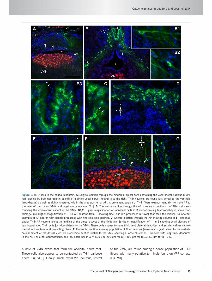
Figure 8. TH-ir cells in the caudal hindbrain. A: Sagittal section through the hindbrain–spinal cord containing the vocal motor nucleus (VMN;
red) labeled by bulk neurobiotin backfill of a single vocal nerve. Rostral is to the right. TH-ir neurons are found just dorsal to the ventricle
(arrowheads) as well as tightly clustered within the area postrema (AP). A prominent stream of TH-ir fibers extends ventrally from the AP to
the level of the rostral VMN and vagal motor nucleus (Xm). B: Transverse section through the AP showing a continuum of TH-ir cells sur-
rounding the dorsolateral aspect of the VMN. B1,2: Higher magnification of individual cells in B demonstrating teardrop-shaped soma mor-
phology. B3: Higher magnification of TH-ir AP neurons from B showing fine, cilia-like processes (arrows) that face the midline. C: Another
example of AP neuron with double processes with fine cilia-type endings. D: Sagittal section through the AP showing column of bi- and mul-
tipolar TH-ir AP neurons along the midline of the dorsal aspect of the hindbrain. E: Higher magnification of (*) in B showing small clusters of
teardrop-shaped TH-ir cells just dorsolateral to the VMN. These cells appear to have thick ventrolateral dendrites and smaller caliber ventro-
medial and ventrolateral projecting fibers. F: Horizontal section showing population of TH-ir neurons (arrowheads) just lateral to the rostral–
caudal extent of the dorsal VMN. G: Transverse section rostral to the VMN showing a loose cluster of TH-ir cells with long thick dendrites
in the XL. For other abbreviations, see list. Scale bar in A 5 500 lm; 200 lm for B,F; 100 lm for D,E,G; 50 lm for B1–3,C.
Catecholamines in auditory and vocal circuity
The Journal of Comparative Neurology | Research in Systems Neuroscience 15
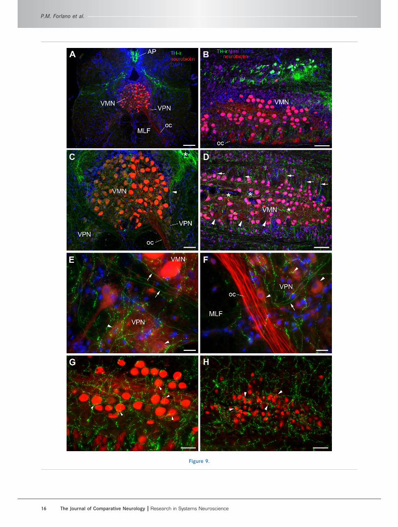
Figure 9.
P.M. Forlano et al.
16 The Journal of Comparative Neurology | Research in Systems Neuroscience

Higher order vocal circuitryOutside of the delineated VPG, other nuclei previously
identified physiologically and neuroanatomically as part of
the descending vocal motor pathway (see above) contain
notably robust TH-ir innervation. In the forebrain, these
areas include subdivisions of the preoptic area, PPa (Figs.
2B,C, 3B), PPp (Figs. 2D, 3D,F) and ventral hypothalamic
nuclei, vT (Fig. 2D,E), and AT (Fig. 2F, 4B–D, 10E,F). All
four of these nuclei include a small local population of
TH-ir neurons, aside from the PPa, which contains a large
number of TH-ir neurons. It is highly likely that most, if
not all, of these areas contain TH-ir fibers and terminals
from other TH-ir populations. The PAG, which receives
input from the nuclei described above and connects
directly to the VPG by innervation of VPP (Goodson and
Bass, 2002; Kittelberger and Bass, 2013; Kittelberger
et al., 2006), contains TH-ir terminals along its ventral bor-
der (Fig. 6B) where the dendritic field of vocal PAG neu-
rons are located, as well as in the adjacent and
connected PTT (Goodson and Bass, 2002; Kittelberger
and Bass, 2013; Kittelberger et al., 2006). In the isthmus,
rostral to the LC, a massive terminal field is found in the
midbrain isthmal nucleus (Is; not nucleus isthmi) and the
adjacent isthmal paraventricular nucleus (IP; Fig. 6D,E). In
more caudal sections, the IP is just lateral to the LC and
is thus a likely afferent target of LC. Comparatively fewer
TH-ir varicose fibers extend into the nucleus of the lateral
lemniscus (nll), whereas thick fiber tracts course through
the paralemniscal nucleus (PL), likely originating in part
from the descending TPp bundle (Figs. 2I,J, 6D,E). All of
the isthmal/lemniscal nuclei noted above are intercon-
nected with the PAG and vocal system and also receive
input from auditory circuitry; they are therefore considered
vocal–acoustic integration sites (Bass et al., 1994; Good-
son and Bass, 2002; Kittelberger and Bass, 2013; Kittel-
berger et al., 2006).
Catecholaminergic innervation of centralauditory circuitryHindbrain auditoryNeurobiotin application on the saccular branch of nerve
VIII, or on the sensory macula of the saccule (main end
organ of hearing), revealed transneuronal labeling of
auditory hindbrain circuitry, as demonstrated previously
(Bass et al., 1994, 2000; Sisneros et al., 2002). TH-
immunofluorescence combined with saccular backfills
allowed for confirmation of TH-ir innervation on neurons
directly connected within the ascending auditory sys-
tem. Examples of neurobiotin-filled neurons throughout
the auditory hindbrain are seen in Figures 11 and 12.
There is a conspicuous concentration of TH-ir fibers
and terminals throughout the longitudinal column of the
dorsal hindbrain that includes the dorsomedial (dm) and
dorsolateral (dl) divisions of the descending octaval
nucleus (DO) and the lateral line recipient nucleus
medialis (MED; Figs. 5A,B, 11A–C, 12A–C). TH-ir fibers
are also present but are less dense within the cerebel-
lar crest (cc), and within the central tract of the poste-
rior and anterior lateral line nerves (LL), adjacent to the
dl (Figs. 2L, 11A–C, 12A–C). TH-ir fibers innervate the
intermediate (i) and rostral intermediate (ri) divisions of
the DO and the magnocellular octaval nucleus (MG; Fig.
11A–C). At rostral levels, TH-ir fibers are highly concen-
trated within the ventral medial hindbrain, which
includes the ventral division of the secondary octaval
nucleus (SOv) and the ventral tegmental nucleus (VT),
just medial to the trigeminal motor nucleus (Vm; Fig.
11A–C). Analysis at higher magnification indeed shows
putative TH-ir terminals on backfilled somata and den-
drites of dm (Fig. 11D), SOv (Fig. 11E), and VT (Fig.
11F) neurons and on somata of ri neurons (Fig. 11G).
More caudal in the ventrolateral hindbrain at the level
of the VMN lie clusters of small, round, backfilled cells
Figure 9. Extensive TH-ir innervation throughout the vocal pattern generator (VPG). The VPG consists of vocal prepacemaker neurons
(VPP), vocal pacemaker neurons (VPN), and the vocal motor nucleus (VMN), which innervates vocal musculature on the swim bladder. All
three components were delineated by bulk neurobiotin backfill (red) of a single vocal nerve. A: Transverse section through the VMN–VPN
showing relative dorsal and dorsolateral position of TH-ir neurons of the area postrema (AP) and vagal group, respectively. Also note
neurobiotin-labeled occipital nerve (oc), which will exit the base of the brain. B: Parasagittal section showing vagal group TH-ir neurons
running in a column parallel to the lateral zone of VMN. TH-ir fibers from this group are seen projecting ventrally and rostrally (to the
right). Neurobiotin-backfilled VMN neurons overlaying Nissl stain appear pink. C: Higher magnification of image in A showing proximity of
TH-ir neurons (*) just lateral to the dorsal zone of VMN and large TH-ir fiber bundles entering the lateral VMN (arrowhead) and terminating
within the nucleus. D: Horizontal section through the ventral VMN. Prominent TH-ir fibers (arrows) intersect and enter the lateral VMN in a
perpendicular fashion along its length. Also note longitudinal fibers (*) coursing centrally through the VMN. Arrowheads indicate cross sec-
tions through bundles of axons that exit the ventrolateral base of the VMN to form the oc. Color legend in B applies to D. E,F: Examples
of TH-ir terminals and varicosities within the neurobiotin-filled VPN (arrowheads). Arrows designate smooth TH-ir axon fibers that follow
the ventral aspect of the VMN. G: Higher magnification of the lower left half of B demonstrating an abundance of TH-ir terminals and vari-
cosities on somata and dendrites in the VMN (arrowheads). H: Prominent TH-ir beaded fibers within and around the VPP in a sagittal
plane. Arrowheads indicate putative terminals on somata. Note the relative size difference of VPP neurons to VMN neurons in G. MLF,
medial longitudinal fasciculus. See Supplementary Figure 1 for magenta–green versions of G and H. Scale bar 5 200 lm in A; 100 lm in
B,D; 80 lm in C; 20 lm in E,F; 50 lm in G,H.
Catecholamines in auditory and vocal circuity
The Journal of Comparative Neurology | Research in Systems Neuroscience 17

Figure 10. TH-ir in higher order auditory nuclei; blue is DAPI nuclear stain. A,B: TH-ir fibers and terminals are abundant in the midbrain
torus semicircularis (TS). A: Horizontal section through the TS. Rostral is to the left, and medial is top of the image. B: Transverse section
through the auditory area centralis of the TS. Compact band of nuclei is the periventricular cell layer (Pe) of TS. C: TH-ir projections and
varicosities in the lateral (PGl) and medial (PGm) division of nucleus preglomerulosus. Image taken from same section shown in Figure 4C.
D: TH-ir terminals in the compact (CPc) and diffuse (CPd) divisions of the central posterior nucleus (auditory thalamus). D, inset: TH-ir ter-
minals on neurobiotin-filled cells (red) in CPc following a bilateral backfill of the saccular branch of nerve VIII. E: A single TH-ir cell (arrow-
head) together with dense TH-ir terminals in the hypothalamic anterior tuberal nucleus (AT). Image taken from same section shown in
Figure 4C. F: TH-ir terminals on neurobiotin-filled cells (red, arrowheads) in the AT following a bilateral backfill of the saccular branch of
nerve VIII. The AT is also part of the descending vocal motor circuitry and contains reciprocal connections with the CP. G: TH-ir terminals
are found intermixed with neurobiotin-filled afferents (red) from a saccular backfill in the eminentia granularis. Scale bar in A 5 200 lm;
100 lm for B–E; 50 lm for F,G.
P.M. Forlano et al.
18 The Journal of Comparative Neurology | Research in Systems Neuroscience

Figure 11.
Catecholamines in auditory and vocal circuity
The Journal of Comparative Neurology | Research in Systems Neuroscience 19

in the inferior olivary complex (IO; Bass et al., 2008;
Sisneros et al., 2002), which also contain putative TH-ir
terminals on their somata and interconnected dendrites
(Fig. 11H).
Octavolateralis efferent nucleus (OE)The OE, which sends projections to the inner ear and
lateral line organs (Bass et al., 1994; Highstein and
Baker, 1986; Weeg et al., 2005), is the most conspicu-
ous area containing robust TH-ir terminals in the brain
(Figs. 5A–D, 12). This nucleus lies in the hindbrain,
medial to the facial motor nucleus (VIIm), just dorsal to
the medial longitudinal fasciculus (MLF), below the
fourth ventricle (IV). The OE is composed of rostral
(OEr) and caudal (OEc) subdivisions (Bass et al., 2000)
separated by the internal arcuate fiber tract (iaf; Figs.
5A–D, 12A–D). Neurobiotin-backfilled neurons in this
nucleus have long, thick, bilateral dendrites that course
ventrolaterally to near the edge of the brain (Bass
et al., 1994). Remarkably, TH-ir fiber distribution in this
region specifically tracks the OE dendritic field and
forms terminals on dendrites as well as on the large
round somata (Fig. 12). Both caudal and rostral OE divi-
sions are highly innervated by TH-ir (Figs. 5A–D, 12A–
D). Somata in both divisions are also lightly labeled by
neurobiotin following backfill of a single vocal nerve
(Fig. 12D; Bass et al., 1994). Analyses from horizontal
and sagittal sections revealed two routes by which TH-
ir fibers reach the OE, and both appear to originate
from the TPp. The large, pear-shaped TH-ir TPp neurons
send a thick tract of dorsal projections that turns to
descend further into the hindbrain, forming part of the
prominent medial longitudinal CA tract (mlct), which
turns sharply medially onto the OE dendritic field (Fig.
5A–C). The mlct courses through the SOv rostral to the
OE, and appears to also branch off dorsally to innervate
the dm (compare Figs. 5B and 11C). A bundle of sev-
eral robust TH-ir axons appears to branch off the mlct
and continue laterally within the efferent bundle (EB),
which serves as the conduit for the ascending OE effer-
ent fibers to reach nerve VIII (compare Fig. 5D,E with
Fig. 11A, in which TH-ir and neurobiotin-labeled OE
axons [ea] run together within the lateral EB just caudal
to where they join nerve VIII). At this juncture, TH-ir
axons of the same caliber that branch laterally also
converge medially within the descending EB (compare
Figs. 5F and 11B,C) and run longitudinally along the
midline, dorsal to the iaf, and clearly innervate the OEr
(Fig. 12A,D).
Higher auditory circuitryOutside of the first- and second-order medullary popula-
tions of auditory neurons described above, major CA
innervation of higher auditory nuclei was found at all
levels except the dorsal telencephalon. Following saccu-
lar backfills Neurobiotin-labeled terminals were consis-
tently found along the lateral edge of the eminentia
granularis (EG), intermixed with abundant TH-ir termi-
nals (Figs. 2K, 7A, 10G). Notably, this area of the EG
also contains input from the vocal hindbrain (Bass et al.,
1994). The nucleus centralis in the midbrain TS is the
major recipient of the ascending auditory nuclei in the
medulla (Bass et al., 2000, 2005; McCormick, 1999) and
is more robustly innervated with TH-ir fibers and termi-
nals compared with lateral and deeper cell layers that
process lateral line stimuli (Figs. 2G–J, 6C,D, 10A,B; Bass
et al., 2000; Weeg and Bass, 2000). The TS, in turn,
projects to several diencephalic nuclei including the AT
(see above), the central posterior nucleus of the thala-
mus (CP), and the lateral division of the nucleus preglo-
merulosus (PGl) (Bass et al., 2000; Goodson and Bass,
2002).
All three of these areas contain TH-ir varicose fibers
and terminals (Fig. 10C [PGl], D [CP], E,F [AT]). Dorsally
projecting TH-ir fibers from TPp neurons appear to
innervate the CP (arrowhead, Figs. 3A, 4C–E), whereas
Figure 11. TH-ir innervation of transneuronal-labeled hindbrain auditory nuclei following a bilateral application of neurobiotin on the saccu-
lar epithelium. A–C: Low-magnification rostral–caudal series of transverse sections showing location of neurobiotin-filled cells in the audi-
tory system and prominent TH-ir terminal fields in the dorsal and ventral hindbrain. Note both TH-ir and neurobiotin-labeled (red)
octavolateralis efferent axons (ea) within the lateral efferent bundle just caudal to where they join nerve VIII. Arrowhead in A indicates TH-
ir axon bundle entering the lateral efferent tract just dorsal to the ventral secondary octaval nucleus (SOv). TH-ir axons are seen descend-
ing within the efferent tract (B) as it turns to run longitudinally along the midline (C) dorsal to the internal arcuate fiber tract (iaf) rostral
to the octavolateralis efferent nucleus (OE; see Fig. 12D). Arrowhead in C shows TH-ir fibers projecting dorsally into the rostral intermedi-
ate (ri) and dorsal medial (dm) subdivisions of the descending octaval nucleus (compare with Fig. 5B). D,E: High magnification of areas
indicated by (*) in C showing TH-ir terminals and varicosities on somata and dendrites (arrowheads) of dm and SOv neurons in D and E,
respectively. F: High magnification of area indicated by (*) in B showing TH-ir varicosities on dendrites of a ventral tegmental (VT) neuron
that lies just medial to the trigeminal motor nucleus (Vm). G,H: TH-ir terminals and varicosities on filled cells in the ri and inferior olive
(IO), respectively. Images in G and H were selected from sections outside of A–C. The IO lies along the ventrolateral border of the caudal
hindbrain. For other abbreviations, see list. See Supplementary Figure 2 for magenta–green versions of D–G. Scale bar 5 200 lm in A
(applies to A–C); 16 lm in D–G; 20 lm in H.
P.M. Forlano et al.
20 The Journal of Comparative Neurology | Research in Systems Neuroscience

Figure 12.
Catecholamines in auditory and vocal circuity
The Journal of Comparative Neurology | Research in Systems Neuroscience 21

laterally projecting TH-ir fibers from the TPp and/or the
VM may innervate the PGl (Fig. 4B,C). In a few instan-
ces transneuronal neurobiotin-labeled cells from saccu-
lar backfills were seen in the AT and CP, and indeed
these cells appear to have TH-ir terminal contacts (Fig.
10D inset, F). Neurobiotin-filled neurons in the PPp were
consistently found in animals after saccular backfills,
and TH-ir terminals were abundant in the PPp dendritic
field just lateral to the densely packed cell layer adja-
cent to the ventricle (Fig. 3F). Although the PPp and AT
contain their own small populations of TH-ir somata
(Figs. 3D, 10E), backfilled cells in these areas were
never TH-ir. The parvocellular preoptic (PPa/p), vT, and
AT, as well as the Vs and Vp, all have reciprocal con-
nections with the CP (Goodson and Bass, 2002). TH-ir
fibers in those areas may originate, in part, from local
TH-ir neurons. In addition, ascending projections from
the TPp may innervate various preoptic nuclei as well as
the subdivisions of areas ventralis (Fig. 4I). The only
efferent target of the CP largely devoid of TH-ir fibers is
the medial zone of the dorsal telencephalon (Dm), which
lies dorsal to the PPa and Vd at the level of the anterior
commissure (Figs. 2B, 3A,B; Goodson and Bass, 2002).
Catecholaminergic innervation of the innerear and its origin in the TPp
Because robust TH-ir fibers were seen entering nerve
VIII in single-labeled tissue (Fig. 13G) and intermingled
with neurobiotin fibers following saccular backfills (Fig.
13E,F), we investigated TH-ir in the saccule, the main
end organ of hearing in the inner ear. Several large,
thick TH-ir fibers are seen coursing through the saccu-
lar branch of nerve VIII that bypass ganglion cells proxi-
mal to the sensory macula and finally terminate in the
sensory epithelium (Fig. 13A,D). TH-ir terminals appear
to contact the base of individual hair cells, whereas
fewer puncta are seen contacting the mid- to apical
end of hair cell somata, clearly delineated by the HCS-1
antibody, which labels otoferlin (Fig. 13B–D). Many
robust TH-ir terminals are found within the support cell
layer (Fig. 13A–C).
By combining neurobiotin backfills of the saccular
branch of nerve VIII with TH-ir in the brain, the origin of
CA saccular efferents was discovered to be a small
population of the numerous large, pear-shaped TH-ir
neurons in the TPp (Figs. 14–16). Backfilled cells on
both sides of the midline were filled regardless of which
saccule was labeled with neurobiotin (Figs. 14D,E,
15C,F,G, 16H). All neurobiotin-backfilled TPp cells were
also TH-ir. Backfilled cells were found throughout the
TPp but were more prevalent in the mid-caudal TPp
along the ventricular midline or within the continuum of
cells that forms between the medial forebrain bundle
and just lateral to the PVO (Fig. 14C,D). When found
more rostrally, backfilled cells were either along the
midline or ventrolateral in the sparse population of simi-
lar type (large, pear-shaped) cells just dorsal to the AT
(Figs. 14G, 15C, 16C,D). The most rostral backfilled
cells were found lateral to the anterior PVO, just dorsal
to the AT. Backfilled cells were never found in the most
caudal group of large cells lateral to the PTN, or in the
few much smaller cells in the PTN or rostral and dorsal
TPp.
Figures 15 and 16 show the distribution of TH-ir neu-
rons in the TPp in comparison with TH-ir neurons that
were also backfilled following neurobiotin application on
the saccular branch of nerve VIII in two individual type I
males. An average of eight backfilled cells was found
across animals in alternate sections (4/5 animals had
7–11 cells, whereas one animal had only 1 backfilled
cell). Differences in numbers of backfilled TPp neurons
may be due to variation in damage of axons within the
saccular branch of nerve VIII during neurobiotin applica-
tion. Interestingly, neurobiotin placement on the caudal
end of the saccular epithelium (vs. damage to nerve
VIII) in an additional two animals resulted in excellent
labeling of hindbrain ascending auditory circuitry but
very few if any completely filled TPp cells. Instead, only
small beads of neurobiotin could be seen in the TH-ir
soma (not shown). Figure 17 shows a size distribution
(area vs. major axis diameter) of backfilled neurons
compared with measured non-backfilled TH-ir neurons
Figure 12. Massive TH-ir innervation on somata and dendrites of the octavolateralis efferent nucleus (OE). A–C: Low-magnification series
of transverse sections that show backfilled neurons of the rostral (OEr) and caudal (OEc) subdivisions of the OE after bilateral application
of neurobiotin on the saccular epithelium. OE neurons lie on the midline between the medial longitudinal fasciculus (MLF) and the fourth
ventricle (IV). OE dendrites (d) extend ventrolaterally toward the edge of the brain, and prominent TH-ir fiber tracts run along the length of
the OE dendrites. The medial efferent axon tract can be seen in A as a caudal continuation from Figure 11C that connects to the OE
somata (also see D). Arrowheads in C indicate single TH-ir cells associated with the rostral vagal lobe (XL). D: In the sagittal plane a bun-
dle of intensely labeled TH-ir axons can be seen entering the OE via the efferent tract (arrowhead). E–H: High-magnification images of
robust TH-ir terminals and varicosities on and around OE somata and dendrites. TH-ir in this area appears highly localized to OE and its
dendritic field. For other abbreviations, see list. See Supplementary Figure 3 for magenta–green versions of E–H. Scale bar 5 200 lm in
A (applies to A–C); 80 lm in D; 25 lm in E; 16 lm in F–H.
P.M. Forlano et al.
22 The Journal of Comparative Neurology | Research in Systems Neuroscience

in the same section. Backfilled cells were tightly
grouped in size, with an average major axis diameter
(mean 6 standard error [SE]) of 21.20 6 0.17 lm and
an area of 273.55 6 7.11lm2. With the techniques
employed in this study, we estimate that 5–10% of the
large TPp TH-ir neurons project to the saccule. Further-
more, thick TH-ir fibers are found in the anterior (Fig.
12C) and posterior lateral line nerves (not shown).
Thus, similar to hindbrain cholinergic octavolateral effer-
ent neurons (Highstein and Baker, 1986), these cells
likely innervate most, if not all, inner ear and lateral line
end organs.
Figure 13. TH-ir innervation of the saccule, the main end organ of hearing. A: Section through the saccular epithelium including the
attached branch of the eighth nerve (VIII). The hair cell layer (HC) is delineated using the hair cell–specific antibody (HCS-1, green), which
labels HC somata and can be distinguished from the basal support cells (SC) labeled by DAPI (blue) alone. Thick and smooth TH-ir fibers
(red) course through nerve VIII prior to terminating largely at the base of the HC layer. Inset is lower magnification of same section. B:
Horizontal section showing larger varicose TH-ir fibers in the SC and finer terminals in the HC layer of the SE. C,D: High-magnification
images showing thick TH-ir varicose fibers along the SC layer, fine-caliber terminals (arrowheads) at the base of the HC, and, less fre-
quently, terminals on the central portion of individual hair cells proximal to the nucleus (arrows). E,F: Transverse section through the lat-
eral hindbrain where nerve VIII converges with the CNS. Arrowheads indicate intermingled TH-ir fibers with neurobiotin-labeled fibers from
a saccular backfill. G: Horizontal section through the hindbrain; medial is top; lateral is bottom of image. Arrowheads show small bundles
of TH-ir axons exiting the brain via nerve VIII. See Supplementary Figure 4 for magenta–green version of D. Scale bar 5 100 lm in A,B;
200 lm in inset to A; 33 lm in C; 50 lm in D; 200 lm in E (applies to E–G).
Catecholamines in auditory and vocal circuity
The Journal of Comparative Neurology | Research in Systems Neuroscience 23

DISCUSSION
The overall distribution of TH-ir in the midshipman
CNS is largely consistent with an earlier preliminary
report of general TH-ir distribution in midshipman brain
using a different antibody and nonfluorescent detection
(Bass et al., 2001b), and a recent report using the same
antibody as the present study with nonfluorescent detec-
tion to describe TH-ir distribution patterns (Goebrecht
et al., 2014). Thus, although the present study is not the
first to report TH-ir in association with several known
vocal and auditory centers in the midshipman CNS, weutilized double-labeling experiments that combined THimmunofluorescence with transneuronal backfill labeling
Figure 14. Neurobiotin backfills of the saccule combined with TH immunofluorescence identify source of TH-ir efferents to the saccular
epithelium as a small population of large, pear-shaped cells in the periventricular posterior tuberculum (TPp). A: TH-ir cells in the TPp just
medial to the medial forebrain bundle and lateral and dorsal to the paraventricular organ. B: Three neurobiotin-labeled neurons (two adja-
cent cells on top) after a saccular backfill. C: Overlay of images in A and B. Arrowheads indicate same neuron in both channels. All
neurobiotin-backfilled cells in this region were also TH-ir. D–H: Additional examples of backfilled TH-ir neurons (arrowheads, yellow overlay)
throughout the TPp. Examples of bilateral filled cells in D and E are from unilateral saccular fills. See regional distribution in Figures 15
and 16. Scale bar 5 50 lm in C (applies to A–H).
P.M. Forlano et al.
24 The Journal of Comparative Neurology | Research in Systems Neuroscience

of auditory circuitry and the vocal pattern generator todemonstrate putative TH-ir terminals on individual cellscomprising both the peripheral and central auditory sys-tems as well as the octavolateralis efferent nucleus and
vocal motor system. This methodology has allowed us todocument TH-ir innervation of the auditory sensory epi-thelium and its origin in the brain in a nonmammalianvertebrate. In addition, by examining TH-ir in the brain at
Figure 15. A–I: Digital tracings of rostral–caudal series of sections depicting distribution of backfilled neurons also labeled by TH-ir (red
triangles) in the posterior tuberculum of an individual after neurobiotin application on the saccular branch of the eighth nerve. Green
circles indicate TH-ir neurons not backfilled. Backfilled cells were always TH-ir and were a small percentage of total population of TH-ir
neurons in this brain region. For abbreviations, see list. Scale bar 5 500 lm in A (applies to A–I).
Catecholamines in auditory and vocal circuity
The Journal of Comparative Neurology | Research in Systems Neuroscience 25

multiple planes of section, we provide a greater under-standing of projection patterns originating from specificTH-ir nuclei such as the TPp. Thus, the present studyextends previous work in this model system and pro-
vides very strong neuroanatomical evidence for CAs asimportant neuromodulators of auditory and vocal motorcircuitry and related vocal–acoustic social behavior inmidshipman fish. Findings or interpretations of TH-ir that
Figure 16. A–I: Digital tracings of rostral–caudal series of sections depicting distribution of backfilled neurons also labeled by TH-ir (red
triangles) in the posterior tuberculum of an individual after neurobiotin application on the saccular branch of the eighth nerve. Green
circles indicate TH-ir neurons not backfilled. Backfilled cells were always TH-ir and were a small percentage of total population of TH-ir
neurons in this brain region. For abbreviations, see list. Scale bar 5 500 lm in A (applies to A–I).
P.M. Forlano et al.
26 The Journal of Comparative Neurology | Research in Systems Neuroscience

are different from what is reported by Goebrecht et al.(2014) will be addressed in specific subsections below.
Several reviews have reported the distribution of TH-
ir cell populations in fishes in comparison with other
vertebrates including proposed homologies (Carrera
et al., 2012; Goodson and Kingsbury, 2013; O’Connell
and Hofmann, 2011; Schweitzer et al., 2012; Smeets
and Gonzalez, 2000; Yamamoto and Vernier, 2011),
which will therefore not be a focus here. In general, the
distribution of TH-ir neurons in midshipman is largely
consistent with that reported in other teleosts: popula-
tions are found in the olfactory bulb, ventral telenceph-
alon, preoptic area, suprachiasmatic nucleus, ventral
thalamus, anterior and periventricular hypothalamus,
posterior tuberculum, locus coeruleus, vagal area, and
area postrema. A recent description of TH-ir distribution
in midshipman (Goebrecht et al., 2014) did not include
labeled neurons in the olfactory bulb, postcommissural
nucleus of area ventralis, and anterior tuberal nucleus
as found in the current study. Unlike some other spe-
cies, midshipman appear to have relatively few TH-ir
cells in the PPp, parvocellular PVO (see below), and
unlike most other teleosts but similar to the eel Anguilla
(Roberts et al., 1989), a pretectal group is entirely
absent (Goebrecht et al., 2014). We report consistent,
but very few TH-ir cells in the PAG that were not
reported previously and could have been easily over-
looked in other midshipman studies (Bass et al., 2001b;
Goebrecht et al., 2014). TH-ir cells in this region have
been reported in Astatotilapia (O’Connell et al., 2011)
as well as in a similar position in zebrafish and Euro-
pean eel, in a region lateral to the nMLF (Kaslin and
Panula, 2001; Roberts et al., 1989).
TH-ir neurons of the periventricular posteriortuberculum
The most conspicuous group of TH-ir neurons in mid-
shipman are undoubtedly the numerous, large, pear-
shaped cells of the TPp. In midshipman there are sev-
eral hundred neurons of this type (Petersen et al.,
2013), which far outnumber the few documented in
zebrafish adults (Ma, 2003; Rink and Wullimann, 2001).
These distinctive TH-ir neurons correspond to those
reported as paraventricular organ–accompanying
(PVOa), magnocellular hypothalamic nucleus, or nucleus
of the TPp (nTPp) cells located lateral to the anterior
PVO described in other teleosts (Ma, 2003; Meek,
1994; Meek and Joosten, 1993; Meek and Nieuwen-
huys, 1998; Rink and Wullimann, 2001, 2002b). Using
the terminology of TH-ir diencephalic neurons by Rink
and Wulliman (2001, 2002b), only a small group of
“type 1” round, parvocellular TH-ir cells are seen in the
rostral dorsal TPp in midshipman, which blends into the
much larger and distinct ventral thalamic group that in
zebrafish is identified as VL (Ma, 2003). The area identi-
fied as the TPp by Goebrecht et al. (2014) appears as
the caudal VM from our analysis because TH-ir cells in
that location are contiguous with VM (and VL) TH-ir
cells more rostrally (Figs. 3D,E, 4A,B). The vast majority
of TPp TH-ir cells correspond to “type 2” PVOa neurons
or large TPp cells described in zebrafish (Kaslin and
Panula, 2001; Ma, 2003; Rink and Wullimann, 2001,
2002b; Tay et al., 2011) and those simply labeled
“PVO” by Goebrecht et al. (2014) in midshipman. How-
ever, few if any cells exhibit the “type 3” parvocellular
“liquor-contacting” phenotype seen in the PVO of zebra-
fish (Rink and Wullimann, 2002b; Yamamoto et al.,
2010; Yamamoto and Vernier, 2011).
Importantly, TH-ir type 3 cells are detected by TH1
and commercially available TH antibodies, and therefore
their absence in midshipman is unlikely due to these
cells expressing only TH2 enzyme (Filippi et al., 2010;
Yamamoto et al., 2010; Yamamoto and Vernier, 2011). If
indeed type 3 cells are characteristic in defining the
PVO, then the majority of TH-ir cells in the posterior
tuberculum of midshipman would not be considered true
PVO neurons. Instead, we propose that these large,
pear-shaped cells are a continuum of the group that lies
Figure 17. Size and shape distribution of TH-ir neurons in the
periventricular posterior tuberculum that were backfilled after
neurobiotin application on the saccular branch of the eighth nerve
(red triangles). These cells had an average soma diameter of 21
lm and all were classified as pear-shaped. Measurements of
other non-backfilled TH-ir neurons within the same sections were
taken for comparison.
Catecholamines in auditory and vocal circuity
The Journal of Comparative Neurology | Research in Systems Neuroscience 27

on the ventricular midline in the ventral TPp and there-
fore should be considered a single group not necessarily
adhering to cytoarchitectural boundaries (Ma, 2003;
Meek and Nieuwenhuys, 1998; Striedter, 1990).
A substantial amount of evidence across several spe-
cies of teleosts indicates that these large TH-ir cells are
DAergic as they are DA-ir but not immunoreactive for
dopamine b-hydroxylase (DBH; the enzyme necessary
for NA synthesis) (Batten et al., 1993; Ekstr€om et al.,
1986, 1990; Filippi et al., 2010; Hornby and Piekut,
1988, 1990; Hornby et al., 1987; Ma, 2003; Meek and
Joosten, 1993; Sas et al., 1990; Yamamoto et al.,
2010; Yamamoto and Vernier, 2011). Furthermore, the
equivalent diencephalic cells identified in zebrafish lar-
vae were demonstrated to be Otp-dependent DAergic
neurons and to contribute the major descending DAer-
gic projections as do otp expressing mammalian A11
cells (Kastenhuber et al., 2010; Ryu et al., 2007;
Schweitzer et al., 2012; Tay et al., 2011). At least in
larval zebrafish, the medial longitudinal CA tract (mlct)
is primarily derived from these descending DA projec-
tions (Schweitzer et al., 2012). These highly conserved
DAergic diencephalic neurons are the first to appear
during development (along with LC neurons) in all
anamniotes including elasmobranchs and agnathans
and are thought to be among the most ancient CA
groups (see Carrera et al., 2012; Schweitzer et al.,
2012; Smeets and Gonzalez, 2000 and references
within). Expression of an equivalent group of DAergic
neurons in Amphioxus further supports its ancient origin
(Moret et al., 2004).
As stated above, TPp neurons in adult and larval
zebrafish were also discovered to send ascending pro-
jections to the proposed striatal homolog in area
Figure 18. Schematic sagittal view of the brain showing vocal motor (green) and central auditory (blue) systems in batrachoidid fish (mid-
shipman and toadfish) (modified from Bass and McKibben, 2003; Kittelberger et al., 2006) with connectivity from large TH-ir neurons
within the periventricular posterior tuberculum (red; TPp; this study). Solid dots represent somata, and lines represent axonal projection
pathways. Two connected dots indicate reciprocal connections. Descending vocal motor pathways (Bass and Baker, 1990; Bass et al.,
1994, 2000, 2001a; Fine and Perini, 1994; Goodson and Bass, 2002; Kittelberger et al., 2006; Remage-Healey and Bass, 2004). Preoptic
(POA, not shown) and ventral (vT) and anterior (AT) tuberal nuclei in the hypothalamic forebrain project to the periaqueductal gray (PAG)
in the midbrain, which then connects to the vocal pattern generator (VPG) in the hindbrain–spinal cord. The VPG consists of vocal prepa-
cemaker (VPP), pacemaker (VPN), and motor (VMN) nuclei. The VMN projects directly via occipital nerve roots to sound-producing muscle
on the swim bladder. Central auditory system (Bass et al., 1994, 2000, 2001a). Social vocalizations are detected by the inner ear, which
projects via the VIIIth nerve to descending (DO) and secondary (SO) octaval nuclei in the hindbrain and further to the auditory midbrain
torus semicircularis (TS). Shown are nuclei interconnected with the TS. The dorsal thalamic central posterior nucleus (CP) contains recipro-
cal connections with the ventral telencephalon (V; includes the supracommissural division) and anterior hypothalamus (AT/vT) (for nomen-
clature, see Braford and Northcutt, 1983). The TS and CP also connect to vocal motor nuclei in the forebrain (AT, vT, POA; not shown)
and midbrain (PAG and isthmal/tegmentum; not shown), whereas auditory-recipient octaval nuclei in the hindbrain connect to the VPG via
the VPP (also see Bass et al., 1994; Goodson and Bass, 2002). The octavolateralis efferent nucleus (OE) projects to the inner ear, which
includes the saccule, the main end organ of hearing (Bass et al., 1994, 2000; Weeg et al., 2005). The OE contains reciprocal connections
with the VPP (Chagnaud et al., 2011) and receives projections from the PAG (Kittelberger and Bass, 2013; not shown). Large, pear-shaped
TH-ir neurons from the TPp send a massive dorsal turned descending tract into the hindbrain that appears to innervate the DO, SO, and
OE and likely the VMN. A branch of this tract exits the brain via the octavolateralis efferent tract in nerve VIII to the inner ear. Other tar-
gets of TPp TH-ir neurons include the CP and PAG. The PAG and TPp are reciprocally connected (Kittelberger and Bass, 2013), but
whether the PAG projects onto TH-ir cells in the TPp is not confirmed. The TPp also has a robust ventral ascending TH-ir projection
although the exact innervation target in the ventral telencephalon (V) is undetermined. See above references for additional known
connectivity.
P.M. Forlano et al.
28 The Journal of Comparative Neurology | Research in Systems Neuroscience

ventralis of the telencephalon (Vd), although only a
small percentage of rostral TPp neurons (10% total) are
known to contribute to this ascending mix of DA and
NA projections (Kastenhuber et al., 2010; Rink and Wul-
limann, 2001; Tay et al., 2011). Ma (2003) reports that
rostral TH-ir PVOa neurons project rostroventrally and
laterally. Ascending projections to the ventral telen-
cephalon from TPp TH-ir neurons can also be seen in
horizontal sections through the midshipman brain,
although we were unable to verify exact end targets in
this study. Kaslin and Panula (2001) report that TPp
neurons send descending fibers and are reciprocally
connected with LC neurons in adult zebrafish, consist-
ent with descending targets of DA A11 neurons in
mammals (Maeda et al., 1991). Thus, the intense TH-ir
terminals seen on LC neurons in the present study (Fig.
7) possibly originate from the TPp as well, or may be
local projections from the LC (Tay et al., 2011). The
heavy TH-ir terminal fields in the area of the dorsal
raphe area (present study; Kaslin and Panula, 2001) are
also consistent with DAergic projections from A11 neu-
rons in mammals (Peyron et al., 1995; Smeets and
Gonzalez, 2000).
The behavioral relevance for diencephalospinal pro-
jections in both zebrafish and mammals is still largely
unknown (Barraud et al., 2010; Jordan et al., 2008; Kas-
tenhuber et al., 2010). The prominent descending pro-
jections of TH-ir neurons in the TPp further support
them as A11 DA homologs. A11 is the major source of
DA in the spinal cord, and in mice, numbers of A11
neurons and DA innervation of the lumbar portion of
the dorsal horn are sexually dimorphic. This sexual
dimorphism is an androgen receptor–dependent mecha-
nism, organized by differential exposure to androgens
perinatally (Pappas et al., 2010). The function of DA in
this area is thought to modulate sensory (including
nociception) information and motor function (Barriere
et al., 2004; Lapirot et al., 2011). Interestingly, as seen
in zebrafish, single A11 neurons in mammals are known
to have both descending spinal as well as ascending
telencephalic projections. Although neocortical ascend-
ing targets appear to be the most robust, subpallial tar-
gets such as striatal, nucleus accumbens, and
amygdala are reported as well (Takada et al., 1988a,b,
1993). In canaries, both A11 and A10 (VTA) TH-ir neu-
rons project to forebrain song control nuclei (Appeltants
et al., 2000, 2002a). Thus, although the absence of
VTA/SN DAergic groups in teleosts appears to be a
derived condition (Carrera et al., 2012), the projection
pattern of the TPp and its connectivity with other brain
areas (e.g., hodology; O’Connell and Hofmann, 2012)
may permit analogous functions to VTA/SN of other
vertebrates in addition to the roles of A11 neurons.
TH-ir neurons in the periventricular posteriortuberculum have widespread projections tothe central and peripheral auditory system
A recent single-cell projectome study in larval zebra-
fish demonstrated that individual TH-ir TPp neurons
send strong descending projections to the hindbrain
and spinal cord and ascending projections to the ven-
tral telencephalon, as well as local projections within
the diencephalon. Thus, TH-ir TPp neurons are well
positioned to be important regulators of sensory–motor
integration (Tay et al., 2011). Similarly, our neuroana-
tomical findings point to TH-ir TPp neurons as modula-
tors and integrators of auditory, motor, and higher
cognitive function in adult midshipman. Figure 18 sum-
marizes TH-ir projections from the TPp supported by
data in the present study. These projections appear to
include efferents to the ventral telencephalon, higher
auditory (CP), midbrain vocal–acoustic center (PAG),
hindbrain auditory nuclei (DO/SO), octavolateralis effer-
ent nucleus (OE), and inner ear (saccule). Although the
direct projection of TH-ir TPp neurons to the saccule is
confirmed by double-label tract tracing experiments and
supports earlier preliminary observations by Sisneros
et al. (2002), projections to the PAG (also see Goe-
brecht et al., 2014; Kittelberger and Bass, 2013) and
CP can be seen originating from TH-ir TPp cells in the
same plane of section, and projections to the hindbrain
auditory and OE are evident as branches off the mlct
tract, which is derived largely from the TPp. The exact
target of ascending projections to the ventral telen-
cephalon was not determined and will require further
investigation. Also, like adult zebrafish (Ma, 2003),
there is a strong lateral component to these projections
that appears to robustly innervate the PGl (Fig. 4I; not
shown in Fig. 18), which receives projections from the
auditory thalamus (CP; Goodson and Bass, 2002).
Thus, similar to what is known for the mammalian LC
system, in which individual neurons project to discrete
nuclei within the ascending somatosensory system (Ber-
ridge and Waterhouse, 2003), individual TH-ir TPp neu-
rons may innervate multiple nuclei along the ascending
auditory pathway. This arrangement is thought to pro-
vide simultaneous release of CA to coordinate informa-
tion transfer along a sensory pathway (Berridge and
Waterhouse, 2003). It is important to note that there is
substantial TH-ir input to areas of neuroanatomical
overlap between the auditory and lateral line systems,
including the DOdl, DOdm, MG, and TS, as well as in
lateral line–specific structures such as anterior and pos-
terior branches of the lateral line nerve and MED. As
these lateral line–specific structures are adjacent to
shared auditory areas (Weeg and Bass, 2000), it is
highly likely that their source of TH-ir terminals is also
Catecholamines in auditory and vocal circuity
The Journal of Comparative Neurology | Research in Systems Neuroscience 29

the TPp. Interestingly, primary afferents of the posterior
lateral line nerve can indeed encode frequencies con-
tained within midshipman vocalizations (Weeg and
Bass, 2002). Whether single TPp neurons innervate
both octaval sensory systems and function in the same
manner as described above to coordinate both auditory
and lateral line processing of acoustic social signals is
worthy of future investigations.
TH-ir innervation of the sensory macula of thesaccule and the octavolateralis efferentnucleusOur results demonstrate two pathways through which
CAs could modulate the sensitivity of the peripheral
auditory system in midshipman. The first pathway
includes TH-ir (DA) neurons in the TPp that project
directly to the base of sensory hair cells in the saccule
via nerve VIII, whereas the second, indirect pathway,
which also appears to originate in the TPp, is a massive
TH-ir input on the dendrites and somata of the OE,
which in turn projects to the inner ear and lateral line
organs (Bass et al., 1994; Bleckmann et al., 1991; High-
stein and Baker, 1986; Highstein et al., 1992; Koppl,
2011; Tomchik and Lu, 2005; Weeg et al., 2005). To
our knowledge this is the first demonstration of CA ter-
minals in the sensory epithelium of the inner ear of any
nonmammalian vertebrate, and suggests a conserved
and important anatomical and functional role for DA in
normal audition. Whether the same neurons in the TPp
that project to the inner ear also heavily innervate the
OE requires further study. The data presented here sug-
gest that only a few TH-ir neurons in the TPp that pro-
ject to the peripheral octavolateralis system via the
lateral EB also branch into the medial descending EB to
terminate on OE somata, whereas a greater number of
TPp cells that form the descending mlct innervate the
OE dendritic field. In support of this, the small bundle
of TH-ir axons that exit the lateral EB appears to have
the same morphology as those that continue to
descend and innervate OE somata (compare Figs. 5E,F
and 12D).
Although our work clearly demonstrates the target of
TPp TH-ir efferents as terminals on the base of hair
cells in the inner ear of a teleost, previous studies in
larval and adult zebrafish showed that these dience-
phalic DAergic neurons project to the lateral line organ
and toward the inner ear and nerve VIII with unknown
targets (Bricaud et al., 2001; Ma, 2003). Furthermore,
DA receptor mRNA expression was recently found in
hair cell preparations of the trout saccule (Drescher
et al., 2010). Thus, there is compelling evidence that
DA action on the inner ear may be a common feature
across unrelated species of teleost fishes, and perhaps
vertebrates in general. In addition, previous tract-
tracing studies have identified a few diencephalic (pos-
terior tubercular) neurons that project to lateral line
nerves in goldfish (Puzdrowski, 1989; Zottoli and Van
Horne, 1983), catfish (New and Singh, 1994), and larval
zebrafish (Metcalfe et al., 1985), all otophysan fishes.
Although those studies did not identify the neurochemi-
cal phenotype of the diencephalic efferents, it is highly
likely based on their neuroanatomical location and mor-
phology that they are DAergic, and they support the
hypothesis that TPp TH-ir neurons may innervate multi-
ple peripheral octavolateral end organs.
Similar to fishes, the auditory system of tetrapod verte-
brates includes sensory hair cells in the inner ear that
synapse onto primary afferent neurons that in turn pro-
ject through the VIIIth cranial nerve to the primary proc-
essing area in the auditory hindbrain. In addition, efferent
neurons from the brainstem project out toward the hair
cells and modulate the signal being transmitted back to
the brain. Efferents contact auditory and vestibular hair
cells directly or make synapses on afferent dendrites
(Koppl, 2011; Nicolson, 2012; Ruel et al., 2007). In mam-
mals, the medial olivocochlear (MOC) efferent system is
comprised of cholinergic neurons that function to inhibit
outer hair cells (cochlear amplifier), whereas the lateral
olivocochlear (LOC) efferent system is a heterogeneous
mix of cholinergic, DAergic, g-aminobutyric acid (GABA)er-
gic and other peptidergic neurons (Darrow et al., 2006b;
Gaborjan et al., 1999; Lendvai et al., 2011), whose func-
tion is less well known but that have been demonstrated
to elicit either slow excitation or slow suppression of the
nerve output from inner hair cells of the cochlea (Groff
and Liberman, 2003). In the LOC, GABA colocalizes with
cholinergic efferent terminals, whereas DA comprises a
small but separate population (5–35%) of efferents (Dar-
row et al., 2006b). The majority of DA terminals lack
clear synaptic specializations (Lendvai et al., 2011). Perfu-
sion of DA on the cochlea decreases sound-evoked activ-
ity and inhibits compound action potentials of afferent
dendrites (Ruel et al., 2001), and acoustic overstimulation
leads to depletion of TH-ir in the cochlea (Niu and Can-
lon, 2002), although, interestingly, low-level (nontoxic)
sound conditioning upregulates DAergic terminals in the
cochlea and reduces subsequent damage by trauma (Niu
et al., 2004). Many studies, mostly in rodents, have inves-
tigated mechanisms of action of DA signaling on the
inner ear, but its functional significance remains unclear.
Largely, these investigations have suggested a role of DA
in protection from acoustic trauma damage, as DA
appears to suppress the excitotoxic effects of acoustic
overstimulation (Lendvai et al., 2011), although a role in
sound localization was also suggested (Darrow et al.,
2006a).
P.M. Forlano et al.
30 The Journal of Comparative Neurology | Research in Systems Neuroscience

Like other vertebrates, octavolateral efferent (OE)
neurons in teleosts are also cholinergic (Brantley and
Bass, 1988; Clemente et al., 2004; Danielson et al.,
1988; Roberts et al., 1994), and project onto saccular
hair cells, where they have been shown to have an
inhibitory role on saccular afferent activity in goldfish
(Sugihara, 2001). However, in the toadfish (Opansus
tau), activation of the octavolateralis efferent neurons
can reduce the gain (or sensitivity) of the evoked recep-
tor potentials of hair cells but also increase the sponta-
neous discharge rate of the inner ear afferents
connected to the peripheral vestibular system (Boyle
et al., 2009). Similarly, recent findings suggest that
cholinergic OE neurons coexpress GABA in the toadfish
Opsanus (Edds-Walton et al., 2010), a close relative of
midshipman. The intensely robust TH-ir innervation pat-
tern of midshipman OE has not been reported in other
teleosts and therefore may either reflect a specializa-
tion of vocal teleosts, or indicate that this brain area
has not been examined closely in most other species.
However, varicose DA-ir fibers as well as both D1A/B
and D2A/B receptors have been localized to OE in the
European eel (Kapsimali et al., 2000; Pasqualini et al.,
2009; Roberts et al., 1989), which supports DAergic
innervation of this area; interestingly, there is evidence
for sound production in eels (Lagardere and Ernande,
2004; Rountree et al., 2002). Importantly, midshipman
OE neurons receive direct input from the VPP, are
active during vocalization in midshipman males, and are
proposed to maintain sensitivity to external acoustic
stimuli during vocal behavior (Chagnaud et al., 2011;
Weeg et al., 2005).
In addition, OE neurons were recently found to gener-
ate a vocal corollary discharge that encodes duration,
thus providing feedback that may prevent self-
generated auditory afferent overstimulation (reafference
suppression; Chagnaud and Bass, 2013) or perhaps
protect from acoustic trauma during courtship calls that
are measured at 130 dB and can be produced continu-
ously for more than an hour (Bass and McKibben,
2003). Thus, modulation of input from the auditory
periphery by dense CA terminals on cholinergic OE neu-
rons likely provides an additional layer of control on the
octavolateral efferent system and may serve as an
important component of adaptive seasonal plasticity of
peripheral auditory tuning in midshipman (Sisneros,
2009; Sisneros et al., 2004; Rohmann and Bass, 2011;
Rohmann et al., 2013), or may increase signal above
ambient noise (Tomchik and Lu, 2006) in females who
are actively localizing concurrently calling males by
sound. However, although OE neurons are responsive
to external sound, the pathway that connects the OE to
primary auditory information has not been identified
(Highstein and Baker, 1986; Tomchik and Lu, 2006).
One possibility is that the OE receives external auditory
input via the PAG (see below; Kittelberger and Bass,
2013). Modulation of hindbrain auditory efferents by
CAs was previously shown in rats, in which norepineph-
rine had largely excitatory but also inhibitory effects on
certain neurons in the MOC (Wang and Robertson,
1997). Interestingly, a recent study in larval zebrafish
indicates that A11-like DAergic TPp neurons also
express glutamate as a neurotransmitter (Filippi et al.,
2014). Therefore, these neurons may excite OE neu-
rons, which in turn may further inhibit neurotransmis-
sion of hair cells of the inner ear.
TH-ir innervation of central auditory circuitryLike Goebrecht et al. (2014), we provide strong ana-
tomical evidence for CA modulation of audition in mid-
shipman, as robust TH-ir innervation is found in nuclei
throughout all levels of the central auditory system,
with the exception of the Dm (Goodson and Bass,
2002). Importantly, we demonstrate here that putative
terminals on dendrites and somata of neurons in audi-
tory nuclei are anatomically connected (some via multi-
ple synapses) to afferents from the saccule. For
example, the DOdm, DOri, and SOv receive input from
primary auditory afferents and project to the auditory-
specific nucleus centralis division (nc) of the TS (Bass
et al., 2000, 2001a). In addition, DOri is a major vocal–
acoustic integrative site in the medulla (Bass et al.,
1994, 2000). The TSnc, in turn, projects to the Is, nll,
Te, PGl, AT, and CP (Bass et al., 2000), all of which
show strong TH-ir innervation. This is similar to findings
by Goebrecht et al. (2014) except they report minimal
TH-ir fibers in PGl. Interestingly, the TSnc sends
ascending projections that appear to terminate adja-
cent to the Vp and Vv (Bass et al., 2000) where clus-
ters of locally projecting TH-ir neurons are located
(current study; Tay et al., 2011).
Notably, TSnc is heavily innervated by TH-ir varicos-
ities and terminals and there is evidence that this may
be more robust in type I males than in females (Goe-
brecht et al., 2014). This robust pattern of TH-ir in the
TS appears to be consistent across teleosts and is
likely a mixture of DA and NAergic input, as there are
reports of DBH-, TH-, and DA-ir fibers and terminals in
this area across a variety of species (Adrio et al., 2002;
Batten et al., 1993; Corio et al., 1991; Ekstr€om et al.,
1986, 1990; Hornby and Piekut, 1990; Kaslin and Pan-
ula, 2001; Ma, 1994; O’Connell et al., 2011; Roberts
et al., 1989; Sas et al., 1990; Vetillard et al., 2002).
Furthermore, the amphibian TS, mammalian inferior col-
liculus (IC; a TS homolog; Bass et al., 2005), and dorsal
lateral mesencephalic nucleus in birds (MLd, IC
Catecholamines in auditory and vocal circuity
The Journal of Comparative Neurology | Research in Systems Neuroscience 31

homolog; Karten, 1967) all receive substantial CA input
(Appeltants et al., 2001; Endepols et al., 2000; Kita-
hama et al., 1996; Klepper and Herbert, 1991; Mello
et al., 1998; Tong et al., 2005), which suggests a con-
served pattern and modulatory role of CAs on central
auditory processing across vertebrates. The primary
source of NAergic fibers is likely the LC (Ma, 1994), as
is the case in mammals (Hormigo et al., 2012; Klepper
and Herbert, 1991) and birds (Kitt and Brauth, 1986a),
as projection patterns of NAergic neurons are highly
conserved (Smeets and Gonzalez, 2000; Tay et al.,
2011). Furthermore, small clusters of retrogradely filled
cells are found in the isthmus just dorsomedial to the
lateral lemniscus after neurobiotin injection into the
TSnc in midshipman (Bass et al., 2000). These cells
appear to approximate the morphology and location of
ventrolateral TH-ir neurons of the LC. The source of
DAergic fibers to the TS may include the VM, which is
known to send efferents to the TS in catfish (Striedter,
1991) and false kelpfish Sebasticus (Ito et al., 1986).
Importantly, the TS in midshipman is known to physi-
ologically encode concurrent male vocalizations (Bodnar
and Bass, 1997, 1999, 2001), which is a significant
computational problem in the intertidal nesting area
(Bass and McKibben, 2003). Thus, DAergic and/or
NAergic input may function to alter temporal encoding
and frequency discrimination in a complex naturalistic
sound field (Hurley et al., 2004). Recently, Gittelman
et al. (2013) demonstrated both excitatory and inhibi-
tory DA modulation of auditory responses in the IC of
mice, and there is evidence for NA-induced suppression
of IC neuronal response in the bat (Hurley et al., 2004).
Less is known about behavioral implications of CA
action in the auditory midbrain, although manipulating
DA receptor activation in the IC of rats disrupts acous-
tic startle-mediated prepulse inhibition (Satake et al.,
2012).
Studies in other teleosts have also reported TH-ir,
DBH-ir, and/or DA-ir in other ascending auditory nuclei
including hindbrain octaval sensory nuclei (Ekstr€om
et al., 1986, 1990; Hornby and Piekut, 1990; Hornby
et al., 1987; Kaslin and Panula, 2001; Ma, 2003; Rob-
erts et al., 1989), CP (Adrio et al., 2002; Batten et al.,
1993; Corio et al., 1991; Ekstr€om et al., 1986, 1990;
Hornby and Piekut, 1990; Hornby et al., 1987; Kaslin
and Panula, 2001; Sas et al., 1990), AT (Adrio et al.,
2002; Anadon et al., 2002; Batten et al., 1993; Ekstr€om
et al., 1990; Hornby et al., 1987; Kaslin and Panula,
2001; Meek et al., 1993; O’Connell et al., 2011; Sas
et al., 1990) and PGl (Batten et al., 1993; Corio et al.,
1991; Ekstr€om et al., 1986; Kaslin and Panula, 2001;
O’Connell et al., 2011; Sas et al., 1990). A few studies
in teleosts have demonstrated DAergic and/or NAergic
receptor distribution in auditory nuclei, which supports
TH-ir innervation of those areas and the auditory sys-
tem as a CA target for sensory modulation. In European
eel, D1B and D2A/B receptor mRNA expression is local-
ized in the TS and D1A2 and D2A/B receptor mRNA in
the MG, close to the nerve VIII exit (Kapsimali et al.,
2000; Pasqualini et al., 2009). Vacher et al. (2003)
showed D2 receptor mRNA in the TS (centralis and lat-
eralis) of trout, and O’Connell et al. (2011) report D1A
and D2 receptor in the TS, CP, and AT of the cichlid A.
burtoni. Using both autoradiography and immunohisto-
chemistry techniques, Ampatzis and Dermon (2010)
showed b 2 adrenergic receptor (AR) in the CP, TS, PGl,
MG, and medial octaval nucleus (MON) in adult zebra-
fish that largely overlaps the distribution of a 2A AR-ir
in the same areas (Ampatzis et al., 2008). Similar distri-
butions of ARs are found in a marine species, the red
porgy, although hindbrain nuclei are not reported (Ziko-
poulos and Dermon, 2005).
Although homologies of auditory nuclei between
fishes and tetrapods are less clear outside of the mid-
brain TS, CA innervation is consistently found in audi-
tory nuclei in the hindbrain and thalamus of mammals,
birds, and frogs (Appeltants et al., 2001; Bottjer, 1993;
Gonzalez and Smeets, 1991; Kitahama et al., 1996;
Kitt and Brauth, 1986a,b; Klepper and Herbert, 1991;
Matragrano et al., 2011; Mello et al., 1998; Woods and
Azeredo, 1999). Thus, CA innervation at multiple levels
of ascending auditory circuitry appears to be a com-
mon feature across vertebrates. Interestingly, Kossl
and Vater (1989) found that NA enhances temporal
auditory contrasts in the cochlear nucleus of the mus-
tached bat and may be relevant for sound localization
and recognition of temporal patterns, both of which
are essential behavioral and computational tasks that
midshipman and other toadfish employ for reproductive
behavior and that may also occur in the auditory hind-
brain (Fay, 2005). The auditory thalamus in fishes, CP,
is thought to process more complex, wideband spectral
features compared with the TS (Lu and Fay, 1995), and
indeed shows a robust cFos-ir response when male
midshipman are exposed to calls of other males
(Petersen et al., 2013). CA modulation of frequency
tuning and sensory gating has been demonstrated in
the auditory thalamus of mammals and birds (Edeline
et al., 2011; Schall et al., 1999). Based on its connec-
tivity to the TSnc, as well as the AT and vT, the CP is a
strong candidate site for auditory–vocal integration
(Goodson and Bass, 2002) and modulation by CAs.
Similarly, studies in songbirds have shown effects of
NA on auditory processing in forebrain nuclei associ-
ated with learned song production (Cardin and
Schmidt, 2004).
P.M. Forlano et al.
32 The Journal of Comparative Neurology | Research in Systems Neuroscience

TH-ir neurons in TPp and LC show cFosinduction in response to social acousticsignals in midshipman
Recently, we demonstrated that TH-ir neurons in the
TPp show a significant cFos-ir response when type I
males are exposed to mate calls of other males
(Petersen et al., 2013). Therefore these neurons appear
to be responsive to social acoustic signals over ambient
noise. Furthermore, there was a significant positive cor-
relation between numbers of cFos-ir cells in both the CP
and AT (higher auditory processing nuclei) and percent
colocalization of cFos in TH-ir TPp neurons (Petersen
et al., 2013). This functional connectivity supports ana-
tomical projections of TPp TH-ir fibers into these auditory
nuclei. Previous neuroanatomical tract-tracing studies
have revealed connectivity of the ascending auditory
pathway with the TPp via the PAG, a major node in the
vocal motor pathway in midshipman, which has bidirec-
tional connections with the TS (Goodson and Bass,
2002; Kittelberger and Bass, 2013). Thus, TPp neurons
have indirect access to auditory information. Because
some of those same neurons may not only project to
the saccule and hindbrain auditory nuclei, but also send
ascending projections to the ventral telencephalon (i.e.,
Vd, Vv; Rink and Wullimann, 2001, 2002; Tay et al.,
2011), these DAergic neurons in the TPp are in a perfect
position to affect the primary auditory processing and
higher brain function involved in behavioral decision mak-
ing upon salient auditory cues (Fig. 18; Goodson and
Kingsbury, 2013; O’Connell and Hofmann, 2011, 2012;
Petersen et al., 2013). Interestingly, about 8% of TPp TH-
ir cells in male midshipman were colabeled with cFos-ir
after mate call playback (Petersen et al., 2013), which
approximates the number of TH-ir neurons that project
to the saccule (current study).
Like TH-ir neurons in the TPp, LC neurons also show
robust cFos-ir response to conspecific vocalizations in
male midshipman. In addition, there were significant
positive correlations between numbers of cFos-ir cells
at all levels of the auditory system we investigated
(DOri, TSnc, CP, and AT) and percent colocalization of
cFos in TH-ir LC neurons (Petersen et al., 2013), which
may reflect LC connectivity to those areas (Hoke et al.,
2005). The LC may receive direct and/or indirect audi-
tory input, as both the TS and PAG send projections to
the corresponding isthmal region in midshipman (Bass
et al., 2000; Kittelberger and Bass, 2013), although
whether contacts are made on LC neurons would
require confirmation by double-label experiments.
Our previous study is consistent with social acoustic
induction of immediate early genes (IEGs) in CA neu-
rons in songbirds (Bharati and Goodson, 2006; Lynch
et al., 2012; Maney et al., 2003; Nordeen et al., 2009)
and supports a conserved role for CAs in vocal–acous-
tic behavior, notably attention and appropriate
responses to acoustic social signals (Maney, 2013). An
intact LC NAergic system is necessary for song-induced
IEG expression in the auditory forebrain (Velho et al.,
2012) as well as normal behavioral response to conspe-
cific song (Appeltants et al., 2002b; Lynch and Ball,
2008; Pawlisch et al., 2011). Neurophysiological studies
in rodent models have shown that direct stimulation of
LC paired with sound can directly affect frequency tun-
ing in the thalamus and cortex (Edeline et al., 2011),
and NA release from LC mediates GABA transmission in
the auditory cortex (Salgado et al., 2012). In zebra
finch, midbrain DA neurons are connected to and acti-
vated by auditory information from a forebrain song
learning nucleus in the basal ganglia (Gale and Perkel,
2010). Lesioning of DA neurons in the posterior tuberc-
ulum by 6-hydroxydopamine disrupts phonotaxis behav-
ior in frogs (Endepols et al., 2004). As proposed for
tungara frogs (Hoke and Pitts, 2012; Hoke et al.,
2007), hormone-sensitive DA neurons in the periventric-
ular posterior tuberculum of midshipman females (For-
lano et al., 2001, 2005, 2010) may function to mediate
phonotaxis to male mate calls via activation of forebrain
centers comparable to basal ganglia (Vd, Vc; O’Connell
and Hofmann, 2011, 2012; Wullimann and Mueller,
2004; Wullimann and Rink, 2002).
TH-ir in vocal motor and vocal–acousticintegration centers
Robust TH-ir innervation is found in nuclei throughout
all levels (forebrain: PPa, PPp, vT, AT; midbrain: PAG,
PTT, Is, IP, PL; hindbrain–spinal VPG) of the descending
vocal motor circuitry (Bass et al., 1994; Chagnaud
et al., 2011; Goodson and Bass, 2002; Kittelberger and
Bass, 2013; Kittelberger et al., 2006), which supports
CAs as important neuromodulators of vocal behavior in
midshipman. Our findings are generally consistent with
the description of TH-ir fiber innervation of vocal cir-
cuitry by Goebrecht et al. (2014), although we are now
able to definitively show connectivity to backfilled neu-
rons at all three levels of the vocal pattern generator
(see below) and have documented additional sparse
populations of TH-ir neurons within the AT and PAG. To
date, other teleost species with defined vocal circuitry
have not been investigated. Like the PAG discussed
above, the other vocally active sites contain reciprocal
connections to the auditory midbrain (TSnc) and thala-
mus (CP) and are therefore considered vocal–acoustic
integration sites (Goodson and Bass, 2002). Aside from
the PPa, which contains a large population of TH-ir neu-
rons, the PPp, vT, AT, and PAG all have a small number
Catecholamines in auditory and vocal circuity
The Journal of Comparative Neurology | Research in Systems Neuroscience 33

of TH-ir cells within their respective nuclear boundaries.
However, it is unclear how much innervation is provided
by these “local” TH-ir neurons. It is likely that each
nucleus may contain a mix of DAergic and NAergic ter-
minals from several TH-ir populations (Tay et al., 2011).
Nuclei in the ventral telencephalon, namely, Vs and Vv,
contain their own population of local-projecting TH-ir
cells and have reciprocal connections to forebrain
vocal–acoustic sites (Goodson and Bass, 2002). The
PPp is known to be interconnected to several vocal–
acoustic integration sites including AT, vT, PAG, and PL;
furthermore, the PPa/p, vT, and AT all have reciprocal
connections with CP and are thus designated the fore-
brain vocal–acoustic complex (Goodson and Bass,
2002). Whether TH-ir cells in the AT/vT and PPp project
to the CP is unknown. It is noteworthy that Vs, Vv,
PPa/p, AT, vT, and PAG are all major nodes in the ver-
tebrate social behavior network and are substrates for
modulation by DA (Goodson, 2005; Goodson and Kings-
bury, 2013; O’Connell and Hofmann, 2011; 2012).
As mentioned above, previous tract-tracing experi-
ments demonstrated reciprocal projections between
the TPp and PAG (Goodson and Bass, 2002; Kittel-
berger and Bass, 2013). Although neurochemical char-
acterization of specific projections was not conducted
in those studies, they support our findings that TH-ir
neurons in the TPp project to and provide DAergic
input to the PAG. Furthermore, the PAG sends projec-
tions to the ri, SOv, VT, and OE in the auditory hind-
brain (see above), which supports these nuclei as
auditory–vocal integration sites (Kittelberger and Bass,
2013). Importantly, the PAG has a conserved role in
vocal motor control across vertebrates (see Goodson
and Bass, 2002; Goodson and Kingsbury, 2013; Kittel-
berger and Bass, 2013 and references within) and is
innervated by CA fibers (Appeltants et al., 2001; Dujar-
din and Jurgens, 2005; Herbert and Saper, 1992; Mello
et al., 1998). Preliminary neurophysiological studies in
midshipman suggest an inhibitory role of DA on vocal
output via the PAG (Kittelberger et al., 2011), which is
consistent with inhibitory effects of systemic DA phar-
macology on calling behavior in frogs (Creighton et al.,
2013) and mice (Scardochio and Clarke, 2013; but see
Brudzynski et al., 2012 for differential effect of DA ago-
nist dosage). A variety of studies in songbirds have
demonstrated a strong positive relationship between
sexually motivated song and activation of mesolimbic
circuits, largely focusing on DA neurons in the VTA
(Goodson et al., 2009; Hara et al., 2007; Leblois and
Perkel, 2012; Lynch et al., 2008; Maney and Ball,
2003; Sasaki et al., 2006; Yanagihara and Hessler,
2006; for review, see Ball and Balthazart, 2010; Riters,
2012).
Although both VTA (A10) and central gray A11 DA
neurons innervate forebrain song circuitry in canaries,
including higher vocal center (HVC) and premotor
robust nucleus of the arcopallium (RA) (Appeltants
et al., 2000, 2002a), it appears that A11 neurons pro-
ject into the nucleus intercollicularis (ICo; Appeltants,
et al., 2001), the vocally active mammalian PAG homo-
log (Goodson and Kingsbury, 2013). Importantly,
expression of IEG products in A11 TH-ir neurons is
strongly coupled to social vocalizations in male zebra
finches and across other Estrildid species (Lynch et al.,
2008; Goodson et al., 2009). Whether A11 equivalent
DA neurons in the TPp (Kastenhuber et al., 2010; Tay
et al., 2011) that project to the PAG in midshipman
show an ancestral, conserved function in song produc-
tion in a social context is an interesting hypothesis for
further investigations.
Vocally active isthmal nuclei contain dense TH-ir ter-
minals. The LC is a good candidate for projecting to
and providing NAergic modulation of these areas,
which, along with the PAG/PTT, make up the midbrain
vocal–acoustic complex in midshipman (Goodson and
Bass, 2002). In particular, the LC sits in close proximity
to the caudal IP and hindbrain paraventricular (HP) cell
groups (Bass et al., 1994; Goodson and Bass, 2002;
Kittelberger and Bass, 2013). In addition, neurobiotin
injections into the PAG indicate projections from this
area as well (Kittelberger and Bass, 2013). In general,
projections from the LC are thought to be highly con-
served and provide the majority of NAergic fibers in the
midbrain and forebrain (Ma, 1994; Tay et al., 2011). In
songbirds, the LC projects widely to forebrain song cir-
cuitry (Appeltants et al., 2001, 2002a; Castelino et al.,
2007), but less is known of its direct projections to
less derived vocal areas (Castelino and Schmidt, 2010).
The dorsomedial nucleus of the ICo (PAG homolog) has
the highest density of DBH-ir fibers compared with
other song control regions (Mello et al., 1998). Thus,
although DBH-ir has not been investigated in midship-
man, one would predict a mixture of DAergic and NAer-
gic fibers in the PAG as well.
Importantly, backfilled neurons throughout the VPG
circuitry (VPP, VPN, and VMN; Bass et al., 2008; Chag-
naud et al., 2011) contain dense putative TH-ir termi-
nals, and thus the VPG itself appears to be a major
target for CA modulation of vocal output. In contrast to
the prominent VMN, the VPN and VPP consist of sparse
numbers of neurons within the reticular formation that
are not easily discernable with Nissl stain alone. This is
likely why TH-ir innervation of VPN was not obvious in
the study by Goebrecht et al. (2014). Results from the
present study support distinct TH-ir fiber populations
entering the VMN, which spans the caudal hindbrain–
P.M. Forlano et al.
34 The Journal of Comparative Neurology | Research in Systems Neuroscience

spinal cord boundary (Bass et al., 2008): those entering
laterally appear to originate from the vagal/AP popula-
tions that lie dorsal and lateral to VMN, whereas those
coursing longitudinally are likely diencephalospinal from
the TPp, as predicted from their proposed A11 homol-
ogy (Fig. 18); TPp TH-ir projections into the spinal cord
have been clearly demonstrated in zebrafish (Kasten-
huber et al., 2010; McLean and Fetcho, 2004a,b; Tay
et al., 2011). Using the same methodology as in the
present study, Bass et al. (1994) and Knapp et al.
(1999) show a few backfilled cells dorsal and lateral to
the VMN in a position similar to that of some vagal TH-
ir cells shown here. We did not find TH-ir cells that
were backfilled from the vocal nerve in our study, possi-
bly because we used largely nonreproductive type I
males (regressed vocal musculature and testes), which
may account for decreased transneuronal transport due
to a regression of vocal musculature and presumed
inactivity of the vocal system in general. Although it is
unconfirmed whether those previously reported back-
filled cells are indeed TH-ir, our study shows that TH-ir
neurons in that position indeed appear to send strong
projections into the midlateral aspect of the VMN.
The TH-ir neurons proximal to VMN likely correspond,
in part, to the highly conserved A1 and A2 CA groups
found in all vertebrates (Kaslin and Panula, 2001;
Smeets and Gonzalez, 2000). Studies in teleosts have
shown that these nuclei contain a mix of DA, NA, and
adrenaline-producing neurons (Batten et al., 1993; Ma,
1997; Roberts et al., 1989; Sas et al., 1990). There is
evidence in adult zebrafish that paraventricular vagal
neurons are DAergic, whereas some extraventricular
neurons are NAergic (Ma, 1997). In mammals, A2 neu-
rons reside within the dorsal vagal complex (which
includes the AP) and are known to have multiple con-
nections to higher brain areas and diverse physiological
functions associated with attention, arousal, motivation,
and learning, etc. (for review, see Rinaman, 2011). As
seen in mammals, Tay et al. (2011) demonstrated that
these medullary CA neurons in larval zebrafish have
strong local projections in addition to far-reaching pro-
jections into the ventral telencephalon, hypothalamus,
and posterior tuberculum. Importantly, these caudal TH-
ir groups (vagal and AP) are proximal to and likely pro-
vide local innervation of the Xm and inferior reticular
formation (Ri) in amphibians (Gonzalez and Smeets,
1991), the tracheosyringeal division of the hypoglossal
motor nucleus in birds (Appeltants et al., 2001; Bottjer,
1993; Mello et al., 1998), and the nucleus ambiguus in
mammals (Rinaman, 2011), all comparable vocal areas
to midshipman VPG nuclei (Bass et al., 2008). Thus, CA
modulation of vocal motor patterning may also be a
general vertebrate feature.
CONCLUSIONS
Here we provide strong neuroanatomical evidence for
CA modulation at multiple levels of auditory, vocal
motor, and vocal–acoustic integration sites in midship-
man and propose that this is a conserved feature
across vocal vertebrates. Although these data do not
unambiguously demonstrate direct synaptic contact of
TH-ir fibers on anatomically identified neurons, we do
provide evidence that CAs could be released in very
close proximity to labeled neurons in these circuits.
CAs in particular are well documented to be released
volumetrically at nonclassical synapses in a paracrine
fashion (Cragg et al., 2001; Descarries et al., 2008;
Pickel et al., 1996). Whether putative TH-ir terminals
are coincident with classical synaptic specializations
will need to be confirmed by future studies using elec-
tron microscopy.
Ultimately, CAs may function to modulate auditory
neural responsiveness in coordination with appropriate
motor output within the context of motivated seasonal
reproductive behaviors. This may include both male
vocalizations in the context of courtship and competi-
tion for mates (Petersen et al., 2013) and the female’s
directed decision (i.e., phonotaxis) to localize a poten-
tial mate’s nest. Our data provide support for CAs as
important modulators of vocal–acoustic circuitry and as
a conserved characteristic across vertebrates that use
acoustic communication in social contexts.
ACKNOWLEDGMENTSWe thank Rob Dickie, AREAC, the Bodega Marine Labo-
ratory, and the Marine Biological Laboratory for logistical
support, Miky Timothy, Chris Petersen, Robert Mohr, and
Ashwin Bhandiwad for help with animal collections and
Lilja Nielsen and Camillia Monestime for histological
assistance and Jeff Corwin for the gift of the HCS-1 anti-
body. We also thank Steve Zottoli and Alberto Pereda for
helpful discussions.
CONFLICT OF INTEREST STATEMENT
The authors have no known or potential conflicts of
interest.
ROLE OF AUTHORS
All authors had full access to all the data in the
study and take responsibility for the integrity of the
data and the accuracy of the data analysis. Study con-
cept and design: PMF. Acquisition of data: PMF, DSK,
ZMK, JAS. Analysis and interpretation of data: PMF,
DSK, ZMK. Drafting of the manuscript: PMF. Critical
revision of the manuscript for important intellectual
content: PMF and JAS. Obtained funding: PMF and JAS.
Catecholamines in auditory and vocal circuity
The Journal of Comparative Neurology | Research in Systems Neuroscience 35

LITERATURE CITEDAdrio F, Anadon R, Rodriguez-Moldes I. 2002. Distribution of
tyrosine hydroxylase (TH) and dopamine beta-hydroxylase (DBH) immunoreactivity in the central nerv-ous system of two chondrostean fishes (Acipenser baeriand Huso huso). J Comp Neurol 448:280–297.
Adrio F, Rodriguez-Moldes I, Anadon R. 2011. Distribution ofglycine immunoreactivity in the brain of the Siberiansturgeon (Acipenser baeri): comparison with gamma-aminobutyric acid. J Comp Neurol 519:1115–1142.
Ampatzis K, Dermon CR. 2010. Regional distribution and cel-lular localization of beta2-adrenoceptors in the adultzebrafish brain (Danio rerio). J Comp Neurol 518:1418–1441.
Ampatzis K, Kentouri M, Dermon CR. 2008. Neuronal and gliallocalization of alpha(2A)-adrenoceptors in the adultzebrafish (Danio rerio) brain. J Comp Neurol 508:72–93.
Anadon R, Rodriguez-Moldes I, Gonzalez A. 2002. Tyrosinehydroxylase immunoreactive neurons in the forebrain ofthe trout: organization, cellular features and innervation.Brain Res Bull 57:389–392.
Appeltants D, Absil P, Balthazart J, Ball GF. 2000. Identifica-tion of the origin of catecholaminergic inputs to HVc incanaries by retrograde tract tracing combined with tyro-sine hydroxylase immunocytochemistry. J Chem Neuroa-nat 18:117–133.
Appeltants D, Ball GF, Balthazart J. 2001. The distribution oftyrosine hydroxylase in the canary brain: demonstrationof a specific and sexually dimorphic catecholaminergicinnervation of the telencephalic song control nulcei. CellTissue Res 304:237–259.
Appeltants D, Ball GF, Balthazart J. 2002a. The origin of cate-cholaminergic inputs to the song control nucleus RA incanaries. Neuroreport 13:649–653.
Appeltants D, Del Negro C, Balthazart J. 2002b. Noradrenergiccontrol of auditory information processing in femalecanaries. Behav Brain Res 133:221–235.
Ball GF, Balthazart J. 2010. Seasonal and hormonal modula-tion of neurotransmitter systems in the song control cir-cuit. J Chem Neuroanat 39:82–95.
Barraud Q, Obeid I, Aubert I, Barriere G, Contamin H, McGuireS, Ravenscroft P, Porras G, Tison F, Bezard E, GhorayebI. 2010. Neuroanatomical study of the A11 diencephalo-spinal pathway in the non-human primate. PLoS One 5:e13306.
Barriere G, Mellen N, Cazalets JR. 2004. Neuromodulation ofthe locomotor network by dopamine in the isolated spi-nal cord of newborn rat. Eur J Neurosci 19:1325–1335.
Bass AH. 1996. Shaping brain sexuality. Am Scientist 84:352–363.
Bass AH, Baker R. 1990. Sexual dimorphisms in the vocalcontrol system of a teleost fish: morphology of physiolog-ically identified neurons. J Neurobiol 21:1155–1168.
Bass AH, Baker R. 1997. Phenotypic specification of hindbrainrhombomeres and the origins of rhythmic circuits in ver-tebrates. Brain Behav Evol 50:3–16.
Bass AH, McKibben JR. 2003. Neural mechanisms and behav-iors for acoustic communication in teleost fish. ProgNeurobiol 69:1–26.
Bass AH, Marchaterre MA, Baker R. 1994. Vocal-acousticpathways in a teleost fish. J Neurosci 14:4025–4039.
Bass AH, Horvath BJ, Brothers EB. 1996. Nonsequential devel-opmental trajectories lead to dimorphic vocal circuitryfor males with alternative reproductive tactics.J Neurobiol 30:493–504.
Bass AH, Bodnar DA, Marchaterre MA. 2000. Midbrain acous-tic circuitry in a vocalizing fish. J Comp Neurol 419:505–531.
Bass AH, Bodnar DA, Marchaterre MA. 2001a. Acoustic nucleiin the medulla and midbrain of the vocalizing gulf toad-fish (Opsanus beta). Brain Behav Evol 57:63–79.
Bass AH, Marchaterre MA, Baker H. 2001b. Catecholaminer-gic innervation of central auditory sytem in a vocal tele-ost. Soc Neurosci Abstr 88.15.
Bass AH, Rose GJ, Pritz MB. 2005. Auditory midbrain offish, amphibians and reptiles: models systems for under-standing auditory function. In: Winer JA, Schreiner CE,editors. The inferior colliculus. New York: Springer.p 459–492.
Bass AH, Gilland EH, Baker R. 2008. Evolutionary origins forsocial vocalization in a vertebrate hindbrain-spinal com-partment. Science 321:417–421.
Batten TF, Berry PA, Maqbool A, Moons L, Vandesande F.1993. Immunolocalization of catecholamine enzymes,serotonin, dopamine and L-dopa in the brain of Dicen-trarchus labrax (Teleostei). Brain Res Bull 31:233–252.
Berridge CW. 2008. Noradrenergic modulation of arousal.Brain Res Rev 58:1–17.
Berridge CW, Waterhouse BD. 2003. The locus coeruleus-noradrenergic system: modulation of behavioral stateand state-dependent cognitive processes. Brain ResBrain Res Rev 42:33–84.
Bharati IS, Goodson JL. 2006. Fos responses of dopamineneurons to sociosexual stimuli in male zebra finches.Neuroscience 143:661–670.
Bleckmann H, Niemann U, Fritzsch B. 1991. Peripheral andcentral aspects of the acoustic and lateral line system ofa bottom dwelling catfish, Ancistrus sp. J Comp Neurol314:452–466.
Bodnar DA, Bass AH. 1997. Temporal coding of concurrentacoustic signals in auditory midbrain. J Neurosci 17:7553–7564.
Bodnar DA, Bass AH. 1999. Midbrain combinatorial code fortemporal and spectral information in concurrent acousticsignals. J Neurophysiol 81:552–563.
Bodnar DA, Bass AH. 2001. Coding of concurrent vocal sig-nals by the auditory midbrain: effects of duration.J Comp Physiol A 187:381–391.
Bottjer SW. 1993. The distribution of tyrosine hydroxylaseimmunoreactivity in the brains of male and female zebrafinches. J Neurobiol 24:51–69.
Boyle R, Rabbitt RD, Highstein SM. 2009. Efferent control ofhair cell and afferent responses in the semicircularcanals. J Neurophysiol 102:1513–1525.
Braford MR Jr, Northcutt RG. 1983. Organization of the dien-cephalon and pretectum of the ray-finned fishes. In:Davis RE, Northcutt RG, editors. Fish neurobiology. AnnArbor, MI: University of Michigan Press. p 117–164.
Brantley RK, Bass AH. 1988. Cholinergic neurons in the brainof a teleost fish (Porichthys notatus) located with amonoclonal antibody to choline acetyltransferase.J Comp Neurol 275:87–105.
Brantley RK, Bass AH. 1994. Alternative male spawning tac-tics and acoustic-signals in the plainfin midshipman fishPorichthys notatus Girard (Teleostei, Batrachoididae).Ethology 96:213–232.
Bricaud O, Chaar V, Dambly-Chaudiere C, Ghysen A. 2001.Early efferent innervation of the zebrafish lateral line.J Comp Neurol 434:253–261.
Brudzynski SM, Komadoski M, St Pierre J. 2012. Quinpirole-induced 50 kHz ultrasonic vocalization in the rat: role ofD2 and D3 dopamine receptors. Behav Brain Res 226:511–518.
Cardin JA, Schmidt MF. 2004. Noradrenergic inputs mediatestate dependence of auditory responses in the aviansong system. J Neurosci 24:7745–7753.
P.M. Forlano et al.
36 The Journal of Comparative Neurology | Research in Systems Neuroscience

Carrera I, Anadon R, Rodriguez-Moldes I. 2012. Development oftyrosine hydroxylase-immunoreactive cell populations andfiber pathways in the brain of the dogfish Scyliorhinuscanicula: new perspectives on the evolution of the verte-brate catecholaminergic system. J Comp Neurol 520:3574–3603.
Castelino CB, Schmidt MF. 2010. What birdsong can teach usabout the central noradrenergic system. J Chem Neuroa-nat 39:96–111.
Castelino CB, Diekamp B, Ball GF. 2007. Noradrenergic pro-jections to the song control nucleus area X of the medialstriatum in male zebra finches (Taeniopygia guttata).J Comp Neurol 502:544–562.
Chagnaud BP, Bass AH. 2013. Vocal corollary discharge com-municates call duration to vertebrate auditory system.J Neurosci 33:18775–18780.
Chagnaud BP, Baker R, Bass AH. 2011. Vocalization frequencyand duration are coded in separate hindbrain nuclei. NatCommun 2:346.
Chagnaud BP, Zee MC, Baker R, Bass AH. 2012. Innovationsin motoneuron synchrony drive rapid temporal modula-tions in vertebrate acoustic signaling. J Neurophysiol107:3528–3542.
Clemente D, Porteros A, Weruaga E, Alonso JR, Arenzana FJ,Aijon J, Arevalo R. 2004. Cholinergic elements in the zebra-fish central nervous system: histochemical and immunohis-tochemical analysis. J Comp Neurol 474:75–107.
Cohen MJ, Winn HE. 1967. Electrophysiological observationson hearing and sound production in the fish, Porichthysnotatus. J Exp Zool 165:355–370.
Corio M, Peute J, Steinbusch HW. 1991. Distribution of sero-tonin- and dopamine-immunoreactivity in the brain of theteleost Clarias gariepinus. J Chem Neuroanat 4:79–95.
Cragg SJ, Nicholson C, Kume-Kick J, Tao L, Rice ME. 2001.Dopamine-mediated volume transmission in midbrain isregulated by distinct extracellular geometry and uptake.J Neurophysiol 85:1761–1771.
Creighton A, Satterfield D, Chu J. 2013. Effects of dopamineagonists on calling behavior in the green tree frog, Hylacinerea. Physiol Behav 116–117:54–59.
Danielson PD, Zottoli SJ, Corrodi JG, Rhodes KJ, Mufson EJ.1988. Localization of choline acetyltransferase to somataof posterior lateral line efferents in the goldfish. BrainRes 448:158–161.
Darrow KN, Maison SF, Liberman MC. 2006a. Cochlear effer-ent feedback balances interaural sensitivity. Nat Neuro-sci 9:1474–1476.
Darrow KN, Simons EJ, Dodds L, Liberman MC. 2006b. Dopa-minergic innervation of the mouse inner ear: evidencefor a separate cytochemical group of cochlear efferentfibers. J Comp Neurol 498:403–414.
Descarries L, Berube-Carriere N, Riad M, Bo GD, Mendez JA,Trudeau LE. 2008. Glutamate in dopamine neurons: syn-aptic versus diffuse transmission. Brain Res Rev 58:290–302.
Drescher MJ, Cho WJ, Folbe AJ, Selvakumar D, Kewson DT,Abu-Hamdan MD, Oh CK, Ramakrishnan NA, Hatfield JS,Khan KM, Anne S, Harpool EC, Drescher DG. 2010. Anadenylyl cyclase signaling pathway predicts direct dopa-minergic input to vestibular hair cells. Neuroscience 171:1054–1074.
Dufour S, Weltzien FA, Sebert ME, Le Belle N, Vidal B, VernierP, Pasqualini C. 2005. Dopaminergic inhibition of repro-duction in teleost fishes: ecophysiological and evolution-ary implications. Ann N Y Acad Sci 1040:9–21.
Dufour S, Sebert ME, Weltzien FA, Rousseau K, Pasqualini C.2010. Neuroendocrine control by dopamine of teleostreproduction. J Fish Biol 76:129–160.
Dujardin E, Jurgens U. 2005. Afferents of vocalization-controlling periaqueductal regions in the squirrel mon-key. Brain Res 1034:114–131.
Edds-Walton PL, Holstein GR, Fay RR. 2010. Gamma-aminobutyric acid is a neurotransmitter in the auditorypathway of oyster toadfish, Opsanus tau. Hear Res 262:45–55.
Edeline JM, Manunta Y, Hennevin E. 2011. Induction of selec-tive plasticity in the frequency tuning of auditory cortexand auditory thalamus neurons by locus coeruleus stimu-lation. Hear Res 274:75–84.
Ekstr€om P, Reschke M, Steinbusch H, van Veen T. 1986. Dis-tribution of noradrenaline in the brain of the teleost Gas-terosteus aculeatus L.: an immunohistochemical analysis.J Comp Neurol 254:297–313.
Ekstr€om P, Honkanen T, Steinbusch HW. 1990. Distribution ofdopamine-immunoreactive neuronal perikarya and fibresin the brain of a teleost, Gasterosteus aculeatus L. com-parison with tyrosine hydroxylase- and dopamine-beta-hydroxylase-immunoreactive neurons. J Chem Neuroanat3:233–260.
Endepols H, Walkowiak W, Luksch H. 2000. Chemoarchitec-ture of the anuran auditory midbrain. Brain Res BrainRes Rev 33:179–198.
Endepols H, Schul J, Gerhardt HC, Walkowiak W. 2004.6-Hydroxydopamine lesions in anuran amphibians: a newmodel system for Parkinson’s disease? J Neurobiol 60:395–410.
Fay RR. 2005. Sound source localization by fishes. In: PopperAN, Fay RR, editors. Sound source localization. NewYork: Springer. p 36–66.
Fergus DJ, Bass AH. 2013. Localization and divergent profilesof estrogen receptors and aromatase in the vocal andauditory networks of a fish with alternative mating tac-tics. J Comp Neurol 521:2850–2869.
Filippi A, Mahler J, Schweitzer J, Driever W. 2010. Expressionof the paralogous tyrosine hydroxylase encoding genesth1 and th2 reveals the full complement of dopaminergicand noradrenergic neurons in zebrafish larval and juve-nile brain. J Comp Neurol 518:423–438.
Filippi A, Mueller T, Driever W. 2014. Vglut2 and gad expres-sion reveal distinct patterns of dual GABAergic versusglutamatergic cotransmitter phenotypes of dopaminergicand noradrenergic neurons in the zebrafish brain.J Comp Neurol. Doi: 10.1002/cne.23524.
Fine ML, Perini MA. 1994. Sound production evoked by elec-trical stimulation of the forebrain in the oyster toadfish.J Comp Physiol [A] 174:173–185.
Forlano PM, Bass AH. 2011. Neural and hormonal mecha-nisms of reproductive-related arousal in fishes. HormBehav 59:616–629.
Forlano PM, Deitcher DL, Myers DA, Bass AH. 2001. Anatomi-cal distribution and cellular basis for high levels of aro-matase activity in the brain of teleost fish: aromataseenzyme and mRNA expression identify glia as source.J Neurosci 21:8943–8955.
Forlano PM, Deitcher DL, Bass AH. 2005. Distribution ofestrogen receptor alpha mRNA in the brain and inner earof a vocal fish with comparisons to sites of aromataseexpression. J Comp Neurol 483:91–113.
Forlano PM, Marchaterre M, Deitcher DL, Bass AH. 2010. Dis-tribution of androgen receptor mRNA expression in vocal,auditory, and neuroendocrine circuits in a teleost fish.J Comp Neurol 518:493–512.
Forlano PM, Krzyminska ZM, Nielsen L, Sisneros JA. 2012.Catecholaminergic connectivity to central and peripheralauditory and vocal motor pathways in a teleost fish. SocNeurosci Abstr 296.10.
Catecholamines in auditory and vocal circuity
The Journal of Comparative Neurology | Research in Systems Neuroscience 37

Gaborjan A, Lendvai B, Vizi ES. 1999. Neurochemical evidenceof dopamine release by lateral olivocochlear efferentsand its presynaptic modulation in guinea-pig cochlea.Neuroscience 90:131–138.
Gale JE, Meyers JR, Corwin JT. 2000. Solitary hair cells are dis-tributed throughout the extramacular epithelium in thebullfrog’s saccule. J Assoc Res Otolaryngol 1:172–182.
Gale SD, Perkel DJ. 2010. A basal ganglia pathway drivesselective auditory responses in songbird dopaminergicneurons via disinhibition. J Neurosci 30:1027–1037.
Gayoso JA, Castro A, Anadon R, Manso MJ. 2011. Differentialbulbar and extrabulbar projections of diverse olfactoryreceptor neuron populations in the adult zebrafish (Daniorerio). J Comp Neurol 519:247–276.
Gittelman JX, Perkel DJ, Portfors CV. 2013. Dopamine modu-lates auditory responses in the inferior colliculus in aheterogeneous manner. J Assoc Res Otolaryngol 14:719–729.
Goebrecht GKE, Kowtoniuk RA, Kelly BG, Kittelberger JM.2014. Sexually-dimorphic expression of tyrosine hydroxy-lase immunoreactivity in the brain of a vocal teleost fish(Porichthys notatus). J Chem Neuroanat 56C:13–34.
Gonzalez A, Smeets WJ. 1991. Comparative analysis of dopa-mine and tyrosine hydroxylase immunoreactivities in thebrain of two amphibians, the anuran Rana ridibunda andthe urodele Pleurodeles waltlii. J Comp Neurol 303:457–477.
Goodson JL. 2005. The vertebrate social behavior network:evolutionary themes and variations. Horm Behav 48:11–22.
Goodson JL, Bass AH. 2002. Vocal-acoustic circuitry anddescending vocal pathways in teleost fish: convergencewith terrestrial vertebrates reveals conserved traits.J Comp Neurol 448:298–322.
Goodson JL, Kingsbury MA. 2013. What’s in a name? Consid-erations of homologies and nomenclature for vertebratesocial behavior networks. Horm Behav 64:103–112.
Goodson JL, Kabelik D, Kelly AM, Rinaldi J, Klatt JD. 2009.Midbrain dopamine neurons reflect affiliation phenotypesin finches and are tightly coupled to courtship. Proc NatlAcad Sci U S A 106:8737–8742.
Goodyear RJ, Legan PK, Christiansen JR, Xia B, Korchagina J,Gale JE, Warchol ME, Corwin JT, Richardson GP. 2010.Identification of the hair cell soma-1 antigen, HCS-1, asotoferlin. J Assoc Res Otolaryngol 11:573–586.
Groff JA, Liberman MC. 2003. Modulation of cochlear afferentresponse by the lateral olivocochlear system: activationvia electrical stimulation of the inferior colliculus.J Neurophysiol 90:3178–3200.
Hara E, Kubikova L, Hessler NA, Jarvis ED. 2007. Role of themidbrain dopaminergic system in modulation of vocalbrain activation by social context. Eur J Neurosci 25:3406–3416.
Hayes L, Zhang Z, Albert P, Zervas M, Ahn S. 2011. Timing ofSonic hedgehog and Gli1 expression segregates midbraindopamine neurons. J Comp Neurol 519:3001–3018.
Herbert H, Saper CB. 1992. Organization of medullary adre-nergic and noradrenergic projections to the periaqueduc-tal gray matter in the rat. J Comp Neurol 315:34–52.
Highstein SM, Baker R. 1986. Organization of the efferentvestibular nuclei and nerves of the toadfish, Opsanustau. J Comp Neurol 243:309–325.
Highstein SM, Kitch R, Carey J, Baker R. 1992. Anatomical orga-nization of the brainstem octavolateralis area of the oystertoadfish, Opsanus tau. J Comp Neurol 319:501–518.
Hoke KL, Pitts NL. 2012. Modulation of sensory-motor integra-tion as a general mechanism for context dependence ofbehavior. Gen Comp Endocrinol 176:465–471.
Hoke KL, Ryan MJ, Wilczynski W. 2005. Social cues shift func-tional connectivity in the hypothalamus. Proc Natl AcadSci U S A 102:10712–10717.
Hoke KL, Ryan MJ, Wilczynski W. 2007. Integration of sensoryand motor processing underlying social behaviour intungara frogs. Proc Biol Sci 274:641–649.
Hormigo S, Horta Junior Jde A, Gomez-Nieto R, Lopez DE.2012. The selective neurotoxin DSP-4 impairs the norad-renergic projections from the locus coeruleus to the infe-rior colliculus in rats. Front Neural Circuits 6:41.
Hornby PJ, Piekut DT. 1988. Immunoreactive dopamine beta-hydroxylase in neuronal groups in the goldfish brain.Brain Behav Evol 32:252–256.
Hornby PJ, Piekut DT. 1990. Distribution of catecholamine-synthesizing enzymes in goldfish brains: presumptivedopamine and norepinephrine neuronal organization.Brain Behav Evol 35:49–64.
Hornby PJ, Piekut DT, Demski LS. 1987. Localization of immu-noreactive tyrosine hydroxylase in the goldfish brain.J Comp Neurol 261:1–14.
Hurley LM, Devilbiss DM, Waterhouse BD. 2004. A matter offocus: monoaminergic modulation of stimulus coding inmammalian sensory networks. Curr Opin Neurobiol 14:488–495.
Ito H, Murakami T, Fukuoka T, Kishida R. 1986. Thalamicfiber connections in a teleost (Sebastiscus marmoratus):visual somatosensory, octavolateral, and cerebellar relayregion to the telencephalon. J Comp Neurol 250:215–227.
Jordan LM, Liu J, Hedlund PB, Akay T, Pearson KG. 2008.Descending command systems for the initiation of loco-motion in mammals. Brain Res Rev 57:183–191.
Joshua M, Adler A, Bergman H. 2009. The dynamics of dopa-mine in control of motor behavior. Curr Opin Neurobiol19:615–620.
Kah O, Dulka JG, Dubourg P, Thibault J, Peter RE. 1987. Neu-roanatomical substrate for the inhibition of gonadotro-phin secretion in goldfish: existence of a dopaminergicpreoptico-hypophyseal pathway. Neuroendocrinology 45:451–458.
Kapsimali M, Vidal B, Gonzalez A, Dufour S, Vernier P. 2000.Distribution of the mRNA encoding the four dopamineD(1) receptor subtypes in the brain of the european eel(Anguilla anguilla): comparative approach to the functionof D(1) receptors in vertebrates. J Comp Neurol 419:320–343.
Karten HJ. 1967. The organization of the ascending auditorypathway in the pigeon (Columba livia). I. Diencephalicprojections of the inferior colliculus (nucleus mesence-phali lateralis, pars dorsalis). Brain Res 6:409–427.
Kaslin J, Panula P. 2001. Comparative anatomy of the histami-nergic and other aminergic systems in zebrafish (Daniorerio). J Comp Neurol 440:342–377.
Kastenhuber E, Kratochwil CF, Ryu S, Schweitzer J, Driever W.2010. Genetic dissection of dopaminergic and noradren-ergic contributions to catecholaminergic tracts in earlylarval zebrafish. J Comp Neurol 518:439–458.
Kaufling J, Veinante P, Pawlowski SA, Freund-Mercier MJ,Barrot M. 2009. Afferents to the GABAergic tail of theventral tegmental area in the rat. J Comp Neurol 513:597–621.
Kitahama K, Sakamoto N, Jouvet A, Nagatsu I, Pearson J.1996. Dopamine-beta-hydroxylase and tyrosine hydroxy-lase immunoreactive neurons in the human brainstem.J Chem Neuroanat 10:137–146.
Kitt CA, Brauth SE. 1986a. Telencephalic projections frommidbrain and isthmal cell groups in the pigeon. I. Locuscoeruleus and subcoeruleus. J Comp Neurol 247:69–91.
P.M. Forlano et al.
38 The Journal of Comparative Neurology | Research in Systems Neuroscience

Kitt CA, Brauth SE. 1986b. Telencephalic projections frommidbrain and isthmal cell groups in the pigeon. II. Thenigral complex. J Comp Neurol 247:92–110.
Kittelberger JM, Bass AH. 2013. Vocal-motor and auditoryconnectivity of the midbrain periaqueductal gray in a tel-eost fish. J Comp Neurol 521:791–812.
Kittelberger JM, Heisler EK, Allen AK. 2011. Dopamine injec-tions to the midbrain periaqueductal gray rapidly andreversibly inhibit vocal production in a teleost fish. SocNeurosci Abstr 413.11.
Kittelberger M, Land BR, Bass AH. 2006. Midbrain periaque-ductal gray and vocal pattering in a teleost fish.J Neurophysiol 96:71–85.
Klepper A, Herbert H. 1991. Distribution and origin of norad-renergic and serotonergic fibers in the cochlear nucleusand inferior colliculus of the rat. Brain Res 557:190–201.
Knapp R, Marchaterre MA, Bass AH. 1999. Early development ofthe motor and premotor circuitry of a sexually dimorphicvocal pathway in a teleost fish. J Neurobiol 38:475–490.
Koppl C. 2011. Evolution of the octavolateralis efferentsystem. In: Ryugo DK, Fay RR, Popper AN, editors. Audi-tory and vestibular efferents. New York: Springer.p 217–259.
Kossl M, Vater M. 1989. Noradrenaline enhances temporalauditory contrast and neuronal timing precision in thecochlear nucleus of the mustached bat. J Neurosci 9:4169–4178.
Kuscha V, Barreiro-Iglesias A, Becker CG, Becker T. 2012.Plasticity of tyrosine hydroxylase and serotonergic sys-tems in the regenerating spinal cord of adult zebrafish.J Comp Neurol 520:933–951.
Lagardere JP, Ernande B. 2004. Sounds recorded in saltmarshes and attributed to the European eel. C R Biol327:353–359.
Lapirot O, Melin C, Modolo A, Nicolas C, Messaoudi Y,Monconduit L, Artola A, Luccarini P, Dallel R. 2011.Tonic and phasic descending dopaminergic controls ofnociceptive transmission in the medullary dorsal horn.Pain 152:1821–1831.
Leblois A, Perkel DJ. 2012. Striatal dopamine modulates songspectral but not temporal features through D1 receptors.Eur J Neurosci 35:1771–1781.
Lendvai B, Halmos GB, Polony G, Kapocsi J, Horvath T, AllerM, Sylvester Vizi E, Zelles T. 2011. Chemical neuropro-tection in the cochlea: the modulation of dopaminerelease from lateral olivocochlear efferents. NeurochemInt 59:150–158.
Lohr H, Ryu S, Driever W. 2009. Zebrafish diencephalic A11-related dopaminergic neurons share a conserved tran-scriptional network with neuroendocrine cell lineages.Development 136:1007–1017.
Lu Z, Fay RR. 1995. Acoustic response properties of singleneurons in the central posterior nucleus of the thalamusof the goldfish, Carassius auratus. J Comp Physiol A 176:747–760.
Lynch KS, Ball GF. 2008. Noradrenergic deficits alter process-ing of communication signals in female songbirds. BrainBehav Evol 72:207–214.
Lynch KS, Diekamp B, Ball GF. 2008. Catecholaminergic cellgroups and vocal communication in male songbirds.Physiol Behav 93:870–876.
Lynch KS, Diekamp B, Ball GF. 2012. Colocalization of imme-diate early genes in catecholamine cells after song expo-sure in female zebra finches (Taeniopygia guttata). BrainBehav Evol 79:252–260.
Ma PM. 1994. Catecholaminergic systems in the zebrafish. II.Projection pathways and pattern of termination of thelocus coeruleus. J Comp Neurol 344:256–269.
Ma PM. 1997. Catecholaminergic systems in the zebrafish. III.Organization and projection pattern of medullary dopami-nergic and noradrenergic neurons. J Comp Neurol 381:411–427.
Ma PM. 2003. Catecholaminergic systems in the zebrafish. IV.Organization and projection pattern of dopaminergic neu-rons in the diencephalon. J Comp Neurol 460:13–37.
Maeda T, Kojima Y, Arai R, Fujimiya M, Kimura H, Kitahama K,Geffard M. 1991. Monoaminergic interaction in the cen-tral nervous system: a morphological analysis in thelocus coeruleus of the rat. Comp Biochem Physiol C 98:193–202.
Maney DL. 2013. The incentive salience of courtship vocaliza-tions: hormone-mediated ’wanting’ in the auditory sys-tem. Hear Res 305:19–30.
Maney DL, Ball GF. 2003. Fos-like immunoreactivity in cate-cholaminergic brain nuclei after territorial behavior infree-living song sparrows. J Neurobiol 56:163–170.
Maney DL, MacDougall-Shackleton EA, MacDougall-ShackletonSA, Ball GF, Hahn TP. 2003. Immediate early generesponse to hearing song correlates with receptivebehavior and depends on dialect in a female songbird.J Comp Physiol A Neuroethol Sens Neural Behav Physiol189:667–674.
Matragrano LL, Sanford SE, Salvante KG, Sockman KW,Maney DL. 2011. Estradiol-dependent catecholaminergicinnervation of auditory areas in a seasonally breedingsongbird. Eur J Neurosci 34:416–425.
McCormick CA. 1999. Anatomy of the central auditory path-ways of fish and amphibians. In: Popper A, Fay RR, edi-tors. Comparative hearing: fish and amphibians. NewYork: Springer. p 155–217.
McCormick CA. 2011. Auditory/lateral line CNS: Anatomy. In:Farrell AP, editor. Encyclopedia of fish physiology: fromgenome to environment. San Diego, CA: Academic Press.
McLean DL, Fetcho JR. 2004a. Ontogeny and innervation pat-terns of dopaminergic, noradrenergic, and serotonergicneurons in larval zebrafish. J Comp Neurol 480:38–56.
McLean DL, Fetcho JR. 2004b. Relationship of tyrosinehydroxylase and serotonin immunoreactivity to sensori-motor circuitry in larval zebrafish. J Comp Neurol 480:57–71.
Meek J. 1994. Catecholamines in the brains of Osteichthyes.In: Smeets W, Reiner A, editors. Phylogeny and develop-ment of catecholamine systems in the CNS of verte-brates: Cambridge, UK: Cambridge University Press. p49–76.
Meek J, Joosten HW. 1993. Tyrosine hydroxylase-immunoreactive cell groups in the brain of the teleost fishGnathonemus petersii. J Chem Neuroanat 6:431–446.
Meek J, Nieuwenhuys R. 1998. Holosteans and teleosts. In:Nieuwenhuys R, Donkelaar HJ, Hans J, Nicholson C, edi-tors. The central nervous system of vertebrates. Berlin:Springer-Verlag.
Meek J, Joosten HW, Hafmans TG. 1993. Distribution ofnoradrenaline-immunoreactivity in the brain of the mor-myrid teleost Gnathonemus petersii. J Comp Neurol 328:145–160.
Mello CV, Pinaud R, Ribeiro S. 1998. Noradrenergic systemof the zebra finch brain: immunocytochemical studyof dopamine-beta-hydroxylase. J Comp Neurol 400:207–228.
Metcalfe WK, Kimmel CB, Schabtach E. 1985. Anatomy of theposterior lateral line system in young larvae of the zebra-fish. J Comp Neurol 233:377–389.
Moret F, Guilland JC, Coudouel S, Rochette L, Vernier P.2004. Distribution of tyrosine hydroxylase, dopamine,and serotonin in the central nervous system of
Catecholamines in auditory and vocal circuity
The Journal of Comparative Neurology | Research in Systems Neuroscience 39

amphioxus (Branchiostoma lanceolatum): implications forthe evolution of catecholamine systems in vertebrates.J Comp Neurol 468:135–150.
Nakano M, Goris RC, Atobe Y, Kadota T, Funakoshi K. 2009.Mediolateral and rostrocaudal topographic organizationof the sympathetic preganglionic cell pool in the spinalcord of Xenopus laevis. J Comp Neurol 513:292–314.
New JG, Singh S. 1994. Central topography of anterior lateralline nerve projections in the channel catfish, Ictaluruspunctatus. Brain Behav Evol 43:34–50.
Nicolson T. 2012. The hair cell synapse. In: Trussell LO, Pop-per AN, Fay RR, editors. Synaptic mechanisms in theauditory system. New York: Springer. p 43–60.
Niu X, Canlon B. 2002. Activation of tyrosine hydroxylase inthe lateral efferent terminals by sound conditioning. HearRes 174:124–132.
Niu X, Canlon B. 2006. The signal transduction pathway forthe dopamine D1 receptor in the guinea-pig cochlea.Neuroscience 137:981–990.
Niu X, Bogdanovic N, Canlon B. 2004. The distribution andthe modulation of tyrosine hydroxylase immunoreactivityin the lateral olivocochlear system of the guinea-pig.Neuroscience 125:725–733.
Nordeen EJ, Holtzman DA, Nordeen KW. 2009. Increased Fosexpression among midbrain dopaminergic cell groupsduring birdsong tutoring. Eur J Neurosci 30:662–670.
O’Connell LA, Hofmann HA. 2011. The vertebrate mesolimbicreward system and social behavior network: a compara-tive synthesis. J Comp Neurol 519:3599–3639.
O’Connell LA, Hofmann HA. 2012. Evolution of a vertebratesocial decision-making network. Science 336:1154–1157.
O’Connell LA, Fontenot MR, Hofmann HA. 2011. Characteriza-tion of the dopaminergic system in the brain of an Afri-can cichlid fish, Astatotilapia burtoni. J Comp Neurol519:75–92.
O’Connell LA, Rigney MM, Dykstra DW, Hofmann HA. 2013.Neuroendocrine mechanisms underlying sensory integra-tion of social signals. J Neuroendocrinol 25:644–654.
Pappas SS, Tiernan CT, Behrouz B, Jordan CL, Breedlove SM,Goudreau JL, Lookingland KJ. 2010. Neonatal androgen-dependent sex differences in lumbar spinal cord dopa-mine concentrations and the number of A11 diencepha-lospinal dopamine neurons. J Comp Neurol 518:2423–2436.
Pasqualini C, Weltzien FA, Vidal B, Baloche S, Rouget C,Gilles N, Servent D, Vernier P, Dufour S. 2009. Two dis-tinct dopamine D2 receptor genes in the European eel:molecular characterization, tissue-specific transcription,and regulation by sex steroids. Endocrinology 150:1377–1392.
Pawlisch BA, Stevenson SA, Riters LV. 2011. alpha(1)-Nora-drenegic receptor antagonism disrupts female songbirdresponses to male song. Neurosci Lett 496:20–24.
Peter RE, Fryer JN. 1983. Endocrine functions of the hypothal-amus of actinopterygians. In: Davis RE, Northcutt RG,editors. Fish neurobiology. Ann Arbor, MI: University ofMichigan Press. p 165–201.
Petersen CL, Timothy M, Kim DS, Bhandiwad AA, Mohr RA,Sisneros JA, Forlano PM. 2013. Exposure to advertise-ment calls of reproductive competitors activates vocal-acoustic and catecholaminergic neurons in the plainfinmidshipman fish, Porichthys notatus. PLoS One 8:e70474.
Peyron C, Luppi PH, Kitahama K, Fort P, Hermann DM,Jouvet M. 1995. Origin of the dopaminergic innervationof the rat dorsal raphe nucleus. Neuroreport 6:2527–2531.
Pickel VM, Nirenberg MJ, Milner TA. 1996. Ultrastructuralview of central catecholaminergic transmission: immuno-cytochemical localization of synthesizing enzymes, trans-porters and receptors. J Neurocytol 25:843–856.
Puzdrowski RL. 1989. Peripheral distribution and central pro-jections of the lateral-line nerves in goldfish, Carassiusauratus. Brain Behav Evol 34:110–131.
Remage-Healey L, Bass AH. 2004. Rapid, hierarchical modula-tion of vocal patterning by steroid hormones. J Neurosci24:5892–5900.
Rinaman L. 2011. Hindbrain noradrenergic A2 neurons:diverse roles in autonomic, endocrine, cognitive, andbehavioral functions. Am J Physiol Regul Integr CompPhysiol 300:R222–235.
Rink E, Wullimann MF. 2001. The teleostean (zebrafish) dopa-minergic system ascending to the subpallium (striatum)is located in the basal diencephalon (posterior tubercu-lum). Brain Res 889:316–330.
Rink E, Wullimann MF. 2002a. Connections of the ventral tel-encephalon and tyrosine hydroxylase distribution in thezebrafish brain (Danio rerio) lead to identification of anascending dopaminergic system in a teleost. Brain ResBull 57:385–387.
Rink E, Wullimann MF. 2002b. Development of the catechola-minergic system in the early zebrafish brain: an immuno-histochemical study. Brain Res Dev Brain Res 137:89–100.
Riters LV. 2012. The role of motivation and reward neural sys-tems in vocal communication in songbirds. Front Neuro-endocrinol 33:194–209.
Roberts BL, Meredith GE, Maslam S. 1989. Immunocytochemi-cal analysis of the dopamine system in the brain and spi-nal cord of the European eel, Anguilla anguilla. AnatEmbryol (Berl) 180:401–412.
Roberts BL, Maslam S, Los I, Van der Jagt B. 1994. Coexis-tence of calcitonin gene-related peptide and choline ace-tyltransferase in eel efferent neurons. Hear Res 74:231–237.
Rohmann KN, Bass AH. 2011. Seasonal plasticity of auditoryhair cell frequency sensitivity correlates with plasma ste-roid levels in a vocal fish. J Exp Biol 214:1931–1942.
Rohmann KN, Fergus DJ, Bass AH. 2013. Plasticity in ionchannel expression underlies variation in hearing duringreproductive cycles. Curr Biol 8:678–683.
Rountree RA, Perkins PJ, Kenney RD, Hinga KR. 2002. Soundsof Western North Atlantic fishes: data rescue. Bioacous-tics 12:242–244.
Ruel J, Nouvian R, Gervais d’Aldin C, Pujol R, Eybalin M, PuelJL. 2001. Dopamine inhibition of auditory nerve activityin the adult mammalian cochlea. Eur J Neurosci 14:977–986.
Ruel J, Wang J, Rebillard G, Eybalin M, Lloyd R, Pujol R, PuelJL. 2007. Physiology, pharmacology and plasticity at theinner hair cell synaptic complex. Hear Res 227:19–27.
Ryu S, Holzschuh J, Mahler J, Driever W. 2006. Genetic analy-sis of dopaminergic system development in zebrafish.J Neural Transm Suppl 70:61–66.
Ryu S, Mahler J, Acampora D, Holzschuh J, Erhardt S, OmodeiD, Simeone A, Driever W. 2007. Orthopedia homeodo-main protein is essential for diencephalic dopaminergicneuron development. Curr Biol 17:873–880.
Salamone JD, Correa M. 2012. The mysterious motivationalfunctions of mesolimbic dopamine. Neuron 76:470–485.
Salgado H, Garcia-Oscos F, Martinolich L, Hall S, Restom R,Tseng KY, Atzori M. 2012. Pre- and postsynaptic effectsof norepinephrine on gamma-aminobutyric acid-mediatedsynaptic transmission in layer 2/3 of the rat auditorycortex. Synapse 66:20–28.
P.M. Forlano et al.
40 The Journal of Comparative Neurology | Research in Systems Neuroscience

Sas E, Maler L, Tinner B. 1990. Catecholaminergic systems inthe brain of a gymnotiform teleost fish: an immunohisto-chemical study. J Comp Neurol 292:127–162.
Sasaki A, Sotnikova TD, Gainetdinov RR, Jarvis ED. 2006.Social context-dependent singing-regulated dopamine.J Neurosci 26:9010–9014.
Satake S, Yamada K, Melo LL, Barbosa Silva R. 2012. Effectsof microinjections of apomorphine and haloperidol intothe inferior colliculus on prepulse inhibition of the acous-tic startle reflex in rat. Neurosci Lett 509:60–63.
Scardochio T, Clarke PB. 2013. Inhibition of 50-kHz ultrasonicvocalizations by dopamine receptor subtype-selectiveagonists and antagonists in adult rats. Psychopharmacol-ogy (Berl) 226:589–600.
Schall U, Keysers C, Kast B. 1999. Pharmacology of sensorygating in the ascending auditory system of the pigeon(Columba livia). Psychopharmacology (Berl) 145:273–282.
Schweitzer J, Lohr H, Filippi A, Driever W. 2012. Dopaminergicand noradrenergic circuit development in zebrafish. DevNeurobiol 72:256–268.
Sisneros JA. 2009. Seasonal plasticity of auditory saccularsensitivity in the vocal plainfin midshipman fish, Porich-thys notatus. J Neurophysiol 102:1121–1131.
Sisneros JA, Marchaterre MA, Bass AH. 2002. Otolithic endor-gan projections of the inner ear in a vocal fish. Bioacous-tics 12:137–139.
Sisneros JA, Forlano PM, Deitcher DL, Bass AH. 2004. Ste-roid-dependent auditory plasticity leads to adaptive cou-pling of sender and receiver. Science 305:404–407.
Sisneros JA, Alderks PW, Leon K, Sniffen B. 2009. Morpho-metric changes associated with the reproductive cycleand behaviour of the intertidal-nesting, male plainfin mid-shipman Porichthys notatus. J Fish Biol 74:18–36.
Smeets WJ, Gonzalez A. 2000. Catecholamine systems in thebrain of vertebrates: new perspectives through a com-parative approach. Brain Res Brain Res Rev 33:308–379.
Striedter GF. 1990. The diencephalon of the channel catfish,Ictalurus punctatus. I. Nuclear organization. Brain BehavEvol 36:329–354.
Striedter GF. 1991. Auditory, electrosensory, and mechano-sensory lateral line pathways through the forebrain inchannel catfishes. J Comp Neurol 312:311–331.
Sugihara I. 2001. Efferent innervation in the goldfish sacculeexamined by acetylcholinesterase histochemistry. HearRes 153:91–99.
Takada M, Li ZK, Hattori T. 1988a. Single thalamic dopaminer-gic neurons project to both the neocortex and spinalcord. Brain Res 455:346–352.
Takada M, Nishihama MS, Nishihama CC, Hattori T. 1988b.Two separate neuronal populations of the rat subthala-mic nucleus project to the basal ganglia and pedunculo-pontine tegmental region. Brain Res 442:72–80.
Takada M, Sugimoto T, Hattori T. 1993. Tyrosine hydroxylaseimmunoreactivity in cerebellar Purkinje cells of the rat.Neurosci Lett 150:61–64.
Tay TL, Ronneberger O, Ryu S, Nitschke R, Driever W. 2011.Comprehensive catecholaminergic projectome analysisreveals single-neuron integration of zebrafish ascendingand descending dopaminergic systems. Nat Commun 2:171.
Tomchik SM, Lu Z. 2005. Octavolateral projections and orga-nization in the medulla of a teleost fish, the sleeper goby(Dormitator latifrons). J Comp Neurol 481:96–117.
Tomchik SM, Lu ZM. 2006. Modulation of auditory signal-to-noise ratios by efferent stimulation. J Neurophysiol 95:3562–3570.
Tong L, Altschuler RA, Holt AG. 2005. Tyrosine hydroxylase inrat auditory midbrain: distribution and changes followingdeafness. Hear Res 206:28–41.
Vacher C, Pellegrini E, Anglade I, Ferriere F, Saligaut C, KahO. 2003. Distribution of dopamine D-2 receptor mRNAsin the brain and the pituitary of female rainbow trout: anin situ hybridization study. J Comp Neurol 458:32–45.
Velho TA, Lu K, Ribeiro S, Pinaud R, Vicario D, Mello CV.2012. Noradrenergic control of gene expression andlong-term neuronal adaptation evoked by learned vocal-izations in songbirds. PLoS One 7:e36276.
Vetillard A, Benanni S, Saligaut C, Jego P, Bailhache T. 2002.Localization of tyrosine hydroxylase and its messengerRNA in the brain of rainbow trout by immunocytochemis-try and in situ hybridization. J Comp Neurol 449:374–389.
Wang X, Robertson D. 1997. Two types of actions of norepi-nephrine on identified auditory efferent neurons in ratbrain stem slices. J Neurophysiol 78:1800–1810.
Warchol ME, Speck JD. 2007. Expression of GATA3 and tenas-cin in the avian vestibular maculae: normative patternsand changes during sensory regeneration. J Comp Neurol500:646–657.
Weeg MS, Bass AH. 2000. Central lateral line pathways in avocalizing fish. J Comp Neurol 418:41–64.
Weeg MS, Bass AH. 2002. Frequency response properties oflateral line superficial neuromasts in a vocal fish, withevidence for acoustic sensitivity. J Neurophysiol 88:1252–1262.
Weeg MS, Land BR, Bass AH. 2005. Vocal pathways modulateefferent neurons to the inner ear and lateral line.J Neurosci 25:5967–5974.
Woods CI, Azeredo WJ. 1999. Noradrenergic and serotonergicprojections to the superior olive: potential for modulationof olivocochlear neurons. Brain Res 836:9–18.
Wullimann MF, Mueller T. 2004. Teleostean and mammalianforebrains contrasted: evidence from genes to behavior.J Comp Neurol 475:143–162.
Wullimann MF, Rink E. 2002. The teleostean forebrain: a com-parative and developmental view based on early prolifer-ation, Pax6 activity and catecholaminergic organization.Brain Res Bull 57:363–370.
Yamamoto K, Vernier P. 2011. The evolution of dopamine sys-tems in chordates. Front Neuroanat 5:21.
Yamamoto K, Ruuskanen JO, Wullimann MF, Vernier P. 2010.Differential expression of dopaminergic cell markers in theadult zebrafish forebrain. J Comp Neurol 519:576–598.
Yanagihara S, Hessler NA. 2006. Modulation of singing-relatedactivity in the songbird ventral tegmental area by socialcontext. Eur J Neurosci 24:3619–3627.
Zikopoulos B, Dermon CR. 2005. Comparative anatomy ofalpha(2) and beta adrenoceptors in the adult and devel-oping brain of the marine teleost the red porgy (Pagruspagrus, Sparidae): [(3)H]clonidine and [(3)H]dihydroalpre-nolol quantitative autoradiography and receptor subtypesimmunohistochemistry. J Comp Neurol 489:217–240.
Zottoli SJ, Van Horne C. 1983. Posterior lateral line afferentand efferent pathways within the central nervous systemof the goldfish with special reference to the Mauthnercell. J Comp Neurol 219:100–111.
Catecholamines in auditory and vocal circuity
The Journal of Comparative Neurology | Research in Systems Neuroscience 41