Uncontrollable, But Not Controllable, Stress Desensitizes 5-HT1A Receptors in the Dorsal Raphe...
23
Uncontrollable, but not controllable, stress desensitizes 5-HT 1A receptors in the dorsal raphe nucleus RR Rozeske 1,* , AK Evans 2 , MG Frank 1 , LR Watkins 1 , CA Lowry 3 , and SF Maier 1 1 University of Colorado-Boulder, Department of Psychology and Neuroscience, Muenzinger Building UCB 345, Boulder, CO 80309-0345 2 Stanford University, Biology Department, Gilbert 109, 371 Serra Mall, Stanford, CA 94305-5020 3 University of Colorado-Boulder, Department of Integrative Physiology, Boulder, CO 80309-0354 Abstract Uncontrollable stressors produce behavioral changes that do not occur if the organism can exercise behavioral control over the stressor. Previous studies suggest that the behavioral consequences of uncontrollable stress depend on hypersensitivity of serotonergic neurons in the dorsal raphe nucleus (DRN), but the mechanisms involved have not been determined. We used ex vivo single unit recording in rats to test the hypothesis that the effects of uncontrollable stress are produced by desensitization of DRN 5-HT 1A autoreceptors. These studies revealed that uncontrollable, but not controllable, tailshock impaired 5-HT 1A receptor-mediated inhibition of DRN neuronal firing. Moreover, this effect was observed only at timepoints when the behavioral effects of uncontrollable stress are present. Furthermore, temporary inactivation of the medial prefrontal cortex with the GABA A receptor agonist muscimol, which eliminates the protective effects of control on behavior, led even controllable stress to now produce functional desensitization of DRN 5-HT 1A receptors. Additionally, behavioral immunization, an experience with controllable stress before uncontrollable stress that prevents the behavioral outcomes of uncontrollable stress, also blocked functional desensitization of DRN 5-HT 1A receptors by uncontrollable stress. Lastly, western blot analysis revealed that uncontrollable stress leads to desensitization rather than downregulation of DRN 5-HT 1A receptors. Thus, treatments that prevent controllable stress from being protective led to desensitization of 5-HT 1A receptors, while treatments that block the behavioral effects of uncontrollable stress also blocked 5-HT 1A receptor desensitization. These data suggest that uncontrollable stressors produce a desensitization of DRN 5-HT 1A autoreceptors, and that this desensitization is responsible for the behavioral consequences of uncontrollable stress. Keywords stress; serotonin; medial prefrontal cortex; dorsal raphe nucleus; single unit recording; learned helplessness Introduction The degree of behavioral control that an organism has over a stressor critically determines the behavioral and neurochemical consequences of that stressor. Behavioral control is typically studied in a paradigm in which one subject can behaviorally terminate tailshocks * Corresponding author: Robert R. Rozeske, Department of Psychology and Neuroscience, Muenzinger Building UCB 345, Boulder, CO 80309-0345, [email protected]. NIH Public Access Author Manuscript J Neurosci. Author manuscript; available in PMC 2012 April 5. Published in final edited form as: J Neurosci. 2011 October 5; 31(40): 14107–14115. doi:10.1523/JNEUROSCI.3095-11.2011. NIH-PA Author Manuscript NIH-PA Author Manuscript NIH-PA Author Manuscript
-
Upload
independent -
Category
Documents
-
view
1 -
download
0
Transcript of Uncontrollable, But Not Controllable, Stress Desensitizes 5-HT1A Receptors in the Dorsal Raphe...

Uncontrollable, but not controllable, stress desensitizes 5-HT1A
receptors in the dorsal raphe nucleus
RR Rozeske1,*, AK Evans2, MG Frank1, LR Watkins1, CA Lowry3, and SF Maier1
1University of Colorado-Boulder, Department of Psychology and Neuroscience, MuenzingerBuilding UCB 345, Boulder, CO 80309-0345
2Stanford University, Biology Department, Gilbert 109, 371 Serra Mall, Stanford, CA 94305-5020
3University of Colorado-Boulder, Department of Integrative Physiology, Boulder, CO 80309-0354
AbstractUncontrollable stressors produce behavioral changes that do not occur if the organism can exercisebehavioral control over the stressor. Previous studies suggest that the behavioral consequences ofuncontrollable stress depend on hypersensitivity of serotonergic neurons in the dorsal raphenucleus (DRN), but the mechanisms involved have not been determined. We used ex vivo singleunit recording in rats to test the hypothesis that the effects of uncontrollable stress are produced bydesensitization of DRN 5-HT1A autoreceptors. These studies revealed that uncontrollable, but notcontrollable, tailshock impaired 5-HT1A receptor-mediated inhibition of DRN neuronal firing.Moreover, this effect was observed only at timepoints when the behavioral effects ofuncontrollable stress are present. Furthermore, temporary inactivation of the medial prefrontalcortex with the GABAA receptor agonist muscimol, which eliminates the protective effects ofcontrol on behavior, led even controllable stress to now produce functional desensitization ofDRN 5-HT1A receptors. Additionally, behavioral immunization, an experience with controllablestress before uncontrollable stress that prevents the behavioral outcomes of uncontrollable stress,also blocked functional desensitization of DRN 5-HT1A receptors by uncontrollable stress. Lastly,western blot analysis revealed that uncontrollable stress leads to desensitization rather thandownregulation of DRN 5-HT1A receptors. Thus, treatments that prevent controllable stress frombeing protective led to desensitization of 5-HT1A receptors, while treatments that block thebehavioral effects of uncontrollable stress also blocked 5-HT1A receptor desensitization. Thesedata suggest that uncontrollable stressors produce a desensitization of DRN 5-HT1A autoreceptors,and that this desensitization is responsible for the behavioral consequences of uncontrollablestress.
Keywords
stress; serotonin; medial prefrontal cortex; dorsal raphe nucleus; single unit recording; learnedhelplessness
Introduction
The degree of behavioral control that an organism has over a stressor critically determinesthe behavioral and neurochemical consequences of that stressor. Behavioral control istypically studied in a paradigm in which one subject can behaviorally terminate tailshocks
*Corresponding author: Robert R. Rozeske, Department of Psychology and Neuroscience, Muenzinger Building UCB 345, Boulder,CO 80309-0345, [email protected].
NIH Public AccessAuthor ManuscriptJ Neurosci. Author manuscript; available in PMC 2012 April 5.
Published in final edited form as:J Neurosci. 2011 October 5; 31(40): 14107–14115. doi:10.1523/JNEUROSCI.3095-11.2011.
NIH
-PA
Author M
anuscriptN
IH-P
A A
uthor Manuscript
NIH
-PA
Author M
anuscript

(escapable shock, ES), while another yoked subject cannot (inescapable shock, IS). Manybehaviors occur following IS (impaired escape learning, exaggerated anxiety, etc.), that donot occur after ES, despite identical shock delivery (Maier and Watkins, 2005). Thisphenomenon is termed “learned helplessness” (Maier and Seligman, 1976).
Considerable effort has been devoted to understand why IS and ES produce differentbehavioral outcomes. Alterations in dorsal raphe nucleus (DRN) serotonergic (5-HT)functioning are clearly involved. IS, relative to ES, intensely activates DRN 5-HT neurons(Grahn et al., 1999), leading to large accumulations of extracellular 5-HT within the DRN(Amat et al., 2005) and projection regions (Bland et al., 2003). This activation sensitizesDRN 5-HT neurons for 24–72 hr. Now, after IS, stimulation of 5-HT neurons, as occursduring behavioral testing, releases exaggerated amounts of 5-HT in projection regions(Amat et al., 1998). Finally, exaggerated 5-HT in projection regions appears responsible forIS behaviors since (i) inhibition of DRN activation during either behavioral testing or ISblocks and prevents, respectively, the expression of IS-induced behaviors (Maier et al.,1995a); (ii) blocking 5-HT receptors in projection regions prevents IS-induced behaviors(Christianson et al., 2010); and (iii) pharmacological activation of DRN 5-HT neurons in theabsence of IS produces IS-induced behaviors (Maier et al., 1995b).
Clearly, sensitization of DRN 5-HT neurons is essential in this process. However, themechanism(s) by which IS sensitizes these neurons is unknown. One possibility is that ISdesensitizes DRN inhibitory autoreceptors. The 5-HT1A receptor (5-HT1A-R) is a likelycandidate. In the DRN, 5-HT1A-Rs are somatodendritically expressed and, upon activation,inhibit 5-HT cell firing (Sprouse and Aghajanian, 1987) and release (Bonvento et al., 1992).Indeed, reduced 5-HT1A-R expression is associated with excessive 5-HT release in DRNprojection regions upon stimulation (Richardson-Jones et al., 2010). Furthermore, acuteadministration of a 5-HT1A-R agonist can desensitize 5-HT1A-Rs (Beer et al., 1990). Asnoted above, IS produces large and prolonged elevations of extracellular 5-HT within theDRN. Thus, the conditions for 5-HT1A-R desensitization are present.
Here we investigate whether stressor controllability alters DRN 5-HT1A-R function andexpression. We do this by measuring both single unit activity of 5-HT cells in midbrainslices and DRN 5-HT1A-R protein expression. We explore causality between DRN 5-HT1A-R desensitization and IS-produced behaviors by using treatments known to (i) eliminate theprotective effects of ES and (ii) block the behavioral effects of IS. Since inactivation of themedial prefrontal cortex (mPFC) during ES eliminates its protective effects on behavior(Amat et al., 2005), then mPFC inactivation during ES should lead ES to desensitize 5-HT1A-Rs. Conversely, since prior exposure to ES blocks the behavioral effects of IS (Amatet al., 2006), then prior ES should also prevent IS-induced 5-HT1A-R desensitization. Theexperiments below investigate these hypotheses.
Materials & Methods
Subjects
Adult male Sprague-Dawley rats (Harlan, Indianapolis, IN, USA) aged approximately2.5-3.5 months and weighing 275–325 g were used in all experiments. Rats were singlyhoused and maintained on a 12 hr light/dark cycle (lights on at 07:00 and off at 19:00) withfood and water provided ad libitum. Rats were allowed at least 7 days to acclimate in thecolony room before any surgery or experimentation. All procedures were approved by theInstitutional Animal Care and Use Committee of the University of Colorado-Boulder.
Rozeske et al. Page 2
J Neurosci. Author manuscript; available in PMC 2012 April 5.
NIH
-PA
Author M
anuscriptN
IH-P
A A
uthor Manuscript
NIH
-PA
Author M
anuscript

Surgery and Cannulation
Rats were anesthetized with isoflurane (Webster Veterinary, Sterling, MA, USA) and a 26-gauge dual guide cannula (Plastics One, Roanoke, VA, USA), 1 mm center-to-centerdistance, was implanted. The tips of the cannulae were directed 1 mm above the border ofthe infralimbic (IL) and prelimbic (PL) subregions of the mPFC (AP +2.7 mm, DV −3.3 mmfrom dura mater, ML 0.5 mm relative to midline) (Paxinos and Watson, 1998). Rats wereallowed to recover at least 7 day before experimentation.
Microinjection
Microinjections were delivered with a Kopf Instruments Model 5000 microinjector (KopfInstruments, Tujunga, CA, USA). A dual 33-gauge microinjector tip (Plastics One) wasconnected to two 10μl microsyringes with polyethylene 50 tubing (Becton, Dickinson andCompany, Sparks, MD, USA). Rats were gently wrapped in a towel and the microinjectortip was inserted into the cannula guides, extending 1 mm beyond the cannula tip.Microinjections were given over the course of 1 min and remained in the cannula for 2 minto ensure drug diffusion. Rats received bilateral microinjections containing 50 ng ofmuscimol (Sigma, St. Louis, MO, USA) or 0.9% saline at a volume of 0.5 μl perhemisphere. Microinjections were considered successful if following removal from theguide cannula fluid was readily dispensable from injector tips.
Stressor Controllability
Rats were placed in clear Plexiglas boxes (11 x 14 x 17 cm) containing a wheel at the frontand a Plexiglas rod extending from the rear. The rat’s tails were taped to the Plexiglas rodsand two copper electrodes were affixed to the tail, augmented with electrode paste. Electricshocks were delivered to the rats with a Precision-Regulated Animal Shocker and GraphicState 3.0 software (Coulbourn Instruments, Allentown, PA, USA). The shock protocolconsisted of 80 shocks separated by an average inter-trial interval of 60 s. Shock intensityincreased every 30 min (1.0 mA, 1.3 mA, 1.6 mA) to maintain reliable escape behavior.Shocks were given in yoked rat pairs (ES and IS); therefore, the shock terminated for bothES and IS rats when the ES rat performed the required operant escape response. Therequired response at the beginning of the stress session was a ¼ turn of the wheel. Theresponse requirement increased to a ½ wheel-turn following three consecutive trials of ¼wheel-turns that were performed within 5 sec. Subsequently, the response requirementincreased by 50% provided the previous response requirement was performed within 5 sec.The maximum response requirement was 4 full wheel-turns. Notably, the only differencebetween shock groups was that ES rats controlled the termination of shock. Rats thatreceived no stress were left in their homecages as homecage controls (HC). Rats used in thetimecourse and the 5- HT1A-R protein expression experiments received 100 tailshocks at anintensity of 1.0 mA in a Plexiglas restraint tube (17.5 cm in length and 7 cm in diameter)with an average inter-trial interval of 60 s. Restraint tubes, rather than wheel-turn boxes,were used for tailshock in the timecourse and protein expression experiments becausestressor controllability was not manipulated.
Rats in the “behavioral immunization” experiment were assigned to 1 of 3 treatment groups.The behavioral immunization group (ES/IS) received ES on Day 1 as described above, and24 hr later on Day 2 received IS. IS on Day 2 consisted of 100 tailshocks in a Plexiglasrestraint tube (17.5 cm in length and 7 cm in diameter) at an intensity of 1.0 mA with anaverage inter-trial interval of 60 s. The IS treatment group (HC/IS) received HC treatmenton Day 1 and IS in a restraint tube on Day 2. Lastly, an unstressed group (HC/HC) remainedin their homecages on Days 1 and 2. A treatment group that received IS on both Day 1 andDay 2 (IS/IS group) was not included as previous comparisons to the HC/IS group revealed
Rozeske et al. Page 3
J Neurosci. Author manuscript; available in PMC 2012 April 5.
NIH
-PA
Author M
anuscriptN
IH-P
A A
uthor Manuscript
NIH
-PA
Author M
anuscript

no differences (Amat et al., 2006). In all experiments, midbrain slices were taken 24 hrfollowing the last shock treatment.
Brain Slice Preparation
Under sodium pentobarbital (60 mg/kg, i.p.), rats were decapitated and the brain was rapidlyremoved, blocked coronally, rostral to the DRN, and affixed to a vibratome stage (DSKMicroslicer; DTK-1000; Dosaka EM, Kyoto, Japan) with cyanoacrylate glue. Brains wereimmersed in cold (4 °C ) artificial cerebrospinal fluid (aCSF) containing (in mM): 124 NaCl,3.25 KCl, 2.4 MgSO4, 2 CaCl2, 1.25 KH2PO4, 10 D-glucose, and 26 NaHCO3 bubbled with95% O2/5% CO2 during slicing. The time from decapitation to immersion in aCSF wasnever more than 150 s. Midbrain slices (450 μm) containing the DRN were collected startingcaudally at about −8.76 mm from Bregma (Paxinos and Watson, 1998). Three consecutivesections of the DRN were taken, with the most rostral slice ending approximately at −7.80mm from Bregma (Paxinos and Watson, 1998). Midbrain slices were placed in a Petri dishcontaining aCSF (RT) bubbled with 95% O2/5% CO2 and allowed to equilibrate for at least1 hr. Following equilibration, midbrain slices were placed on a sloping liquid-gas interfaceperfusion chamber for recording. The slope was lined with a double-layer of lens tissuepaper underneath the slice and tissue paper flanking the four sides of the slice to maintainhydration. Slices in the chamber were heated at 35 ± 1 °C and perfused with oxygenatedaCSF containing 3 μM of phenylephrine hydrochloride (an α1 adrenergic receptor agonist) ata flow rate of 750 μl per min. Phenylephrine hydrochloride was added to increase 5-HTneuronal firing rates to levels observed in vivo (VanderMaelen and Aghajanian, 1983)because noradrenergic afferents to the DRN are severed during slicing. Since the strategy ofthe experiments below was to assess feedback inhibition of baseline 5-HT cell firing rates,restoration of spontaneous 5-HT firing rates with an α1 adrenergic agonist was essential.Additionally, despite the fact that axonal projections of 5-HT neurons are severed in thispreparation, neurons in the DRN remain viable and maintain firing properties that arecharacteristic of in vivo raphe cell activity (Mosko and Jacobs, 1976).
Extracellular Recording
Extracellular recordings were made with borosilicate glass pipettes filled with aCSF. Theglass electrode was connected to an alternating current differential preamplifier (x1000) andvisualized in a window discriminator. Units were screened for characteristics consistent witha serotonergic phenotype (VanderMaelen and Aghajanian, 1983). All cells werepreferentially sampled from the mid-rostrocaudal to caudal (~ −7.80 mm to −8.30 mm,relative to Bregma) dorsomedial DRN, as this region has several efferents to anxiety- andfear-related structures (Lowry et al., 2008). Once isolated, activity of a unit was recordedwith Spike2 software (version 5.05, Cambridge Electronics Design, Cambridge, UK) for 5min to assess the baseline firing rate. Slices were then perfused with 50μM of 5-HT for 2min. Units that were reversibly inhibited by 5-HT and expressed characteristics consistentwith the 5-HT cell phenotype, such as long duration biphasic or triphasic action potentials, aregular firing pattern, and a firing rate approximately ranging from 0.5 Hz to 2.5 Hz(VanderMaelen and Aghajanian, 1983; Burlhis and Aghajanian, 1987), were deemedputative 5-HT cells. Following recovery from application of 50 μM 5-HT, a variety of drugswere applied to the slice and changes in firing rate were calculated.
Drugs
For mPFC microinjections, muscimol (Sigma) was dissolved in 0.9% saline according torequired dose. The 5-HT1A-R antagonist WAY 100635 (N-[2-[4-(2- methoxyphenyl)-1-piperazinyl]ethyl]-N-(2-pyridinyl) cyclohexanecarboxamide trihydrochloride) and 5-hydroxytryptamine hydrochloride were purchased from Sigma and aliquoted with aCSF.The 5-HT1A-R agonist ipsapirone (2-[4-[4-(2-pyrimidinyl)-1- piperazinyl]butyl]-1,2-benzis
Rozeske et al. Page 4
J Neurosci. Author manuscript; available in PMC 2012 April 5.
NIH
-PA
Author M
anuscriptN
IH-P
A A
uthor Manuscript
NIH
-PA
Author M
anuscript
othiazol-3(2H)-one-1,1-dioxide) was purchased from Tocris Bioscience (Ellisville, MO,USA) and aliquoted in dimethyl sulfoxide (Calbiochem, San Diego, CA, USA). Importantly,ipsapirone is a selective 5-HT1A-R agonist in the DRN while a partial agonist in other brainregions (Glaser et al., 1985; Dong et al., 1997). Unless otherwise noted, all drugs used for exvivo extracellular recordings were applied for 2 min and dissolved in aCSF containingphenylephrine hydrochloride (Sigma) during slice application.
Analysis of Firing Rates and Responses
Unless otherwise noted, the dependent measure used throughout all experiments was mean‘percent inhibition’. Percent inhibition was calculated by determining the mean firing rateduring the 2 min prior to drug application, ‘mean baseline’. The mean firing rate during drugperfusion was calculated from the time of drug application until maximal inhibition offiring, the ‘mean drug firing rate’. Therefore, the percent inhibition was calculated as (1–(mean drug firing rate/mean baseline)) x 100. Mean percent inhibition was chosen as thedependent measure because it accounts for the rate at which a cell becomes maximallyinhibited. Other dependent measures such as 'mean maximal inhibition' do not account forthe rate at which a cell reaches maximal inhibition. All drugs were applied for 2 min unlessotherwise noted. Stress-induced alterations of baseline firing rates were analyzed bycomparing the mean baseline firing rate 2 min prior to application of 50 μM 5-HT, as 50 μM5-HT was applied to all cells as a test of a serotonergic phenotype.
Histology
To verify cannula location, brains were frozen by placement over dry ice and then stored at80 °C. Coronal slices measuring 40 μm in thickness were taken throughout the frontal cortexand mounted on gelatin-coated glass slides. Sections were stained with cresyl violet andexamined for cannula tracts under light microscopy. Rats with cannula tips outside of IL orPL subregions were excluded from analysis.
Western blot
Rats were sacrificed 24 hr following IS or HC treatments. An ES group was not includedbecause 5-HT1A-R-mediated inhibitions were similar between HC and ES groups.Following an injection of sodium pentobarbital (60 mg/kg, i.p.), rats were decapitated andbrains were rapidly dissected, placed in ice-cold isopentane, and stored at −80 °C. Frozenbrains were mounted in a cryostat and micropunches measuring ~1 mm3 were taken fromthe DRN, centered on the dorsomedial DRN (AP −7.5 to −8.5 mm, DV −5.4 mm from duramater, ML 0.0 mm) and sonicated in a cocktail containing extraction buffer (Invitrogen,Carlsbad, CA, USA) and protease inhibitors (Sigma). Ice-cold tissue samples werecentrifuged at 10,000 rpm for 5 min. The resulting supernatant was removed and the proteinconcentration of each sample was quantified using the Bradford method.
Samples were heated to 70 °C for 10 min and protein samples were loaded into a standardpolyacrylamide gel (Bis-Tris Gel, Invitrogen). Electrophoresis was performed in MOPSrunning buffer at 150 V for 1.5 hr using the XCell SureLock Mini-Cell (Invitrogen). Proteinwas transferred onto a nitrocellulose membrane (Bio-Rad, Hercules, CA, USA) for 75 minat 40 V. The membrane was blocked with Odyssey blocking buffer (Li-COR Biosciences,Lincoln, NE) for 1.5 hr and incubated with 5-HT1A-R antibody (1:500) (ab44635, Abcam,Cambridge, MA, USA) in blocking buffer overnight at 4 °C. The following day, themembrane was washed in PBS containing Tween (0.1%) and then incubated in blockingbuffer containing goat anti-rabbit IRDye 800CW secondary antibody (1:10,000)(926-32211; Li-COR Biosciences) for 45 min at RT. Protein expression was visualizedusing an Odyssey Infrared Imager (Li-COR Biosciences). Following imaging of 5-HT1A-Rexpression, the membrane was stripped and re-probed to assess whether IS altered the
Rozeske et al. Page 5
J Neurosci. Author manuscript; available in PMC 2012 April 5.
NIH
-PA
Author M
anuscriptN
IH-P
A A
uthor Manuscript
NIH
-PA
Author M
anuscript

expression of the sodium-potassium ATPase (Na-K ATPase) (1:5,000) (ab7671, Abcam), ahousekeeping protein. The following day, the membrane was washed in PBS containingTween (0.1%) and then incubated in blocking buffer containing goat anti-mouse IRDye800CW secondary antibody (1:10,000) (926-32210; Li-COR Biosciences) for 45 min at RT.Protein expression was quantified using Odyssey imaging software (Li-COR Biosciences)and data were expressed as a ratio of 5- HT1A-R / Na-K ATPase expression, normalized toHC.
Statistics
All data are expressed as mean ± SEM. In experiments assessing dose-response, stress was abetween-subjects factor and drug dose was a within-subjects factor. Percent inhibitions offiring rate were analyzed as independent observations using an unpaired t-test, one-wayANOVA, or two-way ANOVA, as appropriate. Statistically significant main effects werefollowed by either Dunnett’s Multiple Comparison test or Fisher’s Protected LeastSignificant Difference post hoc analysis (two-tailed α = 0.05).
Results
Figure 1 shows cannula placements within the mPFC for Experiment 5 and the region of theDRN where 5-HT cell recordings were preferentially taken throughout all experiments. Allcannulated subjects had microinjector tips terminating in either the IL or PL regions of themPFC.
Experiment 1: Effect of stressor controllability on serotonin-mediated inhibition of DRN 5-HT cell firing
Rats received either ES, IS, or HC treatment and were sacrificed 24 hr later for extracellularsingle unit recording. To determine if IS desensitized 5-HT1A-Rs in the DRN theendogenous ligand for the 5-HT1A-R, 5-HT, was used. Varying doses of 5-HT (1, 10, 25, 50,and 100 μM) were applied for 2 min and percent inhibition was calculated. As shown inFigures 2 and 3, serotonin-mediated inhibition of DRN 5-HT cell firing was impairedfollowing IS, but not following ES. This result was confirmed by a two-way ANOVA thatrevealed significant main effects of stress (F(2, 248) = 6.410; p < 0.01) and dose (F(4, 248) =38.530; p < 0.0001), but no significant interaction. Subsequent post hoc comparisonsrevealed that IS rats were significantly different from ES and HC rats. Importantly, the ESand HC post hoc comparison was not significant. Stress-induced changes of baseline firingrate were analyzed but no significant differences were found (F(2, 58) = 3.008; p > 0.05).Indeed, we did not expect to observe stress-induced differences in firing rate because tonic5-HT1A-R-mediated autoinhibition of 5-HT cell firing rates in an in vitro preparation isdependent on exogenous tryptophan availability (Evans et al., 2008).
Experiment 2: Role of the 5-HT1A-R during serotonin-mediated inhibition of DRN 5-HT cellfiring
Although 5-HT is the endogenous ligand for 5-HT receptors, the DRN expresses several 5-HT receptor classes (Barnes and Sharp, 1999). To determine the role of the 5-HT1A-Rduring serotonin-mediated inhibition of DRN cell firing, the 5-HT1A-R antagonist WAY100635 was used. Slices of the midbrain were taken from experimentally naïve rats forextracellular single unit recording. After 5 min of baseline firing rate collection, 100 μM of5-HT was applied for 2 min and percent inhibition was calculated. Following recovery from100 μM 5-HT, 20 nM of WAY 100635 was applied to the slice for 16 min. Aftersuperfusion with WAY 100635, a cocktail of 20 nM of WAY 100635 and 100 μM of 5-HTwas applied to the slice for 2 min and percent inhibition was calculated. This protocol hasbeen used by others to assess the contribution of the 5-HT1A-R during serotonin- mediated
Rozeske et al. Page 6
J Neurosci. Author manuscript; available in PMC 2012 April 5.
NIH
-PA
Author M
anuscriptN
IH-P
A A
uthor Manuscript
NIH
-PA
Author M
anuscript

inhibition in the rat DRN (Fairchild et al., 2003), although in this previous study, recordingswere not targeted specifically to the dorsomedial DRN. WAY 100635 blocked serotonin-mediated inhibition of cell firing in the dorsomedial DRN (data not shown). An unpaired t-test revealed a significant difference between mean percent inhibition of the 5-HTapplications (t(12) = 3.230; p < 0.01).
Experiment 3: Effect of inescapable stress on ipsapirone-mediated inhibition of DRN 5- HTcell firing
Rats received either IS or HC treatment and were sacrificed 24 hr later for extracellularsingle unit recording. Experiment 3 did not contain an ES group as Experiment 1 revealedno significant differences between ES and HC rats. The 5-HT1A-R agonist ipsapirone wasapplied at varying doses (5, 50, 100, 150, 250, and 500 nM) for 2 min and percent inhibitionwas calculated. As shown in Figure 4, ipsapirone-mediated inhibition of DRN cell firing wasimpaired in IS rats at a number of doses. These results were confirmed by a two-wayANOVA. The effect of stress (F(1, 161) = 16.32; p < 0.001), dose (F(5, 161) = 13.76; p <0.001) and interaction of stress x dose (F(5, 161) = 3.27; p < 0.01) were significant.Subsequent post hoc comparisons indicated that IS was significantly different from HC.Additionally, post hoc analysis of stress x dose indicated that IS and HC were significantlydifferent at the 150 and 250 nM ipsapirone doses. Again, stress had no significant effect onbaseline firing rate (t(68) = 0.8031; p > 0.05).
Experiment 4: Timecourse of IS effects on ipsapirone-mediated inhibition of DRN 5-HT cellfiring
Rats received IS in a Plexiglas restraint tube or remained in the colony as HC controls. Ratswere sacrificed either immediately following IS (0 hr), 1 day later (24 hr), or 1 week later (7D) for extracellular single unit recording. These time-points were chosen because thebehavioral effects of IS are observed between 24–72 hr following IS exposure (Glazer andWeiss, 1976; Grau et al., 1981; Maier, 1990). Additionally, since Experiment 3 revealed asignificant difference between IS and HC rats at the 150 nM dose of ipsapirone, only the 150nM dose was used as it was the lowest dose that produced a maximal difference between ISand HC rats. As shown in Figure 5, ipsapirone-mediated inhibitions were significantlyimpaired 24 hr following IS. A one-way ANOVA revealed (F(3, 48) = 3.977; p < 0.05) asignificant stress group effect and a subsequent Dunnett’s Multiple Comparison test revealeda significant difference between the IS 24 hr time-point and HC. The 0 hr and 7 D time-points were not significantly different from HC. No stress-induced changes in baseline firingrate were observed (F(3, 48) = 0.8020; p > 0.05).
Experiment 5: Effect of mPFC inactivation during stress on ipsapirone-mediated inhibitionof DRN 5-HT cell firing
Rats received intra-mPFC saline or 50 ng muscimol 45 min before ES, IS, or HC treatment,24 hr later midbrain slices were taken for single unit recording. Importantly, intra-mPFCmuscimol does not interfere with escape learning during ES (Amat et al., 2005). As shownin Figures 6 and 7, impairment of 5-HT1A-R-mediated inhibition was found in IS rats ascompared to HC at 150 nM of ipsapirone. Intra-mPFC muscimol had no effect on ISsubjects. However, intra-mPFC muscimol led ES to now impair ipsapirone-mediatedinhibition of firing. A two-way ANOVA revealed a significant effect of stress (F(2, 57) =8.08; p < 0.001) and stress x muscimol (F(2, 57) = 4.57; p < 0.05) interaction. The effect ofmuscimol (F(1, 57) = 0.26; p > 0.05) was not significant. Subsequent post hoc comparisonsrevealed a significant difference between muscimol-and saline-ES rats. Importantly, posthoc comparisons also revealed (i) no difference between muscimol-ES and muscimol-ISgroups (ii) no difference between saline-ES and saline-HC groups (iii) a significantdifference between muscimol-ES and muscimol-HC groups (iv) and a significant difference
Rozeske et al. Page 7
J Neurosci. Author manuscript; available in PMC 2012 April 5.
NIH
-PA
Author M
anuscriptN
IH-P
A A
uthor Manuscript
NIH
-PA
Author M
anuscript

between IS and HC groups treated with either saline or muscimol. All comparisons betweenstress groups revealed no significant differences in baseline firing rate.
Experiment 6: The effect of behavioral immunization on ipsapirone-mediated inhibition ofDRN 5-HT cell firing
Since prior studies have shown that an experience with ES before IS (so-called “behavioralimmunization”) blocks the behavioral and neurochemical consequences of IS (Williams andMaier, 1977; Amat et al., 2006), we tested whether ES prior to IS would block IS-inducedfunctional desensitization of DRN 5-HT1A-Rs. Rats received behavioral immunization orexperimental control treatments (see Materials and Methods for detail) and midbrain sliceswere taken 24 hr later. Again, only the 150 nM dose of ipsapirone was used. As shown inFigures 8 and 9, IS again reduced ipsapirone-mediated inhibition of cell firing, but thisreduction was blocked by prior ES. This conclusion was confirmed by a one-way ANOVA(F(2, 41) = 4.497; p < 0.05). Subsequent post hoc comparisons revealed that the HC/IS groupwas significantly different from the ES/IS and HC/HC groups; however, no significantdifference was found between the ES/IS and HC/HC groups. Lastly, stress-induced changesin baseline firing rate were analyzed and no significant differences were found (F(2, 41) =0.06547; p > 0.05).
Experiment 7: The effect of IS on the expression of 5-HT1A-R protein in the DRN
The mechanism by which IS impairs 5-HT1A-R-mediated inhibitions was explored byassessing whether IS selectively downregulates 5-HT1A-R protein expression in the DRN 24hr following IS and HC. The 24 hr time-point was chosen as this is when theelectrophysiological differences between HC and IS are observed. Figure 10 is arepresentative western blot showing protein expression of the 5-HT1A-R and Na-K ATPase.There was no significant difference in 5-HT1A-R protein expression following IS and HCtreatments (t(30) = 0.4207; p > 0.05).
Discussion
Behavioral and neurochemical outcomes following ES and IS are often quite different. Inseveral fear- and anxiety-related tests ES subjects resemble HC controls, while IS subjectsexhibit a fear/anxiety phenotype (Maier and Watkins, 2005). Similarly, IS induces severalneurochemical changes that do not occur following ES (Maswood et al., 1998). Severalstudies have shown that IS, relative to ES, activates 5-HT neurons in the DRN (Grahn et al.,1999; Amat et al., 2005), resulting in the sensitization of these neurons to subsequent input(Amat et al., 1998; Bland et al., 2003; Christianson et al., 2010). Moreover, a number ofstudies have shown that this process is necessary and sufficient to produce typical ISbehaviors (Maier et al., 1995b; Maier et al., 1995a; Will et al., 2004; Christianson et al.,2008). However, the mechanism(s) responsible for IS-induced DRN 5-HT sensitization isunexplored. The present experiments examined whether IS, relative to ES, reduces 5-HTfeedback inhibition on 5-HT cells, an outcome that would sensitize these cells. Sensitizationwould occur if IS reduced DRN 5-HT1A-R function. However, reduced 5-HT1A-R functionwould not, by itself, indicate that this consequence of IS causally mediates IS behaviors.
To investigate the possibility that 5-HT1A-R changes are casual, two strategies wereadopted. The first was to determine whether a manipulation that eliminates the protectiveeffects of control on behavior, i.e. a manipulation that produces IS-like behavior in an ESrat, would now lead ES to also reduce inhibition of DRN cells, as does IS. The second wasto determine whether a manipulation known to block the behavioral effects of IS would alsoblock the effects of IS on 5-HT1A-R inhibition of 5-HT activity. This type of co-variation isnecessary for implicating a mediational role. Ex vivo extracellular single unit recording was
Rozeske et al. Page 8
J Neurosci. Author manuscript; available in PMC 2012 April 5.
NIH
-PA
Author M
anuscriptN
IH-P
A A
uthor Manuscript
NIH
-PA
Author M
anuscript

used as it directly assesses function, and the effects of drug application with multiple dosescan be readily examined.
In Experiment 1 we demonstrated that IS, but not ES, impairs serotonin-mediated inhibitionof 5-HT cell firing. Although Experiment 1 has limitations because 5-HT is not selective forthe 5-HT1A-R, the use of 5-HT rather than a selective 5-HT receptor agonist most closelymimics in vivo 5-HT release. For this reason Experiment 2 was designed to determinewhether 5-HT inhibits DRN 5-HT cell firing primarily via the 5-HT1A-R. Here, a selective5-HT1A-R antagonist blocked the inhibitory effects of 5-HT on DRN cell firing, suggestingthat 5-HT inhibition is exerted primarily via the 5-HT1A-R. Furthermore, Experiment 3utilized the 5-HT1A-R agonist ipsapirone to inhibit cell firing. Again, prior IS interfered withthe neuronal inhibition. Lastly, the timecourse in Experiment 4 demonstrated that impaired5-HT1A-R-mediated inhibition follows the same timecourse as the behavioral effects of IS.Together these experiments indicate that IS, relative to ES, interferes with serotonin-mediated inhibition of DRN neuronal activity 24 hr later, an effect likely mediated byreduced 5-HT1A-R function.
However, the occurrence of IS-induced reductions in 5-HT inhibition of DRN activity doesnot indicate that this change is responsible for the behavioral effects of IS. If this change iscausal, then manipulations that block the protective effects of control on behavior shouldeliminate the lack of effect of ES on DRN 5-HT1A-R sensitivity. Several studiesdemonstrate that the mPFC inhibits DRN 5-HT activity during ES but not during IS. Thus,inactivation of the mPFC with muscimol during ES eliminates the protective effects of ES;consequently, ES now produces the same behavioral outcomes as does IS (Amat et al., 2005;Rozeske et al., 2009). If reduced 5-HT1A-R sensitivity is causal, then inhibition of the mPFCduring stress should lead both IS and ES to now interfere with DRN unit inhibition.Experiment 5 explored exactly this prediction. Muscimol was microinjected at the IL-PLborder as in Amat et al. (2005), and intra-mPFC muscimol during stress led ES to reduceipsapirone-mediated inhibition to the same degree as IS.
A causal role for reduced 5-HT inhibition of DRN neurons would also predict the converse.Namely, manipulations known to prevent the behavioral effects of IS should also eliminatethe IS-induced impairment of 5-HT1A-R inhibition. A number of studies (Williams andMaier, 1977; Amat et al., 2006) have shown that prior experience with ES blocks thebehavioral effects of subsequent IS, a phenomenon labeled behavioral immunization. As acausal role for reduced inhibition of DRN neurons would predict, prior ES eliminated IS-induced reduction in ipsapirone-mediated inhibition of unit activity. Using a similar strategy,voluntary exercise was shown to also block the behavioral effects of IS by increasing DRN5-HT1A-R mRNA (Greenwood et al., 2003). These data together support the idea thatreduced 5-HT1A-R function is part of the causal network that mediates the behavioral effectsof IS.
To establish causality it would also be desirable to determine whether manipulations whichreduce DRN 5-HT1A-R inhibitory function, in the absence of IS treatment, also producetypical IS-induced behaviors. Indeed, acute administration of fluoxetine produces typical IS-induced behaviors, 24 hr after injection (Greenwood et al., 2008). Although not measured byGreenwood et al. (2008), acute fluoxetine is known to cause 5-HT1A-R internalization in theDRN (Riad et al., 2004). Finally, it would be desirable to determine whether preventing 5-HT1A-R desensitization during IS blocks the behavioral effects of IS. Essentially, thebehavioral immunization experiment above provides such data. In addition it should benoted that pharmacological blockade of DRN 5-HT activation during IS, which wouldprevent 5-HT1A-R desensitization since extracellular DRN 5-HT would not rise, blocks thebehavioral effects of IS (Maier et al., 1995a).
Rozeske et al. Page 9
J Neurosci. Author manuscript; available in PMC 2012 April 5.
NIH
-PA
Author M
anuscriptN
IH-P
A A
uthor Manuscript
NIH
-PA
Author M
anuscript

The present experiments used ex vivo extracellular single unit recording in the DRN toinvestigate the relationship between stressor controllability and 5-HT receptor sensitivity.Others have also used this approach to assess the consequences of a stress experience onreceptor function (Laaris et al., 1999; Froger et al., 2004). Although it is remarkable that theeffects of stressor controllability carried into the slice preparation, it should be noted thatsingle unit recording has limitations. Although 33–66% of cells in the DRN are serotonergic,extracellular single unit recording cannot definitively identify cell type. Furthermore, despitethe well-documented firing characteristics of 5-HT cells (VanderMaelen and Aghajanian,1983), these characteristics may be insufficient for serotonergic cell identification (Kirby etal., 2003).
The present experiments also demonstrate that impaired ipsapirone-mediated inhibition 24hr following IS is not due to degradation of 5-HT1A-Rs in the DRN since 5- HT1A-R proteinexpression was similar 24 hr following IS and HC. Functional desensitization of 5-HT1A-Rscould occur via multiple mechanisms including G protein uncoupling (Flugge, 1995; Laariset al., 1999; Hensler, 2002), receptor phosphorylation (Lembo and Albert, 1995), reduced K+ channel function (Kobayashi et al., 2006), and reduced G protein expression (Li et al.,1996). However, the western blot data presented in Experiment 7 cannot dissociate thesepossibilities.
One mechanisms by which stress can produce functional desensitization of DRN 5-HT1A-Rsis via glucocorticoid receptor stimulation (Laaris et al., 1995). This is an unlikelymechanism for the stress-group differences observed here, as the ES and IS treatments usedcause similar release of glucocorticoids (Maier et al., 1986). However, IS and ES do lead tovery different levels of extracellular 5-HT within the DRN (Maswood et al., 1998; Amat etal., 2005), with IS producing large and sustained elevations. Since agonism of the 5-HT1A-Rhas been shown to desensitize 5-HT1A-Rs (Beer et al., 1990; van Huizen et al., 1993;Harrington et al., 1994; Riad et al., 2004), this would be a likely cause. Furthermore,inhibition of the mPFC with muscimol during the stressor not only leads ES to produce thebehavioral outcomes normally produced by only IS, but it also leads ES to produce the samehigh levels of extracellular 5-HT within the DRN as does IS (Amat et al., 2005). Conversely,a prior experience with ES not only blocks the behavioral effects of later IS, but it alsoprevents the IS-induced increase in extracellular 5-HT within the DRN (Amat et al., 2006).Thus, whether 5-HT1A-R desensitization was or was not produced by a given manipulationin the present studies is perfectly predicted by whether the manipulation does or does notlead to elevated levels of extracellular 5-HT within the DRN.
The functional desensitization of DRN 5-HT1A-Rs following IS resembles clinical findingsreporting a relationship between raphe 5-HT1A-R alterations and anxiety- and depression-related psychopathologies. Indeed, patients with depression show reduced binding of the 5-HT1A-R in the raphe (Drevets et al., 1999; Meltzer et al., 2004; Drevets et al., 2007).Additionally, reduced binding of 5-HT1A-Rs in the raphe is also observed in social anxietydisorder (Lanzenberger et al., 2007) and panic disorder (Nash et al., 2008). These clinicalfindings encourage the notion that IS may capitulate some of the endophenotypes associatedwith depression and anxiety.
AcknowledgmentsWe would like to thank J. Christianson and T. Doyle for commentary & technical assistance and the NationalInstitutes of Health for financial support (grants DA023329 (RRR), MH086539 (CAL), and MH050479 (SFM)).
Rozeske et al. Page 10
J Neurosci. Author manuscript; available in PMC 2012 April 5.
NIH
-PA
Author M
anuscriptN
IH-P
A A
uthor Manuscript
NIH
-PA
Author M
anuscript
References
Amat J, Matus-Amat P, Watkins LR, Maier SF. Escapable and inescapable stress differentially alterextracellular levels of 5-HT in the basolateral amygdala of the rat. Brain Res. 1998; 812:113–120.[PubMed: 9813270]
Amat J, Paul E, Zarza C, Watkins LR, Maier SF. Previous experience with behavioral control overstress blocks the behavioral and dorsal raphe nucleus activating effects of later uncontrollable stress:role of the ventral medial prefrontal cortex. J Neurosci. 2006; 26:13264–13272. [PubMed:17182776]
Amat J, Baratta MV, Paul E, Bland ST, Watkins LR, Maier SF. Medial prefrontal cortex determineshow stressor controllability affects behavior and dorsal raphe nucleus. Nat Neurosci. 2005; 8:365–371. [PubMed: 15696163]
Barnes NM, Sharp T. A review of central 5-HT receptors and their function. Neuropharmacology.1999; 38:1083–1152. [PubMed: 10462127]
Beer M, Kennett GA, Curzon G. A single dose of 8-OH-DPAT reduces raphe binding of [3H]8-OH-DPAT and increases the effect of raphe stimulation on 5-HT metabolism. Eur J Pharmacol. 1990;178:179–187. [PubMed: 1691712]
Bland ST, Hargrave D, Pepin JL, Amat J, Watkins LR, Maier SF. Stressor controllability modulatesstress-induced dopamine and serotonin efflux and morphine-induced serotonin efflux in the medialprefrontal cortex. Neuropsychopharmacology. 2003; 28:1589–1596. [PubMed: 12784102]
Bonvento G, Scatton B, Claustre Y, Rouquier L. Effect of local injection of 8-OH-DPAT into thedorsal or median raphe nuclei on extracellular levels of serotonin in serotonergic projection areas inthe rat brain. Neurosci Lett. 1992; 137:101–104. [PubMed: 1385646]
Burlhis TM, Aghajanian GK. Pacemaker potentials of serotonergic dorsal raphe neurons: contributionof a low-threshold Ca2+ conductance. Synapse. 1987; 1:582–588. [PubMed: 3455563]
Christianson JP, Paul ED, Irani M, Thompson BM, Kubala KH, Yirmiya R, Watkins LR, Maier SF.The role of prior stressor controllability and the dorsal raphe nucleus in sucrose preference andsocial exploration. Behav Brain Res. 2008; 193:87–93. [PubMed: 18554730]
Christianson JP, Ragole T, Amat J, Greenwood BN, Strong PV, Paul ED, Fleshner M, Watkins LR,Maier SF. 5-hydroxytryptamine 2C receptors in the basolateral amygdala are involved in theexpression of anxiety after uncontrollable traumatic stress. Biol Psychiatry. 2010; 67:339–345.[PubMed: 19914601]
Dong J, de Montigny C, Blier P. Effect of acute and repeated versus sustained administration of the 5-HT1A receptor agonist ipsapirone: electrophysiological studies in the rat hippocampus and dorsalraphe. Naunyn Schmiedebergs Arch Pharmacol. 1997; 356:303–311. [PubMed: 9303566]
Drevets WC, Thase ME, Moses-Kolko EL, Price J, Frank E, Kupfer DJ, Mathis C. Serotonin-1Areceptor imaging in recurrent depression: replication and literature review. Nucl Med Biol. 2007;34:865–877. [PubMed: 17921037]
Drevets WC, Frank E, Price JC, Kupfer DJ, Holt D, Greer PJ, Huang Y, Gautier C, Mathis C. PETimaging of serotonin 1A receptor binding in depression. Biol Psychiatry. 1999; 46:1375–1387.[PubMed: 10578452]
Evans AK, Reinders N, Ashford KA, Christie IN, Wakerley JB, Lowry CA. Evidence for serotoninsynthesis-dependent regulation of in vitro neuronal firing rates in the midbrain raphe complex. EurJ Pharmacol. 2008; 590:136–149. [PubMed: 18577382]
Fairchild G, Leitch MM, Ingram CD. Acute and chronic effects of corticosterone on 5-HT1A receptor-mediated autoinhibition in the rat dorsal raphe nucleus. Neuropharmacology. 2003; 45:925–934.[PubMed: 14573385]
Flugge G. Dynamics of central nervous 5-HT1A-receptors under psychosocial stress. J Neurosci.1995; 15:7132–7140. [PubMed: 7472467]
Froger N, Palazzo E, Boni C, Hanoun N, Saurini F, Joubert C, Dutriez-Casteloot I, Enache M, MaccariS, Barden N, Cohen-Salmon C, Hamon M, Lanfumey L. Neurochemical and behavioral alterationsin glucocorticoid receptor-impaired transgenic mice after chronic mild stress. J Neurosci. 2004;24:2787–2796. [PubMed: 15028772]
Rozeske et al. Page 11
J Neurosci. Author manuscript; available in PMC 2012 April 5.
NIH
-PA
Author M
anuscriptN
IH-P
A A
uthor Manuscript
NIH
-PA
Author M
anuscript
Glaser T, Rath M, Traber J, Zilles K, Schleicher A. Autoradiographic identification and topographicalanalyses of high affinity serotonin receptor subtypes as a target for the novel putative anxiolyticTVX Q 7821. Brain Res. 1985; 358:129–136. [PubMed: 2866816]
Glazer HI, Weiss JM. Long-Term and Transitory Interference Effects. J Exp Psychol Anim BehavProcess. 1976; 2:191–201.
Grahn RE, Will MJ, Hammack SE, Maswood S, McQueen MB, Watkins LR, Maier SF. Activation ofserotonin-immunoreactive cells in the dorsal raphe nucleus in rats exposed to an uncontrollablestressor. Brain Res. 1999; 826:35–43. [PubMed: 10216194]
Grau JW, Hyson RL, Maier SF, Madden Jt, Barchas JD. Long-term stress-induced analgesia andactivation of the opiate system. Science. 1981; 213:1409–1411. [PubMed: 7268445]
Greenwood BN, Strong PV, Brooks L, Fleshner M. Anxiety-like behaviors produced by acutefluoxetine administration in male Fischer 344 rats are prevented by prior exercise.Psychopharmacology (Berl). 2008; 199:209–222. [PubMed: 18454279]
Greenwood BN, Foley TE, Day HE, Campisi J, Hammack SH, Campeau S, Maier SF, Fleshner M.Freewheel running prevents learned helplessness/behavioral depression: role of dorsal rapheserotonergic neurons. J Neurosci. 2003; 23:2889–2898. [PubMed: 12684476]
Harrington MA, Shaw K, Zhong P, Ciaranello RD. Agonist-induced desensitization and loss of high-affinity binding sites of stably expressed human 5-HT1A receptors. J Pharmacol Exp Ther. 1994;268:1098–1106. [PubMed: 8138923]
Hensler JG. Differential regulation of 5-HT1A receptor-G protein interactions in brain followingchronic antidepressant administration. Neuropsychopharmacology. 2002; 26:565–573. [PubMed:11927181]
Kirby LG, Pernar L, Valentino RJ, Beck SG. Distinguishing characteristics of serotonin and non-serotonin-containing cells in the dorsal raphe nucleus: electrophysiological andimmunohistochemical studies. Neuroscience. 2003; 116:669–683. [PubMed: 12573710]
Kobayashi T, Washiyama K, Ikeda K. Inhibition of G protein-activated inwardly rectifying K+channels by the antidepressant paroxetine. J Pharmacol Sci. 2006; 102:278–287. [PubMed:17072103]
Laaris N, Haj-Dahmane S, Hamon M, Lanfumey L. Glucocorticoid receptor-mediated inhibition bycorticosterone of 5-HT1A autoreceptor functioning in the rat dorsal raphe nucleus.Neuropharmacology. 1995; 34:1201–1210. [PubMed: 8532191]
Laaris N, Le Poul E, Laporte AM, Hamon M, Lanfumey L. Differential effects of stress on presynapticand postsynaptic 5-hydroxytryptamine-1A receptors in the rat brain: an in vitroelectrophysiological study. Neuroscience. 1999; 91:947–958. [PubMed: 10391473]
Lanzenberger RR, Mitterhauser M, Spindelegger C, Wadsak W, Klein N, Mien LK, Holik A,Attarbaschi T, Mossaheb N, Sacher J, Geiss-Granadia T, Kletter K, Kasper S, Tauscher J. Reducedserotonin-1A receptor binding in social anxiety disorder. Biol Psychiatry. 2007; 61:1081–1089.[PubMed: 16979141]
Lembo PM, Albert PR. Multiple phosphorylation sites are required for pathway-selective uncouplingof the 5-hydroxytryptamine1A receptor by protein kinase C. Mol Pharmacol. 1995; 48:1024–1029.[PubMed: 8848001]
Li Q, Muma NA, van de Kar LD. Chronic fluoxetine induces a gradual desensitization of 5-HT1Areceptors: reductions in hypothalamic and midbrain Gi and G(o) proteins and in neuroendocrineresponses to a 5-HT1A agonist. J Pharmacol Exp Ther. 1996; 279:1035–1042. [PubMed:8930214]
Lowry CA, Hale MW, Evans AK, Heerkens J, Staub DR, Gasser PJ, Shekhar A. Serotonergic systems,anxiety, and affective disorder: focus on the dorsomedial part of the dorsal raphe nucleus. Ann NY Acad Sci. 2008; 1148:86–94. [PubMed: 19120094]
Maier SF. Role of fear in mediating shuttle escape learning deficit produced by inescapable shock. JExp Psychol Anim Behav Process. 1990; 16:137–149. [PubMed: 2335769]
Maier SF, Seligman MEP. Learned helplessness: theory and evidence. J Exp Psychol Gen. 1976;105:3–46.
Rozeske et al. Page 12
J Neurosci. Author manuscript; available in PMC 2012 April 5.
NIH
-PA
Author M
anuscriptN
IH-P
A A
uthor Manuscript
NIH
-PA
Author M
anuscript

Maier SF, Watkins LR. Stressor controllability and learned helplessness: the roles of the dorsal raphenucleus, serotonin, and corticotropin-releasing factor. Neurosci Biobehav Rev. 2005; 29:829–841.[PubMed: 15893820]
Maier SF, Grahn RE, Watkins LR. 8-OH-DPAT microinjected in the region of the dorsal raphenucleus blocks and reverses the enhancement of fear conditioning and interference with escapeproduced by exposure to inescapable shock. Behav Neurosci. 1995a; 109:404–412. [PubMed:7662151]
Maier SF, Ryan SM, Barksdale CM, Kalin NH. Stressor controllability and the pituitary-adrenalsystem. Behav Neurosci. 1986; 100:669–674. [PubMed: 3022767]
Maier SF, Busch CR, Maswood S, Grahn RE, Watkins LR. The dorsal raphe nucleus is a site of actionmediating the behavioral effects of the benzodiazepine receptor inverse agonist DMCM. BehavNeurosci. 1995b; 109:759–766. [PubMed: 7576219]
Maswood S, Barter JE, Watkins LR, Maier SF. Exposure to inescapable but not escapable shockincreases extracellular levels of 5-HT in the dorsal raphe nucleus of the rat. Brain Res. 1998;783:115–120. [PubMed: 9479059]
Meltzer CC, Price JC, Mathis CA, Butters MA, Ziolko SK, Moses-Kolko E, Mazumdar S, MulsantBH, Houck PR, Lopresti BJ, Weissfeld LA, Reynolds CF. Serotonin 1A receptor binding andtreatment response in late-life depression. Neuropsychopharmacology. 2004; 29:2258–2265.[PubMed: 15483563]
Mosko SS, Jacobs BL. Recording of dorsal raphe unit activity in vitro. Neurosci Lett. 1976; 2:195–200. [PubMed: 19604840]
Nash JR, Sargent PA, Rabiner EA, Hood SD, Argyropoulos SV, Potokar JP, Grasby PM, Nutt DJ.Serotonin 5-HT1A receptor binding in people with panic disorder: positron emission tomographystudy. Br J Psychiatry. 2008; 193:229–234. [PubMed: 18757983]
Paxinos, G.; Watson, C. The Rat Brain in Stereotaxic Coordinates. San Diego: Academic; 1998.
Riad M, Zimmer L, Rbah L, Watkins KC, Hamon M, Descarries L. Acute treatment with theantidepressant fluoxetine internalizes 5-HT1A autoreceptors and reduces the in vivo binding of thePET radioligand [18F]MPPF in the nucleus raphe dorsalis of rat. J Neurosci. 2004; 24:5420–5426.[PubMed: 15190115]
Richardson-Jones JW, Craige CP, Guiard BP, Stephen A, Metzger KL, Kung HF, Gardier AM,Dranovsky A, David DJ, Beck SG, Hen R, Leonardo ED. 5-HT1A autoreceptor levels determinevulnerability to stress and response to antidepressants. Neuron. 2010; 65:40–52. [PubMed:20152112]
Rozeske RR, Der-Avakian A, Bland ST, Beckley JT, Watkins LR, Maier SF. The medial prefrontalcortex regulates the differential expression of morphine-conditioned place preference following asingle exposure to controllable or uncontrollable stress. Neuropsychopharmacology. 2009;34:834–843. [PubMed: 18368036]
Sprouse JS, Aghajanian GK. Electrophysiological responses of serotoninergic dorsal raphe neurons to5-HT1A and 5-HT1B agonists. Synapse. 1987; 1:3–9. [PubMed: 3505364]
van Huizen F, Bansse MT, Stam NJ. Agonist-induced down-regulation of human 5-HT1A and 5-HT2receptors in Swiss 3T3 cells. Neuroreport. 1993; 4:1327–1330. [PubMed: 8260615]
VanderMaelen CP, Aghajanian GK. Electrophysiological and pharmacological characterization ofserotoninergic dorsal raphe neurons recorded extracellularly and intracellularly in rat brain slices.Brain Res. 1983; 289:109–119. [PubMed: 6140982]
Will MJ, Der-Avakian A, Bland ST, Grahn RE, Hammack SE, Sparks PD, Pepin JL, Watkins LR,Maier SF. Electrolytic lesions and pharmacological inhibition of the dorsal raphe nucleus preventstressor potentiation of morphine conditioned place preference in rats. Psychopharmacology(Berl). 2004; 171:191–198. [PubMed: 13680080]
Williams JL, Maier SF. Transituational immunization and therapy of learned helplessness in the rat. JExp Psychol Anim Behav Process. 1977; 3:240–253.
Rozeske et al. Page 13
J Neurosci. Author manuscript; available in PMC 2012 April 5.
NIH
-PA
Author M
anuscriptN
IH-P
A A
uthor Manuscript
NIH
-PA
Author M
anuscript

Figure 1.A, Placements of microinjection cannulae in the medial prefrontal cortex. Black circlesindicate the location of the cannulae tips. B, Location of single unit extracellular recordingsin the dorsal raphe nucleus. The gray-shaded areas indicate where recordings werepreferentially taken. Numerals indicate distance from Bregma in millimeters. Anatomicalmaps adopted from Paxinos and Watson (1998).
Rozeske et al. Page 14
J Neurosci. Author manuscript; available in PMC 2012 April 5.
NIH
-PA
Author M
anuscriptN
IH-P
A A
uthor Manuscript
NIH
-PA
Author M
anuscript

Figure 2.Spike frequency histograms from extracellular single unit recording in the dorsal raphenucleus following homecage control (A), escapable stress (B), and inescapable stress (C).Midbrain slices were taken 24 hr after stress treatment and varying doses of serotonin (5-HT) were applied. Mean percent inhibition was calculated to account for the rate at whichthe cell reaches maximal serotonin-mediated inhibition.
Rozeske et al. Page 15
J Neurosci. Author manuscript; available in PMC 2012 April 5.
NIH
-PA
Author M
anuscriptN
IH-P
A A
uthor Manuscript
NIH
-PA
Author M
anuscript

Figure 3.Inescapable stress (IS) selectively impairs serotonin-mediated inhibition of dorsal raphenucleus (DRN) cell firing 24 hr following stress treatment. The graph depicts the effect ofstress treatment on serotonin-mediated inhibition of DRN cell firing. Groups are designatedas the following: homecage control (HC), closed triangles; escapable stress (ES), closedsquares; IS open circles. Data are expressed as mean percent inhibition ± SEM. Meanpercent inhibition of DRN cell firing was significantly different in IS, compared to ES andHC (p < 0.05).
Rozeske et al. Page 16
J Neurosci. Author manuscript; available in PMC 2012 April 5.
NIH
-PA
Author M
anuscriptN
IH-P
A A
uthor Manuscript
NIH
-PA
Author M
anuscript

Figure 4.Inescapable stress (IS) impairs ipsapirone-mediated inhibition of dorsal raphe nucleus cellfiring 24 hr following stress treatment. Groups are expressed as the following: closedsquares, homecage control (HC); open circles, IS. Data are expressed as mean percentinhibition ± SEM. IS significantly reduced mean percent inhibition of ipsapirone-mediatedinhibition of DRN cell firing as compared to HC (p < 0.001). *Mean ipsapirone-mediatedinhibition was significantly different between IS and HC at the 150 and 250 nM doses (p <0.05).
Rozeske et al. Page 17
J Neurosci. Author manuscript; available in PMC 2012 April 5.
NIH
-PA
Author M
anuscriptN
IH-P
A A
uthor Manuscript
NIH
-PA
Author M
anuscript

Figure 5.Ipsapirone-mediated inhibition of dorsal raphe nucleus serotonin cell firing is selectivelyimpaired 24 hr following inescapable stress (IS). The white bar indicates non-stressedhomecage control (HC), the gray bar indicates IS rats that were sacrificed immediatelyfollowing IS, the black bar indicates rats sacrificed 24 hr after IS, and the patterned barrepresents rats that were sacrificed 7 days (D) after IS. The data are expressed as meanpercent inhibition ± SEM. Mean percent inhibition was significantly different among groups(p < 0.05). *Dunnett's Multiple Comparison test revealed that only the 24 hr time-point wassignificantly different from HC (p < 0.05).
Rozeske et al. Page 18
J Neurosci. Author manuscript; available in PMC 2012 April 5.
NIH
-PA
Author M
anuscriptN
IH-P
A A
uthor Manuscript
NIH
-PA
Author M
anuscript

Figure 6.Spike frequency histograms from extracellular single unit recording in the dorsal raphenucleus following intra-medial prefrontal cortex (mPFC) injection of muscimol duringhomecage control (HC) (A), intra-mPFC muscimol during inescapable stress (IS) (B), intra-mPFC muscimol during escapable stress (ES) (C), and intra-mPFC saline during ES (D).Midbrain slices were taken 24 hr after stress treatment and 50 μM of serotonin (5-HT) wasapplied followed by 150 nM of ipsapirone. Mean percent inhibition was calculated toaccount for the rate at which the cell reaches maximal ipsapirone-mediated inhibition.
Rozeske et al. Page 19
J Neurosci. Author manuscript; available in PMC 2012 April 5.
NIH
-PA
Author M
anuscriptN
IH-P
A A
uthor Manuscript
NIH
-PA
Author M
anuscript

Figure 7.A microinjection of muscimol into the medial prefrontal cortex (mPFC) during escapablestress (ES) impairs inhibition of dorsal raphe nucleus (DRN) serotonin (5-HT) cell firingwith 150 nM ipsapirone. Open bars indicate rats that received intra-mPFC saline, closed barsindicate rats that received intra-mPFC muscimol microinjections. Data are expressed asmean percent inhibition ± SEM. Mean percent inhibition of DRN 5-HT cell firing wassignificantly different between inescapable stress (IS) and homecage (HC) treatments (p <0.05). *Mean percent inhibition of DRN 5-HT cell firing was significantly different betweenES rats receiving intra-mPFC muscimol and saline (p < 0.05).
Rozeske et al. Page 20
J Neurosci. Author manuscript; available in PMC 2012 April 5.
NIH
-PA
Author M
anuscriptN
IH-P
A A
uthor Manuscript
NIH
-PA
Author M
anuscript
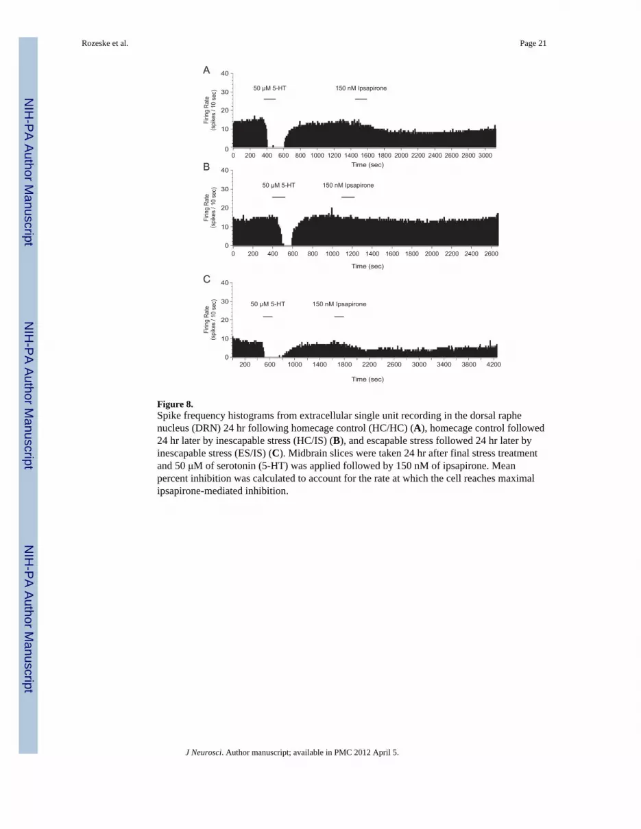
Figure 8.Spike frequency histograms from extracellular single unit recording in the dorsal raphenucleus (DRN) 24 hr following homecage control (HC/HC) (A), homecage control followed24 hr later by inescapable stress (HC/IS) (B), and escapable stress followed 24 hr later byinescapable stress (ES/IS) (C). Midbrain slices were taken 24 hr after final stress treatmentand 50 μM of serotonin (5-HT) was applied followed by 150 nM of ipsapirone. Meanpercent inhibition was calculated to account for the rate at which the cell reaches maximalipsapirone-mediated inhibition.
Rozeske et al. Page 21
J Neurosci. Author manuscript; available in PMC 2012 April 5.
NIH
-PA
Author M
anuscriptN
IH-P
A A
uthor Manuscript
NIH
-PA
Author M
anuscript

Figure 9.Behavioral immunization blocks inescapable stress (IS)-induced impairment of dorsal raphenucleus (DRN) serotonin (5-HT) cell firing with 150 nM ipsapirone. Data are expressed asmean percent inhibition ± SEM. Mean percent inhibition was different among stress groups(p < 0.05). *Mean percent inhibition of DRN 5-HT cell firing was significantly different inrats that received homecage treatment followed by IS (HC/IS) as compared to rats thatreceived no stress (HC/HC) and behavioral immunization (ES/IS) (p < 0.05).
Rozeske et al. Page 22
J Neurosci. Author manuscript; available in PMC 2012 April 5.
NIH
-PA
Author M
anuscriptN
IH-P
A A
uthor Manuscript
NIH
-PA
Author M
anuscript

Figure 10.Protein expression of the serotonin-1A receptor (5-HT1A-R) in the dorsal raphe nucleus isnot changed 24 hr following inescapable stress (IS) and homecage (HC) treatments.Representative western blot of 5-HT1A-R and sodium-potassium ATPase (Na- K ATPase)expression in the dorsal raphe nucleus 24 hr following inescapable stress (IS) and homecage(HC) treatments. No significant difference was observed between HC and IS treatments (p >0.05).
Rozeske et al. Page 23
J Neurosci. Author manuscript; available in PMC 2012 April 5.
NIH
-PA
Author M
anuscriptN
IH-P
A A
uthor Manuscript
NIH
-PA
Author M
anuscript