Developmental changes in protein composition and the actin-binding protein ponticulin in...
34
Developmental changes in the protein composition of Manduca sexta lipid droplets Jose L Soulages, Sarah J Firdaus, Steve Hartson, Xiao Chen, Alisha D. Howard, and Estela L Arrese Department of Biochemistry & Molecular Biology; 147 Noble Research Center, Oklahoma State University, Stillwater, OK, 74074 USA Abstract The lipid droplets (LDs) are intracellular organelles mainly dedicated to the storage and provision of fatty acids. To accomplish these functions the LDs interact with other organelles and cytosolic proteins. In order to explore possible correlations between the physiological states of cells and the protein composition of LDs we have determined and compared the proteomic profiles of lipid droplets isolated from the fat bodies of 5 th -instar larvae and adult Manduca sexta insects and from ovaries. These LD-rich tissues represent three clearly distinct metabolic states in regard to lipid metabolism: 1) Larval fat body synthesizes fatty acids (FA) and accumulates large amounts as triglyceride (TG); 2) Fat body from adult insects provides FA to support reproduction and flight; 3) Ovaries do not synthesize FA, but accumulate considerable amounts of TG in LDs. Major qualitative and semi-quantitative variations in the protein compositions of the LDs isolated from these three tissues were observed by MS/MS and partially validated by immuno-blotting. The differences observed included changes in the abundance of lipid droplet specific proteins, cytosolic proteins, mitochondrial proteins and also proteins associated with the machinery of protein synthesis. These results suggest that changes in the interaction of LDs with other organelles and cytosolic proteins are tightly related to the physiological state of cells. Herein, we summarize and compare the protein compositions of three subtypes of LDs and also describe for the first time the proteomic profile of LDs from an insect ovary. The compositions and compositional differences found among the LDs are discussed to provide a platform for future studies on the role of LDs, and their associated proteins, in cellular metabolism. Keywords Lipid Droplets; Proteomics; Fat Body; Ovaries; Manduca sexta; Metabolism 1. INTRODUCTION The fat body in insects (Arrese and Soulages, 2010) and the adipose tissue in vertebrates (Frayn et al., 2003) play a major role in the homeostasis of energy metabolism. Adipocytes are the main cells found in these tissues and are characterized by the massive presence of lipid storage droplets, or lipid droplets (LDs) (Zweytick et al., 2000). LDs consist of a core of neutral lipids, predominately triglycerides (TG), surrounded by a layer of phospholipid © 2012 Elsevier Ltd. All rights reserved. Publisher's Disclaimer: This is a PDF file of an unedited manuscript that has been accepted for publication. As a service to our customers we are providing this early version of the manuscript. The manuscript will undergo copyediting, typesetting, and review of the resulting proof before it is published in its final citable form. Please note that during the production process errorsmaybe discovered which could affect the content, and all legal disclaimers that apply to the journal pertain. NIH Public Access Author Manuscript Insect Biochem Mol Biol. Author manuscript; available in PMC 2013 May 1. Published in final edited form as: Insect Biochem Mol Biol. 2012 May ; 42(5): 305–320. doi:10.1016/j.ibmb.2012.01.001. NIH-PA Author Manuscript NIH-PA Author Manuscript NIH-PA Author Manuscript
-
Upload
independent -
Category
Documents
-
view
2 -
download
0
Transcript of Developmental changes in protein composition and the actin-binding protein ponticulin in...
Developmental changes in the protein composition of Manducasexta lipid droplets
Jose L Soulages, Sarah J Firdaus, Steve Hartson, Xiao Chen, Alisha D. Howard, and EstelaL ArreseDepartment of Biochemistry & Molecular Biology; 147 Noble Research Center, Oklahoma StateUniversity, Stillwater, OK, 74074 USA
AbstractThe lipid droplets (LDs) are intracellular organelles mainly dedicated to the storage and provisionof fatty acids. To accomplish these functions the LDs interact with other organelles and cytosolicproteins. In order to explore possible correlations between the physiological states of cells and theprotein composition of LDs we have determined and compared the proteomic profiles of lipiddroplets isolated from the fat bodies of 5th-instar larvae and adult Manduca sexta insects and fromovaries. These LD-rich tissues represent three clearly distinct metabolic states in regard to lipidmetabolism: 1) Larval fat body synthesizes fatty acids (FA) and accumulates large amounts astriglyceride (TG); 2) Fat body from adult insects provides FA to support reproduction and flight;3) Ovaries do not synthesize FA, but accumulate considerable amounts of TG in LDs. Majorqualitative and semi-quantitative variations in the protein compositions of the LDs isolated fromthese three tissues were observed by MS/MS and partially validated by immuno-blotting. Thedifferences observed included changes in the abundance of lipid droplet specific proteins,cytosolic proteins, mitochondrial proteins and also proteins associated with the machinery ofprotein synthesis. These results suggest that changes in the interaction of LDs with otherorganelles and cytosolic proteins are tightly related to the physiological state of cells. Herein, wesummarize and compare the protein compositions of three subtypes of LDs and also describe forthe first time the proteomic profile of LDs from an insect ovary. The compositions andcompositional differences found among the LDs are discussed to provide a platform for futurestudies on the role of LDs, and their associated proteins, in cellular metabolism.
KeywordsLipid Droplets; Proteomics; Fat Body; Ovaries; Manduca sexta; Metabolism
1. INTRODUCTIONThe fat body in insects (Arrese and Soulages, 2010) and the adipose tissue in vertebrates(Frayn et al., 2003) play a major role in the homeostasis of energy metabolism. Adipocytesare the main cells found in these tissues and are characterized by the massive presence oflipid storage droplets, or lipid droplets (LDs) (Zweytick et al., 2000). LDs consist of a coreof neutral lipids, predominately triglycerides (TG), surrounded by a layer of phospholipid
© 2012 Elsevier Ltd. All rights reserved.Publisher's Disclaimer: This is a PDF file of an unedited manuscript that has been accepted for publication. As a service to ourcustomers we are providing this early version of the manuscript. The manuscript will undergo copyediting, typesetting, and review ofthe resulting proof before it is published in its final citable form. Please note that during the production process errorsmaybediscovered which could affect the content, and all legal disclaimers that apply to the journal pertain.
NIH Public AccessAuthor ManuscriptInsect Biochem Mol Biol. Author manuscript; available in PMC 2013 May 1.
Published in final edited form as:Insect Biochem Mol Biol. 2012 May ; 42(5): 305–320. doi:10.1016/j.ibmb.2012.01.001.
NIH
-PA Author Manuscript
NIH
-PA Author Manuscript
NIH
-PA Author Manuscript
and a number of embedded or peripherally associated proteins (Brasaemle, 2007a). LDshouse organelle-specific proteins of the PAT family (Pfam 03036)(Lu et al., 2001; Miura etal., 2002), such as perilipin in vertebrates and the lipid droplet storage protein-1 and 2, Lsd1and Lsd2, in insects. These proteins play a major role in the degradation of TG and itsregulation as suggested by studies in vertebrates (Brasaemle, 2007b; Ducharme and Bickel,2008) and insects (Beller et al., 2010b; Bickel et al., 2009). Although the proteins associatedwith the storage and/or hydrolysis of TG have been the main focus of studies centered onlipid droplets, several recent proteomic studies have shown that LDs are associated with alarge number of cellular proteins, including proteins of typical cytosolic, mitochondrial,lyzosomal and endoplasmic reticulum location (Beller et al., 2010b; Murphy et al., 2009;Ohsaki et al., 2009; Welte, 2009). The protein composition of LDs suggests that thesecannot be simply described as a fat globule surrounded by a monolayer of phospholipid andproteins. LDs are probably more accurately described if we think of them as a lipid enrichedcomplex that involves interactions with other organelles and numerous cytosolic proteins(Murphy et al., 2009). The physical interactions between the lipid globule of the LDs andproteins from other compartments are likely to be at the center of the control of cellularmetabolism (Goodman, 2008, 2009). Knowledge of the composition of these complexes is afirst step toward the development of new hypotheses and tests to study and understand therole of LDs in the control of cellular lipid metabolism.
Two proteomic studies of LDs isolated from Drosophila, whole embryos (Cermelli et al.,2006) and larval fat body (Beller et al., 2006), have been previously reported. These are theonly studies that have focused on the sub-proteome of insect LDs.
To explore possible correlations between the protein composition of LDs and thephysiological state of fat body cells, in the present study we have determined and comparedthe proteomic profiles of lipid droplets isolated from the fat bodies of Manduca larvae andadult insects. The comparison of LDs from the fat bodies of larvae and adult insects isinteresting because they represent two quite different metabolic states. During the fastgrowing larval stage, M. sexta accumulates massive amounts energy reserves as lipiddroplets and this accumulation requires the synthesis of fat and proteins. Conversely, theadult stage of M. sexta is characterized by a massive depletion of energy reserves stored inthe fat body. During its short life span, the adult insect uses most of its stored fat to supportreproduction and flight. The metabolic differences between larvae and adult insectssuggested that changes in the protein composition of LDs were possible and could beevidenced using a proteomic approach. The study was extended to LDs isolated from theovaries of M. sexta because, as the fat body during the larval stage of M. sexta, the ovariesof adult females represent a stage of fat accumulation (Ziegler and Van Antwerpen, 2006).The main lipid form in the ovary is also TG, which is utilized to support the subsequentenergy and membrane synthesis demands of the developing embryo. However, whereas thelarval fat body can synthesize fat, the accumulation of fat by ovaries is dependent on theexternal supply of fatty acids stored in the fat body.
In this study we compare the protein compositions of three subtypes of LDs from Manducasexta and also describe for the first time the proteomic profile of LDs from an insect ovary.The compositions and compositional differences found among LDs are discussed to providea platform for future studies on the role of LDs, and their associated proteins, in cellularmetabolism.
Soulages et al. Page 2
Insect Biochem Mol Biol. Author manuscript; available in PMC 2013 May 1.
NIH
-PA Author Manuscript
NIH
-PA Author Manuscript
NIH
-PA Author Manuscript
2. MATERIAL AND METHODS2.1 Insects
Manduca sexta eggs were purchased from Carolina Biological supplies, and larvae werereared on artificial diet (Bell and Joachim, 1976). Adult insects were maintained at roomtemperature without food. Second day 5th instar larvae and adult females were used in theexperiments.
2.2 Lipid droplet preparationFat body tissue from two insects was combined and homogenized with a Potter-Elvehjemglass homogenizer fitted with Teflon pestle, using 6ml of 0.25M sucrose buffer (20mM Tris,pH 7.4, 0.25M sucrose, 1mM EDTA, 1mM benzamidine, 1mM PMSF, 10mg/l leupeptine,1mg/l aprotonin, 1 mM DTT). The homogenate was centrifuged at 1000xg for 10 min. Thesupernatant was adjusted to 1.17 M sucrose and transferred to a SW40 centrifuge tube to besubsequently overlaid with 1ml of each of 1.02M sucrose buffer, 0.87M sucrose buffer,0.58M sucrose buffer, 0.29M sucrose buffer, 0.15M sucrose buffer, and 1.5ml of bufferwithout sucrose. Density gradients were centrifuged at 160,000g for 4 hr. The top fraction ofthe gradient, a distinctive thin white layer floating at the top that contains lipid droplets, wascollected and mixed with 8ml of 0.25M sucrose buffer. The sample was transferred to aSW40 centrifuge tube and overlaid with 3 ml of buffer without sucrose and centrifuged at111,000g for 1hr. The top layer (~1 ml), containing the lipid droplets, was collected.Samples were delipidated by acetone precipitation (85% acetone v/v, final concentration) at−20°C overnight. After centrifugation at 10,000g for 15 min, protein pellets were dissolvedin Laemmli buffer containing 2.5% (w/v) SDS and 17 mM DTT and treated at 70°C for 10min. Ovaries dissected from ten insects were extensively rinsed in phosphate buffer tocompletely remove fat body tissue. Ovaries were homogenized as described above and lipiddroplets were isolated following the same procedure.
2.3 Sample processingSamples were subjected to SDS-PAGE in 4–20% acrylamide gradient gels (Novex,Invitrogen). Each lane of the Coomassie Blue stained gel was divided in six regions. Eachgel slice was finely minced. Proteins from each slice were reduced with tris(2-carboxyethyl)phosphine, alkylated with iodoacetamide, and digested for 6–16 hr with 8 μg/ml trypsin, using 50 mM ammonium bicarbonate as buffer. Digestion products wereanalyzed by LC-MS/MS.
2.4 LC-MS/MSSamples were analyzed on a hybrid LTQ-Orbitrap XL mass spectrometer (Thermo FisherScientific) coupled to a New Objectives PV-550 nanoelectrospray ion source and anEksigent NanoLC-2D chromatography system. Peptides were analyzed by trapping on a 2.5cm ProteoPrepII pre-column (New Objective) and separation on a 75 μm ID fused silicacolumn, packed in house with 10-cm of Magic C18 AQ, terminated with an integral fusedsilica emitter pulled in house. Peptides were eluted using a 5–40% ACN/0.1% formic acidgradient performed over 40 min at a flow rate of 300 nl/min. During each one-second full-range FT-MS scan (nominal resolution of 60,000 FWHM, 300 to 2000 m/z), the three mostintense ions were analyzed via MS/MS in the linear ion trap. MS/MS settings used a triggerthreshold of 8000 counts, monoisotopic precursor selection, and rejection of parent ions thathad unassigned charge states, were previously identified as contaminants on blank gradientruns, or were previously selected for MS/MS (data dependent acquisition using a dynamicexclusion for 150% of the observed chromatographic peak width). Column performance wasmonitored using trypsin autolysis fragments (m/z 421.76), and via blank injections between
Soulages et al. Page 3
Insect Biochem Mol Biol. Author manuscript; available in PMC 2013 May 1.
NIH
-PA Author Manuscript
NIH
-PA Author Manuscript
NIH
-PA Author Manuscript
samples to assay for contamination. Data analysis: Centroided ion masses were extractedusing the extract_msn.exe utility from Bioworks 3.3.1 and were used for database searchingwith Mascot v2.2.04 (Matrix Science) and X! Tandem v2007.01.01.1 (www.thegpm.org). Adatabase containing the predicted proteins from all arthropods from NCBI (December 2009)was utilized for searching. MASCOT was set up to search NCBI. Searches used a fragmention mass tolerance of 0.80Da and a parent ion tolerance of 10.0 ppm. Searches included thefollowing potential peptide modifications: pyroglutamate modification of N-terminalglutamines, oxidation of methionine, acrylamide, and iodoacetamide adducts of cysteine, aswell as potential formylation and acetylation of the N-terminus of the parent protein.Scaffold (v2.6.0; Proteome Software) was used to validate MS/MS-based peptide andprotein identifications. Peptide identifications were accepted if they could be established atgreater than 99.9% probability as specified by the Peptide Prophet algorithm. Proteinidentifications were accepted if they could be established at greater than 99.0% probabilityand contained at least two identified peptides. Protein probabilities were assigned by theProtein Prophet algorithm.
2.5 Light microscopySamples of purified lipid droplets were imaged using a Leica TCS SP2 confocal microscope.Lipid droplets fluorescently labeled with Nile Red or without labeling were observed. Fatbody tissue and ovaries from a female insect two days-old were embedded in paraffin andsections that were stained with Mallory’s Azure II Methylene Blue were imaged using aLeica TCS SP2 confocal microscope.
2.6 Electron microscopyPortions of fat body dissected from a female insect two days-old were fixed for 12 hr in 2%glutaraldehyde in 200 mM sodium cacodylate at 4 °C. Tissue was washed three times inbuffer A (60 mM sodium cacodylate, 180 mM sucrose) for 30 minutes each and post-fixedfor 1.5 hr in 1% osmium tetroxide in 100 mM phosphate buffer (pH 7.2). Samples werewashed three times in buffer A for 30 minutes each followed by dehydration with ethanol.Serial washes in 30%, 50%, 70%, 90%, 95% and 100% ethanol (v/v) for 30 minutes eachstep were followed by three washes in propylene oxide for 30 minutes each step. Tissue wasinfiltrated using EMBed 812 resin (Electron Microscopy Sciences, Hatfield, PA). Tissuewas placed in 3:1 propylene oxide and EMBed for 4 days. Tissue was placed in 1:1propylene oxide and EMBed for 2 days, then 1:3 propylene oxide and EMBed for 2 days.Tissue was placed in 100% embedding medium and placed in oven at 60°C for 48 hours.Embedded tissues were cut using an ultramicrotome into 80 nm thick sections, and placedon carbon film grids. Grids containing thin sections were stained using 2.5% uranyl acetatefor 30 minutes then rinsed. Grids were then stained using Reynold’s lead citrate (Reynolds,1963). Sections were imaged using a JEOL 2100 TEM operated at 120 kV.
2.7 Lipid analysisTotal lipids were extracted from 0.2ml of lipid droplets preparation by adding 5 volumes ofchloroform-methanol (v/v) and 0.2 ml of phosphate buffer containing 150mM NaCl.Samples were vortexed and centrifuged. The organic phase was collected and subjected tolipid analysis by TLC using hexane-ethyl ether-formic acid 70/30/3 (v/v/v) as thedeveloping solvent (Skipski and Barclay, 1969). Lipids were visualized by iodine andidentified by comparing their retention time with that of the standards. A similar procedurewas used to prepare total lipid extract from 100mg of whole tissue (fat body and ovaries).
Soulages et al. Page 4
Insect Biochem Mol Biol. Author manuscript; available in PMC 2013 May 1.
NIH
-PA Author Manuscript
NIH
-PA Author Manuscript
NIH
-PA Author Manuscript
2.8 Western BlotProteins were separated by SDS-PAGE (4–20%) and transferred to nitrocellulosemembranes. Immunodetection was performed using the corresponding horseradishperoxidase-conjugated secondary antibody purchased from Santa Cruz Biotechnology(Santa Cruz, CA) and ECL chemiluminescence reagents (Amersham Pharmacia,Piscataway, NJ) and exposed to X-ray films. Polyclonal antibodies against apoLp-I/apoLp-IIand apoLp-III, were raised by Cocalico Biologicals Inc. (Reamstown, PA) in rabbitsimmunized with the corresponding proteins (HDLp and apoLpIII), which were purified fromhemolymph. Polyclonal antibodies against Lsd1 and Lsd2 were obtained by CocalicoBiologicals Inc. (Reamstown, PA) from rabbits immunized with the recombinant proteins.M. sexta Lsd1 and Lsd2 recombinant proteins (GI: 238846407 and GI: 338858969,respectively) were expressed in E. coli and purified as previously described for DrosophilaLsd proteins (Arrese et al., 2008b; Arrese et al., 2008c). ACC was detected with commercialrabbit polyclonal antibodies from Cell Signaling (Billerica, MA). Lip-DH antibody waspurchased from Abcam (Cambridge, MA). Mouse monoclonal anti-Hsp70 was a gift fromDr. Robert L. Matts.
2.9 Other MethodsSDS-PAGE was performed according to Laemmli (Laemmli, 1970) and proteins werevisualized by Coomassie Brilliant Blue R staining. TG concentration from the gradientfractions was determined using the Infinity Triglyceride reagent kit as described by themanufacturer (Thermo Fisher Scientific Inc., Waltham, MA). Triolein was used as standard.Protein concentrations were determined by the Bradford dye-binding assay (Bradford, 1976)using bovine serum albumin as standard.
3. RESULTS AND DISCUSSION3.1. Purification of Lipid Droplets
The first step for the isolation of lipid droplets (LDs) consisted in the centrifugation ofhomogenates in a sucrose density gradient. This method is commonly used to purify LDsfrom Drosophila fat body (Beller et al., 2006), Drosophila embryos (Cermelli et al., 2006)and from vertebrate adipose tissue (Brasaemle et al., 2004). The LDs accumulate in the topfraction of the gradient (0% sucrose) as reflected by the triglycerides (TG) levels (Fig 1B),whereas most of the cellular proteins accumulate at the bottom of the gradient (Fig 1A). LDsproteins accounted for less than 3% of total fat body homogenate proteins. The substantialremoval of proteins, and the concomitantly high TG to protein ratio of the top fraction of thegradient, shows that this procedure leads to a significant purification of the LDs. The lipiddroplet fraction (top) isolated from the sucrose gradient was re-purified by an additionalhigh speed centrifugation step as indicated in section 2.2. Qualitative changes in the proteinprofiles from Coomassie Blue stained SDS-PAGE were observed between homogenates andLDs obtained after each centrifugation step. Fig 1C shows the profiles obtained for the larvalLDs purification. The lipid to protein ratios of LDs after the first density gradient (12.6 ± 0.2μmol TG/mg protein) and the second purification step (21.8 ± 3.7 μmol TG/mg protein) isshown in the Fig. 1D. Lipid to protein ratios of Manduca purified LDs are consistent withprevious reports (Wolins et al., 2006). The total protein associated with purified LDsaccounted for approximately 1.7% of total protein in the fat body homogenate. Lsd1, aprotein that is found mostly associated with lipid droplets in Manduca sexta fat body (Arreseet al., 2008a), was used as fat body lipid droplet marker. As expected, the LDs fraction wasenriched in this protein. Fig 1E shows that the top fraction of the gradient is highly enrichedin Lsd1 after the first centrifugation and remains practically unchanged after the secondcentrifugation step. The progress of the purification was also assessed by monitoring thepresence of apoLp-III and lipoamide dehydrogenase (LipDH) during the purification.
Soulages et al. Page 5
Insect Biochem Mol Biol. Author manuscript; available in PMC 2013 May 1.
NIH
-PA Author Manuscript
NIH
-PA Author Manuscript
NIH
-PA Author Manuscript
ApoLp-III, a small soluble apolipoprotein, is undetectable by Western Blot after the secondpurification step (Fig 1E). On the other hand, the mitochondrial protein LipDH become lessabundant after the second purification step, but it cannot be completely removed.
The morphology of purified LDs was assessed by confocal microscopy. Fig 1F shows animage of purified LDs from larval fat body. LDs showed a spherical shape with variablesizes and diameters ranging between 2μm and 12μm. This range of sizes is consistent withpreviously reported LDs from the fat body tissue of larval M. sexta (Willott et al., 1988).
LDs purification from adult fat body and ovary homogenates proceeded in a very similarmanner to that shown for the larval fat body. The lipid to protein ratio of LDs from the adultfat body after the first density gradient was 10.5 ±1.0 μmol TG/mg protein and 20.9 ± 1.9μmol TG/mg protein after the second purification step. Likewise, the lipid to protein ratiosfor LDs from ovaries changed from 8.8 ±2.0 to 13.1 ±2.1 μmol TG/mg protein. The relativeenrichment of Lsd1 and Lsd2 in purified LDs from adult fat body and ovaries are shown inFig 5B.
Images of adult fat body tissue and oocyte and their corresponding purified lipid droplets areshown in Fig 2A,B,C,D. Spherical LDs with diameters ranging between 2 μm and 22 μmand 1 and 13 μm were observed for LDs isolated from the adult fat body (Fig 2B) andovaries (Fig 2D), respectively. The range of sizes of purified LDs was consistent with sizesobserved in the tissues (Fig 2A and 2C).
The lipid analysis of isolated LDs confirmed that the integrity of lipids was preserved duringthe purification procedure. TG was the major lipid found in all preparations. A small amountof DG was also observed in LDs isolated from the adult fat body (Fig 2E).
To identify the proteins associated with LDs, the samples of purified LDs were delipidated,subjected to SDS-PAGE (Fig 2F) and subsequently analyzed by MS/MS, as indicated insection 2.4.
3.2. Proteomics of Lipid DropletsA total of 229 different proteins were identified by MS/MS. The complete list of proteins inlipid droplets isolated from M. sexta fat body and ovaries is shown in Table 1. The larval fatbody provided the largest number of identifications (172), followed by the LDs from femalefat body (129), and ovaries (88). Some proteins were only found in one type of LDs (74 inlarval fat body and 13 in LDs from ovaries). The number and variety of proteins identified inM. sexta LDs are consistent with previous proteomic studies of LDs from diverse origins.There are at least 13 papers specifically describing the proteomic composition of lipiddroplets: two from insects (Beller et al., 2006; Cermelli et al., 2006), two from yeast (Binnset al., 2006; Fei et al., 2011) and the remaining of vertebrate origin (Brasaemle et al., 2004;Cavaletto et al., 2008; Cho et al., 2007; Fujimoto et al., 2004; Liu et al., 2004; Sato et al.,2006; Umlauf et al., 2004; Wan et al., 2007; Wu et al., 2000). All these studies havereported a number and diversity of proteins similar to those presented in our study. Theproteins previously reported to be components of the LDs of Drosophila have been markedin the Table 1. Among the proteins found in M. sexta LDs approximately 80 proteins werealso present in Dm LDs.
The list of proteins was classified in subgroups defined on the basis of the expected proteinlocation. In order to have an estimate of the prevalence of each protein-subgroup in differentlipid droplets, we performed a semi-quantitative analysis of the relative protein abundancebased on the fraction of spectral counts obtained for each subgroup. Due to variations in thenumber of unique peptides theoretically derived from different proteins, and the peptide’s
Soulages et al. Page 6
Insect Biochem Mol Biol. Author manuscript; available in PMC 2013 May 1.
NIH
-PA Author Manuscript
NIH
-PA Author Manuscript
NIH
-PA Author Manuscript
ionization efficiency and appropriate mass to charge ratio for detection, MS/MS does notprovide a straightforward quantitative estimate of protein abundance. For instance, an over-estimation of the fraction of larger proteins present in a sample will be expected. In spite ofthese limitations, a comparison of the fractions of spectral counts for a given protein acrossdifferent samples provides a good semi-quantitative estimate of possible changes in theabundance of the selected protein. For a given protein there is no bias due to the number ofunique peptides, ionization efficiency, and other factors. Similarly, it is also possible toperform a semi-quantitative comparison of the abundance of groups of proteins bycomparing the sum of their spectral counts across LDs samples. Fig 3 shows the comparisonof the fraction of spectral counts of different groups of proteins for the three types of LDsanalyzed. Mitochondrial and cytosolic proteins appear highly abundant in the three subtypesof LDs. Conversely, proteins of typical nuclear location and non-mitochondrial membraneproteins were weakly represented in all LDs. Moreover, clear differences among differentLDs subtypes can be seen. Cytosolic and mitochondrial proteins dominate the distribution ofspectral counts in the LDs of both larval and adult fat bodies. Lipid droplet specific proteins,Lsd1 and/or Lsd2, were only significantly abundant in LDs from the fat body of adultinsects.
Most of the protein groups found in LDs from fat body adipocytes are also significantlyrepresented in the LDs isolated from ovaries (Fig 3 and Table 1). The abundance ofapolipophorin (apoLp), the protein component of the major lipoprotein in insects, issignificant in all LDs. However, its high abundance in LDs from ovaries constitutes a uniquefeature of this subtype of LDs.
As observed in previous proteomic studies of lipid droplets of diverse origin (cited above),this proteomic study suggests that M. sexta LDs are also tightly associated withmitochondrial proteins, cytosolic proteins, and proteins of the machinery of proteinsynthesis. The interaction of LDs with other organelles is a topic that is receiving increasingattention by the scientific community (Goodman, 2008, 2009). These interactions are neededand must be at the center of the regulation of metabolism. Interactions between LDs andperoxisomes (Binns et al., 2006) have also been observed in Yeast, which carries out anactive oxidation of fatty acids in these organelles. The ER synthesizes the TG that is storedin LDs and given the insolubility of TG the two apparently independent organelles must bephysically connected. Studies in Yeast have shown that 96% of the LDs are permanentlyassociated to ER (Szymanski et al., 2007). Ribosomal proteins were particularly abundant inLDs from larval fat body and from ovaries. The fast changes in the size of LDs for instancewhen the tissue accumulates TG are likely to require the coupling with protein synthesisand, thus, the presence of ribosomal proteins in LDs is not surprising.
The physical interaction between mitochondria and LDs was observed in multiple studies(Cohen et al., 2004; Novikoff et al., 1980; Stemberger et al., 1984; Vock et al., 1996) andhas been the subject of specific studies to determine nature and function of the proteinsinvolved in the interaction (Pu et al., 2011). It has been recently shown that perilipin 5, alipid droplet-associated protein, provides physical and metabolic linkage to mitochondria(Wang et al., 2011). As shown in the electron micrographs the LDs of M. sexta fat body(Figures 4A–B) are also closely associated with both mitochondria and rough endoplasmicreticulum. Therefore, the presence of mitochondrial and ER proteins in the preparation ofLDs is not surprising. Contrarily, the interaction and even fusion of ER and mitochondrialmembranes with the LDs surface may be essential for the stability and function of LDs andalso to allow a proper crosstalk between organelles.
Soulages et al. Page 7
Insect Biochem Mol Biol. Author manuscript; available in PMC 2013 May 1.
NIH
-PA Author Manuscript
NIH
-PA Author Manuscript
NIH
-PA Author Manuscript
3.2.1. Proteins of Lipid Metabolism Associated with Lipid Droplets3.2.1.1. Lipid droplet specific proteins: Only few proteins have shown a strong preferenceto associate with LDs in animal cells. This small set of proteins were grouped under the PATfamily (Pfam 03036)(Lu et al., 2001; Miura et al., 2002), and comprises proteins such asperilipin, TIP47 and ADRP, in vertebrates, and lipid storage droplet protein-1 and -2, Lsd1and Lsd2, in insects. These proteins share sequence similarity in the N-term region, a regioncalled the PAT domain. The insect genomes encode two proteins of the PAT family (Bickelet al., 2009), Lsd1 and Lsd2. These proteins do not have a known enzymatic activity but, assuggested by studies in vertebrates (Brasaemle, 2007b; Ducharme and Bickel, 2008) and ininsects (Beller et al., 2010a; Gronke et al., 2003; Teixeira et al., 2003), they play a majorrole in the degradation of TG and its regulation. PKA-dependent phosphorylation of Lsd1 isa major mechanism regulating the rate of lipolysis in M. sexta fat body (Arrese et al., 2008b;Patel et al., 2005). Moreover, Ca2+/calmodulin dependent phosphorylation of Lsd1 was alsoshown to be a key factor in the hydrolysis of TG associated with the production of the mainBombyx mori pheromone (Ohnishi et al., 2011). Lsd1 was also shown to control theaccumulation of fat in Drosophila (Beller et al., 2010a) although the role of Lsd1phosphorylation seems to be different to that observed in Lepidoptera.
The MS/MS study showed that Lsd proteins are particularly abundant (~18% of totalspectral counts) in LDs from the fat body of adult insects (Fig 5). Both proteins, Lsd1 andLsd2, were found in LDs from adult fat body by MS/MS and western blotting. However, theMS/MS study suggests that Lsd1 is the predominant Lsd protein (97% of the spectral counts,Fig 5) in the fat body of adult insects.
Two independent estimates, MS/MS and western blotting, of the relative abundance of Lsd1in different subtypes of LDs are shown in Fig 5. The higher abundance of Lsd1 in LDs fromadult insects, as compared to LDs of larval fat body and ovaries, is consistent with thephysiological state of adult insects, which are mobilizing FA from the fat body to theovaries. Previous studies have linked the expression levels of Lsd1 (Arrese et al., 2008a)with the ability of M. sexta fat body to mobilize FA. Moreover, since adult insects are keptwithout food, they are also mobilizing and oxidizing FA to support basal metabolism.Similarly, the low levels of Lsd1 in LDs from larval fat body or from ovaries are alsoconsistent with the notion that these tissues are accumulating rather than mobilizing fattyacids. We previously reported that Lsd1 was not detected in LDs isolated from young larvalfat body by immunoblot analysis (Arrese et al., 2008a). In that study we used an antibodyraised against two Lsd1 peptides. In the present study Lsd1 was detected with a differentantibody, which was raised against the recombinant Lsd1 protein as described in Materialsand Methods. This antibody detected Lsd1 in larval LDs. In agreement with this result, theMS/MS study also detected the presence of Lsd1 in the young larva.
Genetic studies in Drosophila have suggested that Lsd2 plays a role in the accumulation offat. Flies overexpressing Lsd2 accumulated TG, whereas knockout flies were lean (Gronkeet al., 2003; Teixeira et al., 2003). The level of spectral counts of Lsd2 (~1.5%) obtained inthe MS/MS study suggests that Lsd2 represents a significant component of the LDs fromovaries. Since the developing ovaries of adult females accumulate lipids, Lsd2 could beinvolved in this process of TG accumulation. Ovaries accumulate lipid by endocytosis oflipophorin rather than by the novo fatty acid synthesis (Kawooya and Law, 1988). The larvalstage of M. sexta is characterized as a frenzy feeding period during which there is a fastincrease in body size and a concomitant accumulation of fat in the fat body. Thus, on thebasis of the suggested role of Lsd2 in Drosophila’s energy metabolism, we expected to findhigh levels of expression of Lsd2 in larval LDs. However, and somewhat surprising, Lsd2was undetectable in LDs from young larval fat body by MS/MS (Fig 5A) and western
Soulages et al. Page 8
Insect Biochem Mol Biol. Author manuscript; available in PMC 2013 May 1.
NIH
-PA Author Manuscript
NIH
-PA Author Manuscript
NIH
-PA Author Manuscript
blotting (Fig 5B). These results could also indicate a difference between the roles of Lsd2 inflies and moths.
3.2.1.2. Lipogenic and lipolytic enzymes: Several essential enzymes of the lipogenicpathway associate with the LDs (Table 1). Lipid droplets from the fat body of adult femaleinsects showed the larger number of lipogenic enzymes, including acetyl-CoA carboxylase(ACC), FA-synthase (FAS), ATP-citrate lyase (ACL), pyruvate carboxylase (PC), Δ9desaturase (SCD) and NADPH-cytochrome P450 reductase. Some proteins involved inlipogenesis were also found associated with LDs from larval fat body (Table I and Fig 6).ACL was detected in female and larva at similar levels. However, larval LDs lacked FAS(Fig 6D) and, as inferred from the spectral counts and western blotting, contained muchlower levels of ACC than LDs from female FB. PC was also much higher in adult FB-LDsthan in larval LDs (15–20 lower than in adult LDs).
The rate of lipogenesis and fatty acid oxidation are controlled to a great extent by theexpression levels and posttranslational modifications of ACC (Saggerson, 2008). The firststep of the synthesis of FA consists in the synthesis of malonyl-CoA from acetyl-CoA. Thisreaction, which is catalyzed by ACC, is important not only for lipogenesis but also becausemalonyl-CoA is a major inhibitor of carnitine palmitoyltransferase (CPT) (Abu-Elheiga etal., 2001) and, therefore, ACC activity also controls FA oxidation.
The results of MS/MS suggested that ACC was present and highly abundant in fat bodyLDs. These results were confirmed by western blotting (Fig 6A). The immunoblot study alsosuggested that homogenates of larval fat bodies could have a higher content of ACC than fatbodies from adult insects (Fig 6A). This result would be consistent with the fact that thelarval stage is the period of fat accumulation in M. sexta, whereas in the adult stage the mothuses most of the fat reserves to support flight and reproduction. Interestingly, the proportionof ACC associated with LDs of adult fat body was found to be much higher than thatassociated with LDs of larval fat body. The meaning of this change in distribution of ACC isnot known at this time. It is known that human ACC is inhibited by binding of long chainfatty acyl-CoA (Faergeman and Knudsen, 1997). The association of ACC with LDs of adultinsects could be a mechanism of posttranslational regulation of ACC activity. Binding toLDs perhaps inhibits ACC in adult insects. This could be needed to preserve the scarcereserves of glucose stored as glycogen. Alternatively, binding of ACC to LDs couldconstitute a mechanism of storage. Stored ACC could be released from the LDs and becomeactive once the moth feeds on nectar. Clearly, further studies in this area could lead tointeresting insights into the role of the association of ACC with the LDs in the regulation oflipogenesis in Lepidoptera. These findings suggest that LDs in adult insects could recruitlipogenic enzymes to prevent their degradation and preserve them for their use after aneventual nectar feeding period. The role of lipid droplets as protein storage depots has beenrecently reviewed (Hodges and Wu, 2010).
Although the potential role of ACC binding to LDs has not been previously discussed, it isimportant to note that association of ACC with LDs isolated from Drosophila (Beller et al.,2006) and from mammalian sources (Liu et al., 2004; Umlauf et al., 2004) has beenpreviously reported. Moreover, it is well known that ACC2, one of the vertebrates’ isoformsof ACC, associates with mitochondria (Abu-Elheiga et al., 2000). ACC2 is assumed tointeract with the mitochondrial membrane through a hydrophobic N-terminal region.However, we do not know if the N-terminal region of M. sexta ACC shares similarities withACC2. On the other hand, we have not found homology between the N-terminal regions ofinsect ACCs and mouse or human ACC2. Further studies are required in both arthropodsand mammalian systems to understand a possible physiological role of ACC binding to LDs.
Soulages et al. Page 9
Insect Biochem Mol Biol. Author manuscript; available in PMC 2013 May 1.
NIH
-PA Author Manuscript
NIH
-PA Author Manuscript
NIH
-PA Author Manuscript
ACC was not detected by MS/MS or by immunoblot in lipid droplets isolated from ovaries.In addition, western blot analysis of ovary homogenates also showed a very low level ofACC (Fig 6A). This result is in accord with previous studies indicating that de novo FAsynthesis in ovaries is not relevant and only contributes 1% of the oocyte’s fat content(Kawooya and Law, 1988). The fact that the MS/MS study did not detect lipogenic enzymesin these LDs, Fig 6, is in agreement with the expected low lipogenic activity of ovaries.
Fatty acid mobilization is initiated by the action of lipases. Two lipases involved in thehydrolysis of TG are present in Manduca sexta fat body. These are TGL (Arrese et al., 2010;Arrese and Wells, 1994) and the adipose tissue TG lipase (ATGL), also known as Brummerlipase in Drosophila (Gronke et al., 2005; Gronke et al., 2007). ATGL, a highly conservedlipase present in all organisms from yeast to mammals, is expected to be associated with theLDs (Lass et al., 2011). Our MS/MS study detected very low levels of ATGL in LDs fromfat bodies of larval and adult insects. Whether this is due to low expression levels of ATGLor to a particularly low sensitivity of MS/MS to detect tryptic peptides of ATGL remains tobe seen. On the other hand, TGL was not detected by MS/MS in any of the LDs samples.This is in agreement with previous reports showing that TGL is a cytosolic enzyme (Arreseet al., 2006) and does not bind to the LDs with high affinity (Patel et al., 2005).
3.2.3. Lipid Transport Proteins Associated with Lipid Droplets3.2.3.1. Apolipophorin: Lipophorin is the main lipoprotein found in insects (Soulages andWells, 1994). Lipophorin transports lipids from the midgut to the fat body and from the fatbody to other tissues, mainly muscles and ovaries (Arrese et al., 2001). The structuralapolipoproteins of lipophorin (apoLp-I and apoLp-II), were found in all LDs studied. Asshown in the Fig 7B, the MS/MS study suggested that apoLps were particularly abundant inLDs from ovaries, in which ~26% of all the spectral counts corresponded to apoLps.Confirming this result, Western blotting with anti-apoLp antibodies also showed that LDsisolated from ovaries contain the higher levels of apoLps (Fig. 7A).
Lipid accounts for 40% of the dry weight of mature M. sexta eggs (Kawooya et al., 1988).Nearly all the oocyte’s lipid originates in the fat body and is transported to the ovary bylipophorin (95%) and to a lesser extent by vitellogenin (Kawooya and Law, 1988). Studiesin M. sexta have shown that a significant fraction of circulating lipophorin is sequestered bythe follicles and the lipophorin-DG converted to TG for storage (Kawooya and Law, 1988;Kawooya et al., 1988; Ziegler and Van Antwerpen, 2006). These processes take placewithout degradation of the apolipoproteins, which are also stored in the eggs (Kawooya etal., 1988; Sun et al., 2000). In agreement with these previous studies, a lipid analysis ofovaries showed a virtual absence of DG (Fig 1A). Given the large amounts of apolipophorinpresent in ovaries (homogenates and LDs), the fact that only TG is detected suggests thatonce the lipoprotein is taken up by the ovaries the lipoprotein bound DG is rapidly convertedto TG. Since lipophorin represents the major source of lipids for the developing oocyte, theassociation of these apolipoproteins with the lipid droplets may be needed for the conversionof DG into TG and/or the subsequent transference of TG to the LDs core. The content ofapolipophorins also suggests that they may constitute an important structural element of theLDs of ovaries.
ApoLp-I and apoLp-II were also highly abundant in LDs of the fat bodies of insects in bothlarval and adult stages. The spectral counts in adult and larvae represented 5% and 6% of thetotal counts, respectively. Contrarily to the ovaries, which only take up lipophorin, the fatbody is the place of synthesis of lipophorin and also the location for reloading of partiallylipid depleted lipophorin particles. Biosynthesis of lipophorin is expected to involve the co-translational lipidation of apoLps (Dantuma et al., 1999). ApoLp-I and apoLp-II are codedby a single gene and synthesized as a long precursor that is subsequently cleaved. The
Soulages et al. Page 10
Insect Biochem Mol Biol. Author manuscript; available in PMC 2013 May 1.
NIH
-PA Author Manuscript
NIH
-PA Author Manuscript
NIH
-PA Author Manuscript
interaction of fat body LDs with apoLps may be needed to allow an efficient loading of thenascent apoLp chain with neutral lipids and, thus, prevent misfolding, aggregation anddegradation of the nascent apoLps. Apolipophorins share a number of similarities with apoB(Smolenaars et al., 2007), which has been previously shown to associate with LD inhepatome cell lines (Ohsaki et al., 2006; Ohsaki et al., 2008). The apoB-LD interactioncould be needed in vertebrates for either/or both the degradation of apoB containinglipoproteins, LDL and VLDL, (Ohsaki et al., 2006) or for the biosynthesis of VLDL. Infeeding and fast growing larvae the rate of de novo synthesis of lipophorin is high; thus, theassociation of apoLps with LDs is probably due to the coupling of apolipoprotein synthesisand lipid loading. In adult insects, lipophorin works to some extent as a lipid shuttle, orreusable lipoprotein, mobilizing DG from the fat body to other tissues. Therefore, theassociation of apoLps with fat body LDs is more likely due to the process of lipid loading ofpartially lipid-depleted lipophorin particles. The process of lipid loading of Lp particles ininsects is not fully understood, yet. However, endocytosis followed by lipid-loading andresecretion of the lipophorin particles is possibly the most important mechanism (Van Hoofet al., 2005). Adult insects have circulating lipophorin particles of higher lipid content thanlarvae. Two Lps are found in adult insects, HDLp and LDLp. In addition to apoLp-I andapoLp-II, the adult Lps have, a third small (17kDa) apoLp, apoLp-III, which is needed toprevent the aggregation of the highly DG-loaded Lps (Weers and Ryan, 2006). LDLpcontains more lipid and more molecules of apoLp-III than HDLp, which represents the lipiddepleted Lp in adult insects (Soulages et al., 1996). Uptake of HDLp and its binding to theLDs could be needed to allow loading of HDLp with DG. The TG molecules stored in LDsare the precursors of DG. In accordance with this hypothesis, apoLp-III was foundassociated with LDs from the fat bodies of adult insects, but was absent in larval LDs.Moreover, contrasting with the absence of DG in LDs from ovaries and larval fat body, fatbody-LDs from adult insects contain significant amounts of DG (Fig 2E). This difference isprobably explained by the fact that lipophorin loads DG in fat body and unloads it inovaries.
3.2.3.2. Lipophorin Receptor: A lipoprotein receptor with similarities to the mammalianLDL receptor family was initially purified from M. sexta (Tsuchida and Wells, 1990). Thisreceptor is involved in endocytosis of HDLp (Dantuma et al., 1999) and its mRNA isexpressed in several tissues (Ciudad et al., 2007; Gopalapillai et al., 2006; Seo et al., 2003).However, due to the fact that an extensive transference of lipids between lipophorin andtissues takes place in an endocytosis independent manner (Van der Horst and Rodenburg,2010; Van der Horst et al., 2009), the physiological function of this receptor is not clear, yet.The presence of lipophorin receptor in LDs of adult insects (Table 1), which mobilize lipidmainly from the fat body to the ovaries and flight muscles, suggests that its main role couldbe the delivery of lipids from the fat body to other tissues.
Vitellogenin (Vg), the major egg yolk lipoprotein, was found associated with the LDs ofovaries. Vg belongs to the superfamily of lipid transfer proteins that includes mammalianapoB, insect apoLp-I/II and others (Smolenaars et al., 2007). Vg provides only ~5% of thetotal egg lipid (Kawooya and Law, 1988). However, it plays an essential role in embryodevelopment. Vg and microvitellogenin (mVg) represented 1.6% of the total spectral countsof the LDs from ovaries. The role of Vg or mVg in the LDs is not known, but given theirabundances and lipid binding capabilities they could play a structural role. It may beinteresting to know whether these proteins can interact with Lsd2, the lipid droplet storageprotein that is abundant in LDs from ovaries.
Soulages et al. Page 11
Insect Biochem Mol Biol. Author manuscript; available in PMC 2013 May 1.
NIH
-PA Author Manuscript
NIH
-PA Author Manuscript
NIH
-PA Author Manuscript
3.3. Lipid Droplets and Protein SynthesisThe subgroup of larval LD-proteins related to protein synthesis is particularly interestingbecause in addition to a large number of ribosomal proteins (Table 1 and Fig 1), larval LDsalso contain several non-ribosomal proteins needed for protein synthesis, such as translationelongation factors, chaperons and subunits of the signal recognition particle and signalpeptidase (Table I). As is the case for ribosomal proteins, chaperons were particularlyabundant accounting for ~12% of all spectral counts associated to larval LDs. It must benoted that the association of HSP70 (Jiang et al., 2007), Bip (Prattes et al., 2000) and manyother members of the chaperon family has been observed in lipid droplets isolated fromYeast (Binns et al., 2006), Drosophila (Beller et al., 2006; Cermelli et al., 2006) andmammalian tissues or cells (Liu et al., 2004; Turro et al., 2006; Umlauf et al., 2004; Wan etal., 2007; Wu et al., 2000). HSP70 was shown to co-localize with perilipin on the surface ofLDs in adipocytes (Jiang et al., 2007). The association of HSP70 with the LDs of adipocyteshas been proposed to play a role in the stabilization of LDs or in the refolding of LD-associated proteins (Jiang et al., 2007).
Among the chaperons associated with M. sexta LDs, the predominantly cytosolic HSP 70was the most abundant protein (Fig 8). However, ER resident chaperons such as GRP78(Bip), calreticulin and protein disulfide isomerase (PDI) also accounted for a significantproportion of the spectral counts in larval LDs (Fig 8).
With the exception of HSP90 and PDI, which were more abundant in LDs from ovaries, allother chaperons were relatively more abundant in larval LDs (Fig 8B–C). The relative levelsof HSP70 in all LDs studied were also evaluated by immunoblot. As shown in the Fig 8A,and in accordance with the spectral counts, LDs of larval fat body have a higher content ofHSP70 than the LDs of the fat body and ovaries of adult insects.
The relative abundance of HSP70 in the homogenate of larval fat body, as compared to thehomogenates of fat body and ovaries of adult insects (Fig 8A), also shows an overall higherabundance of HSP70 in larvae. The relative content of proteins involved in protein synthesisin larval fat body suggests that there is a tight association of the lipid droplets with roughendoplasmic reticulum (RER). This observation also suggests a possible involvement ofLDs in the synthesis of membrane and secretory proteins in the fat body of larval stages.
A significant number of ribosomal proteins were also found in the lipid droplets of adultfemale M. sexta. However, the absence of translation elongation factors and other proteinscharacteristic of the RER suggests that lipid droplets of adult insects may not be tightlyassociated to RER or involved in protein synthesis. EF1α was the only translation elongationfactor present in lipid droplets from adult insects. However, this is a ubiquitous abundantprotein that plays roles not only in protein synthesis but also in proteolysis, organization ofthe cytoskeleton and apoptosis (Mateyak and Kinzy, 2010).
Ovary-LDs were also found to be associated with several ribosomal proteins and translationelongation factors. Two vasa-like, DEAD-box, proteins were exclusively found in LDs fromovaries. These putative RNA helicases have been involved in initiation of translation and inembryonic development in a number of animal species, including insects (Nakao et al.,2006). Aubergine, a protein involved in siRNA mediated control of translation in oocytes(Kennerdell et al., 2002) was also associated with LDs from ovaries. This set of proteins andthe presence of ribosomal proteins would suggest a role of the LDs of ovaries in proteinsynthesis.
Overall, the analysis of this subgroup of proteins suggests that in the larval stage and,perhaps also in oocytes, the lipid droplets play a significant role in protein synthesis. These
Soulages et al. Page 12
Insect Biochem Mol Biol. Author manuscript; available in PMC 2013 May 1.
NIH
-PA Author Manuscript
NIH
-PA Author Manuscript
NIH
-PA Author Manuscript
two stages of the insect development are characterized by the accumulation of lipid and aconcomitant increase in the size and number of lipid droplets. This process involves proteinsynthesis to provide structural and regulatory proteins of the LDs. A tight associationbetween RER and LDs may be needed to synchronize or allow the proper assembly of thegrowing number of lipid droplets. We have not investigated the presence of RNA andribosomes in the LDs, yet. However, other studies have suggested the presence of RNA(Dvorak et al., 2003) and ribosomes (Wan et al., 2007) in lipid droplets and a possible roleof these organelles in protein synthesis.
3.4. Histones and other Nuclear Proteins Associated with Lipid DropletsLDs of Drosophila embryos were shown to contain large amounts of histones H2A and H2B(Cermelli et al., 2006). During the early hours of embryo development the LDs appear tosupply the newly formed nuclei with histones. These observations suggested that the LDsplay a role as maternal histone storage organelles in Drosophila (Cermelli et al., 2006).
Our study detected three histones (H2A, H2B and H4) in LDs from M. sexta. Histones H2Band H4 were observed in the LDs of all developmental stages. However, histone H2A wasnot detected in ovaries and it was the least abundant histone in the fat bodies of female andlarval stages. Since we isolated ovaries from 2-days old adult and virgin insects, it wouldseem that, contrasting with Drosophila, the LDs from M. sexta ovaries do not accumulateH2A and, therefore, do not function as a storage site and source of H2A for the developingembryo.
On the other hand, the fact that histones H2A, H2B and H4 were found associated with theLDs from larval and adult fat bodies suggests that their association may be linked to cellularfunctions independent of embryo development. Based on the spectral counts, a semi-quantitative comparison of the levels of different histones in larvae and female LDssuggested that H2B is the most abundant LDs histone followed by H4. This relativeabundance is reverted in the case of ovaries, which have a higher ratio of H4 to H2B.
In addition to histones, this study identified 22 proteins of typical nuclear localization. Thevast majority of these proteins, twenty, were only found in LDs from larval fat body (Table1). The large number of nuclear proteins exclusively found in larval LDs suggestsdifferences in the structural organization and perhaps functions of larval LDs as compared toLDs from adult insects or from ovaries. The association of larval LDs with ER and nucleimay be related to the fast rate of fat body growth that takes places during the larval stage.These interactions would be needed to provide the proteins and lipids for both the LDs andthe nuclear membrane envelope.
3.5. Mitochondrial proteinsMitochondrial proteins were highly abundant components of the LDs in terms of both thenumber of proteins identified and the fraction of total spectral counts. Lipid droplets fromthe fat body of adult insects contained more mitochondrial proteins (24 proteins representing~25% of the spectral counts) than LDs from larval fat body (19 proteins representing ~16%of the spectral counts) and ovaries (9 proteins, ~9% of spectral counts).
Early studies have described tight interactions between LDs and mitochondria in intactadipose cells (Novikoff et al., 1980; Stemberger et al., 1984) and muscle cells (Vock et al.,1996). The association of mitochondria with LDs was also observed to increase as theenergy demand of muscle cells increased (Tarnopolsky et al., 2007). These and other studieshave led to the notion that these inter-organelle interactions are needed for an efficienttransport of FA to the mitochondria and subsequent oxidation. Thus, the extensiveassociation of female fat body LDs with mitochondria could be related to the higher
Soulages et al. Page 13
Insect Biochem Mol Biol. Author manuscript; available in PMC 2013 May 1.
NIH
-PA Author Manuscript
NIH
-PA Author Manuscript
NIH
-PA Author Manuscript
catabolic rate of the tissue in females, which are unfed and mobilizing, synthesizing andexporting, nutrients to the ovaries.
In addition to the apparently higher content of mitochondria in female fat body LDs, acomparative analysis of some mitochondrial proteins also suggests, that mitochondriaassociated with these LDs express higher levels of certain enzymes required for energyproduction. When the spectral counts are normalized using the α-subunit of PDH as areference, the ratios of adenine nucleotide translocator (ANT) to α-PDH are similar for allthree LDs subtypes (Fig 9). However, the abundance of the subunits of ATP synthase isgreater in female fat body LDs, followed by larval fat body LDs and ovary-LDs. Alsointeresting is the fact that pyruvate carboxylase, PC, is significantly higher in female LDs.This could be related to the faster oxidation of FAs and the metabolic demand for a greaterproduction of ATP. The increase in the production of oxalacetate would fuel the TCA cycleallowing a faster oxidation of the acetyl-CoA arising from FA-oxidation.
4. CONCLUSIONSThis study compares the protein compositions of three subtypes of LDs that were purified byultracentrifugation in density gradients. This purification procedure allows identifyingproteins that constitute the natural interactome of the LDs. Marked differences among LDsisolated from tissues characterized by distinct metabolic states were observed. Significantvariations in the abundance of lipid droplet specific proteins, mitochondrial proteins andproteins associated with the machinery of protein synthesis were observed. The differencesin the protein composition of LDs strongly suggest that the interaction of LDs with otherorganelles and cytosolic proteins plays a major role defining the physiological state of cells.The protein composition of larval fat body LDs strongly indicates the physical association ofLDs to the machinery of protein synthesis. Since the larval stage is characterized by anintensive accumulation of LDs and this process requires the synthesis of proteins, it wouldbe worthwhile to investigate the role of LDs in protein synthesis. In particular, it would beinteresting to determine the identity of the proteins whose synthesis is physically associatedwith the LDs. Similarly, the higher abundance of mitochondrial proteins in LDs from adultM. sexta fat body suggests a possible role of the LD-mitochondria association in the releaseand oxidation of fatty acids and the production of energy.
In accordance with previous LD subproteomes from other origins, the proteomic profiles ofM. sexta LDs show that LDs are complex organelles that interact with a large number ofcytosolic proteins and organelles. These numerous interactions suggest that we are juststarting to envisage the complexity of the function of LDs in cellular metabolism.
AcknowledgmentsThis project was supported by Oklahoma Agricultural Experiment Station, Oklahoma State University and by GrantNumber R01GM064677 from the National Institute Of General Medical Sciences. The content is solely theresponsibility of the authors and does not necessarily represent the official use of the National Institute Of GeneralMedical Sciences or the National Institutes of Health. The authors are grateful to Janet Rogers and Lisa Whitworthfor their excellent technical assistance in the experiments of mass spectrometry and microscopy, respectively.
ReferencesAbu-Elheiga L, Brinkley WR, Zhong L, Chirala SS, Woldegiorgis G, Wakil SJ. The subcellular
localization of acetyl-CoA carboxylase 2. Proc Natl Acad Sci U S A. 2000; 97:1444–1449.[PubMed: 10677481]
Abu-Elheiga L, Matzuk MM, Abo-Hashema KA, Wakil SJ. Continuous fatty acid oxidation andreduced fat storage in mice lacking acetyl-CoA carboxylase 2. Science. 2001; 291:2613–2616.[PubMed: 11283375]
Soulages et al. Page 14
Insect Biochem Mol Biol. Author manuscript; available in PMC 2013 May 1.
NIH
-PA Author Manuscript
NIH
-PA Author Manuscript
NIH
-PA Author Manuscript
Arrese EL, Canavoso LE, Jouni ZE, Pennington JE, Tsuchida K, Wells MA. Lipid storage andmobilization in insects: current status and future directions. Insect Biochem Mol Biol. 2001; 31:7–17. [PubMed: 11102830]
Arrese EL, Howard AD, Patel RT, Rimoldi OJ, Soulages JL. Mobilization of lipid stores in Manducasexta: cDNA cloning and developmental expression of fat body triglyceride lipase, TGL. InsectBiochem Mol Biol. 2010; 40:91–99. [PubMed: 20060045]
Arrese EL, Mirza S, Rivera L, Howard AD, Chetty PS, Soulages JL. Expression of lipid storagedroplet protein-1 may define the role of AKH as a lipid mobilizing hormone in Manduca sexta.Insect Biochem Mol Biol. 2008a; 38:993–1000. [PubMed: 18793726]
Arrese EL, Patel RT, Soulages JL. The main triglyceride-lipase from the insect fat body is an activephospholipase A(1): identification and characterization. J Lipid Res. 2006; 47:2656–2667.[PubMed: 17005997]
Arrese EL, Rivera L, Hamada M, Mirza S, Hartson SD, Weintraub S, Soulages JL. Function andstructure of lipid storage droplet protein 1 studied in lipoprotein complexes. Arch BiochemBiophys. 2008b; 473:42–47. [PubMed: 18342616]
Arrese EL, Rivera L, Hamada M, Soulages JL. Purification and characterization of recombinant lipidstorage protein-2 from Drosophila melanogaster. Protein Pept Lett. 2008c; 15:1027–1032.[PubMed: 18991782]
Arrese EL, Soulages JL. Insect fat body: energy, metabolism, and regulation. Annu Rev Entomol.2010; 55:207–225. [PubMed: 19725772]
Arrese EL, Wells MA. Purification and properties of a phosphorylatable triacylglycerol lipase from thefat body of an insect, Manduca sexta. J Lipid Res. 1994; 35:1652–1660. [PubMed: 7806979]
Bell RA, Joachim FG. Techniques for rearing laboratory colonies of tobacco hornworms and pinkbollworms. Annals of the Entomological Society of America. 1976; 69:365–373.
Beller M, Bulankina AV, Hsiao HH, Urlaub H, Jackle H, Kuhnlein RP. PERILIPIN-DependentControl of Lipid Droplet Structure and Fat Storage in Drosophila. Cell Metab. 2010a; 12:521–532.[PubMed: 21035762]
Beller M, Riedel D, Jansch L, Dieterich G, Wehland J, Jackle H, Kuhnlein RP. Characterization of theDrosophila lipid droplet subproteome. Mol Cell Proteomics. 2006; 5:1082–1094. [PubMed:16543254]
Beller M, Thiel K, Thul PJ, Jackle H. Lipid droplets: a dynamic organelle moves into focus. FEBSLett. 2010b; 584:2176–2182. [PubMed: 20303960]
Bickel PE, Tansey JT, Welte MA. PAT proteins, an ancient family of lipid droplet proteins thatregulate cellular lipid stores. Biochim Biophys Acta. 2009; 1791:419–440. [PubMed: 19375517]
Binns D, Januszewski T, Chen Y, Hill J, Markin VS, Zhao Y, Gilpin C, Chapman KD, Anderson RG,Goodman JM. An intimate collaboration between peroxisomes and lipid bodies. J Cell Biol. 2006;173:719–731. [PubMed: 16735577]
Bradford MM. A rapid and sensitive method for the quantitation of microgram quantities of proteinutilizing the principle of protein-dye binding. Anal Biochem. 1976; 72:248–254. [PubMed:942051]
Brasaemle DL. The perilipin family of structural lipid droplet proteins: Stabilization of lipid dropletsand control of lipolysis. J Lipid Res. 2007a:R700014-JLR700200.
Brasaemle DL. Thematic review series: adipocyte biology. The perilipin family of structural lipiddroplet proteins: stabilization of lipid droplets and control of lipolysis. J Lipid Res. 2007b;48:2547–2559. [PubMed: 17878492]
Brasaemle DL, Dolios G, Shapiro L, Wang R. Proteomic analysis of proteins associated with lipiddroplets of basal and lipolytically stimulated 3T3-L1 adipocytes. J Biol Chem. 2004; 279:46835–46842. [PubMed: 15337753]
Cavaletto M, Giuffrida MG, Conti A. Milk fat globule membrane components--a proteomic approach.Adv Exp Med Biol. 2008; 606:129–141. [PubMed: 18183927]
Cermelli S, Guo Y, Gross SP, Welte MA. The lipid-droplet proteome reveals that droplets are aprotein-storage depot. Curr Biol. 2006; 16:1783–1795. [PubMed: 16979555]
Cho SY, Shin ES, Park PJ, Shin DW, Chang HK, Kim D, Lee HH, Lee JH, Kim SH, Song MJ, ChangIS, Lee OS, Lee TR. Identification of mouse Prp19p as a lipid droplet-associated protein and its
Soulages et al. Page 15
Insect Biochem Mol Biol. Author manuscript; available in PMC 2013 May 1.
NIH
-PA Author Manuscript
NIH
-PA Author Manuscript
NIH
-PA Author Manuscript
possible involvement in the biogenesis of lipid droplets. J Biol Chem. 2007; 282:2456–2465.[PubMed: 17118936]
Ciudad L, Belles X, Piulachs MD. Structural and RNAi characterization of the German cockroachlipophorin receptor, and the evolutionary relationships of lipoprotein receptors. BMC Mol Biol.2007; 8:53. [PubMed: 17587448]
Cohen AW, Razani B, Schubert W, Williams TM, Wang XB, Iyengar P, Brasaemle DL, Scherer PE,Lisanti MP. Role of caveolin-1 in the modulation of lipolysis and lipid droplet formation.Diabetes. 2004; 53:1261–1270. [PubMed: 15111495]
Dantuma NP, Potters M, De Winther MP, Tensen CP, Kooiman FP, Bogerd J, Van der Horst DJ. Aninsect homolog of the vertebrate very low density lipoprotein receptor mediates endocytosis oflipophorins. J Lipid Res. 1999; 40:973–978. [PubMed: 10224168]
Ducharme NA, Bickel PE. Lipid droplets in lipogenesis and lipolysis. Endocrinology. 2008; 149:942–949. [PubMed: 18202123]
Dvorak AM, Morgan ES, Weller PF. RNA is closely associated with human mast cell lipid bodies.Histol Histopathol. 2003; 18:943–968. [PubMed: 12792906]
Faergeman NJ, Knudsen J. Role of long-chain fatty acyl-CoA esters in the regulation of metabolismand in cell signalling. Biochem J. 1997; 323 (Pt 1):1–12. [PubMed: 9173866]
Fei W, Zhong L, Ta MT, Shui G, Wenk MR, Yang H. The size and phospholipid composition of lipiddroplets can influence their proteome. Biochem Biophys Res Commun. 2011
Frayn KN, Karpe F, Fielding BA, Macdonald IA, Coppack SW. Integrative physiology of humanadipose tissue. Int J Obes Relat Metab Disord. 2003; 27:875–888. [PubMed: 12861227]
Fujimoto Y, Itabe H, Sakai J, Makita M, Noda J, Mori M, Higashi Y, Kojima S, Takano T.Identification of major proteins in the lipid droplet-enriched fraction isolated from the humanhepatocyte cell line HuH7. Biochim Biophys Acta. 2004; 1644:47–59. [PubMed: 14741744]
Goodman JM. The gregarious lipid droplet. J Biol Chem. 2008; 283:28005–28009. [PubMed:18611863]
Goodman JM. Demonstrated and inferred metabolism associated with cytosolic lipid droplets. J LipidRes. 2009; 50:2148–2156. [PubMed: 19696439]
Gopalapillai R, Kadono-Okuda K, Tsuchida K, Yamamoto K, Nohata J, Ajimura M, Mita K.Lipophorin receptor of Bombyx mori: cDNA cloning, genomic structure, alternative splicing, andisolation of a new isoform. J Lipid Res. 2006; 47:1005–1013. [PubMed: 16474173]
Gronke S, Beller M, Fellert S, Ramakrishnan H, Jackle H, Kuhnlein RP. Control of fat storage by aDrosophila PAT domain protein. Curr Biol. 2003; 13:603–606. [PubMed: 12676093]
Gronke S, Mildner A, Fellert S, Tennagels N, Petry S, Muller G, Jackle H, Kuhnlein RP. Brummerlipase is an evolutionary conserved fat storage regulator in Drosophila. Cell Metab. 2005; 1:323–330. [PubMed: 16054079]
Gronke S, Muller G, Hirsch J, Fellert S, Andreou A, Haase T, Jackle H, Kuhnlein RP. Dual lipolyticcontrol of body fat storage and mobilization in Drosophila. PLoS Biol. 2007; 5:e137. [PubMed:17488184]
Hodges BD, Wu CC. Proteomic insights into an expanded cellular role for cytoplasmic lipid droplets. JLipid Res. 2010; 51:262–273. [PubMed: 19965608]
Jiang H, He J, Pu S, Tang C, Xu G. Heat shock protein 70 is translocated to lipid droplets in ratadipocytes upon heat stimulation. Biochim Biophys Acta. 2007; 1771:66–74. [PubMed:17175194]
Kawooya JK, Law JH. Role of lipophorin in lipid transport to the insect egg. J Biol Chem. 1988;263:8748–8753. [PubMed: 3379043]
Kawooya JK, Osir EO, Law JH. Uptake of the major hemolymph lipoprotein and its transformation inthe insect egg. J Biol Chem. 1988; 263:8740–8747. [PubMed: 3379042]
Kennerdell JR, Yamaguchi S, Carthew RW. RNAi is activated during Drosophila oocyte maturation ina manner dependent on aubergine and spindle-E. Genes Dev. 2002; 16:1884–1889. [PubMed:12154120]
Laemmli UK. Cleavage of structural proteins during the assembly of the head of bacteriophage T4.Nature. 1970; 227:680–685. [PubMed: 5432063]
Soulages et al. Page 16
Insect Biochem Mol Biol. Author manuscript; available in PMC 2013 May 1.
NIH
-PA Author Manuscript
NIH
-PA Author Manuscript
NIH
-PA Author Manuscript
Lass A, Zimmermann R, Oberer M, Zechner R. Lipolysis - a highly regulated multi-enzyme complexmediates the catabolism of cellular fat stores. Prog Lipid Res. 2011; 50:14–27. [PubMed:21087632]
Liu P, Ying Y, Zhao Y, Mundy DI, Zhu M, Anderson RG. Chinese hamster ovary K2 cell lipiddroplets appear to be metabolic organelles involved in membrane traffic. J Biol Chem. 2004;279:3787–3792. [PubMed: 14597625]
Lu X, Gruia-Gray J, Copeland NG, Gilbert DJ, Jenkins NA, Londos C, Kimmel AR. The murineperilipin gene: the lipid droplet-associated perilipins derive from tissue-specific, mRNA splicevariants and define a gene family of ancient origin. Mamm Genome. 2001; 12:741–749. [PubMed:11641724]
Mateyak MK, Kinzy TG. eEF1A: thinking outside the ribosome. J Biol Chem. 2010; 285:21209–21213. [PubMed: 20444696]
Miura S, Gan JW, Brzostowski J, Parisi MJ, Schultz CJ, Londos C, Oliver B, Kimmel AR. Functionalconservation for lipid storage droplet association among Perilipin, ADRP, and TIP47 (PAT)-related proteins in mammals, Drosophila, and Dictyostelium. J Biol Chem. 2002; 277:32253–32257. [PubMed: 12077142]
Murphy S, Martin S, Parton RG. Lipid droplet-organelle interactions; sharing the fats. BiochimBiophys Acta. 2009; 1791:441–447. [PubMed: 18708159]
Nakao H, Hatakeyama M, Lee JM, Shimoda M, Kanda T. Expression pattern of Bombyx vasa-like(BmVLG) protein and its implications in germ cell development. Dev Genes Evol. 2006; 216:94–99. [PubMed: 16261344]
Novikoff AB, Novikoff PM, Rosen OM, Rubin CS. Organelle relationships in cultured 3T3-L1preadipocytes. J Cell Biol. 1980; 87:180–196. [PubMed: 7191426]
Ohnishi A, Hull JJ, Kaji M, Hashimoto K, Lee JM, Tsuneizumi K, Suzuki T, Dohmae N, MatsumotoS. Hormone signaling linked to silkmoth sex pheromone biosynthesis involves Ca2+/calmodulin-dependent protein kinase II-mediated phosphorylation of the insect PAT family protein Bombyxmori lipid storage droplet protein-1 (BmLsd1). J Biol Chem. 2011; 286:24101–24112. [PubMed:21572162]
Ohsaki Y, Cheng J, Fujita A, Tokumoto T, Fujimoto T. Cytoplasmic lipid droplets are sites ofconvergence of proteasomal and autophagic degradation of apolipoprotein B. Mol Biol Cell. 2006;17:2674–2683. [PubMed: 16597703]
Ohsaki Y, Cheng J, Suzuki M, Fujita A, Fujimoto T. Lipid droplets are arrested in the ER membraneby tight binding of lipidated apolipoprotein B-100. J Cell Sci. 2008; 121:2415–2422. [PubMed:18577578]
Ohsaki Y, Cheng J, Suzuki M, Shinohara Y, Fujita A, Fujimoto T. Biogenesis of cytoplasmic lipiddroplets: from the lipid ester globule in the membrane to the visible structure. Biochim BiophysActa. 2009; 1791:399–407. [PubMed: 18996222]
Patel RT, Soulages JL, Hariharasundaram B, Arrese EL. Activation of the lipid droplet controls therate of lipolysis of triglycerides in the insect fat body. J Biol Chem. 2005; 280:22624–22631.[PubMed: 15829485]
Prattes S, Horl G, Hammer A, Blaschitz A, Graier WF, Sattler W, Zechner R, Steyrer E. Intracellulardistribution and mobilization of unesterified cholesterol in adipocytes: triglyceride droplets aresurrounded by cholesterol-rich ER-like surface layer structures. J Cell Sci. 2000; 113 (Pt 17):2977–2989. [PubMed: 10934037]
Pu J, Ha CW, Zhang S, Jung JP, Huh WK, Liu P. Interactomic study on interaction between lipiddroplets and mitochondria. Protein Cell. 2011; 2:487–496. [PubMed: 21748599]
Reynolds ES. The use of lead citrate at high pH as an electron-opaque stain in electron microscopy. JCell Biol. 1963; 17:208–212. [PubMed: 13986422]
Saggerson D. Malonyl-CoA, a key signaling molecule in mammalian cells. Annu Rev Nutr. 2008;28:253–272. [PubMed: 18598135]
Sato S, Fukasawa M, Yamakawa Y, Natsume T, Suzuki T, Shoji I, Aizaki H, Miyamura T, NishijimaM. Proteomic profiling of lipid droplet proteins in hepatoma cell lines expressing hepatitis C viruscore protein. J Biochem. 2006; 139:921–930. [PubMed: 16751600]
Soulages et al. Page 17
Insect Biochem Mol Biol. Author manuscript; available in PMC 2013 May 1.
NIH
-PA Author Manuscript
NIH
-PA Author Manuscript
NIH
-PA Author Manuscript
Seo SJ, Cheon HM, Sun J, Sappington TW, Raikhel AS. Tissue- and stage-specific expression of twolipophorin receptor variants with seven and eight ligand-binding repeats in the adult mosquito. JBiol Chem. 2003; 278:41954–41962. [PubMed: 12917414]
Skipski, VP.; Barclay, M. Thin-layer chromatography of lipids. In: John, ML., editor. Methods inEnzymology. Academic Press; 1969. p. 530-598.
Smolenaars MM, Madsen O, Rodenburg KW, Van der Horst DJ. Molecular diversity and evolution ofthe large lipid transfer protein superfamily. J Lipid Res. 2007; 48:489–502. [PubMed: 17148551]
Soulages JL, van Antwerpen R, Wells MA. Role of diacylglycerol and apolipophorin-III in regulationof physiochemical properties of the lipophorin surface: metabolic implications. Biochemistry.1996; 35:5191–5198. [PubMed: 8611503]
Soulages JL, Wells MA. Lipophorin: the structure of an insect lipoprotein and its role in lipid transportin insects. Adv Protein Chem. 1994; 45:371–415. [PubMed: 8154373]
Stemberger BH, Walsh RM, Patton S. Morphometric evaluation of lipid droplet associations withsecretory vesicles, mitochondria and other components in the lactating cell. Cell Tissue Res. 1984;236:471–475. [PubMed: 6733773]
Sun JX, Hiraoka T, Dittmer NT, Cho KH, Raikhel AS. Lipophorin as a yolk protein precursor in themosquito, Aedes aegypti. Insect Biochemistry And Molecular Biology. 2000; 30:1161–1171.[PubMed: 11044662]
Szymanski KM, Binns D, Bartz R, Grishin NV, Li WP, Agarwal AK, Garg A, Anderson RG,Goodman JM. The lipodystrophy protein seipin is found at endoplasmic reticulum lipid dropletjunctions and is important for droplet morphology. Proc Natl Acad Sci U S A. 2007; 104:20890–20895. [PubMed: 18093937]
Tarnopolsky MA, Rennie CD, Robertshaw HA, Fedak-Tarnopolsky SN, Devries MC, Hamadeh MJ.Influence of endurance exercise training and sex on intramyocellular lipid and mitochondrialultrastructure, substrate use, and mitochondrial enzyme activity. Am J Physiol Regul Integr CompPhysiol. 2007; 292:R1271–1278. [PubMed: 17095651]
Teixeira L, Rabouille C, Rorth P, Ephrussi A, Vanzo NF. Drosophila Perilipin/ADRP homologue Lsd2regulates lipid metabolism. Mech Dev. 2003; 120:1071–1081. [PubMed: 14550535]
Tsuchida K, Wells MA. Isolation and characterization of a lipoprotein receptor from the fat body of aninsect, Manduca sexta. J Biol Chem. 1990; 265:5761–5767. [PubMed: 2156827]
Turro S, Ingelmo-Torres M, Estanyol JM, Tebar F, Fernandez MA, Albor CV, Gaus K, Grewal T,Enrich C, Pol A. Identification and characterization of associated with lipid droplet protein 1: Anovel membrane-associated protein that resides on hepatic lipid droplets. Traffic. 2006; 7:1254–1269. [PubMed: 17004324]
Umlauf E, Csaszar E, Moertelmaier M, Schuetz GJ, Parton RG, Prohaska R. Association of stomatinwith lipid bodies. J Biol Chem. 2004; 279:23699–23709. [PubMed: 15024010]
Van der Horst DJ, Rodenburg KW. Lipoprotein assembly and function in an evolutionary perspective.BioMol Concepts. 2010; 1:165–183.
Van der Horst DJ, Roosendaal SD, Rodenburg KW. Circulatory lipid transport: lipoprotein assemblyand function from an evolutionary perspective. Mol Cell Biochem. 2009; 326:105–119. [PubMed:19130182]
Van Hoof D, Rodenburg KW, Van der Horst DJ. Receptor-mediated endocytosis and intracellulartrafficking of lipoproteins and transferrin in insect cells. Insect Biochem Mol Biol. 2005; 35:117–128. [PubMed: 15681222]
Vock R, Hoppeler H, Claassen H, Wu DX, Billeter R, Weber JM, Taylor CR, Weibel ER. Design ofthe oxygen and substrate pathways. VI. structural basis of intracellular substrate supply tomitochondria in muscle cells. J Exp Biol. 1996; 199:1689–1697. [PubMed: 8708576]
Wan HC, Melo RC, Jin Z, Dvorak AM, Weller PF. Roles and origins of leukocyte lipid bodies:proteomic and ultrastructural studies. FASEB J. 2007; 21:167–178. [PubMed: 17135363]
Wang H, Sreenevasan U, Hu H, Saladino A, Polster BM, Lund LM, Gong DW, Stanley WC, SztalrydC. Perilipin 5, a lipid droplet-associated protein, provides physical and metabolic linkage tomitochondria. J Lipid Res. 2011; 52:2159–2168. [PubMed: 21885430]
Weers PM, Ryan RO. Apolipophorin III: role model apolipoprotein. Insect Biochem Mol Biol. 2006;36:231–240. [PubMed: 16551537]
Soulages et al. Page 18
Insect Biochem Mol Biol. Author manuscript; available in PMC 2013 May 1.
NIH
-PA Author Manuscript
NIH
-PA Author Manuscript
NIH
-PA Author Manuscript
Welte MA. Fat on the move: intracellular motion of lipid droplets. Biochem Soc Trans. 2009; 37:991–996. [PubMed: 19754438]
Willott E, Bew LK, Nagle RB, Wells MA. Sequential structural changes in the fat body of the tobaccohornworm, Manduca sexta, during the fifth larval stadium. Tissue and Cell. 1988; 20:635–643.[PubMed: 3238692]
Wolins NE, Quaynor BK, Skinner JR, Tzekov A, Croce MA, Gropler MC, Varma V, Yao-BorengasserA, Rasouli N, Kern PA, Finck BN, Bickel PE. OXPAT/PAT-1 is a PPAR-induced lipid dropletprotein that promotes fatty acid utilization. Diabetes. 2006; 55:3418–3428. [PubMed: 17130488]
Wu CC, Howell KE, Neville MC, Yates JR 3rd, McManaman JL. Proteomics reveal a link between theendoplasmic reticulum and lipid secretory mechanisms in mammary epithelial cells.Electrophoresis. 2000; 21:3470–3482. [PubMed: 11079566]
Ziegler R, Van Antwerpen R. Lipid uptake by insect oocytes. Insect Biochem Mol Biol. 2006; 36:264–272. [PubMed: 16551540]
Zweytick D, Athenstaedt K, Daum G. Intracellular lipid particles of eukaryotic cells. Biochim BiophysActa. 2000; 1469:101–120. [PubMed: 10998572]
Soulages et al. Page 19
Insect Biochem Mol Biol. Author manuscript; available in PMC 2013 May 1.
NIH
-PA Author Manuscript
NIH
-PA Author Manuscript
NIH
-PA Author Manuscript
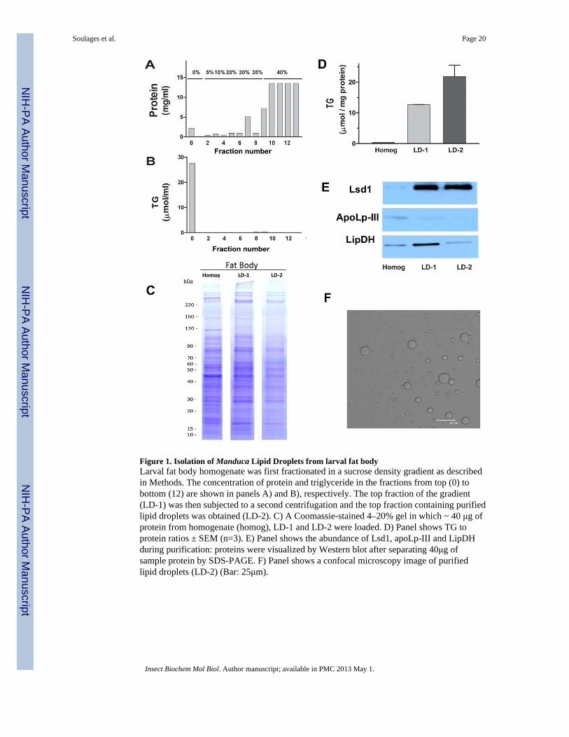
Figure 1. Isolation of Manduca Lipid Droplets from larval fat bodyLarval fat body homogenate was first fractionated in a sucrose density gradient as describedin Methods. The concentration of protein and triglyceride in the fractions from top (0) tobottom (12) are shown in panels A) and B), respectively. The top fraction of the gradient(LD-1) was then subjected to a second centrifugation and the top fraction containing purifiedlipid droplets was obtained (LD-2). C) A Coomassie-stained 4–20% gel in which ~ 40 μg ofprotein from homogenate (homog), LD-1 and LD-2 were loaded. D) Panel shows TG toprotein ratios ± SEM (n=3). E) Panel shows the abundance of Lsd1, apoLp-III and LipDHduring purification: proteins were visualized by Western blot after separating 40μg ofsample protein by SDS-PAGE. F) Panel shows a confocal microscopy image of purifiedlipid droplets (LD-2) (Bar: 25μm).
Soulages et al. Page 20
Insect Biochem Mol Biol. Author manuscript; available in PMC 2013 May 1.
NIH
-PA Author Manuscript
NIH
-PA Author Manuscript
NIH
-PA Author Manuscript
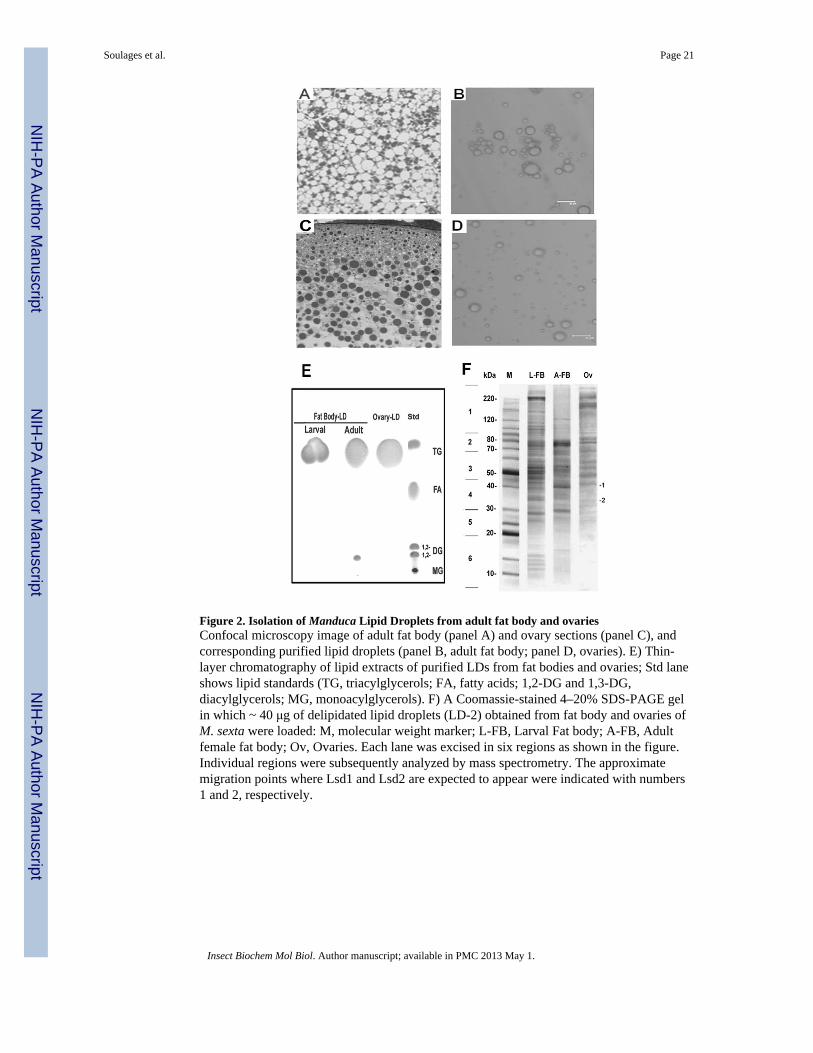
Figure 2. Isolation of Manduca Lipid Droplets from adult fat body and ovariesConfocal microscopy image of adult fat body (panel A) and ovary sections (panel C), andcorresponding purified lipid droplets (panel B, adult fat body; panel D, ovaries). E) Thin-layer chromatography of lipid extracts of purified LDs from fat bodies and ovaries; Std laneshows lipid standards (TG, triacylglycerols; FA, fatty acids; 1,2-DG and 1,3-DG,diacylglycerols; MG, monoacylglycerols). F) A Coomassie-stained 4–20% SDS-PAGE gelin which ~ 40 μg of delipidated lipid droplets (LD-2) obtained from fat body and ovaries ofM. sexta were loaded: M, molecular weight marker; L-FB, Larval Fat body; A-FB, Adultfemale fat body; Ov, Ovaries. Each lane was excised in six regions as shown in the figure.Individual regions were subsequently analyzed by mass spectrometry. The approximatemigration points where Lsd1 and Lsd2 are expected to appear were indicated with numbers1 and 2, respectively.
Soulages et al. Page 21
Insect Biochem Mol Biol. Author manuscript; available in PMC 2013 May 1.
NIH
-PA Author Manuscript
NIH
-PA Author Manuscript
NIH
-PA Author Manuscript
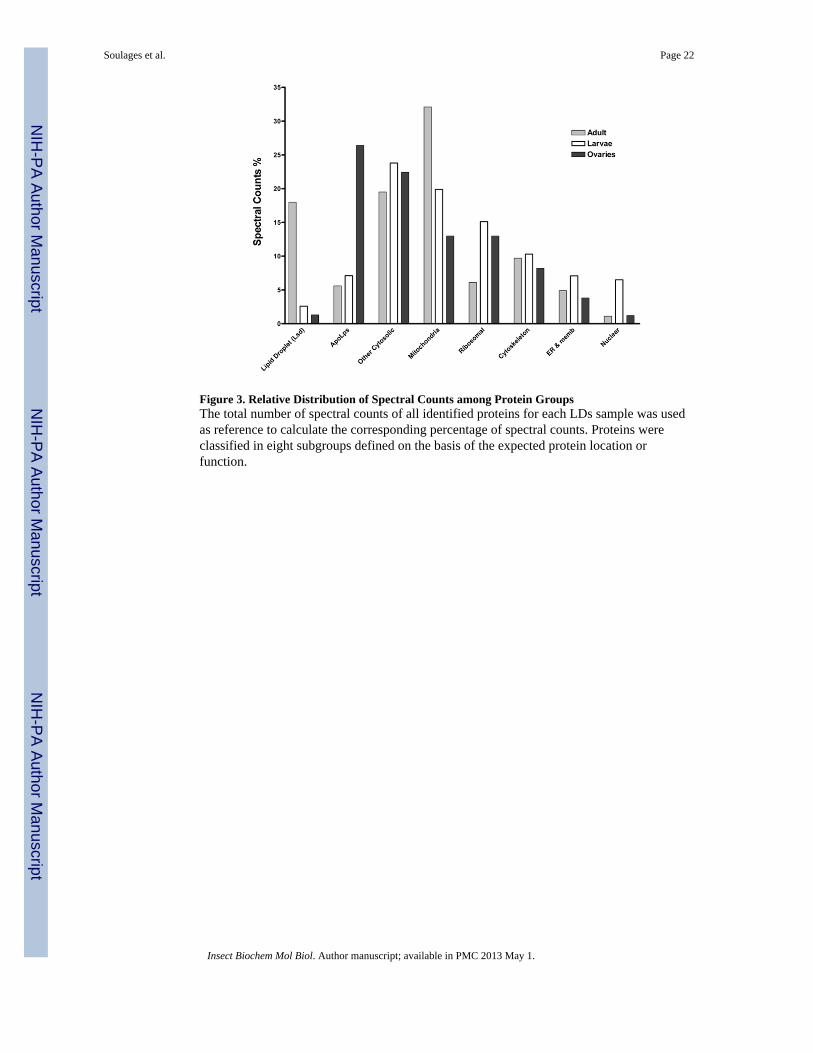
Figure 3. Relative Distribution of Spectral Counts among Protein GroupsThe total number of spectral counts of all identified proteins for each LDs sample was usedas reference to calculate the corresponding percentage of spectral counts. Proteins wereclassified in eight subgroups defined on the basis of the expected protein location orfunction.
Soulages et al. Page 22
Insect Biochem Mol Biol. Author manuscript; available in PMC 2013 May 1.
NIH
-PA Author Manuscript
NIH
-PA Author Manuscript
NIH
-PA Author Manuscript
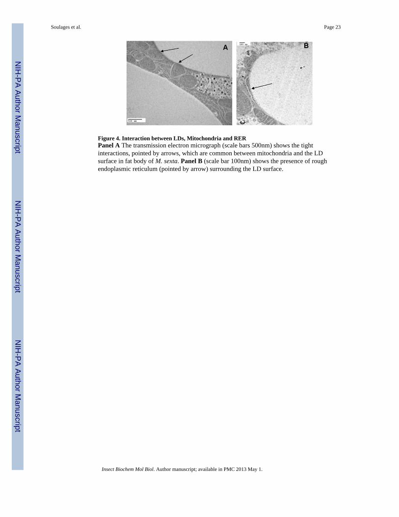
Figure 4. Interaction between LDs, Mitochondria and RERPanel A The transmission electron micrograph (scale bars 500nm) shows the tightinteractions, pointed by arrows, which are common between mitochondria and the LDsurface in fat body of M. sexta. Panel B (scale bar 100nm) shows the presence of roughendoplasmic reticulum (pointed by arrow) surrounding the LD surface.
Soulages et al. Page 23
Insect Biochem Mol Biol. Author manuscript; available in PMC 2013 May 1.
NIH
-PA Author Manuscript
NIH
-PA Author Manuscript
NIH
-PA Author Manuscript
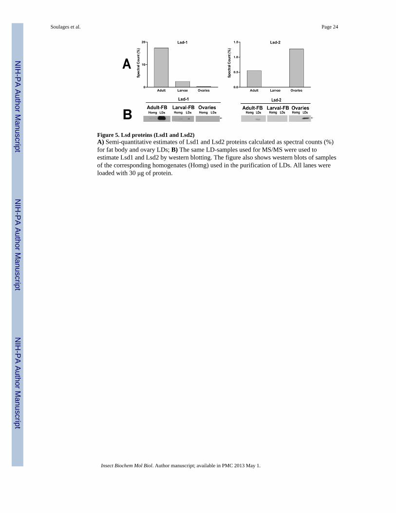
Figure 5. Lsd proteins (Lsd1 and Lsd2)A) Semi-quantitative estimates of Lsd1 and Lsd2 proteins calculated as spectral counts (%)for fat body and ovary LDs; B) The same LD-samples used for MS/MS were used toestimate Lsd1 and Lsd2 by western blotting. The figure also shows western blots of samplesof the corresponding homogenates (Homg) used in the purification of LDs. All lanes wereloaded with 30 μg of protein.
Soulages et al. Page 24
Insect Biochem Mol Biol. Author manuscript; available in PMC 2013 May 1.
NIH
-PA Author Manuscript
NIH
-PA Author Manuscript
NIH
-PA Author Manuscript
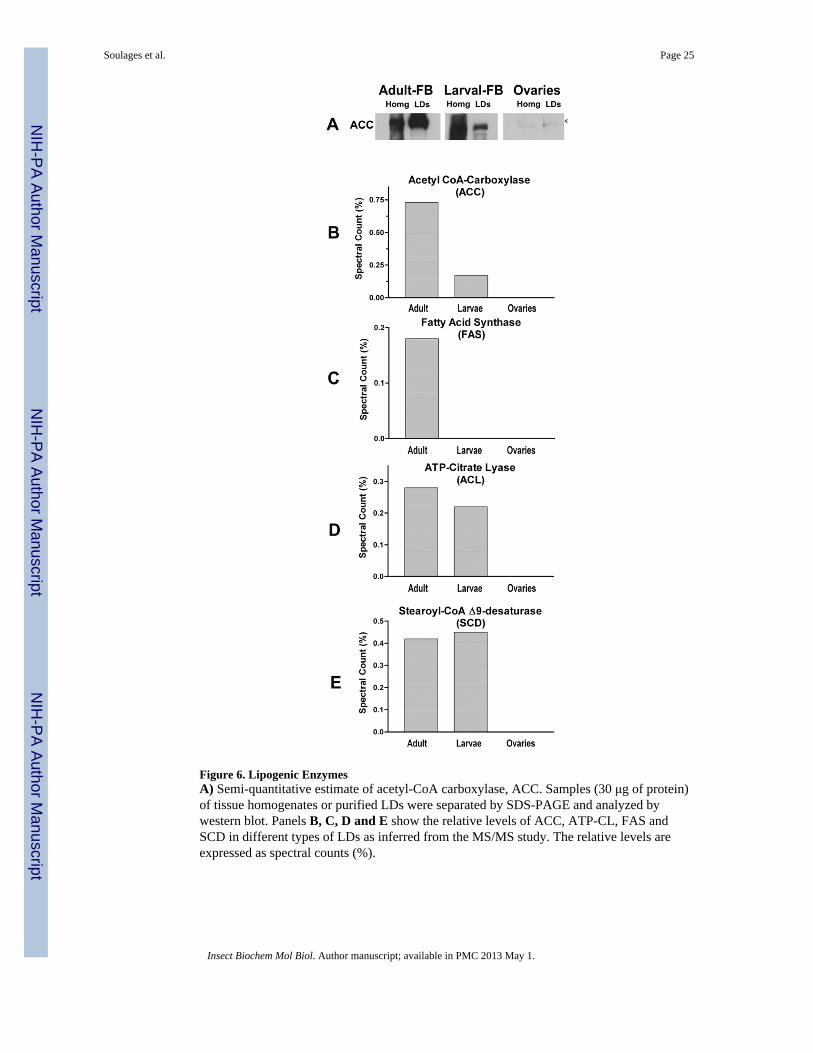
Figure 6. Lipogenic EnzymesA) Semi-quantitative estimate of acetyl-CoA carboxylase, ACC. Samples (30 μg of protein)of tissue homogenates or purified LDs were separated by SDS-PAGE and analyzed bywestern blot. Panels B, C, D and E show the relative levels of ACC, ATP-CL, FAS andSCD in different types of LDs as inferred from the MS/MS study. The relative levels areexpressed as spectral counts (%).
Soulages et al. Page 25
Insect Biochem Mol Biol. Author manuscript; available in PMC 2013 May 1.
NIH
-PA Author Manuscript
NIH
-PA Author Manuscript
NIH
-PA Author Manuscript
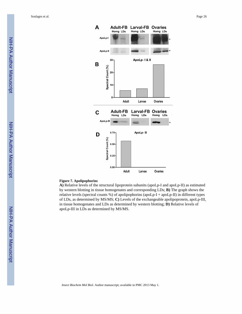
Figure 7. ApolipophorinsA) Relative levels of the structural lipoprotein subunits (apoLp-I and apoLp-II) as estimatedby western blotting in tissue homogenates and corresponding LDs; B) The graph shows therelative levels (spectral counts %) of apolipophorins (apoLp-I + apoLp-II) in different typesof LDs, as determined by MS/MS; C) Levels of the exchangeable apolipoprotein, apoLp-III,in tissue homogenates and LDs as determined by western blotting; D) Relative levels ofapoLp-III in LDs as determined by MS/MS.
Soulages et al. Page 26
Insect Biochem Mol Biol. Author manuscript; available in PMC 2013 May 1.
NIH
-PA Author Manuscript
NIH
-PA Author Manuscript
NIH
-PA Author Manuscript
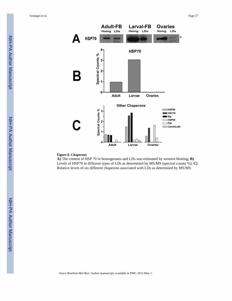
Figure 8. ChaperonsA) The content of HSP 70 in homogenates and LDs was estimated by western blotting; B)Levels of HSP70 in different types of LDs as determined by MS/MS (spectral counts %); C)Relative levels of six different chaperons associated with LDs as determined by MS/MS.
Soulages et al. Page 27
Insect Biochem Mol Biol. Author manuscript; available in PMC 2013 May 1.
NIH
-PA Author Manuscript
NIH
-PA Author Manuscript
NIH
-PA Author Manuscript
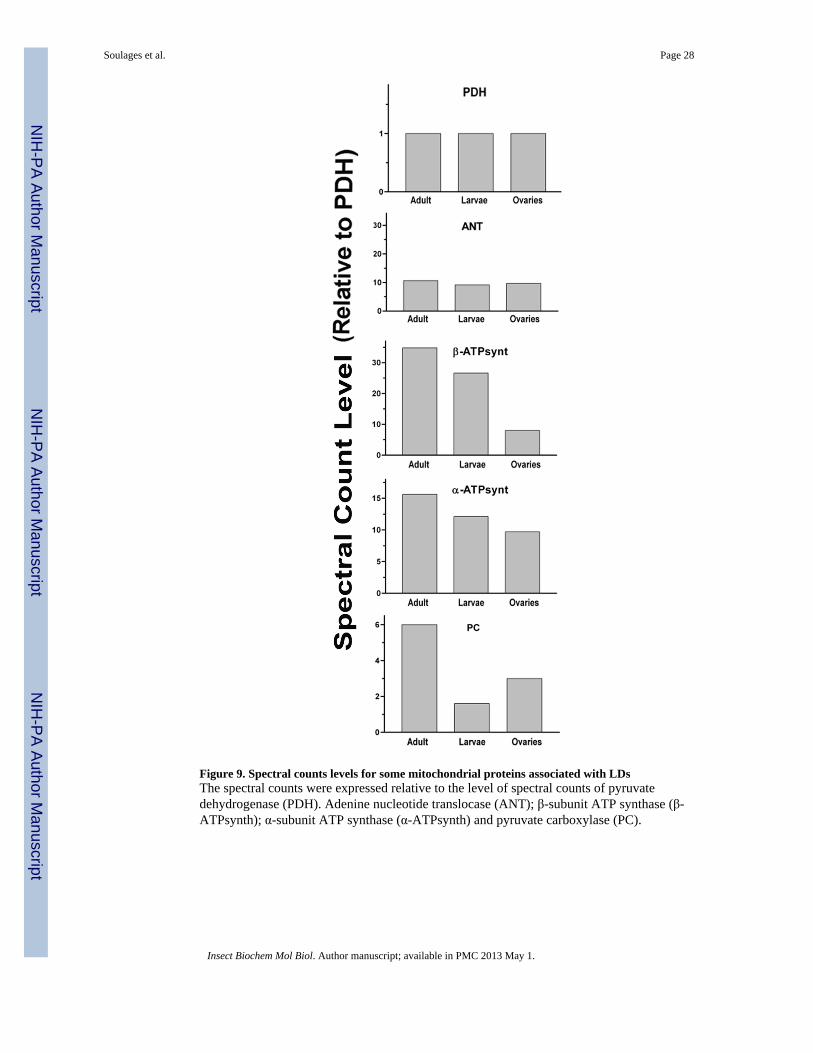
Figure 9. Spectral counts levels for some mitochondrial proteins associated with LDsThe spectral counts were expressed relative to the level of spectral counts of pyruvatedehydrogenase (PDH). Adenine nucleotide translocase (ANT); β-subunit ATP synthase (β-ATPsynth); α-subunit ATP synthase (α-ATPsynth) and pyruvate carboxylase (PC).
Soulages et al. Page 28
Insect Biochem Mol Biol. Author manuscript; available in PMC 2013 May 1.
NIH
-PA Author Manuscript
NIH
-PA Author Manuscript
NIH
-PA Author Manuscript
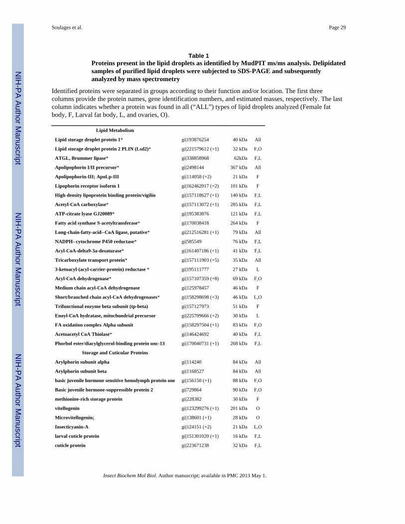
NIH
-PA Author Manuscript
NIH
-PA Author Manuscript
NIH
-PA Author Manuscript
Soulages et al. Page 29
Table 1Proteins present in the lipid droplets as identified by MudPIT ms/ms analysis. Delipidatedsamples of purified lipid droplets were subjected to SDS-PAGE and subsequentlyanalyzed by mass spectrometry
Identified proteins were separated in groups according to their function and/or location. The first threecolumns provide the protein names, gene identification numbers, and estimated masses, respectively. The lastcolumn indicates whether a protein was found in all (“ALL”) types of lipid droplets analyzed (Female fatbody, F, Larval fat body, L, and ovaries, O).
Lipid Metabolism
Lipid storage droplet protein 1* gi|193876254 40 kDa All
Lipid storage droplet protein 2 PLIN (Lsd2)* gi|221579612 (+1) 32 kDa F,O
ATGL, Brummer lipase* gi|338858968 62kDa F,L
Apolipophorin I/II precursor* gi|2498144 367 kDa All
Apolipophorin-III; ApoLp-III gi|114058 (+2) 21 kDa F
Lipophorin receptor isoform 1 gi|162462017 (+2) 101 kDa F
High density lipoprotein binding protein/vigilin gi|157118627 (+1) 140 kDa F,L
Acetyl-CoA carboxylase* gi|157113072 (+1) 285 kDa F,L
ATP-citrate lyase GJ20089* gi|195383876 121 kDa F,L
Fatty acid synthase S-acetyltransferase* gi|170038418 264 kDa F
Long-chain-fatty-acid--CoA ligase, putative* gi|212516281 (+1) 79 kDa All
NADPH--cytochrome P450 reductase* gi|585549 76 kDa F,L
Acyl-CoA-delta9-3a-desaturase* gi|161407186 (+1) 41 kDa F,L
Tricarboxylate transport protein* gi|157111903 (+5) 35 kDa All
3-ketoacyl-(acyl-carrier-protein) reductase * gi|195111777 27 kDa L
Acyl-CoA dehydrogenase* gi|157107359 (+8) 69 kDa F,O
Medium chain acyl-CoA dehydrogenase gi|125978457 46 kDa F
Short/branched chain acyl-CoA dehydrogenases* gi|158298698 (+3) 46 kDa L,O
Trifunctional enzyme beta subunit (tp-beta) gi|157127973 51 kDa F
Enoyl-CoA hydratase, mitochondrial precursor gi|225709666 (+2) 30 kDa L
FA oxidation complex Alpha subunit gi|158297504 (+1) 83 kDa F,O
Acetoacetyl CoA Thiolase* gi|146424692 40 kDa F,L
Phorbol ester/diacylglycerol-binding protein unc-13 gi|170040731 (+1) 268 kDa F,L
Storage and Cuticular Proteins
Arylphorin subunit alpha gi|114240 84 kDa All
Arylphorin subunit beta gi|1168527 84 kDa All
basic juvenile hormone sensitive hemolymph protein one gi|156150 (+1) 88 kDa F,O
Basic juvenile hormone-suppressible protein 2 gi|729864 90 kDa F,O
methionine-rich storage protein gi|228382 30 kDa F
vitellogenin gi|123299276 (+1) 201 kDa O
Microvitellogenin; gi|138601 (+1) 28 kDa O
Insecticyanin-A gi|124151 (+2) 21 kDa L,O
larval cuticle protein gi|151301020 (+1) 16 kDa F,L
cuticle protein gi|223671238 32 kDa F,L
Insect Biochem Mol Biol. Author manuscript; available in PMC 2013 May 1.
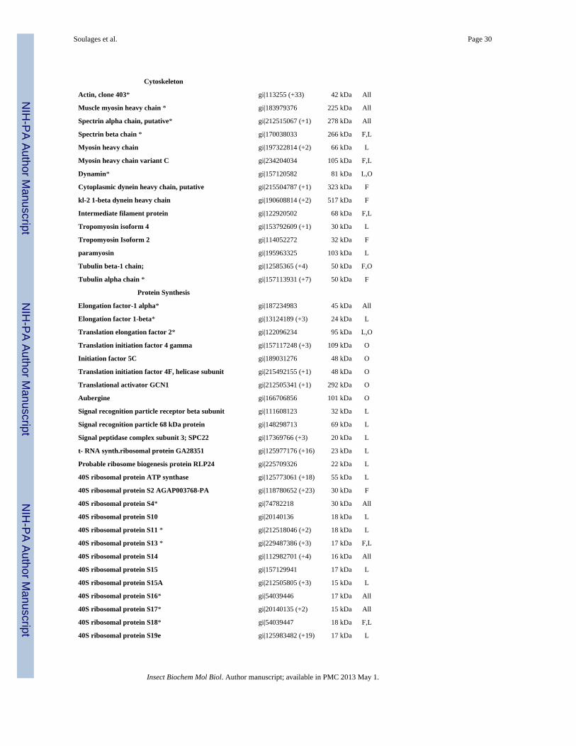
NIH
-PA Author Manuscript
NIH
-PA Author Manuscript
NIH
-PA Author Manuscript
Soulages et al. Page 30
Cytoskeleton
Actin, clone 403* gi|113255 (+33) 42 kDa All
Muscle myosin heavy chain * gi|183979376 225 kDa All
Spectrin alpha chain, putative* gi|212515067 (+1) 278 kDa All
Spectrin beta chain * gi|170038033 266 kDa F,L
Myosin heavy chain gi|197322814 (+2) 66 kDa L
Myosin heavy chain variant C gi|234204034 105 kDa F,L
Dynamin* gi|157120582 81 kDa L,O
Cytoplasmic dynein heavy chain, putative gi|215504787 (+1) 323 kDa F
kl-2 1-beta dynein heavy chain gi|190608814 (+2) 517 kDa F
Intermediate filament protein gi|122920502 68 kDa F,L
Tropomyosin isoform 4 gi|153792609 (+1) 30 kDa L
Tropomyosin Isoform 2 gi|114052272 32 kDa F
paramyosin gi|195963325 103 kDa L
Tubulin beta-1 chain; gi|12585365 (+4) 50 kDa F,O
Tubulin alpha chain * gi|157113931 (+7) 50 kDa F
Protein Synthesis
Elongation factor-1 alpha* gi|187234983 45 kDa All
Elongation factor 1-beta* gi|13124189 (+3) 24 kDa L
Translation elongation factor 2* gi|122096234 95 kDa L,O
Translation initiation factor 4 gamma gi|157117248 (+3) 109 kDa O
Initiation factor 5C gi|189031276 48 kDa O
Translation initiation factor 4F, helicase subunit gi|215492155 (+1) 48 kDa O
Translational activator GCN1 gi|212505341 (+1) 292 kDa O
Aubergine gi|166706856 101 kDa O
Signal recognition particle receptor beta subunit gi|111608123 32 kDa L
Signal recognition particle 68 kDa protein gi|148298713 69 kDa L
Signal peptidase complex subunit 3; SPC22 gi|17369766 (+3) 20 kDa L
t- RNA synth.ribosomal protein GA28351 gi|125977176 (+16) 23 kDa L
Probable ribosome biogenesis protein RLP24 gi|225709326 22 kDa L
40S ribosomal protein ATP synthase gi|125773061 (+18) 55 kDa L
40S ribosomal protein S2 AGAP003768-PA gi|118780652 (+23) 30 kDa F
40S ribosomal protein S4* gi|74782218 30 kDa All
40S ribosomal protein S10 gi|20140136 18 kDa L
40S ribosomal protein S11 * gi|212518046 (+2) 18 kDa L
40S ribosomal protein S13 * gi|229487386 (+3) 17 kDa F,L
40S ribosomal protein S14 gi|112982701 (+4) 16 kDa All
40S ribosomal protein S15 gi|157129941 17 kDa L
40S ribosomal protein S15A gi|212505805 (+3) 15 kDa L
40S ribosomal protein S16* gi|54039446 17 kDa All
40S ribosomal protein S17* gi|20140135 (+2) 15 kDa All
40S ribosomal protein S18* gi|54039447 18 kDa F,L
40S ribosomal protein S19e gi|125983482 (+19) 17 kDa L
Insect Biochem Mol Biol. Author manuscript; available in PMC 2013 May 1.
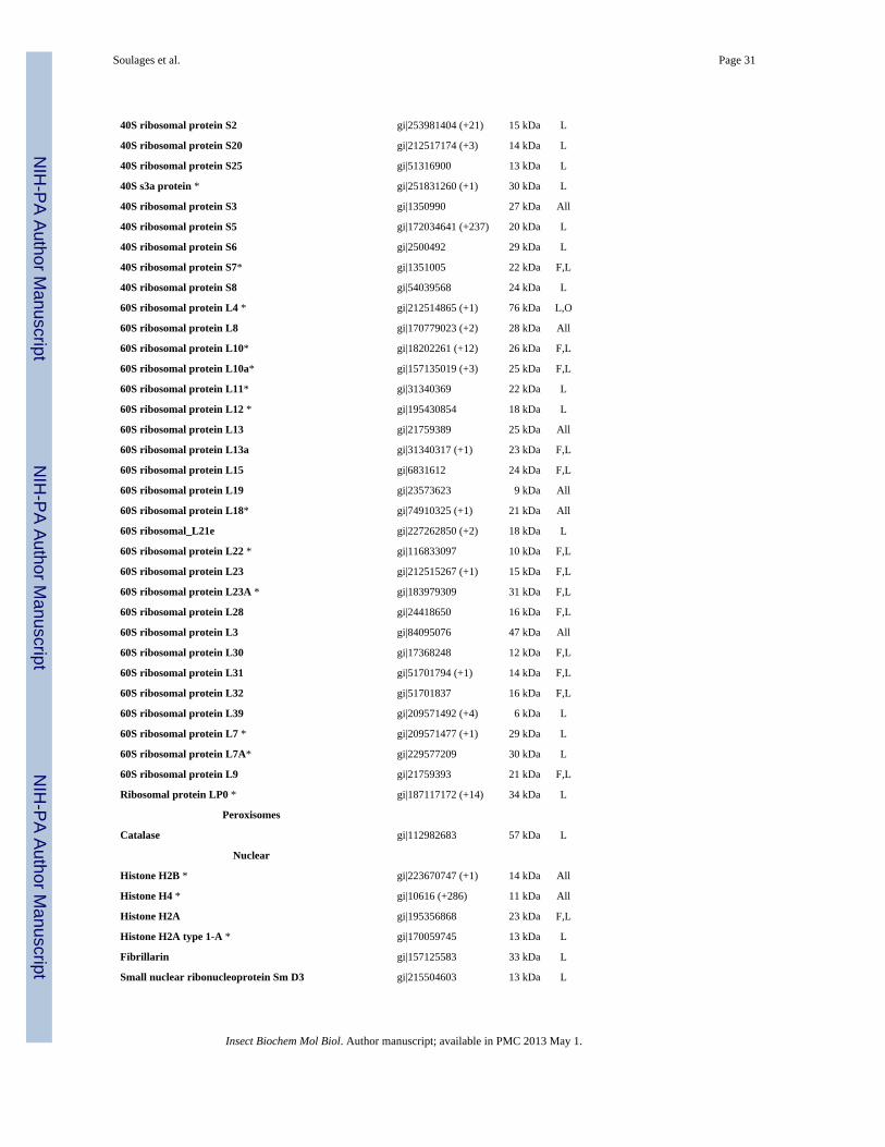
NIH
-PA Author Manuscript
NIH
-PA Author Manuscript
NIH
-PA Author Manuscript
Soulages et al. Page 31
40S ribosomal protein S2 gi|253981404 (+21) 15 kDa L
40S ribosomal protein S20 gi|212517174 (+3) 14 kDa L
40S ribosomal protein S25 gi|51316900 13 kDa L
40S s3a protein * gi|251831260 (+1) 30 kDa L
40S ribosomal protein S3 gi|1350990 27 kDa All
40S ribosomal protein S5 gi|172034641 (+237) 20 kDa L
40S ribosomal protein S6 gi|2500492 29 kDa L
40S ribosomal protein S7* gi|1351005 22 kDa F,L
40S ribosomal protein S8 gi|54039568 24 kDa L
60S ribosomal protein L4 * gi|212514865 (+1) 76 kDa L,O
60S ribosomal protein L8 gi|170779023 (+2) 28 kDa All
60S ribosomal protein L10* gi|18202261 (+12) 26 kDa F,L
60S ribosomal protein L10a* gi|157135019 (+3) 25 kDa F,L
60S ribosomal protein L11* gi|31340369 22 kDa L
60S ribosomal protein L12 * gi|195430854 18 kDa L
60S ribosomal protein L13 gi|21759389 25 kDa All
60S ribosomal protein L13a gi|31340317 (+1) 23 kDa F,L
60S ribosomal protein L15 gi|6831612 24 kDa F,L
60S ribosomal protein L19 gi|23573623 9 kDa All
60S ribosomal protein L18* gi|74910325 (+1) 21 kDa All
60S ribosomal_L21e gi|227262850 (+2) 18 kDa L
60S ribosomal protein L22 * gi|116833097 10 kDa F,L
60S ribosomal protein L23 gi|212515267 (+1) 15 kDa F,L
60S ribosomal protein L23A * gi|183979309 31 kDa F,L
60S ribosomal protein L28 gi|24418650 16 kDa F,L
60S ribosomal protein L3 gi|84095076 47 kDa All
60S ribosomal protein L30 gi|17368248 12 kDa F,L
60S ribosomal protein L31 gi|51701794 (+1) 14 kDa F,L
60S ribosomal protein L32 gi|51701837 16 kDa F,L
60S ribosomal protein L39 gi|209571492 (+4) 6 kDa L
60S ribosomal protein L7 * gi|209571477 (+1) 29 kDa L
60S ribosomal protein L7A* gi|229577209 30 kDa L
60S ribosomal protein L9 gi|21759393 21 kDa F,L
Ribosomal protein LP0 * gi|187117172 (+14) 34 kDa L
Peroxisomes
Catalase gi|112982683 57 kDa L
Nuclear
Histone H2B * gi|223670747 (+1) 14 kDa All
Histone H4 * gi|10616 (+286) 11 kDa All
Histone H2A gi|195356868 23 kDa F,L
Histone H2A type 1-A * gi|170059745 13 kDa L
Fibrillarin gi|157125583 33 kDa L
Small nuclear ribonucleoprotein Sm D3 gi|215504603 13 kDa L
Insect Biochem Mol Biol. Author manuscript; available in PMC 2013 May 1.
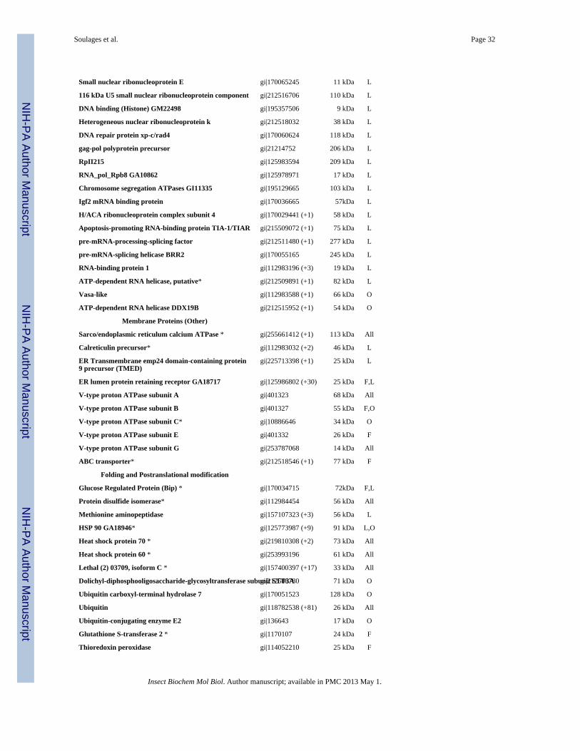
NIH
-PA Author Manuscript
NIH
-PA Author Manuscript
NIH
-PA Author Manuscript
Soulages et al. Page 32
Small nuclear ribonucleoprotein E gi|170065245 11 kDa L
116 kDa U5 small nuclear ribonucleoprotein component gi|212516706 110 kDa L
DNA binding (Histone) GM22498 gi|195357506 9 kDa L
Heterogeneous nuclear ribonucleoprotein k gi|212518032 38 kDa L
DNA repair protein xp-c/rad4 gi|170060624 118 kDa L
gag-pol polyprotein precursor gi|21214752 206 kDa L
RpII215 gi|125983594 209 kDa L
RNA_pol_Rpb8 GA10862 gi|125978971 17 kDa L
Chromosome segregation ATPases GI11335 gi|195129665 103 kDa L
Igf2 mRNA binding protein gi|170036665 57kDa L
H/ACA ribonucleoprotein complex subunit 4 gi|170029441 (+1) 58 kDa L
Apoptosis-promoting RNA-binding protein TIA-1/TIAR gi|215509072 (+1) 75 kDa L
pre-mRNA-processing-splicing factor gi|212511480 (+1) 277 kDa L
pre-mRNA-splicing helicase BRR2 gi|170055165 245 kDa L
RNA-binding protein 1 gi|112983196 (+3) 19 kDa L
ATP-dependent RNA helicase, putative* gi|212509891 (+1) 82 kDa L
Vasa-like gi|112983588 (+1) 66 kDa O
ATP-dependent RNA helicase DDX19B gi|212515952 (+1) 54 kDa O
Membrane Proteins (Other)
Sarco/endoplasmic reticulum calcium ATPase * gi|255661412 (+1) 113 kDa All
Calreticulin precursor* gi|112983032 (+2) 46 kDa L
ER Transmembrane emp24 domain-containing protein9 precursor (TMED)
gi|225713398 (+1) 25 kDa L
ER lumen protein retaining receptor GA18717 gi|125986802 (+30) 25 kDa F,L
V-type proton ATPase subunit A gi|401323 68 kDa All
V-type proton ATPase subunit B gi|401327 55 kDa F,O
V-type proton ATPase subunit C* gi|10886646 34 kDa O
V-type proton ATPase subunit E gi|401332 26 kDa F
V-type proton ATPase subunit G gi|253787068 14 kDa All
ABC transporter* gi|212518546 (+1) 77 kDa F
Folding and Postranslational modification
Glucose Regulated Protein (Bip) * gi|170034715 72kDa F,L
Protein disulfide isomerase* gi|112984454 56 kDa All
Methionine aminopeptidase gi|157107323 (+3) 56 kDa L
HSP 90 GA18946* gi|125773987 (+9) 91 kDa L,O
Heat shock protein 70 * gi|219810308 (+2) 73 kDa All
Heat shock protein 60 * gi|253993196 61 kDa All
Lethal (2) 03709, isoform C * gi|157400397 (+17) 33 kDa All
Dolichyl-diphosphooligosaccharide-glycosyltransferase subunit STT3Agi|212506780 71 kDa O
Ubiquitin carboxyl-terminal hydrolase 7 gi|170051523 128 kDa O
Ubiquitin gi|118782538 (+81) 26 kDa All
Ubiquitin-conjugating enzyme E2 gi|136643 17 kDa O
Glutathione S-transferase 2 * gi|1170107 24 kDa F
Thioredoxin peroxidase gi|114052210 25 kDa F
Insect Biochem Mol Biol. Author manuscript; available in PMC 2013 May 1.
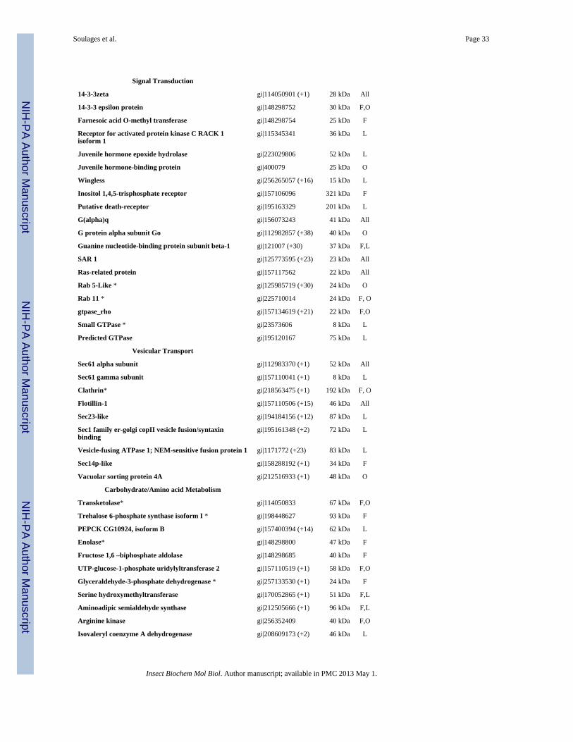
NIH
-PA Author Manuscript
NIH
-PA Author Manuscript
NIH
-PA Author Manuscript
Soulages et al. Page 33
Signal Transduction
14-3-3zeta gi|114050901 (+1) 28 kDa All
14-3-3 epsilon protein gi|148298752 30 kDa F,O
Farnesoic acid O-methyl transferase gi|148298754 25 kDa F
Receptor for activated protein kinase C RACK 1isoform 1
gi|115345341 36 kDa L
Juvenile hormone epoxide hydrolase gi|223029806 52 kDa L
Juvenile hormone-binding protein gi|400079 25 kDa O
Wingless gi|256265057 (+16) 15 kDa L
Inositol 1,4,5-trisphosphate receptor gi|157106096 321 kDa F
Putative death-receptor gi|195163329 201 kDa L
G(alpha)q gi|156073243 41 kDa All
G protein alpha subunit Go gi|112982857 (+38) 40 kDa O
Guanine nucleotide-binding protein subunit beta-1 gi|121007 (+30) 37 kDa F,L
SAR 1 gi|125773595 (+23) 23 kDa All
Ras-related protein gi|157117562 22 kDa All
Rab 5-Like * gi|125985719 (+30) 24 kDa O
Rab 11 * gi|225710014 24 kDa F, O
gtpase_rho gi|157134619 (+21) 22 kDa F,O
Small GTPase * gi|23573606 8 kDa L
Predicted GTPase gi|195120167 75 kDa L
Vesicular Transport
Sec61 alpha subunit gi|112983370 (+1) 52 kDa All
Sec61 gamma subunit gi|157110041 (+1) 8 kDa L
Clathrin* gi|218563475 (+1) 192 kDa F, O
Flotillin-1 gi|157110506 (+15) 46 kDa All
Sec23-like gi|194184156 (+12) 87 kDa L
Sec1 family er-golgi copII vesicle fusion/syntaxinbinding
gi|195161348 (+2) 72 kDa L
Vesicle-fusing ATPase 1; NEM-sensitive fusion protein 1 gi|1171772 (+23) 83 kDa L
Sec14p-like gi|158288192 (+1) 34 kDa F
Vacuolar sorting protein 4A gi|212516933 (+1) 48 kDa O
Carbohydrate/Amino acid Metabolism
Transketolase* gi|114050833 67 kDa F,O
Trehalose 6-phosphate synthase isoform I * gi|198448627 93 kDa F
PEPCK CG10924, isoform B gi|157400394 (+14) 62 kDa L
Enolase* gi|148298800 47 kDa F
Fructose 1,6 –biphosphate aldolase gi|148298685 40 kDa F
UTP-glucose-1-phosphate uridylyltransferase 2 gi|157110519 (+1) 58 kDa F,O
Glyceraldehyde-3-phosphate dehydrogenase * gi|257133530 (+1) 24 kDa F
Serine hydroxymethyltransferase gi|170052865 (+1) 51 kDa F,L
Aminoadipic semialdehyde synthase gi|212505666 (+1) 96 kDa F,L
Arginine kinase gi|256352409 40 kDa F,O
Isovaleryl coenzyme A dehydrogenase gi|208609173 (+2) 46 kDa L
Insect Biochem Mol Biol. Author manuscript; available in PMC 2013 May 1.
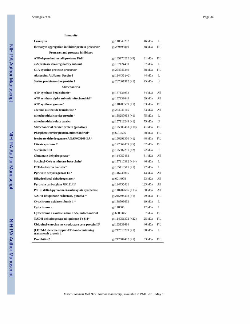
NIH
-PA Author Manuscript
NIH
-PA Author Manuscript
NIH
-PA Author Manuscript
Soulages et al. Page 34
Immunity
Leureptin gi|110649252 46 kDa L
Hemocyte aggregation inhibitor protein precursor gi|259493819 48 kDa F,L
Proteases and protease inhibitors
ATP-dependent metalloprotease FtsH gi|195170272 (+9) 81 kDa F,L
26S protease (S4) regulatory subunit gi|157124490 67 kDa L
C1A cysteine protease precursor gi|254746340 38 kDa F,L
Alaserpin; AltName: Serpin-1 gi|134436 (+2) 44 kDa L
Serine proteinase-like protein 1 gi|237861312 (+1) 45 kDa F
Mitochondria
ATP synthase beta subunit* gi|157136033 54 kDa All
ATP synthase alpha subunit mitochondrial* gi|157131648 59 kDa All
ATP synthase gamma* gi|118789559 (+1) 33 kDa F,L
adenine nucleotide translocase * gi|254946115 33 kDa All
mitochondrial carrier protein * gi|158287093 (+1) 75 kDa L
mitochondrial solute carrier gi|157113249 (+1) 75 kDa F
Mitochondrial carrier protein (putative) gi|125809463 (+10) 41 kDa F,L
Phosphate carrier protein, mitochondrial* gi|6016596 38 kDa F,L
Isocitrate dehydrogenase AGAP003168-PA* gi|158291350 (+1) 48 kDa F,L
Citrate synthase 2 gi|122067459 (+5) 52 kDa F,L
Succinate DH gi|125807291 (+2) 72 kDa F
Glutamate dehydrogenase* gi|114052462 61 kDa All
Succinyl-CoA synthetase beta chain* gi|157110382 (+14) 46 kDa L
ETF-b electron transfer* gi|195113511 (+1) 27 kDa L
Pyruvate dehydrogenase E1* gi|146738085 44 kDa All
Dihydrolipoyl dehydrogenase;* gi|6014978 53 kDa All
Pyruvate carboxylase GF13141* gi|194755401 133 kDa All
P5CS: delta l-pyrroline-5-carboxylate synthetase gi|118782666 (+13) 80 kDa All
NADH-ubiquinone reductase, putative * gi|215494308 (+1) 79 kDa F,L
Cytochrome oxidase subunit 1 * gi|188503652 19 kDa L
Cytochrome c gi|118005 12 kDa L
Cytochrome c oxidase subunit 5A, mitochondrial gi|6685345 7 kDa F,L
NADH dehydrogenase ubiquinone Fe-S 8* gi|114051372 (+22) 25 kDa F,L
Ubiquinol-cytochrome c reductase core protein II* gi|163838684 46 kDa F,L
(LETM-1) leucine zipper-EF-hand-containingtransmemb protein 1
gi|212510209 (+1) 88 kDa L
Prohibitin-2 gi|212507492 (+1) 33 kDa F,L
Insect Biochem Mol Biol. Author manuscript; available in PMC 2013 May 1.