Development of a hybrid robotic system based on an adaptive ...
151
TESIS DOCTORAL Development of a hybrid robotic system based on an adaptive and associative assistance for rehabilitation of reaching movement after stroke Autor: Francisco Resqu´ ın Acosta Directores: Jos´ e Luis Pons Rovira Fernando Brunetti Fern´ andez Tutora: Dolores Blanco Rojas Departamento de Ingenier´ ıa de Sistemas y Autom´ atica Legan´ es, Noviembre de 2017
-
Upload
khangminh22 -
Category
Documents
-
view
1 -
download
0
Transcript of Development of a hybrid robotic system based on an adaptive ...
TESIS DOCTORAL
Development of a hybrid robotic system
based on an adaptive and associative
assistance for rehabilitation of reaching
movement after stroke
Autor:
Francisco Resquın Acosta
Directores:
Jose Luis Pons RoviraFernando Brunetti Fernandez
Tutora:
Dolores Blanco Rojas
Departamento de Ingenierıa de Sistemas y Automatica
Leganes, Noviembre de 2017


TESIS DOCTORAL
Development of a hybrid robotic system based on an
adaptive and associative assistance for rehabilitation of
reaching movement after stroke
Autor: Francisco Resquın Acosta
Directores: Jose Luis Pons Rovira, Fernando Brunetti Fernandez
Tutora: Dolores Blanco Rojas
Firma del Tribunal Calificador:
Presidente:
Vocal:
Secretario:
Calificacion:
Leganes, 29 de Noviembre de 2017


A mis padres,y a mi Anto
v

Agradecimientos
Quisiera comenzar agradeciendo a todas las personas que participaron en los experimentos
que dieron lugar a los resultados presentados en esta tesis. En especial a los pacientes con
dano cerebral, por su buena predisposicion y colaboracion para realizar todos los experimen-
tos.
A mis directores de tesis, Jose Luis y Fernando, por confiar en mı, abrirme las puertas del
grupo de neuro-rehabilitacion y por todo el apoyo recibido en estos anos. De igual forma, a
mis companeros y a todos aquellos que pasaron por del grupo de neuro-rehabilitacion, por el
apoyo mutuo y los momentos compartidos tanto dentro como fuera del instituto. ¡A todos
ustedes, muchısimas gracias!
A los profesionales y colaboradores del Instituto de Neurociencias y Ciencias del Movimiento
(INCIMOV) de la Universidad de la Salle y del Centro de Referencia Estatal de Atencion al
Dano Cerebral (CEADAC), en especial a Iris, Laura, Carlos, Antonio y Susana, por el apoyo
recibido en los estudios y la excelente predisposicion de colaboracion.
A todos mis amigos de Paraguay y Madrid, que hicieron que esta larga carrera se haga mas
placentera.
A mi familia y en especial a mis padres. Por el apoyo incondicional, su carino, sus ensenanzas
y haberme dado todo. Estas lıneas no alcanzan para expresar todo mi agradecimiento y carino
hacia ustedes.
Por ultimo, pero no menos importante, dedico estas ultimas lıneas a mi companera y esposa,
a Anto. Gracias por todo tu apoyo, por aguantar a mi lado todas las adversidades, por el
empujon diario y por hacerme sentir una persona especial.
vii

Abstract
Stroke causes irreversible neurological damage. Depending on the location and the size of
this brain injury, di↵erent body functions could result a↵ected. One of the most common
consequences is motor impairments. The level of motor impairment a↵ectation varies between
post-stroke subjects, but often, it hampers the execution of most activities of daily living.
Consequently, the quality of life of the stroke population is severely decreased.
The rehabilitation of the upper-limb motor functions has gained special attention in the
scientific community due the poor reported prognosis of post-stroke patients for recovering
normal upper-extremity function after standard rehabilitation therapy. Driven by the ad-
vance of technology and the design of new rehabilitation methods, the use of robot devices,
functional electrical stimulation and brain-computer interfaces as a neuromodulation system
is proposed as a novel and promising rehabilitation tools. Although the uses of these tech-
nologies present potential benefits with respect to standard rehabilitation methods, there still
are some milestones to be addressed for the consolidation of these methods and techniques
in clinical settings.
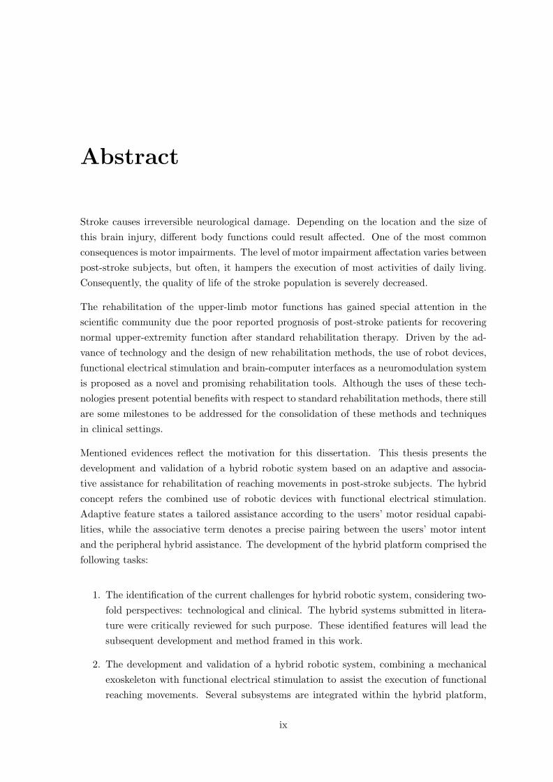
Mentioned evidences reflect the motivation for this dissertation. This thesis presents the
development and validation of a hybrid robotic system based on an adaptive and associa-
tive assistance for rehabilitation of reaching movements in post-stroke subjects. The hybrid
concept refers the combined use of robotic devices with functional electrical stimulation.
Adaptive feature states a tailored assistance according to the users’ motor residual capabi-
lities, while the associative term denotes a precise pairing between the users’ motor intent
and the peripheral hybrid assistance. The development of the hybrid platform comprised the
following tasks:
1. The identification of the current challenges for hybrid robotic system, considering two-
fold perspectives: technological and clinical. The hybrid systems submitted in litera-
ture were critically reviewed for such purpose. These identified features will lead the
subsequent development and method framed in this work.
2. The development and validation of a hybrid robotic system, combining a mechanical
exoskeleton with functional electrical stimulation to assist the execution of functional
reaching movements. Several subsystems are integrated within the hybrid platform,
ix
x
which interact each other to cooperatively complement the rehabilitation task. Comple-
mentary, the implementation of a controller based on functional electrical stimulation
to dynamically adjust the level of assistance is addressed. The controller is conceived to
tackle one of the main limitations when using electrical stimulation, i.e. the highly non-
linear and time-varying muscle response. An experimental procedure was conducted
with healthy and post-stroke patients to corroborate the technical feasibility and the
usability evaluation of the system.
3. The implementation of an associative strategy within the hybrid platform. Three dif-
ferent strategies based on electroencephalography and electromyography signals were
analytically compared. The main idea is to provide a precise temporal association be-
tween the hybrid assistance delivered at the periphery (arm muscles) and the users’
own intention to move and to configure a feasible clinical setup to be use in real reha-
bilitation scenarios.
4. Carry out a comprehensive pilot clinical intervention considering a small cohort of
patient with post-stroke patients to evaluate the di↵erent proposed concepts and assess
the feasibility of using the hybrid system in rehabilitation settings.
In summary, the works here presented prove the feasibility of using the hybrid robotic system
as a rehabilitative tool with post-stroke subjects. Moreover, it is demonstrated the adaptive
controller is able to adjust the level of assistance to achieve successful tracking movement
with the a↵ected arm. Remarkably, the accurate association in time between motor cortex
activation, represented through the motor-related cortical potential measured with electroen-
cephalography, and the supplied hybrid assistance during the execution of functional (multi-
degree of freedom) reaching movement facilitate distributed cortical plasticity. These results
encourage the validation of the overall hybrid concept in a large clinical trial including an
increased number of patients with a control group, in order to achieve more robust clinical
results and confirm the presented herein.
Contents
Abstract ix
Nomenclature xxi
Objectives and description of the work 1
1 Introduction of stroke and rehabilitation of the upper limb motor function 7
1.1 Stroke . . . . . . . . . . . . . . . . . . . . . . . . . . . . . . . . . . . . . . . . 8
1.1.1 Ischemic Stroke . . . . . . . . . . . . . . . . . . . . . . . . . . . . . . . 9
1.1.2 Hemorrhagic stroke . . . . . . . . . . . . . . . . . . . . . . . . . . . . . 10
1.2 Social impact of stroke: the burden . . . . . . . . . . . . . . . . . . . . . . . . 11
1.3 Motor impairment . . . . . . . . . . . . . . . . . . . . . . . . . . . . . . . . . 12
1.4 Rehabilitation of motor impairment after stroke . . . . . . . . . . . . . . . . . 14
1.4.1 Brain plasticity associated with motor recovery . . . . . . . . . . . . . 15
1.4.2 Recovering upper-extremity motor function after stroke . . . . . . . . 17
1.5 Novel therapies for rehabilitation of the upper extremity motor function . . . 18
1.5.1 Robotics devices . . . . . . . . . . . . . . . . . . . . . . . . . . . . . . 18
1.5.2 Functional electrical stimulation . . . . . . . . . . . . . . . . . . . . . 21
1.5.3 Brain-computer interface . . . . . . . . . . . . . . . . . . . . . . . . . 24
1.6 Conclusions of the chapter . . . . . . . . . . . . . . . . . . . . . . . . . . . . . 27
2 State of the art of hybrid robotics system for rehabilitation motor functionsof the upper-extremities 29
2.1 Introduction . . . . . . . . . . . . . . . . . . . . . . . . . . . . . . . . . . . . . 30
2.2 Methods . . . . . . . . . . . . . . . . . . . . . . . . . . . . . . . . . . . . . . . 30
2.3 Results . . . . . . . . . . . . . . . . . . . . . . . . . . . . . . . . . . . . . . . . 31
2.3.1 Technical overview of hybrid systems . . . . . . . . . . . . . . . . . . . 31
2.3.1.1 Hybrid robotic rehabilitation systems for grasping . . . . . . 31
2.3.1.2 Hybrid robotic rehabilitation systems for reaching . . . . . . 33
2.3.2 Clinical evaluation . . . . . . . . . . . . . . . . . . . . . . . . . . . . . 37
2.3.2.1 Clinical outcomes of hybrid system for grasping rehabilitation 37
2.3.2.2 Clinical outcomes of hybrid system for reaching rehabilitation 39
2.4 Discussion . . . . . . . . . . . . . . . . . . . . . . . . . . . . . . . . . . . . . . 40
2.4.1 Improving technical aspects: hybrid approach challenges . . . . . . . . 40
2.4.2 Rehabilitation Outcomes . . . . . . . . . . . . . . . . . . . . . . . . . 42
2.4.3 Improving the human-machine interaction: associating peripheral as-sistance with user’s motor intent . . . . . . . . . . . . . . . . . . . . . 43
2.5 Conclusions of the chapter . . . . . . . . . . . . . . . . . . . . . . . . . . . . . 43
xi
xii CONTENTS
3 Implementation of a FES-based adaptive assistance in a hybrid roboticsystem for rehabilitation of reaching movement after stroke 45
3.1 Introduction . . . . . . . . . . . . . . . . . . . . . . . . . . . . . . . . . . . . . 47
3.2 Hybrid rehabilitation platform for reaching rehabilitation . . . . . . . . . . . 48
3.3 FES-based controller design . . . . . . . . . . . . . . . . . . . . . . . . . . . . 49
3.3.1 Human arm position . . . . . . . . . . . . . . . . . . . . . . . . . . . . 50
3.3.2 Feedback error learning implementation . . . . . . . . . . . . . . . . . 50
3.3.3 Reference generator . . . . . . . . . . . . . . . . . . . . . . . . . . . . 51
3.4 Methods . . . . . . . . . . . . . . . . . . . . . . . . . . . . . . . . . . . . . . . 53
3.4.1 Participants and evaluation protocol . . . . . . . . . . . . . . . . . . . 53
3.4.1.1 Experiment 1 . . . . . . . . . . . . . . . . . . . . . . . . . . . 53
3.4.1.2 Experiment 2 . . . . . . . . . . . . . . . . . . . . . . . . . . . 54
3.4.2 Data analysis . . . . . . . . . . . . . . . . . . . . . . . . . . . . . . . . 55
3.4.2.1 Experiment 1 . . . . . . . . . . . . . . . . . . . . . . . . . . . 55
3.4.2.2 Experiment 2 . . . . . . . . . . . . . . . . . . . . . . . . . . . 56
3.5 Results . . . . . . . . . . . . . . . . . . . . . . . . . . . . . . . . . . . . . . . . 57
3.5.1 Experiment 1 . . . . . . . . . . . . . . . . . . . . . . . . . . . . . . . . 57
3.5.2 Experiment 2 . . . . . . . . . . . . . . . . . . . . . . . . . . . . . . . . 60
3.5.2.1 Performance results . . . . . . . . . . . . . . . . . . . . . . . 60
3.5.2.2 Satisfaction assessment . . . . . . . . . . . . . . . . . . . . . 63
3.6 Discussion . . . . . . . . . . . . . . . . . . . . . . . . . . . . . . . . . . . . . . 63
3.6.1 Technical viability and system performance . . . . . . . . . . . . . . . 63
3.6.2 User satisfaction . . . . . . . . . . . . . . . . . . . . . . . . . . . . . . 65
3.6.3 Limitations . . . . . . . . . . . . . . . . . . . . . . . . . . . . . . . . . 65
3.7 Conclusions of the chapter . . . . . . . . . . . . . . . . . . . . . . . . . . . . . 66
4 Eliciting neural plasticity by the timed association of the user’s motorintent with the peripheral hybrid assistance 67
4.1 Introduction . . . . . . . . . . . . . . . . . . . . . . . . . . . . . . . . . . . . . 69
4.2 Method . . . . . . . . . . . . . . . . . . . . . . . . . . . . . . . . . . . . . . . 71
4.2.1 Subjects . . . . . . . . . . . . . . . . . . . . . . . . . . . . . . . . . . . 71
4.2.2 Materials and Instrumentation . . . . . . . . . . . . . . . . . . . . . . 71
4.2.2.1 EEG and EMG acquisition during intervention . . . . . . . . 71
4.2.2.2 Hybrid robotic system . . . . . . . . . . . . . . . . . . . . . . 71
4.2.2.3 TMS for eliciting motor evoked potentials . . . . . . . . . . . 72
4.2.3 Experimental Procedure . . . . . . . . . . . . . . . . . . . . . . . . . . 72
4.2.3.1 Pre0-, Pre1-, Post- and Post30-assessment . . . . . . . . . . 73
4.2.3.2 Intervention . . . . . . . . . . . . . . . . . . . . . . . . . . . 74
4.2.4 Implemented strategies to define the assistance onset . . . . . . . . . . 75
4.2.4.1 BCI o✏ine . . . . . . . . . . . . . . . . . . . . . . . . . . . . 76
4.2.4.2 BCI online . . . . . . . . . . . . . . . . . . . . . . . . . . . . 77
4.2.4.3 EMG . . . . . . . . . . . . . . . . . . . . . . . . . . . . . . . 78
4.2.4.4 Control experiments . . . . . . . . . . . . . . . . . . . . . . . 79
4.2.5 Outcomes Measures . . . . . . . . . . . . . . . . . . . . . . . . . . . . 79
4.2.6 Statistical Analysis . . . . . . . . . . . . . . . . . . . . . . . . . . . . . 80
4.3 Results . . . . . . . . . . . . . . . . . . . . . . . . . . . . . . . . . . . . . . . . 80
4.3.1 BCI o✏ine . . . . . . . . . . . . . . . . . . . . . . . . . . . . . . . . . 81
4.3.2 BCI online . . . . . . . . . . . . . . . . . . . . . . . . . . . . . . . . . 82
CONTENTS xiii
4.3.3 EMG . . . . . . . . . . . . . . . . . . . . . . . . . . . . . . . . . . . . 83
4.3.4 Control Experiments . . . . . . . . . . . . . . . . . . . . . . . . . . . . 84
4.4 Discussion . . . . . . . . . . . . . . . . . . . . . . . . . . . . . . . . . . . . . . 84
4.4.1 A comparison of the interventions’ e↵ects: BCI o✏ine, BCI online andEMG . . . . . . . . . . . . . . . . . . . . . . . . . . . . . . . . . . . . 85
4.4.2 Comparison with similar studies presented in literature . . . . . . . . 86
4.4.3 Implications for rehabilitation of upper-limb motor function . . . . . . 87
4.5 Conclusion of the chapter . . . . . . . . . . . . . . . . . . . . . . . . . . . . . 88
5 Clinical evaluation of the hybrid robotic system with the adaptive andassociative assistance in post-stroke patients 89
5.1 Introduction . . . . . . . . . . . . . . . . . . . . . . . . . . . . . . . . . . . . . 90
5.2 Methods . . . . . . . . . . . . . . . . . . . . . . . . . . . . . . . . . . . . . . . 90
5.2.1 Participants . . . . . . . . . . . . . . . . . . . . . . . . . . . . . . . . . 90
5.2.2 Inclusion and Exclusion criteria . . . . . . . . . . . . . . . . . . . . . . 91
5.2.3 Materials . . . . . . . . . . . . . . . . . . . . . . . . . . . . . . . . . . 92
5.2.4 Study design . . . . . . . . . . . . . . . . . . . . . . . . . . . . . . . . 92
5.2.4.1 Initial assessment (Ev1) . . . . . . . . . . . . . . . . . . . . . 92
5.2.4.2 Interventional sessions (Se1-12) . . . . . . . . . . . . . . . . . 94
5.2.4.3 Final evaluation (Ev2) . . . . . . . . . . . . . . . . . . . . . . 94
5.2.5 Outcomes measures . . . . . . . . . . . . . . . . . . . . . . . . . . . . 94
5.2.5.1 Clinical Scales . . . . . . . . . . . . . . . . . . . . . . . . . . 95
5.2.5.2 Kinematic data . . . . . . . . . . . . . . . . . . . . . . . . . . 96
5.2.5.3 Satisfaction assessment . . . . . . . . . . . . . . . . . . . . . 96
5.3 Results . . . . . . . . . . . . . . . . . . . . . . . . . . . . . . . . . . . . . . . . 96
5.3.1 Patient with moderate arm motor impairment: P1 . . . . . . . . . . . 97
5.3.1.1 Feasibility of the MRCP . . . . . . . . . . . . . . . . . . . . 97
5.3.1.2 Changes in Functional Scales . . . . . . . . . . . . . . . . . . 98
5.3.1.3 Analysis of the kinematics . . . . . . . . . . . . . . . . . . . 98
5.3.2 Patients with severe arm motor impairment: P2 and P5 . . . . . . . . 98
5.3.2.1 Feasibility of the MRCP . . . . . . . . . . . . . . . . . . . . 98
5.3.2.2 Changes in clinical scales . . . . . . . . . . . . . . . . . . . . 99
5.3.2.3 Analysis of the kinematics . . . . . . . . . . . . . . . . . . . 100
5.3.3 Usability assessment: users’ satisfaction . . . . . . . . . . . . . . . . . 101
5.4 Discussion . . . . . . . . . . . . . . . . . . . . . . . . . . . . . . . . . . . . . . 102
5.5 Conclusion of the chapter . . . . . . . . . . . . . . . . . . . . . . . . . . . . . 104
6 Conclusions and future works 105
6.1 Contributions . . . . . . . . . . . . . . . . . . . . . . . . . . . . . . . . . . . . 106
6.2 Scientific Dissemination . . . . . . . . . . . . . . . . . . . . . . . . . . . . . . 107
6.3 Future works . . . . . . . . . . . . . . . . . . . . . . . . . . . . . . . . . . . . 110
Bibliography 115

List of Figures
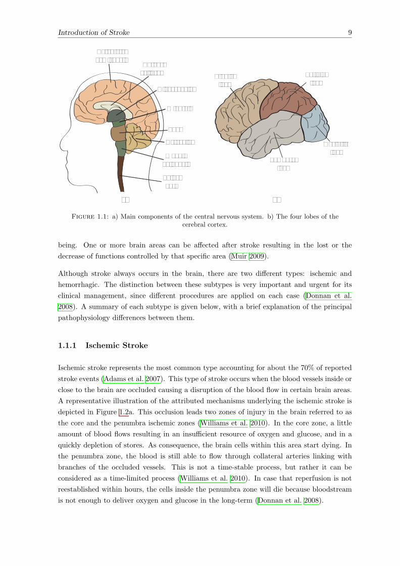
1.1 a) Main components of the central nervous system. b) The four lobes of thecerebral cortex. . . . . . . . . . . . . . . . . . . . . . . . . . . . . . . . . . . . 9
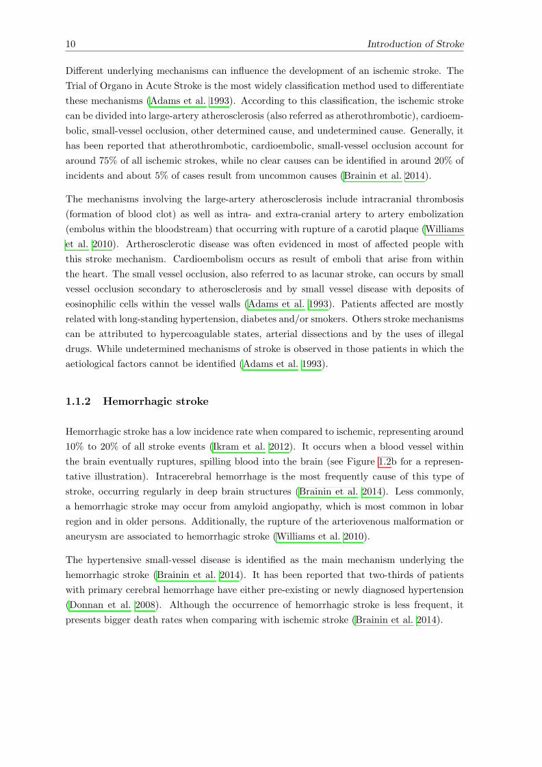
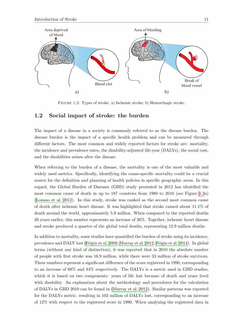
1.2 Types of stroke. a) Ischemic stroke; b) Hemorrhagic stroke. . . . . . . . . . . 11
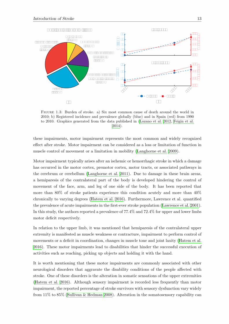
1.3 Burden of stroke. a) Six most common cause of death around the world in2010; b) Registered incidence and prevalence globally (blue) and in Spain (red)from 1990 to 2010. Graphics generated from the data published in (Lozanoet al. 2012, Feigin et al. 2014). . . . . . . . . . . . . . . . . . . . . . . . . . . 13
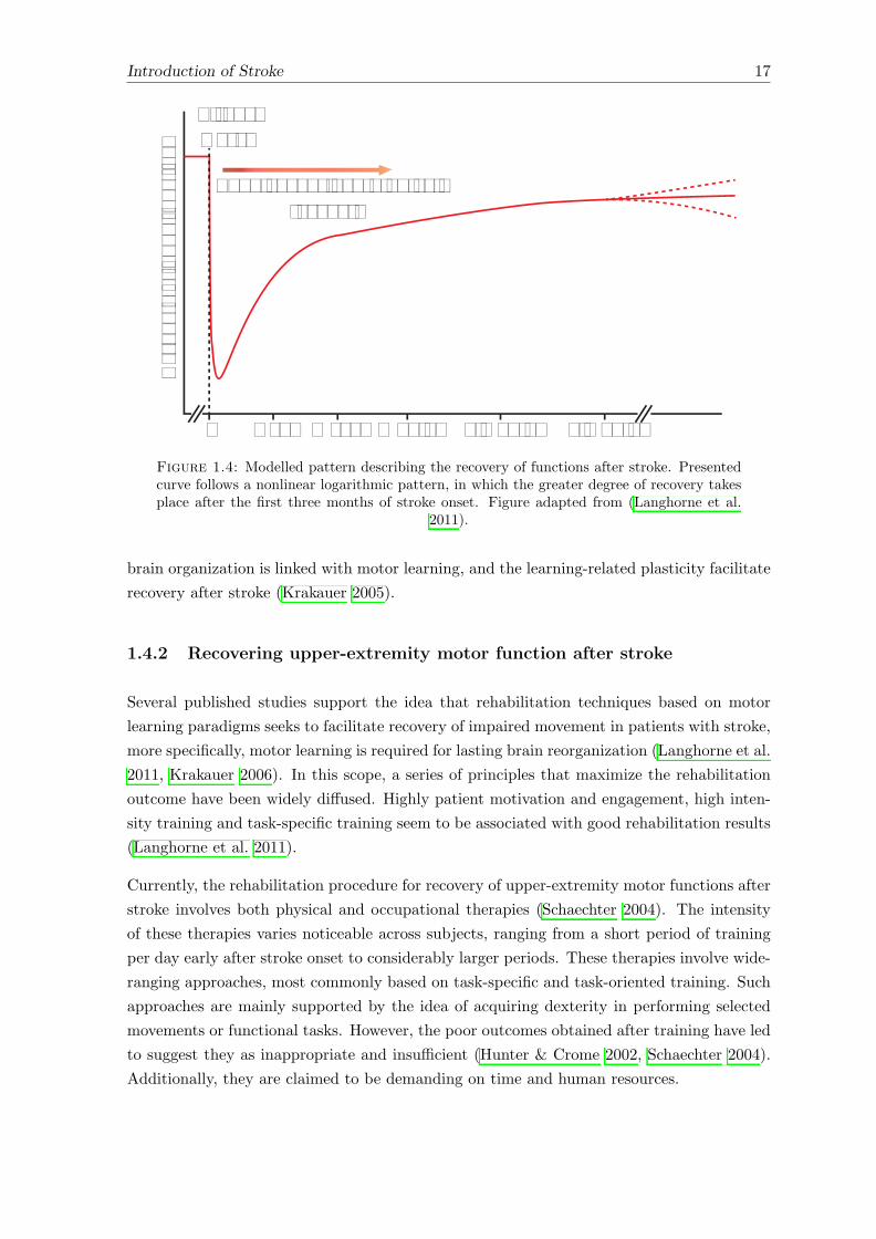
1.4 Modelled pattern describing the recovery of functions after stroke. Presentedcurve follows a nonlinear logarithmic pattern, in which the greater degreeof recovery takes place after the first three months of stroke onset. Figureadapted from (Langhorne et al. 2011). . . . . . . . . . . . . . . . . . . . . . . 17
1.5 Examples of end-e↵ectors robotic devices and upper-limb exoskeletons forupper-extremity rehabilitation. Figure reproduced from (Maciejasz et al. 2014). 22
1.6 Movement generation in healthy subjects and motor impaired stroke survivorsthrough functional electrical stimulation (FES). a) Scheme of the intact neu-rological tract producing tetanic contraction to generate movement; b) Use ofFES to generate movement. . . . . . . . . . . . . . . . . . . . . . . . . . . . . 24
1.7 General overview of a Brain-Computer Interface (BCI) system. A) Electro-physiological signal from the brain acquired using invasively (Single unit andECoG) or superficially (EEG) techniques. B) Acquired signal are processes toextract useful cortical feature. C) Command generated from pre-processingand learning algorithm to operate assistive devices to help patients with com-munication or movement, or to operate rehabilitative devices to help recoverneural function. ECoG: electrocorticography; EEG: electroencephalography. . 26
2.1 Hybrid robotic systems for grasping rehabilitation. a) Newest version of theNESS HandMaster device (Alon et al. 2003, Ring & Rosenthal 2005). b)Hybrid assistive neuromuscular dynamic stimulation (HANDS) (adapted from(Fujiwara et al. 2009)). c) Experimental setup for wrist flexion/extensiontraining (adapted from (Hu et al. 2010)). . . . . . . . . . . . . . . . . . . . . 32
xv

xvi LIST OF FIGURES
2.2 Implemented robot and FES control algorithm for grasping movement. a)EMG-based FES control strategy for hand opening and closing (Fujiwara et al.2009). Dmin and Dmax define the minimum and maximum stimulation pulsewidth. b) Cooperative control strategy by Hu et al. for wrist flexion/extension(Hu et al. 2010, 2011, 2015). Arobot, Afes represent the torque and FESassistance respectively; G and K are the constants gains used to adjust themagnitude of the assistance torque and FES; Timv is the maximal value ofthe torque during isometric maximum extension (Timve) and flexion (Timvf );Wmax,FCR/ECR is the maximum stimulation impulse width applied on theFCR or ECR muscles; EMGflex/ext is the EMG of agonist muscle; EMGrest
is the average of EMG level of the muscle; EMGimv is the maximal EMGamplitude of the muscle. . . . . . . . . . . . . . . . . . . . . . . . . . . . . . . 33
2.3 Hybrid robotic rehabilitation system for reaching movements. a) SMARTdevice combined with FES (figure adapted from (Barker et al. 2008)). b) BATtraining based on two parallel bar and FES (figure adapted from (Wu et al.2011)). c) Planar end-e↵ector work- station combined with FES (Freemanet al. 2009c,b). d) Unconstrained 3D workstation based on ArmeoSpring®
exoskeleton (Meadmore et al. 2012). . . . . . . . . . . . . . . . . . . . . . . . 35
2.4 FES control strategies implemented in hybrid robotic therapies for reachingmovements. a) Control scheme representation implemented in the planar end-e↵ector workstation (Hughes et al. 2009). �⇤
k is the reference trajectory and�k is the measured joint angle at time k; Vk+1 represents the feedforwardupdate signal for application on next trial; ek is the error at time; µk is theactuation signal; ⌧f , ⌧h, ⌧t are the torque generated due to stimulated muscle,contribution of others muscles and the sum of both respectively; h is the torqueprovided by the planar robotic device. b) Block diagram of control strategyfor 3D rehabilitation system (Meadmore et al. 2012). �⇤
k is the referencetrajectory and �k is the measured joint angles at time k; Vk+1 represents thefeedforward update signal for application on next trial; ek is the error at time;zk is the actuation signal for the feedback controller; µk is the total (Vk + zk)the actuation signal; ⌧f , ⌧h, ⌧t are the torque generated due to stimulatedmuscles, contribution of others muscles and the sum of both respectively; ⌧ris the torque provided by the exoskeleton to support the arm against gravity. 36
3.1 a) General overview of the presented hybrid robotic platform for reaching reha-bilitation. b) Visual feedback provided to the users. The green ball representsthe actual arm position, the blue cross is the reference trajectory, the initialand final position are represented by the gray ball and red square respectively.c) Interface for system configuration. . . . . . . . . . . . . . . . . . . . . . . . 48
3.2 Kinematic representation of the rotation axes. a) Exoskeleton ✓ = ✓1, ✓2, ✓3, ✓4, ✓5.b) Human arm � = �1,�2,�3,�4,�5. . . . . . . . . . . . . . . . . . . . . . . . 50
3.3 a) Block diagram of the FES-based Feedback Error Learning (FEL) controller.b) Artificial Neural Network used as feedforward loop. �r, �r, �r) representthe desired angular position, velocity and acceleration respectively; �h is themeasured position of the human arm; e(n) is the error position; µff , µfb arethe control signal generated for the feedback and feedforward controllers re-spectively; µt is the total assistance; µts is the assistance at the output of thesaturator; eu is the di↵erence between ts and t. . . . . . . . . . . . . . . . . . 52
3.4 Position, velocity and acceleration profiles of the minimum jerk trajectoryfunction (Flash & Hogan 1985). . . . . . . . . . . . . . . . . . . . . . . . . . . 52
LIST OF FIGURES xvii
3.5 A representative example of the FEL controller performance in one healthysubject. a) The tracking accuracy during trial 1 (red) and trial 12 (blue) forshoulder (left) and elbow (right) joints. b) The output signal of the feedbackerror learning controller during the first and twelfth movement execution; ufb(in red) is the control signal given by the feedback controller; uff (in blue)represents the control action of the feedforward controller; ut (in black) cor-responds to the output of the FEL controller (uff + ufb). . . . . . . . . . . . 58
3.6 a) Mean values of the normalized root mean squared error (RMSE, blackline) and its standard error (gray shaded areas) across healthy subjects, cor-responding to the shoulder (left) and elbow (right) joints. Dotted lines denotesignificance di↵erence between trials. b) Mean values of provided FES inten-sity, represented as a percentage of the maximum stimulation intensity, acrosssubjects. Light gray and dark gray areas depict the contribution of the feed-forward (ufb) and feedback (uff ) loop to the total FES intensity, measuredwith the power ratio (PR). . . . . . . . . . . . . . . . . . . . . . . . . . . . . 59
3.7 Evolution of the root mean square error of the temporal coordination (TC)index between the generated reference and arm trajectories. Shaded arearepresents standard error. . . . . . . . . . . . . . . . . . . . . . . . . . . . . . 60
3.8 a) Evolution of the root mean square error (RMSE) averaged for each run.The first column represents the RMSE for session one divided in shoulder (top)and elbow (bottom), while the second column depicts the error for session two.The black lines represent the calculated linear regression for each combinationof subject, muscle and session. b) Evolution of the PRff for each subject.The black line represents averaged PRff across subjects and its correspondingstandard error. The first column represents the PRff for session one dividedin shoulder (top) and elbow (bottom), while the second column depicts thevalue corresponding for session 2. . . . . . . . . . . . . . . . . . . . . . . . . . 61
3.9 Averaged tracking performance for each run corresponding to session one andtwo. The black lines represent the linear fitting regression across subjects. . . 61
3.10 a) Mean task’s performance across subjects considering the last assisted andthe unassisted runs. The error bars represent the standard error; b) Meanroot mean squared (RMS) of the temporal coordination index (TC-index)di↵erences between the generated reference and the arm trajectories acrosssubjects considering the last assisted and the unassisted runs. The error barsrepresent the standard error. Asterisks indicate significant di↵erences. . . . . 62
4.1 General overview of the integrated hybrid robotic and the neuromodulationsystem for rehabilitation of reaching movement. . . . . . . . . . . . . . . . . . 72
4.2 a) Experimental protocol used for experimentations; b) Implemented statemachine to present the visual cue and guide users during the intervention. . . 73
4.3 Normalized amplitudes of the average MRCP of all subjects (dotted lines ingray) and average MRCP across subjects (continuous black line). The verticalred line indicates the beginning of the movement state (cue). . . . . . . . . . 81
4.4 Measured input-output response of the MEP peak-to-peak value across thePre0-, Pre-, Post- and Post30-assessment intervals for the three target muscles,Anterior Deltoid (first row), Triceps Brachii (second row) and biceps Brachii(third row), and each experimental intervention BCI o✏ine (first column) BCIonline (second column) and EMG (third column). . . . . . . . . . . . . . . . . 83
4.5 EMG movement onset detection time with respect to the cue event. Black cir-cles represent individual detection (for each trial) while the red circles denotethe average detection time. . . . . . . . . . . . . . . . . . . . . . . . . . . . . 84
xviii LIST OF FIGURES
5.1 General overview and components of the hybrid robotic system. 1) Neuro-modulation system based on a EEG brain-computer interface to estimate thestimulation onset (negative peak of the movement-related cortical potential).2) High Level Controller to provide the adaptive assistance. 3) Hybrid Assis-tance composed of the IntFES stimulator (3a) and the ArmeoSpring (3b). 4)Visual Feedback interface to guide user’s during the interventional session. 5)User Interface to configure system’s parameters (stimulation threshold, rangeof movement, movement repetitions). . . . . . . . . . . . . . . . . . . . . . . . 93
5.2 Description of the pilot study protocol designed to evaluate the hybrid roboticsystem. Ev: evaluation; Se: session. T: trial. . . . . . . . . . . . . . . . . . . 95
5.3 Motor-related cortical potential (MRCP) of the P1 patient. The MRCP’scurve of the most representative EEG channel (C2) is shown in the first columnwhile the spatial distribution of the MRCP is illustrated in the second column.To optimize visualization, baseline was defined within [-3, -2] seconds withrespect to the movement state (vertical red line at t = 0). . . . . . . . . . . . 97
5.4 a) Representative example of the tracking accuracy during the execution of theunassisted movement. Joints trajectories represent the time-averaged responseof the first and last interventional sessions. b) Achieved task’s performancescore during the execution of the unassisted reaching movements along ses-sions. The solid line represents the best linear fitting indicating the trend ofthe score. See Section 3.4.2.2 for the task’s performance score. . . . . . . . . 99
5.5 Motor-related cortical potential (MRCP) of patients P2 and P5. The firscolumn illustrates de MRCP’s curve of the most representative EEG channels,while the second column portrays the spatial distribution of the MRCP. Tooptimize visualization, baseline was defined within [-3, -2] seconds with respectto the movement state (vertical red line at t = 0). . . . . . . . . . . . . . . . 100
5.6 a) Representative example of the tracking accuracy (P2: first column; P5second column) during the execution of the unassisted movements. Joints tra-jectories represent the time-averaged response corresponding to the first andlast interventional sessions. b) Achieved task’s performance score of both pa-tients (P2 and P5) during the execution of the unassisted reaching movementsalong sessions. . . . . . . . . . . . . . . . . . . . . . . . . . . . . . . . . . . . . 101
6.1 a) Main components of the analogous platform developed for further stud-ies targeting upper-limb distal muscles. 1: High level controller; 2: MotorDriver; 3: Mechanical support for the index finger; 4: Motor and embeddedencoder; 5: Power supply. b) Associative intervention concept framed withinthe ASSOCIATIVE project for the lower-limb. . . . . . . . . . . . . . . . . . 112
List of Tables
1.1 Common impairment and most relevant activities a↵ected in stroke patients . 14
2.1 Identified hybrid system for grasping function. . . . . . . . . . . . . . . . . . 31
2.2 Identified hybrid system for reaching function. . . . . . . . . . . . . . . . . . . 34
2.3 Clinical assessment of the reviewed hybrid robotic systems. . . . . . . . . . . 37
3.1 Description of patients recruited for the experimentation with the hybrid system. 54
3.2 Mean and standard deviation values across healthy subjects. . . . . . . . . . 59
3.3 Results of the Wilcoxon post hoc test. . . . . . . . . . . . . . . . . . . . . . . 60
3.4 Satisfaction score of all brain injury patients. . . . . . . . . . . . . . . . . . . 63
4.1 Average values of the experimental condition for each group. . . . . . . . . . 81
4.2 Performance of the cue-based BCI system used in the experimentation of theBCI online group. . . . . . . . . . . . . . . . . . . . . . . . . . . . . . . . . . . 82
5.1 Clinical data of post-stroke patients participating in this study. . . . . . . . . 91
5.2 Changes in clinical scales between pre- and post-intervention assessments. . . 98
5.3 Satisfaction score of post-stroke patients . . . . . . . . . . . . . . . . . . . . . 101
xix

Nomenclature
AD Anterior Deltoid
ADL Activities of Daily Living
ANN Artificial Neural Network
ANOVA Analysis of Variance
ARAT Action Research Arm Test
BAT Bilateral Arm Training
BB Biceps Brachii
BCI Brain-Computer Interface
CNS Central Nervous System
DALY Disability-Adjusted Life Year
DOF Degree of Freedom
ECR Extensor Carpi Radialis
EDC Extensor Digitorum Communis
EEG Electroencephalography
EMG Electromyography
ERD Event-Related Desynchronization
FDS Flexor Digitorum Superficialis
FEL Feedback Error Learning
FES Functional Electrical Stimulation
FMA Fugl-Meyer Assessment
GBD Global Burden of Diseases
HANDS Hybrid Assistive Neuromuscular Dynamic Stimulation
HLC High Level Controller
ILC Iterative Learning Control
LD Lateral Detoid
LTD Long-Term Depression
LTP Long-Term Potentiation
MAS Modified Ashworth Scale
MCID Minimal Clinically Important Di↵erence
MEP Motor Evoked Potential
MMT Manual Muscle Test
MRCP Motor-Related Cortical Potential
PAS Paired Associative Stimulation
xxi
xxii NOMENCLATURE
PID Proportional-Integral-Derivative
PR Power Ratio
PW Pulse Width
QUEST Quebec User Evaluation of Satisfaction with Assistive Technology 2.0
RMSE Root Mean Square Error
rMT Resting Motor Threshold
ROM Range of Movement
SAIL Stimulation Assistance through Iterative Learning
SAM Self-Assessment Manikin
SMART Sensorimotor Active Rehabilitation Training
SMR Sensory Motor Rhythms
TB Triceps Brachii
TMS Transcranial Magnetic Stimulation
TP True positive
WHO World Health Organization
Objectives and description of the
work
Stroke results in an irreversible damage to the brain’s tissues. Motor impairment constitutes
one of the most common consequence arising after this brain lesion. The level of motor
a↵ectation varies between post-stroke subjects, but often, it hampers the execution of most
activities of daily living. Consequently, the dependence and the quality of life of the stroke
survivors are severely a↵ected.
The rehabilitation of the motor functions is one of the cornerstones topics in post-stroke
management. Rehabilitation therapies seek to recover the a↵ected motor functions in order
to achieve dexterity and successfully execute activities of daily living. The main underlying
process explaining this recovery is the adaptive characteristic of the central nervous system,
called plasticity. The primary goal of rehabilitation therapies is to exploit the neural plasticity
to maximize the functional outcomes. However, when exploring the reported outcomes,
e.g. in upper-extremity motor functions, studies evidenced poor results. Certainly, it was
reported that 50-70% of stroke survivors with initial severe or mild paresis of the upper-
extremity continued to experience limited function and disuse of their hemiplegic upper limb
post-stroke.
This makes the development and validation of alternative rehabilitative methods aimed at
recovering (upper-limb) motor impairment a topic of great importance. During the last
decades, a number of novel interventions for rehabilitation of upper-extremity for post-stroke
subjects have been explored. Among them, the use of robotic devices, functional electrical
stimulation and neuromodulation systems has emerged as promising paths. The use of these
novel technologies presents an unprecedented alternative for maximizing motor recovery by
building on their capability for providing repetitive and intense task-specific training, thus
improving motor control and facilitating plasticity.
Objectives
Considering, on the one hand, that upper-extremity motor impairment arisen after stroke
a↵ects the execution of most activities of daily living and, on the other hand, the potential
1

2 Objectives and description of the work
rehabilitative benefits of novel therapies (robot and functional electrical stimulation), the
present doctoral thesis proposes the combined use of a robotic device, functional electrical
stimulation and electroencephalography-based brain-computer interfaces during the execu-
tion of reaching movements with the aims of facilitating neural plasticity and promoting
recovery of the upper-extremity motor functions.
The main objective of this doctoral dissertation is to develop and validate a novel hybrid
robotic system for rehabilitation of reaching movement in stroke population. The
proposed system combines the advantages of the robotic technology, the functional electrical
stimulation techniques and the electroencephalography-based brain-computer interfaces to
assist the execution of unconstrained functional reaching movement. This development is
conducted to address the challenges identified from an in-depth analysis of the relevant
literature.
One of the identified challenges is to provide an optimum level of assistance. An adaptive
assistance is implemented to adjust the current intensity according to the user’s motor resid-
ual capability in order to achieve a successful tracking reaching movement with the a↵ected
arm.
As it will be highlighted later on, brain plasticity plays a crucial role for recovery of motor
functions after stroke. One possible and widely adopted strategy for promoting plasticity
relies on the use of neuromodulation systems. In this work, a brain-computer interface
is proposed to precisely pair, associate and synchronize the user’s own intention to move
with the applied hybrid peripheral assistance, thus ensuring causality. It is demonstrated
that neural plasticity could be facilitated uniquely when the precise and causal association
between motor-related cortical process and the assistance occurs.
Explicitly, the following objectives are framed within this doctoral thesis:
• To identify main challenges of the current hybrid robotic concept focused on rehabili-
tation of upper-limb motor function.
• To develop and implement a prototype of a hybrid robotic system (robot mechani-
cal system with functional electrical stimulation technique) to assist the execution of
reaching movement.
• To implement and validate an adaptive control strategy to adjust the level of assistance
according to the user’s motor residual capabilities during the execution of the reaching
task.
• To implement and integrate a neuromodulation system within the hybrid platform
with the aim of tightly and causally coupling the user’s own intention to move with
the hybrid peripheral assistance.
• To demonstrate that the precise temporal association between the motor cortex activa-
tion identified by motor-related cortical activity and the hybrid assistance could elicit
neural plasticity.
Objectives and description of the work 3
• To provide a validation of the overall hybrid concept through a clinical intervention
with post-stroke subjects, together with a detailed interpretation of the results.
Methods and structure of the work
The methodology followed to achieve the aforementioned objectives relies on a thorough
study of the di↵erent aspects related to the motor recovery in the stroke population. This
study comprised the motor pathological and functional characteristics of the a↵ected upper-
extremity, the principal mechanisms underlying motor recovery after a brain injury and the
clinical implications of this brain disorder. The methodology followed in this dissertation
is fully in line with the organization of the thesis. Thus, the work has been split into
three clear parts. One chapter introduces the rationale of the dissertation. The subsequent
four chapters present a series of studies and the contributions. Finally, the last chapter
summarizes the main conclusions, the scientific results and dissemination, as well as the
future research emerging from this thesis. Accordingly, each chapter (please, see details in
the next paragraphs) contributes to the successful accomplishment of the related objectives
above presented.
Chapter 1 describes the rationale for the dissertation. It provides a detailed review and ac-
count of the di↵erent aspects related to the stroke disorder. First, it provides a brief overview
about the pathophysiology of stroke and explains how stroke is currently classified. The so-
cial burden in terms of mortality, prevalence, cost and disabilities arisen post-stroke were
extracted from literature and are briefly summarized here. The common a↵ected body func-
tions are listed and, epidemiological aspects of upper-limb motor impairment are presented.
The review in this chapter highlights the importance of novel rehabilitation therapies for
improvement current rehabilitation outcomes. Three novel therapies are critically presented,
summarizing their main advantages and disadvantages, giving rise to the hybrid concept,
i.e. combined use of robotic devices and functional electrical stimulation, proposed and
implemented throughout the thesis.
Chapter 2 presents the state of the art on hybrid robotic system with a focus on the reha-
bilitation of the upper-extremity motor functions. Two perspectives are addressed here: the
technical and the clinical one. As a results of this analysis, the main design requirements
of the hybrid systems were identified, which in turn guided the developments and studies
presented in this dissertation.
Chapter 3 presents the design and development of a fully integrated hybrid robotic system
for rehabilitation of functional reaching movements in post-stroke subjects. The hybrid plat-
form is composed of several networked subsystems to jointly enable the novel rehabilitation
task. This platform constitutes a comprehensive self-contained tool aimed at promoting
the recovery of reaching movements. Complementary, the implementation of an adaptive
controller is presented. This controller strategy adjusts the level of assistance according to
the user’s motor residual capabilities. A detailed explanation about its operation and the
4 Objectives and description of the work
assumptions are provided. The technical validation of the platform was conducted in a pilot
study with healthy subjects (n = 12). The usability assessment involved a small sample of
stroke patients (n = 4) with a reduced number of session (two experimental sessions).
Chapter 4 introduces the associative concept. Here, the use of a neuromodulation system is
presented with the main aim of facilitating neural plasticity. This system is integrated into
the hybrid platform to tightly and causally couple the user’s motor planning process with the
peripheral hybrid assistance. Three di↵erent associative strategies are critically compared,
taking as the primary outcome indicator the plastic e↵ects resulting from the intervention.
An experimental protocol is presented with the aim of validating the associative concept.
Results from experimentation with healthy volunteers (n = 21) demonstrate the feasibility
of associative concept for enhancing the excitability of corticospinal projections to target
arm muscles (anterior deltoids, triceps brachii and biceps brachii).
In Chapter 5 the potential rehabilitative e↵ects of the hybrid robotic system are investigated.
The system combined both the adaptive and associative assistance developed and tested
individually in previous chapters. To this aim, a pilot clinical intervention was conducted.
The primary objective was to verify the feasibility of using the hybrid robotic system in clinic
rehabilitation and the inspection of its potential e↵ects for recovery the upper-extremity
motor functions. Five post-stroke subjects were recruited to participate in an interventional
protocol consisting in 12 sessions along 4 consecutive weeks.
Chapter 6 summarizes and concludes the results of this doctoral work. It highlights the
scientific results and dissemination derived from the studies conducted in this thesis in the
following research fields: biomedical and neural engineering, rehabilitation robotics, func-
tional electrical stimulation and neurorehabilitation. Eventually, it proposes emerging future
research activities, out of the outcomes obtained in this thesis.
Framework of the thesis
This work has been carried out with the financial support of the Itaipu Binacional (Paraguay),
and it was carried out at the Neural Rehabilitation Group (NRG), Cajal Institute, Span-
ish National Research Council (CSIC). The first studies presented in this dissertation were
performed in the framework of two funded research projects: HYPER (Hybrid Neuropros-
thetic and Neurorobotic Devices for Functional Compensation and Rehabilitation of Motor
Disorders, grant CSD2009-00067 CONSOLIDER INGENIO 2010) and BRAIN2MOTION
(Exoskeletal - neuroprosthesis hybrid robotic system for the upper limb controlled by a mul-
timodal brain-neural interface, grant DPI2011-27022-C02-02). While the last experiments
were conducted in framework of the ASSOCIATE project (A comprehensive and wearable
robotics based approach to the rehabilitation and assistance to people with stroke and spinal
cord injury, grant DPI2014-58431-C4-1-R). Within these research projects, cutting-edge re-
search was conducted in the fields of biomedical and neural engineering, clinic rehabilitation,
neurorehabilitation and neuroscience amongst others.

Objectives and description of the work 5
Finally, it is worth mentioning that the close and helpful collaboration with the Centro de
Referencia Estatal de Atencion Al Dano Cerebral (CEADAC) and with the Instituto de
Neurociencias y Ciencias del Movimiento (INCIMOV) constituted an essential asset for the
successful progress of the work and studies presented in this thesis.

Chapter 1
Introduction of stroke and
rehabilitation of the upper limb
motor function
Abstract
This chapter introduces the background and rationale for the development and studies framed
in this dissertation. An overview about of stroke is presented. The most relevant consequences
post-stroke are identified, including physiological, functional, social and economic aspects,
globally and in Spain. From the rehabilitation perspective, the importance of the upper-
extremity motor and functional recovery is emphasized. The main rehabilitation premises and
the concept of neural plasticity for maximizing recovery outcomes are also presented. Three
novel rehabilitation methods for rehabilitation of arm motor function are reviewed: robotic
devices, functional electrical stimulation and brain-computer interfaces. Published evidence
shows two important aspects. First, novel approaches are needed to increase the potential
of robotic and functional electrical stimulation interventions for rehabilitation of upper-limb
motor function in stroke survivors. Second, brain-computer interfaces can be exploited to
supply assistance in causal association motor cortical processes to improve current therapies
by means of the promoted neural plasticity.
7

8 Introduction of Stroke
1.1 Stroke
The central nervous system (CNS) is a very complex system, yet fascinating at the same time.
It can be considered as a bilateral and symmetrical structure composed of two main parts:
the brain and the spinal cord. The brain comprise seven structures: the medulla oblongata,
pons, cerebellum, midbrain, diencephalon, corpus callosum and the cerebral hemispheres
(see Figure 1.1a). The two hemispheres of the human brain can be further divided into four
di↵erent regions: the frontal lobe, the parietal lobe, the occipital lobe and the temporal lobe
see Figure 1.1b). Each brain lobe is responsible for controlling specific set of functions. In
this regard, the frontal lobe is mainly related with short-term memory, the planning of motor
action and with the control of the movement; the parietal lobe with the somatic sensation,
representation of body image and its relation with the extra-personal space; the occipital
lobe is mainly responsible for vision; and lastly, the temporal lobe is associated with the
hearing, learning, memory and emotion (Kandel et al. 2000).
As all human cognitive functions occur primary in the brain, it can be seen as the central
processing organ of the CNS, responsible for controlling multiple complex functions. In adult
humans, the brain weights around 1.3 kg (Kandel et al. 2000), representing in average the
2% of the body weight. Although its small size (when compared with the proportion of
the human body), it is estimated that the brain spends around 20% of the oxygen and,
hence, calories consumed by the body. It was also mentioned that this metabolic activity of
the brain is remarkably constant over time, despite of the mental and motor activities are
widely varying (Raichle & Gusnard 2002). The brain, as all other organs of the human body,
has an extremely dependence of the energy to develop its functions and operate normally.
The energy production in the brain relies on metabolism of exogenous compounds with a
high-energy content, primarily the oxidation of glucose (Mohr et al. 2011). As the storage
of substrates for energy metabolism in the brain is minimal, the functional and structural
integrity of the brain depends on a continuous supply of blood by delivering oxygen and
glucose (Mohr et al. 2011).
When the blood supply to the brain is disrupted, the brain stops receiving nutrients and
oxygen. This event is known as a stroke. A stroke, also considered a brain attack, is caused
by a sudden interruption in the blood supply to the brain. The most widely used and
accepted definition of stroke was given by the World Health Organization (WHO), which is
stated as follow:
“rapidly developed signs of focal (or global) disturbance of cerebral function lasting longer
than 24 hours (unless interrupted by death), with no apparent nonvascular cause”.
Stroke is considered a cerebrovascular disease. After the disruption of the bloodstream, the
cells of the brain start to die leading to a damage of the brain areas. Approximately two
million brain cells die every minute during a stroke (Gund et al. 2013). The neurons’ death
results in an irreversible neurological damage that can even cause the death of the living
Introduction of Stroke 9
Cerebreralhemisphere
Spinalcord
Medulaoblongata
Cerebelum
Pons
Midbrain
Diencephalon
Corpus callosum Frontal
lobe
Temporallobe
Occipitallobe
Parietallobe
a) b)
Figure 1.1: a) Main components of the central nervous system. b) The four lobes of thecerebral cortex.
being. One or more brain areas can be a↵ected after stroke resulting in the lost or the
decrease of functions controlled by that specific area (Muir 2009).
Although stroke always occurs in the brain, there are two di↵erent types: ischemic and
hemorrhagic. The distinction between these subtypes is very important and urgent for its
clinical management, since di↵erent procedures are applied on each case (Donnan et al.
2008). A summary of each subtype is given below, with a brief explanation of the principal
pathophysiology di↵erences between them.
1.1.1 Ischemic Stroke
Ischemic stroke represents the most common type accounting for about the 70% of reported
stroke events (Adams et al. 2007). This type of stroke occurs when the blood vessels inside or
close to the brain are occluded causing a disruption of the blood flow in certain brain areas.
A representative illustration of the attributed mechanisms underlying the ischemic stroke is
depicted in Figure 1.2a. This occlusion leads two zones of injury in the brain referred to as
the core and the penumbra ischemic zones (Williams et al. 2010). In the core zone, a little
amount of blood flows resulting in an insu�cient resource of oxygen and glucose, and in a
quickly depletion of stores. As consequence, the brain cells within this area start dying. In
the penumbra zone, the blood is still able to flow through collateral arteries linking with
branches of the occluded vessels. This is not a time-stable process, but rather it can be
considered as a time-limited process (Williams et al. 2010). In case that reperfusion is not
reestablished within hours, the cells inside the penumbra zone will die because bloodstream
is not enough to deliver oxygen and glucose in the long-term (Donnan et al. 2008).
10 Introduction of Stroke
Di↵erent underlying mechanisms can influence the development of an ischemic stroke. The
Trial of Organo in Acute Stroke is the most widely classification method used to di↵erentiate
these mechanisms (Adams et al. 1993). According to this classification, the ischemic stroke
can be divided into large-artery atherosclerosis (also referred as atherothrombotic), cardioem-
bolic, small-vessel occlusion, other determined cause, and undetermined cause. Generally, it
has been reported that atherothrombotic, cardioembolic, small-vessel occlusion account for
around 75% of all ischemic strokes, while no clear causes can be identified in around 20% of
incidents and about 5% of cases result from uncommon causes (Brainin et al. 2014).
The mechanisms involving the large-artery atherosclerosis include intracranial thrombosis
(formation of blood clot) as well as intra- and extra-cranial artery to artery embolization
(embolus within the bloodstream) that occurring with rupture of a carotid plaque (Williams
et al. 2010). Artherosclerotic disease was often evidenced in most of a↵ected people with
this stroke mechanism. Cardioembolism occurs as result of emboli that arise from within
the heart. The small vessel occlusion, also referred to as lacunar stroke, can occurs by small
vessel occlusion secondary to atherosclerosis and by small vessel disease with deposits of
eosinophilic cells within the vessel walls (Adams et al. 1993). Patients a↵ected are mostly
related with long-standing hypertension, diabetes and/or smokers. Others stroke mechanisms
can be attributed to hypercoagulable states, arterial dissections and by the uses of illegal
drugs. While undetermined mechanisms of stroke is observed in those patients in which the
aetiological factors cannot be identified (Adams et al. 1993).
1.1.2 Hemorrhagic stroke
Hemorrhagic stroke has a low incidence rate when compared to ischemic, representing around
10% to 20% of all stroke events (Ikram et al. 2012). It occurs when a blood vessel within
the brain eventually ruptures, spilling blood into the brain (see Figure 1.2b for a represen-
tative illustration). Intracerebral hemorrhage is the most frequently cause of this type of
stroke, occurring regularly in deep brain structures (Brainin et al. 2014). Less commonly,
a hemorrhagic stroke may occur from amyloid angiopathy, which is most common in lobar
region and in older persons. Additionally, the rupture of the arteriovenous malformation or
aneurysm are associated to hemorrhagic stroke (Williams et al. 2010).
The hypertensive small-vessel disease is identified as the main mechanism underlying the
hemorrhagic stroke (Brainin et al. 2014). It has been reported that two-thirds of patients
with primary cerebral hemorrhage have either pre-existing or newly diagnosed hypertension
(Donnan et al. 2008). Although the occurrence of hemorrhagic stroke is less frequent, it
presents bigger death rates when comparing with ischemic stroke (Brainin et al. 2014).
Introduction of Stroke 11
!"#$%&#'"()#&%*+%,-**&
.-**&%/-*0
!"#$%*+%,-##&(12
."#$3%*+%,-**&%)#44#-
$5 ,5
Figure 1.2: Types of stroke. a) Ischemic stroke; b) Hemorrhagic stroke.
1.2 Social impact of stroke: the burden
The impact of a disease in a society is commonly referred to as the disease burden. The
disease burden is the impact of a specific health problem and can be measured through
di↵erent factors. The most common and widely reported factors for stroke are: mortality,
the incidence and prevalence rates, the disability-adjusted life-year (DALYs), the social cost,
and the disabilities arisen after the disease.
When referring to the burden of a disease, the mortality is one of the most valuable and
widely used metrics. Specifically, identifying the cause-specific mortality could be a crucial
source for the definition and planning of health policies in specific geographic areas. In this
regard, the Global Burden of Diseases (GBD) study presented in 2012 has identified the
most common cause of death in up to 187 countries from 1980 to 2010 (see Figure 1.3a)
(Lozano et al. 2012). In this study, stroke was ranked as the second most common cause
of death after ischemic heart disease. It was highlighted that stroke caused about 11.1% of
death around the world, approximately 5.9 million. When compared to the reported deaths
20 years earlier, this number represents an increase of 26%. Together, ischemic heart disease
and stroke produced a quarter of the global total deaths, representing 12.9 million deaths.
In addition to mortality, some studies have quantified the burden of stroke using its incidence,
prevalence and DALY lost (Feigin et al. 2009, Murray et al. 2012, Feigin et al. 2014). In global
terms (without any kind of distinction), it was reported that in 2010 the absolute number
of people with first stroke was 16.9 million, while there were 33 million of stroke survivors.
These numbers represent a significant di↵erence of the score registered in 1990, corresponding
to an increase of 68% and 84% respectively. The DALYs is a metric used in GBD studies,
which it is based on two components: years of life lost because of death and years lived
with disability. An explanation about the methodology and procedures for the calculation
of DALYs in GBD 2010 can be found in (Murray et al. 2012). Similar patterns was reported
for the DALYs metric, resulting in 102 million of DALYs lost, corresponding to an increase
of 12% with respect to the registered score in 1990. When analyzing the registered data in

12 Introduction of Stroke
Spain, a similar increasing trend can be elucidated. As depicted in Figure 1.3b, the global
incidence and prevalence in 2010 presented an increase of 26.9% and 88.5% respectively with
respect to the record in 1990 (Feigin et al. 2014). Similar trend was found when focusing
uniquely in the reported data in Spain. More specifically, Vega et al. reported a stroke
incidence of 141 cases per 100000 inhabitants in woman and 148 in men (Vega et al. 2009).
However, the mortality in 2010 was reduced 15.9% with respect to the data registered in
1990. This reduction in mortality can be attributed to the improvement in medicine and to
the improvements in the social welfare system.
It is worth also mentioning that although the mean age of people with stroke is increasing,
there is a substantial number of stroke occurrence in people younger than 65 years (Feigin
et al. 2014). It was reported than more than 83000 children and youths aged 20 years and
younger are a↵ected by stroke annually, suggesting that stroke should not be considered as
a disease of old age.
Another important social burden is the economic cost of the disease, which is an important
parameter for social health and research policies. Worldwide, stroke consumes about 2-4%
of total health-care costs, and in industrialized countries stroke accounts for more than 4%
of direct health-care costs (Donnan et al. 2008). Olesen et al. provided a quantitative
evaluation of brain disorder in terms of cost within Europe (Olesen et al. 2012). In this
study, the authors estimated that stroke suppose a total annual cost of 64.1 billion AC using
prices of 2010. In a di↵erent study, Alvarez-Sabın et al. reported an estimation of the real
cost of stroke in Spain (Alvarez-Sabın et al. 2017). They reported that the cost of patients
admitted to stroke units in Spain is 27711 AC per patient/years. It has been also reported
that the cost of hemorrhagic stroke was slightly higher than ischemic (30332 AC vs. 23234 AC
per patient/year), attributed to the presence of hypertension and the severity of stroke.
Reported results disclose that the global burden of stroke in terms of the mortality, number
of people a↵ected every year, stroke survivors, and DALYs lost are great and present an
increasing trend over years. Based on these facts, it was estimated that by 2030, there will
be almost 12 million of deaths due stroke, 70 million of stroke survivors and more than 200
million DALYs lost (Feigin et al. 2014). As these numbers influence directly the health cost,
also the cost of stroke will be increased considerably. Consequently, stroke is considered
currently a serious health problem globally (World Health Organization 2003, Bonita et al.
2004).
1.3 Motor impairment
The e↵ects due to stroke are extremely heterogeneous between individuals. These e↵ects are
determined by the site and size of the brain lesion (Brewer et al. 2013). Several sequelae
can arise after a stroke (see Table 1.1), namely, deficit in language, vision and cognitive
capabilities; alterations in body functions as ingestion, defecation, urinary and sexual and
the inability for controlling (in)voluntary movements (Langhorne et al. 2009). Between all
Introduction of Stroke 13
Ischaemic Heart Disease
(7.0m)
Stroke (5.9m)
Chronic Obstructive Pulmonar Disease
(2.9m)
Lower Respiratory Infections
Lung Cancer (1.5m)
HIV/AIDS (1.5m)
Total deaths: 21.61 millions
(2.8m)1012141618
100110120130140
1990
2005
2010
10
20
30
40
200
300
400
500
Mill
ions
Thou
sand
sTh
ousa
nds
Mill
ions
Incidence
Prevalence
Global Spain
a) b)
Figure 1.3: Burden of stroke. a) Six most common cause of death around the world in2010; b) Registered incidence and prevalence globally (blue) and in Spain (red) from 1990to 2010. Graphics generated from the data published in (Lozano et al. 2012, Feigin et al.
2014).
these impairments, motor impairment represents the most common and widely recognized
e↵ect after stroke. Motor impairment can be considered as a loss or limitation of function in
muscle control of movement or a limitation in mobility (Langhorne et al. 2009).
Motor impairment typically arises after an ischemic or hemorrhagic stroke in which a damage
has occurred in the motor cortex, premotor cortex, motor tracts, or associated pathways in
the cerebrum or cerebellum (Langhorne et al. 2011). Due to damage in these brain areas,
a hemiparesis of the contralateral part of the body is developed hindering the control of
movement of the face, arm, and leg of one side of the body. It has been reported that
more than 80% of stroke patients experience this condition acutely and more than 40%
chronically to varying degrees (Hatem et al. 2016). Furthermore, Lawrence et al. quantified
the prevalence of acute impairments in the first-ever stroke population (Lawrence et al. 2001).
In this study, the authors reported a prevalence of 77.4% and 72.4% for upper and lower limbs
motor deficit respectively.
In relation to the upper limb, it was mentioned that hemiparesis of the contralateral upper
extremity is manifested as muscle weakness or contracture, impairment to perform control of
movements or a deficit in coordination, changes in muscle tone and joint laxity (Hatem et al.
2016). These motor impairments lead to disabilities that hinder the successful execution of
activities such as reaching, picking up objects and holding it with the hand.
It is worth mentioning that these motor impairments are commonly associated with other
neurological disorders that aggravate the disability conditions of the people a↵ected with
stroke. One of these disorders is the alteration in somatic sensations of the upper extremities
(Hatem et al. 2016). Although sensory impairment is recorded less frequently than motor
impairment, the reported percentage of stroke survivors with sensory dysfunction vary widely
from 11% to 85% (Sullivan & Hedman 2008). Alteration in the somatosensory capability can

14 Introduction of Stroke
Table 1.1: Common impairment and most relevant activities a↵ected in stroke patients
Common impairments after stroke. Most relevant activities a↵ected.
Reduced ability to control of (in)voluntarymovements.
Use of the arm and hand.
Di�culty in mobility and stability of joints Walking.Reduced muscle power/tone Maintaining body position.Altered proprioception and touch. Reading, writing and calculation; Solving
problems.Di�culty in ingestion, defecation, urinaryand sexual.
Execution of activities of daily living (dress-ing, toileting, eating, etc.).
Cognitive decline. Mobility.Dyphonia/dysarthria/dyphasia. Communication and speaking.Reduced energy and motivation, change inpersonality.
Recreation and leisure.
Data compiled from (Brewer et al. 2013, Langhorne et al. 2011)
result in an impaired detection of sensory information, in disturbed motor tasks performance
requiring somatosensory information and in diminished upper extremity rehabilitation out-
comes (Hunter & Crome 2002). Both, altered sensation and motor impairment contribute
significantly to the loss of function in the upper extremities, and their correct integration for
the successful execution of motor tasks is essential.
In addition, a reduced level of movement could eventually lead to changes in muscle, con-
nective and neural tissues, and therefore, inducing secondary complications (Pollock et al.
2014). Some of these secondary problems are: shortening of muscles (contracture); disor-
dered muscle contraction (spasticity); decreased or lost connectivity of the unused neural
pathways; and pain.
Although the attention was mainly focused in motor impairment, stroke is also associated
with other non-motor impairments that combined with the motor disabilities have an impor-
tant impact on the quality of life of stroke survivors (Langhorne et al. 2009, Hunter & Crome
2002). Such impairments cause a great functional disability, a↵ecting the independency of
stroke survivors and limiting their participation in society (Pollock et al. 2014). Therefore,
impairments, and particularly motor impairments, increase the burden of stroke in society.
1.4 Rehabilitation of motor impairment after stroke
In the ancient cultures, motor impairment and other disabilities were addressed following
mythological or religious basis, e.g. impaired people was considered to be possessed by
spirits or disability was often seen as a punishment for the past misbehaviours (World Health
Organization 2010). The notorious advance in science over the last century boosted the
consolidation of biological and medical basis for what we know now as the discipline of
medical rehabilitation
Introduction of Stroke 15
According to the WHO, rehabilitation is defined as:
“a set of measures that assist individuals, who experience or are likely to experience disability,
to achieve and maintain optimum functioning in interaction with their environments”.
Rehabilitation is mainly aimed at maximizing people’s ability to live, work and learn to their
best potential. Evidence suggests that rehabilitation can reduce the functional di�culties
associated with disabilities, impairments, ageing and improve quality of life (World Health
Organization 2011). In summary, rehabilitation aid in achieving and maintaining optimal
functioning in interaction with the environment through achieving the following outcomes:
• prevention of the loss of function;
• slowing the rate of loss of function;
• improvement or restoration of function;
• compensation for lost function;
• maintenance of current function.
The rehabilitation of motor functions is one of the cornerstones of stroke management
(Brainin et al. 2014). In previous sections, it was mentioned that the trends in stroke
occurrence and the number of stroke survivors are expected to increase over the years, con-
sequently, it is also foreseen an increased quantity of people who benefit from rehabilitation.
Thus, rehabilitation therapies will become primordial for stroke survivors and the society.
Rehabilitation of motor functions, and of any disability in general, involves a well-known and
defined cyclical process consisting of the following steps (Steiner et al. 2002): (1) identifi-
cation of the person’s problems and needs; (2) connecting the problems to relevant factors
of the person and the environment; (3) establishing rehabilitation goals; (4) planning and
implementing the measures and; (5) assessing the e↵ects. This cyclical process helps medical
and physiotherapist sta↵ to personalize the therapy according to the patient’s needs in order
to maximize outcomes of the rehabilitation therapy.
For achieving the objective of maximizing rehabilitation outcomes, rehabilitation therapies
seek to exploit the most important and studied feature of the CNS: the brain plasticity, to
be introduced ad discussed in the following paragraphs.
1.4.1 Brain plasticity associated with motor recovery
For several years in the last century, it was believed that “once development is complete, the
sources of growth and regeneration of axons and dendrites are irretrievably lost. In the adult
brain the nerve paths are fixed and immutable: everything can die, nothing can be regenerated”
(Cajal 1959). Subsequent scientific contributions shifted this approach giving rise to the
current accepted theory that the CNS of an adult human is capable of reorganization and
recovery, and it can be selectively promoted (Dimyan & Cohen 2011).
16 Introduction of Stroke
This reorganization process, which is responsible for the recovery of body function post-
stroke, is named neural plasticity. Neural plasticity was first described with regard to the
function of synapses (Hebb 1949), and later, this principle was extended to the operation
of the overall neural networks (Brainin et al. 2014). Although several definition exist for
plasticity, here, the definition given by Murphy & Corbett is transcribed: “Changes in the
strength of synaptic connections in response to either an environmental stimulus or an al-
teration in synaptic activity in a network” (Murphy & Corbett 2009). Reported evidence
showed that this reorganization (neural plasticity) after stroke can occur in cortical regions
immediately adjacent to the infarct or remote from the infarct, both in the same and in the
opposite hemisphere (Krakauer 2005, Dimyan & Cohen 2011).
Although the rehabilitation of motor impairment after stroke is particularly heterogeneous,
the neurological recovery after stroke follows a nonlinear logarithmic pattern as shown in
Figure 1.4. The recovery of body functions is believed to occur through a combination of
spontaneous recovery and learning-dependent processes (Langhorne et al. 2011). True neuro-
logical recovery takes place during the first 4 to 10 weeks post-stroke, and it is driven by the
spontaneous recovery and non-learning-dependent processes. It is hypothesized that several
mechanisms are involved in this stage, such as: salvation of the penumbra; physiological
and neuroanatomical reorganization (spontaneous neuroplasticity); alleviation of diaschisis
and; reperfusion enhanced by post-stroke angiogenesis (Krakauer 2005, Buma et al. 2013).
In the past, it was believed that the recovery e↵ects of upper extremity due to spontaneous
recovery post-stroke was an inherent behaviour and, nothing could be done to influence it
(Hatem et al. 2016). Some studies evidenced that task-specific training as soon as possible
can assist the natural pattern of functional recovery to maximize outcomes (Langhorne et al.
2011, Krakauer 2006).
After this period, improvements in terms of body functions are believed to be mainly driven
by adaptation or compensatory motor strategies (Buma et al. 2013, Krakauer 2005). At this
point, the recovery curve slow down and it attains a plateau at approximately 6 months
post-stroke. Yet, it was pointed out that the recovery plateau post-stroke may just reflect an
asymptotic learning rather than a true biological limit (Krakauer 2005, 2006). Furthermore,
the poor insensitivity of the clinical scales to detect improvements can also contribute to this
limitation (Dobkin 2004). Some studies carried out with chronic stroke patients (> 6 months)
demonstrated e↵ective results, suggesting the idea that neural reorganization may also take
place in the subacute and chronic phase after stroke (Langhorne et al. 2009, Krakauer 2006,
Hatem et al. 2016). The mechanisms underlying the neural plasticity at this stage that
could result in functional recovery are not well understood yet, but it can be explained by
long-term potentiation (LTP) and depression (LTD) of existing synapses, strengthening of
alternative networks, synaptic remodeling, and axonal sprouting amount others (Brainin
et al. 2014, Dimyan & Cohen 2011). Despite well adaptive plasticity is necessary for recovery
of motor functions, in many cases the reorganization can also led into a maladaptive plasticity
(Dimyan & Cohen 2011, Buma et al. 2013). In this regard, it is accepted that well-adaptive
Introduction of Stroke 17
Stroke Onset
Rec
over
y of
bod
y fu
nctio
ns
0 Days Weeks Months 3 months 6 months
Spontaneous neurological recovery
Figure 1.4: Modelled pattern describing the recovery of functions after stroke. Presentedcurve follows a nonlinear logarithmic pattern, in which the greater degree of recovery takesplace after the first three months of stroke onset. Figure adapted from (Langhorne et al.
2011).
brain organization is linked with motor learning, and the learning-related plasticity facilitate
recovery after stroke (Krakauer 2005).
1.4.2 Recovering upper-extremity motor function after stroke
Several published studies support the idea that rehabilitation techniques based on motor
learning paradigms seeks to facilitate recovery of impaired movement in patients with stroke,
more specifically, motor learning is required for lasting brain reorganization (Langhorne et al.
2011, Krakauer 2006). In this scope, a series of principles that maximize the rehabilitation
outcome have been widely di↵used. Highly patient motivation and engagement, high inten-
sity training and task-specific training seem to be associated with good rehabilitation results
(Langhorne et al. 2011).
Currently, the rehabilitation procedure for recovery of upper-extremity motor functions after
stroke involves both physical and occupational therapies (Schaechter 2004). The intensity
of these therapies varies noticeable across subjects, ranging from a short period of training
per day early after stroke onset to considerably larger periods. These therapies involve wide-
ranging approaches, most commonly based on task-specific and task-oriented training. Such
approaches are mainly supported by the idea of acquiring dexterity in performing selected
movements or functional tasks. However, the poor outcomes obtained after training have led
to suggest they as inappropriate and insu�cient (Hunter & Crome 2002, Schaechter 2004).
Additionally, they are claimed to be demanding on time and human resources.

18 Introduction of Stroke
These facts are verified by the several studies. For instance, Hendrick et al. reported that
when an initial paralysis of the upper extremity occurred as consequence of stroke, complete
motor recovery occurs in 5 to 20% of the patients (Hendricks et al. 2002). In line with
this evidence, it has been also reported that 50-70% stroke survivors with initial severe or
mild upper extremity paresis will continue to experience loss of function and disuse in the
hemiplegic upper limb 2-4 years post-stroke (Hunter & Crome 2002). In a di↵erent study,
Kwakkel et al. informed that 62% of patients presenting a paralysis of the upper extremity
at stroke onset failed to achieve some dexterity at 6 months, indicating a poor prognosis for
functional outcome in these group of patients (Kwakkel et al. 2003).
Based on these facts and considering that the upper extremity disability contributes in loss
of independence and decrease the quality of life of stroke survivors (Samsa & Matchar 2004),
the scientific community is aimed at developing novel methods and technologies to boost
the current rehabilitation outcome, paying special attention to those therapies that enhance
neural plasticity. In the next section, some of these therapies are briefly presented.
1.5 Novel therapies for rehabilitation of the upper extremity
motor function
This section brings an overview of the technologies currently available for rehabilitation of
upper limb motor function after stroke. The development of therapies covered in this section
were driven mainly by the advancement of technology. As explained in previous sections, it
is assumed that neural plasticity of the CNS remains after stroke. Novel therapies seek to
exploit this neurological phenomenon and optimize both, the rehabilitation outcome and the
user experience, during the therapy session.
1.5.1 Robotics devices
Originally, robots were conceived to be used in industrial environments with the aim of
replacing humans. Over the years and with the consolidation of technology, the use of robots
were widen to other areas covering di↵erent disciplines. One of the most interesting scenarios
consist of using robots as assistive and/or rehabilitation tools (Pons 2008). On the one
hand, robots can substitute the loss of certain motor function in disabled people, resulting
in assistive devices. On the other hand, robots can assist the execution of rehabilitation
therapies in order to promote recovery of motor functions (rehabilitation devices). Although
both approaches are of great interest, only rehabilitation robots will be discussed in this
section and along this work. Until the last decade therapeutic robotics was considered to be
in its infancy (Krebs et al. 2008), its use for rehabilitation applications just emerged over the
last decade.
Several devices have been proposed over the last years for rehabilitation of upper limb motor
functions, some examples are shown in Figure 1.5 (see (Maciejasz et al. 2014, Lo & Xie 2012)
Introduction of Stroke 19
for further details). End-e↵ector robots were the earliest considered for rehabilitation of the
upper limb. This type of robotic devices support the user’s upper limb extremity through a
single contact point (commonly the hand or the forearm), so that, the generated forces are
transmitted by this interface to the user’s arm. This type of robot is simpler, however as the
joints of the end-e↵ector robot do not correspond with joints of the human arm, it presents
some limitations. Amongst them, we can highlight the di�culties for determining the human
arm position, the unfeasibility of applying specific torque to certain upper limb joints, the
impossibility of isolate movements at a unique upper limb joint and, the most important,
the movements are commonly constrained to one plane. Subsequently, the research approach
has shifted towards the use of more sophisticated robotic devices such as exoskeletons (Pons
2010). An upper limb exoskeleton is designed to operate side-by-side with the user’s arm,
since its mechanical structure is designed with the aim to mirror the anatomical structure
of the upper limb. This means that each joint of the exoskeleton directly controls a specific
joint of the human arm. Exoskeletons present several advantages for the rehabilitation of
upper limb motor functions, namely, more complex movements can be trained, the human
arm position can be more easily estimated, the movement of upper limb joint can be isolated
and the generated torque can be applied to each joint separately.
Alternatively, rehabilitation upper limb robots can be categorized according to the type of
assistance they deliver, leading to passive and active devices. Passive robotic devices cannot
actively assist the movement execution, so they are limited to delivering gravity compensation
and/or resistive forces (i.e. brakes). The main advantages of this type of devices are the
lower cost and smaller size since they do not include actuators. Yet, its use in rehabilitation
settings is limited for those users with motor remnant capacity. Instead, active devices
possess at least one actuator and, thus, they are able to provide assistance during upper
extremity movements. In addition, the capacity of providing active assistance can also be
used to exert resistance to the movement. The target population for this type of devices is
very broad, spanning the spectrum from users without motor residual capabilities up to those
with good motor control. Their principal drawback is the higher cost. In brief, the more
complex a robotic system is the more di�cult its application in rehabilitation at the clinical
setting (Riener et al. 2005). This means that robotic devices with many degrees of freedom
(DOFs) and a large range of motion (ROM) are more di�cult and time-consuming to apply
in daily therapy at the clinical setting. Being the motor recovery an heterogeneous process,
the selection of the robotic device must be based on the specific conditions and needs of the
user.
As above discussed, motor recovery is associated to motor learning principles. In turn, mo-
tor learning depends of the amount of practice, the intensity of the training exercises and
the user’s motivation (Krakauer 2006, Huang & Krakauer 2009). One of the most impor-
tant advantages of rehabilitation robotics is its capability of delivering, through automated
management, higher dosage (number of movements) and intensity (number of movement per
unit of time -minutes or hours-) of therapy comparing with conventional therapy (Huang
& Krakauer 2009). Traditional post-stroke rehabilitation therapy involves long sessions of
20 Introduction of Stroke
training requiring a significant amount of human e↵ort, rendering it impractical, and in many
cases unfeasible, for many physicians and patients (Huang et al. 2016). A direct implication
of robotics-supported automated processes is their potential to provide more therapy with
less supervision. These observations suggest a possible change of paradigm, where robotic
devices could be useful in maximizing the rehabilitation cost-e↵ectiveness (Reinkensmeyer &
Boninger 2012).
Taking advantage of the embedded sensors available in robotic devices, a more precise and
objective measurement, in terms of kinematics and dynamics,, of the initial motor impair-
ment and its evolution along the treatment can be achieved. In addition, these objective
measurements can be used to feed specific biomechanical model in order to accurately ana-
lyze joint force and other parameters. These user-specific parameters could provide relevant
information for the personalization of the rehabilitation strategy (Huang & Krakauer 2009).
With the aim to increase patient’s motivation and engagement during therapy, robotic de-
vices can be combined with di↵erent types of visual feedback interfaces, such as games or
virtual reality environments. These would allow the implementation of challenging scenarios
that can be used to set specific objectives throughout the rehabilitation process (Harwin
et al. 2006).
The possibility of implementing di↵erent control algorithms to modify the behaviour of the
robotic device during therapy cater for the implementation of di↵erent therapies (Marchal-
Crespo & Reinkensmeyer 2009). The development and implementation of control algorithms
play an important role in rehabilitation since they define the user-machine interaction be-
haviour. Reported evidence in literature indicates that particular behavioural implementa-
tions do not influence learning, while others promote learning (Reinkensmeyer & Boninger
2012, Huang & Krakauer 2009, Harwin et al. 2006). For instance, the robot’s behaviour can
be tailored according to the need of the users in order to attain more e↵ective rehabilitation
results.
In spite of the aforementioned potential of robotic devices for rehabilitation of the upper
limb motor function, its rehabilitation e↵ectiveness compared with traditional therapy is
still controversial (Huang et al. 2016, Langhorne et al. 2009, Kwakkel et al. 2008). In this
regard, two systematic reviews and meta-analyses, including 246 (Prange et al. 2006) and
218 (Kwakkel et al. 2008) subjects with stroke, have found a significant improvement in the
upper-limb motor function after a robotic therapy measured with the Fugl-Meyer scale, in
muscle activation pattern, and in speed of executed movement. Also, authors reported that
these improvements have a long-term e↵ect of several months to several years, as measured at
follow-up. However, both studies reported that these motor function improvements could not
be generalized to improvement of outcomes of activities of daily living (ADL). In line with
these findings, Langhorne et al. reported similar results, in which robotic devices showed
improvements in the arm function, but not in the hand functions (Langhorne et al. 2009).
To sum it all up, high intensity repetitive movements constitute an important contribution
to the e↵ectiveness of a robotic therapy, and second, robotic therapy had no advantages at
low intensity utilization, but it also did not hinder or halt recovery.
Introduction of Stroke 21
In order to increase the benefits of robotics therapy, there are several important aspects to be
addressed. Assuming that too much assistance may not promote recovery and given the fact
that merely repeating a movement is not enough to learn (Krakauer 2006, Reinkensmeyer
et al. 2012), the development of control algorithms to provide the assistance, only when
needed, to complete the task constitute a common goal of robotic therapies. This cooperative
strategy (human-robot) can promote the active participation of patients, and therefore boost
current rehabilitation outcomes. Training more naturalistic movements may also influence
positively rehabilitation (Reinkensmeyer & Boninger 2012). Thus, the development of more
sophisticated robotic devices that allow users to carry out 3D movements involving several
arm joints are necessary. Lastly, the combination of robotic devices with other technologies
following a top-down approach (consisting in defining the rehabilitation therapies based on
the state of the brain), would exploit endogenous sources of learning and facilitate plasticity
(Belda-Lois et al. 2011).
In order to increase the benefits of robotics therapy, there are several important aspects to be
addressed. Assuming that too much assistance may not promote recovery and the fact that
repeating a movement is not enough to learn (Krakauer 2006, Reinkensmeyer et al. 2012),
the development of control algorithms able to provide the assistance, only when needed, to
complete the task constitute a common goal of robotic therapies. This cooperative strategy
(human-robot) can promote the active participation of patients, and therefore boost current
rehabilitation outcomes. Training more naturalistic movements may also influence positively
to rehabilitation (Reinkensmeyer & Boninger 2012). Thus, the development of more sophisti-
cated robotic devices that allow users to carry out 3D movements involving several arm joints
are necessary. Lastly, the combination of robotic devices with other technologies following a
top-down approach (consisting on defining the rehabilitation therapies based on the state of
the brain), would exploit endogenous sources of learning and facilitate plasticity (Belda-Lois
et al. 2011).
1.5.2 Functional electrical stimulation
Functional electrical stimulation (FES) uses short electrical pulses to generate muscle contrac-
tion. When these artificially induced electrical pulses are intense enough, action potentials
in motor-neuron attached to a specific muscle are elicited causing that muscle to contract
(Popovic et al. 2002). A representative illustration of this concept is shown in Figure 1.6.
Electrical stimulation of muscular tissue has been known since the first observations of Luigi
Galvani in 1790, who observed motion after applying electrical wires to paralyzed leg muscles
from frogs. Later in 1831, Michael Faraday showed that electrical currents applied to nerves
could create active movement (Doucet et al. 2012, IJzerman et al. 2009). The use of FES was
first applied over the common peroneal nerve for correction of foot drop during walking in
persons with hemiplegia (Liberson et al. 1961). Ever since, it has been widely investigated as
a means for rehabilitation and compensation of lower and upper extremities motor disorders
(Peckham & Knutson 2005, Popovic et al. 2002).

22 Introduction of Stroke
Figure 1.5: Examples of end-e↵ectors robotic devices and upper-limb exoskeletons forupper-extremity rehabilitation. Figure reproduced from (Maciejasz et al. 2014).
When using FES, the force generated in a muscle is a function of the total charge transferred
to it. The charge is characterized by the stimulus frequency as well as the amplitude and
width of the electrical pulse. The amplitude and pulse width regulate the number of muscle
fibers that are activated, while the temporal summation of muscle fibers is determined by
the frequency at which the pulses are delivered (She✏er & Chae 2007). Thus, the muscle
contraction can be controlled by adjusting these FES parameters.
Electrical stimulation can be supplied using either transcutaneous (over the skin surface),
percutaneous (placed within the muscle) or cu↵ electrodes (wrapper around the nerve that
innervates the muscle) (Popovic 2014). Transcutaneous, or simply surface stimulation, is per-
formed with self-adhesive electrodes placed on the subject’s skin, above the motor-neuron.
Although with invasive electrodes (percutaneous and cu↵ electrodes) a better muscular se-
lectivity is achieved, surface FES systems can be applied at a very early stage of the rehabil-
itation, allowing early benefit for the patient (Popovic et al. 2001). For such reason, surface
stimulation is the most widely used stimulation strategy for rehabilitation of motor function.
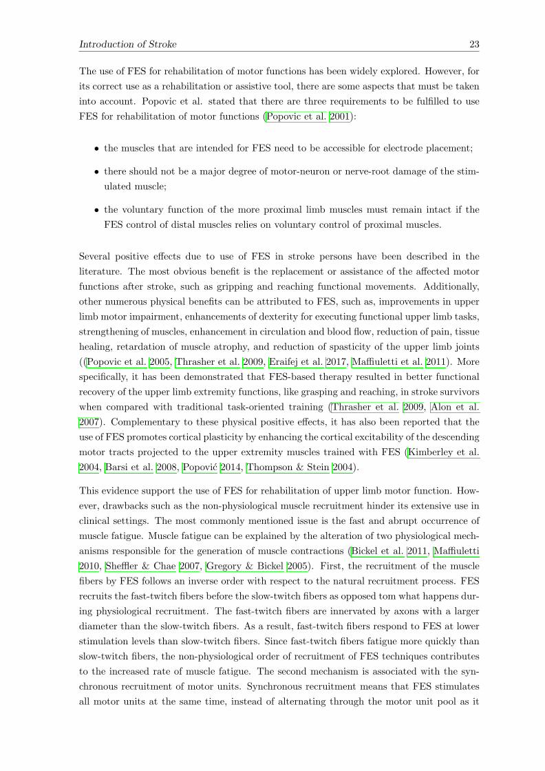
Introduction of Stroke 23
The use of FES for rehabilitation of motor functions has been widely explored. However, for
its correct use as a rehabilitation or assistive tool, there are some aspects that must be taken
into account. Popovic et al. stated that there are three requirements to be fulfilled to use
FES for rehabilitation of motor functions (Popovic et al. 2001):
• the muscles that are intended for FES need to be accessible for electrode placement;
• there should not be a major degree of motor-neuron or nerve-root damage of the stim-
ulated muscle;
• the voluntary function of the more proximal limb muscles must remain intact if the
FES control of distal muscles relies on voluntary control of proximal muscles.
Several positive e↵ects due to use of FES in stroke persons have been described in the
literature. The most obvious benefit is the replacement or assistance of the a↵ected motor
functions after stroke, such as gripping and reaching functional movements. Additionally,
other numerous physical benefits can be attributed to FES, such as, improvements in upper
limb motor impairment, enhancements of dexterity for executing functional upper limb tasks,
strengthening of muscles, enhancement in circulation and blood flow, reduction of pain, tissue
healing, retardation of muscle atrophy, and reduction of spasticity of the upper limb joints
((Popovic et al. 2005, Thrasher et al. 2009, Eraifej et al. 2017, Ma�uletti et al. 2011). More
specifically, it has been demonstrated that FES-based therapy resulted in better functional
recovery of the upper limb extremity functions, like grasping and reaching, in stroke survivors
when compared with traditional task-oriented training (Thrasher et al. 2009, Alon et al.
2007). Complementary to these physical positive e↵ects, it has also been reported that the
use of FES promotes cortical plasticity by enhancing the cortical excitability of the descending
motor tracts projected to the upper extremity muscles trained with FES (Kimberley et al.
2004, Barsi et al. 2008, Popovic 2014, Thompson & Stein 2004).
This evidence support the use of FES for rehabilitation of upper limb motor function. How-
ever, drawbacks such as the non-physiological muscle recruitment hinder its extensive use in
clinical settings. The most commonly mentioned issue is the fast and abrupt occurrence of
muscle fatigue. Muscle fatigue can be explained by the alteration of two physiological mech-
anisms responsible for the generation of muscle contractions (Bickel et al. 2011, Ma�uletti
2010, She✏er & Chae 2007, Gregory & Bickel 2005). First, the recruitment of the muscle
fibers by FES follows an inverse order with respect to the natural recruitment process. FES
recruits the fast-twitch fibers before the slow-twitch fibers as opposed tom what happens dur-
ing physiological recruitment. The fast-twitch fibers are innervated by axons with a larger
diameter than the slow-twitch fibers. As a result, fast-twitch fibers respond to FES at lower
stimulation levels than slow-twitch fibers. Since fast-twitch fibers fatigue more quickly than
slow-twitch fibers, the non-physiological order of recruitment of FES techniques contributes
to the increased rate of muscle fatigue. The second mechanism is associated with the syn-
chronous recruitment of motor units. Synchronous recruitment means that FES stimulates
all motor units at the same time, instead of alternating through the motor unit pool as it
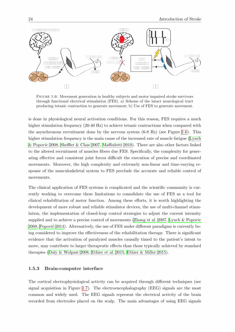
24 Introduction of Stroke
Force
FES20-40 Hz
Stimulation
Movement
Cur
rent
Time
Stroke
AlteredCommand
Movement
Force
Healthy behaviour
6-8 HzPhysiologicalStimulation
b)a)
Figure 1.6: Movement generation in healthy subjects and motor impaired stroke survivorsthrough functional electrical stimulation (FES). a) Scheme of the intact neurological tractproducing tetanic contraction to generate movement; b) Use of FES to generate movement.
is done in physiological neural activation conditions. For this reason, FES requires a much
higher stimulation frequency (20-40 Hz) to achieve tetanic contractions when compared with
the asynchronous recruitment done by the nervous system (6-8 Hz) (see Figure 1.6). This
higher stimulation frequency is the main cause of the increased rate of muscle fatigue (Lynch
& Popovic 2008, She✏er & Chae 2007, Ma�uletti 2010). There are also other factors linked
to the altered recruitment of muscles fibers due FES. Specifically, the complexity for gener-
ating e↵ective and consistent joint forces di�cult the execution of precise and coordinated
movements. Moreover, the high complexity and extremely non-linear and time-varying re-
sponse of the musculoskeletal system to FES preclude the accurate and reliable control of
movements.
The clinical application of FES systems is complicated and the scientific community is cur-
rently working to overcome these limitations to consolidate the use of FES as a tool for
clinical rehabilitation of motor function. Among these e↵orts, it is worth highlighting the
development of more robust and reliable stimulator devices, the use of multi-channel stimu-
lation, the implementation of closed-loop control strategies to adjust the current intensity
supplied and to achieve a precise control of movements (Zhang et al. 2007, Lynch & Popovic
2008, Popovic 2014). Alternatively, the use of FES under di↵erent paradigms is currently be-
ing considered to improve the e↵ectiveness of the rehabilitation therapy. There is significant
evidence that the activation of paralyzed muscles causally timed to the patient’s intent to
move, may contribute to larger therapeutic e↵ects than those typically achieved by standard
therapies (Daly & Wolpaw 2008, Ethier et al. 2015, Ethier & Miller 2015).
1.5.3 Brain-computer interface
The cortical electrophysiological activity can be acquired through di↵erent techniques (see
signal acquisition in Figure 1.7). The electroencephalography (EEG) signals are the most
common and widely used. The EEG signals represent the electrical activity of the brain
recorded from electrodes placed on the scalp. The main advantages of using EEG signals
Introduction of Stroke 25
result from being EEG a non-invasive technique, which is commercially available, easy to set-
up and, under certain circumstances, robust to possible external interferences. Altogether,
EEG is a suitable technique and tool for clinical environments (Chaudhary et al. 2016).
EEG-based brain computer interface (BCI) systems are emerging to provide a communication
channel between the human and an external device using brain activity (Wolpaw et al. 2002).
The general concept and overview of a BCI system is shown in Figure 1.7. This system opens a
door for innovative applications in several fields, with higher relevance in clinical and assistive
applications. Initially, BCI systems were conceived in clinical applications to provide an
alternative communication (blue dashed rectangle in Figure 1.7 means to patients with lost
ability to interact with the environment in any possible natural way (Daly & Wolpaw 2008,
Shih et al. 2012). During the last decade, this approach shifted towards using BCI systems in
rehabilitation applications (red dashed rectangle in Figure 1.7, in which the main goal is to
restore the impaired motor functions of a patient’s limb (Daly & Wolpaw 2008, Chaudhary
et al. 2016). In this scenario (motor restoration), the EEG signals provide a relevant feature
to the communication channel between the human and the assistive device: having real-time
access to movement-related cortical processes allows fast estimations of users’ intentions,
which may in turn lead to achieving causality and more natural interactions. Such natural
interfaces are highly desired in man-machine interaction for rehabilitation purposes for some
relevant reasons:
• Biological reasons. Human-robot interface systems seek to take advantage of the nat-
ural control mechanisms fully optimized in humans.
• Practical reasons. Delays are introduced when natural cognitive processes are encoded
into an imposed sequence of tasks. In addition, a training phase is needed to teach the
user to generate these non-natural commands or to map a cognitive process into a new
set of outputs. Both factors, the delays and the mapping, can also induce fatigue, both
at a musculoskeletal level and at a mental level. These limitations may be circumvented
if the natural outputs of a cognitive process are used instead.
• Rehabilitation. Interacting directly with cognitive processes of movement is a means
to excite them and assess the evolution of the rehabilitation therapy.
The use of EEG-based BCI systems to promote recovery of lost motor function has gained
attention during the past few years. Certainly, a large number of interventions to promote
motor neuro-rehabilitation have been proposed (Daly & Wolpaw 2008, Chaudhary et al.
2016). Several studies proposed BCI systems to perform motor imagery and provide visual
feedback (Pichiorri et al. 2011, 2015), BCIs in combination with physical therapy (Broetz
et al. 2010), BCIs triggering a robotic-based therapy (Ramos-Murguialday et al. 2013, Ang
et al. 2014, Ono et al. 2014) or BCIs triggering FES (Marquez-Chin et al. 2016, Mukaino
et al. 2014).
From this background, two BCI-based strategies in the rehabilitation field can be put forward.
The first one is based on the neuro-feedback, which hypothesizes that training the patients
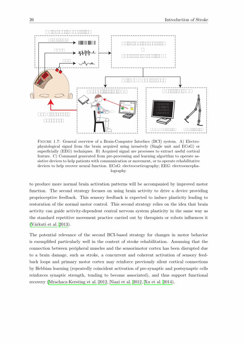
26 Introduction of Stroke
Assistive BCI
A B C D E FG H I J K L
M N O P Q RS T U V W X
Single unit
ECoG
EEG
A) Signal Acquisition
B) Signal Processing&
Learning Algorithms
C) Command Signal
Communication Substitution
Rehabilitative BCI
¡Alcanza el Objetivo!
ObjetivoPos. Inicial
Rendimiento: 100%
SomatosensoryFeedback
Figure 1.7: General overview of a Brain-Computer Interface (BCI) system. A) Electro-physiological signal from the brain acquired using invasively (Single unit and ECoG) orsuperficially (EEG) techniques. B) Acquired signal are processes to extract useful corticalfeature. C) Command generated from pre-processing and learning algorithm to operate as-sistive devices to help patients with communication or movement, or to operate rehabilitativedevices to help recover neural function. ECoG: electrocorticography; EEG: electroencepha-
lography.
to produce more normal brain activation patterns will be accompanied by improved motor
function. The second strategy focuses on using brain activity to drive a device providing
proprioceptive feedback. This sensory feedback is expected to induce plasticity leading to
restoration of the normal motor control. This second strategy relies on the idea that brain
activity can guide activity-dependent central nervous system plasticity in the same way as
the standard repetitive movement practice carried out by therapists or robots influences it
(Varkuti et al. 2013).
The potential relevance of the second BCI-based strategy for changes in motor behavior
is exemplified particularly well in the context of stroke rehabilitation. Assuming that the
connection between peripheral muscles and the sensorimotor cortex has been disrupted due
to a brain damage, such as stroke, a concurrent and coherent activation of sensory feed-
back loops and primary motor cortex may reinforce previously silent cortical connections
by Hebbian learning (repeatedly coincident activation of pre-synaptic and postsynaptic cells
reinforces synaptic strength, tending to become associated), and thus support functional
recovery (Mrachacz-Kersting et al. 2012, Niazi et al. 2012, Xu et al. 2014).
Introduction of Stroke 27
1.6 Conclusions of the chapter
This chapter presented the motivation for the development and the studies framed within
this thesis. This introductory chapter supported the fact that stroke currently represent
a serious and major health-care problem globally. Certainly, it constitutes an important
economic and societal burden. Brain injury caused by stroke can severely a↵ect several body
and/or cognitive functions, and specifically, motor impairments are the most common and
disabling consequence that decreases the quality of life of stroke survivors. In this context,
the recovery of upper-limb motor function constitutes a prime landmark addressed by current
rehabilitation therapies.
With the aim of boosting the current rehabilitation outcome and under the assumption that
high intensity and task-oriented training providing a coherence assistance with respect to
motor-cortical processes are important principles to promote neural plasticity, the use of
robotic devices, FES and BCI technology were proposed. Robotic and FES technologies
hold a considerable potential to drive upper-extremity rehabilitation interventions. Yet,
when considering the individual use of these alternative rehabilitation methods for recovery
the upper-limb motor function, they present limitations that hamper their consolidation in
clinical rehabilitation settings.
Based on this analysis, the combined use of assistive technologies (robot and FES) for reha-
bilitation of reaching movement in stroke patients is explored in this thesis. This combines
action (mechanical and electrical assistance) is referred to as hybrid robotic systems. As it
will be shown in the following chapters (Chapter 2 and Chapter 3), the hybrid approach
holds a great rehabilitation potential since it exploits the main advantages of each technol-
ogy resulting a more adequate and beneficial therapeutic methodology. In addition, the use
of BCI technology to accurately identify motor-related cortical patterns is a key aspect for
the interventions proposed in this dissertation. Indeed, it will be shown how important the
precise association (causality, synchronization) between the onset of voluntary movements
and the hybrid assistance is for promoting neural plasticity (see Chapter 4).

Chapter 2
State of the art of hybrid robotics
system for rehabilitation motor
functions of the upper-extremities1
Abstract
Over recent years, the combined use of functional electrical stimulation and robotic devices,
usually referred to as hybrid robotic rehabilitation systems, has emerged as a promising ap-
proach for healing lower and upper limb motor disorders. This chapter presents a critical
review of the state of the art of current hybrid robotic solutions for upper-limb rehabilitation
after stroke. To this purpose, studies have been selected through a search using web databases:
IEEE-Xplore, Scopus and PubMed. A total of 7 di↵erent hybrid robotic systems were identi-
fied. Selected systems are critically compared with respect to their technological components,
features and control strategies implemented. Additionally, technological and clinical evidence
on the e↵ectiveness of these hybrid robotic therapies are widely presented and discussed.
Eventually, the current technological challenges are identified. These challenges provide a
valuable information that informed developments in subsequent chapters of this thesis.
1This chapter is partly based on:Resquın F, Cuesta Gomez A, Gonzalez-Vargas J, Brunetti F, Torricelli D, Molina Rueda F, Cano de la RuedaR, Miangolarra J. C and Pons J. L. Hybrid robotic systems for upper limb rehabilitation after stroke: Areview. Med Eng Phys. 2016 Nov;38(11):1279-88.
29
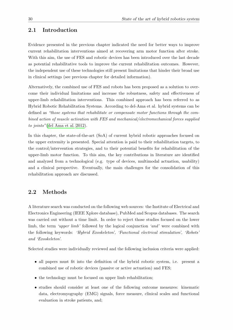
30 State of the art of hybrid robotics system
2.1 Introduction
Evidence presented in the previous chapter indicated the need for better ways to improve
current rehabilitation interventions aimed at recovering arm motor function after stroke.
With this aim, the use of FES and robotic devices has been introduced over the last decade
as potential rehabilitative tools to improve the current rehabilitation outcomes. However,
the independent use of these technologies still present limitations that hinder their broad use
in clinical settings (see previous chapter for detailed information).
Alternatively, the combined use of FES and robots has been proposed as a solution to over-
come their individual limitations and increase the robustness, safety and e↵ectiveness of
upper-limb rehabilitation interventions. This combined approach has been referred to as
Hybrid Robotic Rehabilitation Systems. According to del-Ama et al. hybrid systems can be
defined as “those systems that rehabilitate or compensate motor functions through the com-
bined action of muscle activation with FES and mechanical/electromechanical forces supplied
to joints”(del Ama et al. 2012).
In this chapter, the state-of-the-art (SoA) of current hybrid robotic approaches focused on
the upper extremity is presented. Special attention is paid to their rehabilitation targets, to
the control/intervention strategies, and to their potential benefits for rehabilitation of the
upper-limb motor function. To this aim, the key contributions in literature are identified
and analyzed from a technological (e.g. type of devices, multimodal actuation, usability)
and a clinical perspective. Eventually, the main challenges for the consolidation of this
rehabilitation approach are discussed.
2.2 Methods
A literature search was conducted on the following web sources: the Institute of Electrical and
Electronics Engineering (IEEE Xplore database), PubMed and Scopus databases. The search
was carried out without a time limit. In order to reject those studies focused on the lower
limb, the term ‘upper limb’ followed by the logical conjunction ‘and’ were combined with
the following keywords: ‘Hybrid Exoskeleton’, ‘Functional electrical stimulation’, ‘Robots’
and ‘Exoskeleton’.
Selected studies were individually reviewed and the following inclusion criteria were applied:
• all papers must fit into the definition of the hybrid robotic system, i.e. present a
combined use of robotic devices (passive or active actuation) and FES;
• the technology must be focused on upper limb rehabilitation;
• studies should consider at least one of the following outcome measures: kinematic
data, electromyography (EMG) signals, force measure, clinical scales and functional
evaluation in stroke patients, and;
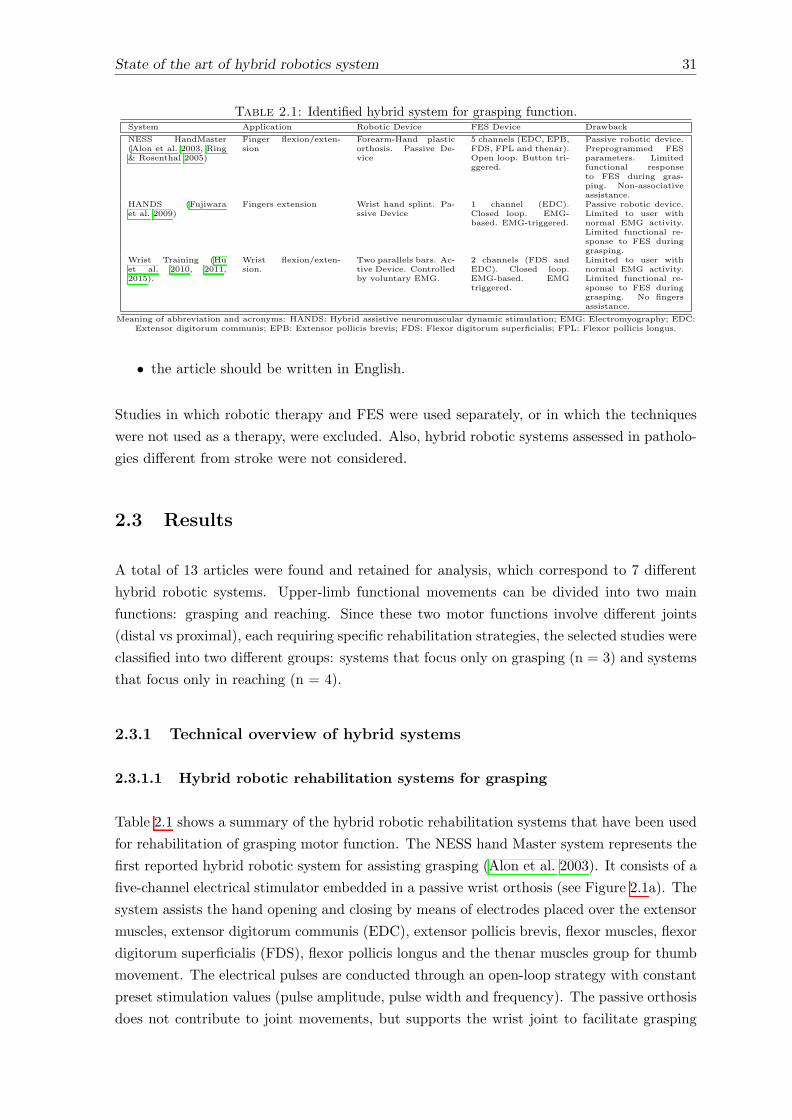
State of the art of hybrid robotics system 31
Table 2.1: Identified hybrid system for grasping function.System Application Robotic Device FES Device Drawback
NESS HandMaster(Alon et al. 2003, Ring& Rosenthal 2005)
Finger flexion/exten-sion
Forearm-Hand plasticorthosis. Passive De-vice
5 channels (EDC, EPB,FDS, FPL and thenar).Open loop. Button tri-ggered.
Passive robotic device.Preprogrammed FESparameters. Limitedfunctional responseto FES during gras-ping. Non-associativeassistance.
HANDS (Fujiwaraet al. 2009)
Fingers extension Wrist hand splint. Pa-ssive Device
1 channel (EDC).Closed loop. EMG-based. EMG-triggered.
Passive robotic device.Limited to user withnormal EMG activity.Limited functional re-sponse to FES duringgrasping.
Wrist Training (Huet al. 2010, 2011,2015).
Wrist flexion/exten-sion.
Two parallels bars. Ac-tive Device. Controlledby voluntary EMG.
2 channels (FDS andEDC). Closed loop.EMG-based. EMGtriggered.
Limited to user withnormal EMG activity.Limited functional re-sponse to FES duringgrasping. No fingersassistance.
Meaning of abbreviation and acronyms: HANDS: Hybrid assistive neuromuscular dynamic stimulation; EMG: Electromyography; EDC:Extensor digitorum communis; EPB: Extensor pollicis brevis; FDS: Flexor digitorum superficialis; FPL: Flexor pollicis longus.
• the article should be written in English.
Studies in which robotic therapy and FES were used separately, or in which the techniques
were not used as a therapy, were excluded. Also, hybrid robotic systems assessed in patholo-
gies di↵erent from stroke were not considered.
2.3 Results
A total of 13 articles were found and retained for analysis, which correspond to 7 di↵erent
hybrid robotic systems. Upper-limb functional movements can be divided into two main
functions: grasping and reaching. Since these two motor functions involve di↵erent joints
(distal vs proximal), each requiring specific rehabilitation strategies, the selected studies were
classified into two di↵erent groups: systems that focus only on grasping (n = 3) and systems
that focus only in reaching (n = 4).
2.3.1 Technical overview of hybrid systems
2.3.1.1 Hybrid robotic rehabilitation systems for grasping
Table 2.1 shows a summary of the hybrid robotic rehabilitation systems that have been used
for rehabilitation of grasping motor function. The NESS hand Master system represents the
first reported hybrid robotic system for assisting grasping (Alon et al. 2003). It consists of a
five-channel electrical stimulator embedded in a passive wrist orthosis (see Figure 2.1a). The
system assists the hand opening and closing by means of electrodes placed over the extensor
muscles, extensor digitorum communis (EDC), extensor pollicis brevis, flexor muscles, flexor
digitorum superficialis (FDS), flexor pollicis longus and the thenar muscles group for thumb
movement. The electrical pulses are conducted through an open-loop strategy with constant
preset stimulation values (pulse amplitude, pulse width and frequency). The passive orthosis
does not contribute to joint movements, but supports the wrist joint to facilitate grasping

32 State of the art of hybrid robotics system
a) b) c)
FES Device
Control &Data Storage
VisualFeedback
Palm Fixor
Motor
EMG & FES Electrode
ForearmFixor
Figure 2.1: Hybrid robotic systems for grasping rehabilitation. a) Newest version of theNESS HandMaster device (Alon et al. 2003, Ring & Rosenthal 2005). b) Hybrid assistiveneuromuscular dynamic stimulation (HANDS) (adapted from (Fujiwara et al. 2009)). c)Experimental setup for wrist flexion/extension training (adapted from (Hu et al. 2010)).
and to smoothen the muscle response to the FES. This orthosis is wired to a control unit
used to configure manually the FES parameters and to trigger the electrical assistance by
pressing a button.
A similar solution, referred to as hybrid assistive neuromuscular dynamic stimulation (HANDS),
was analyzed (Fujiwara et al. 2009). In this study, the authors integrate a wrist hand splint
with a single channel electrical stimulation for assisting fingers extension. In this case, sti-
mulation was given solely to the EDC muscle, whereas the splint contributed to inhibition
of flexors over activated muscles, and therefore the applied electrical stimulation enhanced
agonist muscles recruitment. Although this system relies on a single stimulation channel, its
main advantage is that the stimulation intensity could be set using a pulse width modulation
technique proportional to the recorded volitional electromyography (EMG) from the targeted
muscle (Muraoka et al. 2013, Hara et al. 2006). Figure 2.2a depicts the controller rule imple-
mented in this system, where Dmin corresponds to the minimum pulse width duration that
facilitates voluntary contraction, and Dmax is the threshold pulse duration equivalent to the
highest endurable intensity during maximum voluntary contraction. The voluntary EMG
signal was calculated by taking the raw EMG signal after 20 ms of the electrical stimulus,
thus both artifact and M-wave were discarded.
Hu et al. presented a FES-robot system for wrist flexion/extension rehabilitation (Hu et al.
2010, 2011, 2015), in which both assistive parts are driven by voluntary EMG signals detected
from flexor carpi radialis (FCR) and extensor carpi radialis (ECR) muscles. The robotic
system is based on an actuated end-e↵ector device, composed of two small parallel bars
delimited in the horizontal plane (see Figure 2.1a). Stroke patients seated with their a↵ected
arm mounted on the system to track a cursor displayed on the screen by moving their wrist
at di↵erent angular velocities. The total support was given by the contribution of the robot
(Arobot) and FES (Afes) assistance. The controlled assistance, shown in Figure 2.2b, followed
a proportional relation between the EMG amplitude, the maximum torque value during
isometric contraction (Timv for robot assistance) or maximum stimulation pulse width (Wmax
for FES assistance), and the constant assistance factor (G), used to adjust the support level
(ranged from 0 to 1). Although the assistance factor allows setting di↵erent actuation levels
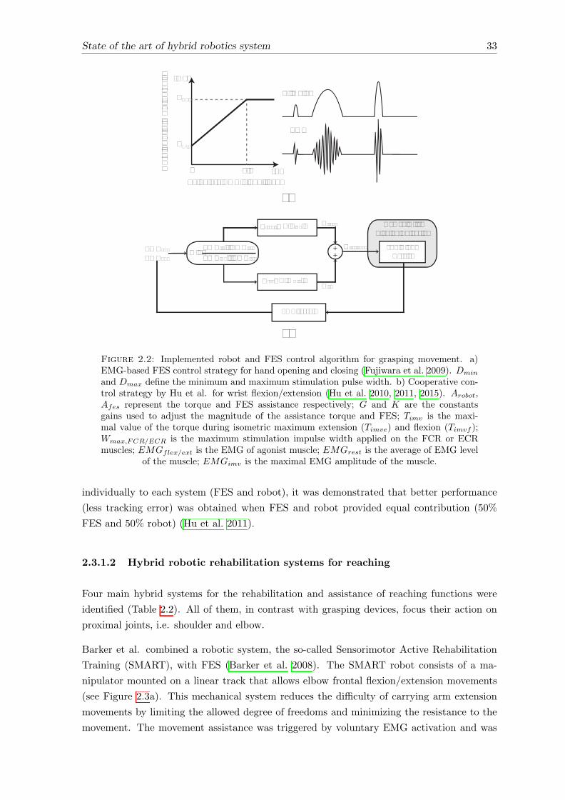
State of the art of hybrid robotics system 33
Dmax
Dmin
0 0.5 (V)
(ms)
Dur
atio
n of
stim
ulat
ion
puls
e
Voluntary EMG signal level
Stimulus
EMG
G.Timv.MArobot =
EMGf/e - EMGrest
EMGimv - EMGrestM =
FCR & ECRmuscles
Arobot
Afes
Afes-robot++
EMGFCR
EMGECR
EMG sensors
Human wristFlexion/Extension
G.Wmax.MAfes =
a)
b)
Figure 2.2: Implemented robot and FES control algorithm for grasping movement. a)EMG-based FES control strategy for hand opening and closing (Fujiwara et al. 2009). Dmin
and Dmax define the minimum and maximum stimulation pulse width. b) Cooperative con-trol strategy by Hu et al. for wrist flexion/extension (Hu et al. 2010, 2011, 2015). Arobot,Afes represent the torque and FES assistance respectively; G and K are the constantsgains used to adjust the magnitude of the assistance torque and FES; Timv is the maxi-mal value of the torque during isometric maximum extension (Timve) and flexion (Timvf );Wmax,FCR/ECR is the maximum stimulation impulse width applied on the FCR or ECRmuscles; EMGflex/ext is the EMG of agonist muscle; EMGrest is the average of EMG level
of the muscle; EMGimv is the maximal EMG amplitude of the muscle.
individually to each system (FES and robot), it was demonstrated that better performance
(less tracking error) was obtained when FES and robot provided equal contribution (50%
FES and 50% robot) (Hu et al. 2011).
2.3.1.2 Hybrid robotic rehabilitation systems for reaching
Four main hybrid systems for the rehabilitation and assistance of reaching functions were
identified (Table 2.2). All of them, in contrast with grasping devices, focus their action on
proximal joints, i.e. shoulder and elbow.
Barker et al. combined a robotic system, the so-called Sensorimotor Active Rehabilitation
Training (SMART), with FES (Barker et al. 2008). The SMART robot consists of a ma-
nipulator mounted on a linear track that allows elbow frontal flexion/extension movements
(see Figure 2.3a). This mechanical system reduces the di�culty of carrying arm extension
movements by limiting the allowed degree of freedoms and minimizing the resistance to the
movement. The movement assistance was triggered by voluntary EMG activation and was
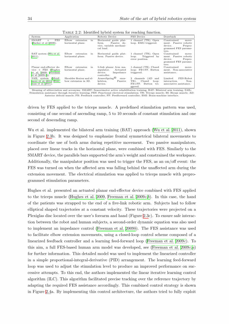
34 State of the art of hybrid robotics system
Table 2.2: Identified hybrid system for reaching function.System Application Robotic Device FES Device Drawback
SMART + FES(Barker et al. 2008).
Elbow extension inhorizontal plane.
Horizontal guide plat-form. Passive de-vice, variable mechani-cal load.
1 channel (TR). Openloop. EMG triggered.
Constrained move-ment. Passive roboticdevice. Prepro-grammed FES parame-ters.
BAT system (Wu et al.2011).
Elbow extension inhorizontal plane.
Horizontal guide plat-form. Passive device.
1 channel (TR). Openloop. Triggered byerror position.
Constrained move-ment. Passive roboticdevice. Prepro-grammed FES parame-ters.
Planar end-e↵ector de-vice + FES (Hugheset al. 2009, Freemanet al. 2009c,b).
Elbow extension inhorizontal plane.
5-link planar Arm ma-nipulator. Actuateddevice. Impedancecontroller.
1 channel (TR). Closedloop: FB+FF. Buttontriggered.
Constrained move-ment. Non-associativeassistance.
SAIL system (Mead-more et al. 2012, Free-man et al. 2011).
Shoulder flexion and el-bow extension in 3D.
ArmeoSpring® exos-keleton. Passivedevice.
2 channels (AD andTR). Closed loop:FB+FF. Button tri-ggered.
Limited FES-Robotinteraction. Non-associative assistance.
Meaning of abbreviation and acronyms: SMART: Sensorimotor active rehabilitation training; BAT: Bilateral arm training; SAIL:Stimulation assistance through iterative learning; FES: Functional electrical stimulation; TR: Triceps muscle; BI: Biceps muscle; AD:
Anterior deltoid muscle; FB: Feedback controller; FF: Feedforward controller; BMI: Brain-machine interface.
driven by FES applied to the triceps muscle. A predefined stimulation pattern was used,
consisting of one second of ascending ramp, 5 to 10 seconds of constant stimulation and one
second of descending ramp.
Wu et al. implemented the bilateral arm training (BAT) approach (Wu et al. 2011), shown
in Figure 2.3b. It was designed to emphasize frontal symmetrical bilateral movements to
coordinate the use of both arms during repetitive movement. Two passive manipulators,
placed over linear tracks in the horizontal plane, were combined with FES. Similarly to the
SMART device, the parallels bars supported the arm’s weight and constrained the workspace.
Additionally, the manipulator position was used to trigger the FES, as an on/o↵ event: the
FES was turned on when the a↵ected arm was falling behind the una↵ected arm during the
extension movement. The electrical stimulation was applied to triceps muscle with prepro-
grammed stimulation parameters.
Hughes et al. presented an actuated planar end-e↵ector device combined with FES applied
to the triceps muscle (Hughes et al. 2009, Freeman et al. 2009c,b). In this case, the hand
of the patients was strapped to the end of a five-link robotic arm. Subjects had to follow
elliptical shaped trajectories at a constant velocity. These trajectories were projected on a
Plexiglas disc located over the user’s forearm and hand (Figure 2.3c). To ensure safe interac-
tion between the robot and human subjects, a second-order dynamic equation was also used
to implement an impedance control (Freeman et al. 2009b). The FES assistance was used
to facilitate elbow extension movements, using a closed-loop control scheme composed of a
linearized feedback controller and a learning feed-forward loop (Freeman et al. 2009c). To
this aim, a full FES-based human arm model was developed, see (Freeman et al. 2009c,a)
for further information. This detailed model was used to implement the linearized controller
in a simple proportional-integral-derivative (PID) arrangement. The learning feed-forward
loop was used to adjust the stimulation level to produce an improved performance on suc-
cessive attempts. To this end, the authors implemented the linear iterative learning control
algorithm (ILC). This algorithm facilitated precise tracking over the reference trajectory by
adapting the required FES assistance accordingly. This combined control strategy is shown
in Figure 2.4a. By implementing this control architecture, the authors tried to fully exploit
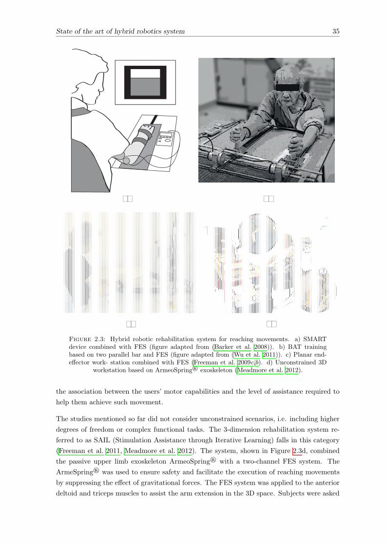
State of the art of hybrid robotics system 35
d)c)
a) b)
Figure 2.3: Hybrid robotic rehabilitation system for reaching movements. a) SMARTdevice combined with FES (figure adapted from (Barker et al. 2008)). b) BAT trainingbased on two parallel bar and FES (figure adapted from (Wu et al. 2011)). c) Planar end-e↵ector work- station combined with FES (Freeman et al. 2009c,b). d) Unconstrained 3D
workstation based on ArmeoSpring® exoskeleton (Meadmore et al. 2012).
the association between the users’ motor capabilities and the level of assistance required to
help them achieve such movement.
The studies mentioned so far did not consider unconstrained scenarios, i.e. including higher
degrees of freedom or complex functional tasks. The 3-dimension rehabilitation system re-
ferred to as SAIL (Stimulation Assistance through Iterative Learning) falls in this category
(Freeman et al. 2011, Meadmore et al. 2012). The system, shown in Figure 2.3d, combined
the passive upper limb exoskeleton ArmeoSpring® with a two-channel FES system. The
ArmeSpring® was used to ensure safety and facilitate the execution of reaching movements
by suppressing the e↵ect of gravitational forces. The FES system was applied to the anterior
deltoid and triceps muscles to assist the arm extension in the 3D space. Subjects were asked
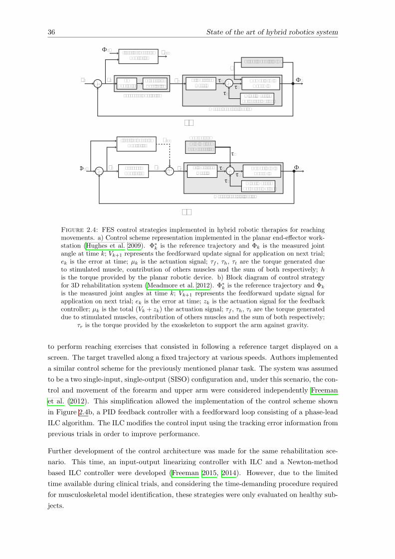
36 State of the art of hybrid robotics system
a)
Iterative LearningController
h
Фk*
uk+
Planar Robotic Arm
Feedback Controller
LinearizingController
PDController
Stimulated muscle
Human Arm Dynamic
++
Other MusclesVoluntary Action
Фk
-vk
vk+1
ek
τt
τf
τh
Musculoskeletal System
b)
Iterative LearningController
Фk* uk+ FeedbackController
Musculoskeletal System
Stimulated muscle
Human Arm Dynamic
++
Other MusclesVoluntary Action
Фk
-
vk+1
ek
τt
τf
τh
++zk
Arm WeightCompensation
Exoskeleton
τr
Figure 2.4: FES control strategies implemented in hybrid robotic therapies for reachingmovements. a) Control scheme representation implemented in the planar end-e↵ector work-station (Hughes et al. 2009). �⇤
k is the reference trajectory and �k is the measured jointangle at time k; Vk+1 represents the feedforward update signal for application on next trial;ek is the error at time; µk is the actuation signal; ⌧f , ⌧h, ⌧t are the torque generated dueto stimulated muscle, contribution of others muscles and the sum of both respectively; his the torque provided by the planar robotic device. b) Block diagram of control strategyfor 3D rehabilitation system (Meadmore et al. 2012). �⇤
k is the reference trajectory and �k
is the measured joint angles at time k; Vk+1 represents the feedforward update signal forapplication on next trial; ek is the error at time; zk is the actuation signal for the feedbackcontroller; µk is the total (Vk + zk) the actuation signal; ⌧f , ⌧h, ⌧t are the torque generateddue to stimulated muscles, contribution of others muscles and the sum of both respectively;
⌧r is the torque provided by the exoskeleton to support the arm against gravity.
to perform reaching exercises that consisted in following a reference target displayed on a
screen. The target travelled along a fixed trajectory at various speeds. Authors implemented
a similar control scheme for the previously mentioned planar task. The system was assumed
to be a two single-input, single-output (SISO) configuration and, under this scenario, the con-
trol and movement of the forearm and upper arm were considered independently Freeman
et al. (2012). This simplification allowed the implementation of the control scheme shown
in Figure 2.4b, a PID feedback controller with a feedforward loop consisting of a phase-lead
ILC algorithm. The ILC modifies the control input using the tracking error information from
previous trials in order to improve performance.
Further development of the control architecture was made for the same rehabilitation sce-
nario. This time, an input-output linearizing controller with ILC and a Newton-method
based ILC controller were developed (Freeman 2015, 2014). However, due to the limited
time available during clinical trials, and considering the time-demanding procedure required
for musculoskeletal model identification, these strategies were only evaluated on healthy sub-
jects.
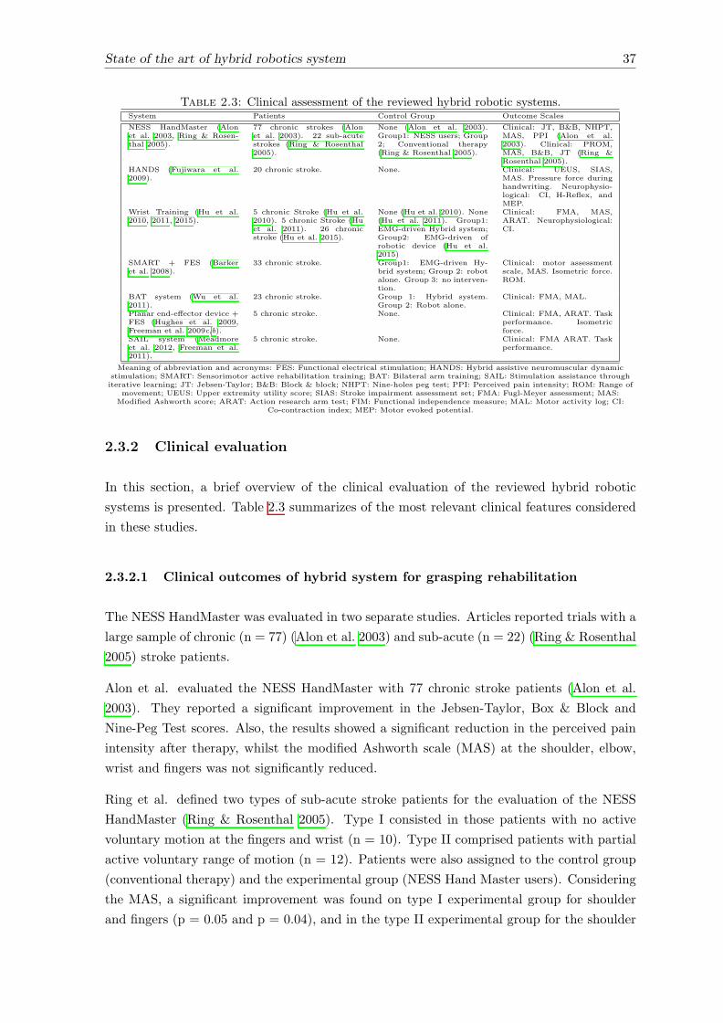
State of the art of hybrid robotics system 37
Table 2.3: Clinical assessment of the reviewed hybrid robotic systems.System Patients Control Group Outcome Scales
NESS HandMaster (Alonet al. 2003, Ring & Rosen-thal 2005).
77 chronic strokes (Alonet al. 2003). 22 sub-acutestrokes (Ring & Rosenthal2005).
None (Alon et al. 2003).Group1: NESS users; Group2; Conventional therapy(Ring & Rosenthal 2005).
Clinical: JT, B&B, NHPT,MAS, PPI (Alon et al.2003). Clinical: PROM,MAS, B&B, JT (Ring &Rosenthal 2005).
HANDS (Fujiwara et al.2009).
20 chronic stroke. None. Clinical: UEUS, SIAS,MAS. Pressure force duringhandwriting. Neurophysio-logical: CI, H-Reflex, andMEP.
Wrist Training (Hu et al.2010, 2011, 2015).
5 chronic Stroke (Hu et al.2010). 5 chronic Stroke (Huet al. 2011). 26 chronicstroke (Hu et al. 2015).
None (Hu et al. 2010). None(Hu et al. 2011). Group1:EMG-driven Hybrid system;Group2: EMG-driven ofrobotic device (Hu et al.2015)
Clinical: FMA, MAS,ARAT. Neurophysiological:CI.
SMART + FES (Barkeret al. 2008).
33 chronic stroke. Group1: EMG-driven Hy-brid system; Group 2: robotalone. Group 3: no interven-tion.
Clinical: motor assessmentscale, MAS. Isometric force.ROM.
BAT system (Wu et al.2011).
23 chronic stroke. Group 1: Hybrid system.Group 2: Robot alone.
Clinical: FMA, MAL.
Planar end-e↵ector device +FES (Hughes et al. 2009,Freeman et al. 2009c,b).
5 chronic stroke. None. Clinical: FMA, ARAT. Taskperformance. Isometricforce.
SAIL system (Meadmoreet al. 2012, Freeman et al.2011).
5 chronic stroke. None. Clinical: FMA ARAT. Taskperformance.
Meaning of abbreviation and acronyms: FES: Functional electrical stimulation; HANDS: Hybrid assistive neuromuscular dynamicstimulation; SMART: Sensorimotor active rehabilitation training; BAT: Bilateral arm training; SAIL: Stimulation assistance throughiterative learning; JT: Jebsen-Taylor; B&B: Block & block; NHPT: Nine-holes peg test; PPI: Perceived pain intensity; ROM: Range of
movement; UEUS: Upper extremity utility score; SIAS: Stroke impairment assessment set; FMA: Fugl-Meyer assessment; MAS:Modified Ashworth score; ARAT: Action research arm test; FIM: Functional independence measure; MAL: Motor activity log; CI:
Co-contraction index; MEP: Motor evoked potential.
2.3.2 Clinical evaluation
In this section, a brief overview of the clinical evaluation of the reviewed hybrid robotic
systems is presented. Table 2.3 summarizes of the most relevant clinical features considered
in these studies.
2.3.2.1 Clinical outcomes of hybrid system for grasping rehabilitation
The NESS HandMaster was evaluated in two separate studies. Articles reported trials with a
large sample of chronic (n = 77) (Alon et al. 2003) and sub-acute (n = 22) (Ring & Rosenthal
2005) stroke patients.
Alon et al. evaluated the NESS HandMaster with 77 chronic stroke patients (Alon et al.
2003). They reported a significant improvement in the Jebsen-Taylor, Box & Block and
Nine-Peg Test scores. Also, the results showed a significant reduction in the perceived pain
intensity after therapy, whilst the modified Ashworth scale (MAS) at the shoulder, elbow,
wrist and fingers was not significantly reduced.
Ring et al. defined two types of sub-acute stroke patients for the evaluation of the NESS
HandMaster (Ring & Rosenthal 2005). Type I consisted in those patients with no active
voluntary motion at the fingers and wrist (n = 10). Type II comprised patients with partial
active voluntary range of motion (n = 12). Patients were also assigned to the control group
(conventional therapy) and the experimental group (NESS Hand Master users). Considering
the MAS, a significant improvement was found on type I experimental group for shoulder
and fingers (p = 0.05 and p = 0.04), and in the type II experimental group for the shoulder

38 State of the art of hybrid robotics system
(p = 0.03), wrist (p = 0.04), fingers (p = 0.01), and thumb (p = 0.04). The evaluated range
of motion of the arm joints showed a greater improvement in proximal upper limb in the type
I group. However, the di↵erence did not reach a level of statistical significance. The type II
group showed greater level of improvements, with significant level for shoulder flexion (p =
0.03), wrist extension (p = 0.02) and wrist flexion (p = 0.04). The functional tests (Box &
Block and the Jebsen-Taylor) presented significant improvement in both types of the treated
group.
In the case of the HANDS system, its e↵ects were evaluated in 20 chronic stroke patients
(Fujiwara et al. 2009). Results revealed significant improvements (p < 0.01) in two of the
four evaluation points (drinking with a glass and turning over a page) of the upper extremity
utility score (UEUS). The finger test as well as the knee-mouth test of the stroke impairment
assessment test (SIAS) also improved significantly (p < 0.001 and p = 0.02). The MAS
were reduced significantly at the elbow wrist and finger extensors (p < 0.001), and the
pen pressure capability increased significantly (p = 0.008). The H-Reflex showed significant
changes of reciprocal inhibition at all three intervals after intervention (0, 20 and 100 ms),
while the co-contraction index measured with EMG and the motor evoked potential elicited
by transcranial magnetic stimulation improved, but not significantly. The clinical outcome
measures and computer-aided ratings were assessed in 15 of the 20 patients three months
after the end of the intervention. The evaluation showed a significant e↵ect in comparison
with the pretreatment assessment in the “drinking with a glass” task and the “turning over
a page” task in the UEUS, the SIAS finger score, the MAS (elbow, wrist, finger) score, the
pen pressure, and the grip strength (p < 0.05). When comparing the post-treatment values
with those at the 3-month follow up, a significant di↵erence was found in the “drinking with
a glass” task and the grip strength (p < 0.05).
With their wrist training system, Hu et al. carried out a single-blinded randomized controlled
trial with 26 chronic stroke patients to evaluate the e↵ectiveness of the wrist training platform
(Hu et al. 2015). Recruited patients were divided into two di↵erent groups, so that fifteen of
them received EMG-driven robot therapy (control group), and the rest (n = 11) EMG-driven
FES robot rehabilitation (experimental group). The clinical evaluations showed significant
improvements in the Fugl-Meyer assessment (FMA) shoulder/elbow, a significant decrease
in the MAS elbow/wrist for both groups after the training and sustained results after 3
months (p < 0.05). The experimental group achieved higher scores (better outcomes) in
the post and 3-month follow-up evaluation. The FMA wrist/hand and the action research
arm test (ARAT) significantly improved only in the experimental group (p < 0.05) and this
increase compared with the baseline remained till 3 months later. The EDC and FCR pair
co-contraction index (CI) was significantly decreased for the experimental group (p < 0.05);
and after session 10, most CI values of experimental group were significantly lower than those
of the control group (p < 0.05). These findings show the potential of the hybrid approach
to conduct both clinical and neurophysiological improvements.
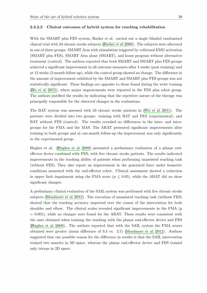
State of the art of hybrid robotics system 39
2.3.2.2 Clinical outcomes of hybrid system for reaching rehabilitation
With the SMART plus FES system, Barker et al. carried out a single blinded randomized
clinical trial with 33 chronic stroke subjects (Barker et al. 2008). The subjects were allocated
in one of three groups: SMART Arm with stimulation triggered by volitional EMG activation
(SMART plus FES), SMART Arm alone (SMART), and home program without alternative
treatment (control). The authors reported that both SMART and SMART plus FES groups
achieved a significant improvement in all outcome measures after 4 weeks (post training) and
at 12 weeks (2-month follow-up), while the control group showed no change. The di↵erence in
the amount of improvement exhibited by the SMART and SMART plus FES groups was not
statistically significant. These findings are opposite to those found during the wrist training
(Hu et al. 2015), where major improvements were reported in the FES plus robot group.
The authors justified the results by indicating that the repetitive nature of the therapy was
principally responsible for the observed changes in the evaluations.
The BAT system was assessed with 23 chronic stroke patients in (Wu et al. 2011). The
patients were divided into two groups: training with BAT and FES (experimental), and
BAT without FES (control). The results revealed no di↵erences in the inter- and intra-
groups for the FMA and the MAS. The ARAT presented significant improvements after
training in both groups and at one-month follow-up the improvement was only significantly
in the experimental group.
Hughes et al. (Hughes et al. 2009) presented a preliminary evaluation of a planar end-
e↵ector device combined with FES, with five chronic stroke patients. The results indicated
improvements in the tracking ability of patients when performing unassisted reaching task
(without FES). They also report an improvement in the generated force under isometric
conditions measured with the end-e↵ector robot. Clinical assessment showed a reduction
in upper limb impairment using the FMA score (p 0.05), while the ARAT did no show
significant changes.
A preliminary clinical evaluation of the SAIL system was performed with five chronic stroke
subjects (Meadmore et al. 2012). The execution of unassisted reaching task (without FES)
showed that the tracking accuracy improved over the course of the intervention for both
shoulder and elbow. The clinical scales revealed significant improvements in the FMA (p
= 0.001), while no changes were found for the ARAT. These results were consistent with
the ones obtained when training the reaching with the planar end-e↵ector device and FES
(Hughes et al. 2009). The authors reported that with the SAIL system the FMA scores
obtained were greater (mean di↵erence of 9.3 vs. 2.5) (Meadmore et al. 2012). Authors
suggested that one possible reason for the di↵erence in results is that the SAIL intervention
trained two muscles in 3D space, whereas the planar end-e↵ector device and FES trained
only triceps in 2D space.
40 State of the art of hybrid robotics system
2.4 Discussion
The hybrid robotic systems reviewed in this chapter represent the state of the art of the
combined use of robotic and FES devices for upper limb rehabilitation after stroke. In this
chapter, the key technological features of this recent emerging rehabilitation method (hybrid
concept) and its clinical potentials were presented. In what remains of this chapter, the
main challenges of the hybrid concept are identified and presented. Three challenges are
considered: technological features, clinical deployment and usability of the system, which
informed the studies presented in the following chapters of this thesis.
2.4.1 Improving technical aspects: hybrid approach challenges
Hybrid robotic systems combine two technologies (FES and robotic devices) into one plat-
form, complementing each other to build on the performance of each individual approach.
For this purpose, it is necessary to establish what is the role of each technology within each
platform and how it will improve the overall rehabilitation results.
The most used hybrid robotic systems combined end-e↵ector robotic devices with FES.
End-e↵ector robots have been mostly used to support the arm’s weight and to delimit the
workspace while the FES was used to drive the movement of the arm during the task exe-
cution. The main advantage of this approach is the simplicity of the setup (usually 1 DOF)
that facilitated the implementation of the control algorithms. However, two main drawbacks
arise when considering these devices. First, the amount of assistance during movement is
limited since the mechanical forces are not applied directly to specific joints and the move-
ment is mainly driven by FES. Second, the range of motion of this type of robotic devices
is limited. Therefore, only a limited set of rehabilitation exercises can be carried out, with
a negative impact in task generalization, which is an important factor for promoting motor
recovery after stroke (Krakauer 2005).
Among the reviewed studies, only the SAIL system presented by Meadmore et al. (Meadmore
et al. 2012) considered the use of a passive exoskeleton in combination with FES for assisting
movement in a 3D space. Passive exoskeletons have the advantage over end-e↵ector robotic
devices of fully supporting the whole arm against gravity at the joint level. Also, they can
provide a larger range of motion in the 3D space. This allows more flexibility to apply FES
and focuses it only on driving the arm movements, this way reducing the stimulation intensity
and the muscle fatigue onset. However, this setup is limited by mechanical constraints and
requires a robust and reliable FES system to drive the movements successfully.
A full hybrid robotic system (from the actuation perspective) is the one that provides support
by combining an exoskeleton with active actuators and FES. Several systems following this
approach have been reported for lower limb and gait rehabilitation (del Ama et al. 2012).
Despite the broad di↵usion of powered upper limb exoskeletons in recent years (Maciejasz
et al. 2014), the combination of these devices with FES has not been reported for the upper
State of the art of hybrid robotics system 41
limb rehabilitation. This combination could improve the performance of current hybrid
robotic systems significantly since the arm movement does not only depend on the FES
assistance. For example, the inability to drive limbs with FES and the reported low rate
of the system performance in (Westerveld et al. 2014), could be significantly improved if
additional mechanical assistance is provided at the joint level. Furthermore, this combination
could allow the development of novel interventions, such as targeting specific muscles groups
with FES, while supporting the rest of the arm movement with the exoskeleton. However, the
main challenge for full hybrid robotic systems is the development of optimal shared control
strategies between the FES, the exoskeleton while taking into account the patients’ remnant
motor capacity to promote and potentiate the e↵ects of therapy.
A robust FES controller plays a crucial role in the successful deployment of hybrid robotic
systems. The performance of FES systems depends on the implemented control strategy,
the number of stimulation channels and the correct electrode placement (Lynch & Popovic
2008, Popovic 2014). The control strategy should be able to compensate the non-linear and
time-varying response of the musculoskeletal system due to the non-physiological motor unit
recruitment (Ma�uletti 2010). In addition, the controller parameter must be individualized
to each user in order to address the inter-subject variability to FES response.
Therefore, simple FES control strategies (e.g. open-loop and linear feedback controllers,
i.e. PID) are often inadequate for controlling the execution of motor tasks, as they can-
not properly manage the high muscle response variability (Lynch & Popovic 2008, Freeman
et al. 2012). Also, when using a model-based feedback controller, the modeling of the mus-
culoskeletal system is a major concern. Indeed, this type of controllers requires a detailed
mathematical description of the musculoskeletal system to work properly. The fitting of
the model and the personalization of the model’s parameters result in a complex and time-
consuming task requiring additional sessions (Freeman et al. 2009a, Zhang et al. 2007).
Furthermore, the non-modeled dynamics and the simplification assumed to build the model
reduce the robustness of the controller performance. For instance, Westerveld et. al used a
model predictive control algorithm based on a second order model with a success rate of less
than 20% for opening the hand (Westerveld et al. 2014), suggesting that this type of control
strategy results inappropriate to drive complex functional task such as the grasping.
Alternatively, the learning feedforward loop reported in (Hughes et al. 2009, Meadmore et al.
2012, 2014, Kutlu et al. 2015) represents an interesting approach, since it exploits the repet-
itive nature of robot-aided rehabilitation to learn from the errors of previous attempts. This
learning capability provides a way to compensate for the physiological changes of patients
(e.g. muscle response variation due to muscle fatigue or spasticity). However, it still re-
quired to model the musculoskeletal response to FES for a proper operation. Although this
strategy is more convenient for driving movement assisted with FES, the controller perfor-
mance is influenced by the non-modeled dynamics, the linearization of the model and the
restrictions imposed when deducing the individualized model. Furthermore, the complexity
of the inferred model increases considerably when considering movements in 3D space, such
as reaching, and movements involving multiple degrees of freedom, such as grasping.

42 State of the art of hybrid robotics system
On the other hand, control strategies based on neurophysiological signals, like EMG, are in
many cases restricted. Certainly, the disorders to the central nervous system caused after
Stroke (e.g. spasticity) could a↵ect significantly the quality of the EMG signals, which limits
the applicability of the EMG-based controllers. Moreover, when considering this approach,
the controller robustness is influenced by the implementation of complex blanking algorithms
to separate the useful physiological signal from artifact generated by the electrical stimuli.
A good control of grasping motion is necessary for promoting the patients’ motivation and
engagement during therapy, otherwise the inability to perform the tasks could lead to frus-
tration. For grasping tasks, the number of stimulation channels influences the precise control
of the hand and fingers movements considerably. Due to the high density of muscles at the
forearm, the use of few stimulation channels typically results in a reduced motor functional re-
sponse and this problem is more noticeable when surface electrodes are used (Popovic 2014).
It has been demonstrated that the use of multi-pad electrodes (e.g. a matrix of electrodes)
improved the precision of controlled movement significantly (Malesevic et al. 2012).
2.4.2 Rehabilitation Outcomes
Published systematic reviews reported that after robot-based therapy, stroke patients pre-
sented a reduction in motor impairment but did not improve in functional abilities (Kwakkel
et al. 2008, Prange et al. 2006). It has been suggested that upper limb rehabilitation is loca-
tion specific (Johnson 2006, Meadmore et al. 2014). Thus, reaching rehabilitation will only
improve motor impairment in the proximal joints (shoulder and elbow) while grasping train-
ing will have impact only in the wrist and fingers. In line with this evidence, hybrid robotic
systems for reaching (Hughes et al. 2009, Meadmore et al. 2012) showed improvements in
the motor impairments (measured by FMA) but not in functional improvements (measured
by ARAT scale). This evidence suggests that the whole upper limb must be considered in
rehabilitation to achieve a better reduction in motor impairment and functional changes.
Under a clinical perspective, performing a direct comparison between studies is challenging
due to the lack of standard evaluation metrics or procedures across studies. This issue was
pointed out by Huang and Krakauer in (Huang & Krakauer 2009) for post-stroke rehabilita-
tion therapies. Also, the relatively small number of studies and their methodology, as well
as the small number of subject that were tested, makes it di�cult to reach a generalization.
Only four studies considered the inclusion of a control group for the evaluation of the hybrid
robotic systems (Ring & Rosenthal 2005, Hu et al. 2015, Wu et al. 2011, Barker et al. 2008),
and these control groups were di↵erent in all cases. The inclusion of a control group for
demonstrating the e↵ectiveness of a hybrid rehabilitation system is important. However, it
is di�cult to be implemented in practice due to the high number of patients that will be re-
quired during the experimentation. In fact, since two therapies are being implemented jointly
(robot and FES), at least three groups of patients would be necessary to check whether the
hybrid robotic system results in significant improvements with respect to robotic and FES
separately.
State of the art of hybrid robotics system 43
However, the significant improvements achieved in most of the reported studies support the
hypothesis of the relative benefits of using combined robotic devices with FES for upper limb
rehabilitation after stroke.
2.4.3 Improving the human-machine interaction: associating peripheral
assistance with user’s motor intent
Associating the peripheral assistance onset with the user’s movement intention in a causal
and synchronized manner has been shown to be an important factor for eliciting neural
plasticity and, therefore, to facilitate neuromuscular recovery (Ethier et al. 2015, Mrachacz-
Kersting et al. 2012). Recent studies have demonstrated that the application of peripheral
assistance precisely synchronized with the user’s motor intent induce long-term potentiation
in the corticospinal pathway in healthy volunteers (Mrachacz-Kersting et al. 2012, Xu et al.
2014) and stroke patients (Mrachacz-Kersting et al. 2015).
In this regard, only a few of the reviewed hybrid systems have addressed the associative
paradigm (applying peripheral assistance in coherence with user’s own intention to move) by
using voluntary EMG activation to trigger the system assistance (Fujiwara et al. 2009, Hu
et al. 2015, Barker et al. 2008). However, the optimal stimulus timing may be degraded by
the intrinsic delay between cortical and peripheral activity (Ethier et al. 2015). Also, the
quality of the EMG could be a↵ected as a result of the level of impairment, which has a
direct impact on the timing and reliability of the intention detection systems.
EEG-based BCI could be used as an alternative to the EMG intention detection for hybrid
robotic systems. The advantage of these interfaces is that the synchronization between motor
intent and motor execution can be realized without much delay. However, these interfaces
have shown low reliability, mainly due noise and artifacts in the acquired signals (Ethier &
Miller 2015). Nevertheless, this is an interesting avenue that could be considered to improve
the rehabilitation outcomes of hybrid robotic systems.
2.5 Conclusions of the chapter
This chapter presented an overview of studies in which the combined use of robotic devices
with FES was reported for rehabilitation of upper-limb motor function in stroke survivors.
These systems represent the state of the art of hybrid robotic system for rehabilitation upper-
limb motor functions. Hybrid robotic systems are an emerging approach aimed at combining
two di↵erent but complementary assistive methods. The main goal under this hybrid per-
spective is to enhance current rehabilitation capabilities of each individual technique and
provide a more robust rehabilitation intervention.
Still many challenges remain to be addressed. From the technological perspective, the in-
clusion of exoskeleton devices would enable a more natural movement in an unconstrained
44 State of the art of hybrid robotics system
environment, with the capability of implementing more complex and sophisticated rehabil-
itation paradigms. In this context, the implementation of adaptive FES-based controllers
addressing the non-linear and time-varying musculoskeletal response to FES is necessary. It
is worth mentioning that long calibration time or additional sessions to calibrate the con-
troller parameters must be avoided for usability. In spite of evidence that optimal association
between the hybrid assistance and the user’s own intention to move would promote plasticity,
none of the systems hereby reviewed succeeded in demonstrating that plasticity was elicited
as a result of the intervention. From the clinical perspective, the development of uncon-
strained systems capable to train complex functional movements is preferred. It has been
shown that training functional movements results in improvements of motor impairment and
in functional abilities. This is the starting point and requirements for the developments to
be introduced in next chapters.
Chapter 3
Implementation of a FES-based
adaptive assistance in a hybrid
robotic system for rehabilitation of
reaching movement after stroke1
Abstract
This chapter presents the integration of a hybrid robotic system for rehabilitation of reaching
movement after stroke. Also, a concise evaluation, from the technical and clinical perspec-
tives, of the usability of the hybrid robotic system in the clinical setting is conducted. The
hybrid system comprises four components. The hybrid assistance is given by a passive exoske-
leton to support the arm weight against gravity and a functional electrical stimulation device
to assist the execution of the reaching task. A feedback error learning (FEL) controller has
been implemented to adjust the intensity of the electrical stimuli delivered to target muscles
according to the performance of the users. This control strategy is based on a proportional-
integral-derivative feedback controller and an artificial neural network as the feedforward
controller. Two experiments have been carried out. First, the technical viability and the
performance of the FEL controller were evaluated in healthy subjects (n = 12). Second, a
small cohort of patients with a brain injury (n = 4) participated in two experimental sessions
to evaluate the system performance. The overall satisfaction and emotional response of the
users after using the system were assessed. In the experiment with healthy subjects, a signif-
icant reduction of the tracking error was found during the execution of reaching movements.
In the experiments with patients, a decreasing trend of the error trajectory was found together
1This chapter is partly based on:- F. Resquın, J. Gonzalez-Vargas, J. Ibanez, F. Brunetti, J.L. Pons. Feedback error learning controller forfunctional electrical stimulation assistance in a hybrid robotic system for reaching rehabilitation. Eur J TranslMyol. 2016;26(3).- F. Resquın, J. Gonzalez-Vargas, J. Ibanez, F. Brunetti, I. Dimbwadyo, L. Carrasco, S. Alves, C. Gonzalez-Alted, A. Gomez-Blanco and J.L. Pons. Adaptive hybrid robotic system for rehabilitation of reaching move-ment after a brain injury: a usability study. J Neuroeng Rehabil. 2017 Dec 12;14(1):104.
45

46 Implementation of a FES-based adaptive assistance
with an increasing trend in the task performance as the movement was repeated. Brain injury
patients enthusiastically accepted the system as a rehabilitation tool. The results presented
in this chapter demonstrate the technical feasibility of using the hybrid robotic system for
rehabilitation of reaching. Patients reported a great satisfaction and acceptance of the hybrid
robotic system.
Implementation of a FES-based adaptive assistance 47
3.1 Introduction
The previous chapter highlighted the potential rehabilitative benefits of using hybrid robotic
systems for rehabilitation of upper extremity motor functions. Only a few studies in the
literature included stroke patients as testing subjects. In order to maximize the treatment’s
outcomes and achieve functional improvement it is necessary to train movements which
involve multiple degrees of freedom (> 1 DOF) as well as functional movements Freeman
et al. (2012), Meadmore et al. (2014). In this scenario, the implementation of a robust and
reliable FES controller is required. As discussed in the previous chapter, the appropriate
design and implementation of FES controllers play a key role to achieve stable and robust
motion control in hybrid robotic systems. The control strategy must be able to drive all
the necessary joints to realize the desired movement, and to compensate any disturbance,
e.g. muscle fatigue onset as well as the strong nonlinear and time-varying response of the
musculoskeletal system to FES (Ma�uletti 2010, Lynch & Popovic 2008).
Meadmore et al. presented an interesting hybrid robotic system for rehabilitation of reaching
movement in 3D space (Meadmore et al. 2012). A model-based iterative learning controller
(ILC) was implemented to adjust the FES intensity based on the tracking error of the pre-
viously executed movement (Freeman et al. 2011, Meadmore et al. 2012). This iterative
adjustment allows compensating for disturbances caused by FES. Although this approach
addresses some of the issues related to motion control with FES, to work properly, it re-
quires a detailed mathematical description of the musculoskeletal system. In this context,
non-modeled dynamics and the linearization of the model can reduce the robustness and
performance. Furthermore, the identification of the model’s parameters is complex and time
consuming, which limits its usability in clinical settings (Lynch & Popovic 2008, Zhang et al.
2007).
The Feedback Error Learning (FEL) scheme proposed by Kawato (Kawato 1990) can be
considered as an alternative to ILC. This scheme was developed to describe how the central
nervous system builds an internal model of the body to improve the motor control. Under this
scheme, the motor control command of a feedback controller is used to train a feedforward
controller to learn implicitly the inverse dynamics of the plant (i.e. the arm) on-line. In
addition, this on-line learning procedure also allows the controller to adapt and compensate
for disturbances. In contrast with the ILC, the main advantage of this strategy is that the
controller does not require an explicit model of the plant to work correctly and that it can
directly learn its non-linear characteristics. Therefore, using the FEL control strategy to
control a hybrid robotic system can simplify the setup of the system considerably, which
makes easier to deploy it in clinical settings as well as to personalize its response according
to each patient’s musculoskeletal characteristics and remnant movement capabilities. The
FEL has been used previously to control the wrist (Kurosawa et al. 2005) and the lower
limb (Koike et al. 2011) motion with FES in healthy subjects. But, this control strategy has
neither been implemented in a hybrid robotic system nor tested with stroke patients.
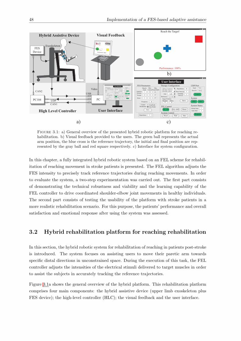
48 Implementation of a FES-based adaptive assistance
!"#$%#&'(
)(*+#,!-) ./0
!-/102&+"23!-/145 !-/14!! 6#*#+
)(*+#,102&+"237286#*#+ )+9"+
!-)1:9"9,#+#"*
02&+"2372;%39+<2&
=,>3<+%;#:%3*#?<;+@
)98#
=&+#"<2"A#3+2<;B"<'#>*
?"<*+
7%*'3#!<#3;
79CD0%""#&+
79CD:%3*#?<;+@
E
EF
G
GH
EI
EF
FJH
FJH
FJH
!"#$%#&'( FH
6#>#+<+<2&* J
)(*+#,1)+9+#*K32L931)+9+#B9*M1)+9+#
6#$%#*+
E
N
B@#"9>(102&O<P%"9+<2&-3L2Q1-C+#&*<2&)@2%3;#"1!3#C<2&)@2%3;#"R-3L2Q
=", /#O+
6#O#"#&'# N1*#'2&;*
6#>#+<+<2&*
6#*+1B<,#1S*TK"9*><&PB<,#1S*T
)98#
G
J
GH
)@2%3;#" -3L2QB9"P#+1:2*<+<2&
EH NH
!"#$%&'(#$)*+#UA:1:2"+)#&;
6#'#<8#
02&#'+
VJHH
EJHHH
LW
'W
6#9'@1+@#1B9"P#+X
:#"O2",9&'#Y1EHHZ
Reach the target!
TargetInit Position
Performance: 100%
!"#$%&'(#$)*+#,-./%0#1#2%34'($422#$
0=5E
U)[
\<;#2!"#$%#&'(
)(*+#,!-) ./0
!-/102&+"23!-/145 !-/14!! 6#*#+
)(*+#,102&+"237286#*#+ )+9"+
!-)1:9"9,#+#"*
02&+"2372;%39+<2&
=,>3<+%;#:%3*#?<;+@
)98#
=&+#"<2"A#3+2<;B"<'#>*
?"<*+
7%*'3#!<#3;
79CD0%""#&+
79CD:%3*#?<;+@
E
EF
G
GH
EI
EF
FJH
FJH
FJH
!"#$%#&'( FH
6#>#+<+<2&* J
)(*+#,1)+9+#*K32L931)+9+#B9*M1)+9+#
6#$%#*+
E
N
B@#"9>(102&O<P%"9+<2&-3L2Q1-C+#&*<2&)@2%3;#"1!3#C<2&)@2%3;#"R-3L2Q
=", /#O+
6#O#"#&'# N1*#'2&;*
6#>#+<+<2&*
6#*+1B<,#1S*TK"9*><&PB<,#1S*T
)98#
G
J
GH
)@2%3;#" -3L2QB9"P#+1:2*<+<2&
EH NH
!"#$%&'(#$)*+#UA:1:2"+)#&;
6#'#<8#
02&#'+
VJHH
EJHHH
,56$-7%8""-"(-1#%9#1-+# :-";*2%<##76*+=
!-)A#8<'#
:0EHF :0
0=5G
UA:
9W
-C2*M#3#+2&
Figure 3.1: a) General overview of the presented hybrid robotic platform for reaching re-habilitation. b) Visual feedback provided to the users. The green ball represents the actualarm position, the blue cross is the reference trajectory, the initial and final position are rep-resented by the gray ball and red square respectively. c) Interface for system configuration.
In this chapter, a fully integrated hybrid robotic system based on an FEL scheme for rehabil-
itation of reaching movement in stroke patients is presented. The FEL algorithm adjusts the
FES intensity to precisely track reference trajectories during reaching movements. In order
to evaluate the system, a two-step experimentation was carried out. The first part consists
of demonstrating the technical robustness and viability and the learning capability of the
FEL controller to drive coordinated shoulder-elbow joint movements in healthy individuals.
The second part consists of testing the usability of the platform with stroke patients in a
more realistic rehabilitation scenario. For this purpose, the patients’ performance and overall
satisfaction and emotional response after using the system was assessed.
3.2 Hybrid rehabilitation platform for reaching rehabilitation
In this section, the hybrid robotic system for rehabilitation of reaching in patients post-stroke
is introduced. The system focuses on assisting users to move their paretic arm towards
specific distal directions in unconstrained space. During the execution of this task, the FEL
controller adjusts the intensities of the electrical stimuli delivered to target muscles in order
to assist the subjects in accurately tracking the reference trajectories.
Figure 3.1a shows the general overview of the hybrid platform. This rehabilitation platform
comprises four main components: the hybrid assistive device (upper limb exoskeleton plus
FES device); the high-level controller (HLC); the visual feedback and the user interface.
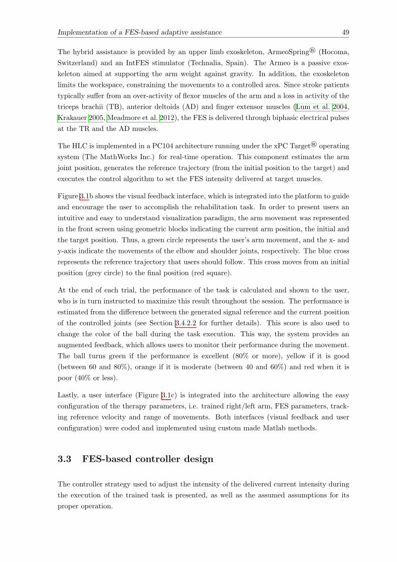
Implementation of a FES-based adaptive assistance 49
The hybrid assistance is provided by an upper limb exoskeleton, ArmeoSpring® (Hocoma,
Switzerland) and an IntFES stimulator (Technalia, Spain). The Armeo is a passive exos-
keleton aimed at supporting the arm weight against gravity. In addition, the exoskeleton
limits the workspace, constraining the movements to a controlled area. Since stroke patients
typically su↵er from an over-activity of flexor muscles of the arm and a loss in activity of the
triceps brachii (TB), anterior deltoids (AD) and finger extensor muscles (Lum et al. 2004,
Krakauer 2005, Meadmore et al. 2012), the FES is delivered through biphasic electrical pulses
at the TR and the AD muscles.
The HLC is implemented in a PC104 architecture running under the xPC Target® operating
system (The MathWorks Inc.) for real-time operation. This component estimates the arm
joint position, generates the reference trajectory (from the initial position to the target) and
executes the control algorithm to set the FES intensity delivered at target muscles.
Figure 3.1b shows the visual feedback interface, which is integrated into the platform to guide
and encourage the user to accomplish the rehabilitation task. In order to present users an
intuitive and easy to understand visualization paradigm, the arm movement was represented
in the front screen using geometric blocks indicating the current arm position, the initial and
the target position. Thus, a green circle represents the user’s arm movement, and the x- and
y-axis indicate the movements of the elbow and shoulder joints, respectively. The blue cross
represents the reference trajectory that users should follow. This cross moves from an initial
position (grey circle) to the final position (red square).
At the end of each trial, the performance of the task is calculated and shown to the user,
who is in turn instructed to maximize this result throughout the session. The performance is
estimated from the di↵erence between the generated signal reference and the current position
of the controlled joints (see Section 3.4.2.2 for further details). This score is also used to
change the color of the ball during the task execution. This way, the system provides an
augmented feedback, which allows users to monitor their performance during the movement.
The ball turns green if the performance is excellent (80% or more), yellow if it is good
(between 60 and 80%), orange if it is moderate (between 40 and 60%) and red when it is
poor (40% or less).
Lastly, a user interface (Figure 3.1c) is integrated into the architecture allowing the easy
configuration of the therapy parameters, i.e. trained right/left arm, FES parameters, track-
ing reference velocity and range of movements. Both interfaces (visual feedback and user
configuration) were coded and implemented using custom made Matlab methods.
3.3 FES-based controller design
The controller strategy used to adjust the intensity of the delivered current intensity during
the execution of the trained task is presented, as well as the assumed assumptions for its
proper operation.
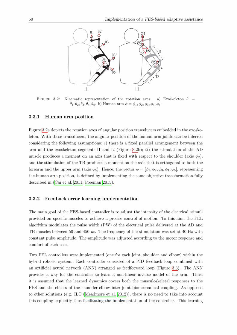
50 Implementation of a FES-based adaptive assistance
θ1 Ø1
a) b)
θ2
θ3
θ3’θ4
θ5
Ø2
Ø4Ø3
Ø5
l1
l2
Figure 3.2: Kinematic representation of the rotation axes. a) Exoskeleton ✓ =✓1, ✓2, ✓3, ✓4, ✓5. b) Human arm � = �1,�2,�3,�4,�5.
3.3.1 Human arm position
Figure 3.2a depicts the rotation axes of angular position transducers embedded in the exoske-
leton. With these transducers, the angular position of the human arm joints can be inferred
considering the following assumptions: i) there is a fixed parallel arrangement between the
arm and the exoskeleton segments l1 and l2 (Figure 3.2b); ii) the stimulation of the AD
muscle produces a moment on an axis that is fixed with respect to the shoulder (axis �2),
and the stimulation of the TB produces a moment on the axis that is orthogonal to both the
forearm and the upper arm (axis �5). Hence, the vector � = [�1,�2,�3,�4,�5], representing
the human arm position, is defined by implementing the same objective transformation fully
described in (Cai et al. 2011, Freeman 2015).
3.3.2 Feedback error learning implementation
The main goal of the FES-based controller is to adjust the intensity of the electrical stimuli
provided on specific muscles to achieve a precise control of motion. To this aim, the FEL
algorithm modulates the pulse width (PW) of the electrical pulse delivered at the AD and
TB muscles between 50 and 450 µs. The frequency of the stimulation was set at 40 Hz with
constant pulse amplitude. The amplitude was adjusted according to the motor response and
comfort of each user.
Two FEL controllers were implemented (one for each joint, shoulder and elbow) within the
hybrid robotic system. Each controller consisted of a PID feedback loop combined with
an artificial neural network (ANN) arranged as feedforward loop (Figure 3.3). The ANN
provides a way for the controller to learn a non-linear inverse model of the arm. Thus,
it is assumed that the learned dynamics covers both the musculoskeletal responses to the
FES and the e↵ects of the shoulder-elbow inter-joint biomechanical coupling. As opposed
to other solutions (e.g. ILC (Meadmore et al. 2012)), there is no need to take into account
this coupling explicitly thus facilitating the implementation of the controller. This learning
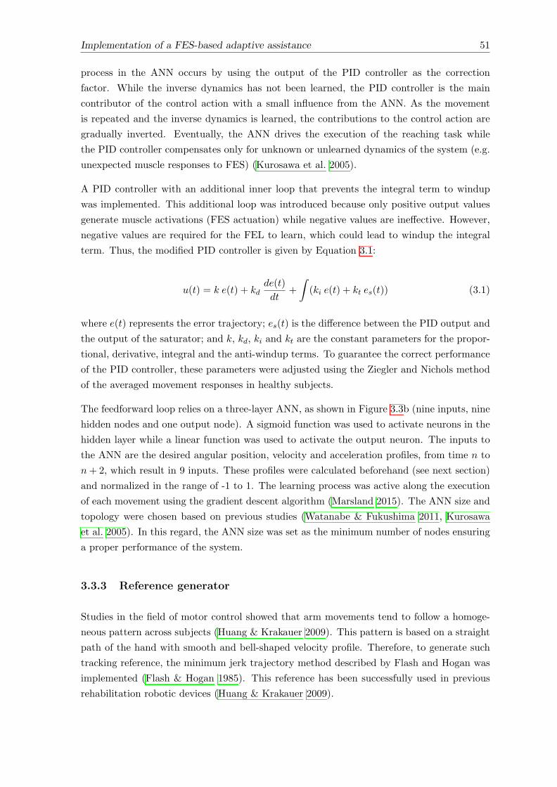
Implementation of a FES-based adaptive assistance 51
process in the ANN occurs by using the output of the PID controller as the correction
factor. While the inverse dynamics has not been learned, the PID controller is the main
contributor of the control action with a small influence from the ANN. As the movement
is repeated and the inverse dynamics is learned, the contributions to the control action are
gradually inverted. Eventually, the ANN drives the execution of the reaching task while
the PID controller compensates only for unknown or unlearned dynamics of the system (e.g.
unexpected muscle responses to FES) (Kurosawa et al. 2005).
A PID controller with an additional inner loop that prevents the integral term to windup
was implemented. This additional loop was introduced because only positive output values
generate muscle activations (FES actuation) while negative values are ine↵ective. However,
negative values are required for the FEL to learn, which could lead to windup the integral
term. Thus, the modified PID controller is given by Equation 3.1:
u(t) = k e(t) + kdde(t)
dt+
Z(ki e(t) + kt es(t)) (3.1)
where e(t) represents the error trajectory; es(t) is the di↵erence between the PID output and
the output of the saturator; and k, kd, ki and kt are the constant parameters for the propor-
tional, derivative, integral and the anti-windup terms. To guarantee the correct performance
of the PID controller, these parameters were adjusted using the Ziegler and Nichols method
of the averaged movement responses in healthy subjects.
The feedforward loop relies on a three-layer ANN, as shown in Figure 3.3b (nine inputs, nine
hidden nodes and one output node). A sigmoid function was used to activate neurons in the
hidden layer while a linear function was used to activate the output neuron. The inputs to
the ANN are the desired angular position, velocity and acceleration profiles, from time n to
n+ 2, which result in 9 inputs. These profiles were calculated beforehand (see next section)
and normalized in the range of -1 to 1. The learning process was active along the execution
of each movement using the gradient descent algorithm (Marsland 2015). The ANN size and
topology were chosen based on previous studies (Watanabe & Fukushima 2011, Kurosawa
et al. 2005). In this regard, the ANN size was set as the minimum number of nodes ensuring
a proper performance of the system.
3.3.3 Reference generator
Studies in the field of motor control showed that arm movements tend to follow a homoge-
neous pattern across subjects (Huang & Krakauer 2009). This pattern is based on a straight
path of the hand with smooth and bell-shaped velocity profile. Therefore, to generate such
tracking reference, the minimum jerk trajectory method described by Flash and Hogan was
implemented (Flash & Hogan 1985). This reference has been successfully used in previous
rehabilitation robotic devices (Huang & Krakauer 2009).
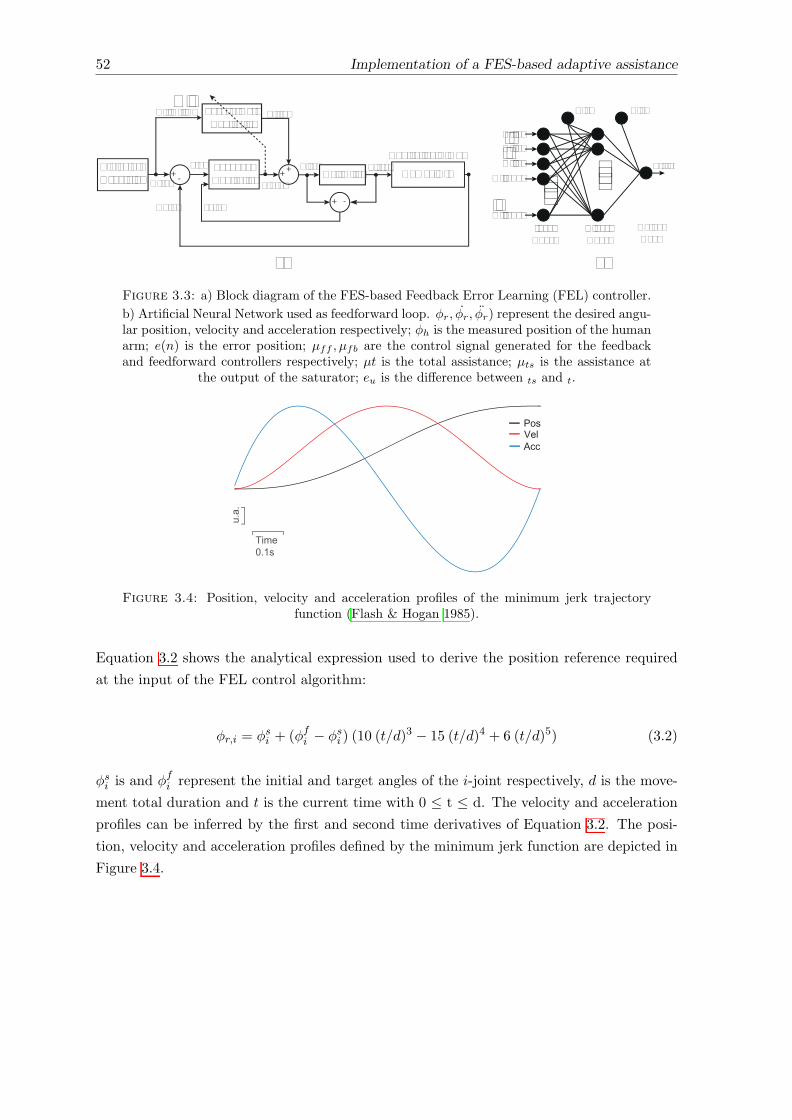
52 Implementation of a FES-based adaptive assistance
TrajectoryGenerator
FeedforwardController
FeedbackController
Controlled Limbs+ - + +
Ør(n)
Øh(n)
e(n)
µff(n)
µfb(n)
µt(n)
Ør, Ør, Ør. ..
Delimiterµts(n)
+ -eu(n)
Human Arm
Ør (n). ..
...µff(n)
Ør (n)Ør (n)
Ør (n+1)
Ør (n+2)
...InputNodes
HiddenNodes
OutputNode
Bias Bias
a) b)
..
Figure 3.3: a) Block diagram of the FES-based Feedback Error Learning (FEL) controller.
b) Artificial Neural Network used as feedforward loop. �r, �r, �r) represent the desired angu-lar position, velocity and acceleration respectively; �h is the measured position of the humanarm; e(n) is the error position; µff , µfb are the control signal generated for the feedbackand feedforward controllers respectively; µt is the total assistance; µts is the assistance at
the output of the saturator; eu is the di↵erence between ts and t.
Time0.1s
PosVelAcc
u.a.
Figure 3.4: Position, velocity and acceleration profiles of the minimum jerk trajectoryfunction (Flash & Hogan 1985).
Equation 3.2 shows the analytical expression used to derive the position reference required
at the input of the FEL control algorithm:
�r,i = �si + (�f
i � �si ) (10 (t/d)
3 � 15 (t/d)4 + 6 (t/d)5) (3.2)
�si is and �f
i represent the initial and target angles of the i-joint respectively, d is the move-
ment total duration and t is the current time with 0 t d. The velocity and acceleration
profiles can be inferred by the first and second time derivatives of Equation 3.2. The posi-
tion, velocity and acceleration profiles defined by the minimum jerk function are depicted in
Figure 3.4.
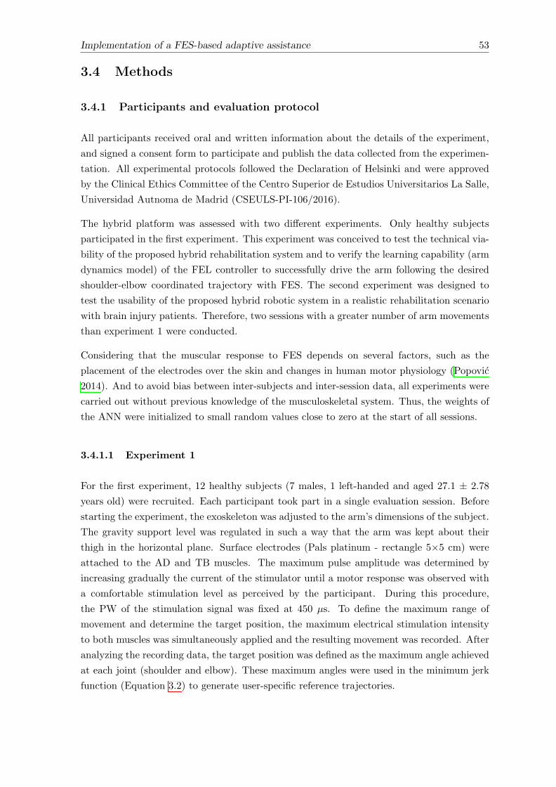
Implementation of a FES-based adaptive assistance 53
3.4 Methods
3.4.1 Participants and evaluation protocol
All participants received oral and written information about the details of the experiment,
and signed a consent form to participate and publish the data collected from the experimen-
tation. All experimental protocols followed the Declaration of Helsinki and were approved
by the Clinical Ethics Committee of the Centro Superior de Estudios Universitarios La Salle,
Universidad Autnoma de Madrid (CSEULS-PI-106/2016).
The hybrid platform was assessed with two di↵erent experiments. Only healthy subjects
participated in the first experiment. This experiment was conceived to test the technical via-
bility of the proposed hybrid rehabilitation system and to verify the learning capability (arm
dynamics model) of the FEL controller to successfully drive the arm following the desired
shoulder-elbow coordinated trajectory with FES. The second experiment was designed to
test the usability of the proposed hybrid robotic system in a realistic rehabilitation scenario
with brain injury patients. Therefore, two sessions with a greater number of arm movements
than experiment 1 were conducted.
Considering that the muscular response to FES depends on several factors, such as the
placement of the electrodes over the skin and changes in human motor physiology (Popovic
2014). And to avoid bias between inter-subjects and inter-session data, all experiments were
carried out without previous knowledge of the musculoskeletal system. Thus, the weights of
the ANN were initialized to small random values close to zero at the start of all sessions.
3.4.1.1 Experiment 1
For the first experiment, 12 healthy subjects (7 males, 1 left-handed and aged 27.1 ± 2.78
years old) were recruited. Each participant took part in a single evaluation session. Before
starting the experiment, the exoskeleton was adjusted to the arm’s dimensions of the subject.
The gravity support level was regulated in such a way that the arm was kept about their
thigh in the horizontal plane. Surface electrodes (Pals platinum - rectangle 5⇥5 cm) were
attached to the AD and TB muscles. The maximum pulse amplitude was determined by
increasing gradually the current of the stimulator until a motor response was observed with
a comfortable stimulation level as perceived by the participant. During this procedure,
the PW of the stimulation signal was fixed at 450 µs. To define the maximum range of
movement and determine the target position, the maximum electrical stimulation intensity
to both muscles was simultaneously applied and the resulting movement was recorded. After
analyzing the recording data, the target position was defined as the maximum angle achieved
at each joint (shoulder and elbow). These maximum angles were used in the minimum jerk
function (Equation 3.2) to generate user-specific reference trajectories.
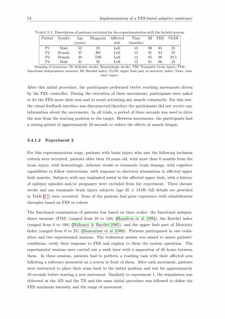
54 Implementation of a FES-based adaptive assistance
Table 3.1: Description of patients recruited for the experimentation with the hybrid system.
Patient Gender Age Diagnosis A↵ected Time BI FIM ULMI(years) side (months)
P1 Male 52 IS Left 13 98 85 25P2 Female 37 HS Left 15 91 84 25P3 Female 30 TBI Left 12 95 90 23.5P4 Male 21 IS Left 12 61 66 25
Meaning of acronyms: IS: Ischemic stroke; Hemorrhagic stroke; TBI: Traumatic brain injury; FIM:functional independence measure; BI: Barthel index; ULMI: upper limb part of motricity index; Time: time
since injury.
After this initial procedure, the participants performed twelve reaching movements driven
by the FEL controller. During the execution of these movements, participants were asked
to let the FES move their arm and to avoid activating any muscle voluntarily. For this test,
the visual feedback interface was disconnected,therefore the participants did not receive any
information about the movements. In all trials, a period of three seconds was used to drive
the arm from the starting position to the target. Between movements, the participants had
a resting period of approximately 10 seconds to reduce the e↵ects of muscle fatigue.
3.4.1.2 Experiment 2
For this experimentation stage, patients with brain injury who met the following inclusion
criteria were recruited: patients older than 18 years old, with more than 6 months from the
brain injury, with hemorrhagic, ischemic stroke or traumatic brain damage, with cognitive
capabilities to follow instructions, with response to electrical stimulation in a↵ected upper
limb muscles. Subjects with any implanted metal in the a↵ected upper limb, with a history
of epilepsy episodes and/or pregnancy were excluded from the experiment. Three chronic
stroke and one traumatic brain injury subjects (age 35 ± 13.09, full details are provided
in Table 3.1) were recruited. None of the patients had prior experience with rehabilitation
therapies based on FES or robots.
The functional examination of patients was based on three scales: the functional indepen-
dence measure (FIM) (ranged from 18 to 126) (Hamilton et al. 1994), the Barthel index
(ranged from 0 to 100) (Mahoney & Barthel 1965), and the upper limb part of Motricity
Index (ranged from 0 to 25) (Demeurisse et al. 1980). Patients participated in one evalu-
ation and two experimental sessions. The evaluation session was aimed to assess patients’
conditions, verify their response to FES and explain to them the system operation. The
experimental sessions were carried out a week later with a separation of 48 hours between
them. In these sessions, patients had to perform a tracking task with their a↵ected arm
following a reference presented on a screen in front of them. After each movement, patients
were instructed to place their arms back in the initial position and rest for approximately
10 seconds before starting a new movement. Similarly to experiment 1, the stimulation was
delivered at the AD and the TB and the same initial procedure was followed to define the
FES maximum intensity and the range of movement.
Implementation of a FES-based adaptive assistance 55
The first day, the session consisted in 5 assisted runs of 8 movements each, plus one additional
run of 3 unassisted (without FES) movements. In the second session, participants carried
out 8 assisted runs (8 movements) and one unassisted run (3 movements). Thus, a total of
40 and 56 assisted movements were performed on the first and second sessions, respectively.
At the start of each session, the feedforward model was reset.
On the pre-session (a week before the experimental sessions) patient P4 presented good
response with no discomfort to FES. Nevertheless, on the first experimental session, he
reported discomfort on the arm when FES was applied. This discomfort could be associated
to an increase in hypersensitivity during those days. As a consequence, the system could not
be used with this subject and he was excluded from the experimental sessions.
3.4.2 Data analysis
3.4.2.1 Experiment 1
The e�cacy of the system to assist in the execution of the reaching movement was assessed
using the root mean squared error (RMSE) between reference and actual trajectories for
each joint. The assistance supplied by the controller was quantified relative to the maximum
electrical stimulation. This metric was computed by dividing the norm of the controller
output (PW) by the norm of the maximum stimulation that could be supplied (450 µs).
Complementary, the FEL capability for learning the inverse dynamics of the controlled limb
was assessed using the power ratio (PR), according to (Equation 3.3)
PRff =
PNk=1 PffPN
k=1 Pfb +PN
k=1 Pff
⇥ 100 (3.3)
In this equation, the Pff and Pfb are the square value of stimulation intensity (output power)
of the ANN and the PID controller, respectively. The PRff represents the proportion of the
ANN output relative to the total controller actuation command. This value should be close
to 100% when the ANN has learnt the inverse dynamics of the controlled limbs.
The inter-joint coordination between the shoulder and elbow joints throughout the execution
of reaching was assessed using the index of the temporal coordination (TC-index) introduced
in (Cirstea et al. 2003). This parameter was proposed to evaluate the temporal coordination
between adjacent joints involved in reaching. In brief, to suppress tremor-like oscillation
in the angular velocity a recurrent exponential smoothing algorithm to the joint velocity
was applied: Vi+1 = aVi + (1 � a)vi, where vi is the angular velocity, Vi is the smoothed
value of velocity, and a is a smoothness coe�cient. The ‘a’ parameter was set to 0.75 based
on previous evidence (Cirstea et al. 2003). Subsequently, a temporal angle (T angle) was
calculated as the angle between the downward vertical and a line from the origin (placed at
the initial position) to successive data points along the velocity-angle plot (ordinate is the
angular velocity; abscissa is the angular displacement). Finally, the TC-index was defined as
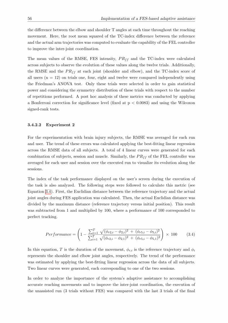
56 Implementation of a FES-based adaptive assistance
the di↵erence between the elbow and shoulder T angles at each time throughout the reaching
movement. Here, the root mean squared of the TC-index di↵erence between the reference
and the actual arm trajectories was computed to evaluate the capability of the FEL controller
to improve the inter-joint coordination.
The mean values of the RMSE, FES intensity, PRff and the TC-index were calculated
across subjects to observe the evolution of these values along the twelve trials. Additionally,
the RMSE and the PRff at each joint (shoulder and elbow), and the TC-index score of
all users (n = 12) on trials one, four, eight and twelve were compared independently using
the Friedman’s ANOVA test. Only these trials were selected in order to gain statistical
power and considering the symmetry distribution of these trials with respect to the number
of repetitions performed. A post hoc analysis of these metrics was conducted by applying
a Bonferroni correction for significance level (fixed at p < 0.0083) and using the Wilcoxon
signed-rank tests.
3.4.2.2 Experiment 2
For the experimentation with brain injury subjects, the RMSE was averaged for each run
and user. The trend of these errors was calculated applying the best-fitting linear regression
across the RMSE data of all subjects. A total of 4 linear curves were generated for each
combination of subjects, session and muscle. Similarly, the PRff of the FEL controller was
averaged for each user and session over the executed run to visualize its evolution along the
sessions.
The index of the task performance displayed on the user’s screen during the execution of
the task is also analyzed. The following steps were followed to calculate this metric (see
Equation 3.4). First, the Euclidian distance between the reference trajectory and the actual
joint angles during FES application was calculated. Then, the actual Euclidian distance was
divided by the maximum distance (reference trajectory versus initial position). This result
was subtracted from 1 and multiplied by 100, where a performance of 100 corresponded to
perfect tracking.
Performance =
1�
PTi=1
p(�r2,i � �2,i)2 + (�r5,i � �5,i)2PT
i=1
p(�r2,i � �2,1)2 + (�r5,i � �5,1)2
!⇥ 100 (3.4)
In this equation, T is the duration of the movement, �r,i is the reference trajectory and �i
represents the shoulder and elbow joint angles, respectively. The trend of the performance
was estimated by applying the best-fitting linear regression across the data of all subjects.
Two linear curves were generated, each corresponding to one of the two sessions.
In order to analyze the importance of the system’s adaptive assistance to accomplishing
accurate reaching movements and to improve the inter-joint coordination, the execution of
the unassisted run (3 trials without FES) was compared with the last 3 trials of the final
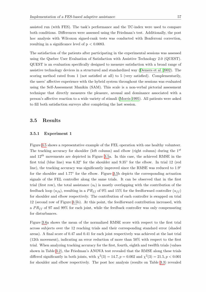
Implementation of a FES-based adaptive assistance 57
assisted run (with FES). The task’s performance and the TC-index were used to compare
both conditions. Di↵erences were assessed using the Friedman’s test. Additionally, the post
hoc analysis with Wilcoxon signed-rank tests was conducted with Bonferroni correction,
resulting in a significance level of p < 0.0083.
The satisfaction of the patients after participating in the experimental sessions was assessed
using the Quebec User Evaluation of Satisfaction with Assistive Technology 2.0 (QUEST).
QUEST is an evaluation specifically designed to measure satisfaction with a broad range of
assistive technology devices in a structured and standardized way (Demers et al. 2002). The
scoring method rated from 1 (not satisfied at all) to 5 (very satisfied). Complementarily,
the users’ a↵ective experience with the hybrid system throughout the sessions was evaluated
using the Self-Assessment Manikin (SAM). This scale is a non-verbal pictorial assessment
technique that directly measures the pleasure, arousal and dominance associated with a
person’s a↵ective reaction to a wide variety of stimuli (Morris 1995). All patients were asked
to fill both satisfaction surveys after completing the last session.
3.5 Results
3.5.1 Experiment 1
Figure 3.5 shows a representative example of the FEL operation with one healthy volunteer.
The tracking accuracy for shoulder (left column) and elbow (right column) during the 1st
and 12th movements are depicted in Figure 3.5a. In this case, the achieved RMSE in the
first trial (blue line) was 6.32� for the shoulder and 9.35� for the elbow. In trial 12 (red
line), the tracking accuracy was significantly improved since the RMSE was reduced to 1.9�
for the shoulder and 1.77� for the elbow. Figure 3.5b depicts the corresponding actuation
signals of the FEL controller along the same trials. It can be observed that in the first
trial (first row), the total assistance (ut) is mostly overlapping with the contribution of the
feedback loop (ufb), resulting in a PRff of 9% and 15% for the feedforward controller (uff )
for shoulder and elbow respectively. The contribution of each controller is swapped on trial
12 (second row of Figure 3.5b). At this point, the feedforward contribution increased, with
a PRff of 97 and 99% for each joint, while the feedback controller was only compensating
for disturbances.
Figure 3.6a shows the mean of the normalized RMSE score with respect to the first trial
across subjects over the 12 reaching trials and their corresponding standard error (shaded
areas). A final score of 0.47 and 0.41 for each joint respectively was achieved at the last trial
(12th movement), indicating an error reduction of more than 50% with respect to the first
trial. When analyzing tracking accuracy for the first, fourth, eighth and twelfth trials (values
shown in Table 3.2), the Friedman’s ANOVA test revealed that the RMSE along these trials
di↵ered significantly in both joints, with �2(3) = 14.7, p = 0.002 and �2(3) = 21.5, p < 0.001
for shoulder and elbow respectively. The post hoc analysis (results on Table 3.3) revealed
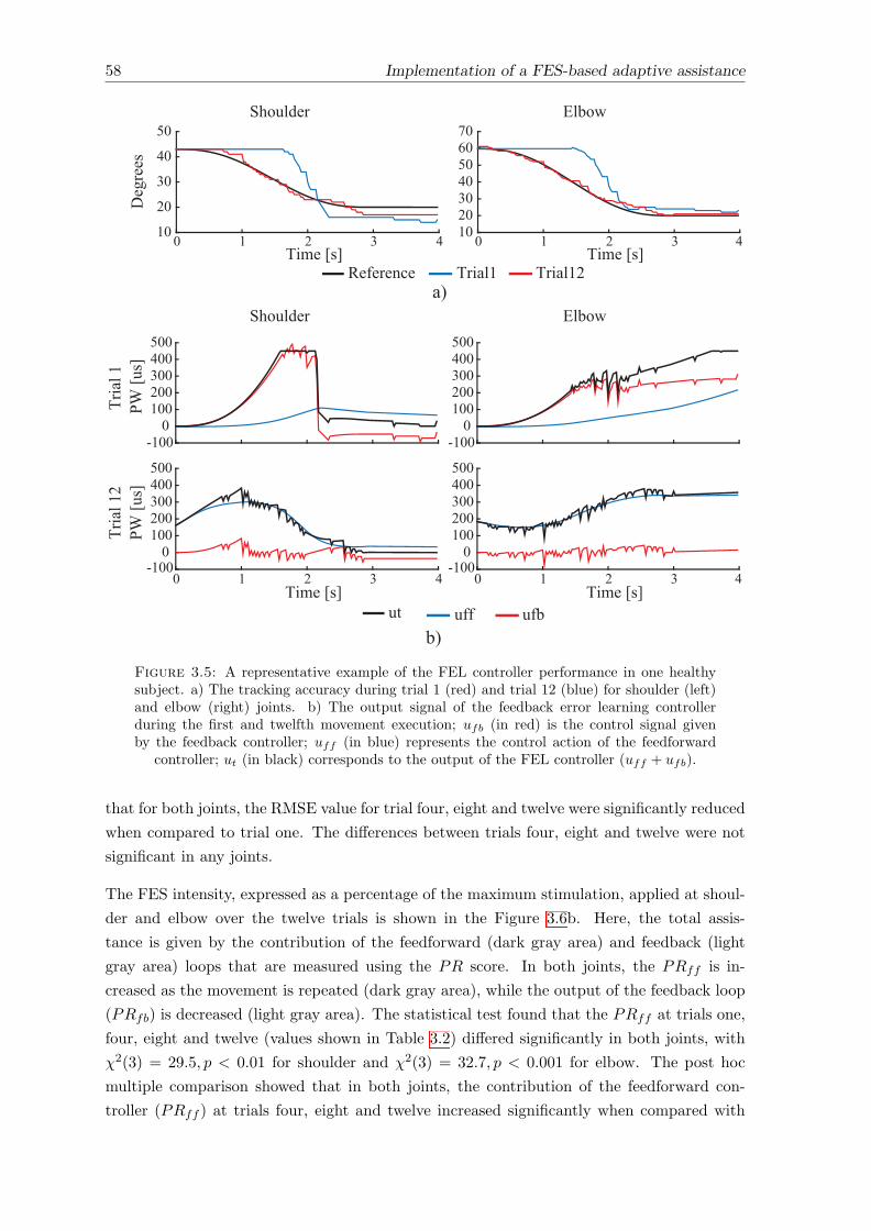
58 Implementation of a FES-based adaptive assistance
Deg
rees
10
20
30
40
50
10203040506070
Tria
l 1PW
[us]
-1000
100200300400500
-1000
100200300400500
Time [s]0 1 2 3 4
Tria
l 12
PW [u
s]
-1000
100200300400500
Time [s]0 1 2 3 4
-1000
100200300400500
Time [s]0 1 2 3 4
Time [s]0 1 2 3 4
a)
b)
Reference Trial1 Trial12
ut uff ufb
Shoulder Elbow
Shoulder Elbow
Figure 3.5: A representative example of the FEL controller performance in one healthysubject. a) The tracking accuracy during trial 1 (red) and trial 12 (blue) for shoulder (left)and elbow (right) joints. b) The output signal of the feedback error learning controllerduring the first and twelfth movement execution; ufb (in red) is the control signal givenby the feedback controller; uff (in blue) represents the control action of the feedforward
controller; ut (in black) corresponds to the output of the FEL controller (uff + ufb).
that for both joints, the RMSE value for trial four, eight and twelve were significantly reduced
when compared to trial one. The di↵erences between trials four, eight and twelve were not
significant in any joints.
The FES intensity, expressed as a percentage of the maximum stimulation, applied at shoul-
der and elbow over the twelve trials is shown in the Figure 3.6b. Here, the total assis-
tance is given by the contribution of the feedforward (dark gray area) and feedback (light
gray area) loops that are measured using the PR score. In both joints, the PRff is in-
creased as the movement is repeated (dark gray area), while the output of the feedback loop
(PRfb) is decreased (light gray area). The statistical test found that the PRff at trials one,
four, eight and twelve (values shown in Table 3.2) di↵ered significantly in both joints, with
�2(3) = 29.5, p < 0.01 for shoulder and �2(3) = 32.7, p < 0.001 for elbow. The post hoc
multiple comparison showed that in both joints, the contribution of the feedforward con-
troller (PRff ) at trials four, eight and twelve increased significantly when compared with
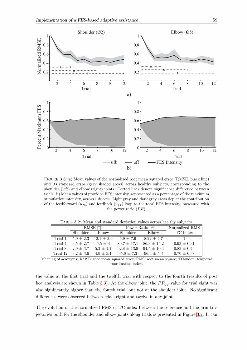
Implementation of a FES-based adaptive assistance 59
! ! !! ! !
Figure 3.6: a) Mean values of the normalized root mean squared error (RMSE, black line)and its standard error (gray shaded areas) across healthy subjects, corresponding to theshoulder (left) and elbow (right) joints. Dotted lines denote significance di↵erence betweentrials. b) Mean values of provided FES intensity, represented as a percentage of the maximumstimulation intensity, across subjects. Light gray and dark gray areas depict the contributionof the feedforward (ufb) and feedback (uff ) loop to the total FES intensity, measured with
the power ratio (PR).
Table 3.2: Mean and standard deviation values across healthy subjects.
RMSE [�] Power Ratio [%] Normalized RMSShoulder Elbow Shoulder Elbow TC-index
Trial 1 5.9 ± 2.3 12.1 ± 3.9 6.9 ± 7.9 8.22 ± 4.7 1Trial 4 3.5 ± 2.7 6.5 ± 4 80.7 ± 17.1 86.3 ± 14.2 0.93 ± 0.31Trial 8 2.9 ± 3.7 5.3 ± 1.7 92.8 ± 13.9 94.5 ± 10.4 0.83 ± 0.46Trial 12 3.2 ± 3.6 4.9 ± 3.1 95.6 ± 7.3 96.9 ± 5.3 0.70 ± 0.38
Meaning of acronyms: RMSE: root mean squared error; RMS: root mean square; TC-index: temporalcoordination index.
the value at the first trial and the twelfth trial with respect to the fourth (results of post
hoc analysis are shown in Table 3.3). At the elbow joint, the PRff value for trial eight was
also significantly higher than the fourth trial, but not at the shoulder joint. No significant
di↵erences were observed between trials eight and twelve in any joints.
The evolution of the normalized RMS of TC-index between the reference and the arm tra-
jectories both for the shoulder and elbow joints along trials is presented in Figure 3.7. It can
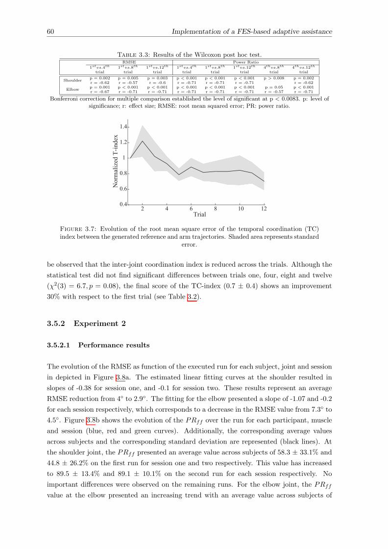
60 Implementation of a FES-based adaptive assistance
Table 3.3: Results of the Wilcoxon post hoc test.RMSE Power Ratio
1stvs.4th 1stvs.8th 1stvs.12th 1stvs.4th 1stvs.8th 1stvs.12th 4thvs.8th 4thvs.12th
trial trial trial trial trial trial trial trial
Shoulderp = 0.002 p = 0.005 p = 0.003 p < 0.001 p < 0.001 p < 0.001 p > 0.008 p = 0.002r = -0.62 r = -0.57 r = -0.6 r = -0.71 r = -0.71 r = -0.71 r = -0.62
Elbowp = 0.001 p < 0.001 p < 0.001 p < 0.001 p < 0.001 p < 0.001 p = 0.05 p < 0.001r = -0.67 r = -0.71 r = -0.71 r = -0.71 r = -0.71 r = -0.71 r = -0.57 r = -0.71
Bonferroni correction for multiple comparison established the level of significant at p < 0.0083. p: level ofsignificance; r: e↵ect size; RMSE: root mean squared error; PR: power ratio.
Figure 3.7: Evolution of the root mean square error of the temporal coordination (TC)index between the generated reference and arm trajectories. Shaded area represents standard
error.
be observed that the inter-joint coordination index is reduced across the trials. Although the
statistical test did not find significant di↵erences between trials one, four, eight and twelve
(�2(3) = 6.7, p = 0.08), the final score of the TC-index (0.7 ± 0.4) shows an improvement
30% with respect to the first trial (see Table 3.2).
3.5.2 Experiment 2
3.5.2.1 Performance results
The evolution of the RMSE as function of the executed run for each subject, joint and session
in depicted in Figure 3.8a. The estimated linear fitting curves at the shoulder resulted in
slopes of -0.38 for session one, and -0.1 for session two. These results represent an average
RMSE reduction from 4� to 2.9�. The fitting for the elbow presented a slope of -1.07 and -0.2
for each session respectively, which corresponds to a decrease in the RMSE value from 7.3� to
4.5�. Figure 3.8b shows the evolution of the PRff over the run for each participant, muscle
and session (blue, red and green curves). Additionally, the corresponding average values
across subjects and the corresponding standard deviation are represented (black lines). At
the shoulder joint, the PRff presented an average value across subjects of 58.3 ± 33.1% and
44.8 ± 26.2% on the first run for session one and two respectively. This value has increased
to 89.5 ± 13.4% and 89.1 ± 10.1% on the second run for each session respectively. No
important di↵erences were observed on the remaining runs. For the elbow joint, the PRff
value at the elbow presented an increasing trend with an average value across subjects of
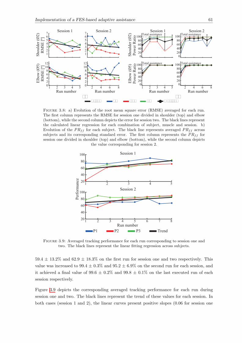
Implementation of a FES-based adaptive assistance 61
a) b)
Shou
lder
(Ø2)
RM
SE [º
]
1234567 Session 1
1234567 Session 2
Run number1 2 3 4 5
Elbo
w (Ø
5)R
MSE
[º]
02468
1012
Run number2 4 6 80
2468
1012
Shou
lder
(Ø2)
Pow
er R
atio
020406080
100
Session 1 Session 2
Run number1 2 3 4 5
Elbo
w (Ø
5)Po
wer
Rat
io
020406080
100
Run number2 4 6 8
P1 P2 P3Trend Average
Total assistance Total assistance
Total assistance Total assistance
020406080
100
020406080
100
Figure 3.8: a) Evolution of the root mean square error (RMSE) averaged for each run.The first column represents the RMSE for session one divided in shoulder (top) and elbow(bottom), while the second column depicts the error for session two. The black lines representthe calculated linear regression for each combination of subject, muscle and session. b)Evolution of the PRff for each subject. The black line represents averaged PRff acrosssubjects and its corresponding standard error. The first column represents the PRff forsession one divided in shoulder (top) and elbow (bottom), while the second column depicts
the value corresponding for session 2.
1 2 3 4 5
Perf
orm
ance 20
40
60
80
100
1 2 3 4 5 6 7 820
40
60
80
100
Session 1
Session 2
Run numberP1 P2 P3 Trend
Figure 3.9: Averaged tracking performance for each run corresponding to session one andtwo. The black lines represent the linear fitting regression across subjects.
59.4 ± 13.2% and 62.9 ± 18.3% on the first run for session one and two respectively. This
value was increased to 99.4 ± 0.3% and 95.2 ± 6.9% on the second run for each session, and
it achieved a final value of 99.6 ± 0.2% and 99.8 ± 0.1% on the last executed run of each
session respectively.
Figure 3.9 depicts the corresponding averaged tracking performance for each run during
session one and two. The black lines represent the trend of these values for each session. In
both cases (session 1 and 2), the linear curves present positive slopes (0.06 for session one
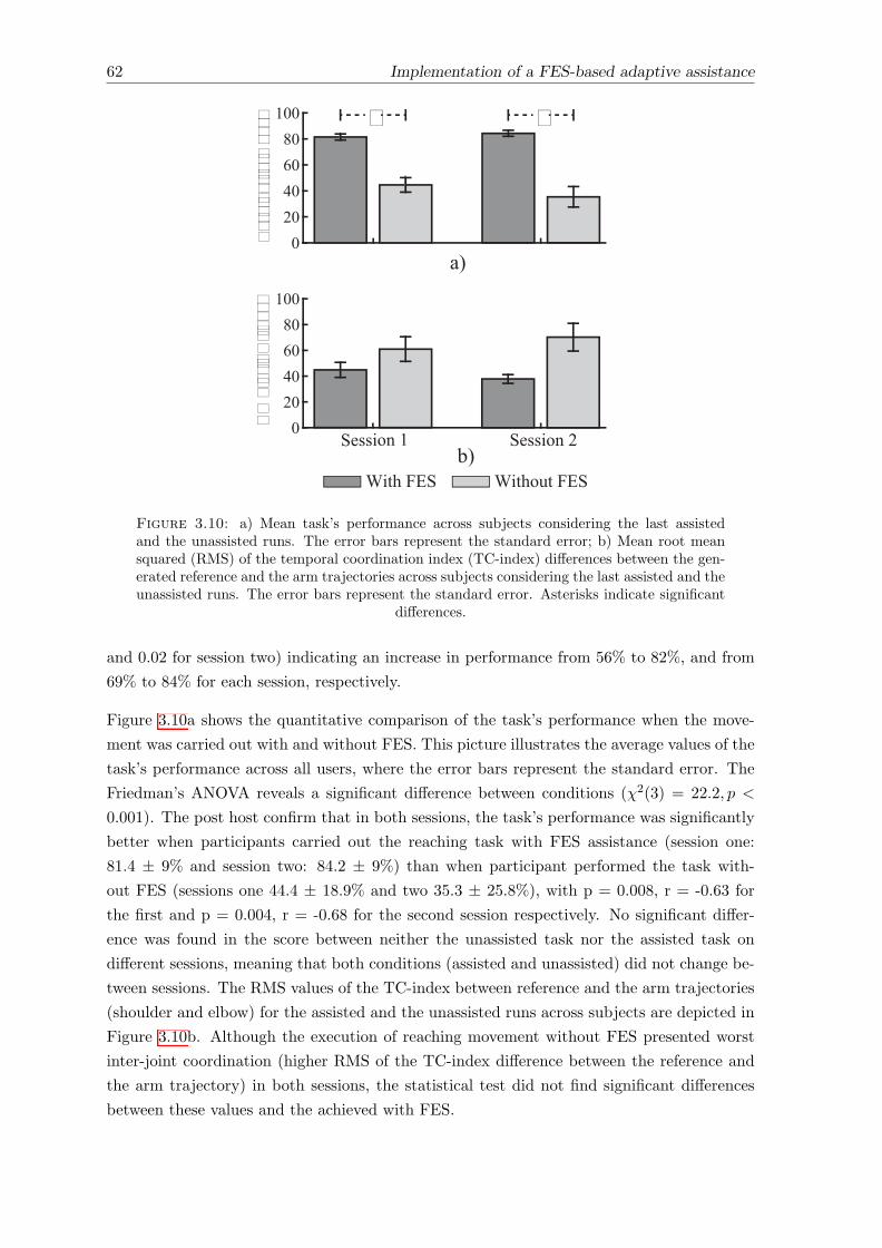
62 Implementation of a FES-based adaptive assistance
020406080
100
020406080
100
Session 1 Session 2
Task
Per
form
ance
RM
S of
TC
-inde
x
With FES Without FES
a)
b)
* *
Figure 3.10: a) Mean task’s performance across subjects considering the last assistedand the unassisted runs. The error bars represent the standard error; b) Mean root meansquared (RMS) of the temporal coordination index (TC-index) di↵erences between the gen-erated reference and the arm trajectories across subjects considering the last assisted and theunassisted runs. The error bars represent the standard error. Asterisks indicate significant
di↵erences.
and 0.02 for session two) indicating an increase in performance from 56% to 82%, and from
69% to 84% for each session, respectively.
Figure 3.10a shows the quantitative comparison of the task’s performance when the move-
ment was carried out with and without FES. This picture illustrates the average values of the
task’s performance across all users, where the error bars represent the standard error. The
Friedman’s ANOVA reveals a significant di↵erence between conditions (�2(3) = 22.2, p <
0.001). The post host confirm that in both sessions, the task’s performance was significantly
better when participants carried out the reaching task with FES assistance (session one:
81.4 ± 9% and session two: 84.2 ± 9%) than when participant performed the task with-
out FES (sessions one 44.4 ± 18.9% and two 35.3 ± 25.8%), with p = 0.008, r = -0.63 for
the first and p = 0.004, r = -0.68 for the second session respectively. No significant di↵er-
ence was found in the score between neither the unassisted task nor the assisted task on
di↵erent sessions, meaning that both conditions (assisted and unassisted) did not change be-
tween sessions. The RMS values of the TC-index between reference and the arm trajectories
(shoulder and elbow) for the assisted and the unassisted runs across subjects are depicted in
Figure 3.10b. Although the execution of reaching movement without FES presented worst
inter-joint coordination (higher RMS of the TC-index di↵erence between the reference and
the arm trajectory) in both sessions, the statistical test did not find significant di↵erences
between these values and the achieved with FES.
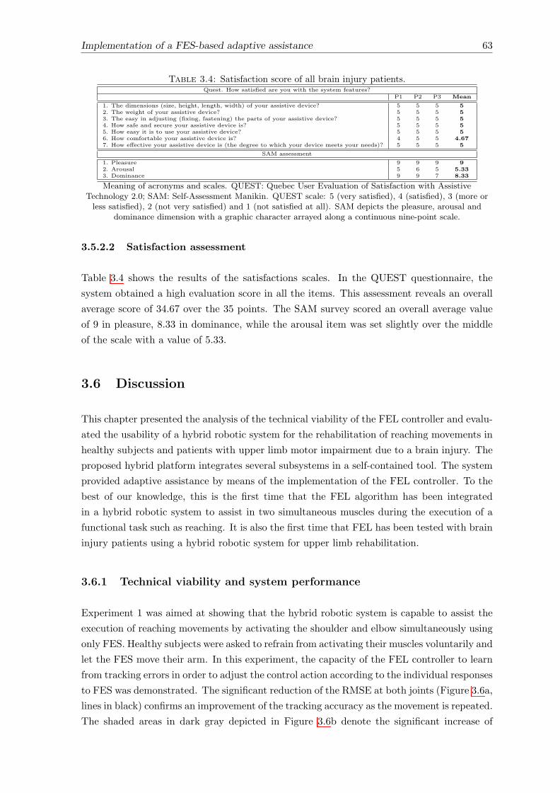
Implementation of a FES-based adaptive assistance 63
Table 3.4: Satisfaction score of all brain injury patients.Quest. How satisfied are you with the system features?
P1 P2 P3 Mean
1. The dimensions (size, height, length, width) of your assistive device? 5 5 5 52. The weight of your assistive device? 5 5 5 53. The easy in adjusting (fixing, fastening) the parts of your assistive device? 5 5 5 54. How safe and secure your assistive device is? 5 5 5 55. How easy it is to use your assistive device? 5 5 5 56. How comfortable your assistive device is? 4 5 5 4.677. How e↵ective your assistive device is (the degree to which your device meets your needs)? 5 5 5 5
SAM assessment
1. Pleasure 9 9 9 92. Arousal 5 6 5 5.333. Dominance 9 9 7 8.33
Meaning of acronyms and scales. QUEST: Quebec User Evaluation of Satisfaction with AssistiveTechnology 2.0; SAM: Self-Assessment Manikin. QUEST scale: 5 (very satisfied), 4 (satisfied), 3 (more orless satisfied), 2 (not very satisfied) and 1 (not satisfied at all). SAM depicts the pleasure, arousal and
dominance dimension with a graphic character arrayed along a continuous nine-point scale.
3.5.2.2 Satisfaction assessment
Table 3.4 shows the results of the satisfactions scales. In the QUEST questionnaire, the
system obtained a high evaluation score in all the items. This assessment reveals an overall
average score of 34.67 over the 35 points. The SAM survey scored an overall average value
of 9 in pleasure, 8.33 in dominance, while the arousal item was set slightly over the middle
of the scale with a value of 5.33.
3.6 Discussion
This chapter presented the analysis of the technical viability of the FEL controller and evalu-
ated the usability of a hybrid robotic system for the rehabilitation of reaching movements in
healthy subjects and patients with upper limb motor impairment due to a brain injury. The
proposed hybrid platform integrates several subsystems in a self-contained tool. The system
provided adaptive assistance by means of the implementation of the FEL controller. To the
best of our knowledge, this is the first time that the FEL algorithm has been integrated
in a hybrid robotic system to assist in two simultaneous muscles during the execution of a
functional task such as reaching. It is also the first time that FEL has been tested with brain
injury patients using a hybrid robotic system for upper limb rehabilitation.
3.6.1 Technical viability and system performance
Experiment 1 was aimed at showing that the hybrid robotic system is capable to assist the
execution of reaching movements by activating the shoulder and elbow simultaneously using
only FES. Healthy subjects were asked to refrain from activating their muscles voluntarily and
let the FES move their arm. In this experiment, the capacity of the FEL controller to learn
from tracking errors in order to adjust the control action according to the individual responses
to FES was demonstrated. The significant reduction of the RMSE at both joints (Figure 3.6a,
lines in black) confirms an improvement of the tracking accuracy as the movement is repeated.
The shaded areas in dark gray depicted in Figure 3.6b denote the significant increase of
64 Implementation of a FES-based adaptive assistance
the PRff measures in the controlled joints, showing the learning process of the controller.
Furthermore, as no significance was found for RMSE after the fourth trial and in the PRff
after the eighth trial with the healthy participants, it can be suggested that the FEL only
requires a few movement examples to attain a stable and appropriate assistance.
Similar behavior was demonstrated in Experiment 2 with brain injury participants. In this
experiment, brain injury patients were asked to realize the movement actively while the FES
provided the activation needed for the patient to complete the task. The negative slopes of
the linear fitting curves derived from the RMSE values over the two sessions (Figure 3.8a)
show a trend towards a reduction of the error as the tracking task was repeated. This
improvement was translated into an enhancement of the performance score fed back to the
users (Figure 3.9). In general, an improvement in the performance from 62.5% to 83%
(averaged from both sessions) was achieved. The PRff values after the second run also
presented important higher score than the first run in both joints. The score obtained
during the execution of the task with and without FES assistance revealed the di�culty of
patients to carry out the required reaching movement without FES (see Figure 3.10). The
average task’s performance of the users during the execution of the assisted reaching task
(82.8%, averaged from both sessions) was twice of the value obtained without assistance.
Therefore, the results confirmed that the hybrid assistance improved significantly the task’s
performance by adapting the delivered FES intensity according to the patients’ needs and
capabilities, helping them to complete the tracking movements.
When looking at the e↵ect of the FEL controller to the inter-joint coordination during the
execution of reaching movement, from experiment 1 can be observed an improvement of
the shoulder-elbow coordination throughout the trials execution (see Figure 3.7). However,
this improvement did not result into a significant improvement when compared with the
first trial. Similarly, experiment 2 showed that assisted movement resulted in better inter-
joint coordination than the movement performed without assistance (without FES). These
results suggest the capability of the FEL controller to learn the shoulder-elbow inter-joint
biomechanical coupling.
Freeman et al. presented the use of ILC to continuously adapt the FES intensity during
reaching movements in a similar hybrid robotic system (Freeman et al. 2011). This algorithm
was tested with healthy subjects performing ten repetitions of a given task. The RMSE
reported in their study was, on average for the first six trials, 9.69�±9.22� and 12.54�±9.87�
for shoulder and elbow angles, respectively. In comparison, the approach presented here
achieves an overall tracking error of 3.2�± 3.6� for the shoulder and 4.9�± 3.1� for the elbow
after 12 trials in the experiment with healthy subjects (n = 12). These improvements could
be attributed to the FEL capability of learning a more precise inverse dynamics model of
the non-linear musculoskeletal characteristics of the arm (Kurosawa et al. 2005, Watanabe
& Fukushima 2011). Therefore, the proposed FEL system represents a robust and reliable
strategy to tackle the subject’s individual di↵erences and the necessity of a complex model
describing the arm dynamics for 3D movement (Meadmore et al. 2012, Freeman et al. 2011).
Implementation of a FES-based adaptive assistance 65
Model-based controllers typically require the definition of multiple parameters before their
use, resulting in time-consuming tasks and requiring one or more additional sessions prior to
the intervention (Hughes et al. 2009, Meadmore et al. 2012, Kutlu et al. 2015). Moreover,
due to the physiological changes occurring over the days, a re-calibration procedure is often
required to maintain the performance within acceptable levels (Kutlu et al. 2015). Unlike
model-based systems, the FEL strategy needs neither a user-specific model nor a previous
model. The algorithm is always learning and adapting in real time. With the approach
proposed here, there is no need to adjust any parameters within one session, between sessions
or between patients, which provides great robustness to the rehabilitation, especially if it is
to be used by clinical (non-technical) operators.
3.6.2 User satisfaction
The users’ perception when dealing with FES or robotic technologies for upper extremity
rehabilitation is scarcely reported in the literature. Nevertheless, if a system is not found
useful and motivational, it will be used less frequently and adherence will be an issue (Hughes
et al. 2011). In this regard, QUEST user’s satisfaction scale reported great satisfaction with
our system in all items, since most scores reached a value of 5 over 5. The SAM scale results
related to pleasure and arousal showed scores of 9 and 5.33 ± 0.58 respectively, suggesting
that patients were satisfied with the use of the system. The result of dominance shows
that patients perceived high level of control (9/10) while using the system. A low score
in dominance may be interpreted as a marker of patients’ feeling of being controlled or
submissive, adopting a passive attitude.
Patients’ motivation has been shown to be an important predictor of long-term changes
in quality of life and rehabilitation outcomes (Grahn et al. 2000). The QUEST and SAM
assessments suggest that patients found the system attractive, and they adopted an active
attitude without feeling under pressure or stressed.
3.6.3 Limitations
Due to the complexity of the shoulder movement during the execution of reaching movements
in unconstrained space (Jarrasse et al. 2014), bigger variability in the PRff metric is observed
at this joint when compared with the elbow. This e↵ect is more noticeable with brain injury
patients (see Figure 3.8b). This larger variation can be attributed to a more varying response
at the shoulder joint to FES. As no mechanical assistance is provided during the movement
execution, these di↵erences can be explained by the amount of electrical current required at
the shoulder to lift the arm up. Meadmore et al. observed similar limitations (Meadmore
et al. 2012). Therefore, the use of mechanical devices with active support could result in a
more consistent response at the shoulder joint. Still, and as discussed in previous chapter, the
development of an optimally shared control between the FES and the mechanical assistance
are needed.
66 Implementation of a FES-based adaptive assistance
The use of the proposed system could be limited by di↵erent conditions of a patient. In the
present study, the participant P4 had to be excluded from the experiment due to a change
in his perception of FES, possibly due to a hypersensibility experienced throughout the ex-
perimental sessions. Therefore, special attention should be paid to refined inclusion criteria.
Certainly, the guidance given by Huang et al can be followed, where it was suggested that
patients with medium level (suggested by Fugl-Meyer Assessment and Motor Assessment
Scale score) of motor skills are preferred when considering robot-based rehabilitation thera-
pies (Huang et al. 2016). Alternatively, active exoskeletons could be considered to reduce the
intensity of FES. With such a system, the exoskeleton assistance can be reduced progressively
to increase the FES stimulation and promote voluntary movement.
The results reported in this study are based on a reduced number of patients and sessions.
As the potential rehabilitation benefits of the present hybrid robotic system is out of the
scope of this study, it is necessary to conduct a larger clinical study involving more patients
and sessions.
3.7 Conclusions of the chapter
In this chapter, a fully-integrated hybrid robotic system for rehabilitation of reaching move-
ment was presented. The system relies on several subsystems that cooperatively assist during
the rehabilitation exercise. The system caters for a rehabilitation scenario where uncon-
strained functional movements in 3D space are promoted.
An easy to deploy FES-based adaptive assistance was implemented in order to adapt the
assist intensity according to user’s motor residual capability. This adaptive controller is based
on the feedback error learning algorithm. It was demonstrated that the FEL is capable to
learn the inverse dynamics model of the arm, and consequently, smoothly adjusting the level
of assistance over the trials. Interestingly, this adaptive strategy does not require additional
sessions neither a tedious and exhaustive procedure to be configured.
Experimentations performed with healthy participants and patients with brain injury demon-
strate the suitability of FEL scheme to assist the execution of reaching movements in 3D
space. Patients’ reports on the intervention reveal a great satisfaction and acceptance of the
hybrid robotic system. These results support the idea that complementing rehabilitation
with the hybrid system hereby proposed might be useful to increase the dosage of therapy
and to augment patient’s engagement and motivation during the rehabilitation process. In
the next chapter, a BCI system is combined with the hybrid robotic system to provide causal
and timed assistance to elicit plasticity.
Chapter 4
Eliciting neural plasticity by the
timed association of the user’s
motor intent with the peripheral
hybrid assistance1
Abstract
This chapter presents the implementation of an associative approach combining the hybrid
assistance delivered by the system presented in Chapter 3 with users’ motor intent. The
adaptive hybrid assistance is triggered when the users’ own intention to move is detected
to assist the execution of functional reaching movements. The first objective framed in this
chapter is to verify whether the precise temporal pairing between the peripheral stimulation
and the user’s motor intent during the execution of a functional reaching movement can
induce an enhancement in excitability of the descending motor corticospinal tract projected
to the arm muscles (plasticity) in healthy subjects. The second objective is to compare the
intervention e↵ects when three di↵erent techniques (EMG, BCI o✏ine and BCI online) are
used to detect the user’s intent to move and associated it with the externally applied hybrid
peripheral assistance. Twenty-one healthy subjects (11 males and 10 females, age: 29.3±3.98
years) participated in the experimentation. Results revealed positive intervention’s e↵ects in
all implemented strategies (BCI o✏ine, BCI online and EMG). However, stronger and more
consistent intervention’s e↵ects were achieved with the BCI o✏ine intervention. Thus, this
strategy reported a significant increase in excitability of the corticospinal projection (larger
MEP size) after the intervention in the assisted (AD, TB) and unassisted (BB) target muscles
1This chapter is partly based on:- F. Resquın, O. Herrero, J. Ibanez, J. Gonzalez-Vargas, F. Brunetti, J. L. Pons. Electroencephalography-guided Upper-Limb Hybrid Robotic Platform to Modulate Cortical Excitability. IEEE International Confer-ence on Systems, Man and Cybernetics, 2017 (Accepted).- F. Resquın, J. Ibanez, O. Herrero, J. Gonzalez-Vargas, F. Brunetti, and J.L. Pons. “User’s Motor IntentGuided Upper-Limb Hybrid Robotic Platform to E�ciently Modulate Cortical Excitability: A comparison ofthree strategies”. (To be submitted)
67

68 Eliciting neural plasticity
of the arm. Because of the e�cacy of this intervention (BCI o✏ine), it represents an e�cient
neuromodulation strategy for inducing plastic changes into the motor cortex.

Eliciting neural plasticity 69
4.1 Introduction
In Chapter 2, the main challenges of the hybrid robotic system for rehabilitation of the
upper-extremity motor functions were identified. Based on these requirements, in Chapter 3
a hybrid robotic system providing adaptive assistance, according to the user motor residual
capability, during the execution of unconstrained reaching movement was presented. It was
demonstrated that, the system can assist accurately tracking movements by supplying FES
assistance at the AD and TB muscles simultaneously. Moreover, brain injury patients rated
the system positively, presenting a great acceptability for using the system as a rehabilitation
tool. However, there is evidence showing the relevance of exploiting the natural plasticity of
the sensorimotor system to maximize rehabilitation potential of therapies (Di Pino et al. 2014,
Dimyan & Cohen 2011). Certainly, intrinsic physiological and anatomical neural plasticity
are important processes that underlie substantial gain of motor function after stroke (Ward
2005, Dimyan & Cohen 2011, Schaechter 2004).
At the cortical level, di↵erent mechanisms are responsible for facilitating these plastic changes
(Ward 2005, Nudo 2006). One of those, is referred to as activity-dependent neural plasticity,
which is commonly linked to the long-term potentiation (LTP) phenomenon (Ziemann 2004,
Sanes & Donoghue 2000). The LTP consists of persistent strengthening of the synaptic
connection associated to the Hebbian theory, which states that synapses that experience
correlated activation of two converging inputs are strengthened whereas those weakened
by uncorrelated activity are lost (Hebb 1949). It has been shown that this physiological
phenomenon can be induced through associative protocols by the precise causal and temporal
pairing of a peripheral and cortical stimulus (Carson & Kennedy 2013, Stefan et al. 2000,
Thabit et al. 2010, Ziemann et al. 2008).
The paired associative stimulation (PAS) is a widely di↵used associative protocol, consist-
ing of a low frequency peripheral stimulation, applied over the peripheral nerve of the wrist
(Stefan et al. 2000) or over the peroneal nerve in the leg (Mrachacz-Kersting et al. 2007,
Mrachacz-Kersting & Stevenson 2017), timed and paired with a transcranial magnetic sti-
mulation (TMS) delivered to the contralateral primary motor cortex. In these studies, the
motor evoked potential (MEP) on target muscles elicited by TMS was used to quantify the
e↵ects of the PAS protocol. The MEP response gives a valuable and quantifiable information
about the excitability of the cortical projection to the target muscles (plasticity) (Rotenberg
et al. 2014, Rossini et al. 2015). It was shown that PAS can induce plastic changes mea-
sured as an increase in the amplitude of the MEPs, and its e↵ect was persistent (duration
> 30 minutes). Although PAS constitutes a great potential for inducing plastic changes in
a short time (⇠30’ minutes), it presents some limitations. The principal one relies on its
functional implication, since the exogenous cortical stimulus activates several brain areas
at the same time rather than following a sequence of neural activation as happens during
voluntary movements.
In the rehabilitation field, a neuromodulation BCI can be defined as a system conceived
for inducing neuroplasticity (Daly & Wolpaw 2008, Soekadar et al. 2015). In this regard
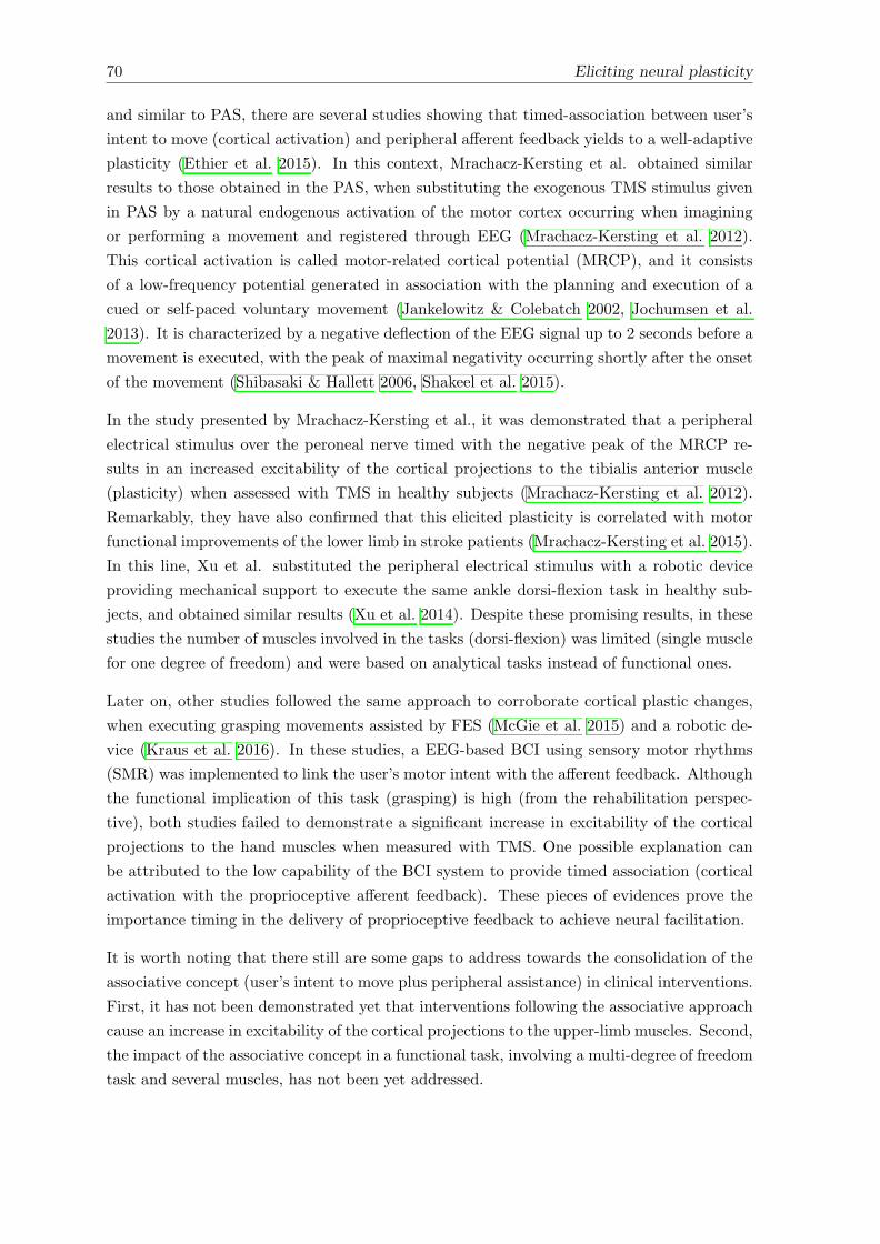
70 Eliciting neural plasticity
and similar to PAS, there are several studies showing that timed-association between user’s
intent to move (cortical activation) and peripheral a↵erent feedback yields to a well-adaptive
plasticity (Ethier et al. 2015). In this context, Mrachacz-Kersting et al. obtained similar
results to those obtained in the PAS, when substituting the exogenous TMS stimulus given
in PAS by a natural endogenous activation of the motor cortex occurring when imagining
or performing a movement and registered through EEG (Mrachacz-Kersting et al. 2012).
This cortical activation is called motor-related cortical potential (MRCP), and it consists
of a low-frequency potential generated in association with the planning and execution of a
cued or self-paced voluntary movement (Jankelowitz & Colebatch 2002, Jochumsen et al.
2013). It is characterized by a negative deflection of the EEG signal up to 2 seconds before a
movement is executed, with the peak of maximal negativity occurring shortly after the onset
of the movement (Shibasaki & Hallett 2006, Shakeel et al. 2015).
In the study presented by Mrachacz-Kersting et al., it was demonstrated that a peripheral
electrical stimulus over the peroneal nerve timed with the negative peak of the MRCP re-
sults in an increased excitability of the cortical projections to the tibialis anterior muscle
(plasticity) when assessed with TMS in healthy subjects (Mrachacz-Kersting et al. 2012).
Remarkably, they have also confirmed that this elicited plasticity is correlated with motor
functional improvements of the lower limb in stroke patients (Mrachacz-Kersting et al. 2015).
In this line, Xu et al. substituted the peripheral electrical stimulus with a robotic device
providing mechanical support to execute the same ankle dorsi-flexion task in healthy sub-
jects, and obtained similar results (Xu et al. 2014). Despite these promising results, in these
studies the number of muscles involved in the tasks (dorsi-flexion) was limited (single muscle
for one degree of freedom) and were based on analytical tasks instead of functional ones.
Later on, other studies followed the same approach to corroborate cortical plastic changes,
when executing grasping movements assisted by FES (McGie et al. 2015) and a robotic de-
vice (Kraus et al. 2016). In these studies, a EEG-based BCI using sensory motor rhythms
(SMR) was implemented to link the user’s motor intent with the a↵erent feedback. Although
the functional implication of this task (grasping) is high (from the rehabilitation perspec-
tive), both studies failed to demonstrate a significant increase in excitability of the cortical
projections to the hand muscles when measured with TMS. One possible explanation can
be attributed to the low capability of the BCI system to provide timed association (cortical
activation with the proprioceptive a↵erent feedback). These pieces of evidences prove the
importance timing in the delivery of proprioceptive feedback to achieve neural facilitation.
It is worth noting that there still are some gaps to address towards the consolidation of the
associative concept (user’s intent to move plus peripheral assistance) in clinical interventions.
First, it has not been demonstrated yet that interventions following the associative approach
cause an increase in excitability of the cortical projections to the upper-limb muscles. Second,
the impact of the associative concept in a functional task, involving a multi-degree of freedom
task and several muscles, has not been yet addressed.
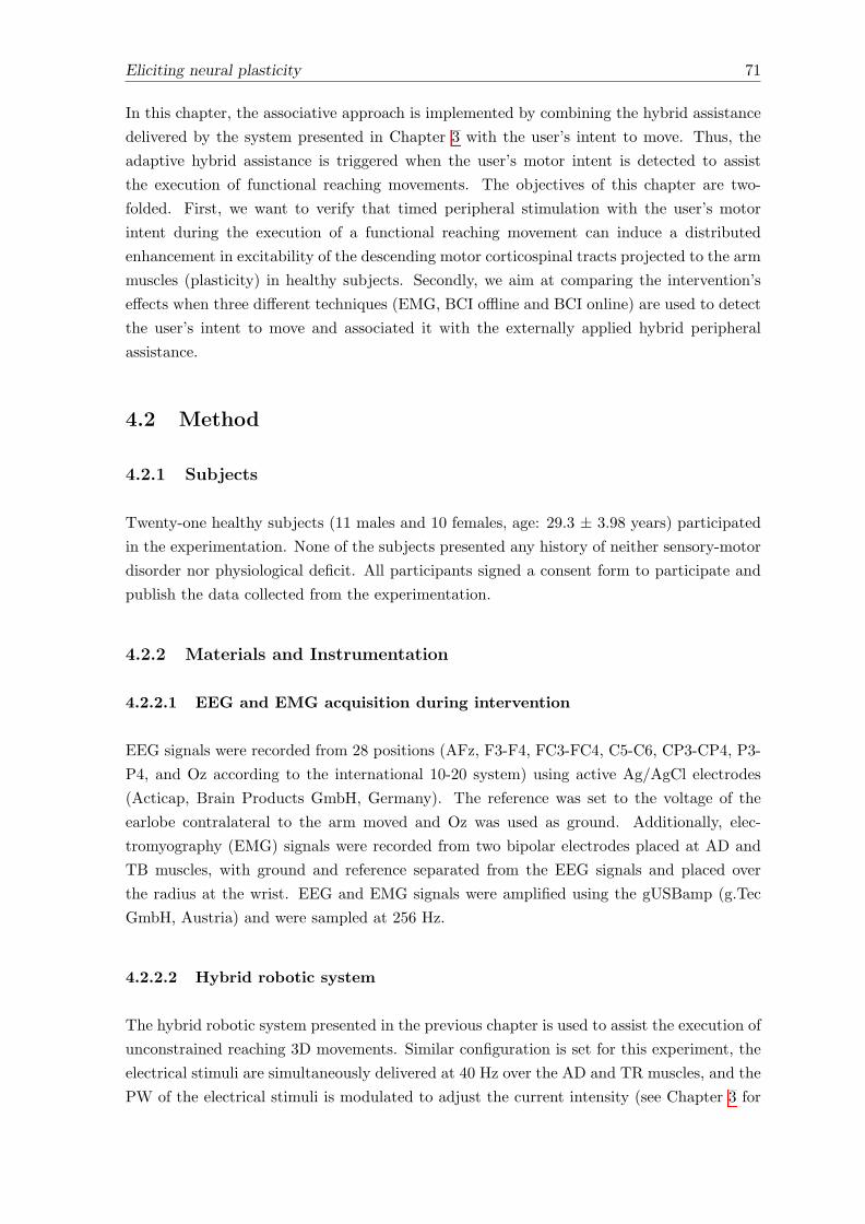
Eliciting neural plasticity 71
In this chapter, the associative approach is implemented by combining the hybrid assistance
delivered by the system presented in Chapter 3 with the user’s intent to move. Thus, the
adaptive hybrid assistance is triggered when the user’s motor intent is detected to assist
the execution of functional reaching movements. The objectives of this chapter are two-
folded. First, we want to verify that timed peripheral stimulation with the user’s motor
intent during the execution of a functional reaching movement can induce a distributed
enhancement in excitability of the descending motor corticospinal tracts projected to the arm
muscles (plasticity) in healthy subjects. Secondly, we aim at comparing the intervention’s
e↵ects when three di↵erent techniques (EMG, BCI o✏ine and BCI online) are used to detect
the user’s intent to move and associated it with the externally applied hybrid peripheral
assistance.
4.2 Method
4.2.1 Subjects
Twenty-one healthy subjects (11 males and 10 females, age: 29.3 ± 3.98 years) participated
in the experimentation. None of the subjects presented any history of neither sensory-motor
disorder nor physiological deficit. All participants signed a consent form to participate and
publish the data collected from the experimentation.
4.2.2 Materials and Instrumentation
4.2.2.1 EEG and EMG acquisition during intervention
EEG signals were recorded from 28 positions (AFz, F3-F4, FC3-FC4, C5-C6, CP3-CP4, P3-
P4, and Oz according to the international 10-20 system) using active Ag/AgCl electrodes
(Acticap, Brain Products GmbH, Germany). The reference was set to the voltage of the
earlobe contralateral to the arm moved and Oz was used as ground. Additionally, elec-
tromyography (EMG) signals were recorded from two bipolar electrodes placed at AD and
TB muscles, with ground and reference separated from the EEG signals and placed over
the radius at the wrist. EEG and EMG signals were amplified using the gUSBamp (g.Tec
GmbH, Austria) and were sampled at 256 Hz.
4.2.2.2 Hybrid robotic system
The hybrid robotic system presented in the previous chapter is used to assist the execution of
unconstrained reaching 3D movements. Similar configuration is set for this experiment, the
electrical stimuli are simultaneously delivered at 40 Hz over the AD and TR muscles, and the
PW of the electrical stimuli is modulated to adjust the current intensity (see Chapter 3 for
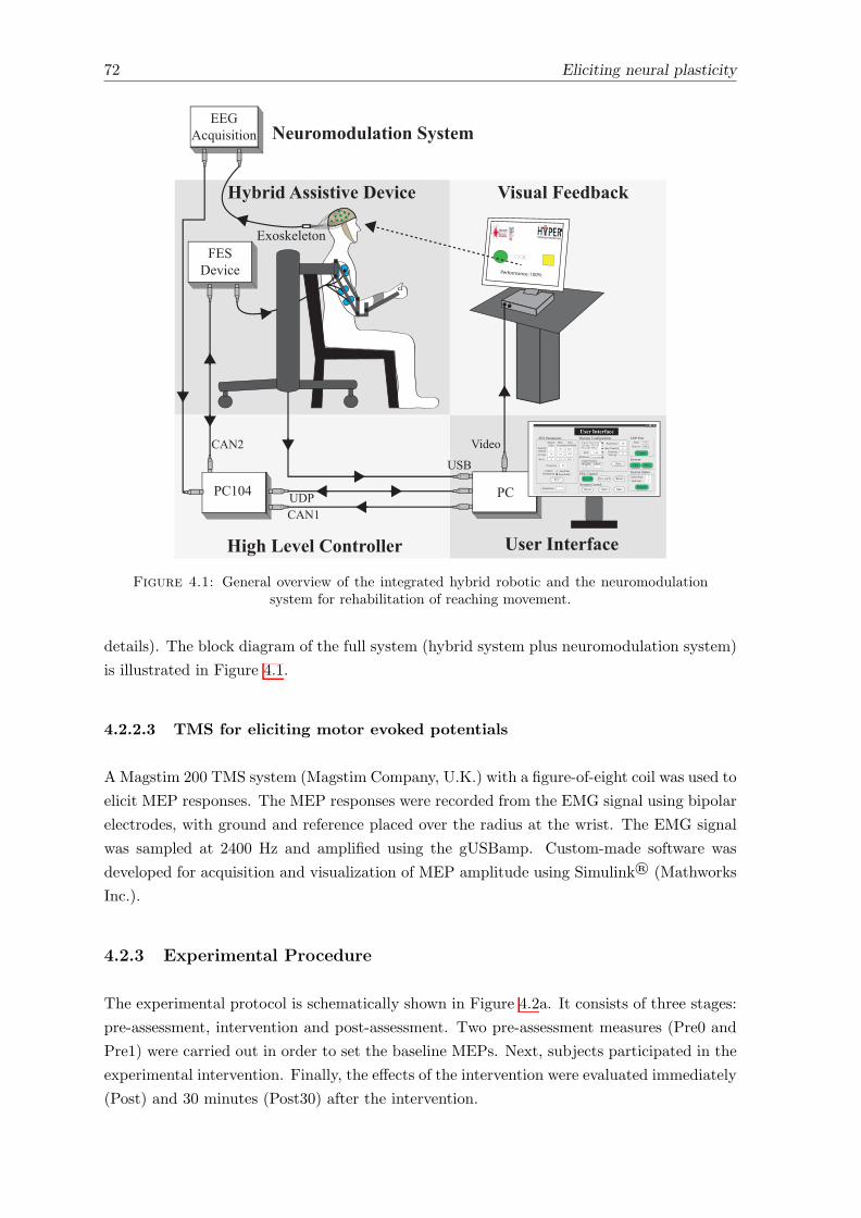
72 Eliciting neural plasticity
Reach the target!
TargetInit Position
Performance: 100%
!"#$%&'(#$)*+#,-./%0#1#2%34'($422#$
!"#$
%&'
()*+,-.+/0+123
&345+6-7& 89!
-79:!,15.,;-79:<# -79:<-- =+4+5
&345+6:!,15.,;>,?=+4+5 &[email protected]
-7&:A@.@6+5+.4
!,15.,;>,*0;@5),1
"6B;)50*+A0;4+C)*5D
&@?+
"15+.),.E+;5,)*F.)2+B4
C.)45
>042;+-)+;*
>@GH!0..+15
>@GHA0;4+C)*5D
$
$I
J
JK
$L
$I
IMK
IMK
IMK
-.+/0+123 IK
=+B+5)5),14 M
&345+6:&5@5+4N;,O@;:&5@5+F@4P:&5@5+
=+/0+45
$
Q
FD+.@B3:!,1R)S0.@5),17;O,T:7G5+14),1&D,0;*+.:-;+G),1&D,0;*+.U7;O,T
".6 9+R5
=+R+.+12+ Q:4+2,1*4
=+B+5)5),14
=+45:F)6+:V4WN.@4B)1SF)6+:V4W
&@?+
J
M
JK
&D,0;*+. 7;O,[email protected]+5:A,4)5),1
$K QK
!"#$%&'(#$)*+#%EA:A,.5
&+1*
=+2+)?+
!,1+25
XMKK
$MKKK
,56$-7%8""-"(-1#%9#1-+# :-";*2%<##76*+=
-7&E+?)2+
A!$KI A!
!"#J
%EA
77N"2/0)4)5),1 >#;$4?47;2*(-4'%@5"(#?
7G,4P+;+5,1
Figure 4.1: General overview of the integrated hybrid robotic and the neuromodulationsystem for rehabilitation of reaching movement.
details). The block diagram of the full system (hybrid system plus neuromodulation system)
is illustrated in Figure 4.1.
4.2.2.3 TMS for eliciting motor evoked potentials
AMagstim 200 TMS system (Magstim Company, U.K.) with a figure-of-eight coil was used to
elicit MEP responses. The MEP responses were recorded from the EMG signal using bipolar
electrodes, with ground and reference placed over the radius at the wrist. The EMG signal
was sampled at 2400 Hz and amplified using the gUSBamp. Custom-made software was
developed for acquisition and visualization of MEP amplitude using Simulink® (Mathworks
Inc.).
4.2.3 Experimental Procedure
The experimental protocol is schematically shown in Figure 4.2a. It consists of three stages:
pre-assessment, intervention and post-assessment. Two pre-assessment measures (Pre0 and
Pre1) were carried out in order to set the baseline MEPs. Next, subjects participated in the
experimental intervention. Finally, the e↵ects of the intervention were evaluated immediately
(Post) and 30 minutes (Post30) after the intervention.
Eliciting neural plasticity 73
!"#$%&'(%
)*&%+%,-.,/%-
"0*/1%2%-*,
""3454")36(78'/'-'*,
94:;4/',<2%4=82/%/4>-4?;@4:;;@4::;@4:A;4>,B4:C;4D4*E4F)G
94)8/(2%/H6,-%F'*F4$%2-*'B/GF'(%=4IF>(J''I'(%=4IF>(J''
:94KF%96//%//+%,- A94L,-%F&%,-'*, C94K*/-96//%//+%,-94M*-4/=*-454F)G
'B%,-'E'(>-'*, 94:;4/',<2%4=82/%/4>-4?;@4:;;@4::;@4:A;4>,B4:C;4D4*E4F)G
94)8/(2%/H6,-%F'*F4$%2-*'B/GF'(%=4IF>(J''I'(%=4IF>(J''
N%>(J4-J%4G>F<%-O
G>/14F%=F%/%,->-'*,
N%E%F%,(%
G>F<%-
L,'- 6F+4=*/'-'*,
P%224$*,%O
K%FE*F+>,(%
G>F<%-
L,'-
!*(8/O
GF>E'(42'<J-/
G>F<%-
L,'-
Figure 4.2: a) Experimental protocol used for experimentations; b) Implemented statemachine to present the visual cue and guide users during the intervention.
4.2.3.1 Pre0-, Pre1-, Post- and Post30-assessment
The brain response to single-pulse TMS delivered over the motor cortex was measured from
the AD and TB and bicep brachii (BB) muscles (target muscles). The TMS pulses were
applied contralateral to the arm moved during the experiment. In 20 of the participants the
TMS stimuli were delivered on the right cerebral hemisphere in order to induce MEPs on
the left arm. While in a single subject, the TMS pulses were applied in the opposite cerebral
hemisphere (left) to record MEPs of target muscles in the right arm.
Before the first pre-assessment measure (Pre0), skin preparation was performed (rubbing
with cotton and alcohol) and EMG electrodes (rectangular 22.225⇥34.925 mm, NeuroPlus™)
were placed over the target muscles (AD, TB and BB). Once these electrodes were placed,
they were fixed until the end of the experiment. All participants seated in a chair (without
armrest) and were instructed to keep their arm extended (in direction to the ground) and
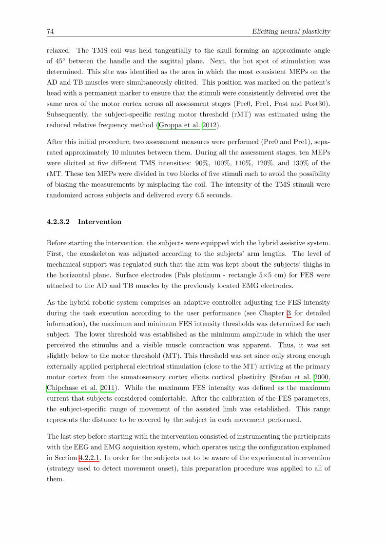
74 Eliciting neural plasticity
relaxed. The TMS coil was held tangentially to the skull forming an approximate angle
of 45� between the handle and the sagittal plane. Next, the hot spot of stimulation was
determined. This site was identified as the area in which the most consistent MEPs on the
AD and TB muscles were simultaneously elicited. This position was marked on the patient’s
head with a permanent marker to ensure that the stimuli were consistently delivered over the
same area of the motor cortex across all assessment stages (Pre0, Pre1, Post and Post30).
Subsequently, the subject-specific resting motor threshold (rMT) was estimated using the
reduced relative frequency method (Groppa et al. 2012).
After this initial procedure, two assessment measures were performed (Pre0 and Pre1), sepa-
rated approximately 10 minutes between them. During all the assessment stages, ten MEPs
were elicited at five di↵erent TMS intensities: 90%, 100%, 110%, 120%, and 130% of the
rMT. These ten MEPs were divided in two blocks of five stimuli each to avoid the possibility
of biasing the measurements by misplacing the coil. The intensity of the TMS stimuli were
randomized across subjects and delivered every 6.5 seconds.
4.2.3.2 Intervention
Before starting the intervention, the subjects were equipped with the hybrid assistive system.
First, the exoskeleton was adjusted according to the subjects’ arm lengths. The level of
mechanical support was regulated such that the arm was kept about the subjects’ thighs in
the horizontal plane. Surface electrodes (Pals platinum - rectangle 5⇥5 cm) for FES were
attached to the AD and TB muscles by the previously located EMG electrodes.
As the hybrid robotic system comprises an adaptive controller adjusting the FES intensity
during the task execution according to the user performance (see Chapter 3 for detailed
information), the maximum and minimum FES intensity thresholds was determined for each
subject. The lower threshold was established as the minimum amplitude in which the user
perceived the stimulus and a visible muscle contraction was apparent. Thus, it was set
slightly below to the motor threshold (MT). This threshold was set since only strong enough
externally applied peripheral electrical stimulation (close to the MT) arriving at the primary
motor cortex from the somatosensory cortex elicits cortical plasticity (Stefan et al. 2000,
Chipchase et al. 2011). While the maximum FES intensity was defined as the maximum
current that subjects considered comfortable. After the calibration of the FES parameters,
the subject-specific range of movement of the assisted limb was established. This range
represents the distance to be covered by the subject in each movement performed.
The last step before starting with the intervention consisted of instrumenting the participants
with the EEG and EMG acquisition system, which operates using the configuration explained
in Section 4.2.2.1. In order for the subjects not to be aware of the experimental intervention
(strategy used to detect movement onset), this preparation procedure was applied to all of
them.
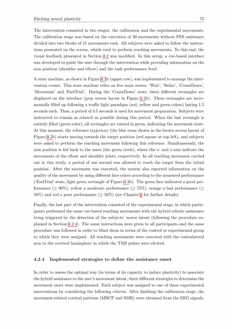
Eliciting neural plasticity 75
The intervention consisted in two stages: the calibration and the experimental movements.
The calibration stage was based on the execution of 30 movements without FES assistance
divided into two blocks of 15 movements each. All subjects were asked to follow the instruc-
tions presented on the screen, which cued to perform reaching movements. To this end, the
visual feedback presented in Section 3.2 was modified. In this setup, a cue-based interface
was developed to guide the user through the intervention while providing information on the
arm position (shoulder and elbow) and the task performance level.
A state machine, as shown in Figure 4.2b (upper row), was implemented to manage the inter-
vention events. This state machine relies on five main states: ‘Wait’, ‘Relax’, ‘CountDown’,
‘Movement’ and ‘EndTrial’. During the ‘CountDown’ state, three di↵erent rectangles are
displayed on the interface (gray screen layout in Figure 4.2b). These rectangles are incre-
mentally filled up following a tra�c light paradigm (red, yellow and green colors) lasting 1.5
seconds each. Thus, a period of 4.5 seconds is used for movement preparation. Subjects were
instructed to remain as relaxed as possible during this period. When the last rectangle is
entirely filled (green color), all rectangles are turned in green, indicating the movement state.
At this moment, the reference trajectory (the blue cross shown in the brown screen layout of
Figure 4.2b) starts moving towards the target position (red square at top left), and subjects
were asked to perform the reaching movement following this reference. Simultaneously, the
arm position is fed back to the users (the green circle), where the x- and y-axis indicate the
movements of the elbow and shoulder joints, respectively. In all reaching movement carried
out in this study, a period of one second was allowed to reach the target from the initial
position. After the movement was executed, the system also reported information on the
quality of the movement by using di↵erent face colors according to the measured performance
(‘EndTrial’ state, light green rectangle of Figure 4.2b). The green face indicated a good per-
formance (� 90%), yellow a moderate performance (� 75%), orange a bad performance (�50%) and red a poor performance (� 50%) (see Chapter 3 for further details).
Finally, the last part of the intervention consisted of the experimental stage, in which partic-
ipants performed the same cue-based reaching movements with the hybrid robotic assistance
being triggered by the detection of the subjects’ motor intent (following the procedure ex-
plained in Section 4.2.4). The same instructions were given to all participants and the same
procedure was followed in order to blind them in terms of the control or experimental group
to which they were assigned. All reaching movements were executed with the contralateral
arm to the cerebral hemisphere in which the TMS pulses were elicited.
4.2.4 Implemented strategies to define the assistance onset
In order to assess the optimal way (in terms of its capacity to induce plasticity) to associate
the hybrid assistance to the user’s movement intent, three di↵erent strategies to determine the
movement onset were implemented. Each subject was assigned to one of these experimental
interventions by considering the following criteria. After finishing the calibration stage, the
movement-related cortical patterns (MRCP and SMR) were obtained from the EEG signals.
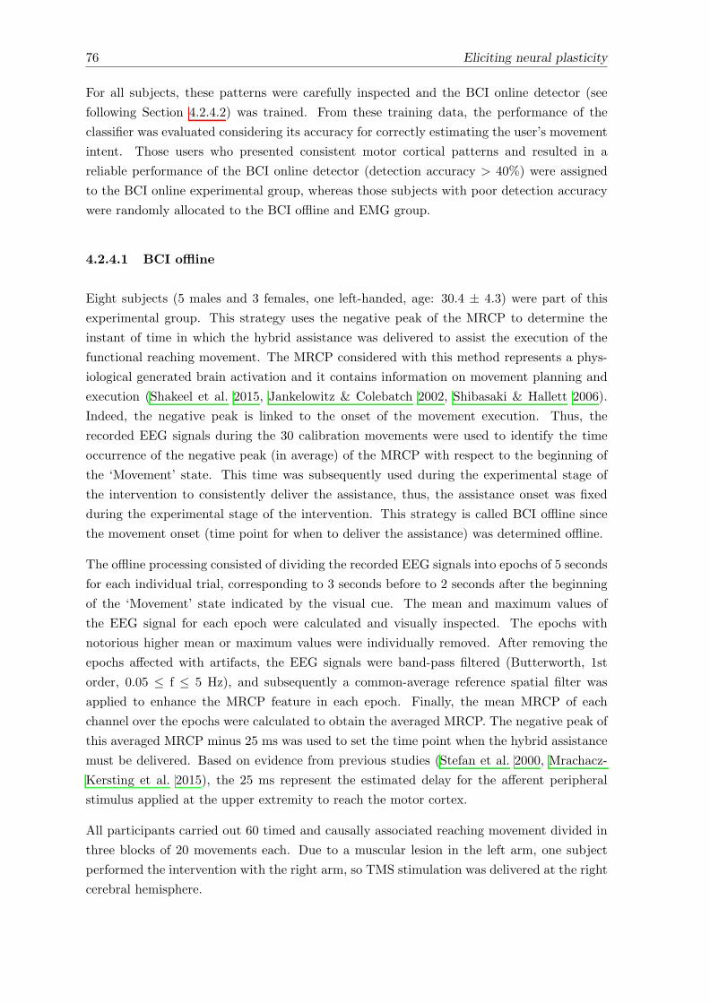
76 Eliciting neural plasticity
For all subjects, these patterns were carefully inspected and the BCI online detector (see
following Section 4.2.4.2) was trained. From these training data, the performance of the
classifier was evaluated considering its accuracy for correctly estimating the user’s movement
intent. Those users who presented consistent motor cortical patterns and resulted in a
reliable performance of the BCI online detector (detection accuracy > 40%) were assigned
to the BCI online experimental group, whereas those subjects with poor detection accuracy
were randomly allocated to the BCI o✏ine and EMG group.
4.2.4.1 BCI o✏ine
Eight subjects (5 males and 3 females, one left-handed, age: 30.4 ± 4.3) were part of this
experimental group. This strategy uses the negative peak of the MRCP to determine the
instant of time in which the hybrid assistance was delivered to assist the execution of the
functional reaching movement. The MRCP considered with this method represents a phys-
iological generated brain activation and it contains information on movement planning and
execution (Shakeel et al. 2015, Jankelowitz & Colebatch 2002, Shibasaki & Hallett 2006).
Indeed, the negative peak is linked to the onset of the movement execution. Thus, the
recorded EEG signals during the 30 calibration movements were used to identify the time
occurrence of the negative peak (in average) of the MRCP with respect to the beginning of
the ‘Movement’ state. This time was subsequently used during the experimental stage of
the intervention to consistently deliver the assistance, thus, the assistance onset was fixed
during the experimental stage of the intervention. This strategy is called BCI o✏ine since
the movement onset (time point for when to deliver the assistance) was determined o✏ine.
The o✏ine processing consisted of dividing the recorded EEG signals into epochs of 5 seconds
for each individual trial, corresponding to 3 seconds before to 2 seconds after the beginning
of the ‘Movement’ state indicated by the visual cue. The mean and maximum values of
the EEG signal for each epoch were calculated and visually inspected. The epochs with
notorious higher mean or maximum values were individually removed. After removing the
epochs a↵ected with artifacts, the EEG signals were band-pass filtered (Butterworth, 1st
order, 0.05 f 5 Hz), and subsequently a common-average reference spatial filter was
applied to enhance the MRCP feature in each epoch. Finally, the mean MRCP of each
channel over the epochs were calculated to obtain the averaged MRCP. The negative peak of
this averaged MRCP minus 25 ms was used to set the time point when the hybrid assistance
must be delivered. Based on evidence from previous studies (Stefan et al. 2000, Mrachacz-
Kersting et al. 2015), the 25 ms represent the estimated delay for the a↵erent peripheral
stimulus applied at the upper extremity to reach the motor cortex.
All participants carried out 60 timed and causally associated reaching movement divided in
three blocks of 20 movements each. Due to a muscular lesion in the left arm, one subject
performed the intervention with the right arm, so TMS stimulation was delivered at the right
cerebral hemisphere.
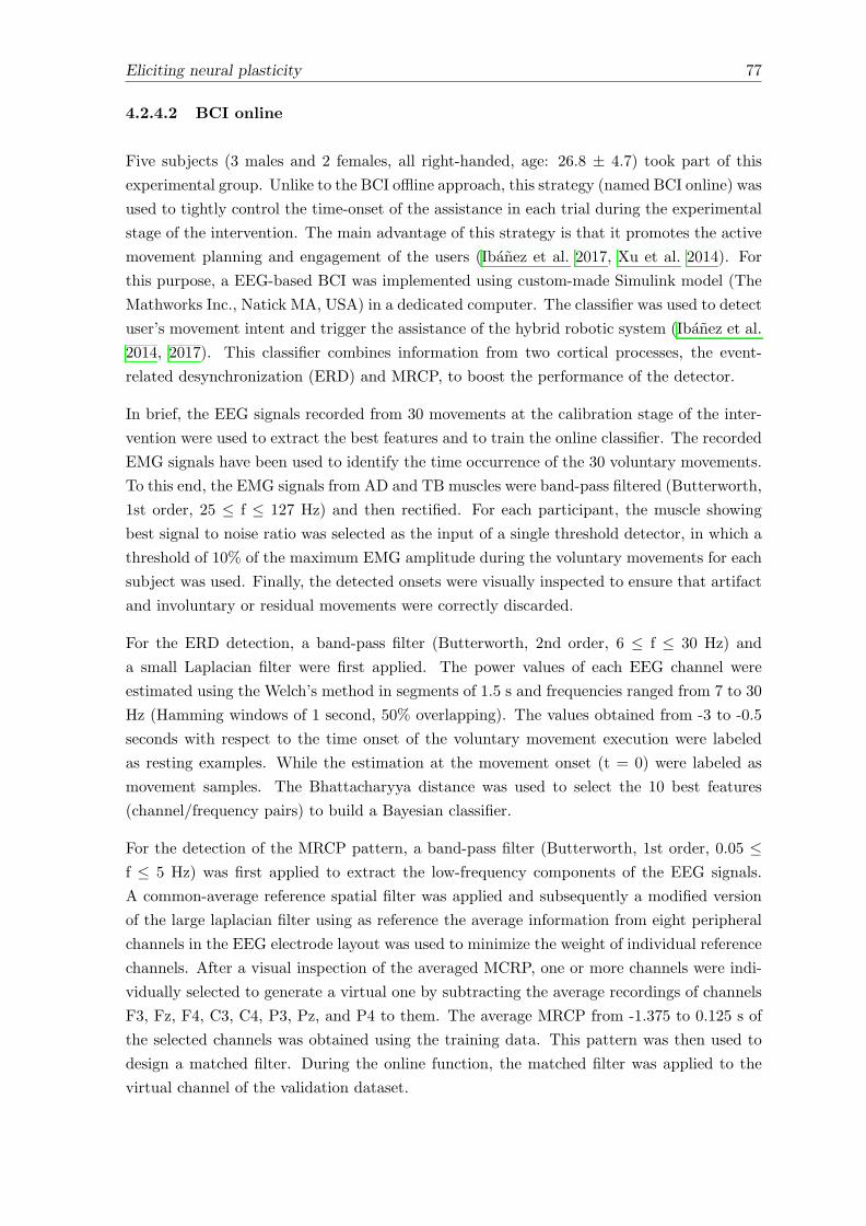
Eliciting neural plasticity 77
4.2.4.2 BCI online
Five subjects (3 males and 2 females, all right-handed, age: 26.8 ± 4.7) took part of this
experimental group. Unlike to the BCI o✏ine approach, this strategy (named BCI online) was
used to tightly control the time-onset of the assistance in each trial during the experimental
stage of the intervention. The main advantage of this strategy is that it promotes the active
movement planning and engagement of the users (Ibanez et al. 2017, Xu et al. 2014). For
this purpose, a EEG-based BCI was implemented using custom-made Simulink model (The
Mathworks Inc., Natick MA, USA) in a dedicated computer. The classifier was used to detect
user’s movement intent and trigger the assistance of the hybrid robotic system (Ibanez et al.
2014, 2017). This classifier combines information from two cortical processes, the event-
related desynchronization (ERD) and MRCP, to boost the performance of the detector.
In brief, the EEG signals recorded from 30 movements at the calibration stage of the inter-
vention were used to extract the best features and to train the online classifier. The recorded
EMG signals have been used to identify the time occurrence of the 30 voluntary movements.
To this end, the EMG signals from AD and TB muscles were band-pass filtered (Butterworth,
1st order, 25 f 127 Hz) and then rectified. For each participant, the muscle showing
best signal to noise ratio was selected as the input of a single threshold detector, in which a
threshold of 10% of the maximum EMG amplitude during the voluntary movements for each
subject was used. Finally, the detected onsets were visually inspected to ensure that artifact
and involuntary or residual movements were correctly discarded.
For the ERD detection, a band-pass filter (Butterworth, 2nd order, 6 f 30 Hz) and
a small Laplacian filter were first applied. The power values of each EEG channel were
estimated using the Welch’s method in segments of 1.5 s and frequencies ranged from 7 to 30
Hz (Hamming windows of 1 second, 50% overlapping). The values obtained from -3 to -0.5
seconds with respect to the time onset of the voluntary movement execution were labeled
as resting examples. While the estimation at the movement onset (t = 0) were labeled as
movement samples. The Bhattacharyya distance was used to select the 10 best features
(channel/frequency pairs) to build a Bayesian classifier.
For the detection of the MRCP pattern, a band-pass filter (Butterworth, 1st order, 0.05 f 5 Hz) was first applied to extract the low-frequency components of the EEG signals.
A common-average reference spatial filter was applied and subsequently a modified version
of the large laplacian filter using as reference the average information from eight peripheral
channels in the EEG electrode layout was used to minimize the weight of individual reference
channels. After a visual inspection of the averaged MCRP, one or more channels were indi-
vidually selected to generate a virtual one by subtracting the average recordings of channels
F3, Fz, F4, C3, C4, P3, Pz, and P4 to them. The average MRCP from -1.375 to 0.125 s of
the selected channels was obtained using the training data. This pattern was then used to
design a matched filter. During the online function, the matched filter was applied to the
virtual channel of the validation dataset.
78 Eliciting neural plasticity
Finally, the outputs from the ERD- and MRCP-based detectors were combined using a
logistic regression classifier. This classifier was trained by taking the output estimation of
both detectors (ERD and MRCP). The time interval from -3 to -0.5 seconds with respect to
the movement onset was considered as resting stage, while the time interval from -0.125 to
0.25 seconds was labeled as movement stage. A threshold was applied to the estimations to
decide every 5 samples (19.53 ms) if a movement intention were detected. This threshold was
optimally obtained using the calibration dataset and following the criterion of maximizing the
true positive (TP) rate, i.e. the percentage of trials with correct motor intention detection
and with no incorrect detections.
Considering that rehabilitation systems must be robust enough to avoid unexpected re-
sponses, e.g. delivering peripheral electrical pulses long before the intention to move occurs,
a contingency method to prevent subjects from receiving electrical stimuli wrongly (long be-
fore the movement intention or once movement has accomplished) was implemented. Thus,
BCI system was only enabled during the time interval from -250 ms to 1 s with respect
to the beginning of the ‘Movement’ state. This time interval is indicated with the vertical
dotted lines in the Figure 4.2b. The subjects were advised that the electrical stimuli ap-
peared whenever motor-related processes were observed within this time window and they
were instructed to report trials (movement executions) in which the FES was received too
soon or too late with respect to their expectation.
All subjects performed sets of 20 movements with their left arm until at least 50 good pairings
(also called association: electrical stimuli arriving on time with the user’s intent to move),
or 110 movements were achieved.
4.2.4.3 EMG
In eight subjects (3 males and 5 females, two left-handed, age: 30.1 ± 2.8) the EMG signal
was used to determine the assist onset, in contrast to previous strategies in which the FES
assistance was set based on the EEG signals. Here, the peripheral electrical stimuli over the
target muscles were triggered when muscle activation was identified. To this aim, an online
detector based on a single threshold was implemented using a custom-made Simulink model
(The Mathworks Inc., Natick MA, USA) in a dedicated computer.
In this detector, a band-pass filter (Butterworth, 1st order, 25 f 127 Hz) was first
applied, and the resulting signal was rectified. The rectified signal was smoothed to obtain
the envelope using a moving average filter with a windows length of 30 samples and 50%
of overlapping. The detector identified muscle activation when the envelope of the EMG
signal was higher than a predefined threshold. The detector’s threshold was set to the mean
value plus seven standard deviations of the envelope signal. These values (mean and standard
deviation) were dynamically updated for each trial, taking the envelope signal during the first
two and half seconds of the ‘Count Down’ state (when users were instructed to be relaxed).
Similarly to the BCI online group, the users’ movement onset was estimated only within
Eliciting neural plasticity 79
the same time interval from -250 ms to 1 s with respect to the beginning of the ‘Movement’
state. All subjects performed 60 trials divided in 3 blocks of 20 movements each with their
left arm.
4.2.4.4 Control experiments
Two control experiments were designed for experimentation. In the first study, five subjects
(3 males and 2 females, all right-handed, age: 30.2 ± 4.9) participated in one additional
session to verify the e↵ects of the reaching movement alone (without FES assistance and
BCI). This control experiment was conceived to verify whether the voluntary execution of
the same motor task (reaching movement with the exoskeleton) without FES assistance
produces changes in the excitability of the cortical projection to the target muscles of the
arm. Five subjects (5 males and 3 females, all right-handed, age: 28 ± 3.5) took part
in the second control experiment. In this case, the control experiment was proposed to
confirm the importance of delivering the assistance timed with the user’s intent to move.
This second control experiment consisted in triggering the FES assistance at the beginning
of the ‘Movement’ state. The same number of movement repetitions as in the BCI o✏ine
and the EMG experimental intervention were performed in both control experiments, i.e. 60
reaching movements divided in 3 blocks of 20 movements each.
4.2.5 Outcomes Measures
The primary outcome measure was the corticospinal descending motor excitability of the tar-
get muscles (AD, TB and BB muscles) measured using the peak-to-peak value of the recorded
MEP response to single-pulse TMS during the Pre0-, Pre1-, Post- and Post30-assessment.
The amplitude of the ten elicited MEP was averaged for each subject, and calculated for
di↵erent TMS intensities (90, 100, 110 120 and 130%) and assessment intervals (Pre0, Pre1,
Post and Post30) using custom-made software developed in Matlab. The average responses
of each individual at each TMS intensity and assessment interval were normalized as a func-
tion of the maximum MEP amplitude at the Pre0 assessment stage. This normalization was
conducted in order to combine all users’ response and evaluate the e↵ects of the intervention.
In addition, the EMG background activity before the TMS stimulus was quantified by cal-
culating its root mean square (RMS) value along a window interval of 30 ms. This metric
was used to verify that all subjects were in a basal condition without any voluntary muscle
contraction prior to the TMS stimuli. In addition, this metric was used to check that the
basal condition prior the TMS pulse did not di↵er between the assessment stages (Pre0,
Pre1, Post and Post30).
Complementary to the analysis of the MEP amplitude, the average negative time occurrence
of the MRCP pattern used to define the stimulation onset in the BCI o✏ine group is informed.
In the BCI online group, the performance of the BCI detector is reported. For this purpose,
80 Eliciting neural plasticity
the accuracy of the detector to estimate correctly the user’s intent to move is quantified
by the true positive (TP) rate, which is defined as the percentage of trials with movement
detection in the time interval from -0.25 to 1 second (BCI enable interval, vertical red lines
depicted in Figure 4.2b) with respect to the beginning of the movement stage (cue). Thus,
to calculate the TR rate, those movements with a good pairing (association) and those in
which the stimulus arrived soon or late (from -0.25 to 1 seconds) according to the user’s
perception are considered. Finally, the accuracy of the muscle activation onset detection and
the average detection time with respect to the cue is computed for the EMG group.
4.2.6 Statistical Analysis
The participant ages as well as the values of rMT and FES thresholds were compared to
assess whether the experimental conditions established between groups di↵ered. A one-way
ANOVA with factor ‘Group’ (1: BCI o✏ine, 2: BCI online; 3: EMG; 4: Control I; 5: Control
II) was conducted to explore whether there were any di↵erences in the participant’s age and
the rMT between groups (experimental and control groups). Three-way mixed ANOVA with
factors Group (1: BCI o✏ine, 2: BCI online; 3: EMG; 4: Control I; 5: Control II), ‘Muscle’
(1: AD and 2: TB) and ‘FES Level’ (1: minimum and 2: maximum) was considered to
evaluate di↵erences in the FES threshold established between groups.
A three-way repeated-measures ANOVA with factors ‘Time’ (1: Pre0; 2: Pre1; 3: Post; and 4:
Post30), ‘Muscle’ (1: AD; 2: TB; 3: BB) and ‘Intensity’ (1: 90%; 2: 100%; 3: 110%; 4: 120%;
and 5: 130%) was used to investigate both, the basal condition prior to the TMS stimulus
through the EMG background and the intervention’s e↵ect throughout the ‘Time’ to change
the excitability of the descending cortico-muscular pathway given by the MEP amplitude
response. The statistical analysis was performed separately for each experimental and control
group (EMG, BCI o✏ine, BCI online, Control I and Control II). Statistical significance was
assumed for p 0.05. Whenever the ANOVA revealed a significance di↵erence, a post hoc
test with Bonferroni correction was carried out to find out the specificity of the e↵ect due
the intervention.
4.3 Results
Table 4.1 shows the average participants’ age for the experimental and control groups. Addi-
tionally, the average rMT and the FES thresholds (minimum and maximum for each muscle)
across groups are presented. The one-way ANOVA reveals no di↵erences between partic-
ipant’s ages across groups neither between the established rMT (p > 0.05 in both cases).
The three-way mixed ANOVA used to identify di↵erences in applied FES thresholds be-
tween groups did not find any di↵erences when considering the factor ‘Group’ nor between
its interaction with other factors (p > 0.05 in all cases). These results indicate that similar
experimental conditions were established between groups (experimental and control groups).

Eliciting neural plasticity 81
Table 4.1: Average values of the experimental condition for each group.
Age rMT FES threshold AD FES threshold TB[years] [%] Min [mA] Max [mA] Min [mA] Max [mA]
BCI o✏ine 30.4 ± 4.3 63.4 ± 11.1 9.8 ± 2.2 14 ± 2.6 8.6 ± 2.1 11.6 ± 2.6BCI online 26.8 ± 4.7 60.6 ± 25.7 9.8 ± 2.4 13.6 ± 2.4 7.8 ± 0.8 12 ± 4.5
EMG 30.1 ± 2.8 70 ± 9.5 9.4 ± 1.3 13.3 ± 2 8.8 ± 1.2 11.3 ± 1.8Control I 30.2 ± 4.9 65.4 ± 11.9 - - - -Control II 28 ± 3.5 65.6 ± 8.1 10.8 ± 1.3 13.8 ± 2.5 10 ± 1.6 12.4 ± 1.1
Meaning of acronyms. rMT: resting motor threshold; FES: functional electrical stimulation; AD: anteriordeltoids; TB triceps brachii; Min: minimum value; Max: maximum value.
Time [s]-3 -2.5 -2 -1.5 -1 -0.5 0 0.5 1
Nor
mal
ized
EEG
am
plitu
de
-1
-0.8
-0.6
-0.4
-0.2
0
0.2
0.4
Movementstate
Figure 4.3: Normalized amplitudes of the average MRCP of all subjects (dotted linesin gray) and average MRCP across subjects (continuous black line). The vertical red line
indicates the beginning of the movement state (cue).
In all interventions (experimental and control), the statistical test confirmed similar baseline
condition prior to the TMS stimulus, measured through the EMG background, across all
assessment stages (Pre0, Pre1, Post and Post30), with p > 0.05 for the ‘Time’ factor and its
respective interactions.
4.3.1 BCI o✏ine
Figure 4.3 illustrates the MRCP signals of the subjects who took part of the BCI o✏ine
experimentation. Across all subjects, the average negative peak of the MRCP occurred at
48.34 ± 129.5 ms with respect to the beginning of the ‘Movement’ state. Thus, the FES
assistance was delivered in average at 23.34 ms after the cue.
The first column of Figure 4.4 shows the averaged DA (first row), TB (second row) and BB
(third row) MEP peak-to-peak value for all subjects plotted as a function of the rMT for each
intervention session. The MEP amplitudes for each individual muscle are expressed as a frac-
tion of the respective maximum MEP size at the Pre0-assessment. When analyzing the e↵ect
of the intervention through the MEP peak-to-peak value, the three-way repeated ANOVA
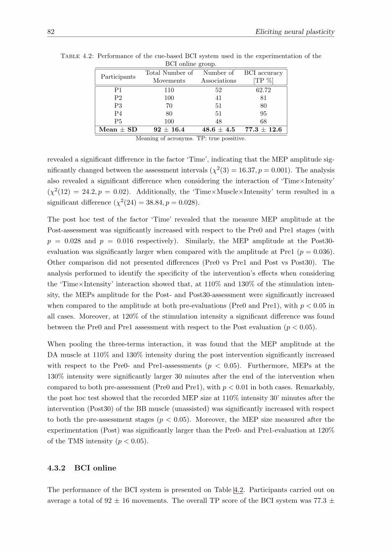
82 Eliciting neural plasticity
Table 4.2: Performance of the cue-based BCI system used in the experimentation of theBCI online group.
ParticipantsTotal Number of Number of BCI accuracy
Movements Associations [TP %]
P1 110 52 62.72P2 100 41 81P3 70 51 80P4 80 51 95P5 100 48 68
Mean ± SD 92 ± 16.4 48.6 ± 4.5 77.3 ± 12.6Meaning of acronyms. TP: true possitive.
revealed a significant di↵erence in the factor ‘Time’, indicating that the MEP amplitude sig-
nificantly changed between the assessment intervals (�2(3) = 16.37, p = 0.001). The analysis
also revealed a significant di↵erence when considering the interaction of ‘Time⇥Intensity’
(�2(12) = 24.2, p = 0.02). Additionally, the ‘Time⇥Muscle⇥Intensity’ term resulted in a
significant di↵erence (�2(24) = 38.84, p = 0.028).
The post hoc test of the factor ‘Time’ revealed that the measure MEP amplitude at the
Post-assessment was significantly increased with respect to the Pre0 and Pre1 stages (with
p = 0.028 and p = 0.016 respectively). Similarly, the MEP amplitude at the Post30-
evaluation was significantly larger when compared with the amplitude at Pre1 (p = 0.036).
Other comparison did not presented di↵erences (Pre0 vs Pre1 and Post vs Post30). The
analysis performed to identify the specificity of the intervention’s e↵ects when considering
the ‘Time⇥Intensity’ interaction showed that, at 110% and 130% of the stimulation inten-
sity, the MEPs amplitude for the Post- and Post30-assessment were significantly increased
when compared to the amplitude at both pre-evaluations (Pre0 and Pre1), with p < 0.05 in
all cases. Moreover, at 120% of the stimulation intensity a significant di↵erence was found
between the Pre0 and Pre1 assessment with respect to the Post evaluation (p < 0.05).
When pooling the three-terms interaction, it was found that the MEP amplitude at the
DA muscle at 110% and 130% intensity during the post intervention significantly increased
with respect to the Pre0- and Pre1-assessments (p < 0.05). Furthermore, MEPs at the
130% intensity were significantly larger 30 minutes after the end of the intervention when
compared to both pre-assessment (Pre0 and Pre1), with p < 0.01 in both cases. Remarkably,
the post hoc test showed that the recorded MEP size at 110% intensity 30’ minutes after the
intervention (Post30) of the BB muscle (unassisted) was significantly increased with respect
to both the pre-assessment stages (p < 0.05). Moreover, the MEP size measured after the
experimentation (Post) was significantly larger than the Pre0- and Pre1-evaluation at 120%
of the TMS intensity (p < 0.05).
4.3.2 BCI online
The performance of the BCI system is presented on Table 4.2. Participants carried out on
average a total of 92 ± 16 movements. The overall TP score of the BCI system was 77.3 ±
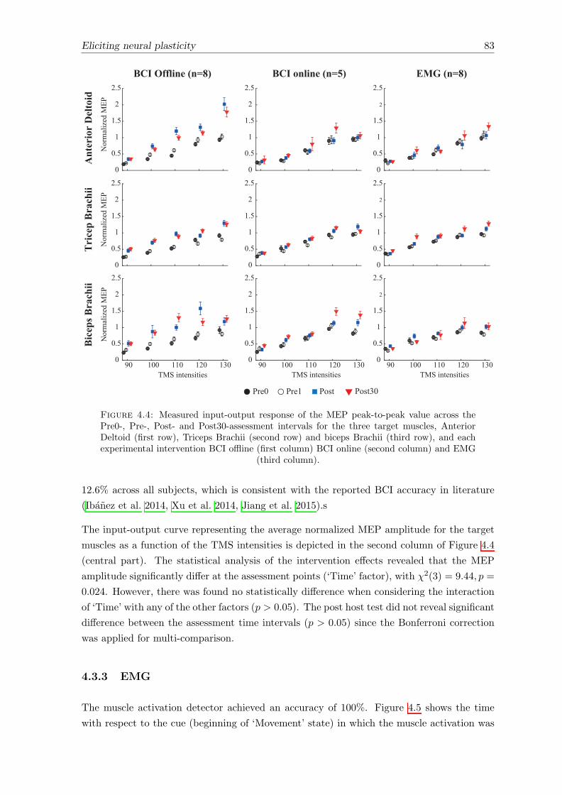
Eliciting neural plasticity 83
BCI Offline (n=8)
0
0.5
1
1.5
2
2.5
BCI online (n=5)
0
0.5
1
1.5
2
2.5
TMS intensities90 100 110 120 130
0
0.5
1
1.5
2
2.5
Nor
mal
ized
MEP
Ant
erio
r D
elto
id
0
0.5
1
1.5
2
2.5
Nor
mal
ized
MEP
Tri
cep
Bra
chii
0
0.5
1
1.5
2
2.5
TMS intensities
Nor
mal
ized
MEP
Bic
eps B
rach
ii
90 100 110 120 1300
0.5
1
1.5
2
2.5
0
0.5
1
1.5
2
2.5
EMG (n=8)
0
0.5
1
1.5
2
2.5
TMS intensities90 100 110 120 130
0
0.5
1
1.5
2
2.5
Pre0 Pre1 Post Post30
Figure 4.4: Measured input-output response of the MEP peak-to-peak value across thePre0-, Pre-, Post- and Post30-assessment intervals for the three target muscles, AnteriorDeltoid (first row), Triceps Brachii (second row) and biceps Brachii (third row), and eachexperimental intervention BCI o✏ine (first column) BCI online (second column) and EMG
(third column).
12.6% across all subjects, which is consistent with the reported BCI accuracy in literature
(Ibanez et al. 2014, Xu et al. 2014, Jiang et al. 2015).s
The input-output curve representing the average normalized MEP amplitude for the target
muscles as a function of the TMS intensities is depicted in the second column of Figure 4.4
(central part). The statistical analysis of the intervention e↵ects revealed that the MEP
amplitude significantly di↵er at the assessment points (‘Time’ factor), with �2(3) = 9.44, p =
0.024. However, there was found no statistically di↵erence when considering the interaction
of ‘Time’ with any of the other factors (p > 0.05). The post host test did not reveal significant
di↵erence between the assessment time intervals (p > 0.05) since the Bonferroni correction
was applied for multi-comparison.
4.3.3 EMG
The muscle activation detector achieved an accuracy of 100%. Figure 4.5 shows the time
with respect to the cue (beginning of ‘Movement’ state) in which the muscle activation was

84 Eliciting neural plasticity
User1
User2
User3
User4
User5
User6
User7
User8
Mov
emen
t Ons
et [s
]
-0.3
-0.2
-0.1
0
0.1
0.2
0.3
0.4
0.5
0.6
Figure 4.5: EMG movement onset detection time with respect to the cue event. Blackcircles represent individual detection (for each trial) while the red circles denote the average
detection time.
detected for each subject (black circles) and the average detection time for each subject (red
circles). On average, the onset of muscle activation occurred at -0.064 ± 0.079 seconds with
respect to the cue event.
The third column of Figure 4.4 illustrates the group averaged peak-to-peak MEP value for all
TMS intensities. When examining the changes in the MEP amplitude due to the intervention,
the statistical test revealed that the MEP size di↵ers significantly across the assessment
intervals (�2(3) = 9.05, p = 0.029), but no significance was found when considering the
interaction ‘Time’ and the other factors (p > 0.05). Due to the Bonferroni correction, the
post host test failed to find out significant di↵erence between the assessment time interval
(p > 0.05).
4.3.4 Control Experiments
Contrasting the results obtained in the experimental groups, for both control experiments (I
and II), the MEP peak-to-peak value did not presented any significant di↵erence across the
assessment intervals (‘Time’), with �2(3) = 0.482, p = 0.7 for Control I and x2(3) = 2.75, p =
0.076 for Control II. Similarly, no e↵ects were detected when considering the interaction of
‘Time’ with the other factors (p > 0.05).
4.4 Discussion
In this chapter, an associative intervention was presented, in which the previously presented
hybrid robotic system for rehabilitation of reaching was causally timed to the user’s motor
intent with the aim to promote neural plasticity. Remarkably, this is the first time that such
an associative intervention shows distributed (more than one muscle) enhancement of the
Eliciting neural plasticity 85
descending motor corticospinal projection (larger MEP size) to the assisted (AD, TB) and
unassisted (BB) muscles of the arm.
4.4.1 A comparison of the interventions’ e↵ects: BCI o✏ine, BCI online
and EMG
When comparing the e↵ects of the di↵erent strategies to associate the user’s motor intent
to the hybrid peripheral assistance, the EMG and BCI online strategies resulted in similar
results, i.e. an increase in MEP size throughout the assessment time intervals, while more
consistent and robust e↵ects were achieved with the BCI o✏ine strategy (see Figure 4.4).
Although the EMG and BCI online intervention presented an increase in MEP amplitude
immediately after and at 30’ minutes follow up, significant e↵ects were only revealed when
considering the ANOVA factor ‘Time’ individually. In contrast, the results of the BCI o✏ine
strategy revealed more reliable and specific e↵ects due the intervention. Indeed, specific
e↵ects were revealed for the Post- and Post30-assessment with respect to both pre-evaluation.
Furthermore, principal e↵ects were observed for 110%, 120% and 130% of the TMS intensity
in target muscles (AD, TB and BB).
Associative paradigms, as the one hereby introduced, are a good example to show that
plastic changes can be elicited in the central nervous system (Stefan et al. 2000, Kujirai et al.
2006, Thabit et al. 2010, Mrachacz-Kersting et al. 2012). Several studies in the literature
showed evidence that the pivotal principle underlying this plastic change (LTP-like e↵ects),
is the consistent, precise and causal synchronicity between the artificially induced a↵erent
volley reaching at the motor cortex and the movement-specific cortical activation (as the
negative peak of the MRCP) (Mrachacz-Kersting et al. 2012, Xu et al. 2014, Niazi et al.
2012, Mrachacz-Kersting et al. 2015). This timing has been demonstrated to be critical for
inducing LTP-like e↵ects (Mrachacz-Kersting et al. 2012). Consequently, the less reliable
intervention’s e↵ect obtained with the EMG intervention strategy when compared to the
BCI o✏ine one can be attributed to the non-optimal stimulus timing, since there exist an
intrinsic delay between the cortical activation of movement and the detection of the EMG
activity (Ethier et al. 2015).
The BCI o✏ine strategy represents a practical solution for implementation a EEG-based BCI
neuromodulation intervention. Under this approach, once the negative peak of the MRCP is
determined, the time to deliver the peripheral stimulus is kept fixed regardless of the mental
or engagement condition of the users. From the rehabilitation perspective, the BCI online
strategy could be seen as more engaging by subjects and better suited for eliciting Hebbian-
based neuroplasticity (Xu et al. 2014, Niazi et al. 2012, Ibanez et al. 2017). The rationale
underlying this strategy is the capability of providing a reliable and timely estimation of the
user’s movement intent, thus adapting the onset of the stimuli according to the inter-trial
variability of the motor-related cortical processes (ERD and MRCP). However, the online
detection of motor-related cortical activities, such as MRCP and ERD, is still challenging
due to noise and artifacts in the raw EEG (Ethier & Miller 2015, Daly & Wolpaw 2008).
86 Eliciting neural plasticity
Although the BCI online detector implemented in this chapter presented similar accuracy to
the data reported in literature (Xu et al. 2014, Niazi et al. 2012, Ibanez et al. 2014, Ibanez
et al. 2014), the less reliable intervention’s e↵ects of the BCI online intervention compared
with BCI o✏ine can be attributed to the intrinsic latencies existing when implementing
online detectors. Consequently, further attention must be paid to reduce latencies and so
that a tight coupling between the hybrid assistance and the motor-related cortical processes
can be achieved.
Results presented in this chapter show that the BCI o✏ine strategy is a robust strategy to
use BCI technology as a neuromodulation technique. Our results are in line with recent
studies reported in literature (Mrachacz-Kersting et al. 2012, 2015). This method is mainly
supported by the fact that the MRCP remains stable within one recording session (Mrachacz-
Kersting et al. 2012). It was also demonstrated that there is no significant variation of this
cortical process over di↵erent recording sessions (Mrachacz-Kersting et al. 2012). All in all,
we can conclude that the more e↵ective results achieved with the BCI o✏ine intervention are
likely due to the consistent activation of the primary motor cortex via neural information
coming from other brain regions (MRCP generation (Lu et al. 2012)) and from the peripher-
ally generated sensory feedback reaching the brain from stimulated target muscles of the arm.
This precise and coherent activation is assumed to be responsible for the achieved significant
increase of the corticospinal projections of target muscles (stimulated and non-stimulated
muscles).
Evidence from literature and the results of the control groups demonstrated that plastic
changes cannot be elicited in the absence of these coherent activations (Mrachacz-Kersting
et al. 2012, Xu et al. 2014). In fact, performing the reaching task alone did not present any
plastic change (control I). It is worth mentioning that the cue and the peak negativity of
the MRCP do not necessarily occur at the same time (see Figure 4.3). Although the time
occurrence of this peak (as a function of the cue) is repeatable within one subject between
days (Mrachacz-Kersting et al. 2012), the variability across subjects is in the order of a few
hundred milliseconds. This variability occurs due to the user’s capacity to react to a cue.
Again, by taking the time occurrence of the peak negativity, it was ensuring that the a↵erent
stimulation arrived during the subjects’ own intention to move.
It is well known that plastic changes can be elicited in the nervous system at di↵erent levels
(e.g. cortical (Stefan et al. 2000) or spinal (Wolpaw 2007) level). Although no measure was
performed to check the location of changes presented in this chapter, from evidence presented
in literature it can be indicated that the plastic changes occur at supra-spinal (cortical) level
(Mrachacz-Kersting et al. 2012, Xu et al. 2014, Niazi et al. 2012).
4.4.2 Comparison with similar studies presented in literature
Former studies consisting in the use of BCI technology as a neuromodulation system to
promote upper-limb motor recovery have been based on sensorimotor rhythms, specifically
Eliciting neural plasticity 87
ERD and event-related synchronization (ERS) cortical processes (Ramos-Murguialday et al.
2013, Pichiorri et al. 2011, 2015, McGie et al. 2015, Kraus et al. 2016). In these studies,
the SMR cortical activity was used to detect the subject’s movement intent. The principal
drawback of these methods is the need for numerous training sessions until subjects are able
to control the signal adequately. However, MRCPs are characterized by occurring naturally
as a person starts or imagines a movement, and thus, they do not require prior training
(Garipelli et al. 2013, Jiang et al. 2015, Xu et al. 2014).
The study presented by McGie et al. compared the e↵ects of the BCI- and EMG-based
control of FES during grasping with healthy subjects using changes in MEP size as the
primary outcome (McGie et al. 2015). In this study, an ERD-based online detector was used
to estimate the movement onset during the BCI intervention. The authors found an increase
in MEP size following BCI-FES and, especially, EMG-FES interventions. However, these
changes were not significant. In contrast, the MRCP-based intervention hereby introduced
clearly illustrates the appropriateness of using the MRCP to promote neural plasticity.
The somatosensory feedback elicited by FES (muscle spindles, mechanoreceptors, propriocep-
tive information, etc.), the execution of a multi-degree of freedom functional movement (high
functional implication) and the voluntary action for executing the reaching task (voluntary
e↵ort) constitute the key features of our approach. In a previous BCI study with chronic
stroke patients, the importance of the somatosensory feedback was emphasized (Ono et al.
2014). In this study, patients who received somatosensory feedback by an active hand or-
thosis triggered by brain signals (ERD events) led to functional improvements after therapy,
while no changes were observed in the group of patients who only received visual feedback.
The contribution of agonist and antagonist muscles and all other a↵erents involved during
the execution of highly functional tasks, such as the reaching, might play an important
role in relaying enough information to the cerebral cortex (Mrachacz-Kersting et al. 2015).
Similarly, the e↵ect of FES to elicit cortical plasticity during the execution of functional
movements was shown to be enhanced when it was combined with volitional e↵ort (Barsi
et al. 2008). Therefore, the strong and distributed intervention e↵ects observed with our
BCI o✏ine strategy can be attributed to the consistent integration of the voluntary action
and the timed peripheral stimulation during the execution of a highly functional task, which
results in inputs to the motor cortex with relevant agonist and antagonist information.
4.4.3 Implications for rehabilitation of upper-limb motor function
It is currently well accepted that sensorimotor and functional improvements have neurophysi-
ological correlates resulting from plastic changes within the central nervous system (Beaulieu
& Milot 2017, Stinear 2010). Indeed, the presence of MEPs in acute stroke subjects is an
important prognosis factor for motor and functional recovery of both lower and upper limbs
(Hendricks et al. 2003a,b, Pizzi et al. 2009, Piron et al. 2005). Noticeably, it was also found
that functional recovery in acute stroke patients is directly correlated with the propensity
of inducing LTP-like changes in the a↵ected and una↵ected motor cortex (Di Lazzaro et al.
88 Eliciting neural plasticity
2010). Similarly and more recently, Mrachacz-Kersting et. al. demonstrated that an asso-
ciative BCI approach induced LTP-like e↵ects measured as an increase in MEPs size, and
remarkably, these plastic changes resulted in functional improvement of the lower limbs with
only three interventional sessions (Mrachacz-Kersting et al. 2015). This evidence and the
results presented in this chapter provide strong support for the use of neuromodulation tech-
niques, as the associative BCI concept, to facilitate a well-adaptive plasticity and promote
motor recovery after stroke. In line with these facts, it is hypothesized that the hybrid system
presented throughout this dissertation could eventually lead to improvements of the upper
limb motor function in stroke patients.
4.5 Conclusion of the chapter
The spontaneous brain plasticity (physiological and anatomical) occurring after a stroke re-
presents one of the most important neurological processes underlying the recovery of motor
functions. This important fact has changed the scope of current rehabilitation therapies,
which explore alternative rehabilitative methods with the goal of exploiting the natural plas-
ticity of the sensorimotor system, and hence, improving motor functional recovery. Pieces of
evidence reviewed in this chapter support the potential of using a BCI as a neuromodulation
system to elicit well-adaptive plasticity.
In this chapter, the hybrid assistance delivered during reaching was tightly coupled to the
user’s motor intent with the aim of eliciting neural plasticity (associative intervention). Three
di↵erent approaches (BCI o✏ine, BCI online and EMG) were implemented to verify the po-
tential e↵ects of the associative intervention. Results with healthy participants revealed
positive intervention’s e↵ects in all implemented strategies since significant di↵erences in
MEPs size measure on target muscles were obtained after the interventions when considering
uniquely the factor ‘Time’ (Pre vs Post and Pre vs Post30). However, stronger and more
consistent intervention’s e↵ects were achieved with the BCI o✏ine intervention. Remarkably,
a distributed e↵ect was achieved with this strategy (BCI o✏ine), an increase in excitabil-
ity of the corticospinal motor projections to the assisted (DA, TR) and unassisted (BB)
muscles. Because of the e�cacy of this intervention (BCI o✏ine), it represents an e�cient
neuromodulation strategy for inducing plastic changes into the motor cortex.
In view of the work presented by Mrachacz-Kersting et al., in which changes in MEP size
of the tibialis anterior muscle through three BCI associative interventional sessions were
correlated with improvements in gait function (Mrachacz-Kersting et al. 2015), and the
results presented in this chapter, a plausible hypothesis would be that the use of the hybrid
robotic system based on the adaptive and associative assistance will result in improvement
of the upper limb motor function in stroke patients. The study presented in the following
chapter is planned to address this hypothesis.

Chapter 5
Clinical evaluation of the hybrid
robotic system with the adaptive
and associative assistance in
post-stroke patients
Abstract
In the previous chapter, we showed that the causal association between motor cortex activa-
tion and the hybrid peripheral stimulation resulted in a facilitation of the cortico-muscular
pathways. In this chapter, a pilot clinical study with a small cohort of port-stroke subjects
is presented with the aim of proving the potential benefits of the hybrid robotic system. The
interventional protocol consists of 12 training sessions along four consecutive weeks, in which
post-stroke participants carried out reaching movements assisted with the hybrid robotic sys-
tem. The BCI o✏ine strategy was implemented to associate the user’s intent to peripheral
assistance. The analysis of the patient’s ability for executing unassisted reaching tasks along
sessions and the assessment of changes in patients’ clinical scales before and after the inter-
vention period were conducted to study the potential impact of the intervention. Complemen-
tarily, the user’s perceived value of using the system as a rehabilitative tool was evaluated.
Moderately a↵ected participant showed an improvement between 40% to 60% in performance
when executing unassisted reaching movement. Likewise, the manual muscle and the box and
block tests showed important improvement of the a↵ected upper extremity. Severely a↵ected
participants did not show improvement in the execution of unassisted reaching movements.
Clinical scales revealed a marginal improvement in the muscle manual test. The overall pa-
tients’ assessment of the therapy reflected their acceptance of the proposed interventional
protocol. The results presented in this chapter demonstrate the feasibility of using the hybrid
robotic system based on the adaptive and associative assistance for post-stroke rehabilitation.
89
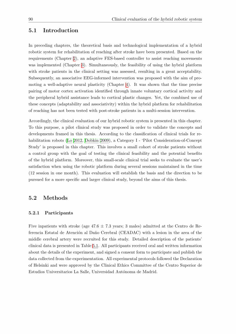
90 Clinical evaluation of the hybrid robotic system
5.1 Introduction
In preceding chapters, the theoretical basis and technological implementation of a hybrid
robotic system for rehabilitation of reaching after stroke have been presented. Based on the
requirements (Chapter 2), an adaptive FES-based controller to assist reaching movements
was implemented (Chapter 3). Simultaneously, the feasibility of using the hybrid platform
with stroke patients in the clinical setting was assessed, resulting in a great acceptability.
Subsequently, an associative EEG-informed intervention was proposed with the aim of pro-
moting a well-adaptive neural plasticity (Chapter 4). It was shown that the time precise
pairing of motor cortex activation identified through innate voluntary cortical activity and
the peripheral hybrid assistance leads to cortical plastic changes. Yet, the combined use of
these concepts (adaptability and associativity) within the hybrid platform for rehabilitation
of reaching has not been tested with post-stroke patients in a multi-session intervention.
Accordingly, the clinical evaluation of our hybrid robotic system is presented in this chapter.
To this purpose, a pilot clinical study was proposed in order to validate the concepts and
developments framed in this thesis. According to the classification of clinical trials for re-
habilitation robots (Lo 2012, Dobkin 2009), a Category I - ‘Pilot Consideration-of-Concept
Study’ is proposed in this chapter. This involves a small cohort of stroke patients without
a control group with the goal of testing the clinical feasibility and the potential benefits
of the hybrid platform. Moreover, this small-scale clinical trial seeks to evaluate the user’s
satisfaction when using the robotic platform during several sessions maintained in the time
(12 session in one month). This evaluation will establish the basis and the direction to be
pursued for a more specific and larger clinical study, beyond the aims of this thesis.
5.2 Methods
5.2.1 Participants
Five inpatients with stroke (age 47.6 ± 7.3 years; 3 males) admitted at the Centro de Re-
ferencia Estatal de Atencion al Dano Cerebral (CEADAC) with a lesion in the area of the
middle cerebral artery were recruited for this study. Detailed description of the patients’
clinical data is presented in Table 5.1. All participants received oral and written information
about the details of the experiment, and signed a consent form to participate and publish the
data collected from the experimentation. All experimental protocols followed the Declaration
of Helsinki and were approved by the Clinical Ethics Committee of the Centro Superior de
Estudios Universitarios La Salle, Universidad Autonoma de Madrid.
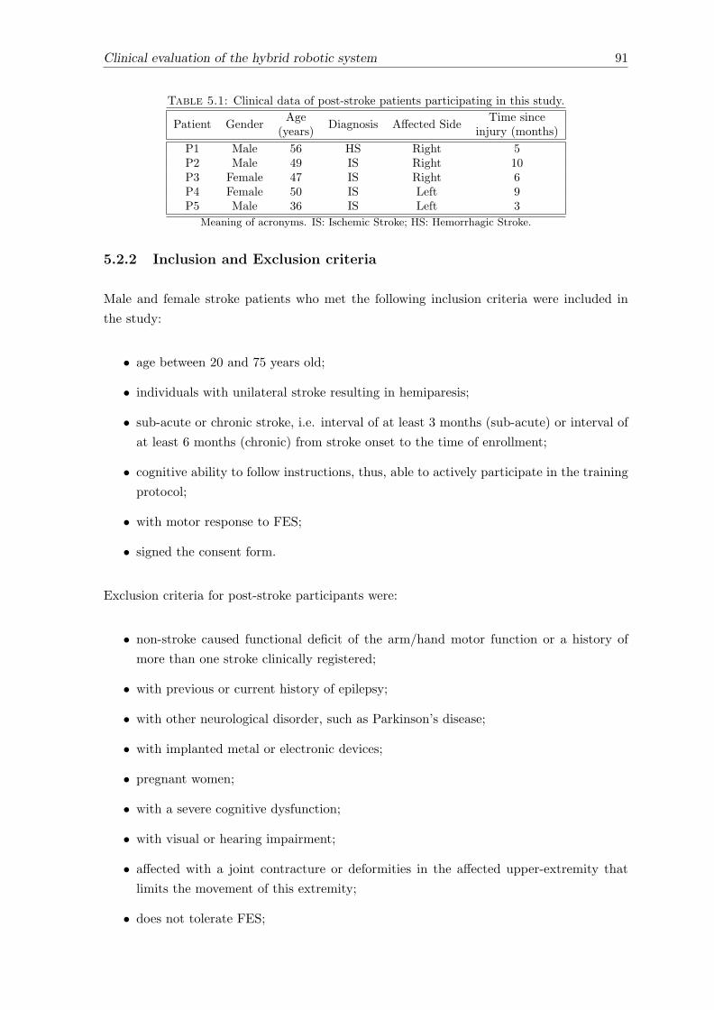
Clinical evaluation of the hybrid robotic system 91
Table 5.1: Clinical data of post-stroke patients participating in this study.
Patient GenderAge
Diagnosis A↵ected SideTime since
(years) injury (months)
P1 Male 56 HS Right 5P2 Male 49 IS Right 10P3 Female 47 IS Right 6P4 Female 50 IS Left 9P5 Male 36 IS Left 3
Meaning of acronyms. IS: Ischemic Stroke; HS: Hemorrhagic Stroke.
5.2.2 Inclusion and Exclusion criteria
Male and female stroke patients who met the following inclusion criteria were included in
the study:
• age between 20 and 75 years old;
• individuals with unilateral stroke resulting in hemiparesis;
• sub-acute or chronic stroke, i.e. interval of at least 3 months (sub-acute) or interval of
at least 6 months (chronic) from stroke onset to the time of enrollment;
• cognitive ability to follow instructions, thus, able to actively participate in the training
protocol;
• with motor response to FES;
• signed the consent form.
Exclusion criteria for post-stroke participants were:
• non-stroke caused functional deficit of the arm/hand motor function or a history of
more than one stroke clinically registered;
• with previous or current history of epilepsy;
• with other neurological disorder, such as Parkinson’s disease;
• with implanted metal or electronic devices;
• pregnant women;
• with a severe cognitive dysfunction;
• with visual or hearing impairment;
• a↵ected with a joint contracture or deformities in the a↵ected upper-extremity that
limits the movement of this extremity;
• does not tolerate FES;
92 Clinical evaluation of the hybrid robotic system
• with skin lesion that may hinder or prevent the application of FES or the upper-limb
exoskeleton;
• ingesting any medical drug that would prevent the application of standard rehabilita-
tion;
• declined to sign the consent form.
5.2.3 Materials
The hybrid robotic system presented in Chapter 3 and Chapter 4 was used to assist the
execution of reaching during the intervention sessions (see Section 3.2 and Section 4.2.2.2 for
further details). The same EEG and EMG acquisition system with the same configuration,
as explained in Section 4.2.2.1, were used to provide timed assistance with respect to the
user’s motor-related cortical process (associative assistance). Likewise, the same cue-based
visual interface (as explained in Section 4.2.3.2) was utilized to guide the user throughout
the training sessions and, to provide information regarding the arm position (shoulder and
elbow) and the task’s performance. Figure 5.1 shows one stroke participant performing the
reaching exercise with our hybrid platform. The general scheme of operation, the principal
functions and components are also shown.
5.2.4 Study design
The study proposed in this chapter consists of a rehabilitation therapy based on the hybrid
robotic system to recover the arm motor function through training of reaching movements.
Patients with stroke underwent this experimental therapy in addition to their usual thera-
peutic routine as prescribed and supplied by clinical experts at CEADAC.
The study protocol was registered retrospectively in the ISRCTN register with study ID
ISRCTN128430061. he general structure of this clinical pilot study protocol is shown in
Figure 5.2. The study consists in three principal stages: the initial assessment, the invention
and the final assessment. These stages are following explained.
5.2.4.1 Initial assessment (Ev1)
The initial assessment consisted in two separated sessions: assessment and MRCP’s relia-
bility, which took place in di↵erent days. The assessment session was aimed to obtain the
baseline motor and functional conditions of the stroke patients participating in this study
(see Section 5.2.5.1). While the MRCP’s reliability session was conceived to introduce the
system operation to the users, to ascertain the suitability of the patients for using the hybrid
1Use of neuromodulation system and assistive devices for rehabilitation of upper limb motor function afterstroke (ISRCTN12843006); DOI: 10.1186/ISRCTN12843006. Available: https://goo.gl/6MqqXd (last visit:15/08/2017).
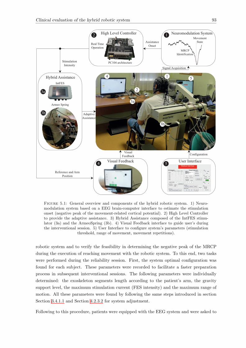
Clinical evaluation of the hybrid robotic system 93
!
"
#$
#%
& '
()*+,-,.*/$01,234560)-
789:;.)201<1=$01,23
7,>)-)20340$0)
!
:9!?&3$+=@10)=0*+)3
A1B@3C)>)/39,20+,//)+
8)$/3D1-)EF)+$01,2
"
G16*$/3H)).%$=I& J6)+3;20)+<$=)'
A5%+1.3K66160$2=);20HL4
K+-),34F+12B
#$
#%
K66160$2=)E26)03
401-*/$01,23;20)26105
K.$F01>)K66160$2=)3
G16*$/H)).%$=I 9,2<1B*+$01,2
41B2$/3K=M*16101,2
8)<)+)2=)3$2.3K+-:,6101,2
Figure 5.1: General overview and components of the hybrid robotic system. 1) Neuro-modulation system based on a EEG brain-computer interface to estimate the stimulationonset (negative peak of the movement-related cortical potential). 2) High Level Controllerto provide the adaptive assistance. 3) Hybrid Assistance composed of the IntFES stimu-lator (3a) and the ArmeoSpring (3b). 4) Visual Feedback interface to guide user’s duringthe interventional session. 5) User Interface to configure system’s parameters (stimulation
threshold, range of movement, movement repetitions).
robotic system and to verify the feasibility in determining the negative peak of the MRCP
during the execution of reaching movement with the robotic system. To this end, two tasks
were performed during the reliability session. First, the system optimal configuration was
found for each subject. These parameters were recorded to facilitate a faster preparation
process in subsequent interventional sessions. The following parameters were individually
determined: the exoskeleton segments length according to the patient’s arm, the gravity
support level, the maximum stimulation current (FES intensity) and the maximum range of
motion. All these parameters were found by following the same steps introduced in section
Section 3.4.1.1 and Section 4.2.3.2 for system adjustment.
Following to this procedure, patients were equipped with the EEG system and were asked to
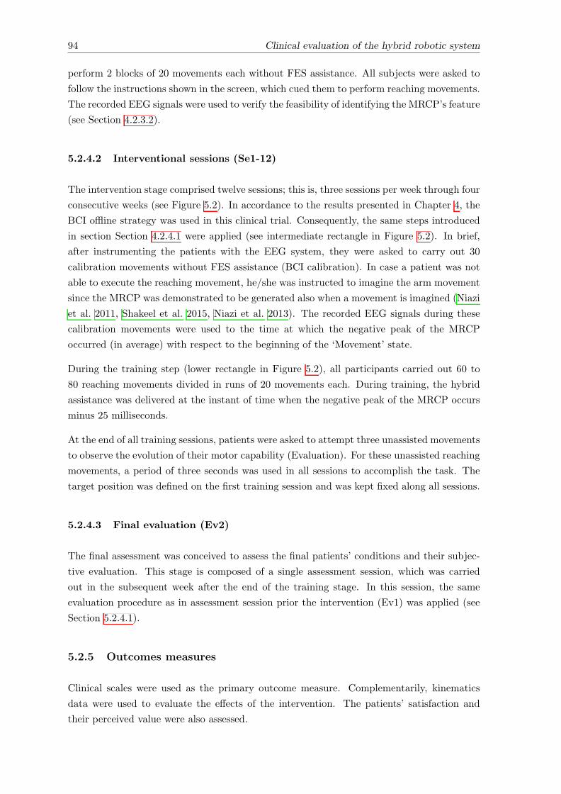
94 Clinical evaluation of the hybrid robotic system
perform 2 blocks of 20 movements each without FES assistance. All subjects were asked to
follow the instructions shown in the screen, which cued them to perform reaching movements.
The recorded EEG signals were used to verify the feasibility of identifying the MRCP’s feature
(see Section 4.2.3.2).
5.2.4.2 Interventional sessions (Se1-12)
The intervention stage comprised twelve sessions; this is, three sessions per week through four
consecutive weeks (see Figure 5.2). In accordance to the results presented in Chapter 4, the
BCI o✏ine strategy was used in this clinical trial. Consequently, the same steps introduced
in section Section 4.2.4.1 were applied (see intermediate rectangle in Figure 5.2). In brief,
after instrumenting the patients with the EEG system, they were asked to carry out 30
calibration movements without FES assistance (BCI calibration). In case a patient was not
able to execute the reaching movement, he/she was instructed to imagine the arm movement
since the MRCP was demonstrated to be generated also when a movement is imagined (Niazi
et al. 2011, Shakeel et al. 2015, Niazi et al. 2013). The recorded EEG signals during these
calibration movements were used to the time at which the negative peak of the MRCP
occurred (in average) with respect to the beginning of the ‘Movement’ state.
During the training step (lower rectangle in Figure 5.2), all participants carried out 60 to
80 reaching movements divided in runs of 20 movements each. During training, the hybrid
assistance was delivered at the instant of time when the negative peak of the MRCP occurs
minus 25 milliseconds.
At the end of all training sessions, patients were asked to attempt three unassisted movements
to observe the evolution of their motor capability (Evaluation). For these unassisted reaching
movements, a period of three seconds was used in all sessions to accomplish the task. The
target position was defined on the first training session and was kept fixed along all sessions.
5.2.4.3 Final evaluation (Ev2)
The final assessment was conceived to assess the final patients’ conditions and their subjec-
tive evaluation. This stage is composed of a single assessment session, which was carried
out in the subsequent week after the end of the training stage. In this session, the same
evaluation procedure as in assessment session prior the intervention (Ev1) was applied (see
Section 5.2.4.1).
5.2.5 Outcomes measures
Clinical scales were used as the primary outcome measure. Complementarily, kinematics
data were used to evaluate the e↵ects of the intervention. The patients’ satisfaction and
their perceived value were also assessed.
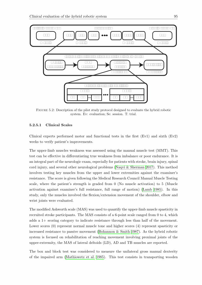
Clinical evaluation of the hybrid robotic system 95
Intervention
Week 2
Se1 Se1 Se3
Week 5
Se10 Se11 Se12
Week 1
Ev1
Week 6
Ev2
Final AssessmentInitial Assessment
Interventional sessions procedure: ~60 to 80 minutesUser’s
PreparationBCI
Training EvaluationTrainingSession
Training session: 60-80 trials
T20T1 T2
Run 1
T20T1 T2
Run N
Assistance Unassisted
Figure 5.2: Description of the pilot study protocol designed to evaluate the hybrid roboticsystem. Ev: evaluation; Se: session. T: trial.
5.2.5.1 Clinical Scales
Clinical experts performed motor and functional tests in the first (Ev1) and sixth (Ev2)
weeks to verify patient’s improvements.
The upper-limb muscles weakness was assessed using the manual muscle test (MMT). This
test can be e↵ective in di↵erentiating true weakness from imbalance or poor endurance. It is
an integral part of the neurologic exam, especially for patients with stroke, brain injury, spinal
cord injury, and several other neurological problems (Naqvi & Sherman 2017). This method
involves testing key muscles from the upper and lower extremities against the examiner’s
resistance. The score is given following the Medical Research Council Manual Muscle Testing
scale, where the patient’s strength is graded from 0 (No muscle activation) to 5 (Muscle
activation against examiner’s full resistance, full range of motion) (Lamb 1985). In this
study, only the muscles involved the flexion/extension movement of the shoulder, elbow and
wrist joints were evaluated.
The modified Ashworth scale (MAS) was used to quantify the upper-limb muscle spasticity in
recruited stroke participants. The MAS consists of a 6-point scale ranged from 0 to 4, which
adds a 1+ scoring category to indicate resistance through less than half of the movement.
Lower scores (0) represent normal muscle tone and higher scores (4) represent spasticity or
increased resistance to passive movement (Bohannon & Smith 1987). As the hybrid robotic
system is focused on rehabilitation of reaching movement involving proximal joints of the
upper-extremity, the MAS of lateral deltoids (LD), AD and TB muscles are reported.
The box and block test was considered to measure the unilateral gross manual dexterity
of the impaired arm (Mathiowetz et al. 1985). This test consists in transporting wooden

96 Clinical evaluation of the hybrid robotic system
blocks from one compartment to another, and the scoring method is the number of blocks
transported within a period of 1 minute.
5.2.5.2 Kinematic data
The three unassisted movements (evaluation) attempted at the end of each training session
are used to quantify the evolution of the kinematics pattern during the reaching execution.
For each session, the RMSE of each joint (shoulder and elbow) and the task’s performance
is calculated (see Section 3.4.2.2 for further details). A best-fitting linear regression across
the RMSE and performance score is applied to examine its trend along sessions.
5.2.5.3 Satisfaction assessment
Similar to the evaluation performed in Chapter 3, the QUEST and SAM tests are considered
to evaluate the user’s satisfaction, perceived comfort, and acceptability of the hybrid platform
after the interventional sessions.
In brief, the QUEST is an instrument specifically designed to measure satisfaction for a broad
range of assistive technology devices in a structured and standardized way (Demers et al.
2002). The test includes 12 items, which are related to device characteristics (n = 8) and
assistive technology services (n = 4). The scoring method rates from 1 (not satisfied at all)
to 5 (very satisfied). Only the items related to the device characteristics were used for this
study, due to the lack of external assistive services. Therefore, the maximum possible score
was 5 for each item, and 35 for the total scale.
Self-Assessment Manikin (SAM) is a non-verbal pictorial assessment technique that directly
measures the pleasure, arousal, and dominance associated with a person’s a↵ective reaction
to a wide variety of stimuli (Hodes et al. 1985). For pleasure, SAM ranges from a smiling,
happy figure to a frowning, unhappy figure; and for arousal, from sleepy with eyes closed to
excited with eyes open figure. The dominance dimension represents changes in control with
changes in the size of SAM: a large figure indicates maximum control in the situation. The
subject can place a mark over a continuous nine-point scale.
5.3 Results
Data presented in this section is based on three stroke participants (P1, P2, and P5), since
two participants (P3 and P4) left the experimental training due to external reasons. One
participant (P3) developed a neuropathic contracture in muscles of the a↵ected arm after
the second week hindering him to carry out reaching movements and receiving electrical
stimulation. The other subject (P4) left the study at the third week of the intervention due
to personal problems.
Clinical evaluation of the hybrid robotic system 97
-6
-4
-2
0
2
4
6
Time [s]
Nor
mal
ized
MR
CP
Curve (C2)
-3 -2 -1 0 1 2-1
-0.8
-0.6
-0.4
-0.2
0
0.2
0.4
0.6Movement
State
Spatial distribution
Figure 5.3: Motor-related cortical potential (MRCP) of the P1 patient. The MRCP’s curveof the most representative EEG channel (C2) is shown in the first column while the spatialdistribution of the MRCP is illustrated in the second column. To optimize visualization,baseline was defined within [-3, -2] seconds with respect to the movement state (vertical red
line at t = 0).
For a better understanding of the data presented in this section, the three stroke participants
who completed the full experimental protocol (P1, P2 and P5) were divided into two groups
according to their level of motor impairment (severe and moderate arm motor impairment).
Participants scoring 0 or 1 in the MMT test, indicating that they cannot move their arm
voluntary at all or have very limited voluntary movements, were classified as severe. Par-
ticipants scoring higher than 1 in the MMT test were classified as moderate. Consequently,
stroke patients P2 and P5 were classified into the severe group, while P1 was allocated to
the moderate group.
5.3.1 Patient with moderate arm motor impairment: P1
5.3.1.1 Feasibility of the MRCP
The negative peak of the MRCP was used to establish the instant of time with respect to
the visual cue in which the hybrid assistance is delivered. Figure 5.3 shows the average
MRCP of participant P1 during the MRCP’s reliability session of the initial assessment and
its spatial distribution. Fieldtrip’s ft multiplotER and ft topoplotER functions in Matlab
(The Mathworks Inc., Natick MA, USA) were used to obtain the patterns (Oostenveld et al.
2011). The negative peak of the illustrated MRCP occurs at 0.8672 seconds after the visual
cue (movement state). For this individual stroke patient, the averaged negative peak event
of the MRCP across all sessions occurred at 0.9512 ± 0.0664 seconds with respect to the
visual cue. Thus, for this individual subject, the hybrid assistance was delivered at 0.9296
seconds after the movement state.
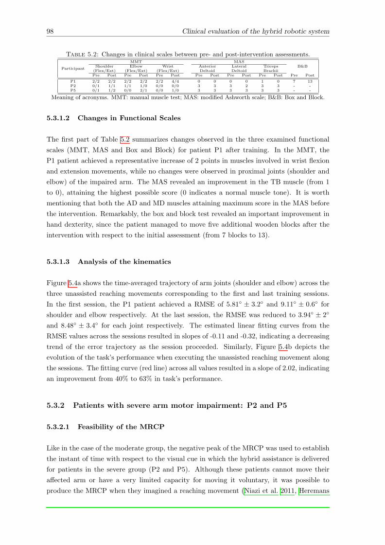
98 Clinical evaluation of the hybrid robotic system
Table 5.2: Changes in clinical scales between pre- and post-intervention assessments.
Participant
MMT MASB&BShoulder Elbow Wrist Anterior Lateral Triceps
(Flex/Ext) (Flex/Ext) (Flex/Ext) Deltoid Deltoid BrachiiPre Post Pre Post Pre Post Pre Post Pre Post Pre Post Pre Post
P1 2/2 2/2 2/2 2/2 2/2 4/4 0 0 0 0 1 0 7 13P2 0/1 1/1 1/1 1/0 0/0 0/0 3 3 3 2 3 3 - -P5 0/1 1/2 0/0 2/1 0/0 1/0 3 3 3 3 3 3 - -
Meaning of acronyms. MMT: manual muscle test; MAS: modified Ashworth scale; B&B: Box and Block.
5.3.1.2 Changes in Functional Scales
The first part of Table 5.2 summarizes changes observed in the three examined functional
scales (MMT, MAS and Box and Block) for patient P1 after training. In the MMT, the
P1 patient achieved a representative increase of 2 points in muscles involved in wrist flexion
and extension movements, while no changes were observed in proximal joints (shoulder and
elbow) of the impaired arm. The MAS revealed an improvement in the TB muscle (from 1
to 0), attaining the highest possible score (0 indicates a normal muscle tone). It is worth
mentioning that both the AD and MD muscles attaining maximum score in the MAS before
the intervention. Remarkably, the box and block test revealed an important improvement in
hand dexterity, since the patient managed to move five additional wooden blocks after the
intervention with respect to the initial assessment (from 7 blocks to 13).
5.3.1.3 Analysis of the kinematics
Figure 5.4a shows the time-averaged trajectory of arm joints (shoulder and elbow) across the
three unassisted reaching movements corresponding to the first and last training sessions.
In the first session, the P1 patient achieved a RMSE of 5.81� ± 3.2� and 9.11� ± 0.6� for
shoulder and elbow respectively. At the last session, the RMSE was reduced to 3.94� ± 2�
and 8.48� ± 3.4� for each joint respectively. The estimated linear fitting curves from the
RMSE values across the sessions resulted in slopes of -0.11 and -0.32, indicating a decreasing
trend of the error trajectory as the session proceeded. Similarly, Figure 5.4b depicts the
evolution of the task’s performance when executing the unassisted reaching movement along
the sessions. The fitting curve (red line) across all values resulted in a slope of 2.02, indicating
an improvement from 40% to 63% in task’s performance.
5.3.2 Patients with severe arm motor impairment: P2 and P5
5.3.2.1 Feasibility of the MRCP
Like in the case of the moderate group, the negative peak of the MRCP was used to establish
the instant of time with respect to the visual cue in which the hybrid assistance is delivered
for patients in the severe group (P2 and P5). Although these patients cannot move their
a↵ected arm or have a very limited capacity for moving it voluntary, it was possible to
produce the MRCP when they imagined a reaching movement (Niazi et al. 2011, Heremans

Clinical evaluation of the hybrid robotic system 99
Number of session
Perf
orm
ance
ReferenceSession 1Session 12
0 0.5 1 1.5 2 2.5 3
Shou
lder
(Ø2)
-0.5
0
0.5
1
1.5
Time [s]0 0.5 1 1.5 2 2.5 3
-1
-0.5
0
Elbo
w (Ø
5)N
orm
aliz
ed A
ngle
s
a) b)
1 2 3 4 5 6 7 8 9 10 11 120
20
40
60
80
100
Figure 5.4: a) Representative example of the tracking accuracy during the execution of theunassisted movement. Joints trajectories represent the time-averaged response of the firstand last interventional sessions. b) Achieved task’s performance score during the executionof the unassisted reaching movements along sessions. The solid line represents the best linearfitting indicating the trend of the score. See Section 3.4.2.2 for the task’s performance score.
et al. 2009, do Nascimento & Farina 2008). Figure 5.5 shows the MRCP curves from the most
reactive EEG electrode and the spatial distributions for both patients during the MRCP’s
reliability session of the initial assessment. Plots were obtained using the same fieldtrip’s
functions previously cited (see Section 5.3.1.1). As shown in the figure, the occurrence of
the negative peak in the MRCP curve varies between both patients, as well as its spatial
distribution. For P2, the negative peak of the MRCP (upper plot in the first column) occurs
at 0.09766 seconds after the visual cue (movement state). When analyzing this peak along the
intervention sessions, this event occurred in average at 0.0583± 0.0435 seconds with respect
to the movement state. The negativity peak in the plotted MRCP curve of patients P5 (lower
plot in the first column) took place at 0.7344 with respect to the cue. For this subject, the
average negative peak across all sessions happened at 0.5918 ± 0.2183. Consequently, the
hybrid assistance was delivered in average at 0.0333 and 0.5668 for each patient respectively.
5.3.2.2 Changes in clinical scales
The second part of Table 5.2 shows the changes observed in clinical scales for subjects clas-
sified within the severe group. When analyzing the results of the MMT, it is observed that
patient P2 achieved an improvement of one point for the shoulder flexion after the interven-
tion, whereas no improvements are observed in the remaining joints. When examining the
score in the same clinical scale for patient P5 after the training sessions, an improvement for
shoulder flexion and extension (one point each), for elbow flexion and extension (two and
one point respectively) and for wrist flexion (one point) can be observed. The MAS scale
revealed no changes for both patients in any muscles. None of these patients could perform
the box and block test either before or after the intervention.
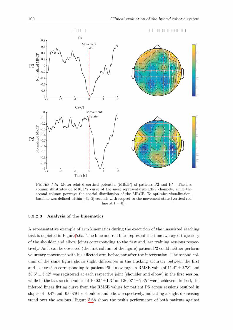
100 Clinical evaluation of the hybrid robotic system
-3
-2
-1
0
1
2
3
Cz
Time [s]
-3 -2 -1 0 1 2-1
-0.8
-0.6
-0.4
-0.2
0
0.2
0.4
0.6
0.8Movement
State
Nor
mal
ized
MR
CP
-4
-3
-2
-1
0
1
2
3
4
-3 -2 -1 0 1 2-1
-0.9
-0.8
-0.7
-0.6
-0.5
-0.4
-0.3
-0.2
-0.1
0
Nor
mal
ized
MR
CP
MovementState
Cz-C1
P2
P5
Curve Spatial distribution
Figure 5.5: Motor-related cortical potential (MRCP) of patients P2 and P5. The firscolumn illustrates de MRCP’s curve of the most representative EEG channels, while thesecond column portrays the spatial distribution of the MRCP. To optimize visualization,baseline was defined within [-3, -2] seconds with respect to the movement state (vertical red
line at t = 0).
5.3.2.3 Analysis of the kinematics
A representative example of arm kinematics during the execution of the unassisted reaching
task is depicted in Figure 5.6a. The blue and red lines represent the time-averaged trajectory
of the shoulder and elbow joints corresponding to the first and last training sessions respec-
tively. As it can be observed (the first column of the figure) patient P2 could neither perform
voluntary movement with his a↵ected arm before nor after the intervention. The second col-
umn of the same figure shows slight di↵erences in the tracking accuracy between the first
and last session corresponding to patient P5. In average, a RMSE value of 11.4�± 2.78� and
38.5�± 3.42� was registered at each respective joint (shoulder and elbow) in the first session,
while in the last session values of 10.02�±1.3� and 36.07�±2.35� were achieved. Indeed, the
inferred linear fitting curve from the RMSE values for patient P5 across sessions resulted in
slopes of -0.47 and -0.0079 for shoulder and elbow respectively, indicating a slight decreasing
trend over the sessions. Figure 5.6b shows the task’s performance of both patients against
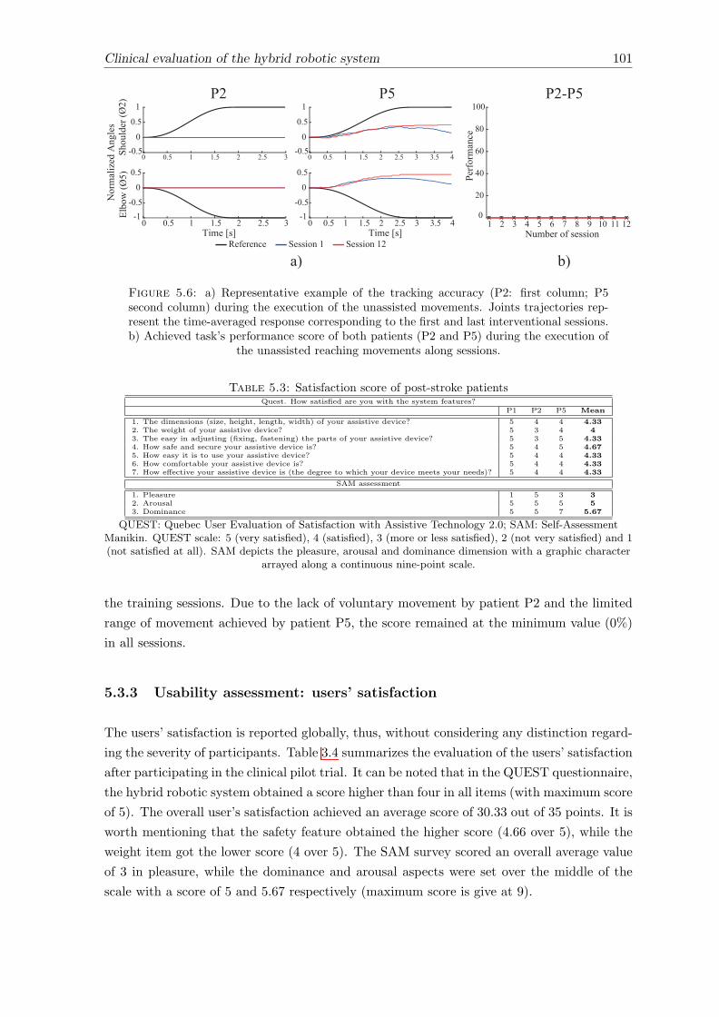
Clinical evaluation of the hybrid robotic system 101
Reference Session 1 Session 12
0 0.5 1 1.5 2 2.5 3 3.5 4-0.5
0
0.5
1
Time [s]0 0.5 1 1.5 2 2.5 3 3.5 4
-1-0.5
0
0.5
0 0.5 1 1.5 2 2.5 3-0.5
0
0.5
1
Time [s]0 0.5 1 1.5 2 2.5 3
-1-0.5
0
0.5
Shou
lder
(Ø2)
Elbo
w (Ø
5)N
orm
aliz
ed A
ngle
sP2 P5
Number of session1 2 3 4 5 6 7 8 9 10 11 12
Perf
orm
ance
0
20
40
60
80
100P2-P5
a) b)
Figure 5.6: a) Representative example of the tracking accuracy (P2: first column; P5second column) during the execution of the unassisted movements. Joints trajectories rep-resent the time-averaged response corresponding to the first and last interventional sessions.b) Achieved task’s performance score of both patients (P2 and P5) during the execution of
the unassisted reaching movements along sessions.
Table 5.3: Satisfaction score of post-stroke patientsQuest. How satisfied are you with the system features?
P1 P2 P5 Mean
1. The dimensions (size, height, length, width) of your assistive device? 5 4 4 4.332. The weight of your assistive device? 5 3 4 43. The easy in adjusting (fixing, fastening) the parts of your assistive device? 5 3 5 4.334. How safe and secure your assistive device is? 5 4 5 4.675. How easy it is to use your assistive device? 5 4 4 4.336. How comfortable your assistive device is? 5 4 4 4.337. How e↵ective your assistive device is (the degree to which your device meets your needs)? 5 4 4 4.33
SAM assessment
1. Pleasure 1 5 3 32. Arousal 5 5 5 53. Dominance 5 5 7 5.67
QUEST: Quebec User Evaluation of Satisfaction with Assistive Technology 2.0; SAM: Self-AssessmentManikin. QUEST scale: 5 (very satisfied), 4 (satisfied), 3 (more or less satisfied), 2 (not very satisfied) and 1(not satisfied at all). SAM depicts the pleasure, arousal and dominance dimension with a graphic character
arrayed along a continuous nine-point scale.
the training sessions. Due to the lack of voluntary movement by patient P2 and the limited
range of movement achieved by patient P5, the score remained at the minimum value (0%)
in all sessions.
5.3.3 Usability assessment: users’ satisfaction
The users’ satisfaction is reported globally, thus, without considering any distinction regard-
ing the severity of participants. Table 3.4 summarizes the evaluation of the users’ satisfaction
after participating in the clinical pilot trial. It can be noted that in the QUEST questionnaire,
the hybrid robotic system obtained a score higher than four in all items (with maximum score
of 5). The overall user’s satisfaction achieved an average score of 30.33 out of 35 points. It is
worth mentioning that the safety feature obtained the higher score (4.66 over 5), while the
weight item got the lower score (4 over 5). The SAM survey scored an overall average value
of 3 in pleasure, while the dominance and arousal aspects were set over the middle of the
scale with a score of 5 and 5.67 respectively (maximum score is give at 9).
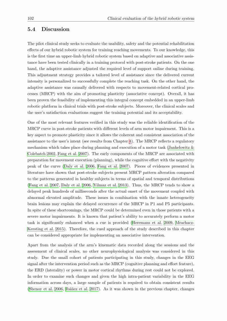
102 Clinical evaluation of the hybrid robotic system
5.4 Discussion
The pilot clinical study seeks to evaluate the usability, safety and the potential rehabilitation
e↵ects of our hybrid robotic system for training reaching movements. To our knowledge, this
is the first time an upper-limb hybrid robotic system based on adaptive and associative assis-
tance have been tested clinically in a training protocol with post-stroke patients. On the one
hand, the adaptive assistance adjusted the required level of support online during training.
This adjustment strategy provides a tailored level of assistance since the delivered current
intensity is personalized to successfully complete the reaching task. On the other hand, the
adaptive assistance was causally delivered with respects to movement-related cortical pro-
cesses (MRCP) with the aim of promoting plasticity (associative concept). Overall, it has
been proven the feasibility of implementing this integral concept embedded in an upper-limb
robotic platform in clinical trials with post-stroke subjects. Moreover, the clinical scales and
the user’s satisfaction evaluations suggest the training potential and its acceptability.
One of the most relevant features verified in this study was the reliable identification of the
MRCP curve in post-stroke patients with di↵erent levels of arm motor impairment. This is a
key aspect to promote plasticity since it allows the coherent and consistent association of the
assistance to the user’s intent (see results from Chapter 4). The MRCP reflects a regulatory
mechanism which takes place during planning and execution of a motor task (Jankelowitz &
Colebatch 2002, Fang et al. 2007). The early components of the MRCP are associated with
preparation for movement execution (planning), while the cognitive e↵ort with the negativity
peak of the curve (Daly et al. 2006, Fang et al. 2007). Pieces of evidences presented in
literature have shown that post-stroke subjects present MRCP pattern alteration compared
to the patterns generated in healthy subjects in terms of spatial and temporal distributions
(Fang et al. 2007, Daly et al. 2006, Yilmaz et al. 2013). Thus, the MRCP tends to show a
delayed peak hundreds of milliseconds after the actual onset of the movement coupled with
abnormal elevated amplitude. These issues in combination with the innate heterogeneity
brain lesions may explain the delayed occurrence of the MRCP in P1 and P5 participants.
In spite of these shortcomings, the MRCP could be determined even in those patients with a
severe motor impairments. It is known that patient’s ability to accurately perform a motor
task is significantly enhanced when a cue is provided (Heremans et al. 2009, Mrachacz-
Kersting et al. 2015). Therefore, the cued approach of the study described in this chapter
can be considered appropriate for implementing an associative intervention.
Apart from the analysis of the arm’s kinematic data recorded along the sessions and the
assessment of clinical scales, no other neurophysiological analysis was considered in this
study. Due the small cohort of patients participating in this study, changes in the EEG
signal after the intervention period such as the MRCP (cognitive planning and e↵ort feature),
the ERD (laterality) or power in motor cortical rhythms during rest could not be explored.
In order to examine such changes and given the high intra-patient variability in the EEG
information across days, a large sample of patients is required to obtain consistent results
(Shenoy et al. 2006, Ibanez et al. 2017). As it was shown in the previous chapter, changes
Clinical evaluation of the hybrid robotic system 103
in cortical excitability derived from non-invasive TMS brain stimulation are better suited to
assess neurophysiological changes (neural plasticity). Yet, due to the inherent physiological
variability of the corticomotor system, larger sample size is also required in order to increase
the statistical power and obtain reliable results (Lewis et al. 2014, Rossini et al. 2015).
Considering that the primary objective of this study is to test the clinical feasibility and the
potential rehabilitative benefits of the hybrid robotic system considering a small cohort of
patients, the verification of neural plastic changes is out of the scope of this study. This
important aspect is planned to be addressed in future works.
Changes in arm kinematics were observed in the patient with moderate arm motor impair-
ment (P1), in terms of task’s performance and tracking accuracy (measured with the RMSE
for each joint along sessions, see Figure 5.4). In the severe group, one patient (P5) achieved
marginal improvement in tracking while the other patient (P2) did not show any change.
Indeed, the improvement of patient P5 was very slight since it was not reflected in the mea-
sure of the task’s performance, where both patients were rated with the minimal score (see
Figure 5.6). The limited range of the arm movement and the lack of sensitivity of the task’s
performance scale may be the reasons why the patient P5 was rated with the minimum value.
As to functional changes, similar results are observed. The patient with moderate arm motor
showed improvements in all clinical scales (see the first part of Table 5.2). The estimation of
the minimal clinically important di↵erence (MCID) of the MMT is not reported in literature.
However, on the basis of clinical experience and estimates reported for similar outcome
measures in di↵erent domains, the MCID was set at 10% of the total range of the scales
(Van Der Lee et al. 1999). Consequently, the scores in wrist flexion/extension obtained
with patient P1 immediately after the intervention (an improvement in 2 points) can be
considered much higher than the MCID. Although the same participant managed to move
five additional blocks with respect to the measure before the intervention, this improvement
almost attained the minimal detectable change (5.5 blocks) reported for chronic post-stroke
survivors (Chen et al. 2009). In the case of patients with severe motor condition (P2 and
P5), minimal improvements were detected in the MMT (one point). Evidence presented
in literature suggests that to obtain clinically relevant results with this group of subjects
(severely a↵ected) longer periods of training with intensive consecutive sessions are required
(12 to 16 weeks, 4 to 5 sessions per week) (Popovic et al. 2005, Thrasher et al. 2009). Although
the clinical results hereby presented cannot be generalized, these preliminary results are
consistent with other robot-based studies in which moderately a↵ected patients were more
responsive to robot-aided therapy than severely a↵ected patients (Ferraro et al. 2003, Stein
et al. 2004). Similarly, Popovic et al. showed that post-stroke subjects with moderate and
severe arm functional conditions responded positively to FES-based therapy (Popovic et al.
2002); but, the group with moderate motor function improved significantly their ability to
carry out functional tasks, while the severe a↵ected did not. This di↵erential responsiveness
can be explained by the fact that high initial motor function likely promotes positive stroke-
rehabilitation outcome in general (Buma et al. 2013, Krakauer 2005, Kwakkel et al. 2003, Di
Lazzaro et al. 2010). Therefore, it can be suggested that the selection criteria of post-stroke
104 Clinical evaluation of the hybrid robotic system
subject enrolled to robotic and FES therapies constitutes an important factor to exploit the
maximum benefits of these novel therapies.
With respect to the satisfaction assessments, the QUEST survey shows a great users’ sat-
isfaction. When comparing the score of this questionnaire obtained in this chapter (30.33)
with the one obtained in Chapter 3 (34.67), a slightly lower score can be observed. This
can be likely explained by the relative lower number of sessions in Chapter 3 (only 2 ex-
perimental session). In general, a great acceptance was obtained in all aspects, indicating
the feasibility of complementing standard rehabilitation therapies with the use of the hybrid
robotic systems.
The principal limitation of this study is the reduced number of enrolled patients and the
lack of a control group. However, the feasibility of using the system in real clinical settings
is demonstrated. Future comprehensive studies should be conducted to prove the potential
clinical benefits of the system to complement standard rehabilitation therapies.
5.5 Conclusion of the chapter
The concepts and the technical developments presented throughout this document have been
validated in a real clinical pilot study with a small cohort of post-stroke subjects. This study
is, to our knowledge, the first one in which the use of a upper extremity hybrid robotic
system based on an adaptive and associative assistance was conducted with stroke patients
along several sessions with the aim of verifying it potential rehabilitative e↵ects.
The results presented here suggested the feasibility of implementing the associative concept
for rehabilitation of arm motor function clinically. Even though the MRCP curve and its
spatial distribution di↵er between subjects, it could be identified in both moderate and severe
arm motor impaired participants. The reported improvements in the clinical scales and in
the arm’s kinematics, in unassisted reaching tasks, reflect the potential rehabilitative e↵ects
of the adaptive and associative concepts for rehabilitation of the arm’s motor functions.
In line with studies reported in literature, subjects with moderate arm motor impairment
may benefit more from the therapy hereby proposed. It is believed that subjects a↵ected
with a severe arm motor impairment could experience further improvements by increasing
the number of training sessions. In general, recruited stroke patients tolerated the hybrid
robotic therapy and no adverse e↵ects were reported. From the acceptability standpoint, all
system features showed to be suited for clinical interventions and fulfilled the expectations
of users.
Finally, it is worth mentioning that the rehabilitative potential should be taken carefully
since this study involves a small cohort of patients and lacks a control group.
Chapter 6
Conclusions and future works
Abstract
This chapter outlines the most relevant contributions presented along this dissertation, as
well as the role that these contributions played for the achievement of our objectives. In
addition, the scientific publications resulting from this work are listed. Finally, the chapter
finishes with a brief outline of the future work emerging from the research line presented in
this work.
105
106 Chapter 6 Conclusions and future works
6.1 Contributions
The work presented in this dissertation was aimed at the design and validation of a hybrid
robotic system providing an adaptive and associative assistance for rehabilitation of reaching
in post-stroke subjects. In order to reach this objective, we elaborated on the requirements
for upper-extremity motor rehabilitation in patients with stroke, the technical and clinical
challenges of hybrid robot-based therapies focused on the upper-limbs, the combination and
applicability of robot and FES strategies to assist the execution of reaching movement and,
the proposal and evaluation of novel algorithms for personalizing the assistance considering
time and intensity factors. We consider that all these activities contributed significantly to
the field of biomedical and neural engineering, rehabilitation robotics, functional electrical
stimulation and neurorehabilitation.
The major contributions of this dissertation are summarized as follows:
• We conducted the first critical review of the state of art of hybrid robotic system
for rehabilitation of upper-limb motor function in post-stroke subjects. Both, technical
and clinical aspects were considered and the main challenges of this novel rehabilitative
method were identified. Some of these challenges were addressed in this dissertation.
• We conducted the design of a fully integrated upper-limb hybrid robotic system for
assisting the execution of unconstrained reaching movement. This platform combined
several subsystems, representing a comprehensive rehabilitative tool. The platform al-
lows acquiring information from di↵erent types of noninvasive sensors (EEG, EMG and
transducers embedded in the exoskeleton) to characterize the planning and execution
processes of reaching movements (with cortical and kinematic features). The platform
was also capable of on-line processing the acquired data and of generating an adequate
feedback.
• We led to the implementation and validation of the FEL controller into a hybrid robotic
system to dynamically adjust the level of assistance according to the users’ motor
capabilities. It is the first time that the FEL controller was tested in healthy and
post-stroke subjects to drive the execution of a multi-degree of freedom upper-limb
movements.
• The development of a cue-based EEG and EMG neuromodulation system to causally
associate the hybrid assistance peripherally applied to the user’s intent during the
execution of functional reaching movements. Three di↵erent associative strategies (BCI
o✏ine, BCI online and EMG) were critically compared, and the importance of a precise
timing association was elucidated.
• We attained the first demonstration that precise temporal association between motor
cortex activation and adaptive hybrid assistance during the execution of functional
motor tasks of the upper-extremity elicited distributed neural plasticity. Remarkably,
Chapter 6 Conclusions and future works 107
the associative intervention’s e↵ect resulted in an improvement of the descending motor
corticospinal projection to the assisted and unassisted arm muscles (AD, TB and BB).
• We proved that the hybrid robotic system based on the adaptive and associative as-
sistance constitutes a feasible alternative for rehabilitation of upper-extremity motor
functions. The results of the pilot clinical intervention with a small cohort of post-
stroke subjects constitute the background of further clinical oriented research in the
field of neurorehabilitation.
6.2 Scientific Dissemination
The work described in this dissertation has produced a number of publications in scientific
journals, national and international conferences, as well as two book chapters. Furthermore,
the outcome of this work has been integrated in other research projects, which have also
been properly disseminated. All the publications are mentioned next.
Publications in journals:
1. F. Resquın, J. Ibanez, O. Herrero, J. Gonzalez-Vargas, F. Brunetti, and J.L. Pons.
“User’s Motor Intent Guided Upper-Limb Hybrid Robotic Platform to E�ciently Mod-
ulate Cortical Excitability: a Comparison of Three Strategies” (To be submitted).
2. F. Resquın, J. Gonzalez-Vargas, J. Ibanez, F. Brunetti, I. Dimbwadyo, L. Carrasco, S.
Alves, C. Gonzalez-Alted, A. Gomez-Blanco and J.L. Pons. Adaptive hybrid robotic
system for rehabilitation of reaching movement after a brain injury: a usability study.
J Neuroeng Rehabil. 2017 Dec 12;14(1):104.
3. F. Trincado, E. Lopez-Larraz, F. Resquın, A. Ardanza, S. Prez-Nombela, J. L. Pons,
L. Montesano, A. Gil-Agudo. A pilot study of the brain-triggered electrical stimulation
with visual feedback in patients with incomplete spinal cord injury. Journal of Medical
and Biological Engineering. (Accepted: 20/09/2017).
4. F. Resquın, J. Gonzalez-Vargas, J. Ibanez, F. Brunetti and J.L. Pons. Feedback error
learning controller for functional electrical stimulation assistance in a hybrid robotic
system for reaching rehabilitation. Eur J Transl Myol. 2016;26(3).
5. F. Resquın, A. Cuesta-Gomez, J. Gonzalez-Vargas, F. Brunetti, D. Torricelli, F. Molina
Rueda, R. Cano de la Rueda, J. C. Miangolarra and J. L. Pons. Hybrid robotic systems
for upper limb rehabilitation after stroke: A review. Med Eng Phys. 2016 Nov; 38 (11):
1279-88.
6. E. Hortal, D. Planelles, F. Resquın, J.M. Climent, J.M. Azorın and J. L. Pons. Using a
brain-machine interface to control a hybrid upper limb exoskeleton during rehabilitation
of patients with neurological conditions. J Neuroeng Rehabil. BioMed Central Ltd;
2015 Oct 17;12(1):92.
108 Chapter 6 Conclusions and future works
Books Chapters:
1. Alessandro C, Beckers N, Gebel P, Resquın F, Gonzalez J, Osu R. Motor Control and
Learning Theories. In: Pons JL, Raya R, Gonzalez J, editors. Emerging Therapies in
Neurorehabilitation II. 2015. p. 225-50.
2. Brunetti F, Resquın F, Gonzalez V, Ceres R, Freire Bastos T. Interfaces basadas en
senales electromiograficas. In: Jose M. Azorın, Ramon Ceres, Anselmo Frizera TFB,
editor. La Interaccion de Personas con Discapacidad con el Computador: Experiencias
y Posibilidades en Iberoamerica. Programa Iberoamericano de Ciencia y Tecnologıa
para el Desarrollo (CYTED); 2013. p. 83-101.
Selected publications in international and national conferences proceedings directly related
to the topics of the dissertation:
1. F. Resquın, O. Herrero, J. Ibanez, J. Gonzalez-Vargas, F. Brunetti, J. L. Pons. Elec-
troencephalography guided Upper-Limb Hybrid Robotic Platform to Modulate Cor-
tical Excitability. IEEE International Conference on Systems, Man and Cybernetics.
Canada, 2017 (Oral presentation)
2. F. Resquın, J. Ibanez, O. Herrero, J. Gonzalez-Vargas, F. Brunetti, J. L. Pons. Au-
mento de la excitabilidad cortical inducido mediante una asistencia hıbrida guiada por
BCI para la rehabilitacion del movimiento de alcance. IX Congreso Iberoamericano
de tecnologıas de Apoyo a la Discapacidad. Bogota, Colombia, 2017. (Accepted, oral
presentation)
3. Resquın F, Gonzalez-Vargas J, Ibanez J, Dimbwadyo I, Alves S, Torres L, et al. Hy-
brid Robotic System for Reaching Rehabilitation after Stroke: reporting an usability
experimentation. In: Proceedings of the 3rd International Conference on NeuroReha-
bilitation (ICNR 2016), Segovia, Spain. 2016. P. 679-84. (Oral presentation)
4. Barbouch H, Resquın F, Gonzalez-Vargas J, Khraief-Hadded N, Belghith S, Pons JL.
Hybrid Robotic System Simulation for the exploration of novel control strategies. In:
Proceedings of the 3rd International Conference on NeuroRehabilitation (ICNR 2016),
Segovia, Spain. 2016. (Poster presentation)
5. Martınez-Exposito A, Ibanez J, Resquın F, Pons JL. Task influence on motor-related
cortical signals: Comparison between upper and lower limb coordinated and analytic
movements. In: Proceedings of the 3rd International Conference on NeuroRehabilita-
tion (ICNR 2016), Segovia, Spain. 2016. (Poster presentation)
6. Dimbwadyo I, Carrasco L, Resquın F, Gonzalez-Vargas J, Ibanez J, Alves S, et al.
Satisfaccion de los usuarios con un sistema robotico hbrido para la rehabilitacion del
miembro superior. In: LXVIII Reunion Anual Sociedad Espanola Neurologıa. Valen-
cia.; 2016. (Poster Presentation)
Chapter 6 Conclusions and future works 109
7. Resquın F, Ibanez J, Gonzalez-Vargas J, Brunetti F, Dimbwadyo I, Alves S, et al.
Combining a hybrid robotic system with a brain-machine interface for the rehabilitation
of reaching movements: A case study with a stroke patient. In: 2016 38th Annual
International Conference of the IEEE Engineering in Medicine and Biology Society
(EMBC). Orlando, Florida.: IEEE; 2016. P. 6381-4. (Oral presentation)
8. Resquın F, Gonzalez-Vargas J, Ibanez J, Brunetti F, Dimbwadyo I, Alves S, et al.
Hybrid Robotic System for Reaching Rehabilitation after Stroke. In: XXI Conference
of the International Society on Electrophysiology and Kinesiology (ISEK). Chicago;
2016. (Oral presentation)
9. Resquın F, Ibanez J, Gonzalez-Vargas J, Dimbwadyo I, Carrasco L, Alves S, et al.
Sistema robotico hıbrido para la rehabilitacion del miembro superior. Cognitive Area
Networks 8o Simposio CEA de Bioingenierıa 2016 Interfaces hombre-maquina Cerebro
y periferia un camino ida y vuelta. Madrid; 2016. p. 97-101. (Oral presentation)
10. Resquın F, Hortal E, Planelles D, Brunetti F, Azorın JM, Pons JL. Sistema Robotico
Hıbrido para la rehabilitacion del miembro superior dirigido por comandos volitivos.
In: VIII Congreso Iberoamericano de Tecnologıas de Apoyo a la Discapacidad. 2015.
p. 189-92. (Oral presentation).
11. Ubeda A, Planelles D, Hortal E, Resquın F, Koutsou AD, Azorın JM, et al. A Brain-
Machine Interface Architecture to Control an Upper Limb Rehabilitation Exoskeleton.
In: Proceedings of the 2nd International Conference on NeuroRehabilitation (ICNR
2014). Springer International Publishing; 2014. P. 795-803. (Biosystems & Biorobotics;
vol. 7). (Oral presentation)
12. Resquın F, Brunetti F, Pons JL. Estudio Comparativo de los Parametros de Estim-
ulacion Electrica Funcional para Movimientos de Alcance utilizando un Controlador
Realimentado. In: VI Jornadas AITADIS de Rehabilitacion y Tecnologıas de Apoyo a
la Discapacidad. Asuncion, Paraguay; 2014. (Poster presentation)
13. Resquın F, Brunetti F, Koutsou AD, Ibanez J, Pons JL. Upper Limb Hybrid Control
Architecture implemented in the Framework of the HYPER Project. In: International
Workshop on Wearable Robotics. Baiona, Spain; 2014. (Poster presentation)
14. Hortal E, Ubeda A, Planelles D, Koutsou AD, Resquın F, Azorın JM, et al. Arqui-
tectura de una Interfaz Cerebro-Maquina para el control de un Exoesqueleto Robot de
Miembro Superior. Cognitive Area Networks, vol1, No 1, 6to Simposio CEA Bioinge-
nierıa Interfaces Mente-computador y Neurotecnologıas. Granada, Espana; 2014. p.
13-7. (Oral presentation).
15. Koutsou AD, Resquın F, Ibanez J, del-Ama AJ, Perez S, Cuesta A, et al. Compen-
sacion de Trastornos Motores del Miembro Superior en Pacientes LM y ACV. Proyecto
HYPER. In: Ceres R, Blanco E, editors. VII Congreso Iberoamericano de Tecnologıas
de Apoyo a la Discapacidad. Santo Domingo, Republica Dominicana; 2013. P. 274-9.
(Oral presentation)

110 Chapter 6 Conclusions and future works
16. Resquın F, Brunetti F, Pons JL. Estudio preliminar para la deteccion en lınea de
espasmos. In: Ceres R, Blanco R, editors. VII Congreso Iberoamericano de Tecnologıas
de Apoyo a la Discapacidad. Santo Domingo, Republica Dominicana; 2013. P. 212-7.
(Oral presentation).
17. Resquın F, Brunetti F, Pons JL. A system for spasm detection during robotic therapies:
preliminary results. In: 18th IFESS Annual Conference 2013 “Bridging Mind and
Body.” San Sebastian, Spain; 2013. (Oral presentation).
18. Resquın F, Bravo E, Gomez-Soriano J, Brunetti F, Pons JL. Protocol and system for
spastic behavior simulation through the generation of cutaneous reflexes. In: Proceed-
ings of the 1st International Conference on NeuroRehabilitation (ICNR 2012). Springer
Berlin Heidelberg; 2012. P. 431-5. (Poster presentation).
Part of the research work presented in this dissertation has been awarded as the “Best
Student Contribution” at the 3rd. International Conference on NeuroRehabilitation
(ICNR2016), Segovia, Spain, 2016. This distinguished award underlines the rele-
vance and quality of the research work presented in this dissertation. The work
leading to this distinguished award is following cited:
“Resquın F, Gonzalez-Vargas J, Ibanez J, Dimbwadyo I, Alves S, Torres L, Carrasco L,
Brunetti F, and Pons J.L. Hybrid Robotic System for Reaching Rehabilitation after Stroke:
reporting an usability experimentation. In: Proceedings of the 3rd International Conference
on NeuroRehabilitation (ICNR 2016), Segovia, Spain.2016. p. 679-84.”
To sum it all up, the work presented in this dissertation has produced four journal pub-
lications (plus one contribution under revision and another that will be soon submitted),
eighteen contributions to international and national conferences, two book chapters, and the
distinguished award for the best student contribution at the ICNR 2016 conference. Addi-
tionally, this work was subject to presentations in a number of seminars and briefings in both
the Department of Systems Engineering and Automation (UC3M) and the Cajal Institute
of the Spanish National Research Council (CSIC).
6.3 Future works
The applied methodology and the presented results in this doctoral thesis represent a refer-
ence milestone that encourages several ongoing studies and projects related with the inves-
tigation framework presented herein. Some of the topics considered are related to questions
that emerged during the analysis or/and interpretation of the results, while others are stud-
ies planned as the continuation to those presented in this dissertation. The future studies
identified can be organized in three groups associated with di↵erent aspects presented along
the thesis.
Chapter 6 Conclusions and future works 111
The most immediate works are related to the continuity of the study presented in Chapter 5.
These works are proposed with the goal of extending the results obtained therein and provide
stronger evidences to support the clinical e↵ectiveness of the hybrid robotic system. To this
end, the group of Neural Rehabilitation (CSIC) is currently collaborating closely with the
Centro de Referencia Estatal de Atencion al Dano Cerebral (CEADAC) and the Instituto de
Neurociencias y Ciencias del Movimiento (INCIMOV). As results of this collaboration, the
following works are planned to be addressed:
• To confirm that the associative concepts lead to cortical plastic changes in post-stroke
subjects. It is expected similar results showed in Chapter 3 with healthy subjects, in
which an enhancement of the corticospinal projection of the arm muscles was demon-
strated.
• To design and carry out a clinical intervention procedure in which a larger sample of
patients can be considered, so that, more reliable clinical results are reachable. In
this regard and considering the classification of clinical trial of rehabilitation (Macie-
jasz et al. 2014, Lo 2012), a category II - ‘Development-of-concept study’ is planned.
This clinical trial is conceived with the aim of verifying the device therapeutic e�cacy
including a control group following a randomization and blinded outcome assessment.
From the technological perspective, and with the aim of increasing the functionality of the
hybrid robotic platform, several research works for further research can be derived. This
works can be classified within the bioengineering and the rehabilitation fields. Some of them
are listed in the next lines:
• To include mechanical exoskeleton with active actuation, capable providing mechanical
assistance during the execution of functional task. The inclusion of exoskeleton with
active assistance will allow that patients without FES response could use the system.
Moreover, it could improve the performance of the current hybrid robotic systems
significantly since the arm movement does not only depend on the FES assistance,
specifically in subject severely a↵ected. However, it opens a window for the develop-
ment of an optimally shared controller for ruling the operation and complementary
assistance contribution of the FES and the exoskeleton to the voluntary motor e↵ort
exerted by the patients.
• To increase the rehabilitative connotation of the system by extending the application of
assistance to the hand and wrist muscles. It is expected that the execution of reaching
a grasping task involving the whole upper limb may achieve motor and functional
improvement of the whole arm.
• The integration of electrode arrays for improving the muscle recruitment response to
FES yielding to a more e↵ective stimulation pattern and to a more beneficial therapeu-
tic response. Particularly, there has been significant interest in using electrode arrays
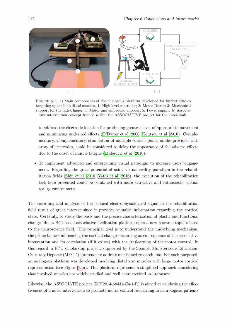
112 Chapter 6 Conclusions and future works
a) b)
12
3
4
5
Acquisition
Characterization of Motor Cortical Processes
PeripheralElectrical Stimulation
MovementOnset
FES device
Figure 6.1: a) Main components of the analogous platform developed for further studiestargeting upper-limb distal muscles. 1: High level controller; 2: Motor Driver; 3: Mechanicalsupport for the index finger; 4: Motor and embedded encoder; 5: Power supply. b) Associa-tive intervention concept framed within the ASSOCIATIVE project for the lower-limb.
to address the electrode location for producing greatest level of appropriate movement
and minimizing undesired e↵ects (O’Dwyer et al. 2006, Koutsou et al. 2016). Comple-
mentary, Complementary, stimulation of multiple contact point, as the provided with
array of electrodes, could be considered to delay the appearance of the adverse e↵ects
due to the onset of muscle fatigue (Malesevic et al. 2010).
• To implement advanced and entertaining visual paradigm to increase users’ engage-
ment. Regarding the great potential of using virtual reality paradigm in the rehabil-
itation fields (Shin et al. 2016, Yates et al. 2016), the execution of the rehabilitation
task here presented could be combined with more attractive and enthusiastic virtual
reality environment.
The recording and analysis of the cortical electrophysiological signal in the rehabilitation
field result of great interest since it provides valuable information regarding the cortical
state. Certainly, to study the basis and the precise characterization of plastic and functional
changes due a BCI-based associative facilitation platform open a new research topic related
to the neuroscience field. The principal goal is to understand the underlying mechanism,
the prime factors influencing the cortical changes occurring as consequence of the associative
intervention and its correlation (if it exists) with the (re)learning of the motor control. In
this regard, a FPU scholarship project, supported by the Spanish Ministerio de Educacion,
Cultura y Deporte (MECD), pretends to address mentioned research line. For such purposed,
an analogous platform was developed involving distal arm muscles with large motor cortical
representation (see Figure 6.1a). This platform represents a simplified approach considering
that involved muscles are widely studied and well characterized in literature.
Likewise, the ASSOCIATE project (DPI2014-58431-C4-1-R) is aimed at validating the e↵ec-
tiveness of a novel intervention to promote motor control re-learning in neurological patients

Chapter 6 Conclusions and future works 113
by means of an associated use of motor planning at brain level, sensory stimulation at cor-
tical level and a↵erent feedback provided with a wearable lower extremity exoskeleton. Due
the di�culties of implementing associative approach in non-stationary scenarios, this con-
cept was instigated un a similar lower-limb functional tasks, as the cycling (see Figure 6.1b).
Similar results than the found in Chapter 4 are expected to be generated in the lower-limbs,
as well as a deep interpretation of the mechanisms occasioning these cortical neural changes.

Bibliography
Adams, H. P., Bendixen, B. H., Kappelle, L. J., Biller, J., Love, B. B., Gordon, D. L. & Marsh,
E. E. (1993), ‘Classification of subtype of acute ischemic stroke. Definitions for use in a multicenter
clinical trial. TOAST. Trial of Org 10172 in Acute Stroke Treatment.’, Stroke 24(1).
Adams, H. P., del Zoppo, G., Alberts, M. J., Bhatt, D. L., Brass, L., Furlan, A., Grubb, R. L.,
Higashida, R. T., Jauch, E. C., Kidwell, C., Lyden, P. D., Morgenstern, L. B., Qureshi, A. I.,
Rosenwasser, R. H., Scott, P. A. & Wijdicks, E. F. (2007), ‘Guidelines for the Early Management
of Adults With Ischemic Stroke’, Circulation 115(20).
Alon, G., Levitt, A. F. & McCarthy, P. A. (2007), ‘Functional Electrical Stimulation Enhancement
of Upper Extremity Functional Recovery During Stroke Rehabilitation: A Pilot Study’, Neurore-
habilitation and Neural Repair 21(3), 207–215.
Alon, G., Sunnerhagen, K. S., Geurts, A. C. H. & Ohry, A. (2003), ‘A home-based, self-administered
stimulation program to improve selected hand functions of chronic stroke.’, NeuroRehabilitation
18(3), 215–225.
Alvarez-Sabın, J., Quintana, M., Masjuan, J., Oliva-Moreno, J., Mar, J., Gonzalez-Rojas, N., Becerra,
V., Torres, C. & Yebenes, M. (2017), ‘Economic impact of patients admitted to stroke units in
Spain’, The European Journal of Health Economics 18(4), 449–458.
Ang, K. K., Guan, C., Phua, K. S., Wang, C., Zhou, L., Tang, K. Y., Ephraim Joseph, G. J., Kuah,
C. W. K. & Chua, K. S. G. (2014), ‘Brain-computer interface-based robotic end e↵ector system
for wrist and hand rehabilitation: results of a three-armed randomized controlled trial for chronic
stroke’, Frontiers in Neuroengineering 7.
Barker, R. N., Brauer, S. G. & Carson, R. G. (2008), ‘Training of reaching in stroke survivors with
severe and chronic upper limb paresis using a novel nonrobotic device: A randomized clinical trial’,
Stroke 39(6), 1800–1807.
Barsi, G. I., Popovic, D. B., Tarkka, I. M., Sinkjær, T. & Grey, M. J. (2008), ‘Cortical excitability
changes following grasping exercise augmented with electrical stimulation’, Experimental Brain
Research 191(1), 57–66.
Beaulieu, L.-D. & Milot, M.-H. (2017), ‘Changes in transcranial magnetic stimulation outcome mea-
sures in response to upper-limb physical training in stroke: A systematic review of randomized
controlled trials’, Annals of Physical and Rehabilitation Medicine .
Belda-Lois, J.-M., Mena-del Horno, S., Bermejo-Bosch, I., Moreno, J. C., Pons, J. L., Farina, D., Iosa,
M., Molinari, M., Tamburella, F., Ramos, A., Caria, A., Solis-Escalante, T., Brunner, C. & Rea,
M. (2011), ‘Rehabilitation of gait after stroke: a review towards a top-down approach’, Journal of
NeuroEngineering and Rehabilitation 8(1), 66.
115
116 BIBLIOGRAPHY
Bickel, C. S., Gregory, C. M. & Dean, J. C. (2011), ‘Motor unit recruitment during neuromuscular
electrical stimulation: a critical appraisal’, European Journal of Applied Physiology 111(10), 2399–
2407.
Bohannon, R. W. & Smith, M. B. (1987), ‘Interrater reliability of a modified Ashworth scale of muscle
spasticity.’, Physical therapy 67(2), 206–7.
Bonita, R., Mendis, S., Truelsen, T., Bogousslavsky, J., Toole, J. & Yatsu, F. (2004), ‘The Global
Stroke Initiative’, The Lancet Neurology 3(7), 391–393.
Brainin, M., Heiss, W.-D. & Tabernig, S. (2014), Textbook of stroke medicine, Cambridge University
Press.
Brewer, L., Horgan, F., Hickey, A. & Williams, D. (2013), ‘Stroke rehabilitation: recent advances and
future therapies’, QJM 106(1), 11–25.
Broetz, D., Braun, C., Weber, C., Soekadar, S. R., Caria, A. & Birbaumer, N. (2010), ‘Combination
of Brain-Computer Interface Training and Goal-Directed Physical Therapy in Chronic Stroke: A
Case Report’, Neurorehabilitation and Neural Repair 24(7), 674–679.
Buma, F., Kwakkel, G. & Ramsey, N. (2013), ‘Understanding upper limb recovery after stroke’,
Restorative neurology and neuroscience 31(6), 707–722.
Cai, Z., Tong, D., Meadmore, K. L., Freeman, C. T., Hughes, A. M., Rogers, E. & Burridge, J. H.
(2011), Design & control of a 3D stroke rehabilitation platform, in ‘2011 IEEE International Con-
ference on Rehabilitation Robotics’, IEEE, pp. 1–6.
Cajal, S. R. y. (1959), Degeneration & regeneration of the nervous system, Vol. 1, Hafner Pub. Co.
Carson, R. G. & Kennedy, N. C. (2013), ‘Modulation of human corticospinal excitability by paired
associative stimulation’, Frontiers in Human Neuroscience 7.
Chaudhary, U., Birbaumer, N. & Ramos-Murguialday, A. (2016), ‘Braincomputer interfaces for com-
munication and rehabilitation’, Nature Reviews Neurology 12(9), 513–525.
Chen, H.-M., Chen, C. C., Hsueh, I.-P., Huang, S.-L. & Hsieh, C.-L. (2009), ‘Test-Retest Repro-
ducibility and Smallest Real Di↵erence of 5 Hand Function Tests in Patients With Stroke’, Neu-
rorehabilitation and Neural Repair 23(5), 435–440.
Chipchase, L., Schabrun, S. & Hodges, P. (2011), ‘Peripheral electrical stimulation to induce cortical
plasticity: A systematic review of stimulus parameters’, Clinical Neurophysiology 122(3), 456–463.
Cirstea, M. C., Mitnitski, A. B., Feldman, A. G. & Levin, M. F. (2003), ‘Interjoint coordination
dynamics during reaching in stroke’, Experimental Brain Research 151(3), 289–300.
Daly, J., Fang, Y., Perepezko, E., Siemionow, V. & Yue, G. (2006), ‘Prolonged Cognitive Planning
Time, Elevated Cognitive E↵ort, and Relationship to Coordination and Motor Control Following
Stroke’, IEEE Transactions on Neural Systems and Rehabilitation Engineering 14(2), 168–171.
Daly, J. J. & Wolpaw, J. R. (2008), ‘Braincomputer interfaces in neurological rehabilitation’, The
Lancet Neurology 7(11), 1032–1043.
del Ama, A. J., Koutsou, A. D., Moreno, J. C., De-los Reyes, A., Gil-Agudo, N. & Pons, J. L.
(2012), ‘Review of hybrid exoskeletons to restore gait following spinal cord injury’, The Journal of
Rehabilitation Research and Development 49(4), 497.
BIBLIOGRAPHY 117
Demers, L., Monette, M., Lapierre, Y., Arnold, D. L. & Wolfson, C. (2002), ‘Reliability, validity, and
applicability of the Quebec User Evaluation of Satisfaction with assistive Technology (QUEST 2.0)
for adults with multiple sclerosis’, Disability and Rehabilitation 24(1-3), 21–30.
Demeurisse, G., Demol, O. & Robaye, E. (1980), ‘Motor evaluation in vascular hemiplegia.’, European
neurology 19(6), 382–9.
Di Lazzaro, V., Profice, P., Pilato, F., Capone, F., Ranieri, F., Pasqualetti, P., Colosimo, C., Pravata,
E., Cianfoni, A. & Dileone, M. (2010), ‘Motor Cortex Plasticity Predicts Recovery in Acute Stroke’,
Cerebral Cortex 20(7), 1523–1528.
Di Pino, G., Pellegrino, G., Assenza, G., Capone, F., Ferreri, F., Formica, D., Ranieri, F., Tombini,
M., Ziemann, U., Rothwell, J. C. & Di Lazzaro, V. (2014), ‘Modulation of brain plasticity in stroke:
a novel model for neurorehabilitation’, Nature Reviews Neurology 10(10), 597–608.
Dimyan, M. A. & Cohen, L. G. (2011), ‘Neuroplasticity in the context of motor rehabilitation after
stroke’, Nature Reviews Neurology 7(2), 76–85.
do Nascimento, O. & Farina, D. (2008), ‘Movement-Related Cortical Potentials Allow Discrimination
of Rate of Torque Development in Imaginary Isometric Plantar Flexion’, IEEE Transactions on
Biomedical Engineering 55(11), 2675–2678.
Dobkin, B. H. (2004), ‘Strategies for stroke rehabilitation’, The Lancet Neurology 3(9), 528–536.
Dobkin, B. H. (2009), ‘Progressive Staging of Pilot Studies to Improve Phase III Trials for Motor
Interventions’, Neurorehabilitation and Neural Repair 23(3), 197–206.
Donnan, G. A., Fisher, M., Macleod, M. & Davis, S. M. (2008), ‘Stroke’, The Lancet 371(9624), 1612–
1623.
Doucet, B. M., Lam, A., Gri�n, L. & Others (2012), ‘Neuromuscular electrical stimulation for skeletal
muscle function’, Yale J Biol Med 85(2), 201–215.
Eraifej, J., Clark, W., France, B., Desando, S. & Moore, D. (2017), ‘E↵ectiveness of upper limb
functional electrical stimulation after stroke for the improvement of activities of daily living and
motor function: a systematic review and meta-analysis’, Systematic Reviews 6(1), 40.
Ethier, C., Gallego, J. & Miller, L. (2015), ‘Brain-controlled neuromuscular stimulation to drive neural
plasticity and functional recovery’, Current Opinion in Neurobiology 33, 95–102.
Ethier, C. & Miller, L. E. (2015), ‘Brain-controlled muscle stimulation for the restoration of motor
function’, Neurobiology of Disease 83, 180–190.
Fang, Y., Yue, G. H., Hrovat, K., Sahgal, V. & Daly, J. J. (2007), ‘Abnormal cognitive planning
and movement smoothness control for a complex shoulder/elbow motor task in stroke survivors’,
Journal of the Neurological Sciences 256(1-2), 21–29.
Feigin, V. L., Forouzanfar, M. H., Krishnamurthi, R., Mensah, G. A., Connor, M., Bennett, D. A.,
Moran, A. E., Sacco, R. L., Anderson, L., Truelsen, T., O’Donnell, M., Venketasubramanian, N.,
Barker-Collo, S., Lawes, C. M. M., Wang, W., Shinohara, Y., Witt, E., Ezzati, M., Naghavi, M.
& Murray, C. (2014), ‘Global and regional burden of stroke during 19902010: findings from the
Global Burden of Disease Study 2010’, The Lancet 383(9913), 245–255.
118 BIBLIOGRAPHY
Feigin, V. L., Lawes, C. M., Bennett, D. A., Barker-Collo, S. L. & Parag, V. (2009), ‘Worldwide stroke
incidence and early case fatality reported in 56 population-based studies: a systematic review’, The
Lancet Neurology 8(4), 355–369.
Ferraro, M., Palazzolo, J. J., Krol, J., Krebs, H. I., Hogan, N. & Volpe, B. T. (2003), ‘Robot-
aided sensorimotor arm training improves outcome in patients with chronic stroke’, Neurology
61(11), 1604–1607.
Flash, T. & Hogan, N. (1985), ‘The coordination of arm movements: an experimentally confirmed
mathematical model.’, The Journal of neuroscience : the o�cial journal of the Society for Neuro-
science 5(7), 1688–1703.
Freeman, C. (2014), ‘Newton-method based iterative learning control for robot-assisted rehabilitation
using FES’, Mechatronics 24(8), 934–943.
Freeman, C. T. (2015), ‘Upper Limb Electrical Stimulation Using Input-Output Linearization and
Iterative Learning Control’, IEEE Transactions on Control Systems Technology 23(4), 1546–1554.
Freeman, C. T., Hughes, A.-M., Burridge, J. H., Chappell, P. H., Lewin, P. L. & Rogers, E. (2009a),
‘A model of the upper extremity using FES for stroke rehabilitation’, Journal of biomechanical
engineering 131(3), 31011.
Freeman, C. T., Hughes, A. M., Burridge, J. H., Chappell, P. H., Lewin, P. L. & Rogers, E. (2009b), ‘A
robotic workstation for stroke rehabilitation of the upper extremity using FES’,Medical Engineering
and Physics 31(3), 364–373.
Freeman, C. T., Hughes, A. M., Burridge, J. H., Chappell, P. H., Lewin, P. L. & Rogers, E. (2009c),
‘Iterative learning control of FES applied to the upper extremity for rehabilitation’, Control Engi-
neering Practice 17(3), 368–381.
Freeman, C. T., Rogers, E., Hughes, A. M., Burridge, J. H. & Meadmore, K. (2012), ‘Iterative
Learning Control in Health Care: Electrical Stimulation and Robotic-Assisted Upper-Limb Stroke
Rehabilitation’, IEEE Control Systems 32(1), 18–43.
Freeman, C. T., Tong, D., Meadmore, K., Cai, Z., Rogers, E., Hughes, A. M. & Burridge, J. H.
(2011), ‘Phase-lead iterative learning control algorithms for functional electrical stimulation-based
stroke rehabilitation’, Proceedings of the Institution of Mechanical Engineers, Part I: Journal of
Systems and Control Engineering 225(6), 850–859.
Fujiwara, T., Kasashima, Y., Honaga, K., Muraoka, Y., Tsuji, T., Osu, R., Hase, K., Masakado, Y.
& Liu, M. (2009), ‘Motor improvement and corticospinal modulation induced by hybrid assistive
neuromuscular dynamic stimulation (HANDS) therapy in patients with chronic stroke.’, Neurore-
habilitation and neural repair 23(2), 125–132.
Garipelli, G., Chavarriaga, R. & del R Millan, J. (2013), ‘Single trial analysis of slow cortical poten-
tials: a study on anticipation related potentials’, Journal of Neural Engineering 10(3), 036014.
Grahn, C., Ekdahl, L. & Borgquist, B. (2000), ‘Motivation as a predictor of changes in quality of life
and working ability in multidisciplinary rehabilitation’, Disability and rehabilitation 22(15), 639–
654.
Gregory, C. M. & Bickel, C. S. (2005), ‘Recruitment patterns in human skeletal muscle during electrical
stimulation’, Physical therapy 85(4), 358–364.
BIBLIOGRAPHY 119
Groppa, S., Oliviero, A., Eisen, A., Quartarone, A., Cohen, L., Mall, V., Kaelin-Lang, A., Mima,
T., Rossi, S., Thickbroom, G., Rossini, P., Ziemann, U., Valls-Sole, J. & Siebner, H. (2012), ‘A
practical guide to diagnostic transcranial magnetic stimulation: Report of an IFCN committee’,
Clinical Neurophysiology 123(5), 858–882.
Gund, G.-M. B. M., Jagtap, M. P. N., Ingale, V. B. & Patil, R. Y. (2013), ‘Stroke: A Brain Attack’,
IOSR Journal Of Pharmacy 3(8), 2250–3013.
Hamilton, B. B., Laughlin, J. A., Fiedler, R. C. & Granger, C. V. (1994), ‘Interrater reliability of the
7-level functional independence measure (FIM)’, Scandinavian journal of rehabilitation medicine
26(3), 115–9.
Hara, Y., Ogawa, S. & Muraoka, Y. (2006), ‘Hybrid Power-Assisted Functional Electrical Stimulation
to Improve Hemiparetic Upper-Extremity Function’, American Journal of Physical Medicine &
Rehabilitation 85(12), 977–985.
Harwin, W., Patton, J. & Edgerton, V. (2006), ‘Challenges and Opportunities for Robot-Mediated
Neurorehabilitation’, Proceedings of the IEEE 94(9), 1717–1726.
Hatem, S. M., Saussez, G., Faille, M., Prist, V. & Dan, B. (2016), ‘Rehabilitation of Motor Function
after Stroke : A Multiple Systematic Review Focused on Techniques to Stimulate Upper Extremity
Recovery’, 10(September), 1–22.
Hebb, D. O. (1949), The organization of behavior: A neuropsychological approach, John Wiley &
Sons.
Hendricks, H. T., Pasman, J. W., van Limbeek, J. & Zwarts, M. J. (2003a), ‘Motor Evoked Poten-
tials in Predicting Recovery from Upper Extremity Paralysis after Acute Stroke’, Cerebrovascular
Diseases 16(3), 265–271.
Hendricks, H. T., Pasman, J. W., van Limbeek, J. & Zwarts, M. J. (2003b), ‘Motor evoked potentials
of the lower extremity in predicting motor recovery and ambulation after stroke: a cohort study
11No commercial party having a direct financial interest in the results of the research supporting
this article has or will confer a be’, Archives of Physical Medicine and Rehabilitation 84(9), 1373–
1379.
Hendricks, H. T., van Limbeek, J., Geurts, A. C. & Zwarts, M. J. (2002), ‘Motor recovery after
stroke: A systematic review of the literature’, Archives of Physical Medicine and Rehabilitation
83(11), 1629–1637.
Heremans, E., Helsen, W. F., De Poel, H. J., Alaerts, K., Meyns, P. & Feys, P. (2009), ‘Facilitation
of motor imagery through movement-related cueing’, Brain Research 1278, 50–58.
Hodes, R. L., Cook, E. W. & Lang, P. J. (1985), ‘Individual Di↵erences in Autonomic Response:
Conditioned Association or Conditioned Fear?’, Psychophysiology 22(5), 545–560.
Hu, X. L., Tong, K. Y., Li, R., Chen, M., Xue, J. J., Ho, S. K. & Chen, P. N. (2010), E↵ectiveness
of functional electrical stimulation (FES)-robot assisted wrist training on persons after stroke, in
‘2010 Annual International Conference of the IEEE Engineering in Medicine and Biology Society,
EMBC’10’, pp. 5819–5822.
Hu, X. L., Tong, K. Y., Li, R., Chen, M., Xue, J. J., Ho, S. K. & Chen, P. N. (2011), Post-stroke
wrist rehabilitation assisted with an intention-driven functional electrical stimulation (FES)-robot
system, in ‘2011 IEEE International Conference on Rehabilitation Robotics’, IEEE, pp. 1–6.
120 BIBLIOGRAPHY
Hu, X. L., Tong, R. K., Ho, N. S., Xue, J.-j., Rong, W. & Li, L. S. (2015), ‘Wrist Rehabilitation As-
sisted by an Electromyography-Driven Neuromuscular Electrical Stimulation Robot After Stroke’,
Neurorehabilitation and Neural Repair 29(8), 767–776.
Huang, V. S. & Krakauer, J. W. (2009), ‘Robotic neurorehabilitation: a computational motor learning
perspective’, Journal of neuroengineering and rehabilitation 6, 5.
Huang, X., Naghdy, F., Naghdy, G., Du, H. & Todd, C. (2016), ‘Robot-assisted post-stroke motion
rehabilitation in upper extremities: a survey’, International Journal on Disability and Human
Development .
Hughes, A. M., Burridge, J., Freeman, C. T., Donnovan-Hall, M., Chappell, P. H., Lewin, P. L.,
Rogers, E. & Dibb, B. (2011), ‘Stroke participants’ perceptions of robotic and electrical stimulation
therapy: a new approach’, Disability and Rehabilitation: Assistive Technology 6(2), 130–138.
Hughes, A. M., Freeman, C. T., Burridge, J. H., Chappell, P. H., Lewin, P. L. & Rogers, E. (2009),
‘Feasibility of iterative learning control mediated by functional electrical stimulation for reaching
after stroke’, Neurorehabilitation and neural repair .
Hunter, S. & Crome, P. (2002), ‘Hand function and stroke’, Reviews in Clinical Gerontology 12(01).
Ibanez, J., Serrano, J. I., del Castillo, M. D., Monge, E., Molina, F., Rivas, F. M., Alguacil, I.,
Miangolarra, J. C. & Pons, J. L. (2014), Upper-limb muscular electrical stimulation driven by
EEG-based detections of the intentions to move: A proposed intervention for patients with stroke,
in ‘2014 36th Annual International Conference of the IEEE Engineering in Medicine and Biology
Society’, IEEE, pp. 1646–1649.
URL: http://ieeexplore.ieee.org/document/6943921/
Ibanez, J., Monge-Pereira, E., Molina-Rueda, F., Serrano, J. I., del Castillo, M. D., Cuesta-Gomez,
A., Carratala-Tejada, M., Cano-de-la Cuerda, R., Alguacil-Diego, I. M., Miangolarra-Page, J. C.
& Pons, J. L. (2017), ‘Low Latency Estimation of Motor Intentions to Assist Reaching Movements
along Multiple Sessions in Chronic Stroke Patients: A Feasibility Study’, Frontiers in Neuroscience
11.
Ibanez, J., Serrano, J. I., Del Castillo, M. D., Monge-Pereira, E., Molina-Rueda, F., Alguacil-Diego,
I. & Pons, J. L. (2014), ‘Detection of the onset of upper-limb movements based on the combined
analysis of changes in the sensorimotor rhythms and slow cortical potentials’, Journal of neural
engineering 11(5), 56009.
IJzerman, M. J., Renzenbrink, G. J. & Geurts, A. C. H. (2009), ‘Neuromuscular stimulation after
stroke: from technology to clinical deployment’, Expert review of neurotherapeutics 9(4), 541–52.
Ikram, M. A., Wieberdink, R. G. & Koudstaal, P. J. (2012), ‘International Epidemiology of Intrac-
erebral Hemorrhage’, Current Atherosclerosis Reports 14(4), 300–306.
Jankelowitz, S. & Colebatch, J. (2002), ‘Movement-related potentials associated with self-paced, cued
and imagined arm movements’, Experimental Brain Research 147(1), 98–107.
Jarrasse, N., Proietti, T., Crocher, V., Robertson, J., Sahbani, A., Morel, G. & Roby-Brami, A.
(2014), ‘Robotic Exoskeletons: A Perspective for the Rehabilitation of Arm Coordination in Stroke
Patients’, Frontiers in Human Neuroscience 8(December), 947.
BIBLIOGRAPHY 121
Jiang, N., Gizzi, L., Mrachacz-Kersting, N., Dremstrup, K. & Farina, D. (2015), ‘A braincomputer
interface for single-trial detection of gait initiation from movement related cortical potentials’,
Clinical Neurophysiology 126(1), 154–159.
Jochumsen, M., Niazi, I. K., Mrachacz-Kersting, N., Farina, D. & Dremstrup, K. (2013), ‘Detection
and classification of movement-related cortical potentials associated with task force and speed’,
Journal of Neural Engineering 10(5), 056015.
Johnson, M. J. (2006), ‘Recent trends in robot-assisted therapy environments to improve real-life
functional performance after stroke’, Journal of NeuroEngineering and Rehabilitation 3(1), 1.
Kandel, E. R., Schwartz, J. H., Jessell, T. M., Siegelbaum, S. A. & Hudspeth, A. J. (2000), Principles
of neural science, Vol. 4, McGraw-hill New York.
Kawato, M. (1990), ‘Feedback-error-learning neural network for supervised motor learning’, Advanced
neural computers 6(3), 365–372.
Kimberley, T. J., Lewis, S. M., Auerbach, E. J., Dorsey, L. L., Lojovich, J. M. & Carey, J. R.
(2004), ‘Electrical stimulation driving functional improvements and cortical changes in subjects
with stroke’, Experimental Brain Research 154(4), 450–460.
URL: http://link.springer.com/10.1007/s00221-003-1695-y
Koike, Y., Gonzalez, J., Gomez, J. & Yu, W. (2011), Implementing Feedback Error Learning for FES
control, in ‘2011 4th International Conference on Biomedical Engineering and Informatics (BMEI)’,
IEEE.
Koutsou, A. D., Moreno, J. C., del Ama, A. J., Rocon, E. & Pons, J. L. (2016), ‘Advances in selective
activation of muscles for non-invasive motor neuroprostheses’, Journal of NeuroEngineering and
Rehabilitation 13(1), 56.
Krakauer, J. W. (2005), Arm function after stroke: from physiology to recovery, in ‘Seminars in
neurology’, Vol. 25, Citeseer, pp. 384–395.
Krakauer, J. W. (2006), ‘Motor learning: its relevance to stroke recovery and neurorehabilitation’,
Current opinion in neurology 19(1), 84–90.
Kraus, D., Naros, G., Bauer, R., Leao, M. T., Ziemann, U. & Gharabaghi, A. (2016), ‘Brain–
robot interface driven plasticity: distributed modulation of corticospinal excitability’, Neuroimage
125, 522–532.
Krebs, H., Dipietro, L., Levy-Tzedek, S., Fasoli, S., Rykman-Berland, A., Zipse, J., Fawcett, J., Stein,
J., Poizner, H., Lo, A., Volpe, B. & Hogan, N. (2008), ‘A paradigm shift for rehabilitation robotics’,
IEEE Engineering in Medicine and Biology Magazine 27(4), 61–70.
Kujirai, K., Kujirai, T., Sinkjaer, T. & Rothwell, J. C. (2006), ‘Associative Plasticity in Human Motor
Cortex During Voluntary Muscle Contraction’, Journal of Neurophysiology 96(3), 1337–1346.
Kurosawa, K., Futami, R., Watanabe, T. & Hoshimiya, N. (2005), ‘Joint Angle Control by FES Using
a Feedback Error Learning Controller’, IEEE Transactions on Neural Systems and Rehabilitation
Engineering 13(3), 359–371.
Kutlu, M., Freeman, C. T., Hallewell, E., Hughes, A.-m. & Laila, D. S. (2015), FES-based upper-limb
stroke rehabilitation with advanced sensing and control, in ‘2015 IEEE International Conference
on Rehabilitation Robotics (ICORR)’, IEEE, pp. 253–258.
122 BIBLIOGRAPHY
Kwakkel, G., Kollen, B. J. & Krebs, H. I. (2008), ‘E↵ects of robot-assisted therapy on upper limb
recovery after stroke: a systematic review’, Neurorehabilitation and neural repair 22(2), 111–21.
Kwakkel, G., Kollen, B. J., van der Grond, J. & Prevo, A. J. (2003), ‘Probability of Regaining
Dexterity in the Flaccid Upper Limb: Impact of Severity of Paresis and Time Since Onset in Acute
Stroke’, Stroke 34(9), 2181–2186.
Lamb, R. (1985), Manual muscle testing, in J. Rothstein, ed., ‘Measurement in physical therapy’,
Churchill Livingstone, New York, pp. 47–56.
Langhorne, P., Bernhardt, J. & Kwakkel, G. (2011), ‘Stroke rehabilitation’, The Lancet
377(9778), 1693–1702.
Langhorne, P., Coupar, F. & Pollock, A. (2009), ‘Motor recovery after stroke: a systematic review’,
Lancet neurology 8(8), 741–54.
Lawrence, E. S., Coshall, C., Dundas, R., Stewart, J., Rudd, A. G., Howard, R. & Wolfe, C. D. A.
(2001), ‘Estimates of the Prevalence of Acute Stroke Impairments and Disability in a Multiethnic
Population’, Stroke 32(6), 1279–1284.
Lewis, G. N., Signal, N. & Taylor, D. (2014), ‘Reliability of lower limb motor evoked potentials
in stroke and healthy populations: How many responses are needed?’, Clinical Neurophysiology
125(4), 748–754.
Liberson, W. T., Holmquest, H. J., Scot, D. & Dow, M. (1961), ‘Functional electrotherapy: stimula-
tion of the peroneal nerve synchronized with the swing phase of the gait of hemiplegic patients.’,
Archives of physical medicine and rehabilitation 42, 101–105.
Lo, A. C. (2012), ‘Clinical designs of recent robot rehabilitation trials’, American Journal of Physical
Medicine & Rehabilitation 91(11), S204—-S216.
Lo, H. S. & Xie, S. Q. (2012), ‘Exoskeleton robots for upper-limb rehabilitation: state of the art and
future prospects.’, Medical engineering & physics 34(3), 261–8.
Lozano, R. et al. (2012), ‘Global and regional mortality from 235 causes of death for 20 age groups
in 1990 and 2010: a systematic analysis for the Global Burden of Disease Study 2010’, The Lancet
380(9859), 2095–2128.
Lu, M.-K., Arai, N., Tsai, C.-H. & Ziemann, U. (2012), ‘Movement related cortical potentials of cued
versus self-initiated movements: Double dissociated modulation by dorsal premotor cortex versus
supplementary motor area rTMS’, Human Brain Mapping 33(4), 824–839.
Lum, P. S., Burgar, C. G. & Shor, P. C. (2004), ‘Evidence for improved muscle activation patterns
after retraining of reaching movements with the MIME robotic system in subjects with post-stroke
hemiparesis’, Neural Systems and Rehabilitation Engineering, IEEE Transactions on 12(2), 186–
194.
Lynch, C. & Popovic, M. R. (2008), ‘Functional Electrical Stimulation’, IEEE Control Systems Mag-
azine 28(2), 40–50.
Maciejasz, P., Eschweiler, J., Gerlach-Hahn, K., Jansen-Troy, A. & Leonhardt, S. (2014), ‘A survey
on robotic devices for upper limb rehabilitation.’, Journal of neuroengineering and rehabilitation
11, 3.
BIBLIOGRAPHY 123
Ma�uletti, N. A. (2010), ‘Physiological and methodological considerations for the use of neuromus-
cular electrical stimulation’, European Journal of Applied Physiology 110(2), 223–234.
Ma�uletti, N. A., Minetto, M. A., Farina, D. & Bottinelli, R. (2011), ‘Electrical stimulation for
neuromuscular testing and training: State-of-the art and unresolved issues’, European Journal of
Applied Physiology 111, 2391–2397.
Mahoney, F. I. & Barthel, D. W. (1965), ‘Functional Evaluation: The Barthel Index’, Maryland state
medical journal 14, 61–5.
Malesevic, N. M., Popovic, L. Z., Schwirtlich, L. & Popovic, D. B. (2010), ‘Distributed low-frequency
functional electrical stimulation delays muscle fatigue compared to conventional stimulation.’, Mus-
cle & nerve 42(4), 556–62.
Malesevic, N. M., Popovic Maneski, L. Z., Ilic, V., Jorgovanovic, N., Bijelic, G., Keller, T. & Popovic,
D. B. (2012), ‘A multi-pad electrode based functional electrical stimulation system for restoration
of grasp’, Journal of NeuroEngineering and Rehabilitation 9(1), 66.
Marchal-Crespo, L. & Reinkensmeyer, D. J. (2009), ‘Review of control strategies for robotic movement
training after neurologic injury’, Journal of NeuroEngineering and Rehabilitation 6(1), 20.
Marquez-Chin, C., Marquis, A. & Popovic, M. R. (2016), ‘EEG-Triggered Functional Electrical Sti-
mulation Therapy for Restoring Upper Limb Function in Chronic Stroke with Severe Hemiplegia’,
Case Reports in Neurological Medicine 2016, 1–11.
Marsland, S. (2015), Machine learning: an algorithmic perspective, CRC press.
Mathiowetz, V., Volland, G., Kashman, N. & Weber, K. (1985), ‘Box and Block test’, Adult norms
for the box and blokc test of manual dextresity 39(6), 386–391.
McGie, S. C., Zari↵a, J., Popovic, M. R. & Nagai, M. K. (2015), ‘Short-Term Neuroplastic E↵ects of
Brain-Controlled and Muscle-Controlled Electrical Stimulation’, Neuromodulation: Technology at
the Neural Interface 18(3), 233–240.
Meadmore, K. L., Exell, T. a., Hallewell, E., Hughes, A. M., Freeman, C. T., Kutlu, M., Benson, V.,
Rogers, E. & Burridge, J. H. (2014), ‘The application of precisely controlled functional electrical
stimulation to the shoulder, elbow and wrist for upper limb stroke rehabilitation: a feasibility
study.’, Journal of NeuroEngineering and Rehabilitation 11(1), 105.
Meadmore, K. L., Hughes, A.-M., Freeman, C. T., Cai, Z., Tong, D., Burridge, J. H. & Rogers, E.
(2012), ‘Functional electrical stimulation mediated by iterative learning control and 3D robotics
reduces motor impairment in chronic stroke’, Journal of NeuroEngineering and Rehabilitation p. 1.
URL: http://eprints.soton.ac.uk/273120/
Mohr, J. P., Grotta, J. C., Wolf, P. A., Moskowitz, M. A., Mayberg, M. R. & Von Kummer, R. (2011),
Stroke: pathophysiology, diagnosis, and management, sixth edn, Elsevier Health Sciences.
Morris, J. D. (1995), ‘OBSERVATIONS: SAM: The Self-Assessment Manikin - An E�cient Cross-
Cultural Measurement of Emotional Response’, Journal of Advertising Research 35(6), 63–68.
Mrachacz-Kersting, N., Fong, M., Murphy, B. A. & Sinkjaer, T. (2007), ‘Changes in Excitability
of the Cortical Projections to the Human Tibialis Anterior After Paired Associative Stimulation’,
Journal of Neurophysiology 97(3), 1951–1958.
124 BIBLIOGRAPHY
Mrachacz-Kersting, N., Jiang, N., Stevenson, A. J. T., Niazi, I. K., Kostic, V., Pavlovic, A.,
Radovanovic, S., Djuric-Jovicic, M., Agosta, F., Dremstrup, K. & Farina, D. (2015), ‘E�cient
neuroplasticity induction in chronic stroke patients by an associative brain-computer interface.’,
Journal of neurophysiology .
Mrachacz-Kersting, N., Kristensen, S. R., Niazi, I. K. & Farina, D. (2012), ‘Precise temporal as-
sociation between cortical potentials evoked by motor imagination and a↵erence induces cortical
plasticity.’, The Journal of physiology 590(Pt 7), 1669–82.
Mrachacz-Kersting, N. & Stevenson, A. J. T. (2017), ‘Paired Associative Stimulation Targeting the
Tibialis Anterior Muscle using either Mono or Biphasic Transcranial Magnetic Stimulation’, Fron-
tiers in Human Neuroscience 11.
Muir, K. W. (2009), ‘Stroke’, Medicine 37(2), 109–114.
Mukaino, M., Ono, T., Shindo, K., Fujiwara, T., Ota, T., Kimura, A., Liu, M. & Ushiba, J. (2014),
‘E�cacy of brain-computer interface-driven neuromuscular electrical stimulation for chronic paresis
after stroke’, Journal of Rehabilitation Medicine 46(4), 378–382.
Muraoka, Y., Tanabe, S., Yamaguchi, T. & Takeda, K. (2013), ‘Specifications of an electromyogram-
driven neuromuscular stimulator for upper limb functional recovery’, Proceedings of the Annual
International Conference of the IEEE Engineering in Medicine and Biology Society, EMBS pp. 277–
280.
Murphy, T. H. & Corbett, D. (2009), ‘Plasticity during stroke recovery: from synapse to behaviour.’,
Nature reviews. Neuroscience 10(12), 861–872.
Murray, C. J. L. et al. (2012), ‘Disability-adjusted life years (DALYs) for 291 diseases and injuries
in 21 regions, 19902010: a systematic analysis for the Global Burden of Disease Study 2010’, The
Lancet 380(9859), 2197–2223.
Naqvi, U. & Sherman, A. (2017), Muscle Strength Grading, StatPearls Publishing.
Niazi, I. K., Jiang, N., Jochumsen, M., Nielsen, J. F., Dremstrup, K. & Farina, D. (2013), ‘Detection of
movement-related cortical potentials based on subject-independent training’, Medical & Biological
Engineering & Computing 51(5), 507–512.
Niazi, I. K., Jiang, N., Tiberghien, O., Nielsen, J. F., Dremstrup, K. & Farina, D. (2011), ‘Detection
of movement intention from single-trial movement-related cortical potentials’, Journal of Neural
Engineering 8(6), 066009.
Niazi, I. K., Mrachacz-Kersting, N., Jiang, N., Dremstrup, K. & Farina, D. (2012), ‘Peripheral elec-
trical stimulation triggered by self-paced detection of motor intention enhances motor evoked po-
tentials’, IEEE transactions on neural systems and rehabilitation engineering 20(4), 595–604.
Nudo, R. J. (2006), ‘Mechanisms for recovery of motor function following cortical damage’, Current
Opinion in Neurobiology 16(6), 638–644.
O’Dwyer, S., O’Kee↵e, D., Coote, S. & Lyons, G. (2006), ‘An electrode configuration technique
using an electrode matrix arrangement for FES-based upper arm rehabilitation systems’, Medical
Engineering & Physics 28(2), 166–176.
Olesen, J., Gustavsson, A., Svensson, M., Wittchen, H.-U. & Jonsson, B. (2012), ‘The economic cost
of brain disorders in Europe’, European Journal of Neurology 19(1), 155–162.
BIBLIOGRAPHY 125
Ono, T., Shindo, K., Kawashima, K., Ota, N., Ito, M., Ota, T., Mukaino, M., Fujiwara, T., Kimura,
A., Liu, M. & Ushiba, J. (2014), ‘Brain-computer interface with somatosensory feedback improves
functional recovery from severe hemiplegia due to chronic stroke’, Frontiers in Neuroengineering 7.
Oostenveld, R., Fries, P., Maris, E. & Scho↵elen, J.-M. (2011), ‘FieldTrip: Open Source Software
for Advanced Analysis of MEG, EEG, and Invasive Electrophysiological Data’, Computational
Intelligence and Neuroscience 2011, 1–9.
Peckham, P. H. & Knutson, J. S. (2005), ‘Functional Electrical Stimulation for Neuromuscular Ap-
plications’, Annual Review of Biomedical Engineering 7(1), 327–360.
Pichiorri, F., De Vico Fallani, F., Cincotti, F., Babiloni, F., Molinari, M., Kleih, S. C., Neuper, C.,
Kubler, A. & Mattia, D. (2011), ‘Sensorimotor rhythm-based braincomputer interface training: the
impact on motor cortical responsiveness’, Journal of Neural Engineering 8(2), 025020.
Pichiorri, F., Morone, G., Petti, M., Toppi, J., Pisotta, I., Molinari, M., Paolucci, S., Inghilleri,
M., Astolfi, L., Cincotti, F. & Mattia, D. (2015), ‘Brain-computer interface boosts motor imagery
practice during stroke recovery’, Annals of Neurology 77(5), 851–865.
Piron, L., Piccione, F., Tonin, P. & Dam, M. (2005), ‘Clinical Correlation Between Motor Evoked
Potentials and Gait Recovery in Poststroke Patients’, Archives of Physical Medicine and Rehabili-
tation 86(9), 1874–1878.
Pizzi, A., Carrai, R., Falsini, C., Martini, M., Verdesca, S. & Grippo, A. (2009), ‘Prognostic value
of motor evoked potentials in motor function recovery of upper limb after stroke’, Journal of
Rehabilitation Medicine 41(8), 654–660.
Pollock, A., Farmer, S. E., Brady, M. C., Langhorne, P., Mead, G. E., Mehrholz, J. & van Wijck, F.
(2014), Interventions for improving upper limb function after stroke, in A. Pollock, ed., ‘Cochrane
Database of Systematic Reviews’, number 11, John Wiley & Sons, Ltd, Chichester, UK.
URL: http://doi.wiley.com/10.1002/14651858.CD010820.pub2
Pons, J. L. (2010), ‘Rehabilitation exoskeletal robotics. The promise of an emerging field’, IEEE
engineering in medicine and biology magazine : the quarterly magazine of the Engineering in
Medicine & Biology Society 29(3), 57–63.
Pons, J. L., ed. (2008), Wearable Robots, John Wiley & Sons, Ltd, Chichester, UK.
Popovic, D. B. (2014), ‘Advances in functional electrical stimulation (FES)’, Journal of Electromyo-
graphy and Kinesiology 24(6), 795–802.
Popovic, M. B., Popovic, D. B., Sinkjær, T., Stefanovic, A. & Schwirtlich, L. (2002), ‘Restitution of
reaching and grasping promoted by functional electrical therapy’, Artificial Organs 26(3), 271–275.
Popovic, M. R., Curt, A., Keller, T. & Dietz, V. (2001), ‘Functional electrical stimulation for grasping
and walking: indications and limitations’, Spinal Cord 39(8), 403–412.
Popovic, M. R., Thrasher, T. A., Zivanovic, V., Takaki, J. & Hajek, V. (2005), ‘Neuroprosthesis
for retraining reaching and grasping functions in severe hemiplegic patients’, Neuromodulation
8(1), 58–72.
Prange, G. B., Jannink, M. J. a., Groothuis-Oudshoorn, C. G. M., Hermens, H. J. & Ijzerman, M. J.
(2006), ‘Systematic review of the e↵ect of robot-aided therapy on recovery of the hemiparetic arm
after stroke.’, Journal of rehabilitation research and development 43(2), 171–184.
126 BIBLIOGRAPHY
Raichle, M. E. & Gusnard, D. A. (2002), ‘Appraising the brain’s energy budget.’, Proceedings of the
National Academy of Sciences of the United States of America 99(16), 10237–9.
Ramos-Murguialday, A., Broetz, D., Rea, M., Laer, L., Yilmaz, O., Brasil, F. L., Liberati, G., Curado,
M. R., Garcia-Cossio, E., Vyziotis, A., Cho, W., Agostini, M., Soares, E., Soekadar, S., Caria, A.,
Cohen, L. G. & Birbaumer, N. (2013), ‘Brain-machine interface in chronic stroke rehabilitation: A
controlled study’, Annals of Neurology 74(1), 100–108.
Reinkensmeyer, D. J. & Boninger, M. L. (2012), ‘Technologies and combination therapies for enhanc-
ing movement training for people with a disability’, Journal of NeuroEngineering and Rehabilitation
9(1), 17.
Reinkensmeyer, D. J., Wolbrecht, E. T., Chan, V., Chou, C., Cramer, S. C. & Bobrow, J. E. (2012),
‘Comparison of 3D, Assist-as-Needed Robotic Arm/Hand Movement Training Provided with Pneu-
WREX to Conventional Table Top Therapy Following Chronic Stroke’, American journal of phys-
ical medicine & rehabilitation/Association of Academic Physiatrists 91(11 0 3), S232.
Riener, R., Lunenburger, L., Jezernik, S., Anderschitz, M., Colombo, G. & Dietz, V. (2005), ‘Patient-
cooperative strategies for robot-aided treadmill training: first experimental results.’, IEEE trans-
actions on neural systems and rehabilitation engineering : a publication of the IEEE Engineering
in Medicine and Biology Society 13(3), 380–94.
URL: http://ieeexplore.ieee.org/articleDetails.jsp?arnumber=1506824
Ring, H. & Rosenthal, N. (2005), ‘Controlled study of neuroprosthetic functional electrical stimulation
in sub-acute post-stroke rehabilitation’, Journal of Rehabilitation Medicine 37(1), 32–36.
Rossini, P., Burke, D., Chen, R., Cohen, L., Daskalakis, Z., Di Iorio, R., Di Lazzaro, V., Ferreri, F.,
Fitzgerald, P., George, M., Hallett, M., Lefaucheur, J., Langguth, B., Matsumoto, H., Miniussi,
C., Nitsche, M., Pascual-Leone, A., Paulus, W., Rossi, S., Rothwell, J., Siebner, H., Ugawa, Y.,
Walsh, V. & Ziemann, U. (2015), ‘Non-invasive electrical and magnetic stimulation of the brain,
spinal cord, roots and peripheral nerves: Basic principles and procedures for routine clinical and
research application. An updated report from an I.F.C.N. Committee’, Clinical Neurophysiology
126(6), 1071–1107.
Rotenberg, A., Horvath, J. C. & Pascual-Leone, A., eds (2014), Transcranial Magnetic Stimulation,
Vol. 89 of Neuromethods, Springer New York, New York, NY.
Samsa, G. P. & Matchar, D. B. (2004), ‘How strong is the relationship between functional status
and quality of life among persons with stroke?’, Journal of rehabilitation research and development
41(3A), 279–82.
Sanes, J. N. & Donoghue, J. P. (2000), ‘Plasticity and Primary Motor Cortex’, Annual Review of
Neuroscience 23(1), 393–415.
Schaechter, J. D. (2004), ‘Motor rehabilitation and brain plasticity after hemiparetic stroke.’, Progress
in neurobiology 73(1), 61–72.
Shakeel, A., Navid, M. S., Anwar, M. N., Mazhar, S., Jochumsen, M. & Niazi, I. K. (2015), ‘A Review
of Techniques for Detection of Movement Intention Using Movement-Related Cortical Potentials’,
Computational and Mathematical Methods in Medicine 2015, 1–13.
She✏er, L. R. & Chae, J. (2007), ‘Neuromuscular electrical stimulation in neurorehabilitation’, Muscle
& nerve 35(5), 562–90.
BIBLIOGRAPHY 127
Shenoy, P., Krauledat, M., Blankertz, B., Rao, R. P. N. & Muller, K.-R. (2006), ‘Towards adaptive
classification for BCI’, Journal of Neural Engineering 3(1), R13–R23.
Shibasaki, H. & Hallett, M. (2006), ‘What is the Bereitschafts potential?’, Clinical Neurophysiology
117(11), 2341–2356.
Shih, J. J., Krusienski, D. J. & Wolpaw, J. R. (2012), ‘Brain-Computer Interfaces in Medicine’, Mayo
Clinic Proceedings 87(3), 268–279.
Shin, J.-H., Kim, M.-Y., Lee, J.-Y., Jeon, Y.-J., Kim, S., Lee, S., Seo, B. & Choi, Y. (2016), ‘E↵ects of
virtual reality-based rehabilitation on distal upper extremity function and health-related quality of
life: a single-blinded, randomized controlled trial’, Journal of NeuroEngineering and Rehabilitation
13(1), 17.
Soekadar, S. R., Birbaumer, N., Slutzky, M. W. & Cohen, L. G. (2015), ‘Brainmachine interfaces in
neurorehabilitation of stroke’, Neurobiology of Disease 83, 172–179.
Stefan, K., Kunesch, E., Cohen, L. G., Benecke, R. & Classen, J. (2000), ‘Induction of plasticity in
the human motor cortex by paired associative stimulation’, Brain 123, 572–584.
Stein, J., Krebs, H. I., Frontera, W. R., Fasoli, S. E., Hughes, R. & Hogan, N. (2004), ‘Comparison
of two techniques of robot-aided upper limb exercise training after stroke’, American journal of
physical medicine & rehabilitation 83(9), 720–728.
Steiner, W. A., Ryser, L., Huber, E., Uebelhart, D., Aeschlimann, A. & Stucki, G. (2002), ‘Use of
the ICF model as a clinical problem-solving tool in physical therapy and rehabilitation medicine.’,
Physical therapy 82(11).
Stinear, C. (2010), ‘Prediction of recovery of motor function after stroke’, The Lancet Neurology
9(12), 1228–1232.
Sullivan, J. E. & Hedman, L. D. (2008), ‘Sensory Dysfunction Following Stroke: Incidence, Signifi-
cance, Examination, and Intervention’, Topics in Stroke Rehabilitation 15(3), 200–217.
Thabit, M. N., Ueki, Y., Koganemaru, S., Fawi, G., Fukuyama, H. & Mima, T. (2010), ‘Movement-
Related Cortical Stimulation Can Induce Human Motor Plasticity’, Journal of Neuroscience
30(34), 11529–11536.
Thompson, A. K. & Stein, R. B. (2004), ‘Short-term e↵ects of functional electrical stimulation
on motor-evoked potentials in ankle flexor and extensor muscles’, Experimental Brain Research
159(4), 491–500.
Thrasher, T. A., Zivanovic, V., McIlroy, W. & Popovic, M. R. (2009), ‘Rehabilitation of reaching and
grasping function in severe hemiplegic patients using functional electrical stimulation therapy.’,
Neurorehabilitation and neural repair 22, 706–714.
Van Der Lee, J., Wagenaar, R., Lankhorst, G., Vogelaar, T., Deville, W. & Bouter, L. (1999), ‘Forced
use of the upper extremity in chronic stroke patients: results from a single-blind randomized clinical
trial’, Stroke 30, 2369–2375.
Varkuti, B., Guan, C., Pan, Y., Phua, K. S., Ang, K. K., Kuah, C. W. K., Chua, K., Ang, B. T.,
Birbaumer, N. & Sitaram, R. (2013), ‘Resting State Changes in Functional Connectivity Correlate
With Movement Recovery for BCI and Robot-Assisted Upper-Extremity Training After Stroke’,
Neurorehabilitation and Neural Repair 27(1), 53–62.

128 BIBLIOGRAPHY
Vega, T., Zurriaga, O., Ramos, J. M., Gil, M., Alamo, R., Lozano, J. E., Lopez, A., Miralles, M. T.,
Vaca, P. & del Mar Alvarez, M. (2009), ‘Stroke in Spain: Epidemiologic Incidence and Patterns; A
Health Sentinel Network Study’, Journal of Stroke and Cerebrovascular Diseases 18(1), 11–16.
Ward, N. S. (2005), ‘Mechanisms underlying recovery of motor function after stroke’, Postgraduate
Medical Journal 81(958), 510–514.
Watanabe, T. & Fukushima, K. (2011), ‘A Study on Feedback Error Learning Controller for Func-
tional Electrical Stimulation: Generation of Target Trajectories by Minimum Jerk Model’, Artificial
Organs 35(3), 270–274.
Westerveld, A. J., Aalderink, B. J., Hagedoorn, W., Buijze, M., Schouten, A. C. & Kooij, H. V. D.
(2014), ‘A damper driven robotic end-point manipulator for functional rehabilitation exercises after
stroke.’, IEEE transactions on bio-medical engineering 61(10), 2646–54.
Williams, J., Perry, L. & Watkins, C. (2010), Acute stroke nursing, Wiley-Blackwell.
Wolpaw, J. R. (2007), ‘Spinal cord plasticity in acquisition and maintenance of motor skills’, Acta
Physiologica 189(2), 155–169.
Wolpaw, J. R., Birbaumer, N., McFarland, D. J., Pfurtscheller, G. & Vaughan, T. M. (2002), ‘Brain-
computer interfaces for communication and control’, Clinical Neurophysiology 113(6), 767–791.
World Health Organization (2003), The world health report 2003 : shaping the future, Geneva: World
Health Organization.
World Health Organization (2010), Community-based Rehabilitation: CBR Guidelines. Livelihood
Component, WHO.
World Health Organization (2011), ‘World report on disability 2011’.
Wu, F.-c., Lin, Y.-t., Kuo, T.-s., Luh, J.-j. & Lai, J.-s. (2011), ‘Clinical e↵ects of combined bilateral
arm training with functional electrical stimulation in patients with stroke’, 2011 IEEE International
Conference on Rehabilitation Robotics pp. 1–7.
Xu, R., Jiang, N., Mrachacz-Kersting, N., Lin, C., Asin Prieto, G., Moreno, J. C., Pons, J. L.,
Dremstrup, K. & Farina, D. (2014), ‘A closed-loop brain-computer interface triggering an active
ankle-foot orthosis for inducing cortical neural plasticity’, IEEE Transactions on Biomedical En-
gineering 61(7), 2092–2101.
Yates, M., Kelemen, A. & Sik Lanyi, C. (2016), ‘Virtual reality gaming in the rehabilitation of the
upper extremities post-stroke’, Brain Injury 30(7), 855–863.
Yilmaz, O., Cho, W., Braun, C., Birbaumer, N. & Ramos-Murguialday, A. (2013), Movement related
cortical potentials in severe chronic stroke, in ‘2013 35th Annual International Conference of the
IEEE Engineering in Medicine and Biology Society (EMBC)’, IEEE, pp. 2216–2219.
Zhang, D., Guan, T. H., Widjaja, F. & Ang, W. T. (2007), Functional electrical stimulation in
rehabilitation engineering, in ‘Proceedings of the 1st international convention on Rehabilitation
engineering & assistive technology in conjunction with 1st Tan Tock Seng Hospital Neurorehabili-
tation Meeting - i-CREATe ’07’, ACM Press, New York, New York, USA, pp. 221–226.
Ziemann, U. (2004), ‘Learning Modifies Subsequent Induction of Long-Term Potentiation-Like
and Long-Term Depression-Like Plasticity in Human Motor Cortex’, Journal of Neuroscience
24(7), 1666–1672.

BIBLIOGRAPHY 129
Ziemann, U., Paulus, W., Nitsche, M. A., Pascual-Leone, A., Byblow, W. D., Berardelli, A., Siebner,
H. R., Classen, J., Cohen, L. G. & Rothwell, J. C. (2008), ‘Consensus: Motor cortex plasticity
protocols’, Brain Stimulation 1(3), 164–182.