Robotic urology in the United Kingdom: experience and overview of robotic-assisted cystectomy
8
J Robotic Surg (2008) 1:235–242 DOI 10.1007/s11701-007-0049-3 123 REVIEW ARTICLE Robotic urology in the United Kingdom: experience and overview of robotic-assisted cystectomy Oussama Elhage · Declan Murphy · Ben Challacombe · Peter Rimington · Mohammad S. Khan · Prokar Dasgupta Received: 12 April 2007 / Accepted: 5 December 2007 / Published online: 8 January 2008 © Springer London 2008 Abstract In this article we look at the evolution of robotic technology in operative urology and the signiWcant early contribution of Mr John Wickham. We explore the ergonomics of robotic technology and discuss Wnancial issues from a British perspective. We share our clinical experience, describe the authors’ robotic-assisted cystec- tomy technique, and conclude by exploring the patients’ perception of this new treatment modality. Keywords Robotic · da Vinci · Cystectomy Introduction The use of robotic-assisted laparoscopic surgery through- out the world has increased signiWcantly in recent years and is now standard practice in many large centres. Brit- ish urologists were among the Wrst to contribute to the development of surgical robotics and embrace this new technology. Currently, robotic-assisted radical prostatec- tomy is the most commonly performed robotic procedure worldwide [1], and it is now gaining popularity in the United Kingdom. History Man’s fascination with machines that are able to perform automated work is hundreds of years old, with the evidence for this scattered in the literature of several diVerent cul- tures. Leonardo da Vinci probably designed the Wrst robot, an automated knight capable of performing basic move- ments to entertain the guests of Leonardo’s patron [2]. It was not until the last three decades of the twentieth century that robotic technology was developed for medical applica- tions. The earliest machines were supportive robots used for transporting pharmaceuticals and medical equipment, and rehabilitation robots which provided physiotherapy to joints and assisted stroke patients with their daily tasks. In the 1980s researchers explored the potential of robotics in operative surgery. Several projects in the United States and in Europe started, some independently and some as collabo- rative eVorts. One of the Wrst pioneers was John Wickham, a urolo- gist from Guy’s Hospital. He developed the Wrst clinically useful robot in urology, the PROBOT in 1989 [3]. In the late 1980s Wickham worked on a TURP robotic frame in a joint project between the Mechanical Engineering Department at Imperial College, Guy’s Hospital and the Institute of Urology in central London. The device attempted to perform an automated robotic transurethral resection of the prostate (TURP). The team felt that as the prostate is a relatively Wxed organ and the procedure requires repeated similar movements, TURP was ideal for robotic control. The frame was constructed to support a six-axis Unimate Puma robot combined with a Wickham Endoscope liquidizer and aspirator. The liquidizer blade rotated at 40,000 rpm and the system was used in initial clinical trials following successful tests on prostate- shaped potatoes. These showed the PROBOT-assisted O. Elhage · D. Murphy · B. Challacombe · P. Rimington · M. S. Khan · P. Dasgupta (&) Department of Urology, Guy’s Hospital and King’s College London School of Medicine, 1st Floor Thomas Guy House, London SE1 9RT, UK e-mail: [email protected] P. Rimington Eastbourne General Hospital, Eastbourne, UK
-
Upload
independent -
Category
Documents
-
view
4 -
download
0
Transcript of Robotic urology in the United Kingdom: experience and overview of robotic-assisted cystectomy
J Robotic Surg (2008) 1:235–242
DOI 10.1007/s11701-007-0049-3REVIEW ARTICLE
Robotic urology in the United Kingdom: experience and overview of robotic-assisted cystectomy
Oussama Elhage · Declan Murphy · Ben Challacombe · Peter Rimington · Mohammad S. Khan · Prokar Dasgupta
Received: 12 April 2007 / Accepted: 5 December 2007 / Published online: 8 January 2008© Springer London 2008
Abstract In this article we look at the evolution ofrobotic technology in operative urology and the signiWcantearly contribution of Mr John Wickham. We explore theergonomics of robotic technology and discuss Wnancialissues from a British perspective. We share our clinicalexperience, describe the authors’ robotic-assisted cystec-tomy technique, and conclude by exploring the patients’perception of this new treatment modality.
Keywords Robotic · da Vinci · Cystectomy
Introduction
The use of robotic-assisted laparoscopic surgery through-out the world has increased signiWcantly in recent yearsand is now standard practice in many large centres. Brit-ish urologists were among the Wrst to contribute to thedevelopment of surgical robotics and embrace this newtechnology. Currently, robotic-assisted radical prostatec-tomy is the most commonly performed robotic procedureworldwide [1], and it is now gaining popularity in theUnited Kingdom.
History
Man’s fascination with machines that are able to performautomated work is hundreds of years old, with the evidencefor this scattered in the literature of several diVerent cul-tures. Leonardo da Vinci probably designed the Wrst robot,an automated knight capable of performing basic move-ments to entertain the guests of Leonardo’s patron [2]. Itwas not until the last three decades of the twentieth centurythat robotic technology was developed for medical applica-tions. The earliest machines were supportive robots usedfor transporting pharmaceuticals and medical equipment,and rehabilitation robots which provided physiotherapy tojoints and assisted stroke patients with their daily tasks. Inthe 1980s researchers explored the potential of robotics inoperative surgery. Several projects in the United States andin Europe started, some independently and some as collabo-rative eVorts.
One of the Wrst pioneers was John Wickham, a urolo-gist from Guy’s Hospital. He developed the Wrst clinicallyuseful robot in urology, the PROBOT in 1989 [3]. In thelate 1980s Wickham worked on a TURP robotic frame ina joint project between the Mechanical EngineeringDepartment at Imperial College, Guy’s Hospital and theInstitute of Urology in central London. The deviceattempted to perform an automated robotic transurethralresection of the prostate (TURP). The team felt that as theprostate is a relatively Wxed organ and the procedurerequires repeated similar movements, TURP was ideal forrobotic control. The frame was constructed to support asix-axis Unimate Puma robot combined with a WickhamEndoscope liquidizer and aspirator. The liquidizer bladerotated at 40,000 rpm and the system was used in initialclinical trials following successful tests on prostate-shaped potatoes. These showed the PROBOT-assisted
O. Elhage · D. Murphy · B. Challacombe · P. Rimington · M. S. Khan · P. Dasgupta (&)Department of Urology, Guy’s Hospital and King’s College London School of Medicine, 1st Floor Thomas Guy House, London SE1 9RT, UKe-mail: [email protected]
P. RimingtonEastbourne General Hospital, Eastbourne, UK
123
236 J Robotic Surg (2008) 1:235–242
TURP to be not only safe, feasible and rapid, but it alsoprovided good haemostasis when used clinically. Oneimportant concept in the design was that the tool could cutor vaporize only within a physically restricted volume,making the device intrinsically safe. Although nevermass-produced, this was the Wrst truly automated roboticdevice in clinical use, as opposed to the subsequent mas-ter–slave devices which technically perform robotic-assisted surgery.
The master–slave systems (initially the telepresencesystem) were developed as a collaborative eVort betweenthe National Aeronautics and Space Administration(NASA) which had expertise in virtual reality and TheStanford Research Institute (SRI) headed by Philip Greenin the 1980s [4]. Several years passed before the nextgeneration of robotic devices became available. Com-puter Motion (Berkeley, CA, USA) introduced the Auto-mated Endoscopic System for Optimal Positioning(AESOP) in the mid-1990s. This system uses voice (orpedal) control to direct the movements of a robotic arm,which usually holds a laparoscopic camera. Anotherrobotic manipulator is the EndoAssist (ArmstrongHealthcare, High Wycombe, UK), a free-standing laparo-scopic camera manipulator controlled by infrared signalsfrom a headset worn by the surgeon. It was also intro-duced in the 1990s and although it is considerably lessexpensive than the AESOP it takes up more space withinthe operating room.
Computer Motion introduced its ZEUS Robotic Surgi-cal System within a year of the introduction of da Vinciby Intuitive Surgical (Sunnyvale, CA, USA). Both werethe Wrst commercially available master–slave robotic sur-gical systems and both had a remote station that couldcontrol laparoscopic instruments at the patient side. TheZEUS system had AESOP incorporated in its design tocontrol the camera. Originally it only had 2-D imaging,but it was later updated to 3-D vision using polarizingglasses. Its robotic arms were attached independently tothe operating table. Tremor elimination and motion scal-ing were possible. The combination of ZEUS with a tele-communication system allowed Prof Marescaux in NewYork to perform a laparoscopic cholecystectomy on apatient in Strasbourg, France. This was the Wrst operationto be performed on a human over a long distance and wascalled the Lindbergh procedure [5]. In a comparativestudy on animals, Sung and Gill noted that ZEUS pro-vided Wve degrees of freedom (DOF) at the operative site,and the learning curve and operations were longer thanthose for the da Vinci system; however, this study wasdone before the introduction of Microwrist to ZEUS sys-tems [6]. After years of legal disputes Intuitive Surgicalacquired Computer Motion in 2003 and subsequently theZEUS system was phased out.
Basic science
The technology
The da Vinci is the most advanced master–slave system incurrent clinical use with the introduction of the updated daVinci S in 2006. The system has been described in detail ina previous publication of this journal and elsewhere [7, 8].Its 3-D vision, enhanced magniWcation, motion scaling andmore importantly the EndoWrist technology make thedaVinci an easy-to-use, surgeon-friendly tool. Anotherpotential direction for the master–slave system is true tele-robotic surgery, which has many applications in militaryenvironments, space exploration and surgery in remoteareas where the skill of the surgeon can be utilised withouthim or her being physically present at the patient side. TheWrst telerobotic procedure was a prostate biopsy performedby an Italian group in 1995 [9]. The Lindbergh operationhas already been mentioned above. Recently, Challacombeet al. [10] found that robotic kidney needle access is slowerand more accurate compared to human in a randomisedcontrolled trial of telerobotic surgery between London andBaltimore. Another application is robotic telementoring[11], which enables an experienced surgeon to direct andmentor another surgeon remotely.
Ergonomics
Laparoscopy remains at the forefront of urological mini-mally-invasive surgery; however, many procedures aretechnically demanding, with a considerable learning curve.Manipulation of long laparoscopic instruments causes anumber of ergonomic problems. There is a fulcrum eVect atthe point of trocar insertion through the abdominal wall,where a hand movement to the right produces a counterin-tuitive movement to the left at the tip of the instrumentwithin the operative Weld. Instruments are long and move ina cone-shaped way with the tip of the cone at the trocarinsertion point on the abdominal wall. Arc-like movementsof the upper extremity are necessary to produce smallmovements of the end eVector, leading to reports of armand neck pain among laparoscopic surgeons [12]. The pis-tol-type handle forces the hand into extreme positions ofXexion and ulnar deviation at the wrist, and this requiresmore muscle contractions to perform a task compared to in-line handle or open techniques [13]. During laparoscopicsurgery the majority of the surgeon’s movements are at thelevel of the hands, wrists and, to a lesser degree, the shoul-ders. The rest of the body is in an upright position, whichmay be responsible for the neck and back discomfort asso-ciated with laparoscopy [14]. The current laparoscopicinstrumentation allows only four DOF. In contrast theEndoWrist in the da Vinci system has seven DOF at the tip
123
J Robotic Surg (2008) 1:235–242 237
of the instrument. The hand of the Robonaut developed byNASA has a total of 14 DOF [15], and the human hand is ofcourse the most dexterous, with 36 DOF [16]. This limita-tion of laparoscopy results in a restriction of the manoeu-vrability of the surgeon and contributes to the ergonomicawkwardness associated with complex laparoscopic proce-dures, especially those involving suturing. Normal 3-Dvision is lost due to the monoscopic camera system, reduc-ing the depth of Weld and causing eyestrain for the surgeon[17]. The cumulative eVect of these problems is to increaseoverall fatigue and stress and restrict the number of mini-mally invasive surgical procedures that can be performedby one surgeon in a given operative session [18]. The mas-ter–slave robotic systems may help resolve some of theergonomic obstacles described above. With the da Vinci™system, the surgeon is seated at a console remote from thepatient, providing a much more ergonomic posture thanthat of the traditional patient-side surgeon. The Wngertipcontrols allow “intuitive” rather than “fulcrum”-type con-trol over the laparoscopic instruments, which may help toreduce fatigue in the upper extremity and neck. The restora-tion of increased DOF compared to the four DOF of con-ventional laparoscopy may oVer beneWts for complexlaparoscopic tasks [19]. This may also decrease the exces-sive wrist strain during laparoscopy. 3-D stereoscopicvision can also provide advantages over the 2-D mono-scopic vision of conventional laparoscopic systems [20].We are currently comparing the impact of the physicalactivities of both techniques on surgeons. Standard tasksare performed using open, laparoscopic and roboticallyassisted techniques. Electromyographic (EMG) sensorsrecord muscular activity; motion capture cameras capturepostural variation. An analysis of the data obtained willallow objective comparison of these techniques and willhelp us to understand their impact on surgeons [21].
Financial hurdles
There are currently 400 da Vinci systems the United Statescompared to seven in the UK; this corresponds to one sys-tem per 750,000 of the US population compared to one in15 million in the UK. DiVerences in the structures of thehealth systems in both countries partially account for thisdiscrepancy. In the UK at present only large training cen-tres are able to purchase da Vinci systems, often with thesupport of large charitable organisations [22]; however,there is increasing interest from the private sector. The cur-rent cost of the da Vinci system is £700,000 ($1.2 million).This is not the only cost; annual maintenance is £70,000($138,000). The instrument cost of a typical robotic-assisted prostatectomy case at Guy’s Hospital, London isaround £1,000 ($1,700), with an additional £650 ($1,100)for consumables, compared to £950 ($1,600) for laparo-
scopic radical prostatectomy and £670 for open radical pro-statectomy [23]. Payment by results is a new systemimplemented recently by the British government, and it ischanging the landscape in health care. This makes length ofstay (LOS) a key productivity and eYciency variable forNational Health Service (NHS) hospitals. A one-day stay inhospital in the UK costs £214 ($360)—£225 ($380) forgeneral wards—and ranges between £1,300 ($2,200) and£1,700 ($2,900) for intensive care beds [24]. This mayprove vital for robotic-assisted operations, as early dis-charge from hospital is a possible advantage.
In their study, Link et al. [25] suggest that depreciationand maintenance costs can be minimised if the number ofrobotic cases is increased. In another study, Scales et al.[26] indicated that while prolonged operative timeincreases the cost exponentially, increasing the number ofrobotic-assisted prostatectomies to ten per week can becost-equivalent to open prostatectomy.
Training of a team
Another essential task to overcome in addition to Wnancialproblems is to train a robotic-assisted surgical team. In ourunit, the members of our robotic group (including surgeonsand nurses) were initially trained in a da Vinci dry lab set-ting and subsequently in a cadaveric lab in Paris. The teamtravelled to the Vatikutti Institute in Detroit to observe livecases. Later an experienced urologist from the VatikuttiInstitute locally mentored our team in our initial cases inthe UK [22]. Other teams had similar training [27].
The clinical experience
Several centres in the UK started robotic surgery pro-grammes shortly after master–slave systems became avail-able commercially. This trend Wrst started with cardiacsurgery. Deeba and Darzi [28] reported 102 cases ofrobotic-assisted cardiac surgery. Undre and Darzi from thesame institution reported robotic-assisted Heller cardiomy-otomy (n = 5) [29] and robotic-assisted adrenalectomy(n = 2) [30]. Subsequently the technology was adoptedmainly by urologists. To date there are seven centres in theUK that have started robotic-assisted surgery programmes.The trend in the UK is the same as in the rest of the world,robotic-assisted prostatectomy (RAP) is the most com-monly performed procedure. In their Wrst 50 RAP cases,Mayer et al. [31] reported an operative time of nearly370 min, a transfusion rate of 12% with median blood lossof 700 ml, a 36% complication rate including two rectalinjuries, a hospital stay of four days, and 22% positive mar-gins. Results are mostly comparable to published early
123
238 J Robotic Surg (2008) 1:235–242
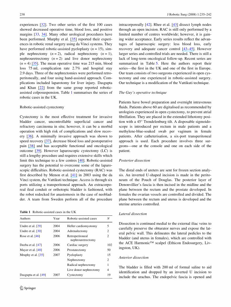
experiences [32]. Two other series of the Wrst 100 casesshowed decreased operative time, blood loss, and positivemargins [33, 34]. Many other urological procedures havebeen performed. Murphy et al. [35] reported their experi-ences in robotic renal surgery using da Vinci systems. Theyhave performed robotic-assisted pyeloplasty (n = 15), sim-ple nephrectomy (n = 2), radical nephrectomy (n = 1),nephrouretrectomy (n = 2) and live donor nephrectomy(n = 4) [35]. The mean operative time was 215 min, bloodloss 75 ml, complication rate 2.7% and hospital stay2.9 days. Three of the nephrectomies were performed retro-peritoneally, and four using hand-assisted approach. Com-plications included laparotomy for liver injury. Dasguptaand Khan [22] from the same group reported robotic-assisted colposuspension. Table 1 summarises the series ofrobotic cases in the UK.
Robotic-assisted cystectomy
Cystectomy is the most eVective treatment for invasivebladder cancer, uncontrollable superWcial cancer andrefractory carcinoma in situ; however, it can be a morbidoperation with high risk of complications and slow recov-ery [36]. A minimally invasive approach was shown tospeed recovery [37], decrease blood loss and postoperativepain [38] and has acceptable functional and oncologicaloutcome [39]. However laparoscopic cystectomy (LC) isstill a lengthy procedure and requires extensive skills whichlimit this technique to a few centres [40]. Robotic-assistedsurgery has the potential to overcome some of the laparo-scopic diYculties. Robotic-assisted cystectomy (RAC) wasWrst described by Menon et al. [41] in 2003 using the daVinci system, the Vattikuti technique. Access is through sixports utilizing a transperitoneal approach. An extracorpo-real ileal conduit or orthotopic bladder is fashioned, withthe robot redocked for anastomosis in the case of neoblad-der. A team from Sweden perform all of the procedure
intracorporeally [42]. Rhee et al. [43] dissect lymph nodesthrough an open incision. RAC is still only performed by alimited number of centres worldwide; however, it is gain-ing wider acceptance. Early series results reXect the advan-tages of laparoscopic surgery: less blood loss, earlyrecovery and adequate cancer control [43–45]. Howeverlarger series and controlled trials are needed. There is still alack of long-term oncological follow-up. Recent series aresummarized in Table 5. Here the authors report theirseries—the Wrst in the UK and one of the Wrst in Europe.Our team consists of two surgeons experienced in open cys-tectomy and one experienced in robotic-assisted surgery.Our technique is a modiWcation of the Vattikuti technique.
The Guy’s operative technique
Patients have bowel preparation and overnight intravenousXuids. Patients above 60 are digitalised as recommended byurologists experienced in open cystectomy, to prevent atrialWbrillation. They are placed in the extended lithotomy posi-tion with a 45° Trendelenburg tilt. A disposable sigmoido-scope is introduced per rectum in male patients and amethylene-blue-soaked swab per vaginum in femalepatients. After catheterisation, a six-port transperitonealapproach is used. Each procedure involves three sur-geons—one at the console and one on each side of thepatient.
Posterior dissection
The distal ends of ureters are sent for frozen section analy-sis. An inverted U-shaped incision is made in the perito-neum of the Pouch of Douglas. The posterior layer ofDenonvillier’s fascia is then incised in the midline and theplane between the rectum and the prostate developed. Infemales the ovarian vessels are controlled and divided. Theplane between the rectum and uterus is developed and theuterine arteries controlled.
Lateral dissection
Dissection is continued medial to the external iliac veins tocarefully preserve the obturator nerves and expose the lat-eral pelvic wall. This delineates the lateral pedicles to thebladder (and uterus in females), which are controlled withthe ACE Harmonic™ scalpel (Ethicon Endosurgery, Liv-ingston, UK).
Anterior dissection
The bladder is Wlled with 200 ml of formal saline to aididentiWcation and dropped by an inverted U incision toinclude the urachus. The endopelvic fascia is opened and
Table 1 Robotic-assisted cases in the UK
Authors Year Robotic-assisted cases N
Undre et al. [29] 2004 Heller cardiomyotomy 5
Undre et al. [30] 2004 Adrenalectomy 2
Rose et al. [46] 2006 Retroperitoneal nephrouretrectomy
2
Deeba et al. [47] 2006 Cardiac surgery 102
Mayer et al. [48] 2006 Prostatectomy 50
Mruphy et al. [35] 2007 Pyeloplasty 15
Nephrectomy 2
Radical nephrectomy 1
Live donor nephrectomy 4
Dasgupta et al. [49] 2007 Cystectomy 19
123
J Robotic Surg (2008) 1:235–242 239
the dorsal vein controlled by a stitch. Nerve sparing is per-formed for potent male patients. The distal urethral marginis sent for frozen section. In females the urethra is dissectedfully to the external meatus and the posterior vaginal fornixopened. The previously placed methylene blue swabbecomes visible, indicating that the correct plane had beenentered. The lateral vaginal walls are transected. The cys-tectomy specimens are secured in a bag for later retrieval.Leakage of carbon dioxide from the vagina is reduced by awaterproof dressing applied externally. The vagina is thenclosed longitudinally by continuous intracorporeal suturingwith 2-0 vicryl.
Lymphadenectomy and transposition of left ureter
Using robotic bipolar forceps and scissors, careful bilaterallymphadenectomy is performed. The limits of the dissec-tion are the genitofemoral nerve laterally, the bifurcation ofthe common iliac artery proximally and the node of Cloquetdistally. Care is taken to preserve the obturator nerve. Thelymph nodal packs are placed in separately marked laparo-scopic sacks. An Endoloop™ (Ethicon Endosurgery, Liv-ingston, UK) is applied on the distal end of the left ureterwhich is then transposed under the sigmoid mesocolon tothe left by pulling the Endoloop™ through. The distal endsof the ureters are held together with a laparoscopic grasperintroduced through the left-sided 5-mm assistant port.
Urinary diversion
All diversions are performed extracorporeally. For ileal con-duits, a 15-cm segment of ileum about 15 cm proximal to theileocaecal junction is held in laparoscopic graspers intro-duced through the most lateral right-sided 10-mm port. Therobot is undocked. The previously bagged bladder andlymph nodal specimens are extracted through a 5–7 cm inci-sion. In thin patients this should be an appendix muscle-splitting incision made by extending a lateral port, while inoverweight patients (BMI > 30 Kg/m2) a subumbilical mid-line incision is preferred for easier left ureteric access. Thegraspers holding the ureters and ileal segment are brought tothe surface through this incision. The ileal loop is isolated onits mesentery, bowel continuity restored with staplers andthe mesenteric window closed. Ureteroileal anastomosis isperformed over 8F feeding tubes using a Wallace I tech-nique. The distal end of the conduit is fashioned as a stomaat a previously marked site on the abdominal wall. A sumpdrain is introduced into the conduit to prevent any anasto-motic pressure and leak from subsequent stomal oedema.Studer pouches are created through lower midline incisionsand anastomosed to the urethral stump by six roboticallyplaced 3-0 monocryl sutures. A 20 F drain is placed in thepelvis. The port sites and wounds are closed with absorbable
sutures. A litre of icodextrin (Adept, ML Pharmaceuticals,Warrington, UK) is instilled into the abdomen and drainedafter an hour to reduce the risk of bowel adhesions.
Post-operative care
Initially all of our patients were electively managed in ahigh-dependency unit, as this was a new procedure in ourcentre. The nasogastric tube is removed next morning andoral liquids started as tolerated. Early mobilisation andchest physiotherapy are encouraged. Most patients are dis-charged with their pelvic drains and ureteric catheters insitu, which are removed at 2–3 weeks. Patients are seenagain at six weeks and have an abdominal ultrasound atthree months, CT scans at six months and then at six-monthly intervals. At these visits they have clinical exami-nation and assessment of serum haemoglobin, electrolytes,creatinine, chloride and bicarbonate.
Cases and outcome
We have performed 19 cases. Patients diagnosed with T4bladder cancer and those having previous pelvic radiother-apy or abdominal surgery were excluded. All procedureswere completed without conversion; two patients had Stu-der pouches fashioned. Bowel function returned on day 1except in the pouches. Patient satisfaction was high: 30(29–31) out of 32 on a validated patient satisfaction ques-tionnaire (CSQ-8). Four patients had complications. Onepatient had rectal injury in a T3 adenocarcinoma such thatthe urethra needed colostomy; one of the Studer pouchpatients developed urethrovesical stricture and was treatedsuccessfully with endoscopic dilatation. A patient had portsite bleeding which needed blood transfusion, and onepatient developed incisional hernia and was surgically
Table 2 Patient demographics of robotic-assisted radical cystectomyat Guy’s Hospital
BMI body mass index, TCC transitional cell carcinoma, CIS carcinomain situa Median value
Patients
N 19
Male 13
Female 6
Age 62 (57–77)a
BMI >30 in ten patients
Indications
Muscle invasive TCC 10
Refractory CIS 4
G3T1 TCC 4
Urethral adenocarcinoma 1
123
240 J Robotic Surg (2008) 1:235–242
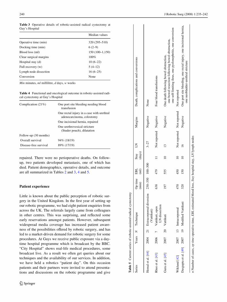
repaired. There were no perioperative deaths. On follow-up, two patients developed metastasis, one of which hasdied. Patient demographics, operative details, and outcomeare all summarized in Tables 2 and 3, 4 and 5.
Patient experience
Little is known about the public perception of robotic sur-gery in the United Kingdom. In the Wrst year of setting upour robotic programme, we had eight patient enquiries fromacross the UK. The referrals largely came from colleaguesin other centres. This was surprising, and reXected someearly reservations amongst patients. However, subsequentwidespread media coverage has increased patient aware-ness of the possibilities oVered by robotic surgery, and hasled to a market-driven demand for robotic surgery for someprocedures. At Guys we receive public exposure via a day-time hospital programme which is broadcast by the BBC.“City Hospital” shows real-life medical procedures, somebroadcast live. As a result we often get queries about ourtechniques and the availability of our services. In addition,we have held a robotics “patient day”. On this occasionpatients and their partners were invited to attend presenta-tions and discussions on the robotic programme and give
Table 3 Operative details of robotic-assisted radical cystectomy atGuy’s Hospital
Min minutes, ml millilitre, d days, w weeks
Median values
Operative time (min) 320 (295–510)
Docking time (min) 6 (2–9)
Blood loss (ml) 150 (100–1,150)
Clear surgical margins 100%
Hospital stay (d) 10 (6–22)
Full recovery (w) 5 (4–12)
Lymph node dissection 16 (6–25)
Conversion None
Table 4 Functional and oncological outcome in robotic-assisted radi-cal cystectomy at Guy’s Hospital
Complication (21%) One port site bleeding needing blood transfusion
One rectal injury in a case with urethral adenocarcinoma, colostomy
One incisional hernia, repaired
One urethrovesical stricture (Studer pouch), dilatation
Follow-up (30 months)
Overall survival 94% (18/19)
Disease-free survival 89% (17/19)
Tab
le5
Cur
rent
ser
ies
of r
obot
ic-a
ssis
ted
radi
cal c
yste
ctom
y
n N
umbe
r of
cas
es, o
p ti
me
oper
ativ
e tim
e, E
BL
est
imat
ed b
lood
loss
, Sta
y ho
spit
al s
tay,
LN
lym
ph n
odes
Seri
esY
ears
NT
echn
ique
Op
time
(min
)E
BL
(m
l)St
ay
(day
s)L
NM
argi
nsD
eath
, com
plic
atio
ns a
nd c
onve
rsio
ns
Hem
al e
tal.
[44]
2004
21E
xtra
corp
orea
l div
ersi
on
(Vat
tiku
ti)23
0–35
010
0–30
03–
27N
egat
ive
Non
e
Rhe
e et
al. [
43]
2006
7V
attik
uti,
open
L
N d
isse
ctio
n63
847
911
Not
rep
orte
dN
egat
ive
Four
blo
od tr
ansf
usio
ns
Gur
u et
al. [
45]
2007
20V
attik
uti
197
555
1013
Neg
ativ
eO
ne d
eath
fol
low
ing
bow
el o
bstr
ucti
on,
one
bow
el r
esec
tion
fol
low
ing
bow
el o
bstr
uctio
n,
one
self
-lim
itin
g ile
us, o
ne p
yelo
neph
riti
s, o
ne c
onve
rsio
n
Wik
lund
[42
]20
0713
Intr
acor
pore
al47
045
010
Not
rep
orte
dN
ot r
epor
ted
Not
rep
orte
d
Das
gupt
a et
al. [
49]
2007
19M
odiW
ed V
attik
uti
320
150
1016
Neg
ativ
eO
ne p
ort s
ite b
leed
ing,
one
rec
tal i
njur
y, o
ne in
cisi
onal
her
nia,
on
e ne
obla
dder
-ure
thra
l str
ictu
re
123
J Robotic Surg (2008) 1:235–242 241
feedback on their experiences. We addressed two mainareas—their expectations of the service, and what level ofinformation should be available to patients undergoingrobotic surgery. Many important topics were discussed,some regarding the good experiences patients had regard-ing the video availability of the procedures, leaXets andpostoperative pain control. Another issue was the diYcultyinvolved in patients obtaining access to robotic surgery dueto the paucity of machines in the UK. Most patients pre-ferred to receive counselling about the robot from a special-ist nurse with a Wxed contact telephone number in case ofany diYculty.
Discussion
Robotic surgery presents many appealing advantages toboth surgeons and patients. It oVers all of the beneWts oflaparoscopic surgery; in addition it provides a steadytremor-free scaled motion. The intuitive movements andthe enhanced DOF with the 3-D stereoscopic vision suggestan advantage over laparoscopic surgery. The early func-tional outcomes of robotic radical prostatectomy are prom-ising; however, most of the comparative studies are fromsingle institutions, and lack a high level of evidence [32].
The high cost of purchasing and maintaining the instru-ments of the robotic system is one of its many disadvan-tages. The absence of haptic feedback remains an importantissue, but this technology may well be introduced in thenear future. The current da Vinci system is still sizeable andrequires a team of trained staV to set it up over a lengthytime period. The availability of the robotic systems to onlya limited number of centres reduces surgical training oppor-tunities, and this is particularly obvious in Britain. How-ever, overall, robotic surgery in the UK is evolving, withpublic acceptance and awareness increasing steadily.
References
1. Peplinski R (2006) Past, present and future of the da Vinci robot.2nd UK Robotic Urology Course. Guy’s Hospital, London
2. Rosheim ME (2006) Leonardo's lost robots. Springer, Berlin3. Challacombe BJ, Khan MS, Murphy D, Dasgupta P (2006) The
history of robotics in urology. World J Urol 24(2):120–1274. Satava RM (2002) Surgical robotics: the early chronicles: a per-
sonal historical perspective. Surg Laparosc Endosc Percutan Tech12(1):6–16
5. Marescaux J, Leroy J, Gagner M, Rubino F, Mutter D, Vix M et al.(2001) Transatlantic robot-assisted telesurgery. Nature 413:379–380
6. Sung GT, Gill IS (2001) Robotic laparoscopic surgery: a compar-ison of the DA Vinci and Zeus systems. Urology 58(6):893–898
7. Thaly R, Shah K, Patel VR (2007) Applications of robots in urol-ogy. J Robot Surg 1(1):3–17
8. Murphy D, Challacombe B, Khan MS, Dasgupta P (2006) Robotictechnology in urology. Postgrad Med J 82(973):743–747
9. Rovetta A, Sala R (1995) Robotics and telerobotics applied to aprostate biopsy on a human patient. Proceedings of the SecondSymposium on Medical Robotics and Computer Assisted Surgery(MRCAS ’95). Wiley, New York, p 104
10. Challacombe B, Patriciu A, Glass J, Aron M, Jarrett T, Kim F et al.(2005) A randomized controlled trial of human versus robotic andtelerobotic access to the kidney as the Wrst step in percutaneousnephrolithotomy. Comput Aided Surg 10(3):165–171
11. Challacombe B, Kavoussi L, Patriciu A, Stoianovici D, DasguptaP (2006) Technology insight: telementoring and telesurgery inurology. Nat Clin Pract Urol 3(11):611–617
12. Nguyen NT, Ho HS, Smith WD, Philipps C, Lewis C, De Vera RMet al. (2001) An ergonomic evaluation of surgeons’ axial skeletaland upper extremity movements during laparoscopic and open sur-gery. Am J Surg 182(6):720–724
13. Berguer R, Gerber S, Kilpatrick G, Beckley D (1998) An ergo-nomic comparison of in-line vs pistol-grip handle conWguration ina laparoscopic grasper. Surg Endosc 12(6):805–808
14. Berguer R, Rab GT, bu-Ghaida H, Alarcon A, Chung J (1997) Acomparison of surgeons’ posture during laparoscopic and opensurgical procedures. Surg Endosc 11(2):139–142
15. NASA (2007) Robonaut hands. National Aeronautics and SpaceAdministration (NASA), Washington, DC
16. Patkin M, Isabel L (1995) Ergonomics, engineering and surgery ofendosurgical dissection. J R Coll Surg Edinb 40(2):120–132
17. Hemal AK, Srinivas M, Charles AR (2001) Ergonomic problemsassociated with laparoscopy. J Endourology 15(5):499–503
18. Berguer R, Smith WD, Chung YH (2001) Performing laparo-scopic surgery is signiWcantly more stressful for the surgeon thanopen surgery. Surg Endosc 15(10):1204–1207
19. Talamini M, Campbell K, StanWeld C (2002) Robotic gastrointes-tinal surgery: early experience and system description. J Laparo-endosc Adv Surg Tech A 12(4):225–232
20. Jourdan IC, Dutson E, Garcia A, Vleugels T, Leroy J, Mutter Det al. (2004) Stereoscopic vision provides a signiWcant advantagefor precision robotic laparoscopy. Br J Surg 91(7):879–885
21. Elhage O, Murphy D, Challacombe B, Shortland A, Dasgupta P(2007) Ergonomics in minimally invasive surgery. Int J Clin Prac61(2):186–188
22. Dasgupta P, Hemal A, Rose K 2005) Robotic urology in the UK:establishing a programme and emerging role. BJU Int 95(6):723–724
23. Urology Department (2007) Guy'sHospital, London, UK24. DoH (2006) NHS reference costs 2005–2006. Department of
Health, London25. Link RE, Bhayani SB, Kavoussi LR (2006) A prospective compar-
ison of robotic and laparoscopic pyeloplasty. Ann Surg243(4):486–491
26. Scales CD Jr, Jones PJ, Eisenstein EL, Preminger GM, Albala DM(2005) Local cost structures and the economics of robot assistedradical prostatectomy. J Urol 174(6):2323–2329
27. Kaul SA, Peabody JO, Shah N, Neal D, Menon M (2006) Estab-lishing a robotic prostatectomy programme: the impact of mentor-ing using a structured approach. BJU Int 97(6):1143–1144
28. Deeba S, Aggarwal R, Sains P, Martin S, Athanasiou T, Casula Ret al. (2006) Cardiac robotics: a review and St Mary’s experience.Int J Med Robot 2(1):16–20
29. Undre S, Moorthy K, Munz Y, Aggarwal R, Hance J, Rockall Tet al. (2004) Robot-assisted laparoscopic Heller cardiomyotomy:preliminary UK results. Dig Surg 21(5–6):396–400
30. Undre S, Munz Y, Moorthy K, Martin S, Rockall T, Vale J et al.(2004) Robot-assisted laparoscopic adrenalectomy: preliminaryUK results. BJU Int 93(3):357–359
31. Mayer EK, Winkler MH, Aggarwal R, Karim O, Ogden C, HroudaD et al (2006) Robotic prostatectomy: the Wrst UK experience. IntJ Med Robot 2(4):321–328
123
242 J Robotic Surg (2008) 1:235–242
32. Ficarra V, Cavalleri S, Novara G, Aragona M, Artibani W (2007)Evidence from robot-assisted laparoscopic radical prostatectomy:a systematic review. Eur Urol 51(1):45–56
33. Goldstraw MA, Corbishley C, Besarani D, Anderson CJ, DasguptaP, Kirby RS (2007) A comparative study of 100 robotic assistedlaparoscopic prostatectomy (RALP) versus 100 open retropubicprostatectomy. BJUI 99(s4):35
34. Tyreman N, Shah N, Basnett G, Neal DE (2007) Learning curveduring robotic-assisted radical prostatectomy. BJU Int 99(s4):36
35. Murphy D, Challacombe B, Olsburgh J, Calder F, Mamode N,Khan MS et al. (2007) Ablative and reconstructive robotic-assist-ed laparoscopic renal surgery. IJCP (in press)
36. Stein JP, Lieskovsky G, Cote R, Groshen S, Feng AC, Boyd Set al. (2001) Radical cystectomy in the treatment of invasive blad-der cancer: long-term results in 1,054 patients. J Clin Oncol19(3):666–675
37. Basillote JB, Abdelshehid C, Ahlering TE, Shanberg AM (2004)Laparoscopic assisted radical cystectomy with ileal neobladder: acomparison with the open approach. J Urol 172(2):489–493
38. Cathelineau X, Arroyo C, Rozet F, Barret E, Vallancien G (2005)Laparoscopic assisted radical cystectomy: the montsouris experi-ence after 84 cases. Eur Urol 47(6):780–784
39. Haber GP, Gill IS (2007) Laparoscopic radical cystectomy forcancer: oncological outcomes at up to 5 years. BJU Int100(1):137–142
40. Hrouda D, Adeyoju AA, Gill IS (2004) Laparoscopic radicalcystectomy and urinary diversion: fad or future? BJU Int94(4):501–505
41. Menon M, Hemal AK, Tewari A, Shrivastava A, Shoma AM,El-Tabey NA et al. (2003) Nerve-sparing robot-assisted radicalcystoprostatectomy and urinary diversion. BJU Int 92(3):232–236
42. Wiklund PN (2007) Robotic assisted radical cystectomy: technicalaspects. In: 2007 World Robotic Urology Symposium, Columbus,OH, 26–27 April 2007
43. Rhee JJ, Lebeau S, Smolkin M, Theodorescu D (2006) Radicalcystectomy with ileal conduit diversion: early prospective evalua-tion of the impact of robotic assistance. BJU Int 98(5):1059–1063
44. Hemal AK, bol-Enein H, Tewari A, Shrivastava A, Shoma AM,Ghoneim MA et al (2004) Robotic radical cystectomy and urinarydiversion in the management of bladder cancer. Urol Clin NorthAm 31(4):719–729
45. Guru KA, Kim HL, Piacente PM, Mohler JL (2007) Robot-assist-ed radical cystectomy and pelvic lymph node dissection: initialexperience at Roswell Park Cancer Institute. Urology 69(3):469–474
46. Rose K, Khan S, Godbole H, Olsburgh J, Dasgupta P (2006) Ro-botic assisted retroperitoneoscopic nephroureterectomy—Wrstexperience and the hybrid port technique. Int J Clin Pract60(1):12–14
47. Deeba S, Aggarwal R, Sains P, Martin S, Athanasiou T, Casula Ret al. (2006) Cardiac robotics: a review and St Mary’s experience.Int J Med Robot 2(1):16–20
48. Mayer EK, Winkler MH, Aggarwal R, Karim O, Ogden C, HroudaD et al. (2006) Robotic prostatectomy: the Wrst UK experience. IntJ Med Robot 2(4):321–328
49. Dasgupta P, Rimington P, Murphy D, Challacombe BJ, O’BrienTS, Khan MS (2007) Robot-assisted radical cystectomy for blad-der cancer and 2 year follow-up. In: Laparoscopic/Robotics Ses-sion, British Association of Urological Surgeons Annual Meeting,Glasgow, UK, 18–22 June 2007
123