
Nuclear qualification of electrical equipment - Brazilian ...
Upload
khangminh22Category
view
2download
0
ISSN 1677-5538
Volume 36, Number , January - February, 2010
Official Journal of the Brazilian Society of Urology
1
Braz J UrolBraz J Urol
InternationalInternational
lFu l T
ext Olin
e Ac
s A
n
cesvai
lelab
www.brazjuro
l.co
.brm
Official Journal of the Thai Urological Association
B
An 84-year-old man with left renal pelvic tumor. A) Retrograde pyelography shows small deformities in the upper calyx. B) On axial diffusion weighted image (DWI) and C) Maximum intensity projection image obtained from axial DWIs, the tumor is depicted
in the parenchyma of upper part of the left kidney as high intensity. (Page 18)
D
ew
- Vi
Section
N
deo
A
B
C

CLINICAL UROLOGY______________________________________________________________________
Cigarette Smoking Impairs Sperm BioenergeticsF.J.B. Sampaio
3
CONTENTS
Volume 36, number 1 January- February, 2010
1
International Braz J Urol
10
29
44
18
REVIEW ARTICLE _________________________________________________________________________
38
49
55
Current Diagnosis and Management of Syringocele: A ReviewJ. Melquist, V. Sharma, D. Sciullo, H. McCaffrey, S.A. Khan (Editorial Comment by J.C. Truzzi)
Laparoscopic Nephropexy Exposes a Possible Underlying Pathogenic Mechanism and Allows Successful Treatment with Tissue Gluing of the Kidney and Fixation of the Colon to the Lateral Abdominal WallJ. Wadstrom, M. Haggman (Editorial Comments by M. Hatzinger and D. Teber)
Diffusion Weighted Imaging in the Detection of Upper Urinary Tract Urothelial TumorsShuji Nishizawa, Shun Imai, Toshikazu Okaneya, Tsuyoshi Nakayama, Takayuki Kamigaito, Tomonori Minagawa (Editorial Comments by O. Kilickesmez and H. Masuda)
What is the Best Drainage Method for a Perinephric Abscess?A.R. EL-Nahas, R. Faisal, T. Mohsen, M.S. AL-Marhoon, H. Abol-Enein (Editorial Comments by E. Mazzucchi, E.O. Kehinde and Reply by The Authors)
Laparoscopic Ureteral Reimplant for Ureteral StrictureR.S.Q. Soares, R.A. de Abreu Jr, J.E.F. Tavora (Editorial Comment by P. Modi)
Outcomes Following Negative Prostate Biopsy for Patients with Persistent Disease after Radiotherapy for Prostate CancerJ.H. Cohen, J. Eastham, R.J. Macchia
Analgesic Efficacy and Safety of Nonsteroidal Anti-Inflammatory Drugs after Transurethral Resection of ProstateC. Kara, B. Resorlu, I. Cicekbilek, A. Unsal (Editorial Comments by E.M. Mazaris & E. Chatzidarellis)
Duration of Preoperative Scrotal Pain May Predict the Success of Microsurgical VaricocelectomyB. Altunoluk, H. Soylemez, E. Efe, O. Malkoc (Editorial Comments by L. Carmignani & L. Lunelli)
Cigarette Smoking Impairs Sperm BioenergeticsK.R. Chohan, S.Z.A. Badawy (Editorial Comments by R. Fraietta & A.P. Cedenho)
60

CONTENTS - continued from previous page
66
UROLOGICAL SURVEY____________________________________________________________________
75
97
98
NEUROUROLOGY _________________________________________________________________________ _________________________________________________________________________
86
99
Experience with Different Botulinum Toxins for the Treatment of Refractory Neurogenic Detrusor Over-activityC.M. Gomes, J.E. de Castro Filho, R.F. Rejowski, F.E. Trigo-Rocha, H. Bruschini, T.E.P. de Barros Filho, M. Srougi (Editorial Comment by J.L. Amaro)
To Circ or not to Circ: Clinical and Pharmacoeconomic Outcomes of a Prospective Trial of Topical Ste-roid Versus Primary CircumcisionY.D. Nobre, R.G. Freitas, M.J. Felizardo, V. Ortiz, A. Macedo Jr. (Editorial Comments by G. Vaos and S. Taskinen)
The Effect of Alpha-Blocker Treatment on Bladder Hypoxia Inducible Factor-1 Alpha Regulation During Lower Urinary Tract ObstructionG. Koritsiadis, S.I. Tyritzis, G. Koutalellis, A.C. Lazaris, K. Stravodimos (Editorial Comments by Y. Chuang and M.B. Chancellor)
STONE DISEASE
Renal functional effects of multiple-tract percutaneous accessJ Endourol. 2009; 23: 1951-6M. Monga
Residual fragments after percutaneous nephrolithotomy: cost comparison of immediate second look flexible nephroscopy versus expectant managementJ Urol. 2010; 183: 188-93M. Monga
ENDOUROLOGY & LAPAROSCOPY
7-year oncological outcomes after laparoscopic and open partial nephrectomyJ Urol. 2010; 183: 473-9F.J. Kim
Cost analysis of robotic versus open radical cystectomy for bladder cancerJ Urol. 2010; 183: 505-9F.J. Kim
IMAGING
Clinical stage T1c prostate cancer: evaluation with endorectal MR imaging and MR spectroscopic imagingRadiology. 2009; 253: 425-34A. Prando
96
PEDIATRIC UROLOGY _____________________________________________________________________ _____________________________________________________________________
BASIC AND TRANSLATIONAL UROLOGY ___________________________________________________ ___________________________________________________
100

International Braz J UrolCONTENTS - continued from previous page
103
104
107
105
101
103
Bladder tumor staging: comparison of contrast-enhanced and gray-scale ultrasoundAJR Am J Roentgenol. 2010; 194: 151-6A. Prando
UROGENITAL TRAUMA
Straddle injuries to the bulbar urethra: management and outcomes in 78 patientsJ Urol. 2004; 171(2 Pt 1): 722-5
Management of low velocity gunshot wounds to the anterior urethra: the role of primary repair versus urinary diversion aloneJ Urol. 1993; 150: 70-2
Straddle injuries to the bulbar urethra: management and outcome in 53 patientsInt Braz J Urol. 2009; 35: 450-8S.B. Brandes
PATHOLOGY
Does perineural invasion on prostate biopsy predict adverse prostatectomy outcomes?BJU Int. 2009 Aug 19. [Epub ahead of print]A. Billis
Transurethral resection specimens of the bladder (TURB): Outcome of invasive urothelial cancer involving muscle bundles indeterminate between muscularis mucosae and muscularis propriaMod Pathol 2010;in press [Abstract from the USCAP meeting, 2010]A. Billis
BASIC AND TRANSLATIONAL UROLOGY
Temporary segmental renal artery occlusion using reverse phase polymer for bloodless robotic partial nephrectomyJ Urol. 2009; 182:1582-7F.J.B. Sampaio
Sildenafil as a protecting drug for warm ischemic kidney transplants: experimental resultsJ Urol. 2009; 182: 1222-5F.J.B. Sampaio
RECONSTRUCTIVE UROLOGY
Multivariate analysis of risk factors for long-term urethroplasty outcomeJ Urol. 2010; 183: 613-7S.P. Elliott
Internal urethrotomy and intraurethral submucosal injection of triamcinolone in short bulbar urethral stricturesInt Urol Nephrol. 2009 Dec 1. [Epub ahead of print]S.P. Elliott
108
109
111
110

CONTENTS - continued from previous page
114
112
113
UROLOGICAL ONCOLOGY
Outcome of patients who refuse cystectomy after receiving neoadjuvant chemotherapy for muscle-invasive bladder cancerEur Urol. 2008; 54: 126-32A. Bohle
Long-term rates of undetectable PSA with initial observation and delayed salvage radiotherapy after radical prostatectomyEur Urol. 2008; 54: 88-94A. Bohle
NEUROLOGY & FEMALE UROLOGY
Repeat synthetic mid urethral sling procedure for women with recurrent stress urinary incontinenceJ Urol. 2010; 183: 241-6S.P. Petrou
An International Urogynecological Association (IUGA)/International Continence Society (ICS) joint report on the terminology for female pelvic floor dysfunctionNeurourol Urodyn. 2010;29(1):4-20.S.P. Petrou
PEDIATRIC UROLOGY
Risk factors for urinary tract infection after dextranomer/hyaluronic acid endoscopic injectionJ Urol. 2009; 182 (4 Suppl): 1708-12B.W. Snow
Straightening ventral curvature while preserving the urethral plate in proximal hypospadias repairJ Urol. 2009; 182 (4 Suppl): 1720-5B.W. Snow
Radical Nephrectomy with IVC Thrombectomy (Level-III) Conducted on Veno-Veno BypassT.S. Hakky, L.R. Wiegand, D. Mangar, A. Alsina, P.E. Spiess (Editorial Comment by A.K. Kader)
Information for Authors
Urological Calendar
116
117
VIDEO ____________________________________________________________________________________
118
GENERAL INFORMATION __________________________________________________________________
120
122
126

�
EDITOR’S COMMENT
International Braz J Urol
Cigarette Smoking Impairs Sperm Bioenergetics
TheJanuary–February2010issueoftheInternationalBrazJUrolpresentsoriginalcontributionsandeditorialsfrommanydifferentcountries,suchasUSA,Japan,Greece,Turkey,Egypt,Sweden,Brazil,Germany,Norway,Taiwan,Kuwait,Italy,India,etc.,andasusual,theeditor’scommenthighlightssomepapers.
DoctorChohanandco-workers,fromUpstateMedicalUniversity,Syracuse,NY,USA,comparedonpage60therateofspermrespirationinsmokersandnon-smokers.Theyevaluatedsemensamplesfrom20smokersand58non-smokers.Aphosphorescenceanalyzer thatmeasuresO2concentration in spermsuspensions as function of time was used to determine the rate of respiration. The authors did not find diffe-rencesbetweensmokersandnon-smokersforejaculatevolume,motility,concentration,normalmorphology,viabilityandhypo-osmoticswellingtest.TherateofspermmitochondrialO2consumptioninthesmokerswas0.96±0.58andinthenon-smokers1.39±0.67(p=0.004).Theauthorsconcludedthattherateofspermrespiration was significantly lower in smokers. This negative impact of cigarette smoking on sperm aerobic metabolismmay,inpart,explainthelowerrateoffertilityinsmokers.
DoctorNishizawaandcolleagues,fromNaganoMunicipalHospital,Japan,evaluatedonpage18thecapabilityandreliabilityofdiffusion-weightedmagneticresonanceimaging(DWI)intheevaluationofupperurinarytracturothelialtumors.DWIwasperformedin17patientswithupperurinarytracturothelialtumor,previouslydiagnosedbyeitherCTorretrogradepyelography.Ahistologicalevaluationwasperformedaftersurgicalresection.In9patientswithrenalpelvistumorsand7patientswithureteraltumors,thelesionswereshownashigh-signalintensityinthecorrespondingregiononDWI.Inonepatientwithcarcinomainsitu(CIS)oftheureter,thelesionwasnotdepictedwithDWI.Inthisstudy,therenalpelvicandureteraltumorsexceptCISwereshownclearlywithDWI.Wheneveravailable,DWImaytaketheplaceofinvasiveretrogradeurographyfordetectingtumorsoftheupperurinarytract.
DoctorEl-Nahasandassociates,fromMansouraUniversity,Egypt,comparedonpage29theresultsofpercutaneousandopendrainageforperinephricabscess.Eighty-sixpatientswhounderwentdrainageforperinephricabscesseswereevaluated.Percutaneoustubedrain(PCD)wasusedfordrainageoftheabscessin43patients(group1),whiletheother43patientsweremanagedwithopendrainage(group2).Theau-thors found that open drainage of perinephric abscesses resulted in a statistically significant higher cure rate (98%versus69%,p<0.001)andshorterhospitalstaythanPCD(3.6versus6days,p<0.001).Failureofcompletedrainageofmultilocularabscesswasobservedin8of13cases(61.5%)ingroup1andoneof38cases(2.6%)ingroup2(P<0.001).Complicationswereobservedin7%ofgroup1and11.5%ingroup2(P=0.45).Aftermeanfollow-upof19months,9of46patients(19.6%)hadrecurrence;7ofthemwereingroup1.Itwasconcludedthatpercutaneousdrainageofperinephricabscessisaneffectiveminimally
doi: 10.1590/S1677-55382010000100001

�
EDITOR’S COMMENT - continued
invasivetreatment.However,PCDisnottheoptimalmethodfordrainageofmultilocularabscessbecauseopensurgicaldrainageprovidedhighercure ratesandshorterhospitalization thanPCD.Dr.Mazzucchi,fromUniversityofSaoPaulo,SP,BrazilandDr.KehindefromKuwaitUniversity,Safat,Kuwait,providedbalancededitorialcommentsonthisarticle.
DoctorAltunolukandco-authors,fromSutcuImamUniversity,Kahramanmaras,Turkey,reportedonpage55theirresultswithmicrosurgicalsubinguinalvaricoceleligationtotreatpain.Theyanalyzed284menwhounderwentsubinguinalmicrosurgicalvaricoceleligationforscrotalpain.Themedianpatientageatthetimesurgerywas23.7years(range16-38years)andtheaveragedurationofpainbeforepresentationwas11.2months(range1monthto40months).In85.6%ofpatientstherewascompleteresolutionofpainand 6.3% had partial resolution. Pain persisted postoperatively in 19 cases (8.1%). A significant difference wasobservedinpostoperativesuccessbetweenpatientswhohadlongperiodandthosewhohadshortperiodofpain.Theauthorsconcludedthatsub-inguinalmicrosurgicalvaricoceleligationisaneffectivetreatmentforpainfulvaricocele.Thedurationofpainpreoperativelymaypredictoutcomesinselectedpatients.
DoctorGomesandcolleagues,fromUniversityofSaoPauloSchoolofMedicine,SaoPaulo,Brazil,reportedonpage66theirexperiencewiththeuseofbotulinumtoxin-A(BoNT/A)formulationsBotox®andProsigne® in the treatment of neurogenic detrusor overactivity (NDO). Forty-five consecutive patients with refractoryurinaryincontinenceduetoNDOreceivedasingleintradetrusor(excludingthetrigone)treatmentwith botulinum toxin type A. Botox was used for the first 22 patients, and Prosigne for the subsequent 23 pa-tients. Significant improvements from baseline in maximum cystometric capacity (MCC), maximum detrusor pressureduringbladdercontraction,andcompliancewereobservedinbothgroups(P<0.05).Improvementin MCC was significantly greater with Botox versus Prosigne (+103.3% vs. +42.2%; P = 0.019). Continence wasachievedbyweek12in16Botoxrecipients(76.2%)and10Prosignerecipients(47.6%;P=0.057).Nosevereadverseeventswereobserved.TheauthorsconcludedthatBotoxandProsigneproducedistincteffectsinpatientswithNDO,withagreaterincreaseinMCCwithBotox.
DoctorKoritsiadisandcolleaguesfromAthensUniversityMedicalSchool.Greece,determinedonpage 86 whether alpha1-blocker treatment, in chronic bladder outlet obstruction (BOO), influences bladder tissueischemia.Theyperformedaprospectivestudyincluding60patientswithBOO,ofwhich40wereunderalpha1-blockermedicationand20withouttreatment.Patientsunderwenttransurethralresectionoftheprostate(TURP)orsuprapubicprostatectomy(SPP).Tissuespecimenswereimmunohistochemicallystainedforhypoxiainduciblefactor-1alpha(HIF-1alpha).TheyfoundthatbladdertissuefromobstructedsubjectsshowedhighimmunoreactivitytoHIF-1alpha.Thespecimensfromthecontrolgroup,showednoor weak, mainly cytoplasmic immunoreactivity to HIF-1alpha. Patients under α -blocker treatment did not differinthenumberofHIF-1alphapositivecellscomparedtosubjectswithnotreatment.Itwasconcludedthattreatmentwithalpha-blockersinobstructedpatientsconsideredasnon-responders,doesnotresultinHIF-1alphadownregulation,thusbladdercontinuestobeunderchronicstress.
Francisco J.B. Sampaio, M.D.Editor-in-Chief

�
Diagnosis and Management of SyringoceleInternational Braz J Urol Vol. 36 (1): 3-9, January - February, 2010
Current Diagnosis and Management of Syringocele: A Review
Jonathan Melquist, Vidit Sharma, Daniella Sciullo, Heather McCaffrey, S. Ali Khan
Department of Urology, SUNY at Stony Brook Medical Center, Stony Brook, New York, USA
ABSTRACT
Cowper’s syringocele is a rare but an under-diagnosed cystic dilation of the Cowper’s ducts and is increasingly being recognized in the adult population. Recent literature suggests that syringoceles be classified based on the configuration of the duct’s orifice to the urethra, either open or closed, as this also allows the clinical presentations of 2 syringoceles to be divided, albeit with some overlap. Usually post-void dribbling, hematuria, or urethral discharge indicate open syringocele, while obstructive symptoms are associated with closed syringoceles. As these symptoms are shared by many serious condi-tions, a working differential diagnosis is critical. Ultrasonography coupled with retro and ante grade urethrography usually suffices to diagnose syringocele, but supplementary procedures - such as cystourethroscopy, computed tomography scan, and magnetic resonance imaging - can prove useful. Conservative observation is first recommended, but persistent symp-toms are usually treated with endoscopic marsupialization unless contraindicated. Upon reviewing the literature, this paper addresses the clinical anatomy, classification, presentation, diagnosis, and treatment of syringoceles in further detail.
Key words: Cowper’s glands; dilation; urethral obstruction; perineum; urinary incontinenceInt Braz J Urol. 2010; 36: 3-9
INTRODUCTION
Cowper’s syringocele is an uncommon but an under-diagnosed cystic dilation of the Cowper’s gland ducts. Syringoceles are traditionally viewed as a rare condition afflicting the pediatric population but are increasingly being recognized in the adult population. They are frequently not detailed in major uropathol-ogy, radiology, and urologic textbooks even though they can cause severe lower urinary tract symptoms by compressing the urethra or diverting urinary flow. This paper reviews the current literature on the clinical anatomy, classification, clinical presentation, diagno-sis and treatment of syringoceles.
�e�ie�� �rticle�e�ie�� �rticle
doi: 10.1590/S1677-55382010000100002
FUNCTIONAL ANATOMY OF COWPER’S GLANDS AND DUCTS
Cowper’s glands are composed of two exocrine structures located in the deep perineal pouch between fascial layers of the urogenital diaphragm. They excrete pre-ejaculate into the genito-urinary tract (1). The glands are composed of lobules made of epithelial cells aligned in acinar formation that secrete into the arborized collect-ing system. The glands eventually form two collecting ducts that measure on average 2.5 cm each. Although anatomic variations exist, the majority of ducts combine to make one confluent passage that opens at the posterior aspect of the bulbous urethra (2,3).

�
Diagnosis and Management of Syringocele
CLINICAL MANIFESTATION AND CLASSIFICATION OF SYRINGOCELES
The true prevalence of Cowper’s syringocele is unknown. It is thought to be more pronounced in the pediatric population perhaps because symptoms are appreciated preferentially at a younger age. However, there is a growing body of literature suggesting the problem exists notably in the adult population as well. There are at least 10 case reports describing this rare anomaly in patients over the age of 18 (4). Traditionally, Cowper’s syringocele has been divided into four types: 1) simple syringocele with a modestly dilated duct; 2) perforated syringocele with patulous communication with the urethra; 3) imperfo-rate syringocele with a dilated bulbous duct; 4) rup-tured syringocele that leaves its covering membrane in the urethra often acting in a “ball-on-chain” fashion to cause obstruction (5). Based on building luminal pressures within the ducts, syringoceles may follow a standard maturation from simple to imperforate to either perforated or ruptured, but more data is needed to confirm this hypothesis. Recent review suggests, however, that syrin-goceles should be grouped based on the configura-tion of the duct’s orifice to the urethra, as this also allows the clinical presentations of syringoceles to be divided (Table-1). For instance, closed syringoceles have cystically occluded ducts that cause the duct to dilate externally against the urethra and cause obstructive symptoms. Open syringoceles have a continuous lumen between the urethra and the cystic ducts, mimicking a urethral saccule and manifesting as post-void dribbling (6-8). Obstructive symptoms
may also manifest in open syringoceles if the rem-nant membrane is oriented in the urethra to impede flow. Furthermore, grouping syringoceles into these categories accounts for the 4 categories of Maizel’s et al., since simple, perforated, and ruptured syringo-celes merge into open syringoceles and imperforate syringoceles are classified as closed. A review of 15 consecutive children with Cowper’s syringocele proposed a similar simplified classification. It classified two variants: non-obstruct-ing syringoceles and obstructing syringoceles. All of the non-obstructing syringoceles presented with a combination of urinary tract infection (UTI), fever, and/or urinary incontinence. All of the obstructing syringoceles had obstructive voiding symptoms or ultrasonographic evidence of obstruction (9). Hematuria, dysuria, frequency, and recurrent UTI have also been associated with both categories of manifestation (10,11). In one of the largest case reviews reported on adult syringoceles, six of seven patients had open syringoceles, five of seven patients had a history of UTI, six of seven had bloody urethral discharge, and five of seven have post-void dribbling (6). Since the symptoms of syringocele (Table-1) are non-specific, a number of possibly more serious conditions can be at play. The functional differential diagnosis upon history and physical examination is urethral web, urethral duplication, anterior urethral valve, anterior urethral diverticulum, congenital narrowing of bulbar urethra - Cobb’s collar, urethral stricture, hydrocele (12), megalourethra, periurethral abscess, perianal abscess, congenital urethral folds, prolapsed posterior urethral valve, urethral tumors, urethral stones (13-19).
Table 1 – Common symptoms of syringocele.
Open Syringocele Closed Syringocele
Post-void dribbling Urethral discharge Urinary tract infection Obstructive voiding symptoms (less common) Perineal pain Hematuria
Obstructive voiding symptoms Dysuria Urinary retention Perineal pain
The four subtypes of Cowper’s syringocele as described by Maizels et al. (5) are merged into two clinical categories. Here we present common symptoms described analytically and anecdotally throughout the literature of adult and pediatric syringocele.

�
Diagnosis and Management of Syringocele
DIAGNOSIS
The initial evaluation of Cowper’s syringocele typically involves a thorough voiding history. A high index of suspicion justifies non-invasive imaging. Ul-trasonography (US) sometimes visualizes closed cystic lesions in the anatomic region of Cowper’s gland (20-22). US has even been used to diagnose open syringo-cele. In one case report, a retrograde urethrogram was positive for large outpouching and sonourethrogram confirmed the cystic outpouchings when the urethra
was distended with normal saline (4). To confirm or question US results the diagnosis should proceed with antegrade and retrograde urethrography, as this step is usually diagnostic (23). In case urethrography is con-traindicated or more data is needed, cystourethroscopy, urodynamic studies, computed tomography (CT) scan, or magnetic resonance imaging (MRI) may be imple-mented. A proctoscopy may serve to shorten the differ-ential diagnosis. This diagnosis algorithm is illustrated in Figure-1, and Table-2 addresses the indications for syringocele in the respective interventions.
Figure 1 – Recommended diagnostic algorithm for testing. If the patient is positive for the symptoms in Table-1 after history and physical exam, syringocele can be suspected. Ultrasonography should be followed by urethrography to reliably detect syringoceles. The dashed arrows indicate the tests that are not usually necessary to diagnose syringoceles.

�
Diagnosis and Management of Syringocele
Symptomatic, closed syringoceles often have abnormal retrograde and voiding cystourethrograms. They can present as a cystic filling defect distal to any potential prostatic obstruction. The radiologic finding can be corroborated by uroflowometry that indicates obstructive voiding rates (24). Cystoure-throscopy sometimes detects an abnormal protrusion from posterior wall of the bulbous urethra, raising the index-of-suspicion for closed syringocele. However, open syringoceles often can pres-ent with simultaneous dysuria and post-void drib-bling. They too can have an obstructive pattern if the membranous flap acts in a “ball-and-chain” fashion to cause transient urethral obstruction. Cystourethro-gram can be non-diagnostic but may indicate obstruc-tion and/or cavernous filling in adjacent urethral structure. Cystourethroscopy often reveals a defect in the continuity of the posterior bulbous urethral wall, a remnant piece of cystic wall, and/or a dilated luminal orifice (25). MRI is a non-invasive diagnostic modality continuing to define itself in diagnosis and manage-ment of Cowper’s syringocele. It is found to be of particular benefit in evaluation of closed syringoceles
and has been successfully applied to both the adult and pediatric population (26,27). MRI has supplanted CT due to its higher soft-tissue resolution; nonetheless CT still has a diagnostic role especially when MRI is contraindicated (28).
TREATMENT OF SYRINGOCELES
Asymptomatic syringoceles are often ob-served (25). Although many symptomatic ones eventually require surgical intervention, a trial pe-riod of conservative management seems prudent, as spontaneous resolution of symptoms over time is not uncommon. Bevers et al. have described several cases of confirmed both open and closed syringoceles whose symptoms resolved on their own. One case resolved after successful treatment for a UTI; others resolved with no intervention (6). In recent years endoscopic intervention has become the preferred intervention for symptomatic syringocele’s. Typically unroofing the cyst by remov-ing its visage to the urethra is a simple, effective way of marsupialization for both open and closed syringo-
Table 2 – Indications for radiologic-interventional studies to diagnose syringocele.
Study Open Syringocele Closed Syringocele
Ultrasound Previously absent cystic lesion appreciated during retrograde
urethrogram
Cystic lesions of the bulbourethral glands and ducts
Antegrade or retrograde cystourethrogram
Cavernous filling-defect distal to prostate, possible obstruction
Cystic defect distal to prostate
Cystourethroscopy Defect in continuity of posterior urethral wall
Abnormal protrusion of posterior urethral wall
Magnetic resonance imaging (MRI) Not described Homogenous, cystic lesion of bulbourethral gland, with high-signal
intensity of T2-weighted scan. Non-enhancement with dimeglumine
gadopentetate contrast.
Computed tomography Not described Homogenous cystic lesion
There are many diagnostic modalities to diagnosis various types of syringocele, but retrograde and voiding cystourethrogram are most common in the literature. Ultrasound has found a role as a minimally invasive means to address clinical suspicion of syringocele but is not sensitive enough to rule-out pathology. The role of MRI is further being defined though its use has been limited to date.

�
Diagnosis and Management of Syringocele
celes. In Bevers et al. case series, all four patients who went this urethroscopic intervention had complete resolution of their symptoms with a maximum fol-low-up interval of 23 months (mean 12 months) (6). Unroofing typically uses a cold-knife; however, the Holmium: YAG laser was successfully used in one case report (29). Alternatively, open procedures such as trans-perineal ligation of the Cowper’s duct are performed but are usually secondary to failed unroofing (30). Open excision may be of benefit when the syringocele presents as a large perineal mass (31). Laparoscopic excision-ligation of Cowper’s gland has been de-scribed as another treatment modality and may be of benefit but no trial has born this out (32). The pediatric population can be treated with transurethral endoscopic unroofing as well. However, current opinion recommends open intervention for certain populations, such as children with large di-verticula and inadequate spongiosum. In such cases, diverticulectomy should be considered (9,33-35). In the infant population where severe reflux exists due to an anterior urethral valve phenomenon secondary to syringocele, urinary diversion and vesicostomy should be considered (36,37).
CONCLUSION
Clinically it is more convenient to classify the cystic dilation of the Cowper’s Gland ducts as either open or closed, in terms of communication with the urethra, than the older system proposed by Maizels et al. The symptoms of the two types of syringocele can be categorized, albeit with some overlap. Usually post-void dribbling, hematuria, or urethral discharge indicates open syringocele, while obstructive symptoms are associated with closed syringoceles. As these symptoms are shared by many serious conditions, a working differential diagnosis is critical. Once the index of suspicion is established, transrectal and perineal US followed by retrograde and antegrade urethrography can effectively diagnose syringoceles. Other diagnostic technologies, such as cystourethroscopy, urodynamic studies, CT scan, and MRI, may be used to attain supplemental data. Treat-ment of the lesion should first proceed conservatively
under observation, as symptoms may spontaneously resolve. Persistent symptoms are the benchmark for intervention, and endoscopic marsupialization has become the standard treatment for both open and closed syringocele, but open ligation-excision may be indicated in children. Although the success rates are high for syringocele diagnosis and treatment, more comparative data is essential for establishing standard protocols.
CONFLICT OF INTEREST
None declared.
REFERENCES
1. Chughtai B, Sawas A, O’Malley RL, Naik RR, Ali Khan S, Pentyala S: A neglected gland: a review of Cowper’s gland. Int J Androl. 2005; 28: 74-7.
2. Masson JC, Suhler A, Garbay B: Cowper’s canals and glands. Pathological manifestations and radiologic aspects. J Urol Nephrol (Paris). 1979; 85: 497-511.
3. Sanders MA: William Cowper and his decorated cop-perplate initials. Anat Rec B New Anat. 2005; 282: 5-12.
4. Kumar J, Kumar A, Babu N, Gautam G, Seth A: Cowper’s syringocele in an adult. Abdom Imaging. 2007; 32: 428-30.
5. Maizels M, Stephens FD, King LR, Firlit CF: Cowper’s syringocele: a classification of dilatations of Cowper’s gland duct based upon clinical characteristics of 8 boys. J Urol. 1983; 129: 111-4.
6. Bevers RF, Abbekerk EM, Boon TA: Cowper’s syrin-gocele: symptoms, classification and treatment of an unappreciated problem. J Urol. 2000; 163: 782-4.
7. Sant GR, Kaleli A: Cowper’s syringocele causing incontinence in an adult. J Urol. 1985; 133: 279-80.
8. Shintaku I, Ono Y, Katoh N, Takeda A, Ohshima S: Anterior urethral diverticulum produced by Cowper’s gland duct cyst. Int J Urol. 1996; 3: 412-3.
9. Campobasso P, Schieven E, Fernandes EC: Cowper’s syringocele: an analysis of 15 consecutive cases. Arch Dis Child. 1996; 75: 71-3.
10. Awakura Y, Nonomura M, Fukuyama T: Cowper’s syringocele causing voiding disturbance in an adult. Int J Urol. 2000; 7: 340-2.
11. Månsson W, Colleen S, Holmberg JT: Cystic dilata-tion of Cowper’s gland duct--an overlooked cause of

�
Diagnosis and Management of Syringocele
urethral symptoms? Scand J Urol Nephrol. 1989; 23: 3-5.
12. Marte A, Prezioso M, Sabatino MD, Borrelli M, Ro-mano M, Del Balzo B, et al.: Syringocele in children: an unusual presentation as scrotal mass. Minerva Pediatr. 2009; 61: 123-7.
13. Vega RE: Distal urethral web: a risk factor in prosta-titis. Prostate Cancer Prostatic Dis. 2002; 5: 180-2.
14. Zugor V, Schreiber M, Labanaris AP, Weissmüller J, Wullich B, Schott GE: Urethral duplication: long-term results for a rare urethral anomaly. Urologe A. 2008; 47: 1603-6.
15. Myers RP, Cahill DR, Kay PA, Camp JJ, Devine RM, King BF, et al.: Puboperineales: muscular boundaries of the male urogenital hiatus in 3D from magnetic resonance imaging. J Urol. 2000; 164: 1412-5.
16. Kajbafzadeh A: Congenital Urethral Anomalies in Boys. Part II. Urol J. 2005; 2: 125-131.
17. Mutlu N, Culha M, Mutlu B, Acar O, Turkan S, Gokalp A: Cobb’s collar and syringocele with stone. Int J Clin Pract. 1998; 52: 352-3.
18. Dewan PA: A study of the relationship between syringo-celes and Cobb’s collar. Eur Urol. 1996; 30: 119-24.
19. Dewan PA, Keenan RJ, Morris LL, Le Quesne GW: Congenital urethral obstruction: Cobb’s collar or prolapsed congenital obstructive posterior urethral membrane (COPUM). Br J Urol. 1994; 73: 91-5.
20. Strasser H, Frauscher F, Klauser A, Mitterberger M, Pinggera GM, Rehder P, et al.: Transrectal three di-mensional sonography. Techniques and indications. Urologe A. 2004; 43: 1371-6.
21. Pavlica P, Barozzi L, Stasi G, Viglietta G: Ultrasonog-raphy in syringocele of the male urethra (ultrasound-urethrography). Radiol Med. 1989; 78: 348-50.
22. Yagci C, Kupeli S, Tok C, Fitoz S, Baltaci S, Gogus O: Efficacy of transrectal ultrasonography in the evaluation of hematospermia. Clin Imaging. 2004; 28: 286-90.
23. Watson RA, Lassoff MA, Sawczuk IS, Thame C: Syringocele of Cowper’s gland duct: an increasingly common rarity. J Urol. 2007; 178: 285.
24. Richter S, Shalev M, Nissenkorn I: Late appear-ance of Cowper’s syringocele. J Urol. 1998; 160: 128-9.
25. Campobasso P, Schieven E, Sica F: Cowper’s syringo-cele in children: report on ten cases. Minerva Pediatr. 1995; 47: 297-302.
26. Selli C, Nesi G, Pellegrini G, Bartoletti R, Travaglini F, Rizzo M: Cowper’s gland duct cyst in an adult male. Radiological and clinical aspects. Scand J Urol Nephrol. 1997; 31: 313-5.
27. Kickuth R, Laufer U, Pannek J, Kirchner TH, Herbe E, Kirchner J: Cowper’s syringocele: diagnosis based on MRI findings. Pediatr Radiol. 2002; 32: 56-8.
28. Merchant SA, Amonkar PP, Patil JA: Imperforate syringoceles of the bulbourethral duct: appearance on urethrography, sonography, and CT. AJR Am J Roentgenol. 1997; 169: 823-4.
29. Piedrahita YK, Palmer JS: Case report: Cowper’s syringocele treated with Holmium:YAG laser. J En-dourol. 2006; 20: 677-8.
30. Santin BJ, Pewitt EB: Cowper’s duct ligation for treat-ment of dysuria associated with Cowper’s syringocele treated previously with transurethral unroofing. Urol-ogy. 2009; 73: 681.e11-3.
31. Redman JF, Rountree GA: Pronounced dilatation of Cowper’s gland duct manifest as a perineal mass: a rec-ommendation for management. J Urol. 1988; 139: 87-8.
32. Cerqueira M, Xambre L, Silva V, Prisco R, Santo R, Lages R, et al.: Imperforate syringocele of the Cowper’s glands laparoscopic treatment. Actas Urol Esp. 2004; 28: 535-8.
33. McLellan DL, Gaston MV, Diamond DA, Lebow-itz RL, Mandell J, Atala A, et al.: Anterior urethral valves and diverticula in children: a result of ruptured Cowper’s duct cyst? BJU Int. 2004; 94: 375-8.
34. Oesch I, Kummer M, Bettex M: Congenital urethral diverticula in boys. Eur Urol. 1983; 9: 139-41.
35. Kaneti J, Sober I, Bar-Ziv J, Barki Y: Congenital anterior urethral diverticulum. Eur Urol. 1984; 10: 48-52.
36. Rushton HG, Parrott TS, Woodard JR, Walther M: The role of vesicostomy in the management of anterior urethral valves in neonates and infants. J Urol. 1987; 138: 107-9.
37. Van Savage JG, Khoury AE, McLorie GA, Bägli DJ: An algorithm for the management of anterior urethral valves. J Urol. 1997; 158: 1030-2.
Accepted after revision: August 31, 2009
Correspondence address:Dr. S. Ali KhanProfessor of UrologyHSC level 9, room 040SUNY at Stony BrookNew York, 11794-8093, USAFax: + 1 631 444-7621E-mail: [email protected]

�
Diagnosis and Management of Syringocele
EDITORIAL COMMENT
Keep in mind the possibility of syringocele diagnosis is the greatest message by Melquist et al. in this article. The authors conducted an excellent review about this disease, unknown by many urolo-gists. Usually identified in the pediatric population, its occurrence has been increasingly reported in adults as well. Once it shares its symptoms with a variety of other urinary tract diseases, auxiliary methods of diagnosis are required. However, the lack of com-parative studies between different imaging methods does not allow a definitive conclusion about the most effective one. Despite the higher cost, MRI adds the greatest amount of information, useful not only for diagnosis but also for the therapeutic decisions to be taken. Among the invasive methods, urethroscopy is the confirmatory procedure.
Another important aspect highlighted in this review was the possibility to simplify the syringocele classification in only two types - non-obstructing syringoceles and obstructing syringoceles. Such categorization allows a better understanding of its physiopathology, as well as, suggesting the appropri-ate treatment. There is limited international published literature about syringocele and this review should encourage urologists to the search for this diagnosis as a differential possibility for bladder outlet obstruction and recurrent urinary tract infections. You need to know the disease before you can identify it.
Dr. José Carlos TruzziSection of Urology
Federal University of Sao Paulo, UNIFESPSao Paulo, SP, Brazil
E-mail: [email protected]

10
Laparoscopic Nephropexy and Underlying Pathogenic MechanismInternational Braz J Urol Vol. 36 (1): 10-17, January - February, 2010
Laparoscopic Nephropexy Exposes a Possible Underlying Pathogenic Mechanism and Allows Successful Treatment with Tissue Gluing of the Kidney and Fixation of the Colon to the Lateral Abdominal Wall
Jonas Wadstrom, Michael Haggman
Department of Surgical Sciences, Uppsala University Hospital, Uppsala, Sweden
ABSTRACT
Objectives: Surgical treatment of “Ren Mobilis” has historically been associated with poor results and fairly high morbidity. We have used a transperitoneal laparoscopic approach in order to minimize morbidity. The goal of this study was to evaluate the success rate and to discuss the possible pathogenic mechanism, which has implications for the surgical strategy.Materials and Methods: Seven women with a right mobile kidney were examined by intravenous pyelogram and CT scans. Symptoms were judged to emanate from the mobile kidney. Transperitoneal laparoscopic nephropexy was performed. The surgical treatment consisted of fixing the kidney to the dorsal abdominal wall using tissue glue (Tisseel®) after diathermy coagulation of the surfaces to induce fibrosis. The right colon was fixed with clips to the lateral abdominal wall, trapping the kidney in place.Results: In 6 of the cases, there was an incomplete rotation of the ascending colon to the right side, allowing the kidney to move freely. In one case, the kidney moved into a retroperitoneal pocket of the mesocolon. The 6 cases with a lateral passage for the kidney were symptom-free at follow-up (30-80 months), but in the 7th case the patient’s kidney quickly loosened and she underwent an open reoperation, after which she was symptom-free.Conclusion: Our series demonstrates that good results can be achieved with a transperitoneal laparoscopic approach, but also indicates that there is a common pathogenic mechanism with incomplete rotation of the ascending colon that can be corrected during surgery, which might contribute to the good results.
Key words: kidney; ptosis; pathology; laparoscopic surgeryInt Braz J Urol. 2010; 36: 10-7
INTRODUCTION
The general definition of “Ren Mobilis” (nephroptosis) is that the kidney descends two verte-bral bodies or 5 cm when the body posture changes from supine to an upright position. Surgical treatment of nephroptosis by fixation or suspension of the kid-ney has been performed since Hahn’s first report in
Clinical UrologyClinical Urology
doi: 10.1590/S1677-55382010000100003
1881 (1). Numerous surgical techniques have been described since, but the success rate has been low and the procedures have been associated with fairly significant morbidity. The procedures were therefore almost abandoned for many years but with the in-troduction of refined diagnostic tools and minimally invasive surgery the diagnosis and surgical treatment of nephroptosis has received renewed interest. Indeed,

11
Laparoscopic Nephropexy and Underlying Pathogenic Mechanism
there are several reports today of successful laparo-scopic nephropexy with low morbidity (2-6). These reports have all focused on modalities for fixation of the kidney by sutures, vaginal tape or gluing, etc. In the present report, we describe a new a possibly pathogenic mechanism behind the condition and a causal therapy of a combination of fixation of the kidney in its normal anatomical position and at-tachment of the right colon to the lateral abdominal wall. The latter appears to be important since we found that in all cases there was malrotation or a long mesocolon that allowed the kidney to move freely in a medial and caudal direction.
MATERIALS AND METHODS
Patient Demographics
Between 1998 and 2004, 7 women, 19 to 57 years old (mean age 33), were referred to our department for surgery of a mobile kidney. All had right-sided symptoms and preoperative work-up with intravenous pyelography and CT scan showed a mobile right kidney, Figure-1. None of the patients had any previous history of urinary tract infections, hypertension or renal calculi. Patient characteristics are given in Table-1.
Laparoscopic Surgical Technique
The patients were positioned in a semi-lateral position with 45-degree rotation. A transperitoneal approach was used for the procedure. Three 12 mm ports were placed in the anterior axillary line and one in the flank. With the patient rotated 45 degrees, it was evident in all cases that there was an incomplete intestinal rotation so that the right colon was flexed medially and the entire kidney was immediately vis-ible. The kidney was also freely movable in a medial and caudal direction. The operation starts with inci-sion of the posterior peritoneum without having to incise the line of Toldt. The ureter is identified and the perirenal fat dissected and the kidney completely mobilized.
Gerota’s fascia was in most cases underde-veloped or absent. The posterior renal capsule was superficially cauterized to induce postoperative scar-ring. The fatty tissue of the posterior renal bed was treated in the same manner. A high viscosity fibrin sealant (TisseelDuo Quick®, Baxter AG, Vienna, Austria) with prolonged setting time was prepared by mixing the fibrinogen component with hyaluronic acid (Healon®, Advanced Medical Optics, Inc., Santa
Figure 1 – A) CT-scan in supine position, depicting both kidneys in normal anatomical position. B) CT-scan in left lateral position, depicting the left kidney now flipped medially and in a caudal direction (right kidney no longer visible).
A
B

12
Laparoscopic Nephropexy and Underlying Pathogenic Mechanism
Ana, CA) at a ratio of 1:1. The thrombin component was diluted with saline to reach a concentration of 40 U/mL. A final total volume of 4 mL was applied to the posterior renal bed with an application catheterapplication catheter (Duplocath®, Baxter AG, Vienna, Austria). The highDuplocath®, Baxter AG, Vienna, Austria). The high®, Baxter AG, Vienna, Austria). The high. The high viscosity combined with a prolonged setting time al-with a prolonged setting time al-lowed sufficient time to reposition the kidney without glue running off or instantaneous setting of the glue. The kidney was then compressed to the posterior bed for 5 minutes allowing the glue to set. After the kidney had been secured in its anatomically correct position, the colon was attached to the lateral abdo-minal wall and the right flexure to cover the kidney under the right liver lobe. Fixation was achieved with clips (Endopath® EMS, Ethicon, Cincinnati,® EMS, Ethicon, Cincinnati, EMS, Ethicon, Cincinnati, USA). The intraoperative situs is depicted in Figure-2A and B. A schematic drawing of intraoperative situs before and after fixation of the right colon to the lateral wall is depicted in Figure-3. Before allowing the patient to wake up after surgery, a girdle was applied to the lower half of the abdomen to prevent the kidney from sliding in a caudal direction. Postoperatively the patients had bed rest for 24 hours to allow initial scarring and fixation without subjecting the kidney to gravitational forces. They were allowed oral feeding immediately. Patients were followed up in the clinic 2-3 months postoperatively and then by phone 1-4 years after their procedure.
Table 1 – Patient characteristics.
Case N. Age Pain BMI Parity IVP US CT scan Double-J Stent
Palpable Mass
1 19 Yes 23.2 0 Pos ND Pos ND Yes2 27 Yes 26.3 0 Pos Pos Pos ND Yes3 32 Yes 20.4 4 Pos Pos Pos Neg Yes4 33 Yes 20.1 3 Pos Pos ND Pos Yes5 33 Yes 20.5 4 Pos Pos Pos ND Yes6 37 Yes 23.6 2 Pos Pos Pos ND Yes7 48 Yes 22.3 2 Pos Pos ND ND Yes
BMI = body mass index; IVP = intravenous pyelography; US = ultrasound; CT = computed tomography; Pos = finding of mobile kidney confirmed; Neg = mobile kidney not confirmed; ND = not done
Figure 2 – A) Laparoscopic view at beginning of procedure. Right kidney (left) and duodenum (right). Ascending colon fur-ther to right, out of picture. B) Laparoscopic view at beginning of procedure from another patient demonstrating a thin Gerota’s fascia covering the right kidney. In the lower left corner is the right flexure of the colon.
A
B

13
Laparoscopic Nephropexy and Underlying Pathogenic Mechanism
constipation after her procedure. The postoperative course is summarized in Table-2.
COMMENTS
In recent years, surgical treatment of nephop-tosis has received renewed interest and laparoscopic and retroperitoneoscopic techniques have been advo-cated (2-13). In these studies, treatment has in most cases been successful with low surgical morbidity. Fixa-tion of the kidney has been achieved predominantly by suturing of the capsule to the posterior abdominal wall. However, none of the studies have tried to analyze and treat a pathogenic cause. Already in our first case, it was evident that there was an incomplete rotation of the ascending colon. The colon was not attached to the posterior-lateral abdominal wall. This allowed direct visualization of the kidney without
RESULTS
Mean operative time was 142 min. (100-145). In all cases, the immediate postoperative course was uneventful, notably, no thromboembolic complica-tions occurred in spite of the 24 hours of bed rest in the dorsal position. Postoperative hospitalization was 3.6 days (3-4), including 1 day of bed rest. The 6 cases with a lateral passage for the kidney were symptom-free at follow-up, but in the 7th case the patient’s kidney quickly loosened and she underwent an open reoperation, this time securing the kidney with traditional suturing in a retroperitoneal pocket, after which no further symptoms occurred. Telephone follow-up at 30-80 months postoperatively revealed no further symptoms associated with the kidney in the operated cases, though the patient who was reoperated with an open procedure has had some scar-related problems in the flank incision. Interest-ingly, one patient had complete relief of previous
Figure 3 – A) and B) A schematic drawing of intraoperative situation before and after fixation of the right colon to the lateral wall.A schematic drawing of intraoperative situation before and after fixation of the right colon to the lateral wall.
A B

14
Laparoscopic Nephropexy and Underlying Pathogenic Mechanism
loosening the colon from the lateral abdominal wall. There was a long and loose right mesocolon and the right flexure was freely movable to the left side of the abdomen. In addition, Gerota’s fascia was weak or missing. This allowed the kidney to move freely in a caudal and medial direction (Figure-1). We therefore decided intraoperatively not only to fix the kidney in its anatomical location, but also to attach the colon to the lateral abdominal wall and the right flexure so that it covered the kidney below the right lobe of the liver (Figure-3). The same anatomical variant was then found in all subsequent cases but one. Instead of fixating the kidney with sutures, we tried to induce scarring by treating the posterior capsule of the kidney and the fatty tissue of the renal bed using cautery. To temporarily hold the kidney in position, we used a viscosity-enhanced fibrin sealant with a prolonged setting time. This allowed suffi-cient time to turn the kidney back into its anatomical position before the glue ran off or started to set. We believe the gluing is preferable to suturing since it is sometimes difficult to suture without risking injury of the kidney or the ilio-hypogastric or genitofemoral nerves. None of our patients had any neuralgia, which is not an uncommon finding when sutures are used (14). An alternative method could be to use polymer clips if the perirenal fat and tissue is strong. This would lower costs (15). Fibrin sealants are absorbed and lose their strength in a fairly short time. The scarring induced by cauterization of the renal capsule and the fatty tissue of the renal bed is unlikely to be strong enough to hold
the kidney in its anatomical position. We therefore believe that attaching the right colon to the lateral abdominal wall and covering the kidney with the right flexure of the colon is an essential part of a successful treatment approach. This view is further supported by the interoperative finding that insufficient rotation of the colon appears to be an underlying pathogenic mechanism for nephroptosis. Most previous open surgical techniques for the treatment of nephroptosis and most of the minimally invasive techniques have been performed by a retroperitoneal approach (7-13). The presently proposed pathogenic mechanism is not revealed and cannot be corrected with a retroperitoneal approach and could thus be an explanation for the poor results in the past. None of the authors who have used a transperitoneal technique have mentioned the find-ing of an incomplete rotation of the colon (2-6). It should, however, be noted that Hübner and Plas (4,5) offer the following description: “Owing to the absence of the fatty capsular tissue, the kidney was easily identified”, a condition, which is very similar to what we describe in Figure-1. In describing the operative procedure, they do not mention mobilizing the colon but rather add that, directly after creating the pneumoperitoneum: “The kidney [is] easily identi-fied. The peritoneum and Gerota’s fascia are opened in a T-shaped incision.” Fornara et al. (6) give the following description: “The line of Toldt was incised and the ureter was identified. The perirenal fat was then dissected and the kidney was completely mobi-lized.” The authors do not describe the necessity of
Table 2 – Postoperative course.
Case N. Operative Time (min.)
Postoperative Hospital Stay
Intraoperative Finding
Reoperation/Complication
1 105 3 days Incomplete rotation No2 100 3 days Incomplete rotation No3 115 4 days Incomplete rotation No4 145 4 days Incomplete rotation No5 145+175 6+4 days Retrocolic pocket Yes, open nephropexy6 120 4 days Incomplete rotation No7 120 3 days Incomplete rotation No

15
Laparoscopic Nephropexy and Underlying Pathogenic Mechanism
mobilizing the ascending colon. It is thus possible that the previous authors who used a transperitoneal technique also encountered an incomplete rotation of the ascending colon but did not realize that it could be a contributing pathogenic mechanism. A further indication that there is a common pathogenic mechanism is the intriguing fact that the condition is almost always on the right side. It is also of interest to note that Curtis et al. (16) have demonstrated that in cases of renal ectopia the distal ascending colon is visualized medial to the anterior part of the right colonic flexure and proximal to the transverse colon. These authors relate this finding to the fact that during fetal development a normal ascent and fixation of the kidney is necessary for the formation of the perirenal fascia and that this process is intimately associated with the formation of extraperitoneal fascial planes and colonic supporting structures. Cases are also de-scribed where there is a malrotation of the left colon, which is associated with a left-sided pelvic kidney (17). There are thus several pieces of circumstantial evidence indicating that incomplete rotation of the colon plays a pathogenic role in “Ren Mobilis” and that fixation of the colon in its correct anatomical position is of advantage in order to achieve an optimal surgical result.
CONCLUSION
Surgical treatment for nephroptosis should only be considered after a careful and cautious preop-erative evaluation. The findings of our present study, however, indicate that a transperitoneal laparoscopic approach can give good results with a low morbid-ity and allow correction for a possible pathogenic mechanism underlying the condition. If the results can be confirmed in larger studies, this might lead to renewed interest in the condition and offer better treatment options for a number of patients currently dissuaded from surgical treatment.
ACKNOWLEDGEMENT
Funding for this study was received from Uppsala University.
CONFLICT OF INTEREST
None declared.
REFERENCES
1. Barber NJ, Thompson PM: Nephroptosis and neph-ropexy--hung up on the past? Eur Urol. 2004; 46: 428-33.
2. Urban DA, Clayman RV, Kerbl K, Figenshau RS, McDougall EM: Laparoscopic nephropexy for symp-tomatic nephroptosis: initial case report. J Endourol. 1993; 7: 27-30.
3. Elashry OM, Nakada SY, McDougall EM, Clayman RV: Laparoscopic nephropexy: Washington University experience. J Urol. 1995; 154: 1655-9.
4. Hübner WA, Schramek P, Pflüger H: Laparoscopic nephropexy. J Urol. 1994; 152: 1184-7.
5. Plas E, Daha K, Riedl CR, Hübner WA, Pflüger H: Long-term followup after laparoscopic nephropexy for symptomatic nephroptosis. J Urol. 2001; 166: 449-52.
6. Fornara P, Doehn C, Jocham D: Laparoscopic neph-ropexy: 3-year experience. J Urol. 1997; 158: 1679-83.
7. El-Moula MG, Izaki H, Kishimoto T, Takahashi M, Fu-kumori T, Kanayama HO: Laparoscopic nephropexy. J Laparoendosc Adv Surg Tech A. 2008; 18: 230-6.
8. Rassweiler JJ, Frede T, Recker F, Stock C, Seemann O, Alken P: Retroperitoneal laparoscopic nephropexy. Urol Clin North Am. 2001; 28: 137-44.
9. Wyler SF, Sulser T, Casella R, Hauri D, Bachmann A: Retroperitoneoscopic nephropexy for symptomatic nephroptosis using a modified three-point fixation technique. Urology. 2005; 66: 644-8.
10. Chueh SC, Hsieh JT, Chen J, Young YL, Chen SC, Tu YP: Retroperitoneoscopic nephropexy for symptom-atic nephroptosis. Surg Endosc. 2002; 16: 1603-7.
11. Ichikawa T, Yamada D, Takao A, Saegusa M, Aramaki K, Kumon H: Retroperitoneoscopic nephropexy for symptomatic nephroptosis. J Endourol. 2003; 17: 767-70.
12. Matsui Y, Matsuta Y, Okubo K, Yoshimura K, Terai A, Arai Y: Laparoscopic nephropexy: treatment outcome and quality of life. Int J Urol. 2004; 11: 1-6.
13. Gözen AS, Rassweiler JJ, Neuwinger F, Bross S, Teber D, Alken P, et al.: Long-term outcome of laparoscopic retroperitoneal nephropexy. J Endourol. 2008; 22: 2263-7.

16
Laparoscopic Nephropexy and Underlying Pathogenic Mechanism
14. Hagmaier V, Heberer M, Leibundgut B, Ferstl A, Buser S, Schoenenberger GA, et al.: Long-term observations on different methods of nephropexy. Helv Chir Acta. 1979; 46: 351-5.
15. Tunc L, Yesil S, Guneri C, Biri H, Ure I, Bozkirli I: The use of polymer clips in transperitoneal laparoscopic nephropexy. Surg Laparosc Endosc Percutan Tech. 2008; 18: 124-6.
16. Curtis JA, Sadhu V, Steiner RM: Malposition of the colon in right renal agenesis, ectopia, and anterior nephrectomy. AJR Am J Roentgenol. 1977; 129: 845-50.
17. Vanmuylder N, Rooze M, Louryan S: A case with undescended testis, left pelvic kidney and gut malrota-tion. Surg Radiol Anat. 2005; 27: 382-4.
Accepted after revision: September 22, 2009
Correspondence address:Dr. Jonas WadströmSurgical DepartmentRikshospitalet University Hospital0027, Oslo, NorwayFax: + 47 2307-0510E-mail: [email protected]
EDITORIAL COMMENT
Ever since the existence of surgical therapy of nephroptosis it has always been a subject of dis-cussion. A partly uncritical jubilation for surgery has led to the fact, that nephropexy was the most performed urological operation in the beginning of the 20th Century with up to 200 different operative variations. Laparoscopy has been reported recently as a minimally invasive approach for nephropexy. The article of Wadström and Häggman offers a new possible underlying pathogenic mechanism for the
nephroptosis. The explanation and the surgical solu-tion seem to be very interesting and impressive. “For all those, who produce urinary obstruc-tion and those with a beginning dilation, nephropexy still has an efficient justification and may - correctly performed - give much blessings”. Nothing needs to be added to this statement of Professor Voelcker from Halle in the year 1911.
Dr. Martin HatzingerKlinik fur Urologie
MarkushospitalFrankfurt, Germany
E-mail: [email protected]

17
Laparoscopic Nephropexy and Underlying Pathogenic Mechanism
EDITORIAL COMMENT
Nephroptosis is a phenomenon that has been known about for centuries. After the first successful surgery for nephroptosis performed by Eugen Hahn (1) surgical therapy of nephroptosis has always been a subject of dis-cussion with up to 200 different surgical variations (2). In one study, excessive kidney mobility is de-tected in almost 30 % of healthy subjects without being able to directly connect the known pathologies which were reported in the literature for this phenomenon (3). Can we accept the neptroptosis as a normal variation of the kidney localization? An interesting paper published in J Endourol has reported about laparoscopic nepropexy for autosomal domi-nant polycystic kidney diseases for reducing the related pain and giving some tips about the reason and possible pathology (4). The main goal of the operation is to achieve permanent fixation of the kidney to ensure that the urinary passage remains unobstructed and patient stay pain free dur-ing the follow-up. This goal can be achieved using different surgical methods, but the precise etiopathology continues to be unclear, even when the fixation of the kidney due to fat, muscle, fascia and tissue. One issue is clear, that we need long time follow-up data and additional investigation about the newly described techniques with excellent short term results.
One can justifiably describe laparoscopic nephro-pexy as a suitable, established method for treating symp-tomatic nephroptosis - and it is one that can be of great therapeutic value to patients when the patients are selected carefully. In this way, laparoscopic nephropexy can help patients become symptom free with an improved quality of life and preserve the kidney from long-term damage.
REFERENCES
1. Hahn E: Die operative Behandlung der beweglichen Niere durch Fixation. Zbl Chir. 1881; 8: 449-52.
2. Hatzinger M, Langbein S, de la Rosette J, Sohn M, Alken P: Nephropexy in the course of time. Aspects of an historical surgical technique. Urologe A. 2007; 46: 166-9.
3. Thomson WN, Innes JA, Munro JF, Geddes AM, Prescott RJ, Murdoch JM: Renal mobility in women attending a pyelonephritis clinic and in controls. Br J Urol. 1978; 50: 73-5.
4. Casale P, Meyers K, Kaplan B: Follow-up for laparo-scopic renal denervation and nephropexy for autoso-mal dominant polycystic kidney disease-related pain in pediatrics. J Endourol. 2008; 22: 991-3.
Dr. Dogu TeberDepartment of Urology
SLK-Kliniken HeilbronnHeilbronn, Germany
E-mail: [email protected]

18
Diffusion Weighted Imaging in the Detection of Upper Urinary Tract Urothelial TumorsInternational Braz J Urol Vol. 36 (1): 18-28, January - February, 2010
Diffusion Weighted Imaging in the Detection of Upper Urinary Tract Urothelial Tumors
Shuji Nishizawa, Shun Imai, Toshikazu Okaneya, Tsuyoshi Nakayama, Takayuki Kamigaito, Tomonori Minagawa
Departments of Urology (SN, TO, TN, TK, TM) and Radiology (SI), Nagano Municipal Hospital, Nagano, Japan
ABSTRACT
Purpose: Diffusion-weighted (DW) magnetic resonance imaging (MRI) provides information about the biophysical properties of tissues such as cell organization and density. DW imaging (DWI) is becoming important in the assessment of malignant tumors. The purpose of our study was to evaluate the capability and reliability of DWI in the evaluation of upper urinary tract urothelial tumors.Materials and Methods: DWI was performed in seventeen patients with upper urinary tract urothelial tumor, previously diagnosed by either CT or retrograde pyelography. An histological evaluation was performed after surgical resection. Each MRI was carried out using a 1.5T superconductive magnet MRI system. DWI images were obtained with b value of 1000 s/mm2 under normal breathing. The apparent diffusion coefficient (ADC) values were measured.Results: In nine patients with renal pelvis tumors and seven patients with ureteral tumors, the lesions were shown as high-signal intensity in the corresponding region on DWI. In one patient with carcinoma in situ (CIS) of the ureter, the lesion was not depicted with DWI. The mean ADC value of the tumor was 1.125 ± 0.217 x 10⁻³ mm2/s and was signifi-cantly lower than those of the renal parenchyma (1.984 ± 0.238 x 10⁻³ mm2/s, p < 0.01) and the urine (2.941 ± 0.315 x 10⁻³ mm2/s, p < 0.01).Conclusions: In our study, the renal pelvic and ureteral tumors except CIS were shown clearly with DWI. Although further studies are required, DWI may take the place of invasive retrograde urography for detecting tumors of the upper urinary tract.
Key words: magnetic resonance imaging; transitional cell; neoplasm; renal pelvis; ureterInt Braz J Urol. 2010; 36: 18-28
INTRODUCTION
Five percent of urothelial tumors occur from the ureter and renal pelvis or calyces, accounting for approximately 10% of upper urinary tract neoplasms (1). Upper urinary tract urothelial cancer is one of the most difficult lesions to be shown by imaging studies. Moreover, it is difficult to depict ureteral or renal pelvic small tumors. Conventionally, invasive
Clinical Urology
radiography, such as retrograde pyelo-uretrography using cystoscopy, has been the imaging modality in detecting urothelial tumors. Diffusion-weighted (DW) magnetic reso-nance imaging (MRI) is a technique used to show water molecular diffusion in vivo. It provides informa-tion about the biophysical properties of tissues such as cell organization and density, microstructure, and microcirculation (2). DW imaging (DWI) has been
doi: 10.1590/S1677-55382010000100004

19
Diffusion Weighted Imaging in the Detection of Upper Urinary Tract Urothelial Tumors
used in the field of neuroradiology. Recently, DWI has become increasingly important in the assessment of malignant tumors. Several authors have reported the usefulness of DWI in the detection of the abdominal and pelvic malignant lesion such as prostate cancer and colon cancer (3,4). The purpose of our study was to evaluate the efficacy and reliability of DWI in the assessment of upper urinary tract urothelial tumors.
MATERIALS AND METHODS
Patient Population
This was a retrospective study performed at Nagano Municipal hospital. Between June 2003 and March 2007, seventeen patients with upper urinary tract urothelial tumor underwent MRI examination including DWI. All patients had upper tract urothe-
lial tumor previously diagnosed either by computed tomography or by retrograde pyelography. Our In-stitutional Ethics Committee reviewed and approved the study protocol. Written informed consent was obtained from all patients. The histological study was performed after surgical resection. The patients’ characteristics are listed in Table-1. Just before the examination, intramuscular or intravenous injection of 20 mg of butyl scopolamine bromide was administered to all patients.
Imaging Protocol
Each MRI was performed using a 1.5T super-conductive magnet MRI system (Signa, Twin Speed Excite version 12.0, GE Medical Systems, Milwau-kee, WI.) with maximum gradient amplitude of 40
Table 1 – Patients’ characteristics.
Case N.
Age Sex Location Tumor Size
(mm)
Classification Grade pT ADC Value(x10-3 mm2/s)
1 73 M Renal pelvis 10, multiple
Papillary G1-2 pTa 1.06
2 61 M Renal pelvis 20 Papillary G2 pTa 1.093 58 F Renal pelvis 40x35 Papillary and infiltrating G2 pT3 0.8844 84 M Renal pelvis 40x45 Papillary and infiltrating G2 pT4 1.2 5 72 M Renal pelvis 20x60 Papillary G2 pTa 1.16 56 M Renal pelvis 45x60 Papillary G2 pT1 1.337 75 M Renal pelvis 35x50 Papillary G1>2 pT3 1.688 78 F Renal pelvis 20x30 Papillary G2-3 pT3 1.269 66 M Renal pelvis 50x35 Papillary and infiltrating Sarcomatoid pT3 1.1110 72 M Upper ureter 30 Papillary and infiltrating G3 with SCC pT3, pN0 1.1711 77 M Upper ureter 10 Papillary and infiltrating G2 pT1 0.92712 65 M Middle ureter 10 Infiltrating G1-2 pT1 1.0213 72 M Middle ureter 8 Infiltrating G2 pT2+CIS 0.94414 75 M Lower ureter 8 Infiltrating G2-3 pT2+CIS 1.0015 75 M Lower ureter 10 Infiltrating G3 pT3 0.75216 79 M Lower ureter 15 Papillary G1 pT3 1.2217 72 M Lower ureter CIS G3 CIS
ADC = apparent diffusion coefficient; SCC = squamous cell carcinoma; CIS = carcinoma in situ.

20
Diffusion Weighted Imaging in the Detection of Upper Urinary Tract Urothelial Tumors
mT/m and a maximum slew rate of 150 mT/m/second, with an 8-channel-body array coil. DW images were obtained in the axial plane under normal breathing in addition to conventional T1/T2 weighted MR images without contrast-enhanced imaging. We obtained multiple axial thin slices DWI and reconstructed 3D images and maximum intensity projection (MIP) im-ages. The imaging parameters used for DWI are listed in Table-2. Typically, presence of the tumor was de-fined when high signal intensity appeared on DWI. Radiological diagnosis was performed by the same radiologist (S.I.). The apparent diffusion coefficient (ADC) val-ues of the tumor, the renal parenchyma and the urine in the bladder were calculated in a circular region of interest for quantitative analysis (Figure-1). Statisti-cal analysis was performed by an un-paired t-test. Results are reported as mean ± standard deviation. A p-value of less than 0.05 was considered statistically significant.
RESULTS
In nine patients with renal pelvic tumors, all lesions were shown as high-signal intensity in the renal pelvis or renal parenchyma on DWI (Figures-2 and 3), whereas conventional T1- and T2-weighted MRI was able to depict the lesion clearly in eight patients. In seven patients with a ureteral tumor, all
tumors were depicted in the corresponding region (Figures-1 and 4). The smallest depicted tumor was approximately 8 mm in diameter. However, conven-tional MRI was able to depict ureteral tumor in five patients. In a patient (case 17) with carcinoma in situ (CIS) in the lower ureter and in two patients (cases 13 and 14) with associated CIS, DWI and conventional MRI failed to show the corresponding lesions. The sensitivity and positive predictive value (PPV) of DWI for detecting the tumor were 94.1% (16 of 17) and 100%, respectively. The sensitivity and PPV of conventional MRI were 76.5% (13 of 17) and 100%, respectively. DWI showed a hyper-intense signal in several normal structures such as spleen, lymph node, spinal cord and mucus in the small intestine. Swollen lymph nodes in a patient (Figure-4, case 10) were shown as high signal intensity by DWI; however, the lymph nodes did not contain malignant cells on histopatho-logical examination. Histopathologically all tumors were di-agnosed as a urothelial carcinoma in the surgical specimens. The cytological tests were negative in five patients with low-grade tumors. The mean ADC value of the tumor was 1.125 ± 0.217 x 10⁻³ mm2/s, while the values of the renal parenchyma and the urine in the bladder were 1.984 ± 0.238 x 10⁻³ mm2/s and 2.941 ± 0.315 x10⁻³ mm2/s, respectively. The mean ADC value of the urothelial tumor was significantly lower than those of the renal parenchyma and the urine (p < 0.01 and p < 0.01).
Table 2 – Imaging parameters of diffusion-weighted imaging (DWI).
Type of Scan Non-breath-hold Scan percentage 100 %Sequence SE-EPI EPI factor 64Mode Single shot ASSET factor 2Coil 8 Channel body MPG 3 axisSlice orientation Axial b-factor 1000s/mm2
RT (repetition time) 5000 ms NEX (number of excitations) 6ET (eco time) 58.4 ms Number of slices 48x2Fat suppression Water excitation Slice thickness 5 mmFOV (field of view) 350 mm Slice gap 0 mmRFOV% (receiver field of view) 100% Acquisition time 240x2 sec.Matrix 128

21
Diffusion Weighted Imaging in the Detection of Upper Urinary Tract Urothelial Tumors
Regarding the tumor classification, the mean ADC value of infiltrating tumor was significantly lower than that of papillary tumor (0.929 ± 0.122 x 10⁻³ mm2/s and 1.245 ± 0.215 x 10⁻³ mm2/s, p < 0.05).
COMMENTS
MRI has been infrequently used in the pri-mary assessment of upper tract urothelial cancer, and the MRI characteristics of this tumor have not been well described. MRI imaging is independent of excretory function and shows multiplaner imaging, which permits direct image acquisition in the plane
of tumor spread (5). Diffusion weighted imaging is an MRI technique and is the only imaging method that can evaluate the diffusion process in vivo. Diffusion is thermally induced motion of water molecules in biological tissues, which is called Brownian motion. The speed of diffusion of water molecules is different in the extracellular and intracellular component of the tissues. In the intracellular component, the diffusion is relatively slow because of the presence of cellular membranes (2). A malignant tumor often has a larger cell diameter and denser cellularity than normal tissue and the cell density may be indicative of tumor aggres-siveness. Restriction of water diffusion is found to be a common feature of tumors (6). Apparent diffusion
Figure 1 – Case 11: A 77-year-old man with left ureteral tumor. A) On axial diffusion weighted image, the tumor (arrow) is depicted as high intensity. B) On axial T1-weighted image, the tumor shows iso-signal to psoas muscle. C) On apparent diffusion coefficient (ADC) map, the ADC value was measured in a circular region of interest within the tumor. D) Magnified view of the ADC map in C.
A B
C D

22
Diffusion Weighted Imaging in the Detection of Upper Urinary Tract Urothelial Tumors
coefficient (ADC) values are quantitative expressions of diffusion characteristics of tissues, and ADC values are related to the proportion of extracellular and intra-cellular components. Since a malignant tumor often has a larger cell diameter and cellularly denser than normal tissue the ADC values of tumors may decrease (7). Therefore, DWI shows the tumor as high signal intensity as well. Takahara et al. showed the potential capability of DWI as a screening tool for malignancy-like positron emission tomography. High-b-value DW-MRI images could be directly used for tumor detection because of the different cellular structures of healthy and neoplastic tissues. They reported a new DWI technique under normal breathing, which allows
acquisition of more slices with multiple signal aver-aging, a higher signal-to-noise ratio, and high-quality MRI images (3). We used multi-excitation for data acquisition under normal breathing as well. Additional advantages of this technique are that it is completely non-invasive, and does not require exposure to ionizing radiation. Furthermore, adding this DWI to a routine MRI protocol requires only few minutes and does not cause patient discomfort. DWI does not require the administration of intravenous contrast material, which may cause allergic reaction or renal toxicity. DWI is available for several malignancies including abdominal and pelvic lesions such as liver, colon, uterus and kidney and prostate cancer (8-12).
Figure 2 – Case 2: A 61-year-old man with right renal pelvic tumor. A) Retrograde pyelography shows a filling defect in the renal pelvis. B) On axial diffusion weighted image (DWI) and C) Maximum intensity projection image obtained from axial DWIs, the tumor is depicted in the right renal pelvis as high intensity.
A
B
C

23
Diffusion Weighted Imaging in the Detection of Upper Urinary Tract Urothelial Tumors
Recently Yoshida et al. reported the application of DWI for a series of renal pelvic neoplasms (13), and Takeuchi et al. demonstrated the feasibility of this method for the detection of ureteral tumors (14), using DWI with diffusion gradient b-value of 800 s/mm2. In these two reports, they demonstrated significantly lower ADC values of the tumors than of the surround-ing tissues. Several authors have recently reported the feasibility of using DWI for the detection of a urinary bladder cancer (15-17). In our study, the renal pelvic and ureteral tumors except CIS were clearly shown with DWI
regardless of the tumor grade. The mean ADC value of the urothelial tumor was significantly lower than those of the renal tissue and the urine. DWI may de-tect the tumor of upper urinary tract more distinctly because urothelial tumors were surrounded by a fluid collection or urine. We were able to demonstrate that the mean ADC value of the infiltrating tumor was sig-nificantly lower than that of papillary tumor. This may depend on the difference of cellular density between the tumor types. Although Takeuchi et al. recently reported that the mean ADC value of G3 bladder cancer was significantly lower than that of G1 and G2
Figure 3 – Case 4: An 84-year-old man with left renal pelvic tumor. A) Retrograde pyelography shows small deformities in the upper calyx. B) On axial diffusion weighted image (DWI) and C) Maximum intensity projection image obtained from axial DWIs, the tumor is depicted in the parenchyma of upper part of the left kidney as high intensity.
A
B
C

24
Diffusion Weighted Imaging in the Detection of Upper Urinary Tract Urothelial Tumors
tumors recently (17), we were not able to demonstrate the difference in ADC value between tumor grades. This may due to our small study population. In our study, the smallest depicted tumor was approximately 8 mm in diameter. Yoshida et al. reported they were able to obtain high signal intensity of small renal pelvic tumors (5 mm and 7 mm in diam-eter) on DWI, despite unclear conventional morpho-logical MRI (14). DWI may provide the information about the characteristics of a small mass even if the mass is not clearly depicted by conventional MRI techniques. The CIS lesion in case 17 could not be depicted either in DWI or in conventional MRI. This was also not possible in the associated CIS lesions in
the other patients. DWI may not be able to delineate the area of CIS at present because the lesion does not form a mass. It might possibly be difficult to differentiate between malignant and benign tumors such as ure-teral polyp by using conventional imaging studies and cytologic examination. Fujii et al. reported that the ADC values of uterine endometrial benign lesion including polyp and leiomyoma were significantly higher than that of the malignant lesion. They con-cluded that ADC measurement could provide useful information in differentiating malignant from benign uterine endometrial cavity lesions (18). Although we have no experience with the cases of benign urinary
Figure 4 – Case 10: A 72-year-old man with left ureteral tumor. A) Retrograde pyelography shows a filling defect in the upper ureter. B) On axial diffusion weighted image, the tumor is depicted in the upper ureter as high intensity. C) An enlarged para-aortic lymph node is shown as high signal intensity. Histopathologically, the lymph node was not a metastatic lesion.
A B
C
25
Diffusion Weighted Imaging in the Detection of Upper Urinary Tract Urothelial Tumors
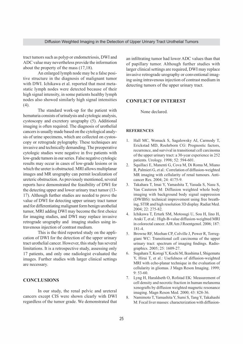
tract tumors such as polyp or endometriosis, DWI and ADC value may nevertheless provide the information about the property of the mass (17,18). An enlarged lymph node may be a false posi-tive structure in the diagnosis of malignant tumor with DWI. Ichikawa et al. reported that most meta-static lymph nodes were detected because of their high signal intensity, in some patients healthy lymph nodes also showed similarly high signal intensities (4). The standard work-up for the patient with hematuria consists of urinalysis and cytologic analysis, cystoscopy and excretory urography (5). Additional imaging is often required. The diagnosis of urothelial cancers is usually made based on the cytological analy-sis of urine specimens, which are collected on cystos-copy or retrograde pylography. These techniques are invasive and technically demanding. The preoperative cytologic studies were negative in five patients with low-grade tumors in our series. False negative cytologic results may occur in cases of low-grade lesions or in which the ureter is obstructed. MRI allows multiplanar images and MR urography can permit localization of ureteric obstruction. As previously mentioned, several reports have demonstrated the feasibility of DWI for the detecting upper and lower urinary tract tumor (13-17). Although further studies are needed to prove the value of DWI for detecting upper urinary tract tumor and for differentiating malignant form benign urothelial tumor, MRI adding DWI may become the first choice for imaging studies, and DWI may replace invasive retrograde urography and imaging studies using in-travenous injection of contrast medium. This is the third reported study on the appli-cation of DWI for the detection of the upper urinary tract urothelial cancer. However, this study has several limitations. It is a retrospective study, assessing only 17 patients, and only one radiologist evaluated the images. Further studies with larger clinical settings are necessary.
CONCLUSIONS
In our study, the renal pelvic and ureteral cancers except CIS were shown clearly with DWI regardless of the tumor grade. We demonstrated that
an infiltrating tumor had lower ADC values than that of papillary tumor. Although further studies with larger clinical settings are required, DWI may replace invasive retrograde urography or conventional imag-ing using intravenous injection of contrast medium in detecting tumors of the upper urinary tract.
CONFLICT OF INTEREST
None declared.
REFERENCES
1. Hall MC, Womack S, Sagalowsky AI, Carmody T, Erickstad MD, Roehrborn CG: Prognostic factors, recurrence, and survival in transitional cell carcinoma of the upper urinary tract: a 30-year experience in 252 patients. Urology. 1998; 52: 594-601.
2. Squillaci E, Manenti G, Cova M, Di Roma M, Miano R, Palmieri G, et al.: Correlation of diffusion-weighted MR imaging with cellularity of renal tumours. Anti-cancer Res. 2004; 24: 4175-9.
3. Takahara T, Imai Y, Yamashita T, Yasuda S, Nasu S, Van Cauteren M: Diffusion weighted whole body imaging with background body signal suppression (DWIBS): technical improvement using free breath-ing, STIR and high resolution 3D display. Radiat Med. 2004; 22: 275-82.
4. Ichikawa T, Erturk SM, Motosugi U, Sou H, Iino H, Araki T, et al.: High-B-value diffusion-weighted MRI in colorectal cancer. AJR Am J Roentgenol. 2006; 187: 181-4.
5. Browne RF, Meehan CP, Colville J, Power R, Torreg-giani WC: Transitional cell carcinoma of the upper urinary tract: spectrum of imaging findings. Radio-graphics. 2005; 25: 1609-27.
6. Sugahara T, Korogi Y, Kochi M, Ikushima I, Shigematu Y, Hirai T, et al.: Usefulness of diffusion-weighted MRI with echo-planar technique in the evaluation of cellularity in gliomas. J Magn Reson Imaging. 1999; 9: 53-60.
7. Lyng H, Haraldseth O, Rofstad EK: Measurement of cell density and necrotic fraction in human melanoma xenografts by diffusion weighted magnetic resonance imaging. Magn Reson Med. 2000; 43: 828-36.
8. Namimoto T, Yamashita Y, Sumi S, Tang Y, Takahashi M: Focal liver masses: characterization with diffusion-

26
Diffusion Weighted Imaging in the Detection of Upper Urinary Tract Urothelial Tumors
weighted echo-planar MR imaging. Radiology. 1997; 204: 739-44.
9. Nasu K, Kuroki Y, Kuroki S, Murakami K, Nawano S, Moriyama N: Diffusion-weighted single shot echo planar imaging of colorectal cancer using a sensitiv-ity-encoding technique. Jpn J Clin Oncol. 2004; 34: 620-6.
10. Shimofusa R, Fujimoto H, Akamata H, Motoori K, Yamamoto S, Ueda T, et al.: Diffusion-weighted imag-ing of prostate cancer. J Comput Assist Tomogr. 2005; 29: 149-53.
11. Charles-Edwards EM, deSouza NM: Diffusion-weighted magnetic resonance imaging and its application to cancer. Cancer Imaging. 2006; 6: 135-43.
12. Squillaci E, Manenti G, Di Stefano F, Miano R, Strigari L, Simonetti G: Diffusion-weighted MR imaging in the evaluation of renal tumours. J Exp Clin Cancer Res. 2004; 23: 39-45.
13. Yoshida S, Masuda H, Ishii C, Saito K, Kawakami S, Kihara K: Initial experience of functional imaging of upper urinary tract neoplasm by diffusion-weighted magnetic resonance imaging. Int J Urol. 2008; 15: 140-3.
14. Takeuchi M, Matsuzaki K, Kubo H, Nishitani H: Diffu-sion-weighted magnetic resonance imaging of urinary epithelial cancer with upper urinary tract obstruction: preliminary results. Acta Radiol. 2008; 49: 1195-9.
15. Matsuki M, Inada Y, Tatsugami F, Tanikake M, Nara-bayashi I, Katsuoka Y: Diffusion-weighted MR imag-ing for urinary bladder carcinoma: initial results. Eur Radiol. 2007; 17: 201-4.
16. El-Assmy A, Abou-El-Ghar ME, Refaie HF, El-Diasty T: Diffusion-weighted MR imaging in diagnosis of superficial and invasive urinary bladder carcinoma: a preliminary prospective study. ScientificWorldJournal. 2008; 8: 364-70.
17. Takeuchi M, Sasaki S, Ito M, Okada S, Takahashi S, Kawai T, et al.: Urinary bladder cancer: diffusion-weighted MR imaging--accuracy for diagnosing T stage and estimating histologic grade. Radiology. 2009; 251: 112-21.
18. Fujii S, Matsusue E, Kigawa J, Sato S, Kanasaki Y, Nakanishi J, et al.: Diagnostic accuracy of the appar-ent diffusion coefficient in differentiating benign from malignant uterine endometrial cavity lesions: initial results. Eur Radiol. 2008; 18: 384-9.
Accepted after revision: August 11, 2009
Correspondence address:Dr. Shuji NishizawaNagano Municipal Hospital1333-1 Tomitake, Nagano, 381-8551, JapanFax: + 81 26 295-1145E-mail: [email protected]
EDITORIAL COMMENT
Urothelial carcinoma of the upper urinaryrothelial carcinoma of the upper urinary tract accounts for 5% of all urothelial carcinomas and 10% of all renal tumors. Patients with a history of urothelial carcinoma are at high risk of developing synchronous or metachronous urothelial carcinoma. This known multifocality requires thorough exami-nation of the entire urinary tract in high-risk patients
(1). The diagnosis is difficult by conventional morp-The diagnosis is difficult by conventional morp-hological imaging, since urothelial carcinomas mays may be overlooked with suboptimal examinations and even though the examination is sufficient, small lesionssmall lesions may not be detected due to volume averaging (2). Excretory urography or computed tomograp-hy (CT) was used to evaluate high-risk patients (3).

27
Diffusion Weighted Imaging in the Detection of Upper Urinary Tract Urothelial Tumors
Multiphasic CT urography offers superior detection of calculi, urothelial tumor, and parenchymal tumor over excretory urography and allows accurate staging of detected lesions at the same examination. However, when the patient has a contraindication to iodinated contrast material, retrograde pyelography or MRI is often used to image the upper urinary tract (4). MR imaging, including MR angiography and MR urography, offers comparable evaluation in patients who cannot tolerate iodinated contrast material and in whom multiplanar, vascular, and collecting system imaging is required (3,4). Recently a new MRI technique, called dif-fusion-weighted MR imaging (DW-MRI) has been applied in various abdominal diseases, especially in detecting tumors without the need for contrast admi-nistration (5). Different from conventional anatomical MR imaging DW-MRI provide functional information (5). By studying molecular diffusion, the ultrastruc-tural characteristics of tissue can be studied in vivo through sampling water molecules and by exploiting the natural sensitivity of MRI to the motion (2). The research of Nishizawa et al. has shownresearch of Nishizawa et al. has shown of Nishizawa et al. has shownNishizawa et al. has shown the superiority of DW-MRI in detecting urothelial tumors with a very high accuracy, demonstrated with excellent images. Also in a study by Yoshida et al. twon a study by Yoshida et al. two cases of highly suspected upper urinary tract neoplasm had been detected clearly on DW-MR images, des-pite unclear conventional morphological MR images (2). Malignant masses are easily discernible against suppressed background signal with visual assessment of DW-MR images (PET like images).
This method may be obtained after a routine abdom-inal MR imaging protocol approximately in 3 to 5 minutes without an additional cost. The additional benefit of DW-MRI is the ability to determine quan-titative indices, which may be important in the as-sessment of tumor cellularity, and disease response to treatment methods and follow-up (2,5). Further investigations will probably increase the use of functional imaging methods and especially DW-MRI in genitourinary diseases.
REFERENCES
1. Zhang J, Lefkowitz RA, Bach A: Imaging of kidneyZhang J, Lefkowitz RA, Bach A: Imaging of kidney cancer. Radiol Clin North Am. 2007; 45: 119-47.
2. Yoshida S, Masuda H, Ishii C, Saito K, KawakamiYoshida S, Masuda H, Ishii C, Saito K, Kawakami S, Kihara K: Initial experience of functional imaging of upper urinary tract neoplasm by diffusion-weigh-ted magnetic resonance imaging. Int J Urol. 2008; 15: 140-3.
3. Takahashi N, Kawashima A, Glockner JF, HartmanTakahashi N, Kawashima A, Glockner JF, Hartman RP, Leibovich BC, Brau AC, ET AL.: Small (<2-cm) upper-tract urothelial carcinoma: evaluation with gadolinium-enhanced three-dimensional spoiled gradient-recalled echo MR urography. Radiology. 2008; 247: 451-7.
4. Browne RF, Meehan CP, Colville J, Power R, Torre-Browne RF, Meehan CP, Colville J, Power R, Torre-ggiani WC: Transitional cell carcinoma of the upper urinary tract: spectrum of imaging findings. Radiog-raphics. 2005; 25: 1609-27.
5. Manenti G, Di Roma M, Mancino S, Bartolucci DA,Manenti G, Di Roma M, Mancino S, Bartolucci DA, Palmieri G, Mastrangeli R, et al.: Malignant renal neoplasms: correlation between ADC values and cellularity in diffusion weighted magnetic resonance imaging at 3 T. Radiol Med. 2008; 113: 199-213.
Dr. Ozgur KilickesmezDepartment of Radiology
Yeditepe University HospitalIstanbul, Turkey
E-mail:[email protected]

28
Diffusion Weighted Imaging in the Detection of Upper Urinary Tract Urothelial Tumors
EDITORIAL COMMENT
A combination of morphological imaging modalities using iodinated radiocontrast media, such as excretory urography and computed tomography with urinary cytologic examination, has been used for diagnosing upper urinary tract cancer (UUTC). However, these media may cause renal insufficiency. Contrast-induced nephropathy due to iodinated radio-contrast media is currently the third most common cause of acute renal failure in hospitalized patients (1). Diffusion-weighted magnetic resonance imaging (DWI) is a functional imaging technique with no con-trast agent and is applicable to patients with allergies against contrast agents or existing renal insufficiency. Furthermore, the addition of DWI to a routine MRI examination can be readily adopted for most current clinical MRI scanners with only a few minutes and no additional equipment. DWI has been reported to be a useful tech-nique to detect UUTC in a noninvasive manner be-cause of clear contrast between high signal intensity of UUTC and well-restrained signal intensity of the surrounding tissue (2). This study has confirmed the above points. The location (pelvis or ureter) and the size of the UUTC seem to have little impact on diagnostic potentiality because of good contrast be-tween the tumor and the surrounding tissues, even if the tumor burden was small (8 mm in this paper, 5 mm in our experience). The impact in the degree of diffusion within the tumor on the estimation of grade or depth has recently been shown on DWI in some malignancies, including bladder cancer (3). In this study, ADC values of the infiltrating tumor were significantly lower than that of papillary tumor. However, overlap among ADC values between them
seems to exist. Also, overlap among ADC values for tumor, renal parenchyma and collecting system exists. Further investigations should be performed to clarify the clinical importance of ADC values in evaluating tumor aggressiveness. The current study also showed the limitation of DWI technique in assessing the UUTC. Depicting carcinoma in situ (CIS) lesions is also challenging in DWI, as well as in other conventional imaging modal-ities. The DWI contrast reflects molecular diffusion, not the presence of existing cancer cells. Therefore, it remains challenging to distinguish a malignant disease from nonmalignant lesions, such as benign neoplasms, hematomas, abscesses and inflammatory condition. We have to keep in mind that we could not gain anatomical information from DWI. Addition of DWI to anatomical imaging increases the accuracy of MRI to UUTC.
REFERENCES
1. Solomon R: Contrast-medium-induced acute renal failure. Kidney Int. 1998; 53: 230-42. Erratum in: Kidney Int 1998; 53: 818. published erratum of serious dosage error appears in Kidney Int 1998; 53: 1109.
2. Yoshida S, Masuda H, Ishii C, Saito K, Kawakami S, Kihara K: Initial experience of functional imaging of upper urinary tract neoplasm by diffusion-weighted magnetic resonance imaging. Int J Urol. 2008; 15: 140-3.
3. Abou-El-Ghar ME, El-Assmy A, Refaie HF, El-Diasty T: Bladder cancer: diagnosis with diffusion-weighted MR imaging in patients with gross hematuria. Radiol-ogy. 2009; 251: 415-21.
Dr. Hitoshi MasudaDepartment of Urology
Tokyo Medical and Dental University Graduate School
Tokyo, JapanE-mail: [email protected]

29
Drainage of Perinephric AbscessInternational Braz J Urol Vol. 36 (1): 29-37, January - February, 2010
What is the Best Drainage Method for a Perinephric Abscess?
Ahmed R. EL-Nahas, Raed Faisal, Tarek Mohsen, Mohammed S. AL-Marhoon, Hassan Abol-Enein
Department of Urology (AREN, RF, MSAM, HAE) and Radiology (TM), Urology and Nephrology Center, Mansoura University, Mansoura, Egypt
ABSTRACT
Purpose: To compare the results of percutaneous and open drainage for perinephric abscess.Materials and Methods: The files of 86 patients who underwent drainage for perinephric abscesses from April 2001 through March 2008 were evaluated. The method of drainage for each patient was performed according to the clinical decision of the treating physician. Percutaneous tube drain (PCD) was used for drainage of the abscess in 43 patients (group 1), while the other 43 patients were managed with open drainage (group 2). Cure was defined as complete obliteration of the abscess cavity. The cure rates, complications, and hospital stay were compared between both groups.Results: The study included 50 males and 36 females with mean age 44.2 ± 17.3. The most common predisposing factors were diabetes mellitus and/or stones. Open drainage of perinephric abscesses resulted in a statistically significant higher cure rate (98% versus 69%, p < 0.001) and shorter hospital stay than PCD (3.6 versus 6 days, p < 0.001). Failure of complete drainage of multilocular abscess was observed in 8 of 13 cases (61.5%) in group 1 and one of 38 cases (2.6%) in group 2 (P < 0.001). Complications were observed in 7% of group 1 and 11.5% in group 2 (P = 0.45). After mean follow-up of 19 months, 9 of 46 patients (19.6%) had recurrence; 7 of them were in group 1.Conclusion: Percutaneous drainage of perinephric abscess is an effective minimally invasive treatment. However, PCD is not the optimal method for drainage of multilocular abscess because open surgical drainage provided higher cure rates and shorter hospitalization than PCD.
Key words: kidney; infection; abscess; perinephric; percutaneous; surgeryInt Braz J Urol. 2010; 36: 29-37
INTRODUCTION
Perinephric abscess (PNA) is defined as an abscess outside the renal capsule but within Gerota’s fascia. They are rare in comparison to other infections involving the genitourinary tract but they can cause significant morbidity and mortality (1). Broad-spec-trum antibiotic therapy is the first step in treatment and should be associated with drainage in the majority of cases. The first report of open surgical drainage of PNA was described by the French physician Germain
�linical Urology�linical Urology
doi: 10.1590/S1677-55382010000100005
Colotin in 1474 (2). Surgical drainage has been the accepted practice since then. After the advent of vari-ous imaging modalities such as ultrasonography and computed tomography, percutaneous drainage (PCD) with radiological guidance has become widely used for most abdominal abscesses including selected cases of PNA (3). There have been many reported studies that have evaluated PCD as a minimally invasive treat-ment for PNA (3-6). Nevertheless, to our knowledge no previous series has compared PCD versus open drainage in the management of PNA. The present

30
Drainage of Perinephric Abscess
study was conducted to compare the efficacy and safety of open drainage with PCD in the management of PNA.
MATERIALS AND METHODS
The computerized files, radiographic images, operative and postoperative data of 86 patients who were treated for perinephric abscess at our center from April 2001 through March 2008 were retrospectively assessed. We included only patients with perineph-ric abscess who required drainage because of either presence of predisposing factors or large abscess size. Preoperative laboratory workup included serum creatinine tests, complete blood count, urinalysis and culture. Suspected patients were clinically evaluated based on detailed medical history and physical exami-nation results. Abdominal ultrasonography was the initial radiological investigation. When the findings were suggestive of perinephric abscess, unenhanced computed tomography (CT) scan of the abdomen was performed to confirm the diagnosis and determine abscess configuration (unilocular or multilocular), volume and associated conditions (such as renal or ureteral stones). All patients received a broad-spectrum of intravenous antibiotics (third generation cephalospo-rin), then PCD or open drainage was considered. The method of drainage for each patient was performed according to the clinical decision of the treating physician. PCD was chosen for severely ill patients who could not withstand general anesthesia. It was performed by a radiologist under local anesthesia using ultrasound guidance. A pigtail catheter of 12 or 14F was percutaneously inserted into the abscess cavity. Open drainage was performed under general anesthesia through a small flank incision. When the abscess cavity was entered, a gentle opening of all loc-ules was performed. This was followed by irrigation of the abscess cavity with saline and gentamycin. Two wide caliber tube drains (24F) were left indwelling. Drainage catheters remained in place until the output was minimal and radiographic resolution was confirmed with ultrasonography. Cure was de-fined as complete obliteration of the abscess cavity. The patients were discharged from the hospital when
they were clinically improved and free of any tubes. Patients were evaluated during follow-up visits for abscess recurrence by ultrasonography. Percutaneous tube drain was inserted for drainage of the abscess in 43 patients (Group 1), while the other 43 patients were managed with open surgical drainage (Group 2). In 20 patients with hydronephro-sis due to obstructing stones, urinary drainage with percutaneous nephrostomy tube (6) or double J stent (14) was also performed. The cure rate, complications, hospital stay and recurrence were compared in both groups using chi-square and Student’s-t-test or Mann-Whitney test.
RESULTS
The study included 50 males and 36 females with a mean age of 44.2 ± 17.3. Demographic data, predisposing factors and presentations are summa-rized in Table-1. The most common predisposing factors were diabetes mellitus and/or kidney stones. In 11 patients, the condition was associated with distant skin or subcutaneous abscess. It was observed that the success rate was 77% in diabetic patients compared with 88% in non-diabetic patients, but the difference was not statistically significant (p = 0.184). Preoperative laboratory and radiological data are summarized in Table-2. The differences between cure rates, complica-tions and hospital stay of group 1 and 2 are presented in Table-3. There was no significant difference be-tween the complication rates of both groups. In group 1, recurrent post-drainage fever was encountered in 2 patients and was controlled by intravenous antibiot-ics and antipyretics. In group 2, two cases of wound infection were managed by frequent dressing changes and a case of wound dehiscence required secondary sutures. Intra-operative bleeding from inadvertent inferior vena cava injury was adequately repaired with 4/0 sutures and blood transfusion. Septic shock developed in one patient in each group and admis-sion to the intensive care unit was necessary. One of them was resuscitated with cardiac inotropic drugs, intravenous fluids and antibiotics, while the other died from septic shock.

31
Drainage of Perinephric Abscess
Table 1 – Demographic data of 86 patients with perinephric abscess.
Characteristics N Group I (PCD)N (%)
Group II (Open)N (%)
p Value
GenderMaleFemale
5036
28 (56) 15 (41.7)
22 (44) 21 (58.3)
0.190
SideRightLeft
4244
18 (43) 25 (56.8)
24 (57) 19 (43.2)
0.196
Predisposing factors(75 patients)DMUrolithiasisImmune-compromisedDM + UrolithiasisOpen renal surgery
292113 6 6
12 (41.4)10 (47.6) 8 (61.5) 2 (33.3) 5 (83.3)
17 (58.6)11 (52.4) 5 (38.5) 4 (66.7) 1 (16.7)
0.413
PresentationFever + loin painLoin painFeverRecurrent UTI
551710 4
26 (47.3)8 (47)7 (70)2 (50)
29 (52.7)9 (53)3 (30)2 (50)
0.610
PCD = percutaneous drainage; N = number of cases; DM = diabetes mellitus; UTI = urinary tract infection.
Open drainage of perinephric abscesses re-sulted in statistically significant higher cure rate and shorter hospital stay than PCD (p < 0.001). Inadequate drainage was found in 17 cases of group 1; 7 of them were managed by readjustment of the PCD (replace-ment of the PCD with wider tube in the same locule or insertion of another PCD in other locules of the abscess) while the remaining 10 cases needed further open drainage. Inadequate drainage was observed in only one patient of group 2. This was due to miss-ing one locule of a multilocular abscess and it was managed with PCD. The abscess configuration had a significant effect on the outcome. Failure of complete drainage of multilocular abscess was found in 8 of 13 cases (61.5%) in group 1 and one of 38 cases (2.6%) in group 2 (P < 0.001). On the other hand, incomplete drainage of a unilocular abscess was observed in one of 29 cases (82%) in group 1 (because of large abscess volume with thick pus) and none of 5 cases (100%) in group 2 (p = 0.315). Eight patients required delayed nephrectomy for non-functioning ipsilateral kidney. After mean
follow-up of 16.7 months (range 3-65), 9 out of 46 patients (19.6%) suffered recurrence of PNA in the ipsilateral side, 7 of them were in group 1.
COMMENTS
While urinary tract infections are common, the severe complications of renal and perinephric abscess formation are uncommon and usually occur in patients with predisposing factors such as diabetes mellitus, urinary calculi, urinary obstruction and immune com-promised patients. Moreover, multiple predisposing factors may be present in the same patient (1). The same findings were observed in the present study. We identified diabetes, urolithiasis, and immune-compro-mised conditions as predisposing factors for PNA and in some patients multiple factors were present (Table-1). Prior to the development and availability of antibiotics, most perinephric abscesses were due to hematogenous spread of gram positive bacterial

32
Drainage of Perinephric Abscess
infection (such as Staphylococcus aureus) result-ing in formation of renal cortical abscess (7,8). Currently, the majority of perinephric abscesses are due to gram negative bacteria which ascend in a retrograde fashion from the lower urinary tract causing corticomedullary abscess (9). A PNA is formed when a cortical or corticomedullary abscess eventually rupture into the perinephric space. Opportunistic organisms such as Candida albicans are also isolated from immune suppressed
Table 2 – Preoperative radiological and laboratory data of 86 patients with perinephric abscess.
Characteristics N Group I (PCD)N (%)
Group II (Open)N (%)
p Value
Kidney statusNormalHydronephrosisPyelonephriticNon-functioning
38 21 19 8
17 (44.7)14 (66.7)10 (52.6)
2 (25)
21 (55.3) 7 (33.3) 9 (47.4)
6 (75)
0.186
Associated renal stoneYesNo
20 66
9 (45)34 (51.5)
11 (55) 32 (48.5)
0.610
Urine cultureNegativePositiveEscherichia coliKlebsiella pneumonia Staphylococcus aureusEnterobacterPseudomonas aeruginosaCandida albicansProteus mirabilis
24 62 26 14 11 4 3 2 2
10 (41.7)33 (53.2)
1112 4 3 2 0 1
14 (58.3) 29 (46.8)
15 2 7 1 1 2 1
0.336
Abscess configurationMultilocularUnilocular
51 35
13 (25.5)30 (85.7)
38 (74.5) 5 (14.3)
< 0.001
Abscess volume (cc)MeanRange
332(40-1110)
260(40-810)
403(120-1110)
0.004
Hemoglobin (gm/dL)MeanRange
10.4(5.2-16.2)
10.6(7-16.2)
10.1(5.2-15.7)
0.283
Serum creatinine (mg/dL)MeanRange
1.97(0.3-9.9)
2(0.3-9.9)
1.9(0.6-9.5)
0.833
PCD = percutaneous drainage; N= number of cases.
and diabetic patients (10). In the present study, the most common isolated organisms were gram nega-tive bacilli (such as Escherichia coli and Klebsiella pneumonia), while Staphylococcus aureus was isolated from 13% of patients. Traditionally, PNA has been associated with significant morbidity and high rates of mortality reaching up to 56% (11). This was attributed to de-lay in diagnosis because the symptoms of PNA are somewhat non-specific and confusing. Due to the

33
Drainage of Perinephric Abscess
introduction of cross-sectional imaging modalities such as CT, in addition to improvement of ultrasound examinations, early diagnosis and minimally invasive treatment represent major advances in management of PNA during the last 20 years. The wide spread utilization of ultrasound for examination of patients with loin pain, fever or other non-specific complaints has resulted in early diagnosis of PNA. When there is any suspicion re-garding the nature of the lesion, CT is a valuable tool for confirmation of the diagnosis (11). Therefore, the mortality rates in recent series have decreased to 12%-14% (1,12). The authors attributed mortality among their patients to medical treatment of abscesses that otherwise needed drainage. In the present study, we used ultrasound for evaluation of all patients with suspicious symptoms or predisposing factors of PNA and CT was used in to confirm the diagnosis. Then, we drained PNA that were large or present in patients with predisposing factors. This may be the reason for very low mortality rate (1%) among our patients. The classic open surgical drainage for peri-nephric abscess has been challenged by the introduc-tion of image guided percutaneous tube drainage methods and antibiotics alone (12). The general consensus is that large abscesses and patients with predisposing factors require drainage in addition to antibiotics (12,13). Therefore, antibiotics without drainage are suitable for selected cases and the deci-sion to treat with antibiotics alone requires consider-ation of other associated medical conditions (1). The reason for not using antibiotics alone in our patients
Table 3 – Comparison between group 1 (PCD) and group 2 (open) regarding outcome parameters.
Variable Group I (PCD)N (%)
Group II (Open)N (%)
p Value
Cure rate 29/42* (69) 42/43 (98) < 0.001Complication rate
Wound complicationsPost-drainage feverPost-drainage septic shockBleeding
3/43 (7)-21-
5/43 (11.5)3-11
0.45
Hospital stay: mean (range) 6 (1-19) 3.6 (1-7) < 0.001
* One patient died and was excluded; PCD = percutaneous drainage; N = number of cases.
was either presence of predisposing factors or large abscess size. Percutaneous drainage with radiological guidance has become the treatment of choice for most abdominal abscesses as it usually provides satisfac-tory clinical results with minimal complications, thus obviates the need for open surgery (14). Percutaneous drainage of PNA has been reported in many series (3-6). The ease of image guided fixation of PCD without the need for general anesthesia has made it the preferred choice for severely ill patients. More-over, the cure rate of 60-67% was the main reason that made PCD the most commonly used minimally invasive intervention for treatment of PNA. The main disadvantage was the need for adjustment or insertion of multiple draining tubes in many patients (1,3-6,13). In the present study, PCD was able to adequately drain the abscess in 60% of patients, and subsequently the cure rate was increased to 69%, after adjustment of the tube in 7 patients. Another disadvantage of PCD was the longer hospital stay because of slow drainage or the need for multiple interventions. In our study, the main cause of PCD failure was multilocular abscess cavity. The PCD can only drain the locule at its site of insertion. Therefore, it may be beneficial to fix multiple tubes from the start in multilocular abscess. On the other hand, a quick extraperitoneal open drainage provided very high cure rate (98%) even in patients with multilocular abscess because of manually disrupting the septa between the abscess locules. Moreover, evacuation of thick pus during open drainage and wide draining tubes resulted

34
Drainage of Perinephric Abscess
in significantly shorter hospital stay. However, these achievements were gained at the expense of using a general anesthesia and a slightly higher complication
Figure 1 – Algorithm for management of perinephric abscess. PCD = percutaneous tube drain; CBC= complete blood count.
rate than PCD. It is also important to emphasis that severely ill patients cannot tolerate nephrectomy of a non-functioning kidney at the time of PNA drainage.

35
Drainage of Perinephric Abscess
Therefore, delayed nephrectomy was performed in 8 patients following complete abscess drainage and improvement of the patients’ general condition. Although the selection bias for each treat-ment option cannot be completely eliminated, it was statistically proven in our study that PCD was not the optimal drainage method for multilocular PNA. This was based on the lower cure rate of PCD in the treat-ment of multilocular PNA and the higher recurrence rate that may have resulted from enlargement of a very small residual. However, a prospective randomized trial is warranted to confirm these findings. A proposed algorithm for treatment of perinephric abscess is il-lustrated in Figure-1.
CONCLUSIONS
Percutaneous drainage of a perinephric ab-scess is an effective minimally invasive treatment modality. Therefore, when a PNA has to be drained, we recommend PCD as the primary drainage method because, as shown in our series, it can save 69% of patients an open surgery. However, and after acknowl-edging the limitations of the retrospective nature of the study. PCD is not the optimal method for drainage of multilocular abscess because open surgical drainage provided higher cure rates, shorter hospitalization and lower recurrence rate than PCD.
ACKNOWLEDGMENT
Dr. Mohamed Motwally assisted in data col-lection.
CONFLICT OF INTEREST
None declared.
REFERENCES
1. Meng MV, Mario LA, McAninch JW: Current treat-ment and outcomes of perinephric abscesses. J Urol. 2002; 168: 1337-40.
2. Morgan WR, Nyberg LM Jr: Perinephric and intrarenal abscesses. Urology. 1985; 26: 529-36.
3. Siegel JF, Smith A, Moldwin R: Minimally invasive treatment of renal abscess. J Urol. 1996; 155: 52-5.
4. Gerzof SG, Gale ME: Computed tomography and ultrasonography for diagnosis and treatment of renal and retroperitoneal abscesses. Urol Clin North Am. 1982; 9: 185-93.
5. Deyoe LA, Cronan JJ, Lambiase RE, Dorfman GS: Percutaneous drainage of renal and perirenal ab-scesses: results in 30 patients. AJR Am J Roentgenol. 1990; 155: 81-3.
6. Lang EK: Renal, perirenal, and pararenal abscesses: percutaneous drainage. Radiology. 1990; 174: 109-13.
7. Truesdale BH, Rous SN, Nelson RP: Perinephric ab-scess: a review of 26 cases. J Urol. 1977; 118: 910-1.
8. Jemni L, Mdimagh L, Jemni-Gharbi H, Jemni M, Kraiem C, Allegue M: Kidney carbuncle: diagnostic, bacteriological and therapeutic considerations. Apro-pos of 11 cases. J Urol (Paris). 1992; 98: 228-31.
9. Noble MJ: Perinephric abscess. AUA update. Series 2002; Vol. XXI, Lesson 10; 74-79.
10. Patterson JE, Andriole VT: Bacterial urinary tract in-fections in diabetes. Infect Dis Clin North Am. 1997; 11: 735-50.
11. Salvatierra O Jr, Bucklew WB, Morrow JW: Peri-nephric abscess: a report of 71 cases. J Urol. 1967; 98: 296-302.
12. Rai RS, Karan SC, Kayastha A: Renal and perinephric abscesses revisited. MJAFI 2007; 63: 223-225.
13. Coelho RF, Schneider-Monteiro ED, Mesquita JL, Mazzucchi E, Marmo Lucon A, Srougi M: Renal and perinephric abscesses: analysis of 65 consecutive cases. World J Surg. 2007; 31: 431-6.
14. vanSonnenberg E, Mueller PR, Ferrucci JT Jr: Percu-taneous drainage of 250 abdominal abscesses and fluid collections. Part I: Results, failures, and complications. Radiology. 1984; 151: 337-41.
Accepted after revision: September 18, 2009
Correspondence address:Dr. Ahmed R. El-NahasUrology & Nephrology CenterMansoura UniversityMansoura, EgyptFax: + 2050 2263717E-mail: [email protected]

36
Drainage of Perinephric Abscess
EDITORIAL COMMENT
Perinephric abscess is a serious entity ne-glected in the urological literature. Morbidity and mortality have fallen in the last decades thanks to the progress in diagnosis with ultrasonography and overall computed tomography (CT) scan and due to improvement in medical and surgical therapies. Nevertheless, the best way of surgical management of such collections has been a subject of debate. In this article, the authors compared retrospectively the outcome of 43 perinephric abscess drained percutane-ously with 43 cases drained by open surgery. Renal stones and diabetes mellitus were the most important predisposing factors. Abscesses drained by open surgery were significantly greater and multiloculated when compared to percutaneously drained abscesses. The cure rate was significantly higher in the group treated by the open access. There were no difference in the complication rate but hospital stay was longer among patients treated percutaneously. This article concerns three important issues: A) Perinephric abscesses occur with some frequency and must be always suspected in patients with pro-longed fever, overall in diabetics and in patients with stones, B) CT is an important tool and should be considered the gold-standard, not only in early diagnosis but also in the planning of the therapy to be instituted, and C) Minimally invasive therapy is not always the best option. In fact, as the authors
clearly showed, a quick open drainage is more effi-cient especially in multiloculated collections. In this setting percutaneous drainage should be reserved for patients in a severe clinical condition, when it can be performed under local anesthesia or for uniloculated abscesses with greater chance of success. The results corroborate those obtained by Coelho et al. (1) that also found diabetes mellitus and stones as the main predisposing factors for perinephric abscesses and by Meng et al. (2) who showed that CT could influence prognosis as it allows for earlier diagnosis and that a 36% failure rate with percutane-ous drainage can be observed in such cases. Another important point is that small perinephric collections (usually less than 2 cm) can be managed successfully with adequate antibiotics and correction of eventual predisposing factors like urinary obstruction (2).
REFERENCES
1. Coelho RF, Schneider-Monteiro ED, Mesquita JL, Mazzucchi E, Marmo Lucon A, Srougi M: Renal and perinephric abscesses: analysis of 65 consecutive cases. World J Surg. 2007; 31: 431-6.
2. Meng MV, Mario LA, McAninch JW: Current treat-ment and outcomes of perinephric abscesses. J Urol. 2002; 168: 1337-40.
Dr. Eduardo MazzucchiDivision of Urology
Medical School, University of Sao Paulo, USPSao Paulo, SP, Brazil
E-mail: [email protected]
EDITORIAL COMMENT
This paper compared 2 methods of drain-age for perinephric abscess: percutaneous versus open surgical drainage. The authors found that open drainage was associated with higher cure rates, lower recurrence rates and shorter hospital stay, particularly
in patients with multilocular abscess. They correctly emphasized that percutaneous drainage is the first line option of treatment in severely ill patients with perinephric unilocular abscess. Currently, perinephric abscesses are uncommon in most urology units (1) and

37
Drainage of Perinephric Abscess
since 1998, in my unit, we have seen fewer cases. It is therefore refreshing to read about the experience of a unit where this clinical entity remains a common clinical problem. A major flaw of this paper is that it was a retrospective analysis and the patients had many variables as shown in tables 1 and 2. For example, the authors stated that percutaneous drainage was chosen for severely ill patients who could not withstand gen-eral anesthesia. The longer hospital stay and higher recurrence rates in patients subjected to percutane-ous drainage could be attributed to the fact that they presented with worse initial disease, compared to those who underwent open drainage. The location of the abscesses, their configuration, etc., are variables that may affect the success of percutaneous drainage. However, the paper contains some useful guidelines (Figure-1) for units that do not have adequate experi-ence in the management of these patients. It must be stated that a randomized controlled study will be dif-ficult to carry out in patients with perinephric abscess because of its declining incidence, the variable ways
in which patients can present and many other variables in individual patients with the disease. A last point that is worth emphasizing is that, as in patients with emphysematous pyelonephritis, a differential reno-gram test should be carried out as soon as possible in patients with moderate to severe perinephric abscess (1). Patients found to have poorly functioning kidneys (< 15%) or non-functioning kidneys are best served by nephrectomy as soon as possible rather than open or prolonged percutaneous drainage. Nephrectomy has been shown to reduce morbidity and mortality in patients with perinephric abscess (1).
REFERENCE
1. Abdul-Halim H, Kehinde EO, Abdeen S, Lashin I, Al-Hunayan AA, Al-Awadi KA: Severe emphysema-tous pyelonephritis in diabetic patients: diagnosis and aspects of surgical management. Urol Int. 2005; 75: 123-8.
Dr. Elijah O. KehindeProfessor of Urological Surgery
Division of Urology, Department of SurgeryFaculty of Medicine, Kuwait University
Safat, KuwaitE-mail: [email protected]
REPLY BY THE AUTHORS
We agree with all the comments of Dr. Eduar-do Mazzucchi. Concerning the comments of Dr. Elijah O. Kehinde, we have to emphasis some points. First, the reference he mentioned is dealing with emphyse-matous pyelonephritis not perinephric abscess (PNA). Second, we recommended percutaneous drainage (PD) for PNA in patients with severe illness and also for pa-tients with unilocular abscess cavity (as mentioned in figure 1 and in the discussion and conclusion). Lastly, we disagree with the comment that “non-functioning
kidneys are best served by nephrectomy as soon as possible rather than open or prolonged percutaneous drainage.” This can be true for nephrectomy in case of emphysematous pyelonephritis because it is a chronic inflammation, but PNA is an acute inflamma-tory condition. We still advise drainage of the abscess with either PCD or open surgery. Then nephrectomy for poorly functioning kidney can be delayed until improvement of the patient’s general condition and recovery from the toxemia of an acute abscess.
The Authors

38
Laparoscopic Ureteral Reimplant for Ureteral StrictureInternational Braz J Urol Vol. 36 (1): 38-43, January - February, 2010
Laparoscopic Ureteral Reimplant for Ureteral Stricture
Rodrigo S. Q. Soares, Rubens A. de Abreu Jr, Jose E. F. Tavora
Department of Urology, Hospital dos Servidores do Estado de Minas Gerais, IPSEMG, Belo Horizonte, Minas Gerais, Brazil
ABSTRACT
Purpose: Evaluate the initial experience of laparoscopic ureteral reimplant for ureteral stenosis.Materials and Methods: From January 2004 to June 2008, 10 patients underwent 11 laparoscopic reconstruction surgeries for ureteral stenosis. Seven cases of stenosis of the distal ureter, two at the level of iliac vessels, a case of bilateral distal stenosis and one in the medium third. Eight ureteroneocystotomies were performed by extravesical technique with anti-reflux mechanism, two cases of vesical reimplant with Boari technique and one case using the psoas hitch technique.Results: The average surgical time was 166 minutes (115-245 min), mean blood loss was 162 mL (100-210 mL) and the average hospital stay was 2.9 days (2-4 days). There were two complications: a lesion of the sigmoid colon identified peroperatively and treated with laparoscopic sutures with good evolution, and a case of ureteral stone obstruction at the 30th day postoperative, treated by laser ureterolitotripsy. All patients had resolution of the stenosis at an average follow-up period of 18 months (3-54 months).Conclusion: Laparoscopic surgery represents a feasible, safe and low morbidity technique for ureteral reimplant in ureteral stenosis.
Key words: ureter; stricture; reconstruction; laparoscopyInt Braz J Urol. 2010; 36: 38-43
INTRODUCTION
The main causes for ureteral stricture are sur-gical traumas, impacted ureteral stones, extrinsic com-pression, tumor and congenital or idiopathic disorders. Ureteral stenoses are the most frequent complications observed in pelvic surgery. Currently, endourological, gynecological and laparoscopic procedures are also reasons for referral for a large number of cases (1). Treatments focus on the anatomic aspects of stenosis, such as length of the lesion, complexity of obstruction and vascularization of the ureter. Partial and segmental stenoses can be treated by endoscopic procedures such as dilation or internal ureterotomy with placement of double J catheter with good fol-low-up results. Reconstruction technique procedures are needed for total complex stenosis.
Clinical Urology
doi: 10.1590/S1677-55382010000010006
In the last decades, open surgeries have been performed for these types of pathologies. With the advancement of technology, the laparoscopic ureter-vesical reimplant was introduced in 1994 by Reddy and Evans to correct vesicoureteral reflux (2). In the literature, major series have been published with similar results (3,4). We report our experience with laparoscopic ureteral reimplant in ureteral stenoses of different etiologies.
MATERIALS AND METHODS
Ten patients (8 females and 2 males) underwent 11 laparoscopic ureteral reimplants due to ureteral steno-sis, at our hospital, from January 2004 to June 2008.

39
Laparoscopic Ureteral Reimplant for Ureteral Stricture
Four patients had stenosis after open surgery and 4 had ureteral stenosis resulting from ureteral stone endoscopic procedure complications. The re-maining two patients had an idiopathic congenital bilateral ureteral stenosis and an extrinsic ureteral compression by the ovarian vein (ovarian vein syn-drome). In one patient after abdominal hysterectomy, the ureteral stricture extended to the mid ureter, caused by ischemic and inflammatory reaction. In all patients, an abdominal CT scan confirmed the localization and the length of the ureteral stricture (Figure-1). Endoscopic treatment was carried out in all cases except in one patient with idiopathic bilateral ureteral stenosis and another with ureteral compres-sion by the ovarian vein. Two of these procedures were interrupted due to complete stricture lesion post hysterectomy. In four cases, the dilation with a balloon catheter was chosen, as well as the placement of a double J stent for six weeks. In two patients with stenosis post ure-teral calculi, a laser ureterotomy was performed and a double-J catheter was left indwelling for 6 weeks.Table-1 shows the characteristics of these cases.
Technique
All patients underwent transperitoneal video laparoscopic surgery. The patient is placed in a flat dorsal Trendelenburg position and the surgery is performed using the four pelvic trocar technique (Figure-2). The surgery is carried out by opening the Toldt fascia, followed by the identification and dis-section of the ureter in the area close to the stenosis (Figure-3). The ureter is transected near the area of the stenosis and spatulated. The vesical dome is fixed to the wall with a stitch for a better exposition. The detru-sor muscle is opened lengthwise for approximately 3 cm to expose the vesical mucosa. The vesical mucosa is opened and the posterior ureterovesical anastomosis is performed with separated vicryl 4.0 sutures (Figure-4). A double J catheter is placed through one of the trocars. The anastomosis is completed and the detrusor muscle is closed by a continuous suture for anti-reflux tunnel.
In cases of tension due to the high ureteral stenosis, the ureteroneocystostomy with a psoas hitch muscle or Boari Flap technique is carried out. In the middle of this opening, a stitch with vicryl 4.0 is tightened, pulling the bladder to facilitate the anasto-mosis to the edge of the ureteral stump. Anastomosis is completed with simple stitches and the bladder is
Figure 1 – Abdominal CT scan of a 44 year old woman showing a left distal ureteral stenosis after an endoscopic ureterolithotripsy for a impacted ureteral stone.
Figure 2 – Immediate postoperative abdominal view of a young female patient after a right laparoscopic uretero-vesical reimplant. A 4 trocar technique was performed with 10 mm trocar for the optic and for the surgeon’s right hand and 2 others for the 5 mm trocars. The suction drain is inserted in the 5 mm left port.

40
Laparoscopic Ureteral Reimplant for Ureteral Stricture
sewn lengthwise. The fixation of the vesical part in the greater psoas muscle is also performed with vicryl 3-0 sutures. As soon as the detrusor closing is completed,
the bladder is filled with 200 mL of physiologic serum to evaluate overflowing. The cavity is drained with ei-ther a Penrose or a tubular suction drain (Figure-2).
Table 1 – Patient demographic and clinical data.
Patient Sex Age Etiology Side Place
1 F 36 Ileocolectomy / Crohn’s disease R Distal2 F 44 Urolithiasis / ureteroscopy L Distal3 F 40 Extrinsic compression R Distal4 M 50 Open external bilateral reimplant RL Distal5 F 62 Urolithiasis / ureteroscopy R Distal6 F 45 Total abdominal hysterectomy L Medium7 F 65 Total vaginal hysterectomy L Distal8 F 32 Urolithiasis / ureteroscopy R Medium9 M 13 Idiopathic stenosis R/L Distal10 F 50 Urolithiasis / ureteroscopy L Medium
Figure 3 – Operative view of a right ureteral laparoscopic dis-section showing the region of the ureteral stricture.
Figure 4 – Operative view of a right laparoscopic uretero-vesi-cal anastomosis.

41
Laparoscopic Ureteral Reimplant for Ureteral Stricture
RESULTS
The average patient age was 44.7 years. The average surgical time was 166 min. (115-245 min.), the average amount of bleeding was 162 mL (100-210 mL) and the mean hospital stay was 2.9 days (2 - 4 days). In one of the patients, with stenosis after ureterovesical reimplant by ureteral reflux, there was a sigmoid colon lesion during dissection of the ureter and it was sutured laparoscopically, with good results. In another patient with reimplant due to a secondary stenosis, after ileocolectomy, there was a migration of a kidney stone to the ureter on the 30th day post sur-gery, and a transureteroscopic laser ureterolitotripsy was carried out, with good evolution (Table-2). On average, the Penrose/tubular drain was removed on the second day post surgery. The double-J catheter was removed 4 weeks post surgery. All patients were followed-up using ultraso-nography and cystourethrography 3 months after the surgery, with a mean follow-up period of 18 months (3 - 54 months), and finally, all of them proved to be asymp-tomatic and without evidence of obstruction or reflux.
COMMENTS
With the improvement of the minimal inva-sive treatment in urological and gynecological disor-
ders, like laparoscopic pelvic surgery or endoscopic ureteral procedures, a large number of complications have been reported in the learning curve of these procedure such as ureteral damage (5). Ureteral stenosis has also been described as a consequence of several etiologies. Malignancy, radio-therapy, ischemia, retroperitoneal fibrosis, endometrio-sis, infection (tuberculosis), congenital and idiopathic disorders are seldom attributed in the large series. Diagnosis is rarely confirmed by using imag-ing procedures. When planning surgery, an excretory urography, CT scan, retrograde pyelography or mag-netic resonance imaging can be performed in order to determine all the characteristics of the lesion. It is advisable to carry out an ureteroscopy with cytology and biopsy in cases of gross hematuria and suspected lesion to avoid malignancy. The recommended approach for each ureteral lesion has to be determined following its diagnosis and localization. The endoscopic treatment by dila-tion or by ureterotomy represents a good alternative for segmental or partial stenosis with good results. However, reconstruction surgeries represent the main choice for complex situations or for failure in more conservative treatment. Traditionally, ureteral lesion reconstruction is performed by open surgery. The first case of lapa-roscopic ureteral management of ureteral injury was first described in a woman who underwent pelvic
Table 2 – Postoperative clinical data.
Patient Surgery Bleeding (mL)
Time (min)
Stay (days)
Complication Follow-up (months)
1 UCN 150 145 3 - 542 UCN 200 150 3 Ureterolithiasis 383 UCN 210 200 4 - 304 UCN 120 240 4 Colon lesion 245 UCN 130 170 3 - 126 Boari 100 115 2 - 97 UCN 150 120 2 - 48 Boari 180 130 2 - 39 UCN 200 245 3 - 310 Psoas 180 150 3 - 3
UCN = ureteroneocystostomy.

42
Laparoscopic Ureteral Reimplant for Ureteral Stricture
endometriosis treatment by Gomel and James, in 1991 (6). The first laparoscopic ureterovesical reimplant was performed in 1994, by Reddy and Evans to cor-rect a vesicoureteral reflux (2). Laparoscopy offers advantages of a minimum invasive procedure and a wide access to the entire urinary system. Currently, it represents an alternative in ureteral reconstruction surgery. The ideal time to perform this reconstruction remains controversial. Some authors recommend a minimum time of 6 weeks after the injury prior to carring out a new surgical operation in cases of le-sions caused by surgical trauma, in order to allow maximum resolution of the inflammatory process. In one of our cases, characterized by ureteral lesions after vaginal hysterectomy, the laparoscopic reimplant was performed 15 days after hysterectomy without any technical difficulties and with good results. In our experience, in cases of ureteral lesions in vaginal and endoscopic surgeries, the laparoscopic access represents a good option that can be performed im-mediately. The most common surgical choice for treat-ment of distal ureteral stenoses is ureteral reimplant (ureteroneocystostomy). It can be performed by extra or intra-vesical technique using Politano-Leadbetter, Lich-Gregoir, the Boari technique (Boari’s flap) or psoas-hitch technique in cases of major stenoses. In the literature, the performance of reimplant with the Boari or psoas-hitch technique is described with favorable results and low occurrence of reflux (7-9). In these cases, the laparoscopic access offers advan-tages such as mobilization of the bladder, ureter and kidney, making the anastomosis easier and without tension and/or adequate size of the vesical flap. We did not experience any difficulty when performing this procedure in 3 of our patients and none of them presented vesicoureteral reflux post-surgery. Data show similar results between an open and laparoscopic ureteroneocystostomy in cases of ureteral stenoses with low morbidity for the last laparoscopic procedure (10,11). Recently, several reported studies on robotic ureteroneocystostomy have been published showing successful results similar to those obtained with the laparoscopic technique (12,13). Ureteroneo-cystostomy has also been described using transumbili-cal endoscopic single port technique (NOTES) (14).
In the present study, an endoscopic proce-dure was carried out before the decision to apply the laparoscopic technique for all patients. Although the endoscopic treatment represents an attractive alterna-tive, we believe that for the cases of complete ureteral stenosis or late diagnosis, the ureteral reimplant rep-resents a definitive treatment. However, an attempt to perform endoscopic dilation or ureterotomy should be considered with caution for ureteral stenosis. A laparoscopic procedure is feasible, practical and cost effective for trained laparoscopic urologists.
CONCLUSION
Ureteral lesion is a common affection that has been increasing due to pelvic endourologic, lapa-roscopic and open procedures. Results show that the laparoscopic ureteral reimplant is an effective alterna-tive with similar results compared to open technique, with minimum morbidity. Laparoscopic ureteral reimplant can be an excellent choice in treatments of distal ureteral stenosis.
CONFLICT OF INTEREST
None declared.
REFERENCES
1. Parpala-Spårman T, Paananen I, Santala M, Ohtonen P, Hellström P: Increasing numbers of ureteric injuries after the introduction of laparoscopic surgery. Scand J Urol Nephrol. 2008; 42: 422-7.
2. Reddy PK, Evans RM: Laparoscopic ureteroneocys-tostomy. J Urol. 1994; 152: 2057-9.
3. Ehrlich RM, Gershman A, Fuchs G: Laparoscopic vesicoureteroplasty in children: initial case reports. Urology. 1994; 43: 255-61.
4. Yohannes P, Gershbaum D, Rotariu PE, Smith AD, Lee BR: Management of ureteral stricture disease during laparoscopic ureteroneocystostomy. J Endourol. 2001; 15: 839-43.
5. Ostrzenski A, Radolinski B, Ostrzenska KM: A review of laparoscopic ureteral injury in pelvic surgery. Obstet Gynecol Surv. 2003; 58: 794-9.

43
Laparoscopic Ureteral Reimplant for Ureteral Stricture
EDITORIAL COMMENT
Lower ureter is involved not only in primary diseases of ureter and bladder but secondarily, in dis-eases of colon and genital organs of the female. It is prudent to establish the pathology prior to consider for the operative approach. In this series, one patient had involvement of the ureter due to Crohn’s disease and laparoscopic ureteral reimplantation was performed successfully. Inflammatory conditions often require disease control prior to subjecting patient for such surgery. Dissection of the diseased lower segment of ureter is often difficult and vascularity could be precarious. In such circumstances, no attempt should be made to dissect deep down into the pelvis. Ureter
should be divided just above the lesion and decision of ureteral reimplantation with or without additional procedure like psoas hitch or Boari bladder flap reconstruction could be planned so that tension free anastomosis is achieved. Regular use of psoas hitch provides good intramural length of ureter into bladder giving anti-reflux mechanism.
Dr. Pranjal ModiInstitute of Kidney Diseases & Research Center
Civil Hospital CampusAhmedabad, Gujarat, India.
E-mail: [email protected]
6. Gomel V, James C: Intraoperative management of ureteral injury during operative laparoscopy. Fertil Steril. 1991; 55: 416-9.
7. Castillo OA, Litvak JP, Kerkebe M, Olivares R, Urena RD: Early experience with the laparoscopic boari flap at a single institution. J Urol. 2005; 173: 862-5.
8. Modi P, Gupta R, Rizvi SJ: Laparoscopic ureteroneo-cystostomy and psoas hitch for post-hysterectomy ureterovaginal fistula. J Urol. 2008; 180: 615-7.
9. Sievert DK, Nagele U, Stenzl A: Laparoscopic uretero-neocystostomy and psoas hitch for post-hysterectomy ureterovaginal fistula. Int Braz J Urol 2008; 34: 530-2.
10. Rassweiler JJ, Gözen AS, Erdogru T, Sugiono M, Teber D: Ureteral reimplantation for management of ureteral strictures: a retrospective comparison of laparoscopic
and open techniques. Eur Urol. 2007; 51: 512-22; discussion 522-3.
11. Simmons MN, Gill IS, Fergany AF, Kaouk JH, Desai MM: Laparoscopic ureteral reconstruction for benign stricture disease. Urology. 2007; 69: 280-4.
12. Yohannes P, Chiou RK, Pelinkovic D: Rapid com-munication: pure robot-assisted laparoscopic ureteral reimplantation for ureteral stricture disease: case re-port. J Endourol. 2003; 17: 891-3.
13. Casale P, Patel RP, Kolon TF: Nerve sparing robotic extravesical ureteral reimplantation. J Urol. 2008; 179: 1987-9; discussion 1990.
14. Desai MM, Stein R, Rao P, Canes D, Aron M, Rao PP, et al.: Embryonic natural orifice transumbilical endo-scopic surgery (E-NOTES) for advanced reconstruc-tion: initial experience. Urology. 2009; 73: 182-7.
Accepted after revision: August 25, 2009
Correspondence address:Dr. Rodrigo S. Quintela SoaresRua Ceará, 450, CTC São LucasBelo Horizonte, MG, 30150-310, BrazilE-mail: [email protected]

44
Post-radiotherapy Prostate Biopsy for Recurrent DiseaseInternational Braz J Urol Vol. 36 (1): 44-48, January - February, 2010
Outcomes Following Negative Prostate Biopsy for Patients with Persistent Disease after Radiotherapy for Prostate Cancer
Jacob H. Cohen, James Eastham, Richard J. Macchia
Department of Urology (JHC, RJM), State University of New York Downstate Medical School, Brooklyn, New York, USA, and Memorial Sloan-Kettering Cancer Center (JE), New York, New York, USA
ABSTRACT
Purpose: When faced with biochemical recurrence after definitive radiotherapy for prostate cancer, clinicians must deter-mine whether the recurrence is local or systemic. Post radiotherapy prostate biopsies to detect persistent local disease are difficult to interpret histopathologically and are subject to sampling error. Our study examines outcomes for patients with a negative prostate biopsy performed for rising prostate-specific antigen (PSA) levels after prostate radiation.Materials and Methods: We performed a retrospective review of 238 prostate cancer patients with a negative biopsy fol-lowing definitive radiotherapy. Seventy-five of these patients had biochemical recurrence at the time of biopsy. A negative biopsy was defined as the absence of prostate cancer without radiation-treatment effect in the specimen.Results: Patients underwent biopsy at a mean of 41 months after the completion of radiation. They had a mean PSA of 6. Patients were followed for an average of 63 months. Thirty-two patients (43%) developed metastasis, and 11 (15%) died of prostate cancer despite a negative post-radiation biopsy. Five of nine patients (56%) with sequential biopsies had a positive second biopsy.Conclusions: Patients with PSA recurrence and a negative post-radiation biopsy have a high chance of persistent local disease, progression, and death from prostate cancer. Furthermore, an initial negative biopsy does not rule-out local recur-rence. Patients with biochemical recurrence after radiotherapy for prostate cancer need to be evaluated earlier for local recurrence.
Key words: prostate neoplasms; prostate-specific antigen; neoplasm recurrence; radiationInt Braz J Urol. 2010; 36: 44-8
INTRODUCTION
Biochemical recurrence after radiotherapy for prostate cancer occurs in approximately 10-60% of patients and varies depending on definition of recurrence, tumor stage and grade at the time of di-agnosis, dosage of radiation, and the use of adjuvant hormonal therapy (1). Salvage therapy for persistent local disease after radiotherapy has shown greatest efficacy for biopsy-proven local recurrence with low
�linical Urolo�y�linical Urolo�y
doi: 10.1590/S1677-55382010000100007
prostate-specific antigen (PSA) level, and negative metastatic evaluation (2). Post-radiotherapy prostate biopsy to detect persistent local disease is difficult to interpret histopathologically and subject to sampling error. In addition, there are no well-defined recom-mendations for when or how to biopsy these patients. Most studies have examined post-radiotherapy bi-opsies regardless of signs or symptoms or disease progression. Our study is the first, to our knowledge, to examine outcomes for patients with a negative

45
Post-radiotherapy Prostate Biopsy for Recurrent Disease
prostate biopsy performed for biochemical recurrence after radiation.
MATERIALS AND METHODS
We performed a retrospective review of 238 prostate cancer patients in a prospectively maintained prostate cancer database who had a negative prostate biopsy following definitive radiotherapy between January 1st, 1992 and December 31st, 2005. Of these patients, 155 underwent prostate biopsy as part of a clinical trial without evidence of biochemical failure, while 83 patients were identified to have biochemical recurrence at the time of their post-radiotherapy bi-opsy. Eight patients were excluded due to missing data or lack of follow-up, leaving 75 patients for analysis. Biochemical recurrence was determined by the treat-ing practitioner, most typically the American Society for Therapeutic Radiology and Oncology (ASTRO) definition of biochemical failure (3). A negative post-radiation biopsy included a pathology report of benign tissue, benign tissue with profound treatment effect, or prostate cancer with profound treatment effect (4).
RESULTS
Our 75 patients had a mean age of 66 years and a mean PSA of 15 ng/mL at initial cancer diag-nosis. Table-1 shows Gleason score and clinical stage at initial cancer diagnosis as well as initial treatment. A post-radiotherapy PSA-nadir of < 1.0 was achieved in 69 patients (92%). Patients underwent biopsy at a mean of 41 months after the completion of radiation and with a mean PSA of 6 ng/mL. Mean and median PSA doubling time at post-radiotherapy biopsy were 12 and 9 months, respectively. For the 55 (73%) patients on whom data were available, there was no standard technique for post-radiotherapy biopsy, with as little as 2 cores and as many as 24 cores sampled. Mean follow-up after the negative post-radiotherapy biopsy was 63 months. Patient outcomes following their negative post-radiation biopsy are presented in Figure-1. There were nine patients undergoing two sequential post-radiotherapy biopsies. Five of nine (56%) had their
second biopsy return positive, and all five were alive after their salvage local therapies. Of the four patients who had two sequential negative post-radiotherapy biopsies, there was one death from prostate cancer, one patient with clinical metastasis, one patient with asymptomatic metastasis, and one patient with bio-chemical recurrence alone. Overall, 32 patients (43%) developed disease progression beyond biochemical recurrence, with 15 (20%) developing radiographic metastasis only, 6 (8%) developing clinically symptomatic metastasis, and 11 (15%) dying from prostate cancer. These pa-tients had a mean PSA of 9 at the time of re-biopsy, with a mean PSA-doubling time of 9 months. Twenty-nine patients (39%) had biochemical recurrence only and were observed. Their mean PSA
Table 1 – Clinical and treatment parameters.
N %
Gleason score3 + 2 = 5 4 53 + 3 = 6 26 353 + 4 = 7 21 284 + 3 = 7 12 164 + 4 = 8 9 125 + 4 = 9 3 4
Clinical stageT1c 35 47T2 30 40T3 9 12T4 1 1
Radiation doseEBRT to 8100 13 17EBRT to 7700 3 4EBRT to 7560 20 27EBRT to 7020 15 20EBRT to 6600 4 5EBRT/brachytherapy 15 20Unknown dose 7 9
Hormones used 36 48Hormones not used or missing data 39 52
EBRT = external beam radiation.

46
Post-radiotherapy Prostate Biopsy for Recurrent Disease
at the time of re-biopsy was 4, with a mean PSA-doubling time of 15 months. This group of patients was followed for a mean 56 months after re-biopsy, and the mean PSA at last follow-up was 11 with four deaths from other causes.
COMMENTS
Post-radiotherapy biopsies are very complex to interpret and depend on the experience of the read-ing pathologist, the elapsed time interval following radiotherapy, the presence or absence of concomitant androgen therapy, the total dose of radiotherapy ad-ministered, and the amount and degree of treatment effect. Radiotherapy is known to induce a variety of histological changes in normal and cancerous prostate tissue, including atrophy, cytology atypia, mucinous metaplasia, cytoplasmic vacuolization, and diminu-tion of neoplastic glands (4). Complicating matters, the effect of radiation on prostate cancer cells changes over time. Crook et
al. prospectively studied 498 patients who underwent systematic 6-core trans-rectal ultrasound-guided prostate biopsies at standard intervals following radiotherapy (at 12 months post radiation and every 6-12 months thereafter) (5). Thirty percent of patients with an initially positive first post-treatment biopsy at 12 months eventually converted to a negative biopsy at a mean time of 30 months. Indeterminate biopsies (those with profound treatment effect) at first post-treatment biopsy converted to negative in 30% and progressed to local failure in 18%. Finally, 19% of those with an initially negative post-treatment biopsy were found to have residual local disease at system-atic 36-month biopsy. The authors concluded that the greatest predictive value of a post-radiation biopsy is between 30 and 36 months, and that biopsies with profound treatment effect (intermediate category) should be repeated as residual radiated tumor eventu-ally declares its biological activity over time. Positive re-biopsy rates have also been shown to correlate with radiation dose. Liebel et al. found lower positive re-biopsy rates at 2.5 years after ra-
Figure 1 – Clinical outcomes.

47
Post-radiotherapy Prostate Biopsy for Recurrent Disease
diation for higher radiation doses, with 57% positive re-biopsy rate at 64.8 Gy, 44% at 70.2 Gy, 45% at 75.6 Gy, and 7% at 81 Gy (6). Zelefsky et al. also cor-roborated these findings (7). In contrast, Pollack et al. found equal positive re-biopsy rates when comparing 70-Gy to 78-Gy (8). In 1999, ASTRO published a consensus statement regarding guidelines for re-biopsy after radiation (9). This group concluded that systematic prostate re-biopsy is not a standard of care for prostate cancer patients, that it should only be considered for patients who are candidates for effective salvage local therapy, and that it should be performed at least two years following the conclusion of radiation therapy. PSA guidelines were not given in this statement. A later consensus statement showed the newer Phoenix definition of recurrence (nadir plus 2) to predict for metastatic failure, implying that biopsies to detect persistent local disease should occur before “nadir plus 2” is reached (10). Many studies have shown increased incidence of local recurrence, distant metastasis, and death from prostate cancer for those patients with a positive post-radiation biopsy (11,12). Our retrospective study specifically examined outcomes for patients with a negative post-radiation prostate biopsy. Decision to re-biopsy was at the discretion of the treating physi-cian, but all patients had a rising PSA at the time of re-biopsy. The patients in our study had generally poor outcomes, with 32 (43%) developing clinical disease progression. All of these patients were treated with hormonal therapy, and an additional 14 patients received hormonal therapy based on biochemical recurrence alone. Indeed, our patients had high PSA levels at the time of re-biopsy (mean 6) and high PSA doubling times at the time of re-biopsy (mean 12 months), indicating that the initiation of the search for persistent local disease occurred relatively late in the disease process. Not surprisingly, those patients with the worst outcomes (radiographic or clinical metastasis or death from prostate cancer), had higher mean PSA (9) and shorter mean PSA-doubling time (9 months) at the time of their biopsy. Another possible contributing factor to the diverse clinical outcomes we observed might be the wide range of radiotherapy doses used during initial treatment, as well as whether
neoadjuvant or adjuvant hormonal therapy was used. Our retrospective review does not allow definitive statements regarding the prognostic value of a negative post-radiation biopsy performed for biochemical recurrence. However, it is interesting that despite five of nine patients (56%) of those undergoing multiple biopsies being upgraded to cancer on repeat biopsy, all of these patients were successfully salvaged with local therapy, and two of these patients had nodal metastasis at the time of salvage prostatectomy. Had these patients not been re-biopsied, they may not have been offered successful local/regional salvage therapy, and their disease outcome would have likely suffered. In conclusion, we documented high rates of disease progression and eventual death from prostate cancer in a group of 75 men who had rising PSA after radiotherapy but a negative post-radiation prostate biopsy. In future studies, we will compare outcomes to patients in the same database with positive post-radiation prostate biopsies. In clinical practice, we believe men with rising PSA after radiation should be offered a systematic prostate biopsy to document persistent local disease and offer the possibility of cure with additional local/regional therapy.
CONFLICT OF INTEREST
None declared.
REFERENCES
1. Khuntia D, Reddy CA, Mahadevan A, Klein EA, Kupelian PA: Recurrence-free survival rates after external-beam radiotherapy for patients with clinical T1-T3 prostate carcinoma in the prostate-specific an-tigen era: what should we expect? Cancer. 2004; 100: 1283-92.
2. Link P, Freiha FS: Radical prostatectomy after de-finitive radiation therapy for prostate cancer. Urology. 1991; 37: 189-92.
3. No authors listed: Consensus statement: guidelines for PSA following radiation therapy. American Society for Therapeutic Radiology and Oncology Consensus Panel. Int J Radiat Oncol Biol Phys. 1997; 37: 1035-41.

48
Post-radiotherapy Prostate Biopsy for Recurrent Disease
4. Gaudin PB, Zelefsky MJ, Leibel SA, Fuks Z, Reuter VE: Histopathologic effects of three-dimensional con-formal external beam radiation therapy on benign and malignant prostate tissues. Am J Surg Pathol. 1999; 23: 1021-31.
5. Crook J, Malone S, Perry G, Bahadur Y, Robertson S, Abdolell M: Postradiotherapy prostate biopsies: what do they really mean? Results for 498 patients. Int J Radiat Oncol Biol Phys. 2000; 48: 355-67.
6. Leibel SA, Zelefsky MJ, Kutcher GJ, Burman CM, Mohan R, Mageras GS, et al.: The biological basis and clinical application of three-dimensional conformal external beam radiation therapy in carcinoma of the prostate. Semin Oncol. 1994; 21: 580-97.
7. Zelefsky MJ, Fuks Z, Hunt M, Lee HJ, Lombardi D, Ling CC, et al.: High dose radiation delivered by intensity modulated conformal radiotherapy improves the outcome of localized prostate cancer. J Urol. 2001; 166: 876-81. Erratum in: J Urol. 2001; 166: 1839.
8. Pollack A, Zagars GK, Antolak JA, Kuban DA, Rosen II: Prostate biopsy status and PSA nadir level as early surrogates for treatment failure: analysis of a prostate cancer randomized radiation dose escalation trial. Int J Radiat Oncol Biol Phys. 2002; 54: 677-85.
9. Cox JD, Gallagher MJ, Hammond EH, Kaplan RS, Schellhammer PF: Consensus statements on radiation therapy of prostate cancer: guidelines for prostate re-biopsy after radiation and for radiation therapy with rising prostate-specific antigen levels after radical prostatectomy. American Society for Therapeutic Ra-diology and Oncology Consensus Panel. J Clin Oncol. 1999; 17: 1155.
10. Roach M 3rd, Hanks G, Thames H Jr, Schellhammer P, Shipley WU, Sokol GH, et al.: Defining biochemi-cal failure following radiotherapy with or without hormonal therapy in men with clinically localized prostate cancer: recommendations of the RTOG-AS-TRO Phoenix Consensus Conference. Int J Radiat Oncol Biol Phys. 2006; 65: 965-74.
11. Kuban DA, el-Mahdi AM, Schellhammer P: The significance of post-irradiation prostate biopsy with long-term follow-up. Int J Radiat Oncol Biol Phys. 1992; 24: 409-14.
12. Zelefsky MJ, Reuter VE, Fuks Z, Scardino P, Shippy A: Influence of local tumor control on distant metas-tases and cancer related mortality after external beam radiotherapy for prostate cancer. J Urol. 2008; 179: 1368-73; discussion 1373.
Accepted after revision: October 5, 2009
Correspondence address:Dr. Jacob H. CohenDivision of UrologyState University of New YorkDownstate Medical SchoolBrooklyn, NY, 11203, USAFax: + 1 718 270-3848E-mail: [email protected]

49
Nonsteroidal Anti-Inflammatory Drugs after TURPInternational Braz J Urol Vol. 36 (1): 49-54, January - February, 2010
Analgesic Efficacy and Safety of Nonsteroidal Anti-Inflammatory Drugs after Transurethral Resection of Prostate
Cengiz Kara, Berkan Resorlu, Izzet Cicekbilek, Ali Unsal
Department of Urology, Kecioren Training and Research Hospital, Ankara, Turkey
ABSTRACT
Objectives: The aim of this study was to assess the analgesic efficacy and safety of nonsteroidal anti-inflammatory drugs (NSAIDs), administered as intramuscular diclofenac in comparison with intravenous paracetamol after transurethral resec-tion of the prostate (TURP).Materials and Methods: Fifty men, aged 55 to 75 years, undergoing TURP at our hospital were included in this study. Patients were divided randomly and prospectively into two groups (25 patients in each group). Group I (NSAID) received 75 mg of diclofenac i.m. at the end of the operation followed by 75 mg of diclofenac i.m. for 24 hours (75 mg x 2 once a day = 150 mg/24 h) postoperatively. The other group (Group II) consisted of patients who received 1g/100 mL i.v. paracetamol 15 minutes twice daily as postoperative analgesia. Postoperative pain scores were evaluated at 30 minutes, 1, 2, 4 and 6 hours after administration of each analgesic, using a visual analogue scale (VAS). Furthermore, preoperative and postoperative hemoglobin (Hb) levels and hemostatic variables (bleeding time, prothrombine time and the international normalized ratio, i.e. the ratio of a patient’s prothrombin time to a normal [control] sample) were recorded in all patients.Results: The pain score changes during a 4 hour period between the two groups was similar (p = 0.162). Thirty minutes after surgery, pain scores were high (> 3 cm) in both groups and without differences between groups (p = 0.11) but 6 hours after surgery, pain scores were significantly higher with paracetamol compared to diclofenac (p < 0.05). No significant difference was observed between the groups regarding the amount of resected tissue, operating time, preoperative-postoperative Hb levels and hemostatic variables. In the both groups, no patient required blood transfusion postoperatively.Conclusions: NSAIDs are not a contraindication to TURP and should be used for the control of postoperative pain if indicated.
Key words: transurethral resection of prostate; pain anti-inflammatory drugs; paracetamol; analgesiaInt Braz J Urol. 2010; 36: 49-54
INTRODUCTION
Pain is a common symptom after endoscopic urologic surgery, and the need for effective pain management is obvious. Pain after TURP is due to bladder spasms and the catheter thus differs from open operations. The ideal postoperative analgesic treat-ment should provide rapid and effective pain relief, have a low incidence of adverse effects, and a minimal impact on organ systems or no significant interaction
�lini�al Urology�lini�al Urology
doi: 10.1590/S1677-55382010000100008
with other pharmacologic agents (1). Nonsteroidal anti-inflammatory drugs (NSAIDs) are commonly used for their potent antipyretic and analgesic effects. These drugs reduce pain after surgery by preventing the synthesis and release of prostaglandins at the site of surgical trauma by inhibition of cyclo-oxygenase-2 (COX-2). COX-1 is found in most tissues under normal conditions. COX-2 is expressed in tissues that are traumatized by surgery or pathology within 2-12 hours after injury (2,3). However, the use of

50
Nonsteroidal Anti-Inflammatory Drugs after TURP
NSAIDs for analgesia after surgery is controversial because NSAIDs, through antiplatelet activity by inhibition of the other isoform of cyclo-oxygenase, may increase the risk of postoperative bleeding. There are no available reported data concerning pain relief and postoperative bleeding effects of NSAIDs imme-diately after transurethral resection of the prostate. The purpose of this prospective, randomized study was to compare the quality of analgesia and side-effects of parenteral NSAIDs versus parenteral paracetamol for postoperative analgesia after trans-urethral resection of the prostate.
MATERIALS AND METHODS
Fifty men with American Society of Anes-thesiologists (ASA) physical status I or II, aged 55 to 75 years, undergoing transurethral resection of prostate (TURP) in our hospital were included in this study. Patients were excluded if they had a pre-vious history of gastric or duodenal ulcer, allergy to NSAIDs or known severe systemic disease and using acetyl salicylic acid or finasteride. All patients were familiarized with a 10 cm visual analog scale (VAS) preoperatively with 0: no pain and 10: the worse imaginable pain. Preoperative VAS scores were ob-tained from all patients. Patients were told to indicate the degree of pain by VAS, when they were asked to evaluate the intensity of their pain. Patients were di-vided randomly and prospectively into two groups (25 patients in each group), each routinely received either an NSAID (diclofenac) or paracetamol for postopera-tive analgesia. Group I (NSAID) received 75 mg of diclofenac i.m. at the end of the operation followed by 75 mg of diclofenac i.m. for 24 hours (75 mg x 2 once a day = 150 mg/24 h) postoperatively. The other group (Group II) consisted of patients who received as postoperative analgesia 1g/100 mL i.v. paracetamol in 15 minutes twice daily. In case of inadequate an-algesia (VAS score greater than 4), patients received meperidine i.m. 1 mg/kg. Postoperative pain scores were evaluated at 30 minutes and 1, 2, 4 and 6 hours after administration of each analgesic, using VAS. All adverse effects were recorded (e.g. nausea, vomiting, allergic reactions and headache). Preoperative and postoperative hemoglobin (Hb) levels were recorded
in all patients. Postoperative Hb measurements were performed on the evening of the operation and during the two postoperative days. In addition, hemostatic variables (bleeding time, prothrombin time and inter-national normalized ratio ) were measured with Hb. Statistical analysis was performed with Student’s-t-test for quantitative data and the chi-square test for categorical data. A value of p < 0.05 was considered statistically significant.
RESULTS
A total of 50 patients were randomized into two groups, parenteral diclofenac group (Group I, n = 25) and intravenous paracetamol group (Group II, n = 25). Both groups (Group I and II) were similar with respect to age, prostate-specific antigen level, prostate volume measured by transrectal ultrasonog-raphy, body weight and height (Table-1). Moreover, no significant difference was observed between groups regarding the amount of resected tissue, op-erating time, preoperative-postoperative Hb levels and hemostatic variables (Table-1 and 2). In both groups, no patient required blood transfusion post-operatively. Postoperative adverse events for each group are recorded in Table-3 and they were similar between the two groups. Nausea, vomiting, injection site pain, pruritus and headache were reported adverse events. No respiratory depression, vertigo, ataxia, somnolence, hypotension and disorders in liver or kidney tests were observed in this study. Finally, pain score changes, during a 4 hour postoperative period
Table 1 – Demographic data of the patients in the group I and II. All values are median. No significant differences in any parameters (p > 0.05).
Group I Group II
Number 25 25Age (year) 66.8 64.3Weight (kg) 73.1 74.3Height (cm) 164.2 166.4TRUS volume (cm3) 63.8 61.0Operating time (min) 59.6 73.2
TRUS = transrectal ultrasound.

51
Nonsteroidal Anti-Inflammatory Drugs after TURP
between the two groups were similar (p = 0.162, Figure-1). Thirty minutes after surgery, pain scores were high (> 3 cm) in both groups yet without any difference between them (p = 0.11) but 6 hours after surgery, pain scores were significantly higher with paracetamol compared with diclofenac (p < 0.05). In the NSAID group, only 1 patient required additional analgesia with the administration of opiates, whereas in the paracetamol group 4 patients required such an additional postoperative analgesia. Bladder irrigation removal was routinely performed on postoperative day 1 in all patients. Urethral catheter was removed on postoperative day 2 or 3 (mean 2.7 days) and if the patient was comfortable, and afebrile, the patient was discharged home on the same day. There were no readmissions to the hospital.
COMMENTS
In this study, we demonstrate that the use of NSAIDs after TURP for analgesia is safe and effec-tive. Besides their analgesic effects, anti-inflammatory properties of NSAIDs make them rational analgesics (4). Therefore, we performed our study with diclof-enac, a NSAID, in patients undergoing TURP. Di-clofenac was selected because it is readily accessible in our department and it is also easily administered to patients. Diclofenac has been successfully used in prevention and treatment of postoperative pain. One
of the main reasons for avoiding NSAID consump-tion for postoperative pain is the fear to cause bleed-ing. NSAIDs are known for their tendency to cause bleeding, as a result of inhibition of cyclooxygenase and thrombocyte aggregation (4-6). Most urolo-gists suggest the withdrawal of NSAIDs in patients undergoing TURP 7 to 10 days before the operation (3,7). However, in our study we did not observe any difference in postoperative bleeding events between NSAID and the control group. This is consistent with previous studies that demonstrated a low incidence of postoperative bleeding with the use of NSAIDs when compared with narcotic analgesia after TURP (8-12). In a meta-analysis of 1,368 patients undergo-ing tonsillectomy, Krishna et al. reported that the incidence of postoperative bleeding was not affected by NSAID consumption (9). This finding was also confirmed by Moiniche et al. (10). In a post marketing study comparing 9,900 patients given ketorolac and
Table 2 – Hemostatic parameters for the 2 groups. No significant differences in any parameters (p > 0.05).
Hemoglobin Values (g/dL) Prothrombine Time (sec) INRGroup I Group II Group I Group II Group I Group II
Preoperative 13.9(10.8-17.2)
14.1(10.5-17.6)
13.1(11.4-14.2)
12.9(11.1-13.9)
1.1(0.9-1.2)
1.1(0.9-1.2)
Evening of day 13.5(10.5-16.8)
13.6(10.2-17.3)
13.0(11.3-14.1)
12.7(11.2-13.7)
1.0(0.9-1.2)
1.1(0.8-1.2)
Postoperative day 1 13.4(10.5-16.6)
13.6(10.3-17.2)
13.2(11.4-14.1)
12.8(11.3-13.8)
1.0(0.8-1.1)
1.0(0.9-1.2)
Postoperative day 2 13.2(10.6-16.8)
13.2(10.6-16.8)
13.3(11.1-14.3)
13.1(11.5-13.9)
1.1(0.9-1.2)
1.0(0.8-1.2)
INR = international normalized ratio; that is the ratio of a patient’s prothrombin time to a normal (control) sample.
Table 3 – Most frequently reports adverse events [number of patients (%)].
Group I Group II
Nausea/vomiting 2 (8%) 3 (12%)Injection site pain 2 (8%) 0 (0%)Headache 0 (0%) 1 (4%)Pruritus 0 (0%) 1 (4%)

52
Nonsteroidal Anti-Inflammatory Drugs after TURP
10,247 patients given an opioid, the only risk factors for operative site bleeding were age older than 75 year, dose higher than 100 mg, and treatment dura-tion longer than 5 days (11). A subsequent subgroup analysis found no increase in the risk of clinically serious operative site bleeding among patients oper-ated by otorhinolaryngologists (12). Furthermore, our results show that 6 hours after prostate surgery performed under general anes-thesia, a single dose NSAID alone is more effective for pain relief than a single infusion of paracetamol. Differences in pain scores were significant 6 hours after surgery. This result is in accordance with our knowledge on the analgesic properties of NSAIDs (13,14). The ideal postoperative analgesic treatment should provide rapid and effective pain relief, have a low incidence of adverse effects, and minimal impact on major organ systems. NSAIDs have been shown to be as potent as opioids in adults and in children for major surgery (13,15). Ehrlich et al. reported that early aspirin initiation after lower urinary tract surgery does not appear to carry an increased risk of postoperative bleeding. Thus, it may be considered in patients at high risk for cardiovascular morbidity (16). NSAIDs are not associated with increased incidence of nausea, vomiting, respiratory depression, decreased mental status and intestinal ileus compared with narcotics af-ter surgery (17). Another potential advantage of using
NSAIDs is the reported decrease in the incidence of bladder spasms through the reduction of the amount of prostaglandins (3,18,19). Moreover, NSAIDs may help reduce postoperative edema, resulting in more successful early catheter removal (3,20). When we compared the drugs for cost effectiveness, the cost for the use of parenteral paracetamol was significantly higher. However, the aim of this study was not to demonstrate that the use of diclofenac is cost-effective but that NSAIDs could be a safe and effective alterna-tive to other types of analgesics. Our study has some limitations. The number of participants was small, thus larger trials are required. Furthermore, in this study VAS scores were evaluated only for six hours. We think that randomized, double-blind, placebo-con-trolled trials are needed to further clarify the safety and efficacy of NSAIDs in the postoperative period with large patients groups.
CONCLUSIONS
There are no available data concerning pain relief and postoperative bleeding effects of NSAIDs immediately after transurethral resection of prostate. Our study shows that after TURP, the use of NSAIDs for postoperative analgesia is efficient for pain relief without an increased risk for bleeding.
Figure 1 – The changes in postoperative visual analogue scale pain scores.

53
Nonsteroidal Anti-Inflammatory Drugs after TURP
CONFLICT OF INTEREST
None declared.
REFERENCES
1. Sinatra RS, Jahr JS, Reynolds LW, Viscusi ER, Groudine SB, Payen-Champenois C: Efficacy and safety of single and repeated administration of 1 gram intravenous acetaminophen injection (paracetamol) for pain management after major orthopedic surgery. Anesthesiology. 2005; 102: 822-31.
2. Huang JJ, Taguchi A, Hsu H, Andriole GL Jr, Kurz A: Preoperative oral rofecoxib does not decrease postop-erative pain or morphine consumption in patients after radical prostatectomy: a prospective, randomized, double-blinded, placebo-controlled trial. J Clin Anesth. 2001; 13: 94-7.
3. Mazaris EM, Varkarakis I, Chrisofos M, Skolarikos A, Ioannidis K, Dellis A, et al.: Use of nonsteroidal anti-inflammatory drugs after radical retropubic pros-tatectomy: a prospective, randomized trial. Urology. 2008; 72: 1293-7.
4. Arslan M, Tuncer B, Babacan A, Taneri F, Karadenizli Y, Onuk E, et al.: Postoperative analgesic effects of lornoxicam after thyroidectomy: a placebo controlled randomized study. Agri. 2006; 18: 27-33.
5. McCormack K: The evolving NSAID: focus on lor-noxicam. Pain Rev. 1999; 6: 262-78.
6. Cooper SA, Hesch VE: Lornoxicam: Analgesic effi-cacy and safety of a new oxicam derivate. Advances in therapy. 1996; 13: 67-77.
7. Nielsen JD, Holm-Nielsen A, Jespersen J, Vinther CC, Settgast IW, Gram J: The effect of low-dose acetylsalicylic acid on bleeding after transurethral prostatectomy--a prospective, randomized, double-blind, placebo-controlled study. Scand J Urol Nephrol. 2000; 34: 194-8.
8. Ala-Opas MY, Grönlund SS: Blood loss in long-term aspirin users undergoing transurethral prostatectomy. Scand J Urol Nephrol. 1996; 30: 203-6.
9. Krishna S, Hughes LF, Lin SY: Postoperative hemor-rhage with nonsteroidal anti-inflammatory drug use after tonsillectomy: a meta-analysis. Arch Otolaryngol Head Neck Surg. 2003; 129: 1086-9.
10. Moiniche S, Romsing J, Dahl JB, Tramèr MR: Non-steroidal antiinflammatory drugs and the risk of opera-tive site bleeding after tonsillectomy: a quantitative systematic review. Anesth Analg. 2003; 96: 68-77.
11. Strom BL, Berlin JA, Kinman JL, Spitz PW, Hen-nessy S, Feldman H, et al.: Parenteral ketorolac and risk of gastrointestinal and operative site bleeding. A postmarketing surveillance study. JAMA. 1996; 275: 376-82.
12. Gumprecht T: Risk of operative site bleeding with parenteral ketorolac. JAMA. 1996; 276: 372.
13. Fourcade O, Sanchez P, Kern D, Mazoit JX, Minville V, Samii K: Propacetamol and ketoprofen after thy-roidectomy. Eur J Anaesthesiol. 2005; 22: 373-7.
14. Souter AJ, Fredman B, White PF: Controversies in the perioperative use of nonsterodial antiinflammatory drugs. Anesth Analg. 1994; 79: 1178-90.
15. Littlejohn IH, Tarling MM, Flynn PJ, Ordman AJ, Aiken A: Post-operative pain relief in children fol-lowing extraction of carious deciduous teeth under general anaesthesia: a comparison of nalbuphine and diclofenac. Eur J Anaesthesiol. 1996; 13: 359-63.
16. Ehrlich Y, Yossepowitch O, Margel D, Lask D, Livne PM, Baniel J: Early initiation of aspirin after prostate and transurethral bladder surgeries is not associated with increased incidence of postoperative bleeding: a prospective, randomized trial. J Urol. 2007; 178: 524-8; discussion 528.
17. See WA, Fuller JR, Toner ML: An outcome study of patient-controlled morphine analgesia, with or without ketorolac, following radical retropubic prostatectomy. J Urol. 1995; 154: 1429-32.
18. Park JM, Houck CS, Sethna NF, Sullivan LJ, Atala A, Borer JG, et al.: Ketorolac suppresses postoperative bladder spasms after pediatric ureteral reimplantation. Anesth Analg. 2000; 91: 11-5.
19. Kanno T, Shibasaki N, Ito M, Tsuji Y, Taki Y, Takeuchi H: Early catheter removal and postoperative status of bladder outflow after retropubic radical prostatectomy. Hinyokika Kiyo. 2004; 50: 773-7.
20. Ead HM: Selective and nonselective nonsteroidal anti-inflammatory drugs in perianesthesia pain man-agement. J Perianesth Nurs. 2008; 23: 335-41.
Accepted after revision: June 5, 2009
Correspondence address:Dr. Cengiz KaraBahçelievler Mah, 71 CadUmutpark Sitesi, 1806830, Golbasi, Ankara, TurkeyFax: + 90 312 356-9002E-mail: [email protected]

54
Nonsteroidal Anti-Inflammatory Drugs after TURP
EDITORIAL COMMENT
Nonsteroidal anti-inflammatory drugs (NSAIDs) are used frequently worldwide either because of their analgesic and anti-inflammatory properties or prophylactically for their anti-thrombotic activity. However, many urologists are reluctant to use NSAIDs before or after endoscopic and open operative procedures due to the increased possibility of hemorrhagic complications. In fact, randomized controlled trials supporting this view are actually lacking in the medical literature, thus there is not enough evidence to support such practice. On the contrary, there are initial results of small but well-designed studies supporting the safety and efficacy of postoperative administration of NSAIDs after open radical prostatectomy (1) and after endoscopic prostate (TURP) or bladder surgery (2). The authors of the present study are to be commended for their effort to assess the use of NSAIDs immediately after transurethral prostatectomy, for the first time in the medical literature. The aim of the study was to determine: 1) whether post-TURP patients on NSAIDs have the potential to bleed and 2) whether NSAIDs are effective in reducing postoperative pain after TURP. The study results confirmed the safety of NSAIDs immediately post-TURP although such patients may have the potential to bleed since vessels are either coagulated or expected to stop bleeding by catheter
insertion and bladder irrigation. This is in contrast to open operations since bleeding vessels are either cauterized or ligated. The study was, however, underpowered to detect a difference in pain score in favor of NSAIDs although theoretically if larger studies are conducted with longer treatment times, they might prove to be advantageous. This is because post-TURP pain is mainly due to bladder spasms or catheter-related and NSAIDs have been found to have a positive effect on them through prostaglandin inhibition. We agree with the authors that larger studies are worth being initiated in order to increase the evidence regarding safety and efficacy of NSAID administration after urological surgery.
REFERENCES
1. Mazaris EM, Varkarakis I, Chrisofos M, Skolarikos A, Ioannidis K, Dellis A, et al.: Use of nonsteroidal anti-inflammatory drugs after radical retropubic prostatectomy: a prospective, randomized trial. Urology. 2008; 72: 1293-7.
2. Ehrlich Y, Yossepowitch O, Margel D, Lask D, Livne PM, Baniel J: Early initiation of aspirin after prostate and transurethral bladder surgeries is not associated with increased incidence of postoperative bleeding: a prospective, randomized trial. J Urol. 2007; 178: 524-8; discussion 528.
Dr. Evangelos M. Mazaris &Dr. Eleftherios Chatzidarellis
2nd Department of UrologySismanoglio HospitalUniversity of Athens
Athens, GreeceE-mail: [email protected]

55
Duration of Pain May Predict Outcomes of VaricocelectomyInternational Braz J Urol Vol. 36 (1): 55-59, January - February, 2010
Duration of Preoperative Scrotal Pain May Predict the Success of Microsurgical Varicocelectomy
Bulent Altunoluk, Haluk Soylemez, Erkan Efe, Onder Malkoc
Department of Urology (BA, EE, OM), Faculty of Medicine, Sutcu Imam University, Kahramanmaras, Turkey and Department of Urology (HS), Izmir Military Hospital, Izmir, Turkey
ABSTRACT
Purpose: Varicocelectomy is used in the treatment of scrotal pain. We report our results with microsurgical subinguinal varicocele ligation to treat pain.Materials and Methods: A total of 284 men underwent subinguinal microsurgical varicocele ligation for scrotal pain. All patients were asked to return for a follow-up evaluation 3 months after surgery, which included a physical examination, as well as questions on pain severity, number of days required before their return to work and development of any post-operative complications.Results: Median patient age at the time of varicocele ligation was 23.7 years (range 16-38 years). The average duration of pain before presentation was 11.2 months (range 1 month to 40 months). In 85.6% patients there was complete resolution of pain and 6.3% had partial resolution. Pain persisted postoperatively in 19 cases (8.1%). There were statistically non-significant differences in the characteristics of the pain and grade of varicocele between postoperative groups. A significant difference was observed in postoperative success between patients who had long period and those who had short period of pain.Conclusions: Sub-inguinal microsurgical varicocele ligation is an effective treatment for painful varicocele. The duration of pain preoperatively may predict outcomes in selected patients.
Key words: testis; varicocele; pain; microsurgery; outcome assessmentInt Braz J Urol. 2010; 36: 55-9
INTRODUCTION
The estimated incidence of varicoceles is approximately 15% in the male population and 37% in subfertile men (1). It is a cause of pain in 2% to 14% of men suffering chronic scrotal pain (2,3). The most common complaint is dull aching pain which be-comes worse after exercise. Traditional indications for varicocele treatment are infertility and pain. Several techniques have been used for the surgical ligation of varicocele such as high, inguinal, subinguinal, scrotal, microscopic and laparoscopic ligation (4-6).
�linical Urolo�y�linical Urolo�y
doi: 10.1590/S1677-55382010000100009
Treatment of a painful varicocele traditionally consists of conservative management, followed by surgery if unsuccessful. This study attempted to examine the success rate of varicocele ligation when performed for the treatment of pain and to evaluate the affect of the duration of pain.
MATERIALS AND METHODS
A total of 284 men with a median age of 23.7 years (range 16-38) underwent microsurgical varico-

56
Duration of Pain May Predict Outcomes of Varicocelectomy
cele ligation for painful varicocele from 2005 to 2008. Ethics Committee approval and informed consent was obtained from all patients. The diagnosis of vari-cocele was based on the findings from both physical examination and color Doppler ultrasound. Patients who had other causes of scrotal pain, such as testicular torsion, epididymitis, inguinal hernia, testicular tumor or trauma, were excluded from the study. Varicocele was graded according to the criteria defined by Lyon and colleagues: Grade I as palpable only with Valsalva maneuver, Grade II as palpable without Valsalva and Grade III as visible from a distance (7). Patients de-scribed pain with testicular discomfort as a dull ache or scrotal heaviness, especially after standing long time. All the patients underwent a preoperative trial of conservative management for pain (nonsteroidal anti-inflammatory medication, scrotal elevation and limita-tions in activity) approximately 1 month. None of the patients had any benefit from conservative treatment. According to the duration of pain before surgery the patients were divided into two groups. The first group consisted of 141 patients whose pain was longer than 3 months. The second group was composed of 96 patients who had a short duration of pain, less than 3 months. Microsurgical subinguinal varicocelectomy was performed in all patients (3) with an operating microscope under general or spinal anesthesia and were hospitalized for 12 to 24 hours. Varicocelectomy was performed through a small transverse skin inci-sion overlying the external inguinal ring. The incision was extended through Camper’s and Scarpa’s fascias, allowing for the spermatic cord to be grasped with a Babcock clamp. The spermatic cord and testicle were then delivered through the incision. The gubernacular veins and external spermatic perforators were isolated and divided. The testicle was placed back into the scrotum, the microscope was then brought into the operating field, and the cord was examined under 8- to 15-power magnification. Once the internal and external spermatic fascias were incised, the underly-ing testicular artery or arteries were identified by their subtle pulsations. The artery was then dissected free from the underlying veins and encircled with a 2-0 silk ligature, for identification. Care was taken to preserve lymphatics to prevent the development of a hydrocele. All internal spermatic veins with the exception of the vasal veins were then ligated with 4-0 silk and divided.
At the end of the procedure, the cord was skeleton-ized so that it only contained the spermatic artery(s), lymphatics, and the vas deferens and its accompanying veins and artery(s) (8,9). All patients were asked to return for a follow-up visit 3 months after surgery. Follow-up evaluation included physical examination, questioning pain severity (compared with preoperative pain severity), number of days required to return to work and de-velopment of any postoperative complications. After surgery patient response was graded as a complete response (pain was completely absent after surgery), partial response (pain persists but was reduced after surgery) and no response (pain remained unchanged after surgery) (10). Preoperative state and postopera-tive outcome of patients was compared by using a chi-squared test. P < 0.05 was considered statistically significant. SPSS v 15.0 software program was used for statistical analysis.
RESULTS
The median patient age at the time of varico-cele ligation was 23.7 years (range 16-38 years). The varicocele was present on the left side in 202 patients (85%) and bilateral in 35 (15%). Varicocele was grade III in 161 (67.9%) patients, grade II in 67 (28.3%) and grade I in 9 (3.8%). Patients described pain with testicular dis-comfort as a heaviness or dull ache, generally after standing all day. The median duration of pain before presentation was 11.2 months (range 1 month to 40 months). Of these 284 men, 237 (83.4%) were available for follow-up 3 months postoperatively. Of the 237 patients with a follow-up visit at 3 months postoperatively, 203 patients (85.6%) reported complete resolution of their pain, and 15 (6.3%) reported partial resolution. Thus, varicocele ligation was successful in 218 (91.9%) patients. 19 patients (8.1%) reported no change from their preoperative condition. There were neither intraoperative nor post-operative complications. The total number of days required to return to work ranged from 5 to 23 days (mean 9.3 days).

57
Duration of Pain May Predict Outcomes of Varicocelectomy
Recurrence was detected in one of 19 patients who had postoperative scrotal pain and 17 of these patients had pain duration shorter than 3 months at presentation. While the success rate of the patients with long period of pain was 139/141 (98.6%), the patients who had short duration of pain had a success rate of 79/96 (82.3%), Table-1.
COMMENTS
Varicocele ligation for the treatment of pain is only recommended where conservative manage-ment has failed and in a highly selected population of men who have specific pain complaints. Several studies have been published examining the effective-ness of varicocele ligation in the treatment of scrotal pain. Surgical approaches include high, inguinal, subinguinal, scrotal, laparoscopic, and microscopic ligation. The microscopic techniques are associated with the least number of complications and the lowest recurrence rates (11). Therefore, we used a microdis-section technique through a subinguinal approach in all patients. Peterson et al. (12) and Yaman et al. (13) reported complete resolution of pain in 86 and 88% of patients, respectively. Yeniyol et al. (14) showed that high ligation of varicocele is effective to treat pain with similar results (82.8%) compared to the other studies. Karademir et al. (15) showed similar results (83.4%) using inguinal and subinguinal ligation and suggested that surgical technique may influence out-comes. Compared to the other studies reported in the literature our success rate (85.6%) had a compatible level.
Yaman et al. (13) suggested that the failure rate was associated with the preoperative varicocele grade. We found no association between varicocele grade and pain resolution after surgery. Yaman et al. (13) examined recurrence using color Doppler ultrasound in the failure group and they found reflux recurrence in two of the nine patients. In our study, recurrence was found in 1 of the nineteen patients who had persistent or worse pain following surgery. We did not detect any recurrent reflux among the other patients with treatment failure. This result would suggest that persistence of the pain was probably not related to varicocele recurrence. Underlying pathology other than varicocele, such as idiopathic orchialgia, epididymitis, or a surgi-cal complication (e.g., hydrocele) might be the cause of treatment failure after non microsurgical varicoce-lectomy (8). Buheissi et al. (16) reported a success rate of 76.5% in their study. These authors stated that patients presenting with dull pain had a significant success in the resolution of pain than patients with sharp pain. It is thought that postoperative success rate is associated with pain characteristics. Another important point of treatment of pain-ful varicocele is defining the character of the pain. As described by Peterson et al. (12) the pain must be dull, aching and throbbing without components of sharp or radiating pain. All our patients’ complaints fully matched these pain criteria. There were no statistically significant dif-ferences in either the quality and intensity of pain or varicocele grade between postoperative outcomes in the failure group. Only the duration of pain seems to be the factor that is considerably associated with pain
Table 1 – Results of varicocelectomy according to duration of pain.
Duration of Pain Varicocelectomy TotalSuccess Failure
> 3 months(N = 141)
139(98.6%)
2(1.4%)
141(100%)
< 3 months(N = 96)
79(82.3%)
17(17.7%)
96(100%)

58
Duration of Pain May Predict Outcomes of Varicocelectomy
resolution. Patients who presented with long time (> 3 months) pain had a significantly higher chance of benefiting from the operation compared to patients who presented with short time pain (p < 0.05). Duration of pain before surgery could be a factor used to predict success where patients present-ing with long-lasting pain had a significant success in the resolution of pain. On the other hand, patients presenting with short period of pain significantly failed to benefit from the varicocele ligation.
CONCLUSIONS
Microsurgical subinguinal varicocele liga-tion for scrotal pain is successful when performed in selected patients who have specific complaints. The duration of pain before surgery may be a factor which could affect the success. The low rate of complications and recurrence of microsurgical varicocelectomy is essential, since complications and recurrence could be indicated by pain persistence. A prospective ran-domized study with long follow-up period and large population is required to support the present data.
CONFLICT OF INTEREST
None declared.
REFERENCES
1. Meacham RB, Townsend RR, Rademacher D, Drose JA: The incidence of varicoceles in the general popu-lation when evaluated by physical examination, gray scale sonography and color Doppler sonography. J Urol. 1994; 151: 1535-8.
2. Kass EJ, Marcol B: Results of varicocele surgery in adolescents: a comparison of techniques. J Urol. 1992; 148: 694-6.
3. Marmar JL, Kim Y: Subinguinal microsurgical varico-celectomy: a technical critique and statistical analysis of semen and pregnancy data. J Urol. 1994; 152: 1127-32.
4. Palomo A: Radical cure of varicocele by a new tech-nique; preliminary report. J Urol. 1949; 61: 604-7.
5. Ivanissevich O: Left varicocele due to reflux; experi-ence with 4,470 operative cases in forty-two years. J Int Coll Surg. 1960; 34: 742-55.
6. Nyirády P, Kiss A, Pirót L, Sárközy S, Bognár Z, Cson-tai A, et al.: Evaluation of 100 laparoscopic varicocele operations with preservation of testicular artery and ligation of collateral vein in children and adolescents. Eur Urol. 2002; 42: 594-7.
7. Lyon RP, Marshall S, Scott MP: Varicocele in child-hood and adolescence: implication in adulthood infertility? Urology. 1982; 19: 641-4.
8. Chawla A, Kulkarni G, Kamal K, Zini A: Microsurgical varicocelectomy for recurrent or persistent varicoceles associated with orchalgia. Urology. 2005; 66: 1072-4.
9. Esteves SC, Glina S: Recovery of spermatogenesis after microsurgical subinguinal varicocele repair in azoospermic men based on testicular histology. Int Braz J Urol. 2005; 31: 541-8.
10. Levine LA, Matkov TG, Lubenow TR: Microsurgical denervation of the spermatic cord: a surgical alterna-tive in the treatment of chronic orchialgia. J Urol. 1996; 155: 1005-7.
11. Goldstein M, Gilbert BR, Dicker AP, Dwosh J, Gnecco C: Microsurgical inguinal varicocelectomy with de-livery of the testis: an artery and lymphatic sparing technique. J Urol. 1992; 148: 1808-11.
12. Peterson AC, Lance RS, Ruiz HE: Outcomes of varico-cele ligation done for pain. J Urol. 1998; 159: 1565-7.
13. Yaman O, Ozdiler E, Anafarta K, Gögüs O: Effect of microsurgical subinguinal varicocele ligation to treat pain. Urology. 2000; 55: 107-8.
14. Yeniyol CO, Tuna A, Yener H, Zeyrek N, Tilki A: High ligation to treat pain in varicocele. Int Urol Nephrol. 2003; 35: 65-8.
15. Karademir K, Senkul T, Baykal K, Ates F, Iseri C, Erden D: Evaluation of the role of varicocelectomy including external spermatic vein ligation in patients with scrotal pain. Int J Urol. 2005; 12: 484-8.
16. Al-Buheissi SZ, Patel HR, Wazait HD, Miller RA, Nathan S: Predictors of success in surgical ligation of painful varicocele. Urol Int. 2007; 79: 33-6.
Accepted after revision: July 27, 2009
Correspondence address:Dr. Bülent AltunolukAssistant ProfessorDepartment of UrologyFaculty of Medicine, Sutcu Imam UniversityKahramanmaras, TurkeyFax: + 90 344 221-2371E-mail: [email protected]

59
Duration of Pain May Predict Outcomes of Varicocelectomy
EDITORIAL COMMENT
Scrotal pain is often a diagnostic and thera-peutic challenge. This work confirms the results al-ready reported in literature (1,2) about the beneficial effects of surgical treatment for a large number of patients suffering from varicoceles and scrotal pain. Microsurgical technique permits better pres-ervation of anatomical structures and this leads to a very low number of complications (3-5). The data about persistent discomfort after long preoperative scrotal pain show that this information could be useful to treat painful varicocele a short time after diagnosis. This technique could therefore be one of the best options to treat painful varicoceles.
REFERENCES
1. Mohammed A, Chinegwundoh F: Testicular varico-cele: an overview. Urol Int. 2009; 82: 373-9.
2. Yaman O, Soygur T, Zumrutbas AE, Resorlu B: Re-sults of microsurgical subinguinal varicocelectomy in children and adolescents. Urology. 2006; 68: 410-2.
3. Cayan S, Shavakhabov S, Kadioðlu A: Treatment of palpable varicocele in infertile men: a meta-analysis to define the best technique. J Androl. 2009; 30: 33-40.
4. Schiff J, Kelly C, Goldstein M, Schlegel P, Poppas D: Managing varicoceles in children: results with micro-surgical varicocelectomy. BJU Int. 2005; 95: 399-402. Erratum in: BJU Int. 2005; 96: 710.
5. Al-Said S, Al-Naimi A, Al-Ansari A, Younis N, Sham-sodini A, A-sadiq K, Shokeir AA: Varicocelectomy for male infertility: a comparative study of open, laparoscopic and microsurgical approaches. J Urol. 2008; 180: 266-70.
Dr. Luca Carmignani &Dr. Luca Lunelli
Clinica Urologica IUniversità degli Studi di Milano
Milan, ItalyE-mail: [email protected]

60
Cigarette Smoking Impairs Sperm BioenergeticsInternational Braz J Urol Vol. 36 (1): 60-65, January - February, 2010
Cigarette Smoking Impairs Sperm Bioenergetics
Kazim R. Chohan, Shawky Z. A. Badawy
Departments of Pathology and Obstetrics & Gynecology, State University of New York, Upstate Medical University, Syracuse, New York, USA
ABSTRACT
Objective: The growing consensus on the negative impact of cigarette smoking on fertility prompted us to compare the rate of sperm respiration in smokers and non-smokers.Materials and Methods: Semen samples from 20 smokers and 58 non-smokers consulting at the andrology laboratory for fertility evaluation were used. Smoking was defined as consumption of at least a half a pack per day. A phosphores-cence analyzer that measures O2 concentration in sperm suspensions as function of time was used to determine the rate of respiration. In a sealed vial, the rate of sperm respiration (k) was defined as -d[O2]/dt; where [O2] was obtained from the phosphorescence decay rate of a palladium phosphor. [O2] in solutions containing sperm and glucose declined linearly with time, showing the kinetics of O2 consumption was zero-order. Inhibition of O2 consumption by cyanide confirmed the oxidations that occurred in the sperm mitochondrial respiratory chain.Results: There were no differences (p > 0.28) between smokers and non-smokers for ejaculate volume, motility, concen-tration, normal morphology, viability and hypo-osmotic swelling test. The rate (mean �� S�, in ��� O�� S�, in ��� O S�, in ��� O��� O�� O2/min/108 sperm) of sperm mitochondrial O2 consumption in the smokers was 0.96 �� 0.58 and in the non-smokers 1.�9 �� 0.6�� (p �� 0.00�).�� 0.58 and in the non-smokers 1.�9 �� 0.6�� (p �� 0.00�). 0.58 and in the non-smokers 1.�9 �� 0.6�� (p �� 0.00�).�� 0.6�� (p �� 0.00�). 0.6�� (p �� 0.00�).Conclusions: The rate of sperm respiration was significantly lower in smokers. This negative impact of cigarette smoking on sperm aerobic metabolism may, in part, explain the lower rate of fertility in smokers.
Key words: male infertility; smoking; sperm; respiration; humanInt Braz J Urol. 2010; 36: 60-5
INTRODUCTION
The detection of nicotine, and its major me-tabolite cotinine, in the seminal plasma of smokers showed that the tobacco compounds cross the blood-testis barrier and create a toxic environment for the spermatozoa (1,2). Toxic components in the cigarette smoke can disrupt the testicular microcirculation and cause DNA or chromosomal damage in germinal cells (�,�). The combustion also reduces semen volume, sperm concentration, motility and normal morphology
Clinical UrologyClinical Urology
doi: 10.1590/S1677-55382010000100010
(5-12). Cigarette smoking also decreases fertilizing capacity of the sperm (1�,1�). ��oreover, oligozoo-spermia has been linked to prenatal tobacco exposure in a dose-dependent manner (15). Smokers have more oxidative DNA damage and aneuploidy in their sperm than non-smokers (16-19). These observations confirm the deleterious effects of cigarette smoking on human fertility. Cigarette smoking is still a major health dilemma, especially in “reproductive age” men and women. In a recent survey in the United States, �0-

61
Cigarette Smoking Impairs Sperm Bioenergetics
�5% of these individuals smoke cigarettes (20). The growing consensus on the negative impact of cigarette smoking on fertility prompted us to compare the rate of sperm respiration in smokers and non-smokers. For this purpose, we used a phosphorescence analyzer that accurately measures O2 concentration as function of time in sperm suspensions (21,22). In this study, we evaluated the effect of cigarette smoking on sperm bioenergetics.
MATERIALS AND METHODS
Chemicals and Solutions
Pd (II) complex of meso-tetra-(�-sulfonato-phenyl)-tetrabenzoporphyrin (Pd phosphor sodium salt) was purchased from Porphyrin Products (Logan, UT). Modified Human Tubal Fluid (mHTF), contain-ing 9��.8 m�� NaCl, �.69 m�� KCl, 0.2 m�� ��gSO�, 0.37 mM KH2PO�, 2.0� m�� CaCl2, 4.0 mM NaHCO�, 21 mM HEPES, 2.78 mM glucose, 0.33 mM Na pyruvate, 21.� m�� Na lactate, 10 �g/mL gentamicin�g/mL gentamicing/mL gentamicin sulfate, 5 mg/L phenol red and 0.5% human serum albumin) was purchased from Irvine Scientific (Santa Ana, CA). The remaining reagents were obtained from Sigma-Aldrich.(Saint Louis, MO) Pd phosphor solution (2 m��) was made by dissolving the powder at 2.5 mg/mL in dH2O and stored at -20oC in small aliquots. NaCN (1.0 M) was made fresh in dH2O, and the pH was adjusted to ~7 with 12N HCl.
Study Population
Seventy-eight (20 smokers and 58 non-smok-ers) patients attending the Andrology Laboratory for fertility testing were included in this study. The study was approved by the Institutional Review Board for protection of human subjects and informed consent was obtained from each patient. The average for number of cigarettes smoked per day was 11.6 ± 6.1 (mean ± SD). A single semen sample with a sexual abstinence period between 2-�� days was collected for each patient. The samples were evaluated according to World Health Organization 1999 criteria. Semen
samples were allowed to liquefy at ���oC for �0 min-utes and only samples with ≤ 5 amorphous cells per hpf were selected. Additionally, peroxidase staining was performed on each sample to detect leukocytes. Only samples with negligible or without any leuko-cytes were used. Samples with similar semen param-eters for smokers and non-smokers were selected for O2 measurement to rule out the contribution of other variables. To measure respiration, an aliquot was diluted (2-fold) in mHTF and centrifuged at 25oC (�00 xg) for 10 min. within one hour after collection. The pellets were suspended at ~108 sperm per mL in mHTF supplemented with 2 �M Pd phosphor. The��� Pd phosphor. The�� Pd phosphor. The sample was immediately transferred to 1-mL glass vial, sealed and placed in the instrument for O2 mea-surement.
Cellular Respiration
[O2] in the sperm suspensions was determined as function of time, using the Pd phosphor. The phos-phorescence decay (1/τ) of the probe was exponential, with τ being linear in [O2], according to τo/τ = 1 + το kq [O2]; τ, lifetime in the presence of O2; τ
o, lifetime in the absence of O2; and kq, second-order O2 quenching constant. Samples were exposed to light flashes (10 per sec) from a pulsed light-emitting diode array with peak output at 625 nm. Emitted light was detected by a Hamamatsu photomultiplier tube after passing through a wide-band interference filter centered at 800 nm. The amplified phosphorescence decay was digitized at a rate of 1 MHz by an A/D converter. The values of τ were determined in a series of ascorbate plus ascorbate oxidase solutions, simultaneously with electrochemical measurements of [O2]. A plot of 1/τ vs. [O2] was linear; the value of the quenching con-stant kq (the slope) was 96.1 ± 1.2 µΜ-1 s-1 and 1/το (the intercept) 1008�� ± 156 s-1 (21). For each run, 1.0 mL of the sperm suspension was placed in 1-mL sealed glass vial. The changes in [O2] with time were measured at ���oC (21,22). ��ixing was accomplished with parylene-coated stirring bar. Rates of respiration were the negative of the slopes of [O2] vs. t (zero-order rate constant, k, in µ�� O2 min-1 per 108 sperm). Representative runs are shown in Figure-1.
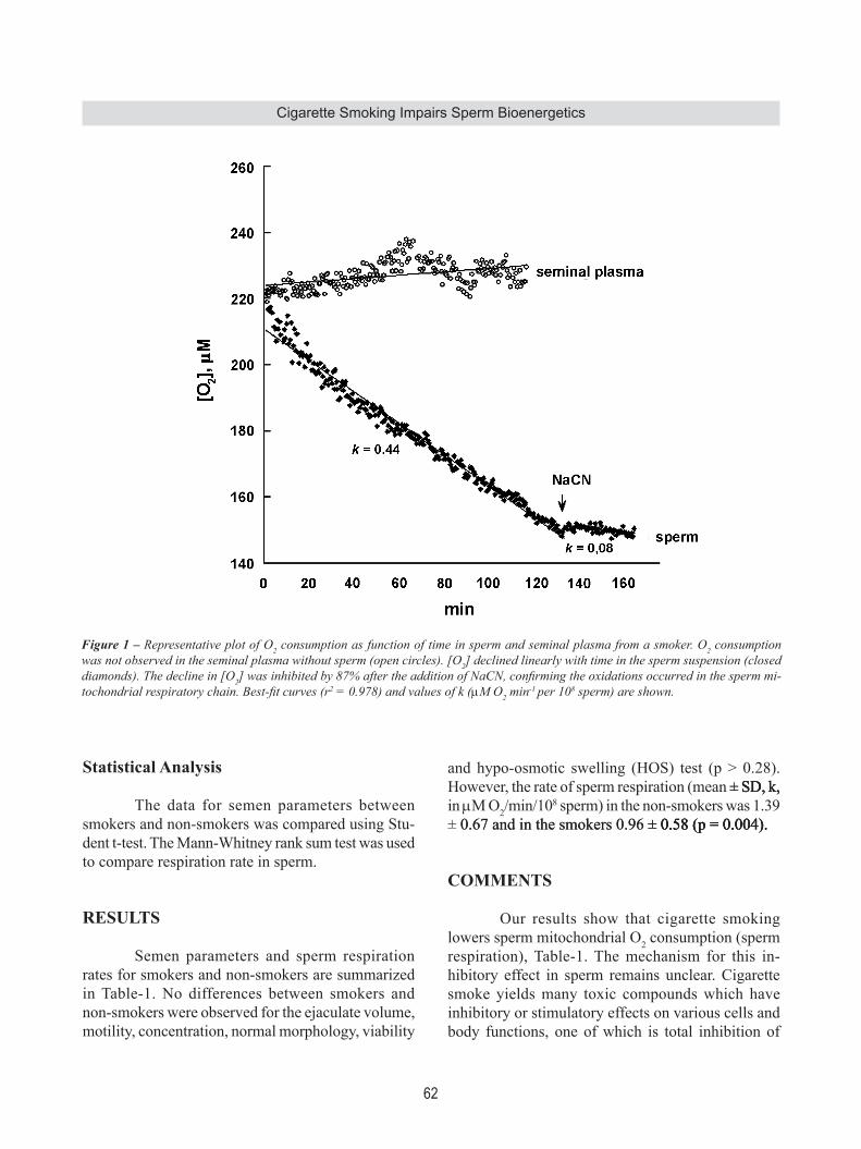
62
Cigarette Smoking Impairs Sperm Bioenergetics
Statistical Analysis
The data for semen parameters between smokers and non-smokers was compared using Stu-dent t-test. The ��ann-Whitney rank sum test was used to compare respiration rate in sperm.
RESULTS
Semen parameters and sperm respiration rates for smokers and non-smokers are summarized in Table-1. No differences between smokers and non-smokers were observed for the ejaculate volume, motility, concentration, normal morphology, viability
and hypo-osmotic swelling (HOS) test (p > 0.28). However, the rate of sperm respiration (mean �� SD, k,�� S�, k, S�, k, in µ�� O2/min/108 sperm) in the non-smokers was 1.�9 �� 0.6�� and in the smokers 0.96 �� 0.58 (p �� 0.00�). 0.6�� and in the smokers 0.96 �� 0.58 (p �� 0.00�).�� 0.58 (p �� 0.00�).0.58 (p �� 0.00�).
COMMENTS
Our results show that cigarette smoking lowers sperm mitochondrial O2 consumption (sperm respiration), Table-1. The mechanism for this in-hibitory effect in sperm remains unclear. Cigarette smoke yields many toxic compounds which have inhibitory or stimulatory effects on various cells and body functions, one of which is total inhibition of
Figure 1 – Representative plot of O2 consumption as function of time in sperm and seminal plasma from a smoker. O2 consumption was not observed in the seminal plasma without sperm (open circles). [O2] declined linearly with time in the sperm suspension (closed diamonds). The decline in [O2] was inhibited by 87% after the addition of NaCN, confirming the oxidations occurred in the sperm mi-tochondrial respiratory chain. Best-fit curves (r2 = 0.978) and values of k (µM O2 min-1 per 108 sperm) are shown.

63
Cigarette Smoking Impairs Sperm Bioenergetics
ciliary movement at bronchial level (2�). The de-creased cellular respiration observed in smokers can occur due to toxic compounds in the cigarette smoke. These include nicotine, cadmium, carbon monoxide, hydrogen cyanide, ammonia volatile hydrocarbons, alcohol, aldehydes and ketones, etc. No differences were observed for conventional semen parameters between smokers and non-smok-ers in the present study. Similar findings have been observed previously (24). Significant negative impact of cigarette smoking on semen quality; however, has also been reported (5-12). Lower sperm mitochondrial function (respiration) in smokers may be a consequence of sperm exposure to smoke-related toxins present in seminal plasma. The impact of these toxins on mitochondrial function seems non-reversible because washed sperm devoid of seminal plasma were used in this study. Irreversible degenerative damage to the architectural elements of the sperm tail or axoneme in smokers has been previously observed (25). This ultra structural damage includes the absence of one or more fiber doublets, central fibers, and coarse outer fibers. The mitochondria are a principal part of sperm tail therefore, such ultra structural damage in the tail region can also result in impaired mitochondrial function. Ex-posure of non-smoker’s sperm to the seminal plasma of a smoker resulted in significantly reduced motility and membrane functional integrity (26). Oxidative stress produced in seminal plasma due to smoking can be another factor for this phenomenon (2��).
In summary, our results show a significantly lower sperm mitochondrial O2 consumption rate in smokers. Further studies are needed to identify those constituents of cigarette smoke that mediate this ef-fect.
ACKNOWLEDGEMENTS
We are grateful for the support of ��s. Violeta Cuenca and ��rs. Crystal Kling for this work.
CONFLICT OF INTEREST
None declared.
REFERENCES
1. Pacifici R, Altieri I, Gandini L, Lenzi A, Pichini S, Rosa ��, et al.: Nicotine, cotinine, and trans-�-hy-droxycotinine levels in seminal plasma of smokers: effects on sperm parameters. Ther �rug ��onit. 199�; 15: �58-6�.
2. Vine MF, Hulka BS, Margolin BH, Truong YK, Hu PC, Schramm ����, et al.: Cotinine concentrations in semen, urine, and blood of smokers and nonsmokers. Am J Public Health. 1993; 83: 1335-8.
3. Collin O, Kilter S, Bergh A: Tobacco smoke disrupts testicular microcirculation in the rat. Int J Androl.
Table 1 – Sperm parameters and rates of respiration in smokers and non-smokers.
Smokers(N �� 20)
Non-smokers(N �� 58)
Ejaculate volume (mL) �.2 �� 1.��� 1.� 1.� �.6 �� 1.��� 1.� 1.�Total motility (%) 62.� �� 8.��� 8.� 8.� 61.8 �� 9.1�� 9.1 9.1Progressive motility (%) 50.2 �� 12.1�� 12.1 12.1 �8.�� �� 10.2�� 10.2 10.2Sperm concentration (106/mL) 125 �� ����� ��� ��� 1�� �� 8��� 8� 8�Normal morphology (%) 21.1 �� 1�.��� 1�.� 1�.� 20.� �� 10.0�� 10.0 10.0Viability (%) 55.8 �� 10.��� 10.� 10.� 55.9 �� 10.8�� 10.8 10.8HOS (%) ��1.1 �� ��.��� ��.� ��.� ��2.0 �� 8.9�� 8.9 8.9Rate of respiration 0.96 �� 0.58�� 0.58 0.58 1.�9 �� 0.6���� 0.6�� 0.6��
The values are mean �� SD. The p value between smokers and non-smokers for the rate of respiration (in �M O�� SD. The p value between smokers and non-smokers for the rate of respiration (in �M O SD. The p value between smokers and non-smokers for the rate of respiration (in �M O2/min/108 sperm) was 0.004. No statistical differences were observed for the remaining parameters. HOS = hypo-osmotic swelling.
64
Cigarette Smoking Impairs Sperm Bioenergetics
1995; 18: 141-5. Erratum in: Int J Androl. 1995; 18: 2�0.
�. Zenzes ��T: Smoking and reproduction: gene damage to human gametes and embryos. Hum Reprod Update. 2000; 6: 122-�1.
5. Evans HJ, Fletcher J, Torrance M, Hargreave TB: Sperm abnormalities and cigarette smoking. Lancet. 1981; 1: 62��-9.
6. Vogt HJ, Heller WD, Borelli S: Sperm quality of healthy smokers, ex-smokers, and never-smokers. Fertil Steril. 1986; �5: 106-10.
7. Rantala ML, Koskimies AI: Semen quality of infertile couples--comparison between smokers and non-smok-ers. Andrologia. 1987; 19: 42-6.
8. Holzki G, Gall H, Hermann J: Cigarette smoking and sperm quality. Andrologia. 1991; 23: 141-4.
9. Künzle R, Mueller MD, Hänggi W, Birkhäuser MH, Drescher H, Bersinger NA: Semen quality of male smokers and nonsmokers in infertile couples. Fertil Steril. 200�; ��9: 28��-91.
10. Pasqualotto FF, Sobreiro BP, Hallak J, Pasqualotto EB, Lucon AM: Cigarette smoking is related to a decrease in semen volume in a population of fertile men. BJU Int. 2006; 9��: �2�-6.
11. Ramlau-Hansen CH, Thulstrup AM, Aggerholm AS, Jensen MS, Toft G, Bonde JP: Is smoking a risk fac-tor for decreased semen quality? A cross-sectional analysis. Hum Reprod. 2007; 22: 188-96.
12. Gaur DS, Talekar M, Pathak VP: Effect of cigarette smoking on semen quality of infertile men. Singapore Med J. 2007; 48: 119-23.
13. Sofikitis N, Takenaka M, Kanakas N, Papadopoulos H, Yamamoto Y, Drakakis P, et al.: Effects of cotinine on sperm motility, membrane function, and fertilizing capacity in vitro. Urol Res. 2000; 28: ���0-5.
14. Zitzmann M, Rolf C, Nordhoff V, Schräder G, Rick-ert-Föhring M, Gassner P, et al.: Male smokers have a decreased success rate for in vitro fertilization and intracytoplasmic sperm injection. Fertil Steril. 200�; ��9 (Suppl �): 1550-�.
15. Jensen MS, Mabeck LM, Toft G, Thulstrup AM, Bonde JP: Lower sperm counts following prenatal tobacco exposure. Hum Reprod. 2005; 20: 2559-66.
16. Rubes J, Lowe X, Moore D 2nd, Perreault S, Slott V, Evenson D, et al.: Smoking cigarettes is associated with increased sperm disomy in teenage men. Fertil Steril. 1998; ��0: ��15-2�.
17. Shi Q, Ko E, Barclay L, Hoang T, Rademaker A, ��artin R: Cigarette smoking and aneuploidy in human sperm. ��ol Reprod �ev. 2001; 59: �1��-21.
18. Horak S, Polanska J, Widlak P: Bulky DNA adducts in human sperm: relationship with fertility, semen quality,
smoking, and environmental factors. ��utat Res. 200�; 5���: 5�-65.
19. Sepaniak S, Forges T, Gerard H, Foliguet B, Bene MC, Monnier-Barbarino P: The influence of cigarette smok-ing on human sperm quality and DNA fragmentation. Toxicology. 2006; 22�: 5�-60.
20. Practice Committee of the American Society for Re-productive ��edicine: Smoking and infertility. Fertil Steril. 2006; 86 (5 Suppl 1): S1��2-��.
21. Tao Z, Withers HG, Penefsky HS, Goodisman J, Souid AK: Inhibition of cellular respiration by doxorubicin. Chem Res Toxicol. 2006; 19: 1051-8.
22. Badawy ZS, Chohan KR, Whyte DA, Penefsky HS, Brown OM, Souid AK: Cannabinoids inhibit the respiration of human sperm. Fertil Steril. 2009; 91: 2���1-6.
23. Marchese-Ragona SP, Johnson KA: Structural and bio-chemical studies of the dynen ATPase. In: Gagnon C (eds.), Controls of Sperm ��otility: Biological and Clinical Aspects. CRC Press, Boca Raton. 1990; pp. 203.
24. Trummer H, Habermann H, Haas J, Pummer K: The impact of cigarette smoking on human semen param-eters and hormones. Hum Reprod. 2002; 17: 1554-9.
25. Zavos PM, Correa JR, Karagounis CS, Ahparaki A, Phoroglou C, Hicks CL, et al.: An electron microscope study of the axonemal ultrastructure in human sper-matozoa from male smokers and nonsmokers. Fertil Steril. 1998; 69: ��0-�.
26. Zavos PM, Correa JR, Antypas S, Zarmakoupis-Zavos PN, Zarmakoupis CN: Effects of seminal plasma from cigarette smokers on sperm viability and longevity. Fertil Steril. 1998; 69: �25-9.
27. Saleh RA, Agarwal A, Sharma RK, Nelson DR, Thomas AJ Jr: Effect of cigarette smoking on levels of seminal oxidative stress in infertile men: a prospective study. Fertil Steril. 2002; ��8: �91-9.
Accepted after revision: August 17, 2009
Correspondence address:�r. Kazim R. ChohanSUNY Upstate Medical University750 East Adams StreetSyracuse, NY, 13210, USAFax: + 1 �15 �6�-6��11E-mail: [email protected]

65
Cigarette Smoking Impairs Sperm Bioenergetics
EDITORIAL COMMENT
Smoking is a lifestyle hazard for both active and passive smokers (1). Although sperm concentra-tions, motility, and/or morphology are often reduced compared to results observed in non-smokers, they often remain within the normal range (2). In this ar-ticle, no differences were found between smokers and non-smokers for these parameters. However, it does show a negative impact of smoking on sperm mito-chondrial function. Although sperm mitochondrial O2 consumption is not a routine test, as a functional test, it may represent a predictive value for smoking men with normal sperm count aiming pregnancy. In accordance with literature, spermatozoa from smokers have reduced fertilizing capacity and embryos display lower implantation rates (�).
As discussed in the article, the mechanism for the inhibitory effect of smoking in sperm mitochon-drial O2 respiration remains unclear. This decreased cellular respiration can occur due to toxic compounds (1), to oxidative stress (4) or to both. According to Gaur et al., direct exposure of spermatozoa to the toxins in cigarette smoke probably tilts the delicate
balance of reactive oxygen species that are produced by spermatozoa for their special functions like de-capitation (1).
In conclusion, couples in reproductive age should be strongly discouraged to smoke (�).
REFERENCES
1. Gaur DS, Talekar M, Pathak VP: Effect of cigarette smoking on semen quality of infertile men. Singapore Med J. 2007; 48: 119-23.
2. The Practice Committee of the American Society for Reproductive ��edicine: Smoking and infertility. Fertil Steril. 2008; 90: S25�-9.
3. Soares SR, Melo MA: Cigarette smoking and repro-ductive function. Current Opin Obstet Gynecol. 2008; 20: 281-91.
4. Saleh RA, Agarwal A, Sharma RK, Nelson DR, Thomas AJ Jr: Effect of cigarette smoking on levels of seminal oxidative stress in infertile men: a prospective study. Fertil Steril. 2002; ��8: �91-9.
Dr. Renato Fraietta &Dr. Agnaldo P. Cedenho
Department of SurgeryDivision of Urologic Surgery
Federal University of Sao Paulo, UNIFESPSao Paulo, SP, Brazil
E-mail: [email protected]

66
Botulinum Toxins for Refractory Detrusor OveractivityInternational Braz J Urol Vol. 36 (1): 66-74, January - February, 2010
Experience with Different Botulinum Toxins for the Treatment of Refractory Neurogenic Detrusor OveractivityCristiano M. Gomes, Jose E. de Castro Filho, Ronald F. Rejowski, Flavio E. Trigo-Rocha, Homero Bruschini, Tarcisio E. P. de Barros Filho, Miguel Srougi
Division of Urology, University of Sao Paulo School of Medicine, Sao Paulo, Brazil
ABSTRACT
Purpose: To report our experience with the use of the botulinum toxin-A (BoNT/A) formulations Botox® and Prosigne® in the treatment of neurogenic detrusor overactivity (NDO).Materials and Methods: At a single institution, 45 consecutive patients with refractory urinary incontinence due to NDO received a single intradetrusor (excluding the trigone) treatment with botulinum toxin type A 200 or 300 units. Botox was used for the first 22 patients, and Prosigne for the subsequent 23 patients. Evaluations at baseline and week 12 included assessment of continence and urodynamics. Safety evaluations included monitoring of vital signs, hematuria during the procedure, hospital stay, and spontaneous adverse event reports.Results: A total of 42 patients were evaluated (74% male; mean age, 34.8 years). Significant improvements from baseline in maximum cystometric capacity (MCC), maximum detrusor pressure during bladder contraction, and compliance were observed in both groups (P < 0.05). Improvement in MCC was significantly greater with Botox versus Prosigne (+103.3% vs. +42.2%; P = 0.019). Continence was achieved by week 12 in 16 Botox recipients (76.2%) and 10 Prosigne recipients (47.6%; P = 0.057). No severe adverse events were observed. Mild adverse events included 2 cases of transient hematuria on the first postoperative day (no specific treatment required), and 3 cases of afebrile urinary tract infection.Conclusions: Botox and Prosigne produce distinct effects in patients with NDO, with a greater increase in MCC with Botox. Further evaluation will be required to assess differences between these formulations.
Key words: botulinum toxins; urinary bladder, overactive; neurogenic bladder; urinary incontinence; urodynamicsInt Braz J Urol. 2010; 36: 66-74
INTRODUCTION
Urinary incontinence due to detrusor overac-tivity is a common problem in patients with neurologi-cal diseases such as spinal cord injury, with significant impact on quality of life. Moreover, in this population, detrusor overactivity is frequently accompanied by high bladder pressure, and may pose a risk to the upper urinary tract (1,2). First-line treatment for de-trusor overactivity is usually pharmacological, with oral anticholinergic agents used to decrease detrusor contractility, resulting in lower bladder pressures and
�eurourolo�y�eurourolo�y
doi: 10.1590/S1677-55382010000100011
improved continence. However, distressing adverse effects, such as dry mouth, constipation, and blurred vision, may limit doses or lead to discontinuation of therapy, decreasing the effectiveness of treatment (3-5). When pharmacological therapy fails, invasive therapies are usually considered. Surgery, such as bladder augmentation, may be an option with good long-term results, but it is a permanent treatment with significant potential complications such as calculi, malignancy, and bowel complications (6,7). The efficacy and safety of local administra-tion of botulinum toxin A (BoNT/A) into the bladder

67
Botulinum Toxins for Refractory Detrusor Overactivity
has been investigated in previously reported studies (8-10). BoNT/A blocks neuromuscular activity in skeletal muscle by preventing neurotransmitter re-lease at presynaptic cholinergic nerve terminals (11). BoNT/A inhibits acetylcholine-mediated detrusor contraction and may inhibit release of other vesicle-bound neurotransmitters in the afferent and efferent pathways of the bladder wall, urothelium, or lamina propria (12,13). While the overwhelming majority of investiga-tors have used the BoNT/A formulation Botox® (Aller-gan, Inc., Irvine, CA), other BoNT/A formulations are being marketed. There is a lack of evidence as regards the clinical efficacy and safety of the recently released Chi-nese BoNT/A (Prosigne®, Lanzhou Biological Products Institute, Lanzhou, China) for the treatment of detrusor overactivity. This product has recently become available in Brazil, but there is scarce data on this pharmaceutical formulation. It is known that they differ in the external excipients that are added to BoNT/A. Botox vials contain sodium chloride 0.9 mg and human albumin 0.5 mg, and the protein load is 5 ng/100 units, while in Prosigne vials, the external excipients are porcine gelatin (Haemacell) 5 mg, dextran 25 mg, and sucrose 25 mg, and the protein load is 4.0-5.0 ng/100 units of BoNT/A (14). In terms of potency, little is known since only two studies have compared both formulations, with conflicting results. In a major Chinese study the two formulations were used in patients with various types of focal dystonias, Botox was found to be 1.5 times more potent than Prosigne (14). In another study in patients with blepharospasm, comparable efficacy was observed (15). There may also be differences in the toxicity profile due to differences in the preparation procedure for both formulations (14,16). Botox is currently the only BoNT/A formula-tion approved in Brazil for the treatment of overac-tive bladder. The aim of our study was to report our experience with the use of the two formulations in the treatment of detrusor overactivity.
MATERIALS AND METHODS
This study was carried-out in accordance with the Ethics Committee regulations and written informed consent was obtained from all patients.
A prospective study was conducted at a single institution in which 45 consecutive patients received a single intradetrusor treatment with BoNT/A between April 2003 and April 2007. Inclusion criteria were urinary incontinence due to neurogenic detrusor overactivity (as demonstrated by urodynamics), fail-ure of oral anticholinergic therapy, and use of clean intermittent catheterization or willingness to do so, if necessary. Exclusion criteria included previous blad-der surgery, previous treatment with an endovesical pharmacological agent, symptomatic urinary tract infection, and a history of neurological disease of less than 6 months. Among the 45 patients enrolled in the study, neurogenic detrusor overactivity resulted from spinal cord injury in 36 patients (80.0%), viral myelitis in 4 (8.9%), multiple sclerosis in 3 (6.7%) and schistosomal myeloradiculopathy in 2 patients (4.4%). The BoNT/A formulation Botox was used for the first 22 patients, whereas the subsequent 23 patients received Prosigne. The different BoNT/A formulations were used because the hospital changed the supplier due to cost restrictions. The injection procedure was performed as described previously by Schurch et al. (9). Briefly, the BoNT/A dose (200 or 300 units) was reconstituted with saline 0.9% at a total volume of 30 mL. The blad-der was distended with 100 mL of saline, and 30 injec-tions of 1.0 mL each were performed intramuscularly throughout the bladder wall, excluding the trigone. A rigid cystoscope and 23-gauge flexible needle (Handle Cook®) were used, yielding an injection depth of 3-5 mm. A Foley catheter was left indwelling overnight, and patients were discharged the following morning, after catheter removal, resuming clean intermittent catheterization. Antibiotics were administered during anesthesia and for 2 days after the procedure. Patients receiving anticholinergic drugs were instructed to stop the medication 2 weeks after BoNT/A injection.
Evaluations
Evaluations at baseline and 12 weeks post-treatment included a clinical assessment of continence and a standard urodynamic study. Twelve weeks was selected as the follow-up duration because it is a mid-

68
Botulinum Toxins for Refractory Detrusor Overactivity
term evaluation, and also because previous studies with Botox have shown that peak efficacy is estab-lished after 4 weeks and maintained up to 12 weeks (and longer) (9). Patients were considered continent when they were not using any pads or diapers and had no episodes of incontinence during the 7 days before evaluation. The primary efficacy variable was improve-ment of urodynamic parameters compared to baseline at the 12-week timepoint. The measurements included maximum cystometric capacity (MCC), volume of first detrusor overactivity (reflex volume), maximum detrusor pressure during bladder contraction, and bladder compliance, based on the terminology of the International Continence Society (17). The secondary outcome measure was continence status. Safety evaluations included monitoring of vital signs and hematuria during the procedure and hospital stay, and spontaneous reports of adverse events.
Statistical Analysis
Numerical data were reported as mean ± standard deviation and range. Categorical variables were reported as numbers and percentages. Results of treatment with the different BoNT/A formulations and doses were analyzed for the whole population as well as for between-group comparisons. Within-group changes from baseline in the urodynamic parameters were analyzed using the paired t-test. Between-group
comparisons were performed using analysis of vari-ance for repeated measurements. The chi-squared (x²) test or the Fisher’s exact test was used for categorical variables. Data were processed using SPSS 12.0 for Windows statistical software (SPSS Inc., Chicago, Ill). P-values < 0.05 were considered statistically significant.
RESULTS
Of the 45 recruited patients, 3 were excluded for not returning for the postoperative follow-up evaluation (1 from the Botox group and 2 from the Prosigne group); thus, 42 patients (21 in each group) were evaluable. Of the 42 evaluable patients, the majority were male (31/42; 73.8%), and the mean age was 34.8 ± 12.7 years (range, 18 to 73 years). No statistically significant differences were found between the two groups for any demographic or baseline characteristics (Table-1). In the Botox group, 9 patients (42.9%) re-ceived a BoNT/A dose of 200 units and 12 (57.1%) received 300 units. In the Prosigne group, 5 patients (23.8%) received a BoNT/A dose of 200 units and 16 (76.2%) received 300 units.
Urodynamic Findings
MCC significantly improved from baseline in both groups, increasing from 184 ± 62 to 375 ± 109
Table 1 – Baseline characteristics of evaluable patients treated with Botox or Prosigne.
Parameter Botox(N = 21)
Prosigne(N = 21)
p Value
Age (years), mean ± SD 37.2 ± 14.4 32.5 ± 10.6 0.234Gender (female), n (%) 6 (28.6%) 5 (23.8%) 0.500MCC (mL), mean ± SD 184 ± 62 204 ± 83 0.388Reflex volume (mL), mean ± SD 180 ± 78 199 ± 102 0.743MDP (cm H2O), mean ± SD 68 ± 33 82 ± 27 0.158Compliance (mL/cm H2O), mean ± SD 19.4 ± 12.8 23.5 ± 10.6 0.267
MCC = maximum cystometric capacity; MDP = maximum detrusor pressure; SD = standard deviation.

69
Botulinum Toxins for Refractory Detrusor Overactivity
mL (+103.3%; P < 0.001) in the Botox group and from 204 ± 83 to 290 ± 134 mL (+42.2%; P = 0.002) in the Prosigne group. The increase from baseline in MCC was significantly greater in the Botox group than in the Prosigne group when considered as a whole (P = 0.019; Figure-1). When the different BoNT/A doses were considered, no statistically significant differences were found between the subgroups (Figure-2). The changes from baseline in reflex volume were from 180 ± 78 to 226 ± 79 mL (P = 0.150) in the Botox group and from 173 ± 71 to 199 ± 102 mL (P = 0.255) in the Prosigne group. The evaluation of this parameter was greatly influenced by the fact that
a substantial number of patients in both groups be-came arefelexic at the week 12 evaluation (11 patients [52.4%] in the Botox group and 6 [28.6%] in the Pro-signe group; P = 0.116). These patients, who had the most favorable results of BoNT/A injection, were not included in the calculation of mean reflex volume. MDP decreased significantly from baseline in both groups, from 68 ± 33 to 28 ± 18 cm H2O (-58.8%; P < 0.001) in the Botox group and from 82 ± 27 to 47 ± 30 cm H2O (-42.7%; P < 0.001) in the Prosigne group. Compliance increased significantly from baseline in both groups, from 19 ± 13 to 42 ± 29 mL/cmH2O (+121.0%; P = 0.006) in the Botox group
Figure 1 – Box-plot analysis of change from baseline in maximum cystometric capacity (MCC) at week 12, according to treatment group.

70
Botulinum Toxins for Refractory Detrusor Overactivity
and from 23 ± 11 to 42 ± 42 mL/cmH2O (82.6%; P = 0.024) in the Prosigne group. In the two groups, significant (P < 0.001) improvements from baseline in the continence status were observed at week 12. Continence was achieved by week 12 in 16 patients (76.2%) in the Botox group and 10 (47.6%) in the Prosigne group (P = 0.057). The administration of BoNT/A was unevent-ful and the entire procedure required no more than 30 minutes in all patients. Anesthesia was general in 28 patients (66.7%), spinal in 10 (23.8%), and local in 4 patients (9.5%).
There were no severe adverse events observed in any patient. Mild adverse events included 2 cases of transient hematuria on the first postoperative day that did not require specific treatment, and 3 cases of afebrile urinary tract infection. All patients were discharged home on the first postoperative day.
COMMENTS
Determining a more precise role of the differ-ent formulations of BoNT/A in the treatment of detru-
Figure 2 – Box-plot analysis of change from baseline in maximum cystometric capacity (MCC) at week 12, according to treatment group and dose level (200 or 300 units).

71
Botulinum Toxins for Refractory Detrusor Overactivity
sor overactivity is of paramount importance, because BoNT/A treatments may have a significant economic impact on health services. To our knowledge, this is the first reported study on the use of Prosigne for the treatment of detrusor overactivity. Our study was originally designed to compare the use of two doses of Botox (200 vs. 300 units) in patients with neuro-genic detrusor overactivity. However, an unpredicted change of the hospital supplier of BoNT/A prevented us from completing the designed study and gave us the opportunity to evaluate the new formulation (Pro-signe). Because our original plan was to compare two doses of BoNT/A (200 vs. 300 units), patients from the Botox group were randomized to one of the two doses, and 12 received 300 units while 10 received 200 units. One of these patients was excluded from the study for not returning for the follow-up evaluation. When we started patients from the Prosigne group, we initially maintained the randomization for the two doses, since the manufacturers of Prosigne claim that the two formulations are comparable in potency, with each preparation expressed in units, 1 unit representing the LD50 for mice (14). However, after unsuccessfully treating a few patients using 200 units, we chose to inject 300 units of BoNT/A in the subsequent patients. For this reason, more patients in the Prosigne group received the higher BoNT/A dose. It should be noted that prescribing informa-tion for Botox states that units of biological activity of this formulation cannot be compared or converted into units of any other botulinum toxin, due to spe-cific details of the assay method used (18). In fact, there are limited published data on Prosigne in the literature, and only two studies have compared it with Botox. In a study conducted in China, Tang and Wan evaluated a large group of patients with hemifacial spasm and various types of focal dystonias (includ-ing blepharospasm) in which Botox was found to be 1.5 times more potent than Prosigne (14). The second study was conducted by Rieder et al. in patients with blepharospasm and hemifacial spasm which found that the two BoNT/A formulations had comparable short-term efficacy and safety in these indications (15). The authors of this study acknowledge that dif-ferent BoNT/A formulations are not considered bio-equivalent and recommend further studies to establish the clinical comparability of these formulations. The
differences observed in these studies may result from differences in patient population, clinical indication and/or application technique. Our results appear to be in accordance with the Chinese study, indicating that Prosigne is not as potent as Botox. It is important to acknowledge that we used it for a different clinical indication, injecting the toxin in the smooth muscle rather than an striated muscle, which may be another possible reason for distinct effects of the formula-tions. Patients in the two groups did not differ sig-nificantly in any of the baseline parameters. Despite the fact that a larger proportion of patients in the Prosigne group received the higher BoNT/A dose, treatment with Botox resulted in a significantly greater increase from baseline in MCC, and, although not statistically significant, improvements in the Botox group were numerically superior on all the other evaluated urodynamic parameters. An interesting finding was that 52% of the patients in the Botox group and 29% of those in the Prosigne group did not experience a hyperreflexive detrusor contraction at the follow-up evaluation. This is a strong indication of the efficacy of therapy with BoNT/A, and this finding appeared to favor Botox. However, it was a confounding factor for the evaluation of the reflex detrusor volume, since it was necessary to exclude patients who became areflexic, who represent the best responders to treatment, from analyses of this endpoint. A tendency for better results was also ob-served for patients treated with Botox in terms of improvement in continence rates. Their complete continence rate at week 12 was 76%, as opposed to 48% for the patients treated with Prosigne. As mentioned previously, our initial objec-tive was to compare two different Botox doses (200 vs. 300 units), but we ultimately had four subgroups based on different doses and BoNT/A formulations. We attempted to compare the two BoNT/A formula-tions based on the doses of 200 or 300 units, but the subgroups were too small for significant compari-sons. Both drugs were well tolerated by patients and no significant adverse event occurred in any group. We acknowledge that our study was not designed to compare the two BoNT/A formulations.

72
Botulinum Toxins for Refractory Detrusor Overactivity
Therefore, patients were not randomized to the two groups. However, the two groups were composed of consecutive patients and the comparison of baseline parameters did not reveal differences between the groups, indicating that the populations were quite comparable.
CONCLUSIONS
Our study provides the first experience with the use of the formulation Prosigne for the treat-ment of refractory detrusor overactivity, indicating that Botox and Prosigne may have distinct effects in the detrusor of patients with neurogenic detrusor overactivity, with Botox promoting superior results in terms of increase in bladder capacity. Due to the limitations of this study in terms of patient selection (not randomized) and small sample size to compare the effect of different doses, as well as the short fol-low-up period, additional studies should be conducted to determine the differences in the safety profile and specific benefits between these two BoNT/A formu-lations for the treatment of patients with neurogenic detrusor overactivity.
ACKNOWLEDGEMENTS
Editorial support was provided by Sushma Soni, funded by Allergan, Inc.
CONFLICT OF INTEREST
None declared.
REFERENCES
1. Weld KJ, Graney MJ, Dmochowski RR: Clinical significance of detrusor sphincter dyssynergia type in patients with post-traumatic spinal cord injury. Urol-ogy. 2000; 56: 565-8.
2. Weld KJ, Graney MJ, Dmochowski RR: Differences in bladder compliance with time and associations of bladder management with compliance in spinal cord injured patients. J Urol. 2000; 163: 1228-33.
3. Andersson KE: Antimuscarinics for treatment of over-active bladder. Lancet Neurol. 2004; 3: 46-53.
4. Rovner ES, Gomes CM, Trigo-Rocha FE, Arap S, Wein AJ: Evaluation and treatment of the overactive bladder. Rev Hosp Clin Fac Med Sao Paulo. 2002; 57: 39-48.
5. Rovner ES, Wein AJ: Update on overactive bladder: pharmacologic approaches on the horizon. Curr Urol Rep. 2003; 4: 385-90.
6. Hohenfellner M, Dahms S, Pfitzenmaier J, Thüroff JW: Orthotopic bladder augmentation and substitution. Curr Opin Urol. 1999; 9: 309-14.
7. Quek ML, Ginsberg DA: Long-term urodynamics fol-lowup of bladder augmentation for neurogenic bladder. J Urol. 2003; 169: 195-8.
8. Mascarenhas F, Cocuzza M, Gomes CM, Leão N: Trigonal injection of botulinum toxin-A does not cause vesicoureteral reflux in neurogenic patients. Neurourol Urodyn. 2008; 27: 311-4.
9. Schurch B, de Sèze M, Denys P, Chartier-Kastler E, Haab F, Everaert K: Botulinum toxin type a is a safe and effective treatment for neurogenic urinary incon-tinence: results of a single treatment, randomized, placebo controlled 6-month study. J Urol. 2005; 174: 196-200.
10. Smith CP, Nishiguchi J, O’Leary M, Yoshimura N, Chancellor MB: Single-institution experience in 110 patients with botulinum toxin A injection into bladder or urethra. Urology. 2005; 65: 37-41.
11. Dolly O: Synaptic transmission: inhibition of neu-rotransmitter release by botulinum toxins. Headache. 2003; 43 (Suppl 1): S16-24.
12. Apostolidis A, Popat R, Yiangou Y, Cockayne D, Ford AP, Davis JB, et al.: Decreased sensory receptors P2X3 and TRPV1 in suburothelial nerve fibers following intradetrusor injections of botulinum toxin for human detrusor overactivity. J Urol. 2005; 174: 977-82; dis-cussion 982-3.
13. Chuang YC, Yoshimura N, Huang CC, Chiang PH, Chancellor MB: Intravesical botulinum toxin a ad-ministration produces analgesia against acetic acid induced bladder pain responses in rats. J Urol. 2004; 172: 1529-32.
14. Tang X, Wan X: Comparison of Botox with a Chinese type A botulinum toxin. Chin Med J (Engl). 2000; 113: 794-8.
15. Rieder CR, Schestatsky P, Socal MP, Monte TL, Fricke D, Costa J, et al.: A double-blind, randomized, crossover study of prosigne versus botox in patients with blepharospasm and hemifacial spasm. Clin Neu-ropharmacol. 2007; 30: 39-42.

73
Botulinum Toxins for Refractory Detrusor Overactivity
EDITORIAL COMMENT
Patients with various neurological condi-tions (e.g. spinal cord injury) may present detrusor overactivity (DO), formally classified as neurogenic DO (NDO), that knowingly causes great social embar-rassment and inconvenience for the patient. The current treatment for NDO consists of a combination of clean intermittent self-catheteriza-tion and the pharmacological management. However, many patients discontinue treatment due to side-ef-fects (1). In such cases where the inability to tolerate the antimuscarinic drug therapy incurs in the failure of the treatment, intradetrusor botulinum neurotoxin type A (BoNT/A) may be an excellent alternative (2). Since it is a minimally invasive treatment, as opposed to a clam ileocystoplasty, a conventional surgical pro-cedure, it has currently been increasing in popularity. However, its results are temporary and can ultimately increase the costs of the treatment. In Brazil, Botox® may cost up to 20% more than Prosigne®*, which could be an obstacle in the way of those seeking to purchase it, considered that this is a developing country. Therefore, it is important
to emphasize the development of comparative stud-ies analyzing the different formulations of BoNT/A and questions such as its potency and final sale price. Nevertheless, aside from this proposed study, there are no comparative studies using different types of BoNT/A to treat NDO. (Botox® versus Prosigne®). In spite of the possible methodological fail-ures prompted by a non-randomized study and small patient samples, the authors proposed an interesting paper, where they analyzed the action of two different formulations of BoNT/A in the treatment of NDO. The urodynamic findings showed that the improvement of maximum cystometric capacity was significantly higher in Botox® group than in the Pros-igne® one. Apart from a better continence on week 12 in the Botox® group (76.2% vs. 47.6% respectively, p = 0.057), all the other parameters did not show significant differences in the two groups. Moreover, perhaps if the quantity of data was increased this would be even more evident. There are several questions to be addressed regarding the intradetrusor injection of BoNT/A to
16. Tamura BM, Cucé LC, Rodrigues CJ: Allergic reaction to botulinum toxin: positive intradermal test. Dermatol Surg. 2008; 34: 1117-9.
17. Abrams P, Cardozo L, Fall M, Griffiths D, Rosier P, Ulmsten U, et al.: The standardisation of terminol-ogy of lower urinary tract function: report from the
Standardisation Sub-committee of the International Continence Society. Neurourol Urodyn. 2002; 21: 167-78.
18. Allergan I: BOTOX (Botulinum toxin type A) US prescribing information [Revised October 2006]. Ref. type: Generic. pp. 1-16.
Accepted after revision: July 29, 2009
Correspondence Address:Dr. Cristiano Mendes GomesDivisão de Clínica UrológicaAv. Dr. Enea de C. Aguiar, 255 / 710FSão Paulo, SP, 05403-000, BrazilFax: + 55 11 3064-7013E-mail: [email protected]

74
Botulinum Toxins for Refractory Detrusor Overactivity
treat NDO. Similar randomized trials should be done to clearly determine which formulation of BoNT/A has the best cost-efficiency with greater safety and lower morbidity.
REFERENCES
1. Colli E, Digesu GA, Olivieri L: Overactive bladder treatments in early phase clinical trials. Expert Opin Investig Drugs. 2007; 16: 999-1007.
2. Karsenty G, Denys P, Amarenco G, De Seze M, Gamé X, Haab F, et al.: Botulinum toxin A (Botox) intradetru-sor injections in adults with neurogenic detrusor over-activity/neurogenic overactive bladder: a systematic literature review. Eur Urol. 2008; 53: 275-87.
Dr. João Luiz AmaroDepartment of Urology
UNESP, School of MedicineBotucatu, São Paulo, Brazil
E-mail: [email protected]

75
Circumcision vs. Topical Corticosteroids in PhimosisInternational Braz J Urol Vol. 36 (1): 75-85, January - February, 2010
Yuri D. Nobre, Ricardo G. Freitas, Maria J. Felizardo, Valdemar Ortiz, Antonio Macedo Jr.
Section of Pediatric Urology, Department of Urology, Federal University of Sao Paulo, Sao Paulo, SP, Brazil
ABSTRACT
Purpose: To compare the efficacy and costs of circumcision versus topical treatment using a prospective pharmacoeco-nomic protocol.Materials and Methods: We treated 59 patients (3-10 years of age) randomized into two groups: 29 underwent an 8-week course of topical treatment with 0.2% betamethasone-hyaluronidase cream twice a day; and 30 underwent circumcision. Topical treatment success was defined as complete exposure of the glans. In cases of treatment failure, circumcision was performed and its cost imputed to that of the initial treatment. The pharmacoeconomic aspects were defined according to the Brazilian National Public Health System database and the Brazilian Community Pharmacies Index.Results: The two groups were statistically similar for all clinical parameters evaluated. Topical treatment resulted in com-plete exposure of the glans in 52% of the patients. Topical treatment was associated with preputial pain and hyperemia. However, treatment suspension was unnecessary. Minor complications were observed in 16.6% of the surgical group patients. The mean cost per patient was US$ 53.70 and US$ 125.20, respectively, for topical steroid treatment (including the costs related to treatment failure) and circumcision. The total costs were US$ 2,825.32 and US$ 3,885.73 for topical treatment and circumcision, respectively.Conclusions: Topical treatment of phimosis can reduce costs by 27.3% in comparison with circumcision. Therefore, topical treatment of phimosis should be considered prior to the decision to perform surgery.
Key words: phimosis; circumcision; male; steroids; pharmacoeconomicsInt Braz J Urol. 2010; 36: 75-85
INTRODUCTION
Circumcision is considered the elective treat-ment for phimosis in newborn infants and children. In the United States, circumcision is the fourth most common surgical procedure, performed in 65.3% of the male population (1). Various factors, such as social norms, cultural traditions and religious beliefs, influence the decision
Pediatric Urology
doi: 10.1590/S1677-553820100000100012
To Circ or Not to Circ: Clinical and Pharmacoeconomic Outcomes of a Prospective Trial of Topical Steroid versus Primary Circumcision
of whether or not to perform circumcision. The Ameri-can Academy of Pediatrics, in one of their most recent publications on this subject, stated that there is no abso-lute medical indication for the routine circumcision of newborn infants, and that, despite the potential medical benefits and advantages, circumcision also presents dis-advantages and risks. When truly indicated, the risks and benefits should be discussed with parents, who should be asked to give written informed consent (2).

76
Circumcision vs. Topical Corticosteroids in Phimosis
In the 1990s, due to the controversy sur-rounding the topic of when circumcision is indicated, various studies were carried out in order to investi-gate alternative treatments for phimosis. Consistent success rates were achieved with the use of topical corticosteroids (3,4). Pharmacoeconomic studies help identify, calculate and compare the costs and risks of specific programs or therapies, as well as their benefits. Such studies also help determine which alternatives provide the best results and constitute the most practical use of the resources invested. Therefore, pharmacoeconom-ics is a relevant tool in the decision-making process, introducing the concept of economic rationality into health care systems in order to complement clinical decision making (5). There have been few studies on the economic impact of circumcision versus topical treatment of phimosis. Therefore, we conducted a prospective randomized study comparing, from the pharmacoeconomic point of view, the implications of topical treatment versus those of circumcision in cases of phimosis.
MATERIALS AND METHODS
A prospective randomized study was carried out in order to evaluate the clinical response to topical treatment of pathologic phimosis, comparing it to that of circumcision. We included 85 children, from 3 to 10 years of age, diagnosed with phimosis. We defined as phimo-sis, in this age bracket, phimosis type I (no retraction of the foreskin) and type II (external urethral meatus exposure only), in accordance with the classification system devised by Kayaba et al. (6). The study was previously approved by the Ethics Committee on Research of our University, and all parents or guardians were instructed and signed the written informed consent before starting the study. In the clinical group, 42 patients were treated with two daily applications of 0.2% betamethasone and hyaluronidase cream for 8 weeks. The same physician examined all of the patients. Prior to the beginning of treatment and in every subsequent con-sultation, patients were photographed.
The therapeutic response was considered favorable only if the glans was easily and completely exposed during the subsequent physical examination, without a phimotic ring or balanopreputial adhesions (Kayaba type V). The therapeutic response was con-sidered unfavorable if there was no exposure of the glans or there was only partial exposure of the glans due to balanopreputial adhesions or a phimotic ring. Therefore, the criterion adopted in order to define treatment success was the same as the expected outcome of circumcision: complete exposure of the glans. Patients who responded well to the topical treatment underwent outpatient follow-up evaluations at 2 and 4 months after the end of treatment. Patients who did not respond to the topical treatment by week 8 or who presented recurrence dur-ing the follow-up period underwent circumcision. Patients in the surgical group were evaluated and then referred to undergo circumcision. Patients underwent outpatient follow-up evaluations on post-operative days 15, 30 and 120. All surgical procedures were carried out at our institution. The anesthesia protocol was general inhalation anesthesia with sevoflurane, in conjunction with nerve block of the penis. In the present study, the pharmacoeconomic analysis was of the cost-minimization type. Only the costs of the surgical procedure and clinical treatment (those directly related to the health care system: medi-cal care, medications and medical materials) were taken into consideration. Indirect costs related to lost productivity, as well as intangible costs (those related to pain, suffering and impaired quality of life), were not calculated. The pharmacoeconomic analysis was carried out from the perspective of a Public Health Hospital, and the resources included in the cost analysis were identified through communications with officials of the Brazilian United Health Care System (BUHCS). The medication costs that were not available in the public health system database were determined by consulting the Brazilian Community Pharmacies Index. For patients in the surgical group, the costs were calculated separately for each of the follow-ing aspects: medical visits; anesthetic medications;
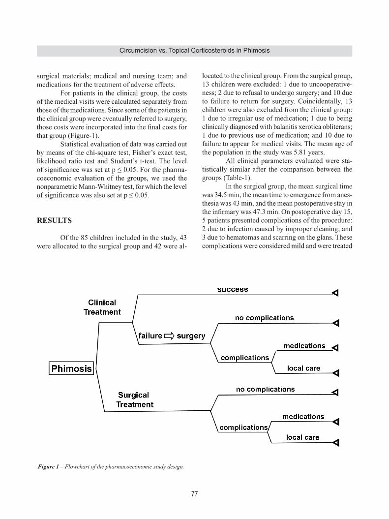
77
Circumcision vs. Topical Corticosteroids in Phimosis
surgical materials; medical and nursing team; and medications for the treatment of adverse effects. For patients in the clinical group, the costs of the medical visits were calculated separately from those of the medications. Since some of the patients in the clinical group were eventually referred to surgery, those costs were incorporated into the final costs for that group (Figure-1). Statistical evaluation of data was carried out by means of the chi-square test, Fisher’s exact test, likelihood ratio test and Student’s t-test. The level of significance was set at p ≤ 0.05. For the pharma-coeconomic evaluation of the groups, we used the nonparametric Mann-Whitney test, for which the level of significance was also set at p ≤ 0.05.
RESULTS
Of the 85 children included in the study, 43 were allocated to the surgical group and 42 were al-
located to the clinical group. From the surgical group, 13 children were excluded: 1 due to uncooperative-ness; 2 due to refusal to undergo surgery; and 10 due to failure to return for surgery. Coincidentally, 13 children were also excluded from the clinical group: 1 due to irregular use of medication; 1 due to being clinically diagnosed with balanitis xerotica obliterans; 1 due to previous use of medication; and 10 due to failure to appear for medical visits. The mean age of the population in the study was 5.81 years. All clinical parameters evaluated were sta-tistically similar after the comparison between the groups (Table-1). In the surgical group, the mean surgical time was 34.5 min, the mean time to emergence from anes-thesia was 43 min, and the mean postoperative stay in the infirmary was 47.3 min. On postoperative day 15, 5 patients presented complications of the procedure: 2 due to infection caused by improper cleaning; and 3 due to hematomas and scarring on the glans. These complications were considered mild and were treated
Figure 1 – Flowchart of the pharmacoeconomic study design.

78
Circumcision vs. Topical Corticosteroids in Phimosis
with intensive local care. In the subsequent follow-up visits, all of the patients presented penile conditions that were considered normal, with proper formation of scar tissue. In the clinical group, 15 (51.7%) of the 29 patients presented complete exposure of the glans at 2 months after the end of treatment. The topical treatment resulted in adverse effects or complica-tions in 10 patients: 4 patients experienced hyperemia and a burning sensation in the foreskin; 4 patients experienced a burning sensation in the foreskin; and 2 patients presented balanopreputial hyperemia. No intervention or treatment interruption was necessary in any of the cases. In the clinical group, patients in whom treat-ment success was achieved were monitored for 4 months. Neither loss of exposure of the glans nor recurrence was detected. Clinical treatment failure occurred in 14 cas-es, and those patients were referred to circumcision. Of those 14 patients, 2 failed to return for surgery. For the 12 who underwent surgery, the mean surgical time was 31.2 min, the mean time to emergence from anesthesia was 30 min, and the mean postoperative stay in the infirmary was 60 min.
Of the 12 clinical group patients undergoing circumcision, 4 presented postoperative complica-tions: 1 due to infection caused by improper cleaning, which demanded oral antibiotic treatment and local care; 1 due to bleeding and scars on the gland, which was treated locally; 1 due to pyogenic granuloma; and 1 due to an inflammatory process close to urethral meatus. All of the clinical group patients presented resolution of the complications during the follow-up period.
Pharmacoeconomic Analysis
In the pharmacoeconomic analysis, we evalu-ated the direct costs related to treatment of the two groups. It is of note that, as previously mentioned, the costs related to circumcision of patients in the clinical group were included in the final costs for that group (Figures 2-3). In the pharmacoeconomic evaluation, the costs were higher in the clinical group only in terms of the medical visits and the use of topical medication. Costs regarding medical and nursing staff, anesthetic medications and surgical materials were higher in the
Table 1 – Distribution of clinical findings regarding the incidence of balanitis, exposure of the urethral meatus and excess foreskin.
Variable Surgical Clinical p Value TestBalanitis 0.9 Chi-square testNo 22 73.3% 21 72.4%Yes 8 26.7% 8 27.6%
Variable Surgical Clinical p Value Test
Visible meatus 0.205 Chi-square testNo 13 43.3% 19 65.5%Yes 17 56.7% 10 34.5%
Variable Surgical Clinical p Value Test
Excess foreskin 1.000 Fisher’s exact testNo 3 10% 2 7%Yes 27 90% 27 93%

79
Circumcision vs. Topical Corticosteroids in Phimosis
surgical group than in the clinical group (p < 0.001). No statistical difference was found in costs regarding medications used for the treatment of postoperative complications. As can be seen in Figure-4, the analysis of the total costs per group, revealed greater economy in the clinical group than in the surgical group US$ 2,825.32 vs. US$ 3,885.73; p = 0.068; median, US$ 53.70 vs. US$ 125.20. Comparing the median cost to the set price stipulated by the BUHCS through the Authorized Hospital Admissions System (US$ 52.20) for ev-ery patient who underwent circumcision, we found considerable discrepancy between the amount paid by the BUHCS and the actual hospital costs of the circumcisions (Figure-5).
COMMENTS
Circumcision is classically considered the gold standard for the treatment of phimosis. Ap-proximately one-sixth of males worldwide are cir-cumcised (7). The indications for performing neonatal circumcision are controversial. Prominent among the factors favoring neonatal circumcision are the lower incidence of penile cancer, the lower incidence of balanitis/urinary infections, and the prevention of sexually transmitted diseases, including AIDS (8,9). In addition, the incidence of postoperative complica-tions is low. These benefits are sufficiently relevant to guarantee the routine practice of neonatal circumci-sion (8). However, most studies in which routine cir-cumcision is recommended are retrospective studies,
Figure 2 – Box plot of the cost analysis of the parameters investigated in both groups.

80
Circumcision vs. Topical Corticosteroids in Phimosis
and the validity of the conclusions should be critically investigated. Circumcision is, arguably, a preventive health care measure, which, in theory, facilitates hygiene. However, the potential benefits occur years after the surgery, and few studies involve adequate, timely follow-up evaluation (10). In Brazil, we do not adopt the concept of neo-natal circumcision and most indications for surgery are considered when the child is about to leave diapers and only patients with phimotic ring are candidates for this kind of treatment. The clinical treatment of phimosis consists of the gentle retraction of the foreskin and application of a topical corticosteroid to the foreskin and phimotic ring, with the objective of achieving complete exposure of the glans. The effects of topical corticosteroids, which have anti-inflammatory and immunosuppressive properties, on the metabolism of arachidonic acid and inhibition
of interleukin-1 synthesis have been well established. Topical corticosteroids can have an antiproliferative ef-fect on the dermal matrix, decreasing skin thickness and obliterating the stratum corneum (11,12). Hyaluronidase acts by modifying intercellular permeability and reducing tissue resistance, thereby increasing the diffusion of substances between planes (13). In a pioneer study on the application of topical corticosteroid in boys with phimosis who were candi-dates for circumcision, clinical treatment was shown to be viable and efficacious (3). Topical treatment of phimosis with corticosteroids has characteristics that make it quite attractive as the first approach to the treatment of phimosis. The method is safe, and complication rates are similar to those found for circumcision. In addition, corticosteroid treatment presents high cure rates and low costs, as well as be-
Figure 3 – Box plot of the cost analysis of the parameters investigated in both groups.

81
Circumcision vs. Topical Corticosteroids in Phimosis
ing easily administered and being well accepted by family members (14-16). In the present study, 51.7% of the patients in the clinical group presented complete exposure of the glans, and none presented recurrence or lack of exposure of the glans during the 4 months of post-treatment follow-up evaluation. It is of note that
Figure 4 – Box plot of the analysis of total costs in both groups.
we adopted a strict criterion for treatment success, excluding partial exposure of the glans, and that, in 65.5% of the patients, only the urethral meatus was exposed (Kayaba type I) prior to treatment. In multi-variate analyses, Kayaba type I presentation has been described as a determining factor for corticosteroid treatment success (16).
Figure 5 – Comparison between the value transferred by the Unified Health Care System for the surgical procedure (circumcision) and median costs of the clinical and surgical groups. AHA-UHCS = Authorized Hospital Admissions-Unified Health Care System.

82
Circumcision vs. Topical Corticosteroids in Phimosis
There have been few studies comparing the economic impact of circumcision with that of topical treatment in individuals with phimosis. It has been estimated that topical treatment reduces costs by 75% in comparison with circumcision, representing, for example, a potential annual savings of 150 million francs in France (17,18). However, in such studies, costs and treatment outcomes are estimated based on the means obtained in previous studies, and the potential economic risks of future complications are only speculated upon. Van Howe (2004) conducted a cost-utility analysis of neonatal circumcision, evaluating the impact on the quality of life of patients for 72 years after circumcision (19). The author concluded that neonatal circumcision is more costly and has more adverse effects on the lifetime health of individuals than does not undergoing this procedure. Neverthe-less, in a retrospective study evaluating the costs of circumcision, the authors concluded that neonatal circumcision provides medical benefit and prevents various pathologies, as well as costing ten times less than postnatal circumcision (20). In general, pharmacoeconomic analysis consists of two essential elements: costs and out-come, which are, respectively, the nominator and the denominator of the equation. The pharmaco-economic analysis in this study was based on cost minimization, in which we compared the costs of two treatment modalities whose final outcome measure was the resolution of pathologic phimosis (5). The analysis showed that the costs of all items related to the clinical treatment were lower than were those related to the surgical treatment, except for the costs regarding medical visits and use of topical medication, which were not used in the surgical group patients. The evaluation showed that the clinical parameters, age brackets, follow-up rates and non-compliance rates were similar in the two groups. No differences were found between the groups in terms of surgical time or incidence of complications. The cost savings corresponded to the total cost of treating the patients in the surgical group (US$ 3,885.73) subtracted from the total cost of treating the patients in the clinical group (US$ 2,825.32). The cost savings, which was the amount of money saved by
using the topical treatment as the first-line treatment, was US$ 1,060.40. Finally, when we compared the total costs of the treatment with the governmental reference values for circumcision, we found that amount provided by the health care system does not cover the costs of the surgical procedure. Therefore, topical corticosteroid administra-tion as the first-line treatment for phimosis could represent a considerable savings to the institution. We acknowledge some criticism of our study when considering that only complete exposure of the glans without a phimotic ring was defined as a favorable result of topic treatment. On the other hand, we tried to make the end-point of both kinds of treatment comparable and this could justify our methodology. A second point of concern by some authors would be that our results with non-surgical treatment were inferior to most other published series that report higher (90-95%) rates of efficiency. We stress that at the age we proposed to study these patients most boys had already experienced spontaneous resolution of phimosis, therefore, we were really comparing patients in the “waiting list” for surgery at our institution.
CONCLUSIONS
As a first-line treatment for phimosis, topi-cal corticosteroid administration reduced costs by 27.3% in comparison with circumcision, even when we included the costs of the circumcisions eventually performed in the clinical treatment group. The mean cost of circumcision per child was US$ 125.00 and for topical treatment was US$ 54.00, so that US$ 35.00 could have been saved in the Brazilian public health system per child if conservative treatment had primarily been used. These data underscore the concept that topi-cal treatment should be offered to patients prior to considering surgery.
ACKNOWLEDGEMENT
Mrs. Rosana Lima Garcia Tsuji for valuable supported the pharmacoeconomic analysis.

83
Circumcision vs. Topical Corticosteroids in Phimosis
CONFLICT OF INTEREST
None declared.
REFERENCES
1. Agency for Healthcare Research and Quality. Most common surgical procedures in the United states 2003. Available at: http://www.ahcpr.gov. Accessed on April 11, 2008.
2. [No authors listed]: American Academy of Pediatrics: Report of the Task Force on Circumcision. Pediatrics. 1989; 84: 388-91. Erratum in: Pediatrics. 1989; 84: 761.
3. Kikiros CS, Beasley SW, Woodward AA: The response of phimosis to local steroid application. Ped Surg Int. 1993; 8: 329-32.
4. Jørgensen ET, Svensson A: The treatment of phimosis in boys, with a potent topical steroid (clobetasol pro-pionate 0.05%) cream. Acta Derm Venereol. 1993; 73: 55-6.
5. Reeder CE: Pharmacoeconomics and health care outcomes management: Focus on biotechnology. Am J Health Syst Pharm. 1995; 52 (Suppl 4): S5-8.
6. Kayaba H, Tamura H, Kitajima S, Fujiwara Y, Kato T, Kato T: Analysis of shape and retractability of the prepuce in 603 Japanese boys. J Urol. 1996; 156: 1813-5.
7. Hutcheson JC: Male neonatal circumcision: indica-tions, controversies and complications. Urol Clin North Am. 2004; 31: 461-7.
8. Schoen EJ, Wiswell TE, Moses S: New policy on cir-cumcision--cause for concern. Pediatrics. 2000; 105: 620-3.
9. Cameron DW, Simonsen JN, D’Costa LJ, Ronald AR, Maitha GM, Gakinya MN, et al.: Female to male
transmission of human immunodeficiency virus type 1: risk factors for seroconversion in men. Lancet. 1989; 2: 403-7.
10. Tran PT, Giacomantonio M: Routine neonatal circum-cision? Can Fam Physician. 1996; 42: 2201-4.
11. Guerrero Fernández J, Guerrero Vázquez J, Russo de la Torre F: Conservative treatment of phimosis, balanitis and perianal streptococcal disease with local steroid therapy. An Pediatr (Barc). 2003; 58: 198-9.
12. Monsour MA, Rabinovitch HH, Dean GE: Medical management of phimosis in children: our experience with topical steroids. J Urol. 1999; 162: 1162-4.
13. DeVries CR, Miller AK, Packer MG: Reduction of paraphimosis with hyaluronidase. Urology. 1996; 48: 464-5.
14. Orsola A, Caffaratti J, Garat JM: Conservative treat-ment of phimosis in children using a topical steroid. Urology. 2000; 56: 307-10.
15. Chu CC, Chen KC, Diau GY: Topical steroid treatment of phimosis in boys. J Urol. 1999; 162: 861-3.
16. Garcia de Freitas R, Nobre YD, Demarchi GT, Hachul M, Macedo A Jr, Srougi M, et al.: Topical treatment for phimosis: Time span and other factors behind treatment effectiveness. J Pediatr Urol. 2006; 2: 380-5.
17. Van Howe RS: Cost-effective treatment of phimosis. Pediatrics. 1998; 102: E43.
18. Berdeu D, Sauze L, Ha-Vinh P, Blum-Boisgard C: Cost-effectiveness analysis of treatments for phimosis: a comparison of surgical and medicinal approaches and their economic effect. BJU Int. 2001; 87: 239-44.
19. Van Howe RS: A cost-utility analysis of neonatal circumcision. Med Decis Making. 2004; 24: 584-601.
20. Schoen EJ, Colby CJ, To TT: Cost analysis of neonatal circumcision in a large health maintenance organiza-tion. J Urol. 2006; 175: 1111-5.
Accepted after revision: August 20, 2009
Correspondence address:Dr. Yuri Dantas NobreAv. Senador César Vergueiro, 409Ribeirão Preto, SP, 14020-500, BrazilFax: + 55 16 3236-3646E-mail: [email protected]

84
Circumcision vs. Topical Corticosteroids in Phimosis
EDITORIAL COMMENT
This randomized trial compares the efficacy and costs of an 8-week topical treatment with 0.2 be-tamethasone and hyaluronidase vs. primary circumci-sion in children with phimosis. The authors concluded that topical steroid treatment reduced the costs by 27.3% in comparison with circumcision. Only a few pharmacoeconomic analyses related to the treatment of these two groups have been published in the lit-erature. However, I am skeptical about the definition of phimosis according to the Kayaba’s (1) classifica-tion. What the authors defined as phimosis may well represent physiological phimosis. In the physiological phimosis, the non-retractile foreskin forms a normal and unscarred preputial orifice and when the foreskin is retracted, the preputial meatus opens as a flower. The appearance of pathological phimosis is clear with the preputial orifice white, indurated, and scarred. I am wondering whether children were treated because of physiological phimosis that would resolve spontane-ously up to adolescence.
I must acknowledge the effort of the authors in setting this study together. However, there are some limitations related to this study. First, because of the relatively small number of patients the results of this trial may reflect a low statistical power rather than real differences between the two methods used. Second, we cannot exclude the possibility that another density of betamethasone would have different results than those observed here. How and why the authors chose a 0.2% betamethasone and hyaluronidase cream? An additional concern is that, although the authors compare a topic steroid to primary circumcision, one might question whether steroid is superior to a control group. Unless the authors include this group, the study may present biased and incomplete information.
REFERENCE
1. Kayaba H, Tamura H, Kitajima S, Fujiwara Y, Kato T, Kato T: Analysis of shape and retractability of the prepuce in 603 Japanese boys. J Urol. 1996; 156: 1813-5.
Dr. George VaosDepartment of Pediatric Surgery
Alexandroupolis University HospitalDemocritus University of Thrace School of Medicine
Alexandroupolis, GreeceE-mail: [email protected]
EDITORIAL COMMENT
Local corticosteroid application has become a popular alternative to the surgical treatment for phimosis during the last decade. Thus, it is useful to compare these treatment modalities also from the economical point of view. It was interesting that the
nonsurgical treatment was more economical despite a quite high failure rate in this study. The reported success rates of the corticosteroid treatment are very variable, perhaps because of the patient selection. It is important to distinguish physiologic congenital

85
Circumcision vs. Topical Corticosteroids in Phimosis
phimosis and pathological phimosis with scars when deciding whether or not to treat. It has been shown that the incidence of phimosis is 50% at the age of 1 year, 8% at the age of 6 years and 1 % at the age of 15 years (1). The possible health benefit for treat-ing physiologic phimosis is controversial. In our hospital, physiologic phimosis is treated only in the rare cases with significant symptoms e.g. in voiding. In non-symptomatic cases, spontaneous resolution of phimosis is delayed until puberty. In pathologic phimosis, the prepuce is scarred mostly due to skin disease balanitis xerotica obliterans. Usually patho-logical phimosis is not observed before school age. In our hospital, the main indication for circumcision
is pathological phimosis with scars. In the present series, no patient had pathological phimosis and few had symptoms. I find the indications for treatment unclear in this study and think that it could have been more economical not to treat most of these patients at all.
REFERENCE
1. Berdeu D, Sauze L, Ha-Vinh P, Blum-Boisgard C: Cost-effectiveness analysis of treatments for phimosis: a comparison of surgical and medicinal approaches and their economic effect. BJU Int. 2001; 87: 239-44.
Dr. Seppo TaskinenDepartment of Pediatric Surgery
Helsinki University Central HospitalHelsinki, Finland
E-mail: [email protected]
86
Alpha-Blocker Treatment and Bladder Hypoxia International Braz J Urol Vol. 36 (1): 86-94, January - February, 2010
The Effect of Alpha-Blocker Treatment on Bladder Hypoxia Inducible Factor-1 Alpha Regulation during Lower Urinary Tract Obstruction
George Koritsiadis, Stavros I. Tyritzis, George Koutalellis, Andreas C. Lazaris, Konstantinos Stravodimos
Department of Urology (GK, SIT, GK, KS) and Pathology (ACL), Athens University Medical School, LAIKO Hospital, Athens, Greece
ABSTRACT
Aims: To determine whether α1-blocker treatment, in chronic bladder outlet obstruction (BOO), influences bladder tissue ischemia.Materials and Methods: This prospective study included 60 patients with BOO, of which 40 were under α1-blocker medication and 20 without treatment. Patients underwent transurethral resection of the prostate (TURP) or suprapubic prostatectomy (SPP). Ten patients with non-muscle invasive bladder cancer underwent transurethral resection of the blad-der tumor and served as the control group. Tissue specimens were immunohistochemically stained for hypoxia inducible factor-1α (HIF-1α).Results: Bladder tissue from obstructed subjects showed high immunoreactivity to HIF-1α. The specimens from the control group, showed no or weak, mainly cytoplasmic immunoreactivity to HIF-1α. Patients under α -blocker treatment did not differ in the number of HIF-1α positive cells compared to subjects with no treatment (median number 86.8 [20-150] and 88.6 [0-175], respectively) (p > 0.05). The lowest bladder pressure at which HIF-1α was up regulated, was detected at detrusor pressure Qmax (PdetQmax) = 60 cm H2O.Conclusions: Treatment with α-blockers in obstructed patients considered as non-responders, does not result in HIF-1α down regulation, thus bladder continues to be under chronic stress.
Key words: bladder; bladder outlet obstruction; hypoxia inducible factor-1; ischemia; alpha-blockers Int Braz J Urol. 2010; 36: 86-94
INTRODUCTION
First line treatment in patients with bladder outlet obstruction (BOO) is alpha1-adrenoreceptor antagonists (1). However, their contribution is limited mostly in alleviating benign prostatic hyperplasia (BPH) symptoms rather than influencing disease progression, which is manifested as acute urinary retention or the need for invasive therapy (2).
Basic and Translational UrologyBasic and Translational Urology
doi: 10.1590/S1677-55382010000100013
There is evidence based on non-obstructed experimental models, that during the emptying phase, bladder pressure increases while oxygen wall tension decreases and returns to premicturition levels, shortly after voiding is completed. In the obstructed animal model however, the pressure generated is much higher and the bladder wall remains ischemic for several minutes even after micturition has ended (3). In BOO, blood flow to the bladder is inversely related to fill-

87
Alpha-Blocker Treatment and Bladder Hypoxia
ing, leading to severe hypoxia and consequent further damage to the detrusor (4). Tissue hypoxia can be evaluated by measuring the levels of hypoxia inducible factor-1α (HIF-1α). HIF-1α is a multipotent protein activated in cells when they are under low oxygen tension, and func-tions as cellular regulator of oxygen. Its multipotency is demonstrated by its crucial role in angiogenesis through vascular endothelial growth factor (VEGF) up-regulation and the regulation of cellular metabolism in diseases such as stroke, heart attack and cancer, which generate a hypoxic microenvironment (5). The close association of hypoxia and HIF-1α has been established in previous reports (6,7), and this associa-tion provides the rationale of using this protein as a credible hypoxia marker. Yet, there is evidence of HIF-1α induction by non-hypoxic stimuli such as several growth factors, coagulation factors, vasoactive pep-tides, cytokines, metal ions or even mechanical stress (8). However, the pathway of their effect is based on reactive oxygen species production, which somehow counterintuitive is up-regulated by hypoxia (9,10). Thus, independently of the type of stimulus, it seems that hypoxia is strongly related to HIF-1α synthesis. To our knowledge, there are no reported stud-ies on the effect of α1-blockers in bladder metabolism and tissue hypoxia in patients with BOO and chronic obstructed bladders, respectively. We made the hy-pothesis that since α1-blockers used in BOO do not alleviate dramatically obstructive parameters, they would not impact on bladder tissue hypoxia. The aim of the present study was to examine tissue distribu-tion of HIF-1α in patients with BOO under α1-blocker medication, using urodynamic parameters and tissue immunohistochemical staining.
MATERIALS AND METHODS
Patient Baseline Characteristics
This prospective, non-blinded study took place from September 2004 to December 2006, including 60 patients with lower urinary tract symp-toms (LUTS) suggestive of BOO secondary to BPH (study group), who all provided informed consent.
Ten patients with non-muscle invasive bladder can-cer scheduled for transurethral resection (TUR-BT), without BOO or LUTS served as control group. Detrusor tissue was retrieved from normal appearing urothelium distant from the bladder cancer and the absence of cancer was verified microscopically. Patients in the study group underwent detailed medical history, physical examination and urinalysis with laboratory blood tests. LUTS were classified according to International Prostate Symptom Score (IPSS). Maxi-mum flow rate (Qmax), post void residual volume (PVR), serum PSA and prostate volume (Vpr) were determined. Indications for surgery were IPSS > 20, flow rate less than 15 mL/sec and a history of acute urinary retention. In the 20 patients with BOO without receiving treatment, we performed urodynamic evaluation prior to surgery. The lowest detrusor pressure at max flow (PdetQmax), maximum detrusor pressure (Pdetmax) and detrusor closure pressure (PdetCL) were recorded. The study group was further divided in those patients who were under tamsulosin 0.4 mg (40 patients) and those who were not (20 patients). The two subgroups were well balanced regarding age, IPSS, Vpr and other comorbid conditions, (diabetes, hypertension, smoking, high cholesterol). Exclusion criteria in the study cohort comprised recurrent urinary tract infection, bladder lithiasis, low hematocrit level, and previous surgery to the bladder or the prostate. Exclusion criteria in the control group included IPSS > 7, recurrent urinary tract infections, high volume disease, suspicion of in situ carcinoma, and previous TUR-BT.
Tissue Procurement and Immunohistochemistry
Bladder tissue was easily retrieved in patients who underwent suprapubic prostatectomy whilst, in patients who underwent a TURP, a cold cup biopsy was done in order to avoid thermal damage to the specimens. Tissue was always retrieved from the dome of the bladder. Tissue samples were fixed in buffered formalin. Paraffin-embedded sections were stained with standard hematoxylin and eosin. The primary antibody used for immunohistochemistry was anti-HIF-1α (Chemicon Inc., Tenecula, CA format: Purified immunoglobulin, clone: Chemicon MAB 5382 - anti-

88
Alpha-Blocker Treatment and Bladder Hypoxia
body specificity: HIF-1α, immunogen: fusion protein from amino acids 432-528 of human HIF-1α, isotope: IgG2b) and was applied at a dilution of 1:200. Antigen retrieval was performed by heating the slides with citrate-buffered solution in microwave oven for 5 minutes in two cycles. Envision (Dako, Denmark) was used as secondary antibody. Finally, diaminobenzidine was applied as chromogen and the slides were slightly counterstained with hematoxylin. In substitute negative controls, the primary antibody was omitted and replaced by phosphate-buffered saline. Two pathologists unaware of the clinical data performed the assessment of staining. Where results were equivocal, the slides were jointly re-examined for a final consensus. A minimum of twenty randomly selected, high-power fields through the whole section was examined and not quantitative histology software was used. The assessment of HIF-1α was based on a previ-ously described method (11). HIF-1α immunoreactivity was expressed in nucleus and cytoplasm of stromal cells. The assessment of staining was carried out according to the number of positive cells and staining distribution. Specimens were grouped into high and low HIF-1α reactivity using a cut off point of 80 reactive cells/slide, which represents the lowest 95% CI value.
Statistics
Data were analyzed through descriptive statistics and for statistical reasons the categorical nature of HIF-1α staining was used. Chi-square test was used to test statistical significance in categorical variables and odds ratios to quantify the strength of association. Differences of categories were evaluated with Kruskall-Wallis and analysis of variance when appropriate, Mann-Whitney-U and Student’s-t-test were used to estimate differences among groups.
RESULTS
The examined baseline parameters of the patients are listed in Table-1. Differences between patients under α1-blocker medication (n = 40) and without medication (n = 20) in terms of age, IPSS,
Vpr, PVR, and Qmax were not statistically significant. The risk, however, to present at least one episode of acute urinary retention, was 3-fold more probable in patients who never used α1-blockers (p = 0.044) OR = 3.439 (95 % CI 1.05-11.06). Subjects under α-blocker treatment were obstructed for a shorter period [mean 3.82 ± 4.11 vs. 6.14 ± 4.33 years (p = 0.019)], which reflects the time receiving medication. Bladder tissue from obstructed subjects showed high immunoreactivity to HIF-1α (mean number of total positive cells 88 ± 48.1), which was diffusely distributed among positive cells and was mainly nuclear and only weakly cytoplasmic (Figure-1). The specimens from the control group, showed no or weak, mainly cytoplasmic immunoreactivity to HIF-1α (< 0-2 ± 0.0) (Figure-1). Cells expressing HIF-1α, both from obstructed and control group pa-tients (in control group when present) were stromal cells located in different proportions between muscle bundles and submucosa, while the urothelium and the detrusor muscle did not show any kind of immu-noreactivity. The difference among study cohort and control group was significant (p < 0.001). The two groups did not differ in the number of HIF-1α positive cells, (mean number 88.6 ± 49.6 in those under medication, 86.8 ± 45.8 in the later) (p = 0.78). Stromal cells exhibited strong positive stain-ing for HIF-1α, in both its nuclear and cytoplasmic components. Twenty patients underwent urodynamic eval-uation; the characteristics are summarized in Table-2. Nine patients were characterized as low immunoreac-tivity and 11 as high reactivity according to HIF-1α positive cells. The 2 groups did not differ regarding age, IPSS, Vpr, PSA, PVR and Qmax. No difference was detected among urodynamic parameters between the two groups, while the lowest bladder pressure at which HIF-1α was up-regulated was detected at PdetQmax = 60 cm H2O, which corresponded at high HIF-1α protein expression (PdetQmax 101.1 ± 29.1 range 60-130 cm H2O) (Table-2).
COMMENTS
In the present study, HIF-1α expression was higher in subjects with BOO than controls, demon-
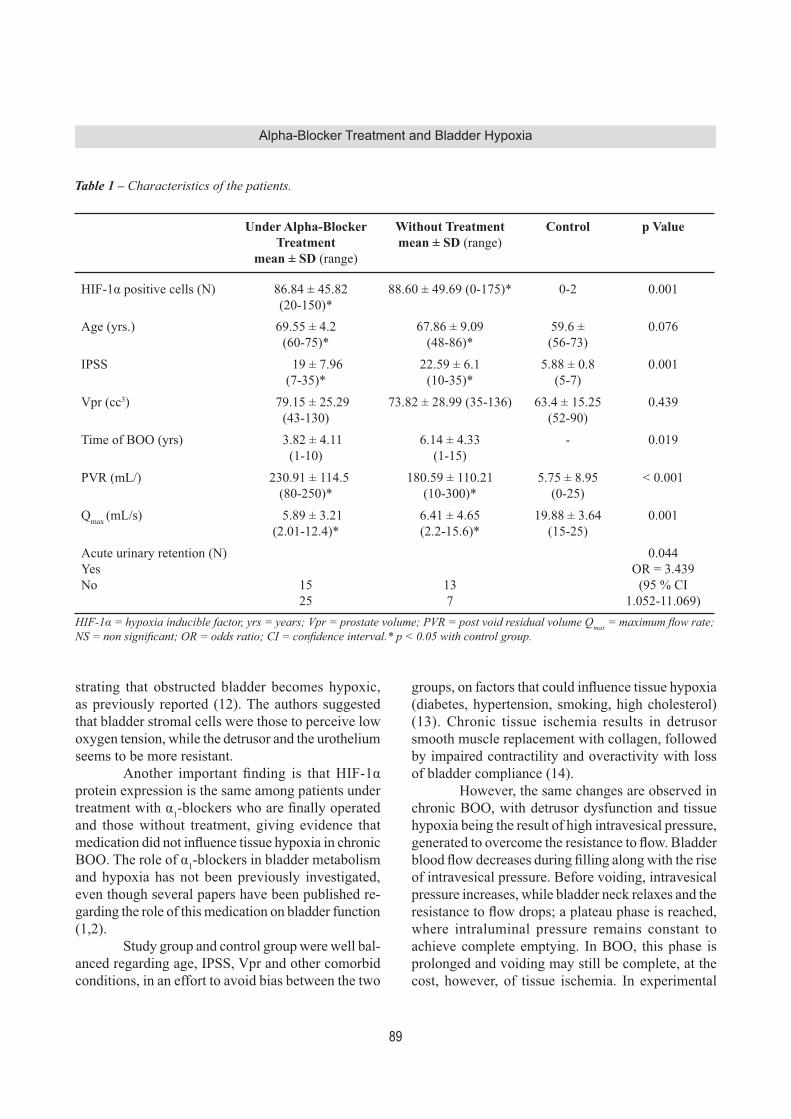
89
Alpha-Blocker Treatment and Bladder Hypoxia
strating that obstructed bladder becomes hypoxic, as previously reported (12). The authors suggested that bladder stromal cells were those to perceive low oxygen tension, while the detrusor and the urothelium seems to be more resistant. Another important finding is that HIF-1α protein expression is the same among patients under treatment with α1-blockers who are finally operated and those without treatment, giving evidence that medication did not influence tissue hypoxia in chronic BOO. The role of α1-blockers in bladder metabolism and hypoxia has not been previously investigated, even though several papers have been published re-garding the role of this medication on bladder function (1,2). Study group and control group were well bal-anced regarding age, IPSS, Vpr and other comorbid conditions, in an effort to avoid bias between the two
groups, on factors that could influence tissue hypoxia (diabetes, hypertension, smoking, high cholesterol) (13). Chronic tissue ischemia results in detrusor smooth muscle replacement with collagen, followed by impaired contractility and overactivity with loss of bladder compliance (14). However, the same changes are observed in chronic BOO, with detrusor dysfunction and tissue hypoxia being the result of high intravesical pressure, generated to overcome the resistance to flow. Bladder blood flow decreases during filling along with the rise of intravesical pressure. Before voiding, intravesical pressure increases, while bladder neck relaxes and the resistance to flow drops; a plateau phase is reached, where intraluminal pressure remains constant to achieve complete emptying. In BOO, this phase is prolonged and voiding may still be complete, at the cost, however, of tissue ischemia. In experimental
Table 1 – Characteristics of the patients.
Under Alpha-Blocker Treatment
mean ± SD (range)
Without Treatmentmean ± SD (range)
Control p Value
HIF-1α positive cells (N) 86.84 ± 45.82(20-150)*
88.60 ± 49.69 (0-175)* 0-2 0.001
Age (yrs.) 69.55 ± 4.2(60-75)*
67.86 ± 9.09(48-86)*
59.6 ± (56-73)
0.076
IPSS 19 ± 7.96(7-35)*
22.59 ± 6.1(10-35)*
5.88 ± 0.8(5-7)
0.001
Vpr (cc3) 79.15 ± 25.29(43-130)
73.82 ± 28.99 (35-136) 63.4 ± 15.25(52-90)
0.439
Time of BOO (yrs) 3.82 ± 4.11(1-10)
6.14 ± 4.33(1-15)
- 0.019
PVR (mL/) 230.91 ± 114.5(80-250)*
180.59 ± 110.21(10-300)*
5.75 ± 8.95(0-25)
< 0.001
Qmax (mL/s) 5.89 ± 3.21(2.01-12.4)*
6.41 ± 4.65(2.2-15.6)*
19.88 ± 3.64(15-25)
0.001
Acute urinary retention (N)YesNo 15
25137
0.044OR = 3.439
(95 % CI 1.052-11.069)
HIF-1α = hypoxia inducible factor, yrs = years; Vpr = prostate volume; PVR = post void residual volume Qmax = maximum flow rate; NS = non significant; OR = odds ratio; CI = confidence interval.* p < 0.05 with control group.

90
Alpha-Blocker Treatment and Bladder Hypoxia
models with obstructed animals, during the voiding phase, bladder wall blood flow decreases to the same extent as in the non-obstructed animals. Nevertheless, since emptying is sustained, significant detrusor hy-
poxia occurs and detrusor pressure becomes the main determinant of blood flow (15). Pinggera and colleagues measured blood per-fusion in arteries in the bladder neck and the prostate
Figure 1 – Staining for HIF-1α (brown color) observed in stromal cells of the bladder wall. A) Negative staining in controls, B) Strong positive staining in obstructed patients receiving alpha-blocker, C) Obstructed patients without treatment exhibiting similar staining to B, and D) Prominent cytoplasmic and nuclear staining in a patient with bladder outlet obstruction (A, B, and C X100, D X400).
A B
C D
Table 2 – Urodynamic parameters according to HIF-1α immunoreactivity.
Low intensitymedian ± SD (range)
high intensitymedian ± SD (range)
p Value
PdetQmax (cm H2O) 100. 50 ± 32.23 (71-154) 101.14 ± 29.19 (60-130) 0.85Pdetmax (cm H2O) 116.00 ± 40.17 (82-166) 127.57 ± 54.9 (63-220) 0.72PdeCL (cm H2O) 64.0 ± 25.26 (20-81) 81.83 ± 32.9(17-186) 0.76Qmax (mL/sec) 5.97 ± 3.50 (1.7-10.2) 5.54 ± 4.70 (2.0-15.6) 0.56
PdetQmax = Detrusor pressure at maximum flow rate; Pdetmax = Maximum detrusor pressure; PCL = Detrusor closure pressure; Qmax = Maximun flow rate; intensity = cut off point of 80 reactive cells/slide which represents the lowest 95% CI value.

91
Alpha-Blocker Treatment and Bladder Hypoxia
at different filing volumes using Doppler ultrasound, while they assessed the influence of α1-blockers in the perfusion of the same arteries during filling. They concluded that perfusion is reduced in LUTS patients and increases to almost normal levels after α1-blocker treatment, suggesting that treatment improved perfu-sion in the lower urinary tract (16). However, in the aforementioned work, blood flow at the dome and the posterior wall was not investigated, while perfusion was not measured during bladder emptying. The ves-sels that authors measured for perfusion were in the bladder neck and prostate and not those that travel in bladder wall (17), the vessels in the trigone are arranged in a looser network with more uniform and larger diameter (18). Therefore, there is no definite conclusion that treatment with a-blockers improved perfusion in the hall bladder. In human bladders without obstruction, there is a 2-fold increase in bladder blood flow as-sociated with filling, compared to the empty state, as measured with laser Doppler in the posterior wall (17). It seems that the posterior wall and the dome of the bladder are more vulnerable to bladder pressure (17,18). The investigators also found that bladder blood flow reaches peak at detrusor pressure of 23 cm H2O. On the other hand, blood flow is decreased to approximately 1.2 times the base line flow, in detrusor pressure of 43 cm H2O, which is measured at maximum cystometric capacity. However, despite the high pressure, it remains higher compared to the empty state. Additionally, those with decreased blad-der compliance had lower perfusion at empty bladder, a smaller increase of blood flow with filling, and a lower perfusion at maximum cystometric capacity. Nevertheless, in this work it could not be addressed whether the causative factor for the low compliance was ischemia or the exact opposite (17). As the au-thors state, bladder perfusion still remains normal even at intraluminal pressure of 43 cm H2O which corresponds to filling at the maximum cystometric capacity. In obstructed patients, however complete voiding requires much higher pressures. Recently it has been described that HIF-1α expression was higher in patients with retention. It is known that urodynamics performed shortly after an episode of retention shows a decompensated bladder with intravesical pressures not exceeding 20-30 cm
H2O. After a period however of 11 to 50 days detru-sor regains its function and can generate intravesical pressures at maximum flow even of 81.6 (35.2) cm H2O (17). It is common practice to wait at least sev-eral weeks after an acute episode of urinary retention before performing surgical treatment for BOO, since prostatectomy immediately after such episode is as-sociated with higher morbidity and mortality (19). Acute detrusor decompensation on the other hand and post void residual is a consequence of depletion of energetic reserves and HIF-1α is known to participate in the cellular metabolism by inducing enzymes in the glycolitic pathway (12). These results may explain why HIF-1α was found up-regulated in patients with retention as in our cohort of patients. In the present study, tissue specimens were obtained from the dome of the bladder, since during filling the dome has the lowest perfusion. There-fore, we hypothesized that hypoxia would be more pronounced at this location (20). We also used uro-dynamics to identify the lowest bladder pressure at which HIF-1α is up-regulated, although this was not possible. In our patients, the lowest detrusor pressure to detect HIF-1α was at 60 cm H2O and corresponded to a high protein expression. Furthermore, there was no difference among urodynamic parameters between high and low reactivity patients. The expression of HIF-1α gives evidence that above the normal detru-sor pressure for voiding (roughly 20-30 cm H2O) the bladder becomes hypoxic. The urodynamic effect of α1-blockers has been extensively explored in earlier studies. It has been found that treatment with α1-blockers increased average maximum flow rate to 2.9 mL/s (range 0.9-5.6 mL/s) and the PdetQmax decreased to average 17.4 cm H2O (38.2-0.0 cm H2O) from a base line of 85.1 cm H2O (range 70-100.4 cm H2O) to 67.1 cm H2O (range 39.7-93 cm H2O), but this interval is always above normal detrusor pressure, for non obstructed voiding (17-21). Patients in these studies however, experi-enced marked symptomatic improvement as assessed by IPSS, although still remaining obstructed. This may explain why these medications do not influence disease progression as defined by the need for invasive treatment and acute urinary retention (2). Recently, Barendrecht and associates questioned the fact that α1-blockers improve LUTS by reducing BOO. In this

92
Alpha-Blocker Treatment and Bladder Hypoxia
work, BOO index improved only slightly compared to IPSS and maximum flow, so the investigators stated that these medications function differently than relax-ing prostate smooth muscle (22). Our study is not without drawbacks. Firstly, we used immunohistochemistry to characterize blad-ders as hypoxic rather than quantify the levels of HIF-1α. Immunohistochemical evaluation of tissue protein expression is susceptible to intraobserver and interobserver variability, however, quantification of the HIF-1α is not necessary for the evaluation of hy-poxia, since we used the categorical nature of HIF-1α protein expression in order to minimize, as possible, the aforementioned limitations. Additionally, we used HIF-1α as a marker of tissue hypoxia, despite the fact that it can also be induced in non ischemic tissues. HIF-1α accumulates in myocytes as an early response to mechanical stress in hearts with diastolic pressure increase, followed by enhanced expression of VEGF genes. Similarly, in obstructed bladders, HIF-1α may also be induced by mechanical stress. Even if this is the case in BOO, treatment with α-blockers does not decrease bladder wall tension to preobstructed levels giving a further stimulus to HIF-1α induction. It would be interesting to verify if HIF-1α levels normalize after the obstruction is relieved. We are also aware that urinary retention may affect HIF-1α expression to those not receiving α1-blocker treatment (12).
CONCLUSIONS
Treatment with α-blockers in obstructed pa-tients does not seem to result in HIF-1α down-regu-lation. It is likely that once obstruction has settled, medication does not change bladder metabolism, which continues to be under chronic stress. Further studies are needed to confirm the present results, and clarify the role of α1-antagonists on bladder homeo-stasis in chronic BOO.
ACKNOWLEDGMENTS
We would like to thank Douka Despina for her support to the study and Kemerli Maria for her assistance with the immunohistochemical analysis.
CONFLICT OF INTEREST
None declared.
REFERENCES
1. Lee E, Lee C: Clinical comparison of selective and non-selective alpha 1A-adrenoreceptor antagonists in benign prostatic hyperplasia: studies on tamsulosin in a fixed dose and terazosin in increasing doses. Br J Urol. 1997; 80: 606-11.
2. McConnell JD, Roehrborn CG, Bautista OM, Andriole GL Jr, Dixon CM, Kusek JW, et al.: The long-term effect of doxazosin, finasteride, and combination therapy on the clinical progression of benign prostatic hyperplasia. N Engl J Med. 2003; 349: 2387-98.
3. Greenland JE, Brading AF: Urinary bladder blood flow changes during the micturition cycle in a conscious pig model. J Urol. 1996; 156: 1858-61.
4. Brading AF: Alterations in the physiological properties of urinary bladder smooth muscle caused by bladder emptying against an obstruction. Scand J Urol Nephrol Suppl. 1997; 184: 51-8.
5. Salceda S, Caro J: Hypoxia-inducible factor 1alpha (HIF-1alpha) protein is rapidly degraded by the ubiq-uitin-proteasome system under normoxic conditions. Its stabilization by hypoxia depends on redox-induced changes. J Biol Chem. 1997; 272: 22642-7.
6. Lee JW, Bae SH, Jeong JW, Kim SH, Kim KW: Hypoxia-inducible factor (HIF-1)alpha: its protein stability and biological functions. Exp Mol Med. 2004; 36: 1-12.
7. Hoppeler H, Vogt M: Muscle tissue adaptations to hypoxia. J Exp Biol. 2001; 204: 3133-9.
8. Kietzmann T, Görlach A: Reactive oxygen species in the control of hypoxia-inducible factor-mediated gene expression. Semin Cell Dev Biol. 2005; 16: 474-86.
9. Chandel NS, Maltepe E, Goldwasser E, Mathieu CE, Simon MC, Schumacker PT: Mitochondrial reactive oxygen species trigger hypoxia-induced transcription. Proc Natl Acad Sci U S A. 1998; 95: 11715-20.
10. Duranteau J, Chandel NS, Kulisz A, Shao Z, Schumacker PT: Intracellular signaling by reactive oxygen species during hypoxia in cardiomyocytes. J Biol Chem. 1998; 273: 11619-24.
11. Birner P, Schindl M, Obermair A, Plank C, Breite-necker G, Oberhuber G: Overexpression of hypoxia-inducible factor 1alpha is a marker for an unfavorable prognosis in early-stage invasive cervical cancer. Cancer Res. 2000; 60: 4693-6.

93
Alpha-Blocker Treatment and Bladder Hypoxia
12. Koritsiadis G, Stravodimos K, Koutalellis G, Agrogi-annis G, Koritsiadis S, Lazaris A, et al.: Immunohisto-chemical estimation of hypoxia in human obstructed bladder and correlation with clinical variables. BJU Int. 2008; 102: 328-32.
13. Ponholzer A, Temml C, Wehrberger C, Marszalek M, Madersbacher S: The association between vascular risk factors and lower urinary tract symptoms in both sexes. Eur Urol. 2006; 50: 581-6.
14. McVary KT: Erectile dysfunction and lower urinary tract symptoms secondary to BPH. Eur Urol. 2005; 47: 838-45.
15. Greenland JE, Brading AF: The effect of bladder outflow obstruction on detrusor blood flow changes during the voiding cycle in conscious pigs. J Urol. 2001; 165: 245-8.
16. Pinggera GM, Mitterberger M, Pallwein L, Schuster A, Herwig R, Frauscher F, et al.: alpha-Blockers im-prove chronic ischaemia of the lower urinary tract in patients with lower urinary tract symptoms. BJU Int. 2008; 101: 319-24.
17. Kershen RT, Azadzoi KM, Siroky MB: Blood flow, pressure and compliance in the male human bladder. J Urol. 2002; 168: 121-5.
18. Miodonski AJ, Litwin JA: Microvascular architecture of the human urinary bladder wall: a corrosion casting study. Anat Rec. 1999; 254: 375-81.
19. Dubey D, Kumar A, Kapoor R, Srivastava A, Mand-hani A: Acute urinary retention: defining the need and timing for pressure-flow studies. BJU Int. 2001; 88: 178-82.
20. Batista JE, Wagner JR, Azadzoi KM, Krane RJ, Siroky MB: Direct measurement of blood flow in the human bladder. J Urol. 1996; 155: 630-3.
21. Kortmann BB, Floratos DL, Kiemeney LA, Wijkstra H, de la Rosette JJ: Urodynamic effects of alpha-adre-noceptor blockers: a review of clinical trials. Urology. 2003; 62: 1-9.
22. Barendrecht MM, Abrams P, Schumacher H, de la Rosette JJ, Michel MC: Do alpha1-adrenoceptor antagonists improve lower urinary tract symptoms by reducing bladder outlet resistance? Neurourol Urodyn. 2008; 27: 226-30.
Accepted after revision: August 11, 2009
Correspondence address:Dr. Stavros TyritzisLAIKO Hospital, Department of UrologyAthens University Medical School17 Agiou Thoma str.11527, Athens, GreeceFax: + 0030 210 9327-744E-mail: [email protected]
EDITORIAL COMMENT
Benign prostatic obstruction will induce bladder hypoxia, which could be detected by eleva-tion of bladder tissue HIF-1-α . The authors studies the effects of α-blocker treatment on bladder HIF-1-α regulation during lower urinary tract obstruction and concluded that α blocker has no effect on bladder
tissue HIF-1-α expression. It is an interesting study, but some bias from the current groups need to be cleared. As all of the biopsy patients are alpha-blocker non-responder, I would contend that the responders may show lower HIF-1 α than the non-responder.

94
Alpha-Blocker Treatment and Bladder Hypoxia
By means of laser Doppler flowmeter, Lin et al. demonstrated that the obstructed bladders from rabbits with partial outlet obstruction had signifi-cantly lower blood flow than the ischemic bladders (1). After 8 weeks partial outlet obstruction, bladder from rabbits with supplement of L-arginine showed significantly greater contractile function compared with the no-treatment group (2). The study demon-strated that increasing blood flow by stimulating nitric oxide synthase significantly protected the bladder from partial bladder outlet obstruction dysfunctions. Therefore, the patients with lower urinary tract ob-struction might still be able to get partial recovery of bladder flow, reverse of ischemia change, and improve bladder function through some therapy.
REFERENCES
1. Lin WY, Mannikarottu A, Li S, Juan YS, Schuler C, Javed Z, et al.: Correlation of in vivo bladder blood flow measurements with tissue hypoxia. World J Urol. 2009; 4. [Epub ahead of print]
2. Lin WY, Levin RM, Chichester P, Leggett R, Juan YS, Johnson A, et al.: Effects of L-arginine and L-NAME on chronic partial bladder outlet obstruction in rabbit. Am J Physiol Regul Integr Comp Physiol. 2007; 293: R2390-9.
Dr. Yao-Chi ChuangDivision of Urology
Chang Gung Memorial Hospital Kaohsiung Medical Center
Kaohsiung, TaiwanE-mail: [email protected]
EDITORIAL COMMENT
This well written paper offers new insight novel mechanism of action α-blocker treatment my have on the obstructed bladder. This study lends further evidence that α-blocker’s benefit in cases of bladder outlet obstruction (BOO) may be more on the bladder than urethral outlet. George and associates from Athens assessed whether α1-blocker treatment, in chronic BOO, influ-ences bladder tissue ischemia. They studied 60 pa-tients with BOO, 40 with and 20 without α1-blockers, respectively. At time of transurethral or suprapubic prostatectomy, bladder biopsies were taken for im-munohistochemically stained for Hypoxia Inducible Factor-1α (HIF-1α). Ten patients with non muscle invasive bladder cancer underwent transurethral re-section of the tumor served as control group.
This study found that HIF-1α expression was higher in patients with BOO than controls, demon-strating that obstructed bladder becomes hypoxic. Another important finding is that HIF-1α protein expression is the same among patients under treat-ment with α1-blockers who are finally operated and those without treatment. It is likely that once obstruc-tion has settled, medication does not change bladder metabolism, which continues to be under chronic stress. Further studies are needed to clarify the role of α1-antagonists on bladder homeostasis in chronic obstruction.
Dr. Michael B. ChancellorNeurourology Program
William Beaumont HospitalRoyal Oak, MI, 48073, USA
E-mail: [email protected]

95
Urological SurveyVol. 36 (1): 95-119, January - February, 2010International Braz J Urol
UROLOGICAL SURVEY
Francisco J.B. SampaioUrogenital Research Unit
State University of Rio de Janeiro
Athanase BillisState University of Campinas
Campinas, SP, Brazil
Andreas BöhleHelios Agnes Karll Hospital
Bad Schwartau, Germany
Steven B. BrandesWashington University in St. Louis
St. Louis, Missouri, USA
Sean P. ElliottUniversity of MinnesotaMinneapolis, MN, USA
Fernando J. KimUniv Colorado Health Sci Ctr
Denver, Colorado, USA
Manoj MongaUniversity of Minnesota
Edina, MN, USA
Steven P. PetrouMayo Medical School
Jacksonville, Florida, USA
Adilson PrandoVera Cruz Hospital
Campinas, SP, Brazil
Brent W. SnowUniversity of Utah
Salt Lake City, Utah, USA

96
Urological Survey
STONE DISEASE _______________________________________________________________
doi: 10.1590/S1677-553820100001000014
Renal functional effects of multiple-tract percutaneous accessHanda RK, Evan AP, Willis LR, Johnson CD, Connors BA, Gao S, Lingeman JE, Matlaga BR, Miller NL, Handa SEDepartment of Anatomy and Cell Biology, Indiana University School of Medicine, Indianapolis, Indiana, USAJ Endourol. 2009; 23: 1951-6
Introduction: Percutaneous nephrolithotomy (PCNL) can involve establishing more than one access into the urinary collecting system. The present study examined whether multiple percutaneous accesses results in a more severe reduction in renal function than that after single-percutaneous access. Methods: Adult female pigs were anesthetized, and percutaneous access to the left urinary collecting system was achieved by puncturing the lower pole calyx (single-tract access, n = 16) or serially puncturing the lower pole, interpolar region, and upper pole calyces [multiple (three)-tract access, n = 11]. Renal function measure-ments included glomerular filtration rate and effective renal plasma flow, and were taken immediately before and 1.5 and 4.5 hours after percutaneous access. We also examined glomerular function in a group of adult patients with normal preoperative serum creatinine (Cr) levels (<or=1.4 mg/dL) who underwent either unilat-eral single-tract PCNL (23 patients) or unilateral multiple (two)-tract PCNL (10 patients). Access tracts were dilated to 30F with a NephroMax balloon dilator system in animal and human patients. Results: Single- and multiple-tract percutaneous access procedures in pigs resulted in a similar renal func-tional response; both glomerular filtration rate and effective renal plasma flow significantly declined by ap-proximately 60% immediately after access and remained depressed throughout the experimental observation period. A retrospective analysis of patients with normal serum Crs (<or=1.4 mg/dL) who underwent single- or multiple-tract PCNL demonstrated that the procedures produced similar and significant increases in serum Cr on postoperative day 1 (0.33 +/- 0.09 [standard error of mean] mg/dL and 0.39 +/- 0.11 mg/dL, respectively) and day 2 (0.33 +/- 0.09 mg/dL and 0.25 +/- 0.09 mg/dL, respectively). Conclusions: Multiple-tract access does not lead to a more severe reduction in renal function than single-tract access; that is, the acute renal hemodynamic response to PCNL appears independent of the number of access tracts.
Editorial Comment The human study is limited as there were significant differences in baseline renal function between the two groups analyzes. Creatinine clearance calculations based on spot serum levels is a relatively crude measure of renal function, and could also be impacted by anemia, hydration and medications in the perioperative period. In the porcine study, though changes in ipsilateral kidney function were marked (>60% decrease GFR and RPF), no difference was noted whether one or three tracts were created. In contrast, multiple tract access appeared to have a greater impact on the contralateral untreated kidney, with greater decreases in GFR and RPF (>45% vs. <20%). Though this did not reach statistical significance, it does warrant concern - suggesting that greater caution is warranted at least in the perioperative period if multiple-tract access is utilized with regards to using medications that rely on renal clearance or have the potential for nephrotoxicity. Long-term prospec-tive studies evaluating the relative impact of multiple vs. single tract access with more liberal use of flexible nephroscopy and/or ureteroscopy as an adjunct are warranted.
Dr. Manoj MongaProfessor, Department of Urology
University of MinnesotaEdina, Minnesota, USA
E-mail: [email protected]

97
Urological Survey
doi: 10.1590/S1677-553820100001000015
Residual fragments after percutaneous nephrolithotomy: cost comparison of immediate second look flexible nephroscopy versus expectant managementRaman JD, Bagrodia A, Bensalah K, Pearle MS, Lotan YDepartment of Urology, University of Texas Southwestern Medical Center, Dallas, Texas, USAJ Urol. 2010; 183: 188-93
Purpose: We performed a cost comparison of immediate second look flexible nephroscopy vs expectant man-agement for post-percutaneous nephrostolithotomy residual fragments.Materials and Methods: We used a decision analysis model to compare the cost of managing residual fragments by second look flexible nephroscopy vs observation. Outcomes of residual fragments after percutaneous neph-rostolithotomy were determined from institutional experience and published shock wave lithotripsy series. Cost data were obtained from billing records. One-way sensitivity analysis was done to evaluate incurred costs of second look flexible nephroscopy while varying the likelihood of a stone event, the probability of surgery and the cost of surgical intervention. Two-way sensitivity analysis was done to assess the model across a range of scenarios. Results: Based on data in the literature and our institutional experience 40% of patients with residual frag-ments 4 mm or less had a stone event, of whom 57% required surgical intervention. Based on these estimates the average cost of expectant management for a residual fragment 4 mm or less vs greater than 4 was $1,743 vs $4,674. The average incremental cost of second look flexible nephroscopy at our institution was $2,475. Two-way sensitivity analysis showed that varying assumptions dramatically altered conclusions about the cost benefit of second look flexible nephroscopy. Conclusions: Our model suggests that second look flexible nephroscopy is not cost advantageous in all patients with post-percutaneous nephrostolithotomy residual fragments. Cost benefit analysis is significantly impacted by the likelihood of a stone related event, the need for surgical intervention and surgical costs. Compared to an observational strategy second look flexible nephroscopy incurs lower costs for greater than 4 mm but not for 4 mm or less residual fragments.
Editorial Comment The authors have conducted a critical appraisal of their protocol for CT scan imaging on postoperative day 1 followed by second look nephroscopy. The current study confirms that second-look flexible nephroscopy (SLFN) is not warranted for residual stone fragments smaller than 2 mm, but is a good approach for fragments larger than 4mm. A “grey zone” exists for stones 3 and 4mm in size; though cost of observation vs. SLFN is equivalent in this group, 75% of those observed will experience a stone related event and as such, these patients may benefit clinically from a SLFN. The authors were limited in their ability to build a decision model based on data from patients undergoing PCNL due to the scarcity of studies reporting long-term outcomes with residual fragments in this setting. The use of SWL literature to build the decision model may be limited by the differ-ence in initial stone burden between the two patient groups. Patients with a smaller stone burden (SWL) may be more likely to undergo less invasive secondary procedures for residual fragments - indeed the distribution of secondary procedures used in the metanalysis strongly favored SWL (77%); one would anticipate a higher use of endoscopic procedures post-PCNL. The decision to select SLFN vs. URS or SWL is often determined by the quality of the initial percutaneous renal access and the location of the RF in relation to the access. With the stone size criteria established, one must now re-evaluate the need for postoperative CT scan imaging - indeed

98
Urological Survey
intraoperative fluoroscopy with magnification in conjunction with endoscopic evaluation may be sufficient to identify those fragments that really matter.
Dr. Manoj MongaProfessor, Department of Urology
University of MinnesotaEdina, Minnesota, USA
E-mail: [email protected]
ENDOUROLOGY & LAPAROSCOPY ______________________________________________
doi: 10.1590/S1677-553820100001000016
7-year oncological outcomes after laparoscopic and open partial nephrectomyLane BR, Gill ISGlickman Urological & Kidney Institute, Cleveland Clinic, Cleveland, Ohio, USAJ Urol. 2010; 183: 473-9
Purpose: Open partial nephrectomy has proven long-term oncological efficacy. Long-term outcomes of lapa-roscopic partial nephrectomy are pending. We present the long-term outcomes of patients undergoing laparo-scopic or open partial nephrectomy for a single cT1 renal cortical tumor 7 cm or less. Materials and Methods: Of 2,246 patients undergoing partial nephrectomy for a single cT1 tumor (1999 to 2008), minimum 7-year followup was available in 77 and 310, and minimum 1-year followup was available in 672 and 944 after laparoscopic and open partial nephrectomy, respectively. Survival and recurrence data obtained from medical records, radiographic reports and patient contact were analyzed retrospectively. Results: Median followup after laparoscopic and open partial nephrectomy was 4.0 and 5.7 years, respec-tively. Oncological outcomes were excellent in both groups. On multivariable analysis predictors of all cause mortality included advancing age (p <0.0001), comorbidity (p <0.0001) and preoperative renal dysfunction (p = 0.0001) but not tumor size (p = 0.6) or operative approach (laparoscopic vs open partial nephrectomy, p = 0.06). Cancer recurred infrequently and only rarely caused mortality after laparoscopic or open partial nephrec-tomy. At 7 years metastasis-free survival was 97.5% and 97.3% (p = 0.47) after laparoscopic and open partial nephrectomy, respectively. After accounting for baseline differences between the cohorts using propensity score matching 7-year metastasis-free survival was similar after laparoscopic and open partial nephrectomy. Conclusions: Laparoscopic and open partial nephrectomy appear to provide similar long-term overall and cancer specific survival in patients undergoing partial nephrectomy for clinical stage T1 (7 cm or less) renal cortical tumors. Oncological outcomes at 7 years after laparoscopic and open partial nephrectomy are excellent with the majority (97%) of patients experiencing metastasis-free survival.
Editorial Comment The authors have demonstrated that laparoscopic and open partial nephrectomy appear to provide sim-ilar long-term overall and cancer specific survival in patients undergoing partial nephrectomy for clinical stage T1. This manuscript describes a well known fact that the laparoscopic surgical technique has not compromised
99
Urological Survey
oncological outcomes particularly in renal cancer and renal surgery. Moreover, the complication rates with the laparoscopic technique have been demonstrated to be comparable to the open technique.
Dr. Fernando J. KimChief of Urology, Denver Health Med. Ctr.
Associate Professor, Univ. Colorado Health Sci. Ctr.Director of Minimally Invasive Urol. Oncology, UCHSC
Denver, Colorado, USAE-mail: [email protected]
doi: 10.1590/S1677-553820100001000017
Cost analysis of robotic versus open radical cystectomy for bladder cancerSmith A, Kurpad R, Lal A, Nielsen M, Wallen EM, Pruthi RSDivision of Urologic Surgery, The University of North Carolina at Chapel Hill, Chapel Hill, North Carolina, USAJ Urol. 2010; 183: 505-9
Purpose: Recently robotic approaches to cystectomy have been reported, and while clinical and oncological efficacy continues to be evaluated, potential financial costs have not been clearly evaluated. In this study we present a financial analysis using current cost structures and clinical outcomes for robotic and open cystectomy for bladder cancer. Materials and Methods: The financial costs of robotic and open radical cystectomy were categorized into oper-ating room and hospital components, and further divided into fixed and variable costs for each. Fixed operating room costs for open cases involved base cost as well as disposable equipment costs while robotic fixed costs included the amortized machine cost as well as equipment and maintenance. Variable operating room costs were directly related to length of surgery. Variable hospital costs were directly related to transfusion require-ment and length of stay. The means of the prior 20 cases of robotic and open cystectomy were used to perform a comparative cost analysis. Results: Mean fixed operating room costs for robotic cases were $1,634 higher than for open cases. Operat-ing room variable costs were also higher by a difference of $570, directly related to increased operating room time. Hospital costs were nearly identical for the fixed component while variable costs were $564 higher for the open approach secondary to higher transfusion costs and longer mean length of stay. Based on these find-ings robotic cystectomy is associated with an overall higher financial cost of $1,640 (robotic $16,248 vs open $14,608). Cost calculators were constructed based on these fixed and variable costs for each surgical approach to demonstrate the expected total costs based on varying operating room time and length of stay. Conclusions: Robotic assisted laparoscopic radical cystectomy is associated with a higher financial cost (+$1,640) than the open approach in the perioperative setting. However, this analysis is limited by its single institution design and a multicenter followup study is required to provide a more comprehensive analysis.
Editorial Comment Independently of the techniques used for the surgical treatment of bladder cancer, the oncological prin-ciples must be followed and outcomes ought to be equal or exceed the tumor control and improve the recovery time.

100
Urological Survey
This article demonstrates that robotic radical cystectomy has similar short-term cancer control and complication rates, less operative time and a shorter hospital stay than laparoscopic or open radical cystec-tomy. The authors recently performed completely intracorporeal robotic cystectomy and diversion setting the bar for minimally invasive radical cystectomy and urinary diversion very high. It will be important for other centers to duplicate these results and take into account the price of acquiring the robot and servicing it plus the cost of disposables utilized during robotic surgery.
Dr. Fernando J. KimChief of Urology, Denver Health Med. Ctr.
Associate Professor, Univ. Colorado Health Sci. Ctr.Director of Minimally Invasive Urol. Oncology, UCHSC
Denver, Colorado, USAE-mail: [email protected]
IMAGING ______________________________________________________________________
doi: 10.1590/S1677-553820100001000018
Clinical stage T1c prostate cancer: evaluation with endorectal MR imaging and MR spectroscopic imagingZhang J, Hricak H, Shukla-Dave A, Akin O, Ishill NM, Carlino LJ, Reuter VE, Eastham JADepartment of Radiology, Memorial Sloan-Kettering Cancer Center, New York, NY, USARadiology. 2009; 253: 425-34
Purpose: To assess the diagnostic accuracy of endorectal magnetic resonance (MR) imaging and MR spectro-scopic imaging for prediction of the pathologic stage of prostate cancer and the presence of clinically nonim-portant disease in patients with clinical stage T1c prostate cancer.Materials and Methods: The institutional review board approved-and waived the informed patient consent requirement for-this HIPAA-compliant study involving 158 patients (median age, 58 years; age range, 40-76 years) who had clinical stage T1c prostate cancer, had not been treated preoperatively, and underwent com-bined 1.5-T endorectal MR imaging-MR spectroscopic imaging between January 2003 and March 2004 before undergoing radical prostatectomy. On the MR images and combined endorectal MR-MR spectroscopic imag-es, two radiologists retrospectively and independently rated the likelihood of cancer in 12 prostate regions and the likelihoods of extracapsular extension (ECE), seminal vesicle invasion (SVI), and adjacent organ invasion by using a five-point scale, and they determined the probability of clinically nonimportant prostate cancer by using a four-point scale. Whole-mount step-section pathology maps were used for imaging-pathologic analysis correlation. Receiver operating characteristic curves were constructed and areas under the curves (AUCs) were estimated nonparametrically for assessment of reader accuracy. Results: At surgical-pathologic analysis, one (0.6%) patient had no cancer; 124 (78%) patients, organ-confined (stage pT2) disease; 29 (18%) patients, ECE (stage pT3a); two (1%) patients, SVI (stage pT3b); and two (1%) patients, bladder neck invasion (stage pT4). Forty-six (29%) patients had a total tumor volume of less than 0.5 cm(3). With combined MR imaging-MR spectroscopic imaging, the two readers achieved 80% accuracy in disease staging and AUCs of 0.62 and 0.71 for the prediction of clinically nonimportant cancer. Conclusion: Clinical stage T1c prostate cancers are heterogeneous in pathologic stage and volume. MR imag-ing may help to stratify patients with clinical stage T1c disease for appropriate clinical management.

101
Urological Survey
Editorial Comment Similar to other studies the authors showed that MR imaging findings might represent additional useful variables for predicting disease extent in patients with clinically localized prostate cancer. Combined endorec-tal MRI-MR spectroscopic imaging had 80% accuracy in the staging of disease in patients with clinical stage T1c prostate cancer. These combined techniques had a moderate accuracy, 62-72%, in the prediction of clini-cally non-important cancer in this group of patients. As the authors pointed out it would be of clinical interest in the future to investigate whether multiparametric examination which combination of conventional T2-w images, spectroscopy, diffusion-weighted image (DWI) and perfusion studies can yield superior diagnostic information for stratifying patients with T1 c prostate cancer. Since 2004, we have been using in our depart-ment this multiparametric evaluation in patients with organ-confined tumor, based on finding of conventional T2-weighted images. We have found that DWI and perfusion techniques, similarly to spectroscopy are very useful to detect tumor > 0.5 cm3 and with higher Gleason grades. All techniques have difficult to detect smaller and low grades tumor. In other words, when we find a lesion with imaging characteristics of a possible aggressive tumor on T2-w images and spectroscopy, but without concordant findings on DWI and perfusion studies, our tendency is to downgrade the lesion to a possible less important one. We have found that usually a large and aggressive tumor will present as an area with restricted diffusion (lower ADC values) and with abnormally elevated values of the pharmacokinetics parameters obtained with perfusion studies. On the other hand, patients with normal multiparametric prostate examination has a very high probability of have a clinically non-important cancer. Another important finding of this study is that from 158, 124 (78%) patients had organ-confined dis-ease (stage pT2), 29 (18%) had extracapsular extension (stage pT3a), two (1%) had seminal vesicle invasion (stage pT3b), and two (1%) had bladder neck invasion (stage pT4). We have to remember that clinically T1 c patients typically are considered to have localized early-stage disease of relatively low risk. Additionally 30 (19%) of the patients met the criteria to be considered for active surveillance as a management strategy, 4(13%) had extraprostatic extension of disease at surgical-pathologic analysis. These findings further enhance the value of endorectal MRI examination in the pre-operative evaluation of patients with T1c prostate carcinoma.
Dr. Adilson PrandoHead, Department of Radiology and
Diagnostic Imaging, Vera Cruz HospitalCampinas, São Paulo, Brazil
E-mail: [email protected]
doi: 10.1590/S1677-553820100001000019
Bladder tumor staging: comparison of contrast-enhanced and gray-scale ultrasoundCaruso G, Salvaggio G, Campisi A, Melloni D, Midiri M, Bertolotto M, Lagalla RDipartimento di Biotecnologie mediche e Medicina legale, Sezione di scienze radiologiche, Università degli Studi di Palermo, Palermo, ItalyAJR Am J Roentgenol. 2010; 194: 151-6
Objective: The purpose of this study was to evaluate the effectiveness of contrast-enhanced sonography in comparison with conventional sonography in differentiating muscle-infiltrating and superficial neoplasms of the urinary bladder.

102
Urological Survey
Subjects and Methods: Conventional and contrast-enhanced sonography were performed on 34 consecutively registered patients with bladder tumors. All examinations were reviewed by two independent sonologists. At gray-scale sonography, interruption of the hyperechoic bladder wall was considered the main diagnostic criterion for differentiating superficial and infiltrating tumors. At contrast-enhanced sonography, a tumor was considered superficial when the hypoenhancing muscle layer of the bladder wall was intact; disruption of the muscle layer by enhancing tumor tissue was considered diagnostic of infiltration. A level of confidence in the diagnosis of tumor infiltration of the muscle layer was assigned on a 5-degree scale. Receiver operating char-acteristic analysis was used to assess overall confidence in the diagnosis of muscle infiltration by tumor at both conventional and contrast-enhanced sonography. Histologic diagnosis was obtained for all patients. Results: Final pathologic staging revealed 25 superficial tumors (Ta-T1 disease) and nine muscle-infiltrating tu-mors (>T1). Conventional sonography depicted five of nine muscle-infiltrating tumors, and contrast-enhanced sonography depicted all nine. The diagnostic performance of contrast-enhanced sonography approached that of the reference standard (area under the receiver operating characteristic curve, 0.996), but the diagnostic performance of gray-scale ultrasound was worse (area under curve, 0.613). Conclusion: Our study showed that contrast-enhanced sonography is better than conventional sonography for differentiating muscle-infiltrating and superficial neoplasms of the urinary bladder.
Editorial Comment According to the American College of Radiology Appropriateness Criteria, the use of transabdominal ultrasound for pretreatment staging of invasive bladder cancer receives rating 3 (rating scale 1 = least appropri-ate and 9 = most appropriate). This poor rating is due to the inherent limitation of the abdominal transducers in the visualization of the layers of the bladder wall, which usually appeared homogeneously hyperechoic. Based on their previous observation that after microbubble administration the layers of the bladder wall were clearly differentiated with conventional ultrasound the authors decided to investigate the effectiveness of contrast-en-hanced sonography compared with conventional gray-scale sonography in differentiating muscle-infiltrating and superficial neoplasms of the urinary bladder. The diagnostic performance of contrast-enhanced sonogra-phy was much better than the gray scale ultrasound (AUC 0.996 x AUC 0.613). As already mentioned by the authors contrast-enhanced sonography has many of the limitations of other ultrasound techniques (difficulty to detect flat lesions; obesity and calcification impairs bladder wall evaluation; columnar hypertrophy of the bladder wall, calcification and tumor location may be troublesome during examination). However, one of the most important limitations of this technique is that the FDA did not approve yet its use for internal medicine examination. Another important limitation is related to the necessity of specialized contrast-specific ultrasound techniques found only in state-of-the art equipments. With contrast-enhanced ultrasound is also very difficult to obtain information on the extent of extra-vesical spread of large, widely infiltrating tumors and on the status of pelvic lymph node. For this reason, we still prefer to use magnetic resonance imaging as the main imaging modality for local staging of possible invasive bladder cancer (T staging accuracies 73% to 96% of cases and 73% to 98 % accuracy for staging of nodes and metastases).
Dr. Adilson PrandoHead, Department of Radiology and
Diagnostic Imaging, Vera Cruz HospitalCampinas, São Paulo, Brazil
E-mail: [email protected]

103
Urological Survey
UROGENITAL TRAUMA _________________________________________________________
doi: 10.1590/S1677-553820100001000020
Straddle injuries to the bulbar urethra: management and outcomes in 78 patientsPark S, McAninch JWDepartment of Urology, University of California School of Medicine and Urology Service, San Francisco General Hospital, USAJ Urol. 2004; 171(2 Pt 1): 722-5
Purpose: We describe our experience with blunt straddle injuries to the anterior urethra and identify factors that may affect patient outcome. Materials and Methods: We reviewed the San Francisco General Hospital Urologic Trauma data base to iden-tify men with blunt straddle injury. We analyzed presentation and initial management, location and length of urethral stricture, surgical options, and long-term outcome after reconstruction. Results: Of 78 patients, 40% presented to the emergency department acutely and 60% presented 6 months to 10 years after injury complaining of obstructive symptoms, of whom 30% reported at least 1 episode of urinary retention. Initial acute management was suprapubic cystostomy in 81% of cases and primary realignment in 19%. Urethral strictures were predominantly located in the proximal bulb. Mean stricture length was signifi-cantly longer in men with delayed presentation (2.7 vs 1.8 cm, p <0.05). No relationship was found between stricture length and the mechanism of injury or initial management technique. However, patients who had undergone primary realignment required complex flap or graft urethroplasty at a greater rate compared with men who had undergone suprapubic diversion (p = 0.054). Transperineal urethroplasty was required in 92% of patients with the majority undergoing end-to-end anastomosis. The success rate was 95% at a mean followup of 25 months (range 10 to 180). Recurrent stricture occurred in 4 men with prior urethral manipulation and it was managed successfully by direct vision internal urethrotomy alone. Conclusions: After blunt straddle injury to the perineum the primary morbidity is anterior urethral stricture, for which suprapubic cystostomy is appropriate initial management. The majority of patients require surgery but with careful preoperative planning and adequate resection of fibrotic tissue the long-term success rate can approach 95%. If it arises, recurrent stricture responds well to direct vision internal urethrotomy alone.
doi: 10.1590/S1677-553820100001000021
Management of low velocity gunshot wounds to the anterior urethra: the role of primary repair versus urinary diversion aloneHusmann DA, Boone TB, Wilson WTDepartment of Surgery, University of Texas Southwestern Medical Center, DallasJ Urol. 1993; 150: 70-2
The management of partial transection of the anterior urethra following penetrating penile injuries is controver-sial. Optional therapeutic techniques range from a primary sutured reapproximation to urinary diversion alone. We recently managed 17 low velocity gunshot wounds to the external genitalia in which the missile traversed the penile corpus cavernosum, and was associated with less than 40% transection of the corpus spongiosum and anterior urethra. Nine patients were managed with suprapubic diversion, skin débridement and corporeal

104
Urological Survey
closure along with placement of a urethral catheter. Eight patients were managed by suprapubic diversion, débridement, closure of the corporeal bodies and a primary sutured reapproximation of the anterior urethra. Urethral strictures developed in 7 patients (78%) managed by a suprapubic tube and urethral stenting during an average followup of 20 months (range 18 to 24). In contrast, 1 patient (12%) managed by a sutured urethral approximation had a urethral stricture during an average followup of 20 months (range 18 to 30, p < 0.01). Our data support a significantly better prognosis for partial transection of the anterior urethra secondary to low velocity gunshot wounds if managed by aggressive wound débridement, corporeal repair, placement of a suprapubic catheter and primary repair of the urethra.
doi: 10.1590/S1677-553820100001000022
Straddle injuries to the bulbar urethra: management and outcome in 53 patientsElgammal MADepartment of Urology, Assiut University Hospital, Assiut, EgyptInt Braz J Urol. 2009; 35: 450-8
Objective: To describe our experience with blunt injuries to the bulbar urethra and their late sequelae to iden-tify factors that may affect patient outcome. Materials And Methods: A retrospective study was performed on 53 male patients who presented, between January 2001 and December 2005, with blunt traumatic injury to the bulbar urethra. The definitive diagnosis of urethral rupture was made by retrograde urethrography, where urethral rupture was classified into partial or complete. The minimum follow-up period was 3 years. The initial management was either suprapubic cys-tostomy or endoscopic urethral realignment over a urethral catheter using a cystoscope to pass a guide-wire over which the catheter was inserted. Stricture formation was managed by visual internal urethrotomy (VIU) for passable strictures and urethroplasty (stricture excision and re-anastomosis) for impassable strictures or recurrence after VIU. The follow-up period was three years. The results were analyzed by SPSS software (chi-square and Student’s-t-test). Results: Stricture formation occurred in 19 of 22 patients (86%) with complete urethral rupture and in 10 of 31 (32%) with partial rupture (p < 0.001). Strictures occurred in 11 of 31 (35%) patients treated initially with suprapubic cystostomy and in 18 of 22 (82%) treated with primary urethral realignment (p < 0.001). The success rate after VIU was 15% (4 of 26 patients) and after urethroplasty it was 96% (24 of 25 patients) (p < 0.001). Conclusions: Suprapubic cystostomy is better than urethral realignment and catheterization as primary man-agement after straddle injury to the bulbar urethra. Stricture excision and re-anastomosis is better than VIU as delayed management for strictures that develop after straddle injury to the bulbar urethra.
Editorial Comment While a few of the above articles are old, they illustrate important teaching points about how urethral injury etiology dictates outcome and the best choice for management. Blunt crush injuries to the urethra typically results in a short segment of spongiofibrosis that occurs in the mid bulbar urethra. Stricture etiology, location and length typically dictate the type of repair selected and the success of the long term outcome. With a blunt injury, the stricture is typically less than 2 cm and the natural elasticity of the mobilized urethra can bridge the gap. The spongiofibrosis from a straddle injury is isolated to a short segment, while the rest of the urethra and the rest of the corpus spongiosum are normal. Inflammatory strictures typically cause a more diffuse spongiofibrosis, and thus are often best managed by an onlay skin flap or buccal mucosal graft.
105
Urological Survey
Straddle injuries are not to be confused with the stenoses that occur from pelvic fracture. With pelvic fracture, the injury is a distraction injury where there is disruption of the urethra and corpus spongiosum at the level of the membranous - bulbar junction or the membranous and the prostate. Here there is no real spongio-sum fibrosis and “urethral stricture” – but scar tissue that fills the gap. Primary realignment is the preferred management of such injuries because it a distraction injury and not a stricture. Historically, the outcomes of primary realignment are a reduction in urethral stricture by 50%, while the rates of erectile dysfunction and incontinence are the same as a suprapubic tube. Furthermore, the eventual stricture that does occur is often shorter and more amenable to urethrotomy. From the above abstracts, I think the conclusion that straddle injuries should be managed by suprapu-bic tube alone, as the best management that should be followed. Intuitively, we would assume that the Denis Browne principle would apply here and stenting would promote epithelialization. However, until a randomized prospective trial takes pace – and I doubt that any such study will be done soon – we should resist the temp-tation to primarily realign the urethra. As to urethral penetrating urethral injuries from low velocity gunshot wounds (no delayed ischemia or blast effect) the site of injury is typically short. A short area of injury can be bridged by adequate mobilization and natural elasticity of the urethra, particularly in the bulbar urethra. In the penile urethra, over mobilization and an anastomosis on tension may result in chordee or stricture failure. Primary realignment of a short penile urethral injury is not the first treatment of choice – but rather surgical exploration and primary repair. When the defect is too long (more than 1 cm or so), urethral marsupialization and a two stage repair (in the method of Johansson) is probable best.
Dr. Steven B. BrandesAssociate Professor, Division of Urologic Surgery
Washington University in St. LouisSt. Louis, Missouri, USA
E-mail: [email protected]
PATHOLOGY ___________________________________________________________________
doi: 10.1590/S1677-553820100001000023
Does perineural invasion on prostate biopsy predict adverse prostatectomy outcomes?Loeb S, Epstein JI, Humphreys EB, Walsh PCJames Buchanan Brady Urological Institute and the Department of Urology, Johns Hopkins Medical Institu-tions, Baltimore, MD, USABJU Int. 2009 Aug 19. [Epub ahead of print]
Objective: To determine the relationship between perineural invasion (PNI) on prostate biopsy and radical prostatectomy (RP) outcomes in a contemporary RP series, as there is conflicting evidence on the prognostic significance of PNI in prostate needle biopsy specimens.

106
Urological Survey
Patients and Methods: From 2002 to 2007, 1256 men had RP by one surgeon. Multivariable logistic regression and Cox proportional hazards models were used to examine the relationship of PNI with pathological tumour features and biochemical progression, respectively, after adjusting for prostate-specific antigen level, clinical stage and biopsy Gleason score. Additional Cox models were used to examine the relationship between nerve-sparing and biochemical progression among men with PNI.Results: PNI was found in 188 (15%) patients, and was significantly associated with aggressive pathology and biochemical progression. On multivariate analysis, PNI was significantly associated with extraprostatic extension and seminal vesicle invasion (P < 0.001). Biochemical progression occurred in 10.5% of patients with PNI, vs 3.5% of those without PNI (unadjusted hazard ratio 3.12, 95% confidence interval 1.77-5.52, P < 0.001). However, PNI was not a significant independent predictor of biochemical progression on multivariate analysis. Finally, nerve-sparing did not adversely affect biochemical progression even among men with PNI. Conclusion: PNI is an independent risk factor for aggressive pathology features and a non-independent risk factor for biochemical progression after RP. However, bilateral nerve-sparing surgery did not compromise the oncological outcomes for patients with PNI on biopsy.
Editorial Comment Perineural invasion (PNI) on needle prostatic biopsies as a marker of extraprostatic extension has been controversial. In almost all studies, perineural invasion has been related to extraprostatic extension in univari-ate analysis but in only a few studies in multivariate analysis. The practical importance relates to the decision of whether to sacrifice part or all of the neurovascular bundle on the side of the biopsy with PNI in planning nerve-sparing radical prostatectomy. Egan and Bostwick (1) found on univariate analysis that PNI on needle biopsy was significantly as-sociated to extraprostatic extension and seminal vesicle invasion. On multivariate analysis, however, only preoperative PSA, proportion of the biopsy involved by cancer, and Gleason score were significant. Ukimura et al. (2) found that PNI on biopsy was a good predictor among others studied for extraprostatic extension on univariate analysis but not on multivariate analysis. In the study by Vargas et al. (3) PNI was not an indepen-dent predictor of extraprostatic extension when PSA was included.D’Amico et al. (4) evaluated the clinical use of PNI at biopsy for predicting time to PSA failure following radi-cal prostatectomy of 750 men with clinically localized or PSA detected prostate cancer. The presence of PNI on biopsy was not a significant predictor of PSA outcome following RP for patients in the intermediate or high risk group. O’Malley et al. (5) compared 78 biopsies with PNI with 78 matched controls without PNI and were unable to show that PNI on needle biopsy influences long-term tumor-free survival. In the study surveyed, Loeb’s et al. found that PNI is an independent risk factor for aggressive pathol-ogy features like extraprostatic extension and seminal vesicle invasion, and a non-independent risk factor for biochemical progression after radical prostatectomy. According to the authors, the findings support the routine reporting of PNI in biopsy pathology reports. They also concluded that nerve-sparing surgery did not adversely affect biochemical progression even among men with PNI.
References1. Egan AJM, Bostwick DG: Prediction of extraprostatic extension of prostate cancer based on needle biopsy findings:
Perineural invasion lacks significance on multivariate analysis. Am J Surg Pathol. 1997; 21: 1496-500.2. Ukimura O, Troncoso P, Ramirez EI, Babaian RJ: Prostate cancer staging: correlation between ultrasound determined
tumor contact length and pathologically confirmed extraprostatic extension. J Urol. 1988; 159: 1251-9.3. Vargas SO, Jiroutek M, Welch WR, Nucci MR, D’Amico AV, Renshaw AA: Perineural invasion in prostate needle
biopsy specimens. Correlation with extraprostatic extension at resection. Am J Clin Pathol. 1999; 111: 223-8.4. D’Amico AV, Wu Y, Chen MH, Nash M, Renshaw AA, Richie JP: Perineural invasion as a predictor of biochemical
outcome following radical prostatectomy for select men with clinically localized prostate cancer. J Urol. 2001; 165: 126-9.

107
Urological Survey
5. O’Malley KJ, Pound CR, Walsh PC, Epstein JI, Partin AW: Influence of biopsy perinerual invasion on long-term biochemical disease-free survival after radical prostatectomy. Urology. 2002; 59: 85-90.
Dr. Athanase BillisFull-Professor of Pathology
State University of Campinas, UnicampCampinas, São Paulo, Brazil
E-mail: [email protected]
doi: 10.1590/S1677-553820100001000024
Transurethral resection specimens of the bladder (TURB): Outcome of invasive urothelial cancer involving muscle bundles indeterminate between muscularis mucosae and muscularis propriaH Miyamoto, JI EpsteinThe Johns Hopkins Hospital, Baltimore, United StatesMod Pathol 2010;in press [Abstract from the USCAP meeting, 2010]
Background: It may be difficult to diagnose muscularis propria on TURB as thin muscle fibers on TURB may represent either muscularis propria destroyed or splayed by urothelial carcinoma or muscularis mucosae, which may be hyperplastic.Design: 95 invasive bladder cancers seen at our instituion (1986-2008) with follow-up (mean 25.4 months) where the initial TUR pathologic stage was ambiguous (T1 vs. T2) were analyzed (73 men; 22 women; mean age 69.4 years).Results: Subsequent restaging TURB or definitive therapeutic procedures performed ≤ 3 months after the origi-nal TURB done in 58 cases revealed 22 (37.9%) patients with non-muscle invasive disease and 32 (55.2%) patients with ≥ pT2 disease. Staging in 4 cases remained ambiguous. 37 cases eventually developed ≥ pT2 disease in 2/22 (9.1%) cases with non-muscle invasive disease on initial restaging TURB, 2/4 (50.0%) of cases with uncertain stage disease, and 14/37 (37.8%) cases with no restaging TURB. Patients with a final stage of non-muscle invasive disease had a lower risk of progression (T4 or metastatic disease) vs. those with a final stage of ≥pT2 (p=0.003), uncertain stage (p=0.012), or no stage confirmation (p=0.043).Conclusions: This is the first study to evaluate follow-up when initial TURB is equivocal for muscularis propria invasion. Similar to an atypical prostate needle biopsy, urologists should be encouraged to perform restaging TURBs in cases of equivocal muscularis propria invasion. Although this may seem intuitive, 37/95 cases did not have repeat staging/therapeutic procedures done within 3 months of initial TURB; 37.8% of these patients eventually developed ≥ T2 disease.
Editorial Comment It is of utmost importance the staging of urothelial carcinomas of the urinary bladder. In stage pT2 (invasion of the muscularis propria) is indicated radical cystectomy. Sometimes the distinction between mus-cularis mucosae and muscularis propria is a dilemma for the pathologist. Invasion of the muscularis mucosa is stage pT1. Morphologically these two muscular layers are distinct. In muscularis mucosa, the fibers are thin and spaced; in muscularis propria, the fibers form compact aggregates. It is interesting to note that description of the muscularis mucosae will not be found in Histology texts. The existence and morphology of this layer was

108
Urological Survey
described in 1983 by Dixon and Gosling (1) and the importance for staging and treatment of bladder urothelial carcinoma by Ro JY et al (2) from the MD Anderson Hospital in Houston. In some cases it is difficult if not impossible for the pathologist to recognize that the invaded muscular layer is the muscularis mucosae. This happens because the fibers of this layer may be thick due to hypertrophy. In doubt, the pathologist should always ask for a restaging TUR of the bladder. Another much commoner con-dition for asking a restaging TUR is whenever the specimen does not contain muscularis propria.
References1. Dixon JS, Gosling JA: Histology and fine structure of the muscularis mucosa of the human urinary bladder. J Anat.
1983; 136: 265-71.2. Ro JY, Ayala AG, El-Naggar A: Muscularis mucosa of urinary bladder: importance for staging and treatment. Am J
Surg Pathol. 1987; 11: 668-73.
Dr. Athanase BillisFull-Professor of Pathology
State University of Campinas, UnicampCampinas, São Paulo, Brazil
E-mail: [email protected]
BASIC AND TRANSLATIONAL UROLOGY ________________________________________
doi: 10.1590/S1677-553820100001000025
Temporary segmental renal artery occlusion using reverse phase polymer for bloodless robotic partial nephrectomyMoinzadeh A, Flacke S, Libertino JA, Merhige J, Vogel JM, Lyall K, Bakal CW, Madras PNLahey Clinic, Burlington and Pluromed, Inc., Woburn, Massachusetts, USAJ Urol. 2009; 182:1582-7
Purpose: Renal vascular clamping with ensuing warm ischemia is typically needed during robotic or laparo-scopic partial nephrectomy. We developed a technique for angiographic delivery of the novel intra-arterial reverse thermoplastic polymer LeGoo-XL that allows temporary selective vascular occlusion with normal perfusion of the remaining kidney. Materials and Methods: Eight pigs underwent a total of 16 selective angiographic occlusions of the lower pole segmental artery using gel polymer. The technical feasibility of 2 hemostatic techniques, perfusion hemostasis and local plug formation, was assessed in 4 pigs each. Selective ischemia time was recorded and the vascular occlusion site was noted radiographically and laparoscopically. The feasibility of reversing the polymer from solid back to liquid state to allow reperfusion was determined. Pathological analysis of the kidney was com-pleted in these acute model pigs. In the last 2 cases lower pole robotic partial nephrectomy was done using the da Vinci surgical system. Results: Selective lower pole ischemia was achieved in all 8 cases. Perfusion hemostasis yielded an inconsis-tent duration of occlusion (zero to greater than 60 minutes). Vascular occlusion time using local plug formation was more reliable (17 to 30 minutes) with consistent ability to reverse the plug to liquid state by cold saline flush. Two lower pole robotic partial nephrectomies were completed with minimal blood loss.

109
Urological Survey
Conclusions: We developed a reliable technique of angiographic delivery of gel polymer for temporary vascu-lar occlusion of selective renal artery branches using local plug formation. Ongoing studies are under way to assess technique consistency and the long-term effects of the polymer.
Editorial Comment This is an interesting experimental study in pigs, on which the authors tested the intra-arterial injection of reverse thermoplastic polymer LeGoo-XL that allows temporary selective vascular occlusion. The polymer was used with the intend of facilitate hemostasis for laparoscopic partial nephrectomy of the lower (caudal) pole. The perfusion hemostasis was not reliable in achieving occlusion while when using a local plug formation for hemostasis the results were consistent, with occlusion time from 13 to 30 minutes. The authors performed 2 robotic partial nephrectomies and concluded that the technique allowed minimal blood loss. Nevertheless, the authors did not take into account previous studies on intra-renal anatomy in pigs. While the collecting system anatomy is very similar to that of humans (1), the arterial (2) and venous (3) intra-renal anatomy in pigs is different from that of humans in many aspects that would be interesting to be discussed. Also, there are many important differences in the upper and lower pole vascular anatomy, being the upper pole vessels much more complex in distribution. Although we cannot transpose the results to clinical setting, the study opened new avenue to enhance the possibility of partial nephrectomy.
References1. Sampaio FJ, Pereira-Sampaio MA, Favorito LA: The pig kidney as an endourologic model: anatomic contribution. J
Endourol. 1998; 12: 45-50.2. Pereira-Sampaio MA, Favorito LA, Sampaio FJ: Pig kidney: anatomical relationships between the intrarenal arteries
and the kidney collecting system. Applied study for urological research and surgical training. J Urol. 2004; 172(5 Pt 1): 2077-81.
3. Bagetti Filho HJ, Pereira-Sampaio MA, Favorito LA, Sampaio FJ: Pig kidney: anatomical relationships between the renal venous arrangement and the kidney collecting system. J Urol. 2008; 179: 1627-30.
Dr. Francisco J. B. SampaioFull-Professor and Chair, Urogenital Research Unit
State University of Rio de JaneiroRio de Janeiro, RJ, Brazil
E-mail: [email protected]
doi: 10.1590/S1677-553820100001000026
Sildenafil as a protecting drug for warm ischemic kidney transplants: experimental resultsLledó-García E, Subirá-Ríos D, Rodríguez-Martínez D, Dulín E, Alvarez-Fernández E, Hernández-Fernández C, del Cañizo-López JFUrology Department, Hospital General Universitario Gregorio Marañón, Madrid, SpainJ Urol. 2009; 182: 1222-5
Purpose: In an experimental model we studied the protective effects of the phosphodiesterase-5 inhibitor silde-nafil on kidney grafts autotransplanted after 45 minutes of warm ischemia by vascular clamping, nephrectomy and 60 minutes of isolated hypothermic pump perfusion.

110
Urological Survey
Materials and Methods: A total of 14 laboratory minipigs were divided into group 1-7 administered 100 mg sildenafil orally 1.5 hours preoperatively and group 2-7 in which no sildenafil was given. Right single nephrec-tomy was completed after 45 minutes of warm ischemia by complete vascular clamping. Before autotrans-plantation all kidneys underwent 60 minutes of hypothermic pulsatile perfusion. Renal flow, arterial pressure and renal vascular resistance were recorded in real time for 60 minutes after autotransplantation. Nitric oxide levels were determined in blood samples from the renal vein at predefined intervals. Optical and electronic microscopy was done in all organs at the end of the procedure. Results: In group 1 vs 2 renal vascular flow was significantly higher (155.30 vs 29.04 ml per minute per 100 gm) and renal vascular resistance was significantly lower (0.59 vs 3.10 mm Hg/ml per minute, each p <0.01). No significant differences were observed in systemic arterial pressure between groups 1 and 2 (84.08 and 84.65 mm Hg, respectively, p >0.05). Nitric oxide levels were significantly higher for all periods in group 1 (49.94 vs 16.85 muM, p <0.01). No significant differences were observed in histological studies, although endothelial cell structure was better preserved in the sildenafil group. Conclusions: To our knowledge our study suggests for the first time in the literature a positive effect of silde-nafil in the immediate posttransplantation outcome of warm ischemic kidneys without secondary systemic effects.
Editorial Comment This a very elegant and complete study on the effects of sildenafil administered as a precondition-ing drug before a period of warm ischemia to protect kidneys for transplantation in 14 minipigs. The authors analyzed its hemodynamic, biochemical and histological effects. The study demonstrated a beneficial effect of sildenafil on immediate post-transplantation reperfusion parameters in warm ischemic kidneys without sig-nificant systemic secondary effects. Since the kidney in pigs is very similar to humans from a physiological standpoint I believe that this new knowledge will be rapidly transposed to clinical setting.
Dr. Francisco J. B. SampaioFull-Professor and Chair, Urogenital Research Unit
State University of Rio de JaneiroRio de Janeiro, RJ, Brazil
E-mail: [email protected]
RECONSTRUCTIVE UROLOGY __________________________________________________
doi: 10.1590/S1677-553820100001000027
Multivariate analysis of risk factors for long-term urethroplasty outcomeBreyer BN, McAninch JW, Whitson JM, Eisenberg ML, Mehdizadeh JF, Myers JB, Voelzke BBDepartment of Urology, University of California, San Francisco, San Francisco, California, USAJ Urol. 2010; 183: 613-7
Purpose: We studied the patient risk factors that promote urethroplasty failure.Materials and Methods: Records of patients who underwent urethroplasty at the University of California, San Francisco Medical Center between 1995 and 2004 were reviewed. Cox proportional hazards regression analy-sis was used to identify multivariate predictors of urethroplasty outcome.
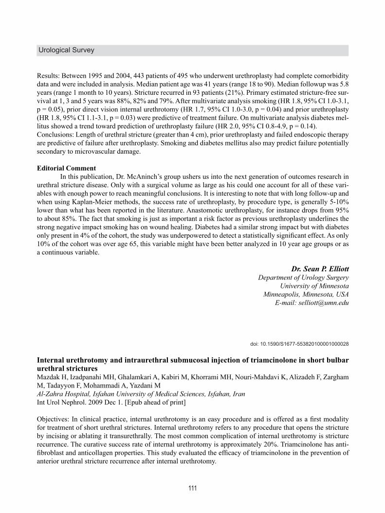
111
Urological Survey
Results: Between 1995 and 2004, 443 patients of 495 who underwent urethroplasty had complete comorbidity data and were included in analysis. Median patient age was 41 years (range 18 to 90). Median followup was 5.8 years (range 1 month to 10 years). Stricture recurred in 93 patients (21%). Primary estimated stricture-free sur-vival at 1, 3 and 5 years was 88%, 82% and 79%. After multivariate analysis smoking (HR 1.8, 95% CI 1.0-3.1, p = 0.05), prior direct vision internal urethrotomy (HR 1.7, 95% CI 1.0-3.0, p = 0.04) and prior urethroplasty (HR 1.8, 95% CI 1.1-3.1, p = 0.03) were predictive of treatment failure. On multivariate analysis diabetes mel-litus showed a trend toward prediction of urethroplasty failure (HR 2.0, 95% CI 0.8-4.9, p = 0.14). Conclusions: Length of urethral stricture (greater than 4 cm), prior urethroplasty and failed endoscopic therapy are predictive of failure after urethroplasty. Smoking and diabetes mellitus also may predict failure potentially secondary to microvascular damage.
Editorial Comment In this publication, Dr. McAninch’s group ushers us into the next generation of outcomes research in urethral stricture disease. Only with a surgical volume as large as his could one account for all of these vari-ables with enough power to reach meaningful conclusions. It is interesting to note that with long follow-up and when using Kaplan-Meier methods, the success rate of urethroplasty, by procedure type, is generally 5-10% lower than what has been reported in the literature. Anastomotic urethroplasty, for instance drops from 95% to about 85%. The fact that smoking is just as important a risk factor as previous urethroplasty underlines the strong negative impact smoking has on wound healing. Diabetes had a similar strong impact but with diabetes only present in 4% of the cohort, the study was underpowered to detect a statistically significant effect. As only 10% of the cohort was over age 65, this variable might have been better analyzed in 10 year age groups or as a continuous variable.
Dr. Sean P. ElliottDepartment of Urology Surgery
University of MinnesotaMinneapolis, Minnesota, USA
E-mail: [email protected]
doi: 10.1590/S1677-553820100001000028
Internal urethrotomy and intraurethral submucosal injection of triamcinolone in short bulbar urethral stricturesMazdak H, Izadpanahi MH, Ghalamkari A, Kabiri M, Khorrami MH, Nouri-Mahdavi K, Alizadeh F, Zargham M, Tadayyon F, Mohammadi A, Yazdani MAl-Zahra Hospital, Isfahan University of Medical Sciences, Isfahan, IranInt Urol Nephrol. 2009 Dec 1. [Epub ahead of print]
Objectives: In clinical practice, internal urethrotomy is an easy procedure and is offered as a first modality for treatment of short urethral strictures. Internal urethrotomy refers to any procedure that opens the stricture by incising or ablating it transurethrally. The most common complication of internal urethrotomy is stricture recurrence. The curative success rate of internal urethrotomy is approximately 20%. Triamcinolone has anti-fibroblast and anticollagen properties. This study evaluated the efficacy of triamcinolone in the prevention of anterior urethral stricture recurrence after internal urethrotomy.

112
Urological Survey
Methods: Fifty male patients with anterior urethral stricture were randomized to undergo internal urethrotomy with or without urethral submucosal injection of triamcinolone. Using general anesthesia urethrotomy was per-formed. Triamcinolone (40 mg) was injected submucosally at the urethrotomy site in 25 patients. The patients were followed for at least 12 months and the stricture recurrence rate was compared between the two groups. Results: 23 patients in the triamcinolone group and 22 in the control group completed the study. There were no significant differences in the baseline characteristics of the patients or the etiology of the stricture between the two groups. Mean follow-up time was 13.7 +/- 5.5 months (range: 1-25 months). Urethral stricture recurred in five patients (21.7%) in the triamcinolone group and in 11 patients (50%) in the control group (P = 0.04). Conclusions: Injection of triamcinolone significantly reduced stricture recurrence after internal urethrotomy. Further investigations are warranted to confirm its efficacy and safety.
Editorial Comment There have been several efforts to increase the efficacy of internal urethrotomy using injection of agents designed to reduce scar formation. Among these, include steroids and botulinum toxin. As described by Wright et al, even a modest increase in the success rate of internal urethrotomy would translate into a much greater preference for urethrotomy over urethroplasty in cost-effectiveness models (1). The current article rep-resents the first randomized trial of steroid injection at the time of internal urethrotomy. The initial results are encouraging. Follow-up was short and the mean time to stricture recurrence was longer in the steroid group. It is possible; therefore, that steroid injection only delays rather than reduces recurrence. Longer follow-up and repeat studies in other clinical settings are needed.
References1. Wright JL, Wessells H, Nathens AB, Hollingworth W: What is the most cost-effective treatment for 1 to 2-cm bulbar
urethral strictures: societal approach using decision analysis. Urology 2006, 67:889-93.
Dr. Sean P. ElliottDepartment of Urology Surgery
University of MinnesotaMinneapolis, Minnesota, USA
E-mail: [email protected]
UROLOGICAL ONCOLOGY _____________________________________________________
doi: 10.1590/S1677-553820100001000029
Outcome of patients who refuse cystectomy after receiving neoadjuvant chemotherapy for muscle-invasive bladder cancerHerr HWDepartment of Urology, Memorial Sloan-Kettering Cancer Center, Weill-Cornell Medical College, New York, NY, USAEur Urol. 2008; 54: 126-32
Objectives: To determine the outcome of patients who refuse cystectomy after receiving neoadjuvant chemo-therapy for muscle-invasive bladder cancer.

113
Urological Survey
Methods: Between 1995 and 2001, 63 patients were evaluated who declined to undergo a planned cystectomy, because they achieved a complete clinical response to neoadjuvant cisplatin-based chemotherapy. Patient, tu-mor, and treatment features were assessed prospectively, and correlated in univariate and multivariate analyses with overall survival. The median follow-up was 86 mo and all patients were followed for more than 5 yr. Results: Forty patients (64%) survived, with 54% of them having an intact functioning bladder. The number and size of invasive tumors were strongly associated with overall survival. The most significant treatment vari-able predicting better survival was complete resection of the invasive tumor on re-staging transurethral resec-tion before starting chemotherapy. Of 23 patients (36%) who subsequently died of disease, 19 (30%) relapsed with invasive cancer in the bladder. Over 90% of surviving patients had solitary, small, and low-stage invasive tumors completely resected, and 83% survived without relapses in the bladder. Conclusions: Selected patients with muscle-invasive bladder cancers may survive after transurethral resection and neoadjuvant chemotherapy, and tumor features can identify which patients responding completely to che-motherapy may survive without cystectomy.
Editorial Comment In Northern America neoadjuvant chemotherapy before radical cystectomy became standard few years ago. What happens if patients (or their doctors, the medical oncologists who deliver chemotherapy) refuse radical cystectomy if a complete response is found in the bladder? This paper gives some very important an-swers. The study group was well chosen with only patients having residual muscle-invasive tumors receiving neoadjuvant chemotherapy. After at least 85% of the planned four cycles of cisplatinum-based chemotherapy, complete clinical response and negative transurethral resection (TUR) of the primary tumor site, these patients were deemed complete responders and were evaluable for follow-up in this group. The good news is that 64% of these patients survived at least 5 years and 54% of them with function-ing bladders. The bad news is that 36% died of bladder cancer after a mean of 32 months. The survivors could be identified by their good prognostic factors, namely single (p < 0.001), or small tumor (p < 0.01), complete restaging TUR (p = 0.02), and noninvasive stage after relapse (p = 0.05). Thus patients with worse tumor features, despite responding completely to chemotherapy, should be strongly advised to undergo radical cys-tectomy at the earliest convenience.
Dr. Andreas BohleProfessor of Urology
HELIOS Agnes Karll HospitalBad Schwartau, Germany
E-mail: [email protected]
doi: 10.1590/S1677-553820100001000030
Long-term rates of undetectable PSA with initial observation and delayed salvage radiotherapy after radical prostatectomyLoeb S, Roehl KA, Viprakasit DP, Catalona WJDepartment of Urology, Johns Hopkins Medical Institutions, Baltimore, MD, USAEur Urol. 2008; 54: 88-94
Background: Randomized trials have shown an improvement in progression-free survival rates with adjuvant radiation therapy (ART) after radical prostatectomy for patients with a high risk of cancer recurrence. Less is
114
Urological Survey
known about the relative advantages and disadvantages of initial observation with delayed salvage radiation therapy (SRT). Objective: To examine the results of SRT in a large single-surgeon radical prostatectomy series. Design, Setting, and Participants: From a radical prostatectomy database, we identified 859 men with positive surgical margins (SM+), extracapsular tumor extension (ECE), or seminal vesicle invasion (SVI) who chose to defer ART. Following a period of initial observation, 192 ultimately received SRT for prostate-specific antigen (PSA) progression. Measurements: Survival analysis was performed to examine the outcomes of initial observation followed by SRT. Results and Limitations: In patients with SM+/ECE and SVI, the 7-yr PSA progression-free survival rates with observation were 62% and 32%, respectively. Among those who had PSA progression, 56% and 26%, respec-tively, maintained an undetectable PSA for 5 yr after SRT. The long-term rates of undetectable PSA associated with an SRT strategy were 83% and 50% for men with SM+/ECE and SVI, respectively. In the subset of 716 men who did not receive any hormonal therapy, the corresponding long-term rates of undetectable PSA were 91% and 75%, respectively. Conclusions: Following radical prostatectomy, initial observation followed by delayed SRT at the time of PSA recurrence is an effective strategy for selected patients with SM+/ECE. Some patients with SVI may also ben-efit from this strategy. However, additional prospective studies are necessary to further examine the survival outcomes following SRT.
Editorial Comment The debate goes on and on. Should a patient with positive surgical margins (SM+) or seminal vesicle infiltration (SVI) after radical prostatectomy be irradiated, and if so – when? This paper supports an affirma-tive standpoint. In short, positive surgical margins might have a relative benign course with a 62% PSA no progression rate if left untreated. In contrast, patients with SVI do worse with only 50% of them not showing up with increasing PSA during the 7-year follow-up. Thus, one may safely choose to wait until PSA becomes measurable after radical prostatectomy. When should I offer adjuvant radiation if PSA shows up? The answer from this paper is – as soon as possible, because the final outcome was better if radiation started when PSA was < 1 ng/ml.
Dr. Andreas BohleProfessor of Urology
HELIOS Agnes Karll HospitalBad Schwartau, Germany
E-mail: [email protected]
NEUROLOGY & FEMALE UROLOGY ____________________________________________
doi: 10.1590/S1677-553820100001000031
Repeat synthetic mid urethral sling procedure for women with recurrent stress urinary incontinenceStav K, Dwyer PL, Rosamilia A, Schierlitz L, Lim YN, Chao F, De Souza A, Thomas E, Murray C, Conway C, Lee JDepartment of Urogynaecology, Mercy Hospital for Women, Melbourne, Victoria, AustraliaJ Urol. 2010; 183: 241-6

115
Urological Survey
Purpose: We reported and compared the outcomes of repeat mid urethral sling with primary mid urethral sling in women with stress urinary incontinence. Materials and Methods: A total of 1,225 consecutive women with urodynamic stress incontinence underwent a synthetic mid urethral sling procedure (955 retropubic, 270 transobturator) at our institution between 1999 and 2007. Of the patients 91% (1,112) were interviewed via telephone call with a structured questionnaire and were included in the analysis. Mean +/- SD followup was 50 +/- 24 months (range 12 to 114). A comparison between repeat (77, mean age 62 +/- 12 years) and primary (1,035, mean age 60 +/- 13 years) mid urethral sling groups was performed. Repeat sling was placed without removal of the previous sling. Results: The preoperative incidence of intrinsic sphincter deficiency was higher in patients who had a repeat mid urethral sling (31% vs 13%, p <0.001). The subjective stress incontinence cure rate was 86% and 62% in the primary and repeat group, respectively (p <0.001). The repeat retropubic approach was significantly more successful than the repeat transobturator approach (71% vs 48%, p = 0.04). The rates of sling related and gen-eral postoperative complications were similar between the primary and the repeat groups. However, de novo urgency (30% vs 14%, p <0.001) and de novo urge urinary incontinence (22% vs 5%, p <0.001) were more frequent in the repeat group compared with the primary group. Conclusions: A repeat synthetic mid urethral sling procedure has a significantly lower cure rate than a primary mid urethral sling procedure. The repeat retropubic approach has a higher success rate than the repeat transob-turator approach. The incidence of de novo urgency and urge incontinence are significantly higher in repeat procedures.
Editorial Comment This is a report on the efficacy of the repeat mid- urethral sling after a failed mid urethral sling. The authors examined an impressive pool of patients numbering well over a thousand of which 77 patients had a repeat mid-urethral sling. The authors noted a significantly lower rate of success (62%) as well as a fairly high rate of failure of the repeat transobturator sling of salvaging continence (53% or less). The authors were able to collate the results of their surgeries through the use of clinical interaction as well as telephone communica-tion. To assess the results, a questionnaire made of select questions from previous validated questionnaires was utilized. The patient population was fairly young being between 60 and 62 years of age. It was noted that the repeat surgery group suffered from a higher rate of de novo urgency as well as urinary urge incontinence. This study is very important in view of its’ large numbers and examining the efficacy of mid-urethral sling. Take home messages include the confirmation of the difficulty in salvaging previously failed mid-ure-thral sling procedures as well as the fairly important singular finding of the limited efficacy of a transobturator sling to salvage either a failed retropubic or a previous transobturator sling. The difficulty in salvaging a gold standard operation has been noted in the past with regard to pubovaginal slings with autologous fascia (1). For further reading on management of failed suburethral slings, I direct the reader to an excellent reference sum-mary article authored by Scarpero and Dmochowski (2).
References1. Petrou SP, Frank I: Complications and initial continence rates after a repeat pubovaginal sling procedure for recurrent
stress urinary incontinence. J Urol. 2001; 2001. 165(6 Pt 1): p. 1979-81.2. Scarpero HM, Dmochowski RR: Sling failures: what’s next? Curr Urol Rep. 2004 Oct;5(5):389-96.
Dr. Steven P. PetrouProfessor of Urology, Associate Dean
Mayo School of Graduate Medical EducationJacksonville, Florida, USA
E-mail: [email protected]

116
Urological Survey
doi: 10.1590/S1677-553820100001000032
An International Urogynecological Association (IUGA)/International Continence Society (ICS) joint report on the terminology for female pelvic floor dysfunctionHaylen BT, de Ridder D, Freeman RM, Swift SE, Berghmans B, Lee J, Monga A, Petri E, Rizk DE, Sand PK, Schaer GNUniversity of New South Wales, Sydney, New South Wales, AustraliaNeurourol Urodyn. 2010;29(1):4-20.
Introduction: Next to existing terminology of the lower urinary tract, due to its increasing complexity, the terminology for pelvic floor dysfunction in women may be better updated by a female-specific approach and clinically based consensus report. Methods: This report combines the input of members of the Standardization and Terminology Committees of two international organizations, the International Urogynecological Association (IUGA), and the International Continence Society (ICS), assisted at intervals by many external referees. Appropriate core clinical categories and a subclassification were developed to give an alphanumeric coding to each definition. An extensive process of 15 rounds of internal and external review was developed to exhaustively examine each definition, with deci-sion-making by collective opinion (consensus). Results: A terminology report for female pelvic floor dysfunction, encompassing over 250 separate definitions, has been developed. It is clinically based with the six most common diagnoses defined. Clarity and user-friend-liness have been key aims to make it interpretable by practitioners and trainees in all the different specialty groups involved in female pelvic floor dysfunction. Female-specific imaging (ultrasound, radiology, and MRI) has been a major addition while appropriate figures have been included to supplement and help clarify the text. Ongoing review is not only anticipated but will be required to keep the document updated and as widely ac-ceptable as possible. Conclusion: A consensus-based terminology report for female pelvic floor dysfunction has been produced aimed at being a significant aid to clinical practice and a stimulus for research.
Editorial Comment This is a very noteworthy review article which should be kept as a reference point for the various terminologies and definitions used in the contemporary literature. It may hold a keen value when preparing manuscripts for publication. As stated in the article, this terminology report is user friendly, clinically based, and quite explanatory in its description. That it was developed by leaders of the specialties concerned with pelvic floor dysfunction, including what appears to be an exhaustive number of internal and external reviews and evaluations, lends to its’ value and strength as a reference article. Also to the interested party, reading the entire journal in which this article is published (Neurourology and Urodynamics, Vol. 29(1), 2010) is of good intellectual value with regards to the time expended and subsequent knowledge gleaned.
Dr. Steven P. PetrouProfessor of Urology, Associate Dean
Mayo School of Graduate Medical EducationJacksonville, Florida, USA
E-mail: [email protected]

117
Urological Survey
PEDIATRIC UROLOGY __________________________________________________________
doi: 10.1590/S1677-553820100001000033
Risk factors for urinary tract infection after dextranomer/hyaluronic acid endoscopic injectionTraxel E, DeFoor W, Reddy P, Sheldon C, Minevich EDivision of Pediatric Urology, Cincinnati Children’s Hospital Medical Center, Cincinnati, Ohio, USAJ Urol. 2009; 182 (4 Suppl): 1708-12
Purpose: Endoscopic injection of dextranomer/hyaluronic acid is an option for primary vesicoureteral reflux. Few groups have assessed the rate of urinary tract infection after dextranomer/hyaluronic acid injection. We reviewed our experience with dextranomer/hyaluronic acid injection, and determined the incidence of and risk factors for postoperative urinary tract infection. Materials and Methods: A retrospective cohort study was performed of all children with primary vesicoureteral reflux treated with dextranomer/hyaluronic acid from 2002 to 2007 at a single institution. Patient demograph-ics and clinical outcomes were abstracted from the medical record. Risk factors for postoperative urinary tract infection, including female gender, preoperative vesicoureteral reflux grade, recurrent urinary tract infection, bladder dysfunction, nephropathy and persistent vesicoureteral reflux after surgery, were analyzed in a multi-variate logistic regression model. Results: We treated 311 children, of whom 87% were female and 13% were male (464 renal units), during the study period. Mode of presentation was urinary tract infection in 85% of cases. Mean followup was 2.6 years. Postoperatively urinary tract infection developed in 40 patients (13%) and febrile urinary tract infection devel-oped in 11 (3.5%). Of patients with urinary tract infection 26 had initially negative postoperative voiding cys-tourethrogram, of whom 16 underwent repeat voiding cystourethrogram and 9 showed recurrent vesicoureteral reflux. Five of these 9 patients had clinical pyelonephritis. Of assessed risk factors only preoperative recurrent urinary tract infection (OR 2.2, p = 0.03) and bladder dysfunction (OR 3.3, p = 0.001) were independent pre-dictors of post-injection urinary tract infection. Conclusions: In our series urinary tract infection after dextranomer/hyaluronic acid injection was rare. Patients with recurrent urinary tract infections and bladder dysfunction preoperatively are at increased risk for urinary tract infection after treatment. Patients with febrile urinary tract infection after dextranomer/hyaluronic acid injection are at high risk for recurrent vesicoureteral reflux.
Editorial Comment This manuscript studies 311 children over a five-year period that had Dx/HA. Secondary causes of re-flux and poor follow-up patients were excluded. Bladder dysfunction included patients with enuresis, frequen-cy/urgency, or urge incontinence and when discovered, standard treatment was instituted prior to surgery. This behavior and dietary modifications were continued after surgery if the bladder dysfunction persisted. Antibi-otic prophylaxis was continued until a follow-up VCUG at 2-3 months showed no further reflux. The results showed 87% of their patients were female and 85% presented with a UTI and 60.5% were febrile. 90% of their patients had Grade III reflux or less. Preoperative nephropathy was present in 62 patients (20%) and bladder dysfunction was present in 64 patients (21%). The mean patient age was 5.7 years with mean follow-up of 2.6 years. The first time success rate for the sting procedure was 70%. With follow-up injections, the overall suc-cess rate by patient was 81% and renal unit 88% and these results correlated with preoperative grade of reflux. Postoperatively 40 patients (13%) developed UTI and 11 (3.5%) had febrile UTI’s. Independent risk factors for postoperative UTI’s by multivariate analysis were preoperative recurrent UTI’s and bladder dysfunction. 4

118
Urological Survey
of the 11 febrile UTI patients had a follow-up VCUG showing vesicoureteral reflux and subsequently 5 more of these patients had a VCUG positive for VUR. 10 patients of the afebrile UTI group were positive for VUR. Upon repeat, 11 more showed VUR later. Vesicoureteral reflux and urinary tract infection are known risk factors for kidney scarring and modifi-cations of both of these risk factors have been sought over the years to prevent permanent kidney damage. As noted in the discussion, the international reflux study group, a 28% incidence of afebrile UTI and 18% instance of febrile UTI in that population over 10 years. It is interesting to note that subureteric injection of Dx/HA seems to add some protective benefit for recurrent UTI’s. In my mind, this may be the most important benefit. Recurrent UTI’s correlate quite well with failed Deflux, although it is interesting to note that in a recent study by Lee et al. (1), it was showed the sting patients only had 46% of a durable reflux resolution after one year. During this 2.6 years of follow-up, that should mean that half of these patients had their reflux return, and yet the sting procedure seems to offer a UTI prevention benefit even in patients that did not have a long-term suc-cess. Readers should watch this data carefully and I think that these issues will become more clear over the next several years.
References1. Lee EK, Gatti JM, Demarco RT, Murphy JP. Long-term followup of dextranomer/hyaluronic acid injection for vesi-
coureteral reflux: late failure warrants continued followup. J Urol. 2009; 181: 1869-74; discussion 1874-5.
Dr. Brent W. SnowDivision of Urology
University of Utah Health Sci CtrSalt Lake City, Utah, USA
E-mail: [email protected]
doi: 10.1590/S1677-553820100001000034
Straightening ventral curvature while preserving the urethral plate in proximal hypospadias repairSnodgrass W, Prieto JDepartment of Urology, Children’s Medical Center, University of Texas Southwestern Medical Center at Dallas, Dallas, Texas, USAJ Urol. 2009; 182 (4 Suppl): 1720-5
Purpose: We report the efficacy of an expanded algorithm for penile straightening in proximal hypospadias surgery to preserve the urethral plate for urethroplasty. We also compared ventral corporotomy with grafting to multiple superficial ventral corporotomies without grafting for straightening greater than 30-degree ventral curvature. Materials and Methods: The need for urethral plate transection was compared in 2 groups comprising consecu-tive patients with proximal shaft to perineal hypospadias repair done by one of us (WS). The 47 patients in group 1 underwent surgery from 2000 to 2005 and had ventral curvature greater than 30 degrees after deglov-ing, leading to urethral plate transection, while in 23 in group 2 from 2006 to 2008 mobilization of the corpus spongiosum/urethral plate and proximal urethra were also performed before urethral plate transection. Patients in group 1 with greater than 30-degree ventral curvature after urethral plate transection underwent ventral cor-porotomy with grafting (7) or multiple transverse corporotomies without grafting (4), while those in group 2

119
Urological Survey
with greater than 30-degree ventral curvature after corpus spongiosum/urethral plate and urethral mobilization underwent multiple transverse corporotomies without grafting.Results: Excluding 10 group 1 and 3 group 2 boys without ventral curvature after degloving the rate of ure-thral plate transection significantly decreased from 54% to 15% using the expanded algorithm (p = 0.005). At a mean followup of 11 months in those with corpus spongiosum/urethral plate and urethral mobilization there was no recognized recurrent ventral curvature. Seven patients with greater than 30-degree ventral curvature underwent ventral corporotomy with grafting, while 11 underwent multiple transverse corporotomies without grafting. At a mean followup of 27 and 19 months, respectively, no patient had recurrent ventral curvature.Conclusions: Mobilization of the corpus spongiosum/urethral plate and the urethra in proximal hypospadias cases with greater than 30-degree ventral curvature after penile degloving decreases the need for urethral plate transection. Ventral lengthening to correct corporeal disproportion can be achieved by corporotomy with graft-ing or by multiple transverse incisions without grafting.
Editorial Comment Seventy patients with proximal shaft or scrotal hypospadias had preoperative testosterone therapy. From 2000-2005 those with less than 30° of curvature, estimated by an artificial erection, were corrected by a dorsal plication. During that same time period, if the curvature was greater than 30° the urethral plate was transected. From 2006-2008, if the curvature was greater than 30°, the urethral plate was mobilized. If after mobilization there was less than 30° of curvature, then a midline dorsal plication was performed, while greater than 30° curvature led to three transverse corporotomies in the region of greatest curvature on the ventrum without grafting and usually in combination with a single dorsal plication. The corporotomies were not deep enough to expose the corpora cavernosa tissue. A third technique included a ventral corporotomy with graft-ing that was done from the dermis. Of their 70 consecutive patients, after degloving the penis 19% had no curvature. Curvature less than 20° was correctable by dorsal plication in 31% of the patients and in the 50% of patients with greater than 30° one group had the urethral plate divided and the other group had plication plus the corporotomy incision or grafting. The outcome showed no difference in the followup. Only two cases in the early group had recurrent ventral curvature and none in the later group with preservation of the urethral plate. It is interesting to note that corporotomies only without grafting in combination with plication and urethral mobilization was sufficient to correct curvature. It could be that grafting is unnecessary and it will be interesting to see if these patients over long-term growth and development continue to do so well. Hypospadias repairs based on the urethral plate enjoy a very good success rate and this shows that extended efforts to pre-serve the urethral plate may very well be worth it. In this era where two stage hypospadias repairs seem to be gaining in popularity, here is a technique to complete the hypospadias repair in one stage dealing with severe curvature at the same time. In simplistic terms, even if two stage repairs were entirely successful after two stages, they will never enjoy the same success rate as single stage repairs with followup surgeries to correct the complications.
Dr. Brent W. SnowDivision of Urology
University of Utah Health Sci CtrSalt Lake City, Utah, USA
E-mail: [email protected]

120
VideoInternational Braz J Urol Vol. 36 (1): 120-121, January - February, 2010
Radical Nephrectomy with IVC Thrombectomy (Level-III) Conducted on Veno-Veno Bypass
Tariq S. Hakky, Lucas R. Wiegand, Devannand Mangar, Angel Alsina, Philippe E. Spiess
Department of Urology; The University of South Florida and H. Lee Moffitt Cancer Center, Tampa, Florida, USA
ABSTRACT
Introduction: We report a 43 year old man who was diagnosed with a level-I thrombus and was managed on oral sunitinib for two months by a community Urologist. The thrombus progressed to a level-III and he subsequently developed a pulmonary embolus, which required oral anticoagulation. He was then referred to our facility for definitive surgical care. A computed tomography scan demonstrated a 12 by 15 centimeter right renal mass and on magnetic resonance venography of the abdomen a tumor-thrombus extending into the infradiaphragmatic inferior vena cava was noted. Pre-operatively consults with hepatobiliary, vascular, and chest surgeons were obtained. Methods: The patient’s surgery was performed by means of an open right extended subcostal incision. Prior to incision, the veno-veno access sites were obtained and an intraoperative transesophageal echocardiography was performed to rule out thrombus in the atria. The right kidney was dissected out and mobilized. The renal artery and vein were dissected, ligated and the en bloc kidney was removed. Control of the inferior vena cava (IVC) was maintained proximally and distally during thrombectomy while tissue perfusion was maintained on veno-veno bypass, no circulatory arrest was required. The estimated blood was 2300 cc; the total bypass time was 25 minutes and the patient was discharged from the hospital after 7 days. Conclusions: It is feasible to perform a radical nephrectomy and IVC thrombectomy while on veno-veno by-pass provided the appropriate multi-disciplinary team is standing by. Veno-veno bypass offers the advantage of minimizing the large hemodynamic drops attributed with suprahepatic IVC clamping. Such high-risk operations requiring skilled surgical teams must only be performed at tertiary care referral centers with extensive experi-ence in the surgical management of such patients.
Int Braz J Urol. 2010; 36 (Video #3): 120_1Available at: www.brazjurol.com.br/videos/january_february_2010/Spiess_120_121video.htm
Accepted:January 15, 2010
Correspondence address:Dr. Philippe E. SpiessDepartment of Urology H Lee Moffitt Cancer CenterE-mail: [email protected]
doi: 10.1590/S1677-553820100001000035

121
Video
EDITORIAL COMMENT
The group of Spiess and colleagues has produced an eloquent and timely video. With a trend towards highlighting minimally invasive surgery, we should also try to maintain our skills with respect to the management of conditions requiring major surgery. The use of venous bypass, which was nicely demonstrated in this video, illustrates the advantage in terms of exposure and blood loss afforded by this technique. Furthermore, due to the controlled nature
of the ensuing dissection, there is a decreased risk of devastating complications such as emboli and damage to the contralateral kidney. The presence of an appropriate infrastructure with availability of a multidisciplinary team cannot be overemphasized. When done safely, as demonstrated in this video, the results have huge benefits as many patients with renal thrombi enjoy long-term survival.
Dr. A. Karim KaderAssistant Professor of UrologyComprehensive Cancer Center
Wake Forest University School of MedicineWinston-Salem, North Carolina, USA
E-mail: [email protected]
Copyright © 2022 FDOKUMEN


![[I Brazilian Guidelines for cardiovascular prevention]](https://static.fdokumen.com/doc/165x107/63360fa2379741109e00e40c/i-brazilian-guidelines-for-cardiovascular-prevention.jpg)


