Evaluation of the Rheologic and Physicochemical Properties ...
Upload
independentCategory
view
6download
0
Diclofenac is one of the most widely prescribed non-steroidal anti-inflammatorydrugs (NSAIDs). Use of diclofenac is associated with two major limitations; first, rare,but serious and sometimes fatal, gastrointestinal (GI) side-effects, including ulceration,and hemorrhage, especially in the elderly (1, 2), and second, poor water solubility.
335
Acta Pharm. 59 (2009) 335–344 Short communication
10.2478/v10007-009-0023-x
Development and physicochemical evaluationof pharmacosomes of diclofenac
AJAY SEMALTY1*
MONA SEMALTY1
DEVENDRA SINGH2
M. S. M. RAWAT2
1Department of Pharmaceutical SciencesH. N. B. Garhwal University, SrinagarGarhwal, India
2 Department of Chemistry, H. N. B.Garhwal University, Srinagar GarhwalIndia
Accepted June 30, 2009
Pharmacosomes are amphiphilic lipid vesicular systemsthat have shown their potential in improving the bio-availability of poorly water soluble as well as poorly li-pophilic drugs. Diclofenac is a poorly water soluble drugand also causes gastrointestinal toxicity. To improve thewater solublity of diclofenac, its pharmacosomes (pho-spholipid complex) have been prepared and evaluatedfor physicochemical analysis. Diclofenac was complexedwith phosphatidylcholine (80 %) in equimolar ratio, inthe presence of dichloromethane, by the conventional sol-vent evaporation technique. Pharmacosomes thus prepa-red were evaluated for drug solubility, drug content, sur-face morphology (by scanning electron microscopy), phasetransition behaviour (by differential scanning calorime-try), crystallinity (by X-ray powder diffraction) and in vitrodissolution. Pharmacosomes of diclofenac were found tobe irregular or disc shaped with rough surfaces in SEM.Drug content was found to be 96.2 ± 1.1 %. DSC thermo-grams and XRPD data confirmed the formation of thephospholipid complex. Water solubility of the preparedcomplex was found to be 22.1 mg mL–1 as compared to10.5 mg mL–1 of diclofenac. This improvement in watersolubility in prepared pharmacosomes may result in im-proved dissolution and lower gastrointestinal toxicity.Pharmacosomes showed 87.8 % while the free diclofenacacid showed a total of only 60.4 % drug release at the endof 10 h of dissolution study.
Keywords: diclofenac, solubility, pharmacosomes, phospho-lipid complex
* Correspondence; e-mail: [email protected]
Different approaches have been applied to decrease NSAID-induced GI toxicity. Forexample, association of NSAIDs with phospholipids has been suggested to improve GIsafety of these drugs (3). The presence of an adsorbed layer of surface-active phospho-lipids on the surface of the mucus that covers the surface epithelium is suggested to pro-tect the GI tissues by providing a hydrophobic layer between the epithelium and the lu-minal contents (4, 5). It has been reported that NSAIDs associated with zwitterionicphospholipids may reduce GI toxicity (3).
Besides GI toxicity of diclofenac, it is also poorly water soluble, due to which its dis-solution in GI fluid is very low, which in turn adversely affects the bioavailability (50–60% only). The association of diclofenac with zwitterionic phospholipids, which may beboth electrostatic and hydrophobic in nature, renders the phospholipids more water-sol-uble and the NSAID more lipid-soluble (6). It has been reported that the diffusion ofNSAIDs across lipid membranes and into target cells is accelerated when they are in acomplex with phosphatidylcholine (PC) (3).
Therefore developing drugs as lipid complexes (pharmacosomes) may be a poten-tial approach to improve solubility and to minimize the GI toxicity of diclofenac. Phar-macosomes are amphiphilic drug-lipid complexes, which are stable and more bioavai-lable drug delivery systems with low interfacial tension between the system and the GIfluid, thereby facilitating the membrane, tissue, or cell wall transfer in the organism (7).
This work aims to develop and characterize the pharmacosomes of diclofenac alongwith its in vitro drug release study.
EXPERIMENTAL
Materials
Diclofenac potassium was obtained as a gift sample from Wings Pharma, India.Soya phosphatidylcholine (LIPOID S-80) was obtained as a gift sample from LIPOID, Ger-many. All other chemicals were of analytical grade.
Formulation of pharmacosomes
Diclofenac salt was converted into the acid form to provide an active hydrogen sitefor complexation. Diclofenac acid was prepared by acidification of an aqueous solutionof diclofenac potassium, extraction into chloroform, and subsequent recrystallization.Diclofenac-PC complex was prepared by associating diclofenac acid with an equimolarconcentration of PC. The equimolar concentration of PC and diclofenac acid were placedin a 100-mL round bottom flask and dissolved in dichloromethane. The solvent was eva-porated under vacuum at 40 °C in a rotary vacuum evaporator (Perfit Model No. 5600,Büchi type, Perfit Ltd., India). The pharmacosomes were collected as the dried residueand placed in a vacuum desiccator overnight and then subjected to characterization.
336
A. Semalty et al.: Development and physicochemical evaluation of pharmacosomes of diclofenac, Acta Pharm. 59 (2009) 335–344.

Drug content
To determine the drug content in pharmacosomes of diclofenac (diclofenac-PC com-plex), a complex equivalent to 50 mg diclofenac was weighed and added into a volumet-ric flask with 100 mL of pH 6.8 phosphate buffer. Then the volumetric flask was stirredcontinuously for 24 h on a magnetic stirrer. At the end of 24 h, suitable dilutions weremade and measured for the drug content at 276 nm UV spectrophotometrically (doublebeam UV-Visible spectrophotometer, Lambda 25, Perkin Elmer, USA).
Solubility
To determine the change in solubility due to complexation, solubility of diclofenacacid and diclofenac-PC complex was determined in pH 6.8 phosphate buffer and n-oc-tanol by the shake-flask method. Diclofenac acid (50 mg) (and 50 mg equivalent in caseof complex) was placed in a 100-mL conical flask. Phosphate buffer pH 6.8 (50 mL) wasadded and then stirred for 15 minutes. The suspension was then transferred to a 250 mLseparating funnel with 50 mL n-octanol and was shaken well for 30 minutes. Then theseparating funnel was kept still for about 30 minutes. Concentration of the drug was de-termined from the aqueous layer spectrophotometrically at 276 nm.
Scanning electron microscopy (SEM)
To detect the surface morphology of the pharmacosomes, SEM of the complex wasrecorded on a scanning electron microscope (JEOL JSM 5600, Japan).
Differential scanning calorimetry (DSC)
Thermograms of diclofenac acid, phosphatidylcholine (80 %) and the diclofenac--PC complex were recorded using a 2910 Modulated Differential Scanning CalorimeterV4.4E (TA Instruments, USA). The thermal behavior was studied by heating 2.0 ± 0.2 mgof each individual sample in a covered sample pan under nitrogen gas flow. The in-vestigations were carried out over the temperature range 25–250 °C at a heating rateof 10 °C min–1.
X-ray powder diffraction (XRPD)
The crystalline state of diclofenac in the different samples was evaluated usingX-ray powder diffraction. Diffraction patterns were obtained on a Bruker Axs- D8 Dis-cover Powder X-ray diffractometer, Germany. The X-ray generator was operated at 40kV tube voltages and 40 mA tube current, using the K a lines of copper as the radiationsource. The scanning angle ranged from 1 to 60° of 2q in the step scan mode (step width0.4° min–1). Diclofenac acid, phosphatidylcholine 80 % (Lipoid S-80) and the preparedcomplex were analyzed. The results are shown in Fig. 3.
337
A. Semalty et al.: Development and physicochemical evaluation of pharmacosomes of diclofenac, Acta Pharm. 59 (2009) 335–344.

Dissolution study
In vitro dissolution studies of diclofenac complex as well as plain diclofenac acidwere performed in triplicate in a USP (8) six station dissolution test apparatus, type II(Veego Model No. 6 DR, India) at 100 rpm and at 37 °C. An accurately weighed amountof the complex equivalent to 100 mg of diclofenac acid was put into 900 mL of pH 6.8phosphate buffer. Samples (3 mL each) of dissolution fluid were withdrawn at differentintervals and replaced with an equal volume of fresh medium to maintain sink condi-tions. Withdrawn samples were filtered (through a 0.45-mm membrane filter), dilutedsuitably and then analyzed spectrophotometrically at 276 nm. Fig. 4 gives a graphicalrepresentation of the in vitro release profile from the formulations.
Statistical analysis
Results are expressed as mean values ± standard deviations and the significance ofthe difference observed was analyzed by Student’s t-test.
RESULTS AND DISCUSSION
Pharmacosomes of diclofenac were prepared with an equimolar ratio (1:1) of diclo-fenac and phopsphatidylcholine in the presence of dichloromethane by the conventionalsolvent evaporation technique. The content of diclofenac in the pharmacosome, as esti-mated by UV spectrophotometry, was found to be 96.2 ± 1.1 % (m/m). Pharmacosomesshowed a high percentage of drug loading, which is specific to them. Complexation pro-vides good percent loading of the drug, which makes the drug delivery clinically feasi-ble. On the other hand, in liposomes of diclofenac sodium, the encapsulation efficiencywas found to be just 59 % (9). Special methods such as coating of these vesicular systemswere needed to improve the loading of the drug. This is not required in the pharmaco-somes in which the drug is reversibly bonded chemically with the lipids and thus showsnot only good percent loading but also better stability than in liposomes.
Water solubility of diclofenac from pharmacosomes was found to be much higherthan that of diclofenac acid. Table I provides the solubility data. The increase in solubi-lity of diclofenac acid in the complex can be explained by the fact that solubilization re-sulted from the micelle formation in the medium and by the amorphous characteristicsof the complex. As amphiphilic surfactants, phospholipids could increase the solubility
338
A. Semalty et al.: Development and physicochemical evaluation of pharmacosomes of diclofenac, Acta Pharm. 59 (2009) 335–344.
Table I. Solubility study of diclofenac acid and its pharmacosomes
DrugSolubility
Aqueous layer (mg mL–1)a n-Octanol layer (mg mL–1) a
Diclofenac acid 10.5 ± 1.5 30.5 ± 1.0
Diclofenac pharmacosomes 22.1 ± 2.4 32.5 ± 1.4
a Mean ± SD; n = 3.

of the drug by the action of wetting and dispersion (3, 6). Unlike the non-polar nature ofdiclofenac, the complex showed amphiphilic nature, which may prove to be responsiblefor improved bioavailability of the drug (10).
Scanning electron micrographs of the complex are shown in Fig. 1. Pharmacosomeswere found to be irregular or disc shaped with rough surface morphology. The complexwas found to involve free flowing particles. Phospholipids are natural components, sotheir different purity grades may have different effects on the shape and surface mor-phology (11).
In order to substantiate the association of diclofenac acid with PC, DSC analysis wasperformed on diclofenac acid, PC, and the pharmacosomes of diclofenac. Results con-firmed the association of diclofenac acid and PC in the complex as both peaks represent-ing diclofenac acid and PC changed positions. Phospholipids (Fig. 2b) showed two ma-jor peaks at 83.21 °C and 107.90 °C and a small peak at 64.45 °C. The first peak of pho-spholipids is a mild peak (at 64.45 °C), which is probably due to the hot movement ofthe phospholipid polar head group. The second (83.21 °C) peak is very sharp, which ap-pears to be due to the phase transition from gel to liquid crystalline state. The non-polarhydrocarbon tail of phospholipids may be melted during this phase, yielding a sharppeak. This melting might have occurred in two phases that subsequently gave anotherpeak (107.90 °C), which is relatively less sharp. Diclofenac acid (Fig. 2a) showed a sharpendothermic peak at 171.31 °C. On the other hand, pharmacosomes of diclofenac (Fig.2c) showed a broad peak at 68.34 °C, which is different from the peaks of the individualcomponents of the complex. It is evident that the original peaks of diclofenac and pho-spholipids disappear from the thermogram of pharmacosomes (complex) and the phasetransition temperature is lower than that of phospholipids. The DSC thermograms of thephospholipid complexes of some phytoconstituents, like silybin, puerarin, curcumin andsome xanthones, revealed similar results (12–14).
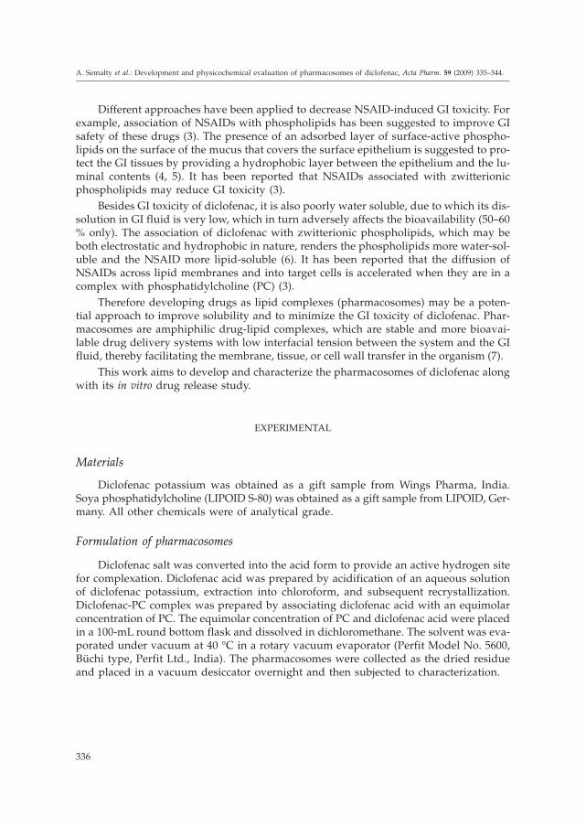
To check whether the changes in the diclofenac crystal morphology correspond to apolymorphic transition and to study the solid state of diclofenac phospholipid complex,XRPD analysis was conducted. The XRPD of diclofenac complex revealed a broad peaksimilar to PC (Fig. 3). It suggested that the diclofenac in the phospholipid complex waseither in amorphous form or molecularly dispersed. These results are well supported by
339
A. Semalty et al.: Development and physicochemical evaluation of pharmacosomes of diclofenac, Acta Pharm. 59 (2009) 335–344.
a) b)
Fig. 1. SEM of pharmacosomes of diclofenac: a) 190X magnification, b) 950X magnification.

our previous studies done with the phospholipid complexes of aspirin (11). The disap-pearance of diclofenac crystalline diffraction peaks confirmed the formation of the pho-spholipid complex. Unlike liposomes, chemical bonding between the drug and phospho-lipids in the development of pharmacosomes might have resulted into a significant changeof its X-ray diffraction.
340
A. Semalty et al.: Development and physicochemical evaluation of pharmacosomes of diclofenac, Acta Pharm. 59 (2009) 335–344.
a)
–4
–3
–2
–1
0
1
0 50 100 150 200 250
Temperature (° C)
Hea
tfl
ow
(mW
)
–4
–3
–2
–1
0
1
0 50 100 150 200 250
Temperature (° C)
Hea
tfl
ow
(mW
)
b)
–4
–3
–2
–1
0
1
0 50 100 150 200 250
Temperature (° C)
Hea
tfl
ow
(mW
)
c)
Fig. 2. DSC thermograms of: a) diclofenac acid, b) phosphatidylcholine, and c) pharmacosome ofdiclofenac.
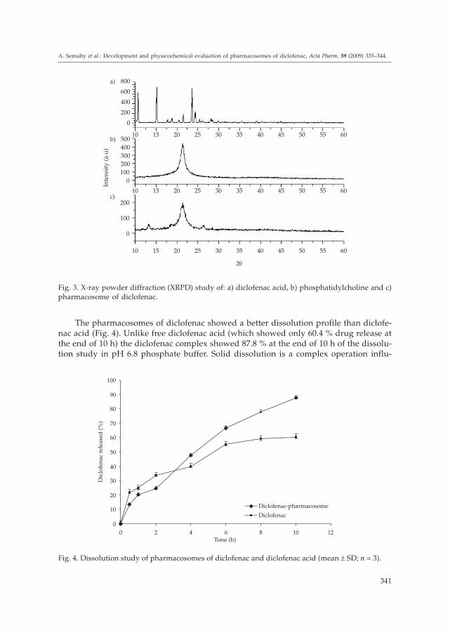
The pharmacosomes of diclofenac showed a better dissolution profile than diclofe-nac acid (Fig. 4). Unlike free diclofenac acid (which showed only 60.4 % drug release atthe end of 10 h) the diclofenac complex showed 87.8 % at the end of 10 h of the dissolu-tion study in pH 6.8 phosphate buffer. Solid dissolution is a complex operation influ-
341
A. Semalty et al.: Development and physicochemical evaluation of pharmacosomes of diclofenac, Acta Pharm. 59 (2009) 335–344.
0
0
100
100
200
200
10
10
10
15
15
15
20
20
20
25
25
25
30
30
30
35
35
35
40
40
40
45
45
45
50
50
50
55
55
55
60
60
60
2q
Inte
nsi
ty(a
.u)
300
400
500
0
200
400
600
800a)
b)
c)
Fig. 3. X-ray powder diffraction (XRPD) study of: a) diclofenac acid, b) phosphatidylcholine and c)pharmacosome of diclofenac.
0
10
20
30
40
50
60
70
80
90
100
0 2 4 6 8 10 12Time (h)
Diclofenac-pharmacosome
Diclofenac
Dic
lofe
nac
rele
ased
(%)
Fig. 4. Dissolution study of pharmacosomes of diclofenac and diclofenac acid (mean ± SD; n = 3).
enced by a number of factors, not only by particle size. Differences in the crystal habit,surface area, surface energies, particle size and wettability may all play a role in affect-ing the dissolution rate of powder (15). Phospholipids being amphiphilic surfactants, in-creased the solubility of the drug by the action of wetting and dispersion. That is whythe dissolution profile of the complex was found to be improved. In some studies donewith silybin and xanthones, the in vitro drug release from the complexes was found to bepH dependent and with the increase of the medium pH the drug dissolution was in-creased (12).
CONCLUSIONS
In the present study, a diclofenac-phospholipid complex (pharmacosomes) was pre-pared by a simple and reproducible method and evaluated for various physicochemicalparameters. Physicochemical investigations showed that diclofenac formed a stoichio-metric complex with phospholipid with improved solubility and dissolution profile. DSCand XRPD studies confirmed the formation of the complex. Thus it can be concludedthat the pharmacosomes of diclofenac may be of potential use for improving dissolutionand for reducing the gastrointestinal toxicity of the drug.
Pharmacosomes may be developed also for other NSAIDs with poor bioavailabilityand GI side effects. Moreover, the pharmacosomes of phytoconstituents (with poor wa-ter and/or lipid solubility) may also be developed for improving their aqueous solubi-lity and lipophilicity and hence bioavailability.
Acknowledgements. – The authors are thankful for the DST Research grant (SRSO/HS/72/2006). The authors acknowledge LIPOID GmbH Germany for providing the gift sam-ple of phosphatidylcholine for the research work. Facilities provided by the UGC-DAEConsortium for Scientific Research, Indore (India), are gratefully acknowledged.
REFERENCES
1. P. M. Peloso, Strategies and practice for the use of nonsteroidal anti-inflammatory drugs, Scan.J. Rheumatol. 25 (1996) 29–48.
2. A. Figueras, D. Capella, J. M. Castel and J. R. Laporte, Spontaneous reporting of adverse drugreactions to nonsteroidal anti-inflammatory drugs, Eur. J. Clin. Pharmacol. 47 (1994) 297–303;DOI: 10.1007/BF00191158.
3. L. M. Lichtenberger, Z. M. Wang, J. J. Romero, C. Ulloa, J. C. Perez, M. N. Giraud and J. C. Barreto,Non steroidal anti-inflammatory drugs (NSAIDs) associate with zwitterionic phospholipids: In-sight into the mechanism and reversal of NSAID-induced gastrointestinal injury, Nature Med. 11(1995) 154–158; DOI: 10.1038/nm0295-154.
4. P. J. Goddard, Y. J. Kao and L. M. Lichtenberger, Luminal surface hydrophobicity of canine gas-tric mucosa is dependent on a surface mucous gel, Gastroenterology 98 (1990) 361–370.
5. L. M. Lichtenberger, L. A. Graziani, E. J. Dial, B. D. Butter and B. A. Hills, Role of surface-activephospholipids in gastric cytoprotection, Science 219 (1983) 1327–1329; DOI: 10.1126/science.6828859.
342
A. Semalty et al.: Development and physicochemical evaluation of pharmacosomes of diclofenac, Acta Pharm. 59 (2009) 335–344.

6. L. M. Lichtenberger, C. Ulloa, J. J. Romero, A. L. Vanous, J. J. Romero, E. J. Dial, P. A. Illich andE. T. Walters, Zwitterionic phospholipids enhance aspirin’s therapeutic activity, as demonstra-ted in rodent model systems, J. Pharmacol. Exp. Ther. 277 (1996) 1221–1227.
7. S. S. Biju, S. Talegaonkar, P. R. Mishra and R. K. Khar, Vesicular systems: an overview, Indian J.Pharm. Sci. 68 (2006) 141–153.
8. United States Pharmacopoeia 23, National Formulary 18, USP Convention, Rockville (MD) 1999,pp. 1791–1792.
9. H. Maswadeh, A. Abdulhalim and C. Demetzos, Improvement of encapsulation efficiency ofdiclofenac sodium into uncoated and chitosan-coated liposomes, Indian J. Pharm. Sci. 66 (2004)607–612.
10. L. M. Lichtenberger, C. Ulloa, J. J. Romero, A. L. Vanous, P. A. Illich and E. J. Dial, Nonsteroidalanti-inflammatory drug and phospholipid prodrugs: Combination therapy with antisecretoryagents in rats, Gastroenterology 111 (1996) 990–995; DOI: 10.1016/S0016-5085(96)70066-5.
11. M. Semalty, A. Semalty, D. Singh, T. Chamoli and M. S. M. Rawat, Effect of purity of phospho-lipids in improving bioavailability performance of aspirin-phospholipid complex, National Con-vention of Chemistry Teachers (NCCT) Annual Conference and Convention, Nov 8–9 2008, Sri-nagar (Garhwal), India, Book of Abstracts, Indian Association of Chemistry Teachers, Srinagar2008, p. 72.
12. X. Yanyu, S. Yunmei, C. Zhipeng and P. Quineng, The preparation of silybin-phospholipid com-plex and the study on its pharmacokinetics in rats, Int. J. Pharm. 307 (2006) 77–82; DOI: 10.1016/j.ijpharm.2005.10.001.
13. Y. Li, D. J. Yang, S. L. Chen, S. B. Chen and A. S. C. Chan, Comparative physicochemical charac-terization of phospholipids complex of puerarin formulated by conventional and supercriticalmethods, Pharm. Res. 25 (2007) 563–577; DOI: 10.1007/s11095-007-9418-x.
14. K. Maiti, K. Mukherjee, A. Gantait, B. P. Saha and P. K. Mukherjee, Curcumin-phospholipid com-plex: Preparation, therapeutic evaluation and pharmacokinetic study in rats, Int. J. Pharm. 330(2007) 155–163; DOI: 10.1016/j.ijpharm.2006.09.025.
15. D. J. Jarmer, C. S. Lengsfeld, K. S. Anseth and T. W. Randolph, Supercritical fluid crystallizationof griseofulvin: Crystal habit modification with a selective growth inhibitor, J. Pharm. Sci. 94(2005) 2688–2702; DOI: 10.1002/jps.20463.
S A � E T A K
Razvoj i fizikokemijsko vrednovanje farmakosoma diklofenaka
AJAY SEMALTY, MONA SEMALTY, DEVENDRA SINGH i M. S. M. RAWAT
Farmakosomi su amfifilni lipidni vezikularni sustavi sa sposobno{}u pobolj{anjabioraspolo`ivosti lijekova slabo topljivih u vodi i organskim otapalima. U svrhu pove}a-nja topljivosti diklofenaka (ljekovite tvari koja je slabo vodotopljiva, a uzrokuje i gastro-intestinalnu toksi~nost) pripravljeni su i evaluirani njegovi farmakosomi (fosfolipidnikompleksi). Diklofenak je kompleksiran s fosfatidilkolinom (80 %) u ekvimolarnom omje-ru, u prisutnosti diklormetana, konvencionalnom metodom evaporacije. Tako priprav-ljenim farmakosomima ispitivana je topljivost, sadr`aj ljekovite tvari, morfologija povr-{ine (pomo}u pretra`ne elektronske mikroskopije), pona{anje pri prijelazu faza (pomo}udiferencijalne pretra`ne kalorimetrije), kristalini~nost (rendgenskom analizom praha) i in
343
A. Semalty et al.: Development and physicochemical evaluation of pharmacosomes of diclofenac, Acta Pharm. 59 (2009) 335–344.

vitro osloba|anje. Farmakosomi diklofenaka su nepravilnog oblika ili u obliku diska teimaju neravnu povr{inu u SEM-u. Sadr`aj ljekovite tvari je 96,2 ± 1,1 %. DSC termogra-mi i XRPD podaci potvrdili su nastajanje fosfolipidnog kompleksa. Topljivost u vodi do-bivenih kompleksa bila je 22,1 mg mL–1, a topljivost samog diklofenaka 10,5 mg mL–1.Postignuto pobolj{anje topljivosti mo`e imati za posljedicu pove}ano osloba|anje i ma-nju gastrointestinalnu toksi~nost. Tijekom 10 h iz farmakosoma se oslobodilo 87,8 %, a izslobodnog diklofenaka samo 60,4 % ljekovite tvari.
Klju~ne rije~i: diklofenak, topljivost, farmakosomi, fosfolipid kompleks
Department of Pharmaceutical Sciences H. N. B. Garhwal University, Srinagar Garhwal, India
Department of Chemistry H.N.B. Garhwal University, Srinagar Garhwal, India
344
A. Semalty et al.: Development and physicochemical evaluation of pharmacosomes of diclofenac, Acta Pharm. 59 (2009) 335–344.
Copyright © 2022 FDOKUMEN