Evaluation of the Rheologic and Physicochemical Properties ...
17
polymers Article Evaluation of the Rheologic and Physicochemical Properties of a Novel Hyaluronic Acid Filler Range with eXcellent Three-Dimensional Reticulation (XTR™) Technology Giovanni Salti 1, * and Salvatore Piero Fundarò 2 1 Medlight Institute, Via Monteverdi 2, 50144 Florence, Italy 2 Multimed Poliambulatorio e Day Surgery, Via dei Fornaciai 29/d, 40129 Bologna, Italy; [email protected] * Correspondence: [email protected] Received: 21 June 2020; Accepted: 21 July 2020; Published: 24 July 2020 Abstract: Soft-tissue fillers made of hyaluronic acid and combined with lidocaine have recently become a popular tool in aesthetic medicine. Several manufacturers have developed their own proprietary formulae with varying manufacturing tools, concentrations, crosslinked three-dimensional network structures, pore size distributions of the fibrous networks, as well as cohesivity levels and rheological properties, lending fillers and filler ranges their unique properties and degradability profiles. One such range of hyaluronic acid fillers manufactured using the novel eXcellent three-dimensional reticulation (XTR™) technology was evaluated in comparison with other HA fillers and filler ranges by an independent research laboratory. Fillers manufactured with the XTR™ technology were shown to have characteristic rheological, crosslinking and biophysical factors that support the suitability of this filler range for certain patient profiles. Keywords: hyaluronic acid; physicochemical properties; rheology; XTR technology 1. Introduction Hyaluronic acid (HA)-based hydrogels have become the most commonly used soft-tissue fillers for facial rejuvenation [1]. Of all available filler material, only HA has fulfilled the need for a biologic substance that is easy to handle, safe, convenient and effective in addressing medically indicated conditions and has lower risk of inducing allergic response [2]. Manufacturers have also supplemented these characteristics by introducing novel HA filler formulations with the aim of stabilizing, increasing tissue longevity and enhancing their tolerability [3]. Distinct proprietary formulae have been developed using various manufacturing tools, HA concentrations, crosslinked three-dimensional network structures, pore size distributions of the fibrous HA networks, cohesivity levels and rheological properties. These properties influence the physical properties of the HA filler product and their variants, often referred to as ‘ranges’, which in turn affect their clinical effects. The most recognized of these formulae are the non-animal stabilized HA (NASHA ® , (Galderma Pharma S.A., Lausanne, Switzerland)), Vycross ® (Allergan Inc., Irvine, CA, USA), cohesive polydensified matrix (CPM ® , (Merz Pharmaceuticals GmbH, Frankfurt am Main, Germany)) and resilient HA (RHA ® , (Teoxane Laboratories, Geneva, Switzerland)), among others [4–6]. Inherently, as the market for these products continues to grow, so does the need for a better understanding of the basic science of HA fillers [6]. No individual product can be used for every indication; hence, the clinician’s experience, as well as the specific guiding principles provided by the originating company, are often used as the bases for choosing the type of HA fillers that are appropriate Polymers 2020, 12, 1644; doi:10.3390/polym12081644 www.mdpi.com/journal/polymers
-
Upload
khangminh22 -
Category
Documents
-
view
2 -
download
0
Transcript of Evaluation of the Rheologic and Physicochemical Properties ...

polymers
Article
Evaluation of the Rheologic and PhysicochemicalProperties of a Novel Hyaluronic Acid Filler Rangewith eXcellent Three-Dimensional Reticulation(XTR™) Technology
Giovanni Salti 1,* and Salvatore Piero Fundarò 2
1 Medlight Institute, Via Monteverdi 2, 50144 Florence, Italy2 Multimed Poliambulatorio e Day Surgery, Via dei Fornaciai 29/d, 40129 Bologna, Italy;
[email protected]* Correspondence: [email protected]
Received: 21 June 2020; Accepted: 21 July 2020; Published: 24 July 2020�����������������
Abstract: Soft-tissue fillers made of hyaluronic acid and combined with lidocaine have recently becomea popular tool in aesthetic medicine. Several manufacturers have developed their own proprietaryformulae with varying manufacturing tools, concentrations, crosslinked three-dimensional networkstructures, pore size distributions of the fibrous networks, as well as cohesivity levels and rheologicalproperties, lending fillers and filler ranges their unique properties and degradability profiles. One suchrange of hyaluronic acid fillers manufactured using the novel eXcellent three-dimensional reticulation(XTR™) technology was evaluated in comparison with other HA fillers and filler ranges by anindependent research laboratory. Fillers manufactured with the XTR™ technology were shown tohave characteristic rheological, crosslinking and biophysical factors that support the suitability of thisfiller range for certain patient profiles.
Keywords: hyaluronic acid; physicochemical properties; rheology; XTR technology
1. Introduction
Hyaluronic acid (HA)-based hydrogels have become the most commonly used soft-tissue fillersfor facial rejuvenation [1]. Of all available filler material, only HA has fulfilled the need for abiologic substance that is easy to handle, safe, convenient and effective in addressing medicallyindicated conditions and has lower risk of inducing allergic response [2]. Manufacturers have alsosupplemented these characteristics by introducing novel HA filler formulations with the aim ofstabilizing, increasing tissue longevity and enhancing their tolerability [3]. Distinct proprietaryformulae have been developed using various manufacturing tools, HA concentrations, crosslinkedthree-dimensional network structures, pore size distributions of the fibrous HA networks, cohesivitylevels and rheological properties. These properties influence the physical properties of the HA fillerproduct and their variants, often referred to as ‘ranges’, which in turn affect their clinical effects.The most recognized of these formulae are the non-animal stabilized HA (NASHA®, (Galderma PharmaS.A., Lausanne, Switzerland)), Vycross® (Allergan Inc., Irvine, CA, USA), cohesive polydensifiedmatrix (CPM®, (Merz Pharmaceuticals GmbH, Frankfurt am Main, Germany)) and resilient HA(RHA®, (Teoxane Laboratories, Geneva, Switzerland)), among others [4–6].
Inherently, as the market for these products continues to grow, so does the need for a betterunderstanding of the basic science of HA fillers [6]. No individual product can be used for everyindication; hence, the clinician’s experience, as well as the specific guiding principles provided by theoriginating company, are often used as the bases for choosing the type of HA fillers that are appropriate
Polymers 2020, 12, 1644; doi:10.3390/polym12081644 www.mdpi.com/journal/polymers

Polymers 2020, 12, 1644 2 of 17
for certain indications and for injection into specific anatomical layers. More importantly, whenevernovel fillers and related technologies are introduced, developing a familiarity of the basic science ofHA fillers would allow aesthetic medicine specialists to select the best options for their patients [7].
The main HA filler properties evaluated in literature include rheology, the degree of crosslinking,resistance to stretch and cohesivity [3,5,8] Together with proper selection of patients with ideal anatomy(e.g., appropriate skin thickness and with good anatomic architecture), knowledge of rheologicalproperties can facilitate the selection of the right products that can help them achieve optimal aestheticoutcomes [5]. For instance, the degree and process of crosslinking changes the three-dimensionalconstruction of the HA chains and affects the consistency of the filler. This altered structure may thusaffect immune acceptance of the filler [5]. Cohesivity is an essential characteristic of gel implants and isdefined as the capacity of a material not to dissociate. This property is naturally important during theproduct distribution into the tissues of the treated area and is considered necessary for the integrityof the solid and fluid phases of a gel. This is known to affect the lifting capacity of the HA fillers [3].This allows for evaluation of the resistance to spreading and adhesion properties of the HA fillers.It also becomes possible to view the product’s homogeneity, which is often considered to equate togood dispersion qualities [3,5].
HA filler degradability by hyaluronidase can be considered as a “safety-feature” and gives afiller a potential edge over its competitors [9]. Technical errors, such as overfilling/overcorrection orfiller placement at the wrong soft tissue plane or misplacement, may be reversed by hyaluronidaseadministration. Nonetheless, appropriate filler longevity in soft tissue is considered to provide structuralstability and resiliency, which is a requirement for volume correction [10]. Thus, it is important forpractitioners to find that delicate balance between stability and degradability to determine whichfillers are best suited for their patients. Degradability studies have highlighted that HA content,cohesivity and crosslinking properties may play a role in the sensitivity of these fillers to enzymaticdegradation [9,11].
In addition, one of the more important but often overlooked determinants of success in skin fillerinjections is control of pain. Preventive pain control helps not only minimize discomfort but alsoreduces procedural downtime while improving patient satisfaction. For this reason, the local anestheticlidocaine has been incorporated in HA fillers. This widely accepted change has led to significantreduction in pain during and after injections. Furthermore, as a result of its antihistamine property,lidocaine reduces erythema, bruising and swelling [2]. However, data suggest that the presence of thelocal anesthetic drug can reduce the viscosity of the filler products. Hence, there is a need to evaluatethe physical properties of HA fillers with adjunctive lidocaine [12].
In this publication, a recently developed line of soft-tissue fillers manufactured using the noveleXcellent three-dimensional reticulation (XTR™) technology have been subjected to the abovementionedtests in comparison with other HA fillers and filler ranges.
2. Methods
2.1. The XTR™ Technology Manufacturing Process
This novel HA filler range manufactured using the proprietary XTR™ technology (Definisse™,RELIFE S.r.l., Florence, Italy) was recently introduced globally. This technique employs a manufacturingprocess made up of a controlled three-step process broken down into the pretreatment, crosslinkingand purification phases (Figure 1) [3]. The pretreatment phase is an initial fracturing step using acontrolled thermal process. This leads to the bioengineered formation of size-specific HA fragmentsthat have viscoelastic properties compatible to skin [3,13–15]. The process enables controlledcleavage of the length of the HA chains and is capable of obtaining two molecular weight ranges(medium: 2.5 × 10⁶–3.2 × 10⁶ Dalton, and high: 3.2 × 10⁶–3.5 × 10⁶ Dalton) [3]. A mixture of differentlengths of HA chains are then intermolecularly bound by a crosslinking agent to produce a stablethree-dimensional HA matrix [16]. The most commonly used chemical agent in creating inter-

Polymers 2020, 12, 1644 3 of 17
and intra-chain bridges between the HA biopolymer is 1,4-butanediol diglycidyl ether (BDDE) [16].Crosslinking enables the HA molecules to have stable viscoelastic properties and allows for a lowextrusion force when injecting the filler. It also improves filling properties and enhances the durabilityof the injectate [6]. Optimization of the cross-linking process enables minimal to almost zero free HAchains [3]. Lastly, the elimination of residual amounts of free unreacted BDDE and its byproducts isdone via a series of downstream purification processes (i.e., precipitation, rehydration). Ultimately, thegoal is to achieve minute amounts, i.e., <2 ppm, of remaining unreacted BDDE, a limit of detectionrecommended by the US Food and Drug Administration (FDA) [17]. BDDE levels are used as aparameter for testing and releasing each manufactured batch of fillers with XTR™ technology [3,18].The size, crosslinking and purity of HA polymers used for filler materials is critical as these propertiesdetermine the biological effects of HA on skin tissue [13].
Polymers 2020, 12, x FOR PEER REVIEW 3 of 18
extrusion force when injecting the filler. It also improves filling properties and enhances the durability of the injectate [6]. Optimization of the cross-linking process enables minimal to almost zero free HA chains [3]. Lastly, the elimination of residual amounts of free unreacted BDDE and its byproducts is done via a series of downstream purification processes (i.e., precipitation, rehydration). Ultimately, the goal is to achieve minute amounts, i.e., <2 ppm, of remaining unreacted BDDE, a limit of detection recommended by the US Food and Drug Administration (FDA) [17]. BDDE levels are used as a parameter for testing and releasing each manufactured batch of fillers with XTR™ technology [3,18]. The size, crosslinking and purity of HA polymers used for filler materials is critical as these properties determine the biological effects of HA on skin tissue [13].
(A) (B) (C)
Figure 1. The eXcellent three-dimensional reticulation (XTR™) technology manufacturing process. (A) The pretreatment phase. (B) The crosslinking phase. (C) The purification process.
The biophysical properties of fillers with XTR™ technology in comparison with the most commonly available commercial fillers have been reviewed in this paper. It is expected that this study will provide an evidence-based scientific rationale for future investigations and clinical applications. The parameters tested in this study include rheology, crosslinking degree and cohesivity. These tests were conducted by the independent R&D Laboratory and Consultancy Center, Rigano Laboratories S.r.l. in Milan, Italy, from the period of June 2019 to July 2020.
The skin fillers tested included derivative HA ranges crosslinked with 1,4-butanediol diglycidyl ether (BDDE) and incorporated with 0.3% lidocaine (Table 1) [19,20].
Table 1. Hyaluronic acid (HA) gel fillers analyzed for biophysical properties.
Filler Identifier Crosslinking Technology [20]. Hyaluronic Acid Concentration (mg/mL) [19–21]. NASHA NASHA® 20 CPM-1
CPM® 22.5
CPM-2 25 CPM-3 26 RHA-1
RHA® 15
RHA-3 23 RHA-4 23 VYC-1
Vycross® 15
VYC-2 17.5 VYC-3 20 XTR-1
XTR™ 23
XTR-2 23 XTR-3 25
2.2. Rheological Evaluation
Rheology tests were performed using a Rheoplus Anton Paar MCR 101 (Anton Paar Company, Graz, Austria) controlled rate rheometer equipped with a PP50-P2 sensor (50 mm parallel plates with serrated surfaces, gap 0.8 mm) and with a Peltier system for temperature control (i.e., 30 ± 0.05 °C). Before running the tests, the gels were allowed to rest for 300 s in order to provide a uniform temperature distribution and a common and consistent shear history amongst the samples [22].
The oscillatory shear stress test or the amplitude sweep test was done to measure the fillers’ linear elastic or storage modulus (G′) and the loss or viscous modulus (G′′), which are reflective of
Figure 1. The eXcellent three-dimensional reticulation (XTR™) technology manufacturing process.(A) The pretreatment phase. (B) The crosslinking phase. (C) The purification process.
The biophysical properties of fillers with XTR™ technology in comparison with the most commonlyavailable commercial fillers have been reviewed in this paper. It is expected that this study will providean evidence-based scientific rationale for future investigations and clinical applications. The parameterstested in this study include rheology, crosslinking degree and cohesivity. These tests were conductedby the independent R&D Laboratory and Consultancy Center, Rigano Laboratories S.r.l. in Milan, Italy,from the period of June 2019 to July 2020.
The skin fillers tested included derivative HA ranges crosslinked with 1,4-butanediol diglycidylether (BDDE) and incorporated with 0.3% lidocaine (Table 1) [19,20].
Table 1. Hyaluronic acid (HA) gel fillers analyzed for biophysical properties.
Filler Identifier Crosslinking Technology [20] Hyaluronic Acid Concentration (mg/mL) [19–21]
NASHA NASHA® 20
CPM-1CPM®
22.5CPM-2 25CPM-3 26
RHA-1RHA®
15RHA-3 23RHA-4 23
VYC-1Vycross®
15VYC-2 17.5VYC-3 20
XTR-1XTR™
23XTR-2 23XTR-3 25
2.2. Rheological Evaluation
Rheology tests were performed using a Rheoplus Anton Paar MCR 101 (Anton Paar Company,Graz, Austria) controlled rate rheometer equipped with a PP50-P2 sensor (50 mm parallel plates with

Polymers 2020, 12, 1644 4 of 17
serrated surfaces, gap 0.8 mm) and with a Peltier system for temperature control (i.e., 30 ± 0.05 ◦C).Before running the tests, the gels were allowed to rest for 300 s in order to provide a uniform temperaturedistribution and a common and consistent shear history amongst the samples [22].
The oscillatory shear stress test or the amplitude sweep test was done to measure the fillers’ linearelastic or storage modulus (G′) and the loss or viscous modulus (G′′), which are reflective of the elasticbehavior and the flow of material when deformed, respectively. This was accomplished by varying theinput shear strain from 0.01% to 4000% while keeping the frequency constant at 1 Hz on each of thesamples. After the limit of the linear viscoelastic region (LVER) was identified, a frequency sweep testwas performed. This was done to determine the mechanical spectra of the gels while varying the inputfrequency from 10 Hz to 0.01 Hz and keeping the shear strain constant within the LVER limit.
G′ measures the energy stored by the HA filler during deformation that re-establishes its originalshape when the shear stress is removed. It corresponds to the elastic segment of its viscoelastic properties(or its semi-solid state). On the other hand, G′′ measures the energy lost on shear deformation becauseof internal friction. It characterizes the viscous segment of its viscoelastic properties (or the liquidstate) of the sample. This property indicates the inability of the HA filler to completely recover itsshape after the deformation. These two parameters are used to calculate the complex modulus (G*):√
(G′2 + G′′2). G* represents the total energy required to deform a gel using shear stress [23].The loss factor (tan δ) is defined as the fraction between the loss modulus G′′ and the storage
modulus G′. It indicates whether the material has mainly an elastic behavior or a viscous behavior,i.e., a tan δ < 1 means that the elastic component is more prominent in the gel structure [22]. Incrosslinked HA fillers, tan δ usually ranges from 0.05 to 0.80; thus, elastic behavior under low shearstress is dominant over viscous behavior [23].
2.3. Degree of Crosslinking
The most common chemical HA crosslinking method uses ether formation by reaction of HA with1,4-butanediol diglycidyl ether (BDDE) [24]. Under alkaline conditions, epoxides of BDDE react withthe HA hydroxyl groups to form derivatives of 1,4-dibutanediol dipropan-2,3-diolyl ether (BDPE).Some of the BDPE form “true cross-links”, binding to HA at both ends, while others only bind HA atone end (“mono-linked”). To describe these HA filler modifications, Kenne and Wende and colleaguesproposed terms that can be applied to characterize HA hydrogels crosslinked with BDDE [25,26]:
i. The degree of modification (MoD) is the stoichiometric ratio between the sum of mono- anddouble-linked BDPE residues and HA disaccharide units. The more crosslink modificationsseen when compared with the acetyl group, the higher the MoD%.
ii. The crosslinker ratio (CrR) indicates the fraction of double-linked crosslinker residues comparedto all linked crosslinkers and this represents the measure of crosslinker efficiency.
iii. The degree of substitution (DS) is the proportion of the HA disaccharides that are substituted.iv. The degree of crosslinking (CrD) is the stoichiometric ratio between BDPE residues that are
double-linked and HA disaccharide units.v. The degree of crosslinking (DC) is the number of HA disaccharides involved in crosslinking in
relation to the total number of HA disaccharides.
Kenne states that MoD and CrD quantify the total amount of linked BDPE and double-linkedBDPE, respectively, in comparison to the total amount of HA. DS and DC are equivalent to the amountof the substituted HA and crosslinked HA, respectively, in comparison to the total amount of HA [26].The relevance of MoD and DS is in describing the total change in the gel after modification. For example,a polymer with low MoD or DS is structurally similar with the original, intact gel. CrD and DC aremore important in terms of describing the physical properties of the gel. A higher value of CrD for agel reveals that it is a stronger gel (i.e., more crosslinked) and would swell less than a weaker gel witha lower CrD.

Polymers 2020, 12, 1644 5 of 17
In this study, the determination of the degree of crosslinking of the nominated HA-based fillerswas performed using the nuclear magnetic resonance (NMR)-based approach described by Wende andcolleagues [25]. HA is hydrolyzed using an acidic medium, and then, the degradation products areanalyzed using one-dimensional NMR. First, the samples were diluted to 4 mg/mL with acid water(HCl 0.1 M, pH 1.5), heated and gently mixed at 75 ◦C overnight. Once cooled, these samples werebuffered at pH 7.0 with NaOH 1 M and 0.1 M, frozen and then lyophilized. After, the samples weredissolved in 0.5 mL of deuterated water (D2O) and then subjected to NMR Bruker 400 mHz analysisfor the determination of MoD%, CrR and CrD. With the 1H-NMR set up, 64 scans and a recycle delay(D1) of 10 s were used. For the 13C-NMR set up, 8192 scans and a D1 of 10 s were used.
The three main parameters were then measured using the following formulae: (1) MoD (%) =
(I δH1.5/4)/(I δH1.9/3) × 100, (2) CrR = 1-IδC62.7/(Iδ25.2/2) and (3) CrD (%) = (CrR ×MoD) × 100.
2.4. Cohesivity
This study used the five-point visual Gavard-Sundaram Cohesivity Scale as a reference [27].HA filler gel behaviors range from fully dispersed (non-cohesive) with only powder-like gel fragmentsvisible to fully cohesive with only intact gel strands visible. Video documentation using standardizeddigital techniques was done to show how gels were extruded as a single filament from a syringe.Cohesivity of each specimen was visually assessed from these images as the ratio of intact gel todispersed gel at each time point [27,28].
Cohesivity of HA filler calculated using the Gavard-Sundaram Cohesivity Scale indicates theaffinity between the gel molecules and, according to the authors of the method, describes the tendencyto maintain a homogeneous distribution within the dermis after injection [27]. It is used also to assessthe ability of fillers to resist vertical compression/stretching and is the expression of the strength ofinternal forces that can be modified based on the HA concentration, the crosslinking technology andthe different gel macrostructures (biphasic or monophasic gel) [4,23].
2.5. Degradability
Select samples were diluted in physiological buffer initially, and then, baseline readings ofabsorbance and relative wavelengths were measured using standard spectrophotometric analysis.Calibration curves (absorbance vs concentration) were calculated from the spectra from visible lightto ultraviolet wavelength for each compound. A 1:8 dilution of all samples was designated as theappropriate concentration as described by La Gatta and colleagues [29].
The fillers were then incubated in the presence of bovine testicular hyaluronidase (BTH,Sigma Aldrich cat N. H3884) at 37 ◦C. The degradation was monitored by documenting the increase insoluble fraction amount during incubation. HA soluble fractions of each of the samples were thenwithdrawn and analyzed at different incubation times (i.e., 0, 30, 60, 300 min) as the ratio of HAconcentration in the permeate versus the total HA concentration of the gel (%). Tests were performedat least in triplicate with the mean and standard deviation provided [30].
3. Results
The following results reflect the analyses of the physicochemical and mechanical properties of theselected injectable crosslinked HA gels based on previously developed pre-specified protocols.
3.1. Rheological Evaluation
The results of the viscoelastic measurements are shown in Figure 2 and Table 2 [22].

Polymers 2020, 12, 1644 6 of 17
Polymers 2020, 12, x FOR PEER REVIEW 6 of 18
(A)
(B)
(C)
Figure 2. Summary of frequency sweep and amplitude sweep measurements of the HA fillers evaluated. A comparison of the (A) elastic moduli (G′), (B) viscous moduli (G′′), and (C) tan δ of fillers made with varying crosslinking technologies.
571.61
426.76
292.37
153.58
329.85265.65
206.73293.46
164.3753.84
233.5136.41
49.9
0100200300400500600
G' a
t 0.7
Hz
Fillers
G'
93.77
26.87 27.86 26.67 22.99 27.44 24.0834.93 30.53
21.13
45.24 42.8224.45
020406080
100
G''
at 0
.7 H
z
Fillers
G''
0.1630.063 0.095
0.1740.067 0.103 0.116 0.119
0.186
0.392
0.1930.313
0.49
00.10.20.30.40.50.6
Tan δ
at 0
.7 H
z
Fillers
Tan δ
Figure 2. Summary of frequency sweep and amplitude sweep measurements of the HA fillers evaluated.A comparison of the (A) elastic moduli (G′), (B) viscous moduli (G′′), and (C) tan δ of fillers made withvarying crosslinking technologies.
Polymers 2020, 12, 1644 7 of 17
Table 2. Comparison of representative fillers from each range with similar HA concentrations.
Product G′ G′′ Tan δ
XTR-1 153.58 26.67 0.174CPM-1 49.9 24.45 0.49VYC-1 206.73 24.08 0.116RHA-1 53.84 21.13 0.392
XTR-2 292.37 27.86 0.095CPM-2 136.41 42.82 0.313VYC-2 265.65 27.44 0.103RHA-3 164.37 30.53 0.186
XTR-3 426.76 26.87 0.063CPM-3 233.5 45.24 0.193RHA-4 293.46 34.93 0.119VYC-3 329.85 22.99 0.067
The rheological tests performed on the HA derivative gels highlighted considerable differencesamong the viscoelastic properties of each range and among fillers of the same group. Compared withthe rest, NASHA appeared to have the highest elastic and viscous responses but also presented withthe lowest critical strain (67%), which translates to less resistance to deformation. Each range is shownto gradually decrease in G′ down the line; lower G′ fillers are softer variants that are suited for mediumto superficial implantation to the soft tissue. CPM-1 and CPM-2 presented a large LVER and the highestcritical strain measurements (1200%), which indicates good strain resistance. The fillers, i.e., CPM,with higher tan δ are described to be more fluid than they are elastic. The ranges can be seen to havehigher fluidity with lower HA concentrations. CPM-1 was the most viscous sample (tan δ = 0.49).The series of XTR, RL and CPM are composed of three variants each, with every sample having differentproperties, while VYC variants were observed to bear more similar intraclass rheological properties.XTR-2 and XTR-3 fillers had relatively higher G′ than their counterparts (Table 2). This may serve as anadvantage in volumizing and lifting tissue and modulating muscular activity when injecting beneaththe superficial musculoaponeurotic system (SMAS) layer but without the granular features of fillerswith extremely high G′. Fillers with XTR™ technology in general have medium (intermediate) G′′
compared to the benchmarked products. [3].
3.2. Degree of Crosslinking
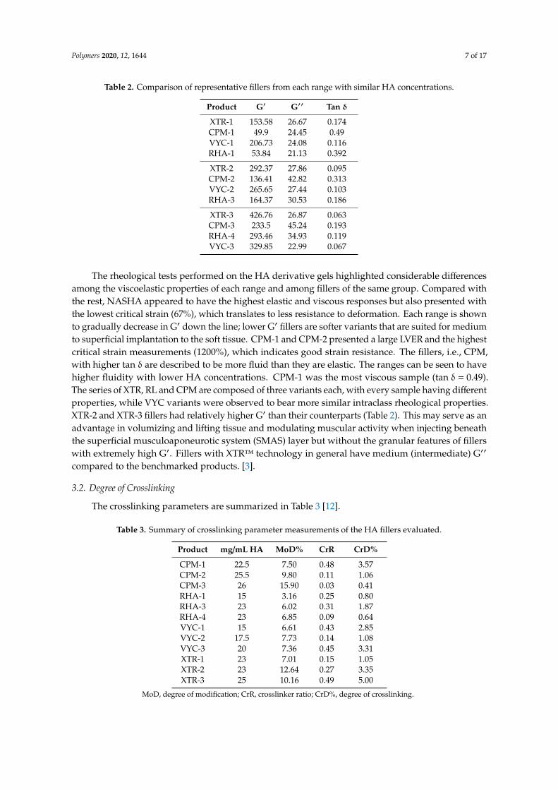
The crosslinking parameters are summarized in Table 3 [12].
Table 3. Summary of crosslinking parameter measurements of the HA fillers evaluated.
Product mg/mL HA MoD% CrR CrD%
CPM-1 22.5 7.50 0.48 3.57CPM-2 25.5 9.80 0.11 1.06CPM-3 26 15.90 0.03 0.41RHA-1 15 3.16 0.25 0.80RHA-3 23 6.02 0.31 1.87RHA-4 23 6.85 0.09 0.64VYC-1 15 6.61 0.43 2.85VYC-2 17.5 7.73 0.14 1.08VYC-3 20 7.36 0.45 3.31XTR-1 23 7.01 0.15 1.05XTR-2 23 12.64 0.27 3.35XTR-3 25 10.16 0.49 5.00
MoD, degree of modification; CrR, crosslinker ratio; CrD%, degree of crosslinking.

Polymers 2020, 12, 1644 8 of 17
Among the fillers, XTR-2 and XTR-3 and CPM-2 and CPM-3 presented with the highest MoD%values (i.e., 9% to 15%), which translates to being those with the highest degree of ether modification.MoD% for RHA-1 is about 3% and consequently is the filler with the lowest degree of modification.Other fillers presented intermediate values (i.e., 6–7.5%). CrR varied from 0.5 to 0.03. XTR-3, CPM-1,VYC-1 and VYC-3 had the highest CrR values, which denotes higher concentration of true crosslinkerscompared to mono-linkers. CPM-3 and RHA-4 had the lowest CrR indicating that mono-linker linkagesare more prevalent. The rest of the other fillers presented intermediate values.
3.3. Cohesivity
The HA samples provided were evaluated in terms of water-binding capacity via the validatedGavard-Sundaram Cohesivity Scale, where 1 means fully dispersed and 5 means fully cohesive atdifferent time frames (Figure 3).
Polymers 2020, 12, x FOR PEER REVIEW 8 of 18
MoD% for RHA-1 is about 3% and consequently is the filler with the lowest degree of modification. Other fillers presented intermediate values (i.e., 6–7.5%). CrR varied from 0.5 to 0.03. XTR-3, CPM-1, VYC-1 and VYC-3 had the highest CrR values, which denotes higher concentration of true crosslinkers compared to mono-linkers. CPM-3 and RHA-4 had the lowest CrR indicating that mono-linker linkages are more prevalent. The rest of the other fillers presented intermediate values.
3.3. Cohesivity
The HA samples provided were evaluated in terms of water-binding capacity via the validated Gavard-Sundaram Cohesivity Scale, where 1 means fully dispersed and 5 means fully cohesive at different time frames (Figure 3).
t = 0 t = 30 s t = 5 min
CPM-1
CPM-2
CPM-3
RHA-1
RHA-3
RHA-4
Figure 3. Cont.

Polymers 2020, 12, 1644 9 of 17
Polymers 2020, 12, x FOR PEER REVIEW 9 of 18
VYC-1
VYC-2
VYC-3
XTR-1
XTR-2
XTR-3
Figure 3. Gel dispersion in water at T = 0, 30 s, and 5 min of evaluated fillers.
CPM gels had high cohesivity, being partially dispersed only at 70 s. CPM-1 was most dispersed at 70 s and maintained a very high cohesivity even after 1 min [28]. The other gels had an intermediate cohesivity and were mostly dispersed in as early as 30 s (i.e., VYC gels) or after 70 s (i.e., XTR). RL gels displayed high cohesivity at the first min and were dispersed only after a prolonged period. Gel dispersion among variants is shown in Figure 4.
Figure 3. Gel dispersion in water at T = 0, 30 s, and 5 min of evaluated fillers.
CPM gels had high cohesivity, being partially dispersed only at 70 s. CPM-1 was most dispersedat 70 s and maintained a very high cohesivity even after 1 min [28]. The other gels had an intermediatecohesivity and were mostly dispersed in as early as 30 s (i.e., VYC gels) or after 70 s (i.e., XTR).RL gels displayed high cohesivity at the first min and were dispersed only after a prolonged period.Gel dispersion among variants is shown in Figure 4.

Polymers 2020, 12, 1644 10 of 17
Polymers 2020, 12, x FOR PEER REVIEW 10 of 18
(A)
(B)
(C)
012345
0 5 0 1 0 0 1 5 0 2 0 0 2 5 0 3 0 0
CO
HES
IVIT
Y V
ALU
ES
TIME (S)
0 15 30 70 300CPM-1 5 5 5 4 2
CPM-2 5 4 4 3 3
CPM-3 4 4 4 3 3
012345
0 5 0 1 0 0 1 5 0 2 0 0 2 5 0 3 0 0
CO
HES
IVIT
Y V
ALU
ES
TIME (S)
0 15 30 70 300RL-1 4 4 4 4 3
RL-2 4 4 4 3 2
RL-3 4 3 2 2 1
012345
0 5 0 1 0 0 1 5 0 2 0 0 2 5 0 3 0 0
CO
HES
IVIT
Y V
ALU
ES
TIME (S)
0 15 30 70 300VYC-1 3 2 1 1 1
VYC-2 3 2 1 1 1
VYC-3 3 3 2 2 1
Polymers 2020, 12, x FOR PEER REVIEW 11 of 18
(D)
Figure 4. Gel dispersion in water at all time points for HA fillers evaluated. (A) CPM gels; (B) RL gels; (C) VYC gels; (D) XTR gels.
3.4. Degradability
The mean soluble fractions (expressed in %) and the standard deviations observed through time were documented for the chosen fillers as follows (Table 4) [30]:
Table 4. Soluble fractions (mean and standard deviation) for each of the HA fillers evaluated.
Enzymatic Degradation Time Product mg/mL HA t = 0 t = 30 min t = 1 h t = 5 h
NASHA 20 51.58 (9.71)
94.95 (8.19)
99.50 (17.89)
94.56 (2.54)
XTR-1 23 27.8
(5.79) 29.79 (9.14)
41.34 (11.35)
55.36 (5.39)
XTR-2 23 40.59 (4.55)
39.20 (7.62)
50.35 (6.23)
58.47 (2.61)
XTR-3 25 32.43 (2.08)
33.72 (3.59)
53.58 (25.46)
78.79 (8.95)
Of the fillers evaluated, NASHA was observed to have the most rapid degradation, ultimately displaying a 95% soluble fraction after 30 min. XTR-1 had the lowest soluble fraction percentage at baseline, and this was seen to persist across the time points. XTR-2 and -3 were slightly more resistant to degradation over time, as shown in Figure 5.
012345
0 5 0 1 0 0 1 5 0 2 0 0 2 5 0 3 0 0
CO
HES
IVIT
Y V
ALU
ES
TIME (S)
0 15 30 70 300XTR-1 4 3 3 2 2
XTR-2 4 3 3 2 1
XTR-3 4 3 3 2 2
Figure 4. Gel dispersion in water at all time points for HA fillers evaluated. (A) CPM gels; (B) RL gels;(C) VYC gels; (D) XTR gels.

Polymers 2020, 12, 1644 11 of 17
3.4. Degradability
The mean soluble fractions (expressed in %) and the standard deviations observed through timewere documented for the chosen fillers as follows (Table 4) [30]:
Table 4. Soluble fractions (mean and standard deviation) for each of the HA fillers evaluated.
Enzymatic Degradation Time
Product mg/mL HA t = 0 t = 30 min t = 1 h t = 5 h
NASHA 20 51.58 (9.71) 94.95 (8.19) 99.50 (17.89) 94.56 (2.54)XTR-1 23 27.8 (5.79) 29.79 (9.14) 41.34 (11.35) 55.36 (5.39)XTR-2 23 40.59 (4.55) 39.20 (7.62) 50.35 (6.23) 58.47 (2.61)XTR-3 25 32.43 (2.08) 33.72 (3.59) 53.58 (25.46) 78.79 (8.95)
Of the fillers evaluated, NASHA was observed to have the most rapid degradation, ultimatelydisplaying a 95% soluble fraction after 30 min. XTR-1 had the lowest soluble fraction percentage atbaseline, and this was seen to persist across the time points. XTR-2 and -3 were slightly more resistantto degradation over time, as shown in Figure 5.Polymers 2020, 12, x FOR PEER REVIEW 12 of 18
Figure 5. Percentage of soluble fraction for each gel type (mean and standard deviation).
4. Discussion
In this study, the aim was to determine the differences among rheological and physicochemical properties of fillers manufactured using XTR™ technology in comparison with other filler ranges of varying proprietary crosslinking technologies. Most of the fillers selected in this study have been used extensively in the past for various indications. In this simple series of laboratory evaluations, fillers manufactured using XTR™ technology were found to have distinct properties that set them apart from these filler ranges. The clinical implications of these unique characteristics are discussed.
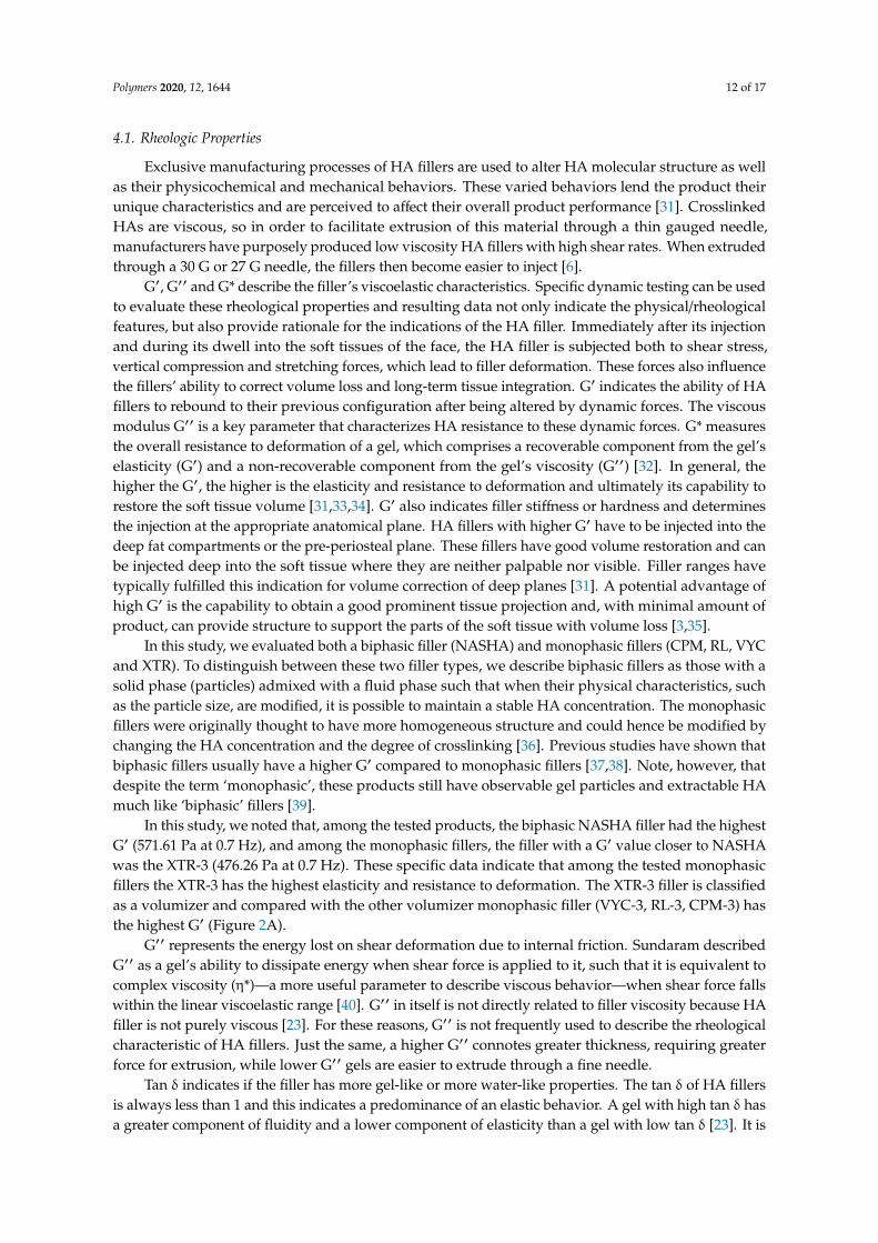
4.1. Rheologic Properties
Exclusive manufacturing processes of HA fillers are used to alter HA molecular structure as well as their physicochemical and mechanical behaviors. These varied behaviors lend the product their unique characteristics and are perceived to affect their overall product performance [31]. Crosslinked HAs are viscous, so in order to facilitate extrusion of this material through a thin gauged needle, manufacturers have purposely produced low viscosity HA fillers with high shear rates. When extruded through a 30 G or 27 G needle, the fillers then become easier to inject [6].
G′, G′′ and G* describe the filler’s viscoelastic characteristics. Specific dynamic testing can be used to evaluate these rheological properties and resulting data not only indicate the physical/rheological features, but also provide rationale for the indications of the HA filler. Immediately after its injection and during its dwell into the soft tissues of the face, the HA filler is subjected both to shear stress, vertical compression and stretching forces, which lead to filler deformation. These forces also influence the fillers’ ability to correct volume loss and long-term tissue integration. G′ indicates the ability of HA fillers to rebound to their previous configuration after being altered by dynamic forces. The viscous modulus G′′ is a key parameter that characterizes HA resistance to these dynamic forces. G* measures the overall resistance to deformation of a gel, which comprises a recoverable component from the gel’s elasticity (G′) and a non-recoverable component from the gel’s viscosity (G′′) [32]. In general, the higher the G′, the higher is the elasticity and resistance to deformation and ultimately its capability to restore the soft tissue volume [31,33,34]. G′ also indicates filler stiffness or hardness and determines the injection at the appropriate anatomical
0
20
40
60
80
100
120
0 30 60 300
Solu
ble
frac
tion
(%)
t (min)
NASHA XTR-1 XTR-2 XTR-3
Figure 5. Percentage of soluble fraction for each gel type (mean and standard deviation).
4. Discussion
In this study, the aim was to determine the differences among rheological and physicochemicalproperties of fillers manufactured using XTR™ technology in comparison with other filler ranges ofvarying proprietary crosslinking technologies. Most of the fillers selected in this study have beenused extensively in the past for various indications. In this simple series of laboratory evaluations,fillers manufactured using XTR™ technology were found to have distinct properties that set themapart from these filler ranges. The clinical implications of these unique characteristics are discussed.
Polymers 2020, 12, 1644 12 of 17
4.1. Rheologic Properties
Exclusive manufacturing processes of HA fillers are used to alter HA molecular structure as wellas their physicochemical and mechanical behaviors. These varied behaviors lend the product theirunique characteristics and are perceived to affect their overall product performance [31]. CrosslinkedHAs are viscous, so in order to facilitate extrusion of this material through a thin gauged needle,manufacturers have purposely produced low viscosity HA fillers with high shear rates. When extrudedthrough a 30 G or 27 G needle, the fillers then become easier to inject [6].
G′, G′′ and G* describe the filler’s viscoelastic characteristics. Specific dynamic testing can be usedto evaluate these rheological properties and resulting data not only indicate the physical/rheologicalfeatures, but also provide rationale for the indications of the HA filler. Immediately after its injectionand during its dwell into the soft tissues of the face, the HA filler is subjected both to shear stress,vertical compression and stretching forces, which lead to filler deformation. These forces also influencethe fillers’ ability to correct volume loss and long-term tissue integration. G′ indicates the ability of HAfillers to rebound to their previous configuration after being altered by dynamic forces. The viscousmodulus G′′ is a key parameter that characterizes HA resistance to these dynamic forces. G* measuresthe overall resistance to deformation of a gel, which comprises a recoverable component from the gel’selasticity (G′) and a non-recoverable component from the gel’s viscosity (G′′) [32]. In general, thehigher the G′, the higher is the elasticity and resistance to deformation and ultimately its capability torestore the soft tissue volume [31,33,34]. G′ also indicates filler stiffness or hardness and determinesthe injection at the appropriate anatomical plane. HA fillers with higher G′ have to be injected into thedeep fat compartments or the pre-periosteal plane. These fillers have good volume restoration and canbe injected deep into the soft tissue where they are neither palpable nor visible. Filler ranges havetypically fulfilled this indication for volume correction of deep planes [31]. A potential advantage ofhigh G′ is the capability to obtain a good prominent tissue projection and, with minimal amount ofproduct, can provide structure to support the parts of the soft tissue with volume loss [3,35].
In this study, we evaluated both a biphasic filler (NASHA) and monophasic fillers (CPM, RL, VYCand XTR). To distinguish between these two filler types, we describe biphasic fillers as those with asolid phase (particles) admixed with a fluid phase such that when their physical characteristics, suchas the particle size, are modified, it is possible to maintain a stable HA concentration. The monophasicfillers were originally thought to have more homogeneous structure and could hence be modified bychanging the HA concentration and the degree of crosslinking [36]. Previous studies have shown thatbiphasic fillers usually have a higher G′ compared to monophasic fillers [37,38]. Note, however, thatdespite the term ‘monophasic’, these products still have observable gel particles and extractable HAmuch like ‘biphasic’ fillers [39].
In this study, we noted that, among the tested products, the biphasic NASHA filler had the highestG′ (571.61 Pa at 0.7 Hz), and among the monophasic fillers, the filler with a G′ value closer to NASHAwas the XTR-3 (476.26 Pa at 0.7 Hz). These specific data indicate that among the tested monophasicfillers the XTR-3 has the highest elasticity and resistance to deformation. The XTR-3 filler is classifiedas a volumizer and compared with the other volumizer monophasic filler (VYC-3, RL-3, CPM-3) hasthe highest G′ (Figure 2A).
G′′ represents the energy lost on shear deformation due to internal friction. Sundaram describedG′′ as a gel’s ability to dissipate energy when shear force is applied to it, such that it is equivalent tocomplex viscosity (η*)—a more useful parameter to describe viscous behavior—when shear force fallswithin the linear viscoelastic range [40]. G′′ in itself is not directly related to filler viscosity because HAfiller is not purely viscous [23]. For these reasons, G′′ is not frequently used to describe the rheologicalcharacteristic of HA fillers. Just the same, a higher G′′ connotes greater thickness, requiring greaterforce for extrusion, while lower G′′ gels are easier to extrude through a fine needle.
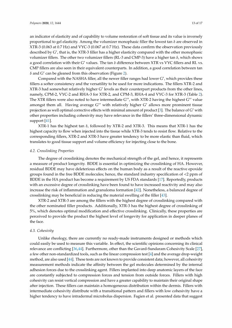
Tan δ indicates if the filler has more gel-like or more water-like properties. The tan δ of HA fillersis always less than 1 and this indicates a predominance of an elastic behavior. A gel with high tan δ hasa greater component of fluidity and a lower component of elasticity than a gel with low tan δ [23]. It is
Polymers 2020, 12, 1644 13 of 17
an indicator of elasticity and of capability to volume restoration of soft tissue and its value is inverselyproportional to gel elasticity. Among the volumizer monophasic filler the lowest tan δ are observed inXTR-3 (0.063 at 0.7 Hz) and VYC-3 (0.067 at 0.7 Hz). These data confirm the observation previouslydescribed by G′, that is, the XTR-3 filler has a higher elasticity compared with the other monophasicvolumizer fillers. The other two volumizer fillers (RL-3 and CMP-3) have a higher tan δ, which showsa good correlation with their G′ values. The tan δ difference between XTR vs VYC fillers and RL vs.CMP fillers are also seen in their equivalent counterparts. In addition, a good correlation between tanδ and G′ can be gleaned from this observation (Figure 2).
Compared with the NASHA filler, all the newer filler ranges had lower G′, which provides thesefillers a softer consistency and the versatility to be used for more indications. The fillers XTR-2 andXTR-3 had somewhat relatively higher G′ levels as their counterpart products from the other lines,namely, CPM-2, VYC-2 and RHA-3 for XTR-2, and CPM-3, RHA-4 and VYC-3 for XTR-3 (Table 2).The XTR fillers were also noted to have intermediate G′′, with XTR-2 having the highest G′′ valueamongst them all. Having average G′′ with relatively higher G′ allows more prominent tissueprojection as well optimal corrective effects with minimal amount of product [3]. The balance of G′ withother properties including cohesivity may have relevance in the fillers’ three-dimensional dynamicsupport [41].
XTR-1 has the highest tan δ, followed by XTR-2 and XTR-3. This means that XTR-1 has thehighest capacity to flow when injected into the tissue while XTR-3 tends to resist flow. Relative to thecorresponding fillers, XTR-2 and XTR-3 have greater tendency to be more elastic than fluid, whichtranslates to good tissue support and volume efficiency for injecting close to the bone.
4.2. Crosslinking Properties
The degree of crosslinking denotes the mechanical strength of the gel, and hence, it representsa measure of product longevity. BDDE is essential in optimizing the crosslinking of HA. However,residual BDDE may have deleterious effects on the human body as a result of the reactive epoxidegroups found in the free BDDE molecules; hence, the standard industry specification of <2 ppm ofBDDE in the HA product has become a requirement by US FDA standards [17]. Reportedly, productswith an excessive degree of crosslinking have been found to have increased reactivity and may alsoincrease the risk of inflammation and granuloma formation [42]. Nonetheless, a balanced degree ofcrosslinking may be beneficial in reducing the material swelling of the filler [43].
XTR-2 and XTR-3 are among the fillers with the highest degree of crosslinking compared withthe other nominated filler products. Additionally, XTR-3 has the highest degree of crosslinking of5%, which denotes optimal modification and effective crosslinking. Clinically, these properties areperceived to provide the product the highest level of longevity for application in deeper planes ofthe face.
4.3. Cohesivity
Unlike rheology, there are currently no ready-made instruments designed or methods whichcould easily be used to measure this variable. In effect, the scientific opinions concerning its clinicalrelevance are conflicting [36,44]. Furthermore, other than the Gavard-Sundaram Cohesivity Scale [27],a few other non-standardized tools, such as the linear compression test [4] and the average drop-weightmethod, are also used [44]. These tests are not known to provide consistent data; however, all cohesivitymeasurement methods indicate the affinity between the gel molecules determined by the internaladhesion forces due to the crosslinking agent. Fillers implanted into deep anatomic layers of the faceare constantly subjected to compression forces and tension from outside forces. Fillers with highcohesivity can resist vertical compression and have a greater capability to maintain their original shapeafter injection. These fillers can maintain a homogeneous distribution within the dermis. Fillers withintermediate cohesivity distribute with a transitional pattern and fillers with low cohesivity have ahigher tendency to have intradermal microbolus dispersion. Fagien et al. presented data that suggest

Polymers 2020, 12, 1644 14 of 17
that as G′ decreases, the gel may exhibit more cohesive properties [31]. The same inverse relationshipwas noted by Edsman et al. [44] further supporting the observation that this relationship appeared toexist only among products made by the same technology [31]. Hence, when comparing the lastingeffects of two HA fillers with the same G′ but different cohesivity, the filler with lower cohesivity losestissue projection more easily than fillers with higher cohesivity.
Our study likewise presents this inverse correlation between G′ and cohesivity: fillers with higherG′ (NASHA) have lower cohesivity, fillers with intermediate G′ (XTR and VYC) have intermediatecohesivity and fillers with lower G′ (RL and CPM) have higher cohesivity. The NASHA filler having alower cohesivity may be attributed to its biphasic structural characteristic.
G′ and cohesivity are related to the capability of soft tissue volume restoration of the fillers,and there appears to be a relationship between them where one compensates for the lack of the other.The two characteristics determine the behavior of the fillers after injection and therefore also the clinicalresults. The mechanistic relationship between these has yet to be established by further studies.
Our study partially confirms the inverse relationship between cohesivity and G′. Cohesivity withinthe groups of XTR and VYC fillers is stable. On the other hand, in the RL and CPM groups, cohesivityincreases with lower G′ (Figure 4D). The cohesivity data of NASHA, VYC and CPM fillers confirm thebehavior described in the pilot validation study of the Gavard-Sundaram Cohesivity Scale [27].
Among the monophasic fillers, the XTR group had the higher G′ and an intermediate cohesivity,while the VYC group has lower G′ and lower cohesivity, and the RL and CPM groups have significantlylower G′ and higher cohesivity. This data might indicate a good volumizing capability by XTR fillerscompared with other fillers evaluated in this study, but further research is necessary to establish this.
4.4. Degradability
The general assumption is that fillers with higher HA concentration and stronger degreeof crosslinking are more likely to be resistant to enzymatic degradation. Therefore, additionalhyaluronidase sessions are usually needed for patients receiving fillers with higher HAconcentration [10]. However, different studies have demonstrated contradictory results attributed todisparities in hyaluronidase source (bovine, ovine, recombinant human), doses used, as well as diverseexperimental formats [9].
Results of this stability study are consistent with the crosslinking evaluation, such that XTR-2and XTR-3 with the highest degree of crosslinking were shown to be more stable to degradationthan NASHA after 5 h of hyaluronidase exposure. Further exploration to a prolonged exposure time,evaluation of a dose-dependent response and use of a broader range of hyaluronidases in vivo mayprovide more insights to these observations.
4.5. Clinical Application, Limitations and Recommendations
Filler ranges come in a series of increasing concentrations, which are supposed to distinguish theirapplication and purpose. Interestingly, concentration alone does not by itself control filler performance.Filler rheological properties, namely the elastic modulus, viscous modulus, tan δ and cohesivity, alongwith crosslinking properties, manufacturing processes and injector technique are observed to morelikely predict the longevity and persistence of HA fillers in tissue [38]. All these features contribute todefining the global physical characteristics of the HA filler, whose sum of all properties determinesits capability to modify soft tissue. In addition, ‘higher’ does not always necessarily translate to‘better’—such that a filler with higher G′ may have greater strength but does not necessarily have agreater ability to lift [45]. Therefore, it is important to interpret these biophysical factors holistically,together with their tissue integration and biostimulatory capabilities in perfect balance to determinetheir suitability in patients with certain profiles (i.e., with specific tissue and anatomical characteristics).
When using the products with XTR™ technology, the general suggestion is to inject less thanusual to avoid instilling excessive deposits in the soft tissue. The stronger gels, XTR-2 and XTR-3,are expected to produce long-term and prominent projection with the use of only small amounts of
Polymers 2020, 12, 1644 15 of 17
product in both subcutaneous and supraperiosteal injections. The sequence of G′ and cohesivity ofthe fillers made with XTR™ technology can be observed to be consistent with the indicated depths ofinjection, as well as their clinical application.
The current data is limited because of the relatively short period that XTR™ Technology has beenin the market. More long-term efficacy and safety studies are expected to provide additional insightsto the clinical advantages of the properties evaluated in this study. We also aim to conduct furtherstudies on the biodegradability, in vivo kinetics, biostimulatory properties and tissue integration of thefillers to ensure a comprehensive evaluation. This premier study of physicochemical and rheologicalproperties of fillers manufactured using XTR™ technology evaluated through laboratory testing isexpected to exemplify an evidence-based approach in the selection of fillers. This, in turn, will help inthe procedural application of fillers, ensuring optimal outcomes in volume restoration, improvementof volume distribution and, ultimately, achieving a balanced facial contour.
5. Conclusions
An evidence-based approach to selection of fillers based on rheologic and other physicochemicalproperties, combined with proper technique, may facilitate the choice of the best product, achievingoptimal results. Since the incorporation of local anesthetic has become standard practice in fillermanufacturing, there is a need for exploration of the physicochemical properties of these fillersupon integration in soft tissue. Lastly, owing to the findings reported in this article, fillers withXTR™ technology may be a viable option for patients wanting an option with lasting lifting andrejuvenating effects.
Author Contributions: G.S. and S.P.F. provided equal contributions to the article. All authors have read andagreed to the published version of the manuscript.
Funding: We would like to thank RELIFE S.r.l. for providing an unrestricted educational grant for the purpose ofwriting this manuscript.
Acknowledgments: We would like to thank Dennis Malvin H. Malgapo, MD, DFM, ISMPP CMPP™, for hismedical writing services.
Conflicts of Interest: The authors have consultancy contracts with RELIFE S.r.l.
References
1. La Gatta, A.; Salzillo, R.; Catalano, C.; D’Agostino, A.; Pirozzi AV, A.; De Rosa, M.; Schiraldi, C.Hyaluronan-based hydrogels as dermal fillers: The biophysical properties that translate into a “volumetric”effect. PLoS ONE 2019, 14, 1–17. [CrossRef] [PubMed]
2. Smith, L.; Cockerham, K. Hyaluronic acid dermal fillers: Can adjunctive lidocaine improve patient satisfactionwithout decreasing efficacy or duration? Patient Prefer. Adherence 2011, 5, 133–139. [PubMed]
3. Salti, G. Meet the Innovative XTRTM (eXcellent Three-dimensional Reticulation) Technology of DefinisseTM
Filler. Prime J. 2020, 10, 84–85.4. Borrell, M.; Leslie, D.B.; Tezel, A. Lift capabilities of hyaluronic acid fillers. J. Cosmet. Laser Ther. 2011, 13,
21–27. [CrossRef] [PubMed]5. Micheels, P.; Sarazin, D.; Tran, C.; Salomon, D. Effect of Different Crosslinking Technologies on Hyaluronic
Acid Behavior: A Visual and Microscopic Study of Seven Hyaluronic Acid Gels. J. Drugs Derm. 2016, 15,600–606.
6. Gavard Molliard, S.; Bon Bétemps, J.; Hadjab, B.; Topchian, D.; Micheels, P.; Salomon, D. Key rheologicalproperties of hyaluronic acid fillers: From tissue integration to product degradation. Plast. Aesthetic Res.2018, 5, 17. [CrossRef]
7. Lee, W.; Yoon, J.-H.; Koh, I.-S.; Oh, W.; Kim, K.-W.; Yang, E.-J. Clinical application of a new hyaluronic acidfiller based on its rheological properties and the anatomical site of injection. Biomed. Dermatol. 2018, 2, 1–5.[CrossRef]
8. Rohrich, R.J.; Bartlett, E.L.; Dayan, E. Practical Approach and Safety of Hyaluronic Acid Fillers. Plast. Reconstr.Surg. Glob. Open 2019, 7, e2172. [CrossRef]

Polymers 2020, 12, 1644 16 of 17
9. Buhren, B.A.; Schrumpf, H.; Bölke, E.; Kammers, K.; Gerber, P.A. Standardized in vitro analysis of thedegradability of hyaluronic acid fillers by hyaluronidase. Eur. J. Med. Res. 2018, 23, 1–6. [CrossRef]
10. Cavallini, M.; Papagni, M.; Trocchi, G. Sensitivity of Hyaluronic Acid Fillers to Hyaluronidase: An in vitroAnalysis. J. Clin. Exp. 2020, 11, 1–6. [CrossRef]
11. Park, S.; Park, K.Y.; Yeo, I.K.; Cho, S.Y.; Ah, Y.C.; Koh, H.J.; Park, W.S.; Kim, B.J. Investigation of thedegradation-retarding effect caused by the low swelling capacity of a novel hyaluronic acid filler developedby solid-phase crosslinking technology. Ann. Dermatol. 2014, 26, 357–362. [CrossRef] [PubMed]
12. Rigano Laboratories. Analysis of Different Types of Injectable Cross-Linked Hyaluronic Acid Gels: Determination ofDegree of Cross-Linking in Hyaluronic Acid-Based Filler; Rigano Laboratories S.r.l.: Milan, Italy, 2019.
13. Stern, R.; Kogan, G.; Jedrzejas, M.J.; Soltes, L. The many ways to cleave hyaluronan. Biotechnol. Adv. 2007, 25,537–557. [CrossRef] [PubMed]
14. Tokita, Y.; Okamoto, A. Degradation of hyaluronic acid—Kinetic study and thermodynamics. Eur. Polym. J.1996, 32, 1011–1014. [CrossRef]
15. Lekander, M.; Troncoso, J.F.; Idjbara, A.; Karlsson, I.; Lindgren, T.; Ström, S. A kinetic study of thedegradation of hyaluronic acid at high concentrations of sodium hydroxide. Mater. Sci. 2016, 1, 1–32,URN: urn:nbn:se:uu:diva-301379.
16. Tran, C.; Carraux, P.; Micheels, P.; Kaya, G.; Salomon, D. In vivo bio-integration of three hyaluronic acidfillers in human skin: A histological study. Dermatology 2014, 228, 47–54. [CrossRef]
17. De Boulle, K.; Glogau, R.; Kono, T.; Nathan, M.; Tezel, A.; Roca-Martinez, J.-X.; Paliwal, S.; Stroumpoulis, D.A review of the metabolism of 1,4-butanediol diglycidyl ether-crosslinked hyaluronic acid dermal fillers.Dermatol. Surg. 2013, 39, 1758–1766. [CrossRef]
18. Guarise, C.; Barbera, C.; Pavan, M.; Panfilo, S.; Beninatto, R.; Galesso, D. HA-based dermal filler: Downstreamprocess comparison, impurity quantitation by validated HPLC-MS analysis, and in vivo residence timestudy. J. Appl. Biomater. Funct. Mater. 2019, 17, 2280800019867075. [CrossRef]
19. US NIH. Restylane + Lidocaine and Restylane Lyft for the Treatment of Nasolabial Folds. Available online:https://clinicaltrials.gov/ct2/show/NCT04174131 (accessed on 17 March 2020).
20. Woodward, J.A. Periocular fillers and related anatomy. Cutis 2016, 98, 330–335.21. Kühne, U.; Esmann, J.; Von Heimburg, D.; Imhof, M.; Weissenberger, P.; Sattler, G.; Heinz, M.; Kliebe-Frisch, C.
Safety and performance of cohesive polydensified matrix hyaluronic acid fillers with lidocaine in the clinicalsetting—An open-label, multicenter study. Clin. Cosmet. Investig. Dermatol. 2016, 9, 373–381. [CrossRef]
22. Rigano Laboratories. Determination of the Viscoelastic Properties of HA Fillers; Rigano Laboratories S.r.l.: Milan,Italy, 2019.
23. Pierre, S.; Liew, S.; Bernardin, A. Basics of dermal filler rheology. Dermatol. Surg. 2015, 41 (Suppl. 1),S120–S126. [CrossRef]
24. Schanté, C.E.; Zuber, G.; Herlin, C.; Vandamme, T.F. Improvement of hyaluronic acid enzymatic stability bythe grafting of amino-acids. Carbohydr. Polym. 2012, 87, 2211–2216. [CrossRef]
25. Wende, F.J.; Gohil, S.; Nord, L.I.; Helander Kenne, A.; Sandström, C. 1D NMR methods for determinationof degree of cross-linking and BDDE substitution positions in HA hydrogels. Carbohydr. Polym. 2017, 157,1525–1530. [CrossRef] [PubMed]
26. Kenne, L.; Gohil, S.; Nilsson, E.M.; Karlsson, A.; Ericsson, D.; Helander Kenne, A.; Nord, L.I. Modification andcross-linking parameters in hyaluronic acid hydrogels–definitions and analytical methods. Carbohydr. Polym.2013, 91, 410–418. [CrossRef] [PubMed]
27. Sundaram, H.; Rohrich, R.J.; Liew, S.; Sattler, G.; Talarico, S.; Trevidic, P.; Molliard, S.G. Cohesivity ofHyaluronic Acid Fillers: Development and Clinical Implications of a Novel Assay, Pilot Validation witha Five-Point Grading Scale, and Evaluation of Six U.S. Food and Drug Administration-Approved Fillers.Plast. Reconstr. Surg. 2015, 136, 678–686. [CrossRef]
28. Rigano Laboratories. Analysis of Different Types of Injectable Cross-Linked Hyaluronic Acid Gels: Cohesivity ofCross-Linked Hyaluronic Acid; Rigano Laboratories S.r.l.: Milan, Italy, 2019.
29. La Gatta, A.; De Rosa, M.; Frezza, M.A.; Catalano, C.; Meloni, M.; Schiraldi, C. Biophysical and biologicalcharacterization of a new line of hyaluronan-based dermal fillers: A scientific rationale to specific clinicalindications. Mater. Sci. Eng. C. Mater. Biol. Appl. 2016, 68, 565–572. [CrossRef]
30. Rigano Laboratories. Analysis of Different Types of Injectable Cross-Linked Hyaluronic Acid Gels; Rigano LaboratoriesS.r.l.: Milan, Italy, 2020.

Polymers 2020, 12, 1644 17 of 17
31. Fagien, S.; Bertucci, V.; von Grote, E.; Mashburn, J.H. Rheologic and Physicochemical Properties Usedto Differentiate Injectable Hyaluronic Acid Filler Products. Plast. Reconstr. Surg. 2019, 143, 707e–720e.[CrossRef]
32. Stocks, D.; Sundaram, H.; Michaels, J.; Durrani, M.J.; Wortzman, M.S.; Nelson, D.B. Rheological evaluationof the physical properties of hyaluronic acid dermal fillers. J. Drugs Dermatol. 2011, 10, 974–980.
33. Edsman, K.; Nord, L.I.; Ohrlund, A.; Lärkner, H.; Kenne, A.H. Gel properties of hyaluronic acid dermalfillers. Dermatol. Surg. 2012, 38, 1170–1179. [CrossRef]
34. Lorenc, Z.P.; Öhrlund, Å.; Edsman, K. Factors Affecting the Rheological Measurement of Hyaluronic AcidGel Fillers. J. Drugs Dermatol. 2017, 16, 876–882.
35. Kablik, J.; Monheit, G.D.; Yu, L.; Chang, G.; Gershkovich, J. Comparative physical properties of hyaluronicacid dermal fillers. Dermatol. Surg. 2009, 35 (Suppl. 1), 302–312. [CrossRef]
36. Flynn, T.C.; Sarazin, D.; Bezzola, A.; Terrani, C.; Micheels, P. Comparative histology of intradermalimplantation of mono and biphasic hyaluronic acid fillers. Dermatol. Surg. 2011, 37, 637–643. [CrossRef]
37. Sundaram, H.; Voigts, B.; Beer, K.; Meland, M. Comparison of the rheological properties of viscosity andelasticity in two categories of soft tissue fillers: Calcium hydroxylapatite and hyaluronic acid. Dermatol. Surg.2010, 36 (Suppl. 3), 1859–1865. [CrossRef] [PubMed]
38. Santoro, S.; Russo, L.; Argenzio, V.; Borzacchiello, A. Rheological properties of cross-linked hyaluronic aciddermal fillers. J. Appl. Biomater. Biomech. 2011, 9, 127–136. [CrossRef] [PubMed]
39. Öhrlund, J.Å.; Edsman, K.L.M. The Myth of the ‘Biphasic’ Hyaluronic Acid Filler. Dermatol. Surg. 2015, 41(Suppl. 1), S358–S364.
40. Sundaram, H. Going with the Flow: An Overview and Clinical Discussion of The Rheologyof Soft Tissue Fillers, Part 1 of 2. Pract. Dermatol. 2010, 11, 21–24. Available online:https://practicaldermatology.com/articles/2010-nov/cosmetics-challenge-going-with-the-flow-an-overview-and-clinical-discussion-of-the-rheology-of-soft-tissue-fillers-part-1-of-2/pdf (accessed on 1 July 2020).
41. Sundaram, H.; Liew, S.; Signorini, M.; Vieira Braz, A.; Fagien, S.; Swift, A.; De Boulle, K.L.; Raspaldo, H.;Trindade De Almeida, A.R.; Monheit, G. Global Aesthetics Consensus: Hyaluronic Acid Fillers and BotulinumToxin Type A-Recommendations for Combined Treatment and Optimizing Outcomes in Diverse PatientPopulations. Plast. Reconstr. Surg. 2016, 137, 1410–1423. [CrossRef]
42. Funt, D.; Pavicic, T. Dermal fillers in aesthetics: An overview of adverse events and treatment approaches.Clin. Cosmet. Investig. Dermatol. 2013, 6, 295–316. [CrossRef]
43. Lee, D.Y.; Cheon, C.; Son, S.; Kim, Y.Z.; Kim, J.T.; Jang, J.W.; Kim, S.S. Influence of molecular weight onswelling and elastic modulus of hyaluronic acid dermal fillers. Polymer 2015, 39, 976–980. [CrossRef]
44. Edsman, K.L.M.; Wiebensjö, Å.M.; Risberg, A.M.; Öhrlund, J.Å. Is There a Method That Can MeasureCohesivity? Cohesion by Sensory Evaluation Compared With Other Test Methods. Dermatol. Surg. 2015, 41(Suppl. 1), S365–S372. [CrossRef]
45. Niamtu, J. 10—Injectable Fillers: Lip Augmentation, Lip Reduction, and Lip Lift. Cosmet. Facial Surg.In Cosmetic Facial Surgery; Elsevier: Amsterdam, The Netherlands, 2018. [CrossRef]
© 2020 by the authors. Licensee MDPI, Basel, Switzerland. This article is an open accessarticle distributed under the terms and conditions of the Creative Commons Attribution(CC BY) license (http://creativecommons.org/licenses/by/4.0/).