evaluation of antimalarial properties of indigenous
192
EVALUATION OF ANTIMALARIAL PROPERTIES OF INDIGENOUS PLANTS USED BY TRADITIONAL HEALERS IN NAMIBIA A THESIS SUBMITTED IN PARTIAL FULFILMENT OF THE REQUIREMENTS FOR THE DEGREE OF MASTER OF SCIENCE OF UNIVERSITY OF NAMIBIA BY IWANETTE DU PREEZ FEBRUARY 2012 Main Supervisor: Dr. D. Mumbengegwi (Multidisciplinary Research Centre, University of Namibia) Co-supervisor: Dr. R. Böck (Department of Biologicial Sciences, University of Namibia)
-
Upload
khangminh22 -
Category
Documents
-
view
3 -
download
0
Transcript of evaluation of antimalarial properties of indigenous

EVALUATION OF ANTIMALARIAL PROPERTIES OF INDIGENOUS
PLANTS USED BY TRADITIONAL HEALERS IN NAMIBIA
A THESIS SUBMITTED IN PARTIAL FULFILMENT
OF THE REQUIREMENTS FOR THE DEGREE OF
MASTER OF SCIENCE
OF
UNIVERSITY OF NAMIBIA
BY
IWANETTE DU PREEZ
FEBRUARY 2012
Main Supervisor: Dr. D. Mumbengegwi (Multidisciplinary Research Centre,
University of Namibia)
Co-supervisor: Dr. R. Böck (Department of Biologicial Sciences,
University of Namibia)

ii
ABSTRACT
Malaria is on the decline in Namibia and the country is moving towards pre-elimination
of the disease. However, some communities preferring traditional medicines and not
accepting allopathic medicine may prevent elimination. Ethnomedicines need to be
integrated into mainstream malaria case management to achieve malaria elimination by
2020. To do so, they need to be documented and validated to allow their safe and
effective use. Seven indigenous plants were investigated for antiplasmodia properties on
the basis of their indigenous uses to treat malaria-like ailments. Extracts of Vahlia
capensis, Nicolasia costata, Rhigozum brevispinosum, Dicerocarym eriocarpum, Senna
occidentalis, Lophiocarpus sp. and Crotalaria flavicarinata were prepared using
aqueous and organic solvents. Phytochemical screening was performed to detect the
presence of selected antiplasmodial compounds. Growth inhibition studies using cellular
infection models of Plasmodium falciparum 3D7 and D10 were carried out to determine
anti-plasmodial effects of extracts from Vahlia capensis, Nicolasia costata and
Dicerocarym eriocarpum. Mechanistic studies were also conducted to determine the
mode of action of the three plants. Phytochemical screening revealed the presence of
alkaloids, anthraquinones, flavonoids, terpenoids, steroids, coumarins and glycosides in
the plant extracts with each plant possessing at least 3 classes of the antimalarial
compounds. Organic extracts of V. capensis, N. costata and D. eriocarpum showed
antiplasmodial activity at concentrations ranging from 50-250 µg/mL. Extracts from
D. eriocarpum showed the highest activity with an IC50 of 63.17µg/mL followed by

iii
V. capensis and N. costata at 93.29µg/mL and 86.63µg/mL, respectively. All the plant
extracts inhibited haemazoin accumulation with D. eriocarpum exhibiting the highest
inhibition. The extracts also inhibited protease activity at the early ring stage where
infection of red blood cells was being established and at the trophozoite stage where
metabolism of the parasites was increased. These results support the ethno-medicinal
uses for these plants as complementary medicine for malaria.
Key words medicinal plants, traditional knowledge, complementary medicine, malaria,
phytochemical screening, antimalarial compounds, alkaloids, anthraquinones,
coumarins, flavonoids, glycosides, steroids, terpenoids, in vitro antiplasmodial activity,
mode of action

iv
TABLE OF CONTENTS
ABSTRACT ....................................................................................................................... ii
ABBREVIATIONS ........................................................................................................viii
FIGURES ......................................................................................................................... xii
TABLES ......................................................................................................................... xiv
ACKNOWLEDGMENTS………………………………………………………..…….xvi
DECLARATIONS .......................................................................................................... xix
PUBLICATIONS AND CONFERENCE PROCEEDINGS ........................................... xx
CHAPTER 1: INTRODUCTION .................................................................................. 1
1.1 General introduction ............................................................................................ 1
1.2 Availability of control measures in the fight against malaria .............................. 6
1.3 Plant medicinal value ........................................................................................... 8
1.4 Documentation and validation of traditional knowledge................................... 10
CHAPTER 2: LITERATURE REVIEW .................................................................... 13
2.1 Plant Medicinal Value and malaria control ....................................................... 13
2.1.1 Secondary metabolites ...................................................................................... 15
2.1.2 Traditional plant medicine in Namibia ............................................................. 25
2.1.3 Drug discovery from antimalarial natural products .......................................... 31

v
2.1.4 Economic benefits of medicinal plants ............................................................. 32
2.1.5 Conservation of medicinal plants ...................................................................... 35
2.2 Malaria, the disease ........................................................................................... 37
2.2.1 Transmission ..................................................................................................... 37
2.2.2 Life cycle of malaria parasites .......................................................................... 38
2.2.3 Diagnostics and clinical symptoms ................................................................... 40
2.2.4 Prevention and treatment of malarial infections ............................................... 42
2.2.5 Intracellular sites of action of antimalarials ...................................................... 48
2.2.6 Drug resistance .................................................................................................. 50
2.3 Statement of the problem .................................................................................. 53
2.4 Aims and objectives .......................................................................................... 54
2.5 Research hypothesis .......................................................................................... 54
2.6 Significance of the study ................................................................................... 55
CHAPTER 3: MATERIALS AND METHODS ........................................................ 56
3.1 Research design ................................................................................................. 56
3.2 Plant collection .................................................................................................. 57
3.3 Preparation of crude extracts ............................................................................. 58
3.4 Phytochemical analysis ...................................................................................... 59
3.4.1 Thin layer chromatography ............................................................................... 60

vi
3.4.2 Preliminary phytochemical screening ............................................................... 61
3.5 Antimalarial bioassay ........................................................................................ 64
3.5.1 Culture technique .............................................................................................. 64
3.5.2 Preparation of stock solutions ........................................................................... 68
3.5.3 Analysis of antimalarial activity ....................................................................... 69
3.5.4 Microscopic analysis ......................................................................................... 69
3.5.5 Data analysis ..................................................................................................... 70
3.6 Mechanism-based assays ................................................................................... 70
3.6.1 Pyridine-hemochrome to measure haem incorporation in hemozoin ............... 70
3.6.2 Protease inhibition assay ................................................................................... 71
CHAPTER 4: RESULTS ............................................................................................. 73
4.1 Ethnopharmacological approach for plant selection/collection ........................ 73
4.2 Effect of extracting solvents on percentage yield ............................................. 80
4.3 Preliminary phytochemical screening ............................................................... 81
4.3.1 Fractionation of crude extracts using thin layer chromatography….…………81
4.3.2 Presence of antimalarial compounds based on TLC profiling.............……….83
4.3.3 Presence of selected antimalarial compounds based on phytochemical tests...85
4.4 Antimalarial activity of the investigated plant extracts ..................................... 87
4.5 Metabolic-based assays ..................................................................................... 96

vii
4.5.1 Pyridine-hemochrome to measure haem incorporation in hemozoin ............... 96
4.5.2 Protease inhibition assay ................................................................................. 98
CHAPTER 5: DISCUSSION ..................................................................................... 100
CHAPTER 6: CONCLUSION AND RECOMMENDATIONS ............................. 116
CHAPTER 7: REFERENCES ................................................................................... 118
APPENDICES .............................................................................................................. 144

viii
ABBREVIATIONS
ACT Artemisinin-based Combination Therapy
AE Aqueous extract(s)
Al Alkaloids
AL Artemether-Lumefantrine
An Anthraquinones
C Coumarins
Ca2+
Calcium-ion
CNS Central nervous system
CO2 Carbon oxide
DCM Dichloromethane
DDT Diphenyl-tricthloroethane
DE Dicerocaryum eriocarpum
DHA Dihydroartemisinin
DMSO Dimethylsulphoxide
F Flavonoids

ix
F254 Florescence at 254nm
FITC Fluorescein isothiocyanate
G Glycosides
GPS Global positioning satellites
H2O Water
HCl Hydrochloric acid
IC50 50% inhibitory concentration
IPT Intermittent preventive treatment
IRS Indoor residual spraying
ITN Insecticide-treated Nets
IV Intravenous
KH2PO4 Potassium dihydrogen orthophosphate
LLIN Long lasting insecticide-treated nets
MCC Millennium challenge corporation
MDA Mass drug administration
MeOH Methanol

x
MET Ministry of Environment and Tourism
MoHSS Ministry of Health and Social Services
N2 Nitrogen
Na2HPO4 Sodium dihydrogen orthophosphate
NaHCO3 Sodium hydrogen carbonate
NaHPO4 Sodium hydrogen phosphate
NaOH Sodium hydroxide
NBRI National Botanical Research Institute
NC Nicolasia costata
O2 Oxygen
OD560 Optical density at 560nm
OE Organic extract
P Parasitaemia
PPT Presumptive preventive treatment
RBCs Red blood cells
RDTs Rapid diagnostic tests
xi
S Steroids
SEPASAL Survey of economic plants for arid and semi-arid lands
SP Sulphadoxine-Pyrimethamine
SS Solvent system
T Terpenoids
TK Traditional Knowledge
TLC Thin Layer Chromatography
UV Ultraviolet
VC Vahlia capensis
WHO World Health Organization

xii
FIGURES
Figure 1: Combined Evidence Map of Malaria Risk Areas in Namibia ........................... 3
Figure 2: Trend in Malaria Burden in Namibia from 2001-2008. ..................................... 4
Figure 3: Structure of an alkaloid, morphine isolated from plants. ................................ 17
Figure 4: Structure of a typical anthraquinone................................................................ 18
Figure 5: Structure of a typical coumarin. ...................................................................... 19
Figure 6: Structure of a flavonoid compound, flavonol. ................................................. 20
Figure 7: Structure of a typical glycoside. ...................................................................... 22
Figure 8: Structure of a typical steroid. .......................................................................... 23
Figure 9: Structure of a typical terpenoid. ...................................................................... 25
Figure 10: Map showing the vegetation cover (biomes) in Namibia. ............................. 26
Figure 11: The life cycle of the malaria parasite. ........................................................... 39
Figure 12: Flow chart showing the valorizing process of the ethnomedicinal uses of
................................................ 56 plants for the potential use as antimalarials in this study.
Figure 13: The geographical locations of sampled plants with their GPS coordinates in
the Oshikoto region. ......................................................................................................... 73
Figure 14: Photo of Vahlia capensis.. ............................................................................. 76
Figure 15: Photo of Nicolasia costata. ........................................................................... 77
Figure 16: Photo of Rhigozum brevispinosum. .............................................................. 77
Figure 17: Photo of Dicerocaryum eriocarpum .............................................................. 78

xiii
Figure 18: Photo of Senna occidentalis. ........................................................................ 78
Figure 19: Photo of Lophiocarpus sp. ............................................................................. 79
Figure 20: Photo of Crotalaria flavicarinata.. ............................................................... 79
Figure 21: Photo micrograph of red blood cells infected with P. falciparum 3D7
24 hours following treatment with plant extracts. ........................................................... 88
Figure 22: Photo micrograph of red blood cells infected with P. falciparum D10
24 hours following treatment with plant extracts. ........................................................... 89
Figure 23: Antiplasmodial activity of Vahlia capensis, Nicolasia costata and
Dicerocaryum eriocarpum extracts on Plasmodium falciparum 3D7 ............................. 91
Figure 24: The concentration dependent effect of the plant extracts on parasitaemia of
the Plasmodium 3D7 strains for Vahlia capensis, Nicolasia costata and Dicerocaryum
eriocarpum. ...................................................................................................................... 92
Figure 25: In vitro interaction of plant extracts for Vahlia capensis, Nicolasia costata
and Dicerocaryum eriocarpum (% inhibition) in chloroquine-sensitive strains. ............ 94
Figure 26: Antiplasmodial activity for Vahlia capensis, Nicolasia costata and
Dicerocaryum eriocarpum at 250µg/mL against the P. falciparum D10 strain ............. 95
Figure 27: Haemazoin accumulation for treated and untreated cultures ........................ 97
Figure 28: Protease activity in P. falciparum D10 for the different time points post
treatment with plant extracts . .......................................................................................... 99

xiv
TABLES
Table 1: The classification of terpenoids based on the number of isoprene units. ......... 24
Table 2: Ethno botanical data of the medicinal plants used by traditional healers in the
Engondi constituency, Ofilu village ................................................................................ 75
Table 3: Percentage yield for aqueous and organic extracts prepared ............................ 80
Table 4: TLC analysis of crude plant extracts fractionated using solvent system
1 (chloroform: methanol: acetone in a 1:2:1 ratio). ......................................................... 82
Table 5: TLC analysis of crude plant extracts fractionated using solvent system
2 (chloroform: ethyl acetate in a 1:1 ratio). ..................................................................... 82
Table 6: TLC analysis of crude plant extracts fractionated using solvent system
3 (chloroform: methanol: water in a 12:6:1 ratio)............................................................ 83
Table 7: TLC analysis for the presence of alkaloids, anthraquinones, coumarines,
flavonoids, glycosides, steroids and terpenoids ............................................................... 84
Table 8: Preliminary phytochemical screening for compounds with known antimalarial
properties .......................................................................................................................... 86
Table 9: Percent (%) growth inhibition of Plasmodium falciparum 37 exerted by
extracts ............................................................................................................................. 94
Table 10: Indigenous plants used as herbal medicine to treat ailments similar to the
symptoms of malaria in the Oshikoto, Ohangwena, Otjizondjupa, Caprivi and Khomas
regions of Namibia. ........................................................................................................ 144
Table 11: Key laws and policies influencing the INP sector in Namibia .................... 154

xv
Table 12: Yield of plant extracts (dry weight) from plant parts investigated. .............. 166
Table 13: Effect of plant extracts on parasitaemia change at a concentration of
50µg/mL ......................................................................................................................... 166
Table 14: The parasitaemia change at a concentration of 100µg/mL ........................... 167
Table 15: The parasitaemia change at a concentration of 250µg/mL ........................... 167
Table 16: The average parasitaemia change for VC, NC and DE at a concentration of
50µg/mL. ........................................................................................................................ 167
Table 17: The average parasitaemia change for VC, NC and DE at a concentration of
100µg/mL ....................................................................................................................... 168
Table 18: The average parasitaemia change for VC, NC and DE at a concentration of
250µg/mL ....................................................................................................................... 168
Table 19: The percentage (%) parasitaemia reduction for VC, NC and DE at
concentrations 50, 100, 250µg/mL for 24 hours post treatment .................................... 168
Table 20: The average parasitaemia change for VC, NC, DE and no treatment cultures
at a concentration of 250µg/mL at 0, 24 and 48 hours. ................................................. 168
Table 21: Effects of plant extracts on haemazoin accumulation. .................................. 169
Table 22: The average haemazoin accumulation for cultures treated with Vahlia
capensis, Nicolasia costata and Dicerocaryum eriocarpum extracts, and for untreated
cultures ........................................................................................................................... 170
Table 23: Protease activity by fluorometric detection for both treated and untreated
cultures. .......................................................................................................................... 170
Table 24: The average protease inhibition activity of the plant extracts. ..................... 170

xvi
ACKNOWLEDGMENTS
This study was funded by the Multidisciplinary Research Centre (MRC) at the
University of Namibia through the NDPIII grant. I am grateful to the MRC which has
supported my studies through the fellowship awarded to me.
My sincerest gratitude goes to my supervisor Dr. Davis Mumbengegwi, for motivating
as well as inspiring me throughout the course of my studies. Thank you for guiding me
into the right direction, assisting me in the laboratory, giving advice where needed, for
the enormous patience and for the moral and valuable support. You encouraged me in
every possible way. Thank you!
My deep gratitude goes to my co-supervisor, Dr. Ronnie Böck. Regardless of his busy
schedule, he always extended a helping hand and advice where needed. Thank you for
your valuable contribution. I would also like to extend my gratitude to the staff members
of the Department of Biological Sciences and the Department of Chemistry and
Biochemistry for rendering assistance during this study.
A special thanks to Ms. Selvia Nghilifilwa, a traditional healer, for rendering assistance
in the identification of the medicinal plants and their uses; to Mr Marius Hedimbi for the
service provided as an Oshiwambo interpreter; to Dr. Renate Hans for her scientific
advice, critique, support, and her technical assistance during the extraction and

xvii
phytochemical screening stages of the study; to Ms. Elizabeth Nashidengo and Ms.
Mary Mutwa for assisting me in the laboratory during the grinding of the plant material
and phytochemical assays.
I am thankful to the Kenya Medical Research Institute (KEMRI)-WELLCOME for their
expertise and training that I received on how to culture the malaria parasites
successfully, which without it would not have made this study possible. I would like to
thank the UNAM Staff Development Office, as well, for funding the trip and making it
possible for me to embark on this journey. I am also thankful to the National Botanical
Research Institute (NBRI) of Namibia for identifying the plants based on their scientific
nomenclature; and to Mr. Jesaya Nakanyla for his assistance in generating two of the
maps, using the ArcView GI53.2 program, which shows the plant collection sites, as
well as the vegetation cover in Namibia.
I am greatly indebted to Dr. Morkel from the School of Medicine, who willingly
provided assistance with the blood collection for my malaria cultures. I am also
indebted to the following individuals who donated blood: Julia Hoveka, Mildred
Johnson, Sylvia Nafuka, Joyce Auala and Marius Hedimbi. Thank you!
I would like to thank my family, especially my mom and dad for supporting my
academic undertakings; my friends, for their support and prayers. Finally, I would like

xviii
to thank the Lord for giving me the strength and wisdom upon completion of this
dissertation. Without Him, nothing is possible!

xix
DECLARATIONS
I, C. Iwanette Du Preez, declare hereby that this study is a true reflection of my own
research, and that this work, or part thereof has not been submitted for a degree in any
other institution of higher education.
No part of this thesis may be reproduced, stored in any retrieval system, or transmitted in
any form, or by means (e.g. electronic, mechanical, photocopying, recording or
otherwise) without the prior permission of the author, or The University of Namibia in
that behalf.
I, C. Iwanette Du Preez, grant The University of Namibia the right to reproduce this
thesis in whole or in part, in any manner or format, which The University of Namibia
may deem fit, for any person or institution requiring it for study and research; provided
that The University of Namibia shall waive this right if the whole thesis has been or is
being published in a manner satisfactory to the University.
……………………………. Date………………………
C. Iwanette du Preez

xx
PUBLICATIONS AND CONFERENCE PROCEEDINGS
The following are articles that have been published, submitted for publication or
presented in conference proceedings based on work carried out in this thesis:
1. Du Preez, I., Mumbengegwi, D. & Böck, R. (2010). Validation of ethno-
medicinal plant knowledge in the Oshikoto region through botanical
identification and biological assessment of its value as complementary medicine
for malaria. Paper presented at the National Research Symposium 2010,
Windhoek, Namibia.
2. Du Preez, I. & Mumbengegwi, D. (2011). Phytochemical investigation on
Namibian plants for anti-malaria compounds. Paper presented at the Faculty of
Humanities and Social Science Annual Conference 2011, Windhoek, Namibia.
3. Du Preez, I., Mumbengegwi, D. & Böck, R. (2011). Anti-plasmodial
properties of Indigenous Namibian plants and their potential use as
complementary medicines for malaria in a pre-elimination setting. Presented at
the American Society of Tropical Medicine and Hygiene (ASTMH) Annual
Conference 2011, Philadelphia, USA.

xxi
4. Mumbengegwi, D. & Du Preez, I. (2011). The use of Indigenous Plants for
Malaria Control in Namibia: A Review. Submitted to the Malaria Journal.
5. Du Preez, I. & Mumbengegwi, D. (2011). Phytochemical investigation of
biologically active anti-plasmodial compounds on some Namibian plants.
(Manuscript in preparation).
6. Du Preez, I. & Mumbengegwi, D. Anti-plasmodial activities of selected
Namibian plants in an in vitro model of malaria. (Manuscript in preparation).

1
CHAPTER 1: INTRODUCTION
1.1 General introduction
The causative agent of malaria is a parasite in the blood called Plasmodia. Four species
of this genus cause malaria in humans, of which the most deadly is Plasmodium
falciparum. These parasites are transmitted through the bite of infected female
Anopheles mosquitoes. Fever is the main symptom of cases of uncomplicated malaria
which can develop into severe malaria as soon as 24 hours after it first appears
(Trampuz, Jareb, Muzlovic & Prabhu, 2003). Therefore, prompt diagnosis and
appropriate treatment of the disease is necessary in the fight against malaria.
Malaria is one of three globally important infectious diseases, including tuberculosis
(TB) and HIV/AIDS (Coppi, Cabinian, Mirelman, & Sinnis, 2006). Malaria results in
mortality and morbidity around the world (Ku et al., 2011) with an estimated 225
million malaria cases and 781,000 deaths being reported globally in 2009 (USAID,
2011). Ninety percent of malaria cases occur in Sub-Saharan African countries every
year (Mohammed, 2009), and a mortality rate of over one million people a year, mainly
in children under the age of five and pregnant women (Rosenthal, 2003; Ogunlana,
Ogunlana, & Ademowo, 2009; Ku et al., 2011).

2
In Namibia, an estimated 69% of inhabitants are at risk of contracting malaria, especially
in the northern regions of the country where malaria is endemic (Figure 1). Malaria
cases and deaths were abnormally high in the years 2001 and 2004 (MoHSS, 2010a)
(Figure 2), as a result of reported cases of high rainfall, lack of vector control and
malaria treatment that had become ineffective. However, malaria deaths have gone
down over the past decade with a reduction rate of 90% (WHO, 2010). The incidence of
malarial infections has also dropped significantly. In fact, Namibia is moving towards
pre-elimination of the disease (i.e. <1 case/1000 people) within its borders (WHO,
2010).
The change in the malaria burden trend is a result of the interventions implemented by
the malaria campaign „Wipe out Malaria‟, established by the Ministry of Health and
Social services (MoHSS) (WHO, 2010). Such interventions include mechanical forms of
protection such as the use of insecticide treated nets; intermittent preventive malaria
treatment for pregnant women; indoor residual spraying; as well as early and appropriate
treatment for all malaria cases (WHO, 2010) together with the administration of
combination drug therapy regimens such as Artemisin Combination Therapy‟s (ACTs)
(MoHSS, 2009).

3
Figure 1: Combined Evidence Map of Malaria Risk Areas in Namibia adopted from MoHSS
(2010a).
4
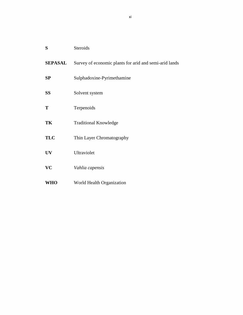
Figure 2: Trend in Malaria Burden in Namibia from 2001-2008 (MoHSS, 2010).
It has been found that antimalarial medicines significantly reduce Plasmodium resevoirs
in populations, including ACT treatments (either artesunate-sulphadoxine, artesunate-
chloroquine or artemether lumefantrine) (White, 2008). Hence malaria treatment plays a
critical role in the control and eventual elimination of the disease. However, in parts of
Cambodia and Thailand, resistance to this current first line drug regimen has been
reported (Wongsrichanalai & Meshnick, 2008; Dondorp et al., 2009) and in parts of
Africa, reduced sensitivity (Dippmann, Bienzle, Harms, & Mockenhaupt, 2008).
Barriers to the elimination of malaria parasite prevalence within the borders of Namibia
exist including resistance of the uptake of interventions by “at risk” communities such as
the lack of treatment seeking behaviour for ACTs, the WHO recommended treatment for
malaria. Government healthcare systems in southern Africa, including Namibia, are
comprised of Western medical facilities such as hospitals and clinics (McGaw, Jager,

5
Grace, Fennel, & Van Staden, 2005). However, due to traditional norms and beliefs,
and lack of access to such health facilities, some communities in malaria-endemic parts
of Namibia rely on ethnomedicinal plants for their primary healthcare (Lumpkin, 1994).
Some communities in malaria-endemic areas do not readily accept conventional
medicine preferring traditional medicines as prescribed by traditional healers (Anyinam,
1987; Ajibade, Fatoba, Raheem, & Odunuga, 2005). In Namibia such communities seek
help from traditional healers before considering going to healthcare facilities for
treatment (Sister Kalumbu, 2011).
The prevention and treatment of the malaria parasites has been investigated for hundreds
of years; and continues up to the present day, since no effective malaria vaccine has yet
been developed and many of the existing antimalarial drugs, including amodiaquine and
sulphadoxine-pyrimethamine, are becoming less sensitive to the Plasmodium parasites
(Willcox et al., 2011). As a result, the need for the integration of traditional medicine
with modern medicine has been recognized. Integration of traditional medicine as viable
treatment options provides an opportunity to introduce novel antimalarials, as well as
providing treatment alternatives for communities that do not readily accept Western
(allopathic) medicine. The WHO Beijing declaration of 2008 acknowledged the need to
integrate traditional medicine into national health systems (WHO, 2008a). In the same
spirit, the MoHSS Directorate of Pharmaceutical Services is also incorporating
traditional or complementary medicine into its updated National Medicines Policy

6
(MoHSS, 2010b). However, concerns about the safety and efficacy prevent traditional
medicine from becoming mainstream (Pamela Talalay & Talalay, 2001).
Plants, as traditional medicine have been used for centuries as herbal remedies, and are
still used up to this day, as they can be found to be non-narcotic, accessible and
affordable (Pandey, Mehta, & Hajra, 2011). The main objective of the study therefore,
was to identify, document and validate the medicinal uses of plants indigenous to
Namibia on the basis of their traditional uses suggesting their toxicity to the Plasmodium
parasites. This may then serve as a primary platform for which traditional medicine can
be integrated into main case malaria management, and might also help Namibia reach its
aim to eliminate malaria within its borders by 2020.
1.2 Availability of control measures in the fight against malaria
The key measures for control and subsequent elimination of malaria are prevention,
vector control, and treatment. Prevention involves disseminating insecticide treated nets
for people to use as protection at night against the vectors for malaria; and educational
activities that are aimed at informing the public about malaria prevention and, correct
and regular use of their nets. Vector control focuses on the killing of mosquitoes by
larviciding and spraying houses with insecticides (Indoor Residual Spraying, IRS).

7
Appropriate treatment is helped by early diagnosis and the availability of malarial
medicines.
Namibia use a permutation of malaria prevention and control measures for vector
control including long lasting insecticide treated nets (LLIN); IRS using Dichloro-
diphenyl-tricthloroethane (DDT) 75 % WP and deltamethrine 250 WG and targeted
larviciding (MoHSS, 2010a; MoHSS, 2010c). Furthermore, more than 500,000 LLIN
were distributed among the community members, especially to pregnant women and
children under the ages of 5 in 2010; and an annual coverage for IRS above 80 % exists.
Other malaria control tools include the dissemination of information at community level
to facilitate the move towards elimination (WHO, 2010).
One of the important aspects in malaria case management is early diagnosis and
appropriate treatment. Diagnosis is carried out based on parasitological diagnosis by
rapid diagnostic tests and microscopy. Treatment upon diagnosis is administered to
patients in the appropriate doses as recommended. Drug therapy regimens such as
artemether lumefantrine (AL) is used to treat uncomplicated malaria; and intermittent
preventive malaria treatment for pregnant women using sulphadoxine pyrimethamine
(SP) when malaria transmission is high, as well as for babies between 2-6 months
(MoHSS, 2010a).

8
Measures to reduce malaria transmissions in local areas or within a communtiy
contribute to the prevention of death, reduces illness, as well as social and economic loss
(Nchinda, 1998). Furthermore, measures for the control of this infectious disease used
concurrently prove to have a higher reduction rate of Plasmodium reservoirs than used
singly (Hans, 2009). Even though many measures need to be used simultaneously in the
fight against malaria, antimalarial drugs remain critical in the control and eventual
elimination of malaria (White, 2008).
1.3 Plant medicinal value
Plants have been used as folk medicine for thousands of years and are still used today as
an important source of medicines proven to be effective against many ailments (Holetz
et al., 2002; Willcox, Bodeker, & Rasoanaivo, 2004; Ajibade et al., 2005). According
to Mohammed (2009) an estimate of 80 % of the world population depends on
traditional medicine as means of primary health care, whereby 70 % are African (Street
& Van Staden, 2009). One of the many reasons that prompted indigenous people from
such communities to rely heavily on plant-based traditional herbs (Tringali, 2001, p.
395) is the high cost of conventional medicine. Therefore, plants are of infinite value to
less affluent populations (Graz, Kitua, & Malebo, 2011). Among other reasons that
prevent communities from using modern medicine include the unavailability of these

9
medicines to communities, as well as negative perceptions by such communities
regarding these treatments (Graz et al., 2011).
Plants are known to synthesize compounds such as secondary metabolites that they use
as protection against herbivores (i.e. these compounds act as a deterrent in plants).
Secondary metabolites also protect plants from disease-causing agents (pathogens)
(Okigbo, Eme, & Ogbogu, 2008). These compounds have been shown to produce certain
therapeutic effects on the human body (Njoku & Obi, 2009). Secondary metabolites
may also provide drugs directly such as artemisinin from the Chinese herb Artemisia
annua (Carmargo, de Oliveira, Basano, & Garcia, 2009), or provide template molecules
on which drug molecules can be synthesized organically such as quinine from Cinchona
bark (Rosenthal, 2001).
There is a growing demand for traditional medicine, especially in Southern Africa
(McGaw et al., 2005). In many African countries where rural people recognize folk
medicine as their key means of healthcare, regardless of the availability and accessibility
status of orthodox medical care (Ajibade et al., 2005), these plants also serve as a source
of income for them (Hamilton, 2004). Reports also show that pharmaceutical companies
mainly synthesize scientific medicine from natural products (Farnsworth, 1988; Joy,
Thomas, Mathew, & Skaria, 1998a; Calixto, 2005). Hence, the development of drugs
and medicinal agents from plants are necessary in the investigation, prevention and,
treatment of infectious diseases such as malaria (Wang, Hao, & Chen, 2007).

10
1.4 Documentation and validation of traditional knowledge
Traditional knowledge (TK) is defined as “a body of knowledge built by a group of
people through generations living in close contact with nature” (Traditional Knowledge
Sector Paper, 1999). It encompasses all aspects of life within a community and is
inherent to the survival and continuity of the community (Krugmann, Cole, & Du
Plessis, 2003). This knowledge may be useful in the search for new medicine and the
development of ethnomedicines from plants that are affordable and accessible to local
people. However, this knowledge is only known to the indigenous people, and is passed
on orally from generation to generation (Nyota & Mapara, 2008; Von Lewinski, 2008).
As TK is transmitted orally, it is exposed to change and hence would be troublesome if
altered or lost during this mode of communication.
TK is being lost at an increasing rate because of rapid population growth, changes in
educational systems, environmental degradation, and development processes all leading
to lifestyle changes, modernization and cultural homogenization (International Institute
of Rural Reconstruction, 1996). Documentation of TK is, therefore important. This will
promote the continuity of its use in providing local solutions or alternatives to Western
means. It will instill pride in rural communities about their culture; acknowledge
knowledge holders allowing them to hold the rights so such information, its use and any
benefits accruing from it. Documentation also provides a platform for validation and
promotion of TK for use in sustainable development.

11
Validation is an important process which must be carried out to allow the acceptance of
traditional medicine as a mainstream alternative to conventional medicine (Batista, de
Jesus Silva Junior, & de Oliveira, 2009; Graz et al., 2011). One of the barriers towards
integration of traditional medicine is the safety concerns surrounding its use. Traditional
medicines from plants are usually not characterized hence their composition in terms of
beneficial compounds and harmful compounds is unknown. Furthermore, questions are
raised on whether their use is beneficial or just anecdotal; this may arise from the fact
that two people with similar ailments may have different clinical outcomes after using
traditional medicines.
Validation may involve the determination of the chemical composition of traditional
medicines as well as their biological activity through a process called phytochemical
investigation (Batista et al., 2009). In some cases, the crude extract of medicinal plants
may be used as the treatment or alternatively, the active compounds are isolated and
identified for the elucidation of the mechanism of action of the compounds. Hence,
research on both mixture of traditional medicine and single active compounds is very
important (Joy, Thomas, Mathew, & Skaria, 1998b). The former allows issues related to
toxicity of the traditional medicines to be examined, whilst the latter permits the issue of
therapeutic efficacy to be determined. Knowledge of both characteristics not only allows
safe and efficacious use of traditional medicines, but also provides a basis for further
development of these medicines. Validation of traditional medicines can also be done

12
comparatively with Western medicines with a focus on efficacy, e.g. reverse
pharmacology.
This study was undertaken to evaluate the efficacy of antimalarial plants used in the
traditional setting to prevent, cure and/or alleviate symptoms of malaria. As malaria
produces a wide range of symptoms, this study focussed mainly on febrile symptoms of
the disease; and because symptoms are associated with the release of parasites from
ruptured red blood cells into the blood stream, the target for this study was to treat the
erythrocytic (blood) stage of the parasites. To validate the uses of these plants as
potential antimalarials, phytochemical and TLC chemical profiling analysis were carried
out to create a chemical profile for these plants. Preliminary screening was carried out
to assess the effects of the plant extracts against the asexual forms of the malaria
parasites and to postulate the modes of action for the crude plant extracts if any
antiplasmodial activity is observed.

13
CHAPTER 2: LITERATURE REVIEW
2.1 Plant Medicinal Value and malaria control
Malaria, the disease, is a clinical diagnosis and traditional medicines have been used to
treat symptomatic malaria for hundreds of years (Willcox & Bodeker, 2004; Moorthy,
Srinivasan, Subramanian, Mohanasundari, & Palaniswamy, 2007). These medicinal
herbs are still used today by the majority of the rural populations in developing countries
(Mohammed, 2009). A need for traditional medicine as a source of malaria treatment
has been recognized in view of the difficulties faced in areas where populations are
either unable to afford or access effective antimalarials or are unwilling to use allopathic
medicines.
Natural products have played a key role in the discovery of leads for the development of
drugs for malaria (Mishra, Dash, Swain, & Dey, 2009). For example, the bark of the
Peruvian Cinchona tree was one of the anti-fever herbs that led to the discovery of
natural quinine (an alkaloid), the first antimalarial drug which is still used today (Bickii,
Tchouya, Tchouankeu, & Tsamo, 2007; Wang et al., 2007; Kayano et al., 2011), as well
as several synthetic quinolones, particularly chloroquine (Batista et al., 2009). This
plant was used to treat malaria since the year 1632 (Haggard, 2004 p.354). Another
example includes artemisinin, which was isolated from a sweet or annual wormwood

14
tree, Artemisia annua by Chinese researchers in 1972 (Van Agtmael, Eggelte, & Van
Boxtel, 1999). This plant has been used as a traditional remedy for chills and fevers for
more than 2000 years by the Chinese. Artemether, a derivative from artemisinin, is
found to be more active than its precursor (Alin, Bjorkman, & Ashton, 1990) and is the
most frequently used artemisinin derivative as a first line treatment for uncomplicated
malaria (World Health Organization, 2008). New antimalarial leads are, therefore
directed towards plant sources.
There is a consensus that traditional medicines as antimalarials are effective (Traore et
al., 2006; Mohammed, 2009), but safety and efficacy concerns of these medicinal plants
prevents them from being an integral part of the current health care systems. Plants, in
general, constitute of a wide array of phytochemicals, and when used in high dosages the
herbal remedies can elicit harmful effects on the body instead (Wang et al., 2007; Street
& Van Staden, 2009). To assure the development of efficient and safe malaria
phytomedicines, the uses of these medicinal plants needs to be scientifically studied (i.e.
critiqued and standardized). The pharmacological efficacy, phytochemical composition,
as well as the toxicity of these plants needs to be investigated as potential antimalarial
medicines.

15
2.1.1 Secondary metabolites
Plants in general constitute chemicals known as primary and secondary metabolites.
Primary metabolites includes compounds that are necessary for cellular processes such
as amino acids, nucleic acids, lipids and simple sugars (Cseke et al., 2006); whereas
secondary metabolites include compounds which are produced in response to stress that
is induced by abiotic (e.g. heat, drought) and biotic (e.g. herbivores, pathogens, humans)
factors on the plant (Keeling & Bohlmann, 2006). Often, secondary metabolites are
referred to as natural products, as these compounds exhibit effects on other organisms
(Zwenger & Basu, 2008). Some of these compounds have been reported as exhibiting
curative effects on the human body (Njoku & Obi, 2009).
Secondary metabolites produced by higher eukaryotes such as plants are highly toxic
(Rasoanaivo, Wright, Willcox, & Gilbert, 2011). These toxins are said to be stored in
specific vesicles or in the vacuole of the plant; this kind of storage functions have been
found as a detoxification of the plant itself and generates a reservoir of, for example,
nitrogen-rich molecules (Roze, Chanda, & Linz, 2011). Even though some secondary
plant products are very common, not every plant can produce every product. Some
secondary metabolites are restricted to single species, others to related groups, but they
are nearly always found only in certain specific plant organs (Sawada et al., 2009).
Also, they are often generated only during a specific developmental period of the plant
(Mounet et al., 2009).

16
Plants are important sources of secondary metabolites that have played an outstanding
role in the development of chemotherapies. For example, morphine or mescaline have
been developed directly from plants products. A number of naturally occurring
antiplasmodial compounds have been reported and seven of the classes will be discussed
(in terms of characteristics, therapeutic applications), of which alkaloids, flavonoids and
glycosides are more potent (i.e. pharmaceutical important) (Namdeo, 2007).
2.1.1.1 Alkaloids
Alkaloids (12 000 types) form the largest single class of plant secondary metabolites,
occurring in approximately 20 % of all plant species; and are said to be heterogeneous
compounds containing nitrogen with an amino acid as a precursor (in most alkaloids)
(Roberts, Ryan, Moore, & Gulder, 2010). Many are found to have pharmacological
activity in low concentrations. In sufficient amounts, these compounds are poisonous to
livestock, herbivores and/or humans. Many alkaloids have been used as narcotics,
stimulants, poisons and more important commercially as pharmaceuticals (Brielmann,
Setzer, Kaufman, Kirakosyan, & Cseke, 2006). Alkaloids such as morphine, cocaine and
quinine were of the earliest compounds isolated from plants (Tringali, 2001; Batista et
al., 2009).

17
Many sub-groups within this class of bioactive compounds have been reported to
possess antimalarial properties, with high to moderate to low activity either in vitro or in
vivo, or both (Tringali, 2001; Batista et al., 2009). For example, echitamine (a major
alkaloid) showed low antimalarial activity, and corialstonine and corialstonidine (two
minor quinoline alkaloids) showed some antimalarial activity in vitro; whilst
benzylisoquinoline alkaloids reticuline and laudanosine exhibit moderate antimalarial
activity in vitro and laudanosine has some antiplasmodial activity in vivo. Indole
alkaloids (31 types), on the other hand, showed high antiplasmodial activity both in vitro
and in vivo. The mode of action of alkaloids on the Plasmodium parasites appears to be
due to the interference of the compound with the haemazoin formation (Soares et al.,
2009).
Figure 3: Structure of an alkaloid, morphine isolated from plants.

18
2.1.1.2 Anthraquinones
Anthraquinones are anthracene derivatives, anthracene = is the main nucleus for
anthraquinone compounds (Kar, 2008. p. 761). Anthraquinones were first described by
Robinson and Simonsen (1909) and are orange or red in color, they generally taste bitter
and are said to be polar. These compounds are found to possess certain therapeutic
properties including laxative, purgative and anti-inflammatory properties (Kar, 2008).
Several anthraquinones isolated from herbs displayed antimalarial activity (Tringali,
2001) and may kill the parasites via different modes. For example, these bioactive
compounds have been found to be cytotoxic due to its aldehyde group at C-2.
Furthermore, anthraquinones may be potential DNA intercalators due to its cyclic planar
structure (Obih, Makinde, & Laoye, 1985; Sittie et al., 1999).
Figure 4: Structure of a typical anthraquinone.
Structure of anthraquinone

19
2.1.1.3 Coumarins
Coumarins belong to a group of compounds known as the benzopyrones and are
comprised of the 2-oxobenzopyrane skeleton (Bruneton, 1999). Coumarin are found to
be in leaves, fruits, roots and stems of dicotyledonous families including the Apicaeae,
Asteraceae, Fabaceae, Moraceae, Rubiaceae, Rutaceae and Solanaceae (Ojala, 2001;
Kar, 2008, p. 345). Ojala further reported coumarins to exert a variety of bioactivities in
humans including anticoagulant, estrogenic, antimicrobial antithelminitic, sedative,
analgesic, hypnotic, as well as antimalarial activities. Subgroups within this class of
compounds show antimalarial activity, for example 5, 6, 7-trimethoxycoumarin and
isofraxidin shows moderate activity; whereas for scopoletin and pectachol less activity
was observed (Tringali, 2001). The mode of action whereby coumarins exert
antimalarial activity is, however, not clear.
Figure 5: Structure of a typical coumarin.
Structure of coumarin

20
2.1.1.4 Flavonoids
Flavonoids, the most common of the phenolics occurs throughout the plant dominion
(Harborne, 1993). Flavonoids are further divided into sub-groups such as flavones,
flavonols, flavanones, chalcones, anthocyanins and isoflavones (Brielmann et al., 2006).
This class of compounds is composed of a C6C3-moity and has shikimic acid as a
precursor. Pharmalogical activities of flavonoids such as anti-inflammatory,
antimicrobial, antioxidant and anticancer activities, decreasing capillary fragility, and
anti-diarrhoeal properties have been reported (Gurib-Fakim, 2006; Lopez-Lazaro, 2009).
Other studies also reported the antiplasmodial activity of flavonoids, whereby many sub-
groups within flavonoids are reported to possess notable antimalarial activities. For
example flavones displayed moderate activity, whereas the isoflavonoids (3-
phenylbenzoprans) have higher antimalarial activity. The mechanism of tested
flavonoids inhibits the influx of L-glutamine and myoinositol into infected RBCs
(Elford, 1986).
Figure 6: Structure of a flavonoid compound, flavonol.
Structure of flavonols

21
2.1.1.5 Glycosides
Glycosides are organic natural compounds, when upon hydrolysis give one or more
sugars (glycone) β_form and a non-sugar (aglycone) or called genin and are said to be
water soluble and bitter in taste (Brito-Arias, 2007). Many glycosides are found in both
plants and animals. Glycosides perform several functions within a plant of which some
protects the plant from bacteria and diseases (Rozavi, Zarrini, & Rad, 2011).
Plants containing glycosides are well known for their pharmacological activity.
Arbutine, salicine and anthraglucosides are all examples of plant-derived glycosides
exhibiting therapeutic (healing) properties whilst other important glycosides are digitalis
glycosides (e.g. digoxin, digitoxin and gitoxin), ouabain, k-strophanthin, scillaren A, B,
convollosides that are essential for good heart activity (Kar, 2008 p.148). Among other
therapeutic effects, glycosides have also been reported to exhibit antimalarial activity. A
mode of action proposed this biological activity is the inhibition of haemoglobin
proteases such as plasmepsin II (Dell‟Agli et al., 2003).

22
Figure 7: Structure of a typical glycoside.
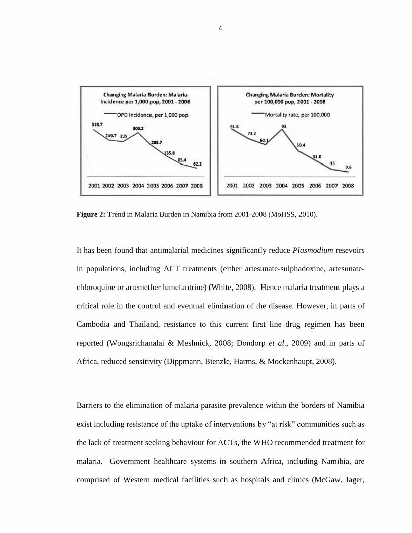
2.1.1.6 Steroids
Steroids, a subgroup belonging to the class terpenoids, have been found to possess a
broad spectrum of biological activities, including antiplasmodial activities (Haines,
2001). Because of their chemical structure they display pharmaceutical activities,
including some antimalarial activity in vitro. Examples of these steroids include lupeol
(Ziegler et al., 2002), betulinic acid and its analogues (Ziegler et al., 2004), and
triterpenoid saponins (Traore et al., 2000). The mechanism of action of these bioactive
compounds is exerted through the prevention of the developmental stages of the
plasmodium parasites from rings to trophozoites (Londono et al., 2006). According to
Sisodia, Negi, Datokar, Dwivedi and Khanuja (2012) terpenoids on the other hand, may
act by preventing the uptake of nutrients by parasites, i.e. inhibiting the permeation
pathway.
Glycoside structure
23
Figure 8: Structure of a typical steroid.
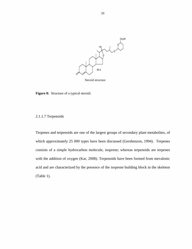
2.1.1.7 Terpenoids
Terpenes and terpenoids are one of the largest groups of secondary plant metabolites, of
which approximately 25 000 types have been discussed (Gershenzon, 1994). Terpenes
consists of a simple hydrocarbon molecule, isoprene; whereas terpenoids are terpenes
with the addition of oxygen (Kar, 2008). Terpenoids have been formed from mevalonic
acid and are characterized by the presence of the isoprene building block in the skeleton
(Table 1).
Steroid structure

24
Table 1: The classification of terpenoids based on the number of isoprene units according to
Tringali (2001, p.410).
Terpenoids Units Carbons
Monoterpenoids 2 10
Sesquiterpenoids 3 15
Diterpenoids 4 20
Sesterpenoids 5 25
Triterpenoids 6 30
Tetraterpenoids 8 40
Mono- and sesquiterpenoids are said to be found in various parts of a plant and often are
volatile (evaporates at warm temperatures) and forms a protective haze around the plant
keeping harmful insects, moulds, bacteria on a distance (Loughrin, Manukian, Heath, &
Turlings, 1994). It has been reported that some terpenoids, as well as terpenes (Batista
et al., 2009) have harmful effects on microorganisms which are found to be effective
against Plasmodium falciparum, the causative agent of severe malaria (Kar, 2008).
Examples of several of these compounds include peroxides (sesquiterpenoids) such as
artemisinin, isonitrile (diterpenoids), quinone ethides (triterpenoids) such as pristimerin
and tingenone (limonoids), and triterpenes (a terpene). These compounds are said to
have an internal peroxide link that is located in the seventh ring which is responsible for
it to exert antiplasmodial properties (Kar, 2008, p. 231). Triterpenes are said to act
through interaction with Plasmodium DNA or inhibition of its DNA synthesis (Goijman,
Turrens, Marini-Bettolo, & Stoppani, 1984). The mode of action for limonoids (bitter
terpenoids) on the other hand is unclear though potent, whilst quassinoids inhibits
protein synthesis of the malaria parasites (Kirby, O‟Neil, Phillipson, & Warhurst, 1989).

25
Figure 9: Structure of a typical terpenoid.
2.1.2 Traditional plant medicine in Namibia
Namibia is a large country and is sparsely populated with 13 different cultural groups
separated by culture and language. Namibia‟s vegetation was described by White (1983)
as falling between 3 phytochoria; Zambezi, Kalahari Highveld and Karoo-Namib centres
of endemism. The main vegetation types are desert, savannah and woodland (Figure 10)
which are subdivided into 14 smaller vegetation units (Giess, 1971). This has also
allowed the development of traditional medicinal systems that use either different plants
to treat similar ailments depending on local availability, or the use of the same plants for
different ailments either way resulting in a country with a rich ethnomedicinal heritage
that can be tapped into to control malaria. It is not surprising, therefore, that the areas of
richest plant diversity are the same ones that rely more strongly on traditional medicine.
Terpenoid structure

26
Figure 10: Map showing the vegetation cover (biomes) in Namibia generated using the
ArcView GI53.2 program.
These areas also fall into the malaria prone areas and should be a rich source of
antimalarial plants. There is decreasing species diversity with decreasing rainfall but
endemism shows the opposite trend. This has a bearing on the availability and range of
plants for use by local communities (Krugmann et al., 2003). A total of 3159 plant
species have been reported to occur in Namibia (Cunningham, 1992; Craven et al.,
1997; Marshall, 1998) . A remarkable 1038 species of flowering plants, 13 ferns and 22
mosses and their relatives have been reported to occur in the Sperrgebiet area alone,
which is the southwestern corner of Namibia (Burke & Mannheimer, 2004).

27
Furthermore, this area is made up of nearly 25% of the entire vegetation of vascular
plants in Namibia and approximately 21% of the plant diversity of the Succulent Karoo
Biome. The Survey of Economic Plants for Arid and Semi-arid Lands (SEPASAL) lists
615 species (19.5%) as being used medicinally.
Namibia has a high diversity of plant species, on which little research has been done
compared to many other parts of the world. Very little information exists on the
ethnomedicinal plants in Namibia, and literature on these plants suggests that they occur
mostly in the northern parts of Namibia (Mapaure & Hatuikulipi, 2007) where there is a
high prevalence of malaria. The northern parts of the country are associated with flora
due to high rainfall (Figure 10). And the majority of traditional homesteads are found in
these areas whereby their means of livelihood depends on the plants they harvest. Plants
that are commonly used by indigenous people in Namibia, within small isolated
populations, are used to treat illnesses that are mainly symptoms of clinically diagnosed
diseases, including cancer, TB and malaria. Based on specimen records/collections,
published sources and own observations, an updated list of plant species used
medicinally in Namibia to treat malaria related symptoms can be seen in Table 1
(Appendix A).
A total of 61 families of plants are recorded to treat symptoms of malaria in the
traditional setting, of which Fabaceae is the most used (32 species), followed by
28
Combretaceae (19 species) and Asteraceae (14 species). The 6 most common malaria-
like-symptoms treated by traditional healers recorded are diarrhoea, abdominal pain,
dysentery (a form of severe diarrhoea caused by an intestinal infection), malaria,
coughing and headaches (Mumbengegwi & Du Preez, 2011).
Uses of these plants vary among the local people. For example Rhigozum
brevispinosum is used as digging sticks and ornaments by the locals in the Otjizondjupa
region, Tsumkwe constituency (Leffers, 2003), whereas this same plant is used in the
Oshikoto region to treat ailments such as headaches (Du Preez, Mumbengegwi, & Bock,
2010), as well as sexually transmitted infections (STIs), namely syphilis, in the Caprivi
region (Cheikhyoussef, Mapaure, & Shapi, 2011). This is just a clear indication that a
plant may be used medicinally in one community and not at all in another. One plant
may also be used for different ailments. As such, Table 1 reveals that Dicerocarym
eriocarpum commonly known as Devil‟s Thorn is used to treat chest pain and abdominal
pain in the respective regions. A single plant may even have multiple uses in the same
area. A leaf infusion of Acrotome inflata, for example, is used in Tsumkwe to cure
coughing, whilst the flower heads are burnt to ward of mosquitoes (Leffers, 2003).
Most of these plants are used in monotherapy, and only a few in a combination therapy
treatment, whereby two or more plants are used (Mumbengegwi & Du Preez, 2011).
Very few plant species used in combination treatments have been recorded. One such

29
example includes Vahlia capensis and Crotalaria flavicarinata. These two plants are
used together to treat fever. For modern medicine, combination therapy is
recommended due to emergence of resistance malaria parasites when monotherapy is
used. However, traditional medicine sometimes requires only one plant species. This
could be as a result of several bioactive compounds found in one plant that may work in
synergy to heal (Rasoanaivo et al., 2011).
Herbal remedies are taken in a wide range of forms, mostly in the form of decoctions.
The plants are pulverized into powder or smaller pieces and then boiled, which will then
be drunk. Other types of treatments included infusions, inhalation of smoke or vapor,
applying the plant material in incisions made into the skin and/or on to the skin surface,
or simply by just sniffing the powder form of the ideal plant part (Mumbengegwi & Du
Preez, 2011). Chewing of plant parts is also one the common treatments. The route of
administration of herbal remedies as described, and especially for currently used
antimalarial drugs, greatly determines whether the medicine/drug will work or not
(Gathirwa et al., 2000). A bioactive compound present in the plant may become
degraded within the digestive system when taken orally and only little amounts of this
active component may be absorbed into the bloodstream, rendering the remedy
ineffective. Other compounds can be easily absorbed by the bloodstream through the
skin, and can thus be applied to the skin, as an ointment for example, or drops under the
tongue or in the eyes, or by nasal uptake.

30
Plants are also used as insect repellants following burning or from their natural volatile
compounds. Plants used as insect repellents including mosquitoes, may also play a vital
role in the control of the malaria (Mohammed, 2009). These plants are burned to create
smoke that in turn repels the mosquitoes. Some plants, on the other hand, naturally repel
mosquitoes by giving off volatile scents in their natural state (Seyoum et al., 2002). A
range of plant parts are used, sometimes together and sometimes alone, to treat the acute
symptoms of malaria. Roots, leaves and the bark are the most used plant parts. Roots,
although frequently used, result in poor conservation and unsustainable harvesting
practices.
Most of the literature on the medicinal plant remedies lacks detail and specificity. There
is a shortage of information on locality, abundance, plant parts used, mode of
preparation, dosage and period of treatment, and the active components present in plants.
Plant names are sometimes only presented in either their vernacular or scientific names,
or only in the local languages. The exact illnesses the medicinal plants are used for are
not properly described. When illnesses are sometimes described by the local
communities, they use vague descriptions such as stomach problems, which may be
misinterpreted in the end. However, in some of the literature plants have been reported
to treat malaria the disease, as well as ailments similar to the symptoms of malaria. The
aforementioned thus presents limitations in validating these traditional herbal remedies

31
in the search for new and effective phytotherapies and for the eventual development of
antimalarial drugs.
2.1.3 Drug discovery from antimalarial natural products
Drug discovery from natural products remain a focal point. Plants have been used for
centuries for various purposes, including the treatment of diseases. The experimental
use of plants has led to the discovery and isolation of drugs (i.e. the pharmaceutical
active components within the plant) such as cocaine (Erythroxylum coca), opium
(Papaver somniferum), and important antimalarial drugs such as artemisinin (Artemisia
annua) (van Agtmael et al., 1999), quinine (Cinchona species) and their respective
analogues (Wang, 2007). As a result, more emphasis is put on medicinal plants in the
quest to develop novel drugs for malaria.
There are many obstacles encountered in the drug discovery process, some of which
includes costs, duration and yield obtained. Drug discovery, whether from plants or
animals or microbes, is expensive. It has been estimated that compounds isolated have a
1 in 5 000 chance of making it to the market place (Balunas & Kinghorn, 2005) due to
the high costs involved. The average cost of commercialization of drugs is estimated to
be over US$ 800 million (DiMasi & Grabowski, 2007).

32
For the drug discovery process, a minimum of 10 years is required for the successful
development of a drug since the discovery of the initial drug as it involves 3 stages:
discovery, development and clinical use. These stages are further divided into a number
of different phases (Tringali, 2001, p. 388). The drug discovery process is also
characterized by low average yield obtained from the natural products of the active
components. This is usually insufficient to use in the drug development process
(Dickson & Gagnon, 2004).
Regardless of the challenges and problems encountered in drug discovery from natural
products, several clinically valuable bioactive compounds have been isolated from plants
and are still used today (Balunas & Kinghorn, 2005). Therefore, the search for drug
leads from plant products continue to be essential to militate against infectious diseases
such as malaria, in the elimination of the disease. Even so, a large number of plant
species are yet to be investigated pharmacologically and phytochemically, concluding
that the efforts in the discovery and isolation of drugs from this higher class of
organisms (vascular plants) are ongoing.
2.1.4 Economic benefits of medicinal plants
The use of medicinal plants plays an essential role in the primary healthcare of 80 % of
the population in some of the Asian and African countries (WHO, 2008b). Plant-based

33
traditional medicines have been used to treat malaria for thousands of years in various
parts of the world (Adebayo & Krettli, 2010). In Namibia, as well as in other African
countries, a variety of plant species are used as herbal remedies by traditional healers in
local communities as curative means for malarial symptoms (Njoroge & Bussmann,
2006; Bickii et al., 2007; Du Preez et al., 2010; Adebayo & Krettli, 2011; Du Preez et
al., 2011).
Plants used to treat malaria or its related symptoms are geographically located in malaria
endemic areas, especially in Namibia where these areas are comprised of the northern
parts (Figures 1 and 10). In such areas communities experience a lack of health care
services. Folk medicine, in this case is readily available and accessible for early and or
immediate interventions, since prompt treatment in certain instances has to be
administered to prevent or to cease the development of severe malaria. Thus,
presumptive treatment given to patients with ailments similar to the symptoms of
malaria, together with plant-based research that could give rise to affordable, accessible
and safe phytotherapies, may contribute to a healthy/good socio-economic status of a
country.
Due to popular use of medicinal plants in the traditional setting, these plants have
become lucrative in the global market. According to WHO (2003), there was a
significant rise in the demand for herbal medicines both in the developing and developed
countries resulting in global sales of about US$60 billion dollars. In China in 2005 the

34
natural product market generated US$14 billion; and for Brazil US160 million was
generated in 2007. In southern Africa, an estimated of US$ 3,428,962,767 worth of
plant material is used yearly (Ariyawardana, Govindasamy, & Simon, 2009). A number
of indigenous natural products (INP) in Namibia are of interest to the market including
Hoodia and other succulents, indigenous vegetables, medicinal species, e.g. Terminalia
and Combretum, gums and resins from plant species Commiphora, Acacia, Kalahari
truffles, essential oils, fruit products and lipid oils (Ipinge, 2004). According to the
MCC (2008) an estimate of US$670,804 worth of plant material was traded in 2005
within the INP sector.
The plant species that are of interest in Namibia among many others include Citrullus
lanatus (Kalahari melon seed), Ximenia americana and X. caffra, Sclerocarya birrea
(marula), Hoodia gordonii,, Harpagophytum procumbens and H. zeyheri (devil’s claw),
and Schinziophyton rautanenii (manketti). The increased interest in these natural
products resulted in the expanding of the INP sector in Namibia, which in turn elucidates
benefits to the livelihoods of rural Namibians especially to women who are the main
people involved in the harvesting, trading and serving as the traditional healers in these
informal settlements; as well as the members of a community as a whole increasing the
supplementary household income from INPs for up to 15,000 producers and processors,
which could support to the livelihoods of up to 75,000 individuals (MCC, 2008).

35
2.1.5 Conservation of medicinal plants
Due to increased demand for plant products, overexploitation of these natural resources
may result. The medicinal application of the roots, bulbs and bark of many medicinal
plants are considered to be the main factor contributing to the unsustainable use of these
vascular plant species (Jain, Katewa, Galav, & Sharma, 2005). The roots and other
underground plant parts, according to many herbalists, are the most potent principles
(Louw, Regnier, & Korsten, 2002); hence the uprooting of certain plants in the
preparation of herbal remedies can contribute to the extinction of some important flora
in the battle with many illnesses, as well as the loss of a country‟s rich biodiversity.
Different approaches to plant harvesting such as plant part substitution and similar plant
species selections as means for sustainable collection of medicinal plants have been
recommended. According to Lewis and Elvin-Lewis (1995), and Kott et al. (1998) plant
species from one genus appear to have the same bioactive compounds present in their
respective plant parts. Furthermore, the different parts for one may possess similar
compounds, thus exhibiting similar pharmacological activity (Street & Van Staden,
2009). Therefore, the use of non-destructive plant parts such as leaves or related species
by traditional herbalists to improve plant conservation should be encouraged. This
would significantly reduce the pressure on the natural-occurring plant populations,
especially on the medicinal plant species.

36
In Namibia national sustainable development policies are in place (Appendix B). These
laws broadly focuses on species conservation and biodiversity protection, access to
genetic resources, benefit sharing, and traditional knowledge protection, the suite of
approaches adopted to manage natural resources in communal areas, and trade and
export (MCC, 2008). One important policy, the Nature Conservation Ordinance (4 of
1975), which among other things, sets in place a permitting system for protected species.
Of the species relevant to this proposal, only H. procumbens and Hoodia spp. are
included. The Ordinance stipulates that a permit is required for the picking and
transport, sale, donation, export, and removal of protected plants, and requires the
written permission of landowners before picking any indigenous plant. Non-compliance
with these provisions of the 1975 Ordinance, results in, for example, a maximum penalty
of N$750 (approximately US$100) and/or imprisonment for a year (MCC, 2008).
The conservation and sustainable use of medicinal plants is essential for both the
commercialization and for the local use of medicinal plants. In this context an effort
should be made to enhance the conservation of medicinal plants. Cheikhyoussef,
Mapaure, et al. (2011) recommend teaching or training harvesters/ traditional healers
sustainable harvest techniques. This is because unsustainable harvesting coupled with
the increase in the demand of medicinal plants will lead to their extinction. Furthermore,
traditional healers or communities should also be encouraged to cultivate the medicinal
plants on a communal scale (Street & Van Staden, 2009).

37
2.2 Malaria, the disease
Malaria is caused by protozoan, parasites from the phylum Apicomplexa (Heelan &
Ingersoll, 2002) and of genus Plasmodium (Nchinda, 1998). Within the latter several
species are found, of which only four cause malaria in humans namely P. vivax, P.
malariae, P. ovale and P. falciparum. P. falciparum is the most serious form of the
disease as it is responsible for the high malarial mortality and morbidity rates in Africa
(Rayner, 2005), and occurs predominantly in Namibia. According to Rosenthal (2003)
resistance to existing antimalarial drugs is mostly seen in P. falciparum.
2.2.1 Transmission
The main vector for the malaria parasites is the Anopheles mosquito. In Africa, the
Anopheles gambiae complex is the most efficient in transmitting the disease (Nchinda,
1998). According to the Ministry of Health and Social Services (2005) there exists three
malaria vectors in Namibia, namely A. arabiensis, A. gambiae and A. funestus. It was
reported that these vectors are inclined to feed at dusk, which means by sunset
individuals should use protective clothing, mosquito repellent, indoor residual spraying
and bed nets soaked in insecticides. Malaria is also transmitted from mother to unborn
baby (i.e. congenitally), by blood transfusions, as well as by sharing contaminated
hypodermic needles (Heelan & Ingersoll, 2002).

38
The transmission of malaria corresponds with the rainy season which provides breeding
sites for the Anopheles vector. The Plasmodium parasites are only carried by these
mosquitoes in warm, humid and wet climates but diappears over winter. Larvae control
is, therefore, imperative in the control of this epidemic disease (MOHSS, 2005). Other
factors that contributes to the increase of malaria incidences include travelling to and
from epidemic areas, the accidental imports of the malaria vectors (a phenomenon
known as „airport malaria‟) and the increase in agricultural activities (Heelan &
Ingersoll, 2002).
2.2.2 Life cycle of malaria parasites
The agents of human malaria, i.e. P. ovale, P. falciparum, P. malariae and P. vivax all
have a complex life cycle (Figure 11) involving two hosts: the human and the
Anopheline mosquito (Heelan & Ingersoll, 2002). Whenever an infected mosquito has a
blood meal, it injects saliva containing the parasite (in the form of sporozoites) into the
pierced skin serving as some kind of anesthesia. The sporozoites now travel through the
bloodstream of the victim and infect the liver. In the liver, the sporozoites reside in the
parenchyma cells (Mehlhorn, 2001) and asexual development or schizogony takes place.
The parenchymal cells transform into a circular shape and start to divide, producing
many merozoites, which are released into the bloodstream upon rupturing of the
hepatocytes. Once in the bloodstream they infect the red blood cells (RBCs) and start to

39
multiply. The erythrocytic life cycle, responsible for all clinical manifestations of
malaria, begins when free merozoites invade erythrocytes. The intraerythrocytic
parasites develop from small ring-stage organisms to larger, more metabolically active
trophozoites and then to multinucleated schizonts. The erythrocytic cycle is completed
when mature schizonts rupture erythrocytes, releasing numerous invasive merozoites.
Figure 11: The life cycle of the malaria parasite adopted from Batista et al. (2009).
According to Mehlhorn (2001) the rest of the merozoites grow as trophozoites (i.e. the
ring forms), increase in size, divides and mature into schizonts. Merozoites are formed
from the schizonts as a result of erythrocytic schizogony. Within 48-72 hours the

40
infected erythrocytes burst/rupture and releases more merozoites, which infects more
RBCs. Some of the merozoites undergo gametocytogenesis (Heelan & Ingersoll, 2002)
and develop into gametocytes (i.e. macro- and microgametocytes) which can only
mature outside the human body. When a mosquito takes a blood meal from an infected
person, both the merozoites and gametocytes travels to the stomach/gut of the mosquito.
The merozoites are digested, whereas the gametocytes mature or develop into gametes in
the digestive gut. During fertilization, the gametes develop into a zygote and then into
an ookinete (fertilized egg). The ookinete matures into an oocyst giving rise to
sporozoites (Heelan & Ingersoll, 2002). These sporozoites, when released, travel to the
salivary gland. When the female Anopheles bites the next victim, the cycle starts all
over again.
2.2.3 Diagnostics and clinical symptoms
The severity of the disease varies from mild asymptotic infection to the critical
complicated disease which causes death (Heelan & Ingersoll, 2002). Common
symptoms include periodic flu-like symptoms such as fever, chills, sweating, muscle
aches and headaches. Other symptoms include nausea, vomiting, coughing, diarrhoea,
abdominal pain, myalgia (limbs and back), loss of appetite, orthostatic hypotension,
jaundice (yellowing of skin and eye white), anemia, liver or kidney failure, enlarged
liver and spleen and convulsions. Severe malaria is regarded as a multisystem disorder

41
and includes cerebral malaria and severe malarial anemia (Weatherall, et al. 2002). Hans
(2009) claims severe malaria delirium, metabolic acidosis and multi-organ dysfunction
which may occur, if untreated could result in coma and eventually death.
Cyclical symptoms such as fever, seizures, chills and anemia are said to correspond with
the erythrocytic stage of the Plasmodium life cycle (Matteelli & Castelli, 1997) during
which merozoites are released into the bloodstream (Rayner, 2005). Together with this
release, there is also a deposit of parasitic waste products and debris, which is believed
to give rise to the malarial paroxysm, i.e. the sequenced events of shaking chills, fever
and sweating (Heelan & Ingersoll, 2002; Weatherall et al., 2002).
Symptoms for complicated or severe malaria includes seizures, altered state of
consciousness (or coma), exessive sleepiness, prostration (feelings of helplessness),
respiratory distress, inability to ingest any fluids, bleeding problems, jaundice, the
absence of urine and the dark coloration of urine if there is any (MOHSS, 2005). Heelan
and Ingersoll (2002) on the other hand maintain that splenomegaly and anemia are the
major/principal clinical symptoms of malaria. The latter is caused by the destruction of
RBCs and the simultaneous loss of hemoglobin or by the removal of the infected
erythrocytes as an immune response. Anemia may also be caused by the ceased
production of RBCs due to the bone marrow suppression.

42
Complications in malaria infections are mainly a cause of blocked capillaries (Heelan &
Ingersoll, 2002). The obstruction of these capillaries are as a result of infected RBCs
and or the parasitic RBC debris. The serious complications that can result from this
obstruction includes glomerulonephritis, nephrotic syndrome and renal failure, which are
all renal complications that may be life-threatening. Heelan and Ingersol (2002) further
maintains that problems in the CNS caused by malarial infections involve severe
headaches, cortical blindness, stroke and death.
2.2.4 Prevention and treatment of malarial infections
2.2.4.1 Mechanical protection and vector control
Humans can lower the risk of infection by reducing the rate of coming into contact with
vectors by the use of protective clothing, screened doors and windows, and mosquito
repellents (MOHSS, 2005). The MOHSS (2005) further stresses that vector control is
more effective when it comes to malaria control activities. Vector control measures
involves the use of indoor residual sprays, larvicides in water sources/breeding sites for
mosquitoes, and/or natural predators or enemies of mosquito larvae. Other measures
include using bed-netting soaked in insecticides, by avoiding the use of shared IV drug
needles, completing or following the prophylactic drug course as prescribed and the

43
screening for malaria in blood should be caried out with care before initiating blood
tranfusions (Heelan & Ingersoll, 2002).
2.2.4.2 Chemotherapy
Weatherall et al. (2002) stresses the importance of the use of effective chemotherapy for
the reduction in global mortality caused by malaria. It is thus imperative for malaria
infections to be diagnosed early so that medication can be administered immediately for
the successful treatment of such an infection (Hans, 2009). The species of the malaria
parasite, antimalarial drug resistance patterns in areas where infection was contracted
and, the severity of disease are all factors that needs to be taken in account when
deciding on a antimalarial chemotherapy regimen (Weatherall et al., 2002).
Malaria treatment has somewhat become a problem because of the unmitigated adaptive
nature and persistance (tenacity) of both the vector and parasite (Heelan & Ingersol,
2001). As a result, resistance to conventional antimalarial treatment regimes, such as
chloroquine and sulphadoxine-pyrimethamine has emerged (Ekland & Fidock, 2007a)
and resistance to current affordable drugs continues to increase (Mohammed, 2009).
Antimalarials such as chloroquine (powder from chinchona bark) and sulfadoxine-
pyrimethamine, both first-line treatments had become ineffective over the years.

44
Chloroquine has lost its effectiveness in almost every part of the world (Jiang, Joy,
Furuya, & Su, 2006); whilst sulphadoxine-pyrimethamine (introduced in1977), now less
effective due to the developing drug resistance (Eriksen et al., 2008).
A new class of antimalarials was then deployed over the past decade on a large scale
(Bosman & Mendis, 2007). In 2005 31.3 million ACT treatment courses were
distributed globally of which 25.5 million were in Africa. However, in 31 of these
countries, of which 29 were African, access to medicine is low. Examples of these
antimalarials include artemether, artesunate, artelinic acid, artenimol, artemotil and
dihydroartemisinin. It has been reported that these artemisinin derivatives elicits a very
quick therapeutic response i.e. a reduction of the parasite load and lessens the symptoms
(Ridley, 2002). These compounds are also said to be active against multidrug resistant
(MDR) P. falciparum, and has very little toxicity effects towards humans i.e. no severe
side effects are visible. Furthermore, these compounds may be able to reduce malaria
transmission i.e. they reduce the gametocyte biomass.
Artemisinins can be used in monotherapy; however this results in the recrudescence of
the parasites i.e. persistence of the parasites in the blood hence the reemergence of
clinical symptoms of the disease due to its short pharmacological half-life the time it
takes for the bioactive constituents to clear from blood/plasma (Matteelli & Castelli,
1997). It was also reported that monotherapy or single-drug artemisinin treatments
results in the emergence of drug resistance (Bosman & Mendis, 2007). The WHO

45
therefore, recommends that artemisinin combination therapies (ACT) to be administered
to treat uncomplicated P. falciparum malaria worldwide in areas of widespread
chloroquine resistance (Bosman & Mendis, 2007; Kayano et al., 2011). The artemisinin
derivatives works by clearing the parasite load at the start of the treatment, whereas the
partner drug such as mefloquine (ASMQ), lumefantrine (Coartem,), amodiaquine
(ASAQ), piperaquine (Duo-Cotecxin) and pyronaridine (Pyramax) with the longer
pharmacological half-life will clear the remaining parasites (German & Aweeka, 2008).
Artesunate, an artemisinin derivative, is administered to patients with severe malaria
(SEAQUAMAT, 2005). Patients who are immunocompromised, for example HIV/AIDs
and TB patients, pregnant women, and children under the age of 6 years, require special
treatment (Brentlinger, Behrens, & Micek, 2006).
Chemoprevention of malarial infections in the contribution to the control of malaria in
endemic areas is also considered important in the process of eliminating the disease
(Greenwood, 2009). He further maintains there are different ways in which to prevent
malaria transmission by using antimalarial drugs. The three main approaches include
chemoprophylaxis, a term that is used to describe the administration of an antimalarial
drug or drug combination in such a way that blood levels are maintained above the
inhibitory level of survival of the local strains of parasite for the whole of the period at
risk and, in the case of travellers, for an appropriate period afterwards in order to kill
emerging liver forms. Intermittent preventive treatment (IPT), a term that is used to
describe the administration of a full curative dose of an antimalarial or antimalarial

46
combination to a selected, target population at specified times without determining
whether or not the subject is infected. It is recognized that between drug administration‟s
blood levels may fall below the protective level and that infections may still occur
although some of these may be truncated by persistent low levels of drug; and mass drug
administration (MDA), is used to describe the administration of an antimalarial drug or
drug combination to a whole population without screening for the presence of infection.
MDA may involve either the administration of a full therapeutic course or a
subtherapeutic one as once practiced through the use of medicated salt.
Chemoprophylaxis is important for the prevention of malaria in pregnant women and
infants in areas with medium or high levels of malaria transmission.
In Namibia, the current first-line treatment for uncomplicated malaria is done by oral
therapy using the drug regimen called Coartem® also known as arthermether-
lumefantrine combination therapy (MoHSS, 2005; MoHSS, 2010a). Coartem is
administered in a six dose-treatment for three days. Pregnant women and children
weighing less than 5 kg are not to be given Coartem. For severe/ complicated malaria,
according to the MoHSS (2005), parenteral quinine (intravenous or intramuscular
injections) is administered and should be taken orally once the patients conditions
improved. Chemoprophylaxis is only recommended for certain groups of people,
including non-immune individuals living in and travelling to malaria endemic areas for
short time periods (MoHSS, 2005). Mefloquine and doxycycline, in the case of
mefloquine contra-indicated patients, are endorsed as the prophylactic drugs in this case.

47
For pregnant women, presumptive prophylactic treatment (PPT)/ IPT in the form of
sulphadoxine-pyrimethamine is prescribed by the current Malaria Policy (MoHSS,
2010). This regimen is only administered during the first and second pregnancies.
However, due to low levels of malaria prevalence, this regimen is no longer
recommended. Sulphadoxine-pyrimethamine is also administered to infants between the
ages 2-6 months as a first-line drug and quinine as the second-line drug (MoHSS, 2005).
Despite the efficacy and relative safety of most available chemotherapies, there is still a
growing concern about potential problems of acquired resistance. The widespread
resistance of Plasmodium parasites, especially of P. falciparum, can give rise to malaria
mortality and morbidity; and can contribute negatively to the socio-economic status of a
country (Nchinda, 1998; Schlagenhauf & Petersen, 2009). Reduced sensitivity to the
current first-line drug regimen ACTs have been reported (Batista et al., 2009).
However, studies have shown that the development of malaria vaccines are taking place
worldwide (Heelan & Ingersol, 2001) and if an effective vaccine is to be synthesized,
this would mean an important breakthrough in the fight against this infectious disease,
but until then efforts in the search for new potential antimalarials are ongoing.

48
2.2.5 Intracellular sites of action of antimalarials
It was found that antimalarials were active during the stage when the parasite is located
inside the RBC, in other words, the drug regimens target mainly the erythrocytic cycle
of the Plasmodium life cycle (Ridley, 2002). During the blood stage of the Plasmodium
parasites between 60-80 % of the haemoglobin is broken into haem (Egan, 2008). The
latter is said to be harmful to the parasites. The parasites, however, neutralize the haem
during the process of polymerization of haemazoin (Huthmacher, Hoppe, Bulik, &
Holzhutter, 2010). This polymerization is inhibited by antimalarials such as chloroquine
mefloquine, halofantrine, lumefantrine (Hempelmann, 2007) and artemisinins; resulting
in the accumulation of haem and eventual death of the parasites. Antimalarial drugs,
therefore, act by preventing globin lysis and haemazoin formation. Quinoline
antimalarials such as chloroquine, for example, act by inhibiting the synthesis of the
hemozoin pigment when they get trapped in the digestive vacuole of the parasite due to
the extra proton they gain due to the acidic environment of the vacuole (Jiang et al.,
2006).
The malaria parasites use hemoglobin/ hemazoin to make amino acids which are vital
for their existence (Hempelmann, 2007). According to Gavigan, Machado, Dalton and
Bell (2001), hemoglobin hydrolysis by the Plasmodium parasite (trophozoite) is
mediated by a number of classes of enzymes (proteases) in the food vacuole including

49
aspartic and cysteine proteases and falcilysin. These proteases appear to mediate the
invasion and rupture of infected erythrocytes as well, possibly to get through the
erythrocyte cytoskeleton, a complex network of proteins (Rosenthal, 1998). In addition,
a number of malarial proteins are made during the late schizont and merozoite life-cycle
stages are likely to aid in the complex series of events involved in the RBCs. Although
the exact roles of various classes of proteases are not entirely clear, inhibitors of cysteine
and serine proteases have consistently repressed erythrocyte rupture and invasion.
Studies have found that the biological activity caused by drugs results in the swelling of
the parasite, as well as the inhibition of the proteolytic activity of these enzymes
(Semenove, Olson, & Rosenthal, 1998).
Malaria parasites are said to be sensitive to the free radicals produced by some
antimalarial drugs (Ridley, 2002) including artemisinin-related compounds.
Artemisinin, in the presence of haem inhibits the polymerization of haemazoin by
alkylation (Mishra et al., 2009). The haem or Fe2+
cleaves the peroxide bridge in the
artemisinin compound leading to the formation of free radicals (Ridley, 2002). These
radicals then alkylate one of the pyrazole rings in the haem nucleus, hence inhibiting the
formation of haemazoin.
Other modes of action shown by antimalarials include pyrimidine synthesis inhibition.
Inhibiting this synthesis would mean inhibiting the growth of the parasites (P.J.
50
Rosenthal, 2003). An example of such an antimalarial drug is atovaquone which inhibits
the respiratory chain of malaria mitochondria at complex III (Ridley, 2002). Some folate
antimalarials such as pyrimethamine and sulfonamides inhibit the formation/ synthesis
of dihydrofolate, which is necessary in the biosynthesis of pyrimidines. Iron chelators,
on the other hand, are active by preventing iron from entering important metabolic
pathways of parasites within the RBCs, as well as combining with iron forming toxins to
the parasites (Olliaro & Yuthavong, 1999).
Among potential targets for new modes of chemotherapy are malarial proteases.
Cysteine and aspartic protease inhibitors are now under study as potential antimalarials
(Gavigan et al., 2001). The increased understanding of the mode of action of a
compound will contribute in the mitigation in the rapid spread of resistance against
presently used antimalarials as some antimalarials given in combination treatments may
have counter effects, causing a relapses of the parasite load.
2.2.6 Drug resistance
Resistance to current antimalarial drugs poses a major public health threat. The demise
of affordable drugs such as chloroquine and sulphadoxine-pyrimethamine and the high
costs for the development of novel drugs has contributed to the reduction in the rate at
which developing countries develop and maintain their healthsystems (Aderounmu,

51
2007). Reduced sensitivity and resistance to artemisinins indicated by the persistance
of parasites in the blood stream or circulation after artemisisin treatment have been
administered (Matteelli & Castelli, 1997;Aderounmu, 2007; Schlagenhauf & Petersen,
2009) are problematic in the combat against malaria or eradication of malaria. With
this rapid spread of drug resistance, lowered levels of drug efficacy and high costs, there
is an urgent need for new, affordable and safe drugs.
Many factors contribute to the emergence and spread of drug resistance. One such
factor includes the occurence of mutations in Plasmodium genes. Gene mutations occur
independently of drug effects, in other words, the drugs have no direct effect on the
changes within the parasite genome (Wongsrichanalai, Pickard, Wernsdorfer, &
Meshnick, 2002). Mutations, instead results from drug pressure, i.e. selective pressure
showing drug resistance only in the most “fit” parasites (Schlagenhauf & Petersen,
2009). Only a small population of these mutants occurs in the natural environment and
results from either single or multiple point mutations in the parasite genome. The
mutated genes in turn encode for certain enzymes responsible for drug resistance
(Tipsuwan, Srichairatanakool, Kamchonwongpaisan, Yuthavong, & Uthaipibull, 2011)
The interaction between several factors including drug characteristics, drug
administering patterns, and host and environmental factors also contributes to the
emergence of drug resistant parasites (Wongsrichanalai et al., 2002). The indiscriminate

52
use of drugs, and non-compliance to drug treatments (i.e. not completing the course of
treatment as prescribed), the use of monotherapies and counterfeit drugs all play a role in
the development of resistance (Enserink, 2008). For example, when antimalarials are
administered to patients, not all parasites may be cleared from the blood during a malaria
infection because of the short elimination half-life of the drug or due to the non-
compliance of the prescribed treatment. The remaining parasites present in the
subtherapeutic concentrations of the antimalarial drugs may then develop resistance to
the drugs (Wongsrichanalai et al., 2002). According to Mu et al. (2010) drugs target
only certain proteins of the parasite, and as a result or over time, the parasites may
become resistant by changing the antimalarial target sites/genes.
The development of resistance to current affordable antimalarials results in increased
human morbidity and mortality. Hence measures to reduce the spread of drug resistant
parasites are necessary. One such measure includes the introduction of new antimalarial
regimens such as the use of combination chemotherapy (Wongsrichanalai et al., 2002).
The combination of two or more antimalarial drugs reduces selective pressure of the
parasites because the parasite has to mutate in several sites simultaneously to become
resistant to a combination, thus blocking the development of resistance (Schlagenhauf &
Petersen, 2009).
Another measure to reduce the spread of drug resistant parasites entails interpreting the
full genome sequence of P. falciparum by identifying more alleles that lead to drug

53
resistance (Tipsuwan et al., 2011). This information can then be used to identify new
drug targets or to construct an effective vaccine. Some of these genetic markers may be
useful for the surveillance of drug resistance (Ekland & Fidock, 2007). However, this
aspect of drug resistance is beyond the scope of this project.
2.3 Statement of the problem
Historically plants have been used as medicine to treat malaria. Today still, indigenous
flora are used as curative means to treat disease and numerous ailments. This
knowledge has been passed on orally from generation to generation among the locals.
Even though these plants may be used as herbal remedies, in very few instances have
their uses been recorded, and more specifically validated. As a result, this knowledge
can become lost (Chinsembu & Hedimbi, 2010). Although it is premature to conclude
that herbal combinations can be used as effective antimalarials, the screening of herbal
medicines provides a foundation for further exploration of their use as antimalarial
medicines. There is a need to record and validate the medicinal uses of these plants to
expand their use to include integration into modern medical healthcare systems. In this
study an attempt was made to validate the ethno-medicinal uses of selected medicinal
plants, from the Oshikoto region of Namibia, through botanical identification and
biological assessment of its value as complementary medicine for malaria.

54
2.4 Aims and objectives
The overall objective of this project was to evaluate selected ethno-medicinal plants for
antimalarial activity for potential treatment of malaria. The specific aims were:
1. To identify medicinal plants used by traditional healers for the treatment of
aliments similar to the symptoms of malaria;
2. To determine the presence of compounds with antimalarial activity;
3. To evaluate the antiplasmodial activity of plant extracts in vitro using bioassays;
4. To screen for possible sites and mechanisms of actions responsible for
antimalarial activity.
2.5 Research hypothesis
For this study the research hypothesis that was investigated stated that extracts from
plants used traditionally to treat symptoms similar to those of malaria exhibit biological
activity with antiplasmodial effects.
55
2.6 Significance of the study
Namibia is moving towards the elimination of malaria, that is the removal of all P.
falciparum resevoirs, including in isolated communities and those that do not have
readily available access to modern medicine. However, reduced sensitivity or
development of drug resistance to ACT can reverse this trend, resulting in malaria
becoming more widespread. Furthermore, there is limited acceptance to Western
medicine and ACTs in some communities. The exploration of plant resources for their
possible anti-malarial activity will mitigate against these factors. Validation of
traditional medicine will encourage their use as therapeutic means in some communities
and thus will ensure removal of all resevoirs in such communities. In addition, their use
alongside Western medicines will also provide a mosaic of treatments which will delay
the onset of reduced sensitivity or resistance to drugs in the malaria parasite.
In this study, in vitro assays were used for studying the effects of herbal antimalarials;
classical techniques such as pharmacology in vivo toxicity studies of herbal drugs,
however, were not conducted. This study, therefore, only provides a foundation which
can be further explored for the eventual synthesis of a new effective herbal drugs or drug
combinations with existing antimalarials.

56
CHAPTER 3: MATERIALS AND METHODS
3.1 Research design
The diagram below gives an outline of the experimental design that was used to validate
and evaluate the efficacy of some Namibian plants used medicinally to treat symptoms
of malaria in the traditional setting.
Figure 12: Flow chart showing the valorizing process of the ethnomedicinal uses of plants
for the potential use as antimalarials in this study.

57
A quantitative, as well as a qualitative research design was used. Quantitative methods
were used to determine the impact of plant extracts on growth rates of Plasmodia in
RBCs, as well as the concentration dependent effect of the extracts on growth inhibition
using in vitro screening assays. Qualitative methods such as using phytochemical
screens to determine the presence of selected antiplasmodial compounds and
mechanism-based assays to determine the modes of action of the compounds present in
the plant extracts if antimalaria activity occurs.
3.2 Plant collection
Indigenous plants used medicinally by traditional healers in and around the Oshikoto
region of Namibia were selected for this study based on the ethnopharmacological
approach. This approach involves selecting plants based on their ethnomedicinal uses
(Tringali, 2001, p. 394). These plants treat ailments similar to the symptoms of malaria,
which suggest their toxicity to the Plasmodium parasites. A research/collecting permit
(permit number 1488/2010) (Appendix C) to collect plant specimens was obtained from
the Ministry of Environment and Tourism, which allows the collection of indigenous
plants for research purposes in a sustainable manner.
Fresh plant specimens including the different plant parts as specified by traditional
healer, Ms. Selvia Nghilifilwa (pers. comm.) were harvested in March 2010 in the

58
Oshikoto region, Engondi Constituency, Ofilu Village. Specific information regarding
the plant (local) name, the plant part used, medicinal uses for febrile and malaria related
ailments and the traditional method of preparation (Table 2) were obtained during a
semi-structured interview with Ms. Nghilifilwa; while the GPS coordinates of the exact
sample points were recorded (Figure 6). The scientific nomenclature of plants was
confirmed by the National Botanical Research Institute (NBRI) of Namibia through the
voucher specimens that were prepared using a plant press together with completed
voucher specimen/collection forms (Appendix D).
3.3 Preparation of crude extracts
The solvents, other reagents and materials used in this study were of analytical grade.
Plant material was air dried for 3-4 weeks at ambient temperature, ground and stored in
airtight containers at -20C. Aqueous and organic extracts were prepared by soaking
half a gram of ground plant material in 10 mL of distilled deionized water and methanol-
dichloromethane in a 1-1 ratio (v/v) respectively.
The aqueous-plant solution was placed in a water bath for 2 hours at 30, 40, 60 and 80
˚C respectively, because some compounds are heat labile, including anthraquinones
(Sanyovaa & Reisse, 2006) and alkaloids (Mattocks, 1961); with intermittent mixing at
30 minute intervals using a vortex. Thereafter, centrifugation was done for 10 min at 5

59
000 rpm, followed by filtration using a Buchner Funnel, to make sure no plant
particulates remain in in the supernatant.
For the organic extract preparation, ground plant material was macerated for 48 hours in
the organic solvent. Filtration followed, using vacuum filtration through Whatman
Grade 1 filter paper. Both the aqueous and organic extracts were concentrated in vacuo
or under reduced pressure with a rotary evaporator, followed by freeze-drying to remove
all the moisture for complete dryness. The dry extracts were weighed, transferred into
1.5 mL eppendorf tubes and stored at -20 ˚C for further analysis. The percentage yield of
the extracts was calculated in terms of the mass of the starting ground plant material.
3.4 Phytochemical analysis
Qualitative phytochemical analysis of crude plant extracts were performed using
techniques such as thin-layer chromatography and preliminary tests to screen for known
antimalarial compounds, such as alkaloids (Al), anthraquinones (An), coumarins (C),
flavonoids (F), glycosides (G), steroids (S) and terpenoids (T) (Tringali, 2001).

60
3.4.1 Thin layer chromatography
3.4.1.1 Fractionation of crude extracts using thin layer chromatography
Silica-backed UV sensitive TLC plates (Silica gel 60 F254) were used to partition
aqueous and organic crude extracts. The plates were developed using 3 different solvent
systems: (1) chloroform: methanol: acetone in a 1:2:1v/v/v ratio (2) chloroform: ethyl
acetate in a 1:1v/v/v ratio and (3) chloroform: methanol: water in a 12:6:1v/v/v ratio over
a distance of 6.5 cm. The plates were dried and visualized under UV light (254 nm). The
chemical profiles for the various extracts were compared to establish which solvent
system is the most efficient in isolating compounds. This solvent system based on the
soluability table (Appendix E) was then used to develop chemical profiles of the plant
extracts for detecting the presence or absence of selected antiplasmodial constituents.
3.4.1.2 Determining the presence of antimalarial phytochemicals using thin layer
chromatography
Aluminum silica-backed TLC plates were developed with chloroform-methanol-water
(12/6/1v/v/v) for both aqueous (prepared at 60 C) and organic extracts with positive and
negative controls for the selected antiplasmodial compounds. The plates were dried and
sprayed with staining reagents for selected compounds (Appendix F) including
flavonoids (Gage, Douglas, & Wender, 1951), steroids and terpenes (Stahl &

61
Kaltenbach, 1961), glycosides (Hoerhammer & Mueller, 1954), alkaloids (Munier,
1953), coumarins (Reznik & Egger, 1961), and anthraquinones (Wagner & Bladt, 1986).
3.4.2 Preliminary phytochemical screening
The preliminary phytochemical tests were conducted using modified versions of the
standard protocols described by Fransworth, (1966); Njoku & Obi (2009); Nobakht,
Kadir, & Stanslas (2010).
Test for Alkaloids
Twenty milligram of plant material was soaked in 10 mL of methanol and placed in a
water bath for 15 minutes. The suspension was filtered using Whatman No. 1 filter
paper. Six drops of Dragendorff‟s reagent were added to 1 mL of a mixture that
consisted of 2 mL of filtrate and 1 mL of hydrochloric acid (HCl). The observation of
an orange precipitate indicated the presence of alkaloids.
Test for Anthraquinones
An extract of plant material was prepared by macerating 1 g of the powder form in 10
mL of ether-chloroform for 15 minutes, followed by gravitation filtration. One milliliter

62
of filtrate was treated with 10 % sodium hydroxide (NaOH). A red coloration indicated
the presence of anthraquinones. A dark coloration was assigned +++, a light coloration
+, whereas no red coloration -.
Test for Coumarins
One gram of ground plant was placed in a small Petri dish, moistened with an aliquot
amount of water and then covered with filter paper that was soaked in 10 % NaOH
solution. The filter paper was exposed to UV light (360 nm) for several minutes. The
appearance of yellow-green fluorescence indicated the presence of coumarins.
Test for Flavonoids
Twenty milligram of homogenized plant material was extracted with 1 mL of methanol
for 30 min, followed by filtration. Aliquot concentrated HCl was added dropwise to the
filtrate. The development of a pink-tomato red indicated the presence of flavonoids
(flavons = orange, flavonois = red, flavones = pink).

63
Test for Glycosides
Equal amounts of chloroform were added to 2 mL of the organic extract, which was
followed by the addition of 2 mL of concentrated sulphuric acid. The development of a
reddish brown color indicated the presence of glycosides in the form of a steroidal ring,
which is a glycine portion of a glycoside.
Test for Steroids
A methanolic extract was prepared by soaking 500 mg of ground plant material in 5 mL
of methanol for 15 minutes, which was then followed by filtration. One milliliter of
chloroform was added to the filtrate, and thereafter 1 mL of sulphuric acid. The
observation of a yellow-green fluorescent indicated the presence of steroids.
Test for Terpenoids
Twenty milligram of ground plant was extracted with 10 mL of chloroform for 15
minutes, followed by gravity filtration. Thereafter, 2 mL of concentrated sulphuric acid
and 2 mL of acetic anhydride was added to the filtrate. In the presence of terpenoids a
blue-green ring appeared on top of the mixture.

64
3.5 Antimalarial bioassay
3.5.1 Culture technique
The Plasmodium strains: 3D7 (chloroquine-sensitive strain) and D10 ACP (signal)-GFP
strains were received from the American Type Culture Collection (ATCC), MR4, USA.
The 3D7 clone was isolated in the Netherlands from an airport malaria case (Rosario,
1981). The D10 clone was isolated from Papua New Guinea by the Queens Medical
Research Institute (U.K) and it is a cell line derived from a cross was made between two
clones of Plasmodium falciparum differing in enzymes, drug sensitivity, antigens, and
chromosome patterns (Walliker, Quakyi, Wellems, McCutchan, Szarfman, London,
Corcoran, Burkot & Carter, 1987). The Plasmodium strains were maintained in a
continuous culture in sealed flasks (75 cm2) using a modified version of the techniques
described by Trager and Jensen (1976).
3.5.1.1 Thawing of parasites
A cryovial containing a 1mL aliquot of glycerolyte preserved parasitized cells were thawed
rapidly by rolling between two gloved hands. The aliquot was opened slowly inside the
Biosafety hood Class II. The cells were then transferred gently (1mL) to a 50mL falcon
tube. Two hundred microliter (0.2 mL) of 12% sodium chloride (NaCl) solution was added
dropwise using a Gilson pipette, with continuous agitation (tapping/shaking of tube). This

65
was done for approximately one minute. The sample stood for 5 minutes, before 10 mL of
1.8% NaCl solution was added dropwise, again with continuous tapping of the tube. The
first 1 mL was spread over one minute as it is the point where lysis of red blood cells
occurs. The culture-NaCl solution was centrifuged at 250 x g for 5 min (with slow
acceleration, i.e. brakes). The supernatant was checked to see whether lysis took place (the
darker/redder the solution, the more lysis took place). The supernatant was aspirated and
10mL of 0.9% NaCl/0.2% d-glucose solution was added dropwise with continuous
agitation. The sample was then centrifuged at 2000rpm (440 x g) for 5 min. Again the
supernatant was checked for lysis and supernatant aspirated. The cells were washed twice
with 10mL "yellow"/ incomplete RPMI medium (centrifuge and aspirate supernatant was
done between washes). After the last aspiration of the supernatant was done, 5mL "blue"
RPMI medium was added. The Falcon tube was inverted several times to mix. The
contents of the tube were then transfer to a small culture flask. An aliquot was removed
and placed in a 1.5mL Eppendorf tube to determine the parasitaemia before setting up the
culture. The culture was gassed with a gas mixture (5 % CO2, 5 % O2 and 90 % N2) and
placed in a 37 C incubator.
3.5.1.2 Preparation of serum and whole blood to be used for culture maintenance
Human blood for Plasmodium culture was collected from malaria-free volunteers by a
medical doctor. The use of materials of human origin was ethically cleared by the
MoHSS Biomedical Reseach Ethics Committee. Fresh human whole blood (Groups A,

66
AB, B, and O) was collected in BD Vacutainer SSTMTM
II Advance Plus blood
collection tubes with coagulant using BD Vacutainer Precision Glide 0.8x38 mm
needles. The blood was inverted 6 times before centrifugation was done at 1300-2000 x
g for 30 minutes. The serum and erythrocytes was fractionated during the process of
centrifugation. The serum was then collected and transferred under sterile conditions to
new sterile 15 mL tube and was heat inactivated by incubation in a water bath for 20
minutes at 65 °C. The serum was then stored at -20 C in the 50 mL tubes until further
use.
Five to ten milliliters of fresh blood (preferably Group 0) was pipetted into a sterile 15
mL conical/falcon tubes and the tube was topped up to 14 mL by adding yellow media.
The media-RBC suspension was then centrifuged at 690 x g (2500 rpm in ROTANTA
460 R centrifuge) for 5 minutes with breaks (set at 3). The supernatant aspirated and
yellow media was added to make a final volume of 14 mL. Centrifugation was done at
440 x g (2000 rpm) for 5 min with breaks. Before aspirating the supernatant, the buffy
coat (whitish deposit just above RBC pellet) was carefully removed by pipetting out a
few milliliters up to 7 mL of supernatant and then removing the coat with a sterile
pipette. Centrifugation was repeated twice, before aspiration of the last supernatant was
done and the appropriate volume of unexpired yellow media was added to give a 50 %
RBC suspension.

67
3.5.1.3 Cultivation of parasites
The parasites were cultivated/grown in culture medium RPMI 1640 supplemented with
25 mM HEPES, 200 mM L-glutamine, 1 M NaOH, 20 % D-glucose, 10 mg/mL
gentamicin, pooled serum and human erythrocytes (blood group 0). The growth
medium was replaced on a daily basis depending on the colour of the medium and to
maintain a parasitaemia and a haematocrit of 2 %. Subsequently the culture was
transferred to a 15 or 50 mL tube, span down at 1200rpm for 5 minutes. An appropriate
volume (5 or 10 mL) of fresh, warm (37 C) of complete RPMI medium was added
according to the pellet size. If the parasitaemia was > 2%, the culture was diluted with the
fresh Group O RBCs. The cells and medium mixture were transferred into sterile culture
flasks and were gassed with a special gas mixture of 5 % CO2, 5 % O2 and 90 % N2. The
cultures were incubated at 37 C. Thin smears were made to determine the parasitaemia
and stages of parasites microscopically. If parasites had reached a desired stage of
growth, the culture was cryopreserved (Appendix F; refer to preservation of P.
falciparum culture). The parasites were unsynchronized and studied at 2 % and at 5
% parasitaemia, and a 2 % haematocrit.
3.5.1.4 Preparation of thin smears
Hundred microliters of culture was spun down by placing it in the microfuge for 30
seconds at a maximum speed. The supernatant was aspirated leaving an equal amount of

68
the supernatant for mixing the cells pellet. Less than 20 µl of the smear was picked and
placed on a Frosted microscope slide. A second slide was placed on the first slide at 45
to 60 angle and moved back into the drop, and then moved forward, smearing a film of
blood across the first slide. The speed was varied according to the size and hematocrit of
the drop. The smear was heat fixed using a hair dryer, where after it was fixed with
methanol. The methanol was discarded and the slide covered with 10 % giemsa solution
for 20 minutes to stain. The staining solution was then rinsed with water and allowed to
dry, where after it was studied microscopically (1000x). Parasitaemia was determined
by counting both uninfected and infected erythrocytes, expressing the parasite count as a
percentage of infected erythrocytes per total erythrocyte count.
A quick method was to find a region in the slide where the total number of erythrocytes
per field was ~100 cells. The number of parasites was counted in 10 such fields and the
% parasitaemia was calculated.
3.5.2 Preparation of stock solutions
The aqueous and organic extracts were resuspended in distilled H20 and DMSO
respectively to produce 20 mg/mL in stock solutions. The suspension was filter
sterilized using 0.45 µn Millipore nylon syringe filters, and diluted in RPMI 1640
medium to reduce the concentration of the DMSO to 0.2 %. Thereafter, different
concentrations of the extracts were prepared and tested: 50, 100 and 250 µg/mL.
69
3.5.3 Analysis of antimalarial activity
The tests were carried out in cell culture flasks (25 cm2) with ring, trophozoite, and
schizont stage parasitized red blood cell suspension to 2.5 % haemocrit, and 2 % and
5 % parasitaemia. The flasks containing the erythrocyte suspension were incubated
for 24 hours. The stock solutions were added to each flask, followed by incubation of
flasks for various time intervals (24 and 48 hours) at 37˚C. The control consisted of the
infected red blood cell suspension incubated with the solvent used during the extraction
process. All assays were done in triplicate. Antimalarial activity was assessed by
parasitaemia determination as well as determination of the inhibitory concentration.
3.5.4 Microscopic analysis
Blood films were prepared everyday from each culture and were stained with a 10 %
Giemsa‟s stain solution to determine the parasitemia level and the growth stage of the
parasites. The final parasitemia level was calculated as a percentage of infected
erythrocytes out ot the total number of erythrocytes for all treatments and were
compared to the control. The morphological characteristics of infected erythrocytes were
also observed.

70
3.5.5 Data analysis
Approximately 24-48 hours of incubation post treatment with plant extracts, the
percentage of growth inhibition of parasites was calculated as:
Ftx/Fcx x 100% = % Growth Inhibition
Where: tx = Number of parasite in test flask at time x
cx = Number of parasites per control flask at time x
The concentration at which growth of parasites was inhibited by 50 % (i.e. IC50) was
estimated from the graph drawn on the inhibition (%) data.
3.6 Mechanism-based assays
Mechanisms of actions screened for plant crude extracts this study are: (i) inhibition of
hemozoin polymerization or ferriprotoporphyrin 1x (FP) in the parasite, e.g.
cryptolepine (an indole alkaloid) and (ii) protease inhibition.
3.6.1 Pyridine-hemochrome to measure haem incorporation in hemozoin
The Plasmodium cultures were harvested at 0, 4 and 24 hours post treatment to measure
the hemozoin content. The contents of the flasks/wells were transferred to 10 mL

71
Falcon tubes and centrifugation was done at 4 C and 13 000 rpm for 45 minutes
(Beckman JA-15 rotar). Triton X-100 was added to a final concentration of 1 % (2mL
of a 2 % Triton X-100 solution to a 2 mL culture). The supernatant was discarded, and
the pellet that remained was resuspended in 1 mL of distilled water and the pellet-
suspension was transferred to a 1.5 mL Eppendorf tube. The suspension was then
centrifuged in a microcentrifuge at 4 C at 13 000 rpm for 15 minutes to make sure any
free haem was washed away and the supernatant was discarded. Two hundred and sixty
microliters of distilled water was added to the tube with the culture hemozoin pellet,
followed by 31 µL of 1 M NaOH and 62 µL of pyridine (for a 4 mL sample). (Half of
these volumes were used for a 2.5 mL sample). The mixture was vortexed to dissolve
the pellet and was then split into equal parts in two microcentrifuge tubes. Ten
microliters of 2.5 mM potassium ferricyanide was added to one tube to oxidize haem. A
pinch of sodium hydrosulfite was added to the other tube to reduce haem. The tubes
were inverted for mixing. The absorbance was measured in a spectrophotometer at the
wavelength of 560 nm and the relative amount of hemozoin at each point was
calculated: Δ OD560 = OD560 (reduced sample) - OD560 (oxidized sample).
3.6.2 Protease inhibition assay
Protease inhibition assay for aspartic and cysteine proteases involved in the metabolism
of P. falciparum was carried out to test for proteolytic activity in the presence of plant

72
extracts. This was done by fluorometric detection using a SIGMA Protease Fluorescent
Detection Kit (PF0100). This method uses labeled fluorescein isothiocyanate (FITC)
casein as the substrate and has been tested for the detection of all four proteolytic classes
including aspartic and cysteine proteases. For each treatment (test sample), 20 µL of
incubation buffer, 20 µL of FITC-Casein Substrate and 25 µL of the culture treatment
was added to a microcentrifuge tube. For the controls, 20 µL of incubation buffer, 20
µL of FITC-Casein Substrate and 25 µL of the control samples (Trypsin and Fluorescein
isothiocyanate (FITC) solutions as the positive and negative controls respectively) were
added to microcentrifuge tubes. The blank samples were prepared by adding 20 µL of
incubation buffer, 20 µL of FITC-Casein Substrate and 25 µL of water. Each tube was
gently mixed and incubated at 37 C in the dark for 1 hour. Hundred and fifty
microliters of 0.6 N TCA solution was added to the tubes after incubation. The contents
were mixed gently and incubated once more for 30 minutes at 37 C. The tubes were
centrifuged at 10 000 x g for 10 minutes. Twenty microliters of the supernatant and 1
mL of the assay buffer was transferred to 1.5 mL cuvettes and gently mixed. The
fluorescence was measured with Green Fluorescence (preset at fluorescence intensity
with excitation at 485 nm and emission wavelength of 535 nm).

73
CHAPTER 4: RESULTS
4.1 Ethnopharmacological approach for plant selection/collection
Oshikoto Region
Specimen GPS coordinates
CID19 18˚04 279S; 17˚01 204E
CID20 18˚04 279S; 17˚00 204E
CID21 18˚04 676S; 17˚00 711E
CID22 18˚04 559S; 17˚00 814E
CID23 18˚04 560S; 17˚00 783E
CID24 18˚04 563S; 17˚00 774E
CID25 18˚04 546S; 17˚00 745E
Figure 13: The geographical locations of sampled plants with their GPS coordinates in the
Oshikoto region.
A total of seven plants were collected from the Ofilu village, Oshikoto region of
Namibia and are used for medicinal purposes to treat febrile illnesses and related
symptoms of malaria traditionally. The collection sites/ geographical locations of the

74
plants with GPS coordinates are indicated in Figure 13 above. Voucher specimens were
collected and deposited with the NBRI for scientific identification. This information
together with the botanical family and local names, mode of preparation of herbal
remedies, and the plant part(s) used are shown in Table 2.
The medicinal plants investigated belong to 6 botanical families and include Vahlia
capensis, Nicolasia costata, Rhigozum brevispinosum, Dicerocarym eriocarpum, Senna
occidentalis, Lophiocarpus sp. and Crotalaria flavicarinata (Figures 14-20). V.
capensis is used in a combination treatment for fever with Nicolasia costata amongst the
locals in the Ofilu village, Namibia. The aerial parts of both plants are placed in
hot/boiling water. A blanket is used to cover the head of the patient. The resulting
vapor is then inhaled. R. brevispinosum is used traditionally to alleviate headaches. The
aerial parts of this plant are dried and burned to create smoke for inhalation purposes.
D. eriocarpum, on the other hand, is used traditionally to alleviate symptoms of
abdominal pain. The powdered form of its roots is prepared in a decoction and then
drunk as treatment.

75
Table 2: Ethnobotanical data of the medicinal plants used by traditional healers in the Engondi constituency, Ofilu village.
Voucher
specimen #
Family name Specie name Local name Ailments Plant
parts
Preparation as done by traditional
healer
CID19 Vahliaceae Vahlia capensis Namushinga Fever Aerial
parts
Steam treatment (Place in hot water,
cover with blanket and inhale vapour)
CID20 Asteraceae Nicolasia costata Okadimba Fever Aerial
parts
Steam treatment (Place in hot water, cover
with blanket and inhale vapour)
CID21 Bignoniaceae
Rhigozum
brevispinosum
Omhumakani
Headache Aerial
parts
Dry, burn and inhale the smoke.
CID22 Pedaliaceae Dicerocaryum
eriocarpum
Okalyata Abdominal
pains
Roots Dry and pound into powder, boil in water
and drink (Aqueous extraction after
boiling)
CID23 Fabaceae Senna
occidentalis
Omuti
weyoka
Coughing,
chest pain
Roots Dry and pound into powder, boil in water
and drink
CID24 Diclidantheraceae Lophiocarpus sp. Shandamaria Inflammation Aerial
parts
Dry and rub, or burn and expose the
individual to the smoke; can also be used
as eye drops after boiling
CID25 Fabaceae Crotalaria
flavicarinata
Ohamu Diarrhoea Whole
plant
Dry and grind to powder, suspend in cold
water and drink

76
S. occidentalis, commonly known as coffee senna or coffee weed, is traditionally used to
treat coughs and chest pain. The powdered form of the roots of S. occidentalis is also
prepared in a decoction and then drunk as treatment.
Lophiocarpus sp. is traditionally used to treat inflammation. The aerial parts are dried,
ground and used as a poultice on the inflamed area; or burned and individual is exposed
to the smoke; or boiled and applied to the eyes as eye drops. Crotalaria flavicarinata is
used as a treatment for diarrhoea by indigenous people of the Ofilu village. The plant is
dried, ground and mixed in cold water and drunk.
Figure 14: Photo of Vahlia capensis. This plant was collected in the Ofilu village, Engondi
constituency, where its aerial parts are used traditionally to treat symptoms of fever.

77
Figure 15: Photo of Nicolasia costata. This plant was collected in the Ofilu village, Engondi
constituency and is used together with V. capensis as a combination febrile treatment in the
traditional setting.
Figure 16: Photo of Rhigozum brevispinosum. The branches together with leaves are used as
an analgesic to alleviate headaches in the Ofilu village.
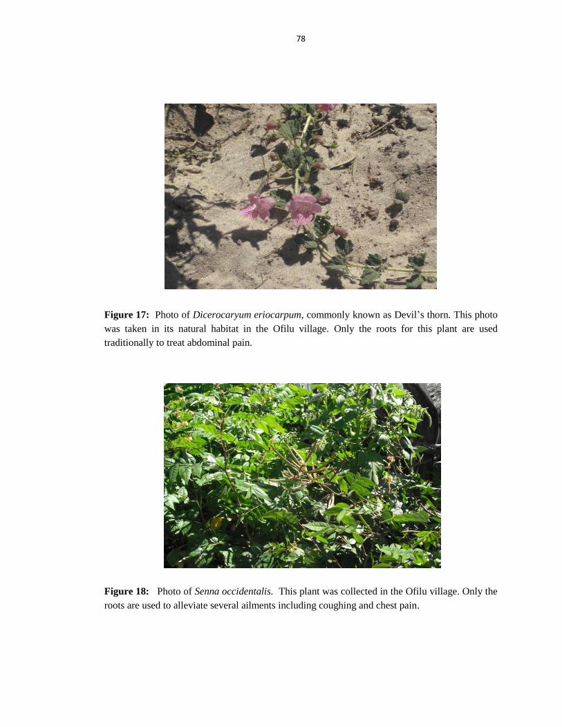
78
Figure 17: Photo of Dicerocaryum eriocarpum, commonly known as Devil‟s thorn. This photo
was taken in its natural habitat in the Ofilu village. Only the roots for this plant are used
traditionally to treat abdominal pain.
Figure 18: Photo of Senna occidentalis. This plant was collected in the Ofilu village. Only the
roots are used to alleviate several ailments including coughing and chest pain.

79
Figure 19: Photo of Lophiocarpus sp. in its natural habitat in the Ofilu village. The foliage for
this plant is traditionally used to treat inflammation.
Figure 20: Photo of Crotalaria flavicarinata. The plant was collected in the Engondi
Constituency, Ofilu village. The entire plant, except for its roots, is used to treat diarrhoea
within the traditional setting for this particular village.

80
4.2 Effect of extracting solvents on percentage yield
Crude extracts, both aqueous and organic extracts were prepared from the various plant
parts specified. The yields of extracts after extraction and drying are given in Table 3
below. The percentage yield was calculated by dividing the mass of dry extract with the
original mass of plant material used for extraction purposes multiplied by a 100.
Overall, the aqueous extracts (40 C > 60 C > 80 C > 30 C) resulted in the highest
quantity of crude dry extract, while the organic extract gave the least quantity. The
aqueous extract (at 40 C) of N. costata gave the highest yield (17.1 %). The lowest
yield (1.2 %) was obtained in the organic extract of R. brevispinosum.
Table 3: Percentage yield for aqueous and organic extracts prepared.
Plants
Extract yield (% w/w)
Water MeOH/DCM(1:1)
30 C 40 C
60 C 80 C
Vahlia capensis 6.0 6.6 12.2 13.2 13.9
Nicolasia costata 7.6 17.1 7.6 12.9 13.9
Rhigozum brevispinosum 7.2 10.3 12.2 8.6 1.2
Dicerocaryum eriocarpum 5.4 3.3 3.8 5.4 7.0
Senna occidentalis 4.5 6.8 6.6 3.5 2.1
Lophiocarpus sp. 6.9 5.0 6.6 5.2 4.5
Crotalaria flavicarinata 12.0 14.5 12.8 9.9 6.0

81
4.3 Preliminary phytochemical screening
4.3.1 Fractionation of crude extracts using thin layer chromatography
Fractionation using solvents of varying polarity was carried out to determine the best
protocol for extraction of compounds (as depicted in Tables 4-6). TLC plates were
developed for aqueous extracts at 30, 40, 60, 80 C and for organic extracts using
methanol: dichloromethane in a 1:1 ratio. Aqueous extractions were carried out at
different temperatures to empirically determine the best extraction temperature. The
TLC plates were visualized under UV light at 254 nm.
Aqueous extracts that were done at 30° and 40°C showed lower levels of isolation of
compounds for solvent system 1 (SS1) and solvent system 3 (SS3), and almost no
compounds for solvent system 2 (SS2). More compounds were extracted at 60˚C than at
80 ˚C for all three solvent systems. The TLC analysis therefore, revealed that 60°C to be
an optimal temperature for the aqueous extractions. It was also observed that SS1 was
more efficient than SS2 for both the aqueous and organic extractions. However, SS3
was the most efficient in the extraction of compounds. Overall, the organic extracts
prepared using methanol-dichloromethane revealed the most compounds in all three
solvent systems compared to the aqueous extracts prepared at 60 C.

82
Table 4: TLC analysis of crude plant extracts fractionated using solvent system 1 (chloroform:
methanol: acetone in a 1:2:1 ratio). Level of extraction based on UV profile of compounds
denoted as high +++, medium ++, low +, no compound –
Voucher
Specimen
no.
Species 30 ˚C
Aqueous
Extract
40 ˚C
Aqueous
Extract
60 ˚C
Aqueous
Extract
80 ˚C
Aqueous
Extract
Organic
Extract
CID19 Vahlia capensis - ++ + + +++
CID20 Nicolasia costata - + + + +++
CID21 Rhigozum
brevispinosum + + +
+++
+++
CID22 Dicerocaryum
eriocarpum ++ ++ ++ ++ +++
CID23 Senna
occidentalis + + ++ + +++
CID24 Lophiocarpus sp + + ++ + +++
CID25 Crotalaria
flavicarinata + + ++ ++ +++
Table 5: TLC analysis of crude plant extracts fractionated using solvent system 2 (chloroform:
ethyl acetate in a 1:1 ratio). Level of extraction based on UV profile of compounds denoted as
high +++, medium ++, low +, no compound –
Voucher
Specimen
no.
Species 30 ˚C
Aqueous
Extract
40 ˚C
Aqueous
Extract
60 ˚C
Aqueous
Extract
80 ˚C
Aqueous
Extract
Organic
Extract
CID19 Vahlia capensis - + - ++ ++
CID20 Nicolasia costata - - + - +
CID21 Rhigozum
brevispinosum - - - + ++
CID22 Dicerocaryum
eriocarpum + ++ + - ++
CID23 Senna
occidentalis + - - - ++
CID24 Lophiocarpus sp - - - - ++
CID25 Crotalaria
flavicarinata - - + + +

83
Table 6: TLC analysis of crude plant extracts fractionated using solvent system 3 (chloroform:
methanol: water in a 12:6:1 ratio). Level of extraction based on UV profile of compounds
denoted as high +++, medium ++, low +, no compound –
Voucher
Specimen no. Species 30 ˚C
Aqueous
Extract
40 ˚C
Aqueous
Extract
60 ˚C
Aqueous
Extract
80 ˚C
Aqueous
Extract
Organic
Extract
CID19
Vahlia capensis + + ++ +++ +++
CID20 Nicolasia costata + ++ + ++ +++
CID21 Rhigozum
brevispinosum ++ +++ ++ +++ +++
CID22 Dicerocaryum
eriocarpum + +++ +++ ++ +++
CID23 Senna
occidentalis ++ + + ++ +++
CID24 Lophiocarpus sp + +++ +++ ++ ++
CID25 Crotalaria
flavicarinata + +++ +++ ++ ++
4.3.2 Presence of selected antimalarial compounds based on TLC chemical
profiling
The TLC chemical profiles for the plant extracts investigated are shown in Table 7. The
plants under investigation were tested for seven known antimalarial compounds
including alkaloids (Al), anthraquinones (An), coumarins (C), flavonoids (F), glycosides
(G), steroids (S) and terpenoids (T). Of the seven phytoconstituents tested only three i.e.
Al, C and G tested positive in all the plant extracts (aqueous and organic). V. capensis
tested positive for Al, C, G and S (57 %); N. costata for Al, C, G and T (57 %); R.
brevispinosum for Al, C, G, and T (57 %); D. eriocarpum for Al, An, C, F, G, and T (86
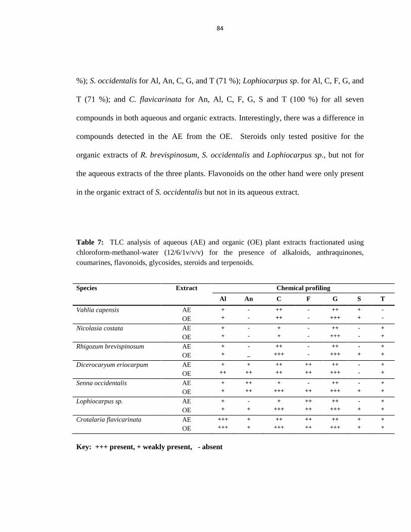
84
%); S. occidentalis for Al, An, C, G, and T (71 %); Lophiocarpus sp. for Al, C, F, G, and
T (71 %); and C. flavicarinata for An, Al, C, F, G, S and T (100 %) for all seven
compounds in both aqueous and organic extracts. Interestingly, there was a difference in
compounds detected in the AE from the OE. Steroids only tested positive for the
organic extracts of R. brevispinosum, S. occidentalis and Lophiocarpus sp., but not for
the aqueous extracts of the three plants. Flavonoids on the other hand were only present
in the organic extract of S. occidentalis but not in its aqueous extract.
Table 7: TLC analysis of aqueous (AE) and organic (OE) plant extracts fractionated using
chloroform-methanol-water (12/6/1v/v/v) for the presence of alkaloids, anthraquinones,
coumarines, flavonoids, glycosides, steroids and terpenoids.
Species Extract Chemical profiling
Al An C F G S T
Vahlia capensis AE
OE
+
+
-
-
++
++
-
-
++
+++
+
+
-
-
Nicolasia costata AE
OE
+
+
-
-
+
+
-
-
++
+++
-
-
+
+
Rhigozum brevispinosum AE
OE
+
+
-
_
++
+++
-
-
++
+++
-
+
+
+
Dicerocaryum eriocarpum AE
OE
+
++
+
++
++
++
++
++
++
+++
-
-
+
+
Senna occidentalis AE
OE
+
+
++
++
+
+++
-
++
++
+++
-
+
+
+
Lophiocarpus sp. AE
OE
+
+
-
+
+
+++
++
++
++
+++
-
+
+
+
Crotalaria flavicarinata AE
OE
+++
+++
+
+
++
+++
++
++
++
+++
+
+
+
+
Key: +++ present, + weakly present, - absent

85
4.3.3 Presence of selected antimalarial compounds based on phytochemical tests
The presence of the selected phytoconstituents was further confirmed by a number of
phytochemical tests. These tests were conducted on six of the plants which revealed the
presence of anthraquinones in D. eriocarpum, S. occidentalis; glycosides in D.
eriocarpum, S. occidentalis; terpenoids in V. capensis; flavonoids and coumarins in V.
capensis, N. costata, R. brevispinosum, D. eriocarpum, S. occidentalis, Lophiocarpus sp.
(Table 8). Alkaloids and steroids were absent in all plants tested. Coumarins and
flavonoids, in contrast, were present in all plant extracts tested. For each of the plants
under investigation, i.e. V. capensis, N. costata, R. brevispinosum, D. eriocarpum, S.
occidentalis, Lophiocarpus sp. a total of 3 (43 %), 2 (29 %), 2 (29 %), 4 (57 %), 4 (57
%), 2 (29 %) of the seven antiplasmodial compounds tested were present. Crotalaria
flavicarinata, unfortunately, could not be screened for the seven phytochemicals because
of limited plant material.
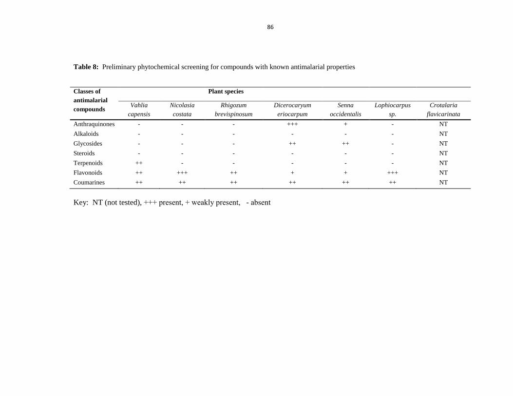
86
Table 8: Preliminary phytochemical screening for compounds with known antimalarial properties
Classes of
antimalarial
compounds
Plant species
Vahlia
capensis
Nicolasia
costata
Rhigozum
brevispinosum
Dicerocaryum
eriocarpum
Senna
occidentalis
Lophiocarpus
sp.
Crotalaria
flavicarinata
Anthraquinones - - - +++ + - NT
Alkaloids - - - - - - NT
Glycosides - - - ++ ++ - NT
Steroids - - - - - - NT
Terpenoids ++ - - - - - NT
Flavonoids ++ +++ ++ + + +++ NT
Coumarines ++ ++ ++ ++ ++ ++ NT
Key: NT (not tested), +++ present, + weakly present, - absent

87
In terms of pharmacological activity, the focus has been moved to Vahlia capensis,
Nicolasia costata and Dicerocaryum eriocarpum. Overall, all the plants under
investigation possess phytoconstituents that are said to be antimalarial, however since
fever is the most obvious and important clinical manifestation of Plasmodium
falciparum infections in terms of it developing into severe malaria over a short period
soon after it first appears prompted us to look at the V. capensis and N. costata. Both
these plants are used to treat fever in the traditional setting. D. eriocarpum conversely
was selected because of it exhibiting high levels of antimalaria compounds of which 3
are considered to be potent.
4.4 Antimalarial activity of the investigated plant extracts
The three lead plants were screened for antimalarial activity against P. falciparum
strains 3D7 (chloroquine-sensitive) and D10 ACP (signal)-GFP strain. The plant
extracts were prepared using methanol-dichloromethane. The parasitized RBCs were
incubated with stock solutions of the extracts at 37˚C for 3 different concentrations (50,
100 and 250 µg/mL). The parasitaemia determination was done at two time points 24
and 48 hours. The extracts of V. capensis, N. costata and D. eriocarpum manifested
significant activities against the Plasmodium falciparum 3D7 strain as seen by reduced
parasitaemia at 24 hours post treatment of cultures (Figure 21) and minimal activities
against the D10 (Figure 22).

88
Figure 21: Photo micrograph of red blood cells infected with P. falciparum 3D7 24 hours
following treatment with a) Control (DMSO), insert shows parasitized red blood cell, b) Vahlia
capensis, insert shows unparasitized red blood cell, c) Nicolasia costata and d) Dicerocaryum
eriocarpum extracts (Magnification x40, oil immersion). Percentage parasitaemia had decreased
after treatment with plant extracts as compared to the DMSO culture.
a) b)
c) d)

89
Figure 22: Photo micrograph of red blood cells infected with P. falciparum D10 24 hours
following treatment with a) Control (no treatment), b) Vahlia capensis, c) Nicolasia costata and
d) Dicerocaryum eriocarpum extracts (Magnification x40, oil immersion). No percentage
reduction of parasitaemia was observed for all 3 cultures after treatment with plant extracts as
compared to the no treatment culture.
The basic measurement of antimalarial activity used in this study was a reduction in the
number of parasitized cells in the test cultures compared to the control at 24 hours for
the three concentrations i.e. 50, 100 and 250µg/mL. Figure 23 shows the change in
parasitaemia for the P. falciparum 3D7 strain over 24 and 48 hours for V. capensis (VC),
N. costata (NC) and D. eriocarpum (DC). The parasitaemia for the VC-treated culture
a)
b)
c)
d)

90
(P24=1.43) at 50 µg/mL had decreased with 24.7 %, the NC-treated culture (P24=1.53)
with 19.5 % and the DE-treated culture (P24=1.01) with 46.8 % compared to the
untreated culture (negative control) (P0=1.90). In contrast, the concentration at
100µg/mL showed moderate antimalarial activity. The parasitaemia for the VC-treated
culture (P24=1.62) had decreased with 26.4 %, the NC-treated culture (P24=1.33) with
39.5 % and the DE-treated culture (P24=1.70) with 22.7 % compared to the untreated
culture (P0=2.2). The concentration 250 µg/mL showed high antimalarial activity. The
parasitaemia for the VC-treated culture (P24=1.62) had decreased with 50.4 %, the NC-
treated culture (P24=1.33) with 60.0 % and the DE-treated culture (P24=1.62) with 62. 7
% compared to the untreated culture (P0=2.5).
Generally, the parasitaemia at 24 hours after treatment (P24) had reduced for all cultures,
therefore, it can be said that the plant extracts under investigation exhibited antimalarial
activity at all 3 concentrations in the first 24 hours after treatment. Notably, there was
no reduction of parasitaemia for all cultures at 48 hours post treatment; instead the
parasite load had increased. This suggests that the effect the plant extract exhibited
against the parasites had worn off between 24 hours and 48 hours after treatment. The
DMSO (negative control) culture also showed no reduction in the parasitaemia. Hence,
the organic solvents used for extraction purposes had no toxic effects on the parasites,
and it can be said that the plant bioactive constituents were responsible for the reduction
in the number of parasites and not the solvent that was used for extraction purposes.

91
Figure 23: Antiplasmodial activity of Vahlia capensis, Nicolasia costata and Dicerocaryum
eriocarpum extracts on Plasmodium falciparum 3D7 at concentrations of (a) 50, (b) 100, (c) 250
µg/mL as compared to the negative control at 0hrs, 24hrs and 48hrs.
0
1
2
3
4
5
Neg C Vahlia capensis Nicolasia costata Dicerocaryum
eriocarpum
Pa
rasi
taem
ia (
%)
50 µg/ml
a)
Negative
control
0
1
2
3
4
5
Neg C Vahlia capensis Nicolasia costata Dicerocaryum
eriocarpum
Pa
rasi
taem
ia (
%)
100 µg/ml
b)
Negative
control
0
1
2
3
4
5
Neg C Vahlia capensis Nicoslasia costata Dicerocaryum
eriocarpum
Pa
rasi
taem
ia (
%)
250 µg/ml
c)
Negative
control

92
Toxicity of the extracts was concentration dependant with the highest anti-plasmodial
effect being observed at 250µg/mL (Figure 24). VC, NC and DE at 250µg/mL had a 50
% and higher parasitaemia reduction rate than at 50 and 100 µg/mL. The plant that
showed highest antiplasmodial activity was D. eriocarpum. DE extract had the greatest
effect on Plasmodium parasitaemia at 50 and 250µg/mL. At 100µg/mL, the
parasitaemia % for both VC and NC was lower than for DE. At 50µg/mL the %
parasitaemia reduction for DE was remarkably higher than for VC and NC; however at
250µg/mL the parasitaemia reduction was relatively higher.
Figure 24: The concentration dependent effect of the plant extracts on parasitaemia of the
Plasmodium 3D7 strains for Vahlia capensis, Nicolasia costata and Dicerocaryum
eriocarpum.
0
10
20
30
40
50
60
70
50 100 250
% p
ara
sita
emia
red
uti
on
Concentration (µg/ml)

93
The percentage of growth inhibition (Table 9) was determined using different
concentrations of plant extracts against the P. falciparum 3D7 strain and was calculated
as (1-Number of parasite in test flask divided by the Number of parasites per control
well) x 100. The results were compared to that of the untreated controls (i.e. parasites
grown without treatment of the plant extract). Maximum growth inhibition (%) was
obtained with D. eriocarpum (66.8 % inhibition) at a concentration of 250µg/mL. The
lowest growth inhibition of the parasites was obtained with V. capensis (26.6 %
inhibition) at a concentration of 50µg/mL. Other concentrations for the same plants
exhibited moderate growth inhibition. Overall, all plants showed growth inhibition
above 50 % at 100 and 250µg/mL.
The concentration at which growth of parasites was inhibited by 50 % (i.e. IC50) was
estimated from the graph (Figure 25) drawn on the percentage (%) growth inhibition
data. The concentration of plant extracts corresponding to 50 % growth inhibition (IC50)
for V. capensis, N. costata and D. eriocarpum are 93.29, 86.63, 63.17µg/mL
respectively; indicating that the amount of VC and NC required for effective
Plasmodium inhibition activity was 1.5 times more than that of DE.

94
Table 9: Percent (%) growth inhibition of Plasmodium parasites by extracts of Vahlia capensis,
Nicolasia costata and Dicerocarym eriocarpum in chloroquine-sensitive (3D7) strains of
Plasmodium falciparum.
Plant organic extracts Concentration of extract (µg/mL) Chloroquine-sensitive strain (%)
inhibition
Vahlia capensis
50 26.6
100 55.5
250 55.8
Nicolasia costata
50 21.5
100 63.4
250 64.4
Dicerocaryum eriocarpum
50 48.1
100 53.3
250 66.8
Figure 25: In vitro interaction of plant extracts for Vahlia capensis, Nicolasia costata and
Dicerocaryum eriocarpum (% inhibition) in Falciparum 3D7chloroquine-sensitive strains.
0
10
20
30
40
50
60
70
80
50 100 250
% i
nh
ibit
ion
Concentration

95
For the antimalarial activity against the D10 strain, the concentration of the plant
extracts that had shown significant activity against the 3D7 strain was used. The three
plant extracts tested for antiplasmodial effects against the D10 strain showed no overall
reduction in parasitaemia (Figure 26). However, suppression in the growth of the
parasites was observed. Compared to the no treatment culture, VC and NC showed
higher levels of suppression than the DE treatment. The parasitaemia at 0 hours (P0 =
5%) and had increased with 4.79 % for the VC treatment culture (P24 = 5.47 %) after 24
hours; whereas for NC (P24 = 5.85 %) and DE (P24 = 6.36 %) cultures, the P24 increased
with 20.87 % and 33.89 % respectively. The parasitaemia at 48 hours P48 post treatment
was comparable for all treated cultures whereby a continued increase in parasitaemia
was observed.
Figure 26: Antiplasmodial activity for Vahlia capensis, Nicolasia costata and Dicerocaryum
eriocarpum at 250µg/mL against the Plasmodium falciparum D10 strain at 0hrs, 24hrs and
kk48hrs. Parasitaemia had relatively increased for all 3 treated cultures compared to the no
treatment which shows significant increase in parasitaemia.
0
2
4
6
8
10
12
14
VC NC DE No treatment
Pa
rasi
taem
ia (
%)
Treatment

96
4.5 Metabolic-based assays
4.5.1 Pyridine-hemochrome to measure haem incorporation in hemozoin
Two strains of P. falciparum D10 and 3D7 were used to evaluate the antiplasmodial
effect of the selected plant extracts. Both exhibited sensitivity to the extracts with 3D7
being more sensitive with parasitaemia at 24 hours post treatment being reduced by 62.7
% at 250 µg/mL. P. falciparum D10 showed no reduction in parasitaemia, however,
there was suppression of growth over 48 hours. The latter strain was used for
mechanistic studies to determine the mode of action of the extracts because of the
prolonged effect it had on the P. falciparum strain.
The Plasmodium cultures were harvested after treatment with Vahlia capensis,
Nicolasia costata and Dicerocaryum eriocarpum extracts at 0, 4, and 24 hours to
measure the relative amount of hemozoin at each point using the formulae Δ OD560 =
OD560 (reduced sample) - OD560 (oxidized sample) (Figure 27). At 4 hours, less
haemazoin accumulation was observed for VC (156 %) and NC (169 %) than for the DE
(244 %) culture which was comparable to the untreated culture (225 %). At 24 hours, a
significant rise in haemazoin levels was observed for the untreated culture but not for the
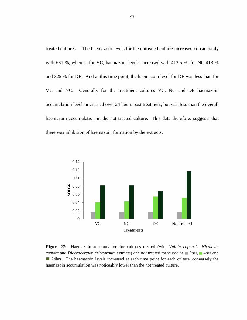
97
treated cultures. The haemazoin levels for the untreated culture increased considerably
with 631 %, whereas for VC, haemazoin levels increased with 412.5 %, for NC 413 %
and 325 % for DE. And at this time point, the haemazoin level for DE was less than for
VC and NC. Generally for the treatment cultures VC, NC and DE haemazoin
accumulation levels increased over 24 hours post treatment, but was less than the overall
haemazoin accumulation in the not treated culture. This data therefore, suggests that
there was inhibition of haemazoin formation by the extracts.
Figure 27: Haemazoin accumulation for cultures treated (with Vahlia capensis, Nicolasia
costata and Dicerocaryum eriocarpum extracts) and not treated measured at 0hrs, 4hrs and
cc 24hrs. The haemazoin levels increased at each time point for each culture, conversely the
haemazoin accumulation was noticeably lower than the not treated culture.
0
0.02
0.04
0.06
0.08
0.1
0.12
0.14
VC NC DE NT
ΔO
D5
6
Treatments
Not treated

98
4.5.2 Protease inhibition assay
Protease inhibition activity of the plant extracts was done by fluorometric detection.
Protease substrate casein was labeled with the fluorescent dye, fluorescein quenching the
signal from the dye. If protease activity was present, the fluorescein isothiocyanate
(FITC) was cleaved from the casein, unquenching the dye, giving off fluorescence
which was then measured. The protease activity was compared for the different time
points (Figure 28). At 0 hours, low levels of protease activity was observed (T0 =
203.25 FSU) compared to the negative and positive controls measured at 197.89 and
1382.87 FSU respectively (not shown on graph). Protease activity observed for the
untreated culture increased at 4 hours by 41.41 %, before falling at 18 hours by -3.42 %
and then increased again at 24 hours by 2.3 %. All treatments followed the same trend
with an increase at 4 and a decrease at 18 hours. Protease activity for VC (7.75 %), NC
(89.22 %) and DC (24.43 %) treated cultures, as well as for the untreated culture, (41.41
%) was observed at 4 hours. Protease activity was reduced at 18 hours for VC (-2.72
%), NC (42.12 %) and DE (22.26 %) and for the untreated culture (196.3 %). NC
treated culture exhibited the highest activity at both time points (89.22 % and 42.12 %
respectively). At 24 hours, only for the treated cultures, a reduction in protease activity
was observed: VC (-28.80 %), NC (-22.39 %) and DE (-31.42 %). For the no treatment
culture (2.30 %), there was a rise in protease activity. Overall low protease inhibition in
the presence of plant extracts was observed compared to the no treatment cultures at 4
and 18 hours, but for the 24 hour time point protease inhibition. This was more evident

99
due to the increase in protease activity for the no treated culture and decrease in protease
activity for the treated cultures.
Figure 28: Protease activity by proteases, such as aspartic and cysteine proteases involved in
the metabolism of P. falciparum is shown for the different time points post treatment with plant
extracts including Vahlia capensis, Nicolasia costata and Dicerocaryum eriocarpum,
and for the untreated culture. Inhibition of protease activity exhibited by plant extracts is
more evident at 24hrs.
0
50
100
150
200
250
300
350
400
450
1 2 3 4
Pro
tea
se a
ctiv
ity
(F
SU
)
Time points (hours)
T0 T4 T18 T24

100
CHAPTER 5: DISCUSSION
Plant materials remain an important source of medicine in the fight against malaria
(Aderounmu, 2007). Since widely used antimalarial drugs such as quinine and
artermisinin were isolated from plants (Rosenthal, 2001), and because of the increased
resistance to and lack of accessibility to existing affordable drugs, the pharmocognostic
investigations of plants for the establishment of complementary medicine for malaria
within traditional plants is necessary (Wang et al., 2007). Malaria poses a major health
problem to more than half of the world. Namibia, a sub-Saharan African country with a
population of approximately 2.2 million people (MoHSS, 2010a) of which two thirds
live in malaria endemic areas, is moving towards elimination of malaria (MoHSS,
2009).
However, with reports of decreased sensitivity to current first lines drugs such as
Artemether-Lumefantrine, an Artemisinin-based combination treatment (ACT) (Kayano
et al., 2011), Namibia runs the risk of not reaching its aim to remove all reservoirs of the
Plasmodium spp. within its borders. The aim of this study therefore, was to biologically
assess the effectiveness of some plants used in the traditional setting native to Namibia.

101
Plants collected for the validation as antimalarial treatments were selected based on the
ethnopharmacological approach, which is the method of selecting plants based on their
ethnomedicinal uses (Tringali, 2001, p. 394; Adebayo & Krettli, 2011). According to
Khafagi & Dewedar (2000) this approach yields better results in drug discovery/
discovering higher plants with good biological activity than random selection. This
approach is also used by pharmaceutical companies in search of bioactive
phytoconstituents to synthesize novel drugs.
In addition to the collection of plant material, voucher specimens of the plants were sent
to the NBRI for scientific identification. This type of identification was important for
accurate documentation as the plants may be known by dissimilar names in different
regions of Namibia or even within the same region where diverse dialects are used
(Sequeira, 1994). This will assist in any licensing or permissions given by the MoHSS
to use the plants as complementary medicines for malaria as they can be issued across
regions regardless of the local name. For example, Acacia erioloba (Camel Thorn)
which is used to treat fever is called Omuthiya in Oshiwambo and ||Ganab in
Nama/Damara. Scientific names also allow for the cross comparison of plants and their
uses in different areas. This documentation would be important because it sanctions
knowledge of the plant and its uses to be preserved for future generations (Hedberg,
1993), it also serves as proof of prior art if any benefits resulting from the use of the
plants are accrued (Elvin-Lewis, 2007).

102
Not all seven plants under study were described in current literature and that in some
cases little or nothing has been described on the medicinal uses; in such circumstances
only the knowledge of the traditional healers are available. Among the plants that were
validated as complementary antimalarials, most of them find wide ethnopharmalogical
uses, including Vahlia capensis, Rhigozum brevispinosum, Dicerocaryum eriocarpum
and Senna occidentalis. According to Gibbons (2004) V. capensis possesses
antibacterial properties (Majinda, Motswaledi, Waigh, & Waterman, 1997) and is used
in Zimbabwe and Botswana to treat eye infections; while Rai and Mares (2003) reports
that V. capensis is effective against fungal infections. According to Cheikhyoussef,
Shapi, Matengu and Mu Ashekele (2011) R. brevispinosum is used to treat syphilis in
the Oshikoto region of Namibia. Other uses as described by Iriondo, Dulloo and Turok
(2008) and Leffers (2003) include ornamental uses in rural gardens, due to its yellow
flowers. Leffers further maintains that its branches are used as digging sticks.
According to Leffers (2003), the foliage of this plant is poisonous and thereby only the
roots are used for medicinal purposes, as in this case the roots are used to treat chest
pain. D. eriocarpum is commonly known as Devil‟s thorn and is said to have
antibacterial and anti-inflammatory properties (Luseba, Elgorashi, Ntloedibec, & Van
Staden, 2007).
Only the roots of Senna occidentalis are used, because of the toxic properties of its
foliage. Babosa-Ferriera, Dogli, Maiorka and Gorniak (2005), and Orech, Akenga,

103
Ochora, Friis and Aagaard-Hansen (2005) also describes this plant to be poisonous to
humans as well as to animals. S. occidentalis is also used for malaria (entire plant) and
against conjunctivitis (roots), rheumatism (roots), gonorrhoea (roots), and asthma (roots)
in Comoro (Kaou et al., 2008). Van Koenen (2001) confers that this plant is also used to
treat malaria, as well as febrile symptoms. Lophiocarpus sp. according to Naples (2005),
treats breast inflammation, itchy skin, skin diseases, as well as sore throats. Other
medicinal uses (of the leaves) include abdominal pain and fever (Van Koenen, 2001).
Medicinal uses for N. costata and Crotalaria flavicarinata are rather narrow. Although
medicinal uses had been described for some plants, scientific validations are non-
existent in almost all cases. However, the information gathered from the traditional
healers corresponded with the documented uses, in most instances. All this validates that
traditional plant knowledge is of substantial value, not just to the indigenous people for
local use, but to researchers for the development of novel antimalarial medicines.
Vahlia capensis, for example, is used as a treatment for fever, in the literature it is used
as a cure against infectious agents that can potentially cause fever. Therefore, there is an
increasing need for the documentation of traditional knowledge and for the scientific
validation of the medicinal uses of such plants as it might become lost to future
generations, as well as for the integration of ethno-medicine into modern medicine.
The plants described by traditional healer Selvia Nghilifilwa are mostly used in their dry
ground state. The plant material was dried, ground and stored at low temperatures (- 20
C). This was done to preserve the presence and the quality of the compounds found

104
within the plant. According to Makkar (2000) the moisture content of fresh plant
material changes the chemical composition and properties of the plant over time.
Grinding of the plant material into fine particles was to create a larger surface area to
allow higher extraction of bioactive compounds.
The successful extraction of bioactive compounds from plants, according to Parekh,
Jadeja and Chanda (2005) is largely dependent on the type of solvent used in the
extraction procedure. Effective extraction from the dried plant material was achieved
using different solvent systems, including the use of a single solvent i.e. water or a series
of solvents such as chloroform (weakest), dichloromethane, acetone, ethyl acetate,
methanol, water (strongest), of increasing polarity. The dried plant material was
extracted using water, a similar extraction method employed by traditional healers in
preparing most of their herbal remedies; and a methanol-dichloromethane solvent
system to obtain the bioactive compounds present in the plants under investigation for
pharmalogical investigation.
Both aqueous and organic extractions were carried out in order to isolate both polar and
non-polar compounds (Table 4-6). Whilst the pattern for finding the compounds was
similar, the abundance varied with organic solvents showing higher levels of the
compounds isolated. This is consistent with other studies which illustrate that plant
phytochemicals are more soluble in organic solvents (Willcox et al., 2004). Willcox et
al. (2004) further maintains that organic solvent extractions are used as a good

105
alternative in evaluating the antimalarial activities of plants, as organic solvents are able
to extract a broad spectrum of chemical constituents. Even though, it is interesting to
note that most medicinal plant preparations are conducted in aqueous solvents (water); it
is noteworthy to observe the effect of organic solvents on the plant material and to
describe them. Therefore, it would be more informative to focus on both the traditional
as well as the organic preparation method. This is because extraction changes the
presence and abundance (quality and quantity) of phytochemicals in the medicinal
treatment and as such changes the very nature of the treatment itself (Jordana,
Cunninghama, & Marles, 2010). The presence and the abundance of the compounds
may be the difference between whether the plant will be medicinal or toxic.
Different temperatures were used during the aqueous extraction process, because some
compounds are heat labile, such as anthraquinones (Sanyovaa & Reisse, 2006) and
alkaloids (Mattocks, 1961); whilst other compounds may be volatile (Hatzakis,
Opsenica, Solaja, & Stratakis, 2007). Thin layer chromatography was used to determine
whether higher temperatures resulted in loss of some compounds. The isolation of
compounds was observed to be more for the aqueous extracts prepared at 60 ˚C, than for
30, 40 and 80 ˚C. This may suggest that at lower temperatures, compounds from plants
are less than likely to be isolated and at higher temperatures these compounds may
become degraded. Therefore, it may be concluded that 60 ˚C was the best temperature

106
for isolating of compounds by aqueous extraction, even though a higher percentage yield
of dry extract was obtained at 40 C.
The extract yield (% of dry weight) from the plants investigated was higher for the
aqueous extracts than for the organic extracts. Parekh et al., (2005) maintains that these
observations can be explained due to the polarity of the compounds extracted. The high
polarity of water probably accounted for the high yield in the dry extracts because both
polar and some less polar compounds were extracted. Hence, the extract yield increased
when solvents of polarity was used.
Past studies have revealed that compounds with antimalarial activity are commonly
polar (Willcox et al., 2004). However, to ensure efficient extraction of compounds, a
two-solvent system was used that contain one polar and one non-polar solvent.
Fractionation of crude extracts using different solvent systems including chloroform:
methanol: acetone (SS1) (1:2:1 v/v/v), chloroform: ethyl acetate (SS2) (1:1 v/v) and
chloroform: methanol: water (SS3) (1:2:1 v/v) ascertained SS3 to be the most efficient in
isolating compounds from the plant material. According to the solubility screening table
(Appendix D), the solvents used for the TLC assay are of different polarities.
Chloroform was used for all three solvent systems and is non-polar, whereas water and
methanol are strictly polar. Ethyl acetate is situated more or less in the middle of this
table. Therefore, it is suggested that the fractionation order 3>1>2 of the solvent

107
systems are as a result of the fact that Solvent system 1 is non-polar in comparison with
the other two solvent systems, as it contains chloroform and ethyl acetate (a non-polar
and less polar solvent).
Phytochemical screens determine the overall „chemical fingerprint‟ or „chemical profile‟
of a plant extract (Bandaranayake, 2006, p. 38). The two phytochemical analysis
techniques used in this study were thin layer chromatography, which works by isolating
and separating the different compounds present in the plant extract on an aluminum plate
coated with silica gel; whereas preliminary phytochemical tests works on a colour-
change basis to determine the presence or absence of certain plant constituents.
Phytochemical screenings were done to determine whether compounds with known
antimalarial properties were present in the plants collected for the study to warrant for
further pharmacological investigations. Previous studies have shown that the
composition of phytochemicals present in the plant is in direct correlation with its
pharmacological activity (Aremu, 2009). Thus, the results obtained from the
phytochemical analysis provide preliminary data on some known antimalarial
compounds that are found in plants investigated, suggesting potential antiplasmodial
activity.
Phytochemical tests revealed the presence of anthraquinones, flavonoids, terpenoids,
coumarines, and glycosides (Table 8), of the seven compounds tested for. Each plant

108
appeared to have more or less only four of the phytoconstituents present. Paradoxically,
thin-layer chromatography analysis tested positive for all compounds including those not
observed during the phytochemical assays (alkaloids and steroids) (Table 7). This may
be as a result of phytochemicals that sometimes have an effect of masking the presence
of each other at high concentrations, hence one compound may easily mask the presence
of another compound (Bugani, Harper, McCulloch, Reppart, & Ireland, 2008); whereas
for thin-layer chromatography analysis, the compounds are allowed to move across the
plate at different rates (Aremu, 2009) thus TLC results in separation of the compounds
permitting them to be detected independent of each other (Holetz et al., 2002).
Although not tested for the preliminary phytochemical tests, C. flavicarinata showed to
have substantial potential i.e. it tested positive for all 7 compounds.
The plants were screened for antimalarial activity using an in vitro model, which
involved growing the parasites in human red blood cells outside the body in cell culture
flasks at 37 C. Three of the seven plant species, namely Vahlia capensis, Nicolasia
costata and Dicerocaryum eriocarpum were tested for antiplasmodial activities at three
different concentration (50, 100, 250µg/mL). From the evaluation, all three plant
species demonstrated antimalarial activity against the Plasmodium falciparum
chloroquine-sensitive strain, 3D7 at 24 hours post treatment. It was observed that at 48
hours, there was an increase in parasitaemia, thus indicating a short pharmacological
half-life of active constituents present in the plant extract; meaning for every 24 hours

109
the dosage would have to be repeated until all parasites are cleared. Potent antimalarials
available today including artemisinin have short elimination half-lives as well, and as a
result are then administered as combination therapies to ensure complete elimination of
the parasite load (Hombhanje & Huang, 2010).
The extract of D. eriocarpum exhibited the highest antiplasmodial activity for all 3
concentrations, then N. costata, with V. capensis showing the lowest activity. These
differences in levels of antimalarial activity can be attributed to the presence of
compounds with antimalarial properties in the plants under study (Jackson & Miller,
2006). DE tested positive for most compounds, i.e. 4 and 6 out of the 7 compounds for
the phytochemical and TLC analysis, respectively. In NC, 4 and 2 out of 7 compounds
and in VC 4 and 3 out of 7 compounds. It is interesting to note that VC and NC both
show moderate antimalarial activity and if used together to treat malaria, higher
antimalarial activity is to be expected. Since these two plants are used in the traditional
setting as a combination therapy, and might be more potent/ toxic if their antiplasmodial
effect is through different mechanisms. These effects may be synergistic and as a result
could be a potent antimalarial.
The treatment was dosage-dependent with 250µg/mL concentration the most effective
against the Plasmodium parasites for all the extracts. The IC50 for the plant extracts
tested ranged from 63.71 to 93.29µg/mL. However, for in vitro studies plant extracts

110
considered exhibiting good activity have IC50 values less than 10µg/mL and those
exhibiting moderate activity with IC50 values of 10-50 µg/mL, low activity with IC50
values of 50-100µg/mL and inactive with IC50 values of greater than 100µg/mL (Basco
et al., 1994; Dolabela et al., 2008). Thus, as potential antimalarials these herbal
remedies at doses as described, exhibit low activity. The crude extracts of these plants
are rendered therapeutically meaningful irrespective of their low antimalarial activity.
This is because it is likely that the active components in the plant extracts are present in
very low concentrations and forms part as part of complex mixtures with many other
compounds within the extract (Batista et al., 2009). Hence, the extracts only exhibited
high activity at high concentrations (100-250µg/mL). This discrepancy in the IC50
values might also be due to factors such as the type of Plasmodium strain used, the
ability of compounds masking the activity of other compounds, solvents used for
extraction, and the fraction of extract used.
Antimalarial activity exerted by plant extracts VC, NC and DE against the P. falciparum
D10 strain at a concentration of 250µg/mL proved to be chemosuppressive. Growth of
the D10 strain was restricted in the presence of the lead 3 plant extracts. This was
observed as a slow increase in the parasite load over 48 hours of incubation post
treatment compared to the no treatment which showed a 2-fold increase in the
parasitized red blood cells. This therefore suggests that the extracts of the selected plants
do not kill the parasites but hinders the growth of these parasites instead, thus it can be

111
said that these plants also exhibit antimalarial activity against the P. falciparum D10
strain. Interestingly, the extract DE showed highest antiplasmodial activity for the P.
falciparum 3D7 strain, whereas for the D10 strain chemosuppression was the least.
Plasmodium cultures, even from the same isolates, at different times after growth in
culture, show considerable variability in drug susceptibility (Thaithong, Beale, &
Chutmongkonkul, 1988). Furthermore, clones from the same isolate show heterogeneity
in drug sensitivity in culture (Thaitong 1983). Studies have found that there are several
protease enzymes involved in Plasmodium infection and metabolism namely cysteine,
serine and aspartic proteases (Pandey, Singh, Arastu-Kapur, Bogyo, & Rosenthal, 2006).
These enzymes play different roles at different parasite growth stages and are sometimes
redundant. The roles of these differ in the clones of the parasite thus determining the
outcome of any antiplasmodia treatment whose mode of action is protease inhibition
based. This redundancy may explain why the outcomes of treatment with the plant
extracts differ for the two P. falciparum strains 3D7 and D10.
Additionally, a heirachical level of activity of gene expression has been described in
Plasmodium falciparum for genes necessary for infection and metabolism.
Redeployment of machinery in the parasite occurs when silencing of dominant pathways
results in expression of underlying alternative ones (Baum, Alexander, Maier, Robert,
Good, Simpson, & Cowman 2005). This provides parasites with the ability to adapt and
evade host immune responses or drug treatments for example P. falciparum PfRh

112
proteins involved in infection. PfRh is expressed in the 3D7 strain but not the D10
(Duraisingh, Triglia, Ralph, Rayner, Barnwell, McFadden, & Cowman, 2003).
Haemozoin polymerization inhibition assay was done at 0, 4 and 24 hour time points.
These points are critical in the ring and trophozoite growth stage of the parasite, which
are metabolically active during this time. Hemoglobin degradation correlates with these
two growth stages (Gardiner, Trenholme, Skinner-Adams, Stack, & Dalton, 2006). At
24 to 30 hours into the life cycle, the parasites have reduced metabolic activity with
cellular activity centred on synthesis and asexual reproduction. Hence this is assay is
usually conducted only up to 30 hours at most. For the haemazoin inhibition assay it was
found that at 24 hours post treatment haemazoin levels were noticeably less for the
treated parasite cultures than for the untreated culture; but at 4 hours these levels were
comparable to that of the treated culture. Similar observations were made for protease
inhibition activity. At 4 hours the protease activity had increased for both the treated
and untreated cultures, and at 24 hours, only for the treated cultures, a reduction in
protease activity was observed. Proteases are involved in the haemazoin crystallization,
which occurs at the later stage of the erythrocyte growth stage, the trophozoite (Gamboa
de Dominquez & Rosenthal, 1996). The period of increased protease activity coincided
with the greatest haemazoin accumulation implying these enzymes were involved in
haemazoin accumulation in this study. Treatment with plant extracts reduced both
protease activity and haemazoin accumulation establishing a causal relationship between

113
protease inhibition and inhibition of haemazoin accumulation. Inhibition of protease
activity was also observed at 4 hours at the ring stage of the infection (Matteelli &
Castelli, 1997). At this stage infection is being established, parasites are entering the
RBCs and hydrolyzing haemoglobin to haem and globin (Gamboa de Dominquez &
Rosenthal, 1996). Different classes of proteases may be in action at this stage and are
also inhibited in their activity leading to suppression but not killing of the plasmodia.
We hypothesize that the plant extracts inhibited some but not all proteases in plasmodia.
Inhibition of earlier growth stage enzymes caused suppression of growth whilst enzymes
that were inhibited in later growth stages were involved in haemazoin metabolism.
Proteases have reported to be redundant in plasmodia proteases (Pandey, Singh, Arastu-
Kapur, Bogyo, & Rosenthal, 2006); hence inhibition of one group may not affect other
groups of proteases depending on the plasmodia clone being used as a lab model. This
redundancy may be the reason that there was more suppression in P. falciparum D10
than P. falciparum 3D7. Further studies on the clones using knockout clones for each of
the enzyme systems or specific inhibitors would shed more light on the differences
between the two lab clones of plasmodia. It has been found that studies on anti-
plasmodial plants most of the time focuses only on killing the parasites but seldom
consider other means (Rasoanaivo et al., 2011). While in this study, mechanism studies
were done on a Plasmodium falciparum strain that was suppressed when exposed to the
plant extracts. The postulated mechanisms of actions for plant crude extracts for this

114
study includes inhibition of hemozoin polymerization or ferriprotoporphyrin 1x (FP) in
the parasite and/ or protease inhibition activity.
In this study, the ethnopharmacological approach was used and the results obtained
proved that the plants‟ uses are rational. The 7 plants that were identified and collected
all showed the presence of antimalarial compounds and the 3 that were tested showed
antiplasmodial activity against the P. falciparum strains. Thus, this study shows that this
approach has a high hit rate in successfully identifying plants with potential biological
activity. The present study confirms hypothesis that plants used traditionally to treat
symtpoms of malaria exhibit biological activity with antiplasmodial effects.
Limitations of study: Some of the limitations include the use of organic extracts
instead of aqueous extracts as prepared in the traditional setting. This means some of
the compounds which may have a therapeutic effect in an aqueous extract may be absent
in the test samples. This may have excluded antimalarial compounds that are polar
resulting in lower antimalarial activity. Another limitation is the use of parasites at
different growth stages. Plasmodium parasites were unsynchronized at the time of the
experiments.This means that if an extract had an effect on a particular growth stage and
not others, the antiplasmodial effect would be reduced. Additionally, a limited number
of replicates of antiplasmodial activity assays were done. This may have introduced
bias, as parasite growth patterns are variable depending on culture conditions; hence

115
using more replicates would have resulted in more representative outcomes in the
antiplasmodial assays. Finally, metabolic assays were carried out on only the D10
strain. Further information on the mechanism of action of the plant extracts could have
been obtained using this Plasmodium strain because it should a reduction in parasitaemia
unlike the D10.
116
CHAPTER 6: CONCLUSION AND RECOMMENDATIONS
This study has been a contribution to the assessment of possible therapeutic properties of
seven plants found in the Oshikoto region of Namibia, in an attempt to validate their
ethnomedicinal uses. The extracts of the plants revealed the presence of seven classes of
antimalarial bioactive compounds. Three lead plants were screened for antimalarial
activity in a cell-based model of malaria; they all showed antiplasmodial activity.
Studies on the mode of action suggest that the plant extracts inhibit haemazoin
polymerization which is important for parasite growth. Furthermore, the plant extracts
inhibited protease enzymes which are involved in infection and metabolism of the
parasites.
The presence of compounds which have anti-malaria properties together with their
antiplasmodial activities strongly supports the position that the plants‟ traditional usage
is rational. Furthermore, knowing the presence of these compounds also addresses safety
concerns because side effects of the compound classes are known and they can be coded.
Additionally, knowledge of the active components of the plant extracts can assist in
preparing the plant extracts as some compounds may be enhanced or lost during use of
certain preparatory methods. Furthermore, knowledge of the plants mechanism of action
also allows them to be used with treatments with different mechanism of action to
achieve an additive or synergistic treatment outcome. The antimalarial activity shown

117
by these plants also confirms the importance of using an ethnopharmacological approach
in screening plants as a source for potential antimalarials.
In conclusion this study shows the value of traditional knowledge in the fight against
malaria. It is possible to balance people‟s cultural beliefs and practices with the
MoHSS‟s objective of malaria elimination. The validation of plants used by the
indigenous populations as described will facilitate in the ongoing efforts to eliminate
malaria within the borders of Namibia by providing treatment to isolated, rural
populations that do not have access or to do not believe in conventional medicine and
rely mostly on ethnomedicine.
Further in vitro investigations should include antimalarial screening and mode of action
studies of both aqueous and organic extracts for all seven plant species. Combination
treatments based on use of plants with different mechanisms of action should be
evaluated as well as repeat treatments with the extracts as these may be plasmodicidal
and not just suppressive. In vivo studies in a small animal model of malaria should also
be conducted to investigate how liver metabolism affects the efficacy of the plant
treatments. These studies will also provide toxicological and pharmacological data to
advise on dosage regimens. It is foreseen that the sum of these studies on scientific
validation of the medicinal uses of such plants may eventually contribute to the
integration of ethno-medicine into mainstream malaria case management.

118
CHAPTER 7: REFERENCES
Adebayo, J. O., & Krettli, A. U. (2011). Potential antimalarials from Nigerian plants: a
review. Journal of Ethnopharmacology, 133(2), 289-302.
Aderounmu, A. O. (2007). The antimalarial activity of the crude organic extracts of four
commonly used Nigerian medicinal plants. Adeolas Malaria Research Files
Word Press.
Ajibade, L. T., Fatoba, P. O., Raheem, U. A., & Odunuga, B. A. (2005). Ethnomedicine
and primary health care in Ilorin, Nigeria. Indian Journal of Traditional
Knowledge, 4(2), 150-158.
Alin, M. H., Bjorkman, A., & Ashton, M. (1990). In vitro activity of artemisinin, its
derivatives, and pyronaridine against different strains of Plasmodium falciparum.
Transactions of the Royal Society of Tropical Medicine and Hygiene, 84(5), 635-
7.
Anyinam, C. (1987). Availability, accessibility, acceptability, and adaptibility: Four
attributes of African ethno-medicine. Social Science & Medicine, 25(7), 803-811.
Aremu, A. O. (2009, December). Pharmacology and phytochemistry of South African
plants used as anthelminitics (Submitted in fulfilment of the requirements for the
degree of Mater of Science). University of KwaZulu-Natal, Pietermaritzburg.
Ariyawardana, A., Govindasamy, R., & Simon, J. E. (2009). The natural products
industry: a global and African economic perspective. In H. R. Juliani, J. E.

119
Simon, & C.-T. Ho (Eds.), African natural plant products: new discoveries and
challenges in chemistry and quality. Washington, DC: American Chemical
Society.
Babosa-Ferriera, M., Dogli, N. L. Z., Maiorka, P. C., & Gorniak, S. L. (2005). Sub-acute
intoxication by Senna occidentalis seeds in rats. Food & Chemical Toxicology,
43(4), 497-503.
Baird, J. K. (2005). Effectiveness of antimalarial drugs. N Engl J M, 352(1565).
Balunas, M. J., & Kinghorn, A. D. (2005). Drug discovery from medicinal plants. Life
Sciences, 78, 431-441.
Bandaranayake, W. M. (2006). Quality control, screening, toxicity, and regulation of
herbal drugs. In I. Ahmad, F. Aqil, & M. Owais (Eds.), Modern phytomedicine.
Turning medicinal plans into drugs. Weinheim: WILEY-VCH Verlag GmbH &
KGaA.
Basco, L., Mitaku, S., Skaltsounis, A. L., Ravelomanantsoa, N., Tillequin, R., Koch, M.,
& Le Bras, J. (1994). In vitro activities of furoquinoline and acridone alkaloids
against Plasmodium falciparum. Antimicrobial Agents and Chemotherapy, 38,
1169-1171.
Batista, R., de Jesus Silva Junior, A., & de Oliveira, A. B. (2009). Plant-derived
antimalarial agents: new leads and efficient phytomedicines. Part II. Non-
alkaloidal natural products. Molecules, 14, 3037-3072.
Baum, J., Maier, A.G., Good, R.T., Simpson, K.M., & Cowman, A.F. (2005). Invasion
120
by Plasmodium falciparum merozoites suggest a hierarchy of molecular
interaction. PLoS Pathog. 1 (4)" e37/ doi:10.137/journal.ppat.0010037.
Bickii, J., Tchouya, G. R. F., Tchouankeu, J. C., & Tsamo, E. (2007). The
antiplasmodial agents of the stem bark of Entandrophragma angolense
(Meliaceae). African Journal of Traditional, Complementary and Alternative
Medicines, 4(2), 135-139.
Bosman, A., & Mendis, K. N. (2007). A major transition in malaria treatment: the
adoption and deployment of Artemisinin-Based Combination Therapies. Am. J.
Trop. Med. Hyg., 77((Suppl 6)), 193-197.
Brentlinger, P. E., Behrens, C. B., & Micek, M. A. (2006). Challenges in the concurrent
management of malaria and HIV in pregnant women in sub-Saharan Africa. The
Lancet, 6(2), 100-111.
Brielmann, H. L., Setzer, W., Kaufman, P. B., Kirakosyan, A., & Cseke, L. J. (2006).
Phytochemicals: the chemical components of plants. Natural products from
plants (Second Edition.). Boca Raton: CRC Press LLC.
Brito-Arias, M. (2007). Hydrolysis of glycosides. Synthesis and characterization of
glycosides (pp. 304-313).
Bruneton, J. (1999). Pharmacognosy, phytochemistry, medicinal plants. Intercept Ltd.,
Hampshire, UK.
Bugani, T. S., Harper, M. K., McCulloch, M. W. B., Reppart, J., & Ireland, C. M.
(2008). Fractionated marine invertebrate extract libraries for drug discovery.
Molecules, 13, 1372-1383. Retrieved from http://www.mdpi.org/molecules
121
Burke, A. (2007). Wild flowers of the central highlands. Windhoek: Namibia scientific
society.
Calixto, J. B. (2005). Twenty-five years of research on medicinal plants in Latin
America personal view. Journal of Ethnopharmacology, 100, 131–134.
Carmargo, L. M. A., de Oliveira, S., Basano, S., & Garcia, C., R.S. (2009).
Antimalarials and the fight against malaria in Brazil. Dove Medical Press Ltd.,
5, 311-317.
Cheikhyoussef, A., Mapaure, I., & Shapi, M. (2011). The use of some indigenous plants
for medicinal and other purposes by local communities in Namibia with
emphasis on Oshikoto region: a review. Research Journal of Medicinal Plant,
5(4), 406-419.
Cheikhyoussef, Shapi, M., Matengu, K., & Mu Ashekele, H. (2011). Ethnobotanical
study of indigenous knowledge on medicinal plant use by traditional healers in
Oshikoto region, Namibia. Journal of Ethnobiology and Ethnomedicine, 7(10),
1-11. Retrieved from http://www.ethnobiomed.com/content/7/1/10
Chinsembu, K., C., & Hedimbi, M. (2010). An ethnobotanical survey of plants used to
manage HIV/AIDS opportunistic infections in Katima Mulilo, Caprivi region,
Namibia. Journal of Ethnobiology and Ethnomedicine, 6(25), 1-9.
Coppi, A., Cabinian, M., Mirelman, D., & Sinnis, P. (2006). Antimalarial activity of
allicin, a biologically active compound from garlic cloves. Antimicrobial Agents
and Chemotherapy, 50(5), 1731-1737.

122
Craven, P., Maggs, G., Kubirske, R., Mannheimer, C., Klaassen, E., & Kolberg, H.
(1997). A check list f Namibian plant species. National Botanical Research
Institute, Windhoek, Namibia.
Cseke, L. J., Kirakosyan, A., Kaufman, P. B., Warber, S. L., Duke, J. A., & Brielmann,
H. L. (2006). Natural products from plants (Second Edition.). New York: CRC
Press LLC.
Cunningham, A. B. (1992). The role of ethnobotany and indigenous knowledge in
conservation of plant genetic resources. Dinteria, (23), 119-131.
Dell‟Agli, M., Parapinin, S., Basillico, W., Verotta, L., Taramelli, D., Berry, C., &
Bosisio, E. (2003). In vitro studies on mechanisms of action of 2 compounds
with antiplasmodial activity: ellogic acid and 3,4,5-trime-thoxyphenyl (6‟-O-
aalloyl)-beta-D-glucopyranoside. Planta Med., 69(2), 162-4.
Dickson, M., & Gagnon, J. P. (2004). Key factors in the rising cost of new drug
discovery and development. 2004, 3, 417-429.
DiMasi, J. A., & Grabowski. (2007). The cost of biopharmaceutical research and
discovery: is biotechnology different? Managerial and decision economics, 28,
469-479.
Dippmann, A. K., Bienzle, U., Harms, G., & Mockenhaupt, F., P. (2008). pfmdr1
mutations in imported African Plasmodium falciparum isolates. Transactions of
the Royal Society of Tropical Medicine and Hygiene, 102(11), 1148-1150.
Dolabela, F. M., Oliveira, S. G., Nascimento, M. J., Peres, J. M., Wagner, H., Povoa, M.
M., & Oliveira, A. B. (2008). In vitro antiplasmodial activity of extract and

123
constituents from Esenbechia febrifuga, a plant traditionally used to treat malaria
in the Brazilian Amazon. Phytomedicine, 15, 376-372.
Dondorp, A., M., Nosten, F., Yi, P., Das, D., Phyo, A. P., Taming, J., Lwin, M., et al.
(2009). Artemisinin resistance in Plasmodium falciparum malaria. The New
England Journal of Medicine, 361, 455-467. Retrieved from
http://www.nejm.org/doi/full/10.1056/NEJMoa0808859
Du Preez, I., Mumbengegwi, D., & Bock, R. (2010). Validation of ethno-medicinal plant
knowledge in the Oshikoto region through botanical identification and biological
assessment of its value as complementary medicine for malaria. Windhoek,
Namibia: University of Namibia.
Du Preez, I., Nepolo, E., Siyengwa, R., Shapi, M., Cheikhyoussef, A., & Mumbengegwi,
D. (2011). Study on Indigenous Medicinal Knowledge in Caprivi Region.
Windhoek, Namibia: Multidisciplinary Research Centre, University of Namibia.
Duraisingh, M.T., Triglia, T., Ralph, S.A., Rayenr, J.C., Barnwell, J.W., McFadden, G.I.
& Cowman, A.F. (2003). Phenotypic variation of Plasmodium falciparum
merozoite protein directs receptor targeting for for invasion of human
erythrocytes. EMBO Journal 22 (5) 1047-1057.
Egan, T. J. (2008). Haemazoin formation. Molecular & Biochemical Parasitology, 157,
127-136.
Ekland, E. H., & Fidock, D. A. (2007a). Advances in understanding the genetic basis of
antimalarial drug resistance. Curr. Opin. Microbiol., 10(4), 363-370.

124
Ekland, E. H., & Fidock, D. A. (2007b). Advances in understanding the genetic basis of
antimalarial drug resistance. Curr. Opin. Microbiol., 10(4), 363-370.
Elford, B. C. (1986). Parasit. Today, 2(309).
Elvin-Lewis, M. (2007). Review - evolving concepts related to achieving benefit sharing
for custodians of traditional knowledge. African Journal of Traditional,
Complementary and Alternative Medicines, 4(4), 443-468.
Enserink, M. (2008). Signs of drug resistance rattle experts, triggers bold plan. Science,
322(1776).
Eriksen, J., Mwankusye, S., Mduma, S., Veiga, M. L., Kitua, A., Tomson, G., Petzold,
M. G., et al. (2008). Antimalarial resistance and DHFR/DHPS genotypes of
Plasmodium falciparum three years after into sulphadoxine-pyrimethamine and
amodiaquine in rural Tanzania. Transactions of the Royal Society of Tropical
Medicine and Hygiene, 102(2), 137-142.
Farnsworth, N. R. (1988). Screening plants for new medicine. In E. O. Wilson (Ed.),
Biodiversity. Washington: National Academy Press.
Fransworth, N. (1966). Biological and Phytochemical Screening of Plants. Journal of
pharmaceutical sciences, 55(3), 225-276.
Gage, T. G., Douglas, C. D., & Wender, S. H. (1951). Analytical Chemistry, 23, 1582.
Gamboa de Dominquez, N. D., & Rosenthal, P. J. (1996). Cysteine protease inhibitors
block early steps in hemoglobin degradation by cultured malaria parasites.
Blood, 87(10), 4448-4454.
Gardiner, D.L., Trenholme, K.R., Skinner-Adams, T.S., Stack, C.M., & Dalton, J.P.

125
(2006). Overexpression of Leucyl Aminopeptidase in Plasmodium falciparum
parasites: target for the antimalarial ctivity of bestatin. The Journal of
Biological Chemistry, 281(3), 1741-1745.
Gathirwa, J. W., Rukungu, G. M., Njagi, E. N. M., Omar, S. A., Mwitari, P. G., Guantai,
A. N., Tolo, F. M., et al. (2000). The in vitro anti-plasmodial and in vivo
antimalarial efficacy of combinations of some medicinal plants used traditionally
for treatment of malaria by the Meru communities in Kenya. Journal of
Ethnopharmacology, 115(12), 223-231.
Gavigan, C. S., Machado, S. G., Dalton, J. P., & Bell, A. (2001). Analysis of
antimalarial synergy between Bestatin and endoprotease inhibitors using
statistical response-surface modeling. Antimicrobial Agents and Chemotherapy,
45(11), 3175-3181.
German, P. I., & Aweeka, F. T. (2008). Clinical pharmacology of artemisinin-based
combination therapies. Clinical Pharmacokinetics, 49(2), 91-102.
Gershenzon, J. (1994). Metabolic costs of terpenoid accumulation in higher plants.
Journal of Chemical Ecology, 20(6), 1281-1328.
Gibbons, S. (2004). Anti-staphylococcal plant products. Natural Product Reports, 21(2),
263-277.
Giess, W. (1971). A preliminary vegetation map of South West Africa. Dinteria, 4, 1-
114.
Goijman, S. G., Turrens, J. F., Marini-Bettolo, O. B., & Stoppani, A. O.
(1984).Medicina, 44(361).

126
Graz, B., Kitua, A. Y., & Malebo, H. M. (2011). To what extent can traditional medicine
contribute a complementary or alternative solution to malaria control
programmes? Malaria Journal, 10((Suppl 1):S6).
Gurib-Fakim, A. (2006). Medicinal plants: traditions of yesterday and drugs of
tomorrow. Molecular Aspects of Medicine, 27, 1-93.
Haggard, H. (2004). From medicine man to doctor: the story to the science of healing.
United States of America: Dover publications.
Haines, T. H. (2001). Do sterols reduce proton and sodium leaks through lipid bilayers?
Prog. Lipid Res., 40, 299-324.
Hamilton, A. C. (2004). Medicinal plants, conservation and livelihoods. Biodiversity and
Conservation, 13, 1477–1517.
Hans, R. H. (2009). Novel Antimalarial and Antitubercular Agents Based on Natural
Products. University of Cape Town, Cape Town.
Harborne, J. B. (1993). The flavonoids: advances in research since 1986. Chapmn and
Hall, London, UK.
Hatzakis, E., Opsenica, I., Solaja, B. A., & Stratakis, M. (2007). Synthesis of novel polar
derivatives of the antimalarial endoperoxides ascaridole and dihydroascaridole.
ARKAT, viii, 124-135.
Hedberg, I. (1993). Botanical methods in ethnopharmacology and the need for
conservation of medicinal plants. Journal of Ethnopharmacology, 33(2-3), 113-
119.

127
Heelan, J. S., & Ingersoll, F. W. (2002). Essentials of Human Parasitology. United
States: Delmar, Thomas Learning.
Hempelmann, E. (2007). Hemozoin biocrytallization in Plasmodium falciparum and the
antimalarial activiy of crystallization inhibitors. Parasitol Res, 100, 671-676.
Hoerhammer, L., & Mueller, K. H. (1954). Pharmacology, 287, 310.
Holetz, F. B., Pessini, G. L., Sanches, N. R., Cortez, G. D. A., Nakamura, C. V., &
Filho, B. P. D. (2002). Screening of some plants used in the Brazilian Folk
Medicine for the treatment of infectious diseases. Mem Inst Oswaldo Cruz,
97(7), 1027-1031.
Hombhanje, F. W., & Huang, Q. (2010). Artemisinin-Naphtoquine Combination
(ARCO): and overview of the progress. Pharmaceuticals, 3, 3581-3593.
Huthmacher, C., Hoppe, A., Bulik, S., & Holzhutter, H.-G. (2010). Antimalarial drug
targets in Plasmodium falciparum predicted by stage-specific metabolic network
analysis. BMC Systems Biology, 4(120).
International Institute of Rural Reconstruction. (1996). Recording and using indigenous
knowledge: A manual. Silang, Cavite, Philippines.
Ipinge, S. (2004). Indigenous plants: opportunities for communities. Presented at the
National CBNRM Conference.
Iriondo, J. M., Dulloo, M. E., & Turok, J. (2008). Crop wild relative conservation and
use. (N. Maxted, B. V. Ford-Lloyd, & S. Kell, Eds.). Oxfordshire, UK: CABI.
Jackson, F., & Miller, J. (2006). Alternative approaches to control - Quo vadit?
Veterinary Parasitology, 139, 371-384.

128
Jain, A., Katewa, S. S., Galav, P. K., & Sharma, P. (2005). Medicinal plant diversity of
Sitamata wildlife sanctuary, Rajasthan, India. Journal of Ethnopharmacology,
102(2), 143-157.
Jiang, H., Joy, D. A., Furuya, T., & Su, X.-Z. (2006). Current understanding of
molecular basis of chloroquine-resistance in Plasmodium falciparum. J Postgrad
Med, 52, 261-7.
Jordana, S., A., Cunninghama, D., G., & Marles, R., J. (2010). Assessment of herbal
medicinal products: Challenges, and opportunities to increase the knowledge
base for safety assessment. Journal of Toxicology and Applied Pharmacology,
243(2), 198-216.
Joy, P. P., Thomas, J., Mathew, S., & Skaria, B. P. (1998a). Medicinal plants (pp. 1-
211). Kerala, India: Kerala Agricultural University Aromatic and Medicinal
Plants Research Station.
Joy, P. P., Thomas, J., Mathew, S., & Skaria, B. P. (1998b). Medicinal Plants. In T. K.
Bose, J. Kabir, P. Das, & P. P. Joy (Eds.), Tropical Horticulture (Vol. 2, pp. 449-
632). Calcutta, India: Naya Prokash.
Kaou, A. M., Mahiou-Leddet, V., Hutter, S., Ainouddine, S., Hassani, S., Yahaya, I.,
Azas, N., et al. (2008). Antimalarial activity of crude extracts from nine African
medicinal plants. Journal of Ethnopharmacology, 116, 74-83. Retrieved from
www.sciencedirect.com
Kar, A. (2008). Pharmacognosy and pharmacobiotechnology (Second Edition.). Kent,
UK: Anshan Ltd.

129
Kayano, A. C. A., Lopes, S. C., Bueno, F. G., Cabral, E. C., Souza-Neiras, W. C.,
Yamauchi, L. M., Foglio, M. A., et al. (2011). In vitro and in vivo assessment of
the antimalarial activity of Caesalpinia pluviosa. Malaria Journal, 10(112).
Retrieved from http://www.malariajournal.com/content/10/1/112
Keeling, C. I., & Bohlmann, J. (2006). Genes, enzymes, and chemicals of terpenoid
diversity in the constitutive and induced defense of conifers against insects and
pathogens. 2006, 170, 657-675.
Khafagi, I. K., & Dewedar, A. (2000). The efficiency of random versus ethno-directed
research in the evaluation of Sinai medicinal plants for bioactive compounds.
Journal of Ethnopharmacology, 71, 365-376.
Kirby, G. C., O‟Neil, M. J., Phillipson, J. D., & Warhurst, D. C. (1989). Biochem.
Pharm., 38(4367).
Kott, V., Barbini, L., Crurier, M., de D Munoz, J., Vivot, E., Cruaries, J., Martino, V., et
al. (1998). Antiviral activity of Argentine medicinal plants. Journal of
Ethnopharmacology, 64(1), 79-84.
Krugmann, H., Cole, D., & Du Plessis, P. (2003). Access and benefit-sharing
mechanisms for the use of botanical resources in Namibia. DEA RESEARCH
DISCUSSION PAPER Number 66.
Ku, M.-J., Dossin, F. M., Choi, Y., Moraes, C. B., Ryu, J., Song, R., & Freitas-Junior, L.
H. (2011). Quantum dots: a new tool for anti-malarial drug assays. Malaria
Journal, 10(118).
130
Leffers, A. (2003). Gemsbok bean & Kalahari Truffle: Traditional plant use by
Julhoansi in North-Eastern Namibia. Windhoek, Namibia: MacMillan Education
Namibia.
Leger, S. (1997). The hidden gifts of nature: a description of today’s use of plants in
west Bushmanland (Namibia). Windhoek, Namibia: German Developing
Service.
Lewis, W. H., & Elvin-Lewis, M. (1995). Medicinal plant sources of new therapeutics.
Annuls of the Missouri Botanical Garden, 82(1), 16-24.
Londono, B., Arango, E., Zapata, C., Herrera, S., Suez, J., Blair, S., & Carmona-
Fanseca, J. (2006). Effect of Solanum nudum Dunal (Solananeae) steroids on
hepatic trophozoites of Plasmodium vivax. Phytother. Res., 20, 267-273.
Lopez-Lazaro, M. (2009). Distribution and biological activities of the flavonoid luteolin.
Mini-reviews in Medicinal Chemistry, 9, 31-59.
Loughrin, J. H., Manukian, A., Heath, R. R., & Turlings, T. C. J. (1994). Diurnal cycle
of emission of induced volatile terpenoids by herbivore-injure cotton plants.
Proc. Natl. Acad. Sci. USA, 91, 11836-11840.
Louw, C. A. M., Regnier, T. J. C., & Korsten, L. (2002). Medicinal bulbous plants of SA
and their traditional relevance in the control of infectious diseases. Journal of
Ethnopharmacology, 82(2-3), 747-754.
Lumpkin, T. W. (1994). Traditional healers and community use of traditional medicine
in Namibia. Windhoek, Namibia: Ministry of Health and Social Services and
UNICEF.

131
Luseba, D., Elgorashi, E. E., Ntloedibec, D. T., & Van Staden, J. (2007). Antibacterial,
anti-inflammatory and mutagenic effects of some medicinal plants used in South
Africa for the treatment of wounds and retained placenta in livestock. South
African Journal of Botany, 73(3), 378-383.
Majinda, R., Motswaledi, M., Waigh, R., & Waterman, P. (1997). Phenolic and
antibacterial constituents of Vahlia capensis. Planta Med., 63(3), 268-270.
Makkar, H. P. S. (2000). Quantification of tannins in tree foliage. A laboratory manual
for the FAO/IAEA co-ordinated research project on use of nuclear and related
techniques to develop simple tannin assays for predicting and improving the
safety and efficiency of feeding ruminants on tanniniferous tree foliage. Joint
FAO/IAEA division of nuclear techniques in food and agriculture.
Mannheimer, C. A., & Curtis, B. A. (Eds.). (2009). Trees & shrubs of Namibia.
Windhoek, Namibia: MacMillan Education Namibia.
Marshall, N. T. (1998). Searching for a cure: Conservation of medicinal wildlife
resources in Eastern and Southern Africa. Cambridge, UK: TRAFFIC
East/southern Africa and TRAFFIC International.
Matteelli, A., & Castelli, F. (1997). Life Cycle of Malaria Parasites. In G. Carosi & F.
Castelli (Eds.), Handbook of malaria infection in the tropics (pp. 72-23). Brescia,
Italy.
Mattocks, A. R. (1961). Extraction of Heat-Labile Alkaloids from Plants. Nature, 191,
1281-1282.

132
MCC. (2008). Indigenous natural product Namibia strategic environmental assessment
(Thematic analysis report No. 14). Millennium Challenge Account Namibia
Compact. Washington.
McGaw, L., Jager, A., Grace, O., Fennel, C., & Van Staden, J. (2005). Medicinal plants.
In A. Van Niekerk (Ed.), Ethics in agriculture - an African perspective (pp. 67-
83). Springer.
Mishra, K., Dash, A. P., Swain, B. K., & Dey, N. (2009). Anti-malarial activities of
Andrographis paniculata and Hedyotis corymbosa extracts and their combination
with curcumin. Malaria Journal, 8(29).
Mohammed, M. S. A. (2009). Traditional medicinal plants and malaria in Africa. In H.
R. Juliani, J. E. Simon, & C.-T. Ho (Eds.), African natural plant products: new
discoveries and challenges in chemistry and quality (Vol. 1021, pp. 1-595).
Presented at the ACS Symposium, Washington, DC: American Chemical
Society.
MoHSS. (2005). National Malaria Policy. Windhoek, Namibia: Directorate: Special
Programmes.
MoHSS. (2009, October). Namibia Malaria Communications and Advocacy Strategy.
Ministry of Health and Social Services.
MoHSS. (2010a, November). Malaria Strategic Plan. Ministry of Health and Social
Services.
MoHSS. (2010b). Namibian National Medicines Policy. Ministry of Health and Social
Services.

133
MoHSS. (2010c). National malaria monitoring and evaluation plan. Ministry of Health
and Social Services.
Moorthy, K., Srinivasan, K., Subramanian, C., Mohanasundari, C., & Palaniswamy, M.
(2007). Phytochemical screening and antibacterial evaluationof stem bark of
Mallotus philippinensis var.Tomentosus. African Journal of Biotechnology,
6(13), 1521-1523.
Mounet, F., Moing, A., Garcia, V., Petit, J., Maucourt, M., Deborde, C., Bernillon, S., et
al. (2009). Gene and metabolite regulatory natural analysis of early developing
fruit tissues highlights new candidate genes for the control of tomato fruit
composition and development. Plant Physiol., 149(3), 1505-1528.
Mumbengegwi, D., & Du Preez, I. (2011). The use of indigenous plants for malaria
control in Namibia: A Review. Unpublished.
Namdeo, A. G. (2007). Plant elicitation for production of secondary metabolites: a
review. Pharmacognosy Reviews, 1, 69-79.
Naples, M. L. (2005). Weeds of Rain Fed Lowland Rice Fields of Laos & Cambodia
(Unpublished MSc thesis,). University of Leiden.
Nchinda, T., C. (1998). Malaria: A Reemerging Disease in Africa. July-September 1998,
4(3).
Njoku, O. V., & Obi, C. (2009). Phytochemical constituents of some selected medicinal
plants. African Journal of Pure and Applied Chemistry, 3(11), 228-233.

134
Njoroge, G., N., & Bussmann, R., W. (2006). Diversity and utilization of antimalarial
ethnophytotherapeutic remedies among the Kikuyus (Central Kenya). Journal of
Ethnobiology and Ethnomedicine, 2(8).
Nobakht, G. M., Kadir, M. A., & Stanslas, J. (2010). Analysis of preliminary
phytochemical screening of Typhonium flagelliforme. African Journal of
Biotechnology, 9(11), 1655-1657.
Nyota, S., & Mapara, J. (2008). Shona Traditional Children‟s Games and Play: Songs as
Indigenous Ways of Knowing. The Journal of Pan African Studies, 2(4).
Obih, P. O., Makinde, M., & Laoye, O. J. (1985). Af. J. Med. Medical Sci., 14(45).
Ogunlana, O. O., Ogunlana, O. E., & Ademowo, O. G. (2009). Comparative in vitro
assessment of the antiplasmodial activity of quinine - zinc complex and quinine
sulphate. Scientific Research and Essay, 4(3), 180-184.
Ojala, T. (2001). Biological screening of plant coumarins (Academic Dissertation).
University of Helsinki, Finland.
Okigbo, R. N., Eme, U. E., & Ogbogu, S. (2008). Biodiversity and conservation of
medicinal and aromatic plants in Africa. Academic Journal, 3(6), 127-134.
Retrieved from http://www.academicjournals.org/BMBR
Olliaro, P. L., & Yuthavong, Y. (1999). An overview of chemotherapeutic targets for
antimalarial drug discovery. Pharmacology & Therapeutics, 81(2), 91-110.
Orech, F. O., Akenga, T., Ochora, J., Friis, H., & Aagaard-Hansen, J. (2005). Potential
toxicity of some traditional leafy vegetables consumed in Nyang‟oma Division,

135
Western Kenya T. 2*, J. Ochora3, H. Friis4, J. 5. African Journal of Food
Agriculture Nutrition and Development, 5(1).
Pandey, K. C., Singh, N., Arastu-Kapur, S., Bogyo, M., & Rosenthal, P. J. (2006).
Falstatin, a cysteine protease inhibitor of, Plasmodium falciparum facilitates
erythrocyte invasion. PLOS Pathogens, 2(11), e117.
Pandey, P., Mehta, A., & Hajra, S. (2011). Evaluation of Antimicrobial Activity of Ruta
graveolens Stem Extracts by Disc Diffusion Method. Journal of Phytology, 3(3),
92-95. Retrieved from www.scholarjournals.org
Parekh, J., Jadeja, D., & Chanda, S. (2005). Efficacy of Aqueous and Methanol Extracts
of Some Medicinal Plants for Potential Antibacterial Activity. Turk J Biol, 29,
203-210.
Rai, M., & Mares, D. (Eds.). (2003). Plant derived antimycotics: current trends and
future prospects. Haworth Press Inc.
Rasoanaivo, P., Wright, C. W., Willcox, M., & Gilbert, B. (2011). Whole plant extracts
versus single compounds for the treatment of malaria: synergy and positive
interactions. Malaria Journal, 10((Suppl 1):S4).
Reznik, H., & Egger, K. (1961). Analytical chemistry, 183, 196.
Ridley, R. G. (2002). Medical need, scientific opportunities and the drive for
antimalarial drugs. Nature, 415, 686-693.
Roberts, A., Ryan, K. S., Moore, B. S., & Gulder, T. (2010). Total (bio)synthesis:
strategies of nature and of chemists. Top Curr Chem, 297, 149-203.

136
Robinson, R., & Simonsen, J. L. (1909). Experiments on the constitution of the aloins
Part I. J. Chem. Soc., Trans., 95, 1085-1096.
Roodt, V. (1998). Trees & shrubs of the Okavango delta: medicinal uses and nutritional
value (The shell field guide series: part 1. Shell Oil Botswana.
Rosario, V. (1981). Cloning of naturally occurring mixed infections of malaria parasites.
Science, 212 (4498), 1037-10.
Rosenthal, P.J. (2003). Antimalarial drug discovery: old and new approaches. Journal
of Experimental Biology, 206, 3735-3744.
Rosenthal, Philip J. (1998). Proteases of malaria parasites: new targets for
chemotherapy. Emerging Infectious Diseases, 4(1), 49-57.
Rosenthal, Philip J. (2001). Antimalarial Chemotherapy: Mechanisms of Action,
Resistance and New Directions in Drug Discovery. Totowa, New Jersey:
Humana Press.
Rosenthal, Philip J. (2003). Review Antimalarial drug discovery: old and new
approaches. The Journal of Experimental Biology, 206, 3735-3744.
Rozavi, S. M., Zarrini, G., & Rad, F. G. (2011). Isoarnottinin 4‟-glucosylated coumarin
from Prangos ulopteria with. Bioorg. Khim., 37(2), 269-272.
Roze, L. V., Chanda, A., & Linz, J. E. (2011). Compartmentalization and molecular
traffic in secondary metabolism: a new understanding of established cellular
processes. Fungal Genet. Biol., 48(1), 35-48.

137
Sanyovaa, J., & Reisse, J. (2006). Development of a mild method for the extraction of
anthraquinones from their aluminum complexes in madder lakes prior to HPLC
analysis. Journal of Cultural Heritage, 7(4), 229-235.
Sawada, Y., Akiyama, K., Sakata, A., Kuwahara, A., Otsuki, H., Sakurai, T., Saito, K.,
et al. (2009). Widely targeted metabolomics based on large-scale MS/MS data
for elucidating metabolite accumulation patterns in plants. Plant Cell Physiol.,
50(1), 37-47.
Schlagenhauf, P., & Petersen, E. (2009). Antimalaria drug resistance: the mono-combi-
counterfeit triangle. Expert Rev. Anti Infect. Ther., 7(9), 1039-1042.
SEAQUAMAT. (2005). Artesunate versus quinine for treatment of severe Falciparum
malaria: randomized trial. The Lancet, 366(9487), 717-725.
Semenove, A., Olson, J. E., & Rosenthal, P. J. (1998). Antimalarial synergy of cysteine
and aspartic protease inhibitors. Antimicrobial Agents and Chemotherapy, 42(9),
2254-2258.
Sequeira, V. (1994). Medicinal plants and conservation in Sao Tome. Journal of
Biodiversitv and Conservation, 3, 910-926.
Seyoum, A., Palsson, K., Kunga, S., Kabiru, E. W., Lwande, W., Killeen, G. F.,
Hassunal, B., et al. (2002). Traditional use of mosquito-repellent plants in
western Kenya and their evaluation in semi-field experimental huts against
Anopheles gambiae: ethnobotanical studies and application by thermal expulsion
and direct burning. Transactions of the Royal Society of Tropical Medicine and
Hygiene, 96(3), 225-231.

138
Sisodia, B. S., Negi, A. S., Datokar, M. P., Dwivedi, U. N., & Khanuja, S. P. S. (2012).
Antiplasmodial activity of steroidal chalcones: evaluation of the effect of
haemazoin synthesis and the new permeation pathway of Plasmodium
falciparum-infected erythrocyte membrane. Chemical biology and drug design,
10.
Sister Kalumbu. (2011, September 30). Interview on diagnosis and treatment of malaria
in rural clinics in Namibia.
Sittie, A. A., Lemmich, E., Olsen, C. E., Hviid, L., Kharazami, A., Nkrumah, F. K., &
Brogger, C. S. (1999). Planta Med., 65(259).
Soares, J. B. R. C., Menezes, D., Vonier-Santos, M. A., Ferreira-Pereira, A., Almeida,
G. T., Venancio, T. M., Verjoviski-Almeida, S., et al. (2009). Interference with
hemozoin formation represents an important mechanism of schistosomicidal act
of antimalarial quinoline methanols. PLOS Neg. Trop. Dis., 3(7).
Stahl, E., & Kaltenbach, U. (1961). Chromatography, 5, 351.
Street, R. A., & Van Staden, J. (2009). Quality and safety of African medicinal plants. In
H. R. Juliani & J. E. Simon (Eds.), African natural plant products: new
discoveries and challenges in chemistry and quality (pp. 347-362). Presented at
the ACS Symposium Series 1021, Washington, DC: American Chemical Society.
Talalay, Pamela, & Talalay, P. (2001). The importance of using scientific principles in
the development of medicinal agents from plants. (A. Grollman, Ed.) Journal of
Academic Medicine, 76(3), 238-247.
Thaitong, S. (1983). Clones of different sensitivities in drug-resistant isolates of P.

139
falciparum. Bulletin of the WHO, 61 (4), 709-712.
Thaitong, S., Beale, G.H., & Chutmongkonkul, M. (1988). Variability in Drug-
susceptibility among Clones and Isolates of P.falciparum. Transactions of the
Royal Society of Tropical Medicine and Hygiene. 82 (1), 33-36.
Tipsuwan, W., Srichairatanakool, S., Kamchonwongpaisan, S., Yuthavong, Y., &
Uthaipibull, C. (2011). Selection of drug resistant mutants from random library
of Plasmodium falciparum dihydrofolate reductase in Plasmodium berghei
model. Malaria Journal, 10(119), 1475-2875.
Traditional Knowledge Sector Paper. (1999). Namibia National Biodiversity Programe
(NNBP). Retrieved from http://www.the-eis.com/data/RDPs/RDP66.pdf
Trager, W., & Jensen, J. B. (1976). Human malaria parasites in continuous culture.
Science, 193(4254), 673-675.
Trampuz, A., Jareb, M., Muzlovic, I., & Prabhu, R.M. (2003). Clinical review: severe
malaria. Critical care, 7, 315-323. (DOI10.1186/CC2183).
Traore, F. F. R., Ollivier, E. G. M., Gasquet, M., Azas, N., Debrauwer, L., Keita, A.,
Timon-David, P., et al. (2000). Structure and antiprotozoal activity of
triterpenoid saponins from Glius oppositifolius. Planta Med., 66, 368-371.
Traore, M., Guiguemde, A., Yago, I., Nikiema, J. B., Tinto, H., Dakuyo, Z. P.,
Ouedraogo, J. B., et al. (2006). Investigation of antiplasmodial compounds from

140
two plants, Cochlospermum tinctorium A. Rich and Gardenia sokotensis Hutch.
Afri. J. Traditional CAM, 3(4), 34-41.
Tringali, C. (2001). Bioactive compounds from natural sources: Isolation,
characterization and biological properties. London and New York: Taylor &
Francis.
USAID. (2011, April 7). Saving lives: a global leader in fighting malaria. USAID:
From the American People. Infectious diseases, . Retrieved October 15, 2011,
from http://ec.europa.eu/research/health/infectious-diseases/poverty diseases/
malaria_en.html
Van Agtmael, M. A., Eggelte, T. A., & van Boxtel, C. J. (1999). Artemisinin drugs in
the treatment of malaria: from medicinal herb to registered medication. TiPS, 20,
199-205.
Van den Eynden, V., Vernemmen, P., & Van Damme. (1999). The ethnobotany of the
topnaar. Europe: University of Gent.
Van Koenen, E. (2001). Medicinal, poisonous and edible plants of Namibia (Vol. 4). 4
Klau Hess Publishers.
Von Lewinski, S. (2008). Indigenous heritage and intellectual property: genetic
resources, traditional knowledge and folklore (2nd ed.). The Netherlands:
Kluwer Law International LB.
Wagner, H., & Bladt, S. (1986). Plant drug analysis - a thin layer chromatography
atlas. Heidelberg: Spring-Verlag.
Walliker, D., Quakyi, I.A., Wellems, T.M., McCutchen, T.F., Szarfman, A., London,

141
W.I., & Corcoran, L.M. (1987). Genetic analysis of human malaria parasite P.
falciparum. Science, 236 (4809), 1661-1668.
Wang, M.-W., Hao, X., & Chen, K. (2007). Biological screening of natural products and
drug innovation in China. Philosophical Transactions of the Royal Society
Biological Sciences, 362, 1093-1105. Retrieved from http://rstb.royalsociety
publishing. Orr
White, F. (1983). The vegetation of Africa, a descriptive memoir to accompany the
UNESCO/AETFAT/UNSO vegetation map of Africa (3 plates, Northwestern
Africa, Northwestern Africa, Northeastern Africa, and Southern Africa,
1:5,000,000. Paris: UNESCO.
White, N. J. (2008). The role of anti-malarial drugs in eliminating malaria. Malaria
Journal, 7(Suppl 1).
WHO. (2003). WHO guidelines on good agricultural and collection practices for
medicinal plants. WHO; Geneva, Switzerland.
WHO. (2008a). Beijing Declaration 2008. WHO; Geneva, Switzerland.
WHO. (2008b). Traditional medicine: fact sheet No. 194. WHO; Geneva, Switzerland.
WHO. (2010). Ministry of Health and Social Services Launches Malaria Elimination
Campaign on World Malaria Day 2010 (Newsflash). Namibia: MoHSS.
Willcox, M., & Bodeker, G. (2004). Traditional herbal medicines for malaria. BMJ, 329,
1156-1159.
Willcox, M., Bodeker, G., & Rasoanaivo, P. (2004). Traditional Medicinal Plants and
Malaria: traditional herbal medicines for modern times. CRC Press LLC.

142
Willcox, M., Graz, B., Falquet, J., Diakite, C., Giani, S., & Diallo, D. (2011). A “reverse
pharmacology” approach for developing an anti-malarial phytomedicine.
Malaria Journal, 10 (Suppl 1):S8).
Wongsrichanalai, C., Pickard, A. L., Wernsdorfer, W. H., & Meshnick, S. R. (2002).
Epidemiology of drug-resistant malaria. Lancet Publishing Group, 2, 209-218.
Wongsrichanalai., C., & Meshnick, S. R. (2008). Declining Artesunate-Mefloquine
efficacy against Falciparum malaria on the Cambodia–Thailand Border.
Emerging Infectious Diseases, 14(5), 716-719. Retrieved from www.cdc.gov/eid
World Health Organization. (2008). World malaria report. Geneva: World Health
Organization. Retrieved from http://www.who.int/malaria/wmr2008,pdf
Wrong ayurvedic practices ruining plant wealth: expert. (2011, February 22).The Times
of India Pune.
Ziegler, H. L., Franzyk, H., Sairafianpour, M., Tabatabai, M., Tehrani, M. D.,
Bagherzadeh, K., Hagerstrand, H., et al. (2004). Erythrocyte membrane
modifying agents and the inhibition of Plasmodium falciparum growth:
structure-activity relationships for betulinic acid analogues. Bioorg. Med. Chem.,
12, 119-127.
Ziegler, H. L., Staerk, D., Christensen, J., Hviid, L., Hagerstrand, H., & Jaroszewski, J.
W. (2002). In vitro Plasmodium falciparum drug sensitivity assay: inhibition of
parasite growth by incorporation of stomato-cytogenic amphiphiles into the
erythrocyte membrane. Antimicrobial Agents and Chemotherapy, 46, 1441-1446.
143
Zwenger, S., & Basu, C. (2008). Plant terpenoids: applications and future potentials.
Biotechnology and Molecular Biology Reviews, 3(1), 001-007.

144
APPENDICES
Appendix A
Table 10: Indigenous plants used as herbal medicine to treat ailments similar to the symptoms
of malaria in the Oshikoto, Ohangwena, Otjizondjupa, Caprivi and Khomas regions of Namibia.
Family Scientific name Local name Plant part
used
Ailment Reference
Acanthaceae Monechma sp. Blomhain Roots General body
pains
(Van den Eynden,
Vernemmen, & Van
Damme, 1999)
Anacardiaceae Ozoroa insignis
subsp. latifolia
N≠uni Roots Diarrhoea (Leger, 1997)
Ozoroa paniculosa baràtatà Roots Persistent
coughing
(Leffers, 2003)
Ozoroa schinzii Hiatata, Oifuku Parts
unspecified
Abdominal
pain
(Mannheimer &
Curtis, 2009)
Rhus tenuinervis glừrúbé, /haieku,
Mourpaphiri
Bark, roots Chronic
diarrhoea,
coughing
(Chinsembu &
Hedimbi, 2010);
(Leger, 1997);
(Leffers, 2003)
Sclerocarya birrea Mulula, Marula,
Morula
Root bark, bark
Diarrhoea,
prophylactic for
malaria,
dysentery
(Chinsembu &
Hedimbi, 2010);
(Roodt, 1998);
(Mannheimer &
Curtis, 2009); (Van
Koenen, 2001)
Annonaceae Xylopia spp Situnduwanga Entire plant Abdominal
pain, malaria
(Chinsembu &
Hedimbi, 2010)
Apiaceae Heteromorpha
stenophylla
Wild parsley tree Leaves Headache ( Mannheimer &
Curtis, 2009)
Steganotaenia
araliaceae
Kaab, epondo Roots, leaves Fever (Van Koenen, 2001)
Apocynaceae Adenium
oleifolium
- Roots Fever (Van Koenen, 2001)
Carissa edulis Tlhaba-dilebanye Roots Coughing (Roodt, 1998)
Cynanchum
orangeanum
≠káníbè Roots Severe
coughing
(Leffers, 2003)
Diplorhynchus
condylocarpum
gllàqrà Roots Persistent
coughing
(Leffers, 2003)
Asclespiadaceae Hoodia currori !khowab, #hawab Stems Abdominal
pain,
(Van den Eynden et
al., 1999)

145
coughing
Orthanthera
albida
|arib Stems or roots Abdominal
pain
(Van den Eynden et
al., 1999)
Pergularia daemia !gubib, !guwib Roots Muscular pain (Van den Eynden et
al., 1999) Trichocaulon
pedicellatum
\goab Stems Abdominal
pain
(Van den Eynden et
al., 1999) Xysmalobium
undulatum
Ombaruru Entire plant Malaria (Van Koenen, 2001)
Asteraceae Artemisia afra - Leaves Fever (Van Koenen, 2001)
Blumea decurrens tunab Leaves or roots Abdominal
pain
(Van den Eynden et
al., 1999)
Dicoma capensis Cuu! Urus Roots Fever (Leger, 1997)
Dicoma schinzii Onuti, Ovaloidi Roots, leaves Fever (Leger, 1997)
Dicoma tomentosa Okalupulupulu Roots Fever (Leger, 1997)
Kleineia longiflora Sambokbossie Stems Headache (Van den Eynden et
al., 1999)
Laggera decurrens
g≠áí
Whole plant Malaria,
headaches,
exhaustion, leg
problems
(Leffers, 2003)
Maerua schinzii Goradab, goardab Leaves Fever,
headache
(Van den Eynden et
al., 1999)
Nicolasia costata Namushinga Aerial parts Fever (Du Preez et al.,
2010)
Pechuel-Loescheu
leubnitziae
Stinkbush,
oshizimba,
bitterbush
Unspecified,
roots,
leaves
Fever, malaria,
abdominal
pain,
coughing,
headache
(Mannheimer &
Curtis, 2009); (Roodt,
1998); (Van den
Eynden et al., 1999))
Schkuharia
pinnata
- Leaves Malaria (Van Koenen, 2001)
Tagetes minuta Stinkbos, khaki
bush
Whole plant Abdominal
pain
(Van den Eynden et
al., 1999)
Tarchonanthus
camphoratus
G!ò, Camphor
bush
Leaves, roots Coughing,
headache,
diarrhoea, fever
(Leffers, 2003);
(Mannheimer &
Curtis, 2009); (Van
Koenen, 2001)
Veronia
amygdalina
Moqune Root bark,
bark, roots
Malaria, fever,
abdominal
pain, coughing,
diarrhoea
(Roodt, 1998)
Bignoniaceae Catophractes
alexandri
!gawas, !gabas Roots, barks or
leaves
Abdominal
pain
(Van den Eynden et
al., 1999); (Burke,
2007)
Kigelia africana Mupolota, Sausage
tree
Bark, fruit Diarrhoea,
dysentery
(Chinsembu &
Hedimbi, 2010);
(Mannheimer &
Curtis, 2009)
Rhigozum
brevispinosum
Omhumakani Stems,
branches,
leaves
Headache (Du Preez et al.,
2010)
Bombaceae Adansonia digitata Mubuyu, Boabab,
Mowana
Leaves, bark,
roots,
Malaria,
diarrhoea,
(Chinsembu &
Hedimbi, 2010);

146
fruit dysentery,
fever
(Roodt, 1998); (Van
Koenen, 2001)
Burseraceae Cammiphora
kraeuseliana
|ana Bark or
branches
Coughing (Van den Eynden et
al., 1999)
Caeslpiniaceae Colophospermum
mopane
Mopane, tsaurahais Leaves Diarrhoea,
abdominal
pain, headache
(Van den Eynden et
al., 1999)
Capparaceae Boscia foetida Xaube|hunis Leaves, twigs Stomach pain (Van den Eynden et
al., 1999)
Capparis
tomentosa
Motawana Roots, root
bark
Chronic
diarrhoea,
severe
headache,
coughing,
malaria
(Chinsembu &
Hedimbi, 2010);
(Roodt, 1998)
Maerua schinzii Ringwood tree,
Omutngu, Goradab
All parts except
fruit,
leaves
Headache,
fever,
weakness,
coughing
(Mannheimer &
Curtis, 2009); (Van
den Eynden et al.,
1999)
Celastraceae Elaeodendron
transvaalense
Omudengambwa,
Transvaal saffron
Bark Abdominal
pain
(Mannheimer &
Curtis, 2009)
Gymnosporia
senegalensis
g≠óg≠óbè Roots Severe
coughing
(Leffers, 2003)
Celtidaceae Trema orientalis Pigeon wood Fruit, leaves Coughing (Mannheimer &
Curtis, 2009)
Chenopodiaceae Lophiocarpus
polystachyus
Oshihamutende Leaves Abdominal
pain, fever
(Van Koenen, 2001)
Lophiocarpus sp Sandamaria Leaves, stems Inflammation (Du Preez et al.,
2010)
Chrysobalanceae Parinari
curatellifolia
Mubula Bark, root Diarrhoea
(Chinsembu &
Hedimbi, 2010)
Clusiaceae Garcinia
livingstonei
Mukononga Bark or root Chronic
diarrhoea
(Chinsembu &
Hedimbi, 2010)
Combretaceae Combretum
alaeagnoides
Mububu/ Muzwili
Bark or root Diarrhoea (Chinsembu &
Hedimbi, 2010)
Combretum
apiculatum
Kudu-bush,
omumbuti
Leaves Diarrhoea (Mannheimer &
Curtis, 2009); (Burke,
2007)
Combretum
glutinosum
Mububu/ Muzwili Leaves Malaria,
diarrhoea
(Chinsembu &
Hedimbi, 2010)
Combretum
hereroense
Mokabi Roots Diarrhoea (Roodt, 1998)
Combretum
imberbe
≠‟ò, Leadwood,
!hās
Leaves,
flowers, roots
Coughing,
abdominal
pain, diarrhoea
(Leffers, 2003);
(Mannheimer &
Curtis, 2009)
(Roodt, 1998);
(Van den Eynden et
al., 1999)
Combretum
latialatum
Mububu/ Muzwili Leaves Malaria,
diarrhoea
(Chinsembu &
Hedimbi, 2010)
Combretum Mububu/ Muzwili Leaves Malaria, (Chinsembu &

147
micranthum diarrhoea Hedimbi, 2010)
Combretum
platysterum
Mububu/ Muzwili Leaves Malaria,
diarrhoea
(Chinsembu &
Hedimbi, 2010)
Combretum
platypetalum
subsp. baumii
!o\hm Roots Diarrhoea (Leger, 1997)
Combretum
psidioides
N\goalla Roots Malaria (Leger, 1997)
Combretum
spinesis
Mububu/ Muzwili
Leaves Malaria,
diarrhoea
(Chinsembu &
Hedimbi, 2010)
Combretum
zeyheri
/nave: Leaves, roots Coughing,
abdominal pain
(Leger, 1997)
Cucumis culeatus Katende Konnsa
Roots Malaria (Chinsembu &
Hedimbi, 2010)
Tephrosia
lupinifolia
Kàq‟m Roots Malaria, severe
headache, fever
(Leffers, 2003);
(Leger, 1997)
Terminalia
prunioides
Purple-pod
terminalia, #kēas
Bark Coughs,
abdominal pain
(Mannheimer &
Curtis, 2009); (Van
den Eynden et al.,
1999)
Terminalia sericea Za‟ò; Ndjao; Silver
terminalia
Bark, roots,
leaves
Persistent
coughing,
diarrhoea,
abdominal
pain
Leffers (2003);
(Leger, 1997)
Terminalia sericea Roots Severe
coughing,
malaria,
headache,
dysentery,
abdominal
pain, diarrhoea
(Leffers, 2003);
(Roodt, 1998)
Terminalia sericea Leaves Malaria, fever (Leffers, 2003)
Terminalia sericea Bark Malaria, fever,
dysentery
(Leffers, 2003);
(Mannheimer &
Curtis, 2009)
Crassulaceae Kalanchoe
lanceolata
!ừbừ!ừbừ Roots Head pain (Leffers, 2003)
Cucurbitaceae Acanthosicyos
horidus
!nara Roots Abdominal
pain
(Van den Eynden et
al., 1999)
Zehneria marlothii húhàháqrò Leaves Malaria-like
symptoms
(Leffers, 2003)
Cyperaceae Cyperus rotundus !haren Tubers Abdominal
pain, malaria
(Van den Eynden et
al., 1999)
Dichapetalaceae Dichapetalum
cymosum
Ma:nya Roots Diarrhoea (Leger, 1997)
Ebanaceae Diospyros
lycioides
!oro Roots Higher fever
(Cheikhyoussef et al.,
2011)
Diospyros
lycioides subsp.
lycioides
Letlhajwa Roots General body
pains,
abdominal pain
(Roodt, 1998)
Diospyros Muchenje Bark, leaves Malaria (Chinsembu &

148
mespiliformis
Hedimbi, 2010)
Diospyros
melanoxylon
Mujongoro Leaves, bark Malaria (Chinsembu &
Hedimbi, 2010))
Diospyros
mespiliformis
Mokhutsômu Roots Dysentery (Roodt, 1998)
Diospyros
peregrina
Mujongoro Leaves, bark Malaria
(Chinsembu &
Hedimbi, 2010)
Diospyros
sylvatica
Mujongoro Leaves, bark Malaria
Chinsembu &
Hedimbi (2010)
Diospyros
tomentosa
Mujongoro Leaves, bark Malaria
(Chinsembu &
Hedimbi, 2010)
Euclea divinorum Motlhakola Roots Abdominal
pain, diarrhoea,
headache
(Roodt, 1998)
Euphorbiaceae Acalypha
fruticosa
- Roots Fever (Van Koenen, 2001)
Antidesma
venosum
Tassel-berry,
Murombe
Roots, fruit,
leaves, young
branchlets
Chronic
diarrhoea,
abdominal pain
(Chinsembu &
Hedimbi, 2010);(A.
Mannheimer &
Curtis, 2009)
Croton
gratissimus
Lavender croton,
Mbango
Bark, roots,
leaves
Fever ( Mannheimer &
Curtis, 2009)
Croton lechleri Mukena Bark Diarrhoea, lack
of appetite
(Chinsembu &
Hedimbi, 2010)
Croton
megalobotrys
Motsebe, Fever-
berry
Seeds, bark Malaria, fever (Roodt, 1998); (C. A.
Mannheimer &
Curtis, 2009)
Ricinus communis Castor bean,
|khēras
Seeds, roots Fever,
diarrhoea,
abdominal pain
Van den Eynden et al.
(1999); Van Koenen
(2001)
Schinziophyton
rautanenii
Mang‟gong‟go Bark Diarrhoea,
stomach pain
(Leger, 1997)
Fabaceae Acacia albida Anas Bark Diarrhoea,
fever
Van den Eynden et al.
(1999)
Acacia
ataxacantha
Mukoro Roots Headache Cheikhyoussef et al.
(2011)
Acacia erioloba \‟ànà, Camelthorn ,
Omuthiya, ||ganab
Leaves, roots,
thorns, bark,
resin
Persistent
coughing,
stabbing pain,
abdominal
pain,
headache,
diarrhoea, fever
Leffers (2003);
(Roodt, 1998); Van
den Eynden et al.
(1999)
(C. A. Mannheimer &
Curtis, 2009)
Acacia fleckii n≠aqng Roots Coughing
Leffers (2003)
Acacia hebeclada Setšhi Roots Diarrhoea (Roodt, 1998)
Acacia hebeclada
subsp. Hebeclada
!oò Roots Persistent
coughing
Leffers (2003)
Acacia
hereroensis
Oroo, Mountain-
thorn
Flowers Coughing (C. A. Mannheimer &
Curtis, 2009)
Acacia reficiens Rotrindenakazi, Roots Diarrhoea (C. A. Mannheimer &

149
Red umbrella-
thorn
Curtis, 2009)
Acacia senegal Three-hook thorn Bark, leaves Diarrhoea (Mannheimer &
Curtis, 2009)
Acacia sieberiana Mouumosėtlha Bark, thorns Analgesic,
diarrhoea
(Roodt, 1998)
Albizia amara Muhoto Leaves Abdominal
pain
(Chinsembu &
Hedimbi, 2010)
Albizia
antheimintica
Mikakanyi, Aru,
Worm-cure albizia,
Omupopo
Bark, roots,
various parts
unspecified
Malaria,
diarrhoea,
fever,
abdominal pain
(Chinsembu &
Hedimbi,
2010)(Mannheimer &
Curtis, 2009); (Van
Koenen, 2001)
Burkea africana !x‟u, Sand syringe Roots Abdominal
pain
(Leger, 1997)
Combretum
collinum
Uguva mububu All barks Coughing
Cheikhyoussef et al.
(2011)
Crotalaria
flavicarinata
Ohamu Leaves, stems,
roots
Diarrhoea (Du Preez et al.,
2010)
Cullen obtusifolia !honab Plant Abdominal
pain
Van den Eynden et al.
(1999)
Dialium
englerianum
Sa:a, Kalahari
podberry
Inner bark,
roots
Diarrhoea,
dysentery
(Leger, 1997);
(Mannheimer &
Curtis, 2009)
Dichrostachys
cinerea subsp.
africana
Mosėlėsėlė, Sickle
bush, outjete
Leaves, seeds,
roots, bark
Analgesic,
diarrhoea
(Roodt, 1998);
(Mannheimer &
Curtis, 2009); (Burke,
2007)
Elephantorrhiza
elephantina
Elands-bean, n/oab Roots, tubers Diarrhoea,
fever,
abdominal pain
(Mannheimer &
Curtis, 2009); (Leger,
1997)
Guibourtia
coleosperma
\gu:‟i Young leaves Coughing (Leger, 1997)
Indigofera
flavicans
N!áng!àìhàn Roots Severe stomach
ailments,
diarrhoea
Leffers (2003)
Lonchocarpus
nelsii
Mupanda Roots Back pains Cheikhyoussef et al.
((2011)
Mundulea sericea Omuubanganyama,
!gaeb
Roots Malaria (Van Koenen, 2001)
Peltophorum
africanum
!geyie‟djao,
Weeping wattle
Roots, bark Coughing,
diarrhoea,
dysentery
(Leger, 1997);
(Roodt, 1998)
Pterocarpus
angolensis
Uguva Mububu;
N#hng
All barks, red
plant sap
Coughing Cheikhyoussef et al.
(2011); (Leger, 1997)
Philenoptera nelsii ||‟haoh Bark Persistent or
severe
coughing
Leffers (2003)
Tephrosia
lupinifolia
Djoan\uma Roots Coughing,
fever
(Leger, 1997)
Tephrosia
oxygona
#ang#ang Roots Coughing (Leger, 1997)
Tephrosia Kasindja Roots Coughing (Leger, 1997)

150
purpurea
Pterocarpus
angolensis
Uguva Murora Roots, pods Leg pains Cheikhyoussef et al.
(2011)
Pterocarpus
angolensis
n≠hàng Wood from
stems,
branches, trunk
Stomach
problems
Leffers (2003)
Senna occidentalis Omuti wejoka Roots, leaves,
roasted pods
Fever, malaria,
coughing
Van Koenen (2001);
(Du Preez et al.,
2010)
Lamiaceae Acrotome inflata dchừnlxànàlxànà Leaves Persistent
coughing
(Leffers, 2003)
Becium
filamentosum
Tchâ Whole herb Fever (Leger, 1997)
Clerodendrum
glabrum
Zunderholz Leaves, young
shoots
Coughing (C. A. Mannheimer &
Curtis, 2009)
Leguminosae Guibourtia
tessmannii
Muzauli Bark Malaria
(Chinsembu &
Hedimbi, 2010)
Liliaceae Aloe asperifolia aukoreb Leaves Abdominal
pain
Van den Eynden et al.
(1999)
Aloe hereroensis aukoreb Leave sap Abdominal
pain
Van den Eynden et al.
(1999)
Gloriosa superba ≠àbèdchừn Roots Stomach and
related ailments
Leffers (2003)
Loganaceae Strychnos
cocculoides
N!o, Corky-bark
monkey orange
Bark Abdominal
pain
(Leger, 1997)
Strychnos pungens Tha, Spine-leaved
monkey orange
Leaves Coughing (Leger, 1997)
Pterocarpus
erinaceus
Mulombe Leaves, stem Dysentery,
diarrhoea
(Chinsembu &
Hedimbi, 2010)
Lythraceae Nesaea rigidula N!àng!aìhàn Roots Severe
diarrhoea
Leffers (2003)
Malvaceae Hibiscus fuscus Sindambi Leaves Chronic
diarrhoea
(Chinsembu &
Hedimbi, 2010)
Hibiscus
sabdariffa
Sindambi Leaves Chronic
diarrhoea
(Chinsembu &
Hedimbi, 2010)
Pavonia
senegalensis
n!áng!àìhìn Roots Stomach
ailments
Leffers (2003)
Meliaceae Nymania capensis Chinese latern,
!gaebe
Leaves Headache (C. A. Mannheimer &
Curtis, 2009)
Menispermaceae Cocculus hirsutus Motswėkėtsane Entire plant Fever (Roodt, 1998)
Mesembryanthemaceae Brownanthus
kuntzei
Wonderplant Entire plant Fever,
abdominal pain
Van den Eynden et al.
(1999)
Moraceae Ficus exasperate Mukwiyu Roots, bark Malaria, lack of
appetite
(Chinsembu &
Hedimbi, 2010)
Ficus sycomorus Motšhaba,
Omukyyu, Cluster
fig
Bark Diarrhoea,
abdominal pain
(Roodt, 1998);
(C. A. Mannheimer &
Curtis, 2009);

151
Van den Eynden et al.
(1999)
Ficus thonningii
Mukwiyu Leaves, roots Lack of
appetite
(Chinsembu &
Hedimbi, 2010)
Moringaceae Moringa
stenopetala
Moringa Leaves Diarrhoea,
vomiting
(Chinsembu &
Hedimbi, 2010)
Myrothamnaceae Myrothamnus
flabellifolius
!khōtorotorosen Roots Headache Van den Eynden et al.
(1999)
Myrtaceae Psidium guajava Mutoya Leaves Chronic
diarrhoea,
coughing
(Chinsembu &
Hedimbi, 2010)
Syzgium guineense Musheshe, Water
berry, Kowa
Bark, fruit
Chronic
diarrhoea,
dysentery
(Chinsembu &
Hedimbi, 2010);
(Roodt, 1998)
Ochnaceae Lophira alata Muywe Leaves, roots Malaria,
coughing
(Chinsembu &
Hedimbi, 2010)
Olacaceae Jasmium
fluminense
Oshihamu lande,
Okarondo
Leaves, shoots Fever (Van Koenen, 2001)
Ximenia
americana
Blue sour plum.
Omheke
Bark, roots Fever,
diarrhoea,
headache,
abdominal pain
(C. A. Mannheimer &
Curtis, 2009); Van
den Eynden et al.
(1999)
Ximenia caffra
var. caffra
Large sour plum,
Kakukuru
Roots Diarrhoea,
fever
(C. A. Mannheimer &
Curtis, 2009)
Papilionaceae Abrus precatorius Móxòbà Roots, seeds Headache,
fatigue,
weakness
Leffers (2003)
Pedaliaceae Dicerocaryum
eriocarpum
Okalyata, Devils
thorn
Roots Abdominal
pain
(Du Preez et al.,
2010)
Sesamum
triphyllum
Tamahg!xừg!xừ
Leaves Malaria (Leffers, 2003)
Poaceae Cynodon dactylon Omwiidi Unspecified Fever (Leger, 1997)
Digitaria sp. lám Leaves Aching body
parts
Leffers (2003)
Polygalaceae Securidaca
longipedunculata
Muinda, omdiku Leaves, bark,
roots, whole
plant
Coughing,
fever, malaise,
malaria
(Chinsembu &
Hedimbi, 2010); Van
Koenen (2001)
Polygonaceae Oxygonum
dregeanum
Oshikanda,
chefuma
Entire plant Malaria Van Koenen, 2001
Proteaceae Protea gaguedi African white
protea, Muririra
Roots Diarrhoea (Mannheimer &
Curtis, 2009)
Ranunculaceae Clematis
brachiata
Traveller‟s joy Leaves Headaches Burke (2007)
Rhamnaceae Berchemia
discolor
Ombe, Bird-plum,
#h
Leaves Headache (Mannheimer &
Curtis, 2009); Van
den Eynden et al.
(1999)
Ziziphus
mucronata
Mokgalo, Buffalo
thorn
Roots, leaves
Pain, dysentery,
fever,
(Roodt, 1998);
(Mannheimer &

152
diarrhoea,
malaria
Curtis, 2009); Van
den Eynden et al.
(1999); (Van Koenen,
2001);
Burke, 2007)
Ziziphus
mucronata subsp.
mucronata
N#a, #aros Roots Diarrhoea (Leger, 1997)
Rubiaceae Ancylanthos
bainessi
Guwatadjoâdjoâ Roots Coughing,
headache
(Leger, 1997)
Crossopteryx
febrifuga
Sand crown-berry,
Murombe
Unspecified,
roots
Diarrhoea,
fever
(C. A. Mannheimer &
Curtis, 2009); (Leger,
1997)
Psydrax livida N\gani Roots Fever (Leger, 1997)
Vangueria
infausta
Nombumbu Roots Coughs,
malaria
(C. A. Mannheimer &
Curtis, 2009)
Ruscaceae Sansevieria
trifasciatai
Unidentified Leaves, bark,
roots
Relieving pain,
inflammation
Chinsembu &
Hedimbi (2010)
Rutaceae Thamnosma
Africana
≠khanap, flea bush Whole herb,
leaves
Stomach pain,
coughing
Van den Eynden et al.
(1999)
Zanthoxylum
ovatifoliolatum
Kaoko; Knobwood Fruit Stomach
complaints
(C. A. Mannheimer &
Curtis, 2009), Van
den Eynden et al.
(1999)
Salicaceae Salix mucronata Omungam Roots Headaches,
fever
Van Koenen (2001)
Salix mucronata
subsp. capensis
Small-leaved
willow or River
willow
Leaves Fever (C. A. Mannheimer &
Curtis, 2009)
Salix mucronata
subsp. mucronata
Safsaf willow Roots Fever,
headaches
(C. A. Mannheimer &
Curtis, 2009)
Salvadoraceae Salvadora persica Mustard bush,
khōris, toothbrush
tree
Roots Diarrhoea,
stomach pains,
coughing
(C. A. Mannheimer &
Curtis, 2009); Van
den Eynden et al.
(1999)
Scrophulariaceae Sutera corymbosa Blomhain Roots All body pains Van den Eynden et al.
(1999)
Solanacea Solanum
delagoense
n!aìglà á-
n!ừnn≠oana
Roots Headaches Leffers (2003)
Solanum incanum Bitter apple,
sorcppees
Roots Headache Van den Eynden et al.
(1999)
Solanum nigrum Ndemukolenghula Leaves Fever,
headaches,
diarrhoea
(Van Koenen, 2001)
Sterculiaceae Melhania
burchellii
Txama!àihàn,
kòq‟ìcè
Roots Used as a tonic
to strengthen
and invigorate
Leffers (2003)
Melhania forbesii Ikóán!àìhàn Roots Severe
diarrhoea
Leffers (2003)
Melhania
virescens
Tzama!àihàn,
n!áng!àìhàn
Roots Stomach
ailments
(diarrhoea)
Leffers (2003)

153
Sterculia africana African star-
chestnut, omuhako
Bark Upset stomach,
stomach pains
(Mannheimer &
Curtis, 2009)
Strychnaceae Strychnos pungens Spine-leaved
monkey orange utu
Bark root Dysentery (Mannheimer &
Curtis, 2009)
Tamaricaceae Tamarix usneoides Daweb tamarisk Roots Diarrhoea,
stomach pains
Van den Eynden et al.
(1999)
Tiliaceae Grewia avellana Muzunzunyani;
Zòqmà;
Djemma
Leaves, bark,
roots
Chronic
diarrhoea,
stomach pains
(Chinsembu &
Hedimbi, 2010)
Leffers (2003);
(Leger, 1997)
Grewia bicolor Muzunzunyani Leaves, bark,
roots
Chronic
diarrhoea
(Chinsembu &
Hedimbi, 2010)
Grewia
falcistipula
Muzunzunyani Leaves, bark,
roots
Chronic
diarrhoea
(Chinsembu &
Hedimbi, 2010)
Grewia flava Muzunzunyani Leaves, bark,
roots
Chronic
diarrhoea
(Chinsembu &
Hedimbi, 2010)
Grewia
occidentalis
Muzunzunyani Leaves, bark,
roots
Chronic
diarrhoea
(Chinsembu &
Hedimbi 2010)
Vahliaceae Vahlia capensis Okadimba Aerial parts Fever (Du Preez et al.,
2010)
Verbenaceae Chascanum
pinnatifidum
càncàn Leaves Muscle aches (Leffers, 2003)
Clerodendrum
glabrum
Omulululu Roots Fever (Leger, 1997)
Clerodendrum
ternatum
Eroia, Shanyu Roots Malaria (Leger, 1997)

154
Appendix B
Table 11: Key laws and policies influencing the INP sector in Namibia (adopted from MCC,
2008).
Namibian Constitution Obliges the Government of the Republic of Namibia (GRN) “to adopt
policies aimed at the maintenance of ecosystems, essential ecological
processes and biodiversity, and the utilization of living natural
resources on a sustainable basis for the benefit of all Namibians, both
at present and in the future.”
Recognizes the existence and importance of customary law, declaring
it to be of the same value as common law.
Vests ownership of all non-privately owned land and natural resources
to the state.
Environmental Management
Act
(7 of 2007)
Requires benefit sharing.
Sets up an Advisory Council that includes access to genetic resources.
Stipulates that activities including (a) the removal of resources and (b)
land-use transformation may require an environmental assessment.
Environmental Investment
Fund of Namibia Act (13 of
2001)
Establishes a fund to support environmental and natural resources
management in Namibia.
Nature Conservation
Ordinance
(4 of 1975)
Primary legislation governing nature conservation in Namibia.
Sets in place a permitting system for protected species, including
Hoodia spp. and Harpagophytum procumbens.
Requires a permit for the picking and transport, sale, donation, export,
and removal of protected plants, and requires the written permission of
landowners before picking any indigenous plant.
Nature Conservation
Amendment Act (Act 5 of
1996)
Gives rights over wildlife and tourism to communal farmers.
Allows communal area residents to apply to the Minister to declare a
conservancy.
Policy on the Harvesting and
Export of Harpagophytum
spp.
(1999)
Sets in place a harvesting season of March to October.
Allows new permits only if there are compliance.
Despite the existence and implementation of this policy, regulations
have not yet been gazetted.
National Agriculture
Policy(1995)
Aims to achieve growth in agricultural production and profitability,
ensure food security, improve living standards for farmers and farm
workers, and promote sustainable use of the land and natural resources.
Aims to promote diversification of rural livelihoods.
Water Resources
Management Act Establishes water point user associations and local water users
associations to plan and control the use of communal land in the

155
(2004)
vicinity of water points.
Forest Act (12 of 2001) Makes provision for the establishment of various kinds of classified
forests, including state and regional forest reserves and community
forests.
Requires a management plan for community forests.
Lists Sclerocarya birrea (marula) as a protected tree.
Communal Land Reform Act
(5 of 2002)
Sets out procedures for the allocation of customary land rights.
Establishes communal land boards to oversee customary land rights
allocations.
Traditional Authorities Act
(17 of 1995)
Requires traditional authorities to ensure that members of their
community use natural resources sustainably and in a manner that
conserves the environment.
Research, Science, and
Technology Act (23 of 2004)
Establishes a National Commission of Research, Science, and
Technology.
Plant Quarantine Bill (2006)
Requires phyto-sanitary certificates to accompany exports of raw
material.

156
Appendix C

157
Appendix D

158
Appendix E
Polarity of Solvents
Water
Acetic Acid
Methanol
Ethanol
Isopropanol
Pyridine
Dimethylsulfoxide
Ethyl acetate
Dioxane
Acetone
Dicholoroethane
Tetrahydrofuran
Dichloromethane
Chloroform
Diethyl ether
Benzene
Toluene
Xylene
Carbon tetrachloride
Cyclohexane
Petroleum ether
Hexane Non-polar
Polar

159
Appendix F
Staining reagents for flavonoids, steroids and terpenes/terpenoids, glycosides, alkaloids,
coumarins and anthraquinones using thin layer chromatography protocol:
a) Flavonoids
The plate was sprayed with a 1% ethanolic solution of aluminum chloride. The presence
of flavonoids was indicated by yellow fluorescence in long wavelength UV light at
360nm (Gage, Douglas, & Wender, 1951).
b) Steroids, terpenes/terpenoids (phenols, sugars)
The plates were sprayed with a solution of freshly prepared 0.5 mL anisaldehyde in
50mL glacial acetic acid and 1 mL 97% sulfuric acid. The TLC plates were then heated
to 100-105°C until the maximal visualization of spots. Water vapour was used to
brighten the background. Positive results was include lichen constituents, phenols,
terpenes/terpenoids, sugars, and steroids turning violet, blue, red, grey or green
respectively (Stahl & Kaltenbach, 1961).
c) Glycosides
TLC plates were placed in a covered beaker with boiling 25 % hydrochloric acid. After
the plates are dried, they was sprayed with solution I that consists of 2% methanolic

160
zirconium(IV) oxide chloride solution. Thereafter, the plates were dried again and
vigorously sprayed with solution II that consists of 5% aqueous citric acid solution
(Hoerhammer & Mueller, 1954).
d) Alkaloids (nitrogen compounds, antiarrhythmic drugs, surfactants)
TLC plates were sprayed with Dragendorff reagent. This reagent was prepared by
dissolving 10 g tartaric acid in 50 mL water and 10 mL ot stock solution. The stock
solution in turn was prepared by mixing equal parts of solution 1 and 2. Solution 1 was
prepared by mixing 1.7 g of basic bismuth nitrate and 20 g of tartaric acid in 80 mL of
water, and solution 2 by mixing 16 g of potassium iodide in 40 mL water (Munier,
1953).
e) Coumarines (and flavonoids with o-dihydroxy groups)
TLC plates were sprayed with Benedict's reagent, which was prepared by dissolving 1.3
g copper (II) sulfate, 17.3 g sodium citrate and 10 g anhydrous sodium carbonate in 100
mL water. The plates were then visualized under long-wave UV light. The fluorescence
in long-wave UV light of coumarines with o-dihydroxy groups is quenched by
Benedict's reagent. Therefore, the compounds without o-dihydroxy groups kept or
showed stronger fluorescence, often connected with a change of colour (Reznik &
Egger, 1961).

161
f) Antrhraquinones
Anthraquinones was visualized by spraying with the TLC plates with 10% potassium
hydroxide-ethanol solution (Wagner & Bladt, 1986).
Cryopreservation of cultures
Preservation of the malaria parasites were done by freezing down the cultures. The
parasitized cell culture was mixed and transferred to a 50 mL tube. The culture was then
centrifuged at 2000rpm (440 X G) and the supernatant removed. Based on the volume size
of the pellet, for every 100ul, 900ul of glycerolyte was added. The glycerolyte was very
slowly, dropwise whilst shaking tube. The culture-glycerolyte mixture was then transferred
to 1mL cryovial, which were labelled and stored at -86 C.
Steps in preparing culture (yellow and blue) media, buffers, solutions:
Preparation of culture media (yellow or incomplete medium)
a) Remove Liquid RPMI - (500 mL) from the refrigerator and using sterile techniques
add to the 500 mL bottle the ingredients as follows: 18.75 mL of 1M Hepes, 5 mL of
200mM glutamine, 1.25 mL of 10mg/mL gentamicin, 5 mL of 20% glucose and
3 mL of 1M NaOH.
162
b) Mix thoroughly by inversion 10 times and keep in the refrigerator until ready to
use. Remember to warm the medium in the 370C water bath or incubator before
use on cells.
c) Aliquot into 50mL tubes for daily use to avoid contamination of main bottle.
Preparation of complete culture medium with serum (blue or complete medium)
a) To make up the final medium for culturing parasitized cells, make a solution of 90%
incomplete medium and 10% sterile non-immune human serum. The serum used was
usually pooled serum (all blood groups) from local donors at the University of
Namibia.
Preparation of 10% glucose and 1m NaOH
a) 1M NaOH - weigh 40g of NaOH and dissolve in 1L of water, filter sterilize.
b) 10% glucose - weigh 20g of glucose and dissolve in 100 mL of water, filter sterilize.
Preparation of the NaCl (thawing salts)
a) Dissolve 12g of NaCl in 100 mL-deionised water and label 12%.
b) Dissolve 18g of NaCl in 1 litre deionised water and label 1.8%.

163
c) Dissolve 9g of NaCl and 2g D-glucose in 1 litre deionised water and label 0.9% + 0.2% D-
glucose
Preparation of Glycerolyte
a) Rinse the conical flask with deionised water even if autoclaved
b) Place the conical flask on the weighing balance and tare the reading to 0:00
c) Weigh out 590g of Sigma Glycerol in 1 litre conical flask
d) Pipette 20.15 mL of 60% lactic acid (sodium salt in liquid form), in a separate
container, or 26.6g of Sodium Lactate (60%), and add to the glycerol.
e) Weigh out 0.3 g KCL and add to the conical flask
f) Weigh out 15.8 g NaH2PO4 and add to the conical flask
g) Adjust the volume to 700 mL with De-ionised water.
h) Using 10 Molar NaOH adjusts the pH to 6.8.
i) Make the volume up to 1 litre with De-ionised water.
j) Mix well, filter sterilise (use 500 mL unit filter) and store at 4 o C.
Preparation of giemsa stain (10 %)
a) Place 45 mL of Buffer pH 7.2 to a 50 mL Falcon tube.
b) Add 5 mL of concentrated giemsa solution/dye.

164
Preparation of malaria staining phosphate buffer pH 7.2
a) Place a clean, empty weighing boat/paper on the analytical balance. Zero the
balance.
b) Using a clean spatula, add potassium dihydrogen phosphate (KH2PO4) to the
weighing boat until you have 0.700 grams.
c) Transfer the KH2PO4 from the weighing boat to a conical flask.
d) Place a new weighing boat/paper on the balance. Zero the balance.
e) Using a clean spatula, add disodium hydrogen orthophosphate (Na2HPO4) to the
weighing boat until you have 1.0 grams.
f) Transfer the Na2HPO4 to the conical flask containing the KH2PO4.
g) Add 1000 mL of distilled or de-ionized water to the conical flask.
h) Put a clean stirring bar in the conical flask and place the flask on a stirring plate.
Allow the contents to stir until the reagents are dissolved.
Technique of preparation of 2% correcting fluids:
a) Place a clean, empty weighing boat/paper on the balance. Zero the balance.
b) Using a clean spatula, add KH2PO4 to the weighing boat until you have 2.00 grams.
c) Add the KH2PO4 to 100 mL of distilled or de-ionized water in a 250 mL lab beaker.
d) Put a stirring bar in the beaker and place it on a stirring plate. Allow the contents to
stir until the reagent is dissolved.
e) Pour the solution into a clean storage bottle and label with contents, date prepared,

165
expiration date, and technician initials.
f) Repeat steps 1-5 using Na2HPO4 instead of the KH2PO4.
Checking the pH of the buffered water:
a) Perform pH meter maintenance as per the manufacturer‟s instructions (Refer to pH
meter operation).
b) Ensure that the pH of the buffered water is between 7.18 and 7.22.
c) If the pH of the buffered water is too acidic, add small quantities of the 2% Na2HPO4
and recheck with the pH meter. Repeat this process until the desired pH is obtained.
d) If the pH of the buffered water is too alkaline, add small quantities of the 2%
KH2PO4 and recheck with the pH meter. Repeat this process until the desired pH is
obtained.
e) Store the buffer in a plastic container and place in the refrigerator 2-8C. NB: The
container should be labeled with contents, date prepared, expiration date, and
technician initials. The buffer is considered expired at 14 days after preparation.

166
Appendix G
Table 12: Yield of plant extracts (dry weight) from plant parts investigated.
Plant species
Amount of dry extracts (in mg)
Aqueous extract (°C) Organic
extract 30 40 60 80
Vahlia capensis 30.0 33.0 61.2 66.0 69.5
Nicolasia costata 37.9 85.4 31.5 64.6 69.4
Rhigozum brevispinosum 36.1 51.5 60.9 43.1 6.2
Dicerocaryum eriocarpum 27.0 16.7 19.2 27.0 35.2
Senna occidentalis 22.7 34.1 32.8 17.7 10.4
Lophiocarpus sp. 34.4 25.0 32.9 26.0 22.4
Crotalaria flavicarinata 60.0 72.6 63.9 49.5 30.0
Table 13: Effect of plant extracts on parasitaemia change measured for three experimental
groups for VC, NC and DE at a concentration of 50µg/mL at 0, 24 and 48 hours compared to the
negative control.
Parasitaemia
Plant Negative control VC NC DE
FLASK E1 E2 E3 E1 E2 E3 E1 E2 E3 E1 E2 E3
0 hours 1.6 2.3 1.8 1.6 2.3 1.8 1.6 2.3 1.8 1.6 2.3 1.8
24 hours 2 1.15 2.7 1.5 1 1.8 0.78 0.6 3.2 1.5 0.8 0.7
48 hours 2.1 2.6 2.6 1.47 2.1 3.2 1.8 1.6 2.6 1.9 2.3 1.9

167
Table 14: The parasitaemia change measured for three experimental groups for VC, NC and DE
at a concentration of 100µg/mL at 0, 24 and 48 hours compared to the negative control.
Parasitaemia
Plant Negative control VC NC DE
FLASK E1 E2 E3 E1 E2 E3 E1 E2 E3 E1 E2 E3
0 hours 2.46 2.46 2.46 2.2 2.2 2.2 2.2 2.2 2.2 2.2 2.2 2.2
24 hours 4.12 3.97 4.12 1.83 1.25 1.78 1.08 1.02 1.9 1.07 2.68 1.36
48 hours 5 3.77 4.3 2.14 2.69 2.13 3.4 1.99 4.8 1.59 2.35 2.55
Table 15: The parasitaemia change measured for three experimental groups for VC, NC and DE
at a concentration of 250µg/mL at 0, 24 and 48 hours compared to the negative control.
Parasitaemia
Plant Negative control VC NC DE
FLASK E1 E2 E3 E1 E2 E3 E1 E2 E3 E1 E2 E3
0 hours 2.46 2.46 2.46 2.6 2.6 2.6 2.6 2.6 2.6 2.6 2.6 2.6
24 hours 3.2 1.98 3.1 1.4 0.86 1.6 1.13 1 0.99 0.88 0.73 1.29
48 hours 3.36 4.12 3.19 2 4.67 3.16 2.4 2.58 3.3 3.03 2.94 3.36
Table 16: The average parasitaemia change for VC, NC and DE at a concentration of 50µg/mL
at 0, 24 and 48 hours compared to the negative control.
Parasitaemia (%)
Plant Negative Cont. Vahlia capensis Nicolasia costata Dicerocaryum eriocarpum
0 hours 1.9 1.9 1.9 1.9
24 hours 1.95 1.433 1.527 1.01
48 hours 2.43333 2.257 2 2.03

168
Table 17: The average parasitaemia change for VC, NC and DE at a concentration of
100µg/mL at 0, 24 and 48 hours compared to the negative control.
Parasitaemia
Plant Negative Cont. Vahlia capensis Nicolasia costata Dicerocaryum eriocarpum
0 hours 2.2 2.2 2.2 2.2
24 hours 4.07 1.62 1.333333 1.703333
48 hours 4.356667 2.32 3.396667 2.163333
Table 18: The average parasitaemia change for VC, NC and DE at a concentration of
250µg/mL at 0, 24 and 48 hours compared to the negative control.
Parasitaemia (%)
Plant Negative Cont. Vahlia capensis Nicolasia costata Dicerocaryum eriocarpum
0 hours 2.46 2.6 2.6 2.6
24 hours 2.76 1.3 1.04 0.96
48 hours 3.6 3.3 2.8 3.1
Table 19: The percentage (%) parasitaemia reduction for VC, NC and DE at concentrations 50,
100, 250µg/mL for 24 hours post treatment compared to the negative control.
Percentage (%) reduction (for 24 hours)
Concentration µg/mL Control Vahlia capensis Nicolasia costata Dicerocaryum eriocarpum
50 -2.6 24.7 19.5 46.8
100 -65.4 26.4 39.5 22.7
250 -22.2 50.4 60 62.7
Table 20: The average parasitaemia change for VC, NC, DE and no treatment cultures at a
concentration of 250µg/mL at 0, 24 and 48 hours.
Time points VC NC DE No treatment
P0 5.22 4.84 4.75 4.68
P24 5.47 5.85 6.36 11.87
P48 6.16 6.23 6.4 12.09

169
Table 21: Effects of plant extracts on haemazoin accumulation. The relative amount of
hemozoin measured at each time point using the formulae Δ OD560 = OD560 (reduced sample) -
OD560 (oxidized sample).
Time point Test sample Reduced sample Oxidized sample ΔOD56
To
0.044 0.035 0.009
0.052 0.034 0.018
0.057 0.036 0.021
T4 VC 0.093 0.036 0.057
0.062 0.037 0.025
0.08 0.038 0.042
NC 0.085 0.038 0.047
0.092 0.037 0.055
0.065 0.037 0.028
DE 0.096 0.038 0.058
0.086 0.037 0.049
0.093 0.036 0.057
No treatment 0.068 0.036 0.032
0.101 0.036 0.065
0.097 0.038 0.059
T24 VC 0.11 0.044 0.066
0.139 0.047 0.092
0.13 0.043 0.087
NC 0.153 0.041 0.112
0.114 0.041 0.073
0.106 0.045 0.061
DE 0.099 0.042 0.057
0.144 0.043 0.101
0.093 0.046 0.047
No treatment 0.181 0.045 0.136
0.129 0.047 0.082
0.178 0.045 0.133

170
Table 22: The average haemazoin accumulation for cultures treated with Vahlia capensis,
Nicolasia costata and Dicerocaryum eriocarpum extracts, and for untreated cultures measured at
0hrs, 4hrs and 24hrs.
Average ΔOD56
Time points VC NC DE NT
T0 0.016 0.016 0.016 0.016
T4 0.041 0.043 0.055 0.052
T24 0.082 0.082 0.068 0.117
Table 23: Protease activity by fluorometric detection for treated and untreated cultures.
Time point Samples
T0 Sample (-) control sample (+) control sample
-125.99 370.00 1318.21
163.73 193.18 1090.72
242.77 197.89 1739.7
T4 VC NC DE NT
283.8 225.55 251.32 201.79
169.7 199.57 355.59 380.1
203.51 728.68 151.83 280.34
T18 VC NC DE NT
189.99 216.65 218.24 178.44
174.58 254.86 204.33 163.24
228.62 395.03 323.07 247.23
197.73 288.84667 248.5466667 196.3033
T24 VC NC DE NT
140.62 134.22 187.5 174.15
135.53 168.55 142.88 193.23
157.94 170.44 194.93 256.48
Table 24: The average protease inhibition activity of the plant extracts.
(-) control sample
(+) control sample
Time
points VC NC DE NT
195.54 1382.871 T0 203.25 203.25 203.25 203.25
T4 219 384.6 252.91 287.41
T18 197.73 288.85 248.55 196.3
T24 144.7 157.74 175.1 207.95

171