Development and optimization of two-dimensional centering algorithm for bacterial rapid detection...
6
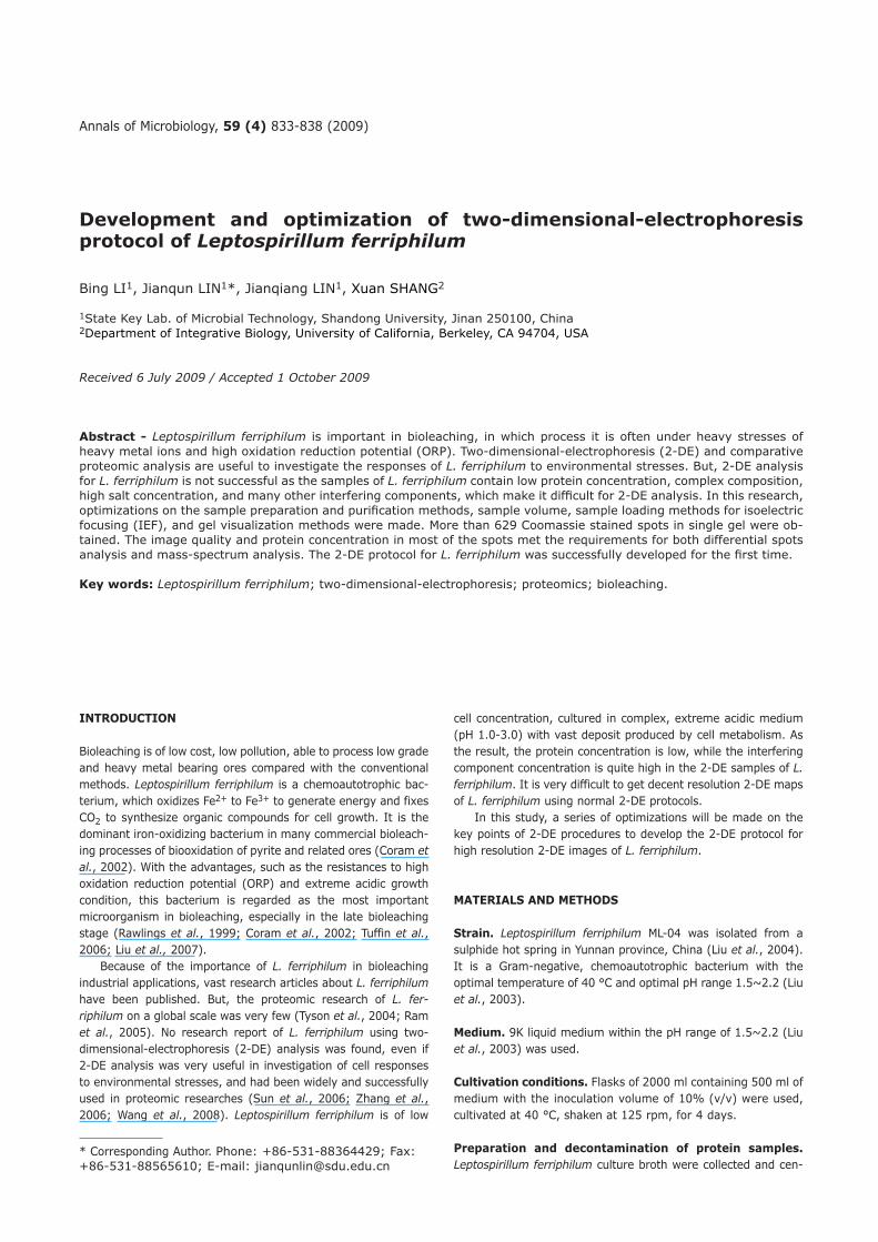
Annals of Microbiology, 59 (4) 833-838 (2009) Development and optimization of two-dimensional-electrophoresis protocol of Leptospirillum ferriphilum Bing LI 1 , Jianqun LIN 1 *, Jianqiang LIN 1 , Xuan SHANG 2 1 State Key Lab. of Microbial Technology, Shandong University, Jinan 250100, China 2 Department of Integrative Biology, University of California, Berkeley, CA 94704, USA Received 6 July 2009 / Accepted 1 October 2009 Abstract - Leptospirillum ferriphilum is important in bioleaching, in which process it is often under heavy stresses of heavy metal ions and high oxidation reduction potential (ORP). Two-dimensional-electrophoresis (2-DE) and comparative proteomic analysis are useful to investigate the responses of L. ferriphilum to environmental stresses. But, 2-DE analysis for L. ferriphilum is not successful as the samples of L. ferriphilum contain low protein concentration, complex composition, high salt concentration, and many other interfering components, which make it difficult for 2-DE analysis. In this research, optimizations on the sample preparation and purification methods, sample volume, sample loading methods for isoelectric focusing (IEF), and gel visualization methods were made. More than 629 Coomassie stained spots in single gel were ob- tained. The image quality and protein concentration in most of the spots met the requirements for both differential spots analysis and mass-spectrum analysis. The 2-DE protocol for L. ferriphilum was successfully developed for the first time. Key words: Leptospirillum ferriphilum; two-dimensional-electrophoresis; proteomics; bioleaching. INTRODUCTION Bioleaching is of low cost, low pollution, able to process low grade and heavy metal bearing ores compared with the conventional methods. Leptospirillum ferriphilum is a chemoautotrophic bac- terium, which oxidizes Fe 2+ to Fe 3+ to generate energy and fixes CO 2 to synthesize organic compounds for cell growth. It is the dominant iron-oxidizing bacterium in many commercial bioleach- ing processes of biooxidation of pyrite and related ores (Coram et al., 2002). With the advantages, such as the resistances to high oxidation reduction potential (ORP) and extreme acidic growth condition, this bacterium is regarded as the most important microorganism in bioleaching, especially in the late bioleaching stage (Rawlings et al., 1999; Coram et al., 2002; Tuffin et al., 2006; Liu et al., 2007). Because of the importance of L. ferriphilum in bioleaching industrial applications, vast research articles about L. ferriphilum have been published. But, the proteomic research of L. fer- riphilum on a global scale was very few (Tyson et al., 2004; Ram et al., 2005). No research report of L. ferriphilum using two- dimensional-electrophoresis (2-DE) analysis was found, even if 2-DE analysis was very useful in investigation of cell responses to environmental stresses, and had been widely and successfully used in proteomic researches (Sun et al., 2006; Zhang et al., 2006; Wang et al., 2008). Leptospirillum ferriphilum is of low cell concentration, cultured in complex, extreme acidic medium (pH 1.0-3.0) with vast deposit produced by cell metabolism. As the result, the protein concentration is low, while the interfering component concentration is quite high in the 2-DE samples of L. ferriphilum. It is very difficult to get decent resolution 2-DE maps of L. ferriphilum using normal 2-DE protocols. In this study, a series of optimizations will be made on the key points of 2-DE procedures to develop the 2-DE protocol for high resolution 2-DE images of L. ferriphilum. MATERIALS AND METHODS Strain. Leptospirillum ferriphilum ML-04 was isolated from a sulphide hot spring in Yunnan province, China (Liu et al., 2004). It is a Gram-negative, chemoautotrophic bacterium with the optimal temperature of 40 °C and optimal pH range 1.5~2.2 (Liu et al., 2003). Medium. 9K liquid medium within the pH range of 1.5~2.2 (Liu et al., 2003) was used. Cultivation conditions. Flasks of 2000 ml containing 500 ml of medium with the inoculation volume of 10% (v/v) were used, cultivated at 40 °C, shaken at 125 rpm, for 4 days. Preparation and decontamination of protein samples. Leptospirillum ferriphilum culture broth were collected and cen- * Corresponding Author. Phone: +86-531-88364429; Fax: +86-531-88565610; E-mail: [email protected]
-
Upload
independent -
Category
Documents
-
view
4 -
download
0
Transcript of Development and optimization of two-dimensional centering algorithm for bacterial rapid detection...
Annals of Microbiology 59 (4) 833-838 (2009)
Development and optimization of two-dimensional-electrophoresis protocol of Leptospirillum ferriphilum
Bing LI1 Jianqun LIN1 Jianqiang LIN1 Xuan SHANG2
1State Key Lab of Microbial Technology Shandong University Jinan 250100 China2Department of Integrative Biology University of California Berkeley CA 94704 USA
Received 6 July 2009 Accepted 1 October 2009
Abstract - Leptospirillum ferriphilum is important in bioleaching in which process it is often under heavy stresses of heavy metal ions and high oxidation reduction potential (ORP) Two-dimensional-electrophoresis (2-DE) and comparative proteomic analysis are useful to investigate the responses of L ferriphilum to environmental stresses But 2-DE analysis for L ferriphilum is not successful as the samples of L ferriphilum contain low protein concentration complex composition high salt concentration and many other interfering components which make it difficult for 2-DE analysis In this research optimizations on the sample preparation and purification methods sample volume sample loading methods for isoelectric focusing (IEF) and gel visualization methods were made More than 629 Coomassie stained spots in single gel were ob-tained The image quality and protein concentration in most of the spots met the requirements for both differential spots analysis and mass-spectrum analysis The 2-DE protocol for L ferriphilum was successfully developed for the first time
Key words Leptospirillum ferriphilum two-dimensional-electrophoresis proteomics bioleaching
INTRODUCTION
Bioleaching is of low cost low pollution able to process low grade and heavy metal bearing ores compared with the conventional methods Leptospirillum ferriphilum is a chemoautotrophic bac-terium which oxidizes Fe2+ to Fe3+ to generate energy and fixes CO2 to synthesize organic compounds for cell growth It is the dominant iron-oxidizing bacterium in many commercial bioleach-ing processes of biooxidation of pyrite and related ores (Coram et al 2002) With the advantages such as the resistances to high oxidation reduction potential (ORP) and extreme acidic growth condition this bacterium is regarded as the most important microorganism in bioleaching especially in the late bioleaching stage (Rawlings et al 1999 Coram et al 2002 Tuffin et al 2006 Liu et al 2007) Because of the importance of L ferriphilum in bioleaching industrial applications vast research articles about L ferriphilum have been published But the proteomic research of L fer-riphilum on a global scale was very few (Tyson et al 2004 Ram et al 2005) No research report of L ferriphilum using two-dimensional-electrophoresis (2-DE) analysis was found even if 2-DE analysis was very useful in investigation of cell responses to environmental stresses and had been widely and successfully used in proteomic researches (Sun et al 2006 Zhang et al 2006 Wang et al 2008) Leptospirillum ferriphilum is of low
cell concentration cultured in complex extreme acidic medium (pH 10-30) with vast deposit produced by cell metabolism As the result the protein concentration is low while the interfering component concentration is quite high in the 2-DE samples of L ferriphilum It is very difficult to get decent resolution 2-DE maps of L ferriphilum using normal 2-DE protocols In this study a series of optimizations will be made on the key points of 2-DE procedures to develop the 2-DE protocol for high resolution 2-DE images of L ferriphilum
MATERIALS AND METHODS
Strain Leptospirillum ferriphilum ML-04 was isolated from a sulphide hot spring in Yunnan province China (Liu et al 2004) It is a Gram-negative chemoautotrophic bacterium with the optimal temperature of 40 degC and optimal pH range 15~22 (Liu et al 2003)
Medium 9K liquid medium within the pH range of 15~22 (Liu et al 2003) was used
Cultivation conditions Flasks of 2000 ml containing 500 ml of medium with the inoculation volume of 10 (vv) were used cultivated at 40 degC shaken at 125 rpm for 4 days
Preparation and decontamination of protein samples Leptospirillum ferriphilum culture broth were collected and cen-
Corresponding Author Phone +86-531-88364429 Fax +86-531-88565610 E-mail jianqunlinsdueducn
834 B LI et al
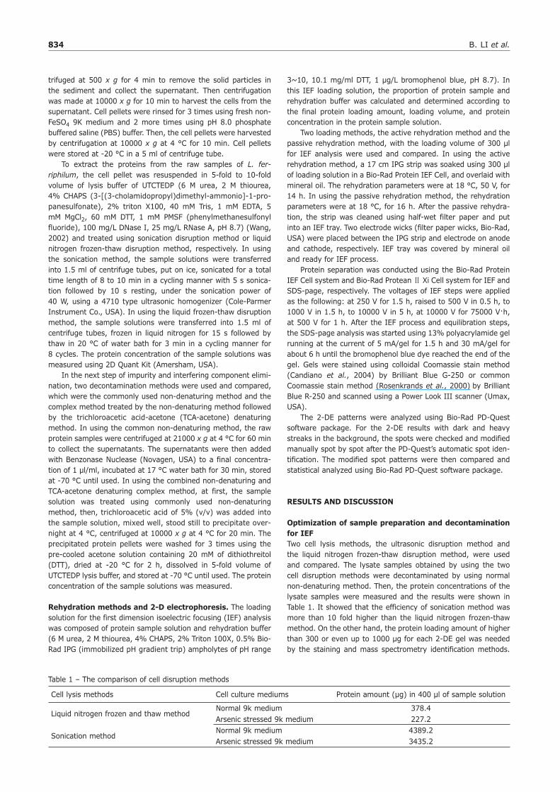
trifuged at 500 x g for 4 min to remove the solid particles in the sediment and collect the supernatant Then centrifugation was made at 10000 x g for 10 min to harvest the cells from the supernatant Cell pellets were rinsed for 3 times using fresh non-FeSO4 9K medium and 2 more times using pH 80 phosphate buffered saline (PBS) buffer Then the cell pellets were harvested by centrifugation at 10000 x g at 4 degC for 10 min Cell pellets were stored at -20 degC in a 5 ml of centrifuge tube To extract the proteins from the raw samples of L fer-riphilum the cell pellet was resuspended in 5-fold to 10-fold volume of lysis buffer of UTCTEDP (6 M urea 2 M thiourea 4 CHAPS (3-[(3-cholamidopropyl)dimethyl-ammonio]-1-pro-panesulfonate) 2 triton X100 40 mM Tris 1 mM EDTA 5 mM MgCl2 60 mM DTT 1 mM PMSF (phenylmethanesulfonyl fluoride) 100 mgL DNase I 25 mgL RNase A pH 87) (Wang 2002) and treated using sonication disruption method or liquid nitrogen frozen-thaw disruption method respectively In using the sonication method the sample solutions were transferred into 15 ml of centrifuge tubes put on ice sonicated for a total time length of 8 to 10 min in a cycling manner with 5 s sonica-tion followed by 10 s resting under the sonication power of 40 W using a 4710 type ultrasonic homogenizer (Cole-Parmer Instrument Co USA) In using the liquid frozen-thaw disruption method the sample solutions were transferred into 15 ml of centrifuge tubes frozen in liquid nitrogen for 15 s followed by thaw in 20 degC of water bath for 3 min in a cycling manner for 8 cycles The protein concentration of the sample solutions was measured using 2D Quant Kit (Amersham USA) In the next step of impurity and interfering component elimi-nation two decontamination methods were used and compared which were the commonly used non-denaturing method and the complex method treated by the non-denaturing method followed by the trichloroacetic acid-acetone (TCA-acetone) denaturing method In using the common non-denaturing method the raw protein samples were centrifuged at 21000 x g at 4 degC for 60 min to collect the supernatants The supernatants were then added with Benzonase Nuclease (Novagen USA) to a final concentra-tion of 1 μlml incubated at 17 degC water bath for 30 min stored at -70 degC until used In using the combined non-denaturing and TCA-acetone denaturing complex method at first the sample solution was treated using commonly used non-denaturing method then trichloroacetic acid of 5 (vv) was added into the sample solution mixed well stood still to precipitate over-night at 4 degC centrifuged at 10000 x g at 4 degC for 20 min The precipitated protein pellets were washed for 3 times using the pre-cooled acetone solution containing 20 mM of dithiothreitol (DTT) dried at -20 degC for 2 h dissolved in 5-fold volume of UTCTEDP lysis buffer and stored at -70 degC until used The protein concentration of the sample solutions was measured
Rehydration methods and 2-D electrophoresis The loading solution for the first dimension isoelectric focusing (IEF) analysis was composed of protein sample solution and rehydration buffer (6 M urea 2 M thiourea 4 CHAPS 2 Triton 100X 05 Bio-Rad IPG (immobilized pH gradient trip) ampholytes of pH range
3~10 101 mgml DTT 1 μgL bromophenol blue pH 87) In this IEF loading solution the proportion of protein sample and rehydration buffer was calculated and determined according to the final protein loading amount loading volume and protein concentration in the protein sample solution Two loading methods the active rehydration method and the passive rehydration method with the loading volume of 300 μl for IEF analysis were used and compared In using the active rehydration method a 17 cm IPG strip was soaked using 300 μl of loading solution in a Bio-Rad Protein IEF Cell and overlaid with mineral oil The rehydration parameters were at 18 degC 50 V for 14 h In using the passive rehydration method the rehydration parameters were at 18 degC for 16 h After the passive rehydra-tion the strip was cleaned using half-wet filter paper and put into an IEF tray Two electrode wicks (filter paper wicks Bio-Rad USA) were placed between the IPG strip and electrode on anode and cathode respectively IEF tray was covered by mineral oil and ready for IEF process Protein separation was conducted using the Bio-Rad Protein IEF Cell system and Bio-Rad Protean Xi Cell system for IEF and SDS-page respectively The voltages of IEF steps were applied as the following at 250 V for 15 h raised to 500 V in 05 h to 1000 V in 15 h to 10000 V in 5 h at 10000 V for 75000 Vh at 500 V for 1 h After the IEF process and equilibration steps the SDS-page analysis was started using 13 polyacrylamide gel running at the current of 5 mAgel for 15 h and 30 mAgel for about 6 h until the bromophenol blue dye reached the end of the gel Gels were stained using colloidal Coomassie stain method (Candiano et al 2004) by Brilliant Blue G-250 or common Coomassie stain method (Rosenkrands et al 2000) by Brilliant Blue R-250 and scanned using a Power Look III scanner (Umax USA) The 2-DE patterns were analyzed using Bio-Rad PD-Quest software package For the 2-DE results with dark and heavy streaks in the background the spots were checked and modified manually spot by spot after the PD-Questrsquos automatic spot iden-tification The modified spot patterns were then compared and statistical analyzed using Bio-Rad PD-Quest software package
RESULTS AND DISCUSSION
Optimization of sample preparation and decontamination for IEFTwo cell lysis methods the ultrasonic disruption method and the liquid nitrogen frozen-thaw disruption method were used and compared The lysate samples obtained by using the two cell disruption methods were decontaminated by using normal non-denaturing method Then the protein concentrations of the lysate samples were measured and the results were shown in Table 1 It showed that the efficiency of sonication method was more than 10 fold higher than the liquid nitrogen frozen-thaw method On the other hand the protein loading amount of higher than 300 or even up to 1000 μg for each 2-DE gel was needed by the staining and mass spectrometry identification methods
Table 1 ndash The comparison of cell disruption methods
Cell lysis methods Cell culture mediums Protein amount (μg) in 400 μl of sample solution
Liquid nitrogen frozen and thaw methodNormal 9k medium 3784Arsenic stressed 9k medium 2272
Sonication methodNormal 9k medium 43892Arsenic stressed 9k medium 34352
Ann Microbiol 59 (4) 833-838 (2009) 835
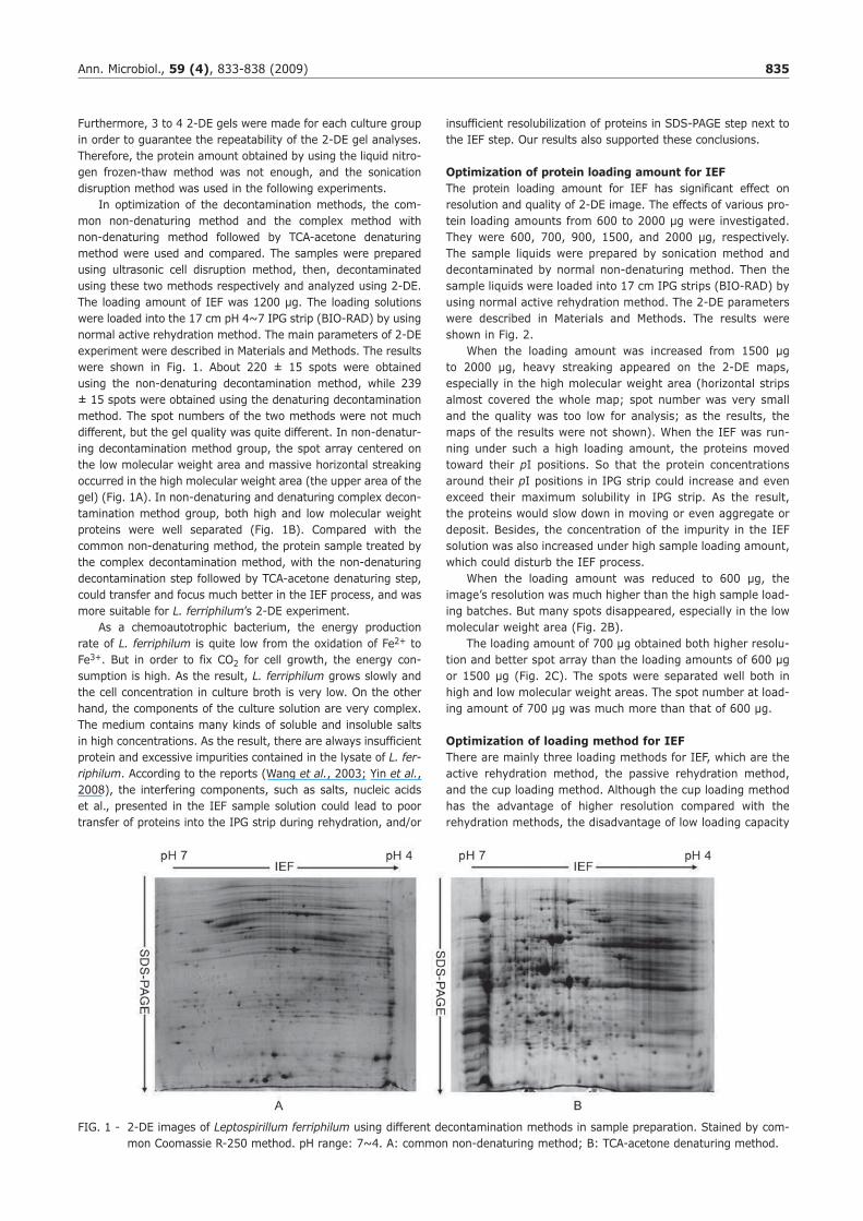
Furthermore 3 to 4 2-DE gels were made for each culture group in order to guarantee the repeatability of the 2-DE gel analyses Therefore the protein amount obtained by using the liquid nitro-gen frozen-thaw method was not enough and the sonication disruption method was used in the following experiments In optimization of the decontamination methods the com-mon non-denaturing method and the complex method with non-denaturing method followed by TCA-acetone denaturing method were used and compared The samples were prepared using ultrasonic cell disruption method then decontaminated using these two methods respectively and analyzed using 2-DE The loading amount of IEF was 1200 μg The loading solutions were loaded into the 17 cm pH 4~7 IPG strip (BIO-RAD) by using normal active rehydration method The main parameters of 2-DE experiment were described in Materials and Methods The results were shown in Fig 1 About 220 plusmn 15 spots were obtained using the non-denaturing decontamination method while 239 plusmn 15 spots were obtained using the denaturing decontamination method The spot numbers of the two methods were not much different but the gel quality was quite different In non-denatur-ing decontamination method group the spot array centered on the low molecular weight area and massive horizontal streaking occurred in the high molecular weight area (the upper area of the gel) (Fig 1A) In non-denaturing and denaturing complex decon-tamination method group both high and low molecular weight proteins were well separated (Fig 1B) Compared with the common non-denaturing method the protein sample treated by the complex decontamination method with the non-denaturing decontamination step followed by TCA-acetone denaturing step could transfer and focus much better in the IEF process and was more suitable for L ferriphilumrsquos 2-DE experiment As a chemoautotrophic bacterium the energy production rate of L ferriphilum is quite low from the oxidation of Fe2+ to Fe3+ But in order to fix CO2 for cell growth the energy con-sumption is high As the result L ferriphilum grows slowly and the cell concentration in culture broth is very low On the other hand the components of the culture solution are very complex The medium contains many kinds of soluble and insoluble salts in high concentrations As the result there are always insufficient protein and excessive impurities contained in the lysate of L fer-riphilum According to the reports (Wang et al 2003 Yin et al 2008) the interfering components such as salts nucleic acids et al presented in the IEF sample solution could lead to poor transfer of proteins into the IPG strip during rehydration andor
insufficient resolubilization of proteins in SDS-PAGE step next to the IEF step Our results also supported these conclusions
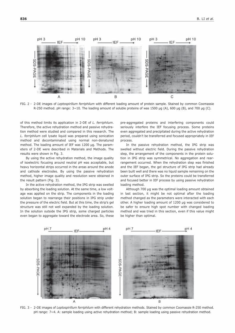
Optimization of protein loading amount for IEFThe protein loading amount for IEF has significant effect on resolution and quality of 2-DE image The effects of various pro-tein loading amounts from 600 to 2000 μg were investigated They were 600 700 900 1500 and 2000 μg respectively The sample liquids were prepared by sonication method and decontaminated by normal non-denaturing method Then the sample liquids were loaded into 17 cm IPG strips (BIO-RAD) by using normal active rehydration method The 2-DE parameters were described in Materials and Methods The results were shown in Fig 2 When the loading amount was increased from 1500 μg to 2000 μg heavy streaking appeared on the 2-DE maps especially in the high molecular weight area (horizontal strips almost covered the whole map spot number was very small and the quality was too low for analysis as the results the maps of the results were not shown) When the IEF was run-ning under such a high loading amount the proteins moved toward their pI positions So that the protein concentrations around their pI positions in IPG strip could increase and even exceed their maximum solubility in IPG strip As the result the proteins would slow down in moving or even aggregate or deposit Besides the concentration of the impurity in the IEF solution was also increased under high sample loading amount which could disturb the IEF process When the loading amount was reduced to 600 μg the imagersquos resolution was much higher than the high sample load-ing batches But many spots disappeared especially in the low molecular weight area (Fig 2B) The loading amount of 700 μg obtained both higher resolu-tion and better spot array than the loading amounts of 600 μg or 1500 μg (Fig 2C) The spots were separated well both in high and low molecular weight areas The spot number at load-ing amount of 700 μg was much more than that of 600 μg
Optimization of loading method for IEFThere are mainly three loading methods for IEF which are the active rehydration method the passive rehydration method and the cup loading method Although the cup loading method has the advantage of higher resolution compared with the rehydration methods the disadvantage of low loading capacity
FIG 1 - 2-DE images of Leptospirillum ferriphilum using different decontamination methods in sample preparation Stained by com-mon Coomassie R-250 method pH range 7~4 A common non-denaturing method B TCA-acetone denaturing method
836 B LI et al
of this method limits its application in 2-DE of L ferriphilum Therefore the active rehydration method and passive rehydra-tion method were studied and compared in this research The L ferriphilum cell lysate liquid was prepared using sonication method and decontaminated using normal non-denatured method The loading amount of IEF was 1200 μg The param-eters of 2-DE were described in Materials and Methods The results were shown in Fig 3 By using the active rehydration method the image quality of isoelectric focusing around neutral pH was acceptable but heavy horizontal strips occurred in the areas around the anode and cathode electrodes By using the passive rehydration method higher image quality and resolution were obtained in the result pattern (Fig 3) In the active rehydration method the IPG strip was swelled by absorbing the loading solution At the same time a low volt-age was applied on the strip The components in the loading solution began to rearrange their positions in IPG strip under the pressure of the electric field But at this time the striprsquos gel structure was still not well expanded by the loading solution In the solution outside the IPG strip some charged particles even began to aggregate toward the electrode area So these
pre-aggregated proteins and interfering components could seriously interfere the IEF focusing process Some proteins even aggregated and precipitated during the active rehydration period couldnrsquot be transferred and focused appropriately in IEF process In the passive rehydration method the IPG strip was swelled without electric field During the passive rehydration step the arrangement of the components in the protein solu-tion in IPG strip was symmetrical No aggregation and rear-rangement occurred When the rehydration step was finished and the IEF began the gel structure of IPG strip had already been built well and there was no liquid sample remaining on the outer surface of IPG strip So the proteins could be transferred and focused better in IEF process by using passive rehydration loading method Although 700 μg was the optimal loading amount obtained in last section it might be not optimal after the loading method changed as the parameters were interacted with each other A higher loading amount of 1200 μg was considered to be safer to ensure high spot number with changed loading method and was tried in this section even if this value might be higher than optimal
FIG 3 - 2-DE images of Leptospirillum ferriphilum with different rehydration methods Stained by common Coomassie R-250 method pH range 7~4 A sample loading using active rehydration method B sample loading using passive rehydration method
FIG 2 - 2-DE images of Leptospirillum ferriphilum with different loading amount of protein sample Stained by common Coomassie R-250 method pH range 3~10 The loading amount of soluble proteins of was 1500 μg (A) 600 μg (B) and 700 μg (C)
Ann Microbiol 59 (4) 833-838 (2009) 837
The summary of optimized 2-DE protocol Based on the optimizations above the final optimized 2-DE protocol of L ferriphilum was obtained The cell pellet was lysed and disrupted using ldquoUTCTEDPrdquo lysate buffer and soni-cation cell disruption method the raw protein solution was cleaned and decontaminated using the complex purification method with common non-denaturing method followed by TCA-acetone denaturing method the loading volume of 300 μl protein loading amount of 700 μg and the passive rehydration loading method were used for the 17 cm IPG IEF After the IEF process SDS-PAGE was started using 13 polyacrylamide gel running at a current of 5 mAgel for 15 h and 30 mAgel for about 6 h until the bromophenol blue dye reached the end of the gel The colloidal Coomassie Brilliant Blue G-250 staining method was used for gel stating which method was proved to be 30~50 more sensitive than the common Coomasie R-250 method (data not show) High sensitivity of colloidal coomas-sie brilliant blue G-250 staining method was ever reported
(Candiano et al 2004) A common result pattern obtained by using this 2-DE protocol of L ferriphilum was shown in Fig 4 More than 629 plusmn 15 distinctly separated protein spots were detected in one single colloidal Coomassie stained 2-DE gel Then several 2-DE experiments using different batches of culture material of L ferriphilum were launched using this protocol Good reproducibility was obtained and shown in Fig 5 The statistical analyses of the 2-DE patterns of Fig 5 were made using Bio-Rad PD-Quest software package and the cor-relation coefficient value of this compare group of 08296 was obtained which value was acceptable
CONCLUSIONS
The 2-DE protocol of L ferriphilum was developed for the first time The main obstacle of 2-DE analysis for L ferriphilum is that the protein concentration is too low while the soluble impurity concentration is too high in the sample solution The protocol developed in this research optimized the sample puri-fication method and IEF procedure obtained high resolution 2-DE experiment for L ferriphilum samples This work could also be a good example for the development of high resolution 2-DE protocol for other extreme micro-organism samples with low protein concentration and high impurity concentration In the subsequent research using the 2-DE analysis meth-od developed in this study the differentially expressed proteins of L ferriphilum under arsenic stress were analyzed Six 2-DE patterns were built up for two groups of L ferriphilum samples which were cultured with or without arsenic stress Because of the good resolution and reproducibility of these 2-DE patterns the differential protein spots were displayed and checked out clearly by using both computer software checking method and manual checking method After that some target differentially expressed protein spots were analyzed using MALDI-TOF mass spectrum and relevant bioinformatics methods 65 differentially expressed protein spots were detected using this 2-DE analy-sis 43 protein samples from the 2-DE spots were analyzed using MS and bioinformatics methods Finally 38 differentially expressed proteins were highly identified for L ferriphilum under arsenic stress (to be published) These results confirmed that the 2-DE protocol of L ferriphilum developed in this research was successful
FIG 4 - 2-DE image of Leptospirillum ferriphilum using opti-mized 2-DE protocol Stained by colloidal Coomassie G-250 method pH range 3~10
FIG 5 - The reproducibility of 2-DE images of Leptospirillum ferriphilum of different batches using optimized 2-DE protocol
838 B LI et al
AcknowledgementsThis work was supported by the grants from the National Basic Research Program (2004CB619202) the National High Technology Research and Development Program (2007AA06A407) of China
REFERENCES
Candiano G Bruschi M Musante L Santucci L Ghiggeri GM Carnemolla B Orecchia P Zardi L Righetti PG (2004) Blue silver a very sensitive colloidal Coomassie G-250 stain-ing for proteome analysis Electrophoresis 25 1327-1333
Coram NJ Rawlings DE (2002) Molecular relationship between two groups of the genus Leptospirillum and the finding that Leptospirillum ferriphilum sp nov dominates South African commercial biooxidation tanks that operate at 40 degrees C Appl Environ Microbiol 68 838-845
Liu J Xie X Xiao S Wang X Zhao W Tian Z (2007) Isolation of Leptospirillum ferriphilum by single-layered solid medi-um J Cent South Univ Technol 4 467-473
Liu Y Liu X Tian K Qi F Yan W (2003) Growth and mor-phology of Leptospirillum ferrooxidans on solid medium Microbiology 30 70-72 (in Chinese)
Liu Y Liu X Zheng L Lin J Yan W (2004) Isolation and char-acterization of a vibrioid-shaped iron-oxidizing bacterium J Shandong Univ 39 116-124 (in Chinese)
Ram RJ VerBerkmoes NC Thelen MP Tyson GW Baker BJ Blake RC Shah M Hettich RL Banfield JF (2005) Community proteomics of a natural microbial biofilm Science 308 1915-1920
Rawlings DE Tributsch H Hansford GS (1999) Reasons why lsquoLeptospirillumrsquo-like species rather than Thiobacillus ferrooxi-dans are the dominant iron-oxidizing bacteria in many com-mercial processes for the biooxidation of pyrite and related ores Microbiology 145 5-13
Rosenkrands I Weldingh K Jacobsen S (2000) Mapping and identification of mycobacterium tuberculosis proteins by two-dimensional gel electrophoresis microsequencing and immunodetection Electrophoresis 21 935-948
Sun B Fan L Li L Zhu W Lu G (2006) Establishment of the 2-D synthetic map of total protein of normal human sperma-tozoa enriched with low abundance protein National J of Andrology 12 295-299 (in Chinese)
Tuffin IM Hector SB Deane SM Rawlings DE (2006) Resistance determinants of a highly arsenic-resistant strain of Leptospirillum ferriphilum isolated from a commercial biooxidation tank Appl Environ Microbiol 72 2247-2253
Tyson GW Chapman J Hugenholtz P Allen EE Ram RJ Richardson PM Solovyev VV Rubin EM Rokhsar DS Banfield JF (2004) Community structure and metabolism through reconstruction of microbial genomes from the envi-ronment Nature 428 25-26
Wang J (2002) Preparation for Proteinrsquos Separation Protein Technology Manual Science Press Beijing (In Chinese)
Wang SB Hu Q Sommerfeld M Chen F (2003) An optimized protocol for isolation of soluble proteins from microalgae for two-dimensional gel electrophoresis analysis J Appl Phycol 15 485-496
Wang M Peng Z Li C Li F Liu C Xia G (2008) Proteomic anal-ysis on a high salt tolerance introgression strain of Triticum aestivumThinopyrum ponticum Proteomics 81470-1489
Yin P Wang YH Zhang SL Chu J Zhuang YP Wang ML Zhou J (2008) Isolation of soluble proteins from an industri-al strain Streptomyces avermitilis in complex culture medium for two-dimensional gel electrophoresis J Microbiol Methods 73 105-110
Zhang X Xiao Z Chen Z Li C Li J Yanhui Y Yang F Yang Y Oyang Y (2006) Comparative proteomics analysis of the proteins associated with laryngeal carcinoma-related gene 1 Laryngoscope 116 224-230
834 B LI et al
trifuged at 500 x g for 4 min to remove the solid particles in the sediment and collect the supernatant Then centrifugation was made at 10000 x g for 10 min to harvest the cells from the supernatant Cell pellets were rinsed for 3 times using fresh non-FeSO4 9K medium and 2 more times using pH 80 phosphate buffered saline (PBS) buffer Then the cell pellets were harvested by centrifugation at 10000 x g at 4 degC for 10 min Cell pellets were stored at -20 degC in a 5 ml of centrifuge tube To extract the proteins from the raw samples of L fer-riphilum the cell pellet was resuspended in 5-fold to 10-fold volume of lysis buffer of UTCTEDP (6 M urea 2 M thiourea 4 CHAPS (3-[(3-cholamidopropyl)dimethyl-ammonio]-1-pro-panesulfonate) 2 triton X100 40 mM Tris 1 mM EDTA 5 mM MgCl2 60 mM DTT 1 mM PMSF (phenylmethanesulfonyl fluoride) 100 mgL DNase I 25 mgL RNase A pH 87) (Wang 2002) and treated using sonication disruption method or liquid nitrogen frozen-thaw disruption method respectively In using the sonication method the sample solutions were transferred into 15 ml of centrifuge tubes put on ice sonicated for a total time length of 8 to 10 min in a cycling manner with 5 s sonica-tion followed by 10 s resting under the sonication power of 40 W using a 4710 type ultrasonic homogenizer (Cole-Parmer Instrument Co USA) In using the liquid frozen-thaw disruption method the sample solutions were transferred into 15 ml of centrifuge tubes frozen in liquid nitrogen for 15 s followed by thaw in 20 degC of water bath for 3 min in a cycling manner for 8 cycles The protein concentration of the sample solutions was measured using 2D Quant Kit (Amersham USA) In the next step of impurity and interfering component elimi-nation two decontamination methods were used and compared which were the commonly used non-denaturing method and the complex method treated by the non-denaturing method followed by the trichloroacetic acid-acetone (TCA-acetone) denaturing method In using the common non-denaturing method the raw protein samples were centrifuged at 21000 x g at 4 degC for 60 min to collect the supernatants The supernatants were then added with Benzonase Nuclease (Novagen USA) to a final concentra-tion of 1 μlml incubated at 17 degC water bath for 30 min stored at -70 degC until used In using the combined non-denaturing and TCA-acetone denaturing complex method at first the sample solution was treated using commonly used non-denaturing method then trichloroacetic acid of 5 (vv) was added into the sample solution mixed well stood still to precipitate over-night at 4 degC centrifuged at 10000 x g at 4 degC for 20 min The precipitated protein pellets were washed for 3 times using the pre-cooled acetone solution containing 20 mM of dithiothreitol (DTT) dried at -20 degC for 2 h dissolved in 5-fold volume of UTCTEDP lysis buffer and stored at -70 degC until used The protein concentration of the sample solutions was measured
Rehydration methods and 2-D electrophoresis The loading solution for the first dimension isoelectric focusing (IEF) analysis was composed of protein sample solution and rehydration buffer (6 M urea 2 M thiourea 4 CHAPS 2 Triton 100X 05 Bio-Rad IPG (immobilized pH gradient trip) ampholytes of pH range
3~10 101 mgml DTT 1 μgL bromophenol blue pH 87) In this IEF loading solution the proportion of protein sample and rehydration buffer was calculated and determined according to the final protein loading amount loading volume and protein concentration in the protein sample solution Two loading methods the active rehydration method and the passive rehydration method with the loading volume of 300 μl for IEF analysis were used and compared In using the active rehydration method a 17 cm IPG strip was soaked using 300 μl of loading solution in a Bio-Rad Protein IEF Cell and overlaid with mineral oil The rehydration parameters were at 18 degC 50 V for 14 h In using the passive rehydration method the rehydration parameters were at 18 degC for 16 h After the passive rehydra-tion the strip was cleaned using half-wet filter paper and put into an IEF tray Two electrode wicks (filter paper wicks Bio-Rad USA) were placed between the IPG strip and electrode on anode and cathode respectively IEF tray was covered by mineral oil and ready for IEF process Protein separation was conducted using the Bio-Rad Protein IEF Cell system and Bio-Rad Protean Xi Cell system for IEF and SDS-page respectively The voltages of IEF steps were applied as the following at 250 V for 15 h raised to 500 V in 05 h to 1000 V in 15 h to 10000 V in 5 h at 10000 V for 75000 Vh at 500 V for 1 h After the IEF process and equilibration steps the SDS-page analysis was started using 13 polyacrylamide gel running at the current of 5 mAgel for 15 h and 30 mAgel for about 6 h until the bromophenol blue dye reached the end of the gel Gels were stained using colloidal Coomassie stain method (Candiano et al 2004) by Brilliant Blue G-250 or common Coomassie stain method (Rosenkrands et al 2000) by Brilliant Blue R-250 and scanned using a Power Look III scanner (Umax USA) The 2-DE patterns were analyzed using Bio-Rad PD-Quest software package For the 2-DE results with dark and heavy streaks in the background the spots were checked and modified manually spot by spot after the PD-Questrsquos automatic spot iden-tification The modified spot patterns were then compared and statistical analyzed using Bio-Rad PD-Quest software package
RESULTS AND DISCUSSION
Optimization of sample preparation and decontamination for IEFTwo cell lysis methods the ultrasonic disruption method and the liquid nitrogen frozen-thaw disruption method were used and compared The lysate samples obtained by using the two cell disruption methods were decontaminated by using normal non-denaturing method Then the protein concentrations of the lysate samples were measured and the results were shown in Table 1 It showed that the efficiency of sonication method was more than 10 fold higher than the liquid nitrogen frozen-thaw method On the other hand the protein loading amount of higher than 300 or even up to 1000 μg for each 2-DE gel was needed by the staining and mass spectrometry identification methods
Table 1 ndash The comparison of cell disruption methods
Cell lysis methods Cell culture mediums Protein amount (μg) in 400 μl of sample solution
Liquid nitrogen frozen and thaw methodNormal 9k medium 3784Arsenic stressed 9k medium 2272
Sonication methodNormal 9k medium 43892Arsenic stressed 9k medium 34352
Ann Microbiol 59 (4) 833-838 (2009) 835
Furthermore 3 to 4 2-DE gels were made for each culture group in order to guarantee the repeatability of the 2-DE gel analyses Therefore the protein amount obtained by using the liquid nitro-gen frozen-thaw method was not enough and the sonication disruption method was used in the following experiments In optimization of the decontamination methods the com-mon non-denaturing method and the complex method with non-denaturing method followed by TCA-acetone denaturing method were used and compared The samples were prepared using ultrasonic cell disruption method then decontaminated using these two methods respectively and analyzed using 2-DE The loading amount of IEF was 1200 μg The loading solutions were loaded into the 17 cm pH 4~7 IPG strip (BIO-RAD) by using normal active rehydration method The main parameters of 2-DE experiment were described in Materials and Methods The results were shown in Fig 1 About 220 plusmn 15 spots were obtained using the non-denaturing decontamination method while 239 plusmn 15 spots were obtained using the denaturing decontamination method The spot numbers of the two methods were not much different but the gel quality was quite different In non-denatur-ing decontamination method group the spot array centered on the low molecular weight area and massive horizontal streaking occurred in the high molecular weight area (the upper area of the gel) (Fig 1A) In non-denaturing and denaturing complex decon-tamination method group both high and low molecular weight proteins were well separated (Fig 1B) Compared with the common non-denaturing method the protein sample treated by the complex decontamination method with the non-denaturing decontamination step followed by TCA-acetone denaturing step could transfer and focus much better in the IEF process and was more suitable for L ferriphilumrsquos 2-DE experiment As a chemoautotrophic bacterium the energy production rate of L ferriphilum is quite low from the oxidation of Fe2+ to Fe3+ But in order to fix CO2 for cell growth the energy con-sumption is high As the result L ferriphilum grows slowly and the cell concentration in culture broth is very low On the other hand the components of the culture solution are very complex The medium contains many kinds of soluble and insoluble salts in high concentrations As the result there are always insufficient protein and excessive impurities contained in the lysate of L fer-riphilum According to the reports (Wang et al 2003 Yin et al 2008) the interfering components such as salts nucleic acids et al presented in the IEF sample solution could lead to poor transfer of proteins into the IPG strip during rehydration andor
insufficient resolubilization of proteins in SDS-PAGE step next to the IEF step Our results also supported these conclusions
Optimization of protein loading amount for IEFThe protein loading amount for IEF has significant effect on resolution and quality of 2-DE image The effects of various pro-tein loading amounts from 600 to 2000 μg were investigated They were 600 700 900 1500 and 2000 μg respectively The sample liquids were prepared by sonication method and decontaminated by normal non-denaturing method Then the sample liquids were loaded into 17 cm IPG strips (BIO-RAD) by using normal active rehydration method The 2-DE parameters were described in Materials and Methods The results were shown in Fig 2 When the loading amount was increased from 1500 μg to 2000 μg heavy streaking appeared on the 2-DE maps especially in the high molecular weight area (horizontal strips almost covered the whole map spot number was very small and the quality was too low for analysis as the results the maps of the results were not shown) When the IEF was run-ning under such a high loading amount the proteins moved toward their pI positions So that the protein concentrations around their pI positions in IPG strip could increase and even exceed their maximum solubility in IPG strip As the result the proteins would slow down in moving or even aggregate or deposit Besides the concentration of the impurity in the IEF solution was also increased under high sample loading amount which could disturb the IEF process When the loading amount was reduced to 600 μg the imagersquos resolution was much higher than the high sample load-ing batches But many spots disappeared especially in the low molecular weight area (Fig 2B) The loading amount of 700 μg obtained both higher resolu-tion and better spot array than the loading amounts of 600 μg or 1500 μg (Fig 2C) The spots were separated well both in high and low molecular weight areas The spot number at load-ing amount of 700 μg was much more than that of 600 μg
Optimization of loading method for IEFThere are mainly three loading methods for IEF which are the active rehydration method the passive rehydration method and the cup loading method Although the cup loading method has the advantage of higher resolution compared with the rehydration methods the disadvantage of low loading capacity
FIG 1 - 2-DE images of Leptospirillum ferriphilum using different decontamination methods in sample preparation Stained by com-mon Coomassie R-250 method pH range 7~4 A common non-denaturing method B TCA-acetone denaturing method
836 B LI et al
of this method limits its application in 2-DE of L ferriphilum Therefore the active rehydration method and passive rehydra-tion method were studied and compared in this research The L ferriphilum cell lysate liquid was prepared using sonication method and decontaminated using normal non-denatured method The loading amount of IEF was 1200 μg The param-eters of 2-DE were described in Materials and Methods The results were shown in Fig 3 By using the active rehydration method the image quality of isoelectric focusing around neutral pH was acceptable but heavy horizontal strips occurred in the areas around the anode and cathode electrodes By using the passive rehydration method higher image quality and resolution were obtained in the result pattern (Fig 3) In the active rehydration method the IPG strip was swelled by absorbing the loading solution At the same time a low volt-age was applied on the strip The components in the loading solution began to rearrange their positions in IPG strip under the pressure of the electric field But at this time the striprsquos gel structure was still not well expanded by the loading solution In the solution outside the IPG strip some charged particles even began to aggregate toward the electrode area So these
pre-aggregated proteins and interfering components could seriously interfere the IEF focusing process Some proteins even aggregated and precipitated during the active rehydration period couldnrsquot be transferred and focused appropriately in IEF process In the passive rehydration method the IPG strip was swelled without electric field During the passive rehydration step the arrangement of the components in the protein solu-tion in IPG strip was symmetrical No aggregation and rear-rangement occurred When the rehydration step was finished and the IEF began the gel structure of IPG strip had already been built well and there was no liquid sample remaining on the outer surface of IPG strip So the proteins could be transferred and focused better in IEF process by using passive rehydration loading method Although 700 μg was the optimal loading amount obtained in last section it might be not optimal after the loading method changed as the parameters were interacted with each other A higher loading amount of 1200 μg was considered to be safer to ensure high spot number with changed loading method and was tried in this section even if this value might be higher than optimal
FIG 3 - 2-DE images of Leptospirillum ferriphilum with different rehydration methods Stained by common Coomassie R-250 method pH range 7~4 A sample loading using active rehydration method B sample loading using passive rehydration method
FIG 2 - 2-DE images of Leptospirillum ferriphilum with different loading amount of protein sample Stained by common Coomassie R-250 method pH range 3~10 The loading amount of soluble proteins of was 1500 μg (A) 600 μg (B) and 700 μg (C)
Ann Microbiol 59 (4) 833-838 (2009) 837
The summary of optimized 2-DE protocol Based on the optimizations above the final optimized 2-DE protocol of L ferriphilum was obtained The cell pellet was lysed and disrupted using ldquoUTCTEDPrdquo lysate buffer and soni-cation cell disruption method the raw protein solution was cleaned and decontaminated using the complex purification method with common non-denaturing method followed by TCA-acetone denaturing method the loading volume of 300 μl protein loading amount of 700 μg and the passive rehydration loading method were used for the 17 cm IPG IEF After the IEF process SDS-PAGE was started using 13 polyacrylamide gel running at a current of 5 mAgel for 15 h and 30 mAgel for about 6 h until the bromophenol blue dye reached the end of the gel The colloidal Coomassie Brilliant Blue G-250 staining method was used for gel stating which method was proved to be 30~50 more sensitive than the common Coomasie R-250 method (data not show) High sensitivity of colloidal coomas-sie brilliant blue G-250 staining method was ever reported
(Candiano et al 2004) A common result pattern obtained by using this 2-DE protocol of L ferriphilum was shown in Fig 4 More than 629 plusmn 15 distinctly separated protein spots were detected in one single colloidal Coomassie stained 2-DE gel Then several 2-DE experiments using different batches of culture material of L ferriphilum were launched using this protocol Good reproducibility was obtained and shown in Fig 5 The statistical analyses of the 2-DE patterns of Fig 5 were made using Bio-Rad PD-Quest software package and the cor-relation coefficient value of this compare group of 08296 was obtained which value was acceptable
CONCLUSIONS
The 2-DE protocol of L ferriphilum was developed for the first time The main obstacle of 2-DE analysis for L ferriphilum is that the protein concentration is too low while the soluble impurity concentration is too high in the sample solution The protocol developed in this research optimized the sample puri-fication method and IEF procedure obtained high resolution 2-DE experiment for L ferriphilum samples This work could also be a good example for the development of high resolution 2-DE protocol for other extreme micro-organism samples with low protein concentration and high impurity concentration In the subsequent research using the 2-DE analysis meth-od developed in this study the differentially expressed proteins of L ferriphilum under arsenic stress were analyzed Six 2-DE patterns were built up for two groups of L ferriphilum samples which were cultured with or without arsenic stress Because of the good resolution and reproducibility of these 2-DE patterns the differential protein spots were displayed and checked out clearly by using both computer software checking method and manual checking method After that some target differentially expressed protein spots were analyzed using MALDI-TOF mass spectrum and relevant bioinformatics methods 65 differentially expressed protein spots were detected using this 2-DE analy-sis 43 protein samples from the 2-DE spots were analyzed using MS and bioinformatics methods Finally 38 differentially expressed proteins were highly identified for L ferriphilum under arsenic stress (to be published) These results confirmed that the 2-DE protocol of L ferriphilum developed in this research was successful
FIG 4 - 2-DE image of Leptospirillum ferriphilum using opti-mized 2-DE protocol Stained by colloidal Coomassie G-250 method pH range 3~10
FIG 5 - The reproducibility of 2-DE images of Leptospirillum ferriphilum of different batches using optimized 2-DE protocol
838 B LI et al
AcknowledgementsThis work was supported by the grants from the National Basic Research Program (2004CB619202) the National High Technology Research and Development Program (2007AA06A407) of China
REFERENCES
Candiano G Bruschi M Musante L Santucci L Ghiggeri GM Carnemolla B Orecchia P Zardi L Righetti PG (2004) Blue silver a very sensitive colloidal Coomassie G-250 stain-ing for proteome analysis Electrophoresis 25 1327-1333
Coram NJ Rawlings DE (2002) Molecular relationship between two groups of the genus Leptospirillum and the finding that Leptospirillum ferriphilum sp nov dominates South African commercial biooxidation tanks that operate at 40 degrees C Appl Environ Microbiol 68 838-845
Liu J Xie X Xiao S Wang X Zhao W Tian Z (2007) Isolation of Leptospirillum ferriphilum by single-layered solid medi-um J Cent South Univ Technol 4 467-473
Liu Y Liu X Tian K Qi F Yan W (2003) Growth and mor-phology of Leptospirillum ferrooxidans on solid medium Microbiology 30 70-72 (in Chinese)
Liu Y Liu X Zheng L Lin J Yan W (2004) Isolation and char-acterization of a vibrioid-shaped iron-oxidizing bacterium J Shandong Univ 39 116-124 (in Chinese)
Ram RJ VerBerkmoes NC Thelen MP Tyson GW Baker BJ Blake RC Shah M Hettich RL Banfield JF (2005) Community proteomics of a natural microbial biofilm Science 308 1915-1920
Rawlings DE Tributsch H Hansford GS (1999) Reasons why lsquoLeptospirillumrsquo-like species rather than Thiobacillus ferrooxi-dans are the dominant iron-oxidizing bacteria in many com-mercial processes for the biooxidation of pyrite and related ores Microbiology 145 5-13
Rosenkrands I Weldingh K Jacobsen S (2000) Mapping and identification of mycobacterium tuberculosis proteins by two-dimensional gel electrophoresis microsequencing and immunodetection Electrophoresis 21 935-948
Sun B Fan L Li L Zhu W Lu G (2006) Establishment of the 2-D synthetic map of total protein of normal human sperma-tozoa enriched with low abundance protein National J of Andrology 12 295-299 (in Chinese)
Tuffin IM Hector SB Deane SM Rawlings DE (2006) Resistance determinants of a highly arsenic-resistant strain of Leptospirillum ferriphilum isolated from a commercial biooxidation tank Appl Environ Microbiol 72 2247-2253
Tyson GW Chapman J Hugenholtz P Allen EE Ram RJ Richardson PM Solovyev VV Rubin EM Rokhsar DS Banfield JF (2004) Community structure and metabolism through reconstruction of microbial genomes from the envi-ronment Nature 428 25-26
Wang J (2002) Preparation for Proteinrsquos Separation Protein Technology Manual Science Press Beijing (In Chinese)
Wang SB Hu Q Sommerfeld M Chen F (2003) An optimized protocol for isolation of soluble proteins from microalgae for two-dimensional gel electrophoresis analysis J Appl Phycol 15 485-496
Wang M Peng Z Li C Li F Liu C Xia G (2008) Proteomic anal-ysis on a high salt tolerance introgression strain of Triticum aestivumThinopyrum ponticum Proteomics 81470-1489
Yin P Wang YH Zhang SL Chu J Zhuang YP Wang ML Zhou J (2008) Isolation of soluble proteins from an industri-al strain Streptomyces avermitilis in complex culture medium for two-dimensional gel electrophoresis J Microbiol Methods 73 105-110
Zhang X Xiao Z Chen Z Li C Li J Yanhui Y Yang F Yang Y Oyang Y (2006) Comparative proteomics analysis of the proteins associated with laryngeal carcinoma-related gene 1 Laryngoscope 116 224-230