Staphylococcus haemolyticus endocarditis: clinical and microbiologic analysis of 4 cases
Original Article
Detection of icaA, icaD genes and biofilm production by Staphylococcus aureus and Staphylococcus epidermidis isolated from urinary tract catheterized patients Gamal Fadl Mahmoud Gad3, Mohamed Ali El-Feky1, Mostafa Said El-Rehewy1, Mona Amin Hassan1, Hassan Abolella2, and Rehab Mahmoud Abd El-Baky3 1Microbiology Department, Faculty of Medicine, Assuit University, Assuit, Egypt
2Urology Department, Faculty of Medicine, Assuit University, Assuit, Egypt
3Microbiology Department, Faculty of Pharmacy, El-Minia University, El-Minia, Egypt
Abstract Background: Staphylococci are a common cause of catheter-associated urinary tract infections. The present study evaluated biofilm forming
capacity and the presence of both icaA and icaD genes among staphylococci strains isolated from patients undergoing ureteral
catheterization.
Methodology: Different bacterial strains were isolated from urine and stents segments collected from 100 patients. Strains were identified by
traditional microbiological methods. Stents were examined for biofilm using a scanning electron microscope (SEM). Staphylococcal isolates
were tested for their ability to produce biofilm using the tissue culture plate assay method (TCP). The presence of icaA and icaD genes was
determined by PCR technique.
Results: Fifty-three staphylococcal strains were isolated and identified from 284 samples (18.7%). Forty-six staphylococcal strains were
isolated from stent segment cultures while only seven strains were isolated from urine samples at the day of stent removal. S. aureus
represented 6.3%, and S. epidermidis represented 12.3%. Out of the 18 S. aureus strains, 15 (83.3%) were biofilm producers and out of 35 S.
epidermidis strains, 31 (88.6%) were biofilm producers. Staphylococcal strains were further classified as high (56.6%), moderate (30.2%)
and non biofilm producers (13.2%). All biofilm producing strains were positive for icaA and icaD genes, and all biofilm negative strains
were negative for both genes.
Conclusion: Staphylococci isolated from catheter segments showed a higher extent of biofilm production than that isolated from urine
samples. All biofilm producing staphylococci were positive for icaA and icaD genes, which indicates the important role of ica genes as
virulence markers in staphylococcal infections associated with urinary catheterization.
Keywords: Staphylococci, biofilm, icaA, icaD
J Infect Dev Ctries 2009; 3(5):342-351.
Received 26 December 2008 - Accepted 15 March 2009
Copyright © 2009 Gad et al. This is an open access article distributed under the Creative Commons Attribution License, which permits unrestricted use,
distribution, and reproduction in any medium, provided the original work is properly cited.
Introduction Staphylococci are most often associated with
chronic infection of implanted medical devices. The
increased use of indwelling medical devices has had
considerable impact on the role of staphylococci in
clinical medicine. The predominant species isolated
in these infections are Staphylococcus epidermidis
and Staphylococcus aureus. It was found that the
major pathogenic factor is the ability to form biofilm
on polymeric surfaces to which it adheres and
colonizes artificial materials [1]. Biofilms are a
population of multilayered cells growing on a surface
and enclosed in exopolysaccharide matrix. Biofilm
formations are considered to be a two-step process in
which the bacteria first adhere to a surface, followed
by multiplication to form a multilayered biofilm.
Microbial biofilms are considered the major problem
posed to catheterized patients because they cause
chronic infections which are difficult to treat, lead to
longer hospitalization time, and can result in much
higher treatment costs [2]. Biofilm formation is
regulated by expression of polysaccharide
intracellular adhesion (PIA), which mediates cell to
cell adhesion and is the gene product of icaADBC
[3]. The intercellular adhesion (ica) locus consisting
of the genes icaADBC encodes proteins mediating the
synthesis of PIA and polysaccharide/adhesin PS/A in
staphylococci species [4].
Among ica genes, icaA and icaD have been
reported to play a significant role in biofilm
formation in S. aureus and S. epidermidis [5]. The
icaA gene encodes N-acetylglucosaminyltransferase,
Gad et al. – Effect of Ciprofloxacin and NAC on biofilm J Infect Dev Ctries 2009; 3(5):342-351.
343
the enzyme involved in the synthesis of N-
acetylglucosamine oligomers from UDP-N-
acetylglucosamine. Further, icaD has been reported
to play a role in the maximal expression of N-
acetylglucosaminyltransferase, leading to the
phenotypic expression of the capsular polysaccharide
[6]. The aim of our study was to determine the
biofilm-forming capacity of microorganisms isolated
from urinary tract catheterized patients and the
occurrence of icaA and icaD genes in biofilm-
producing strains in a collection of staphylococcal
isolates.
Materials and methods Patients, specimens, and strains
Patients
Our institution did not require informed consent
from patients. All patients were selected by Dr.
Hassan Abolella (Professor of Urology, Department
of Urology at Assuit University Hospital) during the
study period from December 2007 to June 2008.
However, all samples were collected after we
obtained informed consent from each patient,
following a discussion with each about our study in
order to facilitate our work. Eight patients of our
study did not return to undergo stent removal in the
hospital. This was the reason that the number of urine
samples and stent segment was 92 instead of 100 on
the day of stent removal.
Specimens
Two hundred and eighty four clinical samples
(urine samples and stent segments) were collected
from 100 in-patients undergoing ureteral
catheterization at the Department of Urology at
Assuit University Hospital. One hundred urine
samples were collected from patients before stent
insertion and 92 urine samples were collected at the
day of stent removal. Ninety-two catheter segments
were collected from the 92 patients undergoing
ureteral stent removal. Urine samples were streaked
onto the surfaces of mannitol salt agar, MacConkey
agar, and blood agar and incubated at 37 C for 24
hours [7]. Each catheter sample was placed in 10 ml
of trypticase soy broth (TSB, Difco), sonicated for
one minute, and then vortexed for 15 seconds.
Exactly 0.1 ml of the sonicated broth was surface-
plated by using a wire loop on trypticase soy agar
(TSA) (with 5% sheep blood) and MacConkey agar.
Organisms were then identified by routine
microbiological techniques [8], and API Staph
system (Biomerieux, France) was used to screen all
coagulase negative staphylococci, following the
instructions of the manufacturer. Different
biochemical activities were performed for
identification of the isolated strains according to the
standard biochemical methods described by
Koneman et al. (9) and Collee et al. (10).
Strains
In the present study, 292 strains were recovered
and identified from 284 clinical samples. Out of 292
isolates, 53 were staphylococci, which were used in
our study. The organisms were stored in trypticase
soy broth (TSB), to which 15% glycerol was added at
-20 C.
Scanning Electron Microscopy (SEM)
Catheter segments were fixed in 2.5% (vol/vol)
glutaraldehyde in Dulbecco PBS (PH 7.2) for 1.5
hours, rinsed with PBS, and then dehydrated through
an ethanol series. Samples were dried and gold-
palladium coated. SEM examinations were made on a
JSM-840 SEM (JEOL Ltd., Tokyo, Japan) [11].
Detection of biofilm formation by tissue culture
plate method (TCP)
The TCP assay is most widely used and was
considered as standard test for detection of biofilm
formation. All isolates were screened for their ability
to form biofilm by the TCP method as described by
Christensen et al. [12] with a modification in duration
of incubation which was extended to 24 hours,
according to O'Toole and Kolter [13].
Isolates from fresh agar plates were inoculated in
trypticase soy broth with 1% glucose and incubated
for 24 hours at 37o
C in stationary condition and
diluted (1 in 100) with fresh medium. Individual
wells of sterile, polystyrene, flat-bottom tissue
culture plates were filled with 0.2 ml aliquots of the
diluted cultures, and only broth served as control to
check sterility and non-specific binding of media.
The tissue culture plates were incubated for 24
hours at 37°C. After incubation, the content of each
well was gently removed by tapping the plates. The
wells were washed four times with 0.2 ml of
phosphate buffer saline (PBS pH 7.2) to remove free-
floating planktonic bacteria; then 25 l of 1% solution
of crystal violet was added to each well (this dye
stains the cells but not the polystyrene) plates. The
plates were incubated at room temperature for 15
minutes, rinsed thoroughly and repeatedly with
water. Adherent cells, which usually formed biofilm
Gad et al. – Effect of Ciprofloxacin and NAC on biofilm J Infect Dev Ctries 2009; 3(5):342-351.
344
on all side wells, were uniformly stained with crystal
violet. Crystal violet-stained biofilm was solubilized
in 200 l of 95 % ethanol (to extract the violet color),
of which 125 l were transferred to a new
polystyrene microtiter dish, which was then read.
Optical densities (OD) of stained adherent bacteria
were determined with a micro ELISA auto reader
(model 680, Bio rad), and the wavelength of values
was considered as an index of bacteria adhering to
surface and forming biofilms. Experiments for each
strain were performed in triplicate and repeated three
times. To compensate for background absorbance,
OD readings of wells with ethanol were used as blank
and subtracted from all tests’ values. Biofilm
production is considered high, moderate, or weak 570
nm (OD570 nm) as shown in Table 1.
PCR for amplification of icaA and icaD sequences
Bacterial DNA extraction
After overnight culture on brain-heart infusion
agar plates, one or two colonies were suspended in 20
ml of sterile distilled water, and the suspension was
then heated at 100ºC for 20 minutes. From this
suspension, a 5 µl aliquot was directly used as a
template for PCR amplification.
The sequences of icaA and icaD were taken from
the GenBank sequence database of the National
Center for Biotechnology Information. Primers
specific for icaA and icaD were picked on the gene
sequences by the Primer3 program. The primers were
synthesized by Koma Biotech Inc. (Kore).
For the detection of icaA, 5'-
TCTCTTGCAGGAGCAATCAA was used as a
forward primer and 5'-
TCAGGCACTAACATCCAGCA was used as a
reverse primer. The two primers include a 188-bp
region. For detection of icaD, 5'-
ATGGTCAAGCCCAGACAGAG was used as a
forward primer and 5'-
CGTGTTTTCAACATTTAATGCAA was used as a
reverse primer. The two primers include a 198-bp
region. PCR was performed in a DNA thermal cycler
(UNO II Thermocycler; Biometra GmbH, Gottingen,
Germany). The reaction volume was 25 l containing
2.5 L of each the forward and reverse primers (1
M each), together with 150 ng (5 l) of the extracted
DNA, 10 l of EzWayTM PCR Master Mix and 5 l
of distilled water.
A thermal step program for both icaA and icaD
was used, including the following parameters:
incubation at 94°C for 5 minutes, followed by 50
cycles at 94°C for 30 seconds (denaturation), 55.5°C
for 30 seconds (annealing), 72°C for 30 seconds
(extension), and 72°C for 1 minute after conclusion
of the 50 cycles. After the first 30 cycles, a further 1
U of Taq DNA polymerase was added. After
amplification, 10 l of the PCR mixture was analyzed
by agarose gel electrophoresis (2% agarose in Tris-
borate-EDTA stained with ethidium bromide). The
Gene Ruler 100 bp DNA ladder (Koma Biotech Inc.,
Kore) was used as a DNA size marker [6].
Results
Seventy-six patients (76%) before stent insertion
and 80 patients (86.95%) on the day of stent removal
had positive urine cultures, and 84 (91.3%) patients
had positive stent cultures.
A total of 292 bacterial isolates were recovered
from 284 clinical samples collected from 100
patients. As shown in Table 2, Klebseilla spp. were
the most prevalent microorganism (21.9%) followed
by Pseudomonas spp. (18.8%) and Staphylococci
spp. (18.2%). Out of 292 isolates, 53 staphylococcal
strains were identified (18.2%). Staphylococcus
aureus represented 6.2% (18 strains) and
Staphylococcus epidermidis represented 12% (35
strains).
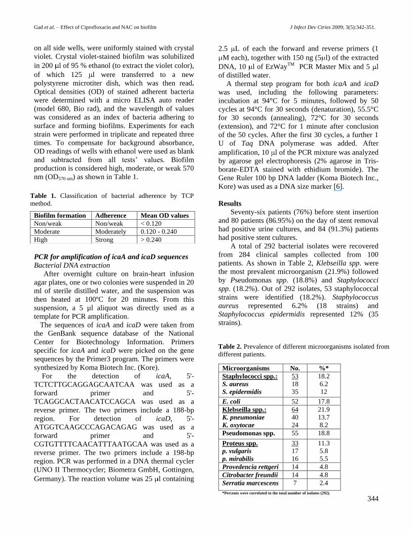
Biofilm formation Adherence Mean OD values
Non/weak Non/weak < 0.120
Moderate Moderately 0.120 - 0.240
High Strong > 0.240
Table 1. Classification of bacterial adherence by TCP
method.
Microorganisms No. %*
Staphylococci spp.:
S. aureus
S. epidermidis
53
18
35
18.2
6.2
12
E. coli 52 17.8
Klebseilla spp.:
K. pneumoniae
K. oxytocae
64
40
24
21.9
13.7
8.2
Pseudomonas spp. 55 18.8
Proteus spp.
p. vulgaris
p. mirabilis
33
17
16
11.3
5.8
5.5
Provedencia rettgeri 14 4.8
Citrobacter freundii 14 4.8
Serratia marcescens 7 2.4
*Percents were correlated to the total number of isolates (292).
Table 2. Prevalence of different microorganisms isolated from
different patients.

Gad et al. – Effect of Ciprofloxacin and NAC on biofilm J Infect Dev Ctries 2009; 3(5):342-351.
345
Polymicrobial bacteriuria represented 22.9% of
positive cultures. All staphylococcal strains were
polymicrobial with klebseilla pneumoniae, E. coli,
Pseudomonas spp., Proteus spp., Providencia
rettgeri, and Serratia marcescens. For S. aureus, five
strains were found mixed with K. pneumoniae, six
strains with Pseudomonas spp., four strains with
Proteus spp., one strain with Providencia rettgeri,
and two strains with Serratia marcescens. For S.
epidermidis, seven strains were found mixed with E.
coli, 10 strains with K. pneumoniae, 12 strains with
Pseudomonas spp., three strains with Proteus spp.
and three strains with Serratia marcescens.
Detection of biofilm producing strains
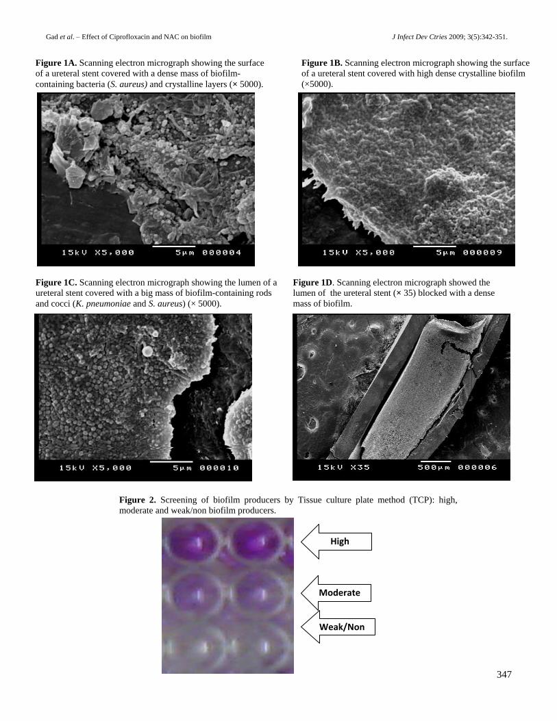
Stents examined by scanning electron
microscope showed two types of bacteria, a dense
mass of biofilm, and a high level of encrustation
(Figure 1). To explain the presence of biofilm mass
and encrustation, biofilm producing ability was tested
by TCP method for all isolates (Table 3). Table 3
shows that most of isolates were biofilm producers,
which explains the presence of a dense mass of
biofilm produced by two microorganisms on both the
surface and the lumen of the catheter. Urease enzyme
production was tested for all isolates. It was found
that most of isolates were urease positive, which
increased urine pH and produced an alkaline
condition resulting in precipitation of Ca and
Magnesium phosphate (Table 4).
A higher incidence of Staphylococci was isolated
from catheter segments than from urine samples.
Table 5 shows the distribution of staphylococcal
isolates among different samples. All staphylococcal
strains were isolated from urine samples on the day
of stent removal (three strains of S. aureus and four
strains of S. epidermidis) and stent segments (15
strains of S. aureus and 31 strains of S. epidermidis).
Staphylococci were not isolated from urine samples
before stent insertion. In addition, staphylococci were
isolated from stent segments cultures in an incidence
higher than that from urine samples on the day of
stent removal. Table 5 also shows that staphylococci
were not isolated from the urine sample and catheter
segment of the same patient, which indicates that the
sensitivity of urine cultures to stent colonization is
low, and negative urine cultures do not rule out a
colonized stent.
Staphylococcal strains of different origin were
further classified according to the extent of biofilm
production to high, moderate and non/weak biofilm
producers. S. aureus and S. epidermidis strains
isolated from urine samples were non/weak biofilm
producers, but those isolated from catheter segments
were moderate and high biofilm producers. Out of
eighteen S. aureus isolates, twelve (66.7%) were
strong biofilm producers, three (16.7%) were
moderate biofilm producers, and three (16.7%) were
considered as non or weak biofilm producers. On the
other hand, out of 35 S. epidermidis, 18 (51.4%) were
strong biofilm producers, 13
(37.1%) were moderate biofilm producers, and four
(11.4%) were considered as weak or non biofilm
producers (Table 6 and Figure 2).
Microorganisms No. of microorganisms Number of biofilm producing organisms
No. %*
S. aureus 18 15 83.3
S. epidermidis 35 31 88.6
E. coli 52 40 76.9
Klebseilla spp. 64 54 84.8
Pseudomonas spp. 55 50 90.9
Proteus spp. 33 28 84.4
Provedencia rettgeri 14 12 85.7
Citrbacter freundii 14 12 85.7
serratia marcescens 7 5 71.4
Total 292 247 84.6 *Percents were correlated to the number of each isolate.
Table 3. The incidence of biofilm production among the isolated microorganisms using
microtiter plate method.
Gad et al. – Effect of Ciprofloxacin and NAC on biofilm J Infect Dev Ctries 2009; 3(5):342-351.
346
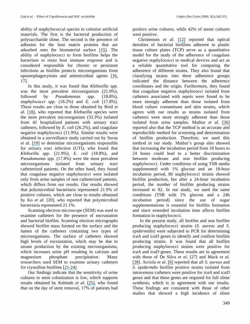
PCR detection of icaA and icaD genes
All strains were tested for the presence of icaA and
icaD genes. All biofilm producing strains isolated
from catheter segments were found to be positive for
both genes, giving a 188-bp band for icaA, and a 198-
bp band for icaD genes. It was also found that all
strains which were positive for icaA were also
positive for icaD. On the other hand, all non biofilm
producing strains isolated from urine samples were
negative for both genes. The
expression of icaA and icaD genes in strains isolated
from catheter segments collected from patients
indicates the role of ica genes in biofilm production
and as virulence markers in staphylococcal infections
associated with urinary tract catheters (Table 7 and
Figure 3).
Discussion Bacterial adhesion has long been considered as a
virulence factor contributing to infections associated
with catheters and other indwelling medical devices
[14]. There are two possible explanations for the
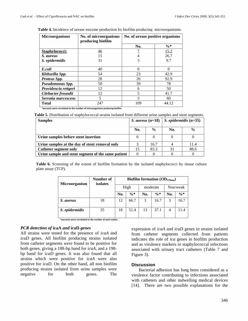
Microorganisms No. of microorganisms
producing biofilm
No. of urease positive organisms
No. %*
Staphylococci:
S. aureus
S. epidermidis
46
15
31
7
4
3
15.2
26.7
9.7
E.coli 40 0 0
Klebseilla Spp. 54 23 42.9
Proteus Spp. 28 26 92.9
Pseudomonas Spp. 50 39 78
Providencia rettgeri 12 6 50
Citrbacter freundii 12 5 41.7
Serratia marcescens 5 3 60
Total 247 109 44.12
*percents were correlated to the number of microorganisms producing biofilm.
percents were correlated to the number of microorganisms producing
biofilm.
Table 4. Incidence of urease enzyme production by biofilm producing microorganisms.
Samples S. aureus (n=18) S. epidermidis (n=35)
No. % No. %
Urine samples before stent insertion 0 0 0 0
Urine samples at the day of stent removal only 3 16.7 4 11.4
Catheter segment only 15 83.3 31 88.6
Urine sample and stent segment of the same patient 0 0 0 0
Table 5. Distribution of staphylococcal strains isolated from different urine samples and stent segments.
Microorganism
Number of
isolates
Biofilm formation (OD570nm)
High moderate Non/weak
No. %* No. %* No. %*
S. aureus 18
12 66.7 3 16.7 3 16.7
S. epidermidis 35 18 51.4 13 37.1 4 11.4
*percents were correlated to the number of each isolate.
Table 6. Screening of the extent of biofilm formation by the isolated staphylococci by tissue culture
plate assay (TCP).
Gad et al. – Effect of Ciprofloxacin and NAC on biofilm J Infect Dev Ctries 2009; 3(5):342-351.
347
Figure 1A. Scanning electron micrograph showing the surface
of a ureteral stent covered with a dense mass of biofilm-
containing bacteria (S. aureus) and crystalline layers (× 5000).
Figure 1B. Scanning electron micrograph showing the surface
of a ureteral stent covered with high dense crystalline biofilm
(×5000).
Figure 1C. Scanning electron micrograph showing the lumen of a
ureteral stent covered with a big mass of biofilm-containing rods
and cocci (K. pneumoniae and S. aureus) (× 5000).
Figure 1D. Scanning electron micrograph showed the
lumen of the ureteral stent (× 35) blocked with a dense
mass of biofilm.
High
Moderate
Weak/Non
Figure 2. Screening of biofilm producers by Tissue culture plate method (TCP): high,
moderate and weak/non biofilm producers.

Gad et al. – Effect of Ciprofloxacin and NAC on biofilm J Infect Dev Ctries 2009; 3(5):342-351.
348
Microorganisms Number of biofilm positive strains Total positive
Strains positive for icaA and
icaD Catheter segments Urine samples at
the day of stent removal
No. %* No. %* No. %* No. %*
S. aureus (n=18)
15 83.3 0 0 15 83.3 15 83.3
S. epidermidis (n=35) 31 88.6 0 0 31 88.6 31 88.6
Total (n=53) 46 86.8 0 0 46 86.8 46 86.8
*Percents were correlated to the number of each isolate.
Table 7. Relationships among biofilm production (TCP assay), the presence of icaA and icaD genes and sample origin.
Strains NC 1 2 3 4 NC 1 2 3 4
Lanes 1 2 3 4 5 6 7 8 9 10 11 12
icaA icaD
bp
1500
1000
100
500
100
Figure 3. PCR detection of icaA and icaD genes in 4 Staphylococcus aureus strains (1 and 3 were biofilm producers and 2 and
4 were non-biofilm producers).
Lane 1: markers 100-pb.
Lanes 2 & 7: negative control (NC) of icaA & icaD respectively (DNA template absent).
Lane 3: 188-pb band (icaA) of strain no. 1 (biofilm producing S. aureus).
Lanes 4 & 6: no bands with DNA from non biofilm producing S. aureus (strains 2 and 4).
Lane 5: 188-pb band of biofilm producing S. aureus (strain no. 3).
Lane 8: 198-pb band (icaD) band of biofilm producing S. aureus (strain no. 1).
Lane 9: no bands with non biofilm producing strains (strains no. 2).
Lane 10: 198-pb band (icaD) of strain no. 3 (biofilm producing S. aureus ).
Lane 11: no bands with non biofilm producing strains (strains no. 4).
Gad et al. – Effect of Ciprofloxacin and NAC on biofilm J Infect Dev Ctries 2009; 3(5):342-351.
349
ability of staphylococcal species to colonize artificial
materials. The first is the bacterial production of
polysaccharide slime. The second is the presence of
adhesins for the host matrix proteins that are
adsorbed onto the biomaterial surface [15]. The
ability of staphylococci to form biofilms helps the
bacterium to resist host immune response and is
considered responsible for chronic or persistent
infections as biofilm protects microorganisms from
opsonophagocytosis and antimicrobial agents [16,
17].
In this study, it was found that Klebseilla spp.
was the most prevalent microorganism (21.9%),
followed by Pseudomonas spp. (18.8%),
staphylococci spp. (18.2%) and E. coli (17.8%).
These results are close to those obtained by Reid et
al. [18], who reported that Klebseilla species were
the most prevalent microorganism (33.3%) isolated
from 42 hospitalized patients with urinary tract
catheters, followed by E. coli (26.2%), and coagulase
negative staphylococci (11.9%). Similar results were
obtained in a surveillance study carried out by Savas
et al. [19] to determine microorganisms responsible
for urinary tract infection (UTI), who found that
Klebseilla spp. (21.9%), E. coli (18.8%), and
Pseudomonas spp. (17.8%) were the most prevalent
microorganisms isolated from urinary tract
catheterized patients. On the other hand, they found
that coagulase negative staphylococci were isolated
only from urine samples of non catheterized patients,
which differs from our results. Our results showed
that polymicrobial bacteriuria represented 21.9% of
positive cultures, which are close to results obtained
by Ko et al. [20], who reported that polymicrobial
bacteriuria represented 21.1%.
Scanning electron microscope (SEM) was used to
examine catheters for the presence of encrustation
and bacterial biofilm. Scanning electron micrographs
showed biofilm mass formed on the surface and the
lumen of the catheters containing two types of
microorganisms. The surface of catheters showed
high levels of encrustation, which may be due to
urease production by the existing microorganisms,
which increases urine pH resulting in calcium and
magnesium phosphate precipitation. Many
researchers used SEM to examine urinary catheters
for crystalline biofilms [21-24].
Our findings indicate that the sensitivity of urine
cultures to stent colonization is low, which supports
results obtained by Kehinde et al. [25], who found
that on the day of stent removal, 17% of patients had
positive urine cultures, while 42% of stents cultures
were positive.
Christensen et al. [12] reported that optical
densities of bacterial biofilms adherent to plastic
tissue culture plates (TCP) serve as a quantitative
model for the study of the adherence of coagulase
negative staphylococci to medical devices and act as
a reliable quantitative tool for comparing the
adherence of different strains. They also found that
classifying strains into three adherence groups
indicated the distance between the adherence
coordinates and the origin. Furthermore, they found
that coagulase negative staphylococci isolated from
catheters associated with sepsis were found to be
more strongly adherent than those isolated from
blood culture contaminant and skin strains, which
agrees with our results as strains isolated from
catheters were more strongly adherent than those
isolated from urine samples. Mathur et al. [26]
reported also that the TCP method is an accurate and
reproducible method for screening and determination
of biofilm production. Therefore, we used this
method in our study. Mather’s group also showed
that increasing the incubation period from 18 hours to
24 hours could lead to a better discrimination
between moderate and non biofilm producing
staphylococci. Under conditions of using TSB media
supplemented with 1% glucose and an 18-hour
incubation period, 80 staphylococci strains showed
biofilm production, but after a 24-hour incubation
period, the number of biofilm producing strains
increased to 82. In our study, we used the same
conditions (TSB with 1% glucose and a 24h
incubation period) since the use of sugar
supplementations is essential for biofilm formation
and since extended incubation time affects biofilm
formation in staphylococci.
In the present study, all biofilm and non biofilm
producing staphylococci strains (S. aureus and S.
epidermidis) were subjected to PCR for determining
icaA and icaD genes to identify and confirm biofilm
producing strains. It was found that all biofilm
producing staphylococci strains were positive for
icaA and icaD genes. These results are in agreement
with those of De Silva et al. [27] and Mack et al.
[28]. Arciola et al. [6] reported that all S. aureus and
S. epidermdis biofilm positive strains isolated from
intravenous catheters were positive for icaA and icaD
genes and that these genes are required for full slime
synthesis, which is in agreement with our results.
These findings are consistent with those of other
studies that showed a high incidence of slime
Gad et al. – Effect of Ciprofloxacin and NAC on biofilm J Infect Dev Ctries 2009; 3(5):342-351.
350
producing staphylococci in isolates from clinically
significant medical device-associated infections of
different origins [29-33].
Biofilm production is an important pathogenic
factor which facilitates adherence of microorganisms
to medical devices and protects them from the host
immune system and antimicrobial therapy. Results
revealed that both icaA and icaD genes were either
present or absent and no single strain had shown the
presence of one gene. These results confirm the fact
that both genes are part of one operon and so the
entire operon was either present or absent. In
addition, our results showed that both genes (icaA
and icaD) were present in all biofilm producing
strains, indicating the important role of ica genes as
virulence markers in staphylococcal infections. In
conclusion, there is a high prevalence of biofilm
production among microorganisms isolated from
catheterized patients, the sensitivity of urine cultures
to stent colonization is low, and staphylococci
isolated from catheter segments showed a higher
extent of biofilm production than those isolated from
urine samples. All biofilm producing staphylococci
were positive for icaA and icaD genes. It is important
to diagnose and to give prophylactic antibiotics just
before and during the surgical procedure to eliminate
plankotonic bacteria before they can form a biofilm.
References 1. Kloos WE, and Bannerman TL (1994) Update on clinical
significance of coagulase–negative Staphylococci. Clin
Microbiol Rev 7: 117–40.
2. Desgrandchamps F, Moulinier F, Doudon M, Teillac P, Le
Duc A. (1997) An in-vitro comparison of urease induced
encrustation of JJ stents in human urine. Br J Urol 79: 24-
7.
3. Ammendolia MG, Rosa RD, Montanaro L, Arciola CR and
Baldassarri L (1999) Slime production and expression of
slime–associated antigen by staphylococcal clinical
isolates. J Clin Microbiol 37: 3235–8.
4. O'Gara J P, and Humphreys H. (2001) Staphylococcus
epidermidis biofilms importance and implications. H Med
Microbiol 50: 582-87.
5. Yazdani R, Oshaghi M, Havayi A, Salehi R, Sadeghizadeh
M, Foroohesh H. (2006) Detection of icaAD gene and
biofilm formation in Staphylococcus aureus isolates from
wound infections. Iranian J Publ Health 35: 25-28.
6. Arciola CR, Baldassarri L, and Montanaro L. (2001)
Presence of icaA and icaD genes and slime production in a
collection of staphylococcal strains from catheter-
associated infections. J Clin Microbiol 39: 2151-2156.
7. Benson HC. (2002) Microbiological Application:
Laboratory manual in general microbiology, 11th ed.,
McGram-Hill Higher Education, San Francisco. p. 168.
8. Sheretz RJ, Raad IL, Balani A (1990) Three-year
experience with sonicated vascular catheter cultures in a
clinical microbiology laboratory. J Clin Microbiol 28: 76-
82.
9. Koneman EW, Allen SD, Janda WM, Schreckenberger
PC, and Winn WC (1994) Introduction to diagnostic
microbiology, J. B. Lippincott Company, USA.
10. Collee JG, Fraser AG, Marmion BP, and Simmons A.
(1996) Tests for identification of bacteria. In: Makie &
McCartney practical medical microbiology, 14th ed.,
Churchill Livingstone In., New York 131-149.
11. Soboh F, Khoury AE, Zamboni AC, Davidson D, and
Mittelman MW (1995) Effects of ciprofloxacin and
protamine sulfate combinations against catheter-associated
Pseudomonas aeruginosa biofilms. Antimicrob Agents
Chemother 39: 1281-1286.
12. Christensen GD, Simpson WA, Younger JA, Baddour LM,
Barrett FF, Melton DM, Beachey EH (1985) Adherence of
coagulase negative Staphylococci to plastic tissue cultures:
a quantitative model for the adherence of staphylococci to
medical devices. J Clin Microbiol 22: 996-1006.
13. O'Toole AG and Kolter R (1998) Initiation of biofilm
formation in Pseudomonas fluorescence WCS365 proceeds
via multiple, convergent signaling pathways: a genetic
analysis. Molecular microbiology 28: 449.
14. Francois P, Vaudaux P, Foster TJ, and Lew DP (1996)
Host-bacteria interactions in foreign body infections. Infect
Control Hosp Epidemiol 17: 514–520.
15. Montanaro L, Arciola CR, Borsetti E, Brigotti M, and
Baldassarri L (1998) A polymerase chain reaction (PCR)
method for the identification of collagen adhesin gene
(cna) in Staphylococcus-induced prosthesis infections.
New Microbiol 21: 359–363.
16. Cramaton SE, Gerke C, and Gotz F (2001) In-vitro method
to study staphylococcal biofilm formation. Methods
enzymol 336: 239-55.
17. Serralta VW, Harrison-Belestra C, Cazzaniga AL, Davis
SC, and Mertz PM (2001) Lifestyle of bacteria in wounds:
Presence of bofilms? Wounds 13: 29-34.
18. Reid G, Poter P, Delaney G, Hsieh J, Nicosia S, and Hayes
K (2000) Ofloxacin for the treatment of urinary tract
infections and biofilms in spinal cord injury. Int J
Antimicrob Agents 13: 305-307.
19. Savas L, Guvel S, Onlen Y, Savas N, and Duran N (2006)
Nosocomial urinary tract Infections: Microorganisms,
antibiotic Sensitivities and Risk Factors. West Indian Med
J 55: 188-193.
20. Ko M, Liu C, Woung L, Lee W, Jeng H, Lu S, Chiang H,
and Li C (2008) Species and antimicrobial resistance of
uropathogens isolated from patients with urinary catheters.
Tohoku J Exp Med 214: 311-319.
21. Jones BV, Young R, Mahenthiraligam E. and Stickler DJ
(2004) Ultrastructure of Proteus mirabilis swarmer cell
rafts and role of swarming in catheter-associated urinary
tract infection. Infect Immun 72: 3941-3950.
22. Stickler DJ (2005) Urinary catheters: ideal sites for the
development of biofilm communities.
www.sgm.ac.uk/pubs/micro_today/pdf/020505.pdf.
Microbiology Today, Feb: 22-25.
23. Sabubba NA, Mahenthiraligam E, and Stickler DJ (2003)
Molecular epidemiology of Proteus mirabilis infections of
the catheterized urinary tract. J Clin Microbiol 41: 4961-
4965.
24. Stickler DJ, Young R, Jones G, Sabubba NA, and Morris
NS (2003) Why are Foley catheters so vulnerable to

Gad et al. – Effect of Ciprofloxacin and NAC on biofilm J Infect Dev Ctries 2009; 3(5):342-351.
351
encrustation and blockage by crystalline bacterial biofilm?
Urol Res 31: 306–311.
25. Kehinde EO, Rotimi VO, Al-Hunayan A, Abdul-Halim H,
Boland F and Al-Awadi KA (2004) Bacteriology of urinary
tract infection associated with indwelling J ureteral stents. J
Endourol 18: 891-896.
26. Mathur T, Singhal S, Khan S, Upadhyay DJ, Fatma T, Rattan
A (2006) Detection of Biofilm Formation among the
clinical isolates of staphylococci: an evaluation of three
different screening methods. Indian J Med Microbiol 24:
25-9.
27. De Silva GD, Kanatazanou M, Massey RC, Wikinson AR,
Day NP, and Peacock SJ (2002) The ica operon and
biofilm production in coagulase negative staphylococci
associated with carriage and disease in neonatal intensive
care unit. J Clin Microbiol 40: 382-388.
28. Mack D, Rohde H, Dobinsky S, Riedewald J, Nedelmann
M, Knobloch JK, Elsner HA, and Feucht HH (2000)
Identification of three essential regulatory gene loci
governing expression of the Staphylococcus epidermidis
polysaccharide intercellular adhesin and biofilm formation.
Infect Immun 68: 3799–3807.
29. Ziebuhr W, Heilmann C, Gotz F, Meyer P, Wilms K,
Straube E, Hacker J (1997) Detection of the intercellular
adhesion gene cluster (ica) and phase variation in
Staphylococcus epidermidis blood culture strains and
mucosal isolates. Infect Immun 65: 890-6.
30. Arciola CR, Campoccia D, Gamerini S, Donati ME,
Baldassarri L, Montanaro L (2003) Occurrence of ica
genes for slime synthesis in a collection of Staphylococcus
epidermidis strains from orthopedic prosthesis infections.
Acta orthop Scand 74: 617-621.
31. Satorres SE, and Alcaráz LE (2007) Prevalence of icaA and
icaD genes in Staphylococcus aureus and Staphylococcus
epidermidis strains isolated from patients and hospital staff.
Cent Eur J Pulic Health 15: 87-90.
32. Chaieb K, Zmantar T, Chehab O, Boucham O, Hasen AB,
Mahdouani K, and Bakhrouf A (2007) Antibiotic resistance
genes detected by multiplex PCR assays in Staphylococcus
epidermidis strains isolated from dialysis service. Jpn J
Infect Dis 60: 183-187.
33. Helmy MM, Allam AA, Mohamed MS, Amer A, and Abd
Aldaem A (2006) Slime forming Staphylococcus
epidermidis isolated from orthopedic prosthesis infections
and its sensitivity to antibiotics. EJMM 15: 205-213.
Corresponding Author
Rehab Mahmoud Abd El-Baky
Address: 56, Adnan El-Malky street, Ard sultan, El-Minia,
Egypt.
Mobil: (02)-0123350610. Email: [email protected]
Conflict of interest: No conflict of interest is declared.
.
Copyright © 2022 FDOKUMEN