An Integrative Meta-analysis of MicroRNAs in Hepatocellular Carcinoma
Upload
independentCategory
view
6download
0
Detection and Identification of Tumor-AssociatedProtein Variants in Human Hepatocellular CarcinomasEvelyn Zeindl-Eberhart,1 Sibylle Haraida,1 Sibylle Liebmann,1 Peter Roman Jungblut,2 Stephanie Lamer,2 Doris Mayer,3
Gundula Jager,4 Stephen Chung,5 and Hartmut Manfred Rabes1
The proteomic approach is a valuable tool to detect and identify proteins that are associatedwith cancer. In previous investigations on experimentally induced rat hepatomas, we de-tected aldose reductase-like protein (ARLP) as a highly significant marker protein. Ourpresent study was intended to look for the presence of similar tumor-associated markerproteins on human hepatocellular carcinomas (HCC). We found several novel tumor-associated protein variants that represent members of the aldo-keto reductase (AKR)superfamily. Human aldose reductase-like protein-1 (hARLP-1) was the most prominent tumor-associated AKR member detected in HCC by 2-dimensional electrophoresis (2-DE) andidentified by mass spectrometric fingerprinting. The enzyme was found in 4 distinct forms(hARLP-1, 36/7.4 (kd/pI); hARLP-2, 36/7.2; hARLP-3, 36/6.4; and hARLP-4, 33/7.35). Inaddition, a human aldose reductase-like protein (hARLP-5, 36/7.6) was identified thatdiffered from hARLP-1 by 1 amino acid (D313N), indicating 2 allelic forms of the humanaldose reductase-like gene. A novel antibody directed against common parts of the hARLPsrevealed hARLP reactivity in human HCC by immunohistochemistry. Furthermore, aldosereductase (AR) was identified and characterized as a tumor-associated variant. In conclusion,in all investigated human HCCs at least one of the various types of the described tumor-associated proteins of the AKR superfamily was clearly present. Of these HCC samples, 95%were positive for hARLPs as proven by 2-DE analysis and/or by use of the antibody directedagainst hARLP. Thus, hARLP is a strong candidate for use as an immunohistochemicaldiagnostic marker of human HCC. (HEPATOLOGY 2004;39:540–549.)
Human hepatocellular carcinoma (HCC) ranksfifth in worldwide cancer incidence and is animportant component of public health. Many
HCC risk factors are known, including hepatitis B or C
(HBV or HCV) infection, ingestion of aflatoxin-contam-inated food, and alcohol.1,2 The development of HCC isassociated with multiple changes at the messenger RNA(mRNA) and/or protein level, some of them serving astumor markers, e.g., �-fetoprotein,3 or, less specifically,cyclin D1 or the proliferating cell nuclear antigen.4
Misprogramming of genetic information in cancer isreflected by quantitative and/or qualitative protein alter-ations. These protein alterations might represent tumormarkers that are useful in the diagnosis of human tumorsand may also help the understanding of mechanisms oftumor induction and development. Proteome analysis ofliver proteins and HCC were predominantly performedusing either chemically induced hepatomas in animals(predominantly the rat5–10) or human HCC cell lines,such as HepG2 and Huh7 cells,11 BEL-7404 cells,12 orHCC-M cells.2,13,14 Numerous so-called tumor-associ-ated or cancer-related proteins were identified; these pro-vide valuable information for the establishment of HCCprotein databases.2,11–14 Comparative analysis of liver tis-sue and hepatocellular carcinomas might give additionalinsights into the induction or repression of tumor-associ-
Abbreviations: ARLP, aldose reductase-like protein; HCC, hepatocellular carci-noma; AKR, aldo-keto reductase; hARLP, human aldose reductase-like protein;2-DE, 2-dimensional electrophoresis; HBV, hepatitis B virus; HCV, hepatitis Cvirus; MS, mass spectrometry; ARL-1, aldose reductase-like-1; PMF, peptide massfingerprinting; MALDI-MS, matrix-assisted laser desorption/ionization-mass spec-trometry; AR, aldose reductase; acc, accession number; rARLP, rat aldose reductase-like protein; TST, thiosulfate sulfurtransferase.
From the 1Institute of Pathology, Ludwig Maximilians University of Munich,Munich; the 2Max Planck Institute for Infection Biology, Berlin; 3Research Groupin Hormones and Signal Transduction, German Cancer Research Center, Heidel-berg; the 4Max von Pettenkofer Institute for Hygiene and Medical Microbiology,Munich, Germany; and the 5Institute of Molecular Biology, University of HongKong, Hong Kong, China.
Received June 26, 2003; accepted November 14, 2003.Supported by a grant from Deutsche Krebshilfe.Address reprint requests to: Evelyn Zeindl-Eberhart, Institute of Pathology, Uni-
versity of Munich, Thalkirchnerstrasse 36, D-80337 Munich, Germany. E-mail:[email protected]; fax: �49-89-5160-4083.
Copyright © 2004 by the American Association for the Study of Liver Diseases.Published online in Wiley InterScience (www.interscience.wiley.com).DOI 10.1002/hep.20060
540

ated protein variants within an intact organism. Clinicalmaterial was used for proteome analysis comparing nor-mal liver tissue and HCC,15–17 or normal liver, cirrhoticliver, and HCC18 or sera from HCC patients for the iden-tification of tumor autoantibodies.19 Different proteinvariants were proposed as tumor-marker candidates. Anintegrated proteome database for the study of humanHCC has been constructed.20 As a result of worldwideinterest in liver diseases, in particular human HCC, initi-atives of the human proteome project (HUPO)21 led tothe initiation of the human liver proteome project(HLPP).
In previous investigations on chemically induced rathepatomas, we detected a number of tumor-associatedprotein variants by 2-dimensional electrophoresis(2-DE), predominantly in an area of 30–40 kd/pI 6.2–7.6, and identified them by mass spectrometry (MS) asmembers of the aldo-keto reductase (AKR) superfam-ily.5,6 Their induction/inhibition was dependent on thetype of initiating carcinogen.
The AKR superfamily includes several main enzymefamilies. The largest family (AKR1) contains the aldehydereductases (AKR1A), the aldose reductases (AKR1B), andthe hydroxysteroid dehydrogenases (AKR1C); nomencla-ture is from Jez et al.22 Members of the AKR superfamilyare monomeric cytoplasmic proteins; they share identicalsequences and possess similar physical and chemical prop-erties. Most of them catalyze the NADPH-dependent re-duction of a large variety of xenobiotics and endogenousaldehydes and ketones and metabolize a wide range ofsubstrates; they are all involved in detoxification pro-cesses.22,23 Members of the AKR superfamily are de-scribed as being involved in carcinogenic processes. Theseare 4 aldose reductase-like proteins (ARLPs) predomi-nantly detected in nitroso-compound-induced rat hepa-tomas.5 In human HCC, an aldose reductase-like-1(ARL-1) mRNA was expressed.24,25 Two aflatoxin B1-inducible aldehyde reductases were discovered in aflatox-in-B1-induced rat hepatomas26 and in human coloniccarcinomas.27
These results led us to perform a proteome analysisof human HCC. New patterns of tumor-associatedprotein variants were observed in 2-DE gels of humanHCC, predominantly in a 2-DE area of �30 – 40 kd/pI 6.2–7.8. We identified these tumor-associated pro-tein variants as members of the AKR superfamily bypeptide mass fingerprinting (PMF) obtained by matrixassisted laser desorption/ionization-mass spectrometry(MALDI-MS) and showed that they might be valuableas diagnostic markers of human HCC in immunohis-tochemistry.
Patients and Methods
Tissue Specimens. Human HCC samples from twodifferent groups, Caucasians (group I) and Asians (groupII), were investigated. Ten different tissue samples of hu-man HCC (group I) from liver explants collected between1994 and 1998 were provided by P. Bannasch, Divisionof Cytopathology, German Cancer Research Center, Hei-delberg, Germany. The HCC tissue collections were ob-tained with informed consent and were approved by theethics committee of Heidelberg University. These tumorsamples were designated 237/98, 126/98, 261/98, 321/97, 14/98, 193/98, and 8/98 (males), and 260/98, 91/97,and 107/94(females). Of the patients in group I, 40%were positive for HBV infection and 50% for HCV in-fection. One patient was positive for HCV with an alco-hol anamnesis; another was alcoholic. All tumor sampleswere histologically verified; classification of differentia-tion was performed according to the WHO grading sys-tem. The samples were stored at �80°C until use.Control liver samples from 2 healthy males,1 female (ageadjusted), and a fetal liver (5th month) were received fromthe Institute of Forensic Medicine, Munich, and desig-nated 51HLN and 29HLN (males), 30HLN (female),and FET (fetal liver), respectively. In addition, we used 5other histologically examined human HCC samples to-gether with the corresponding normal liver tissue fromthe Institute of Molecular Biology, University of HongKong. These tissue samples (group II) were designatedidentically to those published by Cao et al.24: (T, tumor;N, normal tissue) T2;N2, T12;N12, T16;N16, T19;N19, and T23. HBV/HCV status was determined inthese HCC samples by polymerase chain reaction forHBV DNA and HCV RNA. All patients were positive forHBV infection; additional clinical data were not avail-able.
Preparation of Protein Samples. Soluble proteinfractions were extracted from all samples. The group IIHCC samples underwent a lyophilization step prior tothe extraction of the soluble protein fraction.
For lyophilization liver samples frozen at �196°Cwere weighed and transferred to a mortar cooled at�196°C. Two ml of bidistilled water were added in pelletform to each sample. Frozen components were ground topowder in liquid nitrogen. Five ml of bidistilled water(4°C) were added to each powdered sample, the suspen-sion was quickly transferred into a lyophilization flask,and the samples were freeze-dried, weighed, and sent toour laboratory. The original mass of the samples was re-stored by adding bidistilled water. The extraction methodof the soluble protein fraction was the same for all samplesfollowing the method of Klose28 as previously described.5
HEPATOLOGY, Vol. 39, No. 2, 2004 ZEINDL-EBERHART ET AL. 541

Two-Dimensional Electrophoresis. Soluble proteinswere separated by high-resolution 2-DE according toKlose,29 using Iso-Dalt –equipment (Hoefer ScientificInstruments, San Francisco, CA).30,31 Isoelectric focusingwas performed with 4% carrier ampholytes (4 parts Ser-valyte, pH 5-7, Serva, Heidelberg, Germany; 1 part Phar-malyte, pH 3-10 and 1 part Ampholine, pH 3.5-9.5, bothAmersham Pharmacia Biotech, Uppsala, Sweden). Thesecond dimension was run on gels (16 � 16 � 0.15 cm )with a 10%-to-16% polyacrylamide gradient in a Dalttank as previously described in detail.32 Four hundred �gof soluble protein (Lowry protein assay kit, Sigma-Al-drich, Deisenhofen, Germany) were separated for Coo-massie Brilliant Blue staining (CBB R-250, Merck,Darmstadt, Germany), and 100 �g of soluble proteinwere used for silver staining.33
The soluble protein fraction of each sample (n � 24)was applied for 2-DE separation at least twice. Because ofthe variable levels of albumin in the different human sam-ples (first run) that are included in the protein determina-tion, the applied protein amount was adjusted to identicalactin amounts as an internal standard (second run) forevery sample. In group I, 2-DE gels of 10 different HCCsamples were compared with 3 different normal liver tis-sues; in group II, 5 different HCC samples were com-pared with their corresponding normal liver tissues. Theselected area in 2-DE gels, �30–40 kd/pI �6.2–7.8,according to the rat model,5 was evaluated visually at firstaccording to several criteria. All protein spots that werechanged in quantity and/or quality in at least 80% of allHCCs investigated (n � 15) were defined as candidatesfor tumor-associated protein variants. In addition, spotsthat previously had been defined as being tumor-associ-ated by others were included as candidates for tumor-associated proteins. Also included were newly detectedproteins that represent highly related forms of proventumor-associated variants and protein variants predomi-nantly detected in 1 of the 2 groups investigated.
The relative spot intensity of different candidates oftumor-associated protein variants was determined in sil-ver-stained 2-DE gels from normal liver tissue and HCCsamples of Caucasian and Asian patients by TopSpot eval-uation program (Algorithmus, Berlin, Germany). Spotintensities of all candidates of tumor-associated variantswere normalized against the actin spot intensity, which isin the linear range of the intensity curve and does not varybetween HCC and controls. In order to define significanttumor-associated protein variants, Student’s t test wasperformed. Protein spots that fulfilled the criterion P �.025 were defined as tumor-associated variants.
For exclusion of possible unspecific protein changes in2-DE gels due to the lyophilization procedure (performed
for group II samples only), we tested both preparationmethods using a rat hepatoma and a human liver sampleand analyzed the 2-DE pattern run in parallel with andwithout lyophilization. Samples that underwent bothpreparation methods showed identical protein amountsin their 2-DE gels. An influence of lyophilization on the2-DE protein pattern could be excluded.
Protein Identification. For identification, severalprotein spots per spot of interest were excised from CBB-stained gels. PMF after tryptic in-gel digestion was used34
as described.5 Identification was performed by searchingthe National Center for Biotechnology Information(NCBI) and/or Swissprot databases with MS-Fit or Pep-tIdent, respectively. Sequence database was limited to hu-man/rodent proteins. A mass tolerance of 0.1 Da and 2incomplete cleavages were allowed. Identifications wereaccepted when they covered at least 30% of the wholesequence and when comparable molecular mass/P valueswere obtained from the databases.
Immunoblotting of Human Aldose Reductase-likeProtein (hARLP). After 2-DE separation of the solubleprotein fraction of HCC tissue (400 �g protein/sample),2-DE gel areas (�30–40 kd/pI �6.2–7.8) containingthe hARLP region were excised from 2-DE gels and blot-ted onto hydrophobic polyvinylidene difluoride mem-branes (0.2 �m Bio-Rad, Hercules, CA) under semidryconditions for 3 hours at 1 mA/cm2 membrane at roomtemperature with 50mM borate buffer, pH 9.0, 20%methanol. For immunodetection of hARLP the poly-clonal antibody directed against the last 17 amino acids ofthe C-terminus of the hARLP, accession number (acc)O60218,24 was used (dilution 1:1,000) according to themethod described earlier.35 The hARLP antibody wasproduced for our laboratory by Bio-Genes (Berlin, Ger-many).
Immunohistochemical Detection of hARLP in LiverSections. Formaldehyde-fixed, paraffin-embedded tu-mor samples from 10 additional Caucasian patients, dif-ferent from those used for proteome analysis, were used:HCC (n � 6); hepatocellular adenoma (n � 1); livermetastasis of a colonic adenocarcinoma (n � 1); focalnodular hyperplasia (n � 1); and fibrolamellar carcinoma(n � 1). Human ARLP expression was shown on serial 4�m sections using the polyclonal antibody describedabove and the Vectastain –ABC Kit (rabbit immunoglob-ulin G; Vector Laboratories, Burlingame, CA) and diami-nobenzidine (Sigma) as described.8 After unmasking indiluted Target Retrieval Solution (Dako Diagnostica,Hamburg, Germany) for 30 minutes in an autoclave at 1atm, endogenous peroxidase of the sections was inhibitedby 3% H2O2. Human ARLP antibody was used in a di-lution of 1:80 in antibody diluent (Dako). Antibody di-
542 ZEINDL-EBERHART ET AL. HEPATOLOGY, February 2004

luent without antibody served as negative control. Inaddition, the APAAP (alkaline phosphatase–anti-alkalinephosphatase) method was performed. After unmasking asdescribed above, hARLP antibody was used in a dilutionof 1:80 in antibody diluent for 17 hours at 4°C. Thefollowing steps were performed according to the manu-facturer’s instructions (Dako): (1) Fast Red TR/NaphtholAS-MX was used as a detection system for alkaline phos-phatase according to the product information (Sigma);(2) slides were counterstained with Mayer’s hematoxilinfor 2 minutes, blued for 15 minutes in running water, andcover-slipped with Kaiser’s glycerol gelatin (Merck).
ResultsHigh-resolution 2-DE gels performed with soluble
protein fractions of human HCC from Caucasian (groupI, n � 10) and Asian (group II, n � 5) male and femalepatients, compared with controls (n � 8), showed a re-producible protein pattern with some interindividualvariation. A representative 2-DE gel is shown in Fig. 1.The area �30–40 kd/pI �6.2–7.8 was selected for theevaluation of tumor-associated protein variants. In this2-DE area, approximately 90 different spots were regis-tered. Thirteen were classified as tumor-associated vari-ants according to the criteria outlined above. Eachrepresents a well-defined spot with regard to MW and pI.Seven protein spots represent known proteins of differentprotein groups (publication in preparation). Five spots ofthe selected area were identified by PMF-MS (Table 1); 1was characterized by immunoblotting. All 6 spots aremembers of the AKR superfamily and are subjects of thisreport.
Three spots with the same Mr but different pI (36kd/pI 7.4; 7.2; 6.4, respectively) were identified by MS asdifferent forms of hARLP. An additional spot (33 kd/pI7.35) reacted with the hARLP antibody, obviously repre-senting an additional hARLP. We designated these re-lated proteins hARLP-1, hARLP-2, hARLP-3 andhARLP-4, respectively. These proteins are not found innormal liver tissue; but present exclusively in humanHCC (Fig. 2A and B, Table 2) and were defined as tumor-associated variants.
We identified an additional related form of hARLP inHCC; it was designated hARLP-5 (Fig. 2C). This proteinwas first detected by immunoblotting in HCC samplesfrom 2 female patients (260/98 and 91/97). MS analysisconfirmed this result. However, compared to hARLP-1(36/7.4), the hARLP-5 (36/7.6) was different in 1 specificmass peak. The mass peak 2,272.03/2,271.98 (user mass/matching mass) found in hARLP-1 characterized apeptide of the amino acid positions 298 to 316
Fig. 1. Two-dimensional gel of soluble proteins from human HCC. Onehundred �g of soluble proteins (sample T23) were separated in the firstdimension by isoelectric focusing using 4% carrier ampholytes (pH3-10). Separation in the second dimension was performed using anacrylamide gradient (10%-16%) followed by silver staining. Mr axis wascalibrated by standard proteins (Serva). Evaluated area (framed):�30–40 kd/pI �6.2–7.8.
Table 1. Tumor-Associated Protein Spots in 2-DE Gels Determined by PMF MALDI-MS
2-DE MS
kd/pI of Tumor-Associated Spots
TissueExamined
PeptideMatches
*IdentifiedProtein
SequenceCoverage (%)
AminoAcids
Calculatedkd/pI †Accession
36.0/7.4 HCC 16/30 hARLP-1 64.9 316 36.0/7.12 O6021836.0/7.2 HCC 17/35 hARLP-2 67.4 316 36.0/7.12 O6021836.0/6.4 HCC 13/36 hARLP-3 48.7 316 36.0/7.12 O6021836.0/7.6 HCC 15/25 hARLP-5 54.4 316 36.0/7.7 O7589036.0/6.8 HCC 11/21 AR 36.0 316 35.8/6.5 178489
Abbreviations: HCC, hepatocellular carcinoma; hARLP, human aldose reductase-like protein-1, -2, -3, and -5; AR, aldose reductase.*The identified proteins originated either from HCC or from normal human liver tissue.†NCBI and/or Swissprot were used to evaluate MS data.
HEPATOLOGY, Vol. 39, No. 2, 2004 ZEINDL-EBERHART ET AL. 543

(ACNVLQSSHLEDYPFDAEY; acc O60218). The cor-responding peptide of hARLP-5 was slightly different:2,271.01/2,271.002 (user mass/matching mass). Inamino acid position 313, the aspartic acid (D) present inhARLP-1 was substituted by an asparagine (N) (accO75890) in hARLP-5. A detail of these MALDI massspectra is shown in Fig. 3.
Human aldose reductase (AR) was identified byPMF-MS located at 36 kd/pI 6.8 in 2-DE gels obtainedfrom the HCC sample T16 (group II). T16 shows thehighest amount of AR of all HCC investigated (Fig. 2E).In normal liver tissue, AR could be detected in only min-imal amounts but was found slightly increased in 8 addi-tional HCC samples as exemplified in Fig. 2A and B andin Table 2. AR (P � .0014) was defined as a tumor-associated variant.
Interestingly, most of the HCC investigated showedhARLP-1 to hARLP- 4 (12 out of 15 samples). In 3 cases
(260/98 and 91/97 for hARLP-5; T16 for AR), hARLP-5and AR were present as 1 distinct spot in 2-DE gels, buthARLP-1 to hARLP-4 were absent in these samples (Fig.2C and E). In 8 HCC samples, AR was detected to beslightly enhanced in amount but was regularly accompa-nied by hARLP-1 to hARLP-4, as shown in Fig. 2B and Dand Table 2. In 2-DE gels of fetal liver, small spots ofhARLP-1 and hARLP-4 were observed (Fig. 2F).The rel-ative spot intensity of hARLP-1 to hARLP–5 and of ARwas quantified in 2-DE gels from normal liver tissue andin HCC material of both groups using TopSpot as soft-ware (Fig. 2G).
Immunoblots using the polyclonal antibody directedagainst hARLP showed that the antibody detected allforms of hARLP but not AR. Six different tumor samplesand 3 control liver samples were tested. In 4 of the tumorsamples (193/98, 14/98, 321/97, and T23), the presenceof hARLP-1 to hARLP-4 and the absence of hARLP-5
Fig. 2. Members of the AKRsuperfamily represent tumor-as-sociated protein variants in hu-man HCCs. The area of 2-DE gels�30–40 kd/pI � 6.2–7.8 wasevaluated for tumor-associatedprotein variants. Members of theAKR family (arrows)—hARLPsand AR—were changed inamount in HCCs compared tonormal liver. (A) Normal liver tis-sue (sample 51HLN). (B) HumanHCC; hARLP-1, hARLP-2,hARLP-3, and hARLP-4 (Cauca-sian; 193/98). (C) Human HCC;hARLP-5 (260/98). (D) HumanHCC; hARLP-1, hARLP-2,hARLP-3, and hARLP-4 (Asian;T2). (E) Human HCC; AR (T16).(F) Fetal liver; hARLP-1 andhARLP-4. (G) Quantitation of tu-mor-associated protein variantsanalyzed by TopSpot evalua-tion program. Optical density(mean � SD) of AR levels incontrol liver (C; n � 5), in hu-man HCC (T; n � 8), and insample T16; hARLP-1 tohARLP-4 in HCC (n � 8) andhARLP-5 (n � 1). Areas in 2-DEgels of normal liver correspond-ing to the site of hARLP –spots;the staining intensity was in therange of the background inten-sity (11 � 2).
544 ZEINDL-EBERHART ET AL. HEPATOLOGY, February 2004
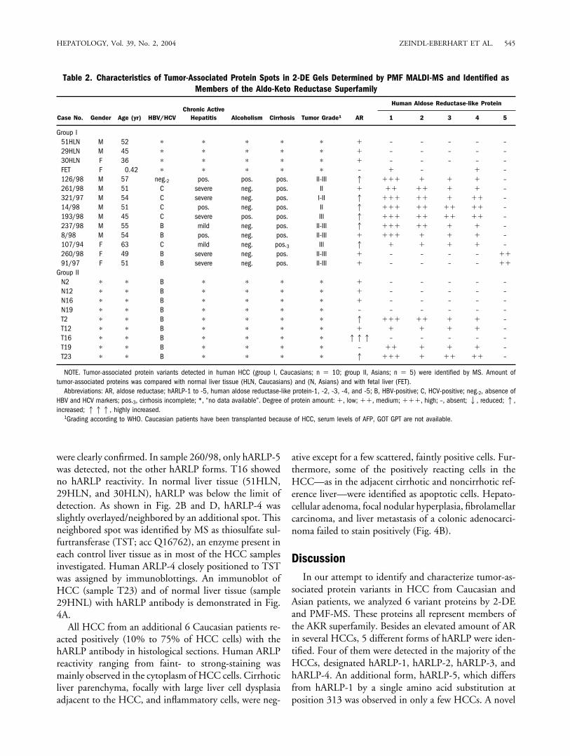
were clearly confirmed. In sample 260/98, only hARLP-5was detected, not the other hARLP forms. T16 showedno hARLP reactivity. In normal liver tissue (51HLN,29HLN, and 30HLN), hARLP was below the limit ofdetection. As shown in Fig. 2B and D, hARLP-4 wasslightly overlayed/neighbored by an additional spot. Thisneighbored spot was identified by MS as thiosulfate sul-furtransferase (TST; acc Q16762), an enzyme present ineach control liver tissue as in most of the HCC samplesinvestigated. Human ARLP-4 closely positioned to TSTwas assigned by immunoblottings. An immunoblot ofHCC (sample T23) and of normal liver tissue (sample29HNL) with hARLP antibody is demonstrated in Fig.4A.
All HCC from an additional 6 Caucasian patients re-acted positively (10% to 75% of HCC cells) with thehARLP antibody in histological sections. Human ARLPreactivity ranging from faint- to strong-staining wasmainly observed in the cytoplasm of HCC cells. Cirrhoticliver parenchyma, focally with large liver cell dysplasiaadjacent to the HCC, and inflammatory cells, were neg-
ative except for a few scattered, faintly positive cells. Fur-thermore, some of the positively reacting cells in theHCC—as in the adjacent cirrhotic and noncirrhotic ref-erence liver—were identified as apoptotic cells. Hepato-cellular adenoma, focal nodular hyperplasia, fibrolamellarcarcinoma, and liver metastasis of a colonic adenocarci-noma failed to stain positively (Fig. 4B).
DiscussionIn our attempt to identify and characterize tumor-as-
sociated protein variants in HCC from Caucasian andAsian patients, we analyzed 6 variant proteins by 2-DEand PMF-MS. These proteins all represent members ofthe AKR superfamily. Besides an elevated amount of ARin several HCCs, 5 different forms of hARLP were iden-tified. Four of them were detected in the majority of theHCCs, designated hARLP-1, hARLP-2, hARLP-3, andhARLP-4. An additional form, hARLP-5, which differsfrom hARLP-1 by a single amino acid substitution atposition 313 was observed in only a few HCCs. A novel
Table 2. Characteristics of Tumor-Associated Protein Spots in 2-DE Gels Determined by PMF MALDI-MS and Identified asMembers of the Aldo-Keto Reductase Superfamily
Case No. Gender Age (yr) HBV/HCVChronic Active
Hepatitis Alcoholism Cirrhosis Tumor Grade1 AR
Human Aldose Reductase-like Protein
1 2 3 4 5
Group I51HLN M 52 � � � � � � – – – – –29HLN M 45 � � � � � � – – – – –30HLN F 36 � � � � � � – – – – –FET F 0.42 � � � � � – � – � –126/98 M 57 neg.2 pos. pos. pos. II-III 1 ��� � � � –261/98 M 51 C severe neg. pos. II � �� �� � � –321/97 M 54 C severe neg. pos. I-II 1 ��� �� � �� –14/98 M 51 C pos. neg. pos. II 1 ��� �� �� �� –193/98 M 45 C severe pos. pos. III 1 ��� �� �� �� –237/98 M 55 B mild neg. pos. II-III 1 ��� �� � � –8/98 M 54 B pos. neg. pos. II-III � ��� � � � –107/94 F 63 C mild neg. pos.3 III 1 � � � � –260/98 F 49 B severe neg. pos. II-III � – – – – ��91/97 F 51 B severe neg. pos. II-III � – – – – ��
Group IIN2 � � B � � � � � – – – – –N12 � � B � � � � � – – – – –N16 � � B � � � � � – – – – –N19 � � B � � � � – – – – – –T2 � � B � � � � 1 ��� �� � � –T12 � � B � � � � � � � � � –T16 � � B � � � � 111 – – – – –T19 � � B � � � � – �� � � � –T23 � � B � � � � 1 ��� � �� �� –
NOTE. Tumor-associated protein variants detected in human HCC (group I, Caucasians; n � 10; group II, Asians; n � 5) were identified by MS. Amount oftumor-associated proteins was compared with normal liver tissue (HLN, Caucasians) and (N, Asians) and with fetal liver (FET).
Abbreviations: AR, aldose reductase; hARLP-1 to -5, human aldose reductase-like protein-1, -2, -3, -4, and -5; B, HBV-positive; C, HCV-positive; neg.2, absence ofHBV and HCV markers; pos.3, cirrhosis incomplete; *, “no data available”. Degree of protein amount: �, low; ��, medium; ���, high; –, absent;2, reduced;1,increased; 111, highly increased.
1Grading according to WHO. Caucasian patients have been transplanted because of HCC, serum levels of AFP, GOT GPT are not available.
HEPATOLOGY, Vol. 39, No. 2, 2004 ZEINDL-EBERHART ET AL. 545

antibody detected these different hARLP forms in immu-noblots and in immunohistochemical sections of HCC.
A subfamily of the AKR superfamily comprises thealdose reductases (AKR1B). They play an important rolein catalyzing the conversion of glucose to the hyperos-motic sugar sorbitol in the sorbitol pathway. Sorbitol hasbeen implicated in the development of diabetic compli-cations.36 AR shares a 70% to 90% sequence identity withARLPs, which form a distinct subgroup in the AKR1Bgroup. ARLPs may be separated into different groups ofspecifically acting proteins depending on their response toexogenous factors.5 In previous experiments, 4 differentrat ARLPs (rARLPs) were found in nitroso-compound-induced rat hepatomas.5 The amino acid sequence ofrARLP (acc AJ277957)5 is 82% identical with humanARL-1 (aldose reductase-like-1; acc O60218).24 Congru-ent location in 2-DE gels and high sequence identity ledus to assume that human hARLPs represent protein ana-logues to rat rARLPs.
Based on our MS results, hARLP-1 was characterizedby aspartic acid (D) at position 313, while asparagine (N)was found at position 313 in hARLP-5. Database com-parison revealed that hARLP-1 was identical to humanARL-1 (acc O60218),24 and hARLP-5 was identical tohuman small intestine protein (HSI; acc O75890).37,38
These data were confirmed by complementary DNA se-quencing. For ARL-1, a G was sequenced at triplet posi-tion 313-1 (acc U37100),24 leading to the triplet GATcoding for D; for HSI at triplet position 313-1, an A wasidentified,37 leading to the triplet AAT coding for N. Thisdata indicates 2 different allelic forms of the hARLP gene.The different allelic forms were found in 12 HCC sam-ples for hARLP-1 and in 2 for hARLP-5.
It is of interest to compare results from proteome stud-ies with mRNA expression. Due to posttranslationalmodifications, a single mRNA transcript may give rise tomore than one protein.39 This was also shown for hAR-LPs. Cao et al.24 found 1 band for ARL-1 mRNA inNorthern blots from human HCC. We used some ofthese tumor samples for proteome analysis. Comparisonsof our 2-DE gels with Northern blots of Cao et al.24
revealed comparable results. However, we found 4 differ-ent forms of hARLPs (hARLP-1 to hARLP-4), suggestingthat these protein variants are the result of posttransla-tional modifications. Interestingly, hARLP-5 and AR ap-peared as only 1 spot each.
HCC of Caucasian patients was characterized byhARLP-1 to hARLP-4 in 80%, and by hARLP-5 in 20%.AR as a tumor-associated variant40 was detected in 60% ofHCC. This is comparable with the results of Cao et al.24;
Fig. 3. Detail of MALDI-mass spectra obtained from trypsin in gel digestion of hARLP-1 and hARLP-5. (A) hARLP-1 was different in 1 mass peak(2,272.03) from (B) hARLP-5 (2,271.01) (arrows). This difference showed a D at amino position 313 for hARLP-1 and an N for hARLP-5. cDNAsequencing24,37 confirmed this difference.
546 ZEINDL-EBERHART ET AL. HEPATOLOGY, February 2004

Fig. 4. Immunodetection of hARLPs withhARLP antibody in Western blots and in paraffinsections of human HCCs. (A) Human ARLPswere detectable by immunoblotting in HCC.After separation of 400 �g soluble protein fromHCC samples T23 and 29HLN as a control by2-DE, and after membrane transfer, mem-branes were incubated with hARLP anti-body (1:1,000). (1) BCIP/NBT-stained spots(hARLP-3 not seen) were counterstained withCBB (2). (3) BCIP/NBT-stained membrane ofnormal liver tissue were counterstained withCBB (4).35 (B) Immunohistochemistry (APAAP)of paraffin sections of human HCCs was per-formed on formalin-fixed sections with hARLPantibody (1:80). (1) Positive HCC with adjacentcirrhotic septa; (2) in HCC, hARLP also posi-tively stained apoptotic cells (arrow, magnifi-cation � 400); (3) HCC with varying stainingintensity; (4) negative hepatocellular adenoma;(5) negative metastasis of a colonic adenocar-cinoma in the liver; (6) large liver cell dysplasiain cirrhotic liver with single faintly positive cell;and (7) noncirrhotic reference liver (in (1),(3)–(7) magnification �100).
HEPATOLOGY, Vol. 39, No. 2, 2004 ZEINDL-EBERHART ET AL. 547

they found that in the Asian population, 54% of investi-gated HCC expressed ARL-1 mRNA and 29% expressedAR mRNA. The expression of a hepatoma-specific aldosereductase-related protein (HARP) mRNA was registeredin 5 HCC samples.25 The partial amino acid sequencededuced from HARP mRNA (positions 232-316)25 wasidentical with ARL-1.24
We detected hARLP-1 and hARLP–4 in fetal liver.This differs from the results of Cao et al.24 They did notfind ARL-1 mRNA expression in fetal liver tissue. Thisdifference might be due to the expression of hARLP atspecific stages of development. However, hARLP obvi-ously represents an additional embryonic liver enzyme,which, similar to the rat analogue rARLP,5 the GST-P(glutathione-S-transferase-P),41 or the �-fetoprotein,3 isreactivated in human HCC.
Human ARLP could be used as a diagnostic marker inimmunohistochemistry. The antibody directed againsthARLP detected HCC in all sections from paraffin-em-bedded human material but adjacent cirrhotic or noncir-rhotic reference liver was negative. Some hARLP-positivecells revealed condensed nuclei, typical of apoptosis, indi-cating that hARLP might be involved in apoptotic pro-cesses, a possible new aspect of hARLP function. AR, with71% sequence identity to hARLP, was described as play-ing a key role in apoptosis.42,43
Early changes in energy metabolism, including enzymaticalterations associated with excessive storage of glycogen inaltered hepatocytes, are typical for experimental hepatocar-cinogenesis induced in rats by various chemicals.44 The ex-cessive glycogen storage reflects changes in various enzymesof carbohydrate metabolism.44 The sorbitol pathway leadingfrom glucose to sorbitol, catalyzed by AR, is not or onlyslightly active in normal adult liver cells.45With altered con-ditions, the sorbitol pathway might be upregulated by anincreased AR amount and/or by the constitutive reactivationof embryonic hARLPs. The increased amount of AR and/orhARLPs, as seen in 2-DE of human HCC samples, may leadto an excess of sorbitol production in HCC cells, and, inconsequence, to an imbalance of organic intracellular os-molytes. This may lead to deleterious effects on hepatocytes,including enlargement, due to increased intracellular os-motic pressure and hydration.46
As a member of the AKR family, AR is involved in thereduction of a variety of xenobiotic and endogenous alde-hydes and ketones.22,23 The AR-related ARLPs also appearinvolved in detoxification processes. Induction of the differ-ent hARLPs and enhancement of AR in human HCC mightreflect different mechanisms of tumor induction. In most ofthe HCC samples investigated, hARLPs were induced inde-pendent of the HBV/HCV status. AR was described as ele-vated in livers with alcohol-associated injury and disease.47
This agrees with our findings, that 2 HCCs originating fromalcoholics show not only a remarkable increase of AR butalso high hARLP amounts. In these cases, toxic acetalde-hyde, a degradation metabolite of alcohol, might have playeda role in the induction of these human HCCs. The tumorcells themselves, however, appear resistant to the toxic actionof acetaldehyde because of an enhanced AR and/or hARLPamount. In addition, the induction of hARLPs in humanscould reflect the detoxification of other factors (e.g., ingestednitroso compounds), as was already proven in the rat model.5
Proteome analysis of cell cultures of human HCC mighthelp to clarify the role of differently acting drugs or xenobi-otics in specifying a carcinogen-dependent protein pattern inHCC.
The increased amount of AR and/or the induction ofhARLPs in all the HCC investigated might reflect anacquired resistance of the initiated liver cells to the toxicaction of carcinogenic factors. While the overexpressionof AR leaves cells more resistant to drugs,48 the inhibitionof AR enhances sensitivity to chemotherapeutic agents.49
Remarkably, in all investigated HCC, at least one of thevarious types of the described tumor-associated proteins ofthe AKR superfamily was induced or clearly enhanced. Forthese 2-DE investigations we used 10 HCC samples fromCaucasian patients and 5 samples from Asian patients. Sixadditional HCC samples were used for immunohistochem-istry. Of these human HCC samples, 95% (20 of 21) werepositive for hARLPs as proven by 2-DE analysis or by the useof the antibody directed against hARLP, while the normalcontrol liver was negative. We conclude that hARLP mightplay a role as an immunohistochemical diagnostic marker ofhuman HCC.
Acknowledgment: The authors thank Sigrid Madsen-Unverfarth for skillful technical assistance in immunohis-tochemistry, Sabine Hartwig for friendly support in theuse of computer programs, Werner Schneider for help instatistical evaluation, and Dr. Rosi Kerler for technicalsupport.
References1. Henry SH, Bosch FX, Troxell TC, Bolger PM. Policy forum: public
health. Reducing liver cancer—global control of aflatoxin. Science 1999;286:2453–2454.
2. Seow TK, Liang RC, Leow CK, Chung MC. Hepatocellular carcinoma:from bedside to proteomics. Proteomics 2001;10:1249–1263.
3. Marwoto W, Miskad UA, Siregar NC, Gani RA, Boedihusodo U, Nurd-janah S, Suwarso, et al. Immunohistochemical study of p53, PCNA andAFP in hepatocellular carcinoma, a comparison between Indonesian andJapanese cases. Kobe J Med Sci 2000;46:217–229.
4. Qin LX, Tang ZY. The prognostic molecular markers in hepatocellularcarcinoma. World J Gastroenterol 2002;8:385–392.
5. Zeindl-Eberhart E, Klugbauer S, Dimitrijevic N, Jungblut PR, Lamer S,Rabes HM. Proteome analysis of rat hepatomas: carcinogen-dependenttumor-associated protein variants. Electrophoresis 2001;22:3009–3018.
548 ZEINDL-EBERHART ET AL. HEPATOLOGY, February 2004

6. Zeindl-Eberhart E, Jungblut PR, Otto A, Rabes HM. Identification oftumor-associated protein variants during rat hepatocarcinogenesis. Aldosereductase. J Biol Chem 1994;269:14589–14594.
7. Zeindl-Eberhart E, Jungblut PR, Rabes HM. Expression of tumor-associ-ated protein variants in chemically induced rat hepatomas and transformedrat liver cell lines determined by two-dimensional gel electrophoresis. Elec-trophoresis 1994;15:372–381.
8. Zeindl-Eberhart E, Jungblut PR, Otto A, Kerler R, Rabes HM. Furthercharacterization of a rat hepatoma-derived aldose-reductase-like protein.Eur J Biochem 1997;247:792–800.
9. Wirth PJ. Two-dimensional polyacrylamide gel electrophoresis in experi-mental hepatocarcinogenesis studies. Electrophoresis 1994;15:358–371.
10. Newsholme SJ, Maleeff BF, Steiner S, Anderson NL, Schwartz LW. Two-dimensional electrophoresis of liver proteins: characterization of a drug-induced hepatomegaly in rats. Electrophoresis 2000;21:2122–2128.
11. Wirth PJ, Hoang TN, Benjamin T. Micropreparative immobilized pHgradient two-dimensional electrophoresis in combination with protein mi-crosequencing for the analysis of human liver proteins. Electrophoresis1995;16:1946–1960.
12. Yu LR, Zeng R, Shao XX, Wang N, Xu YH, Xia QC. Identification ofdifferentially expressed proteins between human hepatoma and normalliver cell lines by two-dimensional electrophoresis and liquid chromatog-raphy-ion trap mass spectrometry. Electrophoresis 2000;21:3058–3068.
13. Seow TK, Ong S-E, Liang RCMY, Ren E-C, Chan L, Ou K, ChungMCM. Two-dimensional electrophoresis map of the human hepatocellu-lar carcinoma cell line, HCC-M, and identification of the separated pro-teins by mass spectrometry. Electrophoresis 2000;21:1787–1813.
14. Liang RC, Neo JC, Lo SL, Tan GS, Seow TK, Chung MC. Proteomedatabase of hepatocellular carcinoma. J Chromatogr B Analyt TechnolBiomed Life Sci 2002;771:303–328.
15. Yoon GS, Lee H, Jung Y, Yu E, Moon HB, Song K, Lee I. Nuclear matrixof calreticulin in hepatocellular carcinoma. Cancer Res 2000;60:1117–1120.
16. Park KS, Kim H, Kim NG, Cho SY, Choi KH, Seong JK, Paik YK.Proteomic analysis and molecular characterization of tissue ferritin lightchain in hepatocellular carcinoma. HEPATOLOGY 2002;35:1459–1466.
17. Park KS, Cho SY, Kim H, Paik YK. Proteomic alterations of the variants ofhuman aldehyde dehydrogenase isozymes correlate with hepatocellularcarcinoma. Int J Cancer 2002;97:261–265.
18. Lim SO, Park SJ, Kim W, Park SG, Kim HJ, Kim YI, Sohn TS, et al.Proteome analysis of hepatocellular carcinoma. Biochem Biophys ResCommun 2002;291:1031–1037.
19. Le Naour F, Brichory F, Misek DE, Brechot C, Hanash SM, Beretta L. Adistinct repertoire of autoantibodies in hepatocellular carcinoma identifiedby proteomic analysis. Mol Cell Proteomics 2002;1:197–203.
20. Cho SY, Park KS, Shim JE, Kwon MS, Joo KH, Lee WS, Chang J, et al. Anintegrated proteome database for two-dimensional electrophoresis dataanalysis and laboratory information management system. Proteomics2002;2:1104–1113.
21. Hanash S. Disease proteomics. Nature 2003;422:226–232.22. Jez JM, Flynn TG, Penning TM. A new nomenclature for the aldo-keto
reductase superfamily. Biochem Pharmacol 1997;54:639–647.23. Maser E. Xenobiotic carbonyl reduction and physiological steroid oxi-
doreduction. Biochem Pharmacol 1995;49:421–440.24. Cao D, Tat Fans S, Chung SSM. Identification and characterization of a novel
human aldose reductase-like gene. J Biol Chem 1998;273:11429–11435.25. Scuric Z, Stain SC, Anderson WF, Hwang J-J. New member of aldose
reductase family proteins overexpressed in human hepatocellular carci-noma. HEPATOLOGY 1998;27:943–950.
26. Ellis EM, Judah DJ, Neal GE, Hayes JD. An ethoxyquin-inducible aldehydereductase from rat liver that metabolizes aflatoxin B1 defines a subfamily ofaldo-keto reductases. Proc Natl Acad Sci USA 1993;90:10350–10354.
27. Praml C, Savelyeva L, Perri P, Schwab M. Cloning of the human aflatoxinB1-aldehyde reductase gene at 1p35-1p36.1 in a region frequently alteredin human tumor cells. Cancer Res 1998;58:5014–5018.
28. Klose J. Fractionated extraction of total tissue proteins from mouse andhuman for 2-D electrophoresis. Methods Mol Biol 1999;112:67–85.
29. Klose J, Kobalz U. Two-dimensional electrophoresis of proteins: an up-dated protocol and implications for a functional analysis of the genome.Electrophoresis 1995;16:1034–1059.
30. Anderson NL, Anderson NG. Analytical techniques for cell fractions.XXII. Two-dimensional analysis of serum and tissue proteins: multiplegradient-slab gel electrophoresis. Anal Biochem 1978;85:341–354.
31. Anderson NG, Anderson NL. Analytical techniques for cell fractions. XXI.Two-dimensional analysis of serum and tissue proteins: multiple isoelectricfocusing. Anal Biochem 1978;85:331–340.
32. Zeindl-Eberhart E, Rabes HM. Variant protein patterns in hepatomas andtransformed liver cell lines as determined by high resolution two-dimen-sional gel electrophoresis (2DE). Carcinogenesis 1992;13:1177–1183.
33. Jungblut PR, Seifert R. Analysis by high-resolution two-dimensional elec-trophoresis of differentiation-dependent alterations in cytosolic proteinpattern of HL-60 leukemic cells. J Biochem Biophys Methods 1990;21:47–58.
34. Lamer S, Jungblut PR. Matrix-assisted laser desorption-ionization massspectrometry peptide mass fingerprinting for proteome analysis: identifi-cation efficiency after on-blot or in-gel digestion with and without desalt-ing procedures. J Chromatogr B Biomed Sci Appl 2001;752:311–322.
35. Zeindl-Eberhart E, Jungblut PR, Rabes HM. A new method to assignimmunodetected spots in the complex two-dimensional electrophoresispattern. Electrophoresis 1997;18:799–801.
36. Carper DA, Wistow G, Nishimura C, Graham C, Watanabe K, Fujii Y,Hayashi O, et al. A superfamily of NADPH-dependent reductases in eu-karyotes and prokaryotes. Exp Eye Res 1989;49:377–388.
37. Hyndman DJ, Flynn TG. Sequence and expression levels in human tissuesof a new member of the aldo-keto reductase family. Biochim Biophys Acta1998;1399:198–202.
38. Hyndman D, Flynn TG. The aldo-keto reductases and their role in cancer.Adv Exp Med Biol 1999;463:427–434.
39. Alaiya AA, Franzen B, Auer G, Linder S. Cancer proteomics: from identi-fication of novel markers to creation of artificial learning models for tumorclassification. Electrophoresis 2000;21:1210–1217.
40. Takahashi M, Fujii J, Miyoshi E, Hoshi A, Taniguchi N. Elevation ofaldose reductase gene expression in rat primary hepatoma and hepatomacell lines: implication in detoxification of cytotoxic aldehydes. Int J Cancer1995;62:749–754.
41. Tsuchida S, Sato K. Glutathione transferases and cancer. Crit Rev Bio-chem Molec Biol 1992;27:337–384.
42. Murata M, Ohta N, Sakurai S, Alam S, Tsai J, Kador PF, Sato S. The roleof aldose reductase in sugar cataract formation: aldose reductase plays a keyrole in lens epithelial cell death (apoptosis). Chem Biol Interact 2001;130–132:617–625.
43. Svanberg B, Ling C, Svensson PA, Johnson M, Carlsson B, Billig H. Isolationof differentially expressed aldose reductase in ovaries after estrogen withdrawalfrom hypophysectomized diethylstilbestrol treated rats: increased expressionduring apoptosis. Mol Cell Endocrinol 2000;164:183–190.
44. Bannasch P. Pathogenesis of hepatocellular carcinoma: sequential cellular,molecular, and metabolic changes. Prog Liver Dis 1996;14:161–197.
45. Jeffery J, Jornvall H. Enzyme relationships in a sorbitol pathway thatbypasses glycolysis and pentose phosphates in glucose metabolism. ProcNatl Acad Sci U S A 1983;80:901–905.
46. Stevens MJ, Henry DN, Thomas TP, Killen PD, Greene DA. Aldosereductase gene expression and osmotic dysregulation in cultured humanretinal pigment epithelial cells. Am J Physiol 1993;265:E428–438.
47. O’connor T, Ireland LS, Harrison DJ, Hayes JD. Major differences exist inthe function and tissue-specific expression of human aflatoxin B1 aldehydereductase and the principal human aldo-keto reductase AKR1 familymembers. Biochem J 1999;343:487–504.
48. Lee KW, Ko BC, Jiang Z, Cao D, Chung SS. Overexpression of aldosereductase in liver cancers may contribute to drug resistance. AnticancerDrugs 2001;12:129–132.
49. Lee EK, Regenold WT, Shapiro P. Inhibition of aldose reductase enhancesHeLa cell sensitivity to chemotherapeutic drugs and involves activation ofextracellular signal-regulated kinases. Anticancer Drugs 2002;13:859–868.
HEPATOLOGY, Vol. 39, No. 2, 2004 ZEINDL-EBERHART ET AL. 549
Copyright © 2022 FDOKUMEN