Delayed neurotrophin treatment following deafness rescues spiral ganglion cells from death and...
11
Delayed Neurotrophin Treatment following Deafness Rescues Spiral Ganglion Cells from Death and Promotes Regrowth of Auditory Nerve Peripheral Processes: Effects of Brain-Derived Neurotrophic Factor and Fibroblast Growth Factor Josef M. Miller, 1,2 * Colleen G. Le Prell, 1 Diane M. Prieskorn, 1 Noel L. Wys, 1 and Richard A. Altschuler 1 1 Kresge Hearing Research Institute, University of Michigan, Ann Arbor, Michigan 2 Center for Hearing and Communication, Karolinska Institutet, Stockholm, Sweden The extent to which neurotrophic factors are able to not only rescue the auditory nerve from deafferenta- tion-induced degeneration but also promote process regrowth is of basic and clinical interest, as regrowth may enhance the therapeutic efficacy of cochlear pros- theses. The use of neurotrophic factors is also relevant to interventions to promote regrowth and repair at other sites of nerve trauma. Therefore, auditory nerve survival and peripheral process regrowth were assessed in the guinea pig cochlea following chronic infusion of BDNF 1 FGF 1 into scala tympani, with treatment initiated 4 days, 3 weeks, or 6 weeks after deafferentation from deafening. Survival of auditory nerve somata (spiral ganglion neurons) was assessed from midmodiolar sec- tions. Peripheral process regrowth was assessed using pan-Trk immunostaining to selectively label afferent fibers. Significantly enhanced survival was seen in each of the treatment groups compared to controls receiving artificial perilymph. A large increase in peripheral proc- esses was found with BDNF 1 FGF 1 treatment after a 3-week delay compared to the artificial perilymph con- trols and a smaller enhancement after a 6-week delay. Neurotrophic factor treatment therefore has the poten- tial to improve the benefits of cochlear implants by maintaining a larger excitable population of neurons and inducing neural regrowth. V V C 2007 Wiley-Liss, Inc. Key words: cochlea; BDNF; FGF 1 In vivo neural deafferentation can result in cell death from neurotrophin deprivation (see Mattson and Scheff, 1994; Mattson, 1998). Withdrawal of neurotro- phic factors (NTFs) leads to reactive oxygen species (ROS) production, and, ultimately, to cell death (for a review, see Kirkland and Franklin, 2003). These mecha- nisms contribute to neurodegeneration in Alzheimer’s disease (de la Monte et al., 2000; Gilgun-Sherki et al., 2003; Chong et al., 2004; Summers, 2004), Parkinson’s disease (Le and Frim, 2002; Tenenbaum et al., 2002; Thomas and Le, 2004), and traumatic brain injury (Lewen et al., 2000). NTF deprivation may also contrib- ute to degeneration of the auditory nerve (AN) and loss of its somata (spiral ganglion cells, SGCs) after the onset of deafness, when sensory cells in the organ of Corti are damaged and lost. Studies have now shown that such degeneration can be reduced in mature cochlea if NTFs are provided to the spiral ganglion neurons (SGN) in vivo (Schindler et al., 1995; Ernfors et al., 1996; Staecker et al., 1996b; Miller et al., 1997; Ylikoski et al., 1998; Shoji et al., 2000; Lalwani et al., 2002; Shinohara et al., 2002; Gillespie et al., 2004; Nakaizumi et al., 2004; Yamagata et al., 2004). The clinical consequence of AN degeneration is hearing loss. The cochlear neuroprosthesis restores hear- ing by bypassing the damaged sensory epithelium and presenting encoded electrical signals directly to the AN. Contract grant sponsor: AMGEN Corporation; Contract grant sponsor: National Institutes of Health; Contract grant numbers: NIH-NIDCD R01 DC03820 and P30 DC005188; Contract grant sponsor: General Motors Corporation; Contract grant sponsor: The Ruth and Lynn Townsend Professor of Communication Disorders. *Correspondence to: J. M. Miller, Kresge Hearing Research Institute, University of Michigan, 1301 East Ann Street, Ann Arbor, MI 48109- 0506. E-mail: [email protected] Received 22 May 2006; Revised 17 January 2007 and 12 February 2007; Accepted 12 February 2007 Published online 10 May 2007 in Wiley InterScience (www. interscience.wiley.com). DOI: 10.1002/jnr.21320 Journal of Neuroscience Research 85:1959–1969 (2007) ' 2007 Wiley-Liss, Inc.
-
Upload
independent -
Category
Documents
-
view
5 -
download
0
Transcript of Delayed neurotrophin treatment following deafness rescues spiral ganglion cells from death and...
Delayed Neurotrophin Treatmentfollowing Deafness Rescues SpiralGanglion Cells from Death and PromotesRegrowth of Auditory Nerve PeripheralProcesses: Effects of Brain-DerivedNeurotrophic Factor and FibroblastGrowth Factor
Josef M. Miller,1,2* Colleen G. Le Prell,1 Diane M. Prieskorn,1 Noel L. Wys,1
and Richard A. Altschuler1
1Kresge Hearing Research Institute, University of Michigan, Ann Arbor, Michigan2Center for Hearing and Communication, Karolinska Institutet, Stockholm, Sweden
The extent to which neurotrophic factors are able tonot only rescue the auditory nerve from deafferenta-tion-induced degeneration but also promote processregrowth is of basic and clinical interest, as regrowthmay enhance the therapeutic efficacy of cochlear pros-theses. The use of neurotrophic factors is also relevantto interventions to promote regrowth and repair at othersites of nerve trauma. Therefore, auditory nerve survivaland peripheral process regrowth were assessed in theguinea pig cochlea following chronic infusion of BDNF1 FGF1 into scala tympani, with treatment initiated 4days, 3 weeks, or 6 weeks after deafferentation fromdeafening. Survival of auditory nerve somata (spiralganglion neurons) was assessed from midmodiolar sec-tions. Peripheral process regrowth was assessed usingpan-Trk immunostaining to selectively label afferentfibers. Significantly enhanced survival was seen in eachof the treatment groups compared to controls receivingartificial perilymph. A large increase in peripheral proc-esses was found with BDNF 1 FGF1 treatment after a3-week delay compared to the artificial perilymph con-trols and a smaller enhancement after a 6-week delay.Neurotrophic factor treatment therefore has the poten-tial to improve the benefits of cochlear implants bymaintaining a larger excitable population of neuronsand inducing neural regrowth. VVC 2007 Wiley-Liss, Inc.
Key words: cochlea; BDNF; FGF1
In vivo neural deafferentation can result in celldeath from neurotrophin deprivation (see Mattson andScheff, 1994; Mattson, 1998). Withdrawal of neurotro-phic factors (NTFs) leads to reactive oxygen species(ROS) production, and, ultimately, to cell death (for areview, see Kirkland and Franklin, 2003). These mecha-
nisms contribute to neurodegeneration in Alzheimer’sdisease (de la Monte et al., 2000; Gilgun-Sherki et al.,2003; Chong et al., 2004; Summers, 2004), Parkinson’sdisease (Le and Frim, 2002; Tenenbaum et al., 2002;Thomas and Le, 2004), and traumatic brain injury(Lewen et al., 2000). NTF deprivation may also contrib-ute to degeneration of the auditory nerve (AN) and lossof its somata (spiral ganglion cells, SGCs) after the onsetof deafness, when sensory cells in the organ of Corti aredamaged and lost. Studies have now shown that suchdegeneration can be reduced in mature cochlea if NTFsare provided to the spiral ganglion neurons (SGN) invivo (Schindler et al., 1995; Ernfors et al., 1996;Staecker et al., 1996b; Miller et al., 1997; Ylikoski et al.,1998; Shoji et al., 2000; Lalwani et al., 2002; Shinoharaet al., 2002; Gillespie et al., 2004; Nakaizumi et al.,2004; Yamagata et al., 2004).
The clinical consequence of AN degeneration ishearing loss. The cochlear neuroprosthesis restores hear-ing by bypassing the damaged sensory epithelium andpresenting encoded electrical signals directly to the AN.
Contract grant sponsor: AMGEN Corporation; Contract grant sponsor:
National Institutes of Health; Contract grant numbers: NIH-NIDCD
R01 DC03820 and P30 DC005188; Contract grant sponsor: General
Motors Corporation; Contract grant sponsor: The Ruth and Lynn
Townsend Professor of Communication Disorders.
*Correspondence to: J. M. Miller, Kresge Hearing Research Institute,
University of Michigan, 1301 East Ann Street, Ann Arbor, MI 48109-
0506. E-mail: [email protected]
Received 22 May 2006; Revised 17 January 2007 and 12 February 2007;
Accepted 12 February 2007
Published online 10 May 2007 in Wiley InterScience (www.
interscience.wiley.com). DOI: 10.1002/jnr.21320
Journal of Neuroscience Research 85:1959–1969 (2007)
' 2007 Wiley-Liss, Inc.

The benefits, including speech perception without lip-reading (Skinner et al., 1994, 1997; Waltzman et al.,1997; Firszt et al., 2004), depend on the density andexcitability of the surviving AN (Clopton et al., 1980;Nadol et al., 1989; Incesulu and Nadol, 1998; also seeSkinner et al., 2002) and electrode location. Interven-tions that deliver NTFs prevent AN degeneration andmaintain SGC electrical excitability (Shinohara et al.,2002; Yamagata et al., 2004). Application of neuralgrowth factor has been shown to be effective in enhanc-ing AN survival following sensory cell loss after delays of2 weeks (Shah et al., 1995; Gillespie et al., 2004) oreven 6 weeks (Yamagata et al., 2004).
There is also benefit to survival or regrowth of ANperipheral processes. With closer proximity to AN fibers,stimulation threshold of cochlear prostheses can bereduced (also reducing electronic power requirement)and neural response dynamic range is enhanced. Wetherefore explored of the use of BDNF þ FGF1 to bothenhance AN survival and induce peripheral processregrowth in vivo. BDNF has consistently been shown topromote AN survival after deafferentation (e.g., Lefebvreet al., 1994; Hartnick et al., 1996; Malgrange et al.,1996; Staecker et al., 1996a, 1996b; Gabaizadeh et al.,1997; Miller et al., 1997; Mou et al., 1997; Gillespieet al., 2001; Nakaizumi et al., 2004). Acidic fibroblastgrowth factor (FGF1) plays an important role in neuriteoutgrowth in the immature auditory system (Dazertet al., 1998; Hossain and Morest, 2000) during develop-ment. Both BDNF and FGF1 can induce neurite out-growth from SGN in vitro (Hartnick et al., 1996;Malgrange et al., 1996; Dazert et al., 1998; Hossain andMorest 2000; Gillespie et al., 2001; Aletsee et al., 2003),and Altschuler et al. (1999) reported previously thatalthough BDNF alone induced process regrowth, therichest and most extended labeling into the region of theprevious organ of Corti was observed in cochlea treatedwith both BDNF and FGF1. FGF1 alone was less effec-tive, with fewer processes and less growth into the organof Corti region. The increased effectiveness of the com-bination of BDNF and FGF1 was explained via BDNFfunctioning both as a survival factor and to induceregrowth, with FGF1 further influencing regrowth. Thecurrent data set extends the work of Altschuler et al.(1999) by precisely defining the timing within which thecombination of BDNF and FGF1 is clinically useful. Weexamined the efficacy of BDNF þ FGF1 to promoteSGC survival and regrowth of the AN peripheral proc-esses, with application beginning 4 days, 3 weeks, or 6weeks following sensory cell loss.
MATERIALS AND METHODS
Subjects
Pigmented male and female guinea pigs (250–300 g, n¼ 40;obtained from Elm Hill Breeding Laboratories, Chelmsford,MA) were used in this study. Normative histological datafrom an additional 8 animals, purchased from the same sup-plier and euthanized within an equivalent age/weight range,
were drawn from an existing laboratory database (unpublishedobservations). The experimental protocol was reviewed andapproved by the Animal Care and Use Committee at theUniversity of Michigan. All procedures conformed to theNational Institutes of Health Guidelines for the Care and Useof Laboratory Animals.
Experimental Design
Subjects were divided into six groups (n � 6 animalsper group) split across two treatment conditions and threetime points. Subjects in these groups were deafened via sys-temic aminoglycoside and diuretic treatment. A shift inthreshold sensitivity, measured using the sound-evoked audi-tory brainstem response (ABR), of at least 60 decibels soundpressure level (dB SPL) post–aminoglycoside/diuretic treat-ment was required for each subject to be included in thestudy. Treatment conditions for deafened animals included ex-perimental NTF groups treated with a combination of BDNF(100 lg/mL) and FGF1 (50 ng/mL) diluted in a modifiedKonishi’s solution (Konishi and Kelsey, 1973) hereafter termedartificial perilymph (AP; 118 mM NaCl, 30 mM KCl, 2.0mM MgSO4, 1.2 mM CaCl2, 5.0 mM HEPES; pH ¼ 7.40,osmolality ¼ 285–294 mOsm) with 0.1% guinea pig serumalbumin; control groups were treated with AP with 0.1%guinea pig serum albumin. All substances were chronicallydelivered into the scala tympani via an intracochlear cannulaand osmotic minipump (0.5 lL/hr for 26 days). Treatment(AP or NTFs) was initiated 4 days, 3 weeks, or 6 weeks afterdeafening. Subjects were sacrificed and then assessed histologi-cally following the 26-day treatment. Thus, two groups weresacrificed 30 days after deafening, two groups were sacrificed47 days after deafening, and the last two groups were sacri-ficed 68 days after deafening.
Auditory Physiology
Normal hearing at the onset of the investigation wasverified using sound-evoked ABR; threshold deficits (of atleast 60 dB) were verified 4–7 days postdeafening. Prior tothe ABR procedures, animals were anesthetized with keta-mine (40 mg/kg intramuscularly) and xylazine (10 mg/kgintramuscularly) and placed on a warm heating pad, and theirear canals were inspected using an operating microscope toassure they were free of wax and that there was no inflamma-tion of the tympanic membrane or effusion of the middle ear.Needle electrodes were then inserted subcutaneously at thenose, at the vertex, and in the thigh. ABR thresholds weremeasured using brief (160-lsec, 50/sec) alternating phaseclicks. Tucker-Davis-Technology (TDT; Alachua, FL) soft-ware was used for signal generation (SigGen 3.2). Signals werepresented using TDT System II/System III hardware, and a200-Ohm transducer (Beyer Dynamic, Farmingdale, NY) wascoupled to the external auditory meatus through a tube con-nected to the transducer (Beyer DT-48; Beyer Dynamic,Farmingdale, NY). Sound levels were initially set at 80 dBSPL for baseline tests and 120 dB SPL for tests conductedpostdeafening. Signal calibration was based on the maximumpeak–peak value of the click waveform, assuming an SPLequivalent to a continuous sinusoid with the same peak–peak
1960 Miller et al.
Journal of Neuroscience Research DOI 10.1002/jnr

value. Waveforms were measured using a storage oscilloscopeand the SPL computed from the microphone sensitivity value.Sound intensity was decreased in 10–20 steps initially, with 5-dB decrements used to determine threshold. Responses to1,024 presentations were averaged for each click/level combi-nation using a Tucker-Davis data acquisition system (BioSig3.2). Threshold was defined as the lowest signal level at whicha response could be reliably elicited based on ABR wave IIIor ABR wave IV. ABR wave III is the most robust compo-nent of the guinea pig ABR waveform and the most resistantto excitotoxic trauma (Puel et al, 1995; Le Prell et al., 2004).
Deafening
Pathology of sensory receptor cells can be induced byaminoglycoside antibiotics (Webster and Webster, 1981;Nadol et al., 1989; McFadden et al., 2004) or noise(Spoendlin, 1971; Spoendlin and Brun, 1973; Rask-Andersenet al., 2000). In these experiments, deafening was accom-plished by treatment with a combination of a systemic amino-glycoside antibiotic (kanamycin) and a diuretic (ethacrynicacid). Spiral ganglion cell death begins after hair cell death;West et al. (1973) clearly demonstrated significant SGCdegeneration as early as 2 weeks after kanamycin/ethacrynicacid treatment, with a much greater decrease in SGC survivalby 8 weeks and then a marginal decrease continuing from 8to 32 weeks postdeafening. Our more recent studies (Jyunget al., 1989) revealed little change in SGC density 2 weeks afterkanamycin/ethacrynic acid treatment, with a rapid and signifi-cant decrease observed by 4 weeks and continuing to at least16 weeks post–kanamycin/ethacrynic acid treatment. Presum-ably, the degeneration of peripheral processes began after the2-week assessment and prior to the 4-week assessment. Takentogether, in the 4-day postdeafened group, treatment beginsin a relatively normal spiral ganglion. In contrast, in the 3-week postdeafened group, treatment begins in a cell popula-tion in which degeneration has clearly begun, and in the6-week postdeafened group, it begins in a cell population inwhich degeneration is extensive (>40% loss).
In this investigation, all subjects were deafened withsubcutaneous kanamycin (450 mg/kg) followed 2 hr later byintravenous ethacrynic acid (60 mg/kg) as in West et al.(1973). For intravenous. injection into the jugular vein,guinea pigs were anesthetized (40 mg/kg ketamine intramusc-ularly, 10 mg/kg xylazine intramuscularly) and placed on awarm heating pad. The jugular vein was surgically exposedand cannulated, and ethacrynic acid (10 mg/mL) was slowlyinjected over 1–2 min. In the guinea pig, this treatment regi-men resulted in loss of both inner and outer hair cells (sensorycells), except for a few scattered inner hair cells remaining inthe fourth turn (Izumikawa et al., 2005). The efficacy of thedeafening was initially assessed by ABR (60 dB threshold shiftnecessary for initial study inclusion) and subsequently con-firmed by assessment of hair cell loss in plastic-embedded mid-modiolar sections. After this procedure only scattered outerand inner hair cells remained and only in the most apical turn.Sections through every cochlea were checked for inner haircell survival, and any subject showing more than a few innerhair cells surviving in the apical turn was excluded from the
study. There were three animals excluded because of remain-ing hair cells remaining.
Microcannulation of the Cochlea
The surgical procedures used in this investigation havebeen described in detail previously (Brown et al., 1993; Prieskornand Miller, 2000). In brief, guinea pigs were anesthetized(40 mg/kg ketamine administered intramuscularly, 10 mg/kgxylazine administered intramuscularly, 1% lidocaine deliveredsubcutaneously for local analgesia) and placed on a warm heat-ing pad, and the dorsal bulla was exposed using aseptic tech-nique. After exposing the middle ear, a cochleostomy wasperformed to access basal-turn scala tympani. A microcannulaconstructed of vinyl (size V/4; SCI Commodities, LakeHavasu, AZ) and polyimide (outer diameter ¼ 0.16 mm,Microlumen, Tampa, FL) tubing was then inserted andsecured to the bulla and the dorsal skull. A ball of silastic0.5 mm distal to the end of the cannula was seated against theouter wall of the cochlea to prevent overinsertion of the can-nula as well as to seal the cochleostomy and prevent fluidleaking out of the cochlea. The bulla defect was sealed withcarboxylate cement (Durelon, 3M ESPE AG, Germany), andthe cannula was cemented to the dorsal skull using methylmethacrylate (Lang Dental Mfg. Co., Wheeling, IL). An os-motic minipump (Alzet model 2002; Durect Corporation,Cupertino, CA), filled with NTFs or AP, was primed in a378C–388C water bath to allow for immediate intracochlearfluid delivery, then connected to the rostral termination of thecannula and inserted into a subcutaneous pocket between thescapulae. Cannulas were similarly filled with NTFs or APprior to implantation. All osmotic minipumps dispensed fluidsat a rate of 0.5 lL/hr for 14 days. To prevent a disruption inintracochlear fluid delivery, pumps were surgically removedon day 13 and replaced with new pumps such that treatmentwas continuous throughout the 26-day interval.
Histological Examination
Subjects were deeply anesthetized with sodium pento-barbital (FatalPlus; Vortech Pharmaceuticals, Dearborn, MI)and perfused intracardially with 0.1M phosphate buffer (pH ¼7.40) followed by 4% paraformaldehyde. After decapitation,the temporal bones were removed and opened to expose thecochleas. The cochleas were dissected open and gently flushedwith fixative, followed by postfixation with 4% paraformalde-hyde for 2 hr. The otic capsule, lateral wall, and Reissner’sand tectorial membranes were removed, and the cochlearspiral was placed in 0.3% hydrogen peroxide to remove en-dogenous peroxidase activity. Following several rinses in PBS,the tissues from all deafened animals were placed in phos-phate-buffered saline (PBS) plus 0.1% Triton-X (ElectronMicroscopy Sciences, Ft. Washington, PA) and 3% normalgoat serum (Vector Laboratories, Burlingame, CA) for 1 hr.The primary antibody was the pan-Trk antibody c-Trk(Ab-1; Oncogene Research Products, Boston, MA); it wasdiluted 1:100, and tissues were placed in this solution for 20–24 hr at 48C. Detection of the primary antibody was accom-plished using an avidin-biotinylated horseradish peroxidasemacromolecular complex (Avidin-Biotin Complex, Vectastain
Delayed Treatment Rescues Neurons 1961
Journal of Neuroscience Research DOI 10.1002/jnr
Elite ABC kit; Vector Laboratories, Burlingame, CA). Visual-ization of the antibody was done with 0.05% 3,30-diamino-benzadine (DAB peroxidase substrate kit, Vector Laboratories,Burlingame, CA).
Tissues (including those from deafened animals andthose from untreated control animals with intact hearing proc-essed as part of the normative database) were decalcified andembedded in JB-4 Resin (a water soluble embedding mediumbased on glycol methacrylate plastic embedding; ElectronMicroscopy Sciences, Ft. Washington, PA). Midmodiolar plas-tic sections were cut 3–4 lm thick; every third midmodiolarsection was mounted on a glass slide until 15 sections hadbeen mounted. These sections were then stained with tolui-dine blue (1.46 g; J. T. Baker, Phillipsburg, NY) and basicfuchsin (0.54 g; Allied Chemical, New York, NY), dissolvedin 30% ethanol (200 mL; PharmCo Products Inc., Brookfield,CT), and saved for SGC counts and density measures. Sixadditional consecutive midmodiolar sections were cut fromimmunolabeled cochleas and mounted on glass slides withoutthe toluidine blue/basic fuchsin stain. These tissues were usedfor assessment of regrowth of peripheral processes based onpan-Trk labeling.
In a small number of additional animals (n ¼ 3), tissueswere not sectioned with a cryostat but were instead preparedas surface dissections. Coimmunofluorescent labeling for pan-Trk and the efferent-specific marker synaptophysin (IGNPharmaceuticals, Inc.) was performed using highly specific sec-ond antibodies (Molecular Probes) with different Alexa chro-mophores to show the specificity of the labeling. Mergedimages were acquired with an Olympus FV-500 laser scanningfocal microscope.
Pan-Trk Antibody Selection
Pan-Trk antibodies immunostain all members of theTrk receptor family (TrkA, TrkB, TrkC) as well as Trk pro-tein inside prostrate epithelial cell bodies (Pflug et al., 1995).Antibodies to pan-Trk have been used to identify the cyto-plasmic domain of Trk receptors on SGC explants (Mouet al., 1997) and also to label the AN peripheral processes(Altschuler et al., 1999). The antibody we selected [C-Trk(Ab-1)] is a polyclonal antibody raised against a peptidesequence (A L A N A P P V Y L D V L G) found in the C-terminal domain corresponding to residues 777–790 (Onco-gene Research Products, catalog PC31).
Pan-Trk-Positive Fiber Quantification of ANPeripheral Processes
Pan-Trk immunostaining was evaluated in tissues fromall deafened animal groups using a 1003 objective. Sixstaining profiles were examined on six sections from eachanimal. Positively stained fibers were counted only if theyemerged from the habenula perforata, indicating afferentfibers that had been maintained in or had regrown into theregion of the basilar membrane/organ of Corti. Fiber lengthcould not be determined; thus, quantification was limited tofiber number.
Cell Density of Spiral Ganglion Cells
Every third midmodiolar section was mounted on a glassslide until 15 sections had been mounted and a random startpoint generated; the six sections from that point were used togenerate an ‘‘analysis length’’ of 72 lm. Six Rosenthal’s canalprofiles, visible on each section (two from the basal cochlea,two from the middle turn, and two from the apical turn),were assessed for each toluidine blue/basic fuchsin–stainedsection by an observer blind to treatment condition. Spiralganglion cells were counted as normal if they met these crite-ria: (1) had a diameter of 14–22 lm and an area of 153–380lm2; and (2) had a nucleus, with a diameter of 5–10 lm. Theoutline of each profile of Rosenthal’s canal was traced using acomputer mouse. Cell count and cell area were then used tocalculate cell density. All measurements were calculated usingthe Metamorph Image Acquisition, Enhancement, and Analy-sis System (Molecular Devices Corporation, Sunnyvale, CA).
Statistical Comparisons
Spiral ganglion cell density was evaluated for each of thesix cochlear-turn profiles in the midmodiolar sections from theanimals tested in this investigation; these values were comparedto normative data from animals that were not deafened or oth-erwise treated (n ¼ 8, unpublished observations, laboratory nor-mative data). Initial statistical comparisons of SGC density werefor untreated animals and used one-way analysis of variance(ANOVA) based on profile number (1–6) to confirm that SGCdensity did not vary with profile number (i.e., cochlear place) innormal (untreated) ears. Two-way ANOVA with treatment andprofile number (1–6) as factors was used for subsequent compar-isons performed at each treatment delay (4 days, 3 weeks, and 6weeks). There was no main effect for profile or interactioneffects for profile (i.e., SGC density did not vary with profilenumber, and the statistical reliability of changes in SGC densityand fiber count associated with NTF treatment did not dependon which profile was evaluated, all P > 0.1). As there was noeffect of profile (cochlear place), data were collapsed across pro-files and additional statistical comparisons were based on datafrom the whole cochlea without regard to profile number.
Two-way ANOVA with treatment (AP, NTFs) andtreatment delay (4 days, 3 weeks, and 6 weeks) as factors wasused to determine the statistical reliability of protectioninduced by NTF treatment in deafened animal groups. Tobalance the data sets for SGC density comparisons includingnormal untreated ears, two procedures were used. First, one-way ANOVA was employed to compare untreated (AP) con-trols with (NTF) treated groups after 4-day, 3-week, and 6-week delays in the AP and in the NTF groups. Second, wearbitrarily assigned time points of 4 days, 3 weeks, or 6 weeksto the baseline data such that two-way ANOVA using timepoint (4 days, 3 weeks, and 6 weeks) and treatment (none,AP, NTFs) was possible. The data handling proceduresyielded the same pattern of results, that is, statistically reliabledifferences revealed with the one-way ANOVA were alsorevealed using the two-way ANOVA, and vice versa. Statisti-cal comparisons of the number of pan-Trk-positive fibersincluded an initial two-way ANOVA with treatment and pro-file number (1–6) as factors, with comparisons conducted at
1962 Miller et al.
Journal of Neuroscience Research DOI 10.1002/jnr
each treatment delay (4 days, 3 weeks, and 6 weeks). Therewas no main effect for profile, and as above, data were col-lapsed across profiles, and additional statistical comparisons[two-way ANOVA with treatment (AP, NTFs) and treatmentdelay (4 days, 3 weeks, and 6 weeks) as factors] were based ondata from the whole cochlea without regard to profile num-ber. This analysis allowed the efficacy of the NTF treatmentto be compared to AP control–treated material at each of the
three postdeafening survival periods and also the influence ofthe percentage of surviving SGCs at the initiation of treatmenton treatment efficacy to be assessed.
Normal untreated ears immunolabeled using antibodiesto pan-Trk were evaluated as surface preparations; thus, therewas no quantification or statistical comparison of normal anddeafened ears on this measure. SigmaStat for Windows (ver-sion 2.03, SPSS., Inc., Chicago, IL) was used for all analyses.
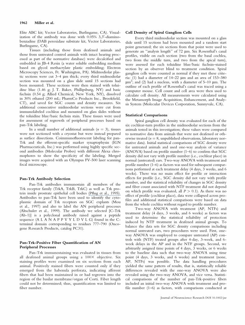
Fig. 1. A: Pan-Trk immunoperoxidase antibody labels afferent proc-esses and synaptic terminals at the bases of inner hair cells in surfacepreparations of normal guinea pig cochlea. Tissue from the secondturn of the cochlea is shown. B: Confocal analysis of coimmuno-fluorescent labeling for pan-Trk and the efferent-specific antisynapto-
physin antibody shows pan-Trk labeling of afferents (green) is distinctfrom synaptophysin labeling of efferents (red). The small portion of‘‘yellowish’’ label from the overlap of the red and green labels mayindicate the regions where efferent neurons make synaptic contactwith afferent neurons.
Fig. 2. Significant scar formation was evident in the region that nor-mally has hair cells (see asterisks, A and B) in deafened control animals(A: 4 days deaf, then artificial perilymph; B: 6 weeks deaf, then artificialperilymph). No pan-Trk-immunostained processes extended out of thehabenula perforata in tissues from deafened control animals (A and B; seearrows). BDNF/FGF1 treatment resulted in one or more pan-Trk-im-
munostained processes. Pan-Trk-immunolabeled processes can be seenextending through the habenula perforata (see arrows) and into the scarregion (see asterisks) in animals that received BDNF/FGF1 treatment 4days after deafening (C) and in animals whose treatment was delayedlonger (D: 6 weeks postdeafening).
Delayed Treatment Rescues Neurons 1963
Journal of Neuroscience Research DOI 10.1002/jnr

RESULTS
SGC Peripheral Processes
Pan-Trk Selectively Labels Afferent Neurons.The appearance of pan-Trk immunoperoxidase labelingof a surface preparation is shown in Figure 1A. Thelabeling is of afferent processes and terminals at the basesof inner hair cells in the second turn of the cochlea froma normal hearing guinea pig. Coimmunofluorescentlabeling for pan-Trk and the efferent-specific marker syn-aptophysin shows that pan-Trk labeling of afferents(green) is distinct from synaptophysin labeling of effer-ents, in red (Fig. 1B), with perhaps a little portion of‘‘yellowish’’ label from the overlap where efferents makesynaptic contact with afferents.
Pan-Trk-Positive Fiber Count. Pan-Trk-im-munostained processes extending out of the habenula per-forata were relatively rare in deafened control animalsreceiving AP infusion, and when observed they neverextended very far past the habenula. One or more pan-Trk-immunostained processes were found in almost allBDNF/FGF1-treated animals (see Fig. 2). Processes inanimals treated with NTFs extended for variable distancesinto the scar region of lost hair cells. Tissues from animalsthat received BDNF/FGF1 after the shortest period ofdeafness (4 days) generally contained more labeled fibersthan did those receiving delayed treatment (i.e., treatment
initiated after 3 or 6 weeks of deafness; see Fig. 3); how-ever, the fibers in the delayed-treatment animals often hadthe longest regrowth in the scar regions.
The increase in pan-Trk-positive SGC peripheralprocesses (fibers) in both the 4-day- (immediate) anddelayed-treatment groups (i.e., Fig. 3) was statisticallysignificant. Two-way ANOVA revealed a statisticallysignificant (P � 0.001) increase in peripheral processes inthe NTF-treated group compared to its AP control (F ¼73.835, df ¼ 1,213; P < 0.001). An overall effect oftreatment delay was also observed (F ¼ 17.564, df ¼2,213; P < 0.001), with greater numbers of fibersobserved after shorter delays. Pairwise comparisons, con-ducted using the Tukey test, revealed that NTF treat-ment reliably increased the number of fibers observedover AP control material regardless of treatment delay(all P < 0.005). There was a reliable interaction betweentreatment and treatment delay (F ¼ 3.582, df ¼ 2,213; P< 0.05). Pairwise comparisons revealed that animals inthe deafened control group, treated with AP, showed animmediate and substantial decrease in pan-Trk-immuno-labeled fiber count (4 days vs. 3 weeks, P < 0.05), withno further loss associated with a 6-week delay (3 weeksvs. 6 weeks, P > 0.05). However, with NTF treatment,the number of fibers decreased from 4 days to 3 weeksto 6 weeks (all P < 0.05).
Fig. 3. Treatment with a combination of NTFs (BDNF þ FGF1)increased pan-Trk-positive spiral ganglion peripheral process (fiber)count relative to the artificial perilymph (AP) control, even withdelayed treatment (all data are means 6 SEs). Pan-Trk-positive fibercount did not vary with cochlear position; therefore, data from acrossthe cochlea were collapsed. *Statistically significant differences ofAP–NTF comparisons within each time point (all P < 0.005). Anoverall effect-of-treatment delay was also observed, with a greaternumber of fibers counted after shorter delays. Animals in the controlgroup, treated with AP, showed an immediate and substantialdecrease in fiber count (4 days vs. 3 weeks, P < 0.05), with no fur-ther loss associated with a 6-week delay (3 weeks vs. 6 weeks, P >0.05). With NTF treatment, the number of fibers decreased from 4days to 3 weeks to 6 weeks (all P < 0.05).
Fig. 4. Representative paramodiolar plastic sections from four condi-tions (groups) with profiles through Rosenthal’s canal and the organof Corti from first (basal) turn. Complete loss of hair cells occurredin all four conditions, with scar tissue replacing the lost hair cells (seesolid black arrows), consistent with all groups being deafened in anequivalent fashion prior to differential treatments. There was variabil-ity in the number of remaining spiral ganglion cells among the fourtreatment conditions. In deafened but untreated control animals (A: 4days deaf, then artificial perilymph; B: 6 weeks deaf, then artificialperilymph), the number of spiral ganglion cells per profile was greatlyreduced. The spiral ganglion cells evident in these tissues alsoappeared smaller (see white arrows, A and B). In place of the missingcells, significant vacuole formation was evident (see asterisks, A andB). Treatment with neurotrophic factors increased spiral ganglion cellsurvival, resulting in more spiral ganglion cells remaining in the pro-files through Rosenthal’s canal. The spiral ganglion cells increased innumber and appeared larger in animals treated with BDNF/FGF1 4days (C) and 6 weeks (D) following deafening.
1964 Miller et al.
Journal of Neuroscience Research DOI 10.1002/jnr

Cell Density of Spiral Ganglion Cells
Representative images of spiral ganglion cells areshown in Figure 4. Treatment with a combination ofNTFs (BDNF þ FGF1) increased spiral ganglion cell den-sity relative to AP control, even with delayed treatment(see Fig. 5). Generally, the SGCs in cochleas with greatersurvival also had a better appearance; they were larger,rounder, without vacuolization, and with surroundingmyelin closely associated. Two-way ANOVA revealed astatistically significant (P � 0.001) enhancement of SGCdensity in the NTF-treated group compared to its APcontrol (F ¼ 404.669, df ¼ 1,216; P < 0.001). An overalleffect of treatment delay was also observed (F ¼ 61.491, df¼ 2,216; P < 0.001), with greater density of fibersobserved after shorter delays. Pairwise comparisons, con-ducted using the Tukey test, revealed that NTF treatmentreliably increased SGC density regardless of treatmentdelay (all P < 0.001). There was a reliable interactionbetween treatment and treatment delay (F ¼ 32.070, df ¼2,216; P < 0.001). Moreover, pairwise comparisonsrevealed that animals in the deafened control group,treated with AP, showed an immediate and substantialdecrease in SGC density (4 days vs. 3 weeks, P < 0.05),with no further loss associated with a 6-week delay (3
weeks vs. 6 weeks, P > 0.05). With NTF treatment, SGCdensity did not decrease as treatment delay increased from4 days to 3 weeks (P > 0.05); however, a decrease in SGCdensity observed with further delay (3 weeks vs. 6 weeks)in the NTF treatment groups was statistically reliable (P <0.001). Additional comparisons revealed that increasedSGC density in 4-day- or 3-week-delayed NTF groupscompared with SGC density in untreated normal ears wasstatistically reliable (all P < 0.01); decreased SGC densityin the AP groups and the 6-week-delayed NTF groupcompared with SGC density in untreated normal ears wasalso reliable (all P < 0.01).
DISCUSSION
Enhancement of SGC Processes, Even withDelayed Treatment
BDNF and FGF1 reliably enhanced the number ofSGC peripheral processes, with significantly greatercounts of pan-Trk-positive processes emerging from thehabenula perforata when treatment was initiated 4 days,3 weeks, or 6 weeks postdeafening. Pan-Trk labelingwas selective for AN processes, distinguishing AN proc-esses from olivocochlear efferent fibers, which were notlabeled by pan-Trk (i.e., Fig. 1B). We found the greatestnumber of pan-Trk-positive fibers with treatmentdelayed only 4 days following deafness. With 3-week-delayed treatment there was still a large increase, fivefoldover the labeling seen in the AP control animals. Therewas a twofold increase over AP control levels in the ani-mals with 6-week-delayed treatment with BDNF andFGF1, but the total number was greatly reduced fromthe 4-day and 3-week delays. This may suggest that at 6weeks the deafferented SGCs had reduced responsivenessto the NTFs. Alternatively, the reduced number ofregrown neurites may reflect that the NTFs have fewersurviving SGCs to affect. Taken together, the combina-tion of BDNF þ FGF1 was effective, even when deaf-ness-induced degeneration had reduced the normal SGCpopulation by more than 40% after 6 weeks of deafness.These results have significant clinical implications, sug-gesting that even with delayed intervention, NTFs deliv-ered at the time of implant will preserve SGCs in thecochlea and initiate regrowth of peripheral processes.
Maintenance Versus Regrowth
A small number of pan-Trk-immunolabeled SGCfibers were present in many control animals in whichAP delivery was initiated 4 days postdeafening andassessment was 40 days following deafening. This sug-gests that a small percentage of SGC peripheral processeshave a slower rate of degeneration, just as SGCs degen-erate at different rates. It is possible that some small por-tion of the increased number of pan-Trk-immunolabeledfibers seen with BDNF and FGF1 treatment could reflectmaintenance of remaining peripheral processes, not justinduced regrowth. There were few pan-Trk-immunola-beled fibers in the control group with longer delays andassessment at longer times after deafness. This suggests
Fig. 5. Treatment with a combination of NTFs (BDNF þ FGF1)increased spiral ganglion cell (SGC) density relative to artificial peril-ymph (AP) control, even with delayed treatment (all data are means6 SEs). SGC density did not vary with cochlear position; therefore,data from across the cochlea were collapsed. *Statistically significantdifferences of AP–NTF comparisons within each time point (all P <0.001). An overall effect-of-treatment delay was also observed, withgreater density of fibers observed after shorter delays. Animals in thecontrol group, treated with AP, showed an early decrease in SGCdensity (4 days vs. 3 weeks, P < 0.05), with no further loss associatedwith a 6-week delay (3 weeks vs. 6 weeks, P > 0.05). With NTFtreatment, SGC density decreased later (4 days vs. 3 weeks, P >0.05; 3 weeks vs. 6 weeks, P < 0.001). SGC density in all AP andNTF groups reliably differed from normative data from 8 normal-hearing untreated animals drawn from an existing laboratory database(mean, dashed line; SE, dotted lines). Relative to that of normal-hearing animals, SGC density was increased in the 4-day- and 3-week-delayed NTF groups and was decreased in the AP groups andthe 6-week-delayed NTF group (all P < 0.01).
Delayed Treatment Rescues Neurons 1965
Journal of Neuroscience Research DOI 10.1002/jnr

that peripheral processes no longer remain at these latertimes and that increases predominantly reflect regrowth.
Delayed NTF Treatment and SGC Survival
The current SGC density data indicate that BDNF/FGF1 treatment is as efficacious as BDNF/CNTFAX1 isin preventing deafferentation-induced cell death. With a2-week delay in treatment, both BDNF/CNTFAX1
(Yamagata et al., 2004) and BDNF/FGF1 treatmentsenhanced SGC density by approximately 3:1. Even witha 6-week delay in treatment, both BDNF/CNTFAX1
(Yamagata et al., 2004) and BDNF/FGF1 treatmentsenhanced SGC density by approximately 2:1. Althoughin this study we found greater SGC density after BDNF/FGF treatment (11–12 cells/10,000 lm2) than afterBDNF/CNTF treatment (3–5 cells/10,000 lm2), SGCdensity ratios for treated and untreated ears were similar,as we observed greater cell density in control tissues rela-tive to that reported by Yamagata et al. (2004). Differen-ces in cell survival in deafened control animals found indifferent investigations may reflect variation in the spe-cific deafening protocol used (kanamycin/ethacrynic acidor 10% neomycin injected into the middle ear). As illus-trated in Figure 4, ‘‘surviving’’ SGCs in AP-treated con-trol animals were not completely normal in appearance,with some vacuolization, mitochondrial clumping, andnuclear displacement. These cells might not then be ca-pable of normal function. Taken together, the dataclearly demonstrate that although earlier treatment issuperior, reliable increases in measures of cell survival canbe observed even after a 6-week delay in treatment witheither BDNF/FGF1 or BDNF/CNTFAX1.
Increase in SGCs: Could There Be EndogenousPopulations of Precursor Cells?
In normal ears, average SGC density was 9.49 (6 0.6)cells/10,000 lm2 (see dashed line in Fig. 5). However,interestingly, in tissues from animals treated 4 days post-deafening, mean SGC density was 11.8 (6 0.4) cells/10,000 lm2. Similarly, in tissues from animals treated 3weeks postdeafening, mean SGC density was 10.9 (6 0.8)cells/10,000 lm2. The observed increased SGC density inNTF-treated ears was statistically significant (P < 0.01).One possible explanation for this increased SGC density isthat NTFs induced conversion of endogenous precursor orstem cells into neurons and thus greater cell density thanthat seen in tissues from normal animals. Indeed, there isnow evidence that human and guinea pig AN both havethe potential for self-renewal. Rask-Andersen et al. (2005)demonstrated that progenitor/stem cells removed to cul-ture from human modiolus divide, multiply, and form neu-rospheres that then spin off individual cells that differentiateinto mature neurons (expressing TrkB and TrkC receptors)or glial cells. They report that this process and the neuritegrowth observed in mature neurons are enhanced byNTFs. Such pluripotential cells have also been generatedfrom whole cochlea and vestibular tissue from mice (Liet al., 2003) . An alternative interpretation is that deafness
induces transdifferentiation of glia into new SGCs and thatNTFs applied at or near the time of trauma cause dediffer-entiation of SGCs to an earlier developmental state wherethey are capable of reentering the cell cycle, dividing, anddifferentiating into new (more) SGCs. The evidence sup-porting this dedifferentiation hypothesis includes the obser-vation that spiral ganglion neurons evaluated after amino-glycoside treatment resemble gestational neurons (Dodson,1997; Dodson and Mohuiddin, 2000). These may thenprovide a future mechanism for AN replacement, alongwith application of exogenous stem cells (Ito et al., 2001;Iguchi et al., 2003; Tamura et al., 2004; Hu et al., 2005;).The potential for clinical impact of increasing AN survival/regrowth is clearly increased when these results are consid-ered in combination with the demonstration of hair cellregeneration in guinea pigs (Izumikawa et al., 2005).
Combinations of Treatments
The benefits of combined treatment with NTFs andelectrical stimulation were shown by Kanzaki et al. (2002),who induced expression of GDNF in the cochlea using vi-ral vectors (delivered 5 days postdeafening) and initiatedelectrical stimulation 8 days postdeafening. Ears treatedwith GDNF (approximately 9 cells/10,000 lm2) had sig-nificantly greater SGC density than did electrically stimu-lated ears (approximately 7.5 cells/10,000 lm2); earstreated with a combination of GDNF þ electrical stimula-tion (approximately 10.5 cells/10,000 lm2) had signifi-cantly greater SGC density than that of ears treated with ei-ther GDNF or electrical stimulation alone. That the factorswere additive indicates that GDNF and electrical stimula-tion exert their protective effects through separate bio-chemical pathways; however, because the concentrationof GDNF delivered cannot be specified or demonstratedto be optimal, mechanistic interpretation of these findingsmust be made cautiously. Because the combination ofBDNF þ FGF1 described in this study (approximately 11cells/10,000 lm2) was more efficacious in supportingSGC survival than was GDNF in the studies of Kanzakiet al. (2002) and others (627–877 cells/10,000 mm2; Yli-koski et al., 1998), these results suggest BDNF þ FGF1 þelectrical stimulation may better enhance SGC survivalthan other NTFþ electrical stimulation combinations.
As reviewed by Gillespie et al. (2003), BDNFtreatment can increase nitric oxide synthase (NOS) andnitric oxide (NO) production. Combination therapy thatincludes NTFs and either NOS inhibitors or free-radicalscavengers (that bind to NO) might therefore be advan-tageous. For example, some interventions use antioxi-dants to reduce ROS associated with neurotrophin de-privation (see Satoh et al., 1999; Yamagata et al., 1999).Similar interventions are likely to be successful in theauditory system: whereas application of BDNF decreasedcisplatin-induced ROS and improved survival, with-drawal of BDNF increased ROS in auditory neurons(Gabaizadeh et al., 1997). Like NTFs, free-radical scav-engers can be delivered directly into the cochlea, usingviral vectors, for example Kawamoto et al., 2004. These
1966 Miller et al.
Journal of Neuroscience Research DOI 10.1002/jnr

agents can also be delivered locally in the cochlea, andboth systemic and local delivery of a combination ofvitamin C (ascorbic acid) and vitamin E (trolox) havebeen shown to effectively increase SGC density and pre-serve auditory function (assessed as eABR threshold) fol-lowing deafening via local intracochlear injection of theaminoglycoside neomycin (Maruyama et al., 2007). Asdescribed above for other NTF combinations, deliveringboth GDNF and vitamins C and E protected auditorythresholds significantly better than did GDNF alone(Maruyama et al., 2003). Therapies that include antioxi-dant agents may have the additional benefit of acting topreserve not only neural populations, but also hair cells(for a review, see Le Prell et al., 2007b).
Clinical Trials for Translation to Humans
Clinical trials with neurotrophic factors have in thepast been challenging because of the short in vivo half-life of NTFs, as well as poor pharmacokinetics, proteo-lytic degradation, and inability of these large polypep-tides to cross the blood–brain barrier when delivered sys-temically (for a review, see Saragovi and Gehring, 2000).However, recent improvements in drug delivery techni-ques, including, for example, gene therapy, stem-celltherapy, microencapsulation of neurotrophins (or geneti-cally engineered cells that secret neurotrophins), anddesign of small-molecule neurotrophin mimetics, all pro-vide potential solutions (for a review, see Saragovi andGehring, 2000). Moreover, in the case of the impairedinner ear, neurotrophic agents can be delivered directlyinto the cochlea during implant procedures, thus reduc-ing potential blood–brain barrier issues and attaining rel-atively high concentrations with restricted systemic sideeffects. Because many nutritional supplements (i.e., vita-mins A, C, and E) are potent free-radical scavengers thatcan be consumed orally with no adverse side effectswhen taken at recommended doses, the potential forcombination therapeutics in humans is clear.
CONCLUSIONS
This study is the first in vivo demonstration thatSGCs are capable of regrowing their peripheral processesfollowing degeneration. Moreover, regrowth is possiblein a population that has sufficiently advanced toward celldeath that more than 40% of the SGCs have beendestroyed. If the NTF intervention initiates neuritegrowth in endogenous neurons, it may be that such anintervention may also be used to stimulate neuritegrowth in exogenous (stem) cell implants, enabling inte-gration of implants into the central pathways. Aregrowth of excitable processes in the implanted ear mayenhance integration of an implant with the nervous sys-tem, reducing battery requirements, increasing frequencyseparation, and allowing more channels of informationand superior benefits to patients receiving cochlearimplants. These findings provide a basis for future studiesthat will examine if the regrowth can be directed towardspecific sites on a prosthesis.
As the criteria for implanting human patients withcochlear prostheses are expanded (National Institutes ofHealth, 1995), patients with greater residual hearing (andthus greater residual populations of apical hair cells andneurons) are receiving intracochlear devices. By combin-ing implant procedures that minimize surgical trauma (toretain these residual populations of apical hair cells andneurons; e.g., Lehnhardt, 1993; Cohen, 1997) and deliv-ering NTFs to stimulate neural proliferation toward theprosthesis, cochlear implant function and the corre-sponding ‘‘acoustic’’ sensation are likely to significantlyimprove. Consistent with this prediction, Gantz et al.implanted shorter (10-mm) intracochlear electrode arraysthat preserve intact low-frequency hearing. Electricalstimulation of the base of the cochlea combined withacoustic stimulation of the intact apical cochlea (viahearing aid use) enhanced perceptual experience forcochlear implant users (Gantz et al., 2000; 2004; Tyleret al., 2000; Gantz and Turner 2003; Turner et al.,2004). Similar results were described in a case study bySkarzynski et al. (2003). Recent data from Le Prell et al.(2006) document the potential for electrical stimulationof intact/remaining hair cell populations to producetonelike auditory sensations with electrical stimulation ofthe cochlea. With the potential that one day we will beable to induce hair cell regeneration in humans, such asthat which has now been done in guinea pigs (Izumi-kawa et al., 2005), the potential for interventions thatincrease neural survival are ever more important. Theaddition of free-radical scavengers to the treatment regi-men is likely to further improve clinical outcome giventhe positive effects on both hair cell and neural survival(Le Prell et al., 2007a; Maruyama et al., 2007). Takentogether, the ability to preserve and induce regrowth ofAN fibers is a key clinical outcome that will be increas-ingly important as mechanisms of hair cell preservationand regeneration continue to improve.
ACKNOWLEDGMENTS
These results were presented in abstract form at the26th midwinter meeting of the Association for Researchin Otolaryngology (Miller et al., 2003). Support for thisresearch was provided by the AMGEN Corporation,which generously provided the BDNF, as well as by theNational Institutes of Health (NIH-NIDCD R01DC03820 and P30 DC005188), the General MotorsCorporation, and the Ruth and Lynn Townsend Profes-sor of Communication Disorders. We are grateful toAlice Mitchell, Jong-Seung Kim, Soo Duk Lee, andLing Tong for their technical assistance.
REFERENCES
Aletsee C, Brors D, Mlynski R, Ryan AF, Dazert S. 2003. Branching of
spiral ganglion neurites is induced by focal application of fibroblast
growth factor-1. Laryngoscope 113:791–796.
Altschuler RA, Cho Y, Ylikoski J, Pirvola U, Magal E, Miller JM. 1999.
Rescue and regrowth of sensory nerves following deafferentation by
neurotrophic factors. Ann N Y Acad Sci 884:305–311.
Delayed Treatment Rescues Neurons 1967
Journal of Neuroscience Research DOI 10.1002/jnr

Brown JN, Miller JM, Altschuler RA, Nuttall AL. 1993. Osmotic pump
implant for chronic infusion of drugs into the inner ear. Hear Res
70(2):167–172.
Chong ZZ, Kang JQ, Maiese K. 2004. Essential cellular regulatory ele-
ments of oxidative stress in early and late phases of apoptosis in the cen-
tral nervous system. Antioxid Redox Signal 6:277–287.
Clopton BM, Spelman FA, Miller JM. 1980. Estimates of essential neural
elements for stimulation through a cochlear prosthesis. Ann Otol Rhinol
Laryngol Suppl 89(2 Pt 2):5–7.
Cohen NL. 1997. Cochlear implant soft surgery: fact or fantasy? Otolaryngol
Head Neck Surg 117(3 Pt 1):214–216.
Dazert S, Kim D, Luo L, Aletsee C, Garfunkel S, Maciag T, Baird A,
Ryan AF. 1998. Focal delivery of fibroblast growth factor-1 by trans-
fected cells induces spiral ganglion neurite targeting in vitro. J Cell
Physiol 177(1):123–129.
de la Monte SM, Neely TR, Cannon J, Wands JR. 2000. Oxidative stress
and hypoxia-like injury cause Alzheimer-type molecular abnormalities in
central nervous system neurons. Cell Mol Life Sci 57:1471–1481.
Dodson HC. 1997. Loss and survival of spiral ganglion neurons in the
guinea pig after intracochlear perfusion with aminoglycosides. J Neuro-
cytol 26:541–556.
Dodson HC, Mohuiddin A. 2000. Response of spiral ganglion neurones to
cochlear hair cell destruction in the guinea pig. J Neurocytol 29:525–537.
Ernfors P, Duan ML, ElShamy WM, Canlon B. 1996. Protection of
auditory neurons from aminoglycoside toxicity by neurotrophin-3. Nat
Med 2:463–467.
Firszt JB, Holden LK, Skinner MW, Tobey EA, Peterson A, Gaggl W,
Runge-Samuelson CL, Wackym PA. 2004. Recognition of speech pre-
sented at soft to loud levels by adult cochlear implant recipients of three
cochlear implant systems. Ear Hear 25:375–387.
Gabaizadeh R, Staecker H, Liu W, Van De Water TR. 1997. BDNF
protection of auditory neurons from cisplatin involves changes in intra-
cellular levels of both reactive oxygen species and glutathione. Brain
Res Mol Brain Res 50(1–2):71–78.
Gantz BJ, Rubinstein JT, Tyler RS, Teagle HF, Cohen NL, Waltzman
SB, Miyamoto RT, Kirk KI. 2000. Long-term results of cochlear
implants in children with residual hearing. Ann Otol Rhinol Laryngol
Suppl 185:33–36.
Gantz BJ, Turner C. 2004. Combining acoustic and electrical speech proc-
essing: Iowa/Nucleus hybrid implant. Acta Otolaryngol 124:344–347.
Gantz BJ, Turner CW. 2003. Combining acoustic and electrical hearing.
Laryngoscope 113:1726–1730.
Gilgun-Sherki Y, Melamed E, Offen D. 2003. Antioxidant treatment in
Alzheimer’s disease: current state. J Mol Neurosci 21(1):1–11.
Gillespie LN, Clark GM, Bartlett PF, Marzella PL. 2001. LIF is more
potent than BDNF in promoting neurite outgrowth of mammalian au-
ditory neurons in vitro. Neuroreport 12(2):275–279.
Gillespie LN, Clark GM, Bartlett PF, Marzella PL. 2003. BDNF-induced
survival of auditory neurons in vivo: Cessation of treatment leads to
accelerated loss of survival effects. J Neurosci Res 71:785–790.
Gillespie LN, Clark GM, Marzella PL. 2004. Delayed neurotrophin treat-
ment supports auditory neuron survival in deaf guinea pigs. Neurore-
port 15:1121–1125.
Hartnick CJ, Staecker H, Malgrange B, Lefebvre PP, Liu W, Moonen
G, Van De Water TR. 1996. Neurotrophic effects of BDNF and
CNTF, alone and in combination, on postnatal day 5 rat acoustic gan-
glion neurons. J Neurobiol 30(2):246–254.
Hossain WA, Morest DK. 2000. Fibroblast growth factors (FGF-1, FGF-2)
promote migration and neurite growth of mouse cochlear ganglion cells
in vitro: immunohistochemistry and antibody perturbation. J Neurosci
Res 62(1):40–55.
Hu Z, Wei D, Johansson CB, Holmstrom N, Duan M, Frisen J, Ulfendahl
M. 2005. Survival and neural differentiation of adult neural stem cells trans-
planted into the mature inner ear. Exp Cell Res 302(1):40–47.
Iguchi F, Nakagawa T, Tateya I, Kim TS, Endo T, Taniguchi Z, Naito
Y, Ito J. 2003. Trophic support of mouse inner ear by neural stem cell
transplantation. Neuroreport 14(1):77–80.
IncesuluA,Nadol JB Jr. 1998. Correlation of acoustic thresholdmeasures and spi-
ral ganglion cell survival in severe to profound sensorineural hearing loss: impli-
cations for cochlear implantation. AnnOtolRhinol Laryngol 107:906–911.
Ito J, Kojima K, Kawaguchi S. 2001. Survival of neural stem cells in the
cochlea. Acta Otolaryngol 121(2):140–142.
Izumikawa M, Minoda R, Kawamoto K, Abrashkin KA, Swiderski DL,
Dolan DF, Brough DE, Raphael Y. 2005. Auditory hair cell replace-
ment and hearing improvement by Atoh1 gene therapy in deaf mam-
mals. Nat Med 11:271–276.
Jyung RW, Miller JM, Cannon SC. 1989. Evaluation of eighth nerve in-
tegrity by the electrically evoked middle latency response. Otolaryngol
Head Neck Surg 101:670–682.
Kanzaki S, Stover T, Kawamoto K, Prieskorn DM, Altschuler RA,
Miller JM, Raphael Y. 2002. Glial cell line-derived neurotrophic factor
and chronic electrical stimulation prevent VIII cranial nerve degenera-
tion following denervation. J Comp Neurol 454:350–360.
Kawamoto K, Sha SH, Minoda R, Izumikawa M, Kuriyama H, Schacht
J, Raphael Y. 2004. Antioxidant gene therapy can protect hearing and
hair cells from ototoxicity. Mol Ther 9(2):173–181.
Kirkland RA, Franklin JL. 2003. Bax, reactive oxygen, and cytochrome c
release in neuronal apoptosis. Antioxid Redox Signal 5:589–596.
Konishi T, Kelsey E. 1973. Effect of potassium deficiency on cochlear poten-
tials and cation contents of the endolymph. Acta Otolaryngol 76:410–418.
Lalwani AK, Han JJ, Castelein CM, Carvalho GJ, Mhatre AN. 2002.
In vitro and in vivo assessment of the ability of adeno-associated virus-
brain-derived neurotrophic factor to enhance spiral ganglion cell sur-
vival following ototoxic insult. Laryngoscope 112:1325–1334.
Le HN, Frim DM. 2002. Gene therapy for Parkinson’s disease. Expert
Opin Biol Ther 2(2):151–161.
Le Prell CG, Hughes LF, Miller JM. 2007a. Free radical scavengers, vita-
mins A, C, and E, plus magnesium reduces noise trauma. Free Rad
Biol Med 42:1454–1463.
Le Prell CG, Kawamoto K, Raphael Y, Dolan DF. 2006. Electromotile
hearing: Acoustic tones mask psychophysical response to high-fre-
quency electrical stimulation of intact guinea pig cochleae. J Acoust Soc
Am 120:3889–3900.
Le Prell CG, Yagi M, Kawamoto K, Beyer LA, Atkin G, Raphael Y,
Dolan D, Bledsoe SC Jr, Moody DB. 2004. Chronic excitotoxicity in
the guinea pig cochlea induces temporary functional deficits and long-
term morphological trauma. J Acoust Soc Am 116:1044–1056.
Le Prell CG, Yamashita D, Minami S, Yamasoba T, Miller JM. 2007b.
Mechanisms of noise-induced hearing loss indicate multiple methods of
prevention. Hear Res 226(1–2):22–43.
Lefebvre PP, Malgrange B, Staecker H, Moghadass M, Van De Water
TR, Moonen G. 1994. Neurotrophins affect survival and neuritogenesis
by adult injured auditory neurons in vitro. Neuroreport 5:865–868.
Lehnhardt E. 1993. [Intracochlear placement of cochlear implant electro-
des in soft surgery technique]. HNO 41:356–359.
Lewen A, Matz P, Chan PH. 2000. Free radical pathways in CNS injury.
J Neurotrauma 17:871–890.
Li H, Liu H, Heller S. 2003. Pluripotent stem cells from the adult mouse
inner ear. Nat Med 9:1293–1299.
Malgrange B, Lefebvre P, Van De Water TR, Staecker H, Moonen G.
1996. Effects of neurotrophins on early auditory neurones in cell cul-
ture. Neuroreport 7:913–917.
Maruyama J, Mannstrom P, Bredberg G, Miller JM, Ulfendahl M. 2003.
Effects of combination treatment with antioxidants and GDNF on auditory
function in deafened guinea pigs. Abs Assoc Res Otolaryngol 26:195.
Maruyama J, Yamagata T, Ulfendahl M, Bredberg G, Altschuler RA,
Miller JM. 2007. Effects of antioxidants on auditory nerve function and
survival in deafened guinea pigs. Neurobiol Dis 25:309–318.
1968 Miller et al.
Journal of Neuroscience Research DOI 10.1002/jnr
Mattson MP. 1998. Neuroprotective strategies based on targeting of post-
recptor signaling events. In: Mattson MP, editor. Neuroprotective sig-
nal transduction. Totowa, NJ: Humana Press. p 301–335,345.
Mattson MP, Scheff SW. 1994. Endogenous neuroprotection factors and
traumatic brain injury: mechanisms of action and implications for ther-
apy. J Neurotrauma 11(1):3–33.
McFadden SL, Ding D, Jiang H, Salvi RJ. 2004. Time course of efferent
fiber and spiral ganglion cell degeneration following complete hair cell
loss in the chinchilla. Brain Res 997(1):40–51.
Miller JM, Chi DH, O’Keeffe LJ, Kruszka P, Raphael Y, Altschuler RA.
1997. Neurotrophins can enhance spiral ganglion cell survival after
inner hair cell loss. Int J Dev Neurosci 15:631–643.
Miller JM, Prieskorn DM, Wys NL, Altschuler RA. 2003. Delayed neu-
rotrophin treatment following deafness rescues spiral ganglion cells from
death and promotes regrowth of auditory nerve peripheral processes.
Abs Assoc Res Otolaryngol 26:248–249.
Mou K, Hunsberger CL, Cleary JM, Davis RL. 1997. Synergistic effects
of BDNF and NT-3 on postnatal spiral ganglion neurons. J Comp
Neurol 386:529–539.
Nadol JB Jr, Young YS, Glynn RJ. 1989. Survival of spiral ganglion cells
in profound sensorineural hearing loss: implications for cochlear implan-
tation. Ann Otol Rhinol Laryngol 98:411–416.
Nakaizumi T, Kawamoto K, Minoda R, Raphael Y. 2004. Adenovirus-
mediated expression of brain-derived neurotrophic factor protects spiral
ganglion neurons from ototoxic damage. Audiol Neurootol 9(3):135–143.
National Institutes of Health, 1995. Cochlear implants in adults and chil-
dren. NIH Consens Statement 13(2):1–30.
Pflug BR, Dionne C, Kaplan DR, Lynch J, Djakiew D. 1995. Expres-
sion of a Trk high affinity nerve growth factor receptor in the human
prostate. Endocrinology 136(1):262–268.
Prieskorn DM, Miller JM. 2000. Technical report: chronic and acute
intracochlear infusion in rodents. Hear Res 140(1–2):212–215.
Puel JL, Saffiedine S, Gervais d’Aldin C, Eybalin M, Pujol R. 1995. Syn-
aptic regeneration and functional recovery after excitotoxic injury in
the guinea pig cochlea. C R Acad Sci III 318(1):67–75.
Rask-Andersen H, Bostrom M, Gerdin B, Kinnefors A, Nyberg G,
Engstrand T, Miller JM, Lindholm D. 2005. Regeneration of human audi-
tory nerve. In vitro/in video demonstration of neural progenitor cells in
adult human and guinea pig spiral ganglion. Hear Res 203:180–191.
Rask-Andersen H, Ekvall L, Scholtz A, Schrott-Fischer A. 2000. Struc-
tural/audiometric correlations in a human inner ear with noise-induced
hearing loss. Hear Res 141(1–2):129–139.
Saragovi HU, Gehring K. 2000. Development of pharmacological agents
for targeting neurotrophins and their receptors. Trends Pharmacol Sci
21(3):93–98.
Satoh T, Yamagata T, Ishikawa Y, Yamada M, Uchiyama Y, Hatanaka
H. 1999. Regulation of reactive oxygen species by nerve growth factor
but not Bcl-2 as a novel mechanism of protection of PC12 cells from
superoxide anion-induced death. J Biochem (Tokyo) 125:952–959.
Schindler RA, Gladstone HB, Scott N, Hradek GT, Williams H, Shah
SB. 1995. Enhanced preservation of the auditory nerve following coch-
lear perfusion with nerve growth factors. Am J Otol 16:304–309.
Shah SB, Gladstone HB, Williams H, Hradek GT, Schindler RA. 1995. An
extended study: protective effects of nerve growth factor in neomycin-
induced auditory neural degeneration. Am J Otol 16:310–314.
Shinohara T, Bredberg G, Ulfendahl M, Pyykko I, Olivius NP, Kaksonen R,
Lindstrom B, Altschuler R, Miller JM. 2002. Neurotrophic factor inter-
vention restores auditory function in deafened animals. Proc Natl Acad
Sci U S A 99:1657–1660.
Shoji F, Yamasoba T,Magal E,DolanDF, AltschulerRA,Miller JM. 2000. Glial
cell line-derived neurotrophic factor has a dose dependent influence on noise-
induced hearing loss in the guinea pig cochlea. HearRes 142(1–2):41–55.
Skarzynski H, Lorens A, Piotrowska A. 2003. A new method of partial
deafness treatment. Med Sci Monit 9(4):CS20–CS24.
Skinner MW, Clark GM, Whitford LA, Seligman PM, Staller SJ, Shipp
DB, Shallop JK, Everingham C, Menapace CM, Arndt PL, et al., 1994.
Evaluation of a new spectral peak coding strategy for the Nucleus 22
Channel Cochlear Implant System. Am J Otol 15(Suppl 2):15–27.
Skinner MW, Holden LK, Holden TA, Demorest ME, Fourakis MS.
1997. Speech recognition at simulated soft, conversational, and raised-
to-loud vocal efforts by adults with cochlear implants. J Acoust Soc Am
101:3766–3782.
Skinner MW, Ketten DR, Holden LK, Harding GW, Smith PG, Gates
GA, Neely JG, Kletzker GR, Brunsden B, Blocker B. 2002. CT-
derived estimation of cochlear morphology and electrode array position
in relation to word recognition in Nucleus-22 recipients. J Assoc Res
Otolaryngol 3:332–350.
Spoendlin H. 1971. Primary structural changes in the organ of Corti after
acoustic overstimulation. Acta Otolaryngol 71(2):166–176.
Spoendlin H, Brun JP. 1973. Relation of structural damage to exposure
time and intensity in acoustic trauma. Acta Otolaryngol 75(2):220–226.
Staecker H, Galinovic-Schwartz V, Liu W, Lefebvre P, Kopke R,
Malgrange B, Moonen G, Van De Water TR. 1996a. The role of the
neurotrophins in maturation and maintenance of postnatal auditory
innervation. Am J Otol 17:486–492.
Staecker H, Kopke R, Malgrange B, Lefebvre P, Van De Water TR.
1996b. NT-3 and/or BDNF therapy prevents loss of auditory neurons
following loss of hair cells. Neuroreport 7:889–894.
Summers WK. 2004. Alzheimer’s disease, oxidative injury, and cytokines.
J Alzheimers Dis 6:651–657; discussion 673–681.
Tamura T, Nakagawa T, Iguchi F, Tateya I, Endo T, Kim TS, Dong Y,
Kita T, Kojima K, Naito Y, Omori K, Ito J. 2004. Transplantation of
neural stem cells into the modiolus of mouse cochleae injured by cis-
platin. Acta Otolaryngol Suppl 551:65–68.
Tenenbaum L, Chtarto A, Lehtonen E, Blum D, Baekelandt V, Velu T,
Brotchi J, Levivier M. 2002. Neuroprotective gene therapy for Parkin-
son’s disease. Curr Gene Ther 2:451–483.
Thomas M, Le WD. 2004. Minocycline: neuroprotective mechanisms in
Parkinson’s disease. Curr Pharm Des 10:679–686.
Turner CW, Gantz BJ, Vidal C, Behrens A, Henry BA. 2004. Speech
recognition in noise for cochlear implant listeners: benefits of residual
acoustic hearing. J Acoust Soc Am 115:1729–1735.
Tyler RS, Kelsay DM, Teagle HF, Rubinstein JT, Gantz BJ, Christ AM.
2000. 7-year speech perception results and the effects of age, residual
hearing and preimplant speech perception in prelingually deaf children
using the Nucleus and Clarion cochlear implants. Adv Otorhinol-
aryngol 57:305–310.
Waltzman SB, Cohen NL, Gomolin RH, Green JE, Shapiro WH, Hoff-
man RA, Roland JT Jr. 1997. Open-set speech perception in congeni-
tally deaf children using cochlear implants. Am J Otol 18:342–349.
Webster M, Webster DB. 1981. Spiral ganglion neuron loss following
organ of Corti loss: a quantitative study. Brain Res 212(1):17–30.
West BA, Brummett RE, Himes DL. 1973. Interaction of kanamycin
and ethacrynic acid. Severe cochlear damage in guinea pigs. Arch Oto-
laryngol 98(1):32–37.
Yamagata T, Miller JM, Ulfendahl M, Olivius NP, Altschuler RA,
Pyykko I, Bredberg G. 2004. Delayed neurotrophic treatment preserves
nerve survival and electrophysiological responsiveness in neomycin-
deafened guinea pigs. J Neurosci Res 78(1):75–86.
Yamagata T, Satoh T, Ishikawa Y, Nakatani A, Yamada M, Ikeuchi T,
Hatanaka H. 1999. Brain-derived neurotropic factor prevents super-
oxide anion-induced death of PC12h cells stably expressing TrkB re-
ceptor via modulation of reactive oxygen species. Neurosci Res
35(1):9–17.
Ylikoski J, Pirvola U, Virkkala J, Suvanto P, Liang XQ, Magal E, Altschuler
R, Miller JM, Saarma M. 1998. Guinea pig auditory neurons are pro-
tected by glial cell line-derived growth factor from degeneration after
noise trauma. Hear Res 124(1–2):17–26.
Delayed Treatment Rescues Neurons 1969
Journal of Neuroscience Research DOI 10.1002/jnr