Cyclodextrin Inclusion Complexes with Antibiotics and ... - MDPI
Cno
Na
b
c
d
a
ARRAA
KCSCNIC
1
whatcbeTs
TT
(
h0
Colloids and Surfaces B: Biointerfaces 129 (2015) 30–38
Contents lists available at ScienceDirect
Colloids and Surfaces B: Biointerfaces
jo ur nal ho me p ag e: www.elsev ier .com/ locate /co lsur fb
yclodextrin-based star polymers as a versatile platform foranochemotherapeutics: Enhanced entrapment and uptakef idarubicin
. Nafeea,d,∗, M. Hirosuec, B. Loretzb, G. Wenzc, C.-M. Lehra,b
Department of Pharmacy, Biopharmaceutics and Pharmaceutical Technology, Saarland University, 66123 Saarbrücken, GermanyDepartment of Drug Delivery, Helmholtz Institute for Pharmaceutical Research Saarland (HIPS), 66123 Saarbrücken, GermanyOrganic Macromolecular Chemistry, Saarland University, 66123 Saarbrücken, GermanyDepartment of Pharmaceutics, Faculty of Pharmacy, Alexandria University, 21521 Alexandria, Egypt
r t i c l e i n f o
rticle history:eceived 26 November 2014eceived in revised form 22 February 2015ccepted 3 March 2015vailable online 10 March 2015
eywords:yclodextrintar copolymerhitosananoparticles
a b s t r a c t
A series of cyclodextrin-based star polymers were synthesized using �-cyclodextrin (CD) as hydrophiliccore, methyl methacrylate (MMA) and tert-butyl acrylate (tBA) as hydrophobic arms. Star polymers, eitherhomopolymers or random/block copolymers, showed narrow molecular weight distributions. Graftinghydrophobic arms created CD-based nanoparticles (CD-NPs) in the size range (130–200 nm) with narrowPdI <0.15 and slightly negative �-potential. Particle surface could be modified with chitosan to impart apositive surface charge. Colloidal stability of CD-NPs was a function of pH as revealed by the pH-titrationcurves. CD-NPs were used as carrier for the chemotherapeutic drug idarubicin (encapsulation efficiency,EE ∼40%) ensuring prolonged release profile (∼80% after 48 h). For cell-based studies, coumarin-6 wasencapsulated as a fluorescent marker (EE ∼75%). Uptake studies carried out on A549 and Caco-2 celllines proved the uptake of coumarin-loaded NPs as a function of time and preferential localization in the
darubicinhemotherapy
cytoplasm. Uptake kinetics revealed no saturation or plateau over 6 h. Chitosan-modified NPs showed sig-nificantly improved, concentration-dependent cellular uptake. Meanwhile, CD-NPs were non-cytotoxicon both cell lines over the concentration range (0.25–3 mg/ml) as studied by MTT and LDH assays. Inconclusion, CD star polymers can be considered a versatile platform for a new class of biocompatiblenanochemotherapy.
© 2015 Elsevier B.V. All rights reserved.
. Introduction
Cyclodextrins (CDs) are cone-shaped, cyclic oligosaccharidesith a hydrophilic outer surface and a lipophilic central cavity. CDsave a wide range of applications in different fields of drug deliverynd pharmaceutical industry [1–3]. Due to their unique steric struc-ure of a truncated cone and their complexation ability, CDs areommonly applied to enhance the solubility, stability, safety andioavailability of various drug moieties. Nowadays, CD-based deliv-
ry systems represent an emergent era for cancer research [4,5].heir long-term biocompatibility, low toxicity and lack of immunetimulation are well-defined in humans making their use as the∗ Corresponding author at: Department of Biopharmaceutics and Pharmaceuticalechnology, Saarland University, Campus A4-1, D-66123 Saarbrücken, Germany.el.: +49 681 302 4764; fax: +49 681 302 4677.
E-mail addresses: [email protected], [email protected]. Nafee).
ttp://dx.doi.org/10.1016/j.colsurfb.2015.03.014927-7765/© 2015 Elsevier B.V. All rights reserved.
building blocks for nanoparticulate carriers of interest [1]. How-ever, their relative hydrophilicity limited such an impact. A possibleapproach would be grafting of hydrophobic moieties to enablenanoparticle assembly. Literature data dealing with the synthesisof amphiphilic derivatives of CDs pointed out distinct properties[6]. Grafting alkyl chains [7], cholesterol or phospholipidyl unit onCD [8] enabled self assembly into liposomes, vesicles or micelles[9]. However, some suffer from poor chemical or physical stabilityand were prone to aggregation in certain media [10]. While cer-tain lipophilic CD derivatives (e.g. methylated �-CD) have shownto be toxic after oral and parenteral administration, others like 2-hydroxy propyl-�-CD and sulfo-butyl ether �-CD were consideredto be safe parenterally [1].
Meanwhile, star-shaped polymers have attracted considerableattention as drug carriers due to their dense molecular architec-
ture with moderate flexibility [6]. The 21-substitutable hydroxylgroups on the peripheral surface of �-CD provide the possibil-ity to produce functionalized �-CDs like star-shaped polymers. Avariety of chemical approaches have been exploited for grafting
ces B:
htTmtmtgcmc[
atat
ttaanstdcibmaatnrimwicc
smswdlat
2
2
CbfMG(foE
N. Nafee et al. / Colloids and Surfa
ydrophobic arms on CD rim including nucleophilic substitu-ion ‘click chemistry’ [11], Atherton–Todd reaction [12] and Atomransfer Radical Polymerization (ATRP) [6]. For detailed reactionechanisms, refer to Kamigaito et al. [13]. Recent studies reported
he synthesis of CD-star polymers using N,N-dimethylaminoethylethacrylate (DMA) and 1,4,7,10-tetraazacyclododecane-1,4,7,10-
etraacetic acid-functionalized gadolinium (DOTA-Gd), for bothene delivery and magnetic resonance imaging [14]. The sameopolymer was conjugated with doxorubicin and folic acid formingicellar nanoparticles. These particles were reported to be non-
ytotoxic and ensured pH-dependent release of the drug over 42 h15].
The suitability of ATRP to produce graft/block/highly branchednd star polymers from various kinds of monomers is restricted byhe significant toxicity of residual catalyst [16]. Instead of Ni cat-lyst, NiBr2 with tris(4-methoxyphenyl) phosphine as ligand washus used as a safer alternative [17].
Despite the huge efforts in cancer treatment, the best therapeu-ic strategy is not found yet. Chemotherapy remains the first linereatment regimens of cancer. One of the most potent chemother-peutic anthracyclines is idarubicin (IDA), approved for use against
wide spectrum of tumors namely acute and chronic myeloge-ous leukemia, acute lymphoblastic leukemia and myelodysplasticyndrome [18]. IDA is a semisynthetic analog of daunorubicinhat acts as topoisomerase II poisons, inducing DNA damage byisrupting the cleavage-religation equilibrium and increasing theoncentration of DNA topoisomerase II covalent complexes lead-ng to apoptosis [19]. IDA is reported to be 5–10 times more potentut less cardiotoxic than daunorubicin. Both IDA and its lipophilicetabolite daunorubicinole are considered unique BBB-crossing
nthracyclines [20]. However, IDA shares the same side effectsssociated with other anthracyclines including multiple drug resis-ance, secondary acute myelogenous leukemia, cardiotoxicity andeutropenia. Promising approaches to encapsulate IDA in nanocar-iers for better therapeutic efficacy and lower toxicological effectsnclude IDA liposomal formulations. Unfortunately, unlike liposo-
al doxorubicin (Doxil®) and daunorubicin (DaunoXome®), IDAas found to rapidly leak from cholesterol-containing liposomes
nto the blood stream [20–22]. Leakage can be attributed toholesterol–IDA interactions enforced by the presence of negativelyharged lipids, leading to drug transport across the bilayer.
In the present study, �-CDs were used as core for the synthe-is of star polymers with hydrophobic arms consisting of methylethacrylate (MMA) and tert-butyl acrylate (tBA) via ATRP. Star-
haped polymers were used for nanoparticle preparation loadedith the antineoplastic IDA to ensure controlled release, improvedelivery and safety. In this context, the in vitro release profile, bio-
ogical interaction with different cancer cell lines (e.g. cytotoxicityspects, intracellular uptake as well as uptake kinetics) were inves-igated.
. Experimental
.1. Materials
�-Cyclodextrin (CAVAMAX®W7) was obtained from Wackerhemie AG (Stuttgart, Germany). Methyl methacrylate (99%), tert-utyl acrylate (98%) and idarubicin hydrochloride were receivedrom Sigma–Aldrich (Munich, Germany). Polyvinyl alcohol (PVA)
owiol 4-88 from Kuraray Specialties Europe GmbH (Frankfurt,
ermany) and ultrapure chitosan chloride: Protasan®UP-CL113molecular weight 50–150 kDa, degree of deacetylation 75–90%)rom NovaMatrix (FMC BioPolymer AS, Oslo, Norway) were used asbtained. Coumarin-6 (Cou-6) was purchased from Polysciences,urope GmbH (Eppelheim, Germany).
Biointerfaces 129 (2015) 30–38 31
2.2. Methods
2.2.1. Polymer synthesisCD star polymers were synthesized via ATRP approach as
described in the supplementary materials.
2.2.2. Polymer characterizationPolymers were characterized in terms of molecular weight,
residual catalyst and chemical structure. Please refer to supplemen-tary materials for detailed description.
2.2.3. Preparation of nanoparticles2.2.3.1. Plain CD-NPs. MMA-�-CD grafted polymer and MMA-tBA-�-CD (random/block) copolymers were used for nanoparticlepreparation by the emulsion-diffusion technique [23]. Briefly, thepolymer (0.1% w/v) dissolved in ethyl acetate was dropped in aque-ous PVA solution (1% w/v) during homogenization at 14,000 rpm(Ultra Turrax® Ika Ltd, Taquara, Brasil) for 15 min. Afterwards,deionized water was added and allowed to stir overnight at roomtemperature to evaporate the organic solvent.
The effect of concentration, emulsifier type and homogenizationspeed on the colloidal properties was evaluated. In addition, surfacecharge modification of selected NPs was accomplished by addingchitosan (0.3% w/v) to the aqueous phase. Uncoated and chitosan-coated NPs were denoted CD4− and CD4+, respectively.
Trials to prepare NPs using �-CD alone (CD-0) were carried outfollowing the same procedure.
2.2.3.2. IDA-loaded NPs. NPs were prepared after precipitation ofIDA base using triethanolamine followed by its extraction withethyl acetate [24]. IDA was added to the organic phase at a (poly-mer: IDA) weight ratio (3:1) giving a final IDA concentration(50 �g/ml) in the NP dispersion.
2.2.3.3. Coumarin-loaded NPs. Fluorescent particles were preparedby adding coumarin-6 (100 �g/ml) to the organic phase andproceed as mentioned above.
2.2.3.4. Nanoparticle purification and freeze-drying. Nanoparticleswere purified from residual polymers, idarubicin or coumarin-6 based on centrifugal ultrafiltration using Centrisart I®, MWCO300,000 (Sartorius AG, Goettingen, Germany). Purified particleswere freeze-dried (Alpha 2-4 LSC, Martin Christ Gefriertrock-nungsanlagen GmbH, Osterode, Germany) and the dried weightwas determined gravimetrically (Cubis®-Precision Balance, Sarto-rius AG, Goettingen, Germany).
2.3. Nanoparticle characterization
The colloidal properties (mean diameter; polydispersity index,PdI; and �-potential) were measured using Malvern Zetasizer NanoZS (Malvern Instruments, Malvern, UK). The colloidal stability afterpurification and freeze-drying was verified.
The surface morphology of CD-NPs was examined by scanningprobe microscopy (SPM. Digital Instruments, Veeco, Santa Barbara,CA) under ambient conditions in tapping mode.
The effect of pH on the colloidal properties of CD-NPs was inves-tigated as a function of surface charge. PH titration curves were runfor both positively and negatively charged CD-NPs at pH range 1–8using Malvern Zetasizer autotitrator unit.
2.4. Determination of the encapsulation efficiency (EE) of IDA and
Cou-6The amount of encapsulated IDA and Cou-6 was determinedindirectly in the supernatant by fluorescence (�ex 485 nm, �em
3 ces B:
5(
2
psbcD1il
2
lmcLewe
2
2
tcsmctc
atSifiS
2
adasumTNc
2
bpHn1
2 N. Nafee et al. / Colloids and Surfa
42 nm) or absorbance measurements (�max 458 nm), respectivelyTecan Deutschland GmbH, Crailsheim, Germany).
.5. In vitro drug release
The release of IDA from CD-NPs was performed in phos-hate buffer saline (PBS, 154 mM, pH 7.4) containing 0.5% w/vodium dodecyl sulphate (SDS) – to improve solubility of IDAase and ensure sink conditions – at 37 ◦C at 100 rpm. IDA-NPsontaining 0.1 mg/ml drug was transferred into a QuixSep Microialyzer (Roth, Karlsuhe, Germany) with a cellulose membrane Mw2–14 kDa (Medicell International Ltd., London, UK) and immersed
nto 100 ml PBS. At predetermined intervals, released IDA was ana-yzed fluorimetrically as specified above.
.6. Cell cultures
A549 cells (CCL-185; ATCC, Manassas, VA, USA) in RPMI with-glutamine (PAA Laboratories GmbH, Pasching, Austria) supple-ented with 10% FBS (PAN Biotech, Aidenbach, Germany) and
aco-2 cells (C2BBe1; ATCC, Manassas, VA, USA) in DMEM (PAAaboratories GmbH, Pasching, Austria) supplemented with 1% Non-ssential amino acids (Gibco, Karlsruhe, Germany) and 10% FBSere kept at 37 ◦C, 5% CO2 and 95% relative humidity. On the day of
xperiment, cells were washed with PBS and medium was changed.
.7. Cytotoxicity assays
.7.1. Viability assayMTT reduction by metabolically active cells was used to quan-
ify cell viability. A549 (1 × 10⁵ cell/ml) and Caco-2 (4 × 10⁵ cell/ml)ells, seeded 24 h before the experiment, were incubated with NPamples (0.25–3 mg/ml) for 4 h. In parallel, cells grown in cultureedium only and others incubated with Triton X (2% w/v) were
onsidered as high and low control, respectively. Substance con-rol (NPs + assay reagents) showed no interference with the assayonditions.
After incubation with NPs, cells were washed with PBSnd allowed to grow in culture medium. Next day, MTT solu-ion (3,4,5-dimethylthiazol-yl)-2,5-diphenyl tetrazolium (M5655,igma–Aldrich), 5 mg/ml in PBS was added. Following incubationn the dark for 3 h, precipitated formazan was dissolved using acidi-ed isopropanol and quantified spectrophotometrically at 550 nm.amples were studied in quadruplicates.
.7.2. LDH assayIt is based on the measurement of lactate dehydrogenase (LDH)
ctivity released from the cytosol of dead or plasma membrane-amaged cells. LDH assay was performed on the same platespplied for MTT assay. After incubation of cells with NP samples, theupernatants (100 �l) were transferred to 96-well plate. Equal vol-me of the reaction mixture was added per well. Absorbance waseasured at 492 nm and the cytotoxicity was calculated relative to
riton X as high control, and cells in culture medium as low control.o interaction of the samples with the assay procedure (substanceontrol) was detectable.
.8. Cellular uptake and uptake kinetics
A549 cells seeded on LabTec chamber slides (Nunc GmbH, Wies-aden, Germany) and caco-2 cells seeded on 24-well imaging
late with transparent bottom (Zell-kontakt GmbH, Noerten-ardenberg, Germany) were incubated with positively andegatively charged Cou-loaded NPs at concentrations 0.5 andmg/ml (n = 3) for 4 h. To study the uptake kinetics, particles
Biointerfaces 129 (2015) 30–38
were incubated with the cells for 0.5–6 h. After 24 h, cell mem-branes were stained with Rhodamine-labeled ricinus communisagglutinin I (RRCA; �ex = 552 nm, �em = 577 nm; Vector Laborato-ries Peterborough, UK), then fixed with 4% paraformaldehyde inPBS. Nuclei were then stained with 4′,6-Diamidino-2-phenylindoledihydrochloride (DAPI, �ex = 374 nm, �em = 461 nm; Fluka ChemieGmbH, Buchs, Switzerland). Fluorescence imaging was performedusing a Zeiss510LSM META NLO confocal laser scanning micro-scope, CLSM (Jena, Germany).
Confocal images were processed with “Image J” softwareto determine the fluorescence intensity. Uptake kinetics wereexpressed as integrated density (ID) versus time and were corre-lated with the percentage of fluorescent cells per image.
3. Results and discussion
3.1. Polymerization
In this study, several polymerization parameters, includingreaction temperature and reagents concentration were varied tooptimize the synthesis of well-defined star polymers. Table S1(supplementary materials) summarizes the properties of the syn-thesized polymers. Synthesis of the macroinitiator was confirmedby 1H NMR and FT-IR, Fig. S1A (supplementary materials).
Homopolymerization of MMA (MMA-�-CD grafted polymer)was performed with a starting [M]0/[I]0 of 20 (CD-1, Table S1A,supplementary materials). IR spectrum, Fig. S1B (supplementarymaterials), depicts the appearance of ester peak at 1700 cm−1
together with the disappearance of hydroxyl peak at 3250 cm−1
indicating the formation of MMA-�-CD. Molecular weight distribu-tion was well controlled (Mw/Mn = 1.51) and no shoulder peak wasobserved confirming the synthesis of star polymer via ATRP withoutany side reaction, Fig. S1C (supplementary materials). Appearanceof a shoulder at the main peak in the higher molar mass regionwould be an indication of irreversible coupling of two star poly-mers. Increasing the monomer ratio [M]0/[I]0 to 50 (CD-2) increasedthe molecular weight to 1 × 10⁵. In all cases, the monomer conver-sion was >90%.
Star copolymers: Random copolymerization of MMA and tBA atan [M]0/[I]0 ratio 20, 80 ◦C, DMSO (CD-4) showed a monomer con-version of 96% (MMA) and 89% (tBA), Table S1B (supplementarymaterials).
Block copolymerization using same MMA:tBA ratio (1:1) resultedin the formation of MMA-tBA-�-CD polymer (CD-5) with relativelybroader distributions (Mw/Mn = 1.76). In order to obtain a long armlength star polymer, [M]0/[I]0 ratio was increased to 50 (CD-6)and 100 (CD-7). An increase in molecular weight and viscosity inaddition to a decrease in polymerization speed and monomer con-version (81 and 70% for CD-6 and CD-7, respectively) was noticedwith increasing the monomer ratio, Table S1B. This suggests thepossibility of star-star coupling and the formation of highly cross-linked polymer with high viscosity.
Star-branched polymers possessing many arms are known tobehave like monomeric rigid spheres, due to big difference in seg-ment densities between the core and outside shell. A more compact,less entangled structure was obtained compared to linear analogswith same molar mass, leading to smaller sizes and lower viscosi-ties [25]. In comparison to dendrimers, star polymers have betterflexibility due to lack of connection between each generation asthat of dendrimers. Interestingly, Kakuchi et al. succeeded in syn-thesizing many-armed AB20 stars composed of either PMMA (A)or PtBMA (A) and PS (B) arms [26]. Meanwhile, well-defined 7-arm
and 21-arm poly(N-isopropylacrylamide) (PNIPAM) star polymerspossessing �-cyclodextrin (�-CD) cores were synthesized via thecombination of ATRP and click reactions for stimuli-sensitive deliv-ery purposes [27].
N. Nafee et al. / Colloids and Surfaces B: Biointerfaces 129 (2015) 30–38 33
CD-N
3
dpuhn7<r
pp0Fa
ioiatu4�F
aIfd
amsscNw
tFmp
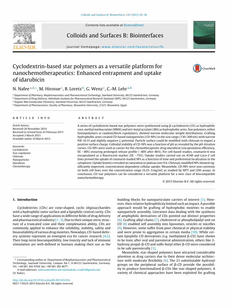
Fig. 1. (A) Effect of polymer type on the colloidal properties of
.2. Characterization of CD-nanoparticles
While CD-reference (CD-0) was not suitable to prepare well-efined NPs (PdI 0.38), grafted CD-MMA star homo-/copolymersroduced NPs in the size range 174–227 nm (Fig. 1A). Although these of iron catalyst was not favored due to purification problems,omogenous NPs were obtained, CD-3 NPs, with PdI 0.08 (resultsot shown). Particles produced using star copolymers (CD-4 to CD-) were <200 nm in diameter with narrow size distribution (PdI0.1) (Fig. 1A). Indeed, the use of random/block copolymer had noemarkable effect on the particle size.
Replacing the emulsifier PVA with Pluronic F68 (formula CD-4)roduced larger particles with higher polydispersity, Fig. S2B (sup-lementary materials). In contrast, reducing PVA concentration to.1–0.5% slightly reduced the NP size and narrowed the PdI <0.05,ig. S2C (supplementary materials). Generally, all particles acquired
weakly negative �-potential (−1 to −6 mV).Positively charged NPs would be of potential interest for load-
ng negatively charged moieties such as DNA, siRNA and antisenseligonucleotides. Polycationic amphiphilic CD derivative compris-ng a cationic cluster and multi-tail hydrophobic moiety was useds efficient vector for DNA [28]. Therefore, the ability of our par-icles to acquire a positive charge was verified. CD-4 polymer wassed to prepare chitosan-modified nanoparticles. NPs (denoted CD-+) were 217 nm in diameter, with PdI value of 0.21 and positive-potential of +23 mV, compared to non-coated formulation CD-4−,ig. S2D and E (supplementary materials).
Encapsulation of coumarin-6 slightly increased the particle sizend size distribution, Fig. S2F (supplementary materials), whereasDA-loaded NPs were ∼30 nm bigger than the corresponding plainormulation. Indeed, the surface potential did not show significantifferences from the corresponding plain particles.
Particle morphology: SPM images clearly demonstrate the poorbility of CD-reference to form nanoparticles, Fig. S3 (supple-entary materials), where we can distinguish the CD cavities
urrounded by random branched network structures. In contrast,tar-CD homo and copolymers resulted in the formation of spheri-al, well-defined nanoparticles with smooth surface (CD-2 to CD-6Ps, Fig. 1B and S3). SPM also corroborate the size ranges obtainedith the Zetasizer measurements.
Colloidal stability: Particles retained their colloidal characteris-
ics (size, PdI and �-potential) after purification and freeze-drying,ig. S4A (supplementary materials). Colloidal stability was alsoaintained upon storage at 4 ◦C up to 6 weeks, Fig. S4B&C (sup-lementary materials).
Ps (error bars represent SD, n = 3). (B) SPM image of CD-2 NPs.
3.3. pH titration curves
In order to investigate the effect of pH on negatively and posi-tively charged CD-4 NP, pH titration curves were done. Both CD-4−
and CD-4+ NPs were physically stable over a pH range from 1 to7, Fig. S5A and B (supplementary materials). Beyond this rangeaggregates of >1 �m with PdI >1 could be recognized. A very weak�-potential 0 to −2 mV was recorded for CD-4− NPs, whereas CD-4+ NPs showed a drop in �-potential from +10 to +1 mV, Fig. S5C(supplementary materials). Starting the titration by bringing themolecule to acidic pH might induce the permanent conversion oftBA to acrylic acid. The original tBA structure would not be thenrestored by gradual increase in pH. The amino group in chitosanhas a pKa value of ∼6.5, which leads to a protonation in acidicto neutral solution with a charge density dependent on pH and%deacetylation.
3.4. Entrapment efficiency
CD-4 NPs ensured an EE of 39.3 ± 0.78% for IDA giving riseto the ability of CD star polymers to entrap lipophilic candi-dates with certain possibility of the transfer of a given amountof drug toward the aqueous phase during particle preparation.The precipitation of IDA base from the hydrochloride salt defi-nitely improved drug entrapment. Numerous trials were reportedto optimize anthracycline entrapment in nanocarriers. For instance,the entrapment of IDA in liposomes was improved by addingEDTA disodium or diammonium salts forming low solubility com-plex [20]. Meanwhile, doxorubicin was solubilized in reversemicelle suspension of Span 80® in Labrafac® prior to its incor-poration into a lipid core to obtain an EE of 37–68% [29]. Bothdoxorubicin and idarubicin were allowed to form ion pair withmonoalkyl phosphate esters; this increased lipophilicity improvedtheir incorporation in solid stearic acid-based lipospheres [30].In the same context, ion pair complexes of doxorubicin withdeoxytaurocholate were better entrapped in neutral solid lipidnanoparticles (to a maximum of 43%) compared to cationic particles[31].
The encapsulation of Cou-6 in CD-4 NPs was found to be stableand promising (74.6 ± 5.03%), highlighting the ability of these CD-
nanocarriers for diagnostic and cellular trafficking purposes. In asimilar study, amphiphilic lipophosphoramidyl-CD nanoparticlesencapsulating carboxyfluorescein were prepared; the encapsula-tion efficiency was 42% [12].
34 N. Nafee et al. / Colloids and Surfaces B: Biointerfaces 129 (2015) 30–38
F = 3). (a
3
p4ffrafprScoi
mima
ccgfthmehfctc
3
cMawro
it
ig. 2. (A) Release profile of idarubicin from CD-4 NPs (error bars represent SD, nssay) (error bars represent SD, n = 4).
.5. In vitro drug release
The release of IDA from CD-4 NPs was studied in PBS. Particlesrovided sustained drug release over 48 h with an initial burst of0% within the first 2 h (Fig. 2A). The burst effect is well knownor nanoparticulate carriers and represents the drug that is sur-ace bound or adsorbed onto NPs, while, the following prolongedelease period refers to IDA perfectly encapsulated in the NP corend released by diffusion. Similar release profile was observedor IDA from surface-modified propyl-starch NPs and pH-sensitivehospholipid-based micelles at pH 6 as well as doxorubicin fromeverse-micelle loaded lipid nanocarriers [29,32,33]. According toiddiqui et al. [31], doxorubicin release from solid lipid nanoparti-les was dependent on the release medium; higher release rate wasbserved in phosphate buffer pH 7.4 > pH 6.8 > water, revealing thempact of phosphate on promoting drug release.
Our release data were subjected to different release kineticodels. The release was best fitted to Higuchi model (R2 = 0.973)
ndicating that drug diffusion from the particles is the main releaseechanism. Generally, anthracycline release from nanocarriers is
ssumed to be governed by Fickian diffusion [29,31].It has been reported that self-assembled micelles based on CD-
onjugated poly(lactic acid)-b-poly(ethylene glycol) amphiphilicopolymer ensured controlled release of indomethacin. This sug-ests that the introduction of CD provided an effective secondaryunctional unit to normal micelles, thus contributing to the con-rolled drug release with decreased initial burst [34]. On the otherand, acrylic acid derivatives including PMMA are among theost frequently used polymers for nanoparticle preparation. How-
ver, their hydrophobic characters limited the encapsulation ofydrophilic drugs. Highly structured, porous PMMA membranes
unctionalized with hydroxypropyl-�-CD were developed for theontrolled release of the anti-inflammatory, ibuprofen [35]. Dueo the hydrophilic nature of CD, the release was a function of CDontent in the membrane.
.6. Cytotoxicity assay
CD4− and CD4+ NPs were incubated with A549 and Caco-2ells (0.25–3 mg/ml). After 4 h incubation, cells were subjected toTT and LDH assays. As shown in Fig. S6A (supplementary materi-
ls), 80–100% of A549 cells retained their viability after incubationith the negatively charged nanoparticles, CD4− NPs, which was
educed to 70–80% when CD4+ NPs were used. However, viability
f Caco-2 cells remained >90% with both nanoparticles (Fig. 2B).Both types of particles had no harmful effect on cell membranentegrity of A549 and Caco-2 cells (LDH released was ±10 comparedo control), Fig. S6C and D (supplementary materials).
B) Cytotoxicity of positively and negatively charged CD-NPs on Caco-2 cells (MTT
CDs were reported to be well tolerable in Calu-3 cells andhave decreased cell viability only at high concentration [1]. Similarresults were observed for CD-based star polymers with cationic21-arms primary, tertiary and quaternary ammonium groups[36], polyamidoamine [37] and poly-l-lysine dendron arms [38].Nevertheless, toxicity of cationic CD-poly(2-(dimethylamino)ethylmethacrylate) (PDMAEMA) star polymer was increased withincreasing arm number and/or length [39].
3.7. Uptake studies
A key factor for the efficacy of chemotherapeutics is theirefficient cellular uptake. For cellular trafficking, Cou-loaded NPsprepared with MMA-tBA-�-CD star copolymer, CD-4, were incu-bated with A549 cells for 0.5–6 h. The fluorescent particles wereinitially detected on the cell membrane half an hour after incu-bation. CLSM images for longer incubation periods show distinct,well-defined green fluorescent spots in the vicinity between thered-stained cell membrane and the blue-stained nuclei (Fig. 3A),revealing the ability of the particles to be internalized. The uptakeseemed to be time-dependent, more fluorescent particles can beobserved inside the cells especially after 4 and 6 h mostly as lumpsor aggregates.
3D-stack confocal imaging and sections in the z-directiontaken after 4 and 6 h incubation proved the presence of flu-orescent particles below the apical cell membrane within thecytoplasm. Noteworthy, no persistent particle accumulation on thecell membrane was obvious, ruling out the possibility of irreversiblehydrophobic interaction between the hydrocarbon tails of CD andmembrane lipids. Release studies showed that >90% of coumarin-6 remained encapsulated within the particles for more than 6 h(the maximum incubation period with the cells) indicating that thespots examined by CLSM represent the particles not the free dye.Our uptake data are in contrast with previous studies that reportedlimited uptake of PVA-stabilized nanoparticles in various cell lines[33]. This point is further endorsed by estimating the uptake kinetics.The increase in fluorescence as a function of time was expressed asintegrated density (ID) via “Image J” software. The graph in Fig. 3Bshows the increase in ID by time, together with a simultaneousincrease in number of transfected cells. Over the study period, noplateau or saturation was noted.
CD molecules are relatively large with a number of hydrogendonors and acceptors; thus, they do not permeate lipophilicmembranes. Their inclusion in star polymers with hydrophobic
MMA and tBA improved the internalization. Such groups areable to protonate at pH 6; this “chemical sensing” mechanism isexpected to trigger a number of processes that provide escapefrom endocytotic vesicles and intracellular release of the payload.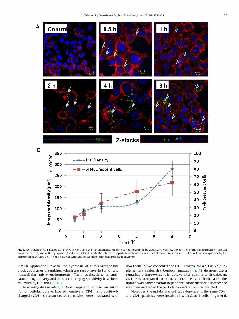
N. Nafee et al. / Colloids and Surfaces B: Biointerfaces 129 (2015) 30–38 35
F e perm articli ent SD
Sbicr
tc
ig. 3. (A) Uptake of Cou-loaded CD-4− NPs in A549 cells at different incubation timembrane (0.5 h) and in the cytoplasm (1–6 h), Z-stacks illustrate the internalized p
ncrease in integrated density and % fluorescent cells versus time (error bars repres
imilar approaches involve the synthesis of stimuli-responsivelock copolymer assemblies, which are responsive to tumor and
ntracellular micro-environments. Their applications in anti-ancer drug delivery and enhanced imaging sensitivity have been
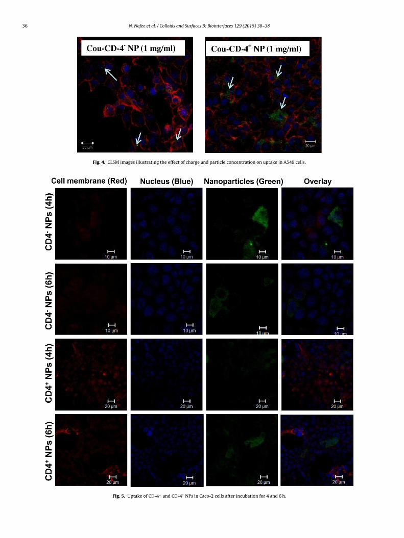
eviewed by Gui and Lui [40].To investigate the role of surface charge and particle concentra-ion on cellular uptake, labeled negatively (CD4−) and positivelyharged (CD4+, chitosan-coated) particles were incubated with
iods examined by CLSM: arrows show the position of the nanoparticles on the celles below the apical part of the cell membrane. (B) Uptake kinetics expressed by the, n = 6).
A549 cells in two concentrations 0.5, 1 mg/ml for 4 h, Fig. S7 (sup-plementary materials). Confocal images (Fig. 4), demonstrate aremarkable improvement in uptake after coating with chitosan,CD4+ NPs compared to uncoated CD4− NPs. In both cases, the
uptake was concentration dependent; more distinct fluorescencewas observed when the particle concentration was doubled.Moreover, the uptake was cell type dependent; the same CD4−
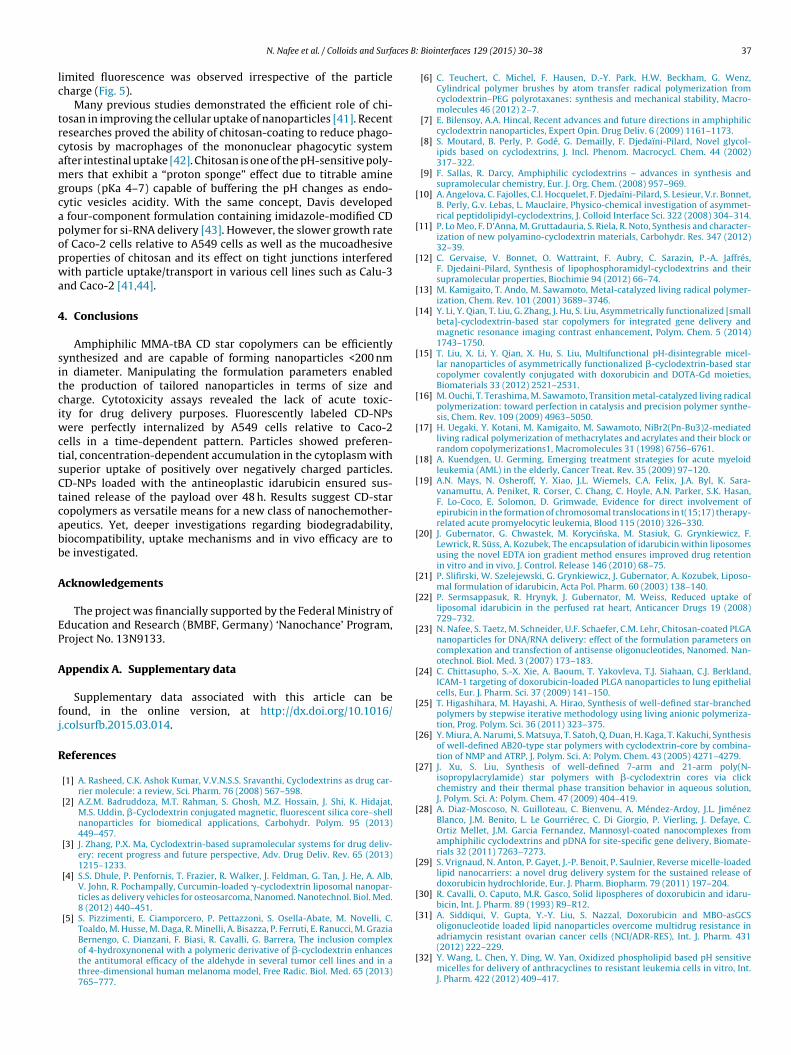
and CD4+ particles were incubated with Caco-2 cells. In general,
36 N. Nafee et al. / Colloids and Surfaces B: Biointerfaces 129 (2015) 30–38
Fig. 4. CLSM images illustrating the effect of charge and particle concentration on uptake in A549 cells.
Fig. 5. Uptake of CD-4− and CD-4+ NPs in Caco-2 cells after incubation for 4 and 6 h.
ces B:
lc
trcamgcapopwa
4
sitciwctsCtcabb
A
EP
A
fj
R
[
[
[
[
[
[
[
[
[
[
[
[
[
[
[
[
[
[
[
[
[
[
N. Nafee et al. / Colloids and Surfa
imited fluorescence was observed irrespective of the particleharge (Fig. 5).
Many previous studies demonstrated the efficient role of chi-osan in improving the cellular uptake of nanoparticles [41]. Recentesearches proved the ability of chitosan-coating to reduce phago-ytosis by macrophages of the mononuclear phagocytic systemfter intestinal uptake [42]. Chitosan is one of the pH-sensitive poly-ers that exhibit a “proton sponge” effect due to titrable amine
roups (pKa 4–7) capable of buffering the pH changes as endo-ytic vesicles acidity. With the same concept, Davis developed
four-component formulation containing imidazole-modified CDolymer for si-RNA delivery [43]. However, the slower growth ratef Caco-2 cells relative to A549 cells as well as the mucoadhesiveroperties of chitosan and its effect on tight junctions interferedith particle uptake/transport in various cell lines such as Calu-3
nd Caco-2 [41,44].
. Conclusions
Amphiphilic MMA-tBA CD star copolymers can be efficientlyynthesized and are capable of forming nanoparticles <200 nmn diameter. Manipulating the formulation parameters enabledhe production of tailored nanoparticles in terms of size andharge. Cytotoxicity assays revealed the lack of acute toxic-ty for drug delivery purposes. Fluorescently labeled CD-NPs
ere perfectly internalized by A549 cells relative to Caco-2ells in a time-dependent pattern. Particles showed preferen-ial, concentration-dependent accumulation in the cytoplasm withuperior uptake of positively over negatively charged particles.D-NPs loaded with the antineoplastic idarubicin ensured sus-ained release of the payload over 48 h. Results suggest CD-staropolymers as versatile means for a new class of nanochemother-peutics. Yet, deeper investigations regarding biodegradability,iocompatibility, uptake mechanisms and in vivo efficacy are toe investigated.
cknowledgements
The project was financially supported by the Federal Ministry ofducation and Research (BMBF, Germany) ‘Nanochance’ Program,roject No. 13N9133.
ppendix A. Supplementary data
Supplementary data associated with this article can beound, in the online version, at http://dx.doi.org/10.1016/.colsurfb.2015.03.014.
eferences
[1] A. Rasheed, C.K. Ashok Kumar, V.V.N.S.S. Sravanthi, Cyclodextrins as drug car-rier molecule: a review, Sci. Pharm. 76 (2008) 567–598.
[2] A.Z.M. Badruddoza, M.T. Rahman, S. Ghosh, M.Z. Hossain, J. Shi, K. Hidajat,M.S. Uddin, �-Cyclodextrin conjugated magnetic, fluorescent silica core–shellnanoparticles for biomedical applications, Carbohydr. Polym. 95 (2013)449–457.
[3] J. Zhang, P.X. Ma, Cyclodextrin-based supramolecular systems for drug deliv-ery: recent progress and future perspective, Adv. Drug Deliv. Rev. 65 (2013)1215–1233.
[4] S.S. Dhule, P. Penfornis, T. Frazier, R. Walker, J. Feldman, G. Tan, J. He, A. Alb,V. John, R. Pochampally, Curcumin-loaded �-cyclodextrin liposomal nanopar-ticles as delivery vehicles for osteosarcoma, Nanomed. Nanotechnol. Biol. Med.8 (2012) 440–451.
[5] S. Pizzimenti, E. Ciamporcero, P. Pettazzoni, S. Osella-Abate, M. Novelli, C.Toaldo, M. Husse, M. Daga, R. Minelli, A. Bisazza, P. Ferruti, E. Ranucci, M. Grazia
Bernengo, C. Dianzani, F. Biasi, R. Cavalli, G. Barrera, The inclusion complexof 4-hydroxynonenal with a polymeric derivative of �-cyclodextrin enhancesthe antitumoral efficacy of the aldehyde in several tumor cell lines and in athree-dimensional human melanoma model, Free Radic. Biol. Med. 65 (2013)765–777.[
Biointerfaces 129 (2015) 30–38 37
[6] C. Teuchert, C. Michel, F. Hausen, D.-Y. Park, H.W. Beckham, G. Wenz,Cylindrical polymer brushes by atom transfer radical polymerization fromcyclodextrin–PEG polyrotaxanes: synthesis and mechanical stability, Macro-molecules 46 (2012) 2–7.
[7] E. Bilensoy, A.A. Hincal, Recent advances and future directions in amphiphiliccyclodextrin nanoparticles, Expert Opin. Drug Deliv. 6 (2009) 1161–1173.
[8] S. Moutard, B. Perly, P. Godé, G. Demailly, F. Djedaïni-Pilard, Novel glycol-ipids based on cyclodextrins, J. Incl. Phenom. Macrocycl. Chem. 44 (2002)317–322.
[9] F. Sallas, R. Darcy, Amphiphilic cyclodextrins – advances in synthesis andsupramolecular chemistry, Eur. J. Org. Chem. (2008) 957–969.
10] A. Angelova, C. Fajolles, C.l. Hocquelet, F. Djedaïni-Pilard, S. Lesieur, V.r. Bonnet,B. Perly, G.v. Lebas, L. Mauclaire, Physico-chemical investigation of asymmet-rical peptidolipidyl-cyclodextrins, J. Colloid Interface Sci. 322 (2008) 304–314.
11] P. Lo Meo, F. D’Anna, M. Gruttadauria, S. Riela, R. Noto, Synthesis and character-ization of new polyamino-cyclodextrin materials, Carbohydr. Res. 347 (2012)32–39.
12] C. Gervaise, V. Bonnet, O. Wattraint, F. Aubry, C. Sarazin, P.-A. Jaffrés,F. Djedaini-Pilard, Synthesis of lipophosphoramidyl-cyclodextrins and theirsupramolecular properties, Biochimie 94 (2012) 66–74.
13] M. Kamigaito, T. Ando, M. Sawamoto, Metal-catalyzed living radical polymer-ization, Chem. Rev. 101 (2001) 3689–3746.
14] Y. Li, Y. Qian, T. Liu, G. Zhang, J. Hu, S. Liu, Asymmetrically functionalized [smallbeta]-cyclodextrin-based star copolymers for integrated gene delivery andmagnetic resonance imaging contrast enhancement, Polym. Chem. 5 (2014)1743–1750.
15] T. Liu, X. Li, Y. Qian, X. Hu, S. Liu, Multifunctional pH-disintegrable micel-lar nanoparticles of asymmetrically functionalized �-cyclodextrin-based starcopolymer covalently conjugated with doxorubicin and DOTA-Gd moieties,Biomaterials 33 (2012) 2521–2531.
16] M. Ouchi, T. Terashima, M. Sawamoto, Transition metal-catalyzed living radicalpolymerization: toward perfection in catalysis and precision polymer synthe-sis, Chem. Rev. 109 (2009) 4963–5050.
17] H. Uegaki, Y. Kotani, M. Kamigaito, M. Sawamoto, NiBr2(Pn-Bu3)2-mediatedliving radical polymerization of methacrylates and acrylates and their block orrandom copolymerizations1, Macromolecules 31 (1998) 6756–6761.
18] A. Kuendgen, U. Germing, Emerging treatment strategies for acute myeloidleukemia (AML) in the elderly, Cancer Treat. Rev. 35 (2009) 97–120.
19] A.N. Mays, N. Osheroff, Y. Xiao, J.L. Wiemels, C.A. Felix, J.A. Byl, K. Sara-vanamuttu, A. Peniket, R. Corser, C. Chang, C. Hoyle, A.N. Parker, S.K. Hasan,F. Lo-Coco, E. Solomon, D. Grimwade, Evidence for direct involvement ofepirubicin in the formation of chromosomal translocations in t(15;17) therapy-related acute promyelocytic leukemia, Blood 115 (2010) 326–330.
20] J. Gubernator, G. Chwastek, M. Korycinska, M. Stasiuk, G. Grynkiewicz, F.Lewrick, R. Süss, A. Kozubek, The encapsulation of idarubicin within liposomesusing the novel EDTA ion gradient method ensures improved drug retentionin vitro and in vivo, J. Control. Release 146 (2010) 68–75.
21] P. Slifirski, W. Szelejewski, G. Grynkiewicz, J. Gubernator, A. Kozubek, Liposo-mal formulation of idarubicin, Acta Pol. Pharm. 60 (2003) 138–140.
22] P. Sermsappasuk, R. Hrynyk, J. Gubernator, M. Weiss, Reduced uptake ofliposomal idarubicin in the perfused rat heart, Anticancer Drugs 19 (2008)729–732.
23] N. Nafee, S. Taetz, M. Schneider, U.F. Schaefer, C.M. Lehr, Chitosan-coated PLGAnanoparticles for DNA/RNA delivery: effect of the formulation parameters oncomplexation and transfection of antisense oligonucleotides, Nanomed. Nan-otechnol. Biol. Med. 3 (2007) 173–183.
24] C. Chittasupho, S.-X. Xie, A. Baoum, T. Yakovleva, T.J. Siahaan, C.J. Berkland,ICAM-1 targeting of doxorubicin-loaded PLGA nanoparticles to lung epithelialcells, Eur. J. Pharm. Sci. 37 (2009) 141–150.
25] T. Higashihara, M. Hayashi, A. Hirao, Synthesis of well-defined star-branchedpolymers by stepwise iterative methodology using living anionic polymeriza-tion, Prog. Polym. Sci. 36 (2011) 323–375.
26] Y. Miura, A. Narumi, S. Matsuya, T. Satoh, Q. Duan, H. Kaga, T. Kakuchi, Synthesisof well-defined AB20-type star polymers with cyclodextrin-core by combina-tion of NMP and ATRP, J. Polym. Sci. A: Polym. Chem. 43 (2005) 4271–4279.
27] J. Xu, S. Liu, Synthesis of well-defined 7-arm and 21-arm poly(N-isopropylacrylamide) star polymers with �-cyclodextrin cores via clickchemistry and their thermal phase transition behavior in aqueous solution,J. Polym. Sci. A: Polym. Chem. 47 (2009) 404–419.
28] A. Diaz-Moscoso, N. Guilloteau, C. Bienvenu, A. Méndez-Ardoy, J.L. JiménezBlanco, J.M. Benito, L. Le Gourriérec, C. Di Giorgio, P. Vierling, J. Defaye, C.Ortiz Mellet, J.M. Garcia Fernandez, Mannosyl-coated nanocomplexes fromamphiphilic cyclodextrins and pDNA for site-specific gene delivery, Biomate-rials 32 (2011) 7263–7273.
29] S. Vrignaud, N. Anton, P. Gayet, J.-P. Benoit, P. Saulnier, Reverse micelle-loadedlipid nanocarriers: a novel drug delivery system for the sustained release ofdoxorubicin hydrochloride, Eur. J. Pharm. Biopharm. 79 (2011) 197–204.
30] R. Cavalli, O. Caputo, M.R. Gasco, Solid lipospheres of doxorubicin and idaru-bicin, Int. J. Pharm. 89 (1993) R9–R12.
31] A. Siddiqui, V. Gupta, Y.-Y. Liu, S. Nazzal, Doxorubicin and MBO-asGCSoligonucleotide loaded lipid nanoparticles overcome multidrug resistance in
adriamycin resistant ovarian cancer cells (NCI/ADR-RES), Int. J. Pharm. 431(2012) 222–229.32] Y. Wang, L. Chen, Y. Ding, W. Yan, Oxidized phospholipid based pH sensitivemicelles for delivery of anthracyclines to resistant leukemia cells in vitro, Int.J. Pharm. 422 (2012) 409–417.

3 ces B:
[
[
[
[
[
[
[
[
[
[
[
8 N. Nafee et al. / Colloids and Surfa
33] R. Jain, P. Dandekar, B. Loretz, A. Melero, T. Stauner, G. Wenz, M. Koch, C.-M.Lehr, Enhanced cellular delivery of idarubicin by surface modification of propylstarch nanoparticles employing pteroic acid conjugated polyvinyl alcohol, Int.J. Pharm. 420 (2011) 147–155.
34] Q. He, W. Wu, K. Xiu, Q. Zhang, F. Xu, J. Li, Controlled drug release system basedon cyclodextrin-conjugated poly(lactic acid)-b-poly(ethylene glycol) micelles,Int. J. Pharm. 443 (2013) 110–119.
35] M. Temtem, D. Pompeu, G. Jaraquemada, E.J. Cabrita, T. Casimiro, A.Aguiar-Ricardo, Development of PMMA membranes functionalized withhydroxypropyl-ß-cyclodextrins for controlled drug delivery using a supercrit-ical CO2-assisted technology, Int. J. Pharm. 376 (2009) 110–115.
36] J. Li, Z. Guo, J. Xin, G. Zhao, H. Xiao, 21-Arm star polymers with different cationicgroups based on cyclodextrin core for DNA delivery, Carbohydr. Polym. 79(2010) 277–283.
37] B. Liang, J.J. Deng, F. Yuan, N. Yang, W. Li, J.R. Yin, S.X. Pu, L.C. Xie, C. Gao, L.M.Zhang, Efficient gene transfection in the neurotypic cells by star-shaped poly-
mer consisting of �-cyclodextrin core and poly(amidoamine) dendron arms,Carbohydr. Polym. 94 (2013) 185–192.38] T. Liu, W. Xue, B. Ke, M.-Q. Xie, D. Ma, Star-shaped cyclodextrin-poly(l-lysine)derivative co-delivering docetaxel and MMP-9 siRNA plasmid in cancer ther-apy, Biomaterials 35 (2014) 3865–3872.
[
Biointerfaces 129 (2015) 30–38
39] K.M. Xiu, J.J. Yang, N.N. Zhao, J.S. Li, F.J. Xu, Multiarm cationic star polymersby atom transfer radical polymerization from �-cyclodextrin cores: influ-ence of arm number and length on gene delivery, Acta Biomater. 9 (2013)4726–4733.
40] Z. Ge, S. Liu, Functional block copolymer assemblies responsive to tumor andintracellular microenvironments for site-specific drug delivery and enhancedimaging performance, Chem. Soc. Rev. 42 (2013) 7289–7325.
41] N. Nafee, M. Schneider, K. Friebel, M. Dong, U.F. Schaefer, T.E. Mürdter, C.M. Lehr,Treatment of lung cancer via telomerase inhibition: self-assembled nanoplexesversus polymeric nanoparticles as vectors for 2′-O-methyl-RNA, Eur. J. Pharm.Biopharm. 80 (2012) 478–489.
42] B. Sarmento, D. Mazzaglia, M.C. Bonferoni, A.P. Neto, M. do Céu Monteiro, V.Seabra, Effect of chitosan coating in overcoming the phagocytosis of insulinloaded solid lipid nanoparticles by mononuclear phagocyte system, Carbohydr.Polym. 84 (2011) 919–925.
43] M.E. Davis, The first targeted delivery of siRNA in humans via a self-assembling,
cyclodextrin polymer-based nanoparticle: from concept to clinic, Mol. Pharm.6 (2009) 659–668.44] B. He, P. Lin, Z. Jia, W. Du, W. Qu, L. Yuan, W. Dai, H. Zhang, X. Wang, J. Wang, X.Zhang, Q. Zhang, The transport mechanisms of polymer nanoparticles in Caco-2epithelial cells, Biomaterials 34 (2013) 6082–6098.
Copyright © 2022 FDOKUMEN


![Cyclodextrin-mediated entrapment of curcuminoid 4-[3,5-bis(2-chlorobenzylidene-4-oxo-piperidine-1-yl)-4-oxo-2-butenoic acid] or CLEFMA in liposomes for treatment of xenograft lung](https://static.fdokumen.com/doc/165x107/6313dbe3fc260b71020f4934/cyclodextrin-mediated-entrapment-of-curcuminoid-4-35-bis2-chlorobenzylidene-4-oxo-piperidine-1-yl-4-oxo-2-butenoic.jpg)


![Chiral separation by a monofunctionalized cyclodextrin derivative: From selector to permethyl-[beta]-cyclodextrin bonded stationary phase](https://static.fdokumen.com/doc/165x107/63327b24576b626f850d70ad/chiral-separation-by-a-monofunctionalized-cyclodextrin-derivative-from-selector.jpg)
