Evaluation of the presence of B-cell attractant chemokines in chronic rhinosinusitis
CXCR2-Driven Ovarian Cancer Progression InvolvesUpregulation of Proinflammatory Chemokines byPotentiating NF-kB Activation via EGFR-TransactivatedAkt SignalingYuan-Lin Dong1, Syeda M. Kabir1, Eun-Sook Lee2, Deok-Soo Son1*
1 Department of Biochemistry and Cancer Biology, Meharry Medical College, Nashville, Tennessee, United States of America, 2 Department of Physiology, Meharry Medical
College, Nashville, Tennessee, United States of America
Abstract
Ovarian cancer is an inflammation-associated malignancy with a high mortality rate. CXCR2 expressing ovarian cancers areaggressive with poorer outcomes. We therefore investigated molecular mechanisms involved in CXCR2-driven cancerprogression by comparing CXCR2 positive and negative ovarian cancer cell lines. Stably CXCR2 transfected SKOV-3 cells hada faster growth rate as compared to control cells transfected with empty vector. Particularly, tumor necrosis factor (TNF),abundantly expressed in ovarian cancer, enhanced cell proliferation by decreasing the G0-G1 phase in CXCR2 transfectedcells. TNF increased nuclear factor-kB (NF-kB) activity to a greater degree in CXCR2 transfected cells than control cells as wellas provided a greater activation of IkB. CXCR2 transfected cells expressed higher levels of its proinflammatory ligands,CXCL1/2 and enhanced more proliferation, migration, invasion and colony formation. CXCR2 positive cells also activatedmore EGFR, which led to higher Akt activation. Enhanced NF-kB activity in CXCR2 positive cells was reduced by a PI3K/Aktinhibitor rather than an Erk inhibitor. CXCL1 added to CXCR2 positive cells led to an increased activation of IkB. CXCL1 alsoled to a significantly greater number of invasive cells in CXCR2 transfected cells, which was blocked by the NF-kB inhibitor,Bay 11-7082. In addition, enhanced cell proliferation in CXCR2 positive cells was more sensitive to CXCL1 antibody or an NF-kB inhibitor. Finally, CXCR2 transfection of parental cells increased CXCL1 promoter activity via an NF-kB site. Thusaugmentation of proinflammatory chemokines CXCL1/2, by potentiating NF-kB activation through EGFR-transactivated Akt,contributes to CXCR2-driven ovarian cancer progression.
Citation: Dong Y-L, Kabir SM, Lee E-S, Son D-S (2013) CXCR2-Driven Ovarian Cancer Progression Involves Upregulation of Proinflammatory Chemokines byPotentiating NF-kB Activation via EGFR-Transactivated Akt Signaling. PLoS ONE 8(12): e83789. doi:10.1371/journal.pone.0083789
Editor: Ichiro Aoki, Yokohama City University School of Medicine, Japan
Received June 5, 2013; Accepted November 8, 2013; Published December 20, 2013
Copyright: � 2013 Dong et al. This is an open-access article distributed under the terms of the Creative Commons Attribution License, which permitsunrestricted use, distribution, and reproduction in any medium, provided the original author and source are credited.
Funding: This work was supported by National Institutes of Health (NIH) National Institute of General Medical Sciences (NIGMS) SC1 089630 (EL) and NationalInstitute of Allergy and Infectious Diseases (NIAID) SC1AI089073 (DS). The funders had no role in study design, data collection and analysis, decision to publish, orpreparation of the manuscript.
Competing Interests: The authors have declared that no competing interests exist.
* E-mail: [email protected]
Introduction
Ovarian cancer, one of several inflammation-associated cancers,
is the fifth leading cause of cancer death among women. It is an
insidious disease because it is typically asymptomatic until tumors
have spread far beyond the ovaries [1]. The proinflammatory tumor
microenvironment of ovarian cancer is clinically associated with
peritoneal tumor dissemination and massive ascites, followed by a
high mortality rate. Ovarian cancer cells express high levels of tumor
necrosis factor (TNF), indicating the potential importance of TNF as
a regulator of the proinflammatory tumor microenvironment in this
malignancy [2–4]. Particularly, TNF has been shown to regulate
chemokine networks in ovarian cancer cells through the nuclear
factor-kB (NF-kB) signaling pathway [5–6]. Chemokines can be
critical mediators in a tumor microenvironment by contributing to
cancer progression and metastasis [7–8]. Among chemokine
receptors, ovarian cancer cells frequently express CXCR2, which
has prompted ovarian cancer progression [9]. CXCR2 is also highly
expressed in certain other cancer cell types such as lung
adenocarcinoma [10], laryngeal squamous cell carcinoma [11],
endometrial carcinoma [12], rectal cancer [13], hepatocellular
carcinoma [14] and gastric cancer [15]. Because of this association,
it may be able to serve as an independent prognostic marker. Thus
CXCR2 knockout mice have a significantly reduced tumor burden
in prostate cancer [16], murine Lewis lung cancer [17] and renal
tumor models [18] when compared to CXCR2 wild-type mice. In
addition, a CXCR2 deficiency profoundly suppressed inflamma-
tion-driven tumorigenesis in skin and intestine [19]. The absence of
CXCR2 in the tumor microenvironment also prevented colon
cancer cell growth [20]. Finally, CXCL1, a CXCR2 ligand, was
inversely associated with recurrence-free survival in colorectal
cancer patients [21].
These facts indicate that a CXCR2-mediated signaling pathway
is closely associated with cancer progression. Though multiple
pathways such as apoptosis, EGFR activation and angiogenesis are
involved in CXCR2-mediated signaling [9,16–20], there is still a
big gap on molecular mechanisms linking between CXCR2 and its
multiple pathways. In our previous study, ovarian cancer cell lines
highly expressed CXCL1-3 and CXCL8 [5–6] which all have a
high affinity for CXCR2 [22]. Even though these CXCR2 ligands
PLOS ONE | www.plosone.org 1 December 2013 | Volume 8 | Issue 12 | e83789

are tightly regulated by NF-kB signaling [5,23], it is unclear how
CXCR2 and NF-kB are mechanically involved in ovarian cancer
progression. Here we used parental ovarian cancer cell lines and
generated stable CXCR2 transfected cells as well as control cells
transfected with empty vector. We then defined the impact of NF-
kB signaling, a main proinflammatory pathway, on the potential
contribution of CXCR2 to ovarian cancer progression.
Materials and Methods
ReagentsRecombinant human TNF, CXCL1 and a CXCL1/2/3 pan
specific antibody for neutralization were obtained from R&D
Systems (Minneapolis, MN). A human CXCL1/2 ELISA kit was
purchased from PeproTech (Rocky Hill, NJ). PD98059 was
purchased from EMD Chemicals Inc. (Gibbstown, NJ), AG-1478
was from Enzo Life Sciences International, Inc., (Plymouth
Meeting, PA) and Bay11-7082 and LY294002 from Cayman
Chemical (Ann Arbor, MI). Antibodies were purchased as follows:
CXCR2 (E-2, sc-7304) and b-actin were from Santa Cruz
Biotechnology (Santa Cruz, CA) and IkB, IKK, EGFR, Erk1/2,
Akt and their phosphorylated forms, such as IkB (Ser32/36),
EGFR (Tyr1173), Erk1/2 (Thr202/Tyr204), IKK (Ser176/180)
and Akt (Ser473), were from Cell Signaling Technology (Beverly,
MA). Lipofectamine 2000, G418 and all liquid culture media were
acquired from Invitrogen (Grand Island, NY). A customized PCR
array for the chemokine network, PCR array sets for cell-cycle
related genes, a SYBRH Green Master Mix, and shRNAs for
control and CXCR2 came from SABiosciences in Qiagen
(Frederick, MD). Chemiluminescent detection kits were from GE
Healthcare (Piscataway, NJ). Antisense and sense oligonucleotides
were obtained from Eurofins MWG Operon (Huntsville, AL).
CXCR2 expression vector was very kindly provided by Dr. Ann
Richmond (Vanderbilt University, Nashville, TN) while the NF-
kB luciferase vector came from BD Biosciences (Palo Alto, CA).
The siRNAs for control and Akt1 were purchased from Cell
Signaling Technology (Beverly, MA). Finally, the Luciferase
Reporter Assay System, Renilla-GloTM Luciferase Assay System
and pRL-TK vector were obtained from Promega (Madison, WI).
Stable CXCR2 Expressing Cell Line and Cell CulturesThe human ovarian cancer cell line SKOV-3 was purchased
from the American Type Culture Collection (Manassas, VA).
CXCR2 expressing cell lines were generated by stably transfecting
CXCR2 or empty vectors into parental SKOV-3 ovarian cancer
cells and selecting G418-resistant clones. Briefly, subconfluent cells
were transfected with CXCR2 or empty vectors using lipofecta-
mine 2000 and then treated with G418 to select drug-resistant
clones. The treated cells were changed with new media every 3
days until G418-resistant clones appeared. The expression of
CXCR2 protein in the selected clones was confirmed by Western
blot and confocal imaging analysis. Because expression of CXCR2
in SKOV-3 cells is controversial (5, 9), we confirmed the absence
or at most trace expression of CXCR2 at mRNA and protein
levels in parental cells using PCR array, qRT-PCR, Western blot
and confocal imaging analysis. The CXCR2 positive cell line was
termed SKCXCR2, and the CXCR2 negative control cell line,
SKA. Ovarian cancer cells (approximately 56104 cells/ml) were
cultured at 37uC in a water-saturated atmosphere of 95% air and
5% CO2 in 24- or 6-well plates with RPMI medium with
penicillin/streptomycin and 10% FBS. After an overnight culture
to allow cellular attachment to the plates, the medium was
removed and fresh medium without FBS was added to remove any
effects of ingredients contained in sera. Treatments with the
various agents are described in detail in Results.
Western BlotsCell lysates were prepared, fractionated on SDS-polyacrylamide
gels and transferred to nitrocellulose membranes as previously
described [6]. Blocking of nonspecific proteins was performed by
incubation of the membranes with 5% nonfat dry milk in Tris
buffered saline Tween-20 (TBST) for 2 h at room temperature.
Blots were incubated with primary antibodies at 1:1,000 dilution
in blocking solution overnight at 4uC. The membranes were
washed 3 times with TBST for 10 min, followed by incubation for
1 h with horseradish peroxidase conjugated secondary antibody
(1:2,500 dilution) in 5% milk/TBST. The membranes were then
rinsed 3 times with TBST for 10 min and the bands visualized by
enhanced chemiluminescence. After membrane stripping for 10
min with methanol containing 3% H2O2, b-actin was detected in
order to serve as an internal loading control of cell lysates.
Cell Proliferation AssaysCell proliferation assays were performed using the cleavage of 3-
(4,5-dimethylthiazol-2-yl)-2,5-diphenyltetrazolium bromide (MTT)
to a colored product. After incubation in a 24-well plate, each well
was washed twice with phosphate-buffered saline (PBS) and then an
MTT solution (1 mg/ml of phenol red-free media:PBS = 4:1) was
added. The plates were incubated for 3 h with protection from light.
The MTT solution was removed and 500 ml of isopropanol was
added. The plates were placed on a shaker for 10 min at room
temperature to thoroughly dissolve the MTT color product. Optical
density was measured at 595 nm using a microplate reader (Bio-
Rad, Hercules, CA). Values were normalized to untreated controls.
Flow CytometryCancer cells were seeded at equal densities and maintained in
culture for 24 h. Cells were then treated in triplicate with TNF
(10 ng/ml) or media alone as a control for 48 h. Adherent and
nonadherent cells were harvested with cold PBS and stained with
propidium iodide [50 mg/ml in 0.1% (w/v) sodium citrate, 0.1%
(v/v) Triton X-100] overnight. After overnight incubation, samples
were analyzed using a FACScan flow cytometer (BD Biosciences)
and the percentage of cells in G0/G1, G2/M and S phases
quantified utilizing FloJo software (Tree Star Inc., Ashland, OR).
Transient Transfections and Luciferase AssaysCXCL1 promoter activity was performed with generated
KC701LUC vector and its mutants as previously described [23].
Ovarian cancer cells at approximately 50% confluency in 24-well
plates were washed once with fresh media without additives and
then transiently transfected with target vectors or Akt1 siRNA
(final concentration: 10 nM) for 24 h at 37uC using Lipofectamine
solution. Transfected cells were treated as outlined in Results and
incubated for 6 h. After rinsing cells with cold PBS and adding
lysis buffer (Promega, Madison, WI), cell lysates were used for
determination of luciferase activity using a microplate luminom-
eter. Luciferase activity, expressed as relative light units, was
normalized to measured protein levels or the activity of each
internal control.
Confocal Imaging AnalysisCells (5000 cells/250 ml media) were seeded on 8-chambered
slides and cellular attachment was allowed overnight. The cells in
the chamber slides were washed 3 times in PBS and fixed with 4%
paraformaldehyde for 10 min at room temperature and blocked
CXCR2-Driven Cancer Progression via NF-kB
PLOS ONE | www.plosone.org 2 December 2013 | Volume 8 | Issue 12 | e83789

with 1% BSA in PBS for 30 min. The primary antibody was applied
for 1 h at room temperature, and then washed with PBS for 30 min.
The slides were washed 3 times with PBS and then incubated with
the second antibody conjugated with Alexa Fluor 594 or Alexa Fluor
488 (LI-COR Biotechnology, Lincoln, NE) for 1 h at room
temperature. Finally, the slides were washed 3 times with PBS,
mounted with mounting medium containing DAPI (Vector
laboratories, Burlingame CA) and observed with a fluorescence
microscope (Nikon A1R laser scanning confocal imaging).
PCR Array and qRT-PCRAfter isolating total RNA and eliminating genomic DNA, the
RT reaction was performed at 42uC for 15 min followed by 94uCfor 5 min. A real-time PCR reaction for cell-cycle related genes or
chemokines was performed according to manufacturer’s instruc-
tions using a Bio-Rad CFX96 (Hercules, CA) and the following
two-step cycling program: 1 cycle at 95uC for 10 min, and 40
cycles at 95uC for 15 sec and at 60uC for 1 min. Data analysis was
performed based on a Web-Based PCR Array Data Analysis
(http://pcrdataanalysis.sabiosciences.com/pcr/arrayanalysis.php)
provided by SABiosciences in Qiagen (Frederick, MD). Primers
used in qRT-PCR were as follows: 5-TGC AGG GAA TTC ACC
CCA AG-3 (forward) and 5-GGA TGC AGG ATT GAG GCA
AG-3 (reverse) for CXCL1 and 5-GCA GGG AAT TCA CCT
CAA G-3 (forward) and 5-GGG GTT GAG ACA AGC TTT C-3
(reverse) for CXCL2.
Enzyme-linked Immunosorbent Assay (ELISA)Human CXCL1/2 activity was measured by a human
CXCL1/2 ELISA kit (PeproTech, Rocky Hill, NJ) according to
the manufacturer’s instructions. The optical density of each well
was determined, using a microplate reader set to 450 nm with
wavelength correction at 570 nm.
Migration and Invasion AssaysTumor cells (2X10⁵ cells/ml in serum-free RPMI-1640 medium
with 1% BSA) to be used for a migration or invasion assay were
seeded in the 24-well Transwell cell culture insert (Greiner Bio-
one) or in Matrigel (BD Biosciences, 1:3 diluted with PBS) coated
Transwell system, respectively. The bottom chamber contained
0.5 ml RPMI supplemented with 10% fetal bovine serum as a
chemoattractant. Cells were treated as indicated in Results and
then incubated for 24 h. The cells that remained inside the insert
were removed with a cotton swab; migrating or invading cells on
the filter were fixed with 3.7% formaldehyde and stained with
0.1% crystal violet followed by washing of the cells with PBS. The
number of migrating or invading cells was counted under the
microscope (X400) using 5 randomly chosen fields.
Colony FormationCells were suspended in RPMI medium containing 0.4%
agarose at concentrations of 26103 cells per well of a six-well plate.
The suspended cells were overlaid onto a bottom layer of solidified
0.8% agarose in RPMI medium with 5% FBS, and incubated for
14 days. Colonies were stained with 0.05% crystal violet,
photographed, and quantified.
Knockdown of CXCR2 by CXCR2 shRNAOvarian cancer cells at approximately 50% confluency in 24- or
6-well plates were washed once with 1% FBS fresh media without
additives and then transiently transfected with Control or CXCR2
shRNA (final concentration: 1 mg/ml) for 72 h at 37uC using
Lipofectamine solution. Transfected cells were confirmed knock-
down of CXCR2 protein and treated as outlined in Results
according to various experiments.
Statistical AnalysisData were analyzed by the paired Student’s t-test and one-way
analysis of variance (ANOVA) as appropriate. If a statistical
significance (p#0.05) was determined by ANOVA, the data were
further analyzed by Tukey’s pairwise comparisons to detect
specific differences between treatments.
Results
CXCR2 Positive Cells Have a Faster Growth Rate and AreMore Responsive to TNF-stimulated Cell ProliferationCompared to CXCR2 Negative Cells
We generated CXCR2 positive (SKCXCR2) and negative
(SKA) cell lines by stably transfecting CXCR2 or empty vectors
into parental SKOV-3 ovarian cancer cells (Figures 1A and 1B).
Growth rates in SKCXCR2 and SKA cells were similar for the
first 24 h of culture but by 48 and 72 h, SKCXCR2 cell growth
rates were roughly double as compared to SKA cells (Figure 1C).
Since TNF is well known to be a proinflammatory cytokine
abundantly expressed in ovarian cancer [2–4], we tested the effects
of TNF on cell proliferation in SKA and SKCXCR2 cells. The
results showed that TNF significantly increased cell proliferation in
SKCXCR2 cells, but had no effect on the proliferation of SKA
cells (Figure 1D). Based on FACS analysis, SKCXCR2 cells had a
reduced G0-G1 phase and an increased S phase (with a slight
increase in the G2-M phase) compared to SKA cells (Figure 1E).
TNF per se, however, clearly decreased SKCXCR2 G0-G1 phase
(with a slight increase in the S and G2-M phases) whereas it had
no effects on SKA cells (Figure 1E).
In addition, we tested the effects of TNF on cell cycle-related
genes in SKA versus SKCXCR2 cells. SKCXCR2 cells had above
50% decrease in cyclin B1 (0.49), cyclin F (0.38), cyclin G2 (0.47)
and p21 (0.25) when compared to SKA cells. TNF had no
significant effect on cell cycle-related genes in Control SKA cells,
but it resulted in . 2 fold increase of GADD45a in SKCXCR2
cells (Table S1). GADD45a has been shown to be a mediator of
synthetic retinoid induced apoptosis in ovarian carcinoma cells
[24]. Thus disruption of GADD45a has been shown to promote
tube formation and the migration of endothelial cells [25]. Cell
migration and invasive abilities were indeed much higher in
GADD45a-deficient mouse embryonic fibroblasts [26]. Based on
these functional characteristics of GADD45a, a TNF-induced
increase in GADD45a is unlikely to be associated with the
enhanced cell proliferation in SKCXCR2 cells.
CXCR2 Positive Cells Enhance NF-kB Activation Followedby an Increase of CXCR2 Ligands (CXCL1 and 2), asCompared to CXCR2 negative Cells
As NF-kB is the primary signaling pathway for TNF functions,
we therefore investigated if the TNF-induced cell proliferation in
SKCXCR2 cells involved NF-kB signaling. Both basal and TNF-
induced levels of NF-kB promoter activity were higher in
SKCXCR2 cells (Figure 2A). Immunofluorescent staining re-
vealed that SKCXCR2 cells had more phosphorylated IkB as
compared to SKA cells (Figure 2B). On the other hand, IkB
expression was higher in SKA cells as compared to SKCXCR2
cells (Figure 2B). Western blot analysis demonstrated that CXCR2
expressing cells had more phosphorylated IKK and IkB (as a
direct downstream effect of IKK) in both their basal states as well
as in response to TNF over time (Figure 2C). CXCR2-mediated
CXCR2-Driven Cancer Progression via NF-kB
PLOS ONE | www.plosone.org 3 December 2013 | Volume 8 | Issue 12 | e83789

NF-kB activation may involve chemokine ligands which contain
kB sites in their promoters [5–6,23]. Therefore we compared the
chemokine network profiles in SKA and SKCXCR2 cells using a
PCR array. The results showed that SKCXCR2 cells had a .2
fold increase in proinflammatory chemokines CXCL1 and
CXCL2 when compared to SKA cells (Figure 2D).
CXCR2 Positive Cells Increase CXCL1/2, and are Involvedin Cell Proliferation and Enhance Migration, Invasion andColony Formation Compared to CXCR2 Negative Cells
We confirmed that SKCXCR2 cells produced more CXCL1 and
CXCL2 than SKA cells by qRT-PCR and ELISA assay (Figures 3A
and 3B). Although SKCXCR2 cells had a larger increase in CXCL2
than CXCL1 at the mRNA level (Figure 3A), as far as total protein,
there was more total CXCL1 protein (Figure 3B) than CXCL2,
probably resulting from higher amount of CXCL1 mRNA (Figure
2D). Based on the presumed CXCR2-NF-kB-CXCL1/2 connec-
tion, we tested if CXCL1 or its antibody affected cell proliferation
differently in SKA and SKCXCR2 cells. Addition of CXCL1 had
no effect on cell proliferation in SKA cells but significantly increased
the proliferation of SKCXCR2 cells (Figure 3C). Also while a pan
antibody for CXCL1/2/3 had no effect on cell proliferation in SKA
cells it significantly decreased proliferation in SKCXCR2 cells
(Figure 3D). Additionally we confirmed that the pan antibody
reduced CXCL1 and CXCL2 mRNA in SKCXCR2 cells (Figure
3E). Because the CXCL1-CXCR2 axis was found to promote
gastric tumor invasion [15], we compared the migration and
invasion capabilities of SKA and SKCXCR2 cells. SKCXCR2 cells
had enhanced migration and invasion properties compared to SKA
cells (Figures 3F and 3G). Based on the increased migration and
invasion in SKCXCR2 cells, we further tested a soft agar colony
formation to detect if there were a higher malignant transformation
in SKCXCR2 cells, and found that SKCXCR2 cells produced more
colonies than SKA cells (Figure 3H).
CXCR2 Positive Cells Transactivate EGFR to a HigherDegree, Resulting in Akt Activation Which Contributes toNF-kB Signaling
Since it was shown that CXCL1 can induce proliferation in
epithelial ovarian cancer cells by transactivation of EGFR [27], we
Figure 1. TNF enhances cell proliferation in CXCR2 positive cancer cells. (A) CXCR2 protein expression in SKA versus SKCXCR2 cells. Wholecell lysates were prepared and western blots carried out using antibodies specific to CXCR2 and b-actin as a loading control. (B) Representativeimmunofluorescent staining of SKA versus SKCXCR2 cells, indicating CXCR2 protein expression levels (in green). (C) Comparison of growth rates inSKA versus SKCXCR2 cells. Cells were incubated for 0, 24, 48 and 72 h and growth rates normalized to 0 h densities in each cell line. Experiments wereperformed in triplicate and all data are shown as mean 6 S.E. * and ** (p#0.05) in each group by ANOVA and Tukey’s pairwise comparisons. #(p#0.05) between SKA and SKCXCR2 cells by the paired Student’s t-test. (D) Effect of TNF on cell proliferation in SKA versus SKCXCR2 cells. Cells wereincubated with vehicle (Control) or TNF (10 ng/ml) for 48 h. A cell proliferation assay was performed using MTT and values normalized to untreatedcontrols. Experiments were performed in triplicate and all data are shown as mean 6 S.E. *(p#0.05) by Student’s t-test. (E) TNF effects on cell cyclestages G0-G1, S and G2-M in SKA versus SKCXCR2 cells. Cells were treated with vehicle (Control) or TNF (10 ng/ml) for 48 h. Flow cytometry assayswere performed to determine the % of cells in each phase. Representative histograms are shown. Experiments were conducted 5 independent timesand data in each insert are shown as mean 6 S.E. Blue and red letters indicate significance (p#0.05) as compared to SKA control and TNF treatment,respectively, by Student’s t-test.doi:10.1371/journal.pone.0083789.g001
CXCR2-Driven Cancer Progression via NF-kB
PLOS ONE | www.plosone.org 4 December 2013 | Volume 8 | Issue 12 | e83789

compared EGFR transactivation in SKA and SKCXCR2 cells.
SKCXCR2 cells had a greater level of phosphorylated EGFR,
resulting to higher Akt activation, but there was little effect on
pErk levels (Figure 4A). Confocal imaging revealed that
SKCXCR2 cells had more phosphorylated Akt as compared to
SKA cells (Figure 4B). Since Akt and Erk pathways relate to cell
survival and proliferation, we checked the comparative effects of
PI3K/Akt or Erk inhibitors on cell proliferation in SKA and
SKCXCR2 cells. Although AG-1478, LY294002 and PD98059
attenuated cell proliferation in both SKA and SKCXCR2 cells,
their effects on proliferation were far greater in SKCXCR2 cells
(Figure 4C). We then determined if EGFR downstream inhibitors
affected NF-kB promoter activity in SKA and SKCXCR2 cells.
AG-1478, a specific EGFR kinase inhibitor, had no significant
effect on NF-kB promoter activity in SKA cells, but it attenuated
the activity in CXCR2 positive cells in a dose-dependent manner
(Figure 4D). Although LY294002, a highly selective PI3K
inhibitor that blocks Akt activation, attenuated NF-kB promoter
activity in both cell types in a dose-dependent manner, it had a
greater inhibitory effect on this activity in SKCXCR2 cells.
Interestingly, PD98059, a specific Erk inhibitor, had no significant
effect on NF-kB promoter activity in either cell type (Figure 4D).
We confirmed the effects of specific inhibitors on EGFR, Akt and
Erk activation (Figure 4E).
CXCL1 Enhances NF-kB Activation in CXCR2 ExpressingCells Which Increases CXCL1 Promoter Activity via an NF-kB Site
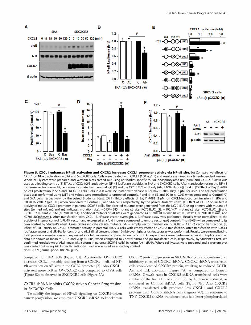
To clarify involvement of NF-kB signaling in the CXCL1-
CXCR2 axis, we tested the comparative effects of added CXCL1
on NF-kB activation in SKA and SKCXCR2 cells. CXCL1
produced more phosphorylated IkB in SKCXCR2 cells compared
to SKA cells (Figure 5A). In addition, a CXCL1/2/3 antibody had
no effects on NF-kB promoter activity in SKA cells but
significantly decreased this activity in SKCXCR2 cells (Figure
5B). Based on the involvement of NF-kB in the CXCL1-CXCR2
axis, we compared effects of Bay11-7082, a specific NF-kB
inhibitor, on cell proliferation in SKA and SKCXCR2 cells.
Bay11-7082 had no effect on cell proliferation in SKA cells but
significantly decreased proliferation in SKCXCR2 cells (Figure
5C). The inhibition was greatest in SKCXCR2 cells, probably
because of the higher activation of NF-kB in these cells (Figures
Figure 2. CXCR2 expressing cells have a higher activation of NF-kB in both basal and TNF-stimulated levels and increase CXCL1/2.(A) Effect of TNF on NF-kB luciferase activities in SKA and SKCXCR2 cells. After transfection using NF-kB luciferase vector overnight, cells were treatedwith TNF (10 ng/ml) for 4 h. Experiments were performed in triplicate and data are shown as mean 6 S.E. * and # (p#0.05) when compared toControl and SKA cells, respectively, by the paired Student’s t-test. (B) Representative immunofluorescent staining of SKA and SKCXCR2 cells indicatingIkB activation and CXCR2 protein expression levels (in green). (C) Effect of TNF (10 ng/ml) over time (0–120 min) on NF-kB activation in SKA andSKCXCR2 cells. Whole cell lysates were prepared and Western blots carried out using antibodies specific to IkB and IKK as well as their phosphorylatedforms (pIkB and pIKK). b-actin was used as a loading control. (D) Chemokine profile comparisons in SKCXCR2 relative to SKA cells. After isolating totalRNA, a human chemokine PCR array was performed. The dotted line indicates a 2-fold increase; those with a .2-fold increase and average cyclethreshold ,30 are recognized as induced chemokines, and in this case represent CXCL1 and 2 (*).doi:10.1371/journal.pone.0083789.g002
CXCR2-Driven Cancer Progression via NF-kB
PLOS ONE | www.plosone.org 5 December 2013 | Volume 8 | Issue 12 | e83789

2A-C). Furthermore, we compared the effects of Bay11-7082 on a
CXCL1-induced cell invasion. Although CXCL1 had a small
effect on cell invasion in SKA cells, the differences were not
significant (Figure 5D). On the other hand, SKCXCR2 cells had
at least a doubling of cell invasion numbers in response to CXCL1
compared to controls (Figure 5D). Bay11-7082 also blocked the
CXCL1-induced cell invasion in SKCXCR2 cells (Figure 5D),
indicating involvement of NF-kB signaling.
Next, we tested if transient transfection of CXCR2 into parental
human SKOV-3 cells affected the activity of the CXCL1
promoter in an NF-kB-dependent manner. The CXCL1 promoter
(KC701LUC) contains three NF-kB sites. Mutants of each kB site
(termed m1, m2 and m3) were prepared as described previously
[23]. The results show that the CXCL1 luciferase activities of
KC701LUC and the kB site mutations m2 and m3 were increased
in CXCR2 transfected cells compared to cells transfected with
empty vector (pA) (Figure 5E). On the other hand, mutations of
the proximal m1 kB site alone (and when in combination with the
mutants KC701LUCm3, KC701LUCm2 and KC701LUCm
3m2) were not responsive to enhanced CXCR2 expression (Figure
5E). These patterns were similar as described by interleukin-1 as
the NF-kB activator in mouse granulaosa cells [23]. This finding
indicates that the proximal kB site is essential for regulation of the
CXCL1 promoter activity in response to CXCR2-mediated NF-
kB and that the other two kB sites support induction of promoter
activity. Furthermore, we tested involvement of Akt on CXCR2-
mediated NF-kB signaling in CXCL1 promoter activity. We used
a commercial siRNA of Akt1 (a main Akt isoform in SKOV-3
cells) to knockdown Akt1. Akt1 siRNA had no significant effect on
CXCL1 promoter activity in CXCR2 negative cells, but it
attenuated the activity in CXCR2 positive cells (Figure 5F).
Figure 3. SKCXCR2 cells increases CXCL1/2, thus contributing to cell proliferation and enhancing migration, invasion and colonyformation relative to values in SKA cells. (A) Confirmation of CXCL1 and CXCL2 expression in SKA and SKCXCR2 cells by qRT-PCR. After isolatingtotal RNA, qRT-PCR was carried out using primers for CXCL1 and CXCL2. (B) Cellular CXCL1 and CXCL2 concentrations in SKA and SKCXCR2 cells over aperiod of 24 h. Whole cell lysates were prepared and ELISA carried out using antibodies specific to CXCL1 and CXCL2 and values were normalized tototal protein. (C) Effect of CXCL1 on cell proliferation in SKA and SKCXCR2 cells for 48 h incubation. (D) Effect of pan antibody for CXCL1/2/3 on cellproliferation in SKA and SKCXCR2 cells. Cells were incubated with normal IgG (Control) and pan antibody (1:100 dilution) for 48 h. The cellproliferation assay was performed using MTT and values were normalized to untreated controls. (E) Effect of pan antibody for CXCL1/2/3 on CXCL1and CXCL2 expression in SKCXCR2 cells by qRT-PCR. After treating with pan antibody for 24 h and then isolating total RNA, qRT-PCR was carried outusing primers for CXCL1 and CXCL2. (F) Migration characteristics between SKA and SKCXCR2 cells. (G) Invasion characteristics between SKA andSKCXCR2 cells. (H) Comparison of colony formation between SKA and SKCXCR2 cells. All experiments were performed at least in triplicate and dataare shown as mean 6 S.E. * and # (p#0.05) as calculated by Student’s t-test.doi:10.1371/journal.pone.0083789.g003
CXCR2-Driven Cancer Progression via NF-kB
PLOS ONE | www.plosone.org 6 December 2013 | Volume 8 | Issue 12 | e83789

Comparison of CXCR2-driven Cancer Progression in OVAVersus OVCXCR2 Cells
To exclude an SKOV-3 cell-type specific response, we confirmed
our data by generating another CXCR2 positive (OVCXCR2)
versus a negative (OVA) cell line using parental OVCAR-3 ovarian
cancer cells which either entirely lack or have trace amounts of
CXCR2 [6]. The OVCAR-3 cell line was purchased from the
American Type Culture Collection (Manassas, VA). OVCXCR2
cells expressed CXCR2 protein and immunofluorescent staining
revealed that like SKCXCR2 cells (Figures 1A and 1B), OVCXCR2
cells had more phosphorylated IkB (Figure 6A) than OVA cells. The
growth rates in OVCXCR2 vs. OVA cells (Figure 6B) were similar to
those observed in SKCXCR2 vs. SKA cells (Figure 1C). OVCXCR2
cells also had more phosphorylated IkB in response to TNF (Figure
6C) as observed in SKCXCR2 cells (Figure 2C). OVCXCR2 cells
had a greater level of phosphorylated EGFR, resulting to higher Akt
and Erk activations (Figure 6D). AG-1478 and LY294002 reduced
more NF-kB luciferase activity in OVCXCR2 cells whereas
PD98059 had no effect (Figure 6E) as demonstrated in SKCXCR2
cells (Figure 4D). Also, a CXCL1/2/3 antibody blocked cell
proliferation in OVCXCR2 cells as compared to OVA cells (Figure
6F). Bay11-7082 blocked the CXCL1-induced cell invasion in
OVCXCR2 cells (Figure 6G) as observed in SKCXCR2 cells (Figure
5D). Because SKCXCR2 cells increased CXCR2 ligands such as
CXCL1 and CXCL2 (Figure 2D), we confirmed that OVCXCR2
also increased CXCR2 ligands such as CXCL1-3 and 6 when
Figure 4. CXCR2 transactivates EGFR which contributes to NF-kB signaling via Akt activation. (A) Comparison of EGFR activation in SKAand SKCXCR2 cells. Whole cell lysates were prepared and Western blots carried out using antibodies specific to EGFR, Akt, Erk and the phosphorylatedforms (pEGFR, pAkt and pErk). The non-phosphorylated forms were used as loading controls. (B) Representative immunofluorescent staining patternsindicating Akt activation and CXCR2 protein expression levels in SKA and SKCXCR2 cells. (C) Comparative effects of AG-1478, LY294002 and PD98059on cell proliferation in SKA and SKCXCR2 cells. Cells were incubated with vehicle (Control), AG-1478 (AG, 2 mM), LY294002 (LY, 2 mM) or PD98059 (PD,20 mM) for 48 h. The cell proliferation assay was performed using MTT and values were normalized to untreated controls. * and # (p#0.05) whencompared to Controls (C) and SKA cells, respectively, by Student’s t-test. (D) Dose-dependent effects of EGFR downstream inhibitors on NF-kBluciferase activities in SKA and SKCXCR2 cells. After transfection with NF-kB luciferase vector overnight, cells were treated with AG-1478 (EGFRinhibitor, 0, 0.5, 1 and 2 mM), LY294002 (Akt inhibitor, 0, 0.5, 1 and 2 mM) or PD98059 (Erk inhibitor, 0, 5, 10 and 20 mM) for 4 h. * and # (p#0.05) whencompared to Controls (0 h) and SKA cells, respectively, by Student’s t-test. All experiments were performed at least in triplicate and data are shown asmean 6 S.E. (E) Confirmation of specific inhibitors on EGFR, Akt and Erk activation in SKA and SKCXCR2 cells. Cells were treated with AG-1478 (2 mM),LY294002 (2 mM) and PD98059 (20 mM) for 4 h. Whole cell lysates were prepared and a western blot was carried out using antibodies specific to EGFR,Akt, Erk and their phosphorylated forms (pEGFR, pAkt and pErk). Non-phosphorylated forms were used as loading controls.doi:10.1371/journal.pone.0083789.g004
CXCR2-Driven Cancer Progression via NF-kB
PLOS ONE | www.plosone.org 7 December 2013 | Volume 8 | Issue 12 | e83789
compared to OVA cells (Figure S1). Additionally OVCXCR2
increased CCL2, probably resulting from a CXCR2-mediated NF-
kB activation on kB sites in the CCL2 promoter [5]. Also CXCL1
activated more IkB in OVCXCR2 cells compared to OVA cells
(Figure S2) as observed in SKCXCR2 cells (Figure 5A).
CXCR2 shRNA Inhibits CXCR2-driven Cancer Progressionin SKCXCR2 Cells
To solidify the impact of NF-kB signaling on CXCR2-driven
cancer progression, we employed CXCR2 shRNA to knockdown
CXCR2 protein expression in SKCXCR2 cells and confirmed an
inhibitory effect of CXCR2 shRNA. CXCR2 shRNA transfected
cells knockdowned CXCR2 protein, resulting to reduced EGFR,
Akt and Erk activation (Figure 7A) as compared to Control
shRNA. Growth rates in CXCR2 shRNA transfected cells were
similar for the first 24 h of culture but by 48 h were reduced, as
compared to Control shRNA cells (Figure 7B). Also CXCR2
shRNA transfected cells produced less CXCL1 and CXCL2
proteins than Control shRNA cells (Figures 7C). In response to
TNF, CXCR2 shRNA transfected cells had lesser phosphorylated
Figure 5. CXCL1 enhances NF-kB activation and CXCR2 increases CXCL1 promoter activity via NF-kB site. (A) Comparative effects ofCXCL1 on NF-kB activation in SKA and SKCXCR2 cells. Cells were treated with CXCL1 (100 ng/ml) and results examined in a time-dependent manner.Whole cell lysates were prepared and Western blots carried out using antibodies specific to IkB, phosphorylated IkB (pIkB) and CXCR2. b-actin wasused as a loading control. (B) Effect of CXCL1/2/3 antibody on NF-kB luciferase activities in SKA and SKCXCR2 cells. After transfection using the NF-kBluciferase vector overnight, cells were incubated with normal IgG (C) and the CXCL1/2/3 antibody (Ab, 1:100 dilution)) for 4 h. (C) Effect of Bay11-7082on cell proliferation in SKA and SKCXCR2 cells. Cells in A-B were incubated with vehicle (C) or Bay11-7082 (Bay, 2 mM) for 48 h. The cell proliferationassay was performed using MTT and values were normalized to untreated controls. * and # in 5B and 5C (p # 0.05) when compared to Control (C)and SKA cells, respectively, by the paired Student’s t-test. (D) Inhibitory effects of Bay11-7082 (2 mM) on CXCL1-induced cell invasion in SKA andSKCXCR2 cells. * (p#0.05) when compared to Control (C) and SKA cells, respectively, by the paired Student’s t-test. (E) Effect of CXCR2 on luciferaseactivity of mouse CXCL1 promoter in parental SKOV-3 cells. Site-directed mutants were generated from the KC701LUC using primers with mutant kBsites (termed m1, m2 and m3 indicates mutation site): 2615/2585 mutant kB site (KC701LUCm3), 2102/271 mutant kB site (KC701LUCm2) and283/252 mutant kB site (KC701LUCm1). Additional mutants of kB sites were generated as KC701LUCm3m2, KC701LUCm3m1, KC701LUCm2m1, andKC701LUCm3m2m1. After transfection with CXCL1 luciferase vector overnight, a luciferase assay was performed. Results were normalized to theactivity of internal control (pRL-TK vector) and expressed as a fold increase compared to empty vector (pA) controls. * (p#0.05) when compared to itsown control by Student’s t-test. Cross circles indicate kB site mutants. pA = empty vector transfection; pCXCR2 = CXCR2 vector transfection. (F)Effect of Akt1 siRNA on CXCL1 promoter activity in parental SKOV-3 cells with empty vector or CXCR2 transfection. After transfection with CXCL1luciferase vector and siRNAs for control and Akt1 (final concentration: 10 nM) overnight, a luciferase assay was performed. Results were normalized tototal protein concentrations and expressed as a fold increase compared to each control. All experiments were performed at least in triplicate and alldata are shown as mean 6 S.E. * and # (p # 0.05) when compared to Control siRNA and pA transfected cells, respectively, by Student’s t-test. Weconfirmed knockdown of Akt1 (main Akt isoform in parental SKOV-3 cells) by using Akt1 siRNA. Whole cell lysates were prepared and a western blotwas carried out using Akt1 specific antibody. b-actin was used as a loading control.doi:10.1371/journal.pone.0083789.g005
CXCR2-Driven Cancer Progression via NF-kB
PLOS ONE | www.plosone.org 8 December 2013 | Volume 8 | Issue 12 | e83789

IKK and IkB as compared to Control shRNA cells (Figure 7D). In
addition, a CXCL1/2/3 antibody had no effects on NF-kB
promoter activity in CXCR2 shRNA transfected cells but
significantly decreased this activity in Control shRNA cells (Figure
7E). AG-1478 and LY294002 reduced NF-kB luciferase activity at
a lesser degree in CXCR2 shRNA transfected cells as compared to
Control shRNA cells and PD98059 had no effect in both cells
(Figure 7F). Also, a CXCL1/2/3 antibody had no significant
effects on cell proliferation in CXCR2 shRNA transfected cells as
compared to Control shRNA cells (Figure 7G). Addition of
CXCL1 had less effects on cell proliferation in CXCR2 shRNA
transfected cells (Figure 7H). CXCR2 shRNA transfected cells had
a reduced CXCL1-induced cell migration and invasion as
compared to Control shRNA cells (Figure 7I). CXCR2 shRNA
transfected cells also had a reduced TNF-induced NF-kB
luciferase activity and had decreased basal and TNF-induced
levels of CXCL1 promoter activity (Figure 7J).
Discussion
A primary finding of this study is that CXCR2-driven cancer
progression involves upregulation of its own ligands such as
CXCL1 and CXCL2 by potentiating NF-kB activation via
EGFR-transactivated Akt signaling followed by accelerated
ovarian cancer cell proliferation, migration and invasion. TNF
appears to have different proliferative characteristics, depending
on the cells involved. As an example, although TNF inhibited
proliferation in ovarian UT-OC-2 carcinoma cells [28], it had a
proliferative effect in ovarian MDAH 2774 cancer cells [29].
Ovarian UT-OC-2 carcinoma cells had no NF-kB activation in
respond to TNF whereas ovarian MDAH 2774 cancer cells
induced TNF-activated NF-kB [28–29], indicating a critical role
of NF-kB signaling on cell proliferation. Interestingly, saxatilin, a
snake venom shown to inhibit TNF-induced proliferation in
ovarian cancer cells, was found to block the TNF effect by
suppressing CXCL8 expression [29]. This fact supports the
Figure 6. Confirmation of CXCR2-potentiated NF-kB signaling in OVCAR-3 cells. (A) CXCR2 protein expression in OVA versus OVCXCR2cells. Western blot and immunofluorescent staining were carried out using antibodies specific to CXCR2 and b-actin as a loading control. (B)Comparison of growth rates in OVA and OVCXCR2 cells. Cells were incubated for 0, 24, 48 and 72 h and growth rates normalized to 0 h densities ineach cell line. Experiments were performed in triplicate and all data are shown as mean 6 S.E. * and ** (p#0.05) in each group by ANOVA and Tukey’spairwise comparisons. # (p#0.05) between OVA and OVCXCR2 cells by Student’s t-test. (C) Effect of TNF (10 ng/ml) effects over time (0-120 min) onNF-kB activation in OVA and OVCXCR2 cells. Whole cell lysates were prepared and Western blots carried out using antibodies specific to IkB, IKK andtheir phosphorylated forms (pIkB and pIKK). b-actin was used as a loading control. (D) Comparison of EGFR activation in OVA and OVCXCR2 cells.Whole cell lysates were prepared and Western blots carried out using antibodies specific to EGFR, Akt, Erk and the phosphorylated forms (pEGFR,pAkt and pErk). The non-phosphorylated forms were used as loading controls. (E) Effects of EGFR downstream inhibitors on NF-kB luciferase activityin OVA and OVCXCR2 cells. After transfection with NF-kB luciferase vector overnight, cells were treated with vehicle (C), AG-1478 (AG, 2 mM),LY294002 (LY, 2 mM) or PD98059 (PD, 20 mM) for 4 h. (F) Effect of CXCL1/2/3 pan specific antibody for neutralization on cell proliferation in OVA andOVCXCR2 cells. Cells were incubated with normal IgG (C) and antibody (1:100 dilution) for 48 h. The cell proliferation assay was performed using MTTand values were normalized to untreated controls. (G) Inhibitory effects of Bay11-7082 (2 mM) on CXCL1-induced cell invasion in OVA and OVCXCR2cells. All experiments were performed at least in triplicate and data are shown as mean 6 S.E. * and # (p#0.05) as calculated by Student’s t-test.doi:10.1371/journal.pone.0083789.g006
CXCR2-Driven Cancer Progression via NF-kB
PLOS ONE | www.plosone.org 9 December 2013 | Volume 8 | Issue 12 | e83789

concept that chemokines are involved in TNF-induced cell
proliferation. Also, our previous studies have reported that TNF
primarily induces CXCR2 ligands (CXCL1-3 and CXCL8) in
ovarian cancer cells [5–6]. In the current study, TNF increased
cell proliferation to a greater extent in CXCR2 positive compared
to CXCR2 negative cells (Figure 1D). In spite of a significant
difference in the G0-G1 phase in CXCR2 positive and negative
cells (Figure 1E), TNF had no significant effects on cell cycle
related genes except for a .2-fold increase of GADD45a in
CXCR2 positive cells (Table S1). Interestingly, GADD45a has
been found to have dual functions as both a promoter in Myc-
driven breast cancer and a suppressor in Ras-driven breast cancer
[30]. Although protecting melanoma cells from ultraviolet B-
induced apoptosis [31] and cell death in neuron cells [32],
GADD45a functions as a mediator of retinoid-induced apoptosis
in ovarian carcinoma cells [24]. Disruption of GADD45a both
promotes cell migration and invasion in endothelial cells [25] and
mouse embryonic fibroblasts [26]. Because GADD45 protein is
well known as a stress sensor [33], TNF-induced GADD45a is
likely to be the cellular response to a TNF stress reaction instead of
serving as a promoter of cell proliferation in CXCR2 positive cells.
Because NF-kB is the primary signaling pathway for TNF
functions, we examined NF-kB signaling in CXCR2 negative and
positive cells. CXCR2 positive cells exhibited higher levels of NF-kB
activation in both the basal and TNF-induced states (Figures 2A, 2C
and 6C). Knockdown of CXCR2 in CXCR2 positive cells
Figure 7. Inhibitory effect of CXCR2 shRNA on CXCR2-deriven cancer progression in SKCXCR2 cells. (A) Knockdown of CXCR2 proteinexpression and comparison of EGFR activation after transfection of Control and CXCR2 shRNA in SKCXCR2 cells. After transfection with shRNAs forcontrol and CXCR2 for 72 h, a Western blot was carried out using antibodies specific to CXCR2, EGFR, Akt, Erk and the phosphorylated forms (pEGFR,pAkt and pErk). The non-phosphorylated forms and b-actin were used as loading controls. (B) Comparison of growth rates in Control (black bars) andCXCR2 shRNA (gray bars) transfected SKCXCR2 cells. Cells were incubated for 0, 24 and 48 h and growth rates normalized to 0 h densities in each cellline. Experiments were performed in triplicate and all data are shown as mean 6 S.E. * and ** (p#0.05) in each group by ANOVA and Tukey’s pairwisecomparisons. # (p#0.05) between Control and CXCR2 shRNA transfected cells by Student’s t-test. (C) Cellular CXCL1 and CXCL2 concentrations inControl and CXCR2 shRNA transfected cells. Whole cell lysates were prepared, an ELISA carried out using antibodies specific to CXCL1 and CXCL2 andvalues normalized to total protein. (D) Effect of TNF (10 ng/ml) over time (0–120 min) on NF-kB activation in Control and CXCR2 shRNA transfectedcells. Whole cell lysates were prepared and Western blots carried out using antibodies specific to IkB, IKK and their phosphorylated forms (pIkB andpIKK). b-actin was used as a loading control. (E) Effect of CXCL1/2/3 antibody on NF-kB luciferase activities in Control and CXCR2 shRNA transfectedcells. After transfection of shRNA for 48 h followed by transfection of NF-kB luciferase vector overnight, cells were incubated with normal IgG and theCXCL1/2/3 antibody (Ab, 1:100 dilution) for 4 h. (F) Effects of EGRF downstream inhibitors on NF-kB luciferase activity in Control and CXCR2 shRNAtransfected cells. After transfection of shRNA for 48 h followed by transfection of NF-kB luciferase vector overnight, cells were treated with vehicle (C),AG-1478 (AG, 2 mM), LY294002 (LY, 2 mM) or PD98059 (PD, 20 mM) for 4 h. (G) Effect of CXCL1/2/3 antibody on cell proliferation in Control and CXCR2shRNA transfected cells. Cells were incubated with normal IgG and CXCL1/2/3 antibody (Ab, 1:100 dilution) for 48 h. The cell proliferation assay wasperformed using MTT and values were normalized to untreated controls. (H) Effect of CXCL1 on cell proliferation in Control and CXCR2 shRNAtransfected cells for 48 h incubation. (I) Comparison of CXCL1-induced cell migration and invasion in Control and CXCR2 shRNA transfected cells. (J)Effect of TNF on NF-kB and mCXCL1 promoter luciferase activities in Control and CXCR2 shRNA transfected cells. After transfection of shRNA for 48 hfollowed by transfection of NF-kB or CXCL1 promoter luciferase vector overnight, cells were treated with TNF (10 ng/ml) for 4 h. All experiments wereperformed at least in triplicate and data are shown as mean 6 S.E. * and # (p#0.05) as calculated by Student’s t-test.doi:10.1371/journal.pone.0083789.g007
CXCR2-Driven Cancer Progression via NF-kB
PLOS ONE | www.plosone.org 10 December 2013 | Volume 8 | Issue 12 | e83789
decreased TNF-induced NF-kB activation (Figure 7D and 7J).
Several prior reports have indirectly suggested the presence of a
positive relationship between NF-kB signaling and the CXCR2
axis. For instance, the CXCR2 antagonist, SCH-527123, was able
to decrease phosphorylation of NF-kB in colorectal cancer cells [34]
while in ovarian cancer cells, CXCR2 stimulated angiogenesis by a
process thought to also involve NF-kB [9]. In addition, neutrophils
from severely NF-kB deficient mice [c-Rel(2/2)NF-kB1(2/
2)RelA(+/2)] express higher levels of CXCR2 [35], which may
serve as a compensatory mechanism for the NF-kB deficiency.
Because CXCR2 ligands such as CXCL1-3, and 5–8 [22]
contain kB sites in their promoters [5–6], it is possible that
CXCR2-mediated NF-kB activation is able to modulate the
chemokine network, which in turn, alters cellular functional
events. Among the CXCR2 ligands in this study, SKCXCR2 cells
highly induced CXCL1 and CXCL2 compared to levels seen in
SKA cells (Figures 2D, 3A and 3B). Knockdown of CXCR2 in
SKCXCR2 cells decreased CXCL1 and CXCL2 production
(Figure 7C). In case of OVCXCR2 cells, CXCL1-3 and 6 were
induced as a CXCR2 ligands (Figure S1). Although CXCL1 has
been reported to suppress malignancy by limiting prostate tumor
metastasis and reinforcing growth arrest [36], many reports
indicate that CXCL1 promotes cancer progression. For instance,
CXCL1 depletion reduced the migration and invasion of gastric
cancer cells [15] and an anti-CXCL1 antibody inhibited growth of
human pancreatic cancer cells [37]. CXCL1/2 reportedly
mediates breast cancer metastasis [38] and esophageal cancer cell
proliferation [39]. CXCL1 also induced proliferation in epithelial
ovarian cancer cells [27]. In addition to cancer cells, CXCL1
caused endothelial cell proliferation, tube formation, and migra-
tion [40]. Consistent with these reports, CXCR2 positive cells in
this study led to a greater increase in proliferation, migration,
invasion and colony formation (Figures 1C, 3F-3H, and 6B).
Knockdown of CXCR2 in SKCXCR2 cells decreased CXCL1-
induced proliferation, migration and invasion (Figure 7H and 7I).
Furthermore, a CXCL1/2/3 antibody or an NF-kB inhibitor
(Bay11-7082) was more inhibitory with regard to cell proliferation
in CXCR2 positive cells (Figures 3D, 5C and 6F).
In these studies, we found that CXCR2 positive cells
transactivated more EGFR followed by increased Akt activation
than negative cells (Figures 4A, 4B and 6D). Thus knockdown of
CXCR2 in SKCXCR2 cells decreased EGFR-activated signaling
(Figure 7A). AG-1478, a specific EGFR inhibitor, and LY294002,
an Akt blocker via PI3K inhibition, attenuated NF-kB promoter
activity in CXCR2 positive cells whereas PD98059, a specific Erk
inhibitor, had no effect (Figure 4D and 6E). On the other hand,
knockdown of CXCR2 in SKCXCR2 cells attenuated effects of
AG-1478 and LY294002 on NF-kB promoter activity (Figure 7F).
Erk activation was relatively low when compared to Akt activation
(Figure 4A, 4E and 6D), probably resulting to slight effects of
PD98059 in this model system. Interestingly PD98059 had
increased trend on NF-kB promoter activity (Figure 4D and 6E).
These findings indicate that CXCR2-mediated EGFR transacti-
vation contributes to NF-kB potentiation through Akt activation
rather than Erk activation. Prior studies showed that blockade of
Akt2 decreased IKKa phosphorylation, NF-kB nuclear transloca-
tion and cell migration in prostate cancer cells [41]. This fact
supports in part the involvement of Akt in CXCR2-mediated NF-
kB signaling as described by our results. Furthermore, because an
NF-kB inhibitor attenuated CXCL1-induced cell invasion in
CXCR2 positive cells (Figure 5D and 6G), our results suggest that
the CXCL1-CXCR2 axis may accelerate cancer progression by
potentiating NF-kB signaling.
In addition to NF-kB, CXCR2-mediated signaling could
involve Akt and/or Erk activation. Prior studies indicate that the
CXCR2 antagonist, SCH-527123, decreased Erk and Akt
activation in colorectal cancer cells [34] and that CXCR2
knockdown reduced Erk activation in ovarian cancer cells [9].
Inhibition of Erk also blocked CXCL8-induced cell proliferation
in non-small cell lung cancer cells [42]. Other investigators
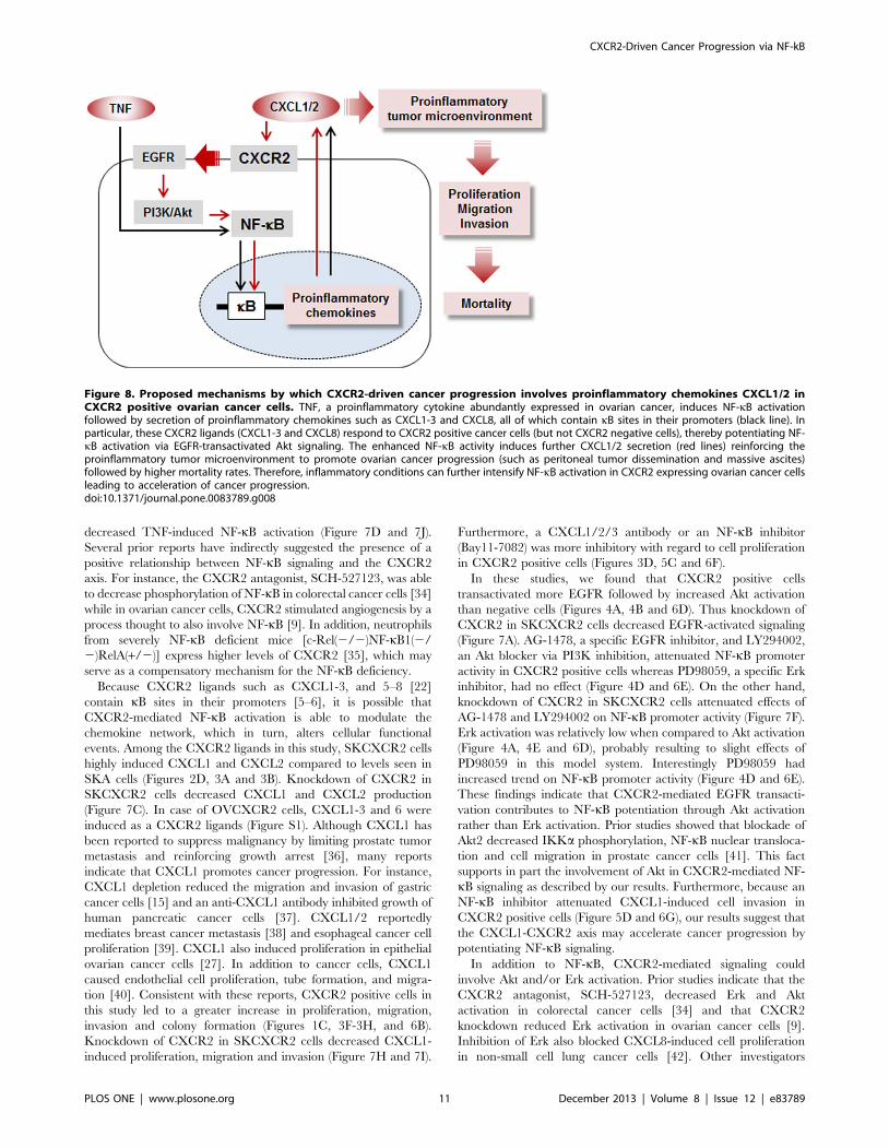
Figure 8. Proposed mechanisms by which CXCR2-driven cancer progression involves proinflammatory chemokines CXCL1/2 inCXCR2 positive ovarian cancer cells. TNF, a proinflammatory cytokine abundantly expressed in ovarian cancer, induces NF-kB activationfollowed by secretion of proinflammatory chemokines such as CXCL1-3 and CXCL8, all of which contain kB sites in their promoters (black line). Inparticular, these CXCR2 ligands (CXCL1-3 and CXCL8) respond to CXCR2 positive cancer cells (but not CXCR2 negative cells), thereby potentiating NF-kB activation via EGFR-transactivated Akt signaling. The enhanced NF-kB activity induces further CXCL1/2 secretion (red lines) reinforcing theproinflammatory tumor microenvironment to promote ovarian cancer progression (such as peritoneal tumor dissemination and massive ascites)followed by higher mortality rates. Therefore, inflammatory conditions can further intensify NF-kB activation in CXCR2 expressing ovarian cancer cellsleading to acceleration of cancer progression.doi:10.1371/journal.pone.0083789.g008
CXCR2-Driven Cancer Progression via NF-kB
PLOS ONE | www.plosone.org 11 December 2013 | Volume 8 | Issue 12 | e83789
reported that a CXCL1-induced cell proliferation in ovarian
cancer cells was related to transactivation of EGFR [24]. CXCL1
also enhanced potassium currents via activation of NF-kB in
sensory neurons [43]. In our previous report, the mouse CXCL1
promoter (containing three kB sites) had a proximal kB site that
served as a critical regulatory element and two other kB sites that
served as supportive elements in mouse granulosa cells [23]. We
demonstrated here that CXCR2 transiently-transfected human
(SKOV-3) ovarian cancer cells similarly increased CXCL1
promoter activities via a critical proximal kB site (Figure 5E).
Also knockdown of CXCR2 in SKCXCR2 cells reduced both
basal and TNF-induced levels of CXCL1 promoter activity
(Figure 7J). This fact further emphasizes the potentiation of NF-
kB signaling in the CXCL1-CXCR2 axis. In addition, attenuation
of Akt1 siRNA on CXCR2-induced CXCL1 promoter activity
(Figure 5F) indicates the involvement of Akt activation.
In one exceptional report, CXCL1 overexpression acted as a
suppressor of malignancy by limiting the escape of prostate tumor
cells from the primary tumor and reinforcing growth arrest [36].
However, in many cases CXCL1 appears to act as autocrine or
paracrine growth factor. Thus CXCL1 has been associated with
tumor size, tumor stage, invasion, metastasis and survival in
colorectal cancer patients [44]. Blocking CXCL1 signaling improved
chemotherapy efficacy by diminishing metastasis in breast cancer
[38]. In addition, an inverse association between CXCL1 and
recurrence-free survival was observed in colorectal cancer patients
[21]. Therefore, based on these functional roles of CXCL1 and
CXCR2-mediated signaling in cancer progression, NF-kB potenti-
ation is likely to play a central role in the CXCL1-CXCR2 axis.
In summary, CXCR2 expressing ovarian cancer cells potenti-
ated NF-kB activation via EGFR-transactivated Akt signaling to
induce CXCL1/2 secretion as an autocrine or paracrine growth
factor. This in turn enhanced ovarian cancer progression including
cell proliferation, migration and invasion events by increasing the
proinflammatory tumor microenvironment (Figure 8). Activation
of the CXCL1/2-CXCR2 axis could therefore augment the
clinical features of ovarian cancer such as peritoneal tumor
dissemination and ascites leading to higher mortality rates (Figure
8). Therefore inhibition of the CXCL1/2-CXCR2 axis may be an
effective preventive or therapeutic action against CXCR2-driven
ovarian cancer progression.
Supporting Information
Figure S1. Chemokine profile comparisons in OVCXCR2relative to OVA cells. After isolating total RNA, a human
chemokine PCR array was performed. The dotted line indicates a 2-
fold increase; those with a .2-fold increase and average cycle
threshold ,30 are recognized as induced chemokines (*). In this
case, significant increases (OVCXCR2 versus OVA) were seen in
CCL2, and CXCL 1-3 and 6.
(TIF)
Figure S2. Comparative effects of CXCL1 on NF-kBactivation in OVA and OVCXCR2 cells. Cells were treated
with CXCL1 (100 ng/ml) and results examined in a time-
dependent manner. Whole cell lysates were prepared and Western
blots carried out using antibodies specific to IkB, phosphorylated
IkB (pIkB) and CXCR2. As a loading control, b-actin was used.
(TIF)
Table S1. Comparative effects of TNF on cell-cyclerelated genes between SKA and SKCXCR2 cells asdetermined by PCR array.
(DOCX)
Acknowledgments
We thank Dr. Ann Richmond (Vanderbilt University, Nashville, TN) for
the CXCR2 expression vector and Dr. Diana Marver (Meharry Medical
College, Nashville, TN) for her comments and editorial assistance.
Author Contributions
Conceived and designed the experiments: YD EL DS. Performed the
experiments: YD SK DS. Analyzed the data: YD EL DS. Contributed
reagents/materials/analysis tools: EL DS. Wrote the paper: YD EL DS.
References
1. Chobanian N, Dietrich CS (2008) Ovarian cancer. Surg Clin North Am 88:285–
299.
2. Dobrzycka B, Terlikowski SJ, Kowalczuk O, Kinalski M (2009) Circulating
levels of TNF-a and its soluble receptors in the plasma of patients with epithelial
ovarian cancer. Eur Cytokine Netw 20:131–134.
3. Maccio A, Madeddu C (2012) Inflammation and ovarian cancer. Cytokine
58:133–147.
4. Szlosarek PW, Grimshaw MJ, Kulbe H, Wilson JL, Wilbanks GD, et al. (2006)
Expression and regulation of tumor necrosis factor alpha in normal and
malignant ovarian epithelium. Mol Cancer Ther 5:382–390.
5. Son DS, Parl AK, Rice VM, Khabele D (2007) Keratinocyte chemoattractant
(KC)/human growth-regulated oncogene (GRO) chemokines and pro-inflam-
matory chemokine networks in mouse and human ovarian epithelial cancer cells.
Cancer Biol Ther 6:1302–1312.
6. Son DS, Kabir SM, Dong YL, Lee E, Adunyah SE (2012) Inhibitory effect of
tumor suppressor p53 on proinflammatory chemokine expression in ovarian
cancer cells by reducing proteasomal degradation of IkB. PLoS One 7:e51116.
7. Balkwill FR (2012) The chemokine system and cancer. J Pathol 226:148–157.
8. Singh R, Lillard JW Jr, Singh S (2011) Chemokines: key players in cancer
progression and metastasis. Front Biosci 3:1569–1582.
9. Yang G, Rosen DG, Liu G, Yang F, Guo X, et al. (2010) CXCR2 promotes
ovarian cancer growth through dysregulated cell cycle, diminished apoptosis,
and enhanced angiogenesis. Clin Cancer Res 16:3875–3886.
10. Saintigny P, Massarelli E, Lin S, Ahn YH, Chen Y, et al. (2013) CXCR2
expression in tumor cells is a poor prognostic factor and promotes invasion and
metastasis in lung adenocarcinoma. Cancer Res 73:571–582.
11. Han L, Jiang B, Wu H, Wang X, Tang X, et al. (2012) High expression of
CXCR2 is associated with tumorigenesis, progression, and prognosis of
laryngeal squamous cell carcinoma. Med Oncol 29:2466–2472.
12. Ewington L, Taylor A, Sriraksa R, Horimoto Y, Lam EW, et al. (2012) The
expression of interleukin-8 and interleukin-8 receptors in endometrial carcino-
ma. Cytokine 59: 417–422.
13. Bondurant KL, Lundgreen A, Herrick JS, Kadlubar S, Wolff RK, et al. (2013)
Interleukin genes and associations with colon and rectal cancer risk and overall
survival. Int J Cancer 132:905–915.
14. Liu Z, Yang L, Xu J, Zhang X, Wang B (2011) Enhanced expression and clinical
significance of chemokine receptor CXCR2 in hepatocellular carcinoma. J Surg
Res 166:241–246.
15. Cheng WL, Wang CS, Huang YH, Tsai MM, Liang Y, et al. (2011) Lin KH.
Overexpression of CXCL1 and its receptor CXCR2 promote tumor invasion in
gastric cancer. Ann Oncol 22:2267–2276.
16. Shen H, Schuster R, Lu B, Waltz SE, Lentsch AB (2006) Critical and opposing
roles of the chemokine receptors CXCR2 and CXCR3 in prostate tumor
growth. Prostate 66:1721–1728.
17. Keane MP, Belperio JA, Xue YY, Burdick MD, Strieter RM (2004) Depletion of
CXCR2 inhibits tumor growth and angiogenesis in a murine model of lung
cancer. J Immunol 172:2853–2860.
18. Mestas J, Burdick MD, Reckamp K, Pantuck A, Figlin RA, et al. (2005) The role
of CXCR2/CXCR2 ligand biological axis in renal cell carcinoma. J Immunol
175:5351–5357.
19. Jamieson T, Clarke M, Steele CW, Samuel MS, Neumann J, et al. (2012)
Inhibition of CXCR2 profoundly suppresses inflammation-driven and sponta-
neous tumorigenesis. J Clin Invest 122:3127–3144.
20. Lee YS, Choi I, Ning Y, Kim NY, Khatchadourian V, et al. (2012) Interleukin-8
and its receptor CXCR2 in the tumour microenvironment promote colon
cancer growth, progression and metastasis. Br J Cancer 106:1833–1841.
21. Oladipo O, Conlon S, O’Grady A, Purcell C, Wilson C, et al. (2011) The
expression and prognostic impact of CXC-chemokines in stage II and III
colorectal cancer epithelial and stromal tissue. Br J Cancer 104:480–487.
CXCR2-Driven Cancer Progression via NF-kB
PLOS ONE | www.plosone.org 12 December 2013 | Volume 8 | Issue 12 | e83789
22. Olson TS, Ley K (2002) Chemokines and chemokine receptors in leukocyte
trafficking. Am J Physiol Regul Integr Comp Physiol 283:R7–R28.
23. Son DS, Roby KF (2006) Interleukin-1a-induced chemokines in mouse
granulosa cells: impact on keratinocyte chemoattractant chemokine, a CXC
subfamily. Mol Endocrinol 20:2999–3013.
24. Jiang T, Soprano DR, Soprano KJ (2007) GADD45A is a mediator of CD437
induced apoptosis in ovarian carcinoma cells. J Cell Physiol 212:771–779.
25. Yang F, Zhang W, Li D, Zhan Q (2013) Gadd45a suppresses tumor
angiogenesis via inhibition of the mTOR/STAT3 pathway. J Biol Chem
288:6552–6560.
26. Shan Z, Li G, Zhan Q, Li D (2012) Gadd45a inhibits cell migration and invasion
by altering the global RNA expression. Cancer Biol Ther 13:1112–1122.
27. Bolitho C, Hahn MA, Baxter RC, Marsh DJ (2010) The chemokine CXCL1
induces proliferation in epithelial ovarian cancer cells by transactivation of the
epidermal growth factor receptor. Endocr Relat Cancer 17:929–940.
28. Seppanen M, Lin L, Saarinen R, Punnonen R, Vihko KK (2008) Regulation of
ovarian UT-OC-2 carcinoma cells by cytokines: effects on cell proliferation,
activation of transcription factors and apoptosis. Acta Obstet Gynecol Scand
87:902–909.
29. Kim DS, Jang YJ, Jeon OH, Kim DS (2007) Saxatilin inhibits TNF-a-induced
proliferation by suppressing AP-1-dependent IL-8 expression in the ovarian
cancer cell line MDAH 2774. Mol Immunol 44:1409–1416.
30. Tront JS, Huang Y, Fornace AJ Jr, Hoffman B, Liebermann DA (2010)
Gadd45a functions as a promoter or suppressor of breast cancer dependent on
the oncogenic stress. Cancer Res 70:9671–9681.
31. Fayolle C, Pourchet J, Caron de Fromentel C, Puisieux A, Dore JF, et al. (2008)
Gadd45a activation protects melanoma cells from ultraviolet B-induced
apoptosis. J Invest Dermatol 128:196–202.
32. Lin CR, Yang CH, Huang CE, Wu CH, Chen YS, et al. (2011) GADD45A
protects against cell death in dorsal root ganglion neurons following peripheral
nerve injury. J Neurosci Res 89:689–699.
33. Liebermann DA, Tront JS, Sha X, Mukherjee K, Mohamed-Hadley A, et al.
(2011) Gadd45 stress sensors in malignancy and leukemia. Crit Rev Oncog 16:
129–140.
34. Ning Y, Labonte MJ, Zhang W, Bohanes PO, Gerger A, et al. (2012) The
CXCR2 antagonist, SCH-527123, shows antitumor activity and sensitizes cellsto oxaliplatin in preclinical colon cancer models. Mol Cancer Ther 11:1353–
1364.
35. von Vietinghoff S, Asagiri M, Azar D, Hoffmann A, Ley K (2010) Defectiveregulation of CXCR2 facilitates neutrophil release from bone marrow causing
spontaneous inflammation in severely NF-kB-deficient mice. J Immunol185:670–678.
36. Benelli R, Stigliani S, Minghelli S, Carlone S, Ferrari N (2013) Impact of
CXCL1 overexpression on growth and invasion of prostate cancer cell. Prostate73:941–951.
37. Takamori H, Oades ZG, Hoch OC, Burger M, Schraufstatter IU (2000)Autocrine growth effect of IL-8 and GROalpha on a human pancreatic cancer
cell line, Capan-1. Pancreas 21:52–56.38. Acharyya S, Oskarsson T, Vanharanta S, Malladi S, Kim J, et al. (2012) A
CXCL1 paracrine network links cancer chemoresistance and metastasis. Cell
150:165–178.39. Wang B, Hendricks DT, Wamunyokoli F, Parker MI (2006) A growth-related
oncogene/CXC chemokine receptor 2 autocrine loop contributes to cellularproliferation in esophageal cancer. Cancer Res 66:3071–3077.
40. Agarwal A, Tressel SL, Kaimal R, Balla M, Lam FH, et al. (2010) Identification
of a metalloprotease-chemokine signaling system in the ovarian cancermicroenvironment: implications for antiangiogenic therapy. Cancer Res
70:5880–5890.41. Kuo PL, Shen KH, Hung SH, Hsu YL (2012) CXCL1/GROa increases cell
migration and invasion of prostate cancer by decreasing fibulin-1 expressionthrough NF-kB/HDAC1 epigenetic regulation. Carcinogenesis 33:2477–2487.
42. Luppi F, Longo AM, de Boer WI, Rabe KF, Hiemstra PS (2007) Interleukin-8
stimulates cell proliferation in non-small cell lung cancer through epidermalgrowth factor receptor transactivation. Lung Cancer 56:25–33.
43. Yang RH, Strong JA, Zhang JM (2009) NF-kB mediated enhancement ofpotassium currents by the chemokine CXCL1/growth related oncogene in small
diameter rat sensory neurons. Mol Pain 5:26.
44. Ogata H, Sekikawa A, Yamagishi H, Ichikawa K, Tomita S, et al. (2010) GROapromotes invasion of colorectal cancer cells. Oncol Rep 24:1479–1486.
CXCR2-Driven Cancer Progression via NF-kB
PLOS ONE | www.plosone.org 13 December 2013 | Volume 8 | Issue 12 | e83789
Copyright © 2022 FDOKUMEN