Cancer risk related to mammary gland structure and development
Expression of the inflammatory chemokines CCL2, CCL5 andCXCL2 and the receptors CCR1–3 and CXCR2 in T lymphocytesfrom mammary tumor- bearing mice
Jennifer L. Owen1, Michael F. Criscitiello2, Stephania Libreros3, Ramon Garcia-Areas3,Kathleen Guthrie3, Marta Torroella-Kouri4, and Vijaya Iragavarapu-Charyulu3,*
1 Department of Physiological Sciences, University of Florida College of Veterinary Medicine,Gainesville, FL 32610, USA2 College of Veterinary Medicine and Biomedical Sciences, Texas A & M University, CollegeStation, TX 77843, USA3 Department of Basic Sciences, Charles E. Schmidt College of Biomedical Sciences, FloridaAtlantic University, Boca Raton, FL 33431, USA4 Department of Microbiology and Immunology, University of Miami Miller School of Medicine,Miami, FL 33136, USA
AbstractChemokines and their receptors have been studied in several solid tumor models as mediators ofinflammation. In turn, inflammation has been implicated in the promotion and progression oftumors, and as such, chemokines have been proposed as novel molecular targets forchemotherapy. While the expression of these molecules has been described in tumor cells,endothelial cells, macrophages, and neutrophils, less attention has been paid to the expressionprofile of these molecules by T lymphocytes in the periphery or infiltrating the tumor. Using theD1-DMBA-3 murine mammary adenocarcinoma model, we aimed to better characterize thedifferential expression of chemokines and/or their receptors in the host and in the tumormicroenvironment, and specifically, in the T cells of tumor-bearing mice compared to normalcontrol animals. We found that T lymphocytes from tumor-bearing mice express the pro-inflammatory chemokines, CCL2, CCL5, and CXCL2, as well as the chemokine receptors, CCR1,CCR2, CCR3 and CXCR2.
Keywordschemokines; chemokine receptors; T lymphocytes; mammary tumor; angiogenesis
© 2011 Elsevier Inc. All rights reserved.*Corresponding author. Florida Atlantic University, Department of Basic Sciences, Charles E. Schmidt College of BiomedicalSciences, 777 Glades Road, P.O. Box 3091, Boca Raton, FL 33431-0991. Tel.: +1 561 297 3304; Fax: +1 561 297 2519;[email protected]'s Disclaimer: This is a PDF file of an unedited manuscript that has been accepted for publication. As a service to ourcustomers we are providing this early version of the manuscript. The manuscript will undergo copyediting, typesetting, and review ofthe resulting proof before it is published in its final citable form. Please note that during the production process errors may bediscovered which could affect the content, and all legal disclaimers that apply to the journal pertain.
NIH Public AccessAuthor ManuscriptCell Immunol. Author manuscript; available in PMC 2012 May 10.
Published in final edited form as:Cell Immunol. 2011 ; 270(2): 172–182. doi:10.1016/j.cellimm.2011.05.004.
NIH
-PA Author Manuscript
NIH
-PA Author Manuscript
NIH
-PA Author Manuscript
1. IntroductionAll tumors, benign or malignant, are comprised of two main components: the neoplasticpopulation and the reactive stroma surrounding that population. Mounting evidencecontinues to implicate the perivascular and vascular elements, as well as the inflammatoryinfiltrate of tumors, as contributors to tumor progression and metastasis [1; 2; 3; 4; 5; 6; 7;8]. Chemokines expressed by the tumor cells recruit a variety of immune cells, includinglymphocytes, which may be immunoregulatory or suppressive, or may enrich the chemokinemilieu by secreting chemotactic molecules that contribute to the inflammatory process. Ithas been suggested that the inflammatory reaction at the breast tumor site enhances tumorgrowth and progression [9]. Since chemokines are known to have angiogenic and growthpromoting characteristics, we sought to determine the role of tumor burden on T cellchemokine and chemokine receptor expression using the D1-DMBA-3 mammaryadenocarcinoma model.
Chemokines are grouped into four families (C, CC, CXC and CX3C), based on the groupingof cysteine residues near the N-terminus of these proteins [10]. The CC or β chemokines area large superfamily of small, inducible, secreted, pro-inflammatory cytokines that areexpressed by various cell types including monocytes, endothelial cells, and fibroblasts [11].CCL2/monocyte chemoattractant protein-1 (MCP-1) is upregulated in different tumor celltypes, including breast, prostate and melanoma cells [12; 13; 14; 15]. CCL5/Regulated uponActivation Normal T cell Expressed and Secreted (RANTES) is expressed by Tlymphocytes, as well as some tumor cells, and has been shown to have a controversial rolein breast tumor progression. Soria et al. have established a positive correlation in breastcancer between aggressiveness of the tumor and tumor-derived CCL5 [16; 17], whereasJayasinghe et al. have reported that tumor-derived CCL5 does not contribute to breast cancerprogression [18]. While many of these studies correlated tumor-derived chemokines todisease progression, there are far fewer analyses of lymphocyte-derived chemokines in atumor model. We have recently reported that CCL2 is elevated in the T lymphocytes ofmammary tumor-bearing mice [19]. In this report, we further expand on CC chemokineexpression in T lymphocytes of mammary tumor-bearing mice.
The CXC chemokines have been shown to regulate angiogenesis in both a positive andnegative manner, depending on the presence or absence of an ELR motif (glutamic acid-leucine-arginine) in the amino terminus. The ELR positive chemokines that promoteangiogenesis include CXCL1, -2, -3, -5, -6, -7 and -8 [20]. IL-8, the functional humananalogue of murine CXCL2/macrophage inflammatory protein-2 (MIP-2), promotes thegrowth and invasiveness of breast cancer cells [21; 22; 23]. CXCL2 is produced bymacrophages, endothelial cells, epithelial cells and tumor cells [24]. However, it is notknown whether lymphocytes from tumor bearers are induced to produce this chemokine. Inthis study, we determined whether CXCL2 is expressed by T lymphocytes of tumor-bearingmice.
Leukocytes are recruited to sites of inflammation and specific microenvironments withinsecondary lymphoid tissues based on chemokine and chemokine receptor expressionpatterns [25; 26]. Previous studies have established similarities between the functions ofchemokine receptors in the physiologic homing of leukocytes and in the roles of thesereceptors in cancer metastasis [27]. Since leukocyte infiltration into tumors is regulated bychemokine production in the tumor microenvironment [28], chemokine receptor expressionby lymphocytes may play a role in tumor infiltration, and in shaping the tumormicroenvironment via inflammation. For example, it has been well documented that CCR2and its principal ligand, CCL2, induce monocyte infiltration and promote inflammation [29],while Huang et al. have found that breast, gastric and ovarian tumor growth is enhanced by
Owen et al. Page 2
Cell Immunol. Author manuscript; available in PMC 2012 May 10.
NIH
-PA Author Manuscript
NIH
-PA Author Manuscript
NIH
-PA Author Manuscript
CCR2+ myeloid suppressor cells attracted to the liver by hepatocyte-produced CCL2 [30].Furthermore, Robinson et al. reported that CCL5 was produced by 410.4 murine breastcancer cells, and that its receptors, CCR1 and CCR5, are expressed by the leukocyteinfiltrates which contribute towards tumor growth [28]. Because chemokine and chemokinereceptor expression is crucial for lymphocyte migration, we evaluated the expression ofchemokine receptors in T lymphocytes and how that expression relates to tumor infiltration.
2. Materials and Methods2.1. Mice and cell lines
8–12 weeks of age BALB/c mice used in these studies were obtained from Charles RiverLaboratories (Charles River Laboratories International, Inc., Wilmington, MA). Animalswere cared for and used according to the guidelines of the National Institutes of Health atthe animal facility at Florida Atlantic University. The D1-DMBA-3 tumor, syngeneic toBALB/c mice, is a transplantable mammary adenocarcinoma derived from a non-viral,noncarcinogen-induced preneoplastic nodule after treatment with 7,12-dimethylbenzanthracene [31]. The D1-DMBA-3 tumor is immunogenic to the host of originand nonmetastatic to the spleen, but metastases to the lung and bone marrow do occur. TheDA-3 mammary tumor cell line was derived in our laboratory from the D1-DMBA-3 tumorand was maintained in DMEM/high glucose, 10% characterized heat inactivated FCS, 100U/ml of penicillin, 100 μg/ml of streptomycin, and OPI media supplement (Sigma ChemicalCo., St. Louis, MO). Tumors were implanted in BALB/c mice by subcutaneous injection of1×106 tumor cells resulting in a measurable tumor 7–10 days post implantation.
2.2. Purification of splenic T cellsSpleens were compressed in Teflon tissue homogenizers and the resulting single cellsuspensions were pelleted at 300g, subjected to hypotonic shock for red cell removal,washed and counted. Macrophages were removed from the cell suspensions by plasticadherence in pre-warmed RPMI-1640, 5% FCS at 37°C for 1 h in a CO2 incubator. The non-adherent T lymphocytes were purified on nylon wool columns according to the method ofJulius et al. [32] and by positive selection using the MACS magnetic separation system(Miltenyi Biotec, Auburn, CA) according to the manufacturer’s instructions. Briefly, single-cell suspensions in cold PEB buffer (PBS supplemented with 2mM EDTA and 0.5% BSA)were incubated with supermagnetic microbeads conjugated to anti-mouse CD90 (Thy1.2),anti-mouse CD4, or anti-mouse CD8 mAb at 4° C for 15 min. Cells were washed twice andloaded onto the magnetic separation columns. The columns were washed three times withcold PEB buffer, and the positively selected Thy1.2+, CD4+, or CD8+ T cells were theneluted. After purification, the cells were routinely >95% viable, as assessed by trypan blueexclusion. FACS analysis using a Becton Dickinson LSR analyzer and anti-mouse FITC-CD90, anti-mouse FITC-CD4, and anti-mouse PE-CD8 antibodies (BD BiosciencesPharmingen, San Diego, CA) confirmed the populations to be ≥93% Thy1.2+, ≥94% CD4+,and ≥90% CD8+ T lymphocytes.
2.3. Cell cultureAfter purification, splenic T cells were cultured for 2h, 4h, or overnight at 2 × 10⁶ cells/mlin complete media consisting of RPMI-1640, 10% FCS, 100 U/ml penicillin, 100 μg/mlstreptomycin, and 50 mM 2-ME. Cell-free supernatants were removed and stored at −80°Cuntil use. Some of the T lymphocyte cultures were stimulated with Concanavalin A (Con A,Sigma Chemical Co., St. Louis, MO) or with recombinant murine CCL2/MCP-1 (rmCCL2/MCP-1) (PeproTech Inc., Rocky Hill, NJ).
Owen et al. Page 3
Cell Immunol. Author manuscript; available in PMC 2012 May 10.
NIH
-PA Author Manuscript
NIH
-PA Author Manuscript
NIH
-PA Author Manuscript
2.4. RNA analysesTotal RNA was isolated using TriReagent (Molecular Research Center, Inc., Cincinnati,Ohio). The detection and quantification of chemokine ligand and receptor mRNA specieswas analyzed by RNase protection assay using the RiboQuant Multiprobe RPA kit (BDBiosciences Pharmingen) according to the manufacturer’s instructions. Briefly, 20 μg RNAwas hybridized overnight to the [α-32P]UTP-radiolabeled anti-sense RNA probe set, whichhad been synthesized from the following template sets: mCK-5 murine chemokine, mCR-5murine chemokine receptors, and a custom designed probe set which included CXCreceptors CXCR2, -4, and -5 (all from BD Biosciences Pharmingen), after which single-stranded RNA and free probe were digested by RNase A and T1. Subsequently, protectedRNA was purified and resolved on a 5% denaturing polyacrylamide gel. The chemokinetranscripts were identified by the length of the respective fragments. The quantity of eachspecies was determined by the intensity of the appropriately-sized, protected probefragment. Equivalence of loading was ascertained by the intensity of the fragments for thehousekeeping genes, L32 and glyceraldehyde-3–phosphate dehydrogenase (GAPDH) foreach lane. Blots were exposed to Kodak BioMax autoradiography film (PerkinElmer LifeAnd Analytical Sciences, Inc., Waltham, MA) overnight.
2.5. Cytokine ELISACell culture supernatants, tumor cystic fluid, and sera were analyzed for murine chemokineprotein levels by OptEIA™ ELISA (BD Biosciences Pharmingen) for CCL2, and analyzedfor CXCL-2 and CCL5 protein levels by DuoSet® ELISA development systems (R&DSystems, Minneapolis, MN) according to manufacturers’ instructions. Absorbance at 450nm with wavelength correction at 570 nm was read on a Tecan SLT Rainbow Reader (LabInstruments, Research Triangle Park, NC) and OD values of samples were converted topicograms against a standard curve of known quantities of recombinant murine chemokines.
2.6. Western Blot analysesT lymphocytes were cultured for 3 h in media alone or media with 10 μg/ml Con A. Totalprotein from T cells of normal and tumor-bearing mice was isolated as previously described[19]. Protein concentration was normalized by comparison with BSA standards (SigmaChemical Co.). The proteins were resolved on 10% SDS-polyacrylamide gels underreducing conditions and transferred to Protran nitrocellulose-1 membrane (0.45 μm poresize; Schleicher & Schuell, Keene, NH) using a Trans-Blot electrophoretic cell (Bio-Rad,Hercules, CA). The membranes were blocked for 1 h at room temperature in PBS containing5% nonfat dry milk and 0.05% Tween 20, and subsequently incubated at room temperaturefor 1 h with rabbit anti-mouse CCR1, -2, and -3 (Santa Cruz Biotechnology Inc., Santa Cruz,CA) and CXCR2 (AbCam Inc., Cambridge, MA). Blots were washed for 30 min with fivechanges of 1× TBS-0.1% Tween 20 solution followed by 1 h incubation at room temperaturewith HRP-conjugated anti-rabbit IgG (Santa Cruz Biotechnology Inc.). Blots were washedagain for 30 min and incubated for 3 min with Supersignal West Pico chemiluminescentsubstrate (Pierce, Rockford, IL). The results were visualized by exposing blots to BioMaxautoradiographic film. The membranes were then stripped and reprobed with anti-mouse-β-Actin Abs (Sigma Chemical Co.) to confirm equivalent protein loading.
2.7. ImmunohistochemistryTo obtain frozen sections, tumors were quickly dissected, embedded in Tissue-Tek® O.C.T.compound (Sakura Finetek U.S.A., Inc., Torrance, CA), frozen in chilled isopentane(−50°C) and stored at −80°C. Sections through the tumors were cut in a cryostat (−20°C) at16 μm, collected onto Superfrost®/Plus charged slides (Fisher Scientific, Pittsburgh, PA),post-fixed in 4% paraformaldehyde for 10 min and then air dried. Cut tissue sections were
Owen et al. Page 4
Cell Immunol. Author manuscript; available in PMC 2012 May 10.
NIH
-PA Author Manuscript
NIH
-PA Author Manuscript
NIH
-PA Author Manuscript
stored at −20°C. Sections were blocked in 4% BSA in 0.1 M phosphate buffer (PB) [33], pH7.4, for 30 min, then labeled with anti-CD3 using goat polyclonal antibody to CD3 (SantaCruz Biotechnology, Inc.) diluted 1:50 in PB containing 3% BSA and rabbit anti-CCR1polyclonal antibody specific for CCR1 (Abcam, Inc.) diluted 1:100 and incubated overnightat 4°C. For lymphocyte subset study, sections were labeled with rat anti-CD8 (Santa CruzBiotechnology, Inc.) diluted 1:50 and goat anti-CCL5 (15 μg/ml) (R&D Scientific) in PBcontaining 3% BSA. Labeling was localized using rabbit-, rat- or goat-specific donkey IgGsconjugated to AlexaFluor 488 or 568 diluted 1:1000 (Molecular Probes, Eugene, OR).Sections were cover-slipped with Vectashield (Vector Laboratories, Burlingame, CA), andexamined using an Olympus AX-70 fluorescence microscope equipped with appropriatebarrier filters (ChromaTech) for viewing the individual emission wavelengths, as well as forsimultaneous localization of both fluorescent labels. Photomicrographs were taken using aMagnafire digital camera system. Double labeled sections were examined using a Bio-RadRadiance 2100 Laser Scanning Confocal Microscope (Bio-Rad Laboratories, Inc., Hercules,CA). Digital images were imported in Adobe Photoshop, with minor adjustments made forbrightness. Controls for staining included the omission of primary antisera.
2.8. In situ hybridization: Co-localization of CD3 and CCL5 or CCR3Tissue was first processed for detection of CD3 immunoreactivity (IR) to identify T cells,then the expression of cytokine and receptor mRNA was localized by in situ hybridization of[35S]-labeled cRNAs. All buffers used for immunohistochemistry were removed forRNAses by DEPC treatment and autoclaving. Cryosections (20 μm) were fixed in 4%paraformaldehyde in 0.1 M PB (pH 7.4) for 15 min and blocked in 5 % BSA in PB for 60min. Sections were then incubated overnight at 4°C in goat antibody to murine CD3 (1:50dilution in 5% BSA in PB) followed by treatment with biotinylated rabbit anti-goat IgG(both Santa Cruz Biotech, Inc.) for 2 h at room temperature. Antibody binding was localizedusing avidin-biotin-HRP with diaminobenzidine (DAB) as chromagen, using kit reagentsaccording to the manufacturer’s instructions (Santa Cruz Biotech, Inc.). Followingimmunostaining, slides were processed for in situ hybridization and emulsionautoradiography (Kodak NTB) as described in detail by Guthrie et al. [34], with the RNAprobes added to the hybridization incubation at a concentration of 1 × 10⁷ cpm/ml. The 360base CCR3 riboprobe was complementary to positions of 241–601 of the GenBank sequenceNM 009914. The CCL5 cRNA comprised 214 bases complementary to positions 61–254 ofGenBank sequence BC 033508. Autoradiographic exposure intervals ranged from 4 to 8weeks. Localization of silver grains, indicative of [35S]-cRNA hybridization, and brownDAB reaction product, indicative of CD3-IR, was assessed by microscopic examinationusing an Olympus AX-70 microscope equipped with a Magnafire digital camera system.Images were imported into Adobe Photoshop for adjustments in brightness and contrast andassembly of composite images.
2.9. Statistical analysesData are expressed as the mean ± SD. Statistical significance was determined using two-tailed Student’s t tests. Values of p < 0.05 were considered statistically significant.
3. Results3.1. Expression of CCL2, CCL5, and CXCL2 in tumor-bearing mice
Prior to assessing the contribution of T cells to the inflammatory cytokine mileu within thehost and the tumor microenvironment, we wanted to identify any chemokine secretion bythe tumor cells themselves. As various breast cancer cell lines are reported to express CCL2,CCL5, and CXCL12 [35; 36], we screened for the expression of various CC and CXCchemokines using multi-probe RNase protection assays (RPA). Analyses of the DA-3
Owen et al. Page 5
Cell Immunol. Author manuscript; available in PMC 2012 May 10.
NIH
-PA Author Manuscript
NIH
-PA Author Manuscript
NIH
-PA Author Manuscript
mammary tumor cell line, derived from the parental D1-DMBA-3 tumor, revealed that thetumor cells constitutively expressed low levels of CCL5 and CXCL2 mRNA, but not CCL2(Fig. 1A). To validate protein secretion of the mRNA expressed by the tumor cells, weevaluated the production of CXCL2 and CCL5 proteins in the tumor cystic fluid and in thesera from control BALB/c and D1-DMBA-3 tumor-bearing mice, using ELISAs. CXCL2was secreted in moderate quantities of 334 ± 80 pg/mL in the tumor cystic fluid of 3-weektumor bearers; however, there was no significant production of CCL5 within the fluid (65 ±4 pg/mL, Fig. 1B). We have previously shown that CCL2 is expressed by tumor-infiltratingT lymphocytes of the D1-DMBA-3 tumor [19]. Thus, we also looked at CCL2 productionwithin the tumor cystic fluid and found high levels of CCL2 protein, 3133 ± 351 pg/mL, inthat fluid (Fig. 1B). The expression of CXCL2 and CCL5 mRNA by the tumor cells, and theproduction of CXCL2 within the tumor cystic fluid, did not correspond to higher levels ofprotein in the sera from 3-week tumor bearers (Fig. 1C). While there were no significantdifferences in the levels of CXCL2 and CCL5 protein in the sera from control and tumor-bearing mice, the levels of CCL2 were significantly elevated in tumor-bearing animals,confirming earlier experiments [19] (Fig. 1C).
3.2. Expression of CCL2, CCL5 and CXCL2 is increased in T cells from tumor-bearing miceOur previous studies have shown that proinflammatory factors produced by the tumordirectly affect the expression of CCL2 in T lymphocytes [19]. To gain insight intodifferential chemokine expression in T cells from mammary tumor-bearing mice, RPAswere performed on total RNA isolated from purified T lymphocytes from normal and tumor-bearing mice, as described in Materials and Methods. As shown in Figure 2A, the geneexpression of CCL5 differed between the T cells of control and tumor-bearing mice. SincemRNA of CCL5 appeared to be down-regulated in tumor bearers’ T cells, bothconstitutively and upon activation with Con A, we investigated whether the protein levels ofthis chemokine were likewise altered. Contrary to expectation, the protein levels inunstimulated splenic T cells of normal mice were lower compared to the protein levels fromnon-activated splenic T cells of tumor bearers. Furthermore, the protein levels of CCL5 weresignificantly increased in Con A stimulated cells of tumor bearers as determined by ELISA(Fig. 2B). A possible explanation for these results is that the mRNA expressed inunstimulated T cells of normal mice may be silenced, preventing translation of the mRNA.Conversely, post-translational modification may be occurring in the T cells of tumor bearers,resulting in increased protein levels.
We have previously reported an increase in the expression of CCL2 in tumor-bearing micecompared to normal BALB/c mice [19]. In this study, we confirmed that Thy1.2+ cells fromnormal mice did not express CCL2 constitutively, while Thy 1.2+ cells from mice bearingmammary tumors did. Furthermore, a significant increase in CCL2 mRNA expression wasobserved in tumor bearers’ T cells cultured with 10 μg/ml Con A (Fig 2A). While negligibleamounts of CCL2 protein were produced by normal T cells, T cells of mammary tumorbearers were capable of producing high levels of CCL2 protein (Fig. 2C). Treatment with 10μg/ml Con A was required to induce this CCL2 protein secretion. These results wereconsistent with other reports that mitogenic stimulation dramatically increased CCL2secretion by freshly isolated human PBMCs [37] and myelomonocytic cell lines [38]. Theseresults also confirmed that secretion of CCL2 parallels increased mRNA expression uponactivation with Con A. Increased levels of CCL2 have correlated with angiogenesis andtumor progression in breast cancer models [39].
We next analyzed the expression of CXCL2, an angiogenic chemokine which has potentchemotactic activity for neutrophils [24]. A substantial increase in mRNA expression of thischemokine was demonstrated by T cells of tumor-bearing mice, both constitutively and withstimulation (Fig. 2A). To determine whether the increased mRNA expression of CXCL2
Owen et al. Page 6
Cell Immunol. Author manuscript; available in PMC 2012 May 10.
NIH
-PA Author Manuscript
NIH
-PA Author Manuscript
NIH
-PA Author Manuscript
translated into enhanced protein levels, ELISAs were performed. Consistent with the mRNAlevels, there was a significant difference in the secretion of this chemokine between normaland tumor bearers’ T cells, with higher levels of CXCL2 protein secretion observed in tumorbearers’ T cells cultured with Con A (Fig. 2D). However, the higher constitutive levels ofCXCL2 mRNA seen in the RPAs did not translate into corresponding increases inconstitutive protein levels in tumor bearers’ T cells. Possible explanations for the decreasedprotein secretion by the T cells of tumor-bearing mice may include decreased mRNAstability, inefficient mRNA translation, or increased proteosomal activity leading to proteindegradation in the tumor bearers’ T cells.
Although there were differences at the mRNA level of XCL1, CCL3 and CCL4 between thecontrol and 3-week tumor-bearing mice, there were no significant differences at the proteinlevel (data not shown). We therefore focused our attention on CCL2, CCL5 and CXCL2 forthe remainder of the studies.
3.3. Chemokine receptor expression is altered in tumor bearers’ T lymphocytesWe then set out to characterize how mammary tumor burden alters chemokine receptorexpression. In order to assess the expression of these receptors by splenic T lymphocytes, anRPA consisting of probes for the mRNA of CCR1, -1b, -2, -3, -4, and -5, along with twohousekeeping genes, was utilized. Figure 3A shows that the CCL2 receptors, CCR1, -2 and-3, were up-regulated in the tumor bearers’ T cells compared to normal T cells, bothconstitutively and with stimulation. The expression of CCR5 and CCR4 was notsignificantly different between normal and tumor bearers’ T cells.
The T lymphocytes from both normal and tumor bearers were then analyzed for chemokinereceptors at the protein level. Because very few murine antibodies are available for the flowcytometric analysis of chemokine receptor expression, Western blots were performed toanalyze the protein levels of chemokine receptors using whole cell lysates of splenic Tlymphocytes from normal and mammary tumor-bearing mice. Consistent with the geneexpression analyses, the protein levels of CCR1, -2 and -3 were elevated in the T cells oftumor bearers compared to those of normal mice (Fig. 3B).
Additionally, we evaluated the expression of CXCR chemokine receptors using a multi-probe RPA consisting of CXCR2, -4 and -5, along with two housekeeping genes.Constitutive and induced CXCR4 receptor expression levels appeared to be equivalent in theT cells of both normal and tumor-bearing mice, indicating that expression of this receptor isnot influenced by the presence of the tumor (Fig. 3C). CXCR5, a receptor normallyexpressed by memory and follicular T helper cells [40], was expressed at low levels bynormal, but not by tumor-bearers’ T lymphocytes. More importantly, the CXCR2 receptor,although not expressed by T cells from normal mice, was expressed by the T cells fromtumor-bearing mice, both constitutively and upon mitogenic stimulation (Fig. 3C).
3.4. Expression of chemokines and their receptors in T cell subsetsSelective recruitment of immune effector cells to the site of inflammation is known to beregulated by the expression of chemokines and the presence of specific chemokine receptorson different leukocyte subsets [41; 42]. Therefore, we analyzed splenic CD4+ and CD8+ Tcell subsets for chemokine and chemokine receptor expression. A higher steady-state levelof mRNA expression of chemokines was seen in cultured CD8+ cells compared to CD4+
cells of both normal and tumor bearers’ T lymphocytes (Fig. 4A). The constitutiveexpression of CCL2 and CXCL2 was higher in CD8+ T cells of tumor bearers compared tothose of normal mice, while the constitutive expression of CCL5 appeared to be equivalentin both normal and tumor bearers’ CD8+ T lymphocytes. Stimulation of T lymphocytes with
Owen et al. Page 7
Cell Immunol. Author manuscript; available in PMC 2012 May 10.
NIH
-PA Author Manuscript
NIH
-PA Author Manuscript
NIH
-PA Author Manuscript
Con A resulted in the enhanced expression of CCL2 and CXCL2 in CD8+ T cells of tumor-bearing mice compared to those of normal mice, while the expression of CCL5 wasminimally increased in CD8+ T cells of tumor-bearing mice (Fig. 4A).
Having observed that CCL2 and CXCL2 gene expression was elevated in tumor bearers’CD8+ T cells, we analyzed the protein levels of CCL2, CCL5 and CXCL2 secreted by CD4+
and CD8+ T cells cultured with Con A. Unlike what was observed at the message level, bothCD4+ and CD8+ T cells of tumor-bearing mice expressed high levels of CCL5 (Fig 4B),CCL2 (Fig 4C) and CXCL2 (Fig 4D). It is possible that post-transcriptional modificationmay be occurring in CD4+ T cells, resulting in higher levels of protein production in tumorbearers.
In contrast to the expression of the chemokine ligands, the expression of the receptorsCCR2, -3 and -4 was higher in the CD4+ T cells compared to the CD8+ T cell subset intumor-bearing animals (Fig. 5). However, the levels of CCR1 and CCR5 seemed to behigher in CD8+ cells. Although previous studies have found differential expression ofchemokine receptors in TH1/TH2 lymphocyte subsets [43], to our knowledge, there are noreports of chemokine receptor studies in CD4+/CD8+ T lymphocytes in a mammary tumormodel.
3.5. The effect of CCL2 treatment on chemokine and chemokine receptor expression in Tcells from mammary tumor-bearing mice
Chemokines are one of the key components of the tumor microenvironment which shapeleukocyte recruitment and induce the production of factors to promote tumor growth andangiogenesis [44]. Since CCL2 is overexpressed in splenic T cells and in TILs of the tumor-bearing mice, we hypothesized that CCL2 may modulate the expression of other chemokinesand chemokine receptors in a paracrine manner. Although we did not notice appreciabledifferences in chemokines or chemokine receptor mRNA expression in T cells from normalmice treated with recombinant murine CCL2 (data not shown), we did see changes in theexpression of chemokine and chemokine receptor mRNA in the T cells from tumor-bearingmice. Treatment of T lymphocytes from mammary tumor-bearing mice resulted insignificant increases in CCL5 and CXCL2 mRNA (Fig. 6A), suggesting a role for CCL2 inthe increased expression of CCL5 and CXCL2. Furthermore, CCL2 treatment of T cellsresulted in the increased expression of CCR1, -2, -3 and -5 receptors, but had no effect onthe expression of CCR4 (Fig. 6B). This study indicates that CCL2 affects the expression ofchemokine receptors in tumor-bearing animals.
3.6. Chemokine receptors and chemokines are expressed by tumor-infiltrating Tlymphocytes
Our previous studies established the infiltration of the D1-DMBA-3 mammary tumors by Tlymphocytes three weeks post-implantation [19]. To further elucidate the effect of tumor-host interactions on T cell chemokine and chemokine receptor expression, we analyzed theexpression of these molecules in the mammary tumors using immunohistochemistry and insitu hybridization. Immunohistochemisty analyses revealed that although CCR1 appeared tobe expressed by T lymphocytes in the tumor, as indicated by co-localization of CD3 withCCR1, this receptor was also expressed in CD3 negative cells (Fig. 7, A–C). CCR3 is areceptor for both CCL2 and CCL5, and we have observed increased levels of CCR3, CCL2and CCL5 in splenic T cells of mammary tumor-bearing mice. Combined immunolabelingin frozen tissue sections with in situ hybridization was performed to determine whethersplenic cells also express CCR3. Immunolabeling of T cells with CD3 antibody (brownreaction product) combined with in situ hybridization (silver grains) using 35S-labeledcRNAs for CCR3 was performed. These studies revealed that CCR3 mRNA is expressed by
Owen et al. Page 8
Cell Immunol. Author manuscript; available in PMC 2012 May 10.
NIH
-PA Author Manuscript
NIH
-PA Author Manuscript
NIH
-PA Author Manuscript
T cells clustered within periarteriole lymphoid sheaths in the spleens of normal mice (Fig. 7,D), and that this expression increased in the spleens of tumor-bearing mice (Fig. 7, E).However, grain density overlying individual T cells within tumors did not exceedbackground levels, compared to that associated with splenic T cells (data not shown),indicating that similar to other studies [45], receptor desensitization may be occurring here.
Our previous studies have shown that although the DA-3 mammary tumor cells do notexpress CCL2, expression of this chemokine is induced in the T lymphocytes of mammarytumor-bearing mice and that it is expressed by the tumor-infiltrating T cells [19]. In thisstudy, we have shown that CCL5 is expressed by the DA-3 mammary tumor cells and that itis also produced by splenic T cells of mammary tumor-bearing mice. To determine the effectof tumor microenvironment on CCL5 expression, we localized CCL5 cRNA using in situhybridization. Figure 7 illustrates grain density indicative of CCL5 mRNA expression bytumor-infiltrating T cells (Fig. 7, F–G). To further delineate the subset of lymphocytesexpressing CCL5 in the tumor, immunohistochemical analyses were performed using frozentumor sections. This study revealed that CCL5 is expressed by CD8+ T cells, as indicated byco-localization of CCL5 and CD8 (Fig. 7, H–I). These results also indicate that CCL5 isexpressed in vivo within the tumor microenvironment, confirming the expression of CCL5 inDA-3 tumor cells shown by RPA.
4. DiscussionChemokines have been reported to be elevated in the plasma of breast cancer patients,compared to healthy individuals [46; 47], and may serve as prognostic indicators for diseasespread and relapse [48; 49]. The inflammatory microenvironment rich in chemokines isthought to be ideal for tumor cell development and growth. Although breast cancer cells andcell lines are known to secrete various pro-inflammatory chemokines including CCL2,CCL5 and CXCL2 (34, 46–48), relatively little is known about the effect of mammarytumors on the expression of these chemokines and their receptors by T lymphocytes. In thisstudy, we report that CCL2 and CCL5 were upregulated in both splenic and tumor-infiltrating T cells and that CXCL2 is overexpressed by splenic T lymphocytes of tumor-bearing mice but not normal controls. We also demonstrate that the chemokine receptors,CCR1, -2 and -3 are upregulated in tumor bearers’ T lymphocytes at both mRNA andprotein levels, while CXCR2 is expressed at the mRNA level. Furthermore, treatment of Tlymphocytes with rmCCL2 increased the expression of CCL5, CXCL2, CCR1, -2 and -5.
The pro-inflammatory and pro-angiogenic chemokine, CCL2, has been shown to be highlyexpressed by breast cancer cells at primary tumor sites [35] and is known to contributetowards tumor growth. We have previously demonstrated that this chemokine is alsosecreted by splenic and tumor-infiltrating T lymphocytes of mammary tumor-bearing miceand that this induced secretion is mediated by tumor-derived phosphatidyl serine and GM-CSF. In this study, we further explored these findings and found that CCL2 is expressedprimarily by the CD8+ subset of T lymphocytes in our tumor model. Since CCL2 is knownto affect tumor growth directly via its pro-angiogenic activity and indirectly by attractingmonocytes that secrete tumor-promoting factors [50], secretion of CCL2 by T cells mayaffect tumor growth in a similar manner. Our previous studies have shown that increasedproduction of CCL2 may also play a role in compromised T cell effector function, asexposure of the T cells to recombinant CCL2 resulted in the decreased secretion of IFN-γ[19]. Thus, the increased levels of CCL2 may contribute towards tumor progression byinhibiting anti-tumor effects of T lymphocytes, as well as by promoting angiogenesis.
The expression of CCL5 by tumor cells has been correlated with tumor progression inseveral types of cancer, including breast cancer [12; 47; 51]. The DA-3 mammary tumor
Owen et al. Page 9
Cell Immunol. Author manuscript; available in PMC 2012 May 10.
NIH
-PA Author Manuscript
NIH
-PA Author Manuscript
NIH
-PA Author Manuscript
cells used in this study expressed low levels of CCL5, which does not account for the levelsobserved in circulation. We therefore investigated for other sources of this chemokine. Inthis report, we have shown that splenic T lymphocytes, as well as tumor-infiltrating CD8+ Tcells, express CCL5 and that the receptors for CCL5, CCR1 and CCR3 are expressed at highlevels in TILs and T cells of tumor bearers, compared to normal mice. Two possibleexplanations may be made for the role of differential expression of CCL5 and CCL5receptors in tumor bearers’ T cells in our study. It is possible that tumor derived CCL5 isresponsible for lymphocyte infiltration into tumors to help establish anti-tumor immunity orconversely, CCL5 may recruit T cells which promote tumor growth. Fischer et al. [52]showed that CCL5 produced by Reed Sternberg cells is one mechanism by which CCR3+
mast cells can be attracted into the tumor tissue in Hodgkin’s lymphoma. Furthermore,Robinson et al. showed that CCR1+ monocytes are attracted to a mammary tumorexpressing CCL5, and that treatment with a CCR1 receptor antagonist resulted in decreasedtumor growth [28]. Likewise, the interaction of mammary tumor cells and lymphocytes mayplay an important role in tumor growth. In contrast, Mellado et al. have shown that thebinding of CCL5 to CCR5 promotes cell death of tumor infiltrating cells via activation of acytochrome c-dependent pathway [53]. Okita et al. have found that gastric tumor cellsacquire an invasive potential through interaction with peripheral blood mononuclear cellsand that CCL5 plays a role in this interaction [54]. Contrary to this, studies by Jayasinghe etal., investigating the role of CCL5 in tumor progression, found that tumor-derived CCL5does not contribute to breast cancer progression and pointed towards a role for host-derivedchemokines in the progress of the disease [18]. Future studies in our laboratory willinvestigate the role of T lymphocyte-derived CCL5 in mammary tumor progression.
The CXC chemokines are important regulators of tumor growth and metastasis, as they playan extensive role in angiogenesis. Production of CXCL2 is associated with macrophages,endothelial cells, epithelial cells and tumor cells [55; 56]. Tumor growth has been linked toincreased angiogenesis due to the interaction of CXCL2 with endothelial cell expressedCXCR2 [57]; in addition, IL-8, which is the human analogue of mouse CXCL2, was foundto act as a direct autocrine growth factor for malignant melanoma [58], liver and pancreatictumors [59], and for colon carcinoma cells [60]. In this study, we show that in addition tobeing produced by the DA-3 mammary tumor cells, splenic T lymphocytes of mammarytumor-bearing mice also secrete CXCL2.
We have demonstrated elevated levels of CCL2 in the circulation of mammary tumor-bearing mice and have shown that T cells and more importantly, tumor-infiltrating T cells,express this chemokine [19]. In this study we analyzed whether CCL2 plays a role in theinduction of other inflammatory chemokines or their receptors. We show for the first timethat T cells treated with CCL2 upregulate the gene expression of CCL5 and CXCL2 and thereceptors, CCR1, -2, and -3. It is important to note that CCL2 is known to recruit monocytesthat differentiate into TAMs within the tumor microenvironment and facilitate tumorprogression [61; 62]. Thus, CCL2 may prove to be a target for immunotherapy in cancer,and is currently being investigated in Phase I clinical trials [63].
Enhanced leukocyte infiltration of tumors has been associated with increased tumorvascularity [64]. The chemokines expressed in our tumor system can exert angiogeniceffects either by directly acting on the cancer cells or indirectly through the effector cellsthey recruit. Soria et al. have shown that co-expression of CCL5 and CCL2 in the sametumor was associated with more advanced stages of breast cancer and have suggested thatbreast tumors “benefit” from interactions between the two chemokines [12]. Our studiesshow that splenic and tumor-infiltrating T lymphocytes of mammary tumor-bearing micesecrete CCL2, CCL5 and CXCL2 and express CCR1, -2, -3 and CXCR2. Other studiessuggest that chemokine receptor expression, e.g., the CCL5 receptors CCR 1 and CCR5,
Owen et al. Page 10
Cell Immunol. Author manuscript; available in PMC 2012 May 10.
NIH
-PA Author Manuscript
NIH
-PA Author Manuscript
NIH
-PA Author Manuscript
contribute to breast tumor development as treatment with CCR1/CCR5 receptor antagonistsreduced the volume and weight of the tumors [28]. Based on these findings, we postulatethat T cells of tumor-bearing mice may be promoting tumor growth instead of inhibition dueto increased expression of angiogenic chemokines and their receptors. In order to validate arole for these T lymphocyte-derived molecules in mammary tumor progression, we plan toexplore the effects of gene silencing on D1-DMBA-3 tumor growth and angiogenesis.
AcknowledgmentsThe excellent technical assistance of Mantley Dorsey Jr. is gratefully acknowledged. We are grateful for Drs. DianaM. Lopez and Mahyar Nouri-Shirazi for reviewing this manuscript.
This work was supported by grant F02S-FAU-1 from the Florida division of the American Cancer Society and NIHR15 CA135513-01 and R15 CA135513-01-OS1
References1. Solinas G, Marchesi F, Garlanda C, Mantovani A, Allavena P. Inflammation-mediated promotion of
invasion and metastasis. Cancer Metastasis Rev. 2010; 29:243–8. [PubMed: 20414701]2. Karin M, Greten FR. NF-kappaB: linking inflammation and immunity to cancer development and
progression. Nat Rev Immunol. 2005; 5:749–59. [PubMed: 16175180]3. Coussens LM, Werb Z. Inflammatory cells and cancer: think different! J Exp Med. 2001; 193:F23–
6. [PubMed: 11257144]4. Coussens LM, Werb Z. Inflammation and cancer. Nature. 2002; 420:860–7. [PubMed: 12490959]5. Balkwill F, Coussens LM. Cancer: an inflammatory link. Nature. 2004; 431:405–6. [PubMed:
15385993]6. Balkwill F, Mantovani A. Inflammation and cancer: back to Virchow? Lancet. 2001; 357:539–45.
[PubMed: 11229684]7. Balkwill F, Charles KA, Mantovani A. Smoldering and polarized inflammation in the initiation and
promotion of malignant disease. Cancer Cell. 2005; 7:211–7. [PubMed: 15766659]8. de Visser KE, Eichten A, Coussens LM. Paradoxical roles of the immune system during cancer
development. Nat Rev Cancer. 2006; 6:24–37. [PubMed: 16397525]9. Azenshtein E, Luboshits G, Shina S, Neumark E, Shahbazian D, Weil M, Wigler N, Keydar I, Ben-
Baruch A. The CC chemokine RANTES in breast carcinoma progression: regulation of expressionand potential mechanisms of promalignant activity. Cancer Res. 2002; 62:1093–102. [PubMed:11861388]
10. Baggiolini M, Dewald B, Moser B. Human chemokines: an update. Annu Rev Immunol. 1997;15:675–705. [PubMed: 9143704]
11. Van Coillie E, Van Damme J, Opdenakker G. The MCP/eotaxin subfamily of CC chemokines.Cytokine Growth Factor Rev. 1999; 10:61–86. [PubMed: 10379912]
12. Soria G, Yaal-Hahoshen N, Azenshtein E, Shina S, Leider-Trejo L, Ryvo L, Cohen-Hillel E,Shtabsky A, Ehrlich M, Meshel T, Keydar I, Ben-Baruch A. Concomitant expression of thechemokines RANTES and MCP-1 in human breast cancer: a basis for tumor-promotinginteractions. Cytokine. 2008; 44:191–200. [PubMed: 18790652]
13. Li X, Loberg R, Liao J, Ying C, Snyder LA, Pienta KJ, McCauley LK. A destructive cascademediated by CCL2 facilitates prostate cancer growth in bone. Cancer Res. 2009; 69:1685–92.[PubMed: 19176388]
14. Molloy AP, Martin FT, Dwyer RM, Griffin TP, Murphy M, Barry FP, O’Brien T, Kerin MJ.Mesenchymal stem cell secretion of chemokines during differentiation into osteoblasts, and theirpotential role in mediating interactions with breast cancer cells. Int J Cancer. 2009; 124:326–32.[PubMed: 19003962]
15. Graves DT, Barnhill R, Galanopoulos T, Antoniades HN. Expression of monocyte chemotacticprotein-1 in human melanoma in vivo. Am J Pathol. 1992; 140:9–14. [PubMed: 1731532]
Owen et al. Page 11
Cell Immunol. Author manuscript; available in PMC 2012 May 10.
NIH
-PA Author Manuscript
NIH
-PA Author Manuscript
NIH
-PA Author Manuscript
16. Luboshits G, Shina S, Kaplan O, Engelberg S, Nass D, Lifshitz-Mercer B, Chaitchik S, Keydar I,Ben-Baruch A. Elevated expression of the CC chemokine regulated on activation, normal T cellexpressed and secreted (RANTES) in advanced breast carcinoma. Cancer Res. 1999; 59:4681–7.[PubMed: 10493525]
17. Saji H, Koike M, Yamori T, Saji S, Seiki M, Matsushima K, Toi M. Significant correlation ofmonocyte chemoattractant protein-1 expression with neovascularization and progression of breastcarcinoma. Cancer. 2001; 92:1085–91. [PubMed: 11571719]
18. Jayasinghe MM, Golden JM, Nair P, O’Donnell CM, Werner MT, Kurt RA. Tumor-derived CCL5does not contribute to breast cancer progression. Breast Cancer Res Treat. 2008; 111:511–21.[PubMed: 17978871]
19. Owen JL, Lopez DM, Grosso JF, Guthrie KM, Herbert LM, Torroella-Kouri M, Iragavarapu-Charyulu V. The expression of CCL2 by T lymphocytes of mammary tumor bearers: role oftumor-derived factors. Cell Immunol. 2005; 235:122–35. [PubMed: 16243300]
20. Strieter RM, Polverini PJ, Kunkel SL, Arenberg DA, Burdick MD, Kasper J, Dzuiba J, VanDamme J, Walz A, Marriott D, et al. The functional role of the ELR motif in CXC chemokine-mediated angiogenesis. J Biol Chem. 1995; 270:27348–57. [PubMed: 7592998]
21. Yao C, Lin Y, Chua MS, Ye CS, Bi J, Li W, Zhu YF, Wang SM. Interleukin-8 modulates growthand invasiveness of estrogen receptor-negative breast cancer cells. Int J Cancer. 2007; 121:1949–57. [PubMed: 17621625]
22. Freund A, Jolivel V, Durand S, Kersual N, Chalbos D, Chavey C, Vignon F, Lazennec G.Mechanisms underlying differential expression of interleukin-8 in breast cancer cells. Oncogene.2004; 23:6105–14. [PubMed: 15208657]
23. Lin Y, Huang R, Chen L, Li S, Shi Q, Jordan C, Huang RP. Identification of interleukin-8 asestrogen receptor-regulated factor involved in breast cancer invasion and angiogenesis by proteinarrays. Int J Cancer. 2004; 109:507–15. [PubMed: 14991571]
24. Baggiolini M, Loetscher P, Moser B. Interleukin-8 and the chemokine family. Int JImmunopharmacol. 1995; 17:103–8. [PubMed: 7657403]
25. Rossi D, Zlotnik A. The biology of chemokines and their receptors. Annu Rev Immunol. 2000;18:217–42. [PubMed: 10837058]
26. Zlotnik A, Yoshie O. Chemokines: a new classification system and their role in immunity.Immunity. 2000; 12:121–7. [PubMed: 10714678]
27. Kakinuma T, Hwang ST. Chemokines, chemokine receptors, and cancer metastasis. J Leukoc Biol.2006; 79:639–51. [PubMed: 16478915]
28. Robinson SC, Scott KA, Wilson JL, Thompson RG, Proudfoot AE, Balkwill FR. A chemokinereceptor antagonist inhibits experimental breast tumor growth. Cancer Res. 2003; 63:8360–5.[PubMed: 14678997]
29. Tylaska LA, Boring L, Weng W, Aiello R, Charo IF, Rollins BJ, Gladue RP. Ccr2 regulates thelevel of MCP-1/CCL2 in vitro and at inflammatory sites and controls T cell activation in responseto alloantigen. Cytokine. 2002; 18:184–90. [PubMed: 12126640]
30. Huang B, Lei Z, Zhao J, Gong W, Liu J, Chen Z, Liu Y, Li D, Yuan Y, Zhang GM, Feng ZH.CCL2/CCR2 pathway mediates recruitment of myeloid suppressor cells to cancers. Cancer Lett.2007; 252:86–92. [PubMed: 17257744]
31. Medina D, DeOme KB. Response of hyperplastic alveolar nodule outgrowth-line D1 to mammarytumor virus, nodule-inducing virus, and prolonged hormonal stimulation acting singly and incombination. J Natl Cancer Inst. 1969; 42:303–10. [PubMed: 4303882]
32. Julius MH, Simpson E, Herzenberg LA. A rapid method for the isolation of functional thymus-derived murine lymphocytes. Eur J Immunol. 1973; 3:645–9. [PubMed: 4587740]
33. Campbell MJ, Scott J, Maecker HT, Park JW, Esserman LJ. Immune dysfunction andmicrometastases in women with breast cancer. Breast Cancer Res Treat. 2005; 91:163–71.[PubMed: 15868444]
34. Guthrie KM, Woods AG, Nguyen T, Gall CM. Astroglial ciliary neurotrophic factor mRNAexpression is increased in fields of axonal sprouting in deafferented hippocampus. J Comp Neurol.1997; 386:137–48. [PubMed: 9303530]
Owen et al. Page 12
Cell Immunol. Author manuscript; available in PMC 2012 May 10.
NIH
-PA Author Manuscript
NIH
-PA Author Manuscript
NIH
-PA Author Manuscript
35. Soria G, Ben-Baruch A. The inflammatory chemokines CCL2 and CCL5 in breast cancer. CancerLett. 2008
36. Lee BC, Lee TH, Avraham S, Avraham HK. Involvement of the chemokine receptor CXCR4 andits ligand stromal cell-derived factor. 1alpha in breast cancer cell migration through human brainmicrovascular endothelial cells. Mol Cancer Res. 2004; 2:327–38. [PubMed: 15235108]
37. Van Damme J, Proost P, Put W, Arens S, Lenaerts JP, Conings R, Opdenakker G, Heremans H,Billiau A. Induction of monocyte chemotactic proteins MCP-1 and MCP-2 in human fibroblastsand leukocytes by cytokines and cytokine inducers. Chemical synthesis of MCP-2 anddevelopment of a specific RIA. J Immunol. 1994; 152:5495–502. [PubMed: 8189067]
38. Steube KG, Meyer C, Drexler HG. Constitutive protein expression of monocyte chemotacticprotein-1 (MCP-1) by myelomonocytic cell lines and regulation of the secretion by anti- andproinflammatory stimuli. Leuk Res. 1999; 23:843–9. [PubMed: 10475624]
39. Soria G, Ben-Baruch A. The inflammatory chemokines CCL2 and CCL5 in breast cancer. CancerLett. 2008; 267:271–85. [PubMed: 18439751]
40. Schaerli P, Willimann K, Lang AB, Lipp M, Loetscher P, Moser B. CXC chemokine receptor. 5expression defines follicular homing T cells with B cell helper function. J Exp Med. 2000;192:1553–62. [PubMed: 11104798]
41. Baggiolini M. Chemokines and leukocyte traffic. Nature. 1998; 392:565–8. [PubMed: 9560152]42. Nelson PJ, Krensky AM. Chemokines, lymphocytes and viruses: what goes around, comes around.
Curr Opin Immunol. 1998; 10:265–70. [PubMed: 9638362]43. Francis JN, Lloyd CM, Sabroe I, Durham SR, Till SJ. T lymphocytes expressing CCR3 are
increased in allergic rhinitis compared with non-allergic controls and following allergenimmunotherapy. Allergy. 2007; 62:59–65. [PubMed: 17156343]
44. Pollard JW. Tumour-educated macrophages promote tumour progression and metastasis. Nat RevCancer. 2004; 4:71–8. [PubMed: 14708027]
45. Kurt RA, Baher A, Wisner KP, Tackitt S, Urba WJ. Chemokine receptor desensitization in tumor-bearing mice. Cell Immunol. 2001; 207:81–8. [PubMed: 11243697]
46. Potter SM, Dwyer RM, Curran CE, Hennessy E, Harrington KA, Griffin DG, Kerin MJ. Systemicchemokine levels in breast cancer patients and their relationship with circulating menstrualhormones. Breast Cancer Res Treat. 2008
47. Niwa Y, Akamatsu H, Niwa H, Sumi H, Ozaki Y, Abe A. Correlation of tissue and plasmaRANTES levels with disease course in patients with breast or cervical cancer. Clin Cancer Res.2001; 7:285–9. [PubMed: 11234881]
48. Benoy IH, Salgado R, Van Dam P, Geboers K, Van Marck E, Scharpe S, Vermeulen PB, Dirix LY.Increased serum interleukin-8 in patients with early and metastatic breast cancer correlates withearly dissemination and survival. Clin Cancer Res. 2004; 10:7157–62. [PubMed: 15534087]
49. Lebrecht A, Grimm C, Lantzsch T, Ludwig E, Hefler L, Ulbrich E, Koelbl H. Monocytechemoattractant protein-1 serum levels in patients with breast cancer. Tumour Biol. 2004; 25:14–7. [PubMed: 15192307]
50. Ben-Baruch A. The multifaceted roles of chemokines in malignancy. Cancer Metastasis Rev. 2006;25:357–71. [PubMed: 17016763]
51. Sugasawa H, Ichikura T, Kinoshita M, Ono S, Majima T, Tsujimoto H, Chochi K, Hiroi S,Takayama E, Saitoh D, Seki S, Mochizuki H. Gastric cancer cells exploit CD4+ cell-derivedCCL5 for their growth and prevention of CD8+ cell-involved tumor elimination. Int J Cancer.2008; 122:2535–41. [PubMed: 18246596]
52. Fischer M, Juremalm M, Olsson N, Backlin C, Sundstrom C, Nilsson K, Enblad G, Nilsson G.Expression of CCL5/RANTES by Hodgkin and Reed-Sternberg cells and its possible role in therecruitment of mast cells into lymphomatous tissue. Int J Cancer. 2003; 107:197–201. [PubMed:12949794]
53. Mellado M, de Ana AM, Moreno MC, Martinez C, Rodriguez-Frade JM. A potential immuneescape mechanism by melanoma cells through the activation of chemokine-induced T cell death.Curr Biol. 2001; 11:691–6. [PubMed: 11369232]
54. Okita K, Furuhata T, Kimura Y, Kawakami M, Yamaguchi K, Tsuruma T, Zembutsu H, Hirata K.The interplay between gastric cancer cell lines and PBMCs mediated by the CC chemokine
Owen et al. Page 13
Cell Immunol. Author manuscript; available in PMC 2012 May 10.
NIH
-PA Author Manuscript
NIH
-PA Author Manuscript
NIH
-PA Author Manuscript
RANTES plays an important role in tumor progression. J Exp Clin Cancer Res. 2005; 24:439–46.[PubMed: 16270531]
55. Matzer SP, Rodel F, Strieter RM, Rollinghoff M, Beuscher HU. Constitutive expression ofCXCL2/MIP-2 is restricted to a Gr-1high, CD11b+, CD62Lhigh subset of bone marrow derivedgranulocytes. Int Immunol. 2004; 16:1675–83. [PubMed: 15466452]
56. Mantovani A, Sozzani S, Locati M, Allavena P, Sica A. Macrophage polarization: tumor-associated macrophages as a paradigm for polarized M2 mononuclear phagocytes. TrendsImmunol. 2002; 23:549–55. [PubMed: 12401408]
57. Strieter RM, Burdick MD, Mestas J, Gomperts B, Keane MP, Belperio JA. Cancer CXCchemokine networks and tumour angiogenesis. Eur J Cancer. 2006; 42:768–78. [PubMed:16510280]
58. Schadendorf D, Moller A, Algermissen B, Worm M, Sticherling M, Czarnetzki BM. IL-8 producedby human malignant melanoma cells in vitro is an essential autocrine growth factor. J Immunol.1993; 151:2667–75. [PubMed: 8360485]
59. Miyamoto M, Shimizu Y, Okada K, Kashii Y, Higuchi K, Watanabe A. Effect of interleukin-8 onproduction of tumor-associated substances and autocrine growth of human liver and pancreaticcancer cells. Cancer Immunol Immunother. 1998; 47:47–57. [PubMed: 9755878]
60. Brew R, Erikson JS, West DC, Kinsella AR, Slavin JSE. Christmas, Interleukin-8 as an autocrinegrowth factor for human colon carcinoma cells in vitro. Cytokine. 2000; 12:78–85. [PubMed:10623446]
61. Loberg RD, Ying C, Craig M, Yan L, Snyder LA, Pienta KJ. CCL2 as an important mediator ofprostate cancer growth in vivo through the regulation of macrophage infiltration. Neoplasia. 2007;9:556–62. [PubMed: 17710158]
62. Ben-Baruch A. Host microenvironment in breast cancer development: inflammatory cells,cytokines and chemokines in breast cancer progression: reciprocal tumor-microenvironmentinteractions. Breast Cancer Res. 2003; 5:31–6. [PubMed: 12559043]
63. Garber K. First results for agents targeting cancer-related inflammation. J Natl Cancer Inst. 2009;101:1110–2. [PubMed: 19671776]
64. Yu JL, Rak JW. Host microenvironment in breast cancer development: inflammatory and immunecells in tumour angiogenesis and arteriogenesis. Breast Cancer Res. 2003; 5:83–8. [PubMed:12631386]
Owen et al. Page 14
Cell Immunol. Author manuscript; available in PMC 2012 May 10.
NIH
-PA Author Manuscript
NIH
-PA Author Manuscript
NIH
-PA Author Manuscript
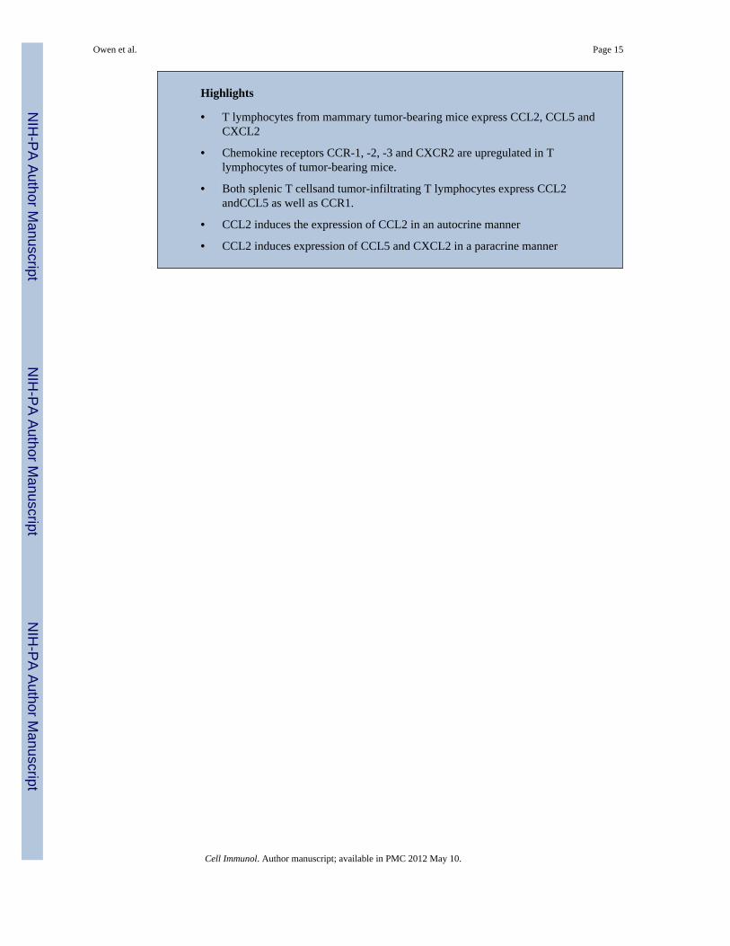
Highlights
• T lymphocytes from mammary tumor-bearing mice express CCL2, CCL5 andCXCL2
• Chemokine receptors CCR-1, -2, -3 and CXCR2 are upregulated in Tlymphocytes of tumor-bearing mice.
• Both splenic T cellsand tumor-infiltrating T lymphocytes express CCL2andCCL5 as well as CCR1.
• CCL2 induces the expression of CCL2 in an autocrine manner
• CCL2 induces expression of CCL5 and CXCL2 in a paracrine manner
Owen et al. Page 15
Cell Immunol. Author manuscript; available in PMC 2012 May 10.
NIH
-PA Author Manuscript
NIH
-PA Author Manuscript
NIH
-PA Author Manuscript
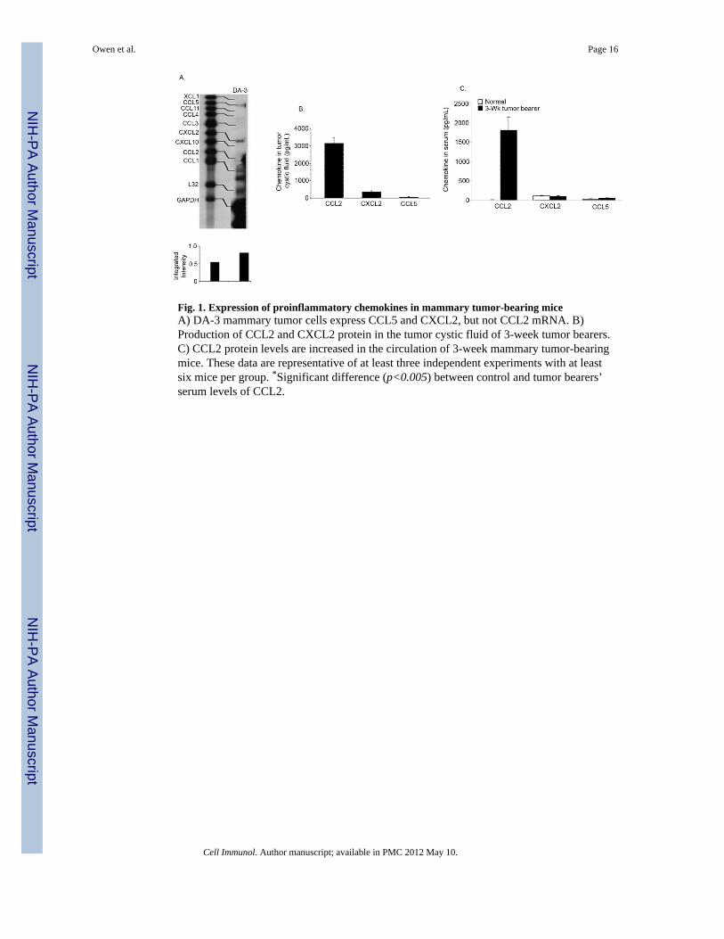
Fig. 1. Expression of proinflammatory chemokines in mammary tumor-bearing miceA) DA-3 mammary tumor cells express CCL5 and CXCL2, but not CCL2 mRNA. B)Production of CCL2 and CXCL2 protein in the tumor cystic fluid of 3-week tumor bearers.C) CCL2 protein levels are increased in the circulation of 3-week mammary tumor-bearingmice. These data are representative of at least three independent experiments with at leastsix mice per group. *Significant difference (p<0.005) between control and tumor bearers’serum levels of CCL2.
Owen et al. Page 16
Cell Immunol. Author manuscript; available in PMC 2012 May 10.
NIH
-PA Author Manuscript
NIH
-PA Author Manuscript
NIH
-PA Author Manuscript
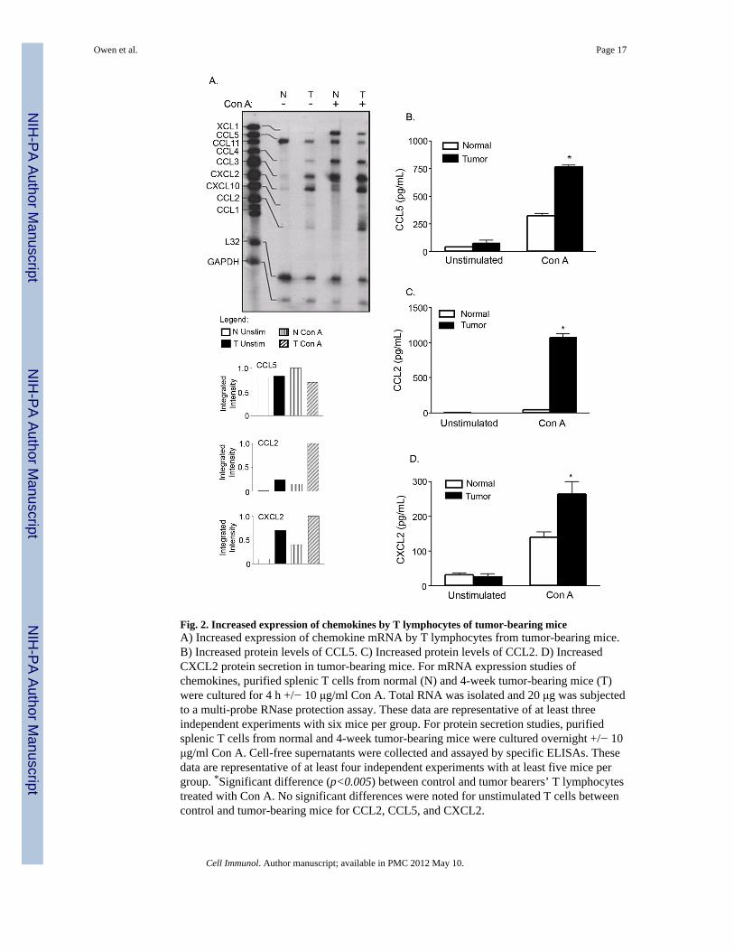
Fig. 2. Increased expression of chemokines by T lymphocytes of tumor-bearing miceA) Increased expression of chemokine mRNA by T lymphocytes from tumor-bearing mice.B) Increased protein levels of CCL5. C) Increased protein levels of CCL2. D) IncreasedCXCL2 protein secretion in tumor-bearing mice. For mRNA expression studies ofchemokines, purified splenic T cells from normal (N) and 4-week tumor-bearing mice (T)were cultured for 4 h +/− 10 μg/ml Con A. Total RNA was isolated and 20 μg was subjectedto a multi-probe RNase protection assay. These data are representative of at least threeindependent experiments with six mice per group. For protein secretion studies, purifiedsplenic T cells from normal and 4-week tumor-bearing mice were cultured overnight +/− 10μg/ml Con A. Cell-free supernatants were collected and assayed by specific ELISAs. Thesedata are representative of at least four independent experiments with at least five mice pergroup. *Significant difference (p<0.005) between control and tumor bearers’ T lymphocytestreated with Con A. No significant differences were noted for unstimulated T cells betweencontrol and tumor-bearing mice for CCL2, CCL5, and CXCL2.
Owen et al. Page 17
Cell Immunol. Author manuscript; available in PMC 2012 May 10.
NIH
-PA Author Manuscript
NIH
-PA Author Manuscript
NIH
-PA Author Manuscript
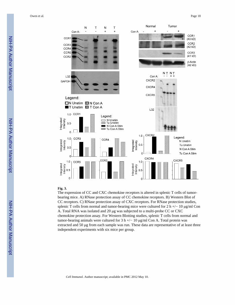
Fig. 3.The expression of CC and CXC chemokine receptors is altered in splenic T cells of tumor-bearing mice. A) RNase protection assay of CC chemokine receptors. B) Western Blot ofCC receptors. C) RNase protection assay of CXC receptors. For RNase protection studies,splenic T cells from normal and tumor-bearing mice were cultured for 2 h +/− 10 μg/ml ConA. Total RNA was isolated and 20 μg was subjected to a multi-probe CC or CXCchemokine protection assay. For Western Blotting studies, splenic T cells from normal andtumor-bearing animals were cultured for 3 h +/− 10 μg/ml Con A. Total protein wasextracted and 50 μg from each sample was run. These data are representative of at least threeindependent experiments with six mice per group.
Owen et al. Page 18
Cell Immunol. Author manuscript; available in PMC 2012 May 10.
NIH
-PA Author Manuscript
NIH
-PA Author Manuscript
NIH
-PA Author Manuscript
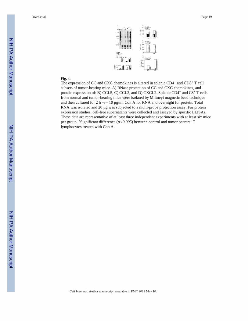
Fig. 4.The expression of CC and CXC chemokines is altered in splenic CD4+ and CD8+ T cellsubsets of tumor-bearing mice. A) RNase protection of CC and CXC chemokines, andprotein expression of: B) CCL5, C) CCL2, and D) CXCL2. Splenic CD4+ and C8+ T cellsfrom normal and tumor-bearing mice were isolated by Miltneyi magnetic bead techniqueand then cultured for 2 h +/− 10 μg/ml Con A for RNA and overnight for protein. TotalRNA was isolated and 20 μg was subjected to a multi-probe protection assay. For proteinexpression studies, cell-free supernatants were collected and assayed by specific ELISAs.These data are representative of at least three independent experiments with at least six miceper group. *Significant difference (p<0.005) between control and tumor bearers’ Tlymphocytes treated with Con A.
Owen et al. Page 19
Cell Immunol. Author manuscript; available in PMC 2012 May 10.
NIH
-PA Author Manuscript
NIH
-PA Author Manuscript
NIH
-PA Author Manuscript
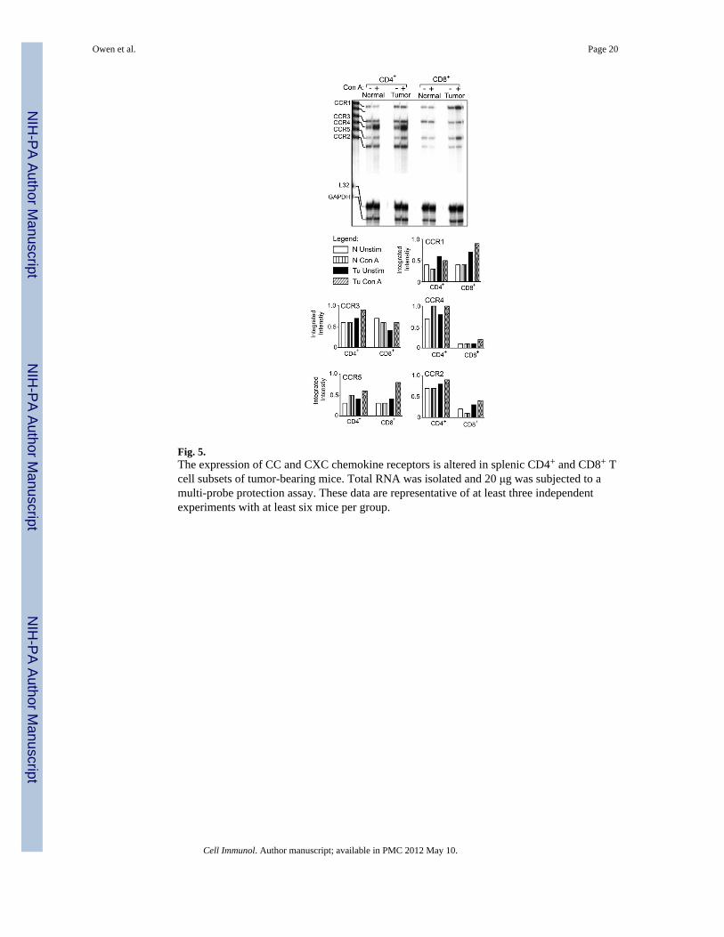
Fig. 5.The expression of CC and CXC chemokine receptors is altered in splenic CD4+ and CD8+ Tcell subsets of tumor-bearing mice. Total RNA was isolated and 20 μg was subjected to amulti-probe protection assay. These data are representative of at least three independentexperiments with at least six mice per group.
Owen et al. Page 20
Cell Immunol. Author manuscript; available in PMC 2012 May 10.
NIH
-PA Author Manuscript
NIH
-PA Author Manuscript
NIH
-PA Author Manuscript
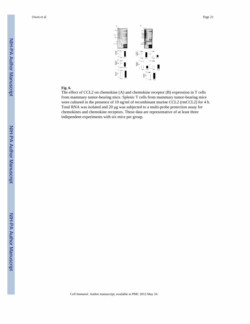
Fig. 6.The effect of CCL2 on chemokine (A) and chemokine receptor (B) expression in T cellsfrom mammary tumor-bearing mice. Splenic T cells from mammary tumor-bearing micewere cultured in the presence of 10 ng/ml of recombinant murine CCL2 (rmCCL2) for 4 h.Total RNA was isolated and 20 μg was subjected to a multi-probe protection assay forchemokines and chemokine receptors. These data are representative of at least threeindependent experiments with six mice per group.
Owen et al. Page 21
Cell Immunol. Author manuscript; available in PMC 2012 May 10.
NIH
-PA Author Manuscript
NIH
-PA Author Manuscript
NIH
-PA Author Manuscript
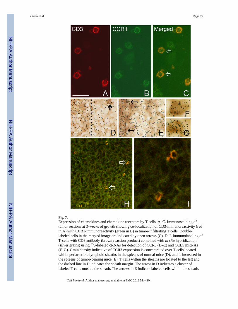
Fig. 7.Expression of chemokines and chemokine receptors by T cells. A–C. Immunostaining oftumor sections at 3-weeks of growth showing co-localization of CD3-immunoreactivity (redin A) with CCR1-immunoreactivity (green in B) in tumor-infiltrating T cells. Double-labeled cells in the merged image are indicated by open arrows (C). D–I. Immunolabeling ofT-cells with CD3 antibody (brown reaction product) combined with in situ hybridization(silver grains) using 35S-labeled cRNAs for detection of CCR3 (D–E) and CCL5 mRNAs(F–G). Grain density indicative of CCR3 expression is concentrated over T cells locatedwithin periarteriole lymphoid sheaths in the spleens of normal mice (D), and is increased inthe spleens of tumor-bearing mice (E). T cells within the sheaths are located to the left andthe dashed line in D indicates the sheath margin. The arrow in D indicates a cluster oflabeled T cells outside the sheath. The arrows in E indicate labeled cells within the sheath.
Owen et al. Page 22
Cell Immunol. Author manuscript; available in PMC 2012 May 10.
NIH
-PA Author Manuscript
NIH
-PA Author Manuscript
NIH
-PA Author Manuscript
F–G. Double labeling of tumor-infiltrating T cells at 3-weeks post tumor implantation forCCL5 and CD3. H-I. Merged confocal microscope images of CD8 (red) and CCL5 (green)immunostained tumor section showing CD8+ T cells that express CCL5 (open arrows in H).These cells are shown in higher maginification in I. Bar in A = 35 μm in A–C and I, 17 μmin D–E, 7 μm in F–G and 93 μm in H.
Owen et al. Page 23
Cell Immunol. Author manuscript; available in PMC 2012 May 10.
NIH
-PA Author Manuscript
NIH
-PA Author Manuscript
NIH
-PA Author Manuscript
Copyright © 2022 FDOKUMEN