CT guided endoscopic recovery of a foreign object from the cranial cavity of an ancient Egyptian...
7
Note: This copy is for your personal non-commercial use only. To order presentation-ready copies for distribution to your colleagues or clients, contact us at www.rsna.org/rsnarights. 2151 SPECIAL EXHIBITS Mislav Čavka, MD • Anja Petaros, MD • Drago Boščić, MD • Lovro Kavur, MD • Ivor Janković, PhD • Radovan Despot, PhD • Jelena Trajković, PhD • Boris Brkljačić, MD, PhD The aim of this study was to test the value of computed tomography (CT)-guided endoscopy in the scientific study of mummified remains and the recovery of unidentified objects from ancient mummified re- mains. CT-guided endoscopy was performed on an Egyptian mummy from the Late Period to help retrieve an unidentified object from its skull. The combined use of CT and endoscopy facilitated the recovery of the object, minimizing further damage to the remains and to the object itself. The successful recovery of the brittle object allowed de- tailed analysis of the item to be performed in an attempt to determine the nature of its presence in the cranial cavity. It was confirmed that the object was a monocotyledon stem fragment. Relying on the exist- ing literature on mummification and excerebration methods in ancient Egypt, we concluded that the stick was probably used for transnasal removal of the brain. The results of this study demonstrate the great potential of CT-guided endoscopy for minimally invasive recovery of small unidentified items from mummies that could yield important in- formation about mummification procedures and the materials used to preserve bodies. © RSNA, 2012 • radiographics.rsna.org Scenes from the Past CT-guided Endoscopic Recovery of a Foreign Object from the Cranial Cavity of an Ancient Egyptian Mummy 1 RadioGraphics 2012; 32:2151–2157 • Published online 10.1148/rg.327115744 • Content Codes: 1 From the University Department of Diagnostic and Interventional Radiology (M.C., B.B.) and Department of Otorhinolaryngology and Cervico- facial Surgery (D.B.), University Hospital Dubrava, Av Gojka Suska 6, 10000 Zagreb, Croatia; Department of Social Medicine and Organization of Health Care, School of Medicine (M.C.), Institute of Wood Science, Faculty of Forestry (R.D., J.T.), and Department of Radiology and Clinical On- cology, School of Medicine (B.B.), University of Zagreb, Zagreb, Croatia; Department of Forensic Medicine and Criminalistics, School of Medicine, Rijeka University, Rijeka, Croatia (A.P.); University Department of Diagnostic and Interventional Radiology, University Hospital Merkur, Zagreb, Croatia (L.K.); and Institute for Anthropological Research, Zagreb, Croatia (I.J.). Received August 31, 2011; revision requested September 15 and received October 10; final version accepted November 15. All authors have no financial relationships to disclose. Address correspondence to M.C. (e-mail: [email protected]). © RSNA, 2012
-
Upload
kbc-zagreb -
Category
Documents
-
view
6 -
download
0
Transcript of CT guided endoscopic recovery of a foreign object from the cranial cavity of an ancient Egyptian...

Note: This copy is for your personal non-commercial use only. To order presentation-ready copies for distribution to your colleagues or clients, contact us at www.rsna.org/rsnarights.
2151SPECIAL EXHIBITS
Mislav Čavka, MD • Anja Petaros, MD • Drago Boščić, MD • Lovro Kavur, MD • Ivor Janković, PhD • Radovan Despot, PhD • Jelena Trajković, PhD • Boris Brkljačić, MD, PhD
The aim of this study was to test the value of computed tomography (CT)-guided endoscopy in the scientific study of mummified remains and the recovery of unidentified objects from ancient mummified re-mains. CT-guided endoscopy was performed on an Egyptian mummy from the Late Period to help retrieve an unidentified object from its skull. The combined use of CT and endoscopy facilitated the recovery of the object, minimizing further damage to the remains and to the object itself. The successful recovery of the brittle object allowed de-tailed analysis of the item to be performed in an attempt to determine the nature of its presence in the cranial cavity. It was confirmed that the object was a monocotyledon stem fragment. Relying on the exist-ing literature on mummification and excerebration methods in ancient Egypt, we concluded that the stick was probably used for transnasal removal of the brain. The results of this study demonstrate the great potential of CT-guided endoscopy for minimally invasive recovery of small unidentified items from mummies that could yield important in-formation about mummification procedures and the materials used to preserve bodies.©RSNA, 2012 • radiographics.rsna.org
Scenes from the PastCT-guided Endoscopic Recovery of a Foreign Object from the Cranial Cavity of an Ancient Egyptian Mummy1
RadioGraphics 2012; 32:2151–2157 • Published online 10.1148/rg.327115744 • Content Codes: 1From the University Department of Diagnostic and Interventional Radiology (M.C., B.B.) and Department of Otorhinolaryngology and Cervico-facial Surgery (D.B.), University Hospital Dubrava, Av Gojka Suska 6, 10000 Zagreb, Croatia; Department of Social Medicine and Organization of Health Care, School of Medicine (M.C.), Institute of Wood Science, Faculty of Forestry (R.D., J.T.), and Department of Radiology and Clinical On-cology, School of Medicine (B.B.), University of Zagreb, Zagreb, Croatia; Department of Forensic Medicine and Criminalistics, School of Medicine, Rijeka University, Rijeka, Croatia (A.P.); University Department of Diagnostic and Interventional Radiology, University Hospital Merkur, Zagreb, Croatia (L.K.); and Institute for Anthropological Research, Zagreb, Croatia (I.J.). Received August 31, 2011; revision requested September 15 and received October 10; final version accepted November 15. All authors have no financial relationships to disclose. Address correspondence to M.C. (e-mail: [email protected]).
©RSNA, 2012
2152 November-December 2012 radiographics.rsna.org
Figure 1. Coronal volume-rendered CT image shows a tubular object in the cranial cavity of a female Egyptian mummy from the Late Period.
IntroductionThe use of radiology to study archaeologic re-mains started soon after the initial discovery of x-rays in 1895 by German physicist Wilhelm Konrad Roentgen. W. Koenig (1) was the first to use x-rays to study the anatomy of mummies in his 1896 monograph entitled “14 Photographs with X rays taken by the Physical Society of Frankfurt am Main.” Other studies of mum-mies followed, and conventional radiology soon became one of the preferred methods for the scientific study of mummies (2–4). (For a review of the use of radiology in the scientific studies of mummies, see the studies by Aufderheide [5] and Böni et al [6] and the references listed therein.) During the pioneering period of the use of radiol-ogy to study archaeologic remains, radiography was mostly performed to determine the authen-ticity of a mummy without having to unwrap the corpse and to reconstruct its biologic profile by assessing its sex and age at the time of death (7). The invention of computed tomography (CT) in the 1970s allowed paleoradiologists to visualize paleopathologic lesions, interpret paleopathologic conditions, and diagnose paleodiseases. Exclud-ing some potential pitfalls in image interpretation caused by postmortem dehydration and decom-position of tissues, CT is the modality of choice for studying mummified remains because it al-lows detailed visualization of the body and its in-ternal contents and associated artifacts, which are often not discernible at conventional radiography (8,9). Today, CT also offers the possibility of ste-reolithographic modeling and facial reconstruc-tion from three-dimensional (3D) CT data (10). Before the invention of CT, endoscopes were used as a minimally invasive tool to investigate the interior of enclosed areas and acquire infor-mation about their contents. Currently, conven-tional endoscopy is used both for tissue biopsy to obtain tissue samples and for recovery of artifacts from within the body that cannot be retrieved via virtual endoscopy (11). Researchers are still in-vestigating and testing novel techniques that may further improve the study of ancient mummified remains, making the imaging field one of the most interesting research areas in paleoanthro-pology and paleopathology (12–14).
In this article, we discuss the aim of this study, which was to test, for the first time (to our knowl-edge), the value of the simultaneous use of CT and endoscopy to image mummies and the ad-ditional effects of CT technology on endoscopic
field of view for visualization of structures that are not directly visible to the endoscope and retrieval of small unidentified items from the corpse with minimal damage. This technique was used to ex-tract an unidentified tubular structure from the cranium of an Egyptian mummy that was detected during an earlier paleoradiologic analysis of the specimen (15). In addition, we were interested in identifying and further analyzing the object.
Materials and MethodsThe mummy that was studied belongs to the Archaeological Museum in Zagreb, Croatia (in-ventory number 666). It was originally donated to the museum by J. Haulik (1837–1869), Bishop and Archibishop of Zagreb, and was consequent-ly stored in a replica of an Egyptian sarcopha-gus. In 2008, results of targeted paleoradiologic analysis with radiography, multidetector CT, and radiocarbon dating suggested that the mummy was a middle-aged woman from the Late Period (XXVIII–XXX dynasty) (15).
During the analysis, a tubular structure was detected in the cranial cavity. The object extend-ed from the left parietal bone to the resin-like flu-id in the occiput. Differences in the density of the structure were observed: The inner portion had attenuation values ranging from 300 HU to 450 HU, whereas the outer surface had attenuation values ranging from 0 HU to 100 HU, findings indicative of an artificial object. Although previ-ous studies have described similar intracranial
RG • Volume 32 Number 7 Cavka et al 2153
structures that were, in fact, remnants of the falx, tentorium, and dura mater (16), it was concluded that only removal of the object would reveal its true nature and origin.
CT-guided endoscopy was performed at the University Department of Diagnostic and Inter-ventional Radiology, University Hospital Dubra-va, Zagreb, Croatia. Axial sections were obtained with a multidetector CT scanner (Sensation 16; Siemens Medical Solutions, Erlangen, Germany) and the following parameters: 16 × 0.75 colli-mation and 0.7-mm reconstruction increments. Sagittal and coronal reformatting and 3D recon-structions were performed on a Leonardo (Sie-mens Medical Solutions, Erlangen, Germany) and Aquarius (TeraRecon, San Mateo, Calif) workstation with OsiriX Imaging Software (Pix-meo, Geneve, Switzerland). Endoscopy was per-formed with a minimal incision in the skin of the nose and virtually no damage by using an artifi-
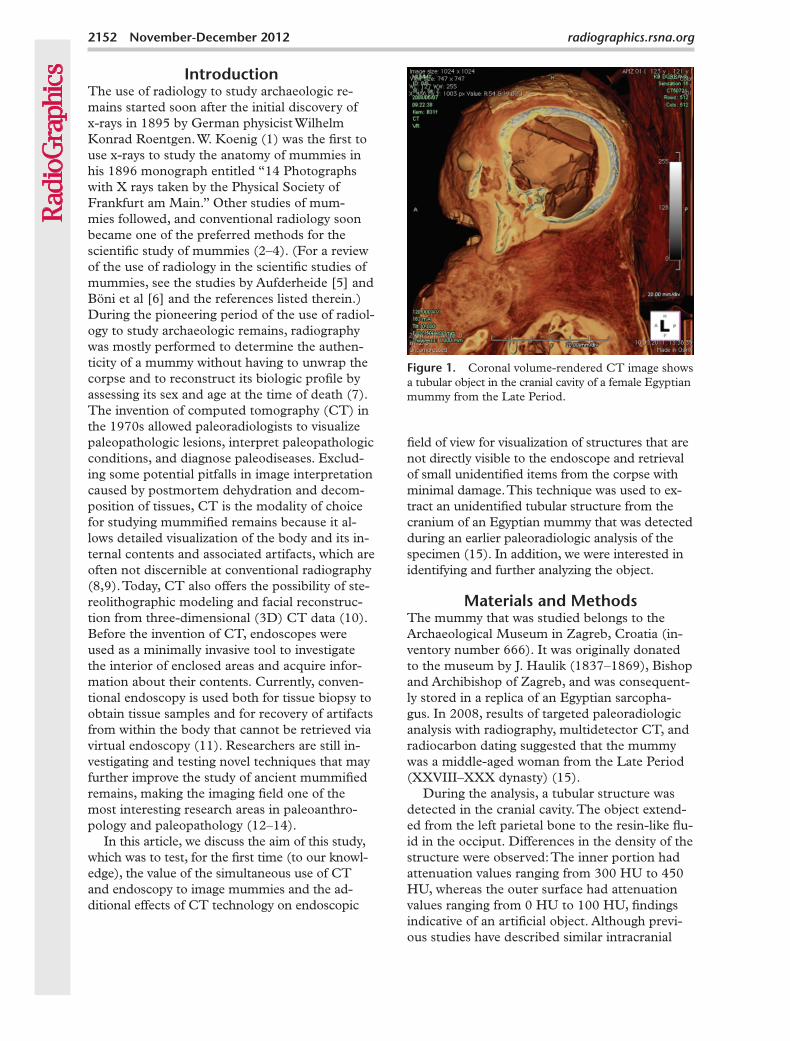
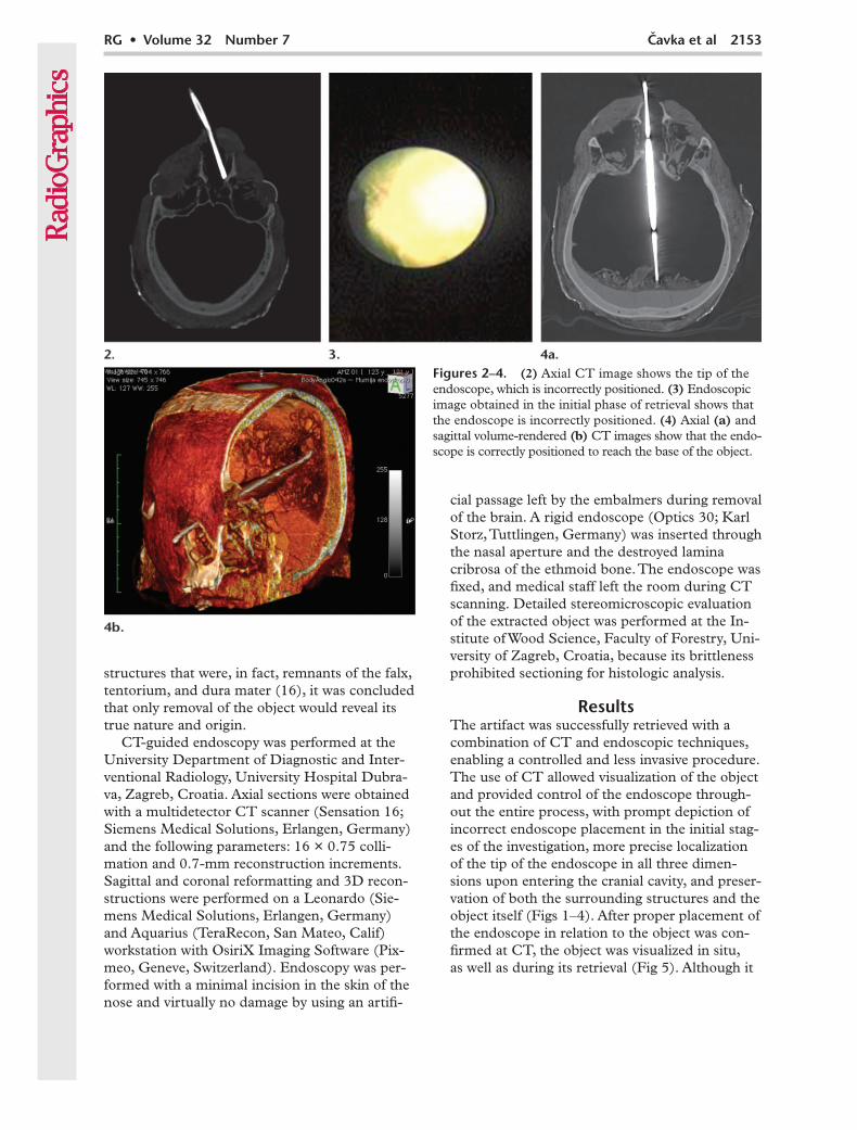
Figures 2–4. (2) Axial CT image shows the tip of the endoscope, which is incorrectly positioned. (3) Endoscopic image obtained in the initial phase of retrieval shows that the endoscope is incorrectly positioned. (4) Axial (a) and sagittal volume-rendered (b) CT images show that the endo-scope is correctly positioned to reach the base of the object.
cial passage left by the embalmers during removal of the brain. A rigid endoscope (Optics 30; Karl Storz, Tuttlingen, Germany) was inserted through the nasal aperture and the destroyed lamina cribrosa of the ethmoid bone. The endoscope was fixed, and medical staff left the room during CT scanning. Detailed stereomicroscopic evaluation of the extracted object was performed at the In-stitute of Wood Science, Faculty of Forestry, Uni-versity of Zagreb, Croatia, because its brittleness prohibited sectioning for histologic analysis.
ResultsThe artifact was successfully retrieved with a combination of CT and endoscopic techniques, enabling a controlled and less invasive procedure. The use of CT allowed visualization of the object and provided control of the endoscope through-out the entire process, with prompt depiction of incorrect endoscope placement in the initial stag-es of the investigation, more precise localization of the tip of the endoscope in all three dimen-sions upon entering the cranial cavity, and preser-vation of both the surrounding structures and the object itself (Figs 1–4). After proper placement of the endoscope in relation to the object was con-firmed at CT, the object was visualized in situ, as well as during its retrieval (Fig 5). Although it

2154 November-December 2012 radiographics.rsna.org
Figure 5. Visualization of the object within the cranial cavity after verification of proper endoscope placement. (a) Endoscopic image shows the object in situ. (b) Endoscopic image shows the base of the object. (c) Endoscopic image shows the clip reaching for the base of the object.
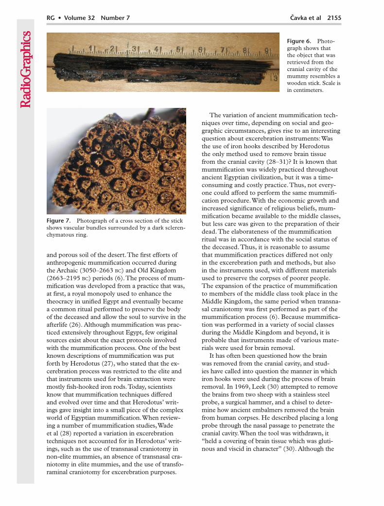
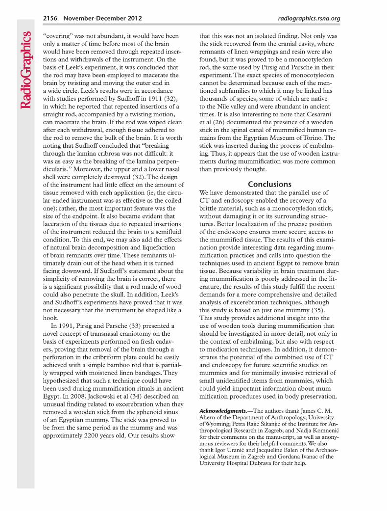
resembled a wooden stick at macroscopic inspec-tion, more detailed microscopic analysis and the presence of lignocelluloses suggested that the ar-tifact was part of a monocotyledon stem (Fig 6). Because the stick was too brittle to make a proper microscopy slide, stereomicroscopic analysis of a cross section of the specimen revealed many vascular bundles, each of which was surrounded by a dark sclerenchymatous ring and scattered in the parenchymal tissue (Fig 7). These features are characteristic of monocot stem anatomy and suggest a monocot plant stem from the botanical family Poaceae, which includes cane (subfamily Arundinoideae) and bamboo (subfamily Bambu-soideae). It is much less probable that the stick is part of a palm stem (subfamily Palmae).
DiscussionIn paleopathology, endoscopy is a minimally in-vasive technique that usually follows noninvasive CT of the specimen (17,18). It has proved to be very useful in paleopathologic investigations of mummified remains because it provides additional information about subtle pathologic alterations that are not easily observable at multidetector CT and enables tissue samples to be obtained for further analysis (19). Depending on the aim of the research, endoscopes may be inserted into dif-ferent body regions by using artificial or natural openings (11). In modern medicine, intracranial endoscopy has been recognized as one of the most important neurosurgical techniques for resect-ing intraventricular, sellar, parasellar, intraspinal,
spinal, extraaxial, and vascular lesions (20,21). The accuracy of these techniques is increased with the use of CT and endoscopy, a combina-tion that allows controlled computerized neuro-navigation (22). Although intracranial endoscopy has already been used in various paleopathologic studies on mummies, CT-guided endoscopy had never been performed (23–25), to our knowledge. Even though the utility of CT-guided biopsy in mummies was previously reported, our study demonstrated the importance of combined CT and endoscopy in recovering small unidentified objects from mummies (5,12). The additive ef-fect of CT data overcame the main limitation of endoscopy (ie, a narrow field of view), helping to better visualize and localize structures that are not directly visible by the endoscope. In this way, it enhances the investigative procedure and extrac-tion. We have demonstrated how the combined use of CT and endoscopy facilitated the recovery of a monocot plant stick enclosed in the cranial cavity of a mummified woman from the Late Period by controlling the approach trajectory of the endo-scope. Both the surrounding structures and the stick were preserved because real time localization of the endoscope guaranteed easy and rapid access to the stick, thereby avoiding unnecessary contact with surrounding structures during its extraction that could damage the brittle artifact.
How and why the stick entered the skull is unknown, but it may be discussed in light of our current knowledge of mummification techniques. The first Egyptian mummies (circa 5000 bc) origi-nate from the Predynastic period. These mummies were naturally desiccated by the arid environment
RG • Volume 32 Number 7 Cavka et al 2155
Figure 7. Photograph of a cross section of the stick shows vascular bundles surrounded by a dark scleren-chymatous ring.
and porous soil of the desert. The first efforts of anthropogenic mummification occurred during the Archaic (3050–2663 bc) and Old Kingdom (2663–2195 bc) periods (6). The process of mum-mification was developed from a practice that was, at first, a royal monopoly used to enhance the theocracy in unified Egypt and eventually became a common ritual performed to preserve the body of the deceased and allow the soul to survive in the afterlife (26). Although mummification was prac-ticed extensively throughout Egypt, few original sources exist about the exact protocols involved with the mummification process. One of the best known descriptions of mummification was put forth by Herodotus (27), who stated that the ex-cerebration process was restricted to the elite and that instruments used for brain extraction were mostly fish-hooked iron rods. Today, scientists know that mummification techniques differed and evolved over time and that Herodotus’ writ-ings gave insight into a small piece of the complex world of Egyptian mummification. When review-ing a number of mummification studies, Wade et al (28) reported a variation in excerebration techniques not accounted for in Herodotus’ writ-ings, such as the use of transnasal craniotomy in non-elite mummies, an absence of transnasal cra-niotomy in elite mummies, and the use of transfo-raminal craniotomy for excerebration purposes.
The variation of ancient mummification tech-niques over time, depending on social and geo-graphic circumstances, gives rise to an interesting question about excerebration instruments: Was the use of iron hooks described by Herodotus the only method used to remove brain tissue from the cranial cavity (28–31)? It is known that mummification was widely practiced throughout ancient Egyptian civilization, but it was a time-consuming and costly practice. Thus, not every-one could afford to perform the same mummifi-cation procedure. With the economic growth and increased significance of religious beliefs, mum-mification became available to the middle classes, but less care was given to the preparation of their dead. The elaborateness of the mummification ritual was in accordance with the social status of the deceased. Thus, it is reasonable to assume that mummification practices differed not only in the excerebration path and methods, but also in the instruments used, with different materials used to preserve the corpses of poorer people. The expansion of the practice of mummification to members of the middle class took place in the Middle Kingdom, the same period when transna-sal craniotomy was first performed as part of the mummification process (6). Because mummifica-tion was performed in a variety of social classes during the Middle Kingdom and beyond, it is probable that instruments made of various mate-rials were used for brain removal.
It has often been questioned how the brain was removed from the cranial cavity, and stud-ies have called into question the manner in which iron hooks were used during the process of brain removal. In 1969, Leek (30) attempted to remove the brains from two sheep with a stainless steel probe, a surgical hammer, and a chisel to deter-mine how ancient embalmers removed the brain from human corpses. He described placing a long probe through the nasal passage to penetrate the cranial cavity. When the tool was withdrawn, it “held a covering of brain tissue which was gluti-nous and viscid in character” (30). Although the
Figure 6. Photo-graph shows that the object that was retrieved from the cranial cavity of the mummy resembles a wooden stick. Scale is in centimeters.
2156 November-December 2012 radiographics.rsna.org
“covering” was not abundant, it would have been only a matter of time before most of the brain would have been removed through repeated inser-tions and withdrawals of the instrument. On the basis of Leek’s experiment, it was concluded that the rod may have been employed to macerate the brain by twisting and moving the outer end in a wide circle. Leek’s results were in accordance with studies performed by Sudhoff in 1911 (32), in which he reported that repeated insertions of a straight rod, accompanied by a twisting motion, can macerate the brain. If the rod was wiped clean after each withdrawal, enough tissue adhered to the rod to remove the bulk of the brain. It is worth noting that Sudhoff concluded that “breaking through the lamina cribrosa was not difficult: it was as easy as the breaking of the lamina perpen-dicularis.” Moreover, the upper and a lower nasal shell were completely destroyed (32). The design of the instrument had little effect on the amount of tissue removed with each application (ie, the circu-lar-ended instrument was as effective as the coiled one); rather, the most important feature was the size of the endpoint. It also became evident that laceration of the tissues due to repeated insertions of the instrument reduced the brain to a semifluid condition. To this end, we may also add the effects of natural brain decomposition and liquefaction of brain remnants over time. These remnants ul-timately drain out of the head when it is turned facing downward. If Sudhoff’s statement about the simplicity of removing the brain is correct, there is a significant possibility that a rod made of wood could also penetrate the skull. In addition, Leek’s and Sudhoff’s experiments have proved that it was not necessary that the instrument be shaped like a hook.
In 1991, Pirsig and Parsche (33) presented a novel concept of transnasal craniotomy on the basis of experiments performed on fresh cadav-ers, proving that removal of the brain through a perforation in the cribriform plate could be easily achieved with a simple bamboo rod that is partial-ly wrapped with moistened linen bandages. They hypothesized that such a technique could have been used during mummification rituals in ancient Egypt. In 2008, Jackowski et al (34) described an unusual finding related to excerebration when they removed a wooden stick from the sphenoid sinus of an Egyptian mummy. The stick was proved to be from the same period as the mummy and was approximately 2200 years old. Our results show
that this was not an isolated finding. Not only was the stick recovered from the cranial cavity, where remnants of linen wrappings and resin were also found, but it was proved to be a monocotyledon rod, the same used by Pirsig and Parsche in their experiment. The exact species of monocotyledon cannot be determined because each of the men-tioned subfamilies to which it may be linked has thousands of species, some of which are native to the Nile valley and were abundant in ancient times. It is also interesting to note that Cesarani et al (26) documented the presence of a wooden stick in the spinal canal of mummified human re-mains from the Egyptian Museum of Torino. The stick was inserted during the process of embalm-ing. Thus, it appears that the use of wooden instru-ments during mummification was more common than previously thought.
ConclusionsWe have demonstrated that the parallel use of CT and endoscopy enabled the recovery of a brittle material, such as a monocotyledon stick, without damaging it or its surrounding struc-tures. Better localization of the precise position of the endoscope ensures more secure access to the mummified tissue. The results of this exami-nation provide interesting data regarding mum-mification practices and calls into question the techniques used in ancient Egypt to remove brain tissue. Because variability in brain treatment dur-ing mummification is poorly addressed in the lit-erature, the results of this study fulfill the recent demands for a more comprehensive and detailed analysis of excerebration techniques, although this study is based on just one mummy (35). This study provides additional insight into the use of wooden tools during mummification that should be investigated in more detail, not only in the context of embalming, but also with respect to medication techniques. In addition, it demon-strates the potential of the combined use of CT and endoscopy for future scientific studies on mummies and for minimally invasive retrieval of small unidentified items from mummies, which could yield important information about mum-mification procedures used in body preservation.
Acknowledgments.—The authors thank James C. M. Ahern of the Department of Anthropology, University of Wyoming; Petra Rajić Šikanjić of the Institute for An-thropological Research in Zagreb; and Nadja Komnenić for their comments on the manuscript, as well as anony-mous reviewers for their helpful comments. We also thank Igor Uranić and Jacqueline Balen of the Archaeo-logical Museum in Zagreb and Gordana Ivanac of the University Hospital Dubrava for their help.

RG • Volume 32 Number 7 Cavka et al 2157
References 1. Koenig W. 14 Photographien von Roenthen-
Strahlen aufgenommen im Physikalischen Verein zu Frankfurt am Main. Leipzig, Germany: Johann Ambrosius Barth, 1896.
2. Dedekind A. Durchleuchtung von Mummien mit-telst Roentgenstrahlen. Prometheus 1897;384: 318–319.
3. Londe A. Les rayons Roentgen et les momies. Na-ture 1897;25(2):103–105.
4. Fiori MG, Nunzi MG. The earliest documented applications of X-rays to examination of mummified remains and archaeological materials. J R Soc Med 1995;88(2):67–69.
5. Aufderheide AC. The scientific study of mummies. Cambridge, England: Cambridge University Press, 2003.
6. Böni T, Rühli FJ, Chhem RK. History of paleora-diology: early published literature, 1896-1921. Can Assoc Radiol J 2004;55(4):203–210.
7. Rühli FJ, Chhem RK, Böni T. Diagnostic paleo-radiology of mummified tissue: interpretation and pitfalls. Can Assoc Radiol J 2004;55(4):218–227.
8. Reyman TA, Nielsen H, Thuesen I, et al. Mummies and technology: new investigative techniques. In: Cockburn A, Cockburn E, Reyman T, eds. Mum-mies, disease and ancient cultures. Cambridge, Eng-land: Cambridge University Press, 1998; 353–394.
9. Hjalgrim H, Lynnerup N, Liversage M, Rosenklint A. Stereolithography: potential applications in an-thropological studies. Am J Phys Anthropol 1995;97 (3):329–333.
10. Lynnerup N. Mummies. Am J Phys Anthropol 2007;45:162–190.
11. Rühli FJ, Hodler J, Böni T. Technical note: CT-guided biopsy—a new diagnostic method for paleo-pathological research. Am J Phys Anthropol 2002; 117(3):272–275.
12. Ohrström L, Bitzer A, Walther M, Rühli FJ. Techni-cal note: terahertz imaging of ancient mummies and bone. Am J Phys Anthropol 2010;142(3):497–500.
13. Rühli FJ, von Waldburg H, Nielles-Vallespin S, Böni T, Speier P. Clinical magnetic resonance imaging of ancient dry human mummies without rehydration. JAMA 2007;298(22):2618–2620.
14. Cavka M, Janković I, Sikanjić PR, et al. Insights into a mummy: a paleoradiological analysis. Coll Antrop-ol 2010;34(3):797–802.
15. Marx M, D’Auria SH. Three-dimensional CT re-constructions of an ancient human Egyptian mum-my. AJR Am J Roentgenol 1988;150(1):147–149.
16. Notman DN, Tashjian J, Aufderheide AC, et al. Modern imaging and endoscopic biopsy techniques in Egyptian mummies. AJR Am J Roentgenol 1986; 146(1):93–96.
17. Lim DS, Lee IS, Choi KJ, et al. The potential for non-invasive study of mummies: validation of the use of computerized tomography by post factum dissection and histological examination of a 17th century female Korean mummy. J Anat 2008;213 (4):482–495.
18. Kim SB, Shin JE, Park SS, et al. Endoscopic investi-gation of the internal organs of a 15th-century child mummy from Yangju, Korea. J Anat 2006;209(5): 681–688.
19. Fries G, Perneczky A. Intracranial endoscopy. Adv Tech Stand Neurosurg 1999;25:21–60.
20. Mayberg MR, LaPresto E, Cunningham EJ. Image-guided endoscopy: description of technique and potential applications. Neurosurg Focus 2005;19(1): E10.
21. Schroeder HW, Gaab MR. Intracranial endoscopy. Neurosurg Focus 1999;6(4):e1.
22. Manialawi M, Meligy R, Bucaille M. Endoscopic examination of Egyptian mummies. Endoscopy 1978;10(3):191–194.
23. Hagedorn HG, Zink A, Szeimies U, Nerlich AG. Macroscopic and endoscopic examinations of the head and neck region in ancient Egyptian mummies [in German]. HNO 2004;52(5):413–422.
24. Sokiranski R, Pirsig W, Richter HP, Lösch S, Struck U, Nerlich AG. Unique paleopathology in a pre-Columbian mummy remnant from Southern Peru: severe cervical rotation trauma with subluxation of the axis as cause of death. Acta Neurochir (Wien) 2011;153(3):609–616.
25. Thumfart WF, Freysinger W, Gunkel AR, Truppe MJ. 3D image-guided surgery on the example of the 5,300-year-old Innsbruck Iceman. Acta Otolar-yngol 1997;117(2):131–134.
26. Cesarani F, Martina MC, Ferraris A, et al. Whole-body three-dimensional multidetector CT of 13 Egyptian human mummies. AJR Am J Roentgenol 2003;180(3):597–606.
27. Herodotus. Histories Book II 86-89 (translation by De Selincourt A). London, England: Penguin Clas-sics, 1996.
28. Wade AD, Nelson AJ, Garvin GJ. A synthetic radio-logical study of brain treatment in ancient Egyptian mummies. Homo 2011;62(4):248–269.
29. Gray PHK. Notes concerning the position of arms and hands of mummies with a view to possible dating of the specimen. J Egypt Archaeol 1972;58: 200–204.
30. Leek FF. The problem of brain removal during em-balming by the ancient Egyptians. J Egypt Archaeol 1969;55:112–116.
31. Pringle H. 7th World Congress on Mummy Studies. For best ticket to the afterlife, pay up. Science 2011; 333(6041):403.
32. Sudhoff K. Agyptische Mumienmacher-Instru-mente. Arch Gesch Med 1911;5:165–171.
33. Pirsig W, Parsche F. Instruments for transnasal brain removal during embalming by the ancient Egyptians. Int J Anthropol 1991;6(1):67–74.
34. Jackowski C, Bolliger S, Thali MJ. Common and un-expected findings in mummies from ancient Egypt and South America as revealed by CT. RadioGraph-ics 2008;28(5):1477–1492.
35. Nelson AJ, Conologue G, Beckett R, et al. Multi-modal analyses of variability in transnasal crani-otomy lesions in Egyptian mummies. In: Poster pre-sented at the 34th annual meeting of the Paleopa-thology Association, Philadelphia, March 27, 2007.