The crystal structure of unmodified tRNAPhe from Escherichia coli
Upload
khangminh22Category
view
7download
0
392 https://doi.org/10.1107/S205698901900241X Acta Cryst. (2019). E75, 392–396
research communications
Received 13 February 2019
Accepted 15 February 2019
Edited by W. T. A. Harrison, University of
Aberdeen, Scotland
‡ Additional correspondence author, e-mail:
Keywords: crystal structure; oxopyridazinyl;
ester; Hirshfeld surface analysis.
CCDC reference: 1897511
Supporting information: this article has
supporting information at journals.iucr.org/e
Ethyl 2-(4-benzyl-3-methyl-6-oxo-1,6-dihydropyri-dazin-1-yl)acetate: crystal structure and Hirshfeldsurface analysis
Younes Zaoui,a Youssef Ramli,a‡ Jamal Taoufik,a Joel T. Mague,b Mukesh M.
Jotani,c Edward R. T. Tiekinkd* and M’hammed Ansara
aLaboratory of Medicinal Chemistry, Drug Sciences Research Center, Faculty of Medicine and Pharmacy, Mohammed V
University, Rabat, Morocco, bDepartment of Chemistry, Tulane University, New Orleans, LA 70118, USA, cDepartment
of Physics, Bhavan’s Sheth R. A. College of Science, Ahmedabad, Gujarat 380001, India, and dResearch Centre for
Crystalline Materials, School of Science and Technology, Sunway University, 47500 Bandar Sunway, Selangor Darul
Ehsan, Malaysia. *Correspondence e-mail: [email protected]
The title compound, C16H18N2O3, is constructed about a central oxopyridazinyl
ring (r.m.s. deviation = 0.0047 A), which is connected to an ethylacetate group at
the N atom closest to the carbonyl group, and benzyl and methyl groups second
furthest and furthest from the carbonyl group, respectively. An approximately
orthogonal relationship exists between the oxopyridazinyl ring and the best
plane through the ethylacetate group [dihedral angle = 77.48 (3)�]; the latter lies
to one side of the central plane [the Nr—Nr—Cm—Cc (r = ring, m = methylene, c
= carbonyl) torsion angle being 104.34 (9)�]. In the crystal, both H atoms of the
N-bound methylene group form methylene-C—H� � �O(ring carbonyl) or
N(pyridazinyl) interactions, resulting in the formation of a supramolecular tape
along the a-axis direction. The tapes are assembled into a three-dimensional
architecture by methyl- and phenyl-C—H� � �O(ring carbonyl) and phenyl-C—
H� � �O(ester carbonyl) interactions. The analysis of the calculated Hirshfeld
surface indicates the dominance of H� � �H contacts to the overall surface (i.e.
52.2%). Reflecting other identified points of contact between molecules noted
above, O� � �H/H� � �O (23.3%), C� � �H/H� � �C (14.7%) and N� � �H/H� � �N (6.6%)
contacts also make significant contributions to the surface.
1. Chemical context
Pyridazin-3(2H)-ones are pyridazine derivatives, being
constructed about a six-membered ring which contains two
adjacent nitrogen atoms, at positions one and two, and with a
carbonyl group at position three. The interest in these nitro-
gen-rich heterocyclic derivatives arises from the fact that they
exhibit a number of promising pharmacological and biological
activities. These include anti-oxidant (Khokra et al., 2016),
anti-bacterial and anti-fungal (Abiha et al. 2018), anti-cancer
(Kamble et al. 2017), analgesic and anti-inflammatory
(Ibrahim et al. 2017), anti-depressant (Boukharsa et al. 2016)
and anti-ulcer activities (Yamada et al., 1981). In addition, a
number of pyridazinone derivatives have been reported to
have potential as agrochemicals, for example as insecticides
(Nauen & Bretschneider, 2002), acaricides (Igarashi & Saka-
moto, 1994) and herbicides (Azaari et al., 2016). Given the
interest in this class of compound and the paucity in structural
data (see Database survey), the crystal and molecular struc-
tures of the the title pyridazin-3(2H)-one derivative, (I), has
ISSN 2056-9890
brought to you by COREView metadata, citation and similar papers at core.ac.uk
provided by Sunway Institutional Repository
been undertaken along with an analysis of the calculated
Hirshfeld surface in order to gain further insight into the
molecular packing.
2. Structural commentary
The molecular structure of (I), Fig. 1, comprises a central
oxopyridazinyl ring connected to an ethylacetate group at the
N1 atom, a methyl group at the C2 position and a benzyl
residue at the C3 atom. The oxopyridazinyl ring is almost
planar, having an r.m.s. deviation of 0.0047 A for the ring
atoms, with the maximum deviation from the ring being
0.0072 (6) A for the C3 atom; the O1 atom lies 0.0260 (13) A
out of the plane in the same direction as the C3 atom. The
ethyl acetate group is close to planar with the r.m.s. deviation
for the O2,O3,C12–C16 atoms being 0.0476 A [the maximum
deviation from the least-squares plane is 0.0711 (7) A for the
O3 atom]. The dihedral angle between the two mentioned
planes is 77.48 (3)�, indicating an approximately orthogonal
relationship. The ethyl acetate group lies to one side of the
central plane, as seen in the value of the N2—N1—C13—C14
torsion angle of 104.34 (9)�. The benzyl ring forms a dihedral
angle of 76.94 (3)� with the central ring, also indicating an
approximately orthogonal relationship but, in this case, the
benzyl ring is bisected by the pseudo mirror plane passing
through the oxopyridazinyl ring. Consistent with this, the
pendant groups form a dihedral angle of 69.74 (3)�. Within the
ester group, it is the carboxylate-O3 atom that is directed away
from the oxopyridazinyl ring so that the carbonyl-O1 and O2
atoms are proximate, at least to a first approximation.
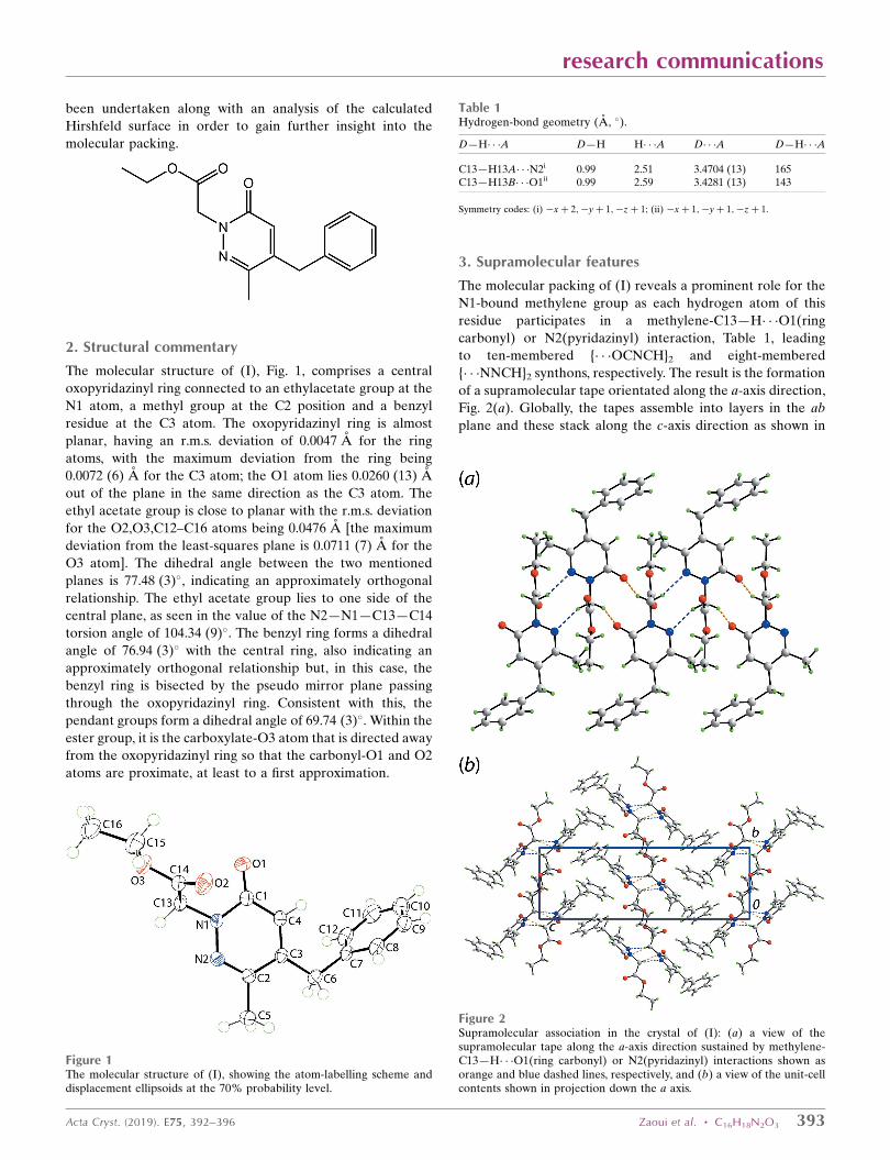
3. Supramolecular features
The molecular packing of (I) reveals a prominent role for the
N1-bound methylene group as each hydrogen atom of this
residue participates in a methylene-C13—H� � �O1(ring
carbonyl) or N2(pyridazinyl) interaction, Table 1, leading
to ten-membered {� � �OCNCH}2 and eight-membered
{� � �NNCH}2 synthons, respectively. The result is the formation
of a supramolecular tape orientated along the a-axis direction,
Fig. 2(a). Globally, the tapes assemble into layers in the ab
plane and these stack along the c-axis direction as shown in
research communications
Acta Cryst. (2019). E75, 392–396 Zaoui et al. � C16H18N2O3 393
Table 1Hydrogen-bond geometry (A, �).
D—H� � �A D—H H� � �A D� � �A D—H� � �A
C13—H13A� � �N2i 0.99 2.51 3.4704 (13) 165C13—H13B� � �O1ii 0.99 2.59 3.4281 (13) 143
Symmetry codes: (i) �xþ 2;�yþ 1;�zþ 1; (ii) �xþ 1;�yþ 1;�zþ 1.
Figure 2Supramolecular association in the crystal of (I): (a) a view of thesupramolecular tape along the a-axis direction sustained by methylene-C13—H� � �O1(ring carbonyl) or N2(pyridazinyl) interactions shown asorange and blue dashed lines, respectively, and (b) a view of the unit-cellcontents shown in projection down the a axis.
Figure 1The molecular structure of (I), showing the atom-labelling scheme anddisplacement ellipsoids at the 70% probability level.
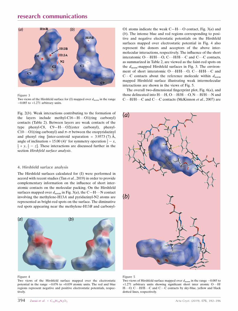
Fig. 2(b). Weak interactions contributing to the formation of
the layers include methyl-C16—H� � �O1(ring carbonyl)
contacts (Table 2). Between layers are weak contacts of the
type phenyl-C8, C9—H� � �O2(ester carbonyl), phenyl-
C10� � �O1(ring carbonyl) and �–� between the oxopyridazinyl
and phenyl ring [inter-centroid separation = 3.9573 (7) A,
angle of inclination = 15.00 (4)� for symmetry operation 32 � x,
12 + y, 1
2 � z]. These interactions are discussed further in the
section Hirshfeld surface analysis.
4. Hirshfeld surface analysis
The Hirshfeld surfaces calculated for (I) were performed in
accord with recent studies (Tan et al., 2019) in order to provide
complementary information on the influence of short inter-
atomic contacts on the molecular packing. On the Hirshfeld
surfaces mapped over dnorm in Fig. 3(a), the C—H� � �N contact
involving the methylene-H13A and pyridazinyl-N2 atoms are
represented as bright-red spots on the surface. The diminutive
red spots appearing near the methylene-H13B and carbonyl-
O1 atoms indicate the weak C—H� � �O contact, Fig. 3(a) and
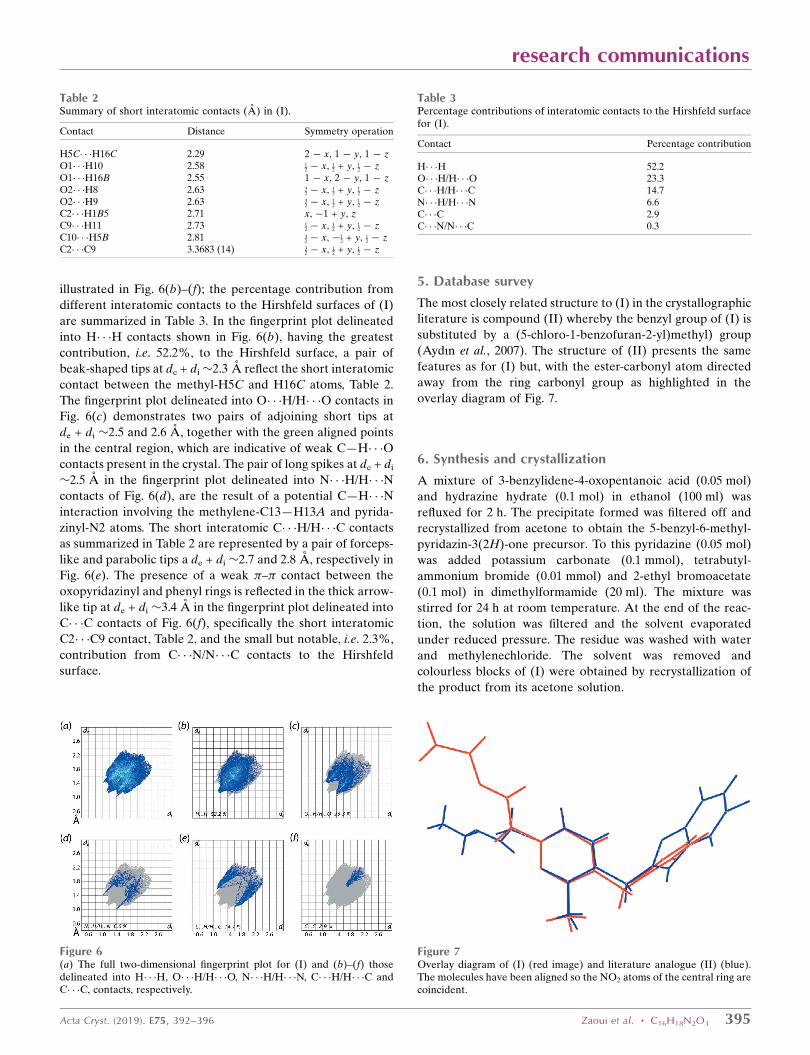
(b). The intense blue and red regions corresponding to posi-
tive and negative electrostatic potentials on the Hirshfeld
surfaces mapped over electrostatic potential in Fig. 4 also
represent the donors and acceptors of the above inter-
molecular interactions, respectively. The influence of the short
interatomic O� � �H/H� � �O, C� � �H/H� � �C and C� � �C contacts,
as summarized in Table 2, are viewed as the faint-red spots on
the dnorm-mapped Hirshfeld surfaces in Fig. 3. The environ-
ment of short interatomic O� � �H/H� � �O, C� � �H/H� � �C and
C� � �C contacts about the reference molecule within dnorm
mapped Hirshfeld surface illustrating weak intermolecular
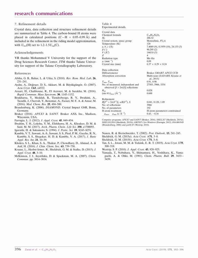
interactions are shown in the views of Fig. 5.
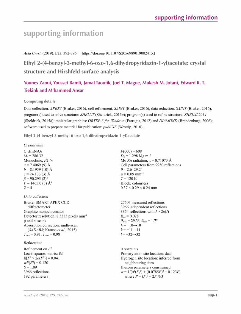
The overall two-dimensional fingerprint plot, Fig. 6(a), and
those delineated into H� � �H, O� � �H/H� � �O, N� � �H/H� � �N and
C� � �H/H� � �C and C� � �C contacts (McKinnon et al., 2007) are
394 Zaoui et al. � C16H18N2O3 Acta Cryst. (2019). E75, 392–396
research communications
Figure 3Two views of the Hirshfeld surface for (I) mapped over dnorm in the range�0.085 to +1.271 arbitrary units.
Figure 4Two views of the Hirshfeld surface mapped over the electrostaticpotential in the range �0.076 to +0.039 atomic units. The red and blueregions represent negative and positive electrostatic potentials, respec-tively.
Figure 5Two views of Hirshfeld surface mapped over dnorm in the range �0.085 to+1.271 arbitrary units showing significant short inter atomic O� � �H/H� � �O, C� � �H/H� � �C and C� � �C contacts by sky-blue, yellow and blackdotted lines, respectively.
illustrated in Fig. 6(b)–(f); the percentage contribution from
different interatomic contacts to the Hirshfeld surfaces of (I)
are summarized in Table 3. In the fingerprint plot delineated
into H� � �H contacts shown in Fig. 6(b), having the greatest
contribution, i.e. 52.2%, to the Hirshfeld surface, a pair of
beak-shaped tips at de + di�2.3 A reflect the short interatomic
contact between the methyl-H5C and H16C atoms, Table 2.
The fingerprint plot delineated into O� � �H/H� � �O contacts in
Fig. 6(c) demonstrates two pairs of adjoining short tips at
de + di �2.5 and 2.6 A, together with the green aligned points
in the central region, which are indicative of weak C—H� � �O
contacts present in the crystal. The pair of long spikes at de + di
�2.5 A in the fingerprint plot delineated into N� � �H/H� � �N
contacts of Fig. 6(d), are the result of a potential C—H� � �N
interaction involving the methylene-C13—H13A and pyrida-
zinyl-N2 atoms. The short interatomic C� � �H/H� � �C contacts
as summarized in Table 2 are represented by a pair of forceps-
like and parabolic tips a de + di �2.7 and 2.8 A, respectively in
Fig. 6(e). The presence of a weak �–� contact between the
oxopyridazinyl and phenyl rings is reflected in the thick arrow-
like tip at de + di �3.4 A in the fingerprint plot delineated into
C� � �C contacts of Fig. 6(f), specifically the short interatomic
C2� � �C9 contact, Table 2, and the small but notable, i.e. 2.3%,
contribution from C� � �N/N� � �C contacts to the Hirshfeld
surface.
5. Database survey
The most closely related structure to (I) in the crystallographic
literature is compound (II) whereby the benzyl group of (I) is
substituted by a (5-chloro-1-benzofuran-2-yl)methyl) group
(Aydın et al., 2007). The structure of (II) presents the same
features as for (I) but, with the ester-carbonyl atom directed
away from the ring carbonyl group as highlighted in the
overlay diagram of Fig. 7.
6. Synthesis and crystallization
A mixture of 3-benzylidene-4-oxopentanoic acid (0.05 mol)
and hydrazine hydrate (0.1 mol) in ethanol (100 ml) was
refluxed for 2 h. The precipitate formed was filtered off and
recrystallized from acetone to obtain the 5-benzyl-6-methyl-
pyridazin-3(2H)-one precursor. To this pyridazine (0.05 mol)
was added potassium carbonate (0.1 mmol), tetrabutyl-
ammonium bromide (0.01 mmol) and 2-ethyl bromoacetate
(0.1 mol) in dimethylformamide (20 ml). The mixture was
stirred for 24 h at room temperature. At the end of the reac-
tion, the solution was filtered and the solvent evaporated
under reduced pressure. The residue was washed with water
and methylenechloride. The solvent was removed and
colourless blocks of (I) were obtained by recrystallization of
the product from its acetone solution.
research communications
Acta Cryst. (2019). E75, 392–396 Zaoui et al. � C16H18N2O3 395
Table 2Summary of short interatomic contacts (A) in (I).
Contact Distance Symmetry operation
H5C� � �H16C 2.29 2 � x, 1 � y, 1 � zO1� � �H10 2.58 1
2 � x, 12 + y, 1
2 � zO1� � �H16B 2.55 1 � x, 2 � y, 1 � zO2� � �H8 2.63 3
2 � x, 12 + y, 1
2 � zO2� � �H9 2.63 3
2 � x, 12 + y, 1
2 � zC2� � �H1B5 2.71 x, �1 + y, zC9� � �H11 2.73 1
2 � x, 12 + y, 1
2 � zC10� � �H5B 2.81 3
2 � x, �12 + y, 1
2 � zC2� � �C9 3.3683 (14) 3
2 � x, 12 + y, 1
2 � z
Table 3Percentage contributions of interatomic contacts to the Hirshfeld surfacefor (I).
Contact Percentage contribution
H� � �H 52.2O� � �H/H� � �O 23.3C� � �H/H� � �C 14.7N� � �H/H� � �N 6.6C� � �C 2.9C� � �N/N� � �C 0.3
Figure 6(a) The full two-dimensional fingerprint plot for (I) and (b)–(f) thosedelineated into H� � �H, O� � �H/H� � �O, N� � �H/H� � �N, C� � �H/H� � �C andC� � �C, contacts, respectively.
Figure 7Overlay diagram of (I) (red image) and literature analogue (II) (blue).The molecules have been aligned so the NO2 atoms of the central ring arecoincident.
7. Refinement details
Crystal data, data collection and structure refinement details
are summarized in Table 4. The carbon-bound H atoms were
placed in calculated positions (C—H = 0.95–0.99 A) and
included in the refinement in the riding model approximation,
with Uiso(H) set to 1.2–1.5Ueq(C).
Acknowledgements
YR thanks Mohammed V University for the support of the
Drug Sciences Research Center. JTM thanks Tulane Univer-
sity for support of the Tulane Crystallography Laboratory.
References
Abiha, G. B., Bahar, L. & Utku, S. (2018). Rev. Rom. Med. Lab, 26,231–241.
Aydın, A., Dogruer, D. S., Akkurt, M. & Buyukgungor, O. (2007).Acta Cryst. E63, o4522.
Azaari, H., Chahboune, R., El Azzouzi, M. & Sarakha, M. (2016).Rapid Commun. Mass Spectrom. 30, 1145–1152.
Boukharsa, Y., Meddah, B., Tiendrebeogo, R. Y., Ibrahimi, A.,Taoufik, J., Cherrah, Y., Benomar, A., Faouzi, M. E. A. & Ansar, M.(2016). Med. Chem. Res. 25, 494–500.
Brandenburg, K. (2006). DIAMOND. Crystal Impact GbR, Bonn,Germany.
Bruker (2016). APEX3 & SAINT. Bruker AXS, Inc., Madison,Wisconsin, USA.
Farrugia, L. J. (2012). J. Appl. Cryst. 45, 849–854.Ibrahim, T. H., Loksha, Y. M., Elshihawy, H. A., Khodeer, D. M. &
Said, M. M. (2017). Arch. Pharm. Chem. Life Sci. 350, e1700093.Igarashi, H. & Sakamoto, S. (1994). J. Pestic. Sci. 19, S243–S251.Kamble, V. T., Sawant, A.-S., Sawant, S. S., Pisal, P. M., Gacche, R. N.,
Kamble, S. S., Shegokar, H. D. & Kamble, V. A. (2017). J. BasicAppl. Res. Int, 21, 10–39.
Khokra, S. L., Khan, S. A., Thakur, P., Chowdhary, D., Ahmad, A. &Asif, H. (2016). J. Chin. Chem. Soc. 63, 739–750.
Krause, L., Herbst-Irmer, R., Sheldrick, G. M. & Stalke, D. (2015). J.Appl. Cryst. 48, 3–10.
McKinnon, J. J., Jayatilaka, D. & Spackman, M. A. (2007). Chem.Commun. pp. 3814–3816.
Nauen, R. & Bretschneider, T. (2002). Pest. Outlook, 13, 241–245.Sheldrick, G. M. (2015a). Acta Cryst. A71, 3–8.Sheldrick, G. M. (2015b). Acta Cryst. C71, 3–8.Tan, S. L., Jotani, M. M. & Tiekink, E. R. T. (2019). Acta Cryst. E75,
308–318.Westrip, S. P. (2010). J. Appl. Cryst. 43, 920–925.Yamada, T., Nobuhara, Y., Shimamura, H., Yoshihara, K., Yama-
guchi, A. & Ohki, M. (1981). Chem. Pharm. Bull. 29, 3433–-3439.
396 Zaoui et al. � C16H18N2O3 Acta Cryst. (2019). E75, 392–396
research communications
Table 4Experimental details.
Crystal dataChemical formula C16H18N2O3
Mr 286.32Crystal system, space group Monoclinic, P21/nTemperature (K) 120a, b, c (A) 7.4069 (9), 8.1959 (10), 24.133 (3)� (�) 90.295 (2)V (A3) 1465.0 (3)Z 4Radiation type Mo K�� (mm�1) 0.09Crystal size (mm) 0.37 � 0.29 � 0.24
Data collectionDiffractometer Bruker SMART APEX CCDAbsorption correction Multi-scan (SADABS; Krause et
al., 2015)Tmin, Tmax 0.91, 0.98No. of measured, independent and
observed [I > 2�(I)] reflections27503, 3966, 3354
Rint 0.028(sin �/�)max (A�1) 0.688
RefinementR[F 2 > 2�(F 2)], wR(F 2), S 0.041, 0.120, 1.09No. of reflections 3966No. of parameters 192H-atom treatment H-atom parameters constrained�max, �min (e A�3) 0.43, �0.16
Computer programs: APEX3 and SAINT (Bruker, 2016), SHELXT (Sheldrick, 2015a),SHELXL2014 (Sheldrick, 2015b), ORTEP-3 for Windows (Farrugia, 2012), DIAMOND(Brandenburg, 2006) and publCIF (Westrip, 2010).
supporting information
sup-1Acta Cryst. (2019). E75, 392-396
supporting information
Acta Cryst. (2019). E75, 392-396 [https://doi.org/10.1107/S205698901900241X]
Ethyl 2-(4-benzyl-3-methyl-6-oxo-1,6-dihydropyridazin-1-yl)acetate: crystal
structure and Hirshfeld surface analysis
Younes Zaoui, Youssef Ramli, Jamal Taoufik, Joel T. Mague, Mukesh M. Jotani, Edward R. T.
Tiekink and M'hammed Ansar
Computing details
Data collection: APEX3 (Bruker, 2016); cell refinement: SAINT (Bruker, 2016); data reduction: SAINT (Bruker, 2016);
program(s) used to solve structure: SHELXT (Sheldrick, 2015a); program(s) used to refine structure: SHELXL2014
(Sheldrick, 2015b); molecular graphics: ORTEP-3 for Windows (Farrugia, 2012) and DIAMOND (Brandenburg, 2006);
software used to prepare material for publication: publCIF (Westrip, 2010).
Ethyl 2-(4-benzyl-3-methyl-6-oxo-1,6-dihydropyridazin-1-yl)acetate
Crystal data
C16H18N2O3
Mr = 286.32Monoclinic, P21/na = 7.4069 (9) Åb = 8.1959 (10) Åc = 24.133 (3) Åβ = 90.295 (2)°V = 1465.0 (3) Å3
Z = 4
F(000) = 608Dx = 1.298 Mg m−3
Mo Kα radiation, λ = 0.71073 ÅCell parameters from 9950 reflectionsθ = 2.6–29.2°µ = 0.09 mm−1
T = 120 KBlock, colourless0.37 × 0.29 × 0.24 mm
Data collection
Bruker SMART APEX CCD diffractometer
Graphite monochromatorDetector resolution: 8.3333 pixels mm-1
φ and ω scansAbsorption correction: multi-scan
(SADABS; Krause et al., 2015)Tmin = 0.91, Tmax = 0.98
27503 measured reflections3966 independent reflections3354 reflections with I > 2σ(I)Rint = 0.028θmax = 29.3°, θmin = 1.7°h = −10→10k = −11→11l = −32→32
Refinement
Refinement on F2
Least-squares matrix: fullR[F2 > 2σ(F2)] = 0.041wR(F2) = 0.120S = 1.093966 reflections192 parameters
0 restraintsPrimary atom site location: dualHydrogen site location: inferred from
neighbouring sitesH-atom parameters constrainedw = 1/[σ2(Fo
2) + (0.0785P)2 + 0.123P] where P = (Fo
2 + 2Fc2)/3
supporting information
sup-2Acta Cryst. (2019). E75, 392-396
(Δ/σ)max < 0.001Δρmax = 0.43 e Å−3
Δρmin = −0.15 e Å−3
Special details
Experimental. The diffraction data were obtained from 3 sets of 400 frames, each of width 0.5° in ω, colllected at φ = 0.00, 90.00 and 180.00° and 2 sets of 800 frames, each of width 0.45° in φ, collected at ω = –30.00 and 210.00°. The scan time was 15 sec/frame.Geometry. All esds (except the esd in the dihedral angle between two l.s. planes) are estimated using the full covariance matrix. The cell esds are taken into account individually in the estimation of esds in distances, angles and torsion angles; correlations between esds in cell parameters are only used when they are defined by crystal symmetry. An approximate (isotropic) treatment of cell esds is used for estimating esds involving l.s. planes.
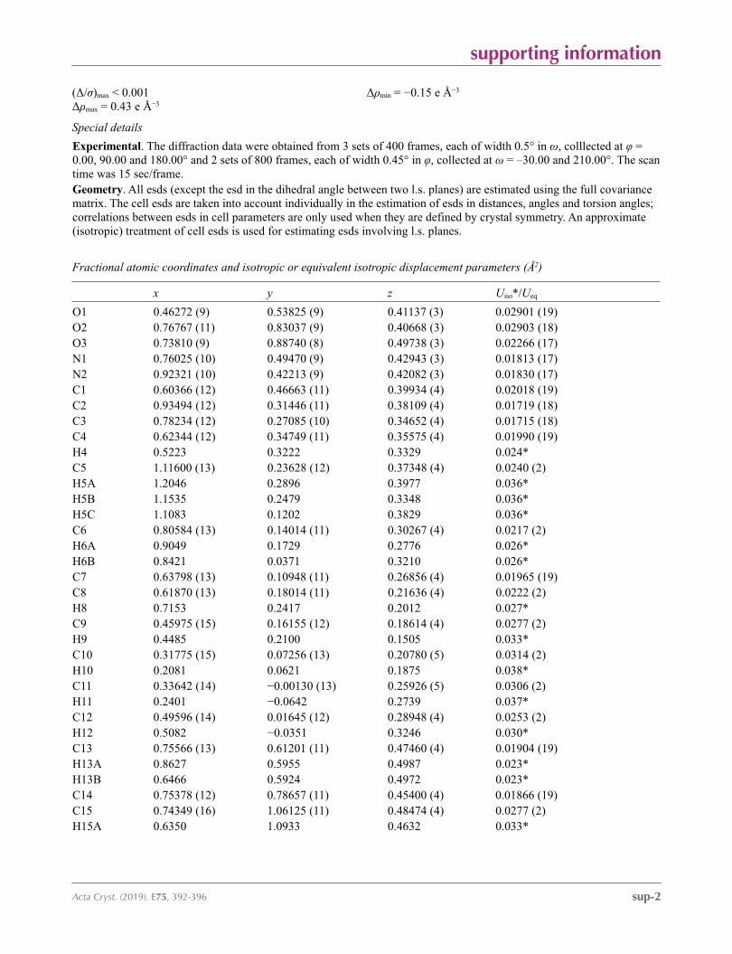
Fractional atomic coordinates and isotropic or equivalent isotropic displacement parameters (Å2)
x y z Uiso*/Ueq
O1 0.46272 (9) 0.53825 (9) 0.41137 (3) 0.02901 (19)O2 0.76767 (11) 0.83037 (9) 0.40668 (3) 0.02903 (18)O3 0.73810 (9) 0.88740 (8) 0.49738 (3) 0.02266 (17)N1 0.76025 (10) 0.49470 (9) 0.42943 (3) 0.01813 (17)N2 0.92321 (10) 0.42213 (9) 0.42082 (3) 0.01830 (17)C1 0.60366 (12) 0.46663 (11) 0.39934 (4) 0.02018 (19)C2 0.93494 (12) 0.31446 (11) 0.38109 (4) 0.01719 (18)C3 0.78234 (12) 0.27085 (10) 0.34652 (4) 0.01715 (18)C4 0.62344 (12) 0.34749 (11) 0.35575 (4) 0.01990 (19)H4 0.5223 0.3222 0.3329 0.024*C5 1.11600 (13) 0.23628 (12) 0.37348 (4) 0.0240 (2)H5A 1.2046 0.2896 0.3977 0.036*H5B 1.1535 0.2479 0.3348 0.036*H5C 1.1083 0.1202 0.3829 0.036*C6 0.80584 (13) 0.14014 (11) 0.30267 (4) 0.0217 (2)H6A 0.9049 0.1729 0.2776 0.026*H6B 0.8421 0.0371 0.3210 0.026*C7 0.63798 (13) 0.10948 (11) 0.26856 (4) 0.01965 (19)C8 0.61870 (13) 0.18014 (11) 0.21636 (4) 0.0222 (2)H8 0.7153 0.2417 0.2012 0.027*C9 0.45975 (15) 0.16155 (12) 0.18614 (4) 0.0277 (2)H9 0.4485 0.2100 0.1505 0.033*C10 0.31775 (15) 0.07256 (13) 0.20780 (5) 0.0314 (2)H10 0.2081 0.0621 0.1875 0.038*C11 0.33642 (14) −0.00130 (13) 0.25926 (5) 0.0306 (2)H11 0.2401 −0.0642 0.2739 0.037*C12 0.49596 (14) 0.01645 (12) 0.28948 (4) 0.0253 (2)H12 0.5082 −0.0351 0.3246 0.030*C13 0.75566 (13) 0.61201 (11) 0.47460 (4) 0.01904 (19)H13A 0.8627 0.5955 0.4987 0.023*H13B 0.6466 0.5924 0.4972 0.023*C14 0.75378 (12) 0.78657 (11) 0.45400 (4) 0.01866 (19)C15 0.74349 (16) 1.06125 (11) 0.48474 (4) 0.0277 (2)H15A 0.6350 1.0933 0.4632 0.033*

supporting information
sup-3Acta Cryst. (2019). E75, 392-396
H15B 0.8518 1.0871 0.4625 0.033*C16 0.74939 (16) 1.15128 (12) 0.53893 (5) 0.0305 (2)H16A 0.6443 1.1210 0.5612 0.046*H16B 0.7475 1.2690 0.5319 0.046*H16C 0.8602 1.1225 0.5590 0.046*
Atomic displacement parameters (Å2)
U11 U22 U33 U12 U13 U23
O1 0.0213 (3) 0.0338 (4) 0.0318 (4) 0.0072 (3) −0.0028 (3) −0.0136 (3)O2 0.0441 (5) 0.0257 (4) 0.0173 (4) 0.0028 (3) 0.0022 (3) 0.0014 (3)O3 0.0351 (4) 0.0156 (3) 0.0173 (3) 0.0007 (3) 0.0015 (3) −0.0014 (2)N1 0.0196 (4) 0.0183 (4) 0.0164 (4) 0.0021 (3) −0.0026 (3) −0.0039 (3)N2 0.0188 (4) 0.0188 (3) 0.0173 (4) 0.0016 (3) −0.0010 (3) 0.0014 (3)C1 0.0195 (4) 0.0211 (4) 0.0200 (4) 0.0010 (3) −0.0018 (3) −0.0034 (3)C2 0.0192 (4) 0.0172 (4) 0.0151 (4) 0.0014 (3) −0.0004 (3) 0.0027 (3)C3 0.0218 (4) 0.0151 (4) 0.0145 (4) −0.0001 (3) −0.0001 (3) 0.0003 (3)C4 0.0203 (4) 0.0204 (4) 0.0190 (4) 0.0002 (3) −0.0033 (3) −0.0043 (3)C5 0.0208 (4) 0.0282 (5) 0.0229 (5) 0.0064 (4) −0.0006 (4) −0.0002 (4)C6 0.0252 (5) 0.0194 (4) 0.0206 (5) 0.0032 (3) −0.0001 (4) −0.0052 (3)C7 0.0253 (5) 0.0162 (4) 0.0175 (4) 0.0008 (3) 0.0012 (3) −0.0045 (3)C8 0.0290 (5) 0.0188 (4) 0.0188 (4) −0.0001 (3) 0.0031 (4) −0.0022 (3)C9 0.0366 (6) 0.0256 (5) 0.0208 (5) 0.0050 (4) −0.0034 (4) −0.0053 (4)C10 0.0298 (5) 0.0295 (5) 0.0350 (6) 0.0000 (4) −0.0071 (4) −0.0138 (4)C11 0.0304 (5) 0.0245 (5) 0.0370 (6) −0.0081 (4) 0.0061 (4) −0.0089 (4)C12 0.0346 (5) 0.0202 (4) 0.0212 (5) −0.0032 (4) 0.0047 (4) −0.0022 (3)C13 0.0244 (4) 0.0181 (4) 0.0146 (4) 0.0004 (3) −0.0019 (3) −0.0026 (3)C14 0.0187 (4) 0.0201 (4) 0.0172 (4) 0.0004 (3) −0.0009 (3) −0.0023 (3)C15 0.0412 (6) 0.0157 (4) 0.0261 (5) 0.0018 (4) 0.0013 (4) 0.0011 (4)C16 0.0397 (6) 0.0185 (5) 0.0332 (6) 0.0025 (4) 0.0003 (4) −0.0054 (4)
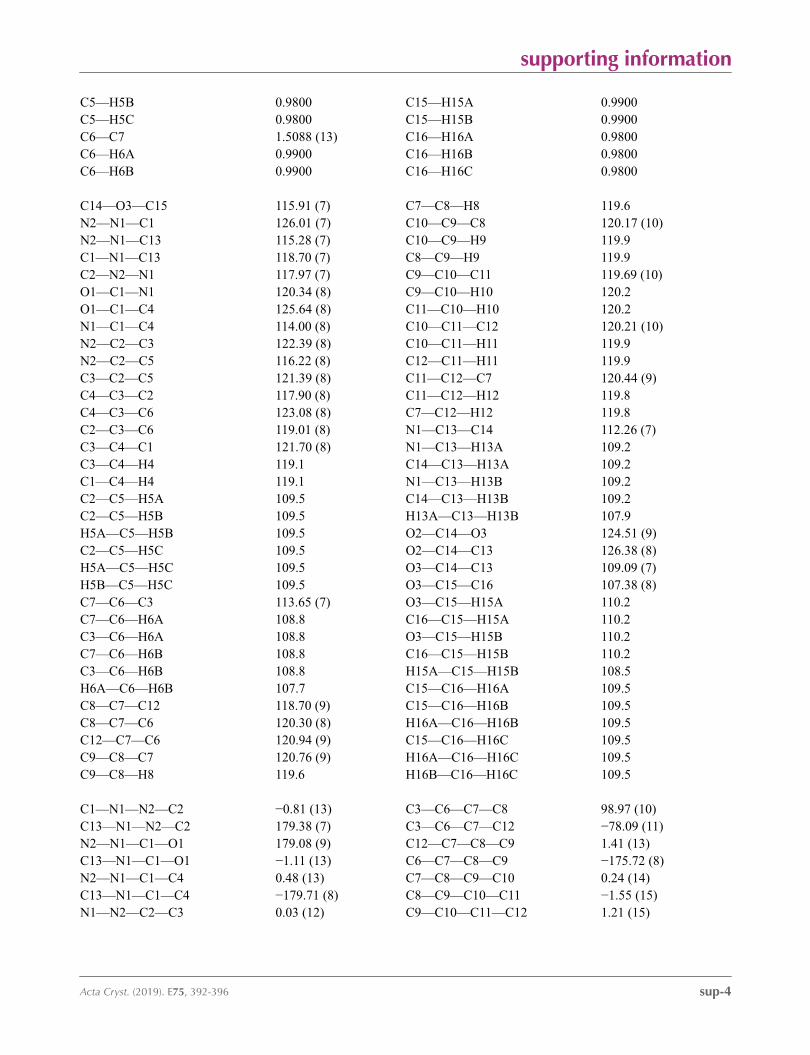
Geometric parameters (Å, º)
O1—C1 1.2336 (11) C7—C8 1.3932 (13)O2—C14 1.2020 (11) C7—C12 1.3958 (13)O3—C14 1.3392 (10) C8—C9 1.3901 (14)O3—C15 1.4577 (11) C8—H8 0.9500N1—N2 1.3625 (10) C9—C10 1.3846 (16)N1—C1 1.3845 (12) C9—H9 0.9500N1—C13 1.4541 (11) C10—C11 1.3878 (16)N2—C2 1.3063 (11) C10—H10 0.9500C1—C4 1.4434 (12) C11—C12 1.3930 (15)C2—C3 1.4463 (12) C11—H11 0.9500C2—C5 1.4985 (12) C12—H12 0.9500C3—C4 1.3535 (13) C13—C14 1.5145 (12)C3—C6 1.5164 (12) C13—H13A 0.9900C4—H4 0.9500 C13—H13B 0.9900C5—H5A 0.9800 C15—C16 1.5020 (14)
supporting information
sup-4Acta Cryst. (2019). E75, 392-396
C5—H5B 0.9800 C15—H15A 0.9900C5—H5C 0.9800 C15—H15B 0.9900C6—C7 1.5088 (13) C16—H16A 0.9800C6—H6A 0.9900 C16—H16B 0.9800C6—H6B 0.9900 C16—H16C 0.9800
C14—O3—C15 115.91 (7) C7—C8—H8 119.6N2—N1—C1 126.01 (7) C10—C9—C8 120.17 (10)N2—N1—C13 115.28 (7) C10—C9—H9 119.9C1—N1—C13 118.70 (7) C8—C9—H9 119.9C2—N2—N1 117.97 (7) C9—C10—C11 119.69 (10)O1—C1—N1 120.34 (8) C9—C10—H10 120.2O1—C1—C4 125.64 (8) C11—C10—H10 120.2N1—C1—C4 114.00 (8) C10—C11—C12 120.21 (10)N2—C2—C3 122.39 (8) C10—C11—H11 119.9N2—C2—C5 116.22 (8) C12—C11—H11 119.9C3—C2—C5 121.39 (8) C11—C12—C7 120.44 (9)C4—C3—C2 117.90 (8) C11—C12—H12 119.8C4—C3—C6 123.08 (8) C7—C12—H12 119.8C2—C3—C6 119.01 (8) N1—C13—C14 112.26 (7)C3—C4—C1 121.70 (8) N1—C13—H13A 109.2C3—C4—H4 119.1 C14—C13—H13A 109.2C1—C4—H4 119.1 N1—C13—H13B 109.2C2—C5—H5A 109.5 C14—C13—H13B 109.2C2—C5—H5B 109.5 H13A—C13—H13B 107.9H5A—C5—H5B 109.5 O2—C14—O3 124.51 (9)C2—C5—H5C 109.5 O2—C14—C13 126.38 (8)H5A—C5—H5C 109.5 O3—C14—C13 109.09 (7)H5B—C5—H5C 109.5 O3—C15—C16 107.38 (8)C7—C6—C3 113.65 (7) O3—C15—H15A 110.2C7—C6—H6A 108.8 C16—C15—H15A 110.2C3—C6—H6A 108.8 O3—C15—H15B 110.2C7—C6—H6B 108.8 C16—C15—H15B 110.2C3—C6—H6B 108.8 H15A—C15—H15B 108.5H6A—C6—H6B 107.7 C15—C16—H16A 109.5C8—C7—C12 118.70 (9) C15—C16—H16B 109.5C8—C7—C6 120.30 (8) H16A—C16—H16B 109.5C12—C7—C6 120.94 (9) C15—C16—H16C 109.5C9—C8—C7 120.76 (9) H16A—C16—H16C 109.5C9—C8—H8 119.6 H16B—C16—H16C 109.5
C1—N1—N2—C2 −0.81 (13) C3—C6—C7—C8 98.97 (10)C13—N1—N2—C2 179.38 (7) C3—C6—C7—C12 −78.09 (11)N2—N1—C1—O1 179.08 (9) C12—C7—C8—C9 1.41 (13)C13—N1—C1—O1 −1.11 (13) C6—C7—C8—C9 −175.72 (8)N2—N1—C1—C4 0.48 (13) C7—C8—C9—C10 0.24 (14)C13—N1—C1—C4 −179.71 (8) C8—C9—C10—C11 −1.55 (15)N1—N2—C2—C3 0.03 (12) C9—C10—C11—C12 1.21 (15)
supporting information
sup-5Acta Cryst. (2019). E75, 392-396
N1—N2—C2—C5 −179.33 (8) C10—C11—C12—C7 0.46 (15)N2—C2—C3—C4 1.02 (13) C8—C7—C12—C11 −1.75 (14)C5—C2—C3—C4 −179.66 (8) C6—C7—C12—C11 175.35 (9)N2—C2—C3—C6 −177.78 (8) N2—N1—C13—C14 104.34 (9)C5—C2—C3—C6 1.55 (12) C1—N1—C13—C14 −75.50 (10)C2—C3—C4—C1 −1.34 (13) C15—O3—C14—O2 −1.63 (13)C6—C3—C4—C1 177.40 (8) C15—O3—C14—C13 176.99 (8)O1—C1—C4—C3 −177.86 (9) N1—C13—C14—O2 −5.26 (14)N1—C1—C4—C3 0.65 (13) N1—C13—C14—O3 176.15 (7)C4—C3—C6—C7 2.78 (13) C14—O3—C15—C16 −172.44 (8)C2—C3—C6—C7 −178.49 (8)
Hydrogen-bond geometry (Å, º)
D—H···A D—H H···A D···A D—H···A
C13—H13A···N2i 0.99 2.51 3.4704 (13) 165C13—H13B···O1ii 0.99 2.59 3.4281 (13) 143
Symmetry codes: (i) −x+2, −y+1, −z+1; (ii) −x+1, −y+1, −z+1.
Copyright © 2022 FDOKUMEN