Crystal structure of human PNP complexed with guanine
6
Crystal structure of human PNP complexed with guanine Walter Filgueira de Azevedo Jr., a,b, * Fernanda Canduri, a,b Denis Marangoni dos Santos, a,b Jos e Henrique Pereira, a,b M arcio Vinicius Bertacine Dias, a Rafael Guimar~ aes Silva, c Maria Anita Mendes, b,d Luiz Augusto Basso, c M ario S ergio Palma, b,d and Di ogenes Santiago Santos c,e, * a Departamento de F ısica, UNESP, S~ ao Jos e do Rio Preto, SP 15054-000, Brazil b Center for Applied Toxinology, Instituto Butantan, S~ ao Paulo, SP 05503-900, Brazil c Rede Brasileira de Pesquisas em Tuberculose, Departamento de Biologia Molecular e Biotecnologia, UFRGS, Porto Alegre, RS 91501-970, Brazil d Laboratory of Structural Biology and Zoochemistry-CEIS/Department of Biology, Institute of Biosciences, UNESP, Rio Claro, SP 13506-900, Brazil e Faculdade de Farm acia/Instituto de Pesquisas Biom edicas, Pontif ıcia Universidade Cat olica do Rio Grande do Sul, Porto Alegre, RS, Brazil Received 23 October 2003 Abstract Purine nucleoside phosphorylase (PNP) catalyzes the phosphorolysis of the N-ribosidic bonds of purine nucleosides and deoxynucleosides. PNP is a target for inhibitor development aiming at T-cell immune response modulation and has been submitted to extensive structure-based drug design. More recently, the 3-D structure of human PNP has been refined to 2.3 A resolution, which allowed a redefinition of the residues involved in the substrate-binding sites and provided a more reliable model for structure-based design of inhibitors. This work reports crystallographic study of the complex of Human PNP:guanine (HsPNP:Gua) solved at 2.7 A resolution using synchrotron radiation. Analysis of the structural differences among the HsPNP:Gua complex, PNP apoenzyme, and HsPNP:immucillin-H provides explanation for inhibitor binding, refines the purine-binding site, and can be used for future inhibitor design. Ó 2003 Elsevier Inc. All rights reserved. Keywords: PNP; Synchrotron radiation; Structure; Drug design PNP catalyzes the reversible phosphorolysis of the ribonucleosides and 2 0 -deoxyribonucleosides of guanine, hypoxanthine, and a number of related nucleoside compounds [1], except adenosine (Fig. 1). Human PNP is an attractive target for drug design and it has been submitted to extensive structure-based design. PNP in- hibitors could be used in the following applications: (1) treatment of T-cell leukemia; (2) suppression of the host-vs.-graft response in organ transplantation recipi- ents; (3) treatment of secondary or xanthine gout by restricting purine catabolites to the more soluble nucle- osides; and (4) in combination with nucleosides to pre- vent their degradation by PNP metabolism [2]. More recently, the 3-D structure of human PNP has been refined to 2.3 A resolution [3], which allowed a redefi- nition of the residues involved in the substrate-binding sites and provided a more reliable model for structure- based design of inhibitors. The crystallographic struc- ture is a trimer and analysis of human PNP in solution, using SAXS, confirmed that the crystallographic trimer is conserved in solution [4]. We have obtained the crystallographic structure of the complex between HsPNP and guanine (HsPNP: Gua). Previously reported structure for the same com- plex showed poor stereochemistry quality [2,5] and the refined model does not show water molecules. Our analyses of the HsPNP:Gua structural data and struc- tural differences between the PNP apoenzyme and HsPNP:Gua complex provide explanation for substrate binding, refine the purine-binding site, identify water molecules, a new phosphate-binding site, and can be used for future inhibitor design. * Corresponding authors. Fax: +55-17-221-2247. E-mail addresses: [email protected] (W.F. de Azevedo Jr.), [email protected] (D.S. Santos). 0006-291X/$ - see front matter Ó 2003 Elsevier Inc. All rights reserved. doi:10.1016/j.bbrc.2003.10.190 Biochemical and Biophysical Research Communications 312 (2003) 767–772 BBRC www.elsevier.com/locate/ybbrc
Transcript of Crystal structure of human PNP complexed with guanine
Biochemical and Biophysical Research Communications 312 (2003) 767–772
BBRCwww.elsevier.com/locate/ybbrc
Crystal structure of human PNP complexed with guanine
Walter Filgueira de Azevedo Jr.,a,b,* Fernanda Canduri,a,b Denis Marangoni dos Santos,a,b
Jos�ee Henrique Pereira,a,b M�aarcio Vinicius Bertacine Dias,a Rafael Guimar~aaes Silva,c
Maria Anita Mendes,b,d Luiz Augusto Basso,c M�aario S�eergio Palma,b,d
and Di�oogenes Santiago Santosc,e,*
a Departamento de F�ıısica, UNESP, S~aao Jos�ee do Rio Preto, SP 15054-000, Brazilb Center for Applied Toxinology, Instituto Butantan, S~aao Paulo, SP 05503-900, Brazil
c Rede Brasileira de Pesquisas em Tuberculose, Departamento de Biologia Molecular e Biotecnologia, UFRGS, Porto Alegre, RS 91501-970, Brazild Laboratory of Structural Biology and Zoochemistry-CEIS/Department of Biology, Institute of Biosciences, UNESP, Rio Claro, SP 13506-900, Brazil
e Faculdade de Farm�aacia/Instituto de Pesquisas Biom�eedicas, Pontif�ııcia Universidade Cat�oolica do Rio Grande do Sul, Porto Alegre, RS, Brazil
Received 23 October 2003
Abstract
Purine nucleoside phosphorylase (PNP) catalyzes the phosphorolysis of the N-ribosidic bonds of purine nucleosides and
deoxynucleosides. PNP is a target for inhibitor development aiming at T-cell immune response modulation and has been submitted
to extensive structure-based drug design. More recently, the 3-D structure of human PNP has been refined to 2.3�AA resolution, which
allowed a redefinition of the residues involved in the substrate-binding sites and provided a more reliable model for structure-based
design of inhibitors. This work reports crystallographic study of the complex of Human PNP:guanine (HsPNP:Gua) solved at 2.7�AAresolution using synchrotron radiation. Analysis of the structural differences among the HsPNP:Gua complex, PNP apoenzyme,
and HsPNP:immucillin-H provides explanation for inhibitor binding, refines the purine-binding site, and can be used for future
inhibitor design.
� 2003 Elsevier Inc. All rights reserved.
Keywords: PNP; Synchrotron radiation; Structure; Drug design
PNP catalyzes the reversible phosphorolysis of the
ribonucleosides and 20-deoxyribonucleosides of guanine,
hypoxanthine, and a number of related nucleoside
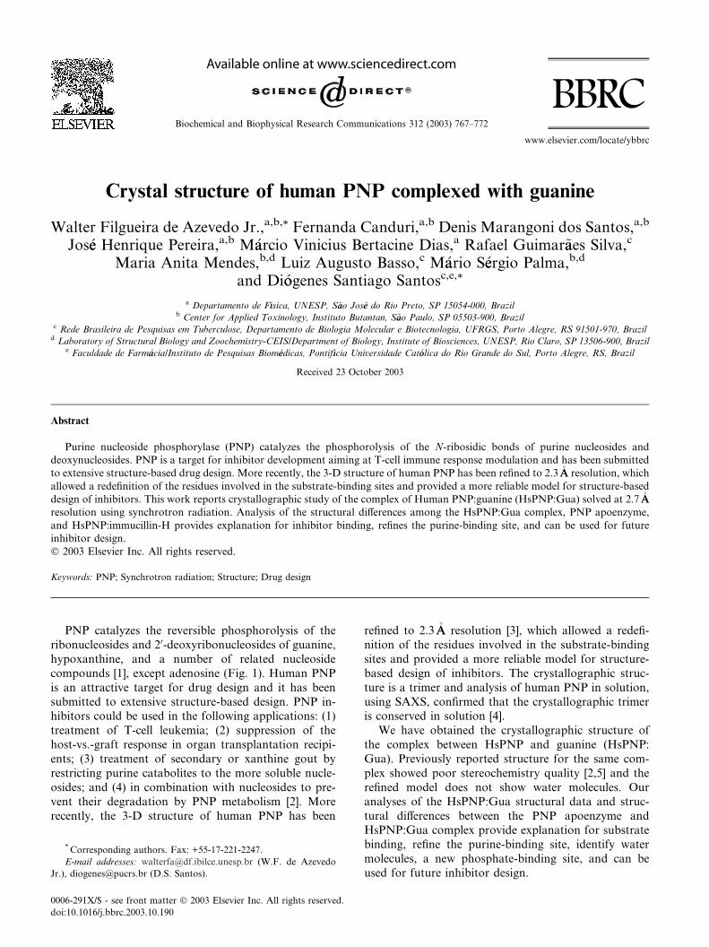
compounds [1], except adenosine (Fig. 1). Human PNP
is an attractive target for drug design and it has been
submitted to extensive structure-based design. PNP in-hibitors could be used in the following applications: (1)
treatment of T-cell leukemia; (2) suppression of the
host-vs.-graft response in organ transplantation recipi-
ents; (3) treatment of secondary or xanthine gout by
restricting purine catabolites to the more soluble nucle-
osides; and (4) in combination with nucleosides to pre-
vent their degradation by PNP metabolism [2]. More
recently, the 3-D structure of human PNP has been
* Corresponding authors. Fax: +55-17-221-2247.
E-mail addresses: [email protected] (W.F. de Azevedo
Jr.), [email protected] (D.S. Santos).
0006-291X/$ - see front matter � 2003 Elsevier Inc. All rights reserved.
doi:10.1016/j.bbrc.2003.10.190
refined to 2.3�AA resolution [3], which allowed a redefi-
nition of the residues involved in the substrate-binding
sites and provided a more reliable model for structure-
based design of inhibitors. The crystallographic struc-
ture is a trimer and analysis of human PNP in solution,
using SAXS, confirmed that the crystallographic trimeris conserved in solution [4].
We have obtained the crystallographic structure of
the complex between HsPNP and guanine (HsPNP:
Gua). Previously reported structure for the same com-
plex showed poor stereochemistry quality [2,5] and the
refined model does not show water molecules. Our
analyses of the HsPNP:Gua structural data and struc-
tural differences between the PNP apoenzyme andHsPNP:Gua complex provide explanation for substrate
binding, refine the purine-binding site, identify water
molecules, a new phosphate-binding site, and can be
used for future inhibitor design.
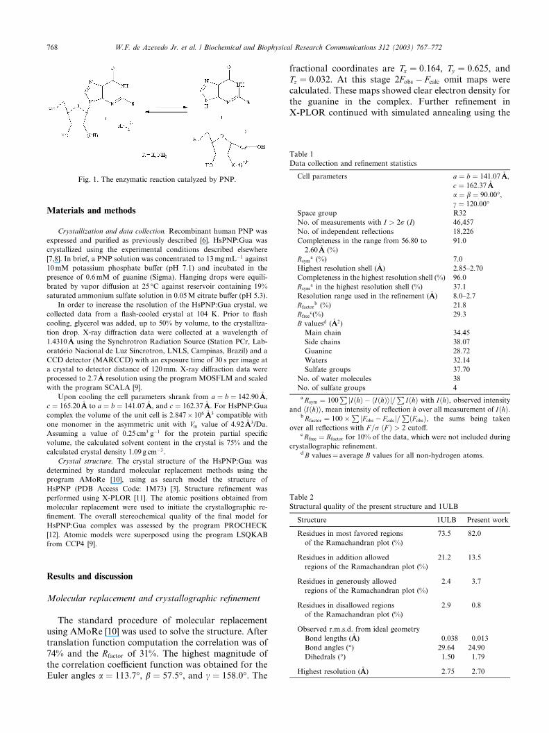
Fig. 1. The enzymatic reaction catalyzed by PNP.
Table 1
Data collection and refinement statistics
Cell parameters a ¼ b ¼ 141:07�AA,
c ¼ 162:37�AA
a ¼ b ¼ 90:00�,c ¼ 120:00�
Space group R32
No. of measurements with I > 2r (I) 46,457
No. of independent reflections 18,226
Completeness in the range from 56.80 to
2.60�AA (%)
91.0
Rsyma (%) 7.0
Highest resolution shell (�AA) 2.85–2.70
Completeness in the highest resolution shell (%) 96.0
Rsyma in the highest resolution shell (%) 37.1
Resolution range used in the refinement (�AA) 8.0–2.7
Rfactorb (%) 21.8
Rfreec(%) 29.3
B valuesd (�AA2)
Main chain 34.45
Side chains 38.07
Guanine 28.72
Waters 32.14
Sulfate groups 37.70
No. of water molecules 38
No. of sulfate groups 4
aRsym ¼ 100P
jIðhÞ � hIðhÞij=P
IðhÞ with IðhÞ, observed intensity
and hIðhÞi, mean intensity of reflection h over all measurement of IðhÞ.bRfactor ¼ 100�
PjFobs � Fcalcj=
PðFobsÞ, the sums being taken
over all reflections with F =r ðF Þ > 2 cutoff.cRfree ¼ Rfactor for 10% of the data, which were not included during
crystallographic refinement.dB values¼ average B values for all non-hydrogen atoms.
Table 2
Structural quality of the present structure and 1ULB
Structure 1ULB Present work
Residues in most favored regions
of the Ramachandran plot (%)
73.5 82.0
768 W.F. de Azevedo Jr. et al. / Biochemical and Biophysical Research Communications 312 (2003) 767–772
Materials and methods
Crystallization and data collection. Recombinant human PNP was
expressed and purified as previously described [6]. HsPNP:Gua was
crystallized using the experimental conditions described elsewhere
[7,8]. In brief, a PNP solution was concentrated to 13mgmL�1 against
10mM potassium phosphate buffer (pH 7.1) and incubated in the
presence of 0.6mM of guanine (Sigma). Hanging drops were equili-
brated by vapor diffusion at 25 �C against reservoir containing 19%
saturated ammonium sulfate solution in 0.05M citrate buffer (pH 5.3).
In order to increase the resolution of the HsPNP:Gua crystal, we
collected data from a flash-cooled crystal at 104 K. Prior to flash
cooling, glycerol was added, up to 50% by volume, to the crystalliza-
tion drop. X-ray diffraction data were collected at a wavelength of
1.4310�AA using the Synchrotron Radiation Source (Station PCr, Lab-
orat�oorio Nacional de Luz S�ııncrotron, LNLS, Campinas, Brazil) and a
CCD detector (MARCCD) with an exposure time of 30 s per image at
a crystal to detector distance of 120mm. X-ray diffraction data were
processed to 2.7�AA resolution using the program MOSFLM and scaled
with the program SCALA [9].
Upon cooling the cell parameters shrank from a ¼ b ¼ 142:90�AA,
c ¼ 165:20�AA to a ¼ b ¼ 141:07�AA, and c ¼ 162:37�AA. For HsPNP:Gua
complex the volume of the unit cell is 2.847� 106 �AA3 compatible with
one monomer in the asymmetric unit with Vm value of 4.92�AA3/Da.
Assuming a value of 0.25 cm3 g�1 for the protein partial specific
volume, the calculated solvent content in the crystal is 75% and the
calculated crystal density 1.09 g cm�3.
Crystal structure. The crystal structure of the HsPNP:Gua was
determined by standard molecular replacement methods using the
program AMoRe [10], using as search model the structure of
HsPNP (PDB Access Code: 1M73) [3]. Structure refinement was
performed using X-PLOR [11]. The atomic positions obtained from
molecular replacement were used to initiate the crystallographic re-
finement. The overall stereochemical quality of the final model for
HsPNP:Gua complex was assessed by the program PROCHECK
[12]. Atomic models were superposed using the program LSQKAB
from CCP4 [9].
Residues in addition allowed
regions of the Ramachandran plot (%)
21.2 13.5
Residues in generously allowed
regions of the Ramachandran plot (%)
2.4 3.7
Residues in disallowed regions
of the Ramachandran plot (%)
2.9 0.8
Observed r.m.s.d. from ideal geometry
Bond lengths (�AA) 0.038 0.013
Bond angles (�) 29.64 24.90
Dihedrals (�) 1.50 1.79
Highest resolution (�AA) 2.75 2.70
Results and discussion
Molecular replacement and crystallographic refinement
The standard procedure of molecular replacement
using AMoRe [10] was used to solve the structure. After
translation function computation the correlation was of
74% and the Rfactor of 31%. The highest magnitude of
the correlation coefficient function was obtained for theEuler angles a ¼ 113:7�, b ¼ 57:5�, and c ¼ 158:0�. The
fractional coordinates are Tx ¼ 0:164, Ty ¼ 0:625, andTz ¼ 0:032. At this stage 2Fobs � Fcalc omit maps were
calculated. These maps showed clear electron density for
the guanine in the complex. Further refinement in
X-PLOR continued with simulated annealing using the
W.F. de Azevedo Jr. et al. / Biochemical and Biophysical Research Communications 312 (2003) 767–772 769
slow-cooling protocol, followed by alternate cycles ofpositional refinement and manual rebuilding using
XtalView [13]. Finally, the positions of guanine, water,
and sulfate molecules were checked and corrected in
Fobs � Fcalc maps. The final model has an Rfactor of 21.8%
and an Rfree of 29.3%, with 38 water molecules, 4 sulfate
ions, and the guanine.
Ignoring low-resolution data, a Luzzati plot [14] gives
the best correlation between the observed and calculateddata for a predicted mean coordinate error of 0.36�AA.
Fig. 2. Ribbon diagram of HsPNP:Gua generated by Molscript [23]
and Raster3d [24].
Fig. 3. Gate movement after bindin
The average B factor for main chain atoms is 34.45�AA2,whereas that for side chain atoms is 38.07�AA2 (Table 1).
Comparison of the present structure with previously
deposited atomic coordinates for the same complex in-
dicates that the present structure shows better overall
stereochemistry (Table 2). Furthermore, analysis of the
electron-density maps of the present structure allowed
the determination of water molecules, not identified in
the previous structure. In addition, three new humanPNP structures [3,7,8] made possible structural com-
parison presented here.
Overall description
Analysis of the crystallographic structure of
HsPNP:Gua complex indicates a trimeric structure.
Each PNP monomer displays an a=b fold consisting of a
mixed b-sheet surrounded by a helices. The structure
contains an eight-stranded mixed b-sheet and a five-
stranded mixed b-sheet, which join to form a distorted
b-barrel. Fig. 2 shows schematic drawings of the
HsPNP:Gua complex.
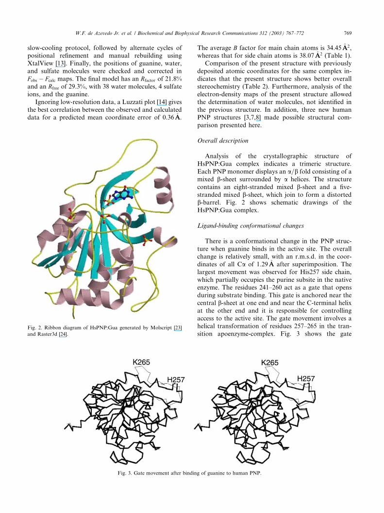
Ligand-binding conformational changes
There is a conformational change in the PNP struc-
ture when guanine binds in the active site. The overallchange is relatively small, with an r.m.s.d. in the coor-
dinates of all Ca of 1.29�AA after superimposition. The
largest movement was observed for His257 side chain,
which partially occupies the purine subsite in the native
enzyme. The residues 241–260 act as a gate that opens
during substrate binding. This gate is anchored near the
central b-sheet at one end and near the C-terminal helix
at the other end and it is responsible for controllingaccess to the active site. The gate movement involves a
helical transformation of residues 257–265 in the tran-
sition apoenzyme-complex. Fig. 3 shows the gate
g of guanine to human PNP.
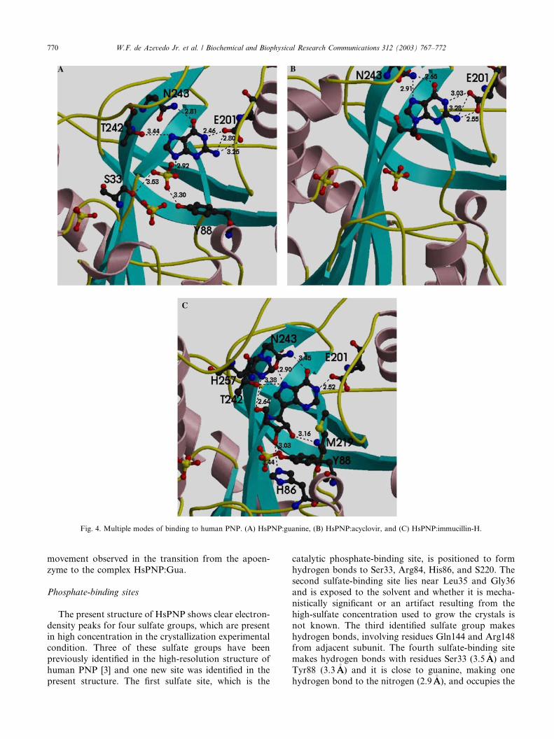
Fig. 4. Multiple modes of binding to human PNP. (A) HsPNP:guanine, (B) HsPNP:acyclovir, and (C) HsPNP:immucillin-H.
770 W.F. de Azevedo Jr. et al. / Biochemical and Biophysical Research Communications 312 (2003) 767–772
movement observed in the transition from the apoen-
zyme to the complex HsPNP:Gua.
Phosphate-binding sites
The present structure of HsPNP shows clear electron-
density peaks for four sulfate groups, which are present
in high concentration in the crystallization experimental
condition. Three of these sulfate groups have been
previously identified in the high-resolution structure of
human PNP [3] and one new site was identified in the
present structure. The first sulfate site, which is the
catalytic phosphate-binding site, is positioned to form
hydrogen bonds to Ser33, Arg84, His86, and S220. The
second sulfate-binding site lies near Leu35 and Gly36
and is exposed to the solvent and whether it is mecha-
nistically significant or an artifact resulting from the
high-sulfate concentration used to grow the crystals is
not known. The third identified sulfate group makes
hydrogen bonds, involving residues Gln144 and Arg148from adjacent subunit. The fourth sulfate-binding site
makes hydrogen bonds with residues Ser33 (3.5�AA) and
Tyr88 (3.3�AA) and it is close to guanine, making one
hydrogen bond to the nitrogen (2.9�AA), and occupies the
W.F. de Azevedo Jr. et al. / Biochemical and Biophysical Research Communications 312 (2003) 767–772 771
ribose-binding site (Fig. 4A). The ribose-binding site ismostly hydrophobic, which is composed of several ar-
omatic amino acids, including Tyr88, Phe159 (of the
adjacent subunit), Phe200, His86, His257, and Met219.
It is tempting to speculate that the presence of a sulfate
(phosphate) group at the ribose-binding site may offer
further hindrance to the binding of substrates, which
may also contribute to the larger IC50 observed for
several inhibitors in the presence of higher phosphateconcentrations [7,8].
Interactions with guanine
The specificity and affinity between enzyme and itsligand depend on directional hydrogen bonds and io-
nic interactions, as well as on shape complementarity
of the contact surfaces of both partners [15–21]. The
electrostatic potential surface of the guanine com-
plexed with HsPNP was calculated with GRASP [22]
(figure not shown). The analysis of the charge distri-
bution of the binding pocket indicates the presence of
some charge complementarity between inhibitor andenzyme, though most of the binding pocket is hy-
drophobic.
Comparison of the present structure with human
PNP complexed with acyclovir (HsPNP:Acy) [7] and
immucillin-H (HsPNP:ImmH) [8] indicates that human
PNP presents multiple modes of binding to the active
site. Figs. 4A–C show the interaction between ligands
and PNP. The main residues involved in binding in allcomplexes are Glu201, Thr242, and Asn242. Analysis
of the hydrogen bonds between immucillin-H and PNP
reveals eight hydrogen bonds, involving the residues
His86, Tyr88, Glu201, Asn243, and His257. For the
complex HsPNP:Acy five hydrogen bonds were ob-
served. These hydrogen bonds involve Glu201 and
Asn243. Five hydrogen bonds between guanine
and human PNP, involving residues Glu201, Thr242,and Asn243 were observed. The previously described
participation of Lys244 [5] in ligand binding was not
identified in the present study and in the structures of
human PNP complexed with inhibitors. Analysis of the
complexes indicates that Glu201 and Thr242 occupy
approximately the same position in all the complexes.
The side-chain of Asn243 shows some flexibility, which
causes differences in the hydrogen bond pattern of thisresidue, the complexes HsPNP:ImmH and HsPNP:Gua
show intermolecular hydrogen bonds involving the
following atom pairs: Asn243 ND2-O6 and Asn243
OD1-N7. The participation of Asn243 OD1 is not
observed in the HsPNP:Acy complex. The precise
definition of the modes of binding to human PNP may
help in future structure-based design of inhibitors.
The atomic coordinates and the structure factors forthe complex HsPNP:Gua have been deposited in the
PDB with the Accession Code: 1V2H.
Acknowledgments
We acknowledge the expertise of Denise Cantarelli Machado for
the expansion of the cDNA library and Deise Potrich for the DNA
sequencing. This work was supported by grants from FAPESP
(SMOLBNet, Proc.01/07532-0 and 02/04383-7), CNPq, CAPES and
Instituto do Mileenio (CNPq-MCT). WFA (CNPq, 300851/98-7), MSP
(CNPq, 300337/2003-5), and LAB (CNPq, 520182/99-5) are re-
searchers for the Brazilian Council for Scientific and Technological
Development.
References
[1] J.A. Montgomery, Purine nucleoside phosphorylase: a target for
drug design, Med. Res. Rev. 13 (3) (1993) 209–228.
[2] S.E. Ealick, Y.S. Babu, C.E. Bugg, M.D. Erion, W.C. Guida, J.A.
Montgomery, J.A. Secrist III, Application of crystallographic and
modelingmethods in the design of purine nucleoside phosphorylase
inhibitors, Proc. Natl. Acad. Sci. USA 91 (1991) 11540–11544.
[3] W.F. De Azevedo, F. Canduri, D.M. dos Santos, R.G. Silva, J.S.
Oliveira, L.P.S. Carvalho, L.A. Basso, M.A. Mendes, M.S. Palma,
D.S. Santos, Crystal structure of human purine nucleoside
phosphorylase at 2.3�AA resolution, Biochem. Biophys. Res. Com-
mun. 308 (3) (2003) 545–552.
[4] W.F. De Azevedo, G.C. Santos, D.M. dos Santos, J.R. Olivieri, F.
Canduri, R.G. Silva, L.A. Basso, M.A. Mendes, M.S. Palma, D.S.
Santos, Docking and small angle X-ray scattering studies of
purine nucleoside phosphorylase, Biochem. Biophys. Res. Com-
mun. 309 (2003) 928–933.
[5] A. Bzowska, E. Kulikowska, D. Shugar, Purine nucleoside
phosphorylases: properties, functions, and clinical aspects, Phar-
macol. Ther. 88 (2000) 349–425.
[6] R.G. Silva, L.P. Carvalho, J.S. Oliveira, C.A. Pinto, M.A.
Mendes, M.S. Palma, L.A. Basso, D.S. Santos, Cloning, overex-
pression, and purification of functional human purine nucleoside
phosphorylase, Protein Expr. Purif. 27 (1) (2003) 158–164.
[7] D.M. dos Santos, F. Canduri, J.H. Pereira, M.V.B. Dias, R.G.
Silva, M.A. Mendes, M.S. Palma, L.A. Basso, W.F. de Azevedo,
D.S. Santos, Crystal structure of human purine nucleoside
phosphorylase complexed with acyclovir, Biochem. Biophys.
Res. Commun. 308 (3) (2003) 553–559.
[8] W.F. De Azevedo, F. Canduri, D.M. dos Santos, J.H. Pereira,
M.V.B. Dias, R.G. Silva, M.A. Mendes, L.A. Basso, M.S. Palma,
D.S. Santos, Structural basis for inhibition of human PNP by
immucillin-H, Biochem. Biophys. Res. Commun. 309 (2003) 922–
927.
[9] Collaborative Computational Project, Number 4. Acta Crystal-
logr. D 50 (1994) 760–763.
[10] J. Navaza, AMoRe: an automated package for molecular
replacement, Acta Crystallogr. A 50 (1994) 157–163.
[11] A.T. Br€uunger, X-PLOR Version 3.1: a System for Crystallography
and NMR, Yale University Press, New Haven, 1992.
[12] R.A. Laskowski, M.W. MacArthur, D.K. Smith, D.T. Jones,
E.G. Hutchinson, A.L. Morris, D. Naylor, D.S. Moss, J.M.
Thorton, PROCHECK v.3.0—Program to Check the Stereochem-
istry Quality of Protein Structures—Operating Instructions, 1994.
[13] D.E. McRee, XtalView/Xfit—a versatile program for manipulat-
ing atomic coordinates and electron density, J. Struct. Biol. 125
(1999) 156–165.
[14] P.V. Luzzati, Traitement statistique des erreurs dans la determi-
nation des structures cristallines, Acta Crystallogr. 5 (1952) 802–
810.
[15] W.F. De Azevedo, H.J. MuellerDieckmann, U. SchulzeGahmen,
P.J. Worland, E. Sausville, S.H. Kim, Structural basis for
specificity and potency of a flavonoid inhibitor of human
772 W.F. de Azevedo Jr. et al. / Biochemical and Biophysical Research Communications 312 (2003) 767–772
CDK2, a cell cycle kinase, Proc. Natl. Acad. Sci. USA 93 (7)
(1996) 2735–2740.
[16] W.F. De Azevedo, S. Leclerc, L. Meijer, L. Havlicek, M. Strnad,
S.H. Kim, Inhibition of cyclin-dependent kinases by purine
analogues—crystal structure of human cdk2 complexed with
roscovitine, Eur. J. Biochem. 243 (1–2) (1997) 518–526.
[17] S.H. Kim, U. Schulze-Gahmen, J. Brandsen, W.F. de Azevedo,
Structural basis for chemical inhibition of CDK2, Prog. Cell Cycle
Res. 2 (1996) 137–145.
[18] W.F. De Azevedo, F. Canduri, N.J.F. da Silveira, Structural basis
for inhibition of cyclin-dependent kinase 9 by flavopiridol,
Biochem. Biophys. Res. Commun. 293 (2002) 566–571.
[19] W.F. De Azevedo, R.T. Gaspar, F. Canduri, J.C. Camera, N.J.F.
da Silveira, Molecular model of cyclin-dependent kinase 5
complexed with roscovitine, Biochem. Biophys. Res. Commun.
297 (2002) 1154–1158.
[20] W.F. De Azevedo, J.S. de Oliveira, L.A. Basso, M.S. Palma, J.H.
Pereira, F. Canduri, D.S. Santos, Molecular model of shikimate
kinase from Mycobacterium tuberculosis, Biochem. Biophys. Res.
Commun. 295 (1) (2002) 142–148.
[21] F. Canduri, N.J.F. Silveira, J.C. Camera, W.F. de Azevedo,
Structural bioinformatics study of cyclin-dependent kinases com-
plexed with inhibitors, Ecletica Quim. 28 (2003) 45–53.
[22] A. Nicholls, K. Sharp, B. Honig, Protein folding and associ-
ation: insights from the interfacial and thermodynamic proper-
ties of hydrocarbons, Proteins Struct. Funct. Genet. 11 (1991)
281–296.
[23] P.J. Kraulis, MOLSCRIPT: a program to produce both detailed
and schematic plots of proteins, J. Appl. Crystallogr. 24 (1991)
946–950.
[24] E.A. Merritt, D.J. Bacon, Raster3D: photorealistic molecular
graphics, Methods Enzymol. 277 (1997) 505–524.


![A Flexible Copper(I)-Complexed [4]Rotaxane Containing Two Face-to-Face Porphyrinic Plates that Behaves as a Distensible Receptor](https://static.fdokumen.com/doc/165x107/632a7148d2eed9476207508b/a-flexible-copperi-complexed-4rotaxane-containing-two-face-to-face-porphyrinic.jpg)