Cryptosporidium Infection in Patients With Primary Immunodeficiencies
7
Cryptosporidium Infection in Patients With Primary Immunodeficiencies Beata Wolska-Kusnierz, y Anna Bajer, z Simone Caccio, Edyta Heropolitanska-Pliszka, Ewa Bernatowska, Piotr Socha, § Jacques van Dongen, y Malgorzata Bednarska, y Anna Paziewska, and y Edward Sinski Gastroenterology, Hepatology and Immunology Clinic, Children’s Memorial Health Institute, { Department of Parasitology, Faculty of Biology, University of Warsaw, Poland, { Department of Infectious, Parasitic and Immunomediated Diseases, Istituto Superiore di Sanita `, Rome, Italy, and § Department of Immunology, Erasmus University, Rotterdam, The Netherlands ABSTRACT Background: Cryptosporidium species infection is usually self-limited in immunocompetent populations, but can be severe and life-threatening among immunocompromised individuals, particularly in patients with AIDS and in these patients with primary immunodeficiencies (PIDs). Patients and Methods: A group of 5 patients with genetically confirmed hyper-IgM syndrome type 1 (XHIM) and one patient with primary CD4 lymphopenia were enrolled in the study. At least 2 stool samples and a bile sample in one patient were examined for Cryptosporidium oocysts by a modified Ziehl- Neelsen technique, by immunofluorescence assay using a commercial kit, as well as by molecular analysis followed by genotyping. Immunological status at the time of PID diagnosis and the complex picture of disease are presented. Results: Chronic cryptosporidiosis was confirmed in 3 patients with XHIM and in one patient with primary CD4 lymphopenia. Molecular diagnosis showed the presence of C parvum, C hominis, and C meleagridis in analyzed specimens. Conclusions: Cryptosporidium infection with serious clinical symptoms observed in patients with hyper-IgM syndrome calls for regular, repeated screening in this group of patients. JPGN 45:458–464, 2007. Key Words: Cryptosporidium species—Hyperimmunoglobulin M syndrome—Primary immunodeficiencies—Sclerosing cholangitis. # 2007 by European Society for Pediatric Gastroenterology, Hepatology, and Nutrition and North American Society for Pediatric Gastroenterology, Hepatology, and Nutrition INTRODUCTION Cryptosporidiosis refers to infection caused by the oocyst-forming parasites of the genus Cryptosporidium. Cryptosporidium infections are increasingly recognized as a cause of diarrhea not only in children (1,2) but also in elderly people (3) and in a range of immunodeficient patients. Among patients with primary immunodeficien- cies (PIDs), particular susceptibility to Cryptosporidium infection is observed in children with X-linked hyper- IgM syndrome type 1 (XHIM) resulting from CD40 ligand (CD40L) deficiency, hyper-IgM syndrome type 3 (HIGM3) caused by CD40 deficiency (4–6), primary CD4 lymphopenia, severe combined immuno- deficiency syndrome, and interferon-g deficiency. In secondary immunodeficiencies such as HIV infection and acute leukemia, the increased risk of infection is also noted (7). Transmission is carried out through the fecal/oral route following direct or indirect contact with the infective stages (Cryptosporidium oocysts), including person-to-person, zoonotic, waterborne, food-borne and, possibly airborne transmission (8,9). The parasite usually infects epithelial cells of the small intestine, but in immunocompromised individuals it can be found along the whole gastrointestinal tract, on the surface of the respiratory tract, and in the bile duct (10). Diarrhea is the typical clinical manifestation of cryptosporidiosis. It lasts longer in immunocompromised individuals as a result of their inability to clear the infection. In such patients, malabsorption, steatorrhea, and extraintestinal manifes- tations develop with time (11). The bile tract is the most common site of extraintestinal infection, which may result in chronic liver inflammation (6,12) or even lead to liver cirrhosis. We present results of a long-term survey of Received June 28, 2006; accepted January 30, 2007. Address correspondence and reprint requests to Beata Wolska- Kusnierz, MD, Children’s Memorial Health Institute, Av Dzieci Polskich 20, Warsaw 04-730, Poland (e-mail: [email protected]). This work was supported by the State Committee for Scientific Research, KBN, through the Faculty of Biology, Warsaw University intra- mural grant, BW no.1601/53 (A.B.); and KBN grant no. 2PO4C09827 (E.S.) and grant EURO-POLICY-PID SP23-CT-2005-006411. Journal of Pediatric Gastroenterology and Nutrition 45:458–464 # 2007 by European Society for Pediatric Gastroenterology, Hepatology, and Nutrition and North American Society for Pediatric Gastroenterology, Hepatology, and Nutrition 458
-
Upload
independent -
Category
Documents
-
view
0 -
download
0
Transcript of Cryptosporidium Infection in Patients With Primary Immunodeficiencies

Copy
Cryptosporidium Infection in Patients WithPrimary Immunodeficiencies
�Beata Wolska-Kusnierz, yAnna Bajer, zSimone Caccio, �Edyta Heropolitanska-Pliszka,�Ewa Bernatowska, �Piotr Socha, §Jacques van Dongen, yMalgorzata Bednarska,
Journal of Pediatric Gastroenterology and Nutrition45:458–464 # 2007 by European Society for Pediatric Gastroenterology, Hepatology, and Nutrition andNorth American Society for Pediatric Gastroenterology, Hepatology, and Nutrition
right © 2007 by
yAnna Paziewska, and yEdward Sinski
Neelsen technique, b
patients. Among patcies (PIDs), particulainfection is observeIgM syndrome typeligand (CD40L)type 3 (HIGM3) ca
Received June 28, 200Address corresponden
Kusnierz, MD, ChildrenPolskich 20, Warsaw 04-
This work was suppoResearch, KBN, through thmural grant, BW no.1601(E.S.) and grant EURO-PO
�Gastroenterology, Hepatology and Immunology Clinic, Children’s Memorial Health Institute, {Department of Parasitology, Faculty
tious
logy,
of Biology, University of Warsaw, Poland, {Department of Infec
Sanita, Rome, Italy, and §Department of Immuno
ABSTRACT
Background: Cryptosporidium species infection is usuallyself-limited in immunocompetent populations, but can besevere and life-threatening among immunocompromisedindividuals, particularly in patients with AIDS and in thesepatients with primary immunodeficiencies (PIDs).Patients and Methods: A group of 5 patients with geneticallyconfirmed hyper-IgM syndrome type 1 (XHIM) and one patientwith primary CD4 lymphopenia were enrolled in the study. At
Lippincott Williams & Wilkins.Un
y immunofluorescence assay using a
ients with primary immunodeficien-r susceptibility to Cryptosporidium
d in children with X-linked hyper-1 (XHIM) resulting from CD40
deficiency, hyper-IgM syndromeused by CD40 deficiency (4–6),
6; accepted January 30, 2007.ce and reprint requests to Beata Wolska-’s Memorial Health Institute, Av Dzieci
730, Poland (e-mail: [email protected]).rted by the State Committee for Scientifice Faculty of Biology, Warsaw University intra-/53 (A.B.); and KBN grant no. 2PO4C09827LICY-PID SP23-CT-2005-006411.
458
, Parasitic and Immunomediated Diseases, Istituto Superiore di
Erasmus University, Rotterdam, The Netherlands
Results: Chronic cryptosporidiosis was confirmed in3 patients with XHIM and in one patient with primary CD4lymphopenia. Molecular diagnosis showed the presence ofC parvum, C hominis, and C meleagridis in analyzedspecimens.Conclusions: Cryptosporidium infection with serious clinicalsymptoms observed in patients with hyper-IgM syndrome callsfor regular, repeated screening in this group of patients.
least 2 stool samples and a bile sample in one patient were
JPGN 45:458–464, 2007. Key Wor examined for Cryptosporidium oocysts by a modified Ziehl-ds: Cryptosporidiumspecies—Hyperimmunoglobulin M syndrome—Primary
immunodeficiencies—Sclerosing cholangitis. # 2007 bycommercial kit, as well as by molecular analysis followed bygenotyping. Immunological status at the time of PID diagnosisand the complex picture of disease are presented.
INTRODUCTION
Cryptosporidiosis refers to infection caused by theoocyst-forming parasites of the genus Cryptosporidium.Cryptosporidium infections are increasingly recognizedas a cause of diarrhea not only in children (1,2) but also inelderly people (3) and in a range of immunodeficient
European Society for Pediatric Gastroenterology, Hepatology,and Nutrition and North American Society for PediatricGastroenterology, Hepatology, and Nutrition
primary CD4 lymphopenia, severe combined immuno-deficiency syndrome, and interferon-g deficiency. Insecondary immunodeficiencies such as HIV infectionand acute leukemia, the increased risk of infection isalso noted (7). Transmission is carried out through thefecal/oral route following direct or indirect contact withthe infective stages (Cryptosporidium oocysts), includingperson-to-person, zoonotic, waterborne, food-borne and,possibly airborne transmission (8,9).
The parasite usually infects epithelial cells of the smallintestine, but in immunocompromised individuals it can befound along the whole gastrointestinal tract, on the surfaceof the respiratory tract, and in the bile duct (10). Diarrhea isthe typical clinical manifestation of cryptosporidiosis. Itlasts longer in immunocompromised individuals as a result
authorized reproduction of this article is prohibited.
of their inability to clear the infection. In such patients,malabsorption, steatorrhea, and extraintestinal manifes-tations develop with time (11). The bile tract is the mostcommon site of extraintestinal infection, which may resultin chronic liver inflammation (6,12) or even lead to livercirrhosis. We present results of a long-term survey of
Cop
TS W
Cryptosporidium infection in patients with selectedPIDs and provide the complex clinical picture of cryptos-poridiosis in infected children attending the Department ofImmunology of Children’s Memorial Health Institute inWarsaw, Poland.
PATIENTS AND METHODS
Between 1980 and 2006, we recognized 6 cases of XHIMamong the group of 987 patients with PIDs. Of these, 5 boysbetween 13 months and 11 years old were involved in the study.One patient with primary CD4 lymphopenia of unknowngenetic cause was also investigated.
We present a retrospective analysis of immunologicalstatus at the time of PID diagnosis and the complex pictureof disease. Particular attention was given to the clinical andlaboratory features, potentially associated with Cryptospori-dium infection.
Detection of Cryptosporidium Oocysts byMicroscopy
Fecal or bile specimens from patients were collected 2 or moretimes and examined for Cryptosporidium oocysts by a modifiedZiehl-Neelsen technique (13) and by immunofluorescence assayusing a commercial kit (MeriIFluor Cryptosporidium/Giardia;Meridian Diagnostics, Cincinnati, OH).
Molecular Analysis of Cryptosporidium Species
Oocyst disruption and DNA purification were carried outusing two methods: extraction from the whole feces using theFastPrep homogenizer and the FastDNA Spin Kit (Bio 101,Carlsbad, CA), as described by da Silva et al (14); and extractionfrom the concentrated samples using Stool Genomic Mini AXStool kit (A & A Biotechnology, Gdynia, Poland). Purified DNAsamples were stored at �208C until analysis.
Amplification of an N-terminal fragment of the Cryptospor-idium oocyst wall (COWP) gene was performed using a nestedpolymerase chain reaction (PCR) protocol (15,16). In the primary
CRYPTOSPORIDIUM INFECTION IN PATIEN
yright © 2007 by Lippincott Williams & Wilkins.U
PCR primers BCOWPF (50 ACCGCTTCTCAACAACCATCTTGTCCTC 30) and BCOWPR (50 CGCACCTGTTCCCACTCAATGTAAACCC 30) were used to produce a �769-bp fragment. Inthe nested PCR reaction, primers Cry15 (50 GTAGATAATGGAAGAGATTGTG 30) and Cry9 (50 GGACTGAAATACAGGCATTATCTTG 30) were used to produce a �550-bp fragment.
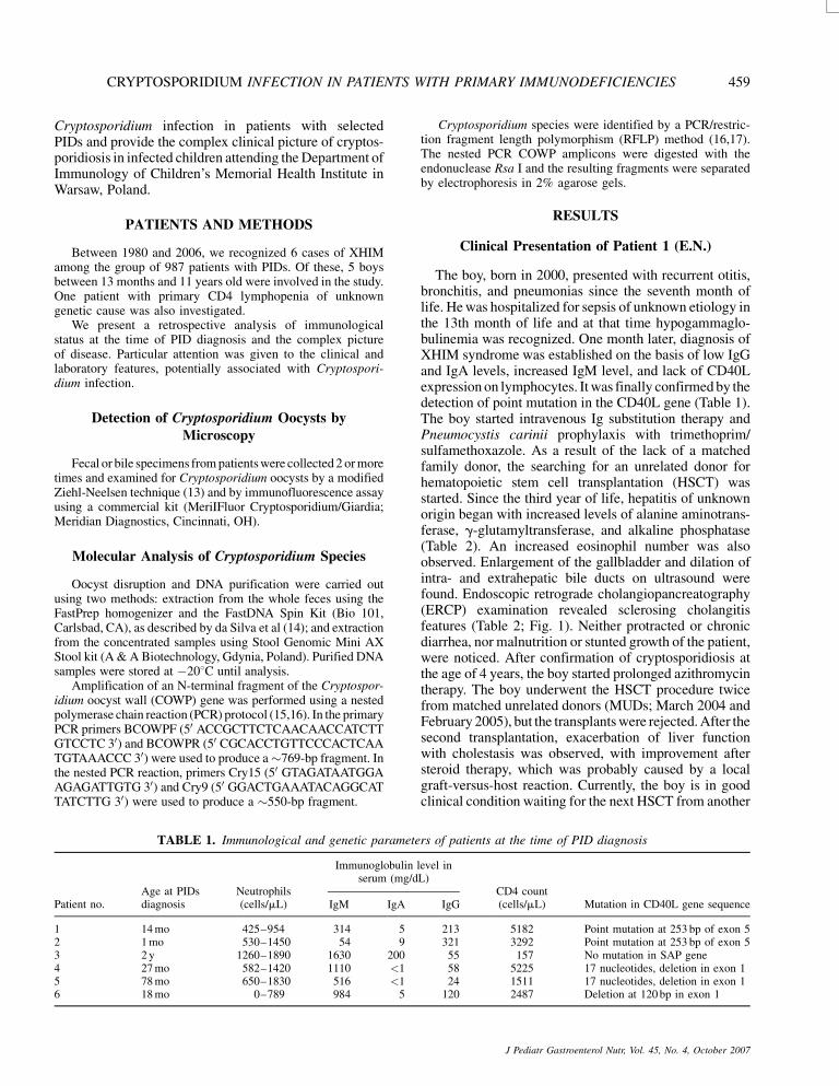
TABLE 1. Immunological and genetic paramete
Patient no.Age at PIDsdiagnosis
Neutrophils(cells/mL)
Immunoglobulin lserum (mg/d
IgM IgA
1 14 mo 425–954 314 52 1 mo 530–1450 54 93 2 y 1260–1890 1630 2004 27 mo 582–1420 1110 <15 78 mo 650–1830 516 <16 18 mo 0–789 984 5
Cryptosporidium species were identified by a PCR/restric-tion fragment length polymorphism (RFLP) method (16,17).The nested PCR COWP amplicons were digested with theendonuclease Rsa I and the resulting fragments were separatedby electrophoresis in 2% agarose gels.
RESULTS
Clinical Presentation of Patient 1 (E.N.)
The boy, born in 2000, presented with recurrent otitis,bronchitis, and pneumonias since the seventh month oflife. He was hospitalized for sepsis of unknown etiology inthe 13th month of life and at that time hypogammaglo-bulinemia was recognized. One month later, diagnosis ofXHIM syndrome was established on the basis of low IgGand IgA levels, increased IgM level, and lack of CD40Lexpression on lymphocytes. It was finally confirmed by thedetection of point mutation in the CD40L gene (Table 1).The boy started intravenous Ig substitution therapy andPneumocystis carinii prophylaxis with trimethoprim/sulfamethoxazole. As a result of the lack of a matchedfamily donor, the searching for an unrelated donor forhematopoietic stem cell transplantation (HSCT) wasstarted. Since the third year of life, hepatitis of unknownorigin began with increased levels of alanine aminotrans-ferase, g-glutamyltransferase, and alkaline phosphatase(Table 2). An increased eosinophil number was alsoobserved. Enlargement of the gallbladder and dilation ofintra- and extrahepatic bile ducts on ultrasound werefound. Endoscopic retrograde cholangiopancreatography(ERCP) examination revealed sclerosing cholangitisfeatures (Table 2; Fig. 1). Neither protracted or chronicdiarrhea, nor malnutrition or stunted growth of the patient,were noticed. After confirmation of cryptosporidiosis atthe age of 4 years, the boy started prolonged azithromycintherapy. The boy underwent the HSCT procedure twicefrom matched unrelated donors (MUDs; March 2004 and
ITH PRIMARY IMMUNODEFICIENCIES 459
nauthorized reproduction of this article is prohibited.
February 2005), but the transplants were rejected. After thesecond transplantation, exacerbation of liver functionwith cholestasis was observed, with improvement aftersteroid therapy, which was probably caused by a localgraft-versus-host reaction. Currently, the boy is in goodclinical condition waiting for the next HSCT from another
rs of patients at the time of PID diagnosis
evel inL)
CD4 count(cells/mL) Mutation in CD40L gene sequenceIgG
213 5182 Point mutation at 253 bp of exon 5321 3292 Point mutation at 253 bp of exon 555 157 No mutation in SAP gene58 5225 17 nucleotides, deletion in exon 124 1511 17 nucleotides, deletion in exon 1
120 2487 Deletion at 120 bp in exon 1
J Pediatr Gastroenterol Nutr, Vol. 45, No. 4, October 2007

Copy
TABLE 2. Pathophysiological parameters of patients in comparison to Cryptosporidium infection
Patient no.Cryptosporidium
infectionATT(U/L)
GGTP(U/L)
AP(U/L)
Eosinophils(cells/mL) USG ERCP
1 Yes 264 522 1182 3225 Enlargement of gallbladder,dilation of intra- and extrahepaticbile ducts
Features of sclerosingcholangitis
2 Yes 39 14 292 440 Normal ND3 Yes 71 455 573 5320 Enlargement and inflammation
of gallbladderFeatures of sclerosing
cholangitis000
D¼ n
460 WOLSKA-KUSNIERZ ET AL.
donor. Despite the long-term combined azithromycin andparomomycin therapy, cryptosporidiosis did not resolve(Table 3), and chronic cholangitis is still observed.
Clinical Presentation of Patient 2 (K.N.)
4 No 34 17 225 465 No 33 14 311 316 Yes 235 235 487 528
AP indicates alkaline phosphatase; GGTP¼g-glutamyltransferase; N
right © 2007 by Lippincott Williams & Wilkins.Un
The brother of patient E.N., born in 2003, was diagnosedwith XHIM at the first month of life due to a positive familyhistory. The lack of expression of CD40L on lymphocytesand the same point mutation as in his brother allowedconfirmation of the diagnosis (Table 1). Intravenous Ig
FIG. 1. ERCP examination in patient 1 (E.N.).
J Pediatr Gastroenterol Nutr, Vol. 45, No. 4, October 2007
substitution therapy and P carinii prophylaxis werestarted. Next, a search for an MUD for HSCT wascommenced. Neither signs of liver or bile duct dysfunc-tion, nor diarrhea or malnutrition, were observed beforethe time of transplantation (Tables 2 and 4). Hewas screened for Cryptosporidium infection at the ageof 11 months with negative microscopic examination(Table 4). At the age of 13 months the boy received HSCTfrom an MUD with full donor chimerism and goodhematological reconstitution. Four weeks after the pro-cedure, acute grade III graft-versus-host disease (GVHD)with skin and intestine involvement occurred. It wascomplicated by intestine perforation and developmentof a generalized fatal infection. After the patient’s death,we retrospectively obtained positive PCR results forCryptosporidium species from stool samples collectedbefore transplantation (Table 4).
Normal NDNormal NDEnlargement of gall bladder, dilation
of intra- and extrahepatic bile ductsDilation of intra- and
extrahepatic bile ducts
ot done.
authorized reproduction of this article is prohibited.
Clinical Presentation of Patient 3 (H.C.)
The boy, born in 1995, presented with recurrent diarrheawhich started at the age of 5 months. Since the first year oflife, a constantly elevated eosinophil number wasobserved, followed 2 years later by chronic hepatitis of
TABLE 3. History of cryptosporidiosis in patient 1
Sampleno.
Date ofcollection
Microscopicdiagnosis N-COWP Genotype
1A 12/03/04 þ þ 3 (C meleagridis)2A 21/10/04 � þ 3 (C meleagridis)3A 15/02/05 þ þ 3 (C meleagridis)3B 15/02/05 � þ 3 (C meleagridis)3C 15/02/05 þ þ 3 (C meleagridis)4A 08/03/05 þ þ 3 (C meleagridis)4B 08/03/05 þ þ 3 (C meleagridis)4C 08/03/05 � þ 3 (C meleagridis)5A May 2005 � þ 3 (C meleagridis)5B May 2005 � þ 3 (C meleagridis)5C May 2005 þ þ 3 (C meleagridis)
N-COWP indicates nested PCR on Cryptosporidium oocyst wallprotein gene.

Cop
TABLE 4. Comparison of symptoms and methods of Cryptosporidium detection
Patient no.Age at Cryptosporidium
diagnosis, yGastrointestinal
symptomsMicroscopic
diagnosis N-COWPGenotype ofCryptosporidium
2 2 – � þ 2 (C parvum)3 10 – þ þ�
1 (C hominis)
tein g
CRYPTOSPORIDIUM INFECTION IN PATIENTS WITH PRIMARY IMMUNODEFICIENCIES 461
unknown etiology. Other clinical manifestations weresevere chronic otitis, recurrent labial herpes infection,and skin abscesses. The diagnosis of primary CD4lymphopenia was established on the basis of the immuno-logical status (Table 1). Genetic analysis did not reveal anymutation in the SAP gene (Table 1), CD40L expressionwas normal, and the molecular background of his PIDremains unknown. Intravenous Ig substitution therapy andantiviral prophylaxis were started. Stunted growth, fol-lowed by chronic diarrhea and malnutrition with intestinalmucosal atrophy of grade III, were indicators for partialparenteral nutrition since the age of 9 years. At that timethe boy was qualified for HSCT, but his parents refused togive consent. At the age of 10 years, he presented withrecurrent bile duct inflammation followed by sclerosingcholangitis symptoms confirmed by ERCP (Table 2). Atthis time diagnostic tests for Cryptosporidium infectionwere performed and the oocysts were found in bileand fecal samples (Table 4). Azithromycin therapy wasapplied, with no improvement in clinical and laboratorystatus of the patient. Severe gallbladder inflammationwas the reason for surgery. Histopathological investigationof the gallbladder showed B cell lymphoma infiltration.The first course of chemotherapy was complicatedby intestine perforation, and during the surgery, interven-tion further dissemination of malignancy was noted.The patient underwent palliative care and died 5 monthslater.
Clinical Presentation of Patient 4 (A.K.)
The boy, born in 1998, was the brother of patient D.K.and presented with relevant family history (there wereboys’ deaths in early childhood in the maternal pedigree).The first manifestation of PID was observed in the12th month of life, when he started to experience recurrentbronchitis. There was no suspicion of PID until the third
4 7 –5 11 –6 5 –
�1 PCR-positive bile sample.
N-COWP indicates nested PCR on Cryptosporidium oocyst wall pro
yright © 2007 by Lippincott Williams & Wilkins.U
year of life, when the boy was screened as a result of XHIMdiagnosis in his brother. A deletion in the CD40L geneconfirmed the diagnosis (Table 1). Despite intravenous Igsubstitution therapy, chronic bronchitis is still observed.
No Cryptosporidium infection and no signs of liver orbile duct damage have been observed during follow-up
(Tables 2 and 4). There is a lack of a family donor, andHSCT from an MUD is being considered.
Clinical Presentation of Patient 5 (D.K.)
The boy, born in 1994, was doing well until his12th month of life when he started to experience recurrentupper and lower respiratory tract infections. At the age of6 years he was hospitalized as a result of sepsis andcandidiasis, and then hypogammaglobulinemia wasrecognized. The XHIM diagnosis was established6 months later, with typical immunological features,and was confirmed by the detection of deletion in theCD40L gene (Table 1). Clinical improvement with a lowincidence of infection was observed after intravenous Igsubstitution therapy was started. No Cryptosporidiuminfection and no signs of liver or bile duct damage havebeen observed (Tables 2 and 4). There is a lack of a familydonor, and HSCT from an MUD is being considered.
Clinical Presentation of Patient 6 (R.M.)
The boy, born in 2000, had recurrent bronchitis andpneumonias since his ninth month of life. Hypogamma-globulinemia was recognized at 20 months of age; 4months later, an XHIM diagnosis was established basedon detection of deletion in the CD40L gene (Table 1). Liverabnormalities were noted for the first time at the age of3 years with periodically elevated liver enzymes andg-glutamyltransferase. Further investigation revealedenlargement of the gallbladder, dilation of the intra-and extrahepatic bile ducts as shown by ERCP, and mildcholangitis described on liver biopsy, but no typicalsclerosing cholangitis features were found (Table 4). Ahigh eosinophil number was observed. Repeated analysisof stool samples performed at the age of 5 years revealedCryptosporidium infection. Despite intravenous Ig
� � �� � �þ þ 2 (C parvum)
ene.
nauthorized reproduction of this article is prohibited.
therapy, there are still recurrences of respiratory infections.There is no chronic diarrhea or malnutrition present(Table 4). Long-term azithromycin treatment did noteradicate Cryptosporidium infection. In this casethere is no matched family or unrelated donor for HSCT.Nevertheless, because of the severe course of XHIM, a
J Pediatr Gastroenterol Nutr, Vol. 45, No. 4, October 2007

Copy
USN
mismatched unrelated HSCT will be performed in thenear future.
Cryptosporidium Infection
Cryptosporidium infection was recognized in 4 of6 investigated patients (Tables 3 and 4). Three sets ofsamples from 3 boys were positive by both methods:microscopic examination of fecal specimens and nestedPCR amplification of a COWP gene fragment; however,the microscopic methods were less sensitive than the PCRassay. In patient 1, all samples (n ¼ 11) were positive bynested PCR, whereas only 6 were positive by microscopy(Table 4). In patient 2, infection was detected only by PCR,with negative microscopic examination in 3 sets ofsamples.
Among these 4 infected cases, three different parasitespecies were identified by COWP PCR/RFLP assay(Fig. 1; Tables 3 and 4). The prolonged infection in patient1 was caused by C meleagridis, whereas C hominis wasidentified in stool and bile samples from patient 3, andC parvum was associated with infections in patients 2and 6.
The expected RFLP patterns from these species wereas follows: for C meleagridis fragments of 372, 147, and34 bp; for C hominis fragments of 284, 129, 106, and34 bp; and for C parvum fragments of 413, 106, and 34 bp(Fig. 1).
DISCUSSION
In selected immunocompromised individuals, Cryptos-poridium infection cannot be eliminated by the host (7,18).Experimental studies in severe combined immunocom-promised or nude mice have shown that resolutionof Cryptosporidium infection requires the activation ofB and/or T lymphocytes (19). The 2 crucial factorsof immune response necessary for prevention and/orresolution of cryptosporidiosis, are CD4þ lymphocytesand interferon-g (19–22). It seems that a defectiveactivation route for macrophages, especially the CD40–CD40L route of activation, may be the cause for theincreased susceptibility to opportunistic infections (23).The lack of CD40–CD40L interaction caused by amutation in the CD40L or in the CD40 protein in individ-uals with XHIM or HIGM3 syndromes is probably themain reason for their increased susceptibility to cryptos-poridiosis. XHIM is a rare inherited immunodeficiencydisorder characterized by recurrent sinopulmonary, bac-terial, and opportunistic infections, and associated with
462 WOLSKA-K
right © 2007 by Lippincott Williams & Wilkins.Un
low IgG, IgA, and IgE serum levels and normal toincreased IgM serum levels (24).
In our group of patients with XHIM, no history ofacute, prolonged diarrhea or malnutrition of a suspectedor proven Cryptosporidium origin was recorded beforethe study. Chronic Cryptosporidium infection probably
J Pediatr Gastroenterol Nutr, Vol. 45, No. 4, October 2007
played an important role in chronic diarrhea, mucosalatrophy, and malabsorption requiring parenteral nutritionin a patient with CD4 lymphopenia (patient 3). In thefurther course of infection, the bile ducts were involved,with fatal complications. In this patient infection wascaused by C. hominis, a strictly anthroponotic parasite.
Five different Cryptosporidium species have beendescribed in immunocompromised patients all over theworld (9,15,25,26). The zoonotic species C parvum(identified in 23%–88% of cases), and the human-specificC hominis (in 12%–57% of cases) are the most commonlyidentified ones. The other 3 species, C felis, C canis, andC meleagridis, are less common (0.2%–11% cases), butthey are frequently found in their specific animal hosts,namely cat, dog, and turkey, respectively (27–29). For thefirst time in Polish patients with PIDs, Cryptosporidiumspecies, including C meleagridis, were identified. Inter-estingly, patients 1 and 2, who are brothers, were infectedwith different species of Cryptosporidium (C meleagridisand C parvum; Table 3), suggesting different sourcesof infection.
In a recent analysis of 126 patients with XHIMsyndrome reported to the European Society for Immu-nodeficiency registry, approximately one sixth developedliver disease, and in more than 50% cases this wasassociated with Cryptosporidium infection (30). In 2 boys(patients 1 and 6), the infection was correlated with theclinical course of sclerosing cholangitis and bile ductinflammation as the only manifestation.
In patient 2, infection with C parvum, which wasconfirmed only retrospectively, probably influenced thefatal complication of intestinal problems in the course ofGVHD after hematopoietic stem cell transplantation.Fatal exacerbation of Cryptosporidium infection earlyafter transplantation was reported in the literature (31).
Chronic infection or inflammation of bile ducts causedby Cryptosporidium species probably plays an importantrole in the development of malignancy. The tumors inmost cases are preceded by chronic cholangiopathy and/or liver cirrhosis (32). Other mechanisms that are not wellrecognized are probably involved in cancer pathogenesis.It is likely that Cryptosporidium infection, which lastedmany years in patient 3, was the main cause of intestinaland liver abnormalities, and was also a trigger for thedevelopment of malignancy. Several antibiotics (azithro-mycin, paromomycin, nitazoxanide) have shown someefficacy against the parasite. Unfortunately, clinical trialson the efficacy of treatment are discouraging: in apercentage of patients there is an improvement in theclinical condition, but it is difficult to eradicate infection.
IERZ ET AL.
authorized reproduction of this article is prohibited.
Better treatment results are achieved in HIV-infectedpatients presenting with intestinal involvement, butthere is no relevant improvement in patients withsclerosing cholangitis (33). Symptoms may also resolvespontaneously within the restoration of immune status(eg, with antiviral therapy in HIV-infected patients). It

Cop
TS W
seems that the best way to treat cryptosporidiosis inpatients with PID is to correct the underlying PID syn-drome. The only curative therapy for XHIM seems to becorrection of the immune defect by hematopoietic stemcell transplantation (34). In long-term observation thereis an extremely high risk of liver disease, leading to liverinsufficiency and malignancy development requiringadditional liver transplantation procedure (5,35). The 2last factors are the main causes of deaths and are import-ant indicators for HSCT in this group of PIDs.
It was observed in some patients that correction ofimmune defects yields a chance to eliminate infectionand also resolve liver and bile duct inflammation (36).Immunosuppressive conditioning therapy in such chil-dren before HSCT may result in acute cryptosporidiosisand cholangiopathy associated with oocyst excretion, andcan occur in patients without previous episodes of diar-rhea (6,25). In the absence of a simultaneous treatmentagainst Cryptosporidium species, this may lead to amassive amplification of the parasite, which results inrapid progress in liver disease and even death (25), asmentioned earlier. Early diagnosis of Cryptosporidiumspecies infection, particularly during the asymptomaticphase and possibly with the use of molecular techniques,is extremely important for patients with PID who mayundergo transplantation and receive proper treatment toavoid serious complications. In our group of patients,after diagnosis of cryptosporidiosis, 2 started azithromy-cin therapy (patients 3 and 6) and patient 1 wasgiven combined treatment with azithromycin andparomomycin. During several months of observation,neither resolution of bile duct abnormalities nor elimin-ation of the parasite was observed.
Conversely, children with symptoms of bile ductinflammation or hepatitis of unknown origin should bemonitored for PID disorders. If possible, every case ofCryptosporidium-associated prolonged diarrhea in chil-dren should be screened for XHIM (in boys) or HIGM3(in both sexes) (36).
In summary, this study underlines the need for a regularscreening of selected patients with PID for infectionby Cryptosporidium species. Unrecognized cases of cryp-tosporidiosis in children with PIDs may lead to seriousconsequences with development of sclerosing cholangitis,liver cirrhosis, and cholangiocarcinoma (32). Cryptospor-idium species that are rarely found in humans, such asC meleagridis, can play an important role in the patho-genesis of cholangitis and diarrhea in PID. In agreementwith a previous study (25), our data confirmed thatmicroscopy is less sensitive than nested PCR for
CRYPTOSPORIDIUM INFECTION IN PATIEN
yright © 2007 by Lippincott Williams & Wilkins.U
the detection of the parasite in asymptomatic cases(Tables 3 and 4). Therefore, molecular diagnostic toolsshould be considered the method of choice in screening ofPIDs. Stool samples from such patients should be checkedregularly, at least 3 times per year, even in the absence ofdiarrhea or other symptoms. Finally, because the eradica-
tion of cryptosporidial infection is difficult to obtain, evenunder long-lasting therapeutic regimes, new efficaciousdrugs are urgently needed.
REFERENCES
1. Huang DB, Chappell C, Okhuysen PC. Cryptosporidiosis in chil-dren. Semin Pediatr Infect Dis 2004;15:253–9.
2. Sinski E, Szklarczyk J, Oralewska B, et al. Cryptosporidium sp.infection in children with symptoms of gastro-enteritis. Acta ParasitolPolon 1988;33:295–301.
3. Neill MA, Rice SK, Ahmad NV, et al. Cryptosporidiosis: anunre-cognised cause of diarrhoea in elderly hospitalised patients. ClinInfect Dis 1996;22:168–70.
4. Kocoshis SA, Cibull ML, Davis TE, et al. Intestinal and pulmonarycryptosporidiosis in an infant with severe combined immunedeficiency. J Pediatr Gastroenetrol Nutr 1984;3:149–57.
5. Levy J, Espanol-Boren T, Thomas C. Clinical spectrum of X linkedhyper-IgM syndrome. J Pediatr 1997;131:47–54.
6. Davies EG, Hadzic N, Jones AM. Cholangiopathy in children withcombined immunodeficiencies [abstract]. Mol Immunol 1998;35:731.
7. Hunter PR, Nichols G. Epidemiology and clinical features ofCryptosporidium infection in immunocompromised patients. ClinMicrobiol Rev 2002;15:145–54.
8. Thompson RC, Olson ME, Zhu G, et al. Cryptosporidium andcryptosporidiosis. Adv Parasitol 2005;59:77–158.
9. Caccio SM, Thompson RCA, McLauchlin J, et al. UnravellingCryptosporidium and Giardia epidemiology. Trends Parasitol 2005;21:430–7.
10. O’Donoghoue PJ. Cryptosporidium and cryptosporidiosis in manand animals. Int J Parasitol 1995;25:139–95.
11. Cello JP. Human immunodeficiency virus associated biliary tractdisease. Semin Liver Dis 1992;12:213–8.
12. Clark DP. New insights into human cryptosporidiosis. Clin MicrobiolRev 1999;12:554–63.
13. Henriksen S, Pohlenz J. Staining of cryptosporidia by modifiedZiehl-Neelsen technique. Acta Vet Scand 1981;22:594–6.
14. Da Silva AJ, Bornay-Llinares FJ, Moura INS, et al. Fast and reliableextraction of protozoan parasite DNA from fecal specimens.Mol Diag 1999;4:57–64.
15. Pedraza-Diaz S, Amar C, Nichols GL, et al. Nested polymerasechain reaction for amplification of the Cryptosporidium oocyst wallprotein gene. Emerg Infect Dis 2001;7:49–56.
16. Spano F, Putignani L, McLauchlin J, et al. PCR-RFLP analysis of theCryptosporidium oocyst wall protein (COWP) gene discriminatesbetween C. wrairi and C. parvum, and between C. parvum isolatesof human and animal origin. FEMS Microbiol Lett 1997;150:209–17.
17. Xiao L, Limor J, Morgan UM, et al. Sequence differences in thediagnostic target region of the oocyst wall protein gene of Cryp-tosporidium parasites. Appl Environ Microbiol 2000;66:5499–502.
18. Fayer R, Speer CA, Dubey JP. The general biology of cryptospor-idium. In: Fayer R (ed). Cryptosporidium and Cryptosporidiosis.Boca Raton, FL: CRC Press; 1997:1–41.
19. Chen W, Harp JA, Harmsen AG, et al. Gamma interferon functionsin resistance to Cryptosporidium parvum infection in SCID mice.Infect Immunol 1993;61:3548–51.
20. McDonald V, Bancroft GJ. Mechanisms of innate and acquiredresistance to Cryptosporidium parvum infection in SCID mice.
ITH PRIMARY IMMUNODEFICIENCIES 463
nauthorized reproduction of this article is prohibited.
Parasite Immunol 1994;16:315–20.21. Perryman LE, Mason PH, Chrisp CE. Effect of spleen cell popula-
tions on resolution of Cryptosporidium parvum infection in SCIDmice. Adv Parasitol 1998;40:87–119.
22. Ungar BLP, Kao TC, Burris JA, et al. Cryptosporidium infection in anadult mouse model. Independent roles for IFNg and CD4þTlymphocytes in protective immunity. J Immunol 1991;147:1014–22.
J Pediatr Gastroenterol Nutr, Vol. 45, No. 4, October 2007

Copy
USN
23. Ferrari S, Giliani S, Insalco A, et al. Mutations of CD40 gene causean autosomal recessive form of immunodeficiency with hyper IgM.Proc Nat Acad Sci U S A 2001;98:12614–9.
24. Winkelstein JA, Marino MC, Ochs H, et al. The X-linked hyper-IgM syndrome. Medicine 2003;82:373–84.
25. McLauchlin J, Amar C, Pedraza-Diaz S, et al. Molecularepidemiological analysis of Cryptosporidium spp. in the UnitedKingdom: results of genotyping Cryptosporidium spp. in 1705 fecalsamples from humans and 105 fecal samples from livestockanimals. J Clin Microbiol 2000;38:3984–90.
26. Caccio S, Pinter E, Fantini R, et al. Human infection withCryptosporidium felis: case report and literature review. EmergInfect Dis 2002;8:85–6.
27. Sargent KD, Morgan UM, Elliot A, et al. Morphological and geneticcharacterisation of Cryptosporidium oocysts from domestic cats.Vet Prasitol 1998;7:221–7.
28. Fayer R, Ttout JM, Xiao L, et al. Cryptosporidium canis n. sp. from
464 WOLSKA-K
right © 2007 by Lippincott Williams & Wilkins.Un
29. Slavin D. Cryptosporidium meleagridis (sp. Nov.). J Comp PatholTher 1955;65:262–6.
30. Toniati P, Savoldi G, Jones AM, et al. Report of the ESIDcollaborative study on clinical features and molecular analysisof X-linked hyper-IgM syndrome. Eur Soc ImmunodeficienciesNewslett 2002;F9 (Suppl):40.
J Pediatr Gastroenterol Nutr, Vol. 45, No. 4, October 2007
31. Gennery AR, Khawaja K, Veys P, et al. Treatment of CD40 liganddeficiency by hematopoietic stem cell transplantation: a surveyof the European experience, 1993–2002. Blood 2004;103:1152–7.
32. Heyward AR, Levy J, Facchetti F, et al. Cholangiopathyand tumors of the pancreas, liver and biliary tree in boys withX-linked immunodeficiency with hyper IgM. J Immunol 1997;158:977–83.
33. Zulu I, Kelly P, et al. Nitazoxanide for persistent diarrhoea inZambian acquired immune deficiency syndrome patients: a rando-mized-controlled trial. Aliment Pharmacol Ther 2005; 21:757–63;Notarangelo LD, Hayward AR, X-linked immunodeficiency withhyper IgM (XHIM). Clin Exp Immunol. 2000;120:399–405.
34. Dimicoli S, Benesoussan D, Latger-Cannard V, et al. Completerecovery from Cryptosporidium parvum infection with gastroenter-ocolitis and sclerosis cholangitis after successful bone marrowtransplantation in two brothers with X-linked hyper-IgM syndrome.
IERZ ET AL.
Bone Marrow Transplant 2003;32:733–7.
domestic dogs. J Parasitol 2001;87:1415–22.authorized reproduction of this article is prohibited.
35. Rodrigues F, Davies EG, Harrison P, et al. Liver disease in childrenwith primary immunodeficiencies. J Pediatr 2004;145:333–9.
36. Kutukculer N, Moratto D, Aydinok Y, et al. DisseminatedCryptosporidium infection in an infant with hyper-IgM syndromecaused by CD40 deficiency. J Pediatr 2003;142:194–6.