Enterocytozoon bieneusi and Cryptosporidium
9
RESEARCH ARTICLE Open Access “Enterocytozoon bieneusi and Cryptosporidium: a cross-sectional study conducted throughout Thailand” Rapeepun Prasertbun 1 , Hirotake Mori 1,2 , Yaowalark Sukthana 1 , Supaluk Popruk 1 , Teera Kusolsuk 3 , Katsuro Hagiwara 4 and Aongart Mahittikorn 1* Abstract Background: Enterocytozoon bieneusi and Cryptosporidium spp. are prevalent zoonotic parasites associated with a high burden among children. To date only limited molecular epidemiological data on E. bieneusi and Cryptosporidium spp. in humans living in Thailand has been published. Methods: PCR-based tools were used to detect and characterize E. bieneusi and Cryptosporidium spp. The internal transcribed spacer (ITS) region of the rRNA gene was used to investigate E. bieneusi, and the small subunit (SSU) rRNA gene was used to investigate Cryptosporidium spp., and 697 fecal samples from villagers and school children in rural areas in Thailand were analyzed. Results: The infection rates were 2.15% (15/697) for E. bieneusi and 0.14% (1/697) for Cryptosporidium spp. The prevalence of E. bieneusi was significantly high in Loei province. Sequence analysis indicated that the Cryptosporidium isolate was C. parvum. Nine E. bieneusi genotypes were identified, EbpC, Peru12, TMH6, TMH3, TMH7, H, D, and two novel genotypes TMLH1 and TMLH2. E. bieneusi prevalence was significantly higher in male participants than in female participants, and in children aged 3–15 years than in participants aged > 15 years. Conclusions: The prevalence, genotypes, and zoonotic potential of E. bieneusi were found to vary significantly high even in one country. Transmission routes and key animal carriers of E. bieneusi may be associated with differences in hygiene, sanitation, and cultural behaviors. Further molecular studies including longitudinal studies will be required to unveil epidemiological characteristics of these opportunistic intestinal protozoa in all over the countries. Keywords: Enterocytozoon, Cryptosporidium, Thailand Background Opportunistic intestinal protozoa such as Cryptosporid- ium spp. and Enterocytozoon bieneusi can cause diar- rhea in humans [1], and are associated with increased mortality and short survival in immunocompromised people, especially AIDS patients [2]. Recently, intestinal cases of infection with these parasites have reportedly increased in non-HIV-infected populations such as organ-transplant recipients, patients with malignant diseases, and diabetes patients [3]. Although they are considered opportunistic pathogens, several outbreaks have been reported both in immunocompetent humans and in domestic and wild animals [4–7]. There are published reports of E. bieneusi and Cryptosporidium infection in children considered HIV-seronegative and in healthy children indicating the presence of enteric carriage of infection in immunocompetent people in developing countries and other regions [8]. The patho- gens therefore pose significant challenges to public health, especially in developing countries because of their high prevalence and the disease burden associated with infec- tions. They have also been linked to impaired growth and cognitive function in children and immunocompromised individuals [9–11]. Among the 14 Cryptosporidium species known to infect humans, C. hominis and C. parvum are the most © The Author(s). 2019 Open Access This article is distributed under the terms of the Creative Commons Attribution 4.0 International License (http://creativecommons.org/licenses/by/4.0/), which permits unrestricted use, distribution, and reproduction in any medium, provided you give appropriate credit to the original author(s) and the source, provide a link to the Creative Commons license, and indicate if changes were made. The Creative Commons Public Domain Dedication waiver (http://creativecommons.org/publicdomain/zero/1.0/) applies to the data made available in this article, unless otherwise stated. * Correspondence: [email protected] 1 Department of Protozoology, Faculty of Tropical Medicine, Mahidol University, Bangkok, Thailand Full list of author information is available at the end of the article Prasertbun et al. BMC Infectious Diseases (2019) 19:808 https://doi.org/10.1186/s12879-019-4422-4
-
Upload
khangminh22 -
Category
Documents
-
view
0 -
download
0
Transcript of Enterocytozoon bieneusi and Cryptosporidium
RESEARCH ARTICLE Open Access
“Enterocytozoon bieneusi andCryptosporidium: a cross-sectional studyconducted throughout Thailand”Rapeepun Prasertbun1, Hirotake Mori1,2, Yaowalark Sukthana1, Supaluk Popruk1, Teera Kusolsuk3,Katsuro Hagiwara4 and Aongart Mahittikorn1*
Abstract
Background: Enterocytozoon bieneusi and Cryptosporidium spp. are prevalent zoonotic parasites associated with ahigh burden among children. To date only limited molecular epidemiological data on E. bieneusi andCryptosporidium spp. in humans living in Thailand has been published.
Methods: PCR-based tools were used to detect and characterize E. bieneusi and Cryptosporidium spp. The internaltranscribed spacer (ITS) region of the rRNA gene was used to investigate E. bieneusi, and the small subunit (SSU)rRNA gene was used to investigate Cryptosporidium spp., and 697 fecal samples from villagers and school childrenin rural areas in Thailand were analyzed.
Results: The infection rates were 2.15% (15/697) for E. bieneusi and 0.14% (1/697) for Cryptosporidium spp. Theprevalence of E. bieneusi was significantly high in Loei province. Sequence analysis indicated that theCryptosporidium isolate was C. parvum. Nine E. bieneusi genotypes were identified, EbpC, Peru12, TMH6, TMH3,TMH7, H, D, and two novel genotypes TMLH1 and TMLH2. E. bieneusi prevalence was significantly higher in maleparticipants than in female participants, and in children aged 3–15 years than in participants aged > 15 years.
Conclusions: The prevalence, genotypes, and zoonotic potential of E. bieneusi were found to vary significantly higheven in one country. Transmission routes and key animal carriers of E. bieneusi may be associated with differencesin hygiene, sanitation, and cultural behaviors. Further molecular studies including longitudinal studies will berequired to unveil epidemiological characteristics of these opportunistic intestinal protozoa in all over the countries.
Keywords: Enterocytozoon, Cryptosporidium, Thailand
BackgroundOpportunistic intestinal protozoa such as Cryptosporid-ium spp. and Enterocytozoon bieneusi can cause diar-rhea in humans [1], and are associated with increasedmortality and short survival in immunocompromisedpeople, especially AIDS patients [2]. Recently, intestinalcases of infection with these parasites have reportedlyincreased in non-HIV-infected populations such asorgan-transplant recipients, patients with malignantdiseases, and diabetes patients [3]. Although they areconsidered opportunistic pathogens, several outbreaks
have been reported both in immunocompetent humansand in domestic and wild animals [4–7]. There arepublished reports of E. bieneusi and Cryptosporidiuminfection in children considered HIV-seronegative andin healthy children indicating the presence of entericcarriage of infection in immunocompetent people indeveloping countries and other regions [8]. The patho-gens therefore pose significant challenges to public health,especially in developing countries because of their highprevalence and the disease burden associated with infec-tions. They have also been linked to impaired growth andcognitive function in children and immunocompromisedindividuals [9–11].Among the 14 Cryptosporidium species known to
infect humans, C. hominis and C. parvum are the most
© The Author(s). 2019 Open Access This article is distributed under the terms of the Creative Commons Attribution 4.0International License (http://creativecommons.org/licenses/by/4.0/), which permits unrestricted use, distribution, andreproduction in any medium, provided you give appropriate credit to the original author(s) and the source, provide a link tothe Creative Commons license, and indicate if changes were made. The Creative Commons Public Domain Dedication waiver(http://creativecommons.org/publicdomain/zero/1.0/) applies to the data made available in this article, unless otherwise stated.
* Correspondence: [email protected] of Protozoology, Faculty of Tropical Medicine, MahidolUniversity, Bangkok, ThailandFull list of author information is available at the end of the article
Prasertbun et al. BMC Infectious Diseases (2019) 19:808 https://doi.org/10.1186/s12879-019-4422-4

predominant [12, 13], and among 14 species of micro-sporidia, E. bieneusi infects humans the most frequently.Transmission routes include direct contact with infectedpeople (anthroponotic transmission) and animals (zoonotictransmission), and consumption of contaminated food andwater [14, 15]. Molecular-based techniques are used to in-vestigate prevalence, characterize species, determine geno-types, and assess the zoonotic potential of these parasites[16]. The prevalence and zoonotic potential of intestinalprotozoa can vary by country and by location within acountry.Only limited molecular epidemiological data on Crypto-
sporidium and E. bieneusi have been published in humansin Thailand. To date, no studies on the prevalence orsubtyping of Cryptosporidium in the community have beenpublished. Previous studies investigating the prevalence andgenotype characteristics of E. bieneusi in limited parts ofThailand revealed divergent genotype characteristics, andzoonotic potential [17, 18]. The aim of the current studywas to determine the prevalence and genotype/subtypecharacteristics of Cryptosporidium and E. bieneusi in thecommunity throughout Thailand, and evaluate the diver-gence and zoonotic risks associated with the pathogens.We conducted a molecular survey among villagers andschool children in northern, northeastern, central, western,and southern Thailand.
MethodsStudy sites and fecal collectionA total of 697 fresh fecal samples were collected from 235healthy villagers and 462 healthy school children fromAugust 2015 to January 2017 in rural areas in Thailand. Ofthe 697 participants, 318 were male and 379 were female.The samples were grouped into two age classes (3 to ≤15years and > 15 years), and they were obtained from sevenprovinces; Chiang rai (n = 32) and Nan (n = 46) in thenorthern area, Tak (n = 151) and Ratchaburi (n = 166) inthe western area, and Loei (n = 37), Chumphon (n = 101),and Sa kaeo (n = 164) respectively located in the northeast-ern, southern, and eastern areas (Fig. 1).Compared with many other areas in Thailand, the study
sites are rural and impoverished. The provision of infra-structure such as water supply, roads, and health servicesis not satisfactory. Most of the residents work in agricul-ture. In rural communities, major problems that havebeen identified are poverty, illiteracy and unemployment.Due to lack of literacy skills, inhabitants may experienceproblems with poor hygiene and sanitation conditions.To preserve anonymity, each sample was given a unique
identification number. All samples were collected fromparticipants at home, kept at 4 °C, then transported to alaboratory at the Faculty of Tropical Medicine, MahidolUniversity, Thailand in ice packs. Fresh unpreserved fecal
samples were aliquoted upon receipt and stored at − 20 °Cprior to DNA extraction.
Molecular analysisDNA was extracted using the PSP Spin Stool DNA Kit(Stratec Inc., Germany) in accordance with the manufac-turer’s instructions. The kit has a rigorous prelysis stepusing Zirconia Beads II with an optimized prelysis bufferunder high temperatures that helps to extract DNA,followed by a preincubation of the sample with InviAdsorbto remove PCR inhibitors very efficiently. For Cryptosporid-ium, fragments of 18S rRNA (830 bp) were amplified vianested PCR as previously described [19]. PCRs were per-formed in a 25-μL reaction containing 1 × PCR buffer, 1.5mM MgCl2, 0.2mM of each dNTP, 2.5 U Taq polymerase(Thermo Scientific, USA), 1.0 μM of each primer, and2.0 μL of DNA template. Each PCR consisted of thirty-fivecycles of 94 °C for 45 s, 55 °C for 45 s, and 72 °C for 60 s.The primary PCR products were then used in second-round PCRs, which entailed thirty-five cycles of 94 °C for45 s, 58 °C for 45 s, and 72 °C for 60 s.For E. bieneusi, the internal transcribed spacer (ITS)
region with a portion of the flanking large and smallsubunit (SSU) ribosomal RNA genes (390 bp) was ampli-fied [20]. Negative and positive controls were includedin all PCRs. Positive control samples were obtainedusing DNA polymerase known to test-positive andconfirmed at the nucleotide level via GenBank, and nu-clease-free water was used as a negative control. ThePCR mixture was the same as that used for Cryptospor-idium except that 2.0 mM MgCl2 was used. Both pri-mary and second-round PCRs entailed thirty-five cycles(denaturation at 94 °C for 30 s, annealing at 55 °C for 30s, and elongation at 72 °C for 40 s). Ten microliters ofPCR products were subjected to electrophoresis in 1.5%agarose gel and visualized via ethidium bromide stainingfor 10 min in a 1 μg/mL ethidium bromide solution. The100 bp DNA ladder (Thermo Scientific, USA) wasincluded in every gel. PCR products were purified usingspin columns (QiaQuick PCR purification kit, Qiagen,Germany) and sequenced in both directions using thesecondary PCR primers for each organism and an ABI3730xl DNA analyzer (Applied Biosystems, USA).Genotypes/subtypes from each positive sample were con-
firmed by homology between the sequenced PCR productsand published sequences in GenBank as determined via theBasic Local Alignment Search Tool (BLAST). The sequenceof E. bieneusi novel genotypes TMLH1 and TMLH2 in thepresent study were submitted and deposited in GenBankwith the respective accession numbers MG366589 andMG366590.Phylogenetic analyses of E. bieneusi ITS sequences
were performed using MEGA Software Version 7 [21].Evolutionary distances between different isolates were
Prasertbun et al. BMC Infectious Diseases (2019) 19:808 Page 2 of 9
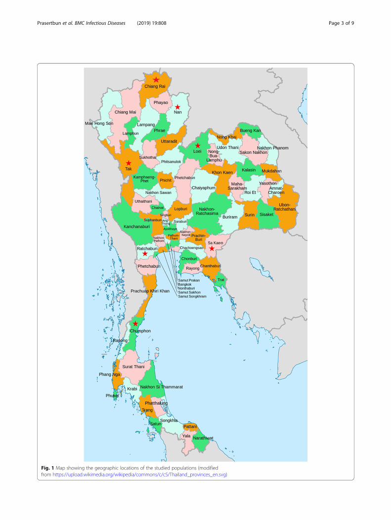
Fig. 1 Map showing the geographic locations of the studied populations (modifiedfrom https://upload.wikimedia.org/wikipedia/commons/c/c5/Thailand_provinces_en.svg)
Prasertbun et al. BMC Infectious Diseases (2019) 19:808 Page 3 of 9
calculated using the Kimura 2-parameter method [22],and phylogenetic trees were constructed using theneighbor-joining algorithm [23]. Branch reliability wasassessed using bootstrap analyses (1000 replicates). TheChi-square test was performed using SPSS statistics 18software (IBM, USA). P values < 0.05 were consideredsignificant in all statistical analyses.
ResultsCryptosporidium spp. was only detected in 1 sample inthe entire study, from Tak Province, yielding a preva-lence of 0.14% (1/697). The total prevalence of E. bien-eusi was 2.15% (15/697). E. bieneusi was detected insamples from 3 provinces, Loei (northeastern area), Tak(western area), and Ratchaburi (western area). Theprevalence of E. bieneusi was significantly higher in Loei(18.9%) than it was in Tak (2.6%) and Ratchaburi (2.4%).Prevalence of E. bieneusi varies significantly even in onecountry. E. bieneusi prevalence and genotype data areshown in Table 1.The prevalence of E. bieneusi was significantly higher
in school children aged 3–15 years (3.0%) than in partici-pants aged > 15 years (0.4%) (P = 0.0258), and it wassignificantly higher in male participants (4.1%) than infemale participants (0.5%) (P = 0.0013). In Loei, E. bieneusiwas only detected in school children aged 3–15 years(18.9%; no E. bieneusi was detected in samples frompeople aged > 15 years), and all positive E. bieneusi sam-ples were from males (Table 1).E. bieneusi genotypes EbpC, Peru12, TMH6, TMH3,
and D were identified in Tak and Ratchaburi. In Loei,genotypes TMH3, TMH7, TMH6, H, and two novelgenotypes TMLH1 and TMLH2 were found. TMLH1
and TMLH2 both differed from genotype TMP11 by asingle base (TMLH1 position 88 A→G, TMLH2 pos-ition 126 A→G) (Table 2). Genotype TMH6 was themost frequently identified (3/15) in this study. Zoonoticgenotypes (EbpC, Peru12, D, and H) were identified insamples from Loei, Tak, and Ratchaburi. In Tak, zoonoticgenotypes were more frequently identified, but in otherprovinces anthroponotic genotypes (TMH3, TMH7, andTMH6) were more prevalent (Table 1). The sequences ofthe single Cryptosporidium-positive sample had 100% hom-ology with those of C. parvum (GenBank accession numberAY204230).In phylogenetic analysis, 13 ITS gene sequences were
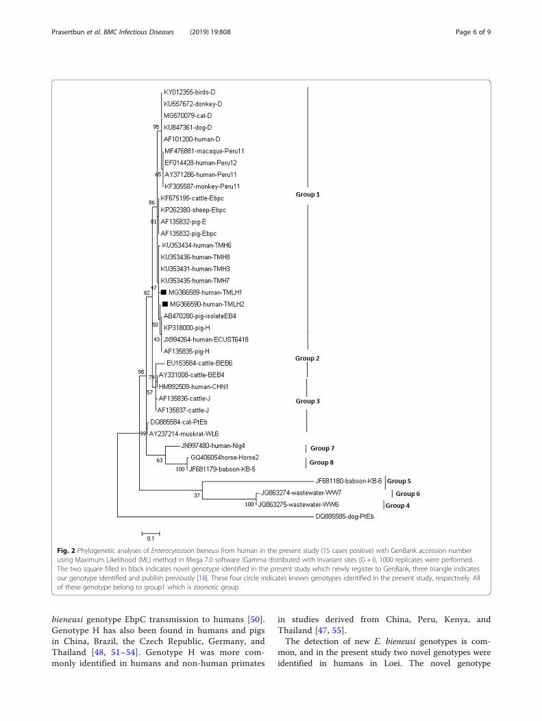
identical to seven genotypes and had 100% homologywith those of genotypes D (GenBank accession number(AF101200), EbpC (KF675195), H (KP318000), TMH3(KU353431), TMH7 (KU353435), TMH6 (KU353434),or Peru12 (EF014428). The remaining two were not previ-ously described, and were designated TMLH1 (MG366589)and TMLH2 (MG366590). Phylogenetic analysis indicatedthat all of these genotype belong to group1 (Fig. 2).
DiscussionThe main objective of this study was to investigate theprevalence, genetic diversity, and zoonotic potential ofCryptosporidium spp. and E. bieneusi in humans in ruralareas throughout Thailand. The prevalence of Crypto-sporidium (0.14%) was lower than that previously re-ported in HIV-negative subjects in Thailand (1.0% [24],and in Honduras in children aged 0–5 years (56.4%; [25].Overall, a range of 6 to 11% of water samples were con-taminated with Cryptosporidium spp. and a occurrenceof the parasite was reported in HIV/AIDS from 1996 to
Table 1 Age, sex distribution and genotype identification of Enterocytozoon bieneusi in fecal samples from human in rural areas,Thailand
Region Province Positive cases /total no. (%) Genotype (number)
Sex Age Total
Male Female 3 to ≤15 Years > 15 Years
Northeastern Loei 7/27 (25.9) 0/10 (0) 7/37 (18.9) 0/0 (0) 7/37 (18.9)* TMH3 (1), TMH7 (2), TMH6 (1),H (1) ***, TMLH1 (1), TMLH2 (1)
Western Tak 2/56 (3.6) 2/95 (2.1) 3/125 (2.4) 1/26 (3.8) 4/151 (2.6) EbpC (1) ***, Peru12 (1) ***, D (2) ***
Ratchaburi 4/104 (3.8) 0/62 (0) 4/166 (2.4) 0/0 (0) 4/166 (2.4) EbpC (1) ***, TMH6 (2),TMH3 (1)
Northern Chiang rai 0/18 (0) 0/14 (0) 0/9 (0) 0/23 (0) 0/32 (0)
Nan 0/14 (0) 0/32 (0) 0/3 (0) 0/43 (0) 0/46 (0)
Southern Chumphon 0/47 (0) 0/54 (0) 0/91 (0) 0/10 (0) 0/101 (0)
Eastern Sakaeo 0/52 (0) 0/112 (0) 0/31 (0) 0/133 (0) 0/164 (0)
Total 13/318 (4.1)** 2/379 (0.5)** 14/462 (3.0)** 1/235 (0.4)** 15/697 (2.15) EbpC (2)***, D (2)***, TMH6 (3),TMH3 (2), TMH7 (2), H (1) ***,TMLH1 (1), TMLH2 (1), Peru12 (1) ***
*P < 0.05 was considered statistically significant positive (Chi-square test)**P < 0.05 was considered statistically significant (Fisher test)***Zoonotic genotype
Prasertbun et al. BMC Infectious Diseases (2019) 19:808 Page 4 of 9
2009 in Thailand at a range of 19 to 34% [26, 27]. Theprevalence of Cryptosporidium varied in animal types.Infection rates between 5.7 and 31.5% have been re-ported in dairy cattle with dogs, cats and monkeysreflecting infection rates of 2.1, 2.5 and 1% respectively[28–31]. The distribution of C. parvum in humans variesaccording to different geographic areas and under differ-ent socioeconomic conditions. Cryptosporidium infec-tion can be transmitted orally by drinking water andthrough environmental contaminants. Seasonality hasalso been reported to affect Cryptosporidium variation[32]. However, as our study spanned a period of 17months, a seasonal effect is unlikely. The high prevalenceof Cryptosporidium has been reported from water suppliesin many parts of the world [4, 33, 34]. The low prevalencerate in our study may suggest an improved living standardin terms of environmental sanitary conditions shared bythe study participants. Moreover, the water supply inThailand is predominantly provided by the provincialwaterworks authority which is unlikely to be the source ofCryptosporidium. In some European countries and NewZealand, both C. hominis and C. parvum are commonlydetected in humans [35]. C. parvum is the predominantspecies detected in humans in developed countries suchas England, France, and New Zealand, whereas C. hominisis reportedly the predominant species in developing coun-tries [35]. In Thailand C. hominis (genotype 1), C. melea-gridis, C. muris, and C. felis were reported in HIV-infectedpatients in 2002 [36]. In HIV/AIDS patients in ThaiAIDS-care centers, C. hominis was the most commonlyidentified species, followed by C. meleagridis [26, 37]. Thepresent study is the first molecular epidemiological inves-tigation of Cryptosporidium in communities throughoutThailand, and the results suggest the epidemiological risksof Cryptosporidium may be minimal.The prevalence of E. bieneusi determined in the
present study was consistent with results of previousstudies in human communities in Thailand. Respectiveprevalences of 3.8 and 2.9% have been determined in
western and northern Thailand via nested PCR [17, 18].However, it was reportedly more prevalent in children inChina (22.5%; [8] and in the elderly in Spain (17.0%;[38]. In the current study prevalence varied by location.It was high in Loei in northeastern Thailand (18.9%), butthe organism was not detected at all in four other prov-inces. Differences in reported prevalences may be associ-ated with hygiene, sanitation, culture, living standards,methods of detection, and parameters used to definestudy populations.Interestingly, in Loei and Ratchaburi E. bieneusi was more
prevalent in males than females, which is concordant with aprevious study in a remote city in the Brazilian Amazon[39]. It may be that the males in these studies engaged inagricultural and farming practices entailing a risk of contactwith the parasite in the environment more frequently thanthe females. In addition, in the present study E. bieneusi wasmore prevalent in school children aged 3–15 years (3.0%)than in participants aged > 15 years (0.4%). A higher preva-lence in children (19.0%) [40] than in adults (12.4%) has alsobeen reported [41]. Similarly, in a study reported in 2002that was conducted in a hospital in Uganda 17.4% of 1779children with diarrhea were evidently infected with E. bien-eusi, and it was concluded that E. bieneusi was widespreadamong children aged 3–36months in Uganda [42]. Intes-tinal microsporidiosis may be more common in males thanin females, and more common in children than in adults.Possible reasons for these findings could be as follows. First,personal hygiene and public health in agricultural and farm-ing environments are poor when compared with otherpopulations. Toilets are often not available in farm areas.Second, children of farming parents were more likely to beinfected with intestinal parasites compared to children ofparents who did not farm as these parents were more likelyto have male children involved in casual labor [43]. Further-more, male children are more likely to come in directcontact with the ground while playing [44]. Altogether, thesecan be related to anthroponotic transmission.In the present study, genotype analysis of E. bieneusi
also suggested zoonotic potential. The genotypes identi-fied were EbpC, Peru12, D, H, TMH3, TMH6, TMH7,TMLH1, and TMLH2, and of these EbpC, Peru12, D,and H have been deemed indicative of zoonotic potential[18, 45, 46]. The genotypes identified in humans inThailand to date include D, A, R, S, T, U, V, W, PigEb10,H, E, EbpA, O, Peru12, PigEbITS7, TMLH1–2, ETMK1and TMH1–8 [17, 18, 37, 47–49] (Table 3).In Thailand genotype D is frequently found in
humans and various other animal species [18, 47, 49],and genotypes EbpC, H, and Peru12 have also beenreported [17, 37, 47, 48]. All are zoonotic genotypes.EbpC is reportedly a common genotype in non-humananimals, especially in pigs and in China, where contactwith pigs was evidently strongly associated with E.
Table 2 The variations in ITS region sequence of rRNA gene ofEnterocytozoon bieneusi isolates from human in this studycompared with ITS sequences of five known genotypes
Genotypes (no.) Nucleotide at position GenBankaccession no.
71 85 88 126 131 133
Novel TMLH1 C G A G T G MG366589
TMLH2 C G G A T G MG366590
Known TMP11 C G G G T G KU353447
EbpC C G G G C A MH745039
PigHN-32 C T G A T G MF406105
KIN-1 C G G G T A KY495647
H T T G A T G KP318000
Prasertbun et al. BMC Infectious Diseases (2019) 19:808 Page 5 of 9
bieneusi genotype EbpC transmission to humans [50].Genotype H has also been found in humans and pigsin China, Brazil, the Czech Republic, Germany, andThailand [48, 51–54]. Genotype H was more com-monly identified in humans and non-human primates
in studies derived from China, Peru, Kenya, andThailand [47, 55].The detection of new E. bieneusi genotypes is com-
mon, and in the present study two novel genotypes wereidentified in humans in Loei. The novel genotype
Fig. 2 Phylogenetic analyses of Enterocytozoon bieneusi from human in the present study (15 cases positive) with GenBank accession numberusing Maximum Likelihood (ML) method in Mega 7.0 software (Gamma distributed with Invariant sites (G + I), 1000 replicates were performed.The two square filled in black indicates novel genotype identified in the present study which newly register to GenBank, three triangle indicatesour genotype identified and publish previously [18]. These four circle indicates known genotypes identified in the present study, respectively. Allof these genotype belong to group1 which is zoonotic group
Prasertbun et al. BMC Infectious Diseases (2019) 19:808 Page 6 of 9
TMH2–8 was also detected in humans in Thailand in aprevious study [18]. The frequent detection of new E.bieneusi genotypes in molecular epidemiological studiessuggests genetic diversity of the species. In three prov-inces in which E. bieneusi was identified in the currentstudy, anthroponotic genotypes were more prevalentthan zoonotic genotypes in Loei and Ratchaburi, but inTak the zoonotic genotypes D, EbpC, and Peru12 werepredominant. Previous studies suggest that there is asubstantial risk of zoonotic transmission in rural areas inThailand [17, 18]. Human contact with other animals,especially pigs, may be a major contributor to the trans-mission of zoonotic genotypes to humans in Tak.
ConclusionsIn summary, in the present study in which fecal samplesfrom 697 people residing in various geographically dis-tinct rural areas in Thailand were analyzed C. parvumwas the only Cryptosporidium species detected, and itwas only detected in 1 person, representing a prevalenceof 0.14%. This suggests that the risk of Cryptosporidiumtransmission to humans in rural areas in Thailand maybe minimal. Conversely, the prevalence, genotypes, andzoonotic potential of E. bieneusi were found to vary sig-nificantly even in one country. Transmission routes andkey animal carriers of E. bieneusi may be associated withdifferences in hygiene, sanitation, and cultural behavior.Further molecular studies, including longitudinal investi-gations, are required to more accurately characterize theepidemiological characteristics of these opportunistic in-testinal protozoa.
AbbreviationsITS: Internal transcribed spacer; SSU: Small subunit
AcknowledgmentsWe thank Dr. Owen Proudfoot from Edanz Group (www.edanzediting.com/ac) and Mr. Robert Sines from Office of Research Services, Faculty of TropicalMedicine, Mahidol University for editing a draft of this manuscript.
Authors’ contributionsRP designed the study, collected samples, carried out molecular analysis,performed statistical analysis, and drafted the manuscript. HM collectedsamples, and drafted the manuscript. YS edited the manuscript. SPperformed statistical analysis and edited the manuscript. TK and KH collectedsamples and edited the manuscript. AM designed the study, collectedsamples, performed statistical analysis and critically drafted and revised themanuscript. All authors read and approved the final manuscript.
FundingThis study was partially supported by the Faculty of Tropical Medicine,Mahidol University. The funders had no role in study design, data collectionand analysis, decision to publish, or preparation of the manuscript.
Availability of data and materialsRepresentative sequences generated in this study were deposited in theGenBank database under the accession numbers MG366589-MG366590.
Ethics approval and consent to participateEthical clearance of this study was obtained from the Ethics Committee ofthe Faculty of Tropical Medicine, Mahidol University, Thailand (applicationnumber TMEC 17–078). The objectives, procedures, and potential risk wereorally explained to all participants. Written informed consent was given andsigned by all participants. Parents/guardians provided consent on behalf ofchild participants.
Consent for publicationNot applicable.
Competing interestsThe authors declare that they have no competing interests.
Table 3 E. bieneusi genotype identified in humans and animals in Thailand
Genotype (synonym) Hosts References
D (WL8, CEbC, Peru9,PigEBITS9, PtEbV1)
Children, cat, human HIV+, pig [17, 18, 37, 47], This study
A (Peru1, AF101197) Human [48]
R, S, T, U, V, W Human HIV+ [47]
PigEb10 Human [18]
H (PEbC) Pig, Human [18, 48], This study
EbpC (E) Human, Pig [17, 18, 47, 48], This study
EbpA (F) Pig [18]
O Pig, Human [18, 47, 48]
Peru12 Human [47], This study
PigEbITS7 Pig [47]
TMH1–8 Human [18] This study
TMLH1–2 Human This study
TMP1–11 Pig [18]
ETMK1 Human [17]
ETMK2–4 Cat [49]
Prasertbun et al. BMC Infectious Diseases (2019) 19:808 Page 7 of 9

Author details1Department of Protozoology, Faculty of Tropical Medicine, MahidolUniversity, Bangkok, Thailand. 2Department of General Medicine, Faculty ofMedicine, Juntendo University, Tokyo, Japan. 3Department of Helminthology,Faculty of Tropical Medicine, Mahidol University, Bangkok, Thailand.4Department of Pathobiology, School of Veterinary Medicine, Rakuno GakuenUniversity, Hokkaido, Japan.
Received: 20 February 2019 Accepted: 29 August 2019
References1. Checkley W, et al. A review of the global burden, novel diagnostics,
therapeutics, and vaccine targets for cryptosporidium. Lancet Infect Dis.2015;15(1):85–94.
2. Mayne DJ, et al. A community outbreak of cryptosporidiosis in Sydneyassociated with a public swimming facility: a case-control study. InterdiscipPerspect Infect Dis. 2011;2011:341065.
3. Didier ES, Weiss LM. Microsporidiosis: not just in AIDS patients. Curr OpinInfect Dis. 2011;24(5):490–5.
4. Efstratiou A, Ongerth JE, Karanis P. Waterborne transmission of protozoanparasites: review of worldwide outbreaks - an update 2011-2016. Water Res.2017;114:14–22.
5. Conrad CC, et al. Farm fairs and petting zoos: a review of animal contact as asource of zoonotic enteric disease. Foodborne Pathog Dis. 2017;14(2):59–73.
6. Ryan U, Hijjawi N, Xiao L. Foodborne cryptosporidiosis. Int J Parasitol.2018;48(1):1–12.
7. Decraene V, et al. First reported foodborne outbreak associated withmicrosporidia, Sweden, October 2009. Epidemiol Infect. 2012;140(3):519–27.
8. Matos O, Lobo ML, Xiao L. Epidemiology of Enterocytozoon bieneusiinfection in humans. J Parasitol Res. 2012;2012:981424.
9. Mmbaga BT, Houpt ER. Cryptosporidium and Giardia infections in children: areview. Pediatr Clin N Am. 2017;64(4):837–50.
10. Mor SM, et al. Microsporidiosis and malnutrition in children with persistentdiarrhea, Uganda. Emerg Infect Dis. 2009;15(1):49–52.
11. Valenzuela O, et al. Molecular characterization of Cryptosporidium spp. inchildren from Mexico. PLoS One. 2014;9(4):e96128.
12. Ryan U, Hijjawi N. New developments in Cryptosporidium research. Int JParasitol. 2015;45(6):367–73.
13. Ryan U, Fayer R, Xiao L. Cryptosporidium species in humans and animals:current understanding and research needs. Parasitology. 2014;141(13):1667–85.
14. Ojuromi OT, et al. Genotypes of Cryptosporidium spp. and Enterocytozoonbieneusi in Human Immunodeficiency Virus-Infected Patients in Lagos,Nigeria. J Eukaryot Microbiol. 2016;63(4):414–8.
15. Xiao L. Molecular epidemiology of cryptosporidiosis: an update. ExpParasitol. 2010;124(1):80–9.
16. Wang T, et al. First survey of Cryptosporidium, Giardia and Enterocytozoon indiarrhoeic children from Wuhan, China. Infect Genet Evol. 2017;51:127–31.
17. Mori H, et al. Zoonotic potential of Enterocytozoon bieneusi among childrenin rural communities in Thailand. Parasite. 2013;20:14.
18. Prasertbun R, et al. Zoonotic potential of Enterocytozoon genotypes inhumans and pigs in Thailand. Vet Parasitol. 2017;233:73–9.
19. Xiao L, Lal AA, Jiang J. Detection and differentiation of Cryptosporidiumoocysts in water by PCR-RFLP, in Public Health Microbiology. Springer; 2004.p. 163–76.
20. Buckholt MA, Lee JH, Tzipori S. Prevalence of Enterocytozoon bieneusi inswine: an 18-month survey at a slaughterhouse in Massachusetts. ApplEnviron Microbiol. 2002;68(5):2595–9.
21. Kumar S, Stecher G, Tamura K. MEGA7: molecular evolutionary geneticsanalysis version 7.0 for bigger datasets. Mol Biol Evol. 2016;33(7):1870–4.
22. Kimura M. A simple method for estimating evolutionary rates of basesubstitutions through comparative studies of nucleotide sequences. J MolEvol. 1980;16(2):111–20.
23. Yue D-M, et al. Occurrence of Enterocytozoon bieneusi in donkeys (Equusasinus) in China: a public health concern. Front Microbiol. 2017;8:565.
24. Pinlaor S, et al. Detection of opportunistic and non-opportunistic intestinalparasites and liver flukes in HIV-positive and HIV-negative subjects.Southeast Asian J Trop Med Public Health. 2005;36(4):841–5.
25. Kaminsky, R. and S. Garcia, Update of Human Infections Caused byCryptosporidium spp. and Cystoisospora belli, Honduras. Clin Microbiol.2017;6:4.
26. Srisuphanunt M, Saksirisampant W, Karanis P. Prevalence and genotyping ofCryptosporidium isolated from HIV/AIDS patients in urban areas of Thailand.Ann Trop Med Parasitol. 2011;105(6):463–8.
27. Pumipuntu N, Piratae S. Cryptosporidiosis: a zoonotic disease concern. VetWorld. 2018;11(5):681–6.
28. Jittapalapong S, et al. Prevalence of Cryptosporidium among dairy cows inThailand. Ann N Y Acad Sci. 2006;1081(1):328–35.
29. Inpankaew T, et al. Molecular detection of Cryptosporidium spp.infections in water buffaloes from Northeast Thailand. Trop Anim HealthProd. 2014;46(2):487–90.
30. Sricharern W, et al. Molecular detection and prevalence of Giardiaduodenalis and Cryptosporidium spp. among long-tailed macaques (Macacafascicularis) in Thailand. Infect Genet Evol. 2016;40:310–4.
31. Koompapong K, et al. Molecular identification of Cryptosporidium spp. inseagulls, pigeons, dogs, and cats in Thailand. Parasite. 2014;21:52.
32. Koompapong K, Sukthana Y. Seasonal variation and potential sources ofCryptosporidium contamination in surface waters of Chao Phraya River andbang Pu nature reserve pier, Thailand. Southeast Asian J Trop Med PublicHealth. 2012;43(4):832–40.
33. Kaur R, et al. Intestinal parasites in children with diarrhea in Delhi, India.Southeast Asian J Trop Med Public Health. 2002;33(4):725–9.
34. Stokdyk JP, et al. Cryptosporidium incidence and surface water influence ofgroundwater supplying public water systems in Minnesota, USA. Environ SciTechnol. 2019;53(7):3391–8.
35. Ryan U, Zahedi A, Paparini A. Cryptosporidium in humans and animals—aone health approach to prophylaxis. Parasite Immunol. 2016;38(9):535–47.
36. Tiangtip R, Jongwutiwes S. Molecular analysis of Cryptosporidium speciesisolated from HIV-infected patients in Thailand. Tropical Med Int Health.2002;7(4):357–64.
37. Saksirisampant W, et al. Intestinal parasitic infections: prevalences in HIV/AIDS patients in a Thai AIDS-care Centre. Ann Trop Med Parasitol. 2009;103(7):573–81.
38. Beatriz L, et al. Intestinal microsporidiosis due to Enterocytozoon bieneusi inelderly human immunodeficiency virus–negative patients from Vigo, Spain.Clin Infect Dis. 2002;34(7):918–21.
39. Gonçalves AQ, et al. Prevalence of intestinal parasites and risk factorsforspecific and multiple helminth infections in a remote city of the BrazilianAmazon. Rev Soc Bras Med Trop. 2016;49(1):119–24.
40. Sulaiman IM, et al. Molecular characterization of microsporidia indicates thatwild mammals harbor host-adapted Enterocytozoon spp. as well as human-pathogenic Enterocytozoon bieneusi. Appl Environ Microbiol. 2003;69(8):4495–501.
41. Samie A, et al. Microsporidiosis in South Africa: PCR detection in stoolsamples of HIV-positive and HIV-negative individuals and schoolchildren in Vhembe district, Limpopo Province. Trans R Soc Trop MedHyg. 2007;101(6):547–54.
42. Tumwine JK, et al. Enterocytozoon bieneusi among children with diarrheaattending Mulago hospital in Uganda. Am J Trop Med Hyg. 2002;67(3):299–303.
43. Butera E, et al. Prevalence and risk factors of intestinal parasites amongchildren under two years of age in a rural area of Rutsiro district, Rwanda–across-sectional study. Pan Afr Med J. 2019;32:11.
44. Forson AO, et al. Intestinal parasitic infections and risk factors: a cross-sectional survey of some school children in a suburb in Accra, Ghana. BMCRes Notes. 2017;10(1):485.
45. Santín M, Fayer R. Microsporidiosis: Enterocytozoon bieneusi in domesticatedand wild animals. Res Vet Sci. 2011;90(3):363–71.
46. da Cunha MJR, Cury MC, Santín M. Widespread presence of human-pathogenic Enterocytozoon bieneusi genotypes in chickens. Vet Parasitol.2016;217:108–12.
47. Leelayoova S, et al. Identification of genotypes of Enterocytozoon bieneusifrom stool samples from human immunodeficiency virus-infected patientsin Thailand. J Clin Microbiol. 2006;44(8):3001–4.
48. Leelayoova S, et al. Genotypic characterization of Enterocytozoon bieneusi inspecimens from pigs and humans in a pig farm community in CentralThailand. J Clin Microbiol. 2009;47(5):1572–4.
49. Mori H, et al. Presence of zoonotic Enterocytozoon bieneusi in cats in atemple in Central Thailand. Vet Parasitol. 2013;197(3–4):696–701.
50. Wan Q, et al. High prevalence and widespread distribution of zoonoticEnterocytozoon bieneusi genotypes in swine in Northeast China: implicationsfor public health. J Eukaryot Microbiol. 2016;63(2):162–70.
Prasertbun et al. BMC Infectious Diseases (2019) 19:808 Page 8 of 9
51. Rinder H, et al. Close genotypic relationship between Enterocytozoonbieneusi from humans and pigs and first detection in cattle. J Parasitol. 2000;86(1):185–8.
52. Breitenmoser AC, et al. High prevalence of Enterocytozoon bieneusi in swinewith four genotypes that differ from those identified in humans.Parasitology. 1999;118(5):447–53.
53. Fiuza V, et al. First report of Enterocytozoon bieneusi in pigs in Brazil. ParasitolInt. 2015;64(4):18–23.
54. Zhao W, et al. High prevalence of Enterocytozoon bieneusi in asymptomaticpigs and assessment of zoonotic risk at the genotype level. Appl EnvironMicrobiol. 2014;80(12):3699–707.
55. Cama VA, et al. Transmission of Enterocytozoon bieneusi between a childand Guinea pigs. J Clin Microbiol. 2007;45(8):2708–10.
Publisher’s NoteSpringer Nature remains neutral with regard to jurisdictional claims inpublished maps and institutional affiliations.
Prasertbun et al. BMC Infectious Diseases (2019) 19:808 Page 9 of 9