Removal and Inactivation of Cryptosporidium and Microbial Indicators by a Quaternary Ammonium...
10
Journal of Environmental Science and Health Part A, 41:1201–1210, 2006 Copyright C Taylor & Francis Group, LLC ISSN: 1093-4529 (Print); 1532-4117 (Online) DOI: 10.1080/10934520600623091 Removal and Inactivation of Cryptosporidium and Microbial Indicators by a Quaternary Ammonium Chloride (QAC)-Treated Zeolite in Pilot Filters Morteza Abbaszadegan, 1,2 Patricia Monteiro, 3 Rudolf N. Ouwens, 4 Hodon Ryu, 1 and Absar Alum 1 1 Department of Civil and Environmental Engineering, Arizona State University, Tempe, Arizona, USA 2 National Science Foundation Water Quality Center, Arizona State University, Tempe, Arizona, USA 3 University of Brasilia, Brasilia, Brazil 4 Wilson and Company, Phoenix, Arizona A set of pilot filters packed with Zeolite filter media treated with a quaternary ammonium chloride (QAC) were evaluated to verify the proof of concept of their combined antimicrobial capabilities. Escherichia coli was removed and inactivated the most (2.83 log), followed by MS-2 (2.75 log), Klebsiella terriena (2.21 log), PRD-1 (1.95 log), Chlorella vulgaris (1.92 log), and Cryptosporidium parvum oocysts (1.78 log). Especially, inactivation of C. parvum oocysts (1.19 log) was higher than physical removal (0.54 log). The data suggest that QAC-treated Zeolite in the pilot filters has microbial inactivation capabilities and may have useful applications in other types of filter media. Key Words: QAC-treated Zeolite filter media; Antimicrobial capabilities. INTRODUCTION It is well known that quaternary ammonium chloride (QAC) functions as cationic surface-active agent with antimicrobial activity due to cellular Received August 22, 2005. Address correspondence to Morteza Abbaszadegan, Department of Civil and Environ- mental Engineering, Arizona State University, ECG 252, Tempe, AZ 85287-5306, USA; E-mail: [email protected] 1201
-
Upload
independent -
Category
Documents
-
view
1 -
download
0
Transcript of Removal and Inactivation of Cryptosporidium and Microbial Indicators by a Quaternary Ammonium...
Journal of Environmental Science and Health Part A, 41:1201–1210, 2006Copyright C© Taylor & Francis Group, LLCISSN: 1093-4529 (Print); 1532-4117 (Online)DOI: 10.1080/10934520600623091
Removal and Inactivationof Cryptosporidiumand Microbial Indicatorsby a Quaternary AmmoniumChloride (QAC)-TreatedZeolite in Pilot Filters
Morteza Abbaszadegan,1,2 Patricia Monteiro,3
Rudolf N. Ouwens,4 Hodon Ryu,1 and Absar Alum1
1Department of Civil and Environmental Engineering, Arizona State University,Tempe, Arizona, USA2National Science Foundation Water Quality Center, Arizona State University, Tempe,Arizona, USA3University of Brasilia, Brasilia, Brazil4Wilson and Company, Phoenix, Arizona
A set of pilot filters packed with Zeolite filter media treated with a quaternaryammonium chloride (QAC) were evaluated to verify the proof of concept of theircombined antimicrobial capabilities. Escherichia coli was removed and inactivatedthe most (2.83 log), followed by MS-2 (2.75 log), Klebsiella terriena (2.21 log), PRD-1(1.95 log), Chlorella vulgaris (1.92 log), and Cryptosporidium parvum oocysts (1.78log). Especially, inactivation of C. parvum oocysts (1.19 log) was higher than physicalremoval (0.54 log). The data suggest that QAC-treated Zeolite in the pilot filters hasmicrobial inactivation capabilities and may have useful applications in other types offilter media.
Key Words: QAC-treated Zeolite filter media; Antimicrobial capabilities.
INTRODUCTION
It is well known that quaternary ammonium chloride (QAC) functionsas cationic surface-active agent with antimicrobial activity due to cellular
Received August 22, 2005.Address correspondence to Morteza Abbaszadegan, Department of Civil and Environ-mental Engineering, Arizona State University, ECG 252, Tempe, AZ 85287-5306, USA;E-mail: [email protected]
1201
1202 Abbaszadegan et al.
disruption of the cell membrane.[1–3] QAC displays its antimicrobial ac-tivity even after being covalently immobilized on inert supports.[4] Chemi-cally treated Zeolite filter media, such as silver and zinc-containing Zeolite(AgION)[5] and surfactant modified Zeolite,[6] has also been shown to inac-tivate viruses and bacteria; however, QAC and Zeolite have not been testedtogether for their combined antimicrobial efficacy. Natural Zeolite (Clinoptilo-lite) has various sizes and shapes of pores and numerous charge sites on thesurfaces available for ion exchange,[7] making it a good substrate for QAC. Inthis study, two commercially available QAC compounds were immobilized onnatural Zeolite using a new Silane surface treatment. In addition, QAC as acationic surfactant reduces the surface tension at interfaces and is attracted tonegatively charged surfaces, including microorganisms.
The objective of this study was to evaluate microbial removal and inacti-vation capabilities of the pilot filters packed with Zeolite filter media treatedwith QAC; and to devise a strategy to calculate and differentiate betweenremoval and inactivation in combined processes. The microorganisms usedin this study included bacteriophages MS-2 and PRD-1, bacteria Escherichiacoli and Klebsiella terriena, algae Chlorella vulgaris, and protozoan parasiteCryptosporidium parvum oocysts, which represent a broad spectrum of sizeand susceptibility to inactivation. We have demonstrated the efficacy ofQAC-treated Zeolite filter media to remove microorganisms in water. Exper-iments have further demonstrated that algae and C. parvum oocysts wereinactivated. The mechanism of this inactivation was elucidated by using aviability assay for algae and an infectivity assay for C. parvum oocysts. Also,since the pilot filters provided dual treatment processes such as filtration(physical removal) and chemical inactivation, the basic theory of probabilitywas used to calculate the antimicrobial capability of each treatment processseparately.
MATERIALS AND METHODS
MicroorganismsMS-2 coliphage (ATCC #15597-B1) and PRD-1 coliphage were propagated
as described by Gerba et al.[8] Briefly, 1-liter flasks containing 200 mL oftryptic soy broth (TSB) were inoculated with 2 mL of host bacteria E. coli(ATCC #15597) and Salmonella typhimurium (LT2), respectively. The cultureflasks were placed in a shaking incubator maintained at 37◦C until thebacterial density reached approximately 1 × 10⁸ colony forming units (cfu)/mL(previously determined), followed by addition of an aliquot of each virus stock.The culture was shaken continuously for 4 h, and 0.02 g of lysozyme and 6 mLof sterile 0.2 M ethylenediaminetetraacetic acid (EDTA) were added to the
Antimicrobial Capabilities in QAC-Treated Zeolite Filters 1203
culture to lyse any intact host cells and incubated for an additional 30 min. Thesuspension was centrifuged at 3,300 × g for 20 min and then passed through a0.45 µm pore size sterile filter to remove cellular debris. The stock was titeredby the agar overlay method and stored at 4◦C until needed.[9] Bacteria hostsE. coli (ATCC #15597) and Salmonella typhimurium (LT2) were used to detectMS-2 coliphages and PRD-1 coliphages, respectively. One ml samples wereadded to molten top agar with the appropriate host and plated on bottom agarplates in duplicate. The plates were incubated at 37◦C overnight, and plaqueswere counted after 12 h. Positive and negative controls were included in eachset of assays and for each coliphage group.
E. coli (ATCC #25922) and Klebsiella terriena (ATCC #33257) were pre-pared the day of the experimental runs by inoculating 100 mL of TSB andincubating overnight at 37◦C. The membrane filter technique using mEndoLES agar (Difco, Sparks, MD) was used to enumerate these bacteria byStandard Method 9222.[10] Samples in triplicate were filtered using 47-mmdiameter cellulose acetate membranes with 0.45 µm pore size (Pall GelmanLaboratory, Ann Arbor, MI). The plates were counted after 24-hour incubationat 35◦C.
Algal cells of Chlorella vulgaris (ATCC #16487) were obtained from ATCC,Manasses, VA. The algal cells were grown in a medium containing 0.33 g/lyeast extract, 0.33 g/l of beef extract, 3.33 g/l glucose, 0.67 g/l of tryptose, and0.6 mg/l of ferrous sulfate per liter of nanopure water at pH 7.2. Algal stock wasmaintained by cell passages every 4 to 5 days in a 75 cm2 tissue culture flaskwith caps equipped with 0.2 µm pore size filter, which allows the exchangeof oxygen and carbon dioxide. The flasks were incubated at room tempera-ture under fluorescent lights. The samples were collected and concentratedby centrifugation. The final volume of the sample after concentration was1.7 ml. The cells in the concentrates were digested and stained with Floures-cein Diacetate (FDA) described as follows. A stock solution of 2% trypsin(mass/vol) (Sigma Chemical Co., St. Louis) in Hanks Balanced Salt Solution(THBSS) was prepared, and a total of 200 µl of THBSS per ml of algal cellsuspension was added to digest the cell walls. These were incubated at 37◦Cfor 30 minutes and subsequently quenched with 200 µl of trypsin inhibitor (5.8mg/ml in HBSS, Sigma Chemical T-6522). Finally, a stock solution containing50 µl of 1% fluorescein diacetate was added. The cells were incubated atroom temperature for 10 minutes in the dark. Samples were placed on ice inthe dark during the counting process. FDA is a non-polar ester that passesthrough cell membranes. Once inside the cell, FDA is hydrolyzed by esterase(an enzyme present in viable cells) to produce fluorescein, which accumulatesinside viable cell walls and fluoresces under UV light. Live and dead algal cellswere enumerated by counting with a hemacytometer in a 400 × magnificationmicroscope equipped with UV light and white light, respectively (OlympusAmerica Inc., Melville, NY).

1204 Abbaszadegan et al.
Figure 1: Schematic of experimental setup.
C. parvum oocysts (Iowa isolate) were obtained from the Sterling para-sitology laboratory, the University of Arizona, Tucson, AZ. The oocysts werestored in antibiotic solution (100 µg/mL penicillin and 100 µg/mL gentamicin)containing 0.01% Tween 20. The number of oocysts on concentrated sampleswas determined by direct count using a hemacytometer. The focus detectionmethod-most probable number (FDM-MPN) assay was used to detect infec-tious oocysts as described by Slifko et al.[11,12]
Assembly of Pilot FiltersPilot filters packed with QAC-treated Zeolite filter media were provided
by Coating Systems Laboratories, Inc., Chandler, Arizona, and NorthernFilter Media International, Muscatine, Iowa. Two commercially available ma-terials, 3-(trimethoxysilyl) propyldimethyloctadecyl ammonium chloride and3-(trimethoxysilyl)propyldidecylmethyl ammonium chloride were utilized toprepare the filter media. The experimental apparatus consisted of a set of threefilters attached to a manifold, which included fittings for hose connections andsample ports at the inlet and outlet for each filter (Fig. 1). An inline mixerwas included in the pipe assembly before the inlet port to maximize microbialmono-dispersivety in test water.
Experimental OperationMunicipal tap water (Tempe, AZ) was dechlorinated by granular activated
carbon (GAC) filters and used for the spiked experiment and also for flushing

Antimicrobial Capabilities in QAC-Treated Zeolite Filters 1205
the system before and after the challenge study. The chlorine residual wasmeasured before and after the dechlorination using Hach Method 8167 (Hach,Loveland, CO). Prior to each microbial challenge, the filters were flushed for25 min with dechlorinated tap water.
The challenge test water was prepared by adding a known numberof microorganisms to 20 L of dechlorinated tap water in a polypropylenecontainer (Nalgene, Rochester, NY). The microorganisms were washed with1× phosphate buffered saline just before spiking in the container. The waterwas continuously mixed with a Teflon-coated stir bar to provide homogenousdistribution of microorganisms in the influent water. The water was pumpedinto each filter inlet using a thermally protected pump (Little Giant PotentPump, Oklahoma City, OK). The flow rate was adjusted to 330 ml/min. Emptybed volume of the pilot filters is about 2.5 liters (2′′ diameter and 36′′ length),and working volume is about 1.25 L assuming 50% media porosity. Based onthe hydraulic parameters of the system, hydraulic residence time (HRT) wasestimated to be 4 min assuming plug flow. For steady state, each filter wasrun for 12 min (3 times HRT). Effluent samples were taken from each filterafter 12 min, and an influent sample was collected from the container after8 min.
Determination of Log Removal and Inactivationof MicroorganismsTo accurately assess microbial removal during filtration processes, both
physically removed and inactivated microbes should be measured. Physicalremoval can be easily exaggerated if dead microbes in the filtrate are notmeasured. In many circumstances, microbes can be inactivated during filtra-tion and still go through the filter media. The culture based technique onlymeasures viable cells and dead cells are not enumerated. Therefore, the deadmicroorganisms which passed through the filter are not measured as inactiveand are assumed physically removed, resulting in lack of accuracy when onlythe physical removal is calculated.
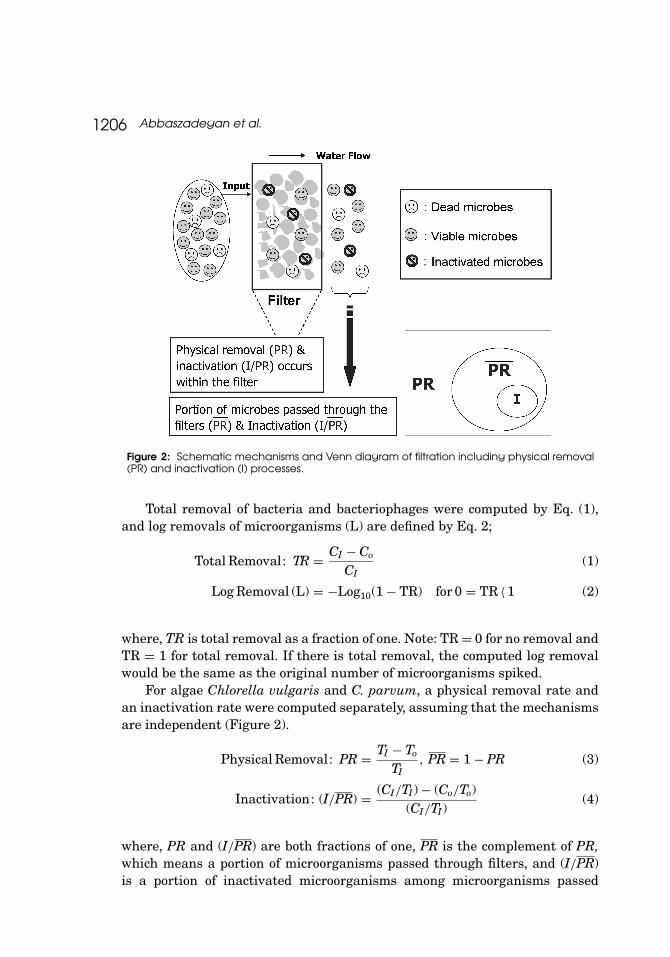
Mechanisms of microbial removal and inactivation by the filter are pre-sented in Figure 2. Culturable assays were used to calculate total removalof microorganisms including physical removal and inactivation, assumingthat physical removal rates for both dead and viable microorganisms areidentical (Eq. (1)); CI and CO are the number of viable/infectious microor-ganisms for input and output, respectively. Total numbers of microorgan-isms, including dead and viable ones determined by direct count using ahemacytometer, were used to calculate physical removal rates (Eq. (3));TI and TO are the number of total microorganisms for input and output,respectively.
1206 Abbaszadegan et al.
Figure 2: Schematic mechanisms and Venn diagram of filtration including physical removal(PR) and inactivation (I) processes.
Total removal of bacteria and bacteriophages were computed by Eq. (1),and log removals of microorganisms (L) are defined by Eq. 2;
Total Removal: TR = CI − Co
CI(1)
Log Removal (L) = −Log10(1 − TR) for 0 = TR 〈 1 (2)
where, TR is total removal as a fraction of one. Note: TR = 0 for no removal andTR = 1 for total removal. If there is total removal, the computed log removalwould be the same as the original number of microorganisms spiked.
For algae Chlorella vulgaris and C. parvum, a physical removal rate andan inactivation rate were computed separately, assuming that the mechanismsare independent (Figure 2).
Physical Removal: PR = TI − To
TI, PR = 1 − PR (3)
Inactivation: (I/PR) = (CI/TI) − (Co/To)(CI/TI)
(4)
where, PR and (I/PR) are both fractions of one, PR is the complement of PR,which means a portion of microorganisms passed through filters, and (I/PR)is a portion of inactivated microorganisms among microorganisms passed
Antimicrobial Capabilities in QAC-Treated Zeolite Filters 1207
through filters. In this study, the portion of microorganisms physically removedand subsequently inactivated, (I/PR), was not determined. Total log removalcredits were calculated using summation of log physical removal and loginactivation, and the total probability theorem[13] was used to confirm thiscalculation.
The union of sets PR and I is indicated as Pr(PR ∪ I); the resulting setincludes all members of PR and I. The intersection of sets PR and I is indicatedas Pr(PR∩I); the resulting set includes only members that belong to both PRand I. The relationship between union and intersection is given by Eq. (5);
Pr(PR ∪ I) = Pr(PR) + Pr(I) − Pr(PR ∩ I) (5)
If PR and I are mutually exclusive, Pr(PR ∩ I) is zero. According the totalprobability theorem, probability of inactivation, Pr(I), can be estimated byEq. (6). The conditional probability of an event A given an event B is denotedas Pr(A/B) for Pr(B) > 0;
Pr (I) = Pr(I/PR) Pr (PR) + Pr(I/PR) Pr (PR) (6)
Pr (A/B) = Pr(A ∩ B)Pr(B)
(7)
As mentioned previously, Pr(I/PR) is zero. Therefore, the following Equationfor probability of total removal of microorganisms can be generated fromcombining Eqs. (5), (6), and (7);
Pr(PR ∪ I) = Pr(PR) + Pr(I/PR) Pr(PR) (8)
RESULTS AND DISCUSSION
The average log removals of MS-2 and PRD-1 by the three filters for thethree experiments were 2.75 ± 0.48 [geometric mean ± standard deviation]and 1.95 ± 0.56, respectively. Our experiments showed less removal andinactivation of PRD-1 than MS-2, suggesting PRD-1’s resistance to removalor greater survival in water.[14] Many factors could potentially affect removaland inactivation of viruses by the filters. These factors include the size andisoelectric points of each virus, protein coat structure or its hydrophobicity,and antimicrobial activity of QAC immobilized on Zeolite. Virus removal wasreported to be poor using direct filtration without prior destabilization ofparticles (Table 1). Gerba et al.[8] reported that the average log removalsof MS-2 and PRD-1 by pilot-scale multimedia filtration were 0.68 and 1.07,respectively. The pilot filters tested in this study resulted in an average of more
1208 Abbaszadegan et al.
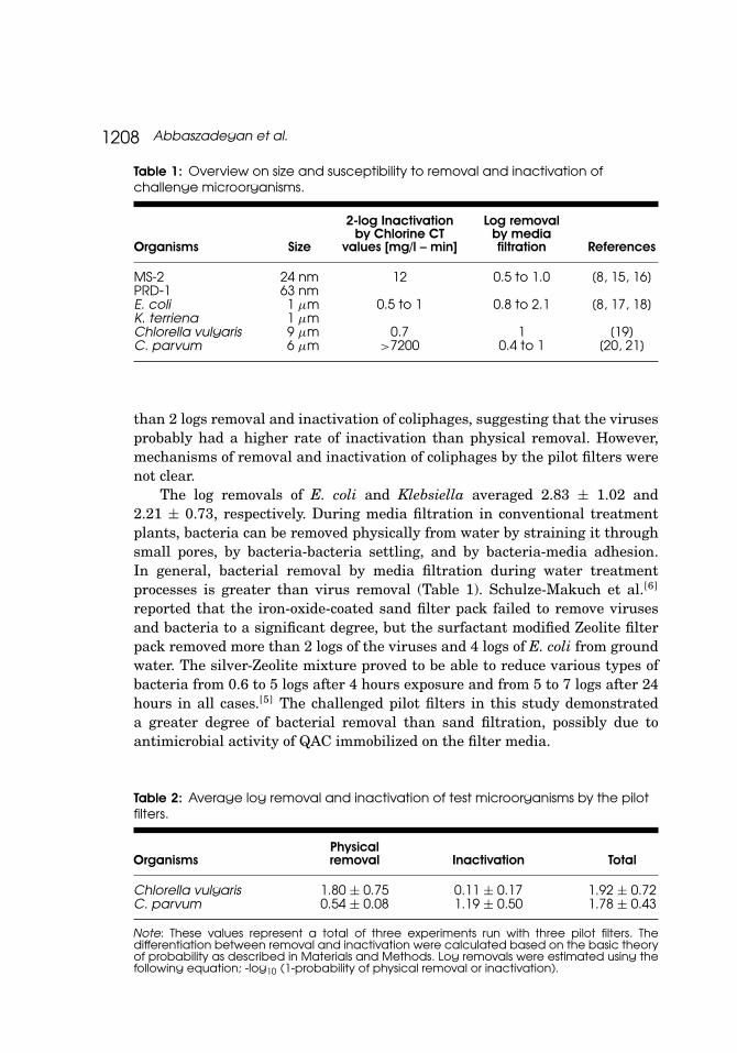
Table 1: Overview on size and susceptibility to removal and inactivation ofchallenge microorganisms.
Organisms Size
2-log Inactivationby Chlorine CT
values [mg/l – min]
Log removalby mediafiltration References
MS-2 24 nm 12 0.5 to 1.0 [8, 15, 16]PRD-1 63 nmE. coli 1 µm 0.5 to 1 0.8 to 2.1 [8, 17, 18]K. terriena 1 µmChlorella vulgaris 9 µm 0.7 1 [19]C. parvum 6 µm >7200 0.4 to 1 [20, 21]
than 2 logs removal and inactivation of coliphages, suggesting that the virusesprobably had a higher rate of inactivation than physical removal. However,mechanisms of removal and inactivation of coliphages by the pilot filters werenot clear.
The log removals of E. coli and Klebsiella averaged 2.83 ± 1.02 and2.21 ± 0.73, respectively. During media filtration in conventional treatmentplants, bacteria can be removed physically from water by straining it throughsmall pores, by bacteria-bacteria settling, and by bacteria-media adhesion.In general, bacterial removal by media filtration during water treatmentprocesses is greater than virus removal (Table 1). Schulze-Makuch et al.[6]
reported that the iron-oxide-coated sand filter pack failed to remove virusesand bacteria to a significant degree, but the surfactant modified Zeolite filterpack removed more than 2 logs of the viruses and 4 logs of E. coli from groundwater. The silver-Zeolite mixture proved to be able to reduce various types ofbacteria from 0.6 to 5 logs after 4 hours exposure and from 5 to 7 logs after 24hours in all cases.[5] The challenged pilot filters in this study demonstrateda greater degree of bacterial removal than sand filtration, possibly due toantimicrobial activity of QAC immobilized on the filter media.
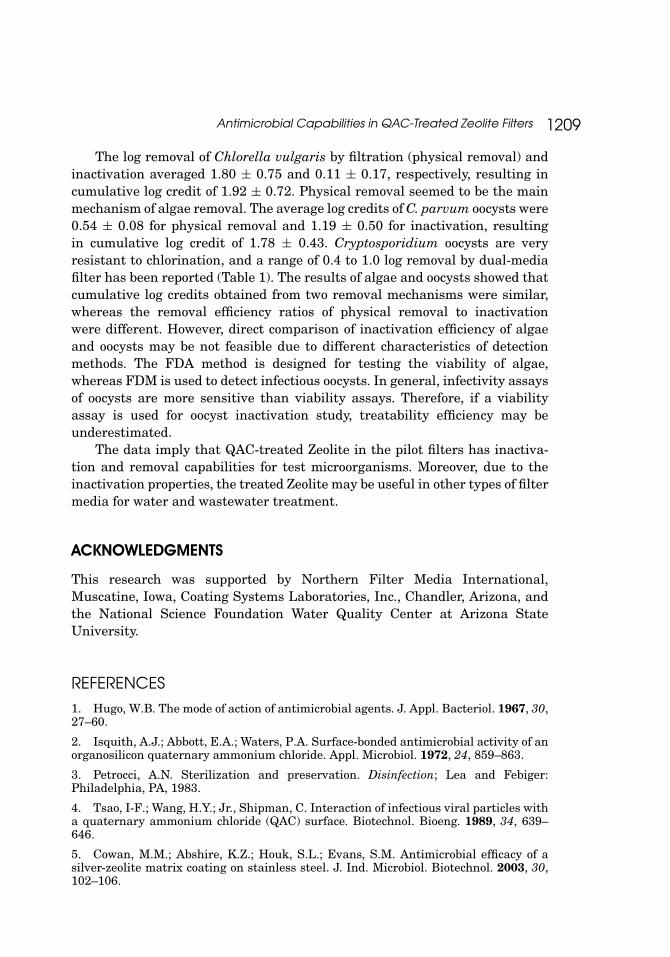
Table 2: Average log removal and inactivation of test microorganisms by the pilotfilters.
OrganismsPhysicalremoval Inactivation Total
Chlorella vulgaris 1.80 ± 0.75 0.11 ± 0.17 1.92 ± 0.72C. parvum 0.54 ± 0.08 1.19 ± 0.50 1.78 ± 0.43
Note: These values represent a total of three experiments run with three pilot filters. Thedifferentiation between removal and inactivation were calculated based on the basic theoryof probability as described in Materials and Methods. Log removals were estimated using thefollowing equation; -log10 (1-probability of physical removal or inactivation).
Antimicrobial Capabilities in QAC-Treated Zeolite Filters 1209
The log removal of Chlorella vulgaris by filtration (physical removal) andinactivation averaged 1.80 ± 0.75 and 0.11 ± 0.17, respectively, resulting incumulative log credit of 1.92 ± 0.72. Physical removal seemed to be the mainmechanism of algae removal. The average log credits of C. parvum oocysts were0.54 ± 0.08 for physical removal and 1.19 ± 0.50 for inactivation, resultingin cumulative log credit of 1.78 ± 0.43. Cryptosporidium oocysts are veryresistant to chlorination, and a range of 0.4 to 1.0 log removal by dual-mediafilter has been reported (Table 1). The results of algae and oocysts showed thatcumulative log credits obtained from two removal mechanisms were similar,whereas the removal efficiency ratios of physical removal to inactivationwere different. However, direct comparison of inactivation efficiency of algaeand oocysts may be not feasible due to different characteristics of detectionmethods. The FDA method is designed for testing the viability of algae,whereas FDM is used to detect infectious oocysts. In general, infectivity assaysof oocysts are more sensitive than viability assays. Therefore, if a viabilityassay is used for oocyst inactivation study, treatability efficiency may beunderestimated.
The data imply that QAC-treated Zeolite in the pilot filters has inactiva-tion and removal capabilities for test microorganisms. Moreover, due to theinactivation properties, the treated Zeolite may be useful in other types of filtermedia for water and wastewater treatment.
ACKNOWLEDGMENTS
This research was supported by Northern Filter Media International,Muscatine, Iowa, Coating Systems Laboratories, Inc., Chandler, Arizona, andthe National Science Foundation Water Quality Center at Arizona StateUniversity.
REFERENCES
1. Hugo, W.B. The mode of action of antimicrobial agents. J. Appl. Bacteriol. 1967, 30,27–60.
2. Isquith, A.J.; Abbott, E.A.; Waters, P.A. Surface-bonded antimicrobial activity of anorganosilicon quaternary ammonium chloride. Appl. Microbiol. 1972, 24, 859–863.
3. Petrocci, A.N. Sterilization and preservation. Disinfection; Lea and Febiger:Philadelphia, PA, 1983.
4. Tsao, I-F.; Wang, H.Y.; Jr., Shipman, C. Interaction of infectious viral particles witha quaternary ammonium chloride (QAC) surface. Biotechnol. Bioeng. 1989, 34, 639–646.
5. Cowan, M.M.; Abshire, K.Z.; Houk, S.L.; Evans, S.M. Antimicrobial efficacy of asilver-zeolite matrix coating on stainless steel. J. Ind. Microbiol. Biotechnol. 2003, 30,102–106.
1210 Abbaszadegan et al.
6. Schulze-Makuch, D.; Bowman, R.S.; Pillai, S.D.; Guan, H. Field evaluation of theeffectiveness of surfactant modified zeolite and iron-oxide-coated sand for removingviruses and bacteria from ground water. Ground Water Monitoring and Remediation.2003, 23 (4), 68–74.
7. Pavelic, K.; Hadzija, M. Medical Application of Zeolite. In Handbook of ZeoliteScience and Technology; Marcel Dekker, Inc.: New York, 2003.
8. Gerba, C.P.; Riley, K.R.; Nwachuku, N.; Ryu, H.; Abbaszadegan, M. Removal ofEncephalitozoon intestinalis, calicivirus, and coliphages by conventional drinking watertreatment. J. Environ. Sci. Health 2003, 38, 1259–1268.
9. Adams, M. Bacteriophages; Interscience Publishers, Inc.: New York, 1959.
10. American Public Health Association (APHA). Part 9000—Microbiological Exami-nation. In Standard Methods for the Examination of Water and Wastewater 20th Ed;Clesceri, L.S.; Greenberg, A.E.; Eaton, A.D., Eds.; Washington, DC, 1998.
11. Slifko, T.R.; Friedman, D.; Rose, J.B.; Jakubowski, W. An in vitro method for de-tecting infectious Cryptosporidium oocysts with cell culture. Appl. Environ. Microbiol.1997, 63, 3669–3775.
12. Slifko, T.R.; Friedman, D.; Rose, J.B. A most probable number assay for theenumeration of infectious Cryptosporidium parvum oocysts. Appl. Environ. Microbiol.1999, 65, 3936–3941.
13. Montgomery, D.C.; Runger, G.C. Applied statistics and probability for engineers3rd Ed; Wiley: New York, 2003.
14. Yahya, M.T.; Galsomies, L.; Gerba, C.P.; Bales, R.C. Survival of bacteriophagesMS2 and PRD1 in groundwater. Water Sci. Technol. 1993, 27, 409–412.
15. DeLeon, R.; Singh, S.N.; Rose, J.B.; Mullinax, R.L.; Gerba, C.P. Virus removal byrapid sand filtration, Tucson Water Reuse Project. Hydrology and Water Resources inArizona and the Southwest 1984, 14, 175–183.
16. Schaper, M.; Duran, A.E.; Jofre, J. Comparative resistance of phages isolates offour genotypes of F + specific RNA bacteriophages to various inactivation processes.Appl. Environ. Microbiol. 2002, 68, 3702–3707.
17. Sobsey, M.D. Inactivation of health-related microorganisms in water by disinfec-tion processes. Water Sci. Technol. 1989, 21 (3), 179–195.
18. Huyman, F.; Verstrate, W. Water facilitated transport of bacteria in unsaturatedsoil columns: influence of inoculation and irrigation methods. Soil Biol. Biochem. 1993,25, 91–97.
19. Junli, H.; Li, W.; Nenqi, R.; Li, L.X.; Fun, S.R.; Guanle, Y. Disinfection effect ofchlorine dioxide on viruses, algae and animal planktons in water. Water Res. 1997, 31,455–460.
20. Rose, J.B.; Lisle, J.T.; LeChevallier, M. Waterborne Cryptosporidiosis: Incidence,outbreaks, and treatment strategies. Cryptosporidium and Cryptosporidiosis; CRCPress: Boca Raton, FL, 1997.
21. Emelko, M.B. Removal of viable and inactivated Cryptosporidium by dual- andtri-media filtration. Water Res. 2003, 37, 2998–3008.