energy balance and seasonality in Human Energetics in Biological Anthropology
Upload
dspzportalCategory
view
4download
0
Seasonality of Cryptosporidium oocyst detection in surfacewaters of Meru, Kenya as determined by two isolation methodsfollowed by PCR
John M. Muchiri,Kenya Methodist University, PO Box 267-60200, Meru, Kenya
Luke Ascolillo,Department of Public Health and Family Medicine, Tufts University School of Medicine, 136 HarrisonAvenue, Boston, Massachusetts 02111, USA
Mutuma Mugambi,Kenya Methodist University, PO Box 267-60200, Meru, Kenya
Titus Mutwiri,Kenya Methodist University, PO Box 267-60200, Meru, Kenya
Honorine D. Ward,Department of Public Health and Family Medicine, Tufts University School of Medicine, 136 HarrisonAvenue, Boston, Massachusetts 02111, USA
Elena N. Naumova,Department of Public Health and Family Medicine, Tufts University School of Medicine, 136 HarrisonAvenue, Boston, Massachusetts 02111, USA
Andrey I. Egorov,Department of Public Health and Family Medicine, Tufts University School of Medicine, 136 HarrisonAvenue, Boston, Massachusetts 02111, USA
Seth Cohen,Department of Public Health and Family Medicine, Tufts University School of Medicine, 136 HarrisonAvenue, Boston, Massachusetts 02111, USA
James G. Else, andYerkes National Primate Research Center, Emory University, 954 Gatewood Road, Atlanta, Georgia30329, USA
Jeffrey K. GriffithsDepartment of Public Health and Family Medicine, Tufts University School of Medicine, 136 HarrisonAvenue, Boston, Massachusetts 02111, USA
AbstractMeru, Kenya has watersheds which are shared by wildlife, humans and domesticated animals. Thesesurface waters can be contaminated by the waterborne pathogen Cryptosporidium. To quantify theseasonality and prevalence of Cryptosporidium in Meru regional surface waters, we used a calciumcarbonate flocculation (CCF) and sucrose floatation method, and a filtration and immunomagneticbead separation method, each of which used PCR for Cryptosporidium detection and genotyping.
© IWA Publishing 2009Tel.: (617) 636 6941, Fax: (617) 636 4017, E-mail: E-mail: [email protected].
NIH Public AccessAuthor ManuscriptJ Water Health. Author manuscript; available in PMC 2009 September 1.
Published in final edited form as:J Water Health. 2009 March ; 7(1): 67–75. doi:10.2166/wh.2009.109.
NIH
-PA Author Manuscript
NIH
-PA Author Manuscript
NIH
-PA Author Manuscript

Monthly water samples were collected from January through June in 2003 and 2004, bracketing twoApril-May rainy seasons. We detected significant seasonality with 8 of 9 positive samples from Mayand June (p < 0.0014), which followed peak rainy season precipitation and includes some of thesubsequent dry season. Six of 9 positive samples revealed C. parvum, and 3 contained C.andersoni. None contained C. hominis. Our results indicate that Meru surface waters areCryptosporidium-contaminated at the end of rainy seasons, consistent with the timing of humaninfections reported by others from East Africa and contrasting with the onset of rainy season peakincidence reported from West Africa.
Keywordscalcium carbonate flocculation; Cryptosporidium; Kenya; PCR detection; seasonality
INTRODUCTIONCryptosporidium is a protozoan parasite which usually causes asymptomatic or mild humanclinical disease, although severe dehydrating disease and death can occur inimmunocompromised populations, such as HIV-infected people. No clearly effective therapyfor cryptosporidiosis in immunocompromised people exists (Griffiths 1998; Tzipori & Ward2002; Abubakar et al. 2007), and thus the avoidance of Cryptosporidium-contaminated wateris important. Zoonotic and human-to-human transmission occurs, often via surface andrecreational waters, and the disease is under-diagnosed except in those with persistent diarrhea(Bagley et al. 1998; Griffiths 1998; Hunter et al. 2001; Rose et al. 2002). Prevalence rates of~ 10% to 33% in those with AIDS have been reported from developing countries (Kaplan etal. 1996; reviewed in Griffiths 1998). Malnutrition, which impairs cellular immunity, is anotherrecognized risk factor for cryptosporidiosis (Gendrel et al. 2003).
We report initial data on Cryptosporidium in surface waters of Meru, Kenya, which lies northof Nairobi. Watersheds in Meru arise on Mt. Kenya in wildlife preserves, and water flows eastthrough intensively farmed, densely populated areas before reaching arid, pastoral areas. Thus,Meru surface waters could be contaminated by parasite oocysts from wildlife, human, orpastoral animals, and infect people with AIDS or malnutrition as well as others in the generalpopulation. In this pilot study, we repetitively collected monthly samples from predeterminedsites during January to June, which is before, during, and after the intensively rainy period ofApril and May. We also field tested two methods for parasite isolation and detection fromsurface waters during 2003 and 2004. Our approach included Cryptosporidium moleculargenotyping of the positive water samples so as to better understand the potential sources of theparasites.
MATERIALS AND METHODSWater collection sites
Meru water supplies are untreated surface waters taken directly from rivers or transient,seasonally flooded areas, which may be piped to households or common urban waterdistribution points. Water collection sites are often common watering points for domestic andwild animals. Ten sites were sampled. Five sites were on the Kathita River, which suppliesMeru Town and surrounding villages. The highest Kathita River site (site A, Figure 1) is wherethe river exits a protected wildlife forest preserve. Downstream sites, coded B, C, E, and F,reflect a graded transition of increasing human and animal influence. Site C is the intake forthe Meru Town municipal water system, which is chlorinated but unfiltered. Site D is thesewage outflow from Meru Town. Four other sites were chosen because of mixed human andanimal use. These included a forest pool where elephants drink (Luuria, site G), and two sites
Muchiri et al. Page 2
J Water Health. Author manuscript; available in PMC 2009 September 1.
NIH
-PA Author Manuscript
NIH
-PA Author Manuscript
NIH
-PA Author Manuscript

near Kiina Town northeast of Meru Town. Kiina Tap (H) and Kiina River (I) are used bypastoralists for their herded animals, and by wildlife such as elephants, as they border MeruNational Park. Miriti Well (site J), one of a group known as the Kiina Wells, is further northeastand is used by both pastoralists and wildlife. Sites H – J are in Isiolo District, and are in thefar eastern, lower parts of the Meru ecosystem. Sites were mapped using a global positioningsatellite (GPS) monitor (Figure 1). Under ideal conditions, all sites could be visited in a 3 dayperiod excepting Miriti Well which required a fourth day.
Sampling framework and precipitation dataSamples were obtained during the first six months of 2003 and 2004, bracketing a majorprecipitation period which occurs in March and April. This period was chosen so that we couldanalyze 2003 samples and revise our 2004 sampling strategy if required. Precipitation data wasobtained from the Meteorology Department, Ministry of Transport and Communication, MeruWeather Station, housed at Kenya Methodist University (KEMU) in Meru.
Sampling methodsWe used a calcium carbonate flocculation (CCF) and sucrose gradient floatation technique in2003, and a filtration method in 2004, each followed by immunomagnetic isolation of parasiteoocysts and PCR analysis. Prior work (Kato et al. 2003) documented that the CCF methodcould successfully isolate oocysts from Meru waters.
Calcium carbonate flocculation (CCF) followed by sucrose gradient floatationTen litres of water were collected into plastic containers certified as free from contaminants(Fisher Scientific, Pittsburgh, USA). Samples were brought to KEMU, saturated with CaCl2and NaHCO3, and adjusted to pH 10 with NaOH (Vesey et al. 1993). Insoluble CaCO3 crystalswere allowed to settle and the supernatant (~9 L) discarded. The precipitate was dissolved insulfamic acid. K2Cr2O7 (25 g per L) was added to kill pathogens until processing, which wastypically within a week. Samples were centrifuged at 5,000 × g for 10 minutes, and thesupernatant was discarded leaving ~20 ml of concentrated suspension. The suspension wasthrice washed with distilled water at 5,000 × g for 10 min at 4°C with supernatant removal viavacuum leaving 3 ml of suspension and sediment. The washed sediment then had distilledwater added to a total volume of 7 ml and was thoroughly mixed. Seven ml of sucrose solution(specific gravity 1.18) was layered below the sediment solution via syringe, and centrifugedat 3,000 × g for 20 minutes at 4°C in a 15 ml polystyrene tube. Two separate layers wereobtained, with soil sediments at the bottom. The top 2 ml of the sucrose layer was removedusing a Pasteur pipette, diluted with water in another 15 ml tube, and centrifuged at 1,800 × gfor 3 min at 4°C. The supernatant was aspirated leaving 500 µl. This was repeated 3 timesleaving a final volume of 500 µl which was subjected to immunomagnetic separation (IMS).
FiltrationFor consistency between Boston and Meru, we used US Environmental Protection Agency(EPA) Method 1623, as our Boston laboratory is required by the EPA to consistently only useMethod 1623, given its certification for Cryptosporidium and Giardia testing in US ambientsurface waters (US EPA 2005). In Kenya, water samples were pumped through a Filta-Max™(IDEXX, Westbrook, ME) filter in a polycarbonate housing, using a car-battery powered pump.Samples ranged from 30 to 40L, with the exact volume measured by an in-line meter. The filterwas stored in a glass jar with 300 ml of phosphate-buffered saline (pH 7.2, 0.01% Tween 20)and transported to KEMU. Oocysts were washed out of the filter according to themanufacturer’s instructions (IDEXX 2002). In brief, a bolt is released allowing the compressedfilter to expand, allowing retained oocysts to be eluted from the sponge matrix. The washsolution is filtered, and oocysts trapped on a membrane are eluted into ~10 ml of fluid. The
Muchiri et al. Page 3
J Water Health. Author manuscript; available in PMC 2009 September 1.
NIH
-PA Author Manuscript
NIH
-PA Author Manuscript
NIH
-PA Author Manuscript

sample is centrifuged to a pellet of <0.5 ml and then purified by IMS (see below). If the pelletwas >0.5 ml the sample was split per Method 1623.
Immunomagnetic Separation (IMS) and DNA extractionIMS was conducted using the Dynabeads GC-Combo (Dynal Biotech, Oslo, Norway) protocol.In brief, anti-Cryptosporidium antibodies attached to magnetic beads were mixed with samplesprepared as described above to a volume of 1 ml. The oocysts were removed through magneticremoval of the beads, then dissociated from the beads using an acid wash step. The acid washsupernatant was aspirated and used for DNA extraction. Oocyst extracts underwent five freezethaw cycles after the addition of 180 µl of tissue lysis buffer (50 Mm Tris-HCl [pH 8.5], 1MmEDTA [pH 8], 0.5% sodium dodecyl sulfate) in cryovials which were incubated for 1 minutein liquid nitrogen and 1 minute in a 56°C heating block per cycle. Samples were centrifugedat 14,000 × g for 30 s and then incubated with 20 µl proteinase K (Sigma, St Louis, MO.) for2 hours at 55°C while being rotated, incubated at 90°C to denature the proteinase K, chilledon ice for 1 minute, and centrifuged at 14,000 × g for 3 minutes. Supernatants were transferredto a new 1.5 ml tube, 200 ul of ethanol was added, then vortexed for 15 seconds. The mixturewas loaded into a QIAmp spin column (Qiagen Inc, Valencia, California) and centrifuged at6,000 × g for 1 minute. This was repeated using wash buffers described by the manufacturer.The final supernatant was stored at −20°C for use in PCR.
PCR and RFLP ProtocolsNested PCR followed by restriction enzyme digestion was carried out using SSU primers, andVSPI and SSPI enzymes respectively (Xiao et al. 1999). Reaction volumes of 100 µl consistedof premixed reagents containing 10 mM of each of the four deoxynucleoside triphosphates, 10× PCR buffer, 25 mM MgCL2, PCR grade water and Taq polymerase (Invitrogen). 2µl oftemplate DNA was used. The primary run was done using 5’-TTCTAGAGCTAATACATGCG-3’ and 5’-CCCATTTCCTTCGAAACAGGA-3’ primers.The samples were incubated in the thermocycler at 94°C for 3 minutes to denature templateDNA. This was followed by 35 cycles, each consisting of 94°C for 45 s, 55°C for 45 s and 72°C for 1 minute and a final extension at 72°C for 10 minutes. A secondary amplification usingthe primary products the primers 5’-GGAAGGGTTGTATTTATTAGATAAAG-3’ and 5’-AAGGAGTAAGGAACAACCTCCA-3’ was performed using cycling conditions identical tothose in the primary PCR. Amplicons were fractionated on a 2% agarose gel and visualized byethidium bromide staining to confirm the presence of characteristic Cryptosporidium products(Figure 2). To differentiate genotypes, 10µl of positive secondary PCR products were digestedin a 40 µl (total volume) reaction mixture containing 20 U of SspI (New England Biolabs,Beverly, Mass.) or 20 U of VspI (GIBCO BRL, Grand Island, N.Y) and 4µl of the appropriaterestriction buffer at 37°C for 1 hr (Xiao et al. 1999). As C. andersoni and C. muris have identicalSspI and VspI restriction patterns, they were differentiated by digesting secondary productswith 20 U of DdeI (New England Biolabs) at 37°C for 1h under conditions recommended bythe supplier. Digested products were fractionated on 2% agarose gel and visualized by ethidiumbromide staining. All positive detections were confirmed in duplicate by RFLP analysis ofadditional, independent PCR products from the same water sample. Positive controls includedTU502 C. hominis (Akiyoshi et al. 2002), obtained from Dr. Saul Tzipori, Tufts UniversityCummings School of Veterinary Medicine, North Grafton, MA and the Iowa C. parvum,obtained from Pleasant Farms, Troy, ID (Abrahamsen et al. 2004).
Statistical analysisData were analyzed using the Statistical Package for the Social Sciences (SPSS) version 15.Specific comparisons (t-tests and chi-square analyses) are denoted in the text.
Muchiri et al. Page 4
J Water Health. Author manuscript; available in PMC 2009 September 1.
NIH
-PA Author Manuscript
NIH
-PA Author Manuscript
NIH
-PA Author Manuscript

RESULTS AND DISCUSSIONOocyst detection: by method and by season
Monthly samples were collected and analyzed for January through June 2003, and Januarythrough June of 2004. In 2003 we used the CCF method, and in 2004 the filtration method. Allsample concentrates underwent PCR for the detection of oocysts. We thus sampled 6 monthsof both low and high rainfall in two sequential years. In 2003, all 10 sites were sampled monthly.In 2004, sampling at Kiina Wells became dangerous and was discontinued, as drought hadincreased the chance of interactions with wild elephants. In January and June of 2004, adverseroad conditions made it impossible to visit several other sites.
108 samples were acquired and 9 (8.3%) were PCR positive for oocysts (Table 1). Two of 60(3.3%) samples obtained in 2003, versus 7 of 48 (14.6%) samples from 2004, were positive.The detection rate in 2004 was 4.375 fold higher than in 2003, which did not reach statisticalsignificance (p = 0.075, 2-tailed Fisher’s exact test). Oocyst detection rates varied significantlyby month (χ2 = 13.53, 5 df, p < 0.019). Table 2 displays detection rates before, during, andafter the peak in precipitation which occurred in March and April of each year, together withmonthly cumulative precipitation for each time period. The apparent seasonal period peak ofdetection at the end of the rainy season is significant (χ2 = 13.17, p < 0.0014). Figure 3 displaysprecipitation data recorded in Meru. Two peaks of precipitation usually occur each year. Theyears of 2003 and 2004 were atypical in that there was rainfall in the months separating thelate 2003 and early 2004 precipitation peaks. The months when we detected oocystscorresponded to the nadirs immediately following seasonal peaks of precipitation (June 2003,March 2004, June 2004) or the descending right-hand slope of a period of decreasingprecipitation after a peak (May 2004).
Genotypes of Cryptosporidium detected in water samplesC. parvum was detected six times. It was isolated three times from the sewage waste streamfrom Meru Town, from Meru Town’s water intake site, from a site downstream of Meru Town,and from a pool of water frequently used by elephants (Table 3). C. hominis was not detectedfrom any site. C. andersoni was detected 3 times (Table 3). This species has been linked tocattle and does not appear to be pathogenic to humans. All 3 C. andersoni isolates were detectedfrom the Kiina River, where pastoralists water their herds of cattle.
DISCUSSIONIn this pilot study, we detected Cryptosporidium surface water contamination in the Meru,Kenya region over the first six months of two sequential years (2003 and 2004). The period atthe end of the sampled rainy season, and the beginning of the following drier, cool season (Mayand June) was significantly associated with oocyst detection in surface waters. Thiscorresponded to the nadirs of precipitation between precipitation peaks, or to periods oftapering rainfall (Figure 3).
Cryptosporidium oocysts are believed to be flushed by rain into surface waters, and so onemight have expected peak detection during the onset of the rainy months. During March andApril, the volume of water which falls is so great that it is possible that the concentration ofoocysts in surface waters is relatively low, because of dilution. Alternatively, there may belittle Cryptosporidium transmission during March and April in Meru, and thus there may befewer fecally excreted parasites to be swept into water bodies. Our surface water data does notallow us to speculate further on the merits of these and other possible explanations regardingtransmission density, as human and animal cryptosporidiosis incidence data is not yet availablefrom Meru.
Muchiri et al. Page 5
J Water Health. Author manuscript; available in PMC 2009 September 1.
NIH
-PA Author Manuscript
NIH
-PA Author Manuscript
NIH
-PA Author Manuscript

However, data from other East African sites may be informative. In a recent survey of 4,899pediatric fecal samples from laboratories near Nairobi, the rainy months of April and May hadthe lowest infection prevalence (1.5%), suggesting that transmission is less common duringthis period (Gatei et al. 2006). Infection rates were highest in children during the Novemberto February period, which usually has little precipitation. Studies from Rwanda, Malawai, andKenya (Bogaerts et al. 1987; Peng et al. 2003; Gatei et al. 2006), respectively, report that thepeak incidence of cryptosporidiosis is at the end of rainy seasons and beginning of the driermonths, as has a study by Moodley et al. (1991) in Durban, South Africa. In contrast, in Guinea-Bissau, West Africa, the peak prevalence of clinical cryptosporidiosis in children is just before,or at the onset of, the rainy season from May to July (Molbak et al. 1990, 1993; Perch et al.2001). Similar results were reported from Zambia (Nchito et al. 1998). The East African humanincidence pattern is congruent with our findings regarding oocysts in surface waters. It ispossible that the apparent seasonality of surface water contamination and of human disease, isreflective of different transmission pathways, hosts, and/or Cryptosporidium species in EastAfrica when compared to other locations. We note that as climate change occurs, transmissionpatterns of many waterborne diseases may shift, and studies in locations with unusualseasonality patterns may help inform our understanding of what climate change may bring(Rose et al. 2002; Hunter 2003; Jagai et al. 2007; Naumova et al. 2007).
A limitation of our study is that we did not directly compare our two oocyst isolation methodsin a side-by-side evaluation during the same year. We substituted the filtration method for theCCF method after unexpectedly low rates of isolation in 2003. While the direct field filtrationof 30–40 L of water led to higher PCR detection rates than did the CCF method with 10 Lsamples, this did not reach statistical significance. The differences in detection that weencountered might be due to the natural variation between the years tested; inhibition of PCRby environmental contaminants (Sluter et al. 1997; Loge et al. 2002); or additional lossesencumbered in the “double purification” via CCF and then IMS. Egorov et al. (2002) andKaranis et al. (2006) found CCF useful when oocysts were detected via immunofluorescentidentification. Although Monis & Saint (2001) reported that the precipitation method isinhibitory for PCR detection, preliminary data (Kato et al. 2003) had suggested that our initialapproach would be successful. In order to address this issue properly, side by side comparisonsof the two methods would be needed which would quantify overall recovery efficiency, as wemay easily have suffered from falsely negative results in 2003. We used PCR methods fordetection, rather than visual ones, because of our interest in the genotypes of theCryptosporidium that might be found in this ecologically unusual region.
C. parvum, but not C. hominis, was thrice detected from human sewage waste waters fromMeru Town, as well as from the intake site for the town water supply and downstream of MeruTown. Studies from Kenya and Uganda (Tumwine et al. 2005; Gatei et al. 2006) show that C.hominis is the dominant species infecting children in the urban centers of Nairobi and Kampala.C. parvum is adapted to multiple host species, including wildlife, humans and cattle (Fayer2004). C. hominis has only been reliably detected in primate hosts, although two studies reportits detection in dugongs (Morgan et al. 2000) and cattle (Smith et al. 2005). Cattle herding isuncommon in the intensely farmed Meru Town region, while pastoralist herding is morecommon northeast of Meru Town. We detected C. andersoni thrice from the Kiina River, wherepastoralists water their cattle. C. andersoni is not thought to be pathogenic for humans, and isdominantly a parasite of older cattle. C. parvum infects HIV infected and uninfected children,while C. meleagridis and C. muris are more frequently found in HIV-infected persons (Caccio2005). In the rural regions of Kenya such as Meru, the spectrum of Cryptosporidium speciesinfecting humans may differ from that in urban areas given different land use patterns(Naumova et al. 2005) and the use of the same water by wildlife as well as by people andherded animals. Our data is consistent with, but not proof of, endemic waterborneCryptosporidium transmission in Meru, including potential transmission between wildlife,
Muchiri et al. Page 6
J Water Health. Author manuscript; available in PMC 2009 September 1.
NIH
-PA Author Manuscript
NIH
-PA Author Manuscript
NIH
-PA Author Manuscript

domestic animals, and humans. Further environmental, epidemiological, and molecularinvestigations are needed to confirm or refute this thesis. In particular, molecular analysis ofCryptosporidium isolates at the sub-type level from potential animal reservoirs, surface watersand humans may be helpful in this regard (Ajjampur et al. 2007). We find it intriguing that C.parvum was detected from a pool after use by migrating elephants at Luuria, as according tothe local population these waters cause diarrhea in people after the elephants have passedthrough.
CONCLUSIONThe Meru region of Kenya is an appropriate ecological zone for studying potential transmissionthrough water between wildlife, humans and domesticated animals. We document C.parvum in the Kathita River, which serves all these groups, as well as Cryptosporidium fromwaters which serve humans, cattle and wildlife, such as elephants. Oocysts were significantlymore commonly detected in surface waters at the end of rainy seasons and at the beginning ofdry seasons, congruent with studies which have shown peak human disease during the sameperiod in East Africa. This seasonality differs from that noted in West Africa and northerntemperature regions. Continued environmental, epidemiological, and molecular studies willbe needed to address questions regarding the actors in, and timing of, transmission in thisregion.
ACKNOWLEDGEMENTSWe thank Denise Castronovo (Mapping Sustainability, www.mappingsustainability.com) for her help with Figure 1,and Siobhan Mor for her comments on cryptosporidiosis seasonality in Africa. We thank the Meru Weather Station,Government of Kenya for the precipitation data. This study was supported by a grant to JKG, MM, ENN and JE fromthe US National Institutes of Health’s Fogarty International Center (R23 TW05821, “An Ecological Analysis ofCryptosporidium in Kenya”), and R01 AI43415 to JKG and ENN from the National Institute of Allergy and InfectiousDiseases, entitled “Waterborne Emerging Diarrheal Diseases Study”.
REFERENCESAbrahamsen MS, Templeton TJ, Enomoto S, Abrahante JE, Zhu G, Lancto CA, Deng M, Liu C, Widmer
G, Tzipori S, Buck GA, Xu P, Bankier AT, Dear PH, Konfortov BA, Spriggs HF, Iyer L, AnantharamanV, Aravind L, Kapur V. Complete genome sequence of the apicomplexan, Cryptosporidiumparvum. Science 2004;304:441–445. [PubMed: 15044751]
Abubakar I, Aliyu SH, Arumugam C, Hunter PR, Usman UK. Prevention and treatment ofcryptosporidiosis in immunocompromised patients. Cochrane Database Syst. Rev 2007;1CD004932
Ajjampur SS, Gladstone BP, Selvapandian D, Muliyil JP, Ward H, Kang G. Molecular and spatialepidemiology of cryptosporidiosis in children in a semiurban community in South India. J. Clin.Microbiol 2007;45:915–920. [PubMed: 17251402]
Akiyoshi DE, Feng X, Buckholt MA, Widmer G, Tzipori S. Genetic analysis of a Cryptosporidiumparvum human genotype 1 isolate passaged through different host species. Infect. Immun2002;70:5670–5675. [PubMed: 12228296]
Bagley ST, Auer MT, Stern DA, Babiera MJ. Sources and fate of Giardia cysts and Cryptosporidiumoocysts in surface water. J. Lake Res. Manage 1998;14:379–392.
Bogaerts J, Lepage P, Rouvroy D, van Goethem C, Nsengumuremye F, Mohamed O, Habyalimana JB,Vandepitte J. Cryptosproridiosis in Rwanda. Clinical and epidemiological features. Ann. Soc. Belge.Méd. Trop 1987;67:157–165.
Caccio SM. Molecular epidemiology of human cryptosporidiosis (Review). Parassitologia 2005;47(2):185–192. [PubMed: 16252472]
Egorov A, Paulauskis J, Petrova L, Tereschenko A, Drizhd N, Ford T. Contamination of water supplieswith Cryptosporidium parvum and Giardia lamblia and diarrheal illness in selected Russian cities. Int.J. Hyg. Environ. Health 2002;205:281–289. [PubMed: 12068747]
Muchiri et al. Page 7
J Water Health. Author manuscript; available in PMC 2009 September 1.
NIH
-PA Author Manuscript
NIH
-PA Author Manuscript
NIH
-PA Author Manuscript

Fayer R. Cryptosporidium: a water-borne zoonotic parasite. Vet. Parasitol 2004;126:37–56. [PubMed:15567578]
Gatei W, Wamae CN, Mbae C, Waruru A, Mulinge E, Waithera T, Gatika SM, Kamwati SK, Revathi G,Hart CA. Cryptosporidiosis: prevalence, genotype analysis, and symptoms associated with infectionsin children in Kenya. Am. J. Trop. Med. Hyg 2006;75:78–82. [PubMed: 16837712]
Gendrel D, Treluyer JM, Richard-Lenoble D. Parasitic diarrhea in normal and malnourished children(Review). Fundam. Clin. Pharmacol 2003;17(2):189–197. [PubMed: 12667229]
Griffiths, JK. Human cryptosporidiosis: epidemiology, transmission, clinical disease and diagnosis. In:Saul, T., editor. Emerging Human Enteric Protozoan Infections. Vol. 40. London, UK: AcademicPress; 1998. p. 37-85.Adv. Parasitol.
Hunter PR. Climate change and waterborne and vector-borne diseases. J. Appl. Microbiol 2003;94:37S–46S. [PubMed: 12675935]
Hunter P, Syed Q, Naumova EN. Possible undetected outbreaks of cryptosporidiosis in areas of the northwest of England supplies by an unfiltered surface water source. Commun. Dis. Public Health2001;4:136–138. [PubMed: 11525003]
IDEXX. Filta-Max Operator’s Guide. Protocol for Use with the Manual Wash Station. 2002 [Accessed28 January, 2008]. http://www.idexx.com/water/refs/060458201MAN.pdf.
Jagai, JS.; Monchak, J.; McEntee, JC.; Castranovo, D.; Naumova, EN. The Use of Remote Sensing toAssess Global Trends in Seasonality of Cryptosporidiosis; Proceedings of the 32nd InternationalSymposium on Remote Sensing of Environment; June 25–29 2007; San Jose, Costa Rica. 2007.
Kaplan JE, Hu DJ, Holmes KK, Jaffe HW, Masure H, De Cock KM. Preventing opportunistic infectionsin human immunodeficiency virus-infected persons: implications for the developing world. Am. J.Trop. Med. Hyg 1996;55:1–11. [PubMed: 8702012]
Karanis P, Sotiriadou I, Kartashev V, Kourenti C, Tsvetkova N, Stojanova K. Occurrence of Giardia andCryptosporidium in water supplies of Russia and Bulgaria. Environ. Res 2006;102:260–271.[PubMed: 16780829]
Kato S, Ascolillo L, Egas J, Elson L, Gostyla K, Naples L, Else J, Sempertegui F, Naumova E, EgorovA, Ojeda F, Griffiths JK. Waterborne Cryptosporidium oocyst identification and genotyping: use ofGIS for Ecosystem Studies in Kenya and Ecuador. J. Eukaryotic. Microbiol 2003;50:548–549.
Loge FJ, Thompson DE, Call DR. PCR detection of specific pathogens in water: a risk-based analysis.Environ. Sci. Technol 2002;36(12):2754–2759. [PubMed: 12099475]
Moodley D, Jackson TF, Gathiram V, van den Ende J. Cryptosporidium infections in children in Durban.Seasonal variation, age distribution and disease status. S. Afr. Med. J 1991;79:295–297. [PubMed:2017736]
Molbak K, Hojlyng N, Ingholt L, Da Silva AP, Jepsen S, Aaby P. An epidemic outbreak ofcryptosporidiosis: a prospective community study from Guinnea-Bissau. Pediatr. Infect. Dis. J1990;9:566–570. [PubMed: 2235172]
Molbak K, Hojlyng N, Gottschau A, Sa JC, Ingholt L, da Silva AP, Aaby P. Cryptosporidiosis in infancyand childhood mortality in Guinea Bissau, west Africa. Br. Med. J 1993;307:417–420. [PubMed:8374453]
Monis PT, Saint CP. Development of a nested-PCR assay for the detection of Cryptosporidium parvumin finished water. Water Res 2001;35:1641–1648. [PubMed: 11329665]
Morgan UM, Xiao L, Hill BD, O’Donoghue P, Limor J, Lal A, Thompson RC. Detection of theCryptosporidium parvum “human” genotype in a dugong (Dugong dugon). J. Parasitol2000;86:1352–1354. [PubMed: 11191916]
Naumova EN, Christodouleas J, Hunter PR, Syed Q. Temporal and spatial variability in cryptosporidiosisrecorded by the surveillance system in North West England in 1990–1999. J. Water Health 2005;3(2):185–196. [PubMed: 16075943]
Naumova EN, Jagai J, Matyas B, DeMaria A, MacNeill IB, Griffiths JK. Seasonality in six entericallytransmitted diseases and ambient temperature. Epidemiol. Infect 2007;135(2):281–292. [PubMed:17291363]
Nchito M, Kelly P, Sianongo S, Luo NP, Feldman R, Farthing M, Baboo KS. Cryptosporidiosis in urbanZambian children: an analysis of risk factors. Am. J. Trop. Med. Hyg 1998;59:435–437. [PubMed:9749640]
Muchiri et al. Page 8
J Water Health. Author manuscript; available in PMC 2009 September 1.
NIH
-PA Author Manuscript
NIH
-PA Author Manuscript
NIH
-PA Author Manuscript

Peng MM, Meshnick SR, Cunliffe NA, Thindwa BDM, Hart CA, Broadhead RL, Xiao L. Molecularepidemiology of cryptosporidiosis in children in Malawi. J. Eukaryotic Microbiol 2003;50:557–559.
Perch M, Sodemann M, Jakobsen MS, Valentiner-Branth P, Steinsland H, Fischer TK, Lopes DD, AabyP, Molbak K. Seven years’ experience with Cryptosporidium parvum in Guinea-Bissau, WestAfrica. Ann. Trop. Paediatr 2001;21:313–318. [PubMed: 11732149]
Rose JB, Huffman DE, Gennaccaro A. Risk and control of waterborne cryptosporidiosis. FEMSMicrobiol. Rev 2002;26:113–123. [PubMed: 12069877]
Sluter SD, Tzipori S, Widmer G. Parameters affecting polymerase chain reaction detection of waterborneCryptosporidium parvum oocysts. Appl. Microbiol. Biotechnol 1997;48(3):325–330. [PubMed:9352675]
Smith HV, Nichols RAB, Mallon M, MacLeod A, Tait A, Reilly WJ, Browning LM, Gray D, Reid SWJ,Wastling JM. Natural Cryptosporidium hominis infections in Scottish cattle. Vet. Rec 2005;156:710–711. [PubMed: 15923554]
Tumwine JK, Kekitiinwa A, Bakeera-Kitaka S, Ndeezi G, Downing R, Feng X, Akiyoshi DE, Tzipori S.Cryptosporidiosis and microsporidiosis in Ugandan children with persistent diarrhea with andwithout concurrent infection with the human immunodeficiency virus. Am. J. Trop. Med. Hyg2005;73:921–925. [PubMed: 16282304]
Tzipori S, Ward H. Cryptosporidiosis: biology, pathogenesis and disease. Microbes Infect 2002;4:1047–1058. [PubMed: 12191655]
United States Environmental Protection Agency 2005 Method 1623. Cryptosporidium and Giardia inWater by Filtration/IMS/FA. 2005 Dec. Office of Water, EPA-815-R-05-002. Available online athttp://epa.gov/nerlcwww/1623de05.pdf
Vesey G, Slade JS, Byrne M, Shepherd K, Fricker CR. A new method for the concentration ofCryptosporidium oocysts from water. J. Appl. Bacteriol 1993;75:82–86. [PubMed: 8365958]
Xiao L, Escalante L, Yang C, Sulaiman I, Escalante AA, Montali RJ, Lal AA. Phylogenetic analysis ofCryptosporidium parasites based on the small-subunit rRNA gene locus. Appl. Environ. Microbiol1999;65(4):1578–1583. [PubMed: 10103253]
Muchiri et al. Page 9
J Water Health. Author manuscript; available in PMC 2009 September 1.
NIH
-PA Author Manuscript
NIH
-PA Author Manuscript
NIH
-PA Author Manuscript

Figure 1.Map of the Meru, Kenya Region.
Muchiri et al. Page 10
J Water Health. Author manuscript; available in PMC 2009 September 1.
NIH
-PA Author Manuscript
NIH
-PA Author Manuscript
NIH
-PA Author Manuscript
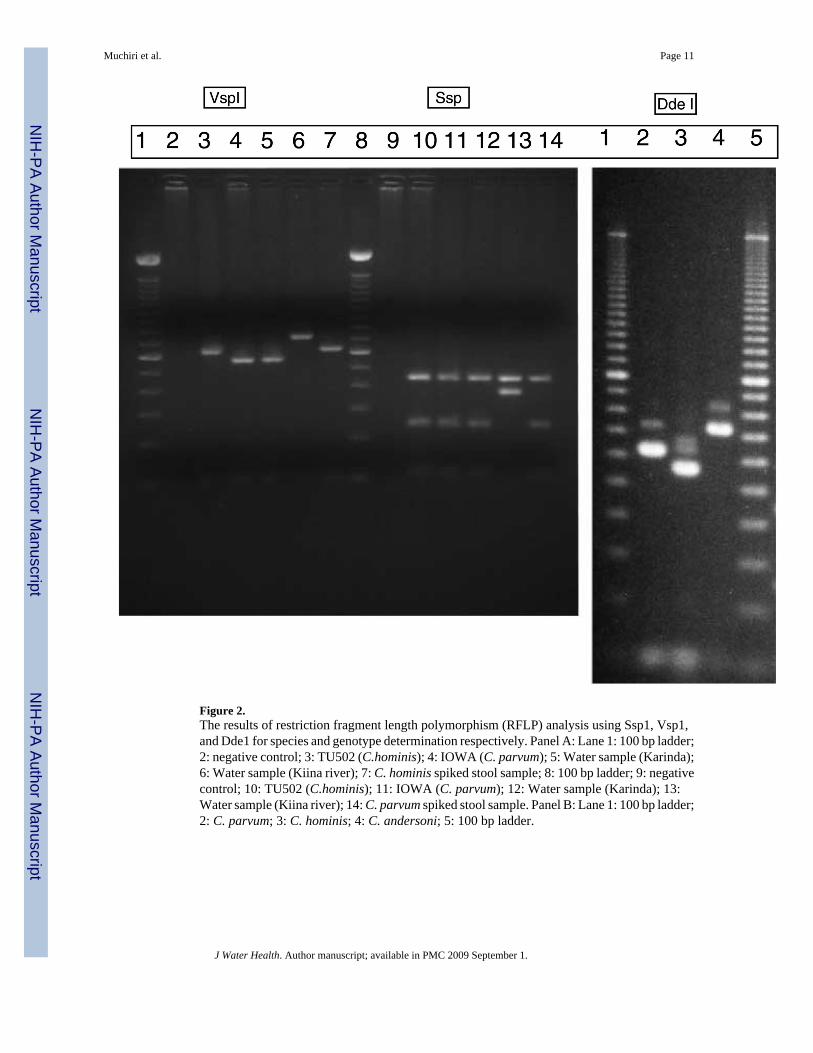
Figure 2.The results of restriction fragment length polymorphism (RFLP) analysis using Ssp1, Vsp1,and Dde1 for species and genotype determination respectively. Panel A: Lane 1: 100 bp ladder;2: negative control; 3: TU502 (C.hominis); 4: IOWA (C. parvum); 5: Water sample (Karinda);6: Water sample (Kiina river); 7: C. hominis spiked stool sample; 8: 100 bp ladder; 9: negativecontrol; 10: TU502 (C.hominis); 11: IOWA (C. parvum); 12: Water sample (Karinda); 13:Water sample (Kiina river); 14: C. parvum spiked stool sample. Panel B: Lane 1: 100 bp ladder;2: C. parvum; 3: C. hominis; 4: C. andersoni; 5: 100 bp ladder.
Muchiri et al. Page 11
J Water Health. Author manuscript; available in PMC 2009 September 1.
NIH
-PA Author Manuscript
NIH
-PA Author Manuscript
NIH
-PA Author Manuscript

Figure 3.Actual monthly precipitation in millimetres in Meru, Kenya by month of 2003 and 2004 andproportion of positive Meru Region surface water samples by month of 2003 and 2004. Thesolid black line represents precipitation in mm (left axis scale), and the dotted black linerepresents the proportion of positive surface water samples (right axis scale), by month (x axis,horizontal).
Muchiri et al. Page 12
J Water Health. Author manuscript; available in PMC 2009 September 1.
NIH
-PA Author Manuscript
NIH
-PA Author Manuscript
NIH
-PA Author Manuscript

NIH
-PA Author Manuscript
NIH
-PA Author Manuscript
NIH
-PA Author Manuscript
Muchiri et al. Page 13Ta
ble
1Te
mpo
ral p
atte
rn o
f C
rypt
ospo
ridi
um d
etec
tion
in M
eru,
Ken
ya w
ater
s, us
ing
a ca
lciu
m c
hlor
ide
prec
ipita
tion
tech
niqu
e (C
CF)
or
filtra
tion,
follo
wed
by
imm
unom
agne
tic b
ead
sepa
ratio
n an
d PC
R
2003
test
ing
usin
g C
CF
2004
test
ing
usin
g fil
trat
ion
Tot
als b
ased
on
both
yea
rs
Mon
thPo
sitiv
eN
egat
ive
Tot
al 2
003
Posi
tive
Neg
ativ
eT
otal
200
4Po
sitiv
eN
egat
ive
Tot
al
Janu
ary
010
100
4 4
014
14
Febr
uary
010
100
9 9
019
19
Mar
ch0
1010
1 8
91
1819
Apr
il0
1010
0 9
90
1919
May
010
104
5 9
415
19
June
2 8
102
6 8
414
18
Tota
l2
5858
741
489
9910
8
J Water Health. Author manuscript; available in PMC 2009 September 1.

NIH
-PA Author Manuscript
NIH
-PA Author Manuscript
NIH
-PA Author Manuscript
Muchiri et al. Page 14
Table 2Cryptosporidium water sample testing by seasonal period
Water sample testing by seasonal period
Seasonal period, cumulative precipitation in mm water (Mean ±SD)
Positive Negative Total
Before peak rainfall(January–February), 54 ± 44 mm
0 33 33
Peak rainfall (March–April),207 ± 204 mm
1 37 38
After peak rainfall and start oftraditional dry season(May–June), 41 ± 48 mm
8 29 37
Totals 9 99 108
J Water Health. Author manuscript; available in PMC 2009 September 1.

NIH
-PA Author Manuscript
NIH
-PA Author Manuscript
NIH
-PA Author Manuscript
Muchiri et al. Page 15
Table 3Genotypes of detected Cryptosporidium by month and year, and by site
Genotypes of Detected Cryptosporidium by time and site
Year Month Genotype Site
2003 June C. parvum (formerly calledbovine genotype Type 2)
Site D. Karinda (sewage waste stream from MeruTown) on the Kathita River
2003 June C. andersoni Site G. Kiina River
2004 March C. andersoni Site G. Kiina River
2004 May C. parvum Site F. Luuria (standing pool in elephant path inforest)
2004 May C. parvum Site C. (Intake for Meru Town water supply), KathitaRiver
2004 May C. parvum Site D. Karinda
2004 May C. andersoni Site G. Kiina River
2004 June C. parvum Site E. Tharaka (Marimanti) on the Kathita River
2004 June C. parvum Site D. Karinda
J Water Health. Author manuscript; available in PMC 2009 September 1.
Copyright © 2022 FDOKUMEN