HOSPITAL INFECTION PREVENTION AND CONTROL ...
65
HOSPITAL INFECTION PREVENTION AND CONTROL GUIDELINES
-
Upload
khangminh22 -
Category
Documents
-
view
1 -
download
0
Transcript of HOSPITAL INFECTION PREVENTION AND CONTROL ...

1
HOSPITAL INFECTION PREVENTION AND CONTROL GUIDELINES

2
CONTENTS
Chapters Page
1. Introduction 3
2. Hospital Infection Control Committee 4
3. Surveillance of Healthcare Associated Infections 7
4. Hospital Outbreak Management 10
5. Infection Control Processes 13
- Standard Precautions
- Hand Hygiene
- Personal Protective Equipment
6. Prevention And Control of Healthcare Associated
Infections 21
- Catheter-associated Urinary Tract Infections
- Surgical Site infections
- Ventilator associated Pneumonia
- Catheter related blood stream Infections
7. Cleaning, Disinfection and Sterilization 31
8. Isolation Precautions 38
9. Antimicrobial Policy and Antimicrobial Stewardship 44
10. Biomedical Waste Management 51
11. Occupational Health and Safety 58
3
1. INTRODUCTION
Healthcare-associated infection (HCAI) is one of the most common complications of health care management. It is a serious health hazard as it leads to increased patients’ morbidity and mortality, length of hospital stay and the costs associated with hospital stay. Effective infection prevention and control is central to providing high quality health care for patients and a safe working environment for those that work in healthcare settings.
It is important to minimize the risk of spread of infection to patients
and staff in hospital by implementing good infection control
programme.
This document outlines the broad principles and practices of infection Control that are essential for the prevention and management of infection. The following Hospital Infection Control Policies are needed to be framed and practiced and monitored by the Hospital Infection Control
Team (HICT) and Hospital Infection Control Committee (HICC).
1. Guidelines for prevention & control of infections
2. Antimicrobial policy 3. Surveillance policy 4. Disinfection policy 5. Isolation policy 6. Policy for investigation of an outbreak of infection
The overall aim of this document is to provide evidence based
information in the prevention and control of infection. It is relevant to all
staff including doctors, nurses, other clinical professionals and managers
working in the hospital. This document will be updated as and when
required.

4
2. HOSPITAL INFECTION CONTROL PROGRAM
Prevention of HCAI in patients is a concern of everyone in the
facility and is the responsibility of all individuals and services providing health care. Risk prevention for patients and staff must be supported at the level of senior administration.
The role of the hospital infection control committee (HICC) is to implement
the annual infection control programme and policies.
• Commitment towards Maintenance of Surveillance over HCAIs. • Develop a system for identifying, reporting, analyzing, investigating
and controlling HCAIs. • Develop and implement preventive and corrective programs in
specific situations where infection hazards exist. • Advice the Medical Superintendent on matters related to the proper
use of antibiotics, develop antibiotic policies and recommend
remedial measures when antibiotic resistant strains are detected. • Review and update hospital infection control policies and procedures from time to time. • Help to provide employee health education regarding matters
related to HCAIs.
HICC shall meet regularly - once a month and as often as required. The
Committee is responsible for establishing and maintaining infection
prevention and control, its monitoring, surveillance, reporting, research
and education.

5
2.1 Infection Control Committee
The Committee is an integral component of the patient safety programme of the health care facility, and is responsible for establishing and maintaining infection prevention and control, its monitoring, surveillance, reporting, research and education. This committee should include wide representation from all relevant disciplines or departments in the facility. The committee has one elected chairperson who is the hospital administrator or a person who has direct access to the head of the hospital.
Structure
i. Chairperson: Head of the Institute (preferably)
ii. Member Secretary: Senior Microbiologist
iii. Members: Representation from
Management/Administration (Dean/Director of Hospital;
Nursing Services; Medical Services; Operations)
iv. Relevant Medical Faculties
v. Support Services: (OT/CSSD, House-
keeping/Sanitation, Engineering, Pharmacologist, Store
Officer / Materials Department)
vi. Infection Control Nurse (s)
vii. Infection Control officer
2.2 Infection Control Team
The Infection control team should comprise of at minimum an infection control officer, a microbiologist (if ICO is not a microbiologist), and infection control nurse. ICT takes daily measures for the prevention and control of infection in hospital.

6
Responsibilities of the Infection Control Team
– Develop a manual of policies and procedures for aseptic, isolation and antiseptic techniques.
– Carry out targeted surveillance of HAIs, data analysis for presentation in HICC meeting and take corrective steps
– Advise staff on all aspects of infection control and maintain a safe environment for patients and staff.
– Supervise and monitor cleanliness and hygienic practices
– Oversee sterilization and disinfection and monitor the use and quality control of disinfectants
– Advise management of at risk patients and supervision of isolation procedures.
– Investigate outbreaks of infection and take corrective measures for control and prevention of outbreak.
– Waste management
– Provide relevant information on infection problems to management.
– Assist in training of all new employees as to the importance of infection control and the relevant policies and procedures.
– Organize regular training programme for the staff to ensure implementation of infection control practices
– Audit infection control procedures and antimicrobial usage
– Monitors Health care workers safety Programme.

7
3. SURVEILLANCE OF HEALTHCARE ASSOCIATED INFECTIONS
3.1 Introduction Surveillance is one of the most important components of an effective infection control program. It is defined as the systematic collection, analysis, interpretation, and dissemination of data about the occurrence of HCAIs in a definite patient population.
3.2 Purpose of Surveillance 1. To establish and maintain a database describing endemic rates of HCAIs. Once endemic rates are known then the occurrence of an epidemic can be detected when infection rates exceed baseline values. 2. To identify trends manifested over a finite period, such as shifts in microbial pathogen spectrum, infection rates, etc. 3. To provide continuous observation of HCAIs cases for the purpose of prevention and control. 4. To obtain useful information for establishing priorities for infection control activities.
3.3 Main components of Surveillance system
1. Definition of HCAI
Infections that occur more than 48 hours after admission (It must be taken into account that different infections have different incubation periods, so that each occurrence must be evaluated individually to determine the relationship between its occurrence and hospitalization).
8
2. Case Definition
Each case definition must be standardized and consistent.
3.4 ACTIVE SURVEILLANCE
Active surveillance of Healthcare associated Infections (HCAI) Active surveillance shall be done at least for high risk areas. High risk areas under various setting include: • Intensive care units (Neonatal ICU, Pediatric ICU, ICUs – Cardio-
Thoracic Vascular Surgery, Respiratory infections (H1N1) units).
• Operation Theatres • Dialysis Unit • Burns Unit • Transfusion services unit • Food handlers • Drinking water • Central Sterile Services Department 3.4.1 Operation Theatres No routine fogging is recommended. Any civil or engineering works
should invite fogging of OTs. If Culture swabs and air sampling plates
are sent from Operation Theatres for investigating surface contamination
and air quality, fogging of OTs may be done on the basis of these
reports and/or clinical procedures carried out in the operating areas
3.4.2 Intensive care units Monitoring of device associated infections needs to be done on regular
basis. The basic indicators required to be monitored are ventilator
associated pneumonia (VAP), Catheter linked blood stream infections

9
(CLBSI) and catheter associated urinary tract infections (CAUTI). Active surveillance is recommended through the emergence /clustering
of positive cultures cases or similar clinical case clustering.

10
4. HOSPITAL OUTBREAK MANAGEMENT
4.1 Introduction
The occurrence of two or more similar cases relating to place and time is
identified as a cluster or an outbreak and needs investigation to discover
the route of transmission of infection, and possible sources of infection in
order to apply measures to prevent further spread. If the cases occur in
steadily increasing numbers and are separated by an interval
approximating the incubation period, the spread of the disease is
probably due to person to person spread. On the other hand if a large
number of cases occur following a shared exposure e.g. an operation, it
is termed a common source outbreak, implying a common source for the
occurrence of the disease.
4.2 Epidemiological methods The investigation of an outbreak may require expert epidemiological
advice on procedures. Formulation of a hypothesis regarding source and
spread is made before undertaking microbiological investigations in
order that the most appropriate specimens are collected.
4.3 Steps to be taken to investigate an outbreak Step 1 • Recognition of the outbreak. Is there an increase in the number of
cases of a particular infection or a rise in prevalence of an
organism? Such findings indicate a possible outbreak. • Preliminary investigation must begin by developing a case definition,
identifying the site, pathogen and affected population. Define the outbreak in time, person and place.

11
• Determination of the magnitude of the problem and if immediate control measures are required. If so general control measures such as isolation or cohorting of infected cases; strict hand washing and asepsis should be immediately applied.
• Verification of the diagnosis. Each case should be reviewed to meet
the definition. • Confirmation that an outbreak exists by comparing the present rate
of occurrence with the endemic rate should be made. Step 2 • The appropriate departments, personnel and the hospital
administration should be notified and involved. Step 3 • Additional cases must be searched for by examining the clinical and
microbiological records. • Line listing for every case, patient details, place and time of
occurrence and infection details should be developed. • An epidemic curve based on place and time of occurrence should
be developed, the date analyzed, the common features of the cases
e.g. age, sex, exposure to various risk factors, underlying diseases
etc. should be identified. • A hypothesis based on literature search and the features common to
the cases; should be formulated about suspected causes of the outbreak.
• Microbiological investigations depending upon the suspected
epidemiology of the causative organism should be carried out. This
will include (a) microbial culture of cases, carriers and environments
(b) epidemiological typing of the isolates to identify clonal
relatedness. • The hypothesis should be tested by reviewing additional cases in a
case control study, cohort study, and microbiological study. Step 4 • Specific control measures should be implemented as soon as the
cause of outbreak is identified. • Monitoring for further cases and effectiveness of control measures
should be done. • A report should be prepared for presentation to the HICC,

12
departments involved in the outbreak and administration.
Immediate control measures Control measures should be initiated during the process of investigation.
An intensive review of infection control measures should be made and
general control measures initiated at once. General measures include: • Strict hand washing • Intensification of environmental cleaning and hygiene • Adherence to aseptic protocols • Strengthening of disinfection and sterilization Microbiological Study The study to be carried out to identify possible sources and routes of
transmission. The investigation may include cultures from other body
sites of the patient, other patients, staff and environment. Careful
selection of specimens to be cultured is essential to obtain meaningful
data.
Specific control measures Specific control measures need to be instituted on the basis of nature of
agent and characteristics of the high-risk group and the possible
sources. These measures may include: • Identification and elimination of the contaminated product • Modification of nursing procedures • Identification and treatment of carriers • Rectification of lapse in clinical technique or procedure
Evaluation of efficacy of control measures • The efficacy of control measures should be evaluated by a
continued follow-up of cases after outbreak, as well as microbiologically. Control measures are clinically effective if cases cease to occur or return to the endemic level.
The outbreak should be documented and the records should be kept
with HICC and should be presented in HICC meeting.

13
5. INFECTION CONTROL PROCESSES
5.1 Standard Precautions Standard Precautions are designed to reduce the risk of transmission of micro-organisms from both recognized and unrecognized sources of infection in the hospital. Standard Precautions applies to all patients regardless of their diagnosis. Standard Precautions shall be implemented when contact with any of the following are anticipated:
Blood
All body fluids, secretions and excretions, with the exception of sweat regardless of whether or not they contain visible blood.
Non-intact skin (this includes rashes)
Mucous membranes
5.2 Standard Precautions Requirements
A. Hand hygiene: Pathogenic organisms from colonized and infected patients (and sometimes from the environment) transiently contaminate the hands of staff during normal clinical activities and can then be transferred to other patients. Hand transmission is one of the most important methods of spread of infectious agents in health care facilities. Proper hand hygiene is an effective method for preventing the transfer of microbes between staff and patients. Increasing hand-washing compliance by 1.5 – 2 folds would result in a 25-50-% decrease in the incidence of healthcare associated infections. Wash hands with plain or antimicrobial soap and water or rub hands with an alcohol-based formulation before handling medication or preparing food (steps shown in figure1 a, b)
14
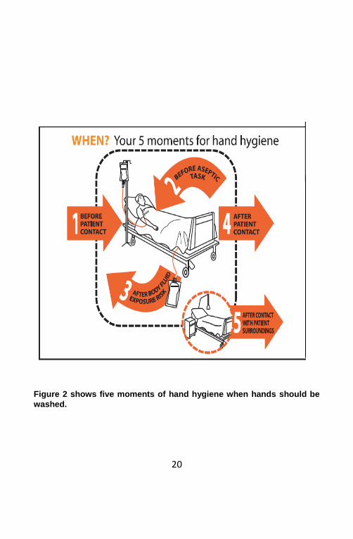
Five (5) Moments in Hand Hygiene- Hand hygiene must be practiced (Figure 2) –
1. Before touching a patient. 2. Immediately before performing a clean or aseptic procedure,
including handling an invasive device for patient care, regardless of whether or not gloves are used.
3. Promptly after contact with body fluids, excretions, mucous membranes, non-intact skin, or wound dressings regardless of whether or not gloves were used.
4. After touching a patient and his/her immediate surroundings, even when leaving the patient’s side.
5. After contact with inanimate objects (including medical equipment and furniture) in the immediate vicinity of the patient.
Perform hand wash when hands are visibly dirty.
Surgical Hand Scrub:
1. Surgical hand scrub with medicated soap or surgical handrub with alcohol-based formulations - Either method is suitable for the prevention of surgical site infection. The combined effect – rapid action at the beginning and inhibition of regrowth of bacteria under the gloved hands – is best achieved by using an alcohol-based compound containing chlorhexidine, or with the addition of a quaternary ammonium compound such as mecetronium sulfate. 2. STEPS FOR SURGICAL HAND PREPARATION - Steps before starting surgical hand preparation -
Keep nails short and pay attention to them when washing your hands – most microbes on hands come from beneath the fingernails.

15
Do not wear artificial nails or nail polish. Remove all jewellery (rings, watches, bracelets) before
entering the operating room suite. Wash hands and arms up to elbows with a non-medicated
soap before entering the operating room area or if hands are visibly soiled.
Clean subungual areas with a nail file. Nailbrushes should not be used as they may damage the skin and encourage shedding of cells. Nailbrushes, if used, must be sterile and used only once. Reusable autoclavable nail brushes are available commercially.
A. Protocol for surgical scrub with a medicated soap - Start timing. Scrub each side of each finger,
between the fingers, and the back and front of the hand for two minutes.
Proceed to scrub the arms, keeping the hand higher than the arm at all times. This helps to avoid recontamination of the hands by water from the elbows and prevents bacteria-laden soap and water from contaminating the hands.
Wash each side of the arm from wrist to the elbow for one minute. •
Repeat the process on the other hand and arm, keeping hands above elbows at all times. If the hand touches anything except the brush at any time, the scrub must be lengthened by one minute for the area that has been contaminated.
Rinse hands and arms by passing them through the water in one direction only, from fingertips to elbow. Do not move the arm back and forth through the water.
Proceed to the operating room suite holding hands above elbows.
16
At all times during the scrub procedure, care should be taken not to splash water onto surgical attire.
Once in the operating room suite, hands and arms should be dried using a sterile towel and aseptic technique before putting on gown and gloves.
B. Protocol for surgical scrub with an alcohol-based preparation –
Start timing. Use sufficient product to keep hands and forearms wet with the handrub throughout the procedure.
After application of the alcohol-based product, allow hands and forearms to dry thoroughly before donning sterile gloves.
Proceed to the operating room suite holding hands above elbows.
If hands are visibly soiled, wash hands with plain soap before surgical hand scrub.
• Training and compliance for hand-hygiene needs to be monitored.
Availability of hand rubs, Soaps hand towels and water should be
ensured. Foot operated and wall mounted dispensing stations are
required. Hand hygiene training programme for doctors, nursing
staff, and housekeeping staff needs to be done regularly for each
category of staff. Hand hygiene compliance need to be monitored.
B. Personal protective equipment-
1. Use of Gloves:
Clean gloves must be worn when touching blood, body fluids, excretions, secretions and contaminated items and when performing venipuncture.
2. Face Mask, eye protection & face shield:
Face Mask must be worn during procedures or patient care activities that are expected to generate splashes or sprays of blood, body fluids,

17
secretions and excretions. For example, suctioning, irrigating a wound, performing certain laboratory tests, etc.
N95 Respirators-
Respirators are masks specifically designed to filter small particles spread by the airborne route, such as tuberculosis, measles and varicella. They are used for aerosol generating procedures that have been shown to expose staff, including: • Sputum induction • Diagnostic bronchoscopy • Autopsy examination • Laboratory handling of Mycobacterium tuberculosis such as concentrating respiratory samples for smear and culture. Staff required to wear N95 Respirators must undergo fitting
3. Gown or Apron:
Gown/apron must be worn to protect skin and to prevent soiling of clothing during procedures or patient care activities that are expected to generate splashes or sprays of blood, body fluid, secretions and excretions. Respiratory hygiene/cough etiquette: Instruct symptomatic persons and health care workers to cover their mouths/noses when coughing or sneezing, use and dispose of tissues, perform hand hygiene after hands have been in contact with respiratory secretions and wear surgical mask if tolerated or maintain spatial separation, >3 feet if possible.

18
Figure 1a shows steps of hand-rubbing

19
Figure 1b shows steps of hand-washing
20
Figure 2 shows five moments of hand hygiene when hands should be
washed.

21
6. PREVENTION OF HEALTHCARE ASSOCIATED INFECTIONS
6.1 The four major HCAIs are:
1. Catheter associated Urinary tract infection (CAUTI) 2. Surgical site Infection (SSI) 3. Catheter related blood stream infection (CRBSI) 4. Ventilator Associated Pneumonia (VAP)
A. CATHETER-ASSOCIATED URINARY TRACT INFECTION
Introduction Urinary tract infections (UTIs) are one of the commonest types of HCAIs. One of the common reasons is the use of urinary catheters. Indications for Catheterization- Placement of an indwelling catheter should be performed only when indicated. It should be removed as soon as possible. The accepted indications for catheterization are:
1. Patient requiring prolonged immobilization, such as in the setting of unstable lumbar/thoracic spine injuries, or multiple traumatic injuries including pelvic fracture For
short-term (days) management of incontinence (the inability to
control urination) needed to assist in healing of sacral or perineal wounds or for retention (the inability to pass urine) not
helped by other methods. 2. To measure urine output over several days in critically ill
patients

22
3. For treatment of bladder outlet obstruction 4. For post-operative management of surgical patients with
impaired bladder function. Recommendations to Prevent Catheter-associated UTI-
1. Personnel Only persons who know the correct technique of aseptic insertion and maintenance of the catheter should handle catheters.
2. Catheter Use
Urinary catheters should be inserted only when necessary and left in place only for as long as it is required. They should not be used solely for the convenience of patient-care personnel. For selected patients, other methods of urinary drainage such as condom catheter drainage, suprapubic catheterization, and intermittent urethral catheterization may be more appropriate.
3. Hand hygiene Hand hygiene should be done immediately before and after any manipulation of the catheter site or apparatus.
4. Catheter Insertion Catheters should be inserted using aseptic technique and sterile equipment. Gloves, drapes, sponges, an appropriate antiseptic solution for peri-urethral cleaning, and a single-use packet of lubricant jelly should be used for insertion. As small a catheter as possible, consistent with good drainage, should be used to minimize bladder neck and urethral trauma. Indwelling catheters should be properly secured after insertion to prevent movement and urethral traction.
23
5. Closed Sterile Drainage The catheter collection system should remain closed and not be opened unless absolutely necessary for diagnostic or therapeutic reasons eg irrigation. If breaks in aseptic technique, disconnection, or leakage occur, the catheter and collecting system should be replaced using aseptic technique and sterile equipment.
6. Irrigation
Continuous irrigation should be avoided unless indicated (e.g. after prostatic or bladder surgery). Continuous irrigation of the bladder with antimicrobials has not proven to be useful and should not be performed as a routine infection prevention measure.
7. Specimen Collection If small volumes of fresh urine are needed for examination, the distal end of the catheter, or preferably the sampling port if present, should be cleansed with a disinfectant, and urine then aspirated with a sterile needle and syringe. Larger volumes of urine for special analysis should be obtained aseptically from the drainage bag.
8. Urinary Flow Unobstructed flow should be maintained. The catheter and collecting tube should be kept free from kinking. Collecting bags should always be kept below the level of the bladder. Do not rest the collecting bag on the floor.
9. Meatal Care
Cleansing of the meatal surface during daily bathing or showering is appropriate.

24
10. Catheter Change Interval
Indwelling catheters should not be changed at arbitrary fixed intervals.
B. SURGICAL SITE INFECTIONS (SSI)
The common source of pathogens is the endogenous flora of the patient’s skin, mucous membranes, or hollow viscera. Therefore, the pathogens isolated from infection differ, primarily depending on the type of surgical procedure. In clean surgical procedures, in which the gastrointestinal, gynaecologic, and respiratory tracts have not been entered; Staphylococcus aureus from the exogenous environment or patient’s skin flora is the usual cause of infection. In other categories of surgical procedures, including clean contaminated, contaminated, and dirty, the polymicrobial aerobic and anaerobic flora closely resembling the normal endogenous microflora of the surgically excised organ are the most frequently isolated pathogens. Other sources of SSI pathogens are from distance focus such as in patients with prosthesis or implant place during the surgery, surgical personnel, operating environment, surgical tools, instruments, and materials brought to the field during an operation.
Surgical site infection prevention- Preparation of the patient:
1. Whenever possible, identify and treat all infections remote to the surgical site before elective operation and postpone elective surgeries on patients with remote site infections until the infection has resolved.
2. Keep preoperative hospital stays as short as possible while allowing for adequate preoperative preparation.
3. Do not remove hair preoperatively unless the hair at or around the incision site will interfere with the operation.
4. If hair needs to be removed, it is done immediately before operation, preferably using electric clippers and not razor blade.

25
5. Adequately control blood glucose levels in all diabetic patients. 6. Encourage nonsmoking/use of cigarettes, cigars, pipes, or any
other form of tobacco consumption for at least 30 days prior to the surgery.
7. Do not withhold necessary blood products transfusion. 8. Encourage patients to shower or bathe at least the night before
the operative day. 9. Use an appropriate antiseptic agent for skin preparation. 10. Apply preoperative antiseptic skin preparation in concentric
circles moving towards the periphery. The prepared area should be large enough to extend the incision or create new incisions or drain sites, if necessary.
Antimicrobial prophylaxis
1. Administer a prophylactic antibiotic agent only when indicated, and select it based on its efficacy against the most common pathogens causing SSI for a specific operation.
2. Administer by IV route the initial dose of prophylactic antibiotic agent, timed such that a bactericidal concentration of the drug is established in serum and tissues when the incision is made. Maintain therapeutic levels of the agent in serum and tissues throughout the operation and until at most a few hours after the incision is closed in the operating room. In most cases,
antibiotic should be given within 60 minutes before the
incision and the antibiotics should be stopped within 24 hours after surgery.
Microbiological sampling Routine environment sampling of the Operation Room (OR) is not required. Perform microbiologic sampling of OR environment surfaces or air as part of an epidemiologic investigation. Cleaning and disinfection of environmental surfaces
1. When visible soiling or contamination with blood or other body fluids of surfaces or equipment occurs during an operation, use approved hospital disinfectant to clean the affected areas before the next operation.

26
2. Do not perform special cleaning or closing of OR after contaminated or dirty operation.
3. Clean the operating room floor after the last operation of the day or night with an approved hospital disinfectant.
Asepsis and surgical technique
1. Adhere to principles of asepsis when intravascular devices, spinal or epidural anesthesia catheters, or when dispensing and administering intravenous drugs.
2. Assemble sterile equipment and solutions immediately prior to use
Sterilization of surgical instruments
Sterilize all surgical instruments according to guidelines. Surgical attire and drapes
1. Wear a surgical mask that fully covers the mouth and nose when entering the operating room if an operation is about to begin or already under way, or if sterile instruments are exposed. Wear the mask throughout the operation.
2. Wear a cap or hood to fully cover the hair on the head and face when entering the operating room.
3. Wear sterile gloves if a scrubbed surgical team member. Put on gloves after donning a sterile gown.
4. Using surgical gowns and drapes that are effective barriers when wet.
5. Change scrub suits that are visibly soiled, contaminated, and/or penetrated by blood or other potentially infectious materials.
Postoperative incision care
1. Protect with a sterile dressing 24 to 48 hours postoperatively an incision that has been closed primarily.
2. Wash hands before and after dressing changes and any contact with the surgical site.
3. Use sterile technique to change incision dressing. 4. Educate the patient and family regarding proper incision care,
symptoms of surgical site infection, and the need to report such symptoms.

27
Develop a good surveillance system to study the incidence of SSI.
1. Use standardized case definitions without modifications for identifying SSI among surgical inpatients and outpatients.
2. Use methods for inpatient and outpatient case-finding that accommodate available resources and data needs.
3. Assign surgical wound classification upon completion of an operation.
4. For each patient undergoing an operation chosen for surveillance, record those variables shown to be associated with increased SSI risk, such as surgical wound class and duration of operation.
5. Periodically calculates operation-specific SSI rates stratified by
variables shown to be associated with increased SSI risk.
6. Report stratified operation-specific rates to surgical team
members.
C. VENTILATOR-ASSOCIATED PNEUMONIA
Pneumonia is one of the three most common HCAIs. Patients who are mechanically ventilated are at risk for ventilator-associated pneumonia (VAP). Most bacterial nosocomial pneumonias occur by aspiration of bacteria colonizing the oropharynx or upper gastrointestinal tract of the patient. Intubation and mechanical ventilation greatly increase the risk of nosocomial bacterial pneumonia because they alter first-line patient defenses. Prevention of VAP-
1. Adhere to hand-hygiene guidelines. 2. Health-care worker should ear a mask and an apron or gown
when anticipates soiling of respiratory secretions from a patient (e.g. intubation, tracheal suctioning, tracheostomy, and

28
bronchoscopy) and change it after the procedure and before providing care to another patient.
3. Elevate the head of the bed 30 – 45 degrees of a patient on mechanical ventilation or at high risk for aspiration (e.g. on oro or nasoenteral tube)
4. Remove devices such as endotracheal, tracheostomy, oro/ nasogastric tubes from patients as soon as they are not indicated.
5. Perform orotracheal rather than nasotracheal intubation unless contraindicated.
6. Use non-invasive ventilation whenever possible. 7. Perform daily assessments of readiness to wean and use
weaning protocols. 8. Avoid unplanned extubation and reintubation. 9. Use a cuffed endotracheal tube with in-line or subglottic
suctioning. 10. Avoid histamine receptor blocking agents and proton pump
inhibitors for patients who are not at high risk for developing a stress ulcer or stress gastritis.
11. Perform regular oral care with an antiseptic solution. 12. Avoid gastric overdistension. 13. Remove condensate from ventilatory circuits. Keep the
ventilatory circuit closed during condensate removal. 14. Change the ventilatory circuit only when visibly soiled or
malfunctioning. 15. Store and disinfect respiratory therapy equipment properly. 16. Educate healthcare workers who provide care for patients
undergoing ventilation about VAP.
Develop a surveillance system to study the incidence of VAP.
1. Conduct active surveillance for VAP in units that care for patients undergoing ventilation who are known or suspected to be at high risk for VAP.
2. Collect data that will support the identification of patients of VAP and calculation of VAP rates.

29
D. CATHETER-RELATED BLOOD STREAM INFECTIONS
Hand hygiene
1. Observe proper hand hygiene. 2. Palpation of the insertion site should not be performed after the
application of antiseptic, unless aseptic technique is maintained. Aseptic technique during catheter insertion and care
1. Maintain aseptic technique for the insertion and care of intravascular catheters. Wearing clean gloves rather than sterile gloves is acceptable for the insertion of peripheral intravascular catheters if the access site is not touched after the application of skin antiseptics.
2. Sterile gloves should be worn for the insertion of arterial, central, and midline catheters.
3. Change the dressing on intravascular catheters using aseptic
technique.
Maximal sterile barrier precautions during catheter insertion Use aseptic technique including the use of a cap, masks, sterile gown, sterile gloves, and a large sterile sheet for the insertion of Central venous catheter (CVCs, including peripherally inserted central catheter- [PICC]) or guidewire exchange. Catheter Site Dressing Regimens
1. Use either sterile gauze or sterile, transparent, semipermeable dressing to cover the catheter site.
2. Replace the catheter site dressing if the dressing becomes damp, loosened, or visibly soiled.
3. Replace dressings used on short-term CVC sites every 2 days for gauze dressings.
4. Replace dressings used on short-term CVC sites at least every 7 days for transparent dressings, except in those pediatric patients in which the risk for dislodging the catheter may outweigh the benefit of changing the dressing.
5. Monitor the catheter sites visually or by palpation through the intact dressing on a regular basis, depending on the clinical

30
situation of individual patients. If patients have tenderness at the insertion site, fever without obvious reasons, or other manifestations suggesting local or BSI (Blood Stream infections), the dressing should be removed to allow thorough examination of the site.
Prevention of CRBSI
1. Select the catheter, insertion technique, and insertion site with the lowest risk for complications (infectious and noninfectious) for the anticipated type and duration of IV therapy.
2. Avoid using the femoral vein for central venous access in adult patients.
3. Use a subclavian, rather than a jugular or a femoral site, in adult patients to minimize infection risk for nontunneled CVC placement.
4. Promptly remove any intravascular catheter that is no longer essential. Do not routinely replace central venous or arterial catheters solely for the purposes of reducing the incidence of infection.
5. There is no need to replace peripheral venous catheters more frequently than 72—96 hours to reduce the risk of infection and phlebitis in adults. Leave peripheral venous catheters in place in children until IV therapy is completed, unless complications (e.g: phlebitis and infiltration) occur.
Develop a surveillance system to study the incidence of CRBSI.
1. Conduct active surveillance for CRBSI in units that care for patients undergoing ventilation who are known or suspected to be at high risk for CRBSI.
2. Collect data that will support the identification of patients of CRBSI and calculation of CRBSI rates.
31
7. CLEANING, DISINFECTION AND STERILIZATION
7.1 DISINFECTION Disinfection is a process where most microbes are removed from
defined object or surface, except spores.
7.2 Disinfectants can be classified according to their ability to
destroy different categories of micro-organisms:
• High Level disinfectants : Glutaraldehyde 2%, Ethylene Oxide • Intermediate Level disinfectant : Alcohols, chlorine compounds,
hydrogen Peroxide, chlorhexidine, • Low level disinfectants : Benzalkonium chloride, some soaps 7.3 GENERAL GUIDELINES FOR DISINFECTION: Critical instruments/equipment (that are those penetrating skin or
mucous membrane) should undergo sterilization before and after use.
e.g. surgical instruments.
Semi-critical instruments /equipments (that are those in contact with
intact mucous membrane without penetration) should undergo high level
disinfection before use and intermediate level disinfection after use. e.g.
endotracheal tubes.
Non-critical instruments /equipments (that are those in contact with intact
skin and no contact with mucous membrane) require only intermediate
or low level disinfection before and after use. e.g. ECG electrodes.

32
7.4 Endoscopes - cleaning and disinfection 1. Clean: mechanically clean internal and external surfaces, including
brushing internal channels and flushing each internal channel with water and a detergent or enzymatic cleaners
2. Disinfect: immerse endoscope in high-level disinfectant such as 2% glutaraldehyde and perfuse disinfectant into all accessible channels, such as the suction/biopsy channel and air/water channel and expose for a time recommended for specific products (20 minutes for 2% glutaraldehyde).
3. Rinse: rinse the endocope and all channels with sterile or filtered water followed by 70-90% ethyl or isopropyl alcohol to remove all traces of disinfectant.
4. Drying: After rinsing, purge the channels using forced air. Hang endoscopes in a vertical position to facilitate drying.
7.5 STEAM STERILIZATION Use biological indicators, such as a commercial preparation of spores of
Geobacillus stearothermophilus, at least weekly to monitor the
effectiveness of steam sterilization. 7.6 FOGGING: In patient care areas regular fogging is not recommended. Necessary decision is taken by in charge of concerned patient care area. 7.7 ENVIRONMENTAL SURFACES Clean housekeeping surfaces (e.g., floors, walls, tabletops) on a regular basis, when spills occur, and when these surfaces are visibly soiled. Disinfect environmental surfaces (e.g., bedside tables, bedrails, and laboratory surfaces) on a regular basis and when surfaces are visibly soiled. Clean walls, blinds, and window curtains in patient-care areas when these surfaces are visibly contaminated or soiled. Decontaminate mops heads and cleaning cloths regularly to prevent contamination (e.g., launder and dry at least daily).

33
Do not use high-level disinfectants or liquid chemical sterilants for disinfection of non-critical surfaces. 7.8 Bedding and blanket Clean and disinfect mattress impermeable covers.
Launder pillow covers, washable pillows, and blankets between patients
or when they become contaminated with body substances.
7.9 Monitoring of biomedical waste management practices A person or persons should be designated to be responsible for
establishing, monitoring, reviewing, and administering a plan for the
collection, handling, predisposal treatment, and terminal disposal of
regulated medical wastes.

34

35

36
7.10 DEALING WITH SPILLAGE: a. LIQUID SPILL MANAGEMENT:
1. Promptly clean and decontaminate spills of blood and other potentially infectious materials.
2. Wear protective gloves. 3. Using a pair of forceps and gloves, carefully retrieve broken
glass and sharps if any, and use a large amount of folded absorbent paper to collect small glass splinters. Place the broken items into the puncture proof sharps container.
4. Cover spills of infected or potentially infected material on the floor with paper towel/ blotting paper/newspaper. Pour 0.5%freshly prepared sodium hypochlorite.
5. Leave for 30 minutes for contact 6. Place all soiled absorbent material and contaminated swabs
into a designated waste container. 7. Then clean the area with gauze or mop with water and
detergent with gloved hands. NB: Any material treated with hypo-chlorite solution should never be sent for incineration b.MERCURY SPILL MANAGEMENT:
If accidental spill of mercury occurs it is to be collected in a special manner as follows:
1. Spilled mercury should be collected with a ‘‘mercury spill kit’’- containing nitrile gloves, N-95 face mask, 2 pieces of cardboards, 2 plastic containers, cello tape, and flashlight.
2. Do not touch mercury. 3. Remove all jewelry, wear gloves, masks. 4. Use flashlight to locate and cardboards to bring mercury beads
together.

37
5. Collect with an eyedropper of a syringe and carefully place it or
‘contain’ in a bottle containing water. 6. Any remaining beads of mercury should be picked up with a
sticky tape and place 7. in the plastic bag, properly labeled.
8. The bottle should be sealed with a tape, labeled as hazardous
waste and securely stored inside another plastic container; awaiting final disposal to Govt. nominated or authorized mercury dealers.
9. After mercury has been recovered the spill area should be covered with calcium sulfide or sodium thiosulfate to neutralize it.
10. Reporting formats will be used to report and register any mercury spills/leakages.

38
8. ISOLATION PRECAUTIONS
8.1 Introduction
Isolation precautions are needed to prevent the transmission of
pathogenic microorganisms within the healthcare setting.
The patients of following disease categories should be treated under isolation. • Severe influenza cases, Subacute respiratory Syndrome (SARS),
Open case of tuberculosis, Anthrax, diphtheria, Pertussis, Pneumonic plague, Chicken pox, and patients infected with multidrug resistant bacterial pathogens.
8.2 Patient placement - Appropriate patient placement is a significant component of
isolation precautions. Determine patient placement based on the following principles:
- Route(s) of transmission of the infectious agent - Risk factors for transmission in the infected patient - Risk factors for adverse outcomes resulting from healthcare-
associated infection in other patients in the area. - Availability of single-patient rooms - Patient options for room-sharing
Contact Precautions:
1. Required for patients with enteric infections, diarrhea that cannot be controlled, or skin lesions that cannot be contained.
2. Patient placement: a. Place patients who require Contact Precautions in a
single-patient room when available; if single-patient rooms are unavailable, then place patients infected with

39
the same pathogen in the same room. b. If it becomes necessary to place a patient who requires
Contact Precautions in a room with a patient who is not infected or colonized with the same infectious agent:
i. Avoid placing patients on Contact Precautions in the same room with patients who have conditions that may increase the risk of adverse outcome from infection or that may facilitate transmission.
ii. Ensure that patients are physically separated (i.e., >3 feet apart) from each other. Draw the privacy curtain between beds to minimize opportunities for direct contact.
iii. Change protective attire and perform hand hygiene between contacts with patients in the same room, regardless of whether one or both patients are on Contact Precautions.
3. Use of personal protective equipment a. Wear gloves whenever touching the patient’s intact skin
or surfaces and articles in close proximity to the patient. Don gloves upon entry into the room or cubicle.
b. Wear gown whenever anticipating that clothing will have direct contact with the patient or potentially contaminated environmental surfaces or equipment in close proximity to the patient. Don gown upon entry into the room or cubicle. Remove gown and observe hand hygiene before leaving the patient-care environment.
4. Patient transport a. Limit transport and movement of patients outside of the
room to medically-necessary purposes. b. When transport or movement in any healthcare setting
is necessary, ensure that infected or colonized areas of the patient’s body are contained and covered.
c. Remove and dispose of contaminated PPE and perform hand hygiene prior to transporting patients on Contact Precautions.
d. Don clean PPE to handle the patient at the transport destination.
5. Patient-care equipment and instruments/devices a. Handle patient-care equipment and
instruments/devices according to Standard Precautions

40
b. Use disposable noncritical patient-care equipment (e.g., blood pressure cuffs) or implement patient-dedicated use of such equipment. If common use of equipment for multiple patients is unavoidable, clean and disinfect such equipment before use on another patient.
6. Environmental measures a. Ensure that rooms of patients on Contact Precautions
are prioritized for frequent cleaning and disinfection with a focus on frequently-touched surfaces and equipment in the immediate vicinity of the patient.
Droplet precautions:
1. Required for patients known or Suspected to be infected with pathogens transmitted by respiratory droplets that are generated by a patient who is coughing, sneezing, or talking, Acute respiratory infection, undiagnosed or meningitis and/or sepsis with petechial rash .
2. Patient placement: a. Place patients who require Droplet Precautions in a
single-patient room when available; if single-patient rooms are unavailable, then place patients infected with the same pathogen in the same room.
b. If it becomes necessary to place a patient who requires Droplet Precautions in a room with a patient who does not have the same infection:
i. Avoid placing patients on Droplet Precautions in the same room with patients who have conditions that may increase the risk of adverse outcome from infection or that may facilitate transmission.
ii. Ensure that patients are physically separated (i.e., >3 feet apart) from each other. Draw the privacy curtain between beds to minimize opportunities for direct contact.
iii. Change protective attire and perform hand hygiene between contact with patients in the same room, regardless of whether one or both patients are on Droplet Precautions.

41
3. Use of personal protective equipment a. Don a mask upon entry into the patient room or cubicle.
4. Patient transport a. Limit transport and movement of patients outside of the
room to medically-necessary purposes. b. When transport or movement in any healthcare setting
is necessary, instruct patient to wear a mask and follow Respiratory Hygiene / Cough Etiquette.
c. No mask is required for persons transporting patients on Droplet Precautions.
Airborne precautions:
1. Required for patients known or suspected to be infected with infectious agents transmitted person-to-person by the airborne route, such as M. tuberculosis, measles, chickenpox, disseminated herpes zoster.
2. Develop systems (e.g., triage, signage) to identify patients with known or suspected infections that requires Airborne Precautions upon entry into the health facility.
3. Patient placement a. Place patients who require Airborne Precautions in an
airborne infection isolation room (AIIR) that has been constructed with the following conditions
i. Provides 6-12 air changes per hour. ii. Directs exhaust or air to the outside, or
through HEPA filters if exhausting to the outside is not possible.
iii. Has a monitor for air pressure with visual indicators.
iv. Can be closed with a door when not required for entry and exit.
b. If an AIIR is not available and transfer to a facility with AIIR is not possible, place a surgical mask on the patient and place him/her in a single room. Once the patient leaves, the room should remain vacant for the appropriate time, generally one hour, to allow for a full exchange of air.
c. Instruct patients with a known or suspected airborne infection to wear a surgical mask and observe Respiratory Hygiene/Cough Etiquette. Once in AIIR,

42
the mask may be removed. 4. Personnel restrictions
a. Restrict susceptible healthcare personnel from entering the rooms of patients known or suspected to have measles, varicella, disseminated zoster, or smallpox if other immune healthcare personnel are available.
5. Use of personal protective equipment a. Healthcare personnel should use a fit-tested
respiratory, such as an N95, before entering the room of a patient with known or suspected tuberculosis or smallpox.
b. A fit-tested N95 or surgical mask may be appropriate for healthcare personnel to wear while caring for patients with known or suspected measles, chickenpox, or disseminated herpes zoster.
6. Patient transport a. Limit transport and movement of patients outside of the
room to medically-necessary purposes. b. If transport or movement outside an AIIR is necessary,
instruct patients to wear a surgical mask, if possible, and observe Respiratory Hygiene/Cough Etiquette.
c. For patients with skin lesions associated with varicella or smallpox or draining skin lesions caused by M. tuberculosis, cover the affected areas to prevent aerosolization or contact with the infectious agent in skin lesions.
d. Healthcare personnel transporting patients who are on Airborne Precautions do not need to wear a mask or respirator during transport if the patient is wearing a mask and infectious skin lesions are covered.
7. Exposure management a. Administer measles vaccine to exposed susceptible
persons within 72 h after the exposure or administer immune globulin within six days of the exposure event for high-risk persons in whom vaccine is contraindicated.
b. Administer varicella vaccine to exposed susceptible persons within 120 h after the exposure or administer varicella immune globulin, when available, within 96 h for high-risk persons in whom vaccine is contraindicated (e.g., immunocompromised patients,

43
pregnant women, newborns whose mother’s varicella onset was <5 d before or within 48 h after delivery).
c. Administer smallpox vaccine to exposed susceptible persons within 4 days after exposure.

44
9. ANTIMICROBIAL POLICY AND ANTIMICROBIAL STEWARDSHIP
9.1 Introduction
The annual antibiogram should be prepared by microbiology
department. Antibiotic susceptibility profile may be is analyzed regularly
and the common resistance patterns of the bacterial isolates to be
reported and discussed in the HICC meetings and the antibiotic policy to
be reviewed accordingly.
Antibiotic policy need to be prepared in consultation with respective clinical departments. 9.2 Antibiotic policy shall be prepared using following general principles: 1. Data is analyzed on a quarterly basis as per hospital records)
(a) Common etiological agents as per
(i) site of infection
(ii) age groups
(iii) patient location – outdoor (OPD), indoor (wards & critical care areas)
(b) Antibiogram data as per
(i) site of infection
(ii) age groups
(iii) patient location – outdoor (OPD), indoor (wards & critical care
areas)
(c) Unusually resistant organisms to be confirmed and submitted for
further characterization to National Centre for Disease Control
(NCDC) for 2. Standard treatment guidelines [categorization of patients as
45
per age and Community acquired infections (CAI) / Health care
associated infections (HCAI)]
(a) Guidelines for empirical antimicrobial therapy as per common clinical syndrome
(i) Adults & older children
1. Blood Stream Infections (BSI)
2. Meningitis
3. UTI
4. Pneumonia
(a) Community Acquired Pneumonia (CAP)
(b) Ventilator Associated Pneumonia (VAP)
5. GIT Infections
6. Conjunctivitis
7. Otitis Media
8. Tonsilltitis / Pharyngitis
9. Skin and Soft Tissue Infection (SSTI)
10. Genital Infections
11. Osteomyelitis
(ii) Neonates (special conditions)
1. Sepsis
2. Meningitis
(iii) Infants & Small Children (special conditions)
1. Meningitis
2. Sepsis
3. Pneumonia
(b) Classification of Antimicrobials into first line, second line and
reserve group of drugs
(c) Chemoprophylaxis
(i) Pre-operative antimicrobials
(ii) Other invasive procedures
(iii) Special high risk groups e.g. Prophylaxis for rheumatic fever,
splenectomy patients, and immuno-compromised patients
46
(d) Special clinical syndromes (e.g. STIs) 4. Prescription auditing
5. Review of surveillance data generated from antibiograms &
prescription auditing. 6. Education and training for all infection control activities in
collaboration with the Hospital Infection Control Committee.
9.3 Measures to control spread of antibiotic resistance-
i. Appropriate antimicrobial use
1. Each health care facility should have an antimicrobial use
programme. The goal is to ensure effective economical
prescribing to minimize the selection of resistant
microorganisms.
2. Formulation of guidelines with a multidisciplinary approach
using the local antibiogram.
3. Provide ongoing education on rational use of antibiotics to
clinicians and ensure implementation of antibiotic policies.
4. Restricted antibiotic use-
5. Use must be justifiable based on clinical diagnosis.
6. Before initiating antibiotic treatment, appropriate specimens for
bacteriological examination must be submitted to laboratory and
selection of an antibiotic must be based on the sensitivity
pattern, patient tolerance, and cost
7. An agent with as narrow a spectrum as possible should be used
with appropriate dosage and duration of antimicrobial therapy.
8. The correct dose must be used.
9. Control antibiotic use - Selected antibiotics may be restricted in
use.
-Cyclic rotation of antibiotics in a class
- Discontinuation of antimicrobial therapy based on predefined
criteria
10. Carry out periodic prescription audits.
11. Restriction of hospital formulary through pharmacy.
12. Standard and contact Precautions including rigorous adherence
to hand hygiene, appropriate use of PPE.

47
13. Isolation and cohorting of patients infected or colonized with
Multi-drug resistant organisms (MDROs).
14. Education and training of HCP.
15. Increased environmental cleaning and patient-dedicated
equipment.
16. Proper sterilization and disinfection.
17. Surveillance for Multidrug resistant organisms especially in high
risk areas.
9.4 Control of spread of specific organisms (MDROs)
a. Methicillin Resistant Staphylococcus aureus
(MRSA)
MRSA strains are resistant to the penicillin’s-resistant penicillins
(methicillin) and cephalosporins and are often resistant to multiple
classes of drugs and occasionally are sensitive only to Vancomycin
and teicoplanin. MRSA are highly-transmissible strains and have a
high potential to spread across hospitals. Since there are few
therapeutic options available for treatment of this resistant organism,
the best strategy to control the spread are the preventive measures.
Transient carriage of the organism on hands of HCWs accounts for
major route of transmission from infected/colonized inpatients to
other patients. Transmission from environmental surfaces and
airborne routes is known to occur. The measures to control MRSA in
hospitals are screening for MRSA carriage or infection in certain
high risk patients or units at admission, standard and contact
precautions, isolation and cohorting of patients, treatment of
infected/colonized patients, environmental cleaning, education and
training of staff.
b. Vancomycin – Resistant Enterococcus (VRE)
Enterococcal infections are difficult to treat because of their intrinsic
resistance to many antimicrobial agents and easily acquire
resistance to almost all antimicrobials including Vancomycin.
Transmission of VRE can occur by direct contact or indirectly via
transient carriage on hands of HCW, contaminated surfaces or
patient-care equipment. To prevent and control the nosocomial
transmission of VRE, judicious use of antibiotics especially

48
Vancomycin, education of HCW, implementation of hospital infection
control practices, equipment and environmental cleaning, using
patient-dedicated or single-use non-critical patient-care equipment,
isolation and cohorting of infected/colonized patients, use of PPE,
and surveillance for VRE infection/colonization should be
implemented.
c. MDR Gram negative (MDRGN) bacteria
MDRGN includes organisms producing ESBLs, plasmid-mediated
AmpC and carbapenemases. Screening of patients in high risk units
and those at high risk of carriage such as recent broad spectrum
antibiotic therapy (carbapenem, quinolones, and 3rd and 4th
generation cephalosporins), long duration of stay and severity of
illness, chronic disease and impaired functional status and presence
of invasive medical devices should be carried out for
infection/colonization with MDRGN organisms. Multiple sites
including rectal or perianal swabs, should be screened. Measures to
prevent spread of MDRGN organisms include stringent hand
hygiene, contact precautions (gloves and gown), isolating, and
cohorting, increased environmental cleaning and dedicated patient
equipment and judicious use of antibiotics.
In healthcare facilities:
Antibiotic usage rates in healthcare facilities are high for some
classes of drugs, and there is considerable unexplained variation
between hospitals in the use of certain antibiotics, particularly broad-
spectrum antibiotics. Problems resulting from inappropriate use of
antibiotics apply to both current and future healthcare facility
patients due to changes in healthcare facility microbial ecology
resulting from the resistance.
Additional costs of infections caused by resistant organisms include:
The need for more expensive and broader spectrum
antibiotics to treat the infections.
The need to isolate patients colonized with resistant
organisms in order to minimize cross-infection.

49
In the community:
Community antibiotic use is high and there is irrational use of
antibiotics including the over the counter sales due to lack of
monitoring mechanism in spite of the existing laws. Thus multi-
resistant bacteria, such as community strains of MRSA(CA-MRSA)
and extended-spectrum beta-lactamase-producing Gram-negative
bacteria are causing increasing human morbidity and there is
concern that past excessive antibiotic use in the community or in
animal production systems (or both) is responsible.
9.5 Antimicrobial Stewardship
This aims to optimize antimicrobial use among patients in order to
reduce antibiotic resistance, improve patient outcomes and safety,
and ensure cost-effective therapy.
At the healthcare facility level, antibiotic stewardship involves:
• Implementing an antibiotic stewardship program; and
• Continuous monitoring and analysis of antibiotic usage, to track
changes in antibiotic resistance and to monitor effects of
containment strategies.
Key requirements of a healthcare facility antibiotic stewardship
program:
1. A multidisciplinary antibiotic stewardship team with core
membership of an infectious diseases physician (lead doctor)
and a clinical pharmacist. Microbiologist, and infection control
professional may also be included.
2. Antibiotic stewardship should be available within the healthcare
facilities for quality improvement and patient safety governance
structure. There should be collaboration between the
stewardship team and drug and therapeutics and infection
prevention and control committees.
3. Implementation of clinical guidelines that comply with national
treatment guidelines and incorporate changes regularly based
on resistance patterns prevailing in the health facility as

50
reported regularly by microbiology department.
4. Microbiology services reporting patient-specific culture and
sensitivity results to optimize individual antibiotic management.
5. Review of antibiotic prescribing with intervention and direct
feedback to the prescriber.
6. Activities according to local priorities and resources & Provision
of effective & regular education of prescribers and pharmacists
about antibiotic usage, development of resistance and judicious
prescribing. Of the antibiotics.
7. Point of care interventions including: streamlining or de-
escalation of therapy, dose optimization, parenteral to oral
conversion.
8. Use of information technology such as electronic prescribing
with clinical decision support, on-line approval systems.
9. Monitor antibiotic prescribing by measuring antibiotic
consumption; drug use evaluations and using quality Use of
Medicine indicators.
10. Support and collaboration of hospital administration including
allocation of resources to provide education and measure and
monitor antibiotic usage.
11. Antibiotic stewardship surveillance methods should be
established at patient level as well as population or community
level.

51
10. BIOMEDICAL WASTE MANAGEMENT
10.1 INTRODUCTION
The Ministry of Environment and Forests, Govt. of India notified the
Bio-Medical Waste (Management and Handling) Rules on 27th July,
1998; under the provisions of Environment Act 1986. These rules have
been framed to regulate the disposal of various categories of Bio-
Medical Waste as envisaged therein; so as to ensure the safety of the
staff, patients, public and the environment. There have been some
amendments to the rules from time to time; presently the rules are being
revised and this section will be duly updated.
10.2 OBJECTIVES
The Bio-Medical Waste Management policy of the hospitals shall meet
the following broad objectives:- (i) Provide a system of management of potentially infectious and
hazardous waste as per guidelines and recommendations of
Biomedical Wastes (Management and Handling Rules) 1998. (ii) Identifying, defining & classifying the various categories of waste
being generated in the hospitals.
Use of separate color coded containers for Segregation of various categories of waste at point of generation.
(iii) Segregation of various categories of waste in separate color coded
containers at the site of generation, so that each category is treated
in a suitable manner to render it harmless. (iv) Disinfection/decontamination of infected items at the site of
generation immediately after use.
(v) Onsite appropriate ‘‘treatment technology’’ to be used depending
upon the category of waste. (vi) Creating a system where all categories and personnel are
responsible as well as accountable for proper waste management.

52
(vii) Environment and patient friendly safety norms.
10.3 Practices in the patient-care areas/clinical areas: 10.3.1 POLICY ON SEGREGATION OF WASTE: The hospital should ensure that clinical and general wastes is
segregated at source and placed in color coded plastic bags and
containers prior to collection and disposal at the site of generation in all
patient care areas/clinical areas. There should be no mixing of waste. Colored coded bags are used for segregation. Segregation is the responsibility of the generator of wastes i.e. the
doctors, nurses or paramedical personnel. Segregation for each of the following:
• Infectious non-sharps • Infectious sharp • Disposable plastic items • Glass • General waste • Paper/ cardboards • Cytotoxic/radiological/radioactive • Microbiological/pathological wastes

53
Table 1. Categories of Biomedical Waste (Schedule 1)
Options Waste Category Treatment and Disposal
Category No. 1
Human Anatomical Waste (human tissues, organs, body parts)
incineration @/deep burial*
Category No. 2
Animal Waste (animal tissues, organs, body parts carcasses, bleeding parts, fluid, blood and experimental animals used in research, waste generated by veterinary hospitals, colleges, discharge from hospitals, animal houses)
incineration@/deep burial*
Category No. 3
Microbiology & Biotechnology Waste (Wastes from laboratory cultures, stocks or micro-organisms live or vaccines, human and animal cell culture used in research and infectious agents from research and industrial laboratories, wastes from production of biologicals, toxins, dishes and devices used for transfer of cultures)
local autoclaving/micro-waving/incineration@
Category No. 4
Waste Sharps (needles, syringes, scalpels, blade, glass, etc. that may cause puncture and cuts. This includes both used and unused sharps)
disinfection (chemical treatment @@@/autoclaving/microwaving and mutilation/shredding##
Category No. 5
Discarded Medicines and Cytotoxic drugs (outdated, contaminated and discarded medicines)
incineration@/destruction and drugs disposal in secured landfills
Category No. 6
Soiled Waste (items contaminated with blood, and body fluids including cotton, dressings, soiled plaster casts, lines, bedding, other material contaminated with blood)
incineration@autoclaving/microwaving
Category No. 7
Solid Waste (Waste generated from disposal items other than the sharps such a tubings, catheters, intravenous sets etc.)
disinfection by chemical treatment@@ autoclaving/microwaving and mutilation/shredding##
Category No. 8
Liquid Waste (Waste generated from laboratory and washing, cleaning, housekeeping and disinfecting activities)
disinfection by chemical treatment@@ and discharge into drains
Category No. 9
Incineration Ash Ash from incineration of any bio-medical waste)
disposal in municipal landfill
Category No. 10
Chemical Waste (Chemicals used in production of biologicals, chemicals used in production of biologicals, chemicals used in disinfection etc.)
chemical treatment@@ and discharge into drains for liquids and secured landfill for solids

54
@ There will be no chemical pretreatment before
incineration. Chlorinated plastics shall not be
incinerated.
* Deep burial shall be an option available only in towns
with population less than five lakhs and in rural areas.
@@ Chemicals treatment using at least 1% hypochlorite
solution or any other equivalent chemical reagent.
It musts be ensured that chemical treatment ensures
disinfection.
## Mutilation/shredding must be such so as to prevent
unauthorised reuse.
Table 2. Color-coded bags & Category wise Treatment
Color Coding Waste Category Treatment option as per
Schedule 1
Yellow (Plastic bag) Cat. 1, 2. 3, 6 Incineration/deep burial
.
Blue Cat. 4,7.
Autoclaving/Chemical Treatment /Microwaving and Shredding
(plastic bag/puncture proof container)
Black (Plastic bag)
Cat. 5, 9 and 10 (solid) Disposal in secured landfill
55
NB: Categories 8 and 10 (liquid) do not require containers/bags. No PVC material should be placed in yellow bag.
Any material treated with hypo-chlorite solution should never be sent for incineration
BINS AND LINERS: The container comprises of an inner bag of color depending on the type
of waste, and should match the chosen outer container is a plastic bin
with handles, and of a size which will depend on the amount of waste
generated. The inner polythene bag should be leak proof, and should fit
into the container with one-fourth of the polythene bag turned over the
rim.
LINERS/PLASTIC BAGS: MATERIALS USED: Biodegradable colored plastic bags to line the same colored bins with
the specifications ad guidelines of BMW Rules.
BINS : Containment of waste: An optimum number of easy to use, Standard,
uniform, covered, foot-operated bins of appropriate size shall be placed
at identified places in all clinical areas.
DISINFECTION OF BINS: Chemical disinfection of the waste bins using hypochlorite solution
should be done frequently at a separate washing facility in the hospital.
DISINFECTION AND MUTILATION OF SHARPS: In order to render them harmless to waste handlers a Pre-Treatment of
the infectious waste generated in the patient-care areas is required, prior
to transportation for onsite treatment and disposal. It is required for the
following infectious items;
• Syringes • Needles • Catheters, I/V sets, gloves
56
10.3.2 COLLECTION STORAGE, LABELING, AND RECORDING OF WASTE All the biomedical waste to be labeled as waste type, site of generation,
date of generation before transportation from the generation site. Waste
should be stored in the areas of generation at an identified safe area, for
an interim period after which it is transported for onsite treatment and
final disposal. No untreated bio-medical waste shall be kept stored
beyond a period of 48 hours. All the staff is required to duly full in the waste book color wise
mentioning the number and size of bags handed over and sign the slip
for further record. 10.4 LIQUID AND CHEMICAL WASTES MANAGEMENT: Chemical disinfection of the liquid waste, at the areas of generation e.g.,
Labor rooms, OTs, labs etc is done. These liquid wastes should be
disinfected by chemical treatment using at least 1% sodium hypochlorite
solution for a contact period of 30 Minutes and them discharged into
drains/sewers where it is taken care of by the principle of dilution and
dispersal.
10.5 DISCARDED MEDICINES AND CYTOTOXIC DRUGS: The discarded medicines and cytotoxic drugs, which need to the disposed, should be certified by Head of the concerned department, put in a relevant bag, tied and sealed and labelled as with ‘Cytotoxic’
10.6 Different labels for Bio-medical waste containers and bags shall be required for identification and safe handling of this waste. These labels for storage/transportation of biomedical waste are as under:

57

58
11. OCCUPATIONAL HEALTH AND SAFETY
11.1 Introduction
Occupational health and safety includes the prevention, reporting and management of sharps injuries, needle stick injuries and other percutaneous exposures to blood and body fluids which may potentially expose an employee to the risk of blood-borne viruses. Definitions of sharp injury Sharps injury can be defined as injury from needle or other sharp device contaminated with blood or a body fluid and penetrates the skin percutaneously mucosal/ cutaneous exposure. Blood borne pathogens are viruses that some people carry in their blood and which may cause severe disease in certain people and few or no symptoms in others. The virus can spread to another person even if the carrier is asymptomatic. The main blood borne viruses of concern are: - Hepatitis B virus (HBV) - Hepatitis C virus (HCV) - Human Immunodeficiency Virus (HIV) Source patient is the person whose blood is present on the item that caused the sharps injury. 11.2 PPE & VACCINATION OF THE HEALTH CARE WORKERS: All waste handlers should be provided with Masks, Caps, Gum Boots,
Gloves, and Disposable apron which they are expected to wear while
dealing with the waste. All health care workers should be vaccinated
against Hepatitis B and tetanus.

59
11.3 SHARPS INJURY MANAGEMENT
The commonest cause of injury while handling the waste is inappropriate
segregation wherein sharp waste is deposited in containers meant for
non-sharp waste. When sharp injury occurs following procedures is to be
followed.
(i) Stop the procedure immediately and wash the wound with soap
and water, encourage bleeding the apply antiseptic.
(ii) Immediately report to Nodal officer in Casualty for First aid and
emergency treatment or any other action and follow-up advice,
if required. ‘PEP’ is provided is casualty round the clock as per
MOHFW guidelines.
(iii) Retention, if possible of the item and details of its source for
identification of possible infection.
(iv) Investigation, determination and implementation of remedial measures.
(v) Recording of Sharp injury: Needle Sticks/ Sharp injury should
be recorded. 11.4 TRAINING OF ALL THE STAFF: A regular training of the staff CME’s, Workshops, are the essential part
to maintain the Hospital Waste Management (HWM) at the best. It is
necessary to conduct the regular refresher training of all the staff
members of the hospital.
11.5. MANAGEMENT OF EXPOSURES TO HBV For percutaneous or mucosal exposures to blood, several factors must
be considered when making a decision to provide prophylaxis, including
the HBsAg status of the source and the hepatitis B vaccination and
vaccine-response status of the exposed person. Such exposures usually
involve persons for whom hepatitis B vaccination is recommended. Any
blood or body fluid exposure to an unvaccinated person should lead to
initiation of the hepatitis B vaccine series. A summary of prophylaxis
recommendations for exposure to blood according to the HBsAg status
of the exposure source and the vaccination and vaccine-response status
of the exposed person is included in the following table:

60
Table 3. RECOMMENDED POSTEXPOSURE PROPHYLAXIS FOR EXPOSURE TO HEPATITIS B VIRUS
Vaccination and antibody response status of exposed workers
*
TREATMENT
Source HbsAg
†
positive
Source- HBsAg negative
Source – Unknown or not available for testing
Unvaccinated HBIG§ x 1 and
initiate HB vaccine series
Initiate HB vaccine series
Initiate HB vaccine
Previously vaccinated
- Serum anti-HBs¶ ≥
10 mIU/mL
No treatment No treatment No treatment
- Serum anti-HBs < 10 mIU/mL
HBIG x 1 and initiate revaccination or HBIG x 2
**
No treatment If known high risk source, treat as if source were HBsAg positive
- Antibody response unknown
Test exposed person for anti-Hbs. - If anti-HBs ≥ 10 mIU/ML, no treatment. - If anti-HbS < 10 mIU/mL, administer HBIG x 1 and vaccine booster
No treatment Test exposed person for anti-Hbs. - If anti-HBs ≥ 10 mIU/ML, no treatment. - If anti-HbS < 10 mIU/mL, administer HBIG x 1 and vaccine booster
* Persons who have previously been infected with HBV are immune to
reinfection and do not require postexposure prophylaxis.

61
† Hepatitis B surface antigen.
§ Hepatitis B immune globulin; does is 0.06 mL/kg intramuscularly.
¶ Antibody to HBsAg.
** The option of giving one dose of HBIG and reinitiating the vaccine series is
preferred for non-responders who have not completed a second 3-dose
vaccine series. For persons who previously completed a second vaccine
series but failed to respond, two doses of HBIG are preferred.
11.6. EXPOSURE TO HIV
Please see NACO 2007 Antiretroviral Therapy Guidelines for HIV-
Infected Adults and Adolescents Including Post-exposure Prophylaxis
Six steps are indicated for managing occupational exposures to
HIV.
1. Manage the exposure site
a. Do remove gloves, if appropriate
b. Do wash the exposed site thoroughly with running water
c. Do irrigate with water or saline if eyes or mouth have been
exposed
d. Do wash the skin with soap and water
e. Do not panic
f. Do not put the pricked finger in the mouth
g. Do not squeeze the wound to bleed it
h. Do not use bleach, chlorine, alcohol, betadine, iodine, or
other antiseptics/detergents on the wound
2. Establish eligibility for PEP
a. Three categories of exposure can be described based on
the amount of blood/fluid involved and the entry port. These
categories are intended to help in assessing the severity of
the exposure but may not cover all possibilities.

62
i. Mild exposure: mucous membrane/non-intact skin
with small volumes.
ii. Moderate exposure: mucous membrane/non-intact
skin with large volumes OR percutaneous
superficial exposure with solid needle
iii. Severe exposure: percutaneous with large volume
b. A baseline rapid HIV testing should be done before starting
PEP. Initiation of PEP where indicated should not be
delayed while waiting for the results of HIV testing of the
source of exposure. Informed consent should be obtained
before testing of the source as per national HIV testing
guidelines.
c. The exposed individual should be assessed for pre-existing
HIV infection intended for people who are HIV negative at
the time of their potential exposure to HIV. Exposed
individuals who are known or discovered to be HIV positive
should not receive PEP. They should be offered counselling
and information on prevention of transmission and referred
to clinical and laboratory assessment to determine eligibility
for antiretroviral therapy (ART).
3. Counsel for PEP
a. Exposed persons (clients) should receive appropriate
information about what PEP is about and the risk and
benefits of PEP in order to provide informed consent. It
should be clear that PEP is not mandatory.
b. There are two types of PEP regimens
i. Basic: 2-drug combination
ii. Expanded: 3-drug combination
c. The decision to initiate the type of regimen depends on the
63
type of exposure and HIV serostatus of the source person
d. PEP must be initiated as soon as possible, preferably within
2 hours. All clients starting on PEP must take 4 weeks (28
days) of medication.
e. If the exposed person is pregnant, the evaluation of risk of
infection and need for PEP should be approached as with
any other person who has had an HIV exposure. However,
the decision to use any antiretroviral drug during pregnancy
should involve discussion between the woman and her
health-care provider (s) regarding the potential benefits and
risks to her and her fetus.
Data regarding the potential effects of antiretroviral drugs
on the developing fetus or neonate are limited. There is a
clear contraindication for Efavirenz (first 3 months of
pregnancy) and Indinavir (prenatal).
In conclusion, for a female HCP considering PEP, a
pregnancy test is recommended if there is any chance that
she may be pregnant. Pregnant HCP are recommended to
begin the basic 2-drug regimen, and if a third drug is
needed, Nelfinavir is the drug of choice.
4. Laboratory evaluation
f. When offered HIV testing, the exposed person should
receive standard pre-test counselling according to the
national HIV testing and counselling guidelines, and should
give informed consent for testing. Confidentiality of the test
result must be ensured. Do not delay PEP if HIV testing is
not available.
g. Recommended baseline laboratory evaluations within 8

64
days of exposure.
i. In persons taking PEP: HIV, HCV, anti-HBs,
complete blood count, transaminases
ii. In persons not taking PEP: HIV, HCV, anti-HBs.

65
CONTRIBUTORS
Dr. S. Venkatesh, Director, National Centre for Disease Control, 22-
Sham Nath Marg, Delhi.
Dr. Sunil Gupta, Additional Director & Head, Microbiology Division &
CARD, National Centre for Disease Control, 22-Sham Nath Marg,
Delhi
Dr. Sarika Jain, Assistant Director, Microbiology Division, National
Centre for Disease Control, 22-Sham Nath Marg, Delhi
Dr. Padmini Srikantiah, CDC India
Dr Rajni Gaind, Consultant & Professor, Microbiology, VMMC &
Safdarjang Hospital, New Delhi