Hospital Infection Control Manual - Kashibai Navale Medical ...
168
-
Upload
khangminh22 -
Category
Documents
-
view
5 -
download
0
Transcript of Hospital Infection Control Manual - Kashibai Navale Medical ...


FOREWORD
Health care associated infections are a major burden for patients, society and health care
management. The emergence of life-threatening infections such as severe acute respiratory syndrome
(SARS) and re-emerging infectious diseases have highlighted the need for efficient infection control
programs in all health care settings, and capacity building for health care workers so they can
implement them.
An infection control program is considered efficient which when implemented appropriately, restricts
the spread of infection among patient and staff in the hospital. Good infection control program also
considerably reduces patient morbidity and mortality, length of hospital stay and cost associated with
hospital stay. This is achieved by the prevention and management of infections through the application
of research based knowledge to practices
Dr. Dilip B Kadam
MBBS MD (Medicine)
OFFICER ON SPECIAL DUTY
Smt. Kashibai Navale
Medical College &
General Hospital

FOREWORD
It is my privilege to write this foreword for the updated manual on Hospital Infection
control prepared by Department of Microbiology.
Proper implementation and practice of policies and procedures on infection control
by healthcare providers is a highly effective strategy in reducing hospital acquired
infections. The infection control policies and procedures, when consistently applied
and integrated in a heath care setting significantly reduces infection rates thus
reducing the mortality and morbidity due to Hospital Acquired Infections (HAIs).
Practicing good infection control measures is the need of the hour to relieve this
burden and limit the spread of infections.
It is my sincere hope that all the healthcare providers of our hospital will adhere to
these guidelines and implement the guidelines in their day to day practices so that we
can minimize the healthcare associated infection rates in our hospital.
I am confident that this refence document will enhance quality of patient care.
I also compliment the infection control team for their sincere efforts.
Dr.Shalini.P.Sardesai
MBBS MD (Anaesthesia)
DEAN
Smt. Kashibai Navale Medical College &
General Hospital

1. Organization of Hospital infection control committee SKNMC& GH. 1-6
2. Objectives of HICC 6
3. Standard /Universal Precautions. 7-24
4. Isolation policies & procedures. 25-36
5. Disinfection & Sterilization. 36-45
6. Laundry services. 46-52
7. Housekeeping. 53-63
8. Biomedical Waste Management. 64-72
9. Protocol for Needle stick injury & post exposure prophylaxis. 73-77
10. Central Sterile Supplies Department (CSSD). 78-84
11. Outbreak Investigation. 85-87
12. High risk areas and High risk procedures. 88-90
13. Surveillance & Reporting of HAIs. 91-96
COVID-19 Pandemic Guidelines 97-125
14. Dietary & Kitchen services. 126-127
15. Abbreviations. 128
16. Reference 129
17. Annexures. (1-17) 130-151
18. Checklists. 152-155
19. Charts. 155-161
CONTENTS

SKNMCGH INFECTION CONTROL COMMITTEE
HOSPITAL INFECTION CONTROL COMMITTEE MEMBERS:
S.No Role Name of member Designation 1. Chairperson Dr.P.Sardesai Dean
2. Member
secretary
Dr.S.V.Wankhede Professor & HOD
Microbiology
3. Infection
control
officer
Dr.Umar.R.Khan Asst.Prof
Microbiology
4. Member -1 Dr.Pawar.P.S Medical
Superintendent
5. Member -2 Dr.Sanjay Natu Prof. Paediatrics
6. Member -3 Dr.Mundhe.D.D Prof.Medicine
7. Member -4 Dr.Kale .J.V Prof. & HOD
Anaesthesiology
8. Member -5 Dr.Uma Bhosale Prof. & HOD
Pharmacology
9. Member -6 Dr.Jeevan Shinde Prof.Surgery

SKNMCGH INFECTION CONTROL COMMITTEE
10. Member -7 Dr.Kalyani Patil Asso.Prof
Anaesthesiology
11. Member -8 Dr.Shilpa Acharya Asso.Prof
Anaesthesiology
12. Member -9 Dr.Shastri Shraddha Asso.Prof.OBGY
13. Member -10 Mrs.Sangita
Dhawade
Deputy Nursing
Superintendent
14. Member -11 Mr.Shukla Ganesh Biomedical
Engineer
15. Member -12 Mr.Ingawale
Nanasaheb
In-charge
Maintenance
16. Member -13 Mr.Shinde Sitaram In-charge
Electrical
Department
17. Member -14 Mr.Takale
Dattatraya
Incharge Laundry
section
18. 7 ICNs a) Mrs.Shobha
Diwakar
b)Mrs.Swami
Shashikala
c)Mrs.Rebecca
Pradhan
d)Mrs.Poornima
Sathe
e)Mrs.Malan
Lokhande
f)Mrs.Vetal Shalan
g)Mrs.VarshaThube
Infection control
Nurses
19. 2 MPWs Mr.Anand Adsul,
Mrs.Arti Gaikwad
Multi Purpose
Workers

SINHGAD TECHNICAL EDUCATION SOCIETY’S
SMT.KASHIBAI NAVALE MEDICAL COLLEGE AND GENERAL HOSPITAL
NARHE, PUNE-411041
HOSPITAL INFECTION CONTROL MANUAL
Version
3.0
Issue Date 19.01.2021
Next Review
20.01.2022 SKNMCGH/MicroLAB/HIC/03 Page 1 of 161
Prepared by:
Dr.Archana.C.Choure
Edited by:
Dr.Umar.R.Khan
Approved by:
Dr.Sachin.V.Wankhede
Validated by:
Dr.Shalini.P.Sardesai
Signature :- Signature :- Signature :- Signature :-
1. ORGANIZATION OF HOSPITAL INFECTION CONTROL COMMITTEE SKNMC&GH.
The Hospital Infection Control (HIC) Manual is a reference guide containing policies as well as
procedures to prevent nosocomial infection among patients and staff. Nosocomial infections or
hospital acquired infections are defined as infections acquired during or as a result of
hospitalization. Any patient who develops an infection after 48 hours of hospitalization is
considered to have nosocomial infection.
It may not be possible to eradicate all hospital-related infections. However, an effective infection
control program provides optimum protection for both the hospital patients and the staff. The
purpose of this manual is to achieve the best possible infection control measures.
The overall aim of this document is to provide evidence-based information on the prevention and
control of infection. To fulfill this aim, a Hospital Infection Control Committee (HICC) needs to be
formed that will look after the infection control needs of the hospital. An HICC provides a forum
for multidisciplinary input and cooperation, and information-sharing.
We have actively functioning HICC in Smt. Kashibai Navale Medical College & General
Hospital. HICC is having
1. Dean as a Chairman,
2. HOD of Microbiology as a Member Secretary
3. Members are: - Medical Superintendent,
Infection control officer,
Professor & HOD Pharmacology,
Professor& HOD Anesthesia,
Professor of Medicine
Professor of Pediatrics,
Professor of Surgery,
Asso.Prof Obstetrics &Gynaecology.

SINHGAD TECHNICAL EDUCATION SOCIETY’S
SMT.KASHIBAI NAVALE MEDICAL COLLEGE AND GENERAL HOSPITAL
NARHE, PUNE-411041
HOSPITAL INFECTION CONTROL MANUAL
Version
3.0
Issue Date 19.01.2021
Next Review
20.01.2022 SKNMCGH/MicroLAB/HIC/03 Page 2 of 161
Prepared by:
Dr.Archana.C.Choure
Edited by:
Dr.Umar.R.Khan
Approved by:
Dr.Sachin.V.Wankhede
Validated by:
Dr.Shalini.P.Sardesai
Signature :- Signature :- Signature :- Signature :-
Asso.Prof Anesthesia,
Deputy Nursing Superintendent.
Infection control nurses.
Sanitary Inspector
Representatives from electrical, maintenance, laundry & supporting departments.
HICC meets every month to discuss and decide various polices and to fix any issue related to
hospital infection. This document will be reviewed and updated at regular intervals by the HICC.
Hospital Infection control Committee Members:
Sr
No.
Role
Name of the
Member
Designation
1 Chairperson Dr. Shalini P Sardesai Dean
2 Member-Secretary Dr. Wankhede S. V. Professor & Head Microbiology
3 Infection Control Officer Dr. Umar R Khan Asst.Professor Microbiology
4 Member Dr. Pawar P.S Medical Superintendent
5 Member Dr. Bhosale Uma Prof & HOD Pharmacology
6 Member Dr. Kale J.V Professor & HOD Anaesthesia
7 Member Dr. Mundhe D.D. Professor of Medicine
8 Member Dr.Jeevan.Shinde Professor Surgery
9 Member Dr. Natu Sanjay Professor Pediatrics
10 Member Dr. Patil Kalyani, Asso.Prof Anaesthesia
11 Member Dr.Shilpa Acharya Asso.Prof Anaesthesia
12 Member Dr.Shastri Shraddha Asso.Prof. Obst & Gynae
SINHGAD TECHNICAL EDUCATION SOCIETY’S
SMT.KASHIBAI NAVALE MEDICAL COLLEGE AND GENERAL HOSPITAL
NARHE, PUNE-411041
HOSPITAL INFECTION CONTROL MANUAL
Version
3.0
Issue Date 19.01.2021
Next Review
20.01.2022 SKNMCGH/MicroLAB/HIC/03 Page 3 of 161
Prepared by:
Dr.Archana.C.Choure
Edited by:
Dr.Umar.R.Khan
Approved by:
Dr.Sachin.V.Wankhede
Validated by:
Dr.Shalini.P.Sardesai
Signature :- Signature :- Signature :- Signature :-
13 Member Dr.Phule Sukhdeo Sr.Resident (I/C ICU) Dept of
Medicine
14 Member Mrs.Sangita.Dhawade DNS
15 ICNs
7- Infection Control
Nurses
a)Mrs Diwakar Shobha,
b)Mrs Varsha Thube,
c)Mrs Swami Shashikala,
d)MrsVetalShalan,
e)Mrs Poornima Sathe,
f)Mrs Rebecca Pradhan,
g)Mrs Malan Lokhande

SINHGAD TECHNICAL EDUCATION SOCIETY’S
SMT.KASHIBAI NAVALE MEDICAL COLLEGE AND GENERAL HOSPITAL
NARHE, PUNE-411041
HOSPITAL INFECTION CONTROL MANUAL
Version
3.0
Issue Date 19.01.2021
Next Review
20.01.2022 SKNMCGH/MicroLAB/HIC/03 Page 4 of 161
Prepared by:
Dr.Archana.C.Choure
Edited by:
Dr.Umar.R.Khan
Approved by:
Dr.Sachin.V.Wankhede
Validated by:
Dr.Shalini.P.Sardesai
Signature :- Signature :- Signature :- Signature :-
CO-OPTED MEMBERS:
Sr
No.
Role
Name of the Member
Designation
16 Member Mr.Shukla Ganesh Biomedical engineer
17 Member Mr.IngawaleNanasaheb Incharge Maintenance
18 Member Mr.Shinde Sitaram Incharge electrical Department
19 Member Dr.Adate Ashwini Incharge Laundry section
20 Member Mr.Narayan Shinde Sanitary Inspector
21 Member Mr.Anand Adsul Multi Purpose Worker
22 Member Mrs.Arti Gaikwad Multi Purpose Worker

SINHGAD TECHNICAL EDUCATION SOCIETY’S
SMT.KASHIBAI NAVALE MEDICAL COLLEGE AND GENERAL HOSPITAL
NARHE, PUNE-411041
HOSPITAL INFECTION CONTROL MANUAL
Version
3.0
Issue Date 19.01.2021
Next Review
20.01.2022 SKNMCGH/MicroLAB/HIC/03 Page 5 of 161
Prepared by:
Dr.Archana.C.Choure
Edited by:
Dr.Umar.R.Khan
Approved by:
Dr.Sachin.V.Wankhede
Validated by:
Dr.Shalini.P.Sardesai
Signature :- Signature :- Signature :- Signature :-
Responsibilities of Committee Members
Sr. No. Designation Role
1
Chairman
HICC
Dean Establishing a multidisciplinary Infection Control
Committee
Making Provision for appropriate resources for Infection
Control Program
Develop hospital wide infection control program
2 Member
Secretary
Head of Dept.
of
Microbiology
Responsible for effective implementation of Infection
Control Program in the hospital
Prepare Infection Control policies and protocols.
Presenting quarterly data of prevalent organisms and
their antimicrobial susceptibility pattern to the antibiotic
stewardship group and helping in formulating the
Hospital Antibiotic Policy.
Conducting Monthly HIC team meetings & present
actionable feedback to committee
To circulate the agenda and minutes of the meetings
Present Outbreak investigation report.
3 Infection
control
officer
Microbiologist Responsible for the prevention, investigation,
monitoring and reporting of nosocomial infections.
Investigation &control of outbreaks.
Training of ICNs to implement infection control
program in the hospital.
Audit of infection control measures.
4 Members Representative
s of clinical
departments,
Medical
Superintendent
& Incharge
sister OT
Monitoring the use of alert antibiotics in the hospital
Present Deviations in Biomedical waste disposal, hand
hygiene
staff immunization status and needle stick injuries report
Implementing and monitoring the Infection control
practices in the OTs,ICU, NICU, SICU & PICU.
Notifying any outbreak in the ICU to the Infection
Control team.
Helping the Antibiotic Stewardship group in
formulating the Antibiotic policy

SINHGAD TECHNICAL EDUCATION SOCIETY’S
SMT.KASHIBAI NAVALE MEDICAL COLLEGE AND GENERAL HOSPITAL
NARHE, PUNE-411041
HOSPITAL INFECTION CONTROL MANUAL
Version
3.0
Issue Date 19.01.2021
Next Review
20.01.2022 SKNMCGH/MicroLAB/HIC/03 Page 6 of 161
Prepared by:
Dr.Archana.C.Choure
Edited by:
Dr.Umar.R.Khan
Approved by:
Dr.Sachin.V.Wankhede
Validated by:
Dr.Shalini.P.Sardesai
Signature :- Signature :- Signature :- Signature :-
Implementing and monitoring the Infection control
practices in respective dept.
Present OT cleaning and fogging reports
4 Invitees
Additional members from the institution who are invited
for a particular meeting when there is a scheduled
discussion pertaining to their field of expertise.
2. OBJECTIVES
The primary aim of the Hospital Infection Control (HIC) program is to prevent or minimize the
potential for nosocomial infections in patients as well as in staff by breaking the chain of
transmission.
The program should have following objectives:
i. To develop written policies and procedures for standards of cleanliness, sanitation,
and asepsis in the SKNMC & GH.
ii. To interpret, uphold, and implement the HIC policies and procedures in the
SKNMC & GH.
iii. To review and analyze on infections that occur in order to take corrective steps.
iv. To review and give input regarding investigations of epidemics.
v. To develop a mechanism to supervise infection control measures in all phases of
hospital activities and to promote improved practice at all levels of the SKNMC &
GH.
vi. Formulate policies on the proper use of antibiotics, develop antibiotic policies, and
recommend remedial measures when antibiotic resistant strains are detected.
vii. To ensure continuing education of employees on aspects of infection control.
viii. Regulate and give recommendation regarding infection control for any construction
or renovation activity in the hospital.

SINHGAD TECHNICAL EDUCATION SOCIETY’S
SMT.KASHIBAI NAVALE MEDICAL COLLEGE AND GENERAL HOSPITAL
NARHE, PUNE-411041
HOSPITAL INFECTION CONTROL MANUAL
Version
3.0
Issue Date 19.01.2021
Next Review
20.01.2022 SKNMCGH/MicroLAB/HIC/03 Page 7 of 161
Prepared by:
Dr.Archana.C.Choure
Edited by:
Dr.Umar.R.Khan
Approved by:
Dr.Sachin.V.Wankhede
Validated by:
Dr.Shalini.P.Sardesai
Signature :- Signature :- Signature :- Signature :-
3. STANDARD PRECAUTIONS
3.1 Universal Precautions
Rules of universal precautions
3.1.1 Consider ALL patients potentially infectious.
3.1.2 Assume ALL blood and body fluids and tissue to be potentially infectious.
3.1.3 Assume ALL unsterile needles and other sharps to be similarly contaminated.
3.2 Standard Precautions
These precautions should be followed in all patient care situations. All staff should be informed of
the need to report exposure to blood or potentially infectious body fluids to the duty doctor without
any delay. Certain standard precautions should be taken in all health care settings as given below:
3.2.1 Wash hands before and after all patient or specimen contact.
3.2.2 Handle the blood of all patients as potentially infectious.
3.2.3 Wear gloves for potential contact with blood and body fluids.
3.2.4 Prevent needle stick/sharp injuries.
3.2.5 Wear personal protective equipment (PPE) while handling blood or body fluids.
3.2.6 Handle all linen soiled with blood and/or body secretion as potentially infectious.
3.2.7 Process all laboratory specimens as potentially infectious.
3.2.8 Wear a mask for TB and other contagious respiratory infections.
3.2.9 Correctly process instruments and patient care equipment.
3.2.10 Maintain environmental cleanliness.
3.2.11 Follow proper waste disposal practices.

SINHGAD TECHNICAL EDUCATION SOCIETY’S
SMT.KASHIBAI NAVALE MEDICAL COLLEGE AND GENERAL HOSPITAL
NARHE, PUNE-411041
HOSPITAL INFECTION CONTROL MANUAL
Version
3.0
Issue Date 19.01.2021
Next Review
20.01.2022 SKNMCGH/MicroLAB/HIC/03 Page 8 of 161
Prepared by:
Dr.Archana.C.Choure
Edited by:
Dr.Umar.R.Khan
Approved by:
Dr.Sachin.V.Wankhede
Validated by:
Dr.Shalini.P.Sardesai
Signature :- Signature :- Signature :- Signature :-
3.3 Reducing Person-To-Person Transmission
3.3.1 Hand Washing and Antisepsis (hand hygiene)
Appropriate hand hygiene can minimize microorganisms acquired on the hands during daily duties
and when there is contact with blood, body fluids, secretions, excretions, and known and unknown
contaminated equipment or surfaces.
Wash or decontaminate hands:
a. After handling any blood, body fluids, secretions, excretions, and contaminated
items,
b. Between contact with different patients,
c. Between tasks and procedures on the same patient to prevent cross contamination
between different body sites,
d. Immediately after removing gloves,
e. Using a plain soap, antimicrobial agent, such as an alcoholic hand rub or
waterless antiseptic agent.

SINHGAD TECHNICAL EDUCATION SOCIETY’S
SMT.KASHIBAI NAVALE MEDICAL COLLEGE AND GENERAL HOSPITAL
NARHE, PUNE-411041
HOSPITAL INFECTION CONTROL MANUAL
Version
3.0
Issue Date 19.01.2021
Next Review
20.01.2022 SKNMCGH/MicroLAB/HIC/03 Page 9 of 161
Prepared by:
Dr.Archana.C.Choure
Edited by:
Dr.Umar.R.Khan
Approved by:
Dr.Sachin.V.Wankhede
Validated by:
Dr.Shalini.P.Sardesai
Signature :- Signature :- Signature :- Signature :-
3.3.2 “My Five Moments for Hand Hygiene” Approach
The newly developed “Five Moments for Hand Hygiene” approach has emerged from the WHO
Guidelines on Hand Hygiene in Health Care to add value to any hand hygiene improvement
strategy. This includes:
a. Before touching a patient
b. Before clean or aseptic procedure
c. After body fluid exposure risk
d. After touching a patient
e. After touching patient surroundings
Posters prompting and reminding healthcare workers about the importance of hand hygiene
and about the appropriate indications and procedures for performing it are displayed in various
sections of hospital.

SINHGAD TECHNICAL EDUCATION SOCIETY’S
SMT.KASHIBAI NAVALE MEDICAL COLLEGE AND GENERAL HOSPITAL
NARHE, PUNE-411041
HOSPITAL INFECTION CONTROL MANUAL
Version
3.0
Issue Date 19.01.2021
Next Review
20.01.2022 SKNMCGH/MicroLAB/HIC/03 Page 10 of 161
Prepared by:
Dr.Archana.C.Choure
Edited by:
Dr.Umar.R.Khan
Approved by:
Dr.Sachin.V.Wankhede
Validated by:
Dr.Shalini.P.Sardesai
Signature :- Signature :- Signature :- Signature :-

SINHGAD TECHNICAL EDUCATION SOCIETY’S
SMT.KASHIBAI NAVALE MEDICAL COLLEGE AND GENERAL HOSPITAL
NARHE, PUNE-411041
HOSPITAL INFECTION CONTROL MANUAL
Version
3.0
Issue Date 19.01.2021
Next Review
20.01.2022 SKNMCGH/MicroLAB/HIC/03 Page 11 of 161
Prepared by:
Dr.Archana.C.Choure
Edited by:
Dr.Umar.R.Khan
Approved by:
Dr.Sachin.V.Wankhede
Validated by:
Dr.Shalini.P.Sardesai
Signature :- Signature :- Signature :- Signature :-
FIGURE-1
.
3.3.3 Steps on how to use alcohol-based hand rub (durationoftheentireprocedureis20-30
seconds) (Figure 2).
Step 1 - Apply a palm full of the………. in a cupped hand, covering all surfaces.
Step 2 - Rub hands palm against palm.
Step 3 - Right palm over left dorsum with interlaced fingers and vice versa.
Step 4 - Palm against palm with fingers interlaced.
Step 5 - Backs of fingers to opposing palms with fingers interlocked.
Step 6 - Rotational rubbing of left thumb clasped in right palm and vice versa.
Step7-Rotational rubbing, backwards and forwards with clasped fingers of right hand in
left palm and vice versa. Once dry, your hands are safe.

SINHGAD TECHNICAL EDUCATION SOCIETY’S
SMT.KASHIBAI NAVALE MEDICAL COLLEGE AND GENERAL HOSPITAL
NARHE, PUNE-411041
HOSPITAL INFECTION CONTROL MANUAL
Version
3.0
Issue Date 19.01.2021
Next Review
20.01.2022 SKNMCGH/MicroLAB/HIC/03 Page 12 of 161
Prepared by:
Dr.Archana.C.Choure
Edited by:
Dr.Umar.R.Khan
Approved by:
Dr.Sachin.V.Wankhede
Validated by:
Dr.Shalini.P.Sardesai
Signature :- Signature :- Signature :- Signature :-

SINHGAD TECHNICAL EDUCATION SOCIETY’S
SMT.KASHIBAI NAVALE MEDICAL COLLEGE AND GENERAL HOSPITAL
NARHE, PUNE-411041
HOSPITAL INFECTION CONTROL MANUAL
Version
3.0
Issue Date 19.01.2021
Next Review
20.01.2022 SKNMCGH/MicroLAB/HIC/03 Page 13 of 161
Prepared by:
Dr.Archana.C.Choure
Edited by:
Dr.Umar.R.Khan
Approved by:
Dr.Sachin.V.Wankhede
Validated by:
Dr.Shalini.P.Sardesai
Signature :- Signature :- Signature :- Signature :-
3.3.4 Steps on how to wash hands when visibly soiled (otherwise, use hand rub. Duration of the
entire procedure is 40-60 seconds):
Step 0 - Wet hands with water.
Step 1- Apply enough soap to cover all hand surfaces.
Step 2 - Rub hands palm against palm.
Step 3 - Right palm over left dorsum with interlaced fingers and vice versa.
Step 4 - Palm against palm with fingers interlaced.
Step 5 - Backs of fingers to opposing palms with fingers interlocked.
Step 6 - Rotational rubbing of left thumb clasped in right palm and vice versa.
Step7-Rotational rubbing, backwards and forwards, with clasped fingers of right hand in
left palm and vice versa.
Step 8 - Rinse hands with water.
Step 9 - Dry hands thoroughly with a single use towel.
Step 10 - Use towel to turn off faucet; your hands are now safe

SINHGAD TECHNICAL EDUCATION SOCIETY’S
SMT.KASHIBAI NAVALE MEDICAL COLLEGE AND GENERAL HOSPITAL
NARHE, PUNE-411041
HOSPITAL INFECTION CONTROL MANUAL
Version
3.0
Issue Date 19.01.2021
Next Review
20.01.2022 SKNMCGH/MicroLAB/HIC/03 Page 14 of 161
Prepared by:
Dr.Archana.C.Choure
Edited by:
Dr.Umar.R.Khan
Approved by:
Dr.Sachin.V.Wankhede
Validated by:
Dr.Shalini.P.Sardesai
Signature :- Signature :- Signature :- Signature :-

SINHGAD TECHNICAL EDUCATION SOCIETY’S
SMT.KASHIBAI NAVALE MEDICAL COLLEGE AND GENERAL HOSPITAL
NARHE, PUNE-411041
HOSPITAL INFECTION CONTROL MANUAL
Version
3.0
Issue Date 19.01.2021
Next Review
20.01.2022 SKNMCGH/MicroLAB/HIC/03 Page 15 of 161
Prepared by:
Dr.Archana.C.Choure
Edited by:
Dr.Umar.R.Khan
Approved by:
Dr.Sachin.V.Wankhede
Validated by:
Dr.Shalini.P.Sardesai
Signature :- Signature :- Signature :- Signature :-
SINHGAD TECHNICAL EDUCATION SOCIETY’S
SMT.KASHIBAI NAVALE MEDICAL COLLEGE AND GENERAL HOSPITAL
NARHE, PUNE-411041
HOSPITAL INFECTION CONTROL MANUAL
Version
3.0
Issue Date 19.01.2021
Next Review
20.01.2022 SKNMCGH/MicroLAB/HIC/03 Page 16 of 161
Prepared by:
Dr.Archana.C.Choure
Edited by:
Dr.Umar.R.Khan
Approved by:
Dr.Sachin.V.Wankhede
Validated by:
Dr.Shalini.P.Sardesai
Signature :- Signature :- Signature :- Signature :-

SINHGAD TECHNICAL EDUCATION SOCIETY’S
SMT.KASHIBAI NAVALE MEDICAL COLLEGE AND GENERAL HOSPITAL
NARHE, PUNE-411041
HOSPITAL INFECTION CONTROL MANUAL
Version
3.0
Issue Date 19.01.2021
Next Review
20.01.2022 SKNMCGH/MicroLAB/HIC/03 Page 17 of 161
Prepared by:
Dr.Archana.C.Choure
Edited by:
Dr.Umar.R.Khan
Approved by:
Dr.Sachin.V.Wankhede
Validated by:
Dr.Shalini.P.Sardesai
Signature :- Signature :- Signature :- Signature :-
3.3.5 Gloves
a. Wear gloves when it can be reasonably anticipated that contact with blood or other
potentially infectious materials, mucous membranes, nonintact skin, or potentially
contaminated intact skin (for example, with stool or urine in an incontinent patient)
could occur.
b. Wear gloves with fit and durability appropriate to the task.
c. Wear disposable medical examination gloves for providing direct patient care.
d. Wear disposable medical examination gloves or reusable utility gloves for cleaning
the environment or medical equipment.
e. Remove gloves after contact with a patient and/or the surrounding environment
(including medical equipment) using proper technique to prevent hand
contamination.
f. Do not wear the same pair of gloves for the care of more than one patient.
g. Do not wash gloves for the purpose of reuses in this practice is associated with
transmission of pathogens.
h. Changeglovesduringpatientcareifthehandsaremovedfromacontaminated body site
(for example, perineal area) to a clean body site (for example, face)
3.3.6 REMEMBER
a. Remove all jewelry from the hands when working in the hospital.
b. Do not wear artificial fingernails or extenders when in direct contact with patients.
c. Keep natural nails short.

SINHGAD TECHNICAL EDUCATION SOCIETY’S
SMT.KASHIBAI NAVALE MEDICAL COLLEGE AND GENERAL HOSPITAL
NARHE, PUNE-411041
HOSPITAL INFECTION CONTROL MANUAL
Version
3.0
Issue Date 19.01.2021
Next Review
20.01.2022 SKNMCGH/MicroLAB/HIC/03 Page 18 of 161
Prepared by:
Dr.Archana.C.Choure
Edited by:
Dr.Umar.R.Khan
Approved by:
Dr.Sachin.V.Wankhede
Validated by:
Dr.Shalini.P.Sardesai
Signature :- Signature :- Signature :- Signature :-
3.3.7 Hand washing could be of two types:
a. Hand washing before general procedures called Routine Hand Washing.
b. Hand scrubbing before a surgical procedure.
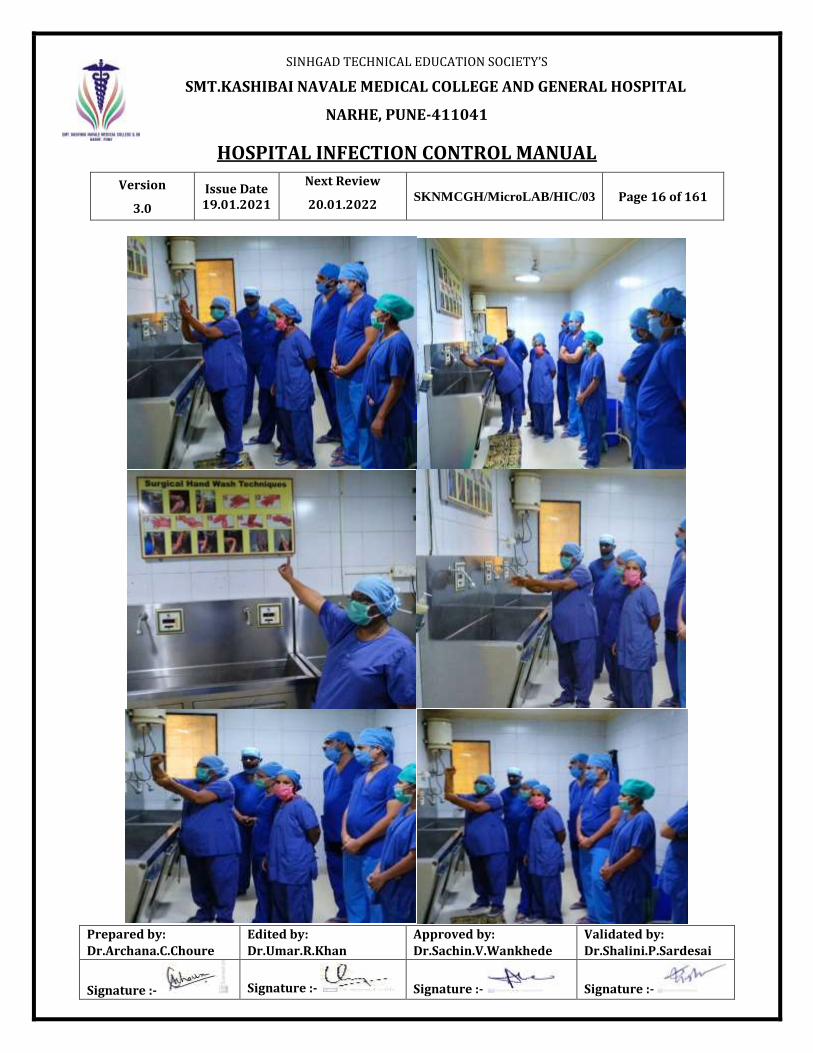
3.3.8 Surgical hand scrubbing: The aim of surgical hand scrubbing with an antiseptic agent is to
minimize the number of microorganisms on hands under the gloves. This reduces the risk
of infection to a client if gloves develop a small hole, tears or nicks during the procedure.
a. Remove all jewelry on hands and wrists.
b. Hold the hands above waist level and wet hands in water.
c. Apply sufficient antiseptic solution; use firm, circular motions to wash hands and arms up
to the wrists, covering all are as including palms, back of the hands, fingers, between
fingers, and lateral side of thumb, knuckles, and wrists for at least three to five minutes
d. Repeat the procedure twice.
e. Rinse both hands one-by-one and keeps the hands above waist level at all times.
f. Dry the hands with a sterile towel keeping them above waist level.
g. Do not touch anything except the gloves after washing hands for a surgical procedure.

SINHGAD TECHNICAL EDUCATION SOCIETY’S
SMT.KASHIBAI NAVALE MEDICAL COLLEGE AND GENERAL HOSPITAL
NARHE, PUNE-411041
HOSPITAL INFECTION CONTROL MANUAL
Version
3.0
Issue Date 19.01.2021
Next Review
20.01.2022 SKNMCGH/MicroLAB/HIC/03 Page 19 of 161
Prepared by:
Dr.Archana.C.Choure
Edited by:
Dr.Umar.R.Khan
Approved by:
Dr.Sachin.V.Wankhede
Validated by:
Dr.Shalini.P.Sardesai
Signature :- Signature :- Signature :- Signature :-
3.4 Personal Protective Equipment (PPE)
Personal protective equipment should be used by:
• Healthcareworkerswhoprovidedirectcaretopatientsandwhoworkinsituations where
they may have contact with blood, body fluids, excretions, and secretions.
• Support staff including medical aides, cleaners, and laundry staff in situations where
they may have contact with blood, body fluids, secretions, and excretions.
• Laboratory staff, who handle patient specimens.
• Familymemberswhoprovidecaretopatientsandareinasituationwheretheymay have
contact with blood, body fluids, secretions, and excretions.
Personal protective equipment includes:
• Gloves
• Protective eye wear (goggles)
• Mask
• Apron
• Gown
• Boots or shoe covers
• Cap or hair cover
3.4.1 Gown
a.Wear a gown that is appropriate to the task, to protect skin and prevents oiling or
contamination of clothing during procedures and patient care activities when
contact with blood, body fluids, secretions, or excretions is anticipated.
b.Wear a gown for direct patient contact if the patient has uncontained secretions or
excretions.

SINHGAD TECHNICAL EDUCATION SOCIETY’S
SMT.KASHIBAI NAVALE MEDICAL COLLEGE AND GENERAL HOSPITAL
NARHE, PUNE-411041
HOSPITAL INFECTION CONTROL MANUAL
Version
3.0
Issue Date 19.01.2021
Next Review
20.01.2022 SKNMCGH/MicroLAB/HIC/03 Page 20 of 161
Prepared by:
Dr.Archana.C.Choure
Edited by:
Dr.Umar.R.Khan
Approved by:
Dr.Sachin.V.Wankhede
Validated by:
Dr.Shalini.P.Sardesai
Signature :- Signature :- Signature :- Signature :-
c. Remove the gown and perform hand hygiene before leaving the patient’s
environment.
d. Do not reuse gowns, even for repeated contacts with the same patient.
e. Routine donning of a gown when entering a high-risk unit (for example, ICU,
NICU, unit) is not indicated.
3.4.2 Mouth, Nose, Eye Protection
a. Use PPE to protect the mucous membranes of the eyes, nose, and mouth during procedures and
patient care activities that are likely to generate splashes or sprays of blood, body fluids,
secretions and excretions. Select masks, goggles, face shields, and combinations of each
according to the need anticipated by the task performed
b.During aerosol-generating procedures (for example, bronchoscopy, suctioning of the
respiratory tract [if not using in-line suction catheters], endotracheal intubation) in patients
who are not suspected of being infected with an agent for which respiratory protection is
otherwise recommended (for example, M.tuberculosis, SARS or hemorrhagic fever viruses),
wear one of the following: a face shield that fully covers the front and sides of the face, mask
with attached shield, or a mask and goggles (in addition to gloves and gown).

SINHGAD TECHNICAL EDUCATION SOCIETY’S
SMT.KASHIBAI NAVALE MEDICAL COLLEGE AND GENERAL HOSPITAL
NARHE, PUNE-411041
HOSPITAL INFECTION CONTROL MANUAL
Version
3.0
Issue Date 19.01.2021
Next Review
20.01.2022 SKNMCGH/MicroLAB/HIC/03 Page 21 of 161
Prepared by:
Dr.Archana.C.Choure
Edited by:
Dr.Umar.R.Khan
Approved by:
Dr.Sachin.V.Wankhede
Validated by:
Dr.Shalini.P.Sardesai
Signature :- Signature :- Signature :- Signature :-

SINHGAD TECHNICAL EDUCATION SOCIETY’S
SMT.KASHIBAI NAVALE MEDICAL COLLEGE AND GENERAL HOSPITAL
NARHE, PUNE-411041
HOSPITAL INFECTION CONTROL MANUAL
Version
3.0
Issue Date 19.01.2021
Next Review
20.01.2022 SKNMCGH/MicroLAB/HIC/03 Page 22 of 161
Prepared by:
Dr.Archana.C.Choure
Edited by:
Dr.Umar.R.Khan
Approved by:
Dr.Sachin.V.Wankhede
Validated by:
Dr.Shalini.P.Sardesai
Signature :- Signature :- Signature :- Signature :-

SINHGAD TECHNICAL EDUCATION SOCIETY’S
SMT.KASHIBAI NAVALE MEDICAL COLLEGE AND GENERAL HOSPITAL
NARHE, PUNE-411041
HOSPITAL INFECTION CONTROL MANUAL
Version
3.0
Issue Date 19.01.2021
Next Review
20.01.2022 SKNMCGH/MicroLAB/HIC/03 Page 23 of 161
Prepared by:
Dr.Archana.C.Choure
Edited by:
Dr.Umar.R.Khan
Approved by:
Dr.Sachin.V.Wankhede
Validated by:
Dr.Shalini.P.Sardesai
Signature :- Signature :- Signature :- Signature :-
3.5 Guidelines for Collection of Blood Samples
Use gloves and take special care if there are cuts or scratches on the hands.
Take care to avoid contamination of hands and surrounding area with the blood.
3.5.1 Use disposable or autoclaved syringes and needles.
3.5.2 Use70percentethanolorisopropylalcoholswabsorspongesforcleaningthesiteof needle
puncture.
3.5.3 Usethickdressingpadsoradsorbentcottonbelowtheforearmwhendrawingbloodand
tourniquet above.
3.5.4 Tourniquet must be removed before the needle is withdrawn.
3.5.5 Place dry cotton swab and flex the elbow to keep the swab in place till bleeding stops.
3.5.6 Place used needles and syringes in a puncture-resistant container containing disinfectant.
3.5.7 Do not recap used needles.
3.6 Proper Disposal of Needles
3.6.1 Needles and sharps are the commonest mode of transmission of blood-borne pathogens to
the healthcare worker.
3.6.2 Precautions should be taken to prevent injuries by sharp instruments, especially hollow
bore needles that have been used for venipuncture or other vascular access procedures.
3.6.3 Needles should not be recapped, bent or broken by hand. Disposable needles and other
sharps should be disposed immediately after use into puncture-resistant containers which
should be located at the site of the procedure.
3.6.4 When a needle has to be removed from a syringe, do it with utmost care.
3.7 Good Practice for Safe Handling and Disposal of Sharps
3.7.1 ALWAYS dispose of your own sharps.

SINHGAD TECHNICAL EDUCATION SOCIETY’S
SMT.KASHIBAI NAVALE MEDICAL COLLEGE AND GENERAL HOSPITAL
NARHE, PUNE-411041
HOSPITAL INFECTION CONTROL MANUAL
Version
3.0
Issue Date 19.01.2021
Next Review
20.01.2022 SKNMCGH/MicroLAB/HIC/03 Page 24 of 161
Prepared by:
Dr.Archana.C.Choure
Edited by:
Dr.Umar.R.Khan
Approved by:
Dr.Sachin.V.Wankhede
Validated by:
Dr.Shalini.P.Sardesai
Signature :- Signature :- Signature :- Signature :-
3.7.2 NEVER pass used sharps directly from one person to another.
3.7.3 During exposure-prone procedures, the risk of injury should be minimized by ensuring
that the operator has the best possible visibility; for example, by positioning the patient,
adjusting the light source, and controlling bleeding.
3.7.4 Protect fingers from injury by using forceps instead of fingers for guiding suturing.
3.7.5 Locate sharps disposal containers close to the point of use, for example, in patient’s
room, on the medicine trolley, and in the treatment room.
3.7.6 Cardboard boxes with blue colored marking are used for sharps.

SINHGAD TECHNICAL EDUCATION SOCIETY’S
SMT.KASHIBAI NAVALE MEDICAL COLLEGE AND GENERAL HOSPITAL
NARHE, PUNE-411041
HOSPITAL INFECTION CONTROL MANUAL
Version
3.0
Issue Date 19.01.2021
Next Review
20.01.2022 SKNMCGH/MicroLAB/HIC/03 Page 25 of 161
Prepared by:
Dr.Archana.C.Choure
Edited by:
Dr.Umar.R.Khan
Approved by:
Dr.Sachin.V.Wankhede
Validated by:
Dr.Shalini.P.Sardesai
Signature :- Signature :- Signature :- Signature :-
4. ISOLATION POLICIES AND PROCEDURES
Isolation procedures are meant to prevent or interrupt transmission of pathogenic
microorganisms within the hospital. Selected patients may require specific precautions to limit
transmission of potential infecting organisms to other patients.
4.1 Recommended Isolation Precautions: Routes of Transmission
Microorganisms are transmitted by three main routes:
• Contact
• Air
• Droplet
In nosocomial infections, transmission by contact, droplet, and air plays a major role.
4.1.1 Infection by direct or indirect contact: Infection occurs through direct contact between the
source of infection and the recipient or indirectly through contaminated objects.
4.1.2 Air-borne infection: Infection usually occurs by the respiratory route, with the agent present
in aerosols (infectious particles less than 5 µmin diameter).
4.1.3 Droplet infection: Large droplets carry the infectious agent (greater than 5 µm in diameter).
4.2 Contact Precautions
These apply to patients with any of the following conditions and/or diseases:
4.2.1 Presence of stool incontinence (may include patients with norovirus, rotavirus, or
Clostridium difficile), draining wounds, uncontrolled secretions, pressure ulcers, or
presence of ostomy tubes and/or bags draining body fluids.
4.2.2 Presence of generalized rash or exanthemas.

SINHGAD TECHNICAL EDUCATION SOCIETY’S
SMT.KASHIBAI NAVALE MEDICAL COLLEGE AND GENERAL HOSPITAL
NARHE, PUNE-411041
HOSPITAL INFECTION CONTROL MANUAL
Version
3.0
Issue Date 19.01.2021
Next Review
20.01.2022 SKNMCGH/MicroLAB/HIC/03 Page 26 of 161
Prepared by:
Dr.Archana.C.Choure
Edited by:
Dr.Umar.R.Khan
Approved by:
Dr.Sachin.V.Wankhede
Validated by:
Dr.Shalini.P.Sardesai
Signature :- Signature :- Signature :- Signature :-
4.2.3 Prioritizeplacementofpatientsinanexaminationroomiftheyhavestoolincontinence, draining
wounds and/or skin lesions that cannot be covered, or uncontrolled secretions.
4.2.4 Perform hand hygiene before touching the patient and prior to wearing gloves. Also
perform hand hygiene after touching the patient and after removing gloves.
4.2.5 PPE use
a. Wear gloves when touching the patient and the patient’s immediate environment
or belongings.
b. Wearagownifsubstantialcontactwiththepatientorthepatient’senvironmentis
anticipated.
c. Perform hand hygiene after removal of PPE. Use soap and water when hands are
visibly soiled (for example, with blood, body fluids), or after caring for patients with
known or suspected infectious diarrhea (for example, Clostridium difficile,
norovirus).
d. Clean or disinfect the examination room accordingly.
e. Instructpatientswithknownorsuspectedinfectiousdiarrheatouseaseparate bathroom,
if available; clean or disinfect the bathroom before it can be used again.
f. IN ADDITION to Standard Precautions, use contact precautions for specified
patients known or suspected to be infected or colonized with epidemiologically
important microorganisms that can be transmitted by direct contact with the
patient or patient care items.
4.2.6 Patient placement
A single room is preferable. Cohort only with patients who are affected by the same
organism.
4.2.7 Patient transport

SINHGAD TECHNICAL EDUCATION SOCIETY’S
SMT.KASHIBAI NAVALE MEDICAL COLLEGE AND GENERAL HOSPITAL
NARHE, PUNE-411041
HOSPITAL INFECTION CONTROL MANUAL
Version
3.0
Issue Date 19.01.2021
Next Review
20.01.2022 SKNMCGH/MicroLAB/HIC/03 Page 27 of 161
Prepared by:
Dr.Archana.C.Choure
Edited by:
Dr.Umar.R.Khan
Approved by:
Dr.Sachin.V.Wankhede
Validated by:
Dr.Shalini.P.Sardesai
Signature :- Signature :- Signature :- Signature :-
Limit the movement and transport of the patient from the room for essential purposes
only. Where necessary ensure that adequate precautions are taken to minimize the risk of
transmission to others, and contamination of environmental surfaces or equipment.
4.2.8 Patient care equipment
Where possible dedicate the use of patient care equipment to a single patient. Otherwise,
ensure that all items are adequately cleaned or disinfected before use for another patient.

SINHGAD TECHNICAL EDUCATION SOCIETY’S
SMT.KASHIBAI NAVALE MEDICAL COLLEGE AND GENERAL HOSPITAL
NARHE, PUNE-411041
HOSPITAL INFECTION CONTROL MANUAL
Version
3.0
Issue Date 19.01.2021
Next Review
20.01.2022 SKNMCGH/MicroLAB/HIC/03 Page 28 of 161
Prepared by:
Dr.Archana.C.Choure
Edited by:
Dr.Umar.R.Khan
Approved by:
Dr.Sachin.V.Wankhede
Validated by:
Dr.Shalini.P.Sardesai
Signature :- Signature :- Signature :- Signature :-
4.3 Droplet Transmission
In the case of droplets (large particle droplets more than 5µm in size), the mechanism of transfer
of the organism is quite distinct from either direct or indirect contact transmission. Droplets are
generated from the patient primarily during coughing, sneezing, and during certain procedures such
as suctioning and bronchoscopy. Transmission occurs when droplets containing microorganisms
generated from the infected person are propelled a short distance through the air
And deposited on the host’s conjunctiva, nasal mucosa, or mouth. Because droplets do not remain
suspended in the air, special air handling and ventilation are not required.
4.3.1 Droplet precautions
These should be applied to patients know nor suspected to be infected with a pathogen that can be
transmitted by the droplet route. These precautions include, but are not limited to:
• Respiratory viruses (for example, influenza, parainfluenza virus, adenovirus,
respiratory syncytial virus, human metapneumovirus).
• Bordetella pertussis.
• For first 24 hours of therapy: Neisseria meningitides, group A streptococcus.
Place the patient in an examination room with a closed door as soon as possible (prioritize patients
who have excessive cough and sputum production); if an examination room is not available, the
patient should be provided a face mask and placed in a separate area as far from other patients as
possible while awaiting care.

SINHGAD TECHNICAL EDUCATION SOCIETY’S
SMT.KASHIBAI NAVALE MEDICAL COLLEGE AND GENERAL HOSPITAL
NARHE, PUNE-411041
HOSPITAL INFECTION CONTROL MANUAL
Version
3.0
Issue Date 19.01.2021
Next Review
20.01.2022 SKNMCGH/MicroLAB/HIC/03 Page 29 of 161
Prepared by:
Dr.Archana.C.Choure
Edited by:
Dr.Umar.R.Khan
Approved by:
Dr.Sachin.V.Wankhede
Validated by:
Dr.Shalini.P.Sardesai
Signature :- Signature :- Signature :- Signature :-
4.3.2 PPE use
a. Wear a face mask, such as a procedure or surgical mask, when in close contact with
the patient; don the face mask upon entering the examination room.
b. If substantial spraying of respiratory fluids is anticipated, gloves and gown as well
as goggles (or face shield in place of goggles) should be worn.
c. Perform hand hygiene before and after touching the patient and after contact with
respiratory secretions and contaminated objects or materials. Use soap and water when
hands are visibly soiled (for example, with blood, body fluids).

SINHGAD TECHNICAL EDUCATION SOCIETY’S
SMT.KASHIBAI NAVALE MEDICAL COLLEGE AND GENERAL HOSPITAL
NARHE, PUNE-411041
HOSPITAL INFECTION CONTROL MANUAL
Version
3.0
Issue Date 19.01.2021
Next Review
20.01.2022 SKNMCGH/MicroLAB/HIC/03 Page 30 of 161
Prepared by:
Dr.Archana.C.Choure
Edited by:
Dr.Umar.R.Khan
Approved by:
Dr.Sachin.V.Wankhede
Validated by:
Dr.Shalini.P.Sardesai
Signature :- Signature :- Signature :- Signature :-
d. Instruct the patient to wear a face mask when exiting the examination room, avoid
coming into close contact with other patients, and practice respiratory hygiene and cough etiquette.
e. Clean and disinfect the examination room accordingly (in addition to Standard
Precautions).
4.3.3 Patient placement
a. Single Room. Special air handling or ventilation is not necessary. Only cohort with
patient/patients who are infected with the same organism.
b. Mask. Wear a mask when working within three feet of a patient with
meningococcal meningitis.
c. Spacing between beds. In open wards there should be adequate spacing between
each bed to reduce the risk of cross-contamination or infection occurring from direct
or indirect contact or droplet transmission. Optimum spacing between beds is 1-2
meters.
4.3.4 Patient transport purposes only. If transport or movement is necessary minimize
dispersal of droplets from the patient.
4.4 Air-Borne Transmission
This occurs through dissemination of either air-borne droplet nuclei (small particle residue less
than 5µmin size) of evaporated droplets containing microorganisms that remain suspended in the
air for long periods of time, or dust particles containing the infectious agent. Microorganisms
carried in this manner can be dispersed widely by air currents and may be inhaled by a
susceptible host within the same room or over a longer distance from the source patient.
Microorganisms transmitted by air-borne transmission include mycobacterium tuberculosis,
measles, and the varicella virus.
4.4.1 Air-borne precautions

SINHGAD TECHNICAL EDUCATION SOCIETY’S
SMT.KASHIBAI NAVALE MEDICAL COLLEGE AND GENERAL HOSPITAL
NARHE, PUNE-411041
HOSPITAL INFECTION CONTROL MANUAL
Version
3.0
Issue Date 19.01.2021
Next Review
20.01.2022 SKNMCGH/MicroLAB/HIC/03 Page 31 of 161
Prepared by:
Dr.Archana.C.Choure
Edited by:
Dr.Umar.R.Khan
Approved by:
Dr.Sachin.V.Wankhede
Validated by:
Dr.Shalini.P.Sardesai
Signature :- Signature :- Signature :- Signature :-
Apply to patients known or suspected to be infected with a pathogen that may be transmitted by
the air-borne route; these include, but are not limited to:
• Tuberculosis
• Measles
• Chickenpox (until lesions are crusted over)
• Localized (in immunocompromised patient) or disseminated herpes zoster (until
lesions are crusted over)
a. Have the patient enter through a separate entrance to the facility (for example,
dedicated isolation entrance) if available, to avoid the reception and registration
area.
b. Place the patient immediately in an air-borne infection isolation room (AIIR).
c. If AIIR is not available, provide a face mask (for example, procedure or surgical
mask) to the patient and place the patient immediately in an examination room with
a closed door.
d. Initiate protocol to transfer patient to a healthcare facility that has the recommended
infection-control capacity to properly manage the patient
4.4.2 PPE use
a. If substantial spraying of respiratory fluids is anticipated, gloves and gown, as well
as goggles or face shield should be worn.
b. Perform hand hygiene before and after touching the patient and after contact with
respiratory secretions and/or body fluids and contaminated objects or materials. Use
soap and water when hands are visibly soiled (for example, with blood, body fluids).

SINHGAD TECHNICAL EDUCATION SOCIETY’S
SMT.KASHIBAI NAVALE MEDICAL COLLEGE AND GENERAL HOSPITAL
NARHE, PUNE-411041
HOSPITAL INFECTION CONTROL MANUAL
Version
3.0
Issue Date 19.01.2021
Next Review
20.01.2022 SKNMCGH/MicroLAB/HIC/03 Page 32 of 161
Prepared by:
Dr.Archana.C.Choure
Edited by:
Dr.Umar.R.Khan
Approved by:
Dr.Sachin.V.Wankhede
Validated by:
Dr.Shalini.P.Sardesai
Signature :- Signature :- Signature :- Signature :-
c. Instruct patient to wear a face mask when exiting the examination room, avoid
coming in close contact with other patients, and practice respiratory hygiene and
cough etiquette.
d. Once the patient leaves, the examination room should remain vacant for generally
one hour before anyone enters; however, adequate wait time may vary depending
on the ventilation rate of the room and should be determined accordingly.
e. If staff must enter the room during the wait time, they should use respiratory
protection (in addition to Standard Precautions).

SINHGAD TECHNICAL EDUCATION SOCIETY’S
SMT.KASHIBAI NAVALE MEDICAL COLLEGE AND GENERAL HOSPITAL
NARHE, PUNE-411041
HOSPITAL INFECTION CONTROL MANUAL
Version
3.0
Issue Date 19.01.2021
Next Review
20.01.2022 SKNMCGH/MicroLAB/HIC/03 Page 33 of 161
Prepared by:
Dr.Archana.C.Choure
Edited by:
Dr.Umar.R.Khan
Approved by:
Dr.Sachin.V.Wankhede
Validated by:
Dr.Shalini.P.Sardesai
Signature :- Signature :- Signature :- Signature :-
4.4.3 Patient Placement
a. Single room. Negative air pressure.
b. Self-closing devices on doors to keep the door closed.
c. Ventilation system should provide a means to discharge air from the room to the
outside, such as an exhaust fan. Exhaust fan should be on emergency power.
d. Ensure that all doors and windows remain properly closed in the isolation room.
The slit at the bottom of the door is sufficient to provide a controlled airflow path.
e. The TB isolation room needs to be checked for negative pressure.
f. Tissues Test to check negative pressure: A thin strip of tissue should be held parallel
to the door with one end of the tissue in front of the gap. The direction of the tissue’s
movement will indicate the direction of air movement.
4.4.4 Respiratory Protection
a. Heavy duty N95 or N97 masks should be used for Open Pulmonary Tuberculosis
or suspected Pulmonary Tuberculosis, Surgical Mask for Meningococcal or
suspected Meningococcal Meningitis.
b. Non-immune or pregnant staff should not enter the room of patients known or
suspected to have rubella or varicella. Persons with immunity to varicella and
rubella do not require masks.
4.4.5 Patient Transport
a. Limit movement or transport of patient from the room to essential purposes only.
b. If transport or movement is necessary, minimize patient dispersal of organisms.

SINHGAD TECHNICAL EDUCATION SOCIETY’S
SMT.KASHIBAI NAVALE MEDICAL COLLEGE AND GENERAL HOSPITAL
NARHE, PUNE-411041
HOSPITAL INFECTION CONTROL MANUAL
Version
3.0
Issue Date 19.01.2021
Next Review
20.01.2022 SKNMCGH/MicroLAB/HIC/03 Page 34 of 161
Prepared by:
Dr.Archana.C.Choure
Edited by:
Dr.Umar.R.Khan
Approved by:
Dr.Sachin.V.Wankhede
Validated by:
Dr.Shalini.P.Sardesai
Signature :- Signature :- Signature :- Signature :-
4.5 Isolation Policy for Special Groups of Organisms
4.5.1 MRSA (Methicillin Resistant Staphylococcus Aureus) / VRE (Vancomycin Resistant
Enterococcus). The following procedures should be followed in addition to Standard
Precautions:
a. Isolate the MRSA/VRE positive patient under Contact Isolation with mask category.
Accommodate such patients away from those with open wounds or those who are
immunocompromised.
b. Hand washing is the single most important factor in containing MRSA.
c. The bed used by the patient, and other equipment used for the patient should be disinfected
before use for another patient.
d. Disinfection procedures should be carried out on a daily basis, as out lined under
Isolation Procedures.
e. Linen: Sheets, pillow cases, and blankets should be changed on a daily basis and more often
if soiling occurs. Linen should not be shaken in order to prevent
dissemination of microorganisms to the environment. The same applies to masks, gowns
and gloves. Soiled linen should be placed in a laundry bag in the patient’s room or at the
location where it was used. It should be placed in bags that prevent leakage.
f. Disposable dishes and utensils used for eating are not required for patients in isolation.
Reusable dishes may be used for patients in isolation, because the combination of
dishwasher detergents and high water temperature adequately decontaminates dishes.
g. Procedures for decolonization of the patient which include daily bath with an antimicrobial
soap should be followed.
h. When the patient is discharged, terminal disinfection should be carried out as
outlined under Isolation Procedures.

SINHGAD TECHNICAL EDUCATION SOCIETY’S
SMT.KASHIBAI NAVALE MEDICAL COLLEGE AND GENERAL HOSPITAL
NARHE, PUNE-411041
HOSPITAL INFECTION CONTROL MANUAL
Version
3.0
Issue Date 19.01.2021
Next Review
20.01.2022 SKNMCGH/MicroLAB/HIC/03 Page 35 of 161
Prepared by:
Dr.Archana.C.Choure
Edited by:
Dr.Umar.R.Khan
Approved by:
Dr.Sachin.V.Wankhede
Validated by:
Dr.Shalini.P.Sardesai
Signature :- Signature :- Signature :- Signature :-
DURATIONOF CONTACT PRECAUTIONS
For patients, colonized or infected with microorganisms like MRSA or VRE, three negative
cultures taken one week apart can be used to discontinue contact precautions. In other patients,
resolution of symptoms that lead to the isolation (such as diarrhoea in the case of C.
Difficile infection) may be a reasonable time to stop theisolation.
4.5.2 Tuberculosis
a. Respiratory precautions should be taken for smear-positive pulmonary
tuberculosis.
b. A separate room is recommended for such patients.
c. Elective surgery for patients with active TB infection is recommended.
Elective operative procedures on patients with active pulmonary or laryngeal TB should be
postponed until the patient is no longer infectious.
4.6 Visitor's Policy When Patient is in Isolation
4.6.1 The ward sisters and doctors concerned have the responsibility of informing the patients’
relatives of the measures to be taken and the importance of restriction of visitors.
4.6.2 The patient and there relatives must be given health education about the cause, spread, and
prevention of the infection in detail. The need for isolation and restriction of visitors should
be discussed with them.
4.6.3 Hand washing after all contact with the patient has to be stressed.
4.6.4 Visitors need to wear a N95 respirator. Be aware of restrictions on visitation due to outbreak
or other conditions within the facility.
4.6.5 No more than two adult visitors should be allowed at a time during the hospital visiting
hours and the length of stay should be governed by the needs of the patient.
4.6.6 Children below 12 years of age should not be allowed into isolation areas.

SINHGAD TECHNICAL EDUCATION SOCIETY’S
SMT.KASHIBAI NAVALE MEDICAL COLLEGE AND GENERAL HOSPITAL
NARHE, PUNE-411041
HOSPITAL INFECTION CONTROL MANUAL
Version
3.0
Issue Date 19.01.2021
Next Review
20.01.2022 SKNMCGH/MicroLAB/HIC/03 Page 36 of 161
Prepared by:
Dr.Archana.C.Choure
Edited by:
Dr.Umar.R.Khan
Approved by:
Dr.Sachin.V.Wankhede
Validated by:
Dr.Shalini.P.Sardesai
Signature :- Signature :- Signature :- Signature :-
• Visitors’ footwear, bags, and other belongings should be left outside the room.
• Visitors should not be allowed to sit on the patient’s bed.
• Visitors should wash their hands well with soap and water before entering and
when leaving the room.
• Any prophylactic medication or active immunization for attendants should be
conducted by the physician in charge.
5. DISINFECTION AND STERILIZATION
5.1 Sterilization
5.1.1 Sterilization is defined as a process where all microbes are removed from a defined
object, inclusive of bacterial endospores.
5.1.2 Methods of Sterilization Used
i. Steam autoclave
ii. H o t air oven
STERILIZATION RECOMMENDATIONS
Hot Air Oven 160 0C for 1 hr, 180
0C for 30 min
Autoclave Gravity-Displacement:
• 15 min holding time at 121 0C
• 1.1 kg/cm2
or 15 lb/in2(PSI)
Pre-vacuum:
• 3 min holding time at 134 0
C
• 2.2 kg/cm2
or 32 lb/in2 (PSI)

SINHGAD TECHNICAL EDUCATION SOCIETY’S
SMT.KASHIBAI NAVALE MEDICAL COLLEGE AND GENERAL HOSPITAL
NARHE, PUNE-411041
HOSPITAL INFECTION CONTROL MANUAL
Version
3.0
Issue Date 19.01.2021
Next Review
20.01.2022 SKNMCGH/MicroLAB/HIC/03 Page 37 of 161
Prepared by:
Dr.Archana.C.Choure
Edited by:
Dr.Umar.R.Khan
Approved by:
Dr.Sachin.V.Wankhede
Validated by:
Dr.Shalini.P.Sardesai
Signature :- Signature :- Signature :- Signature :-
5.2 Disinfection
Disinfection is a process where most microbes are removed from a defined object or surface, except
bacterial endospores.
Disinfectants maybe classified according to their ability to destroy different categories of
microorganisms. The agent which destroys only vegetative bacteria is termed a low level
disinfectant. If the agent is capable of rendering mycobacteria nonviable, it is termed as an
intermediate level disinfectant. It is safe to assume that all the other categories of microbes which
are classified more susceptible are also destroyed if efficacy against mycobacteria can be
demonstrated. High level disinfection is in other words sterilization where in all microbial
life is destroyed inclusive of endospores.

SINHGAD TECHNICAL EDUCATION SOCIETY’S
SMT.KASHIBAI NAVALE MEDICAL COLLEGE AND GENERAL HOSPITAL
NARHE, PUNE-411041
HOSPITAL INFECTION CONTROL MANUAL
Version
3.0
Issue Date 19.01.2021
Next Review
20.01.2022 SKNMCGH/MicroLAB/HIC/03 Page 38 of 161
Prepared by:
Dr.Archana.C.Choure
Edited by:
Dr.Umar.R.Khan
Approved by:
Dr.Sachin.V.Wankhede
Validated by:
Dr.Shalini.P.Sardesai
Signature :- Signature :- Signature :- Signature :-
Classification of disinfectants:
5.2.1 High level disinfectants: glutaraldehyde 2 percent, ethylene oxide.
5.2.1 Intermediate level disinfectants: alcohols, chlorine compounds, hydrogen peroxide,
chlorhexidine, glutaraldehyde (short-term exposure).
5.2.2 Low level disinfectants: benzalkonium chloride, some soaps.
5.3 General Equipment
Disinfection of General Equipment
Equipment Frequency of Change Recommendation
Oral Thermometer
Single for all IPD patients
After each use, the thermometer is
disinfected by wiping with a swab
saturated with 70 percent isopropyl
alcohol.
For OPD: Each thermometer is kept in a
separate holder. After each out patient
session, the thermometer holder is
washed in warm water and detergent, and
the thermometer is disinfected in 70
percent alcohol for 5 minutes.
Other methods for thermometer:
immersing in glutaraldehyde, or
hexachlorophene and cetrimide for at
least10 minutes
Sphygmomanometer Cuffs
As required
Change covers regularly(1perweek) and
wash in flat able section in detergent and
water, dry thoroughly
Or use 70 percent alcohol.
Change after each use in infected
patients.

SINHGAD TECHNICAL EDUCATION SOCIETY’S
SMT.KASHIBAI NAVALE MEDICAL COLLEGE AND GENERAL HOSPITAL
NARHE, PUNE-411041
HOSPITAL INFECTION CONTROL MANUAL
Version
3.0
Issue Date 19.01.2021
Next Review
20.01.2022 SKNMCGH/MicroLAB/HIC/03 Page 39 of 161
Prepared by:
Dr.Archana.C.Choure
Edited by:
Dr.Umar.R.Khan
Approved by:
Dr.Sachin.V.Wankhede
Validated by:
Dr.Shalini.P.Sardesai
Signature :- Signature :- Signature :- Signature :-
Rectal Thermometer
After each patient
Thoroughly wash with detergent and
water, then dry. Store dry and
separately from oral thermometers.
Disinfectwith70percentalcoholfor
5minutes.
Auriscope
After each patient
Disposable earpieces should be used
where possible; when not available
clean in detergent and water.
DisinfectinCSSDor70percent
alcoholfor5minutes.
Earpieces After each patient Wash with hot water and detergent,
store dry. Disinfection CSSD or 70
percent alcohol for 5 minutes.
Patients having(preop) After each patient Use disposable OR shaver blade, not
a razor.

SINHGAD TECHNICAL EDUCATION SOCIETY’S
SMT.KASHIBAI NAVALE MEDICAL COLLEGE AND GENERAL HOSPITAL
NARHE, PUNE-411041
HOSPITAL INFECTION CONTROL MANUAL
Version
3.0
Issue Date 19.01.2021
Next Review
20.01.2022 SKNMCGH/MicroLAB/HIC/03 Page 40 of 161
Prepared by:
Dr.Archana.C.Choure
Edited by:
Dr.Umar.R.Khan
Approved by:
Dr.Sachin.V.Wankhede
Validated by:
Dr.Shalini.P.Sardesai
Signature :- Signature :- Signature :- Signature :-
Equipment Recommendation
Bed ends and frames, Bedside
locker, Cardiac table, Baby
bassinets
Mopwith1percentsodiumhypochlorite.Allowtodry.
Bowls-Bedpans/Urinals Heat disinfection in a rinse temperature of minimum 82°C for 2
minutes. If not possible, bed pans, urine pots, and kidney trays should
be kept in 7 percent Lysol for 24 hours or 3-5 percent sodium
hypochlorite solution for 30 minutes; then they are washed with soap
and water and dried in sunlight.
Bowls(washing) Clean with detergent and water and store dry or as above.
Hand Basins Clean with detergent and water.
Cleaning cloths, Brushes, and
Equipment
Supplied daily from the laundry. They are provided for use and then
discarded to wash.
Wash brushes and buckets in detergent and water, then hang or invert
to dry, then store dry.
Disposable cloths are also available.
Curtain Rails As for bed ends.
Lockers Detergent and water as necessary and after patient discharge.

SINHGAD TECHNICAL EDUCATION SOCIETY’S
SMT.KASHIBAI NAVALE MEDICAL COLLEGE AND GENERAL HOSPITAL
NARHE, PUNE-411041
HOSPITAL INFECTION CONTROL MANUAL
Version
3.0
Issue Date 19.01.2021
Next Review
20.01.2022 SKNMCGH/MicroLAB/HIC/03 Page 41 of 161
Prepared by:
Dr.Archana.C.Choure
Edited by:
Dr.Umar.R.Khan
Approved by:
Dr.Sachin.V.Wankhede
Validated by:
Dr.Shalini.P.Sardesai
Signature :- Signature :- Signature :- Signature :-
Mattresses and Pillows All should be covered with an impervious plastic cover and should be
wiped over with detergent and water if visibly contaminated.
Mattresses should be cleaned regularly, and if contaminated, with the
covers removed. If possible keep in sunlight for 24 hours.
Plastic and rubber covers of mattresses and pillows should be washed
with soap and water, cleaned with a suitable disinfectant, for example,
7 percent Lysol.
Mop Heads Daily cleaning of mops. At the completion of each task of floor
mopping, them mops should be thoroughly washed in a bucket
containing HOT water and detergent. Squeeze as much water out of
mop as possible and shake strands loose; leave hanging to dry in the
sun if possible, or alternatively, in the cleaner’s room. The bucket
should be turned upside down to allow overnight drainage.
Detachable mop heads should be sent to the laundry, while reusable
mops should be cleaned in hot soapy water, then left to dry ideally in
the sun.
Nail Brushes The use of nail brushes is discouraged as they may cause skin damage
that may result in increase in bacterial flora.
If a nail brush is required, a sterile, antiseptic impregnated brush may
be used. Reusable brushes require autoclaving between uses.
Toilet Bowls At least daily brushing with a commercial bowl cleanser. Additional
cleaning as necessary for stubborn stains.
Toilet Brushes Should be rinsed in flushing water, and stored to dry.
Walls Remove visible soiling with detergent as necessary.
Clinic Trolleys Clean with a cloth dampened with detergent and water.

SINHGAD TECHNICAL EDUCATION SOCIETY’S
SMT.KASHIBAI NAVALE MEDICAL COLLEGE AND GENERAL HOSPITAL
NARHE, PUNE-411041
HOSPITAL INFECTION CONTROL MANUAL
Version
3.0
Issue Date 19.01.2021
Next Review
20.01.2022 SKNMCGH/MicroLAB/HIC/03 Page 42 of 161
Prepared by:
Dr.Archana.C.Choure
Edited by:
Dr.Umar.R.Khan
Approved by:
Dr.Sachin.V.Wankhede
Validated by:
Dr.Shalini.P.Sardesai
Signature :- Signature :- Signature :- Signature :-
Ampoules/vials Wipe neck(ampoule) or top surface of rubber cap(vials)with a 70
percent isopropyl alcohol impregnated swab and allow to dry before
opening or piercing.
Cardiac monitors,
Defibrillators and ECG
equipment
If patient contact, then surface is cleaned and disinfected.
Fixtures and fittings In clinical areas wipe damp, dust daily with detergent solution.
In known contaminated and special areas, wipe damp dust with a
disinfectant solution.
Furniture and ledges In clinical areas clean damp dust daily with warm water and detergent.
Disinfection of Specialist Outpatient Equipment:
A toothbrush may be used for cleaning the instruments. Workers should be asked to wear
utility gloves while cleaning instruments.
5.4 Decontamination
The objective of decontamination is to protect individuals who handle surgical instruments and
other items which have been in contact with blood or body fluids, from serious diseases. Once
instruments and other items have been decontaminated, they can be safely further processed. This
consists of cleaning and finally either sterilization or high-level disinfection.
5.4.1 Decontamination Tips: Use a plastic container for decontamination to help
prevent:
• Dulling of sharps (for example, scissors) due to contact with metal containers.
• Rusting of instruments due to a chemical reaction (electrolysis) that can occur
between two different metals when placed in water.

SINHGAD TECHNICAL EDUCATION SOCIETY’S
SMT.KASHIBAI NAVALE MEDICAL COLLEGE AND GENERAL HOSPITAL
NARHE, PUNE-411041
HOSPITAL INFECTION CONTROL MANUAL
Version
3.0
Issue Date 19.01.2021
Next Review
20.01.2022 SKNMCGH/MicroLAB/HIC/03 Page 43 of 161
Prepared by:
Dr.Archana.C.Choure
Edited by:
Dr.Umar.R.Khan
Approved by:
Dr.Sachin.V.Wankhede
Validated by:
Dr.Shalini.P.Sardesai
Signature :- Signature :- Signature :- Signature :-
• Do not soak metal instruments that are electroplated (that is, not100 percent
stainless steel) even in plain water for more than an hour because rusting will occur.
5.4.2 How to prepare a disinfectant cleaning solution: A disinfectant cleaning solution is one that
contains both a disinfectant and a detergent (soap).
5.4.3 Precautions when using chlorine solutions: Although chlorine-containing solutions (sodium
hypochlorite) are excellent, inexpensive disinfectants, they should NOT be mixed with cleaning
solutions containing an acid (for example, phosphoric acid), ammonia or ammonium
chloride(NH2Cl). Doing this will release chlorine gas and other by-products that can result in
temporary illness (nausea, tearing, headache or shortness of breath) to staff breathing fumes in a
poorly ventilated area.NOTE:Tofindoutifacleaningsolutioncontainsammonia,firstcheckthelabel.Ifit
does not say there is ammonia, you may be able to detect ammonia when opening the product by its
pungent, burning smell.
If you are exposed to chlorine gas or ammonium chloride or other unpleasant (noxious) gases with
strong odors, leave the room or area immediately until the room can be completely ventilated.5.4.3
Instructions
Step 1: Prepare a 0.5 percent chlorine solution from liquid concentrates or from chlorine
compounds.
Step 2: Add enough detergent to the 0.5 percent chlorine solution or other disinfectants to
make a mild, soapy cleaning solution.
5.4.4 After decontamination, instruments should be rinsed immediately with cool water to remove
visible organic material before being thoroughly cleaned. For example, some healthcare facilities
now keep two buckets in the procedure areas or operating rooms, one filled with 0.5 percent
chlorine solution and one with water, so that the instruments can be placed in the water after
soaking in the chlorine solution for 10 minutes. Although this will help to prevent corrosion, even
leaving the instruments in plain water for more than 1 hour can lead to rusting.

SINHGAD TECHNICAL EDUCATION SOCIETY’S
SMT.KASHIBAI NAVALE MEDICAL COLLEGE AND GENERAL HOSPITAL
NARHE, PUNE-411041
HOSPITAL INFECTION CONTROL MANUAL
Version
3.0
Issue Date 19.01.2021
Next Review
20.01.2022 SKNMCGH/MicroLAB/HIC/03 Page 44 of 161
Prepared by:
Dr.Archana.C.Choure
Edited by:
Dr.Umar.R.Khan
Approved by:
Dr.Sachin.V.Wankhede
Validated by:
Dr.Shalini.P.Sardesai
Signature :- Signature :- Signature :- Signature :-
WHO recommends 0.5 percent chlorine solution to be used for decontaminating instruments before
cleaning them. The objective of decontamination is to protect individuals who handle surgical
instruments and other items which have been in contact with blood or body fluids, from serious
diseases. Once instruments and other items have been decontaminated, they can safely be further
processed. This consists of cleaning and finally either sterilization or high-level disinfection.
5.4.5Disinfection of endoscopes: Glutaraldehyde 2% (cidex) solution is effective high level
disinfectant for flexible endoscopes& other devices which cannot withstand high
temperature methods of sterilization. Soak instruments in cidex for 20 min to achieve high
level disinfection.
Precautions:
Keep away from eyes.
Wear gloves while preparing the solution.
Avoid skin contact.
Use in well ventilated area.
Add 100ml of activator solution to 5 liter of glutaraldehyde.
Once prepared it can be used for 14 days.
5.5 Fogging
5.5.1 Fogging is done by using 0.25%Virkon(10g m in4literofwater) by fogger method for 20
minutes. The room must be kept closed for 12 hours before use by housekeeping personnel.
5.5.2 Fogging is done only in the high-risk areas like OTs, ICU, PICU, NICU, Labor room.
wards are excluded for fogging (done only if required).
5.5.3 Frequency of fumigation in all OTs is weekly and in all ICUs is once in three months.

SINHGAD TECHNICAL EDUCATION SOCIETY’S
SMT.KASHIBAI NAVALE MEDICAL COLLEGE AND GENERAL HOSPITAL
NARHE, PUNE-411041
HOSPITAL INFECTION CONTROL MANUAL
Version
3.0
Issue Date 19.01.2021
Next Review
20.01.2022 SKNMCGH/MicroLAB/HIC/03 Page 45 of 161
Prepared by:
Dr.Archana.C.Choure
Edited by:
Dr.Umar.R.Khan
Approved by:
Dr.Sachin.V.Wankhede
Validated by:
Dr.Shalini.P.Sardesai
Signature :- Signature :- Signature :- Signature :-
5.5.4 1%Virkon (10gm in 1 lit water) is used for surface cleaning of OT and in-between the
operative cases.
5.5.5 1% Virkon is used for carbolization of OT material (OT lights, OT table, Trolleys, OT
pendant, ICU pendant & ICU beds)
Application
Surface Disinfection & Cleaning Floors; Walls; Ceiling
Disinfection Of Equipment
(Carbolisation)
OT Light, OT Table, Trolleys,
Pendant, ICU Beds; ICU Pendant;
Fogging Aerial Fogging
Dilution & Preparation
AREA CONCENTRATION DILUTION
For Surface Disinfection
& Cleaning
1 % Concentration Mix 10gram (1 Spoon) of
RelyOnVirkon Powder in 1 Litre of
Tap Water
Recomanded for critical area.
For Surface Disinfection
& Cleaning
0.5 % Concentration Mix 10gram (1 Spoon) of
RelyOnVirkon Powder in 2 Litre of
Tap Water
Recomanded for non-critical area.
For Equipment
Disinfection
1 % Concentration Mix 10gram (1 Spoon) of
RelyOnVirkon Powder in 1 Litre of
Tap Water for critical area.
For Equipment
Disinfection
0.5 % Concentration Mix 10gram (1 Spoon) of
RelyOnVirkon Powder in 2 Litre of
Tap Water for non-critical area.
For Fogging 0.25% Concentration Mix 10gram (1 Spoon) of
RelyOnVirkon Powder in 4 Litre of
Tap Water
After Fogging close the door for 45 minutes. After 45 Minutes you can use OT, ICU,
Wards, Cath Labs, etc where the area has been fogged.

SINHGAD TECHNICAL EDUCATION SOCIETY’S
SMT.KASHIBAI NAVALE MEDICAL COLLEGE AND GENERAL HOSPITAL
NARHE, PUNE-411041
HOSPITAL INFECTION CONTROL MANUAL
Version
3.0
Issue Date 19.01.2021
Next Review
20.01.2022 SKNMCGH/MicroLAB/HIC/03 Page 46 of 161
Prepared by:
Dr.Archana.C.Choure
Edited by:
Dr.Umar.R.Khan
Approved by:
Dr.Sachin.V.Wankhede
Validated by:
Dr.Shalini.P.Sardesai
Signature :- Signature :- Signature :- Signature :-
6. LAUNDRY SERVICES
Soiled linen can be a source of large amounts of microbial contamination which may cause
infections in hospital patients and personnel. In addition, improperly processed linen can cause
chemical reactions or dermatitis in those who come in contact with the linen. A hospital’s linen
service should process soiled linen such that the risk of disease to patients who may be unusually
susceptible or to employees, who may handle linen, is avoided. Adequate procedures for collecting,
transporting, processing, and storing linen should therefore be established.
Washing with hot water and detergent has been shown to result inadequate cleaning of laundry. If
needed for other reasons, bleach or ironing will reduce microbial contamination. Textile softeners
added in the final rinse, though of no value in preventing infections, make linen easier to handle
and rewash, and reduce lint.
6.1 Principles and Key Steps in Processing Linen
6.1.1 HousekeepingandlaundrypersonnelshouldwearglovesandotherPPEasindicated when
collecting, handling, transporting, sorting, and washing soiled linen.
6.1.2 When collecting and transporting soiled linen, handle it as little as possible and with
minimum contact to avoid accidental injury and spreading of microorganisms.
6.1.3 Consider all cloth items (for example, surgical drapes, gowns, wrappers) used during a
procedure as infectious. Even if there is no visible contamination, the item must be
laundered.
6.1.4 Carry soiled linen in covered containers or plastic bags to prevent spills and splashes, and
confine the soiled linen to designated areas (interim storage area) until transported to the
laundry.
6.1.5 Carefully sort all linen in the laundry area before washing.

SINHGAD TECHNICAL EDUCATION SOCIETY’S
SMT.KASHIBAI NAVALE MEDICAL COLLEGE AND GENERAL HOSPITAL
NARHE, PUNE-411041
HOSPITAL INFECTION CONTROL MANUAL
Version
3.0
Issue Date 19.01.2021
Next Review
20.01.2022 SKNMCGH/MicroLAB/HIC/03 Page 47 of 161
Prepared by:
Dr.Archana.C.Choure
Edited by:
Dr.Umar.R.Khan
Approved by:
Dr.Sachin.V.Wankhede
Validated by:
Dr.Shalini.P.Sardesai
Signature :- Signature :- Signature :- Signature :-
6.2 Recommended PPE for Processing Linen
6.2.1 Gloves (preferably household utility gloves) and closed shoes that protect feet from
dropped items (sharps) and spilled blood and body fluids, should be used when:
• Handling disinfectant solutions
• Collecting and handling soiled linen
• Transporting soiled linen
• Sorting soiled linen
• Hand washing soiled linen
• Loading automatic washers
6.2.2 Plastic or rubber apron and protective eyewear should be worn when
• Sorting soiled linen
• Hand washing soiled linen
• Loading automatic washers
6.3 Sorting Soiled Linen
6.3.1 Soiled linen should be handled as little as possible with a minimum amount of agitation to
prevent gross microbial contamination of the air and of personnel handling the linen.
6.3.2 The processing area for soiled linen must be separate from other areas such as those used
for folding and storing clean linen, patient care areas, and food preparation areas.
6.3.3 In addition, there should be adequate ventilation and physical barriers (walls) between the
clean and soiled linen areas.
6.3.4 Safe sorting of linen is extremely important. Sorting must be carefully performed because
soiled linen (large drapes and towel drapes) from the operating room or other procedure
areas, frequently contain sharps (for example, scalpels, sharp-tipped scissors, hypodermic
and suture needles, and sharp-tipped towel clips). In addition, bedding from patients’ rooms
may contain soiled dressings and be blood-stained or wet with other body fluids.

SINHGAD TECHNICAL EDUCATION SOCIETY’S
SMT.KASHIBAI NAVALE MEDICAL COLLEGE AND GENERAL HOSPITAL
NARHE, PUNE-411041
HOSPITAL INFECTION CONTROL MANUAL
Version
3.0
Issue Date 19.01.2021
Next Review
20.01.2022 SKNMCGH/MicroLAB/HIC/03 Page 48 of 161
Prepared by:
Dr.Archana.C.Choure
Edited by:
Dr.Umar.R.Khan
Approved by:
Dr.Sachin.V.Wankhede
Validated by:
Dr.Shalini.P.Sardesai
Signature :- Signature :- Signature :- Signature :-
6.3.5 Soiled linen and items containing sharps must be handled carefully by wearing protective
gloves, protective eyewear and plastic or rubber apron, and should be disposed of
properly. Though infrequent, infections related to sorting have been attributed to failure
of hand washing and proper use of PPE.
REMEMBER: Disposable sharps (suture needles, scalpel blades, and hypodermic needles)
must be placed in sharps containers located near the point of use.
Soiled linen may also contain non-infectious items such as dentures, eye glasses, and
hearing aids. These items pose no threat of infection and require no special handling.

SINHGAD TECHNICAL EDUCATION SOCIETY’S
SMT.KASHIBAI NAVALE MEDICAL COLLEGE AND GENERAL HOSPITAL
NARHE, PUNE-411041
HOSPITAL INFECTION CONTROL MANUAL
Version
3.0
Issue Date 19.01.2021
Next Review
20.01.2022 SKNMCGH/MicroLAB/HIC/03 Page 49 of 161
Prepared by:
Dr.Archana.C.Choure
Edited by:
Dr.Umar.R.Khan
Approved by:
Dr.Sachin.V.Wankhede
Validated by:
Dr.Shalini.P.Sardesai
Signature :- Signature :- Signature :- Signature :-
6.4 Laundering Linen
6.4.1 Washing and Drying
a. All linen items (for example, bedsheets, surgical drapes, masks, gowns) used in the
direct care of a patient must be thoroughly washed before reuse.
b. Decontamination prior to washing is NOT NECESSARY; unless linen is heavily
soiled and will be hand washed (repeated soaking of linen in chlorine, even dilute
solutions, will cause the fabric to deteriorate more quickly).
c. In addition, workers should not carry wet, soiled linen close to their bodies even if
they are wearing a plastic or rubber apron.
REMEMBER: The storage time for soiled linen before washing is related to practical issues,
such as available storage space and aesthetics, NOT to infection prevention concerns.
6.4.2 Washing Linen by Hand
Step 1: Wash heavily soiled linen separately from no soiled linen.
Step2: Wash the entire item in water with liquid soap to remove all soilage, even
if not visible.
REMEMBER: Pre-soak in soap, water and bleach ONLY if linen is heavily soiled.
• Use warm water if available.
• Add bleach(forexample,30–60ml[about2–3tablespoons],ofa5percent
chlorine solution) to aid cleaning and bactericidal action.
• Add sour (a mild acid agent) to prevent yellowing of
linen, if desirable.
Step 3: Check the item for cleanliness. Rewash if dirty or stained.
Step 4: Rinse the item with clean water.
6.4.3 Machine Washing

SINHGAD TECHNICAL EDUCATION SOCIETY’S
SMT.KASHIBAI NAVALE MEDICAL COLLEGE AND GENERAL HOSPITAL
NARHE, PUNE-411041
HOSPITAL INFECTION CONTROL MANUAL
Version
3.0
Issue Date 19.01.2021
Next Review
20.01.2022 SKNMCGH/MicroLAB/HIC/03 Page 50 of 161
Prepared by:
Dr.Archana.C.Choure
Edited by:
Dr.Umar.R.Khan
Approved by:
Dr.Sachin.V.Wankhede
Validated by:
Dr.Shalini.P.Sardesai
Signature :- Signature :- Signature :- Signature :-
Step 1: Wash heavily soiled linen separately from no soiled linen.
Step 2: Adjust the temperature and time cycle of the machine according to
manufacturer’sinstructionsandthetypeofsoaporotherwashingproductbeingused.
Bothcoldandhotwaterwashingcyclesthatincludebleachreducebacterialcountsin the linen.
Hot water washing:
a. Use hot water above 71ºC (160 F) and soap to aid in loosening soil.
b. Washinglinenat80-90oCforover20minuteswithadetergentinwaterisan effective
method for cleaning and killing most vegetative bacteria.
c. Add bleach.
d. Adjust the time cycle of the machine according to the manufacturer’s
instructions.
Step 3: When the wash cycle is complete, check the linen for cleanliness. Rewash if it is
dirty or stained. (Heavily soiled linen may require two wash cycles.)
NOTE: Uniforms and scrub suits or gowns worn by housekeeping or cleaning staff can be
safely laundered at home (that is, home laundering does not increase the risk of infection to
patients or staff).
Lower temperatures or cold water washing are satisfactory if the cleaning products (type of
soap or detergent, amount of bleach and other additives) are appropriate and used in proper
concentrations. Using cold water also saves energy.
6.5 Processing Linen
6.5.1 Drying, Checking and Folding Linen. For both hand and machine washed linens, the
steps are the same.
Step 1: Completely air or machine dry before further processing. Airdry in direct
sunlight, if possible, keeping the fabric off the ground, away from dust and moisture.
Step 2: After linen items are totally dry, check for holes and threadbare areas. If these
are present, the item must be discarded or repaired before reuse or storage. (If there are

SINHGAD TECHNICAL EDUCATION SOCIETY’S
SMT.KASHIBAI NAVALE MEDICAL COLLEGE AND GENERAL HOSPITAL
NARHE, PUNE-411041
HOSPITAL INFECTION CONTROL MANUAL
Version
3.0
Issue Date 19.01.2021
Next Review
20.01.2022 SKNMCGH/MicroLAB/HIC/03 Page 51 of 161
Prepared by:
Dr.Archana.C.Choure
Edited by:
Dr.Umar.R.Khan
Approved by:
Dr.Sachin.V.Wankhede
Validated by:
Dr.Shalini.P.Sardesai
Signature :- Signature :- Signature :- Signature :-
any holes or many repaired areas, the item should not be used as a drape. It can be cut
into pieces to be used as cleaning rags.)
Setting standards helps determine when drapes (lap sheets) or linen wrappers should be made
into rags. For example, a drape should have no more than 5 patches per 1foot (12 inches)
square area or when 20 percent of the drape is covered with patches. Patches should be
avoided because they increase the thickness of the linen item and decreases team
penetrability if sterilization is required.
Step3: Clean and dry linen should be ironed as needed and folded. For example, if a
clean, dry drape is acceptable, the drape can be ironed before placing it on a shelf or in
a container for storage.
NOTE: If surgical drapes are to be sterilized, do not iron. Ironing dries out the material,
making autoclaving more difficult. If sterile linens are required, prepare and sterilize wrapped
packs.
6.5.2 Sterile Linen
Onlylinenusedinproceduresrequiringsteriletechniqueshouldbesterilized.Thisprocessis done in
the TSSU and CSSD.
If tap water is contaminated, use water that has been boiled for 10 minutes and filtered to remove
particulate matter (if necessary), or use chlorinated water, that is, water treated with a dilute
bleach solution (sodium hypochlorite) to make the final concentration 0.001 percent.
6.6 Storing, Transporting, and Distributing Clean Linen
6.6.1 Storing Clean Linen
a. Keep clean linen in clean, closed storage areas.
b. Use physical barriers to separate folding and storage rooms from soiled areas.
c. Keep shelves clean.
d. Handle stored linen as little as possible.

SINHGAD TECHNICAL EDUCATION SOCIETY’S
SMT.KASHIBAI NAVALE MEDICAL COLLEGE AND GENERAL HOSPITAL
NARHE, PUNE-411041
HOSPITAL INFECTION CONTROL MANUAL
Version
3.0
Issue Date 19.01.2021
Next Review
20.01.2022 SKNMCGH/MicroLAB/HIC/03 Page 52 of 161
Prepared by:
Dr.Archana.C.Choure
Edited by:
Dr.Umar.R.Khan
Approved by:
Dr.Sachin.V.Wankhede
Validated by:
Dr.Shalini.P.Sardesai
Signature :- Signature :- Signature :- Signature :-
6.6.2 Transporting Clean Linen
a. Clean and soiled linen should be transported separately.
b. Containersorcartsusedtotransportsoiledlinenshouldbethoroughlycleaned before
being used to transport clean linen. If different containers or carts are used to
transport clean and soiled linen, they should be labeled.
c. Clean linen must be wrapped or covered when transporting to
avoid contamination.
6.6.3 Distributing Clean Linen
a. Protect clean linen until it is distributed for use.
b. Do not leave extra linen in patients’ rooms.
c. Handle clean linen as little as possible.
d. Avoid shaking clean linen. It releases dust and lint into the room.
e. Clean soiled mattresses before putting clean linen on the.
SINHGAD TECHNICAL EDUCATION SOCIETY’S
SMT.KASHIBAI NAVALE MEDICAL COLLEGE AND GENERAL HOSPITAL
NARHE, PUNE-411041
HOSPITAL INFECTION CONTROL MANUAL
Version
3.0
Issue Date 19.01.2021
Next Review
20.01.2022 SKNMCGH/MicroLAB/HIC/03 Page 53 of 161
Prepared by:
Dr.Archana.C.Choure
Edited by:
Dr.Umar.R.Khan
Approved by:
Dr.Sachin.V.Wankhede
Validated by:
Dr.Shalini.P.Sardesai
Signature :- Signature :- Signature :- Signature :-
7. HOUSEKEEPING
7.1 General Principles
Routine cleaning is necessary to ensure a hospital environment which is visibly clean and free
from dust and soil.
7.1.1 90 percent of microorganisms are present within “visible dirt”, and the purpose of routine
cleaning is to eliminate this dirt. Neither soap nor detergents have antimicrobial activity,
and the cleaning process depends essentially on mechanical action.
7.1.2 The frequency of cleaning and cleaning agents used for walls, floors, windows, beds,
curtains, screens, fixtures, furniture, baths and toilets, and all reused medical devices must
be specified.
7.1.3 Methods must be appropriate for potential contamination, and the necessary level of
asepsis. This may be achieved by classifying areas into one of four hospital zones:
Zone A: No patient contact. Normal domestic cleaning is recommended (for example,
administration, and library).
Zone B: Care of patients who are not infected, and not highly susceptible, should be done
by a procedure that does not raise dust. Dry sweeping or vacuum cleaners are not
recommended. The use of a detergent solution improves the quality of cleaning. Disinfect
any areas with visible contamination with blood or body fluids prior to cleaning.
Zone C: Infected patients (isolation wards). Clean with a detergent or disinfectant solution,
with separate cleaning equipment for each room.
Zone D: Highly susceptible patients (protective isolation) or protected areas such as
operating suites, delivery rooms, intensive care units, premature baby units. Clean using a
detergent or disinfectant solution and separate cleaning equipment.
All horizontal surfaces in Zones B, C, and D, and all toilet areas should be cleaned daily.

SINHGAD TECHNICAL EDUCATION SOCIETY’S
SMT.KASHIBAI NAVALE MEDICAL COLLEGE AND GENERAL HOSPITAL
NARHE, PUNE-411041
HOSPITAL INFECTION CONTROL MANUAL
Version
3.0
Issue Date 19.01.2021
Next Review
20.01.2022 SKNMCGH/MicroLAB/HIC/03 Page 54 of 161
Prepared by:
Dr.Archana.C.Choure
Edited by:
Dr.Umar.R.Khan
Approved by:
Dr.Sachin.V.Wankhede
Validated by:
Dr.Shalini.P.Sardesai
Signature :- Signature :- Signature :- Signature :-
7.1.4 Disinfectant or detergent formulations registered with the Environment Protection Agency
(EPA) are used for environmental surface cleaning, but the actual physical removal of
microorganisms and soil by wiping or scrubbing is as important, if not more so, than any
antimicrobial effect of the cleaning agent used.
7.1.5 Housekeeping surfaces can be divided into two groups – those with minimal hand contact
(for example, floors, and ceilings), and those with frequent hand contact (“high touch
surfaces”).
7.2 Housekeeping in Wards
7.2.1. The floor should be cleaned at least three times in 24 hours. Detergents and copious amounts
of water should be used during one cleaning. Floor cleaning solution or any other equivalent
disinfectant may be used to mop the floor for the remaining time.
7.2.2. The walls should be washed with a scrubber, using detergent and water.
7.2.3. High dusting should be done once in a month and whenever necessary.
7.2.4. Fans and lights should be cleaned with soap and water once a month. This should be handled
by the electrical department.
7.2.5. All work surfaces should be disinfected by wiping with 1 percent bacillocid and then
cleaned with detergent and water twice a day.
7.2.6. Cupboards, shelves, beds, lockers, IV stands, stools and other fixtures should be cleaned
with detergent and water once a week.
7.2.7. Curtains should be changed once a month and once every 15 days in critical areas or
whenever soiled.
7.2.8. In certain high-risk areas such as the ICU, more frequent changes of curtains are required.

SINHGAD TECHNICAL EDUCATION SOCIETY’S
SMT.KASHIBAI NAVALE MEDICAL COLLEGE AND GENERAL HOSPITAL
NARHE, PUNE-411041
HOSPITAL INFECTION CONTROL MANUAL
Version
3.0
Issue Date 19.01.2021
Next Review
20.01.2022 SKNMCGH/MicroLAB/HIC/03 Page 55 of 161
Prepared by:
Dr.Archana.C.Choure
Edited by:
Dr.Umar.R.Khan
Approved by:
Dr.Sachin.V.Wankhede
Validated by:
Dr.Shalini.P.Sardesai
Signature :- Signature :- Signature :- Signature :-
7.2.9. Patients cots should be cleaned everyday with 1 percent bacillocid solution. Orite should be
used when soiled with blood or body fluids. In the isolation ward, cleaning should be done
daily.
7.2.10. Store rooms should be mopped once a day and high dusted once a month.
7.2.11. Bathroom floors should be scrubbed with a broom and cleaning powder once a day and
cleaned at frequent intervals. For disinfection, phenol can be used.
7.2.12. Toilets should be cleaned with a brush using a detergent thrice daily. Disinfection may be
done using phenol. A stain removing liquid can be used to remove stains.
7.2.13. Wash basins should be cleaned with cleaning powder every morning and with a stain
removing liquid once a month.
7.2.14. Regular air-conditioning maintenance is essential. The electrical section should draw up a
protocol for this.
7.2.15. Follow proper procedures for effective uses of mops, cloths, and solutions.
7.2.16. Prepare floor cleaning solutions daily or as needed, and replace with fresh solution.
7.2.17. Clean mop heads daily, at the beginning and end of each day.
7.2.18. The mop head should be changed every day and the wash sent to the laundry every day.
7.2.19. A laundered mop head to be used in the morning.
7.2.20. The water should be changed twice in a room when it appears to be dirty.
7.2.21. When cleaning patient rooms or contaminated areas at any time, washing laundry or
instruments, collecting and disposing of trash, or using any type of cleaner (cleaning

SINHGAD TECHNICAL EDUCATION SOCIETY’S
SMT.KASHIBAI NAVALE MEDICAL COLLEGE AND GENERAL HOSPITAL
NARHE, PUNE-411041
HOSPITAL INFECTION CONTROL MANUAL
Version
3.0
Issue Date 19.01.2021
Next Review
20.01.2022 SKNMCGH/MicroLAB/HIC/03 Page 56 of 161
Prepared by:
Dr.Archana.C.Choure
Edited by:
Dr.Umar.R.Khan
Approved by:
Dr.Sachin.V.Wankhede
Validated by:
Dr.Shalini.P.Sardesai
Signature :- Signature :- Signature :- Signature :-
equipment), personnel must wear utility gloves and protective shoes. Wear a mask, rubber
apron, and goggles if there are spills or when expecting anything to splash.
7.2.22. For mopping floors and cleaning blood spills, a housekeeping trolley should be used.
7.3 Patient Linen
7.3.1. Bedlinenmaybechangedonceintwodaysandwheneversoiledwithbloodandbody fluids.
7.3.2. Patients’gownshouldbechangedeverydayandwheneversoiledwithbloodandbody fluids.
7.3.3. Dry dirty linen should be sent to the laundry for regular wash.
7.3.4. Linensoiledwithbloodorbodyfluidsandalllinenshouldbepackedinleak-proofbags and sent
for primary wash.
7.4 Mattresses And Pillow Covers
7.4.1. Clean and disinfect moisture resistant mattress covers between patient uses by using
bacillocid.
7.4.2. If the mattress cover is completely made of fabric, change these covers and launder before
patient use.
7.4.3. Launder pillow covers and wash able pillows in the hot water cycle before patient use or
when they become contaminated with body substances.
7.4.4. Rubber sheets: Rubber sheets should be cleaned with soap and water, disinfected, dried,
powdered, rolled and stored.
7.4.5. Thermometer: In areas where a common thermometer is used like OPDs, it should be
washed with plain tap water and disinfected between patients with an alcohol swab.
7.4.6. Plastic buckets and dustbins should be cleaned with detergent powder once every week.
7.4.7. Miscellaneous items: K basins, bed pans, urinals, should be cleaned with detergent powder
and water once in a week.

SINHGAD TECHNICAL EDUCATION SOCIETY’S
SMT.KASHIBAI NAVALE MEDICAL COLLEGE AND GENERAL HOSPITAL
NARHE, PUNE-411041
HOSPITAL INFECTION CONTROL MANUAL
Version
3.0
Issue Date 19.01.2021
Next Review
20.01.2022 SKNMCGH/MicroLAB/HIC/03 Page 57 of 161
Prepared by:
Dr.Archana.C.Choure
Edited by:
Dr.Umar.R.Khan
Approved by:
Dr.Sachin.V.Wankhede
Validated by:
Dr.Shalini.P.Sardesai
Signature :- Signature :- Signature :- Signature :-
7.5 Housekeeping in Isolation Ward 7.5.1. Before admission: The admitting physician should inform the Sister In-charge of the
Isolation Ward at least one hour prior to admission, mentioning the diagnosis, sex, and the
general state of the patient.
7.5.2. Prerequisites for Isolation
a. A provision for disinfecting of the hands is ideally placed prominently at the
entrance of an isolation room.
b. The mattress and pillow should have an impervious cover such as mackintosh so
that it can easily be damp dusted.
c. Clean gowns should always be available.
d. Separate urinals, bed pans should be used for each patient.
e. A bin lined with an appropriate color-coded plastic cover should be available in
each room for disposal of biomedical waste.
f. Rooms should be isolated according to disease conditions and should be well lit.
7.5.3. Cleaning Procedure for Isolation Ward
a. Linen should be stripped from the bed taking care not to shake the linen during this
action. The linen should be bagged properly before being sent to the laundry in a
leak-proof bag.
b. Articles like IV stands and furniture should be cleaned with detergent and
disinfected with 2 percent bacillocid.
c. Walls should be cleaned with detergent and disinfected with 2 percent bacillocid.
d. Bathrooms should be cleaned with detergent and disinfected with phenol.
7.5.4 At Discharge (terminal disinfection)
SINHGAD TECHNICAL EDUCATION SOCIETY’S
SMT.KASHIBAI NAVALE MEDICAL COLLEGE AND GENERAL HOSPITAL
NARHE, PUNE-411041
HOSPITAL INFECTION CONTROL MANUAL
Version
3.0
Issue Date 19.01.2021
Next Review
20.01.2022 SKNMCGH/MicroLAB/HIC/03 Page 58 of 161
Prepared by:
Dr.Archana.C.Choure
Edited by:
Dr.Umar.R.Khan
Approved by:
Dr.Sachin.V.Wankhede
Validated by:
Dr.Shalini.P.Sardesai
Signature :- Signature :- Signature :- Signature :-
a. The pillows and mattress should be cleaned with detergent, disinfected with2
percent bacillocid and dried in sunlight for 24 hours.
b. Bed sheets, curtains, gowns, and dusters must be removed, and then sent to the
laundry.
c. After disinfection, wash the room, walls, windows, doors, bathrooms, sink and
furniture with soap solution after thorough high dusting in that cubicle.
d. 1 percent sodium hypochlorite solution should be used to soak bed pan, urinal, and
kidney basin for 15-20 minutes, wash with detergent, and dry.
e. Bath basins, multibin, bucket, jugs, mugs should be washed with solution and dried
in sunlight, if possible.
f. Rubber sheets should be cleaned with detergent and dried.
g. Soak the thermometer tray and its contents in 2 percent bacillocid after cleaning.
h. Fumigate with bacillocid if indicated.
7.6 Housekeeping in Operating Theatre (OT)
The OT complex should be absolutely clean at all times. Dust should not
accumulate on any part of the OT. Soap solution is recommended for cleaning
floor and other surfaces. Operating rooms (ORs) should be cleaned daily and
the entire OT complex cleaned thoroughly once a week.
7.6.1. Before the start of the first case
a. Wipe all furniture, equipment, room lights, suction points, OR table, surgical light
reflectors, other light fittings, slabs with 2 percent bacillocid solution. This should
be completed at least one hour before the surgery.
7.6.2. After each case

SINHGAD TECHNICAL EDUCATION SOCIETY’S
SMT.KASHIBAI NAVALE MEDICAL COLLEGE AND GENERAL HOSPITAL
NARHE, PUNE-411041
HOSPITAL INFECTION CONTROL MANUAL
Version
3.0
Issue Date 19.01.2021
Next Review
20.01.2022 SKNMCGH/MicroLAB/HIC/03 Page 59 of 161
Prepared by:
Dr.Archana.C.Choure
Edited by:
Dr.Umar.R.Khan
Approved by:
Dr.Sachin.V.Wankhede
Validated by:
Dr.Shalini.P.Sardesai
Signature :- Signature :- Signature :- Signature :-
a. Linen: Gather all soiled linen and towels that are blood-stained, pack in a leak- proof
bag or closed bin, and transport to laundry suite for wash. Other linen should also
be transported to the laundry suite. Appropriate PPE should be used while handling
soiled linen. Disposable drapes should be disposed of in the Biomedical Red bag.
b. Instruments: Used instruments should be cleaned immediately by the scrub nurse
and the attender. All the instruments should first be decontaminatedin1percent
sodium hypochlorite solution for 20 minutes and then soaked in a multienzyme
cleaner for 30 minutes followed by scrubbing with a brush using liquid soap in
warm water and then dried. They should then be sent for sterilization to CSSD.
c. Environment: Wipe used equipment, furniture, OR table with detergent and water.
If there is a blood spill, disinfect with sodium hypochlorite before wiping. Empty and clean suction
bottles and tubing with disinfectant.
7.6.3. After the last case
The same procedure as mentioned above should be followed. In addition, the following
should be carried out:
a. Wipe over headlights, cabinets, waste receptacles, equipment, and furniture with a
detergent.
b. Wash floor and wet mop with liquid soap and then remove water, and wet mop with
a disinfectant solution.
c. Clean the storage shelves, scrub and clean sluice room.
7.6.4. Surface cleaning in OT
a. Surface Cleaning: All surfaces in OT have to be cleaned with 2 percent bacillocid
thoroughly in between cases.
b. Biohazard Cleaning: After biohazard or infected cases, all surfaces must be cleaned with 2 percent bacillocid spray.
7.6.5. Primary Disinfection
SINHGAD TECHNICAL EDUCATION SOCIETY’S
SMT.KASHIBAI NAVALE MEDICAL COLLEGE AND GENERAL HOSPITAL
NARHE, PUNE-411041
HOSPITAL INFECTION CONTROL MANUAL
Version
3.0
Issue Date 19.01.2021
Next Review
20.01.2022 SKNMCGH/MicroLAB/HIC/03 Page 60 of 161
Prepared by:
Dr.Archana.C.Choure
Edited by:
Dr.Umar.R.Khan
Approved by:
Dr.Sachin.V.Wankhede
Validated by:
Dr.Shalini.P.Sardesai
Signature :- Signature :- Signature :- Signature :-
Following surgery, primary decontamination should be performed before forwarding to Laundry
or CSSD. Use freshly prepared disinfectant and discard disinfectant after use. Persons handling
linen should be adequately protected with gloves.
7.6.6. Boyles Apparatus
a. Surface Cleaning: Use 2 percent bacillocid.
b. Biohazard Cleaning: Disinfect with 2 percent bacillocid.
7.6.7. The air-conditioner filter should be washed once a week before refixing.
7.6.8. Complete servicing for OT should be done for a week, once a year. Each OT is done in
rotation.
7.7 Housekeeping in Intensive Care Unit, Labor Room, and Postpartum Recovery
Room
In addition to routine cleaning it is suggested that thorough cleaning with soap and water should
be done once a week. A brush can be used in hard-to-reach areas.
7.8 Routine Cleaning Procedure
7.8.1. Remove all portable equipment.
7.8.2. Damp wipe lights and other fixtures with detergent.
7.8.3. Clean doors, hinges, facings, glass inserts, and rinse with a moistened cloth.
7.8.4. Wipe down walls with clean cloth and detergent.
7.8.5. Scrub floor using detergent and water.

SINHGAD TECHNICAL EDUCATION SOCIETY’S
SMT.KASHIBAI NAVALE MEDICAL COLLEGE AND GENERAL HOSPITAL
NARHE, PUNE-411041
HOSPITAL INFECTION CONTROL MANUAL
Version
3.0
Issue Date 19.01.2021
Next Review
20.01.2022 SKNMCGH/MicroLAB/HIC/03 Page 61 of 161
Prepared by:
Dr.Archana.C.Choure
Edited by:
Dr.Umar.R.Khan
Approved by:
Dr.Sachin.V.Wankhede
Validated by:
Dr.Shalini.P.Sardesai
Signature :- Signature :- Signature :- Signature :-
7.9 Stainless Steel Surfaces
7.9.1. Wash with detergent, rinse and clean with warm water.
7.9.2. Replace portable equipment: clean wheel castors by rolling across toweling saturated
with detergent.
7.9.3. Wash (clean) and dry all furniture and equipment, such as suction holders, foot and
sitting stools, Mayo stands, IV poles, basin stands, X-Ray view boxes, hamper stands, all
tables in the room, hoses to oxygen tank, kick buckets and holder, and wall cupboard.
7.9.4. After washing floors, allow disinfectant solution to remain on the floor for 5 minutes to
ensure destruction of bacteria.
7.9.5. Do not remove or disturb delicate equipment.
7.9.6. While wiping cabinets, see to it that the solution does not get inside and contaminate
sterile supplies.
7.9.7. Operating rooms and scrub rooms should never be dry dusted.
7.10 Maintenance and Repairs
7.10.1 Machinery and equipment should be checked, cleaned and repaired routinely on Sundays.
Urgent repairs should be carried out at the end of the days list.
7.10.2 Air-conditioners and suction points should be checked, cleaned and repaired on a weekly
basis.
7.10.3 Preventive maintenance on all theatre equipment should be carried out every Saturday,
and major work to be done at least once a year.
7.10.4 Surveillance of housekeeping procedures should be done on a routine basis every month
by the HIC Nurse as defined by the HCO
7.11 Cleaning Methods for Blood Spills and Body Substances

SINHGAD TECHNICAL EDUCATION SOCIETY’S
SMT.KASHIBAI NAVALE MEDICAL COLLEGE AND GENERAL HOSPITAL
NARHE, PUNE-411041
HOSPITAL INFECTION CONTROL MANUAL
Version
3.0
Issue Date 19.01.2021
Next Review
20.01.2022 SKNMCGH/MicroLAB/HIC/03 Page 62 of 161
Prepared by:
Dr.Archana.C.Choure
Edited by:
Dr.Umar.R.Khan
Approved by:
Dr.Sachin.V.Wankhede
Validated by:
Dr.Shalini.P.Sardesai
Signature :- Signature :- Signature :- Signature :-
7.11.1 Clean spills with a 1.0 percent Hypochlorite solution.
7.11.2 Clean spills of blood, body fluids and other potentially infectious fluids immediately:
a. Cover the area immediately with any absorbent material like tissue paper, old newspaper,
and gauze piece.
b. For small spills: While wearing utility or examination gloves, remove visible material using
a cloth soaked in a 0.5-1.0 percent chlorine solution, then wipe clean with a disinfectant
cleaning solution.
For large spills: While wearing gloves, flood the area with a 0.5-1.0 percent chlorine
solution, mop up the solution, and then clean as usual with detergent and water.
NOTE: Wait for a few minutes, preferably 15-20 minutes after pouring chlorine solution.
After disinfection thorough cleaning of the floor with soap and water is necessary.
The formula for making a dilute chlorine solution from any concentrated hypochlorite
solution is:
- Check concentration (percentage of concentrate) of the chlorine product you are using.
- Determine total parts water needed using the formula below.
Total Parts (TP) water = [% Concentrate ]- 1
% Dilute
- Mix 1 part concentrated bleach with total parts water required.
Example: Make a dilute solution (0.5 percent) from 5 percent concentrated solution.
Step 1: Calculate TP water: [ 5.0% ] - 1 = 10 – 1 = 9
0.5 %
Step 2: Take 1 part concentrated solution and add to 9 parts water
Formula for Making Chlorine Solutions from Dry Powders
- Check concentration (percentage of concentrate) of the powder you are using.

SINHGAD TECHNICAL EDUCATION SOCIETY’S
SMT.KASHIBAI NAVALE MEDICAL COLLEGE AND GENERAL HOSPITAL
NARHE, PUNE-411041
HOSPITAL INFECTION CONTROL MANUAL
Version
3.0
Issue Date 19.01.2021
Next Review
20.01.2022 SKNMCGH/MicroLAB/HIC/03 Page 63 of 161
Prepared by:
Dr.Archana.C.Choure
Edited by:
Dr.Umar.R.Khan
Approved by:
Dr.Sachin.V.Wankhede
Validated by:
Dr.Shalini.P.Sardesai
Signature :- Signature :- Signature :- Signature :-
Determine quantity of bleach needed using the formula below.
-Bleach (g/l) = [ % Dilute ] x 1000
% Concentrate
Mix measured amount of bleach powder with 1 liter of water.
Example: Make a dilute chlorine-releasing solution (1.0 percent) from a concentrated
Powder (35 percent).
Step 1: Calculate g/l: x 1000 = [ 1.0percent ] X 1000 = 28.57 g/1
35 percent
Step 2: Add 28.57 g (approximately 29 g) to 1 liter of water.
7.12 Cleaning Soiled and Contaminated Cleaning Equipment
Step 1: Decontaminate cleaning equipment that has been contaminated with blood or body fluids
by soaking it for 10 minutes in a 0.5 percent chlorine solution or other locally available and
approved disinfectants.
Step 2: Wash cleaning buckets, cloths, brushes and mops with detergent and water daily, or
sooner if visibly dirty.
Step 3: Rinse in clean water.
Step 4: Dry completely before reuse. (Wet cloths and mop heads are heavily contaminated
with microorganisms.)
NOTE: Hot water may be used as an alternative to disinfection for environmental cleaning for
some objects.
Disinfection with hot water Temperature Duration
1. Sanitary Equipment 80 degree Celsius 45–60 seconds
2. Linen 70 degree Celsius 25 minutes
or 95 degree Celsius 10 minutes

SINHGAD TECHNICAL EDUCATION SOCIETY’S
SMT.KASHIBAI NAVALE MEDICAL COLLEGE AND GENERAL HOSPITAL
NARHE, PUNE-411041
HOSPITAL INFECTION CONTROL MANUAL
Version
3.0
Issue Date 19.01.2021
Next Review
20.01.2022 SKNMCGH/MicroLAB/HIC/03 Page 64 of 161
Prepared by:
Dr.Archana.C.Choure
Edited by:
Dr.Umar.R.Khan
Approved by:
Dr.Sachin.V.Wankhede
Validated by:
Dr.Shalini.P.Sardesai
Signature :- Signature :- Signature :- Signature :-
8. BIOMEDICAL WASTE MANAGEMENT
Hospital waste is a potential reservoir of pathogenic microorganisms and requires appropriate, safe
and reliable handling. The main risk associated with infection is sharps contaminated with blood.
There should be a person or persons responsible for the organization and management of waste
collection, handling, storage and disposal. Waste management should be conducted in coordination
with the infection control team.
In SKNMC & GH we have separate Biomedical Waste Management committee for proper
handling and disposal of Biomedical Waste. Committee includes
1. Dean
2. The medical superintendent.
3. The HOD of Microbiology.
4. The HOD of PSM.
5. The Professor of surgery.
6. The Professor of medicine.
7. The officer in charge of central store.
8. The nursing superintendent.
9. The accounts officer.
10. The administrative officer.
11. The house keeping in charge.
12. The sanitary inspector.
13. The officer in charge BMW (Microbiologist).

SINHGAD TECHNICAL EDUCATION SOCIETY’S
SMT.KASHIBAI NAVALE MEDICAL COLLEGE AND GENERAL HOSPITAL
NARHE, PUNE-411041
HOSPITAL INFECTION CONTROL MANUAL
Version
3.0
Issue Date 19.01.2021
Next Review
20.01.2022 SKNMCGH/MicroLAB/HIC/03 Page 65 of 161
Prepared by:
Dr.Archana.C.Choure
Edited by:
Dr.Umar.R.Khan
Approved by:
Dr.Sachin.V.Wankhede
Validated by:
Dr.Shalini.P.Sardesai
Signature :- Signature :- Signature :- Signature :-
Steps in the management of hospital waste include:
• Generation
• Segregation/separation
• Collection
• Transportation, storage
• Treatment
• Final disposal
Waste management practices must meet national and local requirements.
Biomedical wastes categories and their segregation, collection, treatment,
processing and disposal options Category Type of waste Type of bag or
container to be
used
Treatment & disposal
options
Yellow (a) Human anatomical
waste:
Human tissues, organs,
body parts and fetus
below the viability period
(as per the Medical
Termination of Pregnancy
Act 1971, amended from
time to time).
Yellow colored non-
chlorinated plastic
bags
Incineration or plasma
pyrolysis or deep burial
(b)Animal Anatomical
Waste: Experimental animal
carcasses, body parts,
organs, tissues, including
the waste generated from
animals used in
experiments or testing in
veterinary hospitals or
colleges or animal
houses.

SINHGAD TECHNICAL EDUCATION SOCIETY’S
SMT.KASHIBAI NAVALE MEDICAL COLLEGE AND GENERAL HOSPITAL
NARHE, PUNE-411041
HOSPITAL INFECTION CONTROL MANUAL
Version
3.0
Issue Date 19.01.2021
Next Review
20.01.2022 SKNMCGH/MicroLAB/HIC/03 Page 66 of 161
Prepared by:
Dr.Archana.C.Choure
Edited by:
Dr.Umar.R.Khan
Approved by:
Dr.Sachin.V.Wankhede
Validated by:
Dr.Shalini.P.Sardesai
Signature :- Signature :- Signature :- Signature :-
(c) Soiled Waste:
Items contaminated with
blood, body fluids like
dressings, plaster casts,
gloves, cotton swabs,
vacutainers and bags
containing residual or
discarded blood and
blood components.
Incineration or Plasma
Pyrolysis or deep burial
In absence of above
facilities, autoclaving or
microwaving/hydroclaving
followed by shredding or
mutilation or combination
of sterilization and
shredding. Treated waste
to be sent for energy
recovery.
(d) Expired or
Discarded Medicines: Pharmaceutical waste like
antibiotics, cytotoxic
drugs including all items
contaminated with
cytotoxic drugs along
with glass or plastic
ampoules, vials etc.
Yellow colored non-
chlorinated plastic
bags or containers.
Cytotoxic material is
marked as “C” on
the yellow bag.
Expired `cytotoxic drugs
and items contaminated
with cytotoxic drugs to be
returned back to the
manufacturer or supplier
for incineration at
temperature >1200 0C or
to common bio-medical
waste treatment facility or
hazardous waste treatment,
storage and disposal
facility for incineration at
>12000C or Encapsulation
or Plasma Pyrolysis at
>12000C.
All other discarded
medicines shall be either
sent back to manufacturer
or disposed by
incineration.
(e) Chemical Waste: Chemicals used in
production of biological
Yellow colored non-
chlorinated plastic
bags or containers
Disposed of by
incineration or Plasma
Pyrolysis or Encapsulation
in hazardous waste

SINHGAD TECHNICAL EDUCATION SOCIETY’S
SMT.KASHIBAI NAVALE MEDICAL COLLEGE AND GENERAL HOSPITAL
NARHE, PUNE-411041
HOSPITAL INFECTION CONTROL MANUAL
Version
3.0
Issue Date 19.01.2021
Next Review
20.01.2022 SKNMCGH/MicroLAB/HIC/03 Page 67 of 161
Prepared by:
Dr.Archana.C.Choure
Edited by:
Dr.Umar.R.Khan
Approved by:
Dr.Sachin.V.Wankhede
Validated by:
Dr.Shalini.P.Sardesai
Signature :- Signature :- Signature :- Signature :-
and used or discarded
disinfectants.
treatment, storage and
disposal facility.
(f) Discarded linen,
mattresses, beddings
contaminated with blood
or body fluid.
Non-chlorinated
yellow plastic bags
or suitable packing
material
Non- chlorinated chemical
disinfection followed by
incineration or Plasma
Pyrolysis or for energy
recovery.
In absence of above
facilities, shredding or
mutilation or combination
of sterilization and
shredding. Treated waste
to be sent for energy
recovery or incineration or
Plasma Pyrolysis.
(g) Microbiology,
Biotechnology and other
clinical laboratory
waste: Blood bags,
Laboratory cultures,
stocks or specimens of
micro- organisms, live or
attenuated vaccines,
human and animal cell
cultures used in research,
industrial laboratories,
production of biological,
residual toxins, dishes
and devices used for
cultures.
Autoclave safe
plastic bags or
containers
Pre-treat to sterilize with
nonchlorinated chemicals
on-site as per National
AIDS Control
Organisation or World
Health Organisation
guidelines thereafter for
Incineration.

SINHGAD TECHNICAL EDUCATION SOCIETY’S
SMT.KASHIBAI NAVALE MEDICAL COLLEGE AND GENERAL HOSPITAL
NARHE, PUNE-411041
HOSPITAL INFECTION CONTROL MANUAL
Version
3.0
Issue Date 19.01.2021
Next Review
20.01.2022 SKNMCGH/MicroLAB/HIC/03 Page 68 of 161
Prepared by:
Dr.Archana.C.Choure
Edited by:
Dr.Umar.R.Khan
Approved by:
Dr.Sachin.V.Wankhede
Validated by:
Dr.Shalini.P.Sardesai
Signature :- Signature :- Signature :- Signature :-
Category Type of waste Type of bag or
container to be used
Treatment &
disposal options
Red Contaminated Waste
(Recyclable)
(a) Wastes generated
from disposable items
such as tubing,
bottles, intravenous
tubes and sets,
catheters, urine bags,
syringes (without
needles)
vaccutainersand
gloves.
Red colored non-
chlorinated plastic
bags or containers
Autoclaving or micro-
waving/ hydroclaving
followed by shredding
or mutilation or
combination of
sterilization and
shredding. Treated
waste to be sent to
registered or
authorized recyclers
or for energy recovery
or plastics to diesel or
fuel oil or for road
making, whichever is
possible.
Plastic waste should
not be sent to landfill
sites.

SINHGAD TECHNICAL EDUCATION SOCIETY’S
SMT.KASHIBAI NAVALE MEDICAL COLLEGE AND GENERAL HOSPITAL
NARHE, PUNE-411041
HOSPITAL INFECTION CONTROL MANUAL
Version
3.0
Issue Date 19.01.2021
Next Review
20.01.2022 SKNMCGH/MicroLAB/HIC/03 Page 69 of 161
Prepared by:
Dr.Archana.C.Choure
Edited by:
Dr.Umar.R.Khan
Approved by:
Dr.Sachin.V.Wankhede
Validated by:
Dr.Shalini.P.Sardesai
Signature :- Signature :- Signature :- Signature :-
White (Translucent) Waste sharps
including Metals:
Needles, syringes
with fixed needles,
needles from needle
tip cutter or burner,
scalpels, blades, or
any other
contaminated sharp
object that may cause
puncture and cuts.
This includes both
used, discarded and
contaminated metal
sharps
Puncture proof, Leak
proof, tamper proof
containers
Autoclaving or Dry
Heat Sterilization
followed by shredding
or mutilation or
encapsulation in metal
container or cement
concrete; combination
of shredding cum
autoclaving; and sent
for final disposal to
iron foundries (having
consent to operate
from the State
Pollution Control
Boards or Pollution
Control Committees)
or sanitary landfill or
designated concrete
waste sharp pit.
Translucent
container with blue
marking
(a) Glassware:
Broken or discarded
and contaminated
glass including
medicine vials and
ampoules except those
contaminated with
cytotoxic wastes
(b)Metallic Body
Implants
Plastic or Cardboard
boxes with blue
colored marking
Disinfection (by
soaking the washed
glass waste after
cleaning with
detergent and Sodium
Hypochlorite
treatment) or through
autoclaving or
microwaving or
hydroclaving and then
sent for recycling.
Green Wet waste Green colored bin and
bag
Organic waste
converter
Blue Dry waste Blue colored bin or
bag
Organic waste
converter

SINHGAD TECHNICAL EDUCATION SOCIETY’S
SMT.KASHIBAI NAVALE MEDICAL COLLEGE AND GENERAL HOSPITAL
NARHE, PUNE-411041
HOSPITAL INFECTION CONTROL MANUAL
Version
3.0
Issue Date 19.01.2021
Next Review
20.01.2022 SKNMCGH/MicroLAB/HIC/03 Page 70 of 161
Prepared by:
Dr.Archana.C.Choure
Edited by:
Dr.Umar.R.Khan
Approved by:
Dr.Sachin.V.Wankhede
Validated by:
Dr.Shalini.P.Sardesai
Signature :- Signature :- Signature :- Signature :-
Type of waste Type of bag or container to
be used
Treatment & disposal
options
Chemical liquid waste:
Liquid waste generated due
to use of chemicals in
production of biological and
used or discarded
disinfectants, Silver X-ray
film developing liquid,
discarded Formalin, infected
secretions, aspirated body
fluids, liquid from
laboratories and floor
washings, cleaning, house-
keeping and disinfecting
activities etc.
Separate collection system
leading to effluent treatment
system
After resource recovery, the
chemical liquid waste shall
be pre-treated before mixing
with other waste water.
Chemical treatment using at least 10% Sodium Hypochlorite having 30% residual
chlorine for twenty minutes or any other equivalent chemical reagent that should
demonstrate Log104 reduction efficiency for microorganisms.
STANDARDS FOR EFFICACY OF CHEMICAL DISINFECTION
Microbial inactivation efficacy is equated to “Log10 kill” which is defined as the
difference between the logarithms of number of test microorganisms before and after
chemical treatment. Chemical disinfection methods shall demonstrate a 4 Log10 reduction
or greater for Bacillus subtilis (ATCC 19659) in chemical treatment systems.

SINHGAD TECHNICAL EDUCATION SOCIETY’S
SMT.KASHIBAI NAVALE MEDICAL COLLEGE AND GENERAL HOSPITAL
NARHE, PUNE-411041
HOSPITAL INFECTION CONTROL MANUAL
Version
3.0
Issue Date 19.01.2021
Next Review
20.01.2022 SKNMCGH/MicroLAB/HIC/03 Page 71 of 161
Prepared by:
Dr.Archana.C.Choure
Edited by:
Dr.Umar.R.Khan
Approved by:
Dr.Sachin.V.Wankhede
Validated by:
Dr.Shalini.P.Sardesai
Signature :- Signature :- Signature :- Signature :-
STANDARDS FOR LIQUID WASTE-
(1) The effluent generated or treated from the premises of occupier or operator of a common
bio medical waste treatment and disposal facility, before discharge into the sewer should
conform to the following limits-
PARAMETERS PERMISSIBLE LIMITS
pH 6.5-9.0
Suspended solids 100 mg/l
Oil and grease 10 mg/l
BOD 30 mg/l
COD 250 mg/l
Bio-assay test 90% survival of fish after 96
hours in 100% effluent.
(2) Sludge from Effluent Treatment Plant shall be given to common bio-medical waste
treatment facility for incineration or to hazardous waste treatment, storage and disposal
facility for disposal.
STANDARDS FOR AUTOCLAVING OF BIO-MEDICAL WASTE-
When operating a gravity flow autoclave, medical waste shall be subjected to:
The autoclave should be dedicated for the purposes of disinfecting and treating bio-medical
waste.
i) Temperature of not less than 121° C and pressure of 15 pounds per square inch (psi) for
an autoclave residence time of not less than 60 minutes or
ii) Temperature of not less than 135° C and a pressure of 31 psi for an autoclave residence
time of not less than 45 minutes; or
(iii) Temperature of not less than 149° C and a pressure of 52 psi for an autoclave
residence time of not less than 30 minutes.

SINHGAD TECHNICAL EDUCATION SOCIETY’S
SMT.KASHIBAI NAVALE MEDICAL COLLEGE AND GENERAL HOSPITAL
NARHE, PUNE-411041
HOSPITAL INFECTION CONTROL MANUAL
Version
3.0
Issue Date 19.01.2021
Next Review
20.01.2022 SKNMCGH/MicroLAB/HIC/03 Page 72 of 161
Prepared by:
Dr.Archana.C.Choure
Edited by:
Dr.Umar.R.Khan
Approved by:
Dr.Sachin.V.Wankhede
Validated by:
Dr.Shalini.P.Sardesai
Signature :- Signature :- Signature :- Signature :-
Validation test for autoclave:
The validation test shall use four biological indicator strips, one shall be used as a
control and left at room temperature, and three shall be placed in the approximate center
of three containers with the waste.
Occupier or operator of a common biomedical waste treatment facility shall conduct
this test once in three months and records in this regard shall be maintained.
Routine Test: A chemical indicator strip or tape that changes colour when a certain
temperature is reached can be used to verify that a specific temperature has been
achieved. The occupier or operator of a common bio medical waste treatment facility
shall conduct this test during autoclaving of each batch and records in this regard
shall be maintained.
Spore testing: Biological indicator for autoclave shall be Geobacillus
stearothermophilus spores using vials or spore Strips; with at least 1X10⁶ spores.
Under no circumstances will an autoclave have minimum operating parameters less than
a residence time of 30 minutes, a temperature less than
121o C or a pressure less than 15 psi. The occupier or operator of a common bio medical
waste treatment and disposal facility shall conduct this test at least once in every week
and records in this regard shall be maintained.

SINHGAD TECHNICAL EDUCATION SOCIETY’S
SMT.KASHIBAI NAVALE MEDICAL COLLEGE AND GENERAL HOSPITAL
NARHE, PUNE-411041
HOSPITAL INFECTION CONTROL MANUAL
Version
3.0
Issue Date 19.01.2021
Next Review
20.01.2022 SKNMCGH/MicroLAB/HIC/03 Page 73 of 161
Prepared by:
Dr.Archana.C.Choure
Edited by:
Dr.Umar.R.Khan
Approved by:
Dr.Sachin.V.Wankhede
Validated by:
Dr.Shalini.P.Sardesai
Signature :- Signature :- Signature :- Signature :-
9. PROTOCOL FOR NEEDLE-STICK INJURY
9.1 Immediate
9.1.1 For Injury: Wash with soap and running water.
9.1.2 For Non-intact Skin Exposure: Wash with soap and water.
9.1.3 For Mucosal Exposure: Wash thoroughly.
9.2 Reporting
All sharps injury and mucosal exposure MUST be reported to the immediate supervisor and to the
Casualty Medical Officer to evaluate the injury. Details of the needle-stick injury should be filled
by the supervisor and handed over to the HIC nurse for further follow-up
9.3 Management
Management is on a case to case basis.
9.4 Follow-Up
Follow-up and statistics of needle-stick injury are done by the HIC nurse on a weekly basis. This
information is presented at the HICC meeting and preventive actions to avoid needle-stick injuries,
if any, are recorded.
9.5 Post-HIV Exposure Management / Prophylaxis (PEP)
ItisnecessarytodeterminethestatusoftheexposureandtheHIVstatusoftheexposuresource before
starting post exposure prophylaxis (PEP).
9.6 Immediate measures
9.6.1. Wash with soap and water.
9.6.2. Do not use antiseptic or bleach.
9.7 Next steps
9.7.1. Prompt reporting
a. Allneedle-stick/sharpinjuriesshouldbereportedtotheimmediatesupervisor, and then
to the Casualty Medical Officer.
b. An entry is made in the Needle-Stick Injury Register in the Casualty.
Post exposure treatment should begin as soon as possible preferably within two hours, and is not
recommended after 72 hours.

SINHGAD TECHNICAL EDUCATION SOCIETY’S
SMT.KASHIBAI NAVALE MEDICAL COLLEGE AND GENERAL HOSPITAL
NARHE, PUNE-411041
HOSPITAL INFECTION CONTROL MANUAL
Version
3.0
Issue Date 19.01.2021
Next Review
20.01.2022 SKNMCGH/MicroLAB/HIC/03 Page 74 of 161
Prepared by:
Dr.Archana.C.Choure
Edited by:
Dr.Umar.R.Khan
Approved by:
Dr.Sachin.V.Wankhede
Validated by:
Dr.Shalini.P.Sardesai
Signature :- Signature :- Signature :- Signature :-
PEP is not needed for all types of exposures.
9.8 Post exposure Prophylaxis
The decision to start PEP is made on the basis of degree of exposure to HIV and the HIV status
of the source from where the exposure/infection has occurred.
ART PEP
ART PEP is best initiated within 2 hours of the incidence and maximum within 72 hours
o Low risk – asymptomatic, viral load < 1500 copies / ml
o High risk – symptomatic with opportunistic infections or AIDS, acute
seroconversion, high viral load.

SINHGAD TECHNICAL EDUCATION SOCIETY’S
SMT.KASHIBAI NAVALE MEDICAL COLLEGE AND GENERAL HOSPITAL
NARHE, PUNE-411041
HOSPITAL INFECTION CONTROL MANUAL
Version
3.0
Issue Date 19.01.2021
Next Review
20.01.2022 SKNMCGH/MicroLAB/HIC/03 Page 75 of 161
Prepared by:
Dr.Archana.C.Choure
Edited by:
Dr.Umar.R.Khan
Approved by:
Dr.Sachin.V.Wankhede
Validated by:
Dr.Shalini.P.Sardesai
Signature :- Signature :- Signature :- Signature :-
Serological
status of
index case
Status of index case Recommendations for health care worker Follow up of health
care worker
HIV
If positive
Counselling
Initiate HAART within 1-2 hours and
continue for 28 days
HAART regimen is a combination
of
ATV/r + TDF/FTC or
RAL + TDF/FTC or
LPV/r (once or twice daily) +
TDF/FTC or
EFV+ABC/3TC(only for patients
who are HLA-B*5701 negative)
(Combination of ATV/r, RAL,
LPV/r, with ABC/3TC only for
patients who are HLA-B*5701
negative)
Check HIV antibody
levels at 6 weeks 3
months and 6 months
If negative
Counselling
No prophylaxis needed
Check HIV antibody
leaves at 6 weeks 3
months and 6 months
HBV if positive Counselling
Give HBIG prophylaxis (0.6 mIU/ml
intramuscularly)within 24 hours Anti-HBs
antibody levels in HCW
>100 mIU/ml- no vaccination needed
10-100 mIU/ml- Booster only
<10 mIU/ml – Full vaccination + HBIG
Follow up is not required
If negative
Counselling
No prophylaxis needed
Follow up is not required
HCV if positive No prophylaxis available
Early identification of the disease
by regular follow-up
Treatment if disease occurs
Check anti – HCV
antibody levels at 3
months and 6 months

SINHGAD TECHNICAL EDUCATION SOCIETY’S
SMT.KASHIBAI NAVALE MEDICAL COLLEGE AND GENERAL HOSPITAL
NARHE, PUNE-411041
HOSPITAL INFECTION CONTROL MANUAL
Version
3.0
Issue Date 19.01.2021
Next Review
20.01.2022 SKNMCGH/MicroLAB/HIC/03 Page 76 of 161
Prepared by:
Dr.Archana.C.Choure
Edited by:
Dr.Umar.R.Khan
Approved by:
Dr.Sachin.V.Wankhede
Validated by:
Dr.Shalini.P.Sardesai
Signature :- Signature :- Signature :- Signature :-
Hepatits B PEP
HB PEP depends upon the immunization history of the victim, anti-HBsAg level and
the serological status of the source.
HBIG is given immediately, preferably within 24 hours, and definitely within 7 days
of the incidence.
Persons who have previously been infected with HBV are immune and do not need HB PEP.
TABLE: Postexposure management of health-care personnel after occupational percutaneous and mucosal
exposure to blood and body fluids, by health-care personnel HepB vaccination and response status
Health-carepersonnel status
Postexposure testing Postexposure prophylaxis Post-vaccination serologic testing
Source patient (HBsAg)
HCP testing
(anti-HBs)
HBIG* 0.06ml/kg
Vaccination
Documented responder§ after complete series (≥3 doses)
No Action Needed
Documented
nonresponder¶
after 6 doses
Positive/unknown —** HBIG x2
separated by 1 month
— No
Negative No action needed
Response unknown after
3 doses
Positive/unknown <10mIU/mL** HBIG x1 Initiate revaccination
Yes
Negative <10mIU/mL None Initiate
revaccination
Yes
Any result ≥10mIU/mL No action needed
Unvaccinated/in
completely vaccinated or vaccine refusers
Positive/unknown —** HBIG x1 Complete
vaccination Yes
Negative — None Complete vaccination
Yes

SINHGAD TECHNICAL EDUCATION SOCIETY’S
SMT.KASHIBAI NAVALE MEDICAL COLLEGE AND GENERAL HOSPITAL
NARHE, PUNE-411041
HOSPITAL INFECTION CONTROL MANUAL
Version
3.0
Issue Date 19.01.2021
Next Review
20.01.2022 SKNMCGH/MicroLAB/HIC/03 Page 77 of 161
Prepared by:
Dr.Archana.C.Choure
Edited by:
Dr.Umar.R.Khan
Approved by:
Dr.Sachin.V.Wankhede
Validated by:
Dr.Shalini.P.Sardesai
Signature :- Signature :- Signature :- Signature :-
Abbreviations: HCP = health-care personnel; HBsAg = hepatitis B surface antigen; anti-HBs =
antibody to hepatitis B surface antigen; HBIG = hepatitis B immune globulin.
* HBIG should be administered intramuscularly as soon as possible after exposure when indicated.
The effectiveness of HBIG when administered >7 days after percutaneous, mucosal, or nonintact
skin exposures is unknown. HBIG dosage is 0.06 mL/kg. † Should be performed 1–2 months after the last dose of the HepB vaccine series (and 4–6 months
after administration of HBIG to avoid detection of passively administered anti-HBs) using a
quantitative method that allows detection of the protective concentration of anti-HBs (≥10 mIU/mL). § A responder is defined as a person with anti-HBs ≥10 mIU/mL after ≥3 doses of HepB vaccine. ¶ A nonresponder is defined as a person with anti-HBs <10 mIU/mL after ≥6 doses of HepB
vaccine.
** HCP who have anti-HBs <10mIU/mL, or who are unvaccinated or incompletely vaccinated, and
sustain an exposure to a source patient who is HBsAg-positive or has unknown HBsAg status, should
undergo baseline testing for HBV infection as soon as possible after exposure, and follow-up testing
approximately 6 months later. Initial baseline tests consist of total anti-HBc; testing at approximately
6 months consists of HBsAg and total anti-HBc.
(Source: Adapted from CDC guidelines 2013)
SINHGAD TECHNICAL EDUCATION SOCIETY’S
SMT.KASHIBAI NAVALE MEDICAL COLLEGE AND GENERAL HOSPITAL
NARHE, PUNE-411041
HOSPITAL INFECTION CONTROL MANUAL
Version
3.0
Issue Date 19.01.2021
Next Review
20.01.2022 SKNMCGH/MicroLAB/HIC/03 Page 78 of 161
Prepared by:
Dr.Archana.C.Choure
Edited by:
Dr.Umar.R.Khan
Approved by:
Dr.Sachin.V.Wankhede
Validated by:
Dr.Shalini.P.Sardesai
Signature :- Signature :- Signature :- Signature :-
10. CENTRAL STERILE SUPPLIES DEPARTMENT (CSSD)
The purpose of the CSSD is to provide all the required sterile items in order to meet the needs of
all patient care areas.
10.1 Items Supplied by CSSD
10.1.1 Instrument packs for various procedures
10.1.2 Dressing pad
10.1.3 Dressing packs, cotton and gauze
10.2 Protocol
The central processing area(s) ideally should be divided into at least three zones: soiled zone
(decontamination), clean zone (packaging), and sterile zone (sterilization and storage).
10.2.1. Soiled zone: In the decontamination area reusable contaminated supplies (and possibly
disposable items that are reused) are received, sorted, and decontaminated.
10.2.2. Clean zone: The packaging area is for inspecting, assembling, and packaging clean, but
not sterile, material.
10.2.3. Sterile zone: The sterile storage area should be a limited access area. Following the
sterilization process, medical and surgical devices must be handled using aseptic
technique in order to prevent contamination. Medical and surgical supplies should not be
stored under sinks or in other locations where they can become wet. Sterile items that
become wet are considered contaminated because moisture brings with it microorganisms
from the air and surfaces. Closed or covered cabinets are ideal but open shelving may be
used for storage. Any package that has fallen or been dropped on the floor must be
inspectedfordamagetopackagingandcontents(iftheitemsarebreakable).If the package is
heat-sealed in impervious plastic and the seal is still intact, the package should be

SINHGAD TECHNICAL EDUCATION SOCIETY’S
SMT.KASHIBAI NAVALE MEDICAL COLLEGE AND GENERAL HOSPITAL
NARHE, PUNE-411041
HOSPITAL INFECTION CONTROL MANUAL
Version
3.0
Issue Date 19.01.2021
Next Review
20.01.2022 SKNMCGH/MicroLAB/HIC/03 Page 79 of 161
Prepared by:
Dr.Archana.C.Choure
Edited by:
Dr.Umar.R.Khan
Approved by:
Dr.Sachin.V.Wankhede
Validated by:
Dr.Shalini.P.Sardesai
Signature :- Signature :- Signature :- Signature :-
considered not contaminated. If undamaged, items packaged in plastic need not be
reprocessed.
10.3 Collection and Distribution of Items
10.3.1. All items should be collected and distributed twice a day, if necessary whenever required.
10.3.2. CSSD items should be transported to the wards in a manner so as to ensure that sterility of
the items is maintained
10.3.3. When the items are collected back from the patient care areas the quantity of each item that
is collected is recorded in a book. These items are transported to CSSD. Another set of
personnel transport sterile items to the various wards, depending on the requirement.
10.3.4. Items which have crossed the expiry date should be returned and new ones obtained.
10.4 Monitoring Sterilization
There are two ways of monitoring sterilization of CSSD items:
10.4.1. All sterile items can be monitored by using the chemical indicator tape which shows that
the item has been adequately sterilized
10.4.2. In addition to chemical sterilization, microbiological surveillance may be conducted using
B. stearothermophilius spore suspension which is kept in the autoclave to check the
efficiency.
10.5 Moist Heat Sterilization
10.5.1. This is used for steel instruments, latex rubber tubes, gloves, dressing packs, cotton and
gauze.
10.5.2. CSSD has electric autoclaves, gravity type of autoclaves, and a high pressure autoclave.
The high pressure autoclaves operate using a central steam supply.

SINHGAD TECHNICAL EDUCATION SOCIETY’S
SMT.KASHIBAI NAVALE MEDICAL COLLEGE AND GENERAL HOSPITAL
NARHE, PUNE-411041
HOSPITAL INFECTION CONTROL MANUAL
Version
3.0
Issue Date 19.01.2021
Next Review
20.01.2022 SKNMCGH/MicroLAB/HIC/03 Page 80 of 161
Prepared by:
Dr.Archana.C.Choure
Edited by:
Dr.Umar.R.Khan
Approved by:
Dr.Sachin.V.Wankhede
Validated by:
Dr.Shalini.P.Sardesai
Signature :- Signature :- Signature :- Signature :-
10.6 Recommended Practice Guidelines for All Types of Steam Sterilizers
10.6.1. Device Preparation
Devices should be prepared for sterilization in the following manner:
a. Clean, and remove excess water.
b. Jointed instruments should be in the open or unlocked position.
c. Multipiece or sliding pieces should be disassembled unless otherwise indicated by
the device manufacturer.
d. Devices with concave surfaces that retain water should be placed in a manner
such that condensate does not collect.
e. Instruments with lumens should be moistened with distilled water immediately
prior to sterilization.
f. Heavy items should be arranged so as to not damage lighter more delicate items.
g. Sharp instruments should have tips protected.
10.6.2. Packaging: Packaging materials for steam sterilization should:
a. Be validated for steam sterilization. b.
Contain no toxic ingredients or dyes.
c. Be capable of withstanding high temperatures. d.
Allow air removal from packages and contents. e.
Permit sterile contact with the package contents. f.
Permit drying of the package and contents.
g. Prevent the entry of microbes, dust, and moisture during storage and handling.
h. Have a proven and tamper-proof seal.
i. Withstand normal handling and resist tearing or puncturing.

SINHGAD TECHNICAL EDUCATION SOCIETY’S
SMT.KASHIBAI NAVALE MEDICAL COLLEGE AND GENERAL HOSPITAL
NARHE, PUNE-411041
HOSPITAL INFECTION CONTROL MANUAL
Version
3.0
Issue Date 19.01.2021
Next Review
20.01.2022 SKNMCGH/MicroLAB/HIC/03 Page 81 of 161
Prepared by:
Dr.Archana.C.Choure
Edited by:
Dr.Umar.R.Khan
Approved by:
Dr.Sachin.V.Wankhede
Validated by:
Dr.Shalini.P.Sardesai
Signature :- Signature :- Signature :- Signature :-
10.6.3. Unloading
Upon completion of the cycle, the operator responsible for unloading the sterilizer should:
Review the sterilizer printout for the following:
a. Correct sterilization parameters.
b. Cycle time and date.
c. Cycle number matches the lot control label for the load.
d. Verify and initial that the correct cycle parameters have been met.
e. Examine the load items for:
• Any visible signs of moisture.
• Any signs of compromised packaging integrity.
Printed records of each cycle parameter (that is, temperature, time) should be retained in
accordance with the healthcare settings requirements.
10.6.4. Load Cool-Down
Upon removal of the sterilized load the operator should:
a. Visually verify the results of the external chemical indicators.
b. Allow the load to cool to room temperature (the amount of time for cooling
depends on the devices that have been sterilized).
c. Ensure cool down occurs in a traffic-free area without strong warm or cool air
currents.
10.7 Troubleshooting - Wet Pack Problems
Packages are considered wet when moisture in the form of dampness, droplets or puddles is found
on or within a package. There are two types of wet packs; those with external wetness and those
with internal wetness. Sterility is considered compromised and the package contents considered

SINHGAD TECHNICAL EDUCATION SOCIETY’S
SMT.KASHIBAI NAVALE MEDICAL COLLEGE AND GENERAL HOSPITAL
NARHE, PUNE-411041
HOSPITAL INFECTION CONTROL MANUAL
Version
3.0
Issue Date 19.01.2021
Next Review
20.01.2022 SKNMCGH/MicroLAB/HIC/03 Page 82 of 161
Prepared by:
Dr.Archana.C.Choure
Edited by:
Dr.Umar.R.Khan
Approved by:
Dr.Sachin.V.Wankhede
Validated by:
Dr.Shalini.P.Sardesai
Signature :- Signature :- Signature :- Signature :-
contaminated when wet packs are found. There are several causes of wet packs. The following is a
list of possible causes:
10.7.1 Packages are improperly prepared or loaded incorrectly.
10.7.2 Condensation drips from the sterilizer cart shelf above the item.
10.7.3 Condensation drips from rigid sterilization containers placed above absorbent packaging.
10.7.4 Condensate blows through the steam lines into the sterilizer chamber.
10.7.5 Instrument or basin sets are too dense or lack absorbent material to wick moisture away.
10.7.6 Linen packs are wrapped too tightly.
10.7.7 Sterilization containers with a low metal-to-plastic ratio.
10.8 Flash Sterilization / Immediate Use Steam Sterilization
This form of sterilization is used only when there is an immediate requirement for items to be
sterilized. Containers used for Immediate Use Steam Sterilization of devices should be validated
for that purpose.
Immediate Use Steam Sterilization should not be used to:
a. Sterilize implants
b. Sterilize complete sets or trays of instruments
10.8.1. Compensate for inventory shortages or scheduling difficulties.
10.9 ETO
10.9.1Only devices that can’t be sterilized by other means and that are compatible with ETO should
be processed by ETO. The device manufacturers’ instructions for cleaning, preparation
and packing should be followed.
10.9.2Cycle Parameters:
Written instructions of the Manufacturer of ETO sterilizer in respect of temperature, humidity, time
and sequence of aeration, exposure etc must be followed.

SINHGAD TECHNICAL EDUCATION SOCIETY’S
SMT.KASHIBAI NAVALE MEDICAL COLLEGE AND GENERAL HOSPITAL
NARHE, PUNE-411041
HOSPITAL INFECTION CONTROL MANUAL
Version
3.0
Issue Date 19.01.2021
Next Review
20.01.2022 SKNMCGH/MicroLAB/HIC/03 Page 83 of 161
Prepared by:
Dr.Archana.C.Choure
Edited by:
Dr.Umar.R.Khan
Approved by:
Dr.Sachin.V.Wankhede
Validated by:
Dr.Shalini.P.Sardesai
Signature :- Signature :- Signature :- Signature :-
10.10 Quality Assurance
10.10.1. All documentation should be dated and signed by the person completing the
documentation and/or verifying the test results.
10.10.2. Documentation of the sterilization process should include:
10.10.3. Package label:
a. Name of device (when necessary).
b. Initials of technician packaging the device.
c. Lot control information which includes a load or cycle number, sterilizer number,
and the date of sterilization.
d. Detailed list of sterilizer load contents
e. Date, time, and results of all tests performed (for example, print out, Chemical
Indicator, Biological Indicator, Bowie-Dick, leak test).
f. Sterilizer physical parameters should be verified by the individual responsible for
releasing the load prior to load release. Verification should be documented (for
example, printout is initialed).
g. If any indicator fails, the failure should be investigated. Loads may be recalled
according to the results of the investigation. All actions associated with an
investigation should be documented.
h. A process to address any indicator failure, for example, printout, chemical indicator
or biological indicator.
i. Record retention according to corporate administrative directives and / or quality
management system requirements.

SINHGAD TECHNICAL EDUCATION SOCIETY’S
SMT.KASHIBAI NAVALE MEDICAL COLLEGE AND GENERAL HOSPITAL
NARHE, PUNE-411041
HOSPITAL INFECTION CONTROL MANUAL
Version
3.0
Issue Date 19.01.2021
Next Review
20.01.2022 SKNMCGH/MicroLAB/HIC/03 Page 84 of 161
Prepared by:
Dr.Archana.C.Choure
Edited by:
Dr.Umar.R.Khan
Approved by:
Dr.Sachin.V.Wankhede
Validated by:
Dr.Shalini.P.Sardesai
Signature :- Signature :- Signature :- Signature :-
10.11 Recall Procedure
As soon as CSSD staff receive the result from the microbiologist about biological indicators not
being satisfactory, the CSSD In-charge or Staff nurse should take the following action:
a. Inform to the Chief Nursing Officer and Hospital Infection Control Committee.
b. Check the autoclave number, batch number, and expiry date.
c. Trace out the department which issued the items and the specific date.
d. Inform the ward in-charge regarding the biological indicator growth.
e. Take back all the items to CSSD.
f. Rewash all the articles and repack for reautoclave.
g. Clean the autoclave thoroughly with clean water.
h. Sterilize the items with Bowie-Dick and biological indicator.
i. Wait for the report; only then issue the items to the wards.
j. Update the register.

SINHGAD TECHNICAL EDUCATION SOCIETY’S
SMT.KASHIBAI NAVALE MEDICAL COLLEGE AND GENERAL HOSPITAL
NARHE, PUNE-411041
HOSPITAL INFECTION CONTROL MANUAL
Version
3.0
Issue Date 19.01.2021
Next Review
20.01.2022 SKNMCGH/MicroLAB/HIC/03 Page 85 of 161
Prepared by:
Dr.Archana.C.Choure
Edited by:
Dr.Umar.R.Khan
Approved by:
Dr.Sachin.V.Wankhede
Validated by:
Dr.Shalini.P.Sardesai
Signature :- Signature :- Signature :- Signature :-
11. OUTBREAK INVESTIGATION
The occurrence of two or more epidemiologically related infections caused by an organism of the
same type relating to place and time is defined as an outbreak. Once the factors causing the
occurrence of the outbreak are defined, appropriate control and prevention measures can be
formulated.
The team of outbreak investigations will include HOD Microbiology, HOD Pathology and HOD
PSM. Depending on the type of outbreak in the hospital, these members will report Dean and form
the committee.
In an outbreak investigation, data are collected, collated according to time, place and person, and
analyzed to draw inferences. This may be done according to the following steps:
11.1.1. Identify the outbreak.
11.1.2. Describe the outbreak.
• Formulate a hypothesis on the type of infection.
• Identify the source and route of infection.
Suggest and implement initial control measures. 11.1.3. Control measures and follow-up
• Immediate control measures
• Specific control measures
11.1.4. Evaluation and efficacy of control measures
11.1.5. Communication
Legislation requires that the relevant public health authority be informed of outbreaks related to
notifiable infections. It may also be prudent to involve public health officers at an early stage, if an

SINHGAD TECHNICAL EDUCATION SOCIETY’S
SMT.KASHIBAI NAVALE MEDICAL COLLEGE AND GENERAL HOSPITAL
NARHE, PUNE-411041
HOSPITAL INFECTION CONTROL MANUAL
Version
3.0
Issue Date 19.01.2021
Next Review
20.01.2022 SKNMCGH/MicroLAB/HIC/03 Page 86 of 161
Prepared by:
Dr.Archana.C.Choure
Edited by:
Dr.Umar.R.Khan
Approved by:
Dr.Sachin.V.Wankhede
Validated by:
Dr.Shalini.P.Sardesai
Signature :- Signature :- Signature :- Signature :-
outbreak is likely to come to the attention of the media. The principles for investigating outbreaks in
health care facilities are the same as for community-based outbreaks. There are three basic steps
1. describing the outbreak
2. developing a hypothesis
3. Testing the hypothesis with analytical epidemiology.
The tasks involved in any investigation can be summarized as follows:
Confirm that an outbreak is occurring.
Determine the background rate of infection, as a temporal cluster of cases may be due to
chance alone.
Confirm the diagnosis using microbiological methods. If possible, confirm that cases are
related by typing methods (which may require reference laboratory facilities).
Define a case, and count cases. Develop a case definition that may include clinical and
laboratory data. Start with a broad definition that can be redefined later. In health care
establishments, case definition can be relatively easy, with data available through laboratory
records and infection prevention and control surveillance data. Remember that cases may
have been discharged from the establishment.
Describe the data in terms of time, place and person and construct an epidemic curve. In
health care facilities, age, gender and underlying disease are the most useful ‘person’
attributes to record. The location may suggest risk factors.
Determine who is at risk of becoming ill.
Look at changes that may have affected the rate of infection (e.g. new staff, new procedures,
new tests, new units and health care worker : patient ratios).
Develop a hypothesis and test it by comparison with the facts.

SINHGAD TECHNICAL EDUCATION SOCIETY’S
SMT.KASHIBAI NAVALE MEDICAL COLLEGE AND GENERAL HOSPITAL
NARHE, PUNE-411041
HOSPITAL INFECTION CONTROL MANUAL
Version
3.0
Issue Date 19.01.2021
Next Review
20.01.2022 SKNMCGH/MicroLAB/HIC/03 Page 87 of 161
Prepared by:
Dr.Archana.C.Choure
Edited by:
Dr.Umar.R.Khan
Approved by:
Dr.Sachin.V.Wankhede
Validated by:
Dr.Shalini.P.Sardesai
Signature :- Signature :- Signature :- Signature :-
Undertake analytical epidemiology, such as a case–control or retrospective cohort study, to
test the hypothesis quickly.
After interim control measures are in place, a larger, more systematic study may be
warranted, possibly with a different analytical methodology.
Evaluate the data and prepare a written report.
Implement longer-term infection prevention and control measures for the prevention of
similar outbreaks.
In the interests of public safety (and because of the threat of litigation), all outbreaks, however minor,
should be investigated thoroughly and the outcomes of such investigations documented.

SINHGAD TECHNICAL EDUCATION SOCIETY’S
SMT.KASHIBAI NAVALE MEDICAL COLLEGE AND GENERAL HOSPITAL
NARHE, PUNE-411041
HOSPITAL INFECTION CONTROL MANUAL
Version
3.0
Issue Date 19.01.2021
Next Review
20.01.2022 SKNMCGH/MicroLAB/HIC/03 Page 88 of 161
Prepared by:
Dr.Archana.C.Choure
Edited by:
Dr.Umar.R.Khan
Approved by:
Dr.Sachin.V.Wankhede
Validated by:
Dr.Shalini.P.Sardesai
Signature :- Signature :- Signature :- Signature :-
12. HIGH-RISK AREAS AND HIGH-RISK PROCEDURES
12.1 Introduction
12.1.1.Nosocomial Infection rates in the intensive care units are higher than in the general
population. This is related to severity of illness and greater susceptibility to acquiring
microorganisms related to the ICU.
12.1.2. ICUs have higher rates of invasive procedures, patients on ventilators for prolonged
periods, and a large category of health workers. The risk of transmission of Potentially
Pathogenic Microorganisms is very high.
12.1.3. In the ICU, during urgent critical care interventions there is often a possibility of
suboptimal infection control practices.
12.2 Five Main Infection Control Maneuvers to Control Transmission
12.2.1. Hand hygiene
12.2.2. Personal protective equipment (gloves, gowns and aprons)
12.2.3. Isolation where required
12.2.4. Proper handling and decontamination of patient care equipment
12.2.5. Proper handling of patient care environment.
Certain areas of the hospital are identified as high-risk areas for acquisition and transmission
of pathogenic microorganisms. The Manual has identified the following high-risk areas and
high- risk procedures which have a high potential for healthcare associated infections.
12.3 General Principles to be followed in High-Risk Areas
12.3.1 Standard precautions: Standard precautions as appropriate should be followed by all
staff while handling patients or samples (refer to the section on Standard Precautions
in Healthcare described in this manual).

SINHGAD TECHNICAL EDUCATION SOCIETY’S
SMT.KASHIBAI NAVALE MEDICAL COLLEGE AND GENERAL HOSPITAL
NARHE, PUNE-411041
HOSPITAL INFECTION CONTROL MANUAL
Version
3.0
Issue Date 19.01.2021
Next Review
20.01.2022 SKNMCGH/MicroLAB/HIC/03 Page 89 of 161
Prepared by:
Dr.Archana.C.Choure
Edited by:
Dr.Umar.R.Khan
Approved by:
Dr.Sachin.V.Wankhede
Validated by:
Dr.Shalini.P.Sardesai
Signature :- Signature :- Signature :- Signature :-
12.3.2 Handwashing: Importance of this cannot be over emphasized in the ICU setting. Use
hand rubs with 2 percent chlorhexidine between patients and clinical hand wash
solution (4 percent chlorhexidine) prior to invasive procedures.
12.3.3 Aprons and gloves: Wear aprons and gloves when necessary. Remove and discard them
into the appropriate bin immediately after each patient. Use gloves when in contact with
body fluids (examination gloves) and invasive procedures (sterile gloves)
12.3.4 Mask: Wear a mask while examining patients with potential air-borne pathogens.
Wearing a mask is mandatory when in isolation areas.
12.3.5 Goggles: Use goggles when you anticipate a splash or when handling biohazardous
materials.
12.4 Some of the High-Risk Areas
12.4.1. Intensive care units: MICU, PICU, NICU & SICU.
12.4.2. Operation theatres
12.4.3. Obstetrics and labor room
12.4.4. Emergency Medicine
12.4.5. Hemodialysis unit
12.4.6. CSSD
12.4.7. Laboratories
12.4.8. Gastroendoscopy unit
12.4.9. Blood bank
12.4.10. Cardiac cathlab
12.5 Surveillance of High-Risk Areas
High-risk areas are an important area of targeted surveillance in the HOSPITAL.
12.5.1. The staff and doctors in high-risk areas should actively liaise with the Infection
Control Department in monitoring reporting and analyzing infections.

SINHGAD TECHNICAL EDUCATION SOCIETY’S
SMT.KASHIBAI NAVALE MEDICAL COLLEGE AND GENERAL HOSPITAL
NARHE, PUNE-411041
HOSPITAL INFECTION CONTROL MANUAL
Version
3.0
Issue Date 19.01.2021
Next Review
20.01.2022 SKNMCGH/MicroLAB/HIC/03 Page 90 of 161
Prepared by:
Dr.Archana.C.Choure
Edited by:
Dr.Umar.R.Khan
Approved by:
Dr.Sachin.V.Wankhede
Validated by:
Dr.Shalini.P.Sardesai
Signature :- Signature :- Signature :- Signature :-
12.5.2. Surveillance is done actively in the following cases:
a. Hospital acquired infections:
- Catheter Associated Urinary Tract Infection (CAUTI)
- Central Line Associated Bloodstream Infection (CLABSI)
- Surgical site infection (SSI)
- Ventilator associated pneumonia (VAP)
b. Bed sore analysis
c. Needle-stick injuries
d. Multidrug-resistant organisms:
- Methicillin Resistant Staphylococcus Aureus (MRSA)
- Vancomycin Resistant Enterococci (VRE)

SINHGAD TECHNICAL EDUCATION SOCIETY’S
SMT.KASHIBAI NAVALE MEDICAL COLLEGE AND GENERAL HOSPITAL
NARHE, PUNE-411041
HOSPITAL INFECTION CONTROL MANUAL
Version
3.0
Issue Date 19.01.2021
Next Review
20.01.2022 SKNMCGH/MicroLAB/HIC/03 Page 91 of 161
Prepared by:
Dr.Archana.C.Choure
Edited by:
Dr.Umar.R.Khan
Approved by:
Dr.Sachin.V.Wankhede
Validated by:
Dr.Shalini.P.Sardesai
Signature :- Signature :- Signature :- Signature :-
Environmental surveillance:
Following protocol is followed in SKNMC and GH
Sr No Location
Settle plate Aerobic swabs
1 All operation theaters weekly -
2 Ophthalmology OT weekly weekly
3 MICU, SICU, BICU, IRCU &
ICCU
3 monthly 3 monthly
4 Labor room - weekly
5 NICU, PICU monthly monthly
6 Surgery minor OT monthly -
7 Cath lab weekly weekly
8 CSSD weekly -
9 Dialysis unit 3 monthly 3 monthly
10 Dental OPD 6 monthly
11 RHTC Kondhawa (minor OT &
labor room)
- monthly

SINHGAD TECHNICAL EDUCATION SOCIETY’S
SMT.KASHIBAI NAVALE MEDICAL COLLEGE AND GENERAL HOSPITAL
NARHE, PUNE-411041
HOSPITAL INFECTION CONTROL MANUAL
Version
3.0
Issue Date 19.01.2021
Next Review
20.01.2022 SKNMCGH/MicroLAB/HIC/03 Page 92 of 161
Prepared by:
Dr.Archana.C.Choure
Edited by:
Dr.Umar.R.Khan
Approved by:
Dr.Sachin.V.Wankhede
Validated by:
Dr.Shalini.P.Sardesai
Signature :- Signature :- Signature :- Signature :-
12.6 Definitions
12.6.1 CLABSI is defined as follows: Central line-associated BSI: A laboratory-confirmed
bloodstream infection (LCBI) where central line (CL) or umbilical catheter (UC) was in place
for >2 calendar days on the date of event, with day of device placement being Day 1,
AND
the line was also in place on the date of event or the day before calendar days and then removed,
the date of event of the LCBI must be the day of discontinuation or the next day to be a CLABSI.
Laboratory-Confirmed Bloodstream Infection (LCBI) must meet one of the following
criteria:
1. LCBI 1- Patient of any age has recognized pathogen identified (i.e., an organism which
is not on the NHSN common commensal list) from one or more blood specimens by a
culture or non-culture based microbiologic testing method.
AND
Organism(s) identified in blood is not related to an infection at another site
2. LCBI 2 : Patient of any age has at least one of the following signs or symptoms: fever
(>38.0oC), chills, or hypotension
AND
Organism(s) identified from blood is not related to an infection at another site.
AND
the same NHSN common commensal is identified from two or more blood specimens drawn on
separate occasions by a culture or non-culture based microbiologic testing method.
3. LCBI 3: Patient ≤ 1 year of age has at least one of the following signs or symptoms:
fever (>38.0oC), hypothermia (<36.0oC), apnea, or bradycardia
AND
Organism(s) identified from blood is not related to an infection at another site

SINHGAD TECHNICAL EDUCATION SOCIETY’S
SMT.KASHIBAI NAVALE MEDICAL COLLEGE AND GENERAL HOSPITAL
NARHE, PUNE-411041
HOSPITAL INFECTION CONTROL MANUAL
Version
3.0
Issue Date 19.01.2021
Next Review
20.01.2022 SKNMCGH/MicroLAB/HIC/03 Page 93 of 161
Prepared by:
Dr.Archana.C.Choure
Edited by:
Dr.Umar.R.Khan
Approved by:
Dr.Sachin.V.Wankhede
Validated by:
Dr.Shalini.P.Sardesai
Signature :- Signature :- Signature :- Signature :-
AND
the same NHSN common commensal is identified from two or more blood specimens drawn on
separate occasions by a culture or non-culture based microbiologic testing method. re. If a CL
or UC was in place for >2
12.6.2 There are three definition tiers within the VAE algorithm:
1) Ventilator-Associated Condition (VAC);
2) Infection-related Ventilator-Associated Complication (IVAC); and
3) Possible VAP (PVAP).
VAE: VAEs are identified by using a combination of objective criteria: deterioration in
respiratory status after a period of stability or improvement on the ventilator, evidence of
infection or inflammation, and laboratory evidence of respiratory infection.
NOTE: Patients must be mechanically ventilated for at least 4 calendar days to fulfill VAE
criteria (where the day of intubation and initiation of mechanical ventilation is day 1). The
earliest date of event for VAE (the date of onset of worsening oxygenation) is day 3 of
mechanical ventilation.
NOTE: The baseline period of stability or improvement on the ventilator is defined as the 2
calendar days immediately preceding the first day of increased daily minimum PEEP or FiO2,
and must be characterized by ≥ 2 calendar days of stable or decreasing daily minimum FiO2 or
PEEP values (i.e., the daily minimum PEEP or FiO2 on the second day of the baseline period of
stability or improvement must be equal to or less than the daily minimum PEEP or FiO2 on the

SINHGAD TECHNICAL EDUCATION SOCIETY’S
SMT.KASHIBAI NAVALE MEDICAL COLLEGE AND GENERAL HOSPITAL
NARHE, PUNE-411041
HOSPITAL INFECTION CONTROL MANUAL
Version
3.0
Issue Date 19.01.2021
Next Review
20.01.2022 SKNMCGH/MicroLAB/HIC/03 Page 94 of 161
Prepared by:
Dr.Archana.C.Choure
Edited by:
Dr.Umar.R.Khan
Approved by:
Dr.Sachin.V.Wankhede
Validated by:
Dr.Shalini.P.Sardesai
Signature :- Signature :- Signature :- Signature :-
first day of the baseline period of stability or improvement). The definitions of “daily minimum
PEEP” and “daily minimum FiO2” are included below. Note that the minimum daily PEEP or
FiO2 used for VAE surveillance is the lowest setting during a calendar day that was maintained
for at least 1 hour (see daily minimum PEEP and FiO2 definitions for exception to 1 hour
requirement).
12.6.3Catheter-associated UTI (CAUTI): A UTI where an indwelling urinary catheter was
in place for >2 calendar days on the date of event, with day of device placement being Day 1,
AND
an indwelling urinary catheter was in place on the date of event or the day before. If an indwelling
urinary catheter was in place for > 2 calendar days and then removed, the date of event for the
UTI must be the day of discontinuation or the next day for the UTI to be catheter-associated.
12.6.4 Superficial incisional SSI
Must meet the following criteria:
Date of event for infection occurs within 30 days after any NHSN operative procedure (where
day 1 = the procedure date)
AND
involves only skin and subcutaneous tissue of the incision
AND
patient has at least one of the following
a. purulent drainage from the superficial incision
b. organisms identified from an aseptically-obtained specimen
from the superficial incision or subcutaneous tissue by a culture or non-culture based
microbiologic testing method which is performed for purposes of clinical diagnosis or

SINHGAD TECHNICAL EDUCATION SOCIETY’S
SMT.KASHIBAI NAVALE MEDICAL COLLEGE AND GENERAL HOSPITAL
NARHE, PUNE-411041
HOSPITAL INFECTION CONTROL MANUAL
Version
3.0
Issue Date 19.01.2021
Next Review
20.01.2022 SKNMCGH/MicroLAB/HIC/03 Page 95 of 161
Prepared by:
Dr.Archana.C.Choure
Edited by:
Dr.Umar.R.Khan
Approved by:
Dr.Sachin.V.Wankhede
Validated by:
Dr.Shalini.P.Sardesai
Signature :- Signature :- Signature :- Signature :-
treatment (e.g., not Active Surveillance Culture/Testing (ASC/AST).
c. superficial incision that is deliberately opened by a surgeon, attending physician** or
other designee and culture or non-culture based testing is not performed
AND
patient has at least one of the following signs or symptoms: pain or tenderness; localized
swelling; erythema; or heat.
diagnosis of a superficial incisional SSI by the surgeon or attending physician** or other
designee
Deep incisional SSI
Must meet the following criteria:
The date of event for infection occurs within 30 or 90 days after the NHSN operative procedure
(where day 1 = the procedure date) according to the list in
AND
involves deep soft tissues of the incision (e.g., fascial and muscle layers)
AND
patient has at least one of the following:
a. Purulent drainage from the deep incision.
a deep incision that spontaneously dehisces, or is deliberately opened or aspirated by a
surgeon, attending physician** or other designee and organism is identified by a culture or non-
culture based microbiologic testing method which is performed for purposes of clinical diagnosis
or treatment (e.g., not Active Surveillance Culture/Testing (ASC/AST) or culture or non-culture
SINHGAD TECHNICAL EDUCATION SOCIETY’S
SMT.KASHIBAI NAVALE MEDICAL COLLEGE AND GENERAL HOSPITAL
NARHE, PUNE-411041
HOSPITAL INFECTION CONTROL MANUAL
Version
3.0
Issue Date 19.01.2021
Next Review
20.01.2022 SKNMCGH/MicroLAB/HIC/03 Page 96 of 161
Prepared by:
Dr.Archana.C.Choure
Edited by:
Dr.Umar.R.Khan
Approved by:
Dr.Sachin.V.Wankhede
Validated by:
Dr.Shalini.P.Sardesai
Signature :- Signature :- Signature :- Signature :-
based microbiologic testing method is not performed
AND
patient has at least one of the following signs or symptoms: fever (>38°C); localized pain or
tenderness. A culture or non-culture based test that has a negative finding does not meet this
criterion.
an abscess or other evidence of infection involving the deep incision that is detected on gross
anatomical or histopathologic exam, or imaging test

SINHGAD TECHNICAL EDUCATION SOCIETY’S
SMT.KASHIBAI NAVALE MEDICAL COLLEGE AND GENERAL HOSPITAL
NARHE, PUNE-411041
HOSPITAL INFECTION CONTROL MANUAL
Version
3.0
Issue Date 19.01.2021
Next Review
20.01.2022 SKNMCGH/MicroLAB/HIC/03 Page 97 of 161
Prepared by:
Dr.Archana.C.Choure
Edited by:
Dr.Umar.R.Khan
Approved by:
Dr.Sachin.V.Wankhede
Validated by:
Dr.Shalini.P.Sardesai
Signature :- Signature :- Signature :- Signature :-
Smt. Kashibai Navale Medical College & GH, Narhe, Pune.
Guidelines for COVID-19 PANDEMIC
Standard precautions to be taken to avoid infection by COVID 19:
1. Hand hygiene
2. PPE
3. Respiratory etiquettes
4. Cleaning and disinfection
5. Biomedical waste management.
6. Standard Precautions to be followed by health care workers while
handling dead bodies of COVID19.
7. Specimen collection, packaging and transport guidelines for 2019
Novel Coronavirus (SARS-CoV-2).

SINHGAD TECHNICAL EDUCATION SOCIETY’S
SMT.KASHIBAI NAVALE MEDICAL COLLEGE AND GENERAL HOSPITAL
NARHE, PUNE-411041
HOSPITAL INFECTION CONTROL MANUAL
Version
3.0
Issue Date 19.01.2021
Next Review
20.01.2022 SKNMCGH/MicroLAB/HIC/03 Page 98 of 161
Prepared by:
Dr.Archana.C.Choure
Edited by:
Dr.Umar.R.Khan
Approved by:
Dr.Sachin.V.Wankhede
Validated by:
Dr.Shalini.P.Sardesai
Signature :- Signature :- Signature :- Signature :-
1.HAND HYGIENE

SINHGAD TECHNICAL EDUCATION SOCIETY’S
SMT.KASHIBAI NAVALE MEDICAL COLLEGE AND GENERAL HOSPITAL
NARHE, PUNE-411041
HOSPITAL INFECTION CONTROL MANUAL
Version
3.0
Issue Date 19.01.2021
Next Review
20.01.2022 SKNMCGH/MicroLAB/HIC/03 Page 99 of 161
Prepared by:
Dr.Archana.C.Choure
Edited by:
Dr.Umar.R.Khan
Approved by:
Dr.Sachin.V.Wankhede
Validated by:
Dr.Shalini.P.Sardesai
Signature :- Signature :- Signature :- Signature :-

SINHGAD TECHNICAL EDUCATION SOCIETY’S
SMT.KASHIBAI NAVALE MEDICAL COLLEGE AND GENERAL HOSPITAL
NARHE, PUNE-411041
HOSPITAL INFECTION CONTROL MANUAL
Version
3.0
Issue Date 19.01.2021
Next Review
20.01.2022 SKNMCGH/MicroLAB/HIC/03 Page 100 of 161
Prepared by:
Dr.Archana.C.Choure
Edited by:
Dr.Umar.R.Khan
Approved by:
Dr.Sachin.V.Wankhede
Validated by:
Dr.Shalini.P.Sardesai
Signature :- Signature :- Signature :- Signature :-
2. Personal Protective Equipment’s (Donning & Doffing)

SINHGAD TECHNICAL EDUCATION SOCIETY’S
SMT.KASHIBAI NAVALE MEDICAL COLLEGE AND GENERAL HOSPITAL
NARHE, PUNE-411041
HOSPITAL INFECTION CONTROL MANUAL
Version
3.0
Issue Date 19.01.2021
Next Review
20.01.2022 SKNMCGH/MicroLAB/HIC/03 Page 101 of 161
Prepared by:
Dr.Archana.C.Choure
Edited by:
Dr.Umar.R.Khan
Approved by:
Dr.Sachin.V.Wankhede
Validated by:
Dr.Shalini.P.Sardesai
Signature :- Signature :- Signature :- Signature :-

SINHGAD TECHNICAL EDUCATION SOCIETY’S
SMT.KASHIBAI NAVALE MEDICAL COLLEGE AND GENERAL HOSPITAL
NARHE, PUNE-411041
HOSPITAL INFECTION CONTROL MANUAL
Version
3.0
Issue Date 19.01.2021
Next Review
20.01.2022 SKNMCGH/MicroLAB/HIC/03 Page 102 of 161
Prepared by:
Dr.Archana.C.Choure
Edited by:
Dr.Umar.R.Khan
Approved by:
Dr.Sachin.V.Wankhede
Validated by:
Dr.Shalini.P.Sardesai
Signature :- Signature :- Signature :- Signature :-
Steps for Donning of PPE
(1) Do Hand Wash with soap and water.
(2) Put on Shoe Cover.
(3) Wear proper fitting N95 mask to cover your nose and mouth.
(4) Put on Surgical Cap / Hood.
(5) Wear Inner Gloves.
(6) Put on the coverall Body suit.
(7) Wear Surgical Mask.
(8) Put on Goggles.
(9) Put on Face Shield
(10) Finally put on Outer Gloves.

SINHGAD TECHNICAL EDUCATION SOCIETY’S
SMT.KASHIBAI NAVALE MEDICAL COLLEGE AND GENERAL HOSPITAL
NARHE, PUNE-411041
HOSPITAL INFECTION CONTROL MANUAL
Version
3.0
Issue Date 19.01.2021
Next Review
20.01.2022 SKNMCGH/MicroLAB/HIC/03 Page 103 of 161
Prepared by:
Dr.Archana.C.Choure
Edited by:
Dr.Umar.R.Khan
Approved by:
Dr.Sachin.V.Wankhede
Validated by:
Dr.Shalini.P.Sardesai
Signature :- Signature :- Signature :- Signature :-

SINHGAD TECHNICAL EDUCATION SOCIETY’S
SMT.KASHIBAI NAVALE MEDICAL COLLEGE AND GENERAL HOSPITAL
NARHE, PUNE-411041
HOSPITAL INFECTION CONTROL MANUAL
Version
3.0
Issue Date 19.01.2021
Next Review
20.01.2022 SKNMCGH/MicroLAB/HIC/03 Page 104 of 161
Prepared by:
Dr.Archana.C.Choure
Edited by:
Dr.Umar.R.Khan
Approved by:
Dr.Sachin.V.Wankhede
Validated by:
Dr.Shalini.P.Sardesai
Signature :- Signature :- Signature :- Signature :-
Steps for Doffing of PPE
(1) Remove outer Gloves.
(2) Remove Shoe Cover.
(3) Remove Face Shield.
(4) Remove Goggles.
(5) Now remove Cover all Body suit INSIDE OUT.
(6) Remove Cap.
(7) Remove outside Surgical mask.
(8) Remove Inner Gloves.
(9) Remove N-95 respirator.
NOTE: HAND RUB TO BE DONE AFTER EVERY STEP.

SINHGAD TECHNICAL EDUCATION SOCIETY’S
SMT.KASHIBAI NAVALE MEDICAL COLLEGE AND GENERAL HOSPITAL
NARHE, PUNE-411041
HOSPITAL INFECTION CONTROL MANUAL
Version
3.0
Issue Date 19.01.2021
Next Review
20.01.2022 SKNMCGH/MicroLAB/HIC/03 Page 105 of 161
Prepared by:
Dr.Archana.C.Choure
Edited by:
Dr.Umar.R.Khan
Approved by:
Dr.Sachin.V.Wankhede
Validated by:
Dr.Shalini.P.Sardesai
Signature :- Signature :- Signature :- Signature :-
Protocol for use of Personal protective equipments (PPE) In different hospital
settings
Setting Healthcare
worker/patient
Activity Type of PPE
1.Outpatient facility
a. COVID-19 OPD Doctor/nurses Physical examination of
patient
1. Three layered mask
2. Gown
3. Gloves
4. Face shield/eye
protection
5. Shoe cover
6. Head cover
Patient Any Medical mask
Multipurpose Workers
(MPW)
After and between
consultations with
patients with
respiratory symptoms
1.Medical mask
2. Gown
3.Heavy duty gloves
4. Face shield/eye
protection
5.Boots or closed work
shoes b. Other OPDs with
Physical
examination
of patients without
Respiratory symptoms.
Doctor/nurses Physical examination of
patient
Three layered mask
Patient Any No PPE required
c. Waiting area patients with
respiratory symptoms
Any Provide medical mask
Ensure safe distance of
1 m.
patients without
Respiratory symptoms.
Any No PPE required
2. Inpatient facilities
Doctor/nurses Providing direct care 1. Three layered mask
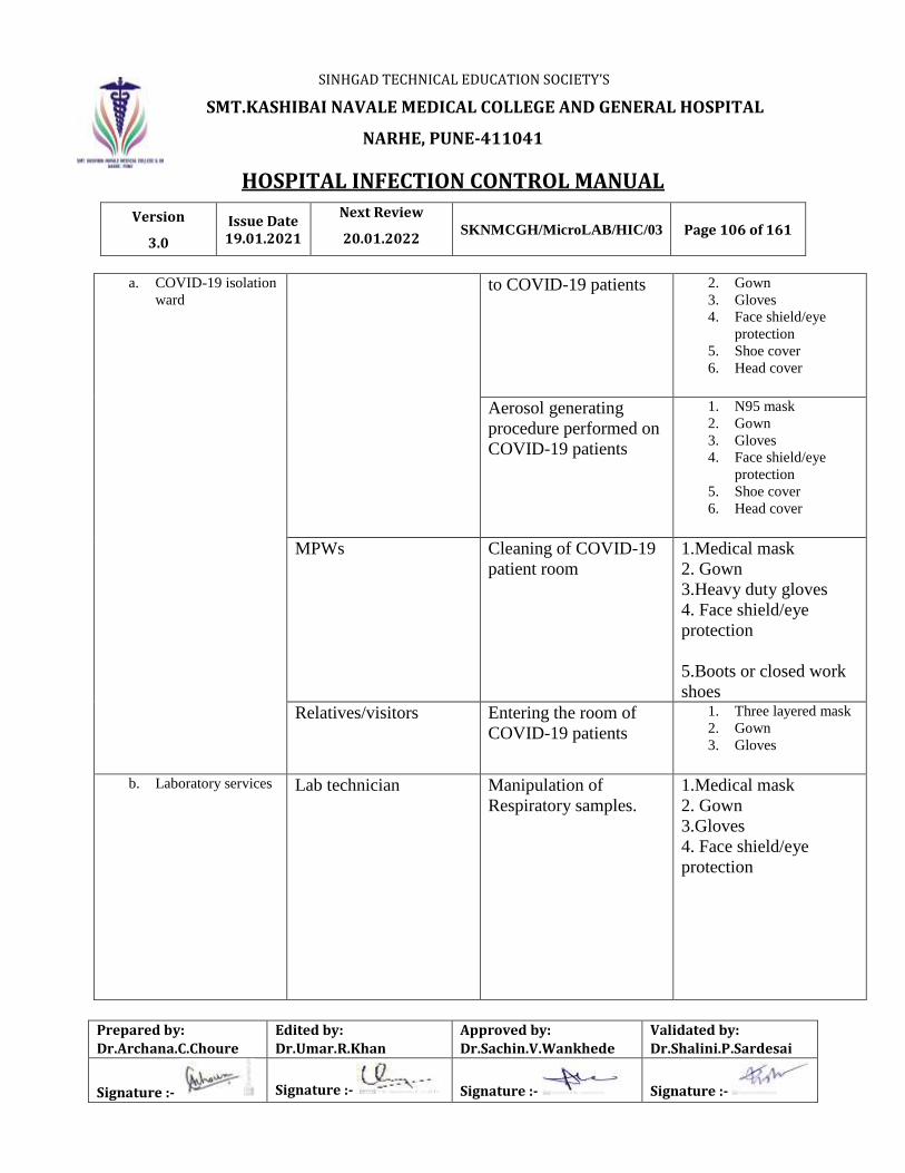
SINHGAD TECHNICAL EDUCATION SOCIETY’S
SMT.KASHIBAI NAVALE MEDICAL COLLEGE AND GENERAL HOSPITAL
NARHE, PUNE-411041
HOSPITAL INFECTION CONTROL MANUAL
Version
3.0
Issue Date 19.01.2021
Next Review
20.01.2022 SKNMCGH/MicroLAB/HIC/03 Page 106 of 161
Prepared by:
Dr.Archana.C.Choure
Edited by:
Dr.Umar.R.Khan
Approved by:
Dr.Sachin.V.Wankhede
Validated by:
Dr.Shalini.P.Sardesai
Signature :- Signature :- Signature :- Signature :-
a. COVID-19 isolation
ward to COVID-19 patients 2. Gown
3. Gloves
4. Face shield/eye
protection
5. Shoe cover
6. Head cover
Aerosol generating
procedure performed on
COVID-19 patients
1. N95 mask
2. Gown
3. Gloves
4. Face shield/eye
protection
5. Shoe cover
6. Head cover
MPWs Cleaning of COVID-19
patient room
1.Medical mask
2. Gown
3.Heavy duty gloves
4. Face shield/eye
protection
5.Boots or closed work
shoes
Relatives/visitors Entering the room of
COVID-19 patients
1. Three layered mask
2. Gown
3. Gloves
b. Laboratory services Lab technician Manipulation of
Respiratory samples.
1.Medical mask
2. Gown
3.Gloves
4. Face shield/eye
protection

SINHGAD TECHNICAL EDUCATION SOCIETY’S
SMT.KASHIBAI NAVALE MEDICAL COLLEGE AND GENERAL HOSPITAL
NARHE, PUNE-411041
HOSPITAL INFECTION CONTROL MANUAL
Version
3.0
Issue Date 19.01.2021
Next Review
20.01.2022 SKNMCGH/MicroLAB/HIC/03 Page 107 of 161
Prepared by:
Dr.Archana.C.Choure
Edited by:
Dr.Umar.R.Khan
Approved by:
Dr.Sachin.V.Wankhede
Validated by:
Dr.Shalini.P.Sardesai
Signature :- Signature :- Signature :- Signature :-
3.Transport teams
Ambulance Healthcare personnel Transport of
suspected COVID-
19 patients to referral
centre
1.Medical mask
2. Gown
3.Gloves
4. Face shield/eye
protection
Driver Involved only in
driving the patient
with suspected
COVID-
19disease and the
driver’s compartment
is separated from
theCOVID-19patient
Ensure safe distance of
1 m.
No PPE required
Patient with suspected
COVID-19disease
Transport to the referral
centre
Medical mask if
tolerated
MPW Cleaning after and
between transport of
patients with
suspected COVID-19
disease to the referral
centre
1.Medical mask
2. Gown
3.Heavy duty gloves
4. Face shield/eye
protection
5.Boots or closed work
shoes

SINHGAD TECHNICAL EDUCATION SOCIETY’S
SMT.KASHIBAI NAVALE MEDICAL COLLEGE AND GENERAL HOSPITAL
NARHE, PUNE-411041
HOSPITAL INFECTION CONTROL MANUAL
Version
3.0
Issue Date 19.01.2021
Next Review
20.01.2022 SKNMCGH/MicroLAB/HIC/03 Page 108 of 161
Prepared by:
Dr.Archana.C.Choure
Edited by:
Dr.Umar.R.Khan
Approved by:
Dr.Sachin.V.Wankhede
Validated by:
Dr.Shalini.P.Sardesai
Signature :- Signature :- Signature :- Signature :-
4. Respiratory hygiene/etiquette

SINHGAD TECHNICAL EDUCATION SOCIETY’S
SMT.KASHIBAI NAVALE MEDICAL COLLEGE AND GENERAL HOSPITAL
NARHE, PUNE-411041
HOSPITAL INFECTION CONTROL MANUAL
Version
3.0
Issue Date 19.01.2021
Next Review
20.01.2022 SKNMCGH/MicroLAB/HIC/03 Page 109 of 161
Prepared by:
Dr.Archana.C.Choure
Edited by:
Dr.Umar.R.Khan
Approved by:
Dr.Sachin.V.Wankhede
Validated by:
Dr.Shalini.P.Sardesai
Signature :- Signature :- Signature :- Signature :-
Smt. Kashibai Navale Medical College & GH, Narhe, Pune.
Guidelines for COVID-19 PANDEMIC
Protocol for disinfection of Personal protective equipment’s (PPE)
1. Gloves – discard in yellow bag
2. Goggles / Face shield-disinfect with 70% Alcohol
3. Gown, head cover, shoe cover and Three layered Mask - discard in the
container with 1% sodium hypochlorite and keep it for 20 min. Wash with
running water. Send to the laundry with proper label and prior intimation to
them.
4. Respirator (N 95 Mask) - Only single use is recommended. Discard any
respirator that is obviously damaged or becomes hard to breathe discard into
the yellow bag.
5. Heavy duty gloves and boots- Wash with soap and water and dry.

SINHGAD TECHNICAL EDUCATION SOCIETY’S
SMT.KASHIBAI NAVALE MEDICAL COLLEGE AND GENERAL HOSPITAL
NARHE, PUNE-411041
HOSPITAL INFECTION CONTROL MANUAL
Version
3.0
Issue Date 19.01.2021
Next Review
20.01.2022 SKNMCGH/MicroLAB/HIC/03 Page 110 of 161
Prepared by:
Dr.Archana.C.Choure
Edited by:
Dr.Umar.R.Khan
Approved by:
Dr.Sachin.V.Wankhede
Validated by:
Dr.Shalini.P.Sardesai
Signature :- Signature :- Signature :- Signature :-
USE OF KIOSK FOR SAMPLE COLLECTION AS A BARRIER PRECAUTION
Nasopharyngeal swab collection Oropharyngeal swab collection

SINHGAD TECHNICAL EDUCATION SOCIETY’S
SMT.KASHIBAI NAVALE MEDICAL COLLEGE AND GENERAL HOSPITAL
NARHE, PUNE-411041
HOSPITAL INFECTION CONTROL MANUAL
Version
3.0
Issue Date 19.01.2021
Next Review
20.01.2022 SKNMCGH/MicroLAB/HIC/03 Page 111 of 161
Prepared by:
Dr.Archana.C.Choure
Edited by:
Dr.Umar.R.Khan
Approved by:
Dr.Sachin.V.Wankhede
Validated by:
Dr.Shalini.P.Sardesai
Signature :- Signature :- Signature :- Signature :-
Smt. Kashibai Navale Medical College & GH, Narhe, Pune.
Guidelines for COVID-19 PANDEMIC
5. Protocol for environmental cleaning and disinfection
Area Item/equipment Frequency of cleaning /
disinfection
Method of
cleaning/disinfection
1.COVID-19
OPD
Floor 2 hourly Clean floor with 1% sodium
hypochlorite solution
Ceiling, wall, window, once a week Damp dusting
Change the mop
head/cover when soiled
Door and door knobs,
light switches
Daily Clean with Damp cloth of
soap and water
Equipments such as
thermometer,
stethoscope, BP
apparatus
After each use Wipe with 70% alcohol
Desks, Desk, chairs,
telephone, and other
frequently touched
surfaces
2 hourly Clean with Damp cloth of soap
and water

SINHGAD TECHNICAL EDUCATION SOCIETY’S
SMT.KASHIBAI NAVALE MEDICAL COLLEGE AND GENERAL HOSPITAL
NARHE, PUNE-411041
HOSPITAL INFECTION CONTROL MANUAL
Version
3.0
Issue Date 19.01.2021
Next Review
20.01.2022 SKNMCGH/MicroLAB/HIC/03 Page 112 of 161
Prepared by:
Dr.Archana.C.Choure
Edited by:
Dr.Umar.R.Khan
Approved by:
Dr.Sachin.V.Wankhede
Validated by:
Dr.Shalini.P.Sardesai
Signature :- Signature :- Signature :- Signature :-
For cleaning of COVID 19 ISOLATION WARD and ICU worker should wear:
Rubber boots, heavy duty gloves & triple layer mask with all PPE.
Area Item/equipment Frequency of
cleaning /
disinfection
Method of cleaning/disinfection
2.COVID-19
ISOLATION
WARD
Floor 2 hourly Clean floor with 1% sodium
hypochlorite solution
Ceiling , wall ,window, Daily Damp dusting
Change the mop head/cover
when soiled ( for metal surfaces use
70%alcohol)
High contact
surfaces
Door and door knobs,
light switches, intercom,
public counters
TWICE Daily Clean with Damp cloth soaked in
1% sodium hypochlorite solution
Frequently touch
surfaces
Equipments such as
thermometer, stethoscope,
BP apparatus
After EACH use Wipe with 70% alcohol
Desks, chairs, telephone,
and other frequently
touched surfaces
2 hourly Clean with Damp cloth soaked in
1% sodium hypochlorite solution (
for metal surfaces use 70% alcohol)
For infected patient bed
in ICU
After use 0.1% sodium hypochlorite solution
for contact period of 10 minutes

SINHGAD TECHNICAL EDUCATION SOCIETY’S
SMT.KASHIBAI NAVALE MEDICAL COLLEGE AND GENERAL HOSPITAL
NARHE, PUNE-411041
HOSPITAL INFECTION CONTROL MANUAL
Version
3.0
Issue Date 19.01.2021
Next Review
20.01.2022 SKNMCGH/MicroLAB/HIC/03 Page 113 of 161
Prepared by:
Dr.Archana.C.Choure
Edited by:
Dr.Umar.R.Khan
Approved by:
Dr.Sachin.V.Wankhede
Validated by:
Dr.Shalini.P.Sardesai
Signature :- Signature :- Signature :- Signature :-
Area Item/equipment Frequency of
cleaning /
disinfection
Method of cleaning/disinfection
3.COVID-19 ICU Floor 2 hourly Clean floor with 1% sodium
hypochlorite solution
Ceiling , wall ,window, Daily Damp dusting
Change the mop head/cover
when soiled ( for metal surfaces
use 70%alcohol)
High contact surfaces Door and door knobs,
light switches, intercom,
public counters
TWICE Daily Clean with Damp cloth soaked
in 1% sodium hypochlorite
solution
Frequently touch
surfaces
Equipments such as
thermometer,
stethoscope, BP
apparatus
After EACH use Wipe with 70% alcohol
Desks, chairs, telephone,
and other frequently
touched surfaces
2 hourly Clean with Damp cloth soaked
in 1% sodium hypochlorite
solution ( for metal surfaces use
70% alcohol)
High risk equipments Ventilator circuits, O2
mask, nasal prongs,
suction jar & tubes,
blood & body fluid
stained instruments, linen
After EACH use 1% sodium hypochlorite
solution for contact period of 15
minutes
For large blood spill After each event 10% sodium hypochlorite
solution for contact period of 15
minutes

SINHGAD TECHNICAL EDUCATION SOCIETY’S
SMT.KASHIBAI NAVALE MEDICAL COLLEGE AND GENERAL HOSPITAL
NARHE, PUNE-411041
HOSPITAL INFECTION CONTROL MANUAL
Version
3.0
Issue Date 19.01.2021
Next Review
20.01.2022 SKNMCGH/MicroLAB/HIC/03 Page 114 of 161
Prepared by:
Dr.Archana.C.Choure
Edited by:
Dr.Umar.R.Khan
Approved by:
Dr.Sachin.V.Wankhede
Validated by:
Dr.Shalini.P.Sardesai
Signature :- Signature :- Signature :- Signature :-
For infected patient bed
in ICU(Mattress to be
covered with non-
absorbent sheet)
After use For non-absorbent sheet 0.1%
sodium hypochlorite solution
for contact period of 10
minutes
5. Guidelines for Handling, Treatment and Disposal of COVID-
19 Biomedical Waste
In addition to existing BMW Management rules, 2016 and amendments, the following additional
steps need to be followed during management of COVID-19 patients.
Keep separate dedicated color coded bins (with foot operated lids) in corona isolation
wards & RT PCR laboratory and should be labeled as “COVID-19 Waste”.
Use double layered bags (using 2 bags) should be used for collection of waste from
COVID-19 isolation wards so as to ensure adequate strength and no-leaks.
Use dedicated trolley and collection bins and label as “COVID-19 Waste”.
General solid waste comprising of wrappers of medicines/syringes, fruit peel offs, empty
juice bottles or tetra packs, used water bottles, discarded papers, carton boxes of medicines,
empty bottles of disinfectants, left-over food, disposable food plates etc., should be
collected separately in wet and dry solid waste bags. The wet and dry solid waste bags to
be tied securely in leak-proof bags, sprayed with sodium hypo-chlorite solution and hand
over to waste collector on daily basis.
Disinfection: The (inner and outer) surface of bags/containers/collection bins/trolleys used
for storage of COVID-19 waste should be disinfected with 1% sodium hypochlorite
solution daily.
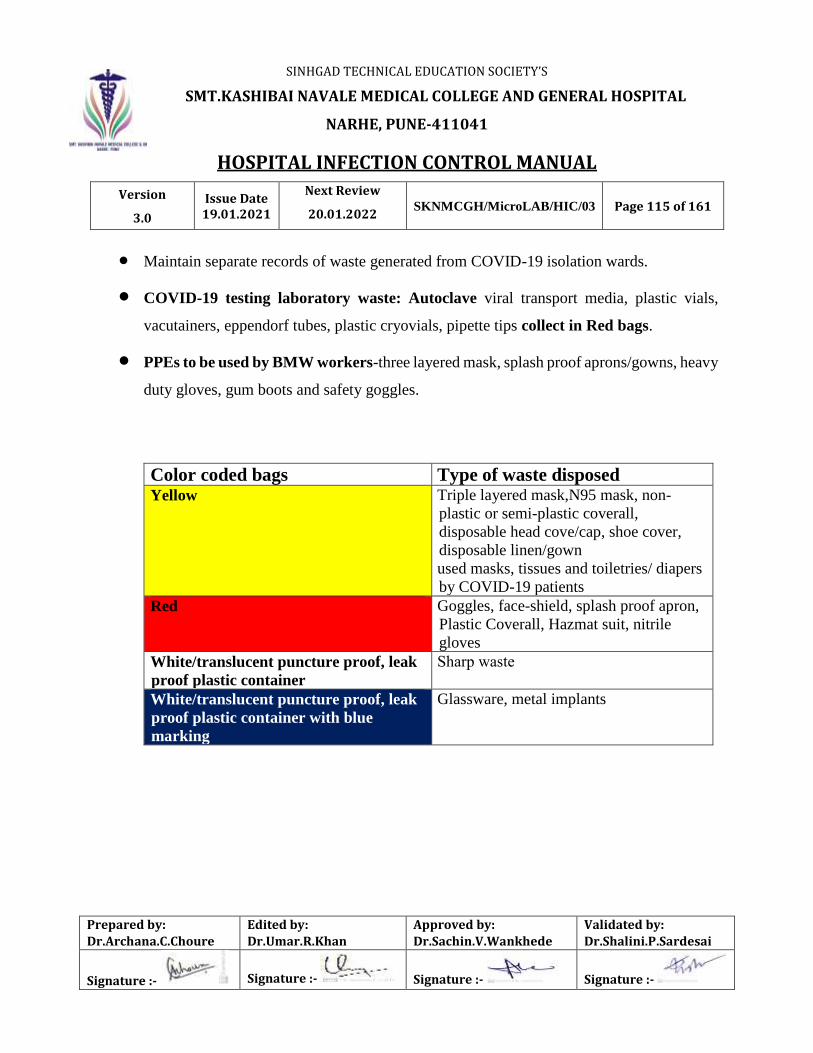
SINHGAD TECHNICAL EDUCATION SOCIETY’S
SMT.KASHIBAI NAVALE MEDICAL COLLEGE AND GENERAL HOSPITAL
NARHE, PUNE-411041
HOSPITAL INFECTION CONTROL MANUAL
Version
3.0
Issue Date 19.01.2021
Next Review
20.01.2022 SKNMCGH/MicroLAB/HIC/03 Page 115 of 161
Prepared by:
Dr.Archana.C.Choure
Edited by:
Dr.Umar.R.Khan
Approved by:
Dr.Sachin.V.Wankhede
Validated by:
Dr.Shalini.P.Sardesai
Signature :- Signature :- Signature :- Signature :-
Maintain separate records of waste generated from COVID-19 isolation wards.
COVID-19 testing laboratory waste: Autoclave viral transport media, plastic vials,
vacutainers, eppendorf tubes, plastic cryovials, pipette tips collect in Red bags.
PPEs to be used by BMW workers-three layered mask, splash proof aprons/gowns, heavy
duty gloves, gum boots and safety goggles.
Color coded bags Type of waste disposed Yellow Triple layered mask,N95 mask, non-
plastic or semi-plastic coverall,
disposable head cove/cap, shoe cover,
disposable linen/gown
used masks, tissues and toiletries/ diapers
by COVID-19 patients
Red Goggles, face-shield, splash proof apron,
Plastic Coverall, Hazmat suit, nitrile
gloves
White/translucent puncture proof, leak
proof plastic container
Sharp waste
White/translucent puncture proof, leak
proof plastic container with blue
marking
Glassware, metal implants

SINHGAD TECHNICAL EDUCATION SOCIETY’S
SMT.KASHIBAI NAVALE MEDICAL COLLEGE AND GENERAL HOSPITAL
NARHE, PUNE-411041
HOSPITAL INFECTION CONTROL MANUAL
Version
3.0
Issue Date 19.01.2021
Next Review
20.01.2022 SKNMCGH/MicroLAB/HIC/03 Page 116 of 161
Prepared by:
Dr.Archana.C.Choure
Edited by:
Dr.Umar.R.Khan
Approved by:
Dr.Sachin.V.Wankhede
Validated by:
Dr.Shalini.P.Sardesai
Signature :- Signature :- Signature :- Signature :-
SEPARATE COVID 19 BINS & DOUBLE BAGS FOR BMW
WITH APPROPRIATE PPE

SINHGAD TECHNICAL EDUCATION SOCIETY’S
SMT.KASHIBAI NAVALE MEDICAL COLLEGE AND GENERAL HOSPITAL
NARHE, PUNE-411041
HOSPITAL INFECTION CONTROL MANUAL
Version
3.0
Issue Date 19.01.2021
Next Review
20.01.2022 SKNMCGH/MicroLAB/HIC/03 Page 117 of 161
Prepared by:
Dr.Archana.C.Choure
Edited by:
Dr.Umar.R.Khan
Approved by:
Dr.Sachin.V.Wankhede
Validated by:
Dr.Shalini.P.Sardesai
Signature :- Signature :- Signature :- Signature :-
Standard operating procedure for biomedical waste
management of COVID-19 waste
Collection and segregation of COVID-19 waste as per guidelines at the point of generation
(Double layered bags and bins with “COVID-19 Waste” label)
The (inner and outer) surface of bags/containers/collection bins/trolleys used for storage of
COVID-19 waste -disinfection with 1% sodium hypochlorite solution
Handing over the collected waste to BMW Worker in designated bins/trolley with “COVID-19
Waste” label
Transport of COVID-19 Waste to the central storage area
Autoclaving of all COVID waste
Handing over of all COVID waste to the CBWTF ( Passco )
Maintain separate records of waste generated from COVID-19 and autoclave record.

SINHGAD TECHNICAL EDUCATION SOCIETY’S
SMT.KASHIBAI NAVALE MEDICAL COLLEGE AND GENERAL HOSPITAL
NARHE, PUNE-411041
HOSPITAL INFECTION CONTROL MANUAL
Version
3.0
Issue Date 19.01.2021
Next Review
20.01.2022 SKNMCGH/MicroLAB/HIC/03 Page 118 of 161
Prepared by:
Dr.Archana.C.Choure
Edited by:
Dr.Umar.R.Khan
Approved by:
Dr.Sachin.V.Wankhede
Validated by:
Dr.Shalini.P.Sardesai
Signature :- Signature :- Signature :- Signature :-
COVID-19 WASTE SEGRGATION
YELLOW BAG
Triple layered mask
N95 mask
Non-plastic or semi-plastic coverall
Disposable head cover/cap
Disposable shoe cover
Disposable linen/gown
Used masks, tissues and toiletries/ diapers by COVID-19 patients
RED BAG
Goggles
Face-shield
Splash proof apron
Plastic Coverall
Hazmat suit
Nitrile gloves

SINHGAD TECHNICAL EDUCATION SOCIETY’S
SMT.KASHIBAI NAVALE MEDICAL COLLEGE AND GENERAL HOSPITAL
NARHE, PUNE-411041
HOSPITAL INFECTION CONTROL MANUAL
Version
3.0
Issue Date 19.01.2021
Next Review
20.01.2022 SKNMCGH/MicroLAB/HIC/03 Page 119 of 161
Prepared by:
Dr.Archana.C.Choure
Edited by:
Dr.Umar.R.Khan
Approved by:
Dr.Sachin.V.Wankhede
Validated by:
Dr.Shalini.P.Sardesai
Signature :- Signature :- Signature :- Signature :-
A) COVID -19 Isolation wards / ICU /OPD/ Sample collection centers and laboratories
Keep separate color-coded bins/bags /containers in wards and maintain proper
segregation of waste.
As precaution double layered bags (using 2 bags) should be used for collection of waste
Collect and store biomedical waste separately and use dedicated collection bin labelled
as “COVID-19.”
Maintain separate record of waste generated from COVID-19 isolation wards.
-Use dedicated trolleys and collection bins in isolation wards
The inner and outer surface of bins / containers /trolleys used for storage of waste should
be disinfected with 1% sodium hypochlorite solution.
An extra set of Personal Protective Equipment and Spill management Kit should be kept
ready in case of any emergency.

SINHGAD TECHNICAL EDUCATION SOCIETY’S
SMT.KASHIBAI NAVALE MEDICAL COLLEGE AND GENERAL HOSPITAL
NARHE, PUNE-411041
HOSPITAL INFECTION CONTROL MANUAL
Version
3.0
Issue Date 19.01.2021
Next Review
20.01.2022 SKNMCGH/MicroLAB/HIC/03 Page 120 of 161
Prepared by:
Dr.Archana.C.Choure
Edited by:
Dr.Umar.R.Khan
Approved by:
Dr.Sachin.V.Wankhede
Validated by:
Dr.Shalini.P.Sardesai
Signature :- Signature :- Signature :- Signature :-
6. Standard Precautions to be followed by health care workers while
handling dead bodies of COVID19
1. Hand hygiene.
2. Use of personal protective equipment (e.g., water resistant apron, gloves, masks, eyewear).
3. Safe handling of sharps.
4. Disinfect bag housing dead body; instruments and devices used on the patient.
5. Disinfect linen. Clean and disinfect environmental surfaces.
Removal of the body from the isolation room or area
The health worker attending to the dead body should perform hand hygiene, ensure proper
use of PPE (water resistant apron, goggles, N95 mask, gloves).
All tubes, drains and catheters on the dead body should be removed.
Any puncture holes or wounds (resulting from removal of catheter, drains, tubes, or
otherwise) should be disinfected with 1% hypochlorite and dressed with impermeable
material.
Apply caution while handling sharps such as intravenous catheters and other sharp devices.
They should be disposed into a sharps container.
Plug Oral, nasal orifices of the dead body to prevent leakage of body fluids.
If the family of the patient wishes to view the body at the time of removal from the isolation
room or area, they may be allowed to do so with the application of Standard Precautions.
Place the dead body in leak-proof plastic body bag. The exterior of the body bag can be
decontaminated with 1% hypochlorite. The body bag can be wrapped with a mortuary sheet
or sheet provided by the family members.
SINHGAD TECHNICAL EDUCATION SOCIETY’S
SMT.KASHIBAI NAVALE MEDICAL COLLEGE AND GENERAL HOSPITAL
NARHE, PUNE-411041
HOSPITAL INFECTION CONTROL MANUAL
Version
3.0
Issue Date 19.01.2021
Next Review
20.01.2022 SKNMCGH/MicroLAB/HIC/03 Page 121 of 161
Prepared by:
Dr.Archana.C.Choure
Edited by:
Dr.Umar.R.Khan
Approved by:
Dr.Sachin.V.Wankhede
Validated by:
Dr.Shalini.P.Sardesai
Signature :- Signature :- Signature :- Signature :-
The body will be either handed over to the relatives or taken to mortuary.
All used/ soiled linen should be handled with standard precautions, put in bio-hazard bag
and the outer surface of the bag disinfected with hypochlorite solution.
Used equipment should be autoclaved or decontaminated with disinfectant solutions in
accordance with established infection prevention control practices.
All medical waste must be handled and disposed of in accordance with Bio-medical waste
management rules.
The health staff who handled the body will remove personal protective equipment and will
perform hand hygiene.
Provide counseling to the family members and respect their sentiments.
Environmental cleaning and disinfection
All surfaces of the isolation area (floors, bed, railings, side tables, IV stand, etc.) should be wiped
with 1% Sodium Hypochlorite solution; allow a contact time of 30 minutes, and then allowed to
air dry.
REFERENCES:
1. CDC guidelines
2. ICMR guidelines
3. WHO guidelines
4. Ministry of health and family welfare

SINHGAD TECHNICAL EDUCATION SOCIETY’S
SMT.KASHIBAI NAVALE MEDICAL COLLEGE AND GENERAL HOSPITAL
NARHE, PUNE-411041
HOSPITAL INFECTION CONTROL MANUAL
Version
3.0
Issue Date 19.01.2021
Next Review
20.01.2022 SKNMCGH/MicroLAB/HIC/03 Page 122 of 161
Prepared by:
Dr.Archana.C.Choure
Edited by:
Dr.Umar.R.Khan
Approved by:
Dr.Sachin.V.Wankhede
Validated by:
Dr.Shalini.P.Sardesai
Signature :- Signature :- Signature :- Signature :-
Infection control and management Training workshop for COVID -19 For All Medical
And Paramedical Staff
As per the instructions of Dean and Officer on special duty (OSD), the Department of
Microbiology organized Infection control and management Training workshop for COVID -19
For All Medical and Paramedical Staff.
Training workshop for the teaching faculty, resident doctors and matron, DNS and ANS
was conducted on 7.4.2020 from 11:00 to 1:00 p.m. Dr. Shiva Iyer, Chief Intensivist, BVMC
shared his experience on COVID-19 management. He resolved queries of faculties during
interactive session of question answer. This was followed by training about infection control and
management by the faculties of Department of Microbiology, Anesthesia and Medicine. The
training was conducted on following topics-
1. Hand hygiene
2. Personal protective equipment
3. Surface cleaning and disinfection
4. Biomedical waste management for COVID-19
5. Sample collection and transport
6. Management of COVID-19
7. Dead body management of COVID-19
All staff was seated as per social distancing in the hospital auditorium. Total 171 faculties, 101
resident doctors and 2 matrons, 5 ICNs, 25 ANS and 7 DNS were present for the training.
Similar training workshop was conducted for nurses, MPWs and other paramedical
staff from 8.4.2020 to 13.4.2020 in four lecture halls with taking care of social distancing. Total
about 581 nursing staff, 334 MPWs and 93 paramedical staff were present. The training also

SINHGAD TECHNICAL EDUCATION SOCIETY’S
SMT.KASHIBAI NAVALE MEDICAL COLLEGE AND GENERAL HOSPITAL
NARHE, PUNE-411041
HOSPITAL INFECTION CONTROL MANUAL
Version
3.0
Issue Date 19.01.2021
Next Review
20.01.2022 SKNMCGH/MicroLAB/HIC/03 Page 123 of 161
Prepared by:
Dr.Archana.C.Choure
Edited by:
Dr.Umar.R.Khan
Approved by:
Dr.Sachin.V.Wankhede
Validated by:
Dr.Shalini.P.Sardesai
Signature :- Signature :- Signature :- Signature :-
included demonstration of donning and doffing of PPE. Training session was conducted in Marathi
for MPWs and other paramedical staff.
MULTIDISCIPLINARY TRAINING WORKSHOP ON PANDEMIC COVID 19 FOR TEACHING
FACULTY
DR KADAM DR WANKHEDE DR IYER (Guest speaker) ICU Head BVP
OVERVIEW OF COVID 19 VIROLOGY OF SARS COV-2 ICU MANAGEMEENT OF COVID19
DR PATIL DR SHAH DR DHOPE
PPE DONNING & DOFFING HAND HYGIENE CLEANING & DISINFECTION

SINHGAD TECHNICAL EDUCATION SOCIETY’S
SMT.KASHIBAI NAVALE MEDICAL COLLEGE AND GENERAL HOSPITAL
NARHE, PUNE-411041
HOSPITAL INFECTION CONTROL MANUAL
Version
3.0
Issue Date 19.01.2021
Next Review
20.01.2022 SKNMCGH/MicroLAB/HIC/03 Page 124 of 161
Prepared by:
Dr.Archana.C.Choure
Edited by:
Dr.Umar.R.Khan
Approved by:
Dr.Sachin.V.Wankhede
Validated by:
Dr.Shalini.P.Sardesai
Signature :- Signature :- Signature :- Signature :-
SAMPLE COLLECTION & TRANSPORT
Dr.Archana Choure
TREATMENT PROTOCOLS FOR COVID 19
Dr.Ingole
TEACHING FACULTY

SINHGAD TECHNICAL EDUCATION SOCIETY’S
SMT.KASHIBAI NAVALE MEDICAL COLLEGE AND GENERAL HOSPITAL
NARHE, PUNE-411041
HOSPITAL INFECTION CONTROL MANUAL
Version
3.0
Issue Date 19.01.2021
Next Review
20.01.2022 SKNMCGH/MicroLAB/HIC/03 Page 125 of 161
Prepared by:
Dr.Archana.C.Choure
Edited by:
Dr.Umar.R.Khan
Approved by:
Dr.Sachin.V.Wankhede
Validated by:
Dr.Shalini.P.Sardesai
Signature :- Signature :- Signature :- Signature :-
MICROBIOLOGISTS AND ICNs IMPARTING TRAINING TO MEDICAL &
PARAMEDICAL STAFF FOR PREVENTION & CONTROL OF
COVID-19 PANDEMIC INFECTION

SINHGAD TECHNICAL EDUCATION SOCIETY’S
SMT.KASHIBAI NAVALE MEDICAL COLLEGE AND GENERAL HOSPITAL
NARHE, PUNE-411041
HOSPITAL INFECTION CONTROL MANUAL
Version
3.0
Issue Date 19.01.2021
Next Review
20.01.2022 SKNMCGH/MicroLAB/HIC/03 Page 126 of 161
Prepared by:
Dr.Archana.C.Choure
Edited by:
Dr.Umar.R.Khan
Approved by:
Dr.Sachin.V.Wankhede
Validated by:
Dr.Shalini.P.Sardesai
Signature :- Signature :- Signature :- Signature :-
DIETARY AND KITCHEN SERVICES
13.1 Hygiene and Infection Control
13.1.1. Food service establishments are frequently identified as places that lead to outbreaks of
food-borne diseases. The need for adequate food hygiene facilities is of paramount
importance. Assuring safe food requires management and control of microbiological,
chemical, and physical hazards.
13.1.2. Staff hygiene/health: Everyone who handles, prepares, processes and distributes food
must understand the principles of basic food hygiene and the need for trained personnel
and catering hygiene.
a. All food handlers should complete a pre-employment health check-up which
includes stool routine examination, and past history of enteric fever.
b. All food handlers with infectious diarrhoea, GI infection, must stop working and
return only after communicable disease personnel certify their fitness. Hair and
nails of all food handlers should be checked weekly and recorded.
c. Routine medical check-up should be done twice in a year.
13.1.3 Inspection: Daily inspection of kitchen and food handling areas is a must for hygiene,
and reports documented.
13.1.4 Kitchen: Cleaning procedures should be done on a regular basis.
13.1.5 Food stores should be generally clean and uncluttered with good access for cleaning.
Shelves should be easy to clean.
13.1.6 Any food capable of supporting microbial growth should be stored either below 8°C or
above 65°C. Cooked-chilled food should be stored below 3°C.
13.1.7 Food trolleys should be used to make transport easier and reduce movement of people.

SINHGAD TECHNICAL EDUCATION SOCIETY’S
SMT.KASHIBAI NAVALE MEDICAL COLLEGE AND GENERAL HOSPITAL
NARHE, PUNE-411041
HOSPITAL INFECTION CONTROL MANUAL
Version
3.0
Issue Date 19.01.2021
Next Review
20.01.2022 SKNMCGH/MicroLAB/HIC/03 Page 127 of 161
Prepared by:
Dr.Archana.C.Choure
Edited by:
Dr.Umar.R.Khan
Approved by:
Dr.Sachin.V.Wankhede
Validated by:
Dr.Shalini.P.Sardesai
Signature :- Signature :- Signature :- Signature :-
13.1.8 Trolleys should be cleaned daily or more frequently if contamination occurs.
13.1.9 A cleaning schedule for the kitchen is suggested so as to ensure that hygiene is
maintained.
13.1.10Storage:
a. All drying ingredients should be cleaned before they are stored in storage
containers (plastic bins)
b. All green leafy and other vegetables should be stored in the refrigerator and thoroughly
washed in water before usage.
13.1.11Milk should be purchased on a daily basis and stored in the refrigerator at (8° C).
13.1.12Food should be prepared half an hour before service and stored in a bain-marie at a
temperature of 75–100°C.
13.1.13Vegetables like potatoes, onion, other root vegetables, should be stored in plastic trays in
the store room.
13.1.14Other vegetables should be bought at an interval of 2-3 days, as and when there is a
requirement. They should be stored in the refrigerator and washed thoroughly when taken
out for cooking.
13.1.15Ingredients (all the drying ingredients) like rice, broken wheat, pulses should be washed
twice in cold water and then in hot water before cooking.
13.2 Waste Disposal
13.2.1. Waste should be identified and collected in color coded containers.
13.2.2. Left over waste, vegetable peels should be collected in the green container and sent for
disposal thorough municipal authorities.

SINHGAD TECHNICAL EDUCATION SOCIETY’S
SMT.KASHIBAI NAVALE MEDICAL COLLEGE AND GENERAL HOSPITAL
NARHE, PUNE-411041
HOSPITAL INFECTION CONTROL MANUAL
Version
3.0
Issue Date 19.01.2021
Next Review
20.01.2022 SKNMCGH/MicroLAB/HIC/03 Page 128 of 161
Prepared by:
Dr.Archana.C.Choure
Edited by:
Dr.Umar.R.Khan
Approved by:
Dr.Sachin.V.Wankhede
Validated by:
Dr.Shalini.P.Sardesai
Signature :- Signature :- Signature :- Signature :-
Abbreviations
1. HIC: Hospital Infection Control
2. HICC: Hospital Infection Control Committee
3. ICT: Infection Control Team
4. ICN: Infection Control Nurse
5. ICD: Infection Control Doctor
6. HCAI: Health care Associated Infections
7. HAI: Hospital Acquired Infections
8. VAE: Ventilator Associated Events
9. VAP: Ventilator Associated Pneumonia
10. CLABSI: Central Line Associated Blood Stream Infections
11. CAUTI: Catheter Associated Urinary Tract Infections
12. SSI: Surgical Site Infections
13. NSI: Needle Stick Injury
14. BMW: Biomedical Waste
15. PPE: Personal Protective Equipment’s
16. HCW: Health Care Worker
17. BICU: Burn ICU

SINHGAD TECHNICAL EDUCATION SOCIETY’S
SMT.KASHIBAI NAVALE MEDICAL COLLEGE AND GENERAL HOSPITAL
NARHE, PUNE-411041
HOSPITAL INFECTION CONTROL MANUAL
Version
3.0
Issue Date 19.01.2021
Next Review
20.01.2022 SKNMCGH/MicroLAB/HIC/03 Page 129 of 161
Prepared by:
Dr.Archana.C.Choure
Edited by:
Dr.Umar.R.Khan
Approved by:
Dr.Sachin.V.Wankhede
Validated by:
Dr.Shalini.P.Sardesai
Signature :- Signature :- Signature :- Signature :-
References
1. https://www.cdc.gov/mmwr/preview/mmwrhtml/rr5210a1.htm
2. www.nabh.co/Images/PDF/HIC_Guidebook.
3. icmr.nic.in/guidelines/Hospital%20Infection%20control%20guidelines-2
4. www.wpro.who.int/publications/docs/practical_guidelines_infection_control
5. www.ncdc.gov.in/writereaddata/mainlinkfile/File571
6. CDC guidelines for PEP: 2013
7. mpcb.gov.in/biomedical/pdf/BMW_Rules_2016.pd
8. www.indiaenvironmentportal.org.in/.../bio-medical-waste-management-rules-2016
9. CLSI 2020 -M100, 30th ed. January 2020.

Version
3.0
Issue Date 19.01.2021
Next Review
20.01.2022 SKNMCGH/MicroLAB/HIC/03 Page 130 of 161
Prepared by:
Dr.Archana.C.Choure
Edited by:
Dr.Umar.R.Khan
Approved by:
Dr.Sachin.V.Wankhede
Validated by:
Dr.Shalini.P.Sardesai
Signature :- Signature :- Signature :- Signature :-
Smt Kashibai Navale Medical College & General Hospital, PUNE
Annexure: 1
CAUTI surveillance form
Patient OPD No : Patient IPD No :
Patient Name :
Age/Sex DOA :
Ward :
Event Type: UTI Date of Event:
Post-procedure UTI: Yes No Date of Procedure:
MDRO Infection Surveillance:
□ Yes, this infection’s pathogen & location are in-plan for Infection Surveillance in the MDRO
□ No, this infection’s pathogen & location are not in-plan for Infection Surveillance in the MDRO
Risk Factors
Urinary Catheter status: □ In place – Urinary catheter in place > 2 days on the date of event
□ Removed – Urinary catheter in place > 2 days but removed the day before the date of event
□ Neither – Not catheter associated –Neither in place nor removed
Location of Device Insertion: ________________________
Date of Device Insertion: ____ /____ /_______
Event Details
Specific Event:
□ Symptomatic UTI (SUTI) □ Asymptomatic Bacteremic UTI (ABUTI) □ Urinary System
Infection (USI)
Specify Criteria Used: (check all that apply) Signs & Symptoms
Any Patient ≤ 1 year old Laboratory & Diagnostic Testing □ Fever □ Urgency □ Fever □ 1 positive culture with no more
than 2 species of organisms, at least one of which is a bacterium of ≥ 105 CFU/ml
□ Frequency □ Dysuria □ Hypothermia
□ Pain or tenderness
□ Abscess □ Apnea □ Organism(s) identified from fluid or tissue from affected site (excluding urine)
□ Acute pain, swelling, or tenderness of testes, epididymis, or prostate
□ Bradycardia
□ Suprapubic tenderness □ Lethargy □ Organism(s) identified from blood specimen □ Costovertebral angle pain or tenderness □ Vomiting
□ Purulent drainage from affected site □ Imaging test evidence of infection □ Other evidence of infection found on invasive
procedure, gross anatomic exam, or histopathologic exam
Secondary Bloodstream Infection: Yes No
Died: Yes No UTI Contributed to Death: Yes No
Discharge Date: Pathogens Identified: Yes No

Version
3.0
Issue Date 19.01.2021
Next Review
20.01.2022 SKNMCGH/MicroLAB/HIC/03 Page 131 of 161
Prepared by:
Dr.Archana.C.Choure
Edited by:
Dr.Umar.R.Khan
Approved by:
Dr.Sachin.V.Wankhede
Validated by:
Dr.Shalini.P.Sardesai
Signature :- Signature :- Signature :- Signature :-
Organism isolated:- Antibiotic used:-
DAY1
DAY2
DAY3
DAY4
DAY5
Remarks
Sign of ICN Sign of ICO Sign of Consultant

Version
3.0
Issue Date 19.01.2021
Next Review
20.01.2022 SKNMCGH/MicroLAB/HIC/03 Page 132 of 161
Prepared by:
Dr.Archana.C.Choure
Edited by:
Dr.Umar.R.Khan
Approved by:
Dr.Sachin.V.Wankhede
Validated by:
Dr.Shalini.P.Sardesai
Signature :- Signature :- Signature :- Signature :-
Smt Kashibai Navale Medical College & General Hospital, PUNE
Annexure : 1
Surveillance form for CLABSI
Patient OPD No : Patient IPD No :
Patient Name : Ward :
Age &Sex : DOA :
Event Type: BSI Date of Event:
Post-procedure BSI: Yes No Date of Procedure:
MDRO Infection Surveillance:
□ Yes, this infection’s pathogen & location are in-plan for Infection Surveillance in the MDRO
□ No, this infection’s pathogen & location are not in-plan for Infection Surveillance in the MDRO
Risk Factors
If ICU/Other locations, Central line: Yes No Location of Device Insertion: _______________________
Date of Device Insertion: ___ /___ /________
Event Details
Specific Event: Laboratory-confirmed
Specify Criteria Used:
Signs & Symptoms (check all that apply) Laboratory (check one)
Any Patient ≤ 1 year old
□ Recognized pathogen(s) identified from one or more blood specimens
□ Common commensal identified from ≥ 2 blood specimens
□ Fever □ Fever
□ Chills □ Hypothermia
□ Hypotension □ Apnea
□ Bradycardia
Died: Yes No BSI Contributed to Death: Yes No
Discharge Date: Pathogens Identified: Yes No

Version
3.0
Issue Date 19.01.2021
Next Review
20.01.2022 SKNMCGH/MicroLAB/HIC/03 Page 133 of 161
Prepared by:
Dr.Archana.C.Choure
Edited by:
Dr.Umar.R.Khan
Approved by:
Dr.Sachin.V.Wankhede
Validated by:
Dr.Shalini.P.Sardesai
Signature :- Signature :- Signature :- Signature :-
Organism isolated :-
Antibiotics used:-
DAY1
DAY2
DAY3
DAY4
DAY5
Remarks
Sign of ICN Sign of ICO Sign of Consultant

Version
3.0
Issue Date 19.01.2021
Next Review
20.01.2022 SKNMCGH/MicroLAB/HIC/03 Page 134 of 161
Prepared by:
Dr.Archana.C.Choure
Edited by:
Dr.Umar.R.Khan
Approved by:
Dr.Sachin.V.Wankhede
Validated by:
Dr.Shalini.P.Sardesai
Signature :- Signature :- Signature :- Signature :-
Smt Kashibai Navale Medical College & General Hospital, PUNE
Annexure : 1
NSI/splash reporting form Sharps Injury/ Blood-Body Fluid Exposure Reporting Form- Action Taken Trail
Sharps/ Body Fluid Exposure Incident No: Incident Register Sr. No:
Name of the Injured / Exposed Person: Date & Time of Incident: Date & Time of Reporting: Job-Title: Department/ Unit: Place of Injury: Sharps Injury/ Blood or Body Fluid Exposure Job-Title and Name of person filling up the form:
To be filled in by CMO Date, Time and Details of First Aid given: Name of CMO Sign of CMO Date & Time To be filled in by ICN/ ICD Whether Injured/ Exposed person had undergone training on such incident? Yes / No Whether Injured/ Exposed person had undergone vaccination? Yes / No If yes, give details:
To be filled as narrated by person involved in the incident
Exposure Details: Blood/ Blood Products Visibly Bloody Solution Body Fluid (Visibly Bloody or Not) If Body Fluid: Cerebrospinal/ Amniotic/ Pericardial/ Pleural/ Synovial/ Peritoneal/ Semen-Vaginal/ Urine/ Sputum/ Saliva/ Faces-Stool/ Other: /Unknown
Body Site of Exposure: Hand-Finger/ Arm/ Leg/ Face/ Nose/ Mouth/ Eye/ Other:
If Percutaneous Exposure-Depth of Injury: Superficial (scratch, no blood)/ Moderate (skin penetration with blood)/ Deep (muscular penetration) Whether Blood was visible on the device before exposure? Yes/ No/ Unsure or Unknown

Version
3.0
Issue Date 19.01.2021
Next Review
20.01.2022 SKNMCGH/MicroLAB/HIC/03 Page 135 of 161
Prepared by:
Dr.Archana.C.Choure
Edited by:
Dr.Umar.R.Khan
Approved by:
Dr.Sachin.V.Wankhede
Validated by:
Dr.Shalini.P.Sardesai
Signature :- Signature :- Signature :- Signature :-
If Mucous membrane or Skin exposure- Approximate volume of material involved: Small (few drops)/ Large (major blood splash) If Skin Exposure- was skin intact before exposure: Yes/ No/ Unsure or Unknown
Source Information
Was the source individual identified:Yes/ No/ Unknown or Unsure
Provide serostatus of source individual for following’: HIV Antibody: Positive/ Negative/ Refused/ Unknown HCV Antibody: Positive/ Negative/ Refused/ Unknown HbsAg: Positive/ Negative/ Refused/ Unknown If known, when was the serostatus of source individual determined: Known at the time of exposure/ Determined by testing soon after exposure
Percutaneous Injury Circumstances
What Device or Item caused the injury: Hollow Bore Needles Hypodermic/ Prefilled Cartridge/ Winged steel/ IV stylet/ Phlebotomy/ Spinal or Epidural/ Bone-Marrow/ Biopsy/ Huber/ Other: specify Suture Needle:Yes/ No Glass: Capillary/ Pipette/ Slide/ Specimen/ Tube/ Vacuum/ Other specify: Other Sharp Objects: Bone-Tooth chip/ Bone cutter/ Electrocautery/ Bur/ Explorer/ Extraction forceps/ Elevator/ Cutting Blade/ Lancet/ Pin/ Razor/ Retractor/ Rod/ Root Canal File/ Scaler or Curette/ Scalpel blade/ Scissors/ Tenaculum/ Trocar/ Wire/ Other, specify: Any other Device, Specify:
Mucous membrane Exposure Circumstances
What PPE was donned while handling the patient? Gloves/ Mask/ Goggles/ Eyeglasses/ Face- shield/ Gown/ None
Activity/ Event when exposure occurred: Patient Spit/ Coughed/ Vomited Airway manipulation/ Endoscopic procedure/ Dental procedure/ Tube removal/ placement/ manipulation Phlebotomy/ Irrigation procedure/ Vaginal delivery/ IV or Arterial line removal/ placement/ manipulation Surgical procedure/ Bleeding Vessel/ Changing dressing/ wound care/ Cleaning/ Transporting contaminated equipment/ Manipulating blood tube/ bottle/ specimen container Any other, specify:
Details of Post Exposure Prophylaxis given:

Version
3.0
Issue Date 19.01.2021
Next Review
20.01.2022 SKNMCGH/MicroLAB/HIC/03 Page 136 of 161
Prepared by:
Dr.Archana.C.Choure
Edited by:
Dr.Umar.R.Khan
Approved by:
Dr.Sachin.V.Wankhede
Validated by:
Dr.Shalini.P.Sardesai
Signature :- Signature :- Signature :- Signature :-
Smt Kashibai Navale Medical College & General Hospital, PUNE
Annexure :1
Surveillance form SSI
Patient OPD No : Patient IPD :
Patient Name : DOA
:
Age &Sex : Ward :
Event Type: SSI Date of Event:
Date of Procedure: Outpatient Procedure: Yes No
MDRO Infection Surveillance: □ Yes, this infection’s pathogen & location are in-plan for Infection Surveillance in the MDRO □ No, this infection’s pathogen & location are not in-plan for Infection Surveillance in the MDRO
Event Details
Specific Event:
□ Superficial Incisional Primary (SIP) □ Deep Incisional Primary (DIP)
□ Superficial Incisional Secondary (SIS) □ Deep Incisional Secondary (DIS)
□Organ/Space (specify site): _______________________
Infection present at the time of surgery (PATOS): □ Yes □ No
Specify Criteria Used (check all that apply):
Signs & Symptoms Laboratory □ Drainage or material† □ Sinus tract □ Organism(s) identified
□ Pain or tenderness □ Hypothermia □ Culture or non-culture based testing not performed
□ Swelling or inflammation □ Apnea □ Organism(s) identified from blood specimen
□ Erythema or redness □ Bradycardia □ Organism(s) identified from ≥ 2 periprosthetic specimens □ Heat □ Lethargy
□ Fever □ Cough □ Other positive laboratory tests† □ Incision deliberately opened/drained
□ Nausea □ Imaging test evidence of infection
□ Wound spontaneously dehisces
□ Vomiting
□ Abscess □ Dysuria Clinical Diagnosis □ Other evidence of infection found on invasive
procedure, gross anatomic exam, or histopathologic exam †
□ Physician diagnosis of this event type
□ Physician institutes appropriate antimicrobial therapy
□ Other signs & symptoms
Detected: □A (During admission) □P (Post-discharge surveillance)
□RF (Readmission to facility where procedure performed) □ RO (Readmission to facility other than where procedure was performed)
Secondary Bloodstream Infection: Yes No Died: Yes No SSI Contributed to Death: Yes No
Discharge Date: Pathogens Identified: Yes No

Version
3.0
Issue Date 19.01.2021
Next Review
20.01.2022 SKNMCGH/MicroLAB/HIC/03 Page 137 of 161
Prepared by:
Dr.Archana.C.Choure
Edited by:
Dr.Umar.R.Khan
Approved by:
Dr.Sachin.V.Wankhede
Validated by:
Dr.Shalini.P.Sardesai
Signature :- Signature :- Signature :- Signature :-
Organism isolated :-
Antibiotics used:-
Day 1
Day 2
Day 3
Day 4
Day 5
Remarks :
Sign of ICN Sign of ICD Sign of Consultant

Version
3.0
Issue Date 19.01.2021
Next Review
20.01.2022 SKNMCGH/MicroLAB/HIC/03 Page 138 of 161
Prepared by:
Dr.Archana.C.Choure
Edited by:
Dr.Umar.R.Khan
Approved by:
Dr.Sachin.V.Wankhede
Validated by:
Dr.Shalini.P.Sardesai
Signature :- Signature :- Signature :- Signature :-
Smt Kashibai Navale Medical College & General Hospital, PUNE
Annexure:1
Surveillance form VAE
Patient OPD No : Patient IPD :
Patient Name : DOA :
Age &Sex : Ward :
Event Type: VAE Date of Event:
Post-procedure VAE: Yes No Date of Procedure:
MDRO Infection Surveillance:
□ Yes, this infection’s pathogen & location are in-plan for Infection Surveillance in the MDRO
□ No, this infection’s pathogen & location are not in-plan for Infection Surveillance in the MDRO
Location of Mechanical Ventilation Initiation: ______________ Date Initiated: __ /__ /_____ APRV: Yes No
Event Details
Specific Event: □ VAC □ IVAC □ PVAP
Specify Criteria Used:
STEP 1: VAC (≥1 REQUIRED)
□Daily min FiO2 increase ≥ 0.20 (20 points) for ≥ 2 days† OR □Daily min PEEP increase ≥ 3 cm H2O for ≥ 2
days† †after 2+ days of stable or decreasing daily minimum values.
STEP 2: IVAC
□Temperature > 38°C or < 36° OR□White blood cell count ≥ 12,000 or ≤ 4,000 cells/mm3
AND
□A new antimicrobial agent(s) is started, and is continued for ≥ 4 days
STEP 3: PVAP
□Criterion #1:Positive culture of one of the following specimens, meeting quantitative or semi-quantitative
thresholds as outlined in protocol,‡without requirement for purulent respiratory secretions:
□ Endotracheal aspirate □Lung tissue
□ Bronchoalveolar lavage □ Protected specimen brush
OR
□Criterion #2: Purulent respiratory secretions‡ (defined in the protocol) plus organism(s) identified from one of
the following specimens:‡
□ Sputum □ Lung tissue
□ Endotracheal aspirate □Protected specimen brush
□ Bronchoalveolar lavage
OR
□Criterion #3: One of the following positive tests (as outlined in the protocol):‡ □ Organism(s) identified from
pleural fluid □ Diagnostic test for Legionella species
□ Lung histopathology □ Diagnostic test for selected viral pathogens
collected after 2 days of mechanical ventilation and within +/- 2 days of onset of increase in FiO2 or PEEP.
Secondary Bloodstream Infection: Yes No
Died: Yes No VAE Contributed to Death: Yes No

Version
3.0
Issue Date 19.01.2021
Next Review
20.01.2022 SKNMCGH/MicroLAB/HIC/03 Page 139 of 161
Prepared by:
Dr.Archana.C.Choure
Edited by:
Dr.Umar.R.Khan
Approved by:
Dr.Sachin.V.Wankhede
Validated by:
Dr.Shalini.P.Sardesai
Signature :- Signature :- Signature :- Signature :-
Discharge Date: Pathogens Identified: Yes No
Organism isolated :-
Antibiotics used:-
Day 1
Day 2
Day 3
Day 4
Day 5
Remarks
Sign of ICN Sign of ICD Sign of Consultant
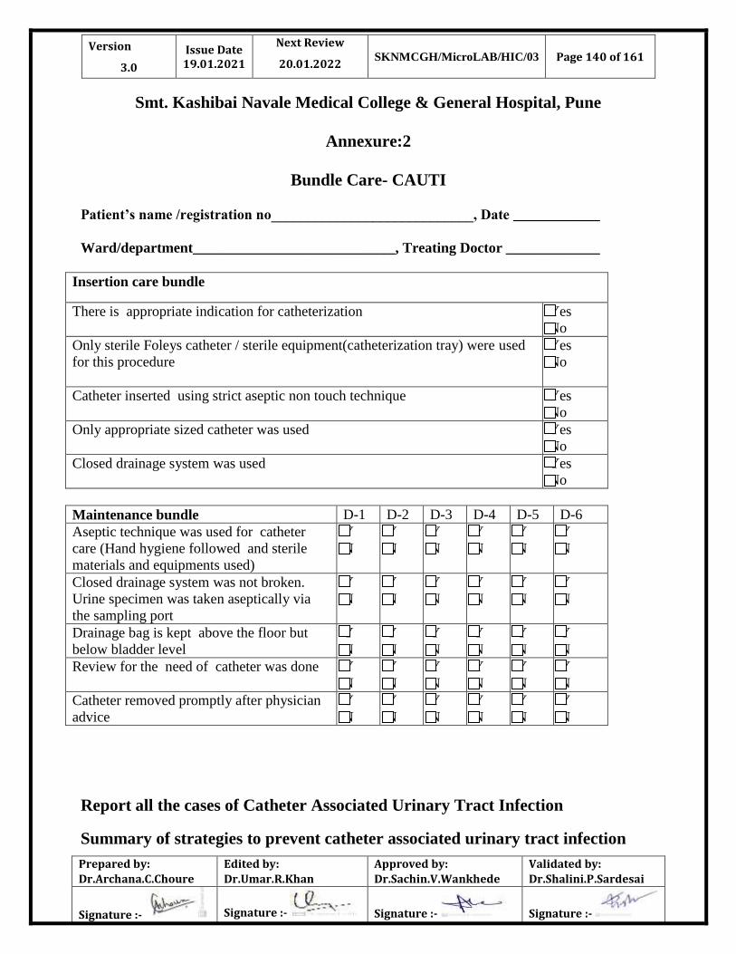
Version
3.0
Issue Date 19.01.2021
Next Review
20.01.2022 SKNMCGH/MicroLAB/HIC/03 Page 140 of 161
Prepared by:
Dr.Archana.C.Choure
Edited by:
Dr.Umar.R.Khan
Approved by:
Dr.Sachin.V.Wankhede
Validated by:
Dr.Shalini.P.Sardesai
Signature :- Signature :- Signature :- Signature :-
Smt. Kashibai Navale Medical College & General Hospital, Pune
Annexure:2
Bundle Care- CAUTI
Patient’s name /registration no____________________________, Date ____________
Ward/department____________________________, Treating Doctor _____________
Insertion care bundle
There is appropriate indication for catheterization Yes
No
Only sterile Foleys catheter / sterile equipment(catheterization tray) were used
for this procedure
Yes
No
Catheter inserted using strict aseptic non touch technique
Yes
No
Only appropriate sized catheter was used
Yes
No
Closed drainage system was used
Yes
No
Maintenance bundle D-1 D-2 D-3 D-4 D-5 D-6
Aseptic technique was used for catheter
care (Hand hygiene followed and sterile
materials and equipments used)
Y
N
Y
N
Y
N
Y
N
Y
N
Y
N
Closed drainage system was not broken.
Urine specimen was taken aseptically via
the sampling port
Y
N
Y
N
Y
N
Y
N
Y
N
Y
N
Drainage bag is kept above the floor but
below bladder level
Y
N
Y
N
Y
N
Y
N
Y
N
Y
N
Review for the need of catheter was done Y
N
Y
N
Y
N
Y
N
Y
N
Y
N
Catheter removed promptly after physician
advice
Y
N
Y
N
Y
N
Y
N
Y
N
Y
N
Report all the cases of Catheter Associated Urinary Tract Infection
Summary of strategies to prevent catheter associated urinary tract infection

Version
3.0
Issue Date 19.01.2021
Next Review
20.01.2022 SKNMCGH/MicroLAB/HIC/03 Page 141 of 161
Prepared by:
Dr.Archana.C.Choure
Edited by:
Dr.Umar.R.Khan
Approved by:
Dr.Sachin.V.Wankhede
Validated by:
Dr.Shalini.P.Sardesai
Signature :- Signature :- Signature :- Signature :-
S.N Entry point for bacteria Preventive measures
1 External urethral meatus and urethra
Bacteria carried into bladder during
insertion of catheter
Ascending colonization/ infection up
urethra around outside of catheter
Pass catheter when bladder is full for wash out effect
Before catheterization prepare urinary meatus with an antiseptic
(e.g. 0.2% chlorhexidine aqueous solution or povidone iodine)
Inject single use sterile lubricant gel or use 2% lignocaine
anaesthetic gel into urethra and hold there for 3 min before
inserting catheter
Use sterile catheter
Use non touch technique for insertion
Keep periurethral area clean and dry; bladder wash and
ointments are of no value
Secure catheter to prevent movement in urethra
After faecal incontinence, clean area and change catheter
2 Junction between catheter and
drainage tube
Do not disconnect catheter unless absolutely necessary
Always use aseptic technique for irrigation
For urine specimen collection, disinfect, sampling port by
applying alcoholic impregnated wipe and allow it to dry
completely, then aspire urine with sterile needle and syringe
3 Junction between drainage tube and
collection bag
Disconnection
Reflux from bag into catheter
Drainage tube should be welded to inlet of bag during
manufacture
Drip chamber or non return valve at inlet to bag
Keep bag below level of bladder. If it is necessary to raise
collection bag above bladder level for a short period, drainage
tube must be clamped temporarily
Empty bag every 8h or earlier if full
Do not hold bag upside down when emptying
4 Tap at bottom of collection bag
Emptying of bag
Collection bag must never touch floor
Always wash or disinfect hands with an alcoholic hand rub
before and after opening tap
Use a separate disinfected jug to collect urine from each bag
Don’t instill disinfectant/antiseptic into urinary bag after
emptying.

Version
3.0
Issue Date 19.01.2021
Next Review
20.01.2022 SKNMCGH/MicroLAB/HIC/03 Page 142 of 161
Prepared by:
Dr.Archana.C.Choure
Edited by:
Dr.Umar.R.Khan
Approved by:
Dr.Sachin.V.Wankhede
Validated by:
Dr.Shalini.P.Sardesai
Signature :- Signature :- Signature :- Signature :-
Smt. Kashibai Navale Medical College & General Hospital, Pune
Bundle Care- VAP
Patient’s name /registration no____________________________, Date ____________
Ward/department____________________________, Treating Doctor _____________
Regular observation D-1 D-2 D-3 D-4 D-5 D-6
Ensure elevation of the head of the bed to
30-45 degree C
Y
N
Y
N
Y
N
Y
N
Y
N
Y
N
Assessment of sedation with readiness to
extubate was done
Y
N
Y
N
Y
N
Y
N
Y
N
Y
N
Gastric ulcer prophylaxis is being given Y
N
Y
N
Y
N
Y
N
Y
N
Y
N
Management of ventilator tubing is being
done
Y
N
Y
N
Y
N
Y
N
Y
N
Y
N
Appropriate humidification of inspired gas
is done
Y
N
Y
N
Y
N
Y
N
Y
N
Y
N
Deep vein thrombosis prophylaxis is being
done
Y
N
Y
N
Y
N
Y
N
Y
N
Y
N
Ongoing care D-1 D-2 D-3 D-4 D-5 D-6
Adherence to hand hygiene and aseptic
technique all the time while dealing with
ventilator and tubes
Y
N
Y
N
Y
N
Y
N
Y
N
Y
N
Oral hygiene being maintained
Y
N
Y
N
Y
N
Y
N
Y
N
Y
N
Subglottic suctioning of respiratory
secretion is being done
Y
N
Y
N
Y
N
Y
N
Y
N
Y
N

Version
3.0
Issue Date 19.01.2021
Next Review
20.01.2022 SKNMCGH/MicroLAB/HIC/03 Page 143 of 161
Prepared by:
Dr.Archana.C.Choure
Edited by:
Dr.Umar.R.Khan
Approved by:
Dr.Sachin.V.Wankhede
Validated by:
Dr.Shalini.P.Sardesai
Signature :- Signature :- Signature :- Signature :-
Report all the cases of Ventilator Associated Pneumonia
Major interventions used in prevention of VAP
S.N Procedure/ Device Preventive measures
1
Suctioning
Use single use disposable gloves and wash hands before
and
after the procedure
Use sterile suction catheter and sterile fluid to flush
catheter
Change suction tubing between patients
Use closed suction system, if possible
2
Suction Bottle
Use single use disposable, if possible
Non disposable bottle should be washed with detergent
and allowed to dry or heat disinfect in washing machine or
send to sterile supply unit
3 Humidifiers
Fill with sterile water which must be changed every 24
hour or sooner if necessary
Clean and sterile humidifiers between patients. Single use
disposable humidifiers are available but they are
expensive
4 Ventilators
After every patient, clean and disinfect ventilators
Sterilize/disinfect (high level) reusable components as per
manufacturer’s instructions
5 Ventilator breathing
circuits
Replace mechanical ventilators, if soiled or
malfunctioning
Periodically drain breathing tube condensation traps,
taking care not to spill it down the patients trachea; wash
hands after procedure
Use HME (heat and moisture exchangers) ventilator
circuits. If possible
6 Nebulizers
Fill with sterile water only
Change nebulizers between patients by using sterilization
or a high level disinfection or single use nebulizers if
possible.

Version
3.0
Issue Date 19.01.2021
Next Review
20.01.2022 SKNMCGH/MicroLAB/HIC/03 Page 144 of 161
Prepared by:
Dr.Archana.C.Choure
Edited by:
Dr.Umar.R.Khan
Approved by:
Dr.Sachin.V.Wankhede
Validated by:
Dr.Shalini.P.Sardesai
Signature :- Signature :- Signature :- Signature :-
Smt. Kashibai Navale Medical College & General Hospital, Pune
Bundle Care- CLABSI
Patient’s name /registration no____________________________, Date ____________
Ward/department____________________________, Treating Doctor _____________
Insertion care bundle
There is appropriate indication for cannulation
Yes
No
Maximal sterile barrier precautions was taken during insertion Yes
No
Skin was disinfected with 2% chlorhexidine gluconate in 70% isopropyl
alcohol and allowed it to dry
Yes
No
Single lumen catheter was used
( unless indicated otherwise)
Yes
No
Sterile,semipermeable dressing was used
Yes
No
Maintenance bundle
D-1 D-2 D-3 D-4 D-5 D-6
Aseptic technique is being used for daily
care (hand hygiene before accessing the
device and use of sterile single use of
antiseptic solution to disinfect hubs)
Y
N
Y
N
Y
N
Y
N
Y
N
Y
N
Inspection of CVC site done for sign of
infection
Y
N
Y
N
Y
N
Y
N
Y
N
Y
N
Review for the need of CVC was done Y
N
Y
N
Y
N
Y
N
Y
N
Y
N
CVC removed promptly after physician
advice
Y
N
Y
N
Y
N
Y
N
Y
N
Y
N

Version
3.0
Issue Date 19.01.2021
Next Review
20.01.2022 SKNMCGH/MicroLAB/HIC/03 Page 145 of 161
Prepared by:
Dr.Archana.C.Choure
Edited by:
Dr.Umar.R.Khan
Approved by:
Dr.Sachin.V.Wankhede
Validated by:
Dr.Shalini.P.Sardesai
Signature :- Signature :- Signature :- Signature :-
Report all the cases of Central Line Associated Blood Stream Infection
Smt. Kashibai Navale Medical College & General Hospital, Pune
Bundle Care- SSI
Patient’s name _______________________________ AGE/ SEX _____________
Registration no____________________________, Date ____________
Ward/department____________________________, Treating Doctor _____________
Removal of dressing within 48 hrs of surgery YES
NO
Daily washing the incision with Chlorhexidine YES
NO
Maintenance of Euglycemia & Normothermia YES
NO
No infected patients around the patient YES
NO
The drains and catheters are patent and functioning YES
NO
Dedicated dressing drums are being used for the patient YES
NO
The patient is sponged with Chlorhexidine wipes daily YES
NO
Maintenance Bundle for SSI
Ongoing care D-1 D-2 D-3 D-4 D-5 D-6
Daily Cleaning of SS with all aseptic
precautions
Y
N
Y
N
Y
N
Y
N
Y
N
Y
N
Patient is Euglycemic
Y
N
Y
N
Y
N
Y
N
Y
N
Y
N
Fever spike and charting Y
N
Y
N
Y
N
Y
N
Y
N
Y
N
Inflammation / discharge in or around
surgical site
Y
N
Y
N
Y
N
Y
N
Y
N
Y
N
Receiving appropriate antibiotics &
analgesics
Y
N
Y
N
Y
N
Y
N
Y
N
Y
N
Name of ICN: Date :
Sign: Place:

Version
3.0
Issue Date 19.01.2021
Next Review
20.01.2022 SKNMCGH/MicroLAB/HIC/03 Page 146 of 161
Prepared by:
Dr.Archana.C.Choure
Edited by:
Dr.Umar.R.Khan
Approved by:
Dr.Sachin.V.Wankhede
Validated by:
Dr.Shalini.P.Sardesai
Signature :- Signature :- Signature :- Signature :-
Smt. Kashibai Navale Medical College & General Hospital, Pune
Annexure:2
Bundle Care- PHLEBITIS
Patient’s name /registration no____________________________, Date ____________
Ward/department____________________________, Treating Doctor _____________
Insertion care bundle
There is appropriate indication for cannulation
Yes
No
Maximal sterile aseptic techniques was taken during insertion Yes
No
Skin was disinfected with spirit followed by betadine and spirit again and
allowed it to dry
Yes
No
Sterile transparent dressing used to allow observation of insertion site Yes
No
Date of insertion recorded in medical notes
Yes
No
Maintenance bundle D-1 D-2 D-3 D-4 D-5 D-6
Review need for catheter was done Y
N
Y
N
Y
N
Y
N
Y
N
Y
N
Inspection of cannula site done for sign of
infection as per VIP score
Y
N
Y
N
Y
N
Y
N
Y
N
Y
N
Use of aseptic technique for daily care( e.g.
hand hygiene before accessing the device
and disinfect catheter hubs)
Y
N
Y
N
Y
N
Y
N
Y
N
Y
N
Replace cannula in a new site after 72 hours
or earlier if clinically indicated
Y
N
Y
N
Y
N
Y
N
Y
N
Y
N
Replace cannula immediately after
administration of blood / blood products
Y
N
Y
N
Y
N
Y
N
Y
N
Y
N

Version
3.0
Issue Date 19.01.2021
Next Review
20.01.2022 SKNMCGH/MicroLAB/HIC/03 Page 147 of 161
Prepared by:
Dr.Archana.C.Choure
Edited by:
Dr.Umar.R.Khan
Approved by:
Dr.Sachin.V.Wankhede
Validated by:
Dr.Shalini.P.Sardesai
Signature :- Signature :- Signature :- Signature :-
Report all the cases of Phlebitis
Smt. Kashibai Navale Medical College & General Hospital, Pune
Annexure : 3
GENERAL ENVIRONMENT CLEANING SCHEDULE
Area / Object Cleaning method Frequency of
cleaning
Walls, Windows,
Fans, Lights,
Ceilings,
Ventilation, Doors
Spot clean when visibly dirty
with a clean cloth, detergent
and water
Once in a week and
whenever visibly
soiled
Floors
Clean floors frequently with a
wet mop Phenyl and water
(Triple bucket technique )
Hypochlorite Solution should
be used when contamination is
present such as blood or other
body fluid spills
Daily and as needed
Sink
Brush with a disinfectant
cleaning solution (Texma).
Rinse with water
Daily or more often
as needed
Toilets and latrines
Scrub frequently with a
separate mop or brush and a
disinfectant cleaning solution
(Phenyl)
Daily or more often
as needed
Patient Rooms
Clean with mop and
disinfectant cleaning
solution(Phenyl)
Daily and after
patient discharge
Procedure Rooms
Clean all equipments and
furniture’s used for the
procedures with 2% Bacillocid.
Clean Blood or other body fluid
After each procedure
and whenever visibly
soiled
C
O
M
M
O
N
A
R
E
A

Version
3.0
Issue Date 19.01.2021
Next Review
20.01.2022 SKNMCGH/MicroLAB/HIC/03 Page 148 of 161
Prepared by:
Dr.Archana.C.Choure
Edited by:
Dr.Umar.R.Khan
Approved by:
Dr.Sachin.V.Wankhede
Validated by:
Dr.Shalini.P.Sardesai
Signature :- Signature :- Signature :- Signature :-
spills according to spillage
policy.
Smt. Kashibai Navale Medical College & General Hospital, Pune
Annexure : 4
FREQUENT TOUCH SURFACES CLEANING SCHEDULE
Area / Object Cleaning method Frequency of cleaning
Phone Disinfect with 70% Alcohol Daily
Computer Disinfect with 70% Alcohol Daily
Nursing Counter Disinfect with 70% Alcohol Daily
Chair Disinfect with 70% Alcohol Daily
Table Disinfect with 70% Alcohol Daily
Cupboards Disinfect with 70% Alcohol Daily
Door Knobs Disinfect with 70% Alcohol Daily
Fan Regulators Clean with dry cloth Daily
Switch Board Clean with dry cloth Daily

Version
3.0
Issue Date 19.01.2021
Next Review
20.01.2022 SKNMCGH/MicroLAB/HIC/03 Page 149 of 161
Prepared by:
Dr.Archana.C.Choure
Edited by:
Dr.Umar.R.Khan
Approved by:
Dr.Sachin.V.Wankhede
Validated by:
Dr.Shalini.P.Sardesai
Signature :- Signature :- Signature :- Signature :-
Smt. Kashibai Navale Medical College & General Hospital, Pune
Annexure : 5
PATIENT CARE EQUIPMENT CLEANING SCHEDULE (ICU)
Area / Object Cleaning method Frequency of cleaning
Stethoscope
Wipe with alcohol Always clean in between
patient use
BP Apparatus with
cuff
Wipe with alcohol Always clean in between
patient use
Cardiac Monitor Wipe with dry cloth Daily
Cardiac Table Wipe with2% Bacillocid Daily and whenever
visibly soiled
Ventilator
Clean machine with detergent
and water, dry, and
disinfect with 70% alcohol
Filters should be single use
and discarded
Change tubing/filters
every 48
Hours. Ensure tubing is
single patient use
Oxygen Humidifier
Wash the entire humidifier
bottle in a solution of mild
dish soap and warm water
(NO anti-bacterial soaps).
Always clean in between
patient use. Store in a
clean area when not in
use.
Bed Rails Clean with1% Bacillocid Daily &clean after
discharge
ECG Cable Clean with70% Alcohol Daily and when ever
visibly soiled
Probes Clean with70% Alcohol Daily and when ever
visibly soiled

Version
3.0
Issue Date 19.01.2021
Next Review
20.01.2022 SKNMCGH/MicroLAB/HIC/03 Page 150 of 161
Prepared by:
Dr.Archana.C.Choure
Edited by:
Dr.Umar.R.Khan
Approved by:
Dr.Sachin.V.Wankhede
Validated by:
Dr.Shalini.P.Sardesai
Signature :- Signature :- Signature :- Signature :-
Patient Trolley Clean with70% Alcohol or
1% Bacillocid
Always clean in between
patient use
Dressing Trolley
Clean with70% isopropyl
alcohol impregnated wipes. If
contaminated clean with
detergent and then disinfect
with 70% isopropyl alcohol.
Or 1% Bacillocid
Daily and whenever
visibly soiled
Suction Equipment
Clean with warm water and
soap,Soakit in disinfectant
solution(Sodium
Hypochlorite) for one hour,
and again rinse it with clean
water. Equipment must be
completely dry before
reassembling.
Daily and whenever
indicated
IV Stand Clean with70% Alcohol Daily and whenever
visibly soiled
Glucometer Clean with 70% Alcohol Whenever indicated
Wheel Chair
Clean with detergent and
water and dry. Wipe with
sodium hypochlorite and dry
after cleaning
Daily and whenever
visibly soiled

Version
3.0
Issue Date 19.01.2021
Next Review
20.01.2022 SKNMCGH/MicroLAB/HIC/03 Page 151 of 161
Prepared by:
Dr.Archana.C.Choure
Edited by:
Dr.Umar.R.Khan
Approved by:
Dr.Sachin.V.Wankhede
Validated by:
Dr.Shalini.P.Sardesai
Signature :- Signature :- Signature :- Signature :-
Smt. Kashibai Navale Medical College & General Hospital, Pune
Annexure : 6
PATIENT CARE EQUIPMENT CLEANING SCHEDULE (WARD)
Area / Object Cleaning method Frequency of cleaning
Stethoscope
Wipe with 70% Alcohol Always clean in between
patient use
BP Apparatus with
cuff
Wipe with 70% Alcohol Always clean in between
patient use
Bed Rails Clean with 70% Alcohol Always clean in between
patient use
Patient Trolley Clean with70% Alcohol Always clean in between
patient use
Dressing Trolley Clean with 70% Alcohol Daily and whenever
visibly soiled
Suction Equipment
Clean with warm water and
soap, Soak it in disinfectant
solution(Sodium
Hypochlorite) for one hour,
and again rinse it with clean
water. Equipment must be
completely dry before
reassembling.
Daily and whenever
indicated
IV Stand Clean with70% Alcohol Daily and whenever
visibly soiled
Glucometer Clean with Alcohol Whenever indicated
Wheel Chair
Clean with detergent and
water and dry. Wipe with
sodium hypochlorite and dry
after cleaning
Daily and whenever
visibly soiled
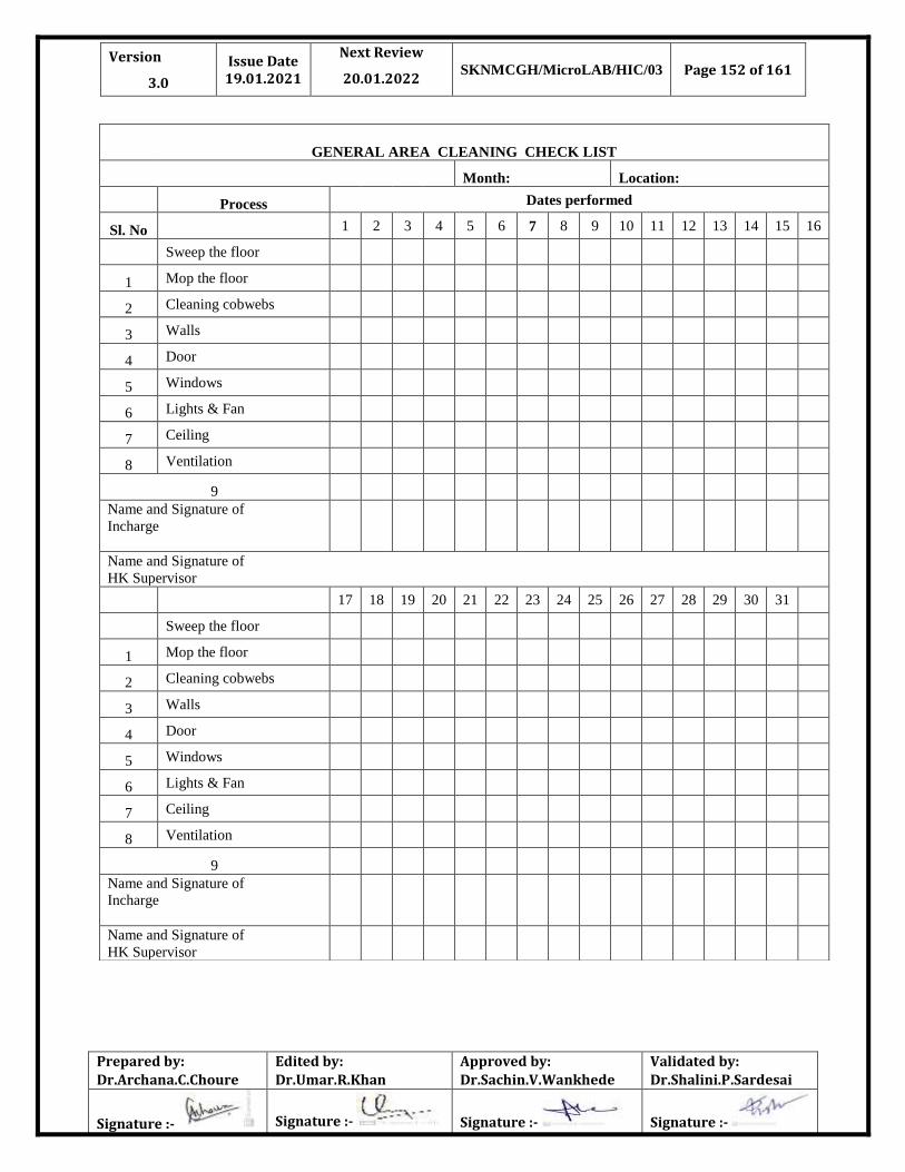
Version
3.0
Issue Date 19.01.2021
Next Review
20.01.2022 SKNMCGH/MicroLAB/HIC/03 Page 152 of 161
Prepared by:
Dr.Archana.C.Choure
Edited by:
Dr.Umar.R.Khan
Approved by:
Dr.Sachin.V.Wankhede
Validated by:
Dr.Shalini.P.Sardesai
Signature :- Signature :- Signature :- Signature :-
GENERAL AREA CLEANING CHECK LIST
Month: Location:
Process Dates performed
Sl. No 1 2 3 4 5 6 7 8 9 10 11 12 13 14 15 16
Sweep the floor
1 Mop the floor
2 Cleaning cobwebs
3 Walls
4 Door
5 Windows
6 Lights & Fan
7 Ceiling
8 Ventilation
9 Name and Signature of
Incharge
Name and Signature of
HK Supervisor
17 18 19 20 21 22 23 24 25 26 27 28 29 30 31
Sweep the floor
1 Mop the floor
2 Cleaning cobwebs
3 Walls
4 Door
5 Windows
6 Lights & Fan
7 Ceiling
8 Ventilation
9 Name and Signature of
Incharge
Name and Signature of
HK Supervisor
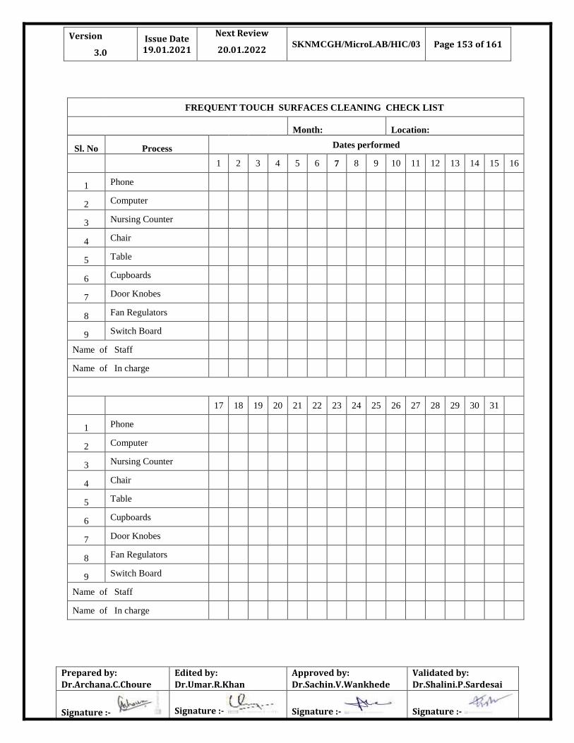
Version
3.0
Issue Date 19.01.2021
Next Review
20.01.2022 SKNMCGH/MicroLAB/HIC/03 Page 153 of 161
Prepared by:
Dr.Archana.C.Choure
Edited by:
Dr.Umar.R.Khan
Approved by:
Dr.Sachin.V.Wankhede
Validated by:
Dr.Shalini.P.Sardesai
Signature :- Signature :- Signature :- Signature :-
FREQUENT TOUCH SURFACES CLEANING CHECK LIST
Month: Location:
Sl. No Process Dates performed
1 2 3 4 5 6 7 8 9 10 11 12 13 14 15 16
1 Phone
2 Computer
3 Nursing Counter
4 Chair
5 Table
6 Cupboards
7 Door Knobes
8 Fan Regulators
9 Switch Board
Name of Staff
Name of In charge
17 18 19 20 21 22 23 24 25 26 27 28 29 30 31
1 Phone
2 Computer
3 Nursing Counter
4 Chair
5 Table
6 Cupboards
7 Door Knobes
8 Fan Regulators
9 Switch Board
Name of Staff
Name of In charge

Version
3.0
Issue Date 19.01.2021
Next Review
20.01.2022 SKNMCGH/MicroLAB/HIC/03 Page 154 of 161
Prepared by:
Dr.Archana.C.Choure
Edited by:
Dr.Umar.R.Khan
Approved by:
Dr.Sachin.V.Wankhede
Validated by:
Dr.Shalini.P.Sardesai
Signature :- Signature :- Signature :- Signature :-
PATIENT CARE EQUIPMENT CHECK LIST (ICU)
Month: Location:
Sl. No Process Dates performed
1 2 3 4 5 6 7 8 9
1
0
1
1
1
2
1
3
1
4
1
5
1
6
1 Stethoscope
2 BP Apparatus with cuff
3 Cardiac Monitor
4 Cardiac Table
5 Ventilator
6 Oxygen Flow meter
7 Bed Rails
8 ECG Cable
9 Probes
10 Patient Trolley
11 Dressing Trolley
12 Suction Apparatus
13 IV Stand
14 Glucometer
15 Wheel Chair
Name of Staff
Name of In charge
1
7
1
8
1
9
2
0
2
1
2
2
2
3
2
4
2
5
2
6
2
7
2
8
2
9
3
0
3
1
1 Stethescope
2 BP Apparatus with cuff
3 Cardiac Monitor
4 Cardiac Table
5 Ventilator
6 Oxygen Flow meter
7 Bed Rails
8 ECG Cable
9 Probes
10 Patient Trolley
11 Dressing Trolley
12 Suction Apparatus
13 IV Stand
14 Glucometer
15 Wheel Chair

Version
3.0
Issue Date 19.01.2021
Next Review
20.01.2022 SKNMCGH/MicroLAB/HIC/03 Page 155 of 161
Prepared by:
Dr.Archana.C.Choure
Edited by:
Dr.Umar.R.Khan
Approved by:
Dr.Sachin.V.Wankhede
Validated by:
Dr.Shalini.P.Sardesai
Signature :- Signature :- Signature :- Signature :-
Name of Staff
Name In charge

Version
3.0
Issue Date 19.01.2021
Next Review
20.01.2022 SKNMCGH/MicroLAB/HIC/03 Page 156 of 161
Prepared by:
Dr.Archana.C.Choure
Edited by:
Dr.Umar.R.Khan
Approved by:
Dr.Sachin.V.Wankhede
Validated by:
Dr.Shalini.P.Sardesai
Signature :- Signature :- Signature :- Signature :-

Version
3.0
Issue Date 19.01.2021
Next Review
20.01.2022 SKNMCGH/MicroLAB/HIC/03 Page 157 of 161
Prepared by:
Dr.Archana.C.Choure
Edited by:
Dr.Umar.R.Khan
Approved by:
Dr.Sachin.V.Wankhede
Validated by:
Dr.Shalini.P.Sardesai
Signature :- Signature :- Signature :- Signature :-

Version
3.0
Issue Date 19.01.2021
Next Review
20.01.2022 SKNMCGH/MicroLAB/HIC/03 Page 158 of 161
Prepared by:
Dr.Archana.C.Choure
Edited by:
Dr.Umar.R.Khan
Approved by:
Dr.Sachin.V.Wankhede
Validated by:
Dr.Shalini.P.Sardesai
Signature :- Signature :- Signature :- Signature :-

Version
3.0
Issue Date 19.01.2021
Next Review
20.01.2022 SKNMCGH/MicroLAB/HIC/03 Page 159 of 161
Prepared by:
Dr.Archana.C.Choure
Edited by:
Dr.Umar.R.Khan
Approved by:
Dr.Sachin.V.Wankhede
Validated by:
Dr.Shalini.P.Sardesai
Signature :- Signature :- Signature :- Signature :-

Version
3.0
Issue Date 19.01.2021
Next Review
20.01.2022 SKNMCGH/MicroLAB/HIC/03 Page 160 of 161
Prepared by:
Dr.Archana.C.Choure
Edited by:
Dr.Umar.R.Khan
Approved by:
Dr.Sachin.V.Wankhede
Validated by:
Dr.Shalini.P.Sardesai
Signature :- Signature :- Signature :- Signature :-

Version
3.0
Issue Date 19.01.2021
Next Review
20.01.2022 SKNMCGH/MicroLAB/HIC/03 Page 161 of 161
Prepared by:
Dr.Archana.C.Choure
Edited by:
Dr.Umar.R.Khan
Approved by:
Dr.Sachin.V.Wankhede
Validated by:
Dr.Shalini.P.Sardesai
Signature :- Signature :- Signature :- Signature :-