Sree Balaji Medica laji Medical College & Hospital ollege & Hospital
45
Sree Bala 1 DEPA SYLLABUS Features of in AN1 4.1 Identify the given important featur anatomical posit AN1 4.2 Identify & descri the given bone AN1 4.3 Describe the imp ossification of low upper end of tibi AN1 4.4 Identify and nam the articulated fo muscle attachme Topic: Fro AN1 5.1 Describe and dem course, relations tributaries), term nerves and vesse AN1 5.2 Describe and dem muscles with the supply and actio AN1 5.3 Describe and dem boundaries, floor femoral triangle AN1 5.4 Explain anatomi abscess & Femor AN1 5.5 Describe and dem canal with its co aji Medical College & H ARTMENT OF ANATOMY S RECOMMENDED BY NMC ndividual bones (Lower Limb) n bone, its side, res & keep it in tion K/S S ibe joints formed by K/S S portance of wer end of femur & ia K K me various bones in oot with individual ent K/S S ont & Medial side of thigh monstrate origin, s, branches (or mination of important els of anterior thigh K/S S monstrate major eir attachment, nerve ons K/S S monstrate r, roof and contents of K/S S ical basis of Psoas ral hernia K K monstrate adductor ontent K/S S Hospital SH Y SH Y KH Y SH N SH Y SH Y SH Y KH N SH Y
-
Upload
khangminh22 -
Category
Documents
-
view
1 -
download
0
Transcript of Sree Balaji Medica laji Medical College & Hospital ollege & Hospital

Sree Balaji Medical College & Hospital
1
DEPARTMENT OF ANATOMY
SYLLABUS RECOMMENDED BY
Features of individual bones (Lower Limb)
AN14.1
Identify the given bone, its side, important features & keep it in anatomical position
AN14.2
Identify & describe joints formed by the given bone
AN14.3
Describe the importance of ossification of lower end of femur & upper end of tibia
AN14.4
Identify and name various bones in the articulated foot with individual muscle attachment
Topic: Front & Medial side of thigh
AN15.1
Describe and demonstrate origin, course, relations, branches (or tributaries), termination of important nerves and vessels of
AN15.2
Describe and demonstrate major muscles with their attachment, nerve supply and actions
AN15.3
Describe and demonstrate boundaries, floor, roof and contents of femoral triangle
AN15.4
Explain anatomical basis of Psoas abscess & Femoral hernia
AN15.5
Describe and demonstrate adductor canal with its content
laji Medical College & Hospital
DEPARTMENT OF ANATOMY
SYLLABUS RECOMMENDED BY NMC
Features of individual bones (Lower Limb)
Identify the given bone, its side, important features & keep it in anatomical position
K/S SH
Identify & describe joints formed by K/S SH
Describe the importance of ossification of lower end of femur & upper end of tibia
K KH
Identify and name various bones in the articulated foot with individual
attachment
K/S SH
Topic: Front & Medial side of thigh
Describe and demonstrate origin, course, relations, branches (or tributaries), termination of important nerves and vessels of anterior thigh
K/S SH
Describe and demonstrate major muscles with their attachment, nerve supply and actions
K/S SH
Describe and demonstrate boundaries, floor, roof and contents of
K/S SH
Explain anatomical basis of Psoas abscess & Femoral hernia
K KH
Describe and demonstrate adductor canal with its content
K/S SH
laji Medical College & Hospital
Features of individual bones (Lower Limb)
SH Y
SH Y
KH Y
SH N
Topic: Front & Medial side of thigh
SH Y
SH Y
SH Y
KH N
SH Y
Sree Balaji Medical College & Hospital
2
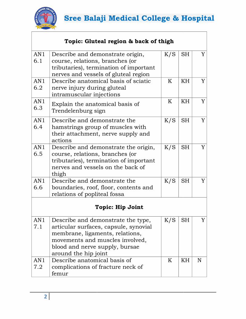
Topic: Gluteal region & back of thigh
AN16.1
Describe and demonstrate origin, course, relations, branches (or tributaries), termination of important nerves and vessels of gluteal region
AN16.2
Describe anatomical basis of sciatic nerve injury during gluteal intramuscular injections
AN16.3
Explain the anatomical basis of Trendelenburg sign
AN16.4
Describe and demonstrate the hamstrings group of muscles with their attachment, nerve supply and actions
AN16.5
Describe and demonstrate the origin, course, relations, tributaries), termination of important nerves and vessels on the back of thigh
AN16.6
Describe and demonstrate the boundaries, roof, floor, contents and relations of popliteal fossa
Topic: Hip Joint
AN17.1
Describe and demonstrate the type, articular surfaces, capsule, synovial membrane, ligaments, relations, movements and muscles involved, blood and nerve supply, bursae around the hip joint
AN17.2
Describe anatomical basis of complications of fracture neck of femur
laji Medical College & Hospital
Topic: Gluteal region & back of thigh
Describe and demonstrate origin, course, relations, branches (or tributaries), termination of important nerves and vessels of gluteal region
K/S SH
Describe anatomical basis of sciatic nerve injury during gluteal intramuscular injections
K KH
Explain the anatomical basis of Trendelenburg sign
K KH
Describe and demonstrate the hamstrings group of muscles with their attachment, nerve supply and
K/S SH
Describe and demonstrate the origin, course, relations, branches (or tributaries), termination of important nerves and vessels on the back of
K/S SH
Describe and demonstrate the boundaries, roof, floor, contents and relations of popliteal fossa
K/S SH
Topic: Hip Joint
Describe and demonstrate the type, surfaces, capsule, synovial
membrane, ligaments, relations, movements and muscles involved, blood and nerve supply, bursae around the hip joint
K/S SH
Describe anatomical basis of complications of fracture neck of
K KH
laji Medical College & Hospital
Topic: Gluteal region & back of thigh
SH Y
KH Y
KH Y
SH Y
SH Y
SH Y
Topic: Hip Joint
SH Y
KH N
Sree Balaji Medical College & Hospital
3
AN17.3
Describe dislocation of hip joint and surgical hip replacement
Topic: Knee joint, Anterolateral compartment of leg &
AN18.1
Describe and demonstrate major muscles of anterolateralof leg with their attachment, nerve supply and actions
AN18.2
Describe and demonstrate origin, course, relations, branches (or tributaries), termination of important nerves and vessels of anterior compartment of leg
AN18.3
Explain the anatomical basis of foot drop
AN18.4
Describe and demonstrate the type, articular surfaces, capsule, synovial membrane, ligaments, relations, movements and muscles involved, blood and nerve supply, bursae around the knee joint
AN18.5
Explain the anatomical basis of locking and unlocking of the knee joint
AN18.6
Describe knee joint injuries with its applied anatomy
AN18.7
Explain anatomical basis of Osteoarthritis
Topic: Back of Leg &
AN19.1
Describe and demonstrate the major muscles of back of leg with their attachment, nerve supply and actions
laji Medical College & Hospital
dislocation of hip joint and surgical hip replacement
K KH
Topic: Knee joint, Anterolateral compartment of leg & dorsum of foot
Describe and demonstrate major muscles of anterolateral compartment of leg with their attachment, nerve supply and actions
K/S SH
Describe and demonstrate origin, course, relations, branches (or tributaries), termination of important nerves and vessels of anterior compartment of leg
K/S SH
Explain the anatomical basis of foot K KH
Describe and demonstrate the type, articular surfaces, capsule, synovial membrane, ligaments, relations, movements and muscles involved, blood and nerve supply, bursae around the knee joint
K/S SH
Explain the anatomical basis of locking and unlocking of the knee
K KH
Describe knee joint injuries with its applied anatomy
K KH
Explain anatomical basis of K KH
Topic: Back of Leg & Sole
Describe and demonstrate the major muscles of back of leg with their attachment, nerve supply and actions
K/S SH
laji Medical College & Hospital
KH N
Topic: Knee joint, Anterolateral compartment of leg &
SH Y
SH Y
KH Y
SH Y
KH Y
KH N
KH N
Sole
SH Y
Sree Balaji Medical College & Hospital
4
AN19.2
Describe and demonstrate the origin, course, relations, branches (or tributaries), termination of important nerves and vessels of back of leg
AN19.3
Explain the concept of “Peripheral heart”
AN19.4
Explain the anatomical basis of rupture of calcaneal tendon
AN19.5
Describe factors maintaining importance arches of the foot with its importance
AN19.6
Explain the anatomical basis of Flat foot & Club foot
AN19.7
Explain the anatomical basis of Metatarsalgia & Plantar fasciitis
Topic: General Features, Joints, radiographs & surface
AN20.1
Describe and demonstrate the type, articular surfaces, capsule, synovial membrane, ligaments, relations, movements and muscles involved, blood and nerve supply of and ankle joint
AN20.2
Describe the subtalar and transverse tarsal joints
AN20.3
Describe and demonstrate Fascia lata, Venous drainage, Lymphatic drainage, Retinacula & Dermatomes of lower limb
AN20.4
Explain anatomical basis of enlarged inguinal lymph nodes
laji Medical College & Hospital
Describe and demonstrate the origin, course, relations, branches (or tributaries), termination of important nerves and vessels of back of leg
K/S SH
Explain the concept of “Peripheral K KH
Explain the anatomical basis of rupture of calcaneal tendon
K KH
Describe factors maintaining importance arches of the foot with its
K KH
Explain the anatomical basis of Flat
K KH
Explain the anatomical basis of Metatarsalgia & Plantar fasciitis
K KH
Topic: General Features, Joints, radiographs & surface marking
Describe and demonstrate the type, articular surfaces, capsule, synovial membrane, ligaments, relations, movements and muscles involved, blood and nerve supply of tibiofibular
K/S SH
Describe the subtalar and transverse K KH
Describe and demonstrate Fascia lata, Venous drainage, Lymphatic drainage, Retinacula & Dermatomes of lower
K/S SH
anatomical basis of enlarged inguinal lymph nodes
K KH
laji Medical College & Hospital
SH Y
KH Y
KH N
KH Y
KH N
KH N
Topic: General Features, Joints, radiographs & surface
SH Y
KH N
SH Y
KH N
Sree Balaji Medical College & Hospital
5
AN20.5
Explain anatomical basis of varicose veins and deep vein thrombosis
AN20.6
Identify the bones and joints of lower limb seen in anteroposteriorlateral view radiographs of various regions of lower limb
AN20.7
Identify & demonstrate important bony landmarks of lower limb: Vertebral levels of highest point of iliac crest, posterior superior iliac spines, iliac tubercle, pubic tubercle, ischial tuberosity, adductor tubercle,-Tibial tuberosity, head of fibula,-Medial and lateral malleoli, Condyles of femur and tibia, sustentaculum tali, tuberosity of fifth metatarsal, tuberosity of the navicular
AN20.8
Identify & demonstrate palpation of femoral, popliteal, post tibial, anti tibial & dorsalis pedisa simulated environment
AN20.9
Identify & demonstrate Palpation of vessels (femoral, popliteal,dorsalis pedis,post tibial), Mid inguinal point, Surface projection of: femoral nerve, Saphenous opening, Sciatic, tibial, common peroneal & deep peroneal nerve, Great and small saphenous veins
AN20.10
Describe basic concept of development of lower limb
Topic: Thoracic cage
AN21.1
Identify and describe the salient features of sternum, typical rib, Iand typical thoracic vertebra
laji Medical College & Hospital
Explain anatomical basis of varicose veins and deep vein thrombosis
K KH
Identify the bones and joints of lower limb seen in anteroposterior and lateral view radiographs of various regions of lower limb
K/S SH
demonstrate important bony landmarks of lower limb: -Vertebral levels of highest point of iliac crest, posterior superior iliac spines, iliac tubercle, pubic tubercle, ischial tuberosity, adductor tubercle, Tibial tuberosity, head of fibula,
lateral malleoli, Condyles of femur and tibia, sustentaculum tali, tuberosity of fifth metatarsal, tuberosity of the navicular
K/S SH
Identify & demonstrate palpation of femoral, popliteal, post tibial, anti tibial & dorsalis pedis blood vessels in a simulated environment
K/S SH
Identify & demonstrate Palpation of vessels (femoral, popliteal,dorsalis pedis,post tibial), Mid inguinal point, Surface projection of: femoral nerve, Saphenous opening, Sciatic, tibial,
peroneal & deep peroneal nerve, Great and small saphenous
K/S SH
Describe basic concept of development K KH
Topic: Thoracic cage
Identify and describe the salient features of sternum, typical rib, Ist rib and typical thoracic vertebra
K/S SH
laji Medical College & Hospital
KH Y
SH Y
SH Y
SH Y
SH Y
KH N
Topic: Thoracic cage
SH Y
Sree Balaji Medical College & Hospital
6
AN21.2
Identify & describe the features of 211th and 12th ribs, 1thoracic vertebrae
AN21.3
Describe & demonstrate the boundaries of thoracic inlet, cavity and outlet
AN21.4
Describe & demonstrate extent, attachments, direction of fibres, nerve supply and actions of intercostal muscles
AN21.5
Describe & demonstrate origin, course, relations and branches of a typical intercostal nerve
AN21.6
Mention origin, course and branches/ tributaries of: 1) anterior & posterior intercostal vessels 2) internal thoracic vessels
AN21.7
Mention the origin, course, relations and branches of 1) atypical intercostal nerve2) superior intercostal artery, subcostal artery
AN21.8
Describe & demonstrate type, articular surfaces & movements of manubriosternal, costovertebral, costotransverse and joints
AN21.9
Describe & demonstrate mechanics and types of respiration
AN21.10
Describe costochondral and interchondral joints
AN21.11
Mention boundaries and contents of the superior, anterior, middle and posterior mediastinum
laji Medical College & Hospital
Identify & describe the features of 2nd, ribs, 1st, 11th and 12th
thoracic vertebrae
K/S SH
& demonstrate the boundaries of thoracic inlet, cavity
K/S SH
Describe & demonstrate extent, attachments, direction of fibres, nerve supply and actions of intercostal
K/S SH
demonstrate origin, course, relations and branches of a typical intercostal nerve
K/S SH
Mention origin, course and branches/
1) anterior & posterior intercostal
2) internal thoracic vessels
K KH
origin, course, relations and branches of 1) atypical intercostal nerve 2) superior intercostal artery,
K KH
Describe & demonstrate type, articular surfaces & movements of manubriosternal, costovertebral, costotransverse and xiphisternal
K/S SH
Describe & demonstrate mechanics and types of respiration
K/S SH
Describe costochondral and interchondral joints
K KH
Mention boundaries and contents of the superior, anterior, middle and posterior mediastinum
K KH
laji Medical College & Hospital
SH N
SH Y
SH Y
SH Y
KH Y
KH N
SH Y
SH Y
KH N
KH Y
Sree Balaji Medical College & Hospital
7
Topic: Heart & Pericardium
AN22.1
Describe & demonstrate subdivisions, sinuses in pericardium, blood supply and nerve supply of pericardium
AN22.2
Describe & demonstrate external and internal features of each chamber of heart
AN22.3
Describe & demonstrate origin, course and branches of coronary arteries
AN22.4
Describe anatomical basis of ischaemic heart disease
AN22.5
Describe & demonstrate the formation, course, tributaries and termination of coronary sinus
AN22.6 Describe the fibrous skeleton of heart
AN22.7
Mention the parts, position and arterial supply of the conducting system of heart
Topic: Mediastinum
AN23.1
Describe & demonstrate the external appearance, relations, blood supply, nerve supply,lymphatic drainage and applied anatomy of oesophagus
AN23.2
Describe & demonstrate the extent, relations tributaries of thoracic duct and enumerate its applied anatomy
AN23.3
Describe & demonstrate origin, course, relations, tributaries and termination of superior venacava, azygos, hemiazygos and accessory hemiazygos veins
laji Medical College & Hospital
Topic: Heart & Pericardium
Describe & demonstrate subdivisions, sinuses in pericardium, blood supply and nerve supply of pericardium
K/S SH
Describe & demonstrate external and internal features of each chamber of
K/S SH
Describe & demonstrate origin, course and branches of coronary arteries
K/S SH
Describe anatomical basis of ischaemic heart disease
K KH
Describe & demonstrate the formation, course, tributaries and termination of coronary sinus
K/S SH
Describe the fibrous skeleton of heart K KH
Mention the parts, position and arterial supply of the conducting
K KH
Topic: Mediastinum
demonstrate the external appearance, relations, blood supply, nerve supply,lymphatic drainage and applied anatomy of oesophagus
K/S SH
Describe & demonstrate the extent, relations tributaries of thoracic duct and enumerate its applied anatomy
K/S SH
Describe & demonstrate origin, course, relations, tributaries and termination of superior venacava, azygos, hemiazygos and accessory hemiazygos veins
K/S SH
laji Medical College & Hospital
Topic: Heart & Pericardium
SH Y
SH Y
SH Y
KH Y
SH Y
KH Y
KH Y
Topic: Mediastinum
SH Y
SH Y
SH Y

Sree Balaji Medical College & Hospital
8
AN23.4
Mention the extent, branches and relations of arch of aorta & descending thoracic aorta
AN23.5
Identify & Mention the location and extent of thoracic sympathetic chain
AN23.6 Describe the splanchnic nerves
AN23.7
Mention the extent, relations and applied anatomy of lymphatic duct
Topic: Lungs & Trachea
AN24.1
Mention the blood supply, lymphatic drainage and nerve supply of pleura, extent of pleura and describe the pleural recesses and their applied anatomy
AN24.2
Identify side, external features and relations of structures which form root of lung & bronchial tree and their clinical correlate
AN24.3
Describe a bronchopulmonary segment
AN24.4
Identify phrenic nerve &formation & distribution
AN24.5
Mention the blood supply, lymphatic drainage and nerve supply of lungs
AN24.6
Describe the extent, length, relations, blood supply, lymphatic drainage and nerve supply of trachea
laji Medical College & Hospital
Mention the extent, branches and relations of arch of aorta & descending thoracic aorta
K KH
Identify & Mention the location and extent of thoracic sympathetic chain
K/S SH
Describe the splanchnic nerves K KH
Mention the extent, relations and applied anatomy of lymphatic duct
K KH
Topic: Lungs & Trachea
Mention the blood supply, lymphatic drainage and nerve supply of pleura, extent of pleura and describe the pleural recesses and their applied
K KH
Identify side, external features and relations of structures which form root of lung & bronchial tree and their clinical correlate
K/S SH
Describe a bronchopulmonary K KH
Identify phrenic nerve & describe its formation & distribution
K/S SH
Mention the blood supply, lymphatic drainage and nerve supply of lungs
K KH
Describe the extent, length, relations, blood supply, lymphatic drainage and nerve supply of trachea
K KH
Topic: Thorax
laji Medical College & Hospital
KH Y
SH Y
KH N
KH Y
Topic: Lungs & Trachea
KH Y
SH Y
KH Y
SH Y
KH Y
KH N
Topic: Thorax
Sree Balaji Medical College & Hospital
9
AN25.1
Identify, draw and label a slide of trachea and lung
AN25.2
Describe development of pleura, lung & heart
AN25.3
Describe fetal circulation and changes occurring at birth
AN25.4
Describe embryological basis of:1) atrial septal defect, 2) ventricular septal defect, 3) Fallot’s tetralogy &4) tracheo-oesophageal fistula
AN25.5
Describe developmental basis of congenital anomalies, transposition of great vessels, dextrocardia, patent ductus arteriosus and coarctation of aorta
AN25.6
Mention development of aortic arch arteries, SVC, IVC and coronary sinus
AN25.7
Identify structures seen on a plain xray chest (PA view)
AN25.8
Identify and describe in brief a barium swallow
AN25.9
Demonstrate surface marking of lines of pleural reflection, lung borders and fissures, trachea, heart borders, apexbeat & surface projection of valves of heart
Topic: Skull osteology
AN26.1
Demonstrate anatomical position of skull, Identify and locate individual skull bones in skull
laji Medical College & Hospital
Identify, draw and label a slide of trachea and lung
K/S SH
Describe development of pleura, lung K KH
Describe fetal circulation and changes occurring at birth
K KH
Describe embryological basis of: 1) atrial septal defect, 2) ventricular septal defect, 3) Fallot’s tetralogy &
oesophageal fistula
K KH
Describe developmental basis of congenital anomalies, transposition of great vessels, dextrocardia, patent ductus arteriosus and coarctation of
K KH
Mention development of aortic arch arteries, SVC, IVC and coronary sinus
K KH
Identify structures seen on a plain x-ray chest (PA view)
K/S SH
Identify and describe in brief a barium K/S SH
Demonstrate surface marking of lines of pleural reflection, lung borders and fissures, trachea, heart borders, apex beat & surface projection of valves of
K/S SH
Topic: Skull osteology
Demonstrate anatomical position of skull, Identify and locate individual
bones in skull
K/S SH
laji Medical College & Hospital
SH Y
KH Y
KH Y
KH Y
KH Y
KH N
SH Y
SH N
SH Y
Topic: Skull osteology
SH Y

Sree Balaji Medical College & Hospital
10
AN26.2
Describe the features of norma frontalis, verticalis, occipitalis, lateralis and basalis
AN26.3
Describe cranial cavity, its subdivisions, foramina and structures passing through them
AN26.4
Describe morphological features of mandible
AN26.5
Describe features of typical and atypical cervical vertebrae (atlas and axis)
AN26.6
Explain the concept of bones that ossify in membrane
AN26.7
Describe the features of the 7cervical vertebra
AN27.1
Describe the layers of scalp, its blood supply, its nerve supply and surgical importance
AN27.2
Describe emissary veins with its role in spread of infection from extracranial route to intracranial venous sinuses
Topic: Face & parotid region
AN28.1
Describe & demonstrate muscles of facial expression and their nerve supply
AN28.2 Describe sensory innervation of face
laji Medical College & Hospital
Describe the features of norma frontalis, verticalis, occipitalis, lateralis and basalis
K/S SH
Describe cranial cavity, its subdivisions, foramina and structures passing through them
K/S SH
morphological features of K/S SH
Describe features of typical and atypical cervical vertebrae (atlas and
K/S SH
Explain the concept of bones that ossify in membrane
K KH
Describe the features of the 7th cervical vertebra
K/S SH
Topic: Scalp
Describe the layers of scalp, its blood supply, its nerve supply and surgical
K KH
Describe emissary veins with its role in spread of infection from extracranial route to intracranial
K KH
Topic: Face & parotid region
demonstrate muscles of facial expression and their nerve
K/S SH
Describe sensory innervation of face K KH
laji Medical College & Hospital
SH Y
SH Y
SH Y
SH Y
KH N
SH N
Topic: Scalp
KH Y
KH Y
Topic: Face & parotid region
SH Y
KH Y

Sree Balaji Medical College & Hospital
11
AN28.3
Describe & demonstrate origin /formation, course, branches /tributaries of facial vessels
AN28.4
Describe & demonstrate branches of facial nerve with distribution
AN28.5
Describe cervical lymph nodes and lymphatic drainage of head, face and neck
AN28.6
Identify superficial muscles of face, their nerve supply and actions
AN28.7
Explain the anatomical basis of facial nerve palsy
AN28.8
Explain surgical importance of deep facial vein
AN28.9
Describe & demonstrate the parts, borders, surfaces, contents, relations and nerve supply of parotid gland with course of its duct andimportance
AN28.10
Explain the anatomical basis of Frey’s syndrome
Topic: Posterior triangle of neck
AN29.1
Describe & demonstrate attachments, nerve supply, relations and actions of sternocleidomastoid
AN29.2
Explain anatomical basis of Erb’s & Klumpke’s palsy
AN29.3 Explain anatomical basis of wry neck
laji Medical College & Hospital
Describe & demonstrate origin /formation, course, branches /tributaries of facial vessels
K/S SH
demonstrate branches of facial nerve with distribution
K/S SH
Describe cervical lymph nodes and lymphatic drainage of head, face and
K KH
Identify superficial muscles of face, their nerve supply and actions
K/S SH
the anatomical basis of facial K KH
Explain surgical importance of deep K KH
Describe & demonstrate the parts, borders, surfaces, contents, relations and nerve supply of parotid gland with course of its duct and surgical
K/S SH
Explain the anatomical basis of Frey’s K KH
Topic: Posterior triangle of neck
demonstrate attachments, nerve supply, relations and actions of sternocleidomastoid
K/S SH
Explain anatomical basis of Erb’s & Klumpke’s palsy
K KH
Explain anatomical basis of wry neck K KH
laji Medical College & Hospital
SH Y
SH Y
KH Y
SH Y
KH Y
KH Y
SH Y
KH N
Topic: Posterior triangle of neck
SH Y
KH Y
KH N

Sree Balaji Medical College & Hospital
12
AN29.4
Describe & demonstrate attachments of 1) inferior belly of omohyoid, 2)scalenus anterior, 3) scalenus medius & 4) levator scapulae
Topic: Cranial cavity
AN30.1
Describe the cranial fossae & identify related structures
AN30.2
Describe & identify major foramina with structures passing through them
AN30.3
Describe & identify dural folds & dural venous sinuses
AN30.4
Describe clinical importance of dural venous sinuses
AN30.5
Explain effect of pituitary tumours on visual pathway
AN31.1
Describe & identify extra ocular muscles of eyeball
AN31.2
Describe & demonstrate nerves and vessels in the orbit
AN31.3
Describe anatomical basis of Horner’s syndrome
AN31.4
Enumerate components of lacrimal apparatus
AN31.5
Explain the anatomical basis of oculomotor, trochlear and abducent nerve palsies along with strabismus
laji Medical College & Hospital
demonstrate attachments of 1) inferior belly of omohyoid, 2)scalenus anterior, 3) scalenus medius & 4) levator scapulae
K/S SH
Topic: Cranial cavity
Describe the cranial fossae & identify related structures
K/S SH
Describe & identify major foramina with structures passing through them
K/S SH
Describe & identify dural folds & dural venous sinuses
K/S SH
Describe clinical importance of dural
K KH
Explain effect of pituitary tumours on K KH
Topic: Orbit
identify extra ocular muscles of eyeball
K/S SH
Describe & demonstrate nerves and vessels in the orbit
K/S SH
Describe anatomical basis of Horner’s K KH
Enumerate components of lacrimal K KH
Explain the anatomical basis of oculomotor, trochlear and abducent nerve palsies along with strabismus
K KH
laji Medical College & Hospital
SH N
Topic: Cranial cavity
SH Y
SH Y
SH Y
KH Y
KH N
Topic: Orbit
SH Y
SH Y
KH N
KH Y
KH Y
Sree Balaji Medical College & Hospital
13
Topic: Anterior Triangle
AN32.1
Describe boundaries and of anterior triangle
AN32.2
Describe & demonstrate boundaries and contents of muscular, carotid, digastric and submental triangles
Topic: Temporal and Infratemporal
AN33.1
Describe & demonstrate extent, boundaries and contents of temporal and infratemporal fossae
AN33.2
Describe & demonstrate attachments, direction of fibres, nerve supply and actions of muscles of mastication
AN33.3
Describe & demonstrate articulating surface, type & movements of temporomandibular joint
AN33.4
Explain the clinical significance of pterygoid venous plexus
AN33.5
Describe the features of dislocation of temporomandibular joint
Topic: Submandibular region
AN34.1
Describe & demonstrate the morphology, relations and nerve supply of submandibular salivary gland & submandibular ganglion
AN34.2
Describe the basis of formation of submandibular stones
laji Medical College & Hospital
Topic: Anterior Triangle
Describe boundaries and subdivisions of anterior triangle
K KH
Describe & demonstrate boundaries and contents of muscular, carotid, digastric and submental triangles
K/S SH
Topic: Temporal and Infratemporal regions
Describe & demonstrate extent, boundaries and contents of temporal and infratemporal fossae
K/S SH
Describe & demonstrate attachments, direction of fibres, nerve supply and actions of muscles of mastication
K/S SH
Describe & demonstrate articulating surface, type & movements of temporomandibular joint
K/S SH
Explain the clinical significance of pterygoid venous plexus
K KH
Describe the features of dislocation of temporomandibular joint
K KH
Topic: Submandibular region
Describe & demonstrate the morphology, relations and nerve
submandibular salivary gland & submandibular ganglion
K/S SH
Describe the basis of formation of submandibular stones
K KH
laji Medical College & Hospital
Topic: Anterior Triangle
KH Y
SH Y
regions
SH Y
SH Y
SH Y
KH Y
KH N
Topic: Submandibular region
SH Y
KH N

Sree Balaji Medical College & Hospital
14
Topic: Deep structures in the neck
AN35.1
Describe the parts, extent, attachments, modifications of deep cervical fascia
AN35.2
Describe & demonstrate location, parts, borders, surfaces, relations & blood supply of thyroid gland
AN35.3
Demonstrate & describe the origin, parts, course & branches subclavian artery
AN35.4
Describe & demonstrate origin, course, relations, tributaries and termination of internal jugular & brachiocephalic veins
AN35.5
Describe and demonstrate extent, drainage & applied anatomy of cervical lymph nodes
AN35.6
Describe and demonstrate the extent, formation, relation & branches of cervical sympathetic chain
AN35.7
Describe the course and branches of IX, X, XI & XII nerve in the neck
AN35.8
Describe the anatomically clinical features of Thyroid swellings
AN35.9
Describe the clinical features of compression of subclavian artery and lower trunk of brachial plexus by cervical rib
AN35.10 Describe the fascial spaces of neck
Topic: Mouth, Pharynx & Palate
laji Medical College & Hospital
Topic: Deep structures in the neck
the parts, extent, attachments, modifications of deep
K KH
Describe & demonstrate location, parts, borders, surfaces, relations & blood supply of thyroid gland
K/S SH
Demonstrate & describe the origin, & branches subclavian
K/S SH
Describe & demonstrate origin, course, relations, tributaries and termination of internal jugular & brachiocephalic veins
K/S SH
Describe and demonstrate extent, drainage & applied anatomy of
lymph nodes
K/S SH
Describe and demonstrate the extent, formation, relation & branches of cervical sympathetic chain
K/S SH
Describe the course and branches of IX, X, XI & XII nerve in the neck
K KH
Describe the anatomically relevant clinical features of Thyroid swellings
K KH
Describe the clinical features of compression of subclavian artery and lower trunk of brachial plexus by
K KH
Describe the fascial spaces of neck K KH
Topic: Mouth, Pharynx & Palate
laji Medical College & Hospital
Topic: Deep structures in the neck
KH Y
SH Y
SH Y
SH Y
SH Y
SH Y
KH Y
KH N
KH N
KH N
Topic: Mouth, Pharynx & Palate

Sree Balaji Medical College & Hospital
15
AN36.1
Describe the 1) morphology, relations, blood supply and applied anatomy of palatine tonsil 2) composition of soft palate
AN36.2
Describe the components and functions of Waldeyer’s lymphatic ring
AN36.3
Describe the boundaries and clinical significance of pyriform fossa
AN36.4
Describe the anatomical basis of tonsillitis, tonsillectomy, adenoids and peri-tonsillar abscess
AN36.5
Describe the clinical significance of Killian’s dehiscence
Topic: Cavity of Nose
AN37.1
Describe & demonstrate features of nasal septum, lateral wall of nose, their blood supply and nerve supply
AN37.2
Describe location and functional anatomy of paranasal sinuses
AN37.3
Describe anatomical basis of sinusitis & maxillary sinus tumours
AN38.1
Describe the morphology, identify structure of the wall, nerve supply, blood supply and actions of intrinsic and extrinsic muscles of
AN38.2
Describe the anatomical aspects of laryngitis
laji Medical College & Hospital
Describe the 1) morphology, relations, blood supply and applied anatomy of palatine tonsil 2) composition of soft
K KH
Describe the components and functions of Waldeyer’s lymphatic
K KH
Describe the boundaries and clinical significance of pyriform fossa
K KH
Describe the anatomical basis of tonsillitis, tonsillectomy, adenoids and
abscess
K KH
Describe the clinical significance of Killian’s dehiscence
K KH
Topic: Cavity of Nose
Describe & demonstrate features of septum, lateral wall of nose,
their blood supply and nerve supply
K/S SH
Describe location and functional anatomy of paranasal sinuses
K KH
Describe anatomical basis of sinusitis & maxillary sinus tumours
K KH
Topic: Larynx
Describe the morphology, identify structure of the wall, nerve supply, blood supply and actions of intrinsic and extrinsic muscles of the larynx
K/S SH
Describe the anatomical aspects of K KH
laji Medical College & Hospital
KH Y
KH Y
KH N
KH N
KH N
Topic: Cavity of Nose
SH Y
KH Y
KH N
Topic: Larynx
SH Y
KH N

Sree Balaji Medical College & Hospital
16
AN38.3
Describe anatomical basis of recurrent laryngeal nerve injury
AN39.1
Describe & demonstrate the morphology, nerve supply, embryological basis of nerve supply, blood supply, lymphatic drainage and actions of extrinsic and intrinsic muscles of tongue
AN39.2
Explain the anatomical basis of hypoglossal nerve palsy
Topic: Organs of hearing and equilibrium
AN40.1
Describe & identify the parts, blood supply and nerve supply of external ear
AN40.2
Describe & demonstrate the boundaries, contents, relations and functional anatomy of middle ear and auditory tube
AN40.3 Describe the features of internal ear
AN40.4
Explain anatomical basis of otitis externa and otitis media
AN40.5
Explain anatomical basis of myringotomy
AN41.1
Describe & demonstrate parts and layers of eyeball
laji Medical College & Hospital
Describe anatomical basis of recurrent laryngeal nerve injury
K KH
Topic: Tongue
Describe & demonstrate the morphology, nerve supply, embryological basis of nerve supply, blood supply, lymphatic drainage and actions of extrinsic and intrinsic muscles of tongue
K/S SH
Explain the anatomical basis of hypoglossal nerve palsy
K KH
Topic: Organs of hearing and equilibrium
identify the parts, blood supply and nerve supply of external
K/S SH
Describe & demonstrate the boundaries, contents, relations and functional anatomy of middle ear and
K/S SH
Describe the features of internal ear K KH
Explain anatomical basis of otitis externa and otitis media
K KH
Explain anatomical basis of K KH
Topic: Eyeball
Describe & demonstrate parts and
K/S SH
laji Medical College & Hospital
KH N
Topic: Tongue
SH Y
KH N
Topic: Organs of hearing and equilibrium
SH Y
SH Y
KH N
KH N
KH N
Topic: Eyeball
SH Y
Sree Balaji Medical College & Hospital
17
AN41.2
Describe the anatomical aspects of cataract, glaucoma & central retinal artery occlusion
AN41.3
Describe the position, nerve supply and actions of intraocular muscles
Topic: Back Region
AN42.1
Describe the contents of the vertebral canal
AN42.2
Describe & demonstrate the boundaries and contents of Suboccipital triangle
AN42.3
Describe the position, direction of fibres, relations, nerve supply, actions of semispinalis capitis and splenius capitis
Topic: Head & neck Joints, Histology, Development, Radiography & Surface marking
AN43.1
Describe & demonstrate the movements with muscles producing the movements of atlantooccipital joint & atlantoaxial joint
AN43.2
Identify, describe and draw the microanatomy of pituitary gland, thyroid, parathyroid gland, tongue, salivary glands, tonsil, epiglottis, cornea, retina
AN43.3
Identify, describe and draw microanatomy of olfactory epithelium, eyelid, lip, sclerooptic nerve, cochleapineal gland
laji Medical College & Hospital
Describe the anatomical aspects of cataract, glaucoma & central retinal
K KH
Describe the position, nerve supply and actions of intraocular muscles
K KH
Topic: Back Region
Describe the contents of the vertebral K/S SH
Describe & demonstrate the boundaries and contents of
triangle
K/S SH
Describe the position, direction of fibres, relations, nerve supply, actions of semispinalis capitis and splenius
K KH
Topic: Head & neck Joints, Histology, Development, Radiography & Surface marking
Describe & demonstrate the
with muscles producing the movements of atlantooccipital joint & atlantoaxial joint
K/S SH
Identify, describe and draw the microanatomy of pituitary gland, thyroid, parathyroid gland, tongue,
glands, tonsil, epiglottis,
K/S SH
Identify, describe and draw microanatomy of olfactory epithelium, eyelid, lip, sclero-corneal junction, optic nerve, cochlea- organ of corti,
K/S SH
laji Medical College & Hospital
KH N
KH N
Topic: Back Region
SH Y
SH Y
KH N
Topic: Head & neck Joints, Histology, Development,
SH Y
SH Y
SH N
Sree Balaji Medical College & Hospital
18
AN43.4
Describe the development and developmental basis of congenital anomalies of face, palate, tongue, branchial apparatus, pituitary gland, thyroid gland & eye
AN43.5
Demonstrate- 1) Testing of muscles of facial expression, extraocular muscles, musclesPalpation of carotid arteries, facial artery, superficial temporal artery, 3) Location of internal and external jugular veins, 4) Location of hyoid bone, thyroid cartilage and cricoid cartilage with their vertebral levels
AN43.6
Demonstrate surface projection ofThyroid gland, Parotid gland and duct, Pterion, Common carotid artery, Internal jugular vein, Subclavian vein, External jugular vein, Facial artery in the face & accessory nerve
AN43.7
Identify the anatomical structures in 1) Plain x-ray skull, 2) AP view and lateral view 3) Plain xspine-AP and lateral view 4) Plain xray of paranasal sinuses
AN43.8
Describe the anatomical route used for carotid angiogram andangiogram
AN43.9
Identify anatomical structures in carotid angiogram and vertebral angiogram
Topic: Anterior abdominal wall
laji Medical College & Hospital
Describe the development and developmental basis of congenital anomalies of face, palate, tongue, branchial apparatus, pituitary gland, thyroid gland & eye
K KH
1) Testing of muscles of facial expression, extraocular muscles, muscles of mastication, 2) Palpation of carotid arteries, facial artery, superficial temporal artery, 3) Location of internal and external jugular veins, 4) Location of hyoid bone, thyroid cartilage and cricoid cartilage with their vertebral levels
K/S SH
Demonstrate surface projection of- Thyroid gland, Parotid gland and duct, Pterion, Common carotid artery, Internal jugular vein, Subclavian vein, External jugular vein, Facial artery in the face & accessory nerve
K/S SH
Identify the anatomical structures in ray skull, 2) AP view and
lateral view 3) Plain x-ray cervical AP and lateral view 4) Plain x-
ray of paranasal sinuses
K/S SH
Describe the anatomical route used for carotid angiogram and vertebral
K/S SH
Identify anatomical structures in carotid angiogram and vertebral
K/S SH
Topic: Anterior abdominal wall
laji Medical College & Hospital
KH Y
SH Y
SH N
SH Y
SH N
SH N
Topic: Anterior abdominal wall
Sree Balaji Medical College & Hospital
19
AN44.1
Describe & demonstrate the Planes (transpyloric, transtubercular, subcostal, lateral vertical, linea alba, linea semilunaris), regions & Quadrants of abdomen
AN44.2
Describe & identify the Fascia, nerves & blood vessels of anterior abdominal wall
AN44.3
Describe the formation of rectus sheath and its contents
AN44.4
Describe & demonstrate extent, boundaries, contents of Inguinal canal including Hesselbach’s triangle.
AN44.5
Explain the anatomical basis of inguinal hernia.
AN44.6
Describe & demonstrate attachments of muscles of anterior abdominal wall
AN44.7
Enumerate common Abdominal incisions
Topic: Posterior abdominal wall
AN45.1 Describe Thoracolumbar fascia
AN45.2
Describe & demonstrate Lumbar plexus for its root value, formation & branches
AN45.3
Mention the major subgroups of back muscles, nerve supply and action
Topic: Male external genitalia
laji Medical College & Hospital
demonstrate the Planes (transpyloric, transtubercular, subcostal, lateral vertical, linea alba, linea semilunaris), regions & Quadrants of abdomen
K/S SH
Describe & identify the Fascia, nerves & blood vessels of anterior abdominal
K/S SH
Describe the formation of rectus sheath and its contents
K KH
Describe & demonstrate extent, boundaries, contents of Inguinal canal including Hesselbach’s triangle.
K/S SH
Explain the anatomical basis of
K KH
Describe & demonstrate attachments of muscles of anterior abdominal wall
K/S SH
Enumerate common Abdominal K KH
Topic: Posterior abdominal wall
Describe Thoracolumbar fascia K KH
Describe & demonstrate Lumbar plexus for its root value, formation &
K/S SH
Mention the major subgroups of back muscles, nerve supply and action
K KH
Topic: Male external genitalia
laji Medical College & Hospital
SH Y
SH Y
KH Y
SH Y
KH Y
SH Y
KH N
Topic: Posterior abdominal wall
KH Y
SH Y
KH N
Topic: Male external genitalia
Sree Balaji Medical College & Hospital
20
AN46.1
Describe & demonstrate coverings, internal structure, side determination, blood supply, nerve supply, lymphatic drainage & descent of testis applied anatomy
AN46.2 Describe parts of Epididymis
AN46.3
Describe Penis under following headings: (parts, components, blood supply and lymphatic drainage)
AN46.4
Explain the anatomical basis of Varicocoele
AN46.5
Explain the anatomical basis of Phimosis & Circumcision
Topic: Abdominal cavity
AN47.1
Describe & identify boundaries and recesses of Lesser & Greater sac
AN47.2
Name & identify various peritoneal folds & pouches with its explanation.
AN47.3
Explain anatomical basis of Ascites & Peritonitis
AN47.4
Explain anatomical basis of Subphrenic abscess
AN47.5
Describe & demonstrate of abdomen under following headings (anatomical position, external and internal features, important peritoneal and other relations, blood supply, nerve supply, lymphatic drainage and applied aspects)
laji Medical College & Hospital
Describe & demonstrate coverings, internal structure, side determination, blood supply, nerve supply, lymphatic drainage & descent of testis with its applied anatomy
K/S SH
Describe parts of Epididymis K KH
Describe Penis under following headings: (parts, components, blood supply and lymphatic drainage)
K KH
Explain the anatomical basis of K KH
Explain the anatomical basis of Phimosis & Circumcision
K KH
Topic: Abdominal cavity
Describe & identify boundaries and recesses of Lesser & Greater sac
K/S SH
Name & identify various peritoneal folds & pouches with its explanation
K/S SH
Explain anatomical basis of Ascites & K KH
Explain anatomical basis of Subphrenic abscess
K KH
Describe & demonstrate major viscera of abdomen under following headings (anatomical position, external and internal features, important peritoneal and other relations, blood supply, nerve supply, lymphatic drainage and
K/S SH
laji Medical College & Hospital
SH Y
KH Y
KH Y
KH N
KH N
Topic: Abdominal cavity
SH Y
SH Y
KH N
KH N
SH Y
Sree Balaji Medical College & Hospital
21
AN47.6
Explain the anatomical basis of Splenic notch, Accessory spleens, Kehr’s sign, Different types of vagotomy, Liver biopsy (site of needle puncture), Referred pain in cholecystitis, Obstructive jaundice, Referred pain around umbilicus, Radiating pain of kidnLymphatic spread in carcinoma stomach
AN47.7
Mention the clinical importance of Calot’s triangle
AN47.8
Describe & identify the formation, course relations and tributaries of Portal vein, Inferior vena cava & Renal vein
AN47.9
Describe & identify the origin, course, important relations and branches of Abdominal aorta, Coeliac trunk, Superior mesenteric, Inferior mesenteric & Common iliac artery
AN47.10
Enumerate the sites of portosystemic anastomosis
AN47.11
Explain the anatomic basis of hematemesis& caput medusae in portal hypertension
AN47.12
Describe important nerve plexuses of posterior abdominal wall
AN47.13
Describe & demonstrate the attachments, openings, nerve supply & action of the thoracoabdominal diaphragm
AN47.14
Describe the abnormal openings of thoracoabdominal diaphragm and diaphragmatic hernia
laji Medical College & Hospital
Explain the anatomical basis of Splenic notch, Accessory spleens, Kehr’s sign, Different types of vagotomy, Liver biopsy (site of needle puncture), Referred pain in cholecystitis, Obstructive jaundice, Referred pain around umbilicus, Radiating pain of kidney to groin & Lymphatic spread in carcinoma
K KH
Mention the clinical importance of K KH
Describe & identify the formation, course relations and tributaries of Portal vein, Inferior vena cava & Renal
K/S SH
Describe & identify the origin, course, important relations and branches of Abdominal aorta, Coeliac trunk, Superior mesenteric, Inferior mesenteric & Common iliac artery
K/S SH
Enumerate the sites of portosystemic K KH
Explain the anatomic basis of hematemesis& caput medusae in portal hypertension
K KH
Describe important nerve plexuses of posterior abdominal wall
K KH
Describe & demonstrate the attachments, openings, nerve supply
action of the thoracoabdominal
K/S SH
Describe the abnormal openings of thoracoabdominal diaphragm and diaphragmatic hernia
K KH
laji Medical College & Hospital
KH N
KH N
SH Y
SH Y
KH Y
KH Y
KH N
SH Y
KH N

Sree Balaji Medical College & Hospital
22
Topic: Pelvic wall and viscera
AN48.1
Describe & identify the muscles of Pelvic diaphragm
AN48.2
Describe & demonstrate the (position, features, important peritoneal and other relations, blood supply, nerve supply, lymphatic drainage and clinical aspects of) important male & female pelvic viscera
AN48.3
Describe & demonstrate the origin, course, important relations and branches of internal iliac artery
AN48.4 Describe the branches of sacral plexus
AN48.5
Explain the anatomical basis of suprapubic cystostomy, Urinary obstruction in benign prostatic hypertrophy, Retroverted uterus, Prolapse uterus, Internal and external haemorrhoids, Anal fistula, Vasectomy, Tubal pregnancy & Tubal ligation
AN48.6
Describe the neurological basis of Automatic bladder
AN48.7
Mention the lobes involved in benign prostatic hypertrophy & prostatic cancer
AN48.8
Mention the structures palpable during vaginal & rectal examination
Topic: Perineum
laji Medical College & Hospital
Topic: Pelvic wall and viscera
Describe & identify the muscles of Pelvic diaphragm
K/S SH
Describe & demonstrate the (position, features, important peritoneal and other relations, blood supply, nerve supply, lymphatic drainage and clinical aspects of) important male & female pelvic viscera
K/S SH
Describe & demonstrate the origin, course, important relations and branches of internal iliac artery
K/S SH
Describe the branches of sacral plexus K KH
Explain the anatomical basis of cystostomy, Urinary
obstruction in benign prostatic hypertrophy, Retroverted uterus, Prolapse uterus, Internal and external haemorrhoids, Anal fistula, Vasectomy, Tubal pregnancy & Tubal
K KH
Describe the neurological basis of bladder
K KH
Mention the lobes involved in benign prostatic hypertrophy & prostatic
K KH
Mention the structures palpable during vaginal & rectal examination
K KH
Topic: Perineum
laji Medical College & Hospital
Topic: Pelvic wall and viscera
SH Y
SH Y
SH Y
KH Y
KH N
KH N
KH N
KH N
Topic: Perineum
Sree Balaji Medical College & Hospital
23
AN49.1
Describe & demonstrate the superficial & deep perineal pouch (boundaries and contents)
AN49.2 Describe & identify Perineal body
AN49.3
Describe & demonstrate Perineal membrane in male & female
AN49.4
Describe & demonstrate boundaries, content & applied anatomy of Ischiorectal fossa
AN49.5
Explain the anatomical basis of Perineal tear, Episiotomy, abscess and Anal fissure
Topic: Vertebral column
AN50.1
Describe the curvatures of the vertebral column
AN50.2
Describe & demonstrate the type, articular ends, ligaments and movements of Intervertebral joints, Sacroiliac joints & Pubic symphysis
AN50.3
Describe lumbar puncture (site, direction of the needle, structures pierced during the lumbar puncture)
AN50.4
Explain the anatomical basis of Scoliosis, Lordosis, Prolapsed disc, Spondylolisthesis & Spina bifida
Topic: Sectional Anatomy
AN51.1
Describe & identify the at the level of T8, T10 and L1 (transpyloric plane)
laji Medical College & Hospital
Describe & demonstrate the superficial & deep perineal pouch (boundaries and contents)
K/S SH
Describe & identify Perineal body K/S SH
Describe & demonstrate Perineal membrane in male & female
K/S SH
Describe & demonstrate boundaries, content & applied anatomy of Ischiorectal fossa
K/S SH
Explain the anatomical basis of Perineal tear, Episiotomy, Perianal abscess and Anal fissure
K KH
Topic: Vertebral column
Describe the curvatures of the vertebral column
K KH
Describe & demonstrate the type, articular ends, ligaments and movements of Intervertebral joints, Sacroiliac joints & Pubic symphysis
K/S SH
Describe lumbar puncture (site, direction of the needle, structures pierced during the lumbar puncture)
K KH
Explain the anatomical basis of Scoliosis, Lordosis, Prolapsed disc, Spondylolisthesis & Spina bifida
K KH
Topic: Sectional Anatomy
Describe & identify the cross-section at the level of T8, T10 and L1 (transpyloric plane)
K/S SH
laji Medical College & Hospital
SH Y
SH Y
SH Y
SH Y
KH N
Topic: Vertebral column
KH Y
SH Y
KH Y
KH N
Topic: Sectional Anatomy
SH Y

Sree Balaji Medical College & Hospital
24
AN51.2
Describe & identify the midsagittal section of male and female pelvis
Topic: Histology &
AN52.1
Describe & identify the microanatomical features of Gastrointestinal system:Oesophagus, Fundus of stomach, Pylorus of stomach, Duodenum, Jejunum, Ileum, Large inAppendix, Liver, Gall bladder, Pancreas & Suprarenal gland
AN52.2
Describe & identify the microanatomical features of: Urinary system: Kidney, Ureter & Urinary bladder Male Reproductive System: Testis, Epididymis,Vas deferens, Prostate &penis Female reproductive system: Ovary, Uterus, Uterine tube, Cervix, Placenta & Umbilical cord
AN52.3
Describe & identify the microanatomical features of Cardiooesophageal junction, Corpus luteum
AN52.4
Describe the development of abdominal wall
AN52.5
Describe the development and congenital anomalies of Diaphragm
AN52.6
Describe the development and congenital anomalies of: Foregut, Midgut & Hindgut
laji Medical College & Hospital
Describe & identify the midsagittal section of male and female pelvis
K SH
Topic: Histology & Embryology
Describe & identify the microanatomical features of Gastro-intestinal system: Oesophagus, Fundus of stomach, Pylorus of stomach, Duodenum, Jejunum, Ileum, Large intestine, Appendix, Liver, Gall bladder, Pancreas & Suprarenal gland
K/S SH
Describe & identify the microanatomical features of: Urinary system: Kidney, Ureter & Urinary
Male Reproductive System: Testis, Epididymis,Vas deferens, Prostate &
Female reproductive system: Ovary, Uterus, Uterine tube, Cervix, Placenta & Umbilical cord
K/S SH
Describe & identify the microanatomical features of Cardiooesophageal junction, Corpus
K/S SH
Describe the development of anterior
K KH
Describe the development and congenital anomalies of Diaphragm
K KH
Describe the development and congenital anomalies of: Foregut, Midgut & Hindgut
K KH
laji Medical College & Hospital
SH Y
Embryology
SH Y
SH Y
SH N
KH N
KH Y
KH Y

Sree Balaji Medical College & Hospital
25
AN52.7
Describe the development of Urinary system
AN52.8
Describe the development of male & female reproductive system
Topic: Osteology
AN53.1
Identify & hold the bone in the anatomical position,salient features, articulations & demonstrate the attachments of muscle groups
AN53.2
Demonstrate the anatomical position of bony pelvis & show boundaries of pelvic inlet, pelvic cavity, pelvic outlet
AN53.3
Define true pelvis and false pelvis and demonstrate sex determination in male & female bony pelvis
AN53.4
Explain and demonstrate clinical importance of bones of abdominopelvic region (sacralization of lumbar vertebra, Lumbarization1st sacral vertebra, types of bony pelvis & Coccyx)
Topic: Radiodiagnosis
AN54.1
Describe & identify features of plain X ray abdomen
AN54.2
Describe & identify the special radiographs of abdominopelvic region (contrast X ray Barium swallow, Barium meal, Barium enema, Cholecystography, Intravenous pyelography & Hysterosalpingography)
laji Medical College & Hospital
Describe the development of Urinary K KH
Describe the development of male & female reproductive system
K KH
Topic: Osteology
Identify & hold the bone in the anatomical position, Describe the salient features, articulations & demonstrate the attachments of
K/S SH
Demonstrate the anatomical position of bony pelvis & show boundaries of pelvic inlet, pelvic cavity, pelvic outlet
K/S SH
pelvis and false pelvis and demonstrate sex determination in male & female bony pelvis
K/S SH
Explain and demonstrate clinical importance of bones of abdominopelvic region (sacralization of lumbar vertebra, Lumbarization of 1st sacral vertebra, types of bony pelvis & Coccyx)
K/S SH
Topic: Radiodiagnosis
Describe & identify features of plain X K/S SH
Describe & identify the special radiographs of abdominopelvic region (contrast X ray Barium swallow, Barium meal, Barium enema, Cholecystography, Intravenous pyelography & Hysterosalpingography)
K/S SH
laji Medical College & Hospital
KH Y
KH Y
Topic: Osteology
SH Y
SH Y
SH Y
SH N
Topic: Radiodiagnosis
SH Y
SH Y
Sree Balaji Medical College & Hospital
26
AN54.3
Describe role of ERCP, CT abdomen, MRI, Arteriography in radiodiagnosis of abdomen
Topic: Surface marking
AN55.1
Demonstrate the surface marking of; Regions and planes of abdomen, Superficial inguinal ring, Deep inguinal ring , McBurney’s point, Renal Angle & Murphy’s point
AN55.2
Demonstrate the surface projections of: Stomach, Liver, Fundusbladder, Spleen, Duodenum, Pancreas, Ileocaecal junction, Kidneys & Root of mesentery
Topic: Meninges & CSF
AN56.1
Describe & identify various layers of meninges with its extent & modifications
AN56.2
Describe circulation of CSF with its applied anatomy
Topic: Spinal Cord
AN57.1
Identify external features of spinal cord
AN57.2
Describe extent of spinal cord in child & adult with its clinical implication
AN57.3
Draw & label transverse section of spinal cord at midthoracic level
laji Medical College & Hospital
Describe role of ERCP, CT abdomen, Arteriography in radiodiagnosis
K KH
Topic: Surface marking
Demonstrate the surface marking of; Regions and planes of abdomen, Superficial inguinal ring, Deep inguinal ring , McBurney’s point, Renal Angle & Murphy’s point
K/S SH
Demonstrate the surface projections of: Stomach, Liver, Fundus of gall bladder, Spleen, Duodenum, Pancreas, Ileocaecal junction, Kidneys & Root of mesentery
K/S SH
Topic: Meninges & CSF
identify various layers of meninges with its extent &
K/S SH
Describe circulation of CSF with its applied anatomy
K KH
Topic: Spinal Cord
Identify external features of spinal K/S SH
Describe extent of spinal cord in child & adult with its clinical implication
K KH
Draw & label transverse section of spinal cord at mid-cervical & mid-
K KH
laji Medical College & Hospital
KH N
Topic: Surface marking
SH Y
SH Y
Topic: Meninges & CSF
SH Y
KH Y
Topic: Spinal Cord
SH Y
KH Y
KH Y

Sree Balaji Medical College & Hospital
27
AN57.4
Enumerate ascending & descending tracts at mid thoracic level of spinal cord
AN57.5
Describe anatomical basis of syringomyelia
Topic: Medulla Oblongata
AN58.1
Identify external features of medulla oblongata
AN58.2
Describe transverse section of medulla oblongata at the level of 1) pyramidal decussation, 2) sensory decussation 3) ION
AN58.3
Enumerate cranial nerve nuclei in medulla oblongata with their functional group
AN58.4
Describe anatomical basis & effects of medial & lateral medullary syndrome
AN59.1 Identify external features of pons
AN59.2
Draw & label transverse section of pons at the upper and lower level
AN59.3
Enumerate cranial nerve nuclei in pons with their functional group
Topic: Cerebellum
AN60.1
Describe & demonstrate external & internal features of cerebellum
laji Medical College & Hospital
Enumerate ascending & descending tracts at mid thoracic level of spinal
K KH
Describe anatomical basis of K KH
Topic: Medulla Oblongata
Identify external features of medulla K/S SH
Describe transverse section of medulla oblongata at the level of 1) pyramidal decussation, 2) sensory decussation 3)
K KH
Enumerate cranial nerve nuclei in medulla oblongata with their functional group
K KH
Describe anatomical basis & effects of medial & lateral medullary syndrome
K KH
Topic: Pons
Identify external features of pons K/S SH
Draw & label transverse section of pons at the upper and lower level
K KH
Enumerate cranial nerve nuclei in pons with their functional group
K KH
Topic: Cerebellum
Describe & demonstrate external & internal features of cerebellum
K/S SH
laji Medical College & Hospital
KH Y
KH N
Topic: Medulla Oblongata
SH Y
KH Y
KH Y
KH N
Topic: Pons
SH Y
KH Y
KH Y
Topic: Cerebellum
SH Y

Sree Balaji Medical College & Hospital
28
AN60.2
Describe connections of cerebellar cortex and intracerebellar nuclei
AN60.3
Describe anatomical basis of cerebellar dysfunction
Topic: Midbrain
AN61.1
Identify external & internal featuresmidbrain
AN61.2
Describe internal features of midbrain at the level of superior & inferior colliculus
AN61.3
Describe anatomical basis & effects of Benedikt’s and Weber’s syndrome
Topic: Cranial nerve nuclei &
AN62.1
Enumerate cranial nerve nuclei with its functional component
AN62.2
Describe & demonstrate surfaces, sulci, gyri, poles, & functional areas of cerebral hemisphere
AN62.3 Describe the white matter of cerebrum
AN62.4
Enumerate parts & major connections of basal ganglia & limbic lobe
AN62.5
Describe boundaries, parts, gross relations, major nuclei and connections of dorsal thalamus, hypothalamus, epithalamus, metathalamus and subthalamus
laji Medical College & Hospital
Describe connections of cerebellar intracerebellar nuclei
K KH
Describe anatomical basis of cerebellar dysfunction
K KH
Topic: Midbrain
Identify external & internal features of K/S SH
Describe internal features of midbrain at the level of superior & inferior
K KH
Describe anatomical basis & effects of Benedikt’s and Weber’s syndrome
K KH
Topic: Cranial nerve nuclei & Cerebral hemispheres
Enumerate cranial nerve nuclei with its functional component
K KH
Describe & demonstrate surfaces, sulci, gyri, poles, & functional areas of cerebral hemisphere
K/S SH
Describe the white matter of cerebrum K KH
Enumerate parts & major connections of basal ganglia & limbic lobe
K KH
Describe boundaries, parts, gross relations, major nuclei and connections of dorsal thalamus, hypothalamus, epithalamus, metathalamus and subthalamus
K KH
laji Medical College & Hospital
KH Y
KH N
Topic: Midbrain
SH Y
KH Y
KH N
Cerebral hemispheres
KH Y
SH Y
KH Y
KH Y
KH Y
Sree Balaji Medical College & Hospital
29
AN62.6
Describe & identify formation, branches & major areas of distribution of circle of Willis
Topic: Ventricular System
AN63.1
Describe & demonstrate parts, boundaries & features of IIIrd, IVth & lateral ventricle
AN63.2
Describe anatomical basis of congenital hydrocephalus
Topic: Histology &
AN64.1
Describe & identify the microanatomical features of Spinal cord, Cerebellum & Cerebrum
AN64.2
Describe the development of neural tube, spinal cord, medulla oblongata, pons, midbrain, cerebral hemisphere & cerebellum
AN64.3
Describe various types of open neural tube defects with its embryological basis
Topic: Epithelium histology
AN65.1
Identify epithelium under the microscope & describe the various types that correlate to its function
AN65.2
Describe the ultrastructureepithelium
Topic: Connective tissue histology
laji Medical College & Hospital
Describe & identify formation, branches & major areas of distribution of circle of Willis
K/S SH
Topic: Ventricular System
Describe & demonstrate parts, boundaries & features of IIIrd, IVth &
K/S SH
Describe anatomical basis of congenital hydrocephalus
K KH
Topic: Histology & Embryology
Describe & identify the microanatomical features of Spinal cord, Cerebellum & Cerebrum
K/S SH
Describe the development of neural tube, spinal cord, medulla oblongata, pons, midbrain, cerebral hemisphere
K KH
Describe various types of open neural tube defects with its embryological
K KH
Topic: Epithelium histology
Identify epithelium under the microscope & describe the various types that correlate to its function
K/S
Describe the ultrastructure of K KH
Topic: Connective tissue histology
laji Medical College & Hospital
SH Y
Topic: Ventricular System
SH Y
KH N
Embryology
SH Y
KH Y
KH N
Topic: Epithelium histology
P Y
KH N
Topic: Connective tissue histology

Sree Balaji Medical College & Hospital
30
AN66.1
Describe & identify various types of connective tissue with functional correlation
AN66.2
Describe the ultrastructure of connective tissue
Topic: Muscle histology
AN67.1
Describe & identify various types of muscle under the microscope
AN67.2
Classify muscle and describe the structure-function correlation of thesame
AN67.3
Describe the ultrastructure of muscular tissue
Topic: Nervous tissue histology
AN68.1
Describe & Identify multipolar & unipolar neuron, ganglia, peripheral nerve
AN68.2
Describe the structurecorrelation of neuron
AN68.3
Describe the ultrastructure of nervous tissue
Topic: Blood Vessels
AN69.1
Identify elastic & muscular blood vessels, capillaries under the microscope
AN69.2
Describe the various types and structure-function correlation of blood vessel
laji Medical College & Hospital
Describe & identify various types of connective tissue with functional
K/S SH
ultrastructure of connective tissue
K KH
Topic: Muscle histology
Describe & identify various types of muscle under the microscope
K/S SH
muscle and describe the function correlation of the
K KH
Describe the ultrastructure of
K KH
Topic: Nervous tissue histology
Describe & Identify multipolar & unipolar neuron, ganglia, peripheral
K/S SH
Describe the structure-function correlation of neuron
K KH
Describe the ultrastructure of nervous K KH
Topic: Blood Vessels
Identify elastic & muscular blood vessels, capillaries under the
K/S SH
Describe the various types and function correlation of blood
K KH
laji Medical College & Hospital
SH Y
KH N
Topic: Muscle histology
SH Y
KH Y
KH N
Topic: Nervous tissue histology
SH Y
KH Y
KH N
Topic: Blood Vessels
SH Y
KH Y

Sree Balaji Medical College & Hospital
31
AN69.3
Describe the ultrastructure of blood vessels
Topic: Glands & Lymphoid tissue
AN70.1
Identify exocrine gland under the microscope & distinguish between serous, mucous and mixed acini
AN70.2
Identify the lymphoid tissue under the microscope & describe microanatomy of lymph node, spleen, thymus, tonsil and correlate the structure function
Topic: Bone & Cartilage
AN71.1
Identify bone under the microscope; classify various types and describe the structure-function correlation of the same
AN71.2
Identify cartilage under the microscope & describe various types and structure- function correlation of the same
Topic: Integumentary System
AN72.1
Identify the skin and its appendages under the microscope and correlate the structure with function
Topic: Chromosomes
AN73.1
Describe the structure of chromosomes with classification
laji Medical College & Hospital
Describe the ultrastructure of blood K KH
Topic: Glands & Lymphoid tissue
Identify exocrine gland under the microscope & distinguish between serous, mucous and mixed acini
K/S SH
Identify the lymphoid tissue under the microscope & describe microanatomy of lymph node, spleen, thymus, tonsil and correlate the structure with
K/S SH
Topic: Bone & Cartilage
Identify bone under the microscope; classify various types and describe the
function correlation of the
K/S SH
Identify cartilage under the microscope & describe various types
function correlation of
K/S SH
Topic: Integumentary System
Identify the skin and its appendages under the microscope and correlate the structure with function
K/S SH
Topic: Chromosomes
Describe the structure of chromosomes with classification
K KH
laji Medical College & Hospital
KH Y
Topic: Glands & Lymphoid tissue
SH Y
SH Y
Topic: Bone & Cartilage
SH Y
SH Y
Topic: Integumentary System
SH Y
Topic: Chromosomes
KH Y

Sree Balaji Medical College & Hospital
32
AN73.2
Describe technique of karyotyping with its applications
AN73.3 Describe the Lyon's hypothesis
Topic: Patterns of Inheritance
AN74.1
Describe the various modes of inheritance with examples
AN74.2
Draw pedigree charts for the various types of inheritance & give of diseases of each mode of inheritance
AN74.3
Describe multifactorial inheritance with examples
AN74.4
Describe the genetic basis & clinical features of Achondroplasia, Cystic Fibrosis, Vitamin D resistant rickets, Haemophilia, Duchene’s muscular dystrophy & Sickle cell anaemia
Topic: Principle of Genetics, Chromosomal Aberrations & Clinical Genetics
AN75.1
Describe the structural and numerical chromosomal aberrations
AN75.2
Explain the terms mosaics andchimeras with example
AN75.3
Describe the genetic basis & clinical features of Prader Willi syndrome, Edward syndrome & Patau syndrome
AN75.4
Describe genetic basis of variation: polymorphism and mutation
laji Medical College & Hospital
Describe technique of karyotyping with its applications
K KH
Describe the Lyon's hypothesis K KH
Topic: Patterns of Inheritance
Describe the various modes of inheritance with examples
K KH
Draw pedigree charts for the various types of inheritance & give examples of diseases of each mode of
K KH
Describe multifactorial inheritance K KH
Describe the genetic basis & clinical features of Achondroplasia, Cystic Fibrosis, Vitamin D resistant rickets,
, Duchene’s muscular dystrophy & Sickle cell anaemia
K KH
Topic: Principle of Genetics, Chromosomal Aberrations & Clinical Genetics
Describe the structural and numerical chromosomal aberrations
K KH
Explain the terms mosaics and chimeras with example
K KH
Describe the genetic basis & clinical features of Prader Willi syndrome, Edward syndrome & Patau syndrome
K KH
Describe genetic basis of variation: polymorphism and mutation
K KH
laji Medical College & Hospital
KH Y
KH Y
Topic: Patterns of Inheritance
KH Y
KH Y
KH Y
KH N
Topic: Principle of Genetics, Chromosomal Aberrations &
KH Y
KH N
KH N
KH Y
Sree Balaji Medical College & Hospital
33
AN75.5
Describe the principles of genetic counselling
Topic: Introduction to embryology
AN76.1 Describe the stages of human life
AN76.2
Explain the termsontogeny, trimester, viability
Topic: Gametogenesis and fertilization
AN77.1
Describe the uterine changes occurring during the menstrual cycle
AN77.2
Describe the synchrony between the ovarian and menstrual cycles
AN77.3
Describe spermatogenesis and oogenesis along with diagrams
AN77.4
Describe the stages and consequences of fertilisation
AN77.5
Enumerate and describe the anatomical principles underlying contraception
AN77.6
Describe teratogenic influences; fertility and sterility, surrogate motherhood, social significance of “sex-ratio”.
Topic: Second week of development
AN78.1
Describe cleavage and formation of blastocyst
laji Medical College & Hospital
Describe the principles of genetic K KH
Topic: Introduction to embryology
Describe the stages of human life K KH
Explain the terms- phylogeny, trimester, viability
K KH
Topic: Gametogenesis and fertilization
Describe the uterine changes occurring during the menstrual cycle
K KH
Describe the synchrony between the ovarian and menstrual cycles
K KH
Describe spermatogenesis and oogenesis along with diagrams
K KH
Describe the stages and consequences K KH
Enumerate and describe the anatomical principles underlying
K KH
Describe teratogenic influences; fertility and sterility, surrogate motherhood, social significance of
K KH
Topic: Second week of development
Describe cleavage and formation of K KH
laji Medical College & Hospital
KH Y
Topic: Introduction to embryology
KH Y
KH Y
Topic: Gametogenesis and fertilization
KH Y
KH Y
KH Y
KH Y
KH Y
KH N
Topic: Second week of development
KH Y

Sree Balaji Medical College & Hospital
34
AN78.2
Describe the development of trophoblast
AN78.3
Describe the process of implantation & common abnormal sites of implantation
AN78.4
Describe the formation of extraembryonic mesoderm and coelom, bilaminar disc and prochordal plate
AN78.5
Describe in brief abortion; decidual reaction, pregnancy test
Toic: 3rd to 8th week of development
AN79.1
Describe the formation & fate of the primitive streak
AN79.2
Describe formation & fate of notochord
AN79.3 Describe the process of
AN79.4
Describe the development of somites and intra-embryonic coelom
AN79.5
Explain embryological basis of congenital malformations, nucleus pulposus, sacrococcygeal teratomas, neural tube defects
AN79.6
Describe the diagnosis of pregnancy in first trimester and role of teratogens, alpha-fetoprotein
Topic: Fetal membranes
AN80.1
Describe formation, functions & fate of-chorion: amnion; yolk sac; allantois & decidua
laji Medical College & Hospital
Describe the development of K KH
Describe the process of implantation & common abnormal sites of
K KH
Describe the formation of extra-embryonic mesoderm and coelom, bilaminar disc and prochordal plate
K KH
Describe in brief abortion; decidual reaction, pregnancy test
K KH
: 3rd to 8th week of development
Describe the formation & fate of the
K KH
Describe formation & fate of K KH
Describe the process of neurulation K KH
Describe the development of somites embryonic coelom
K KH
Explain embryological basis of congenital malformations, nucleus pulposus, sacrococcygeal teratomas, neural tube defects
K KH
Describe the diagnosis of pregnancy in first trimester and role of teratogens,
fetoprotein
K KH
Topic: Fetal membranes
Describe formation, functions & fate chorion: amnion; yolk sac; allantois
K KH
laji Medical College & Hospital
KH Y
KH Y
KH Y
KH Y
: 3rd to 8th week of development
KH Y
KH Y
KH Y
KH Y
KH N
KH N
Topic: Fetal membranes
KH Y
Sree Balaji Medical College & Hospital
35
AN80.2
Describe formation & structure of umbilical cord
AN80.3
Describe formation of placenta, its physiological functions, foetomaternal circulation & placental barrier
AN80.4
Describe embryological basis of twinning in monozygotic & dizygotic twins
AN80.5
Describe role of placental hormones in uterine growth & parturition
AN80.6
Explain embryological basis of estimation of fetal age.
AN80.7
Describe various types of umbilical cord attachments
Topic: Prenatal Diagnosis
AN81.1
Describe various methods of prenatal diagnosis
AN81.2
Describe indications, processdisadvantages of amniocentesis
AN81.3
Describe indications, process and disadvantages of chorion villus biopsy
Topic: Ethics in Anatomy
AN 82.1
Demonstrate respect and follow the correct procedure when handling cadavers and other biologic tissue
Column C: K- Knowledge, S professionalism, CColumn D: K – Knows, KH how, P- performs independently,
laji Medical College & Hospital
Describe formation & structure of K KH
Describe formation of placenta, its physiological functions, foetomaternal circulation & placental barrier
K KH
Describe embryological basis of twinning in monozygotic & dizygotic
K KH
Describe role of placental hormones in uterine growth & parturition
K KH
Explain embryological basis of estimation of fetal age.
K KH
various types of umbilical cord attachments
K KH
Topic: Prenatal Diagnosis
Describe various methods of prenatal K KH
Describe indications, process and disadvantages of amniocentesis
K KH
Describe indications, process and disadvantages of chorion villus biopsy
K KH
Topic: Ethics in Anatomy
Demonstrate respect and follow the correct procedure when handling cadavers and other biologic tissue
S SH
Knowledge, S – Skill, A - Attitude / professionalism, C- Communication.
Knows, KH - Knows How, SH - performs independently,
laji Medical College & Hospital
KH Y
KH Y
KH Y
KH Y
KH N
KH N
Topic: Prenatal Diagnosis
KH Y
KH Y
KH Y
Topic: Ethics in Anatomy
SH Y
Attitude / Communication.
Shows

Sree Balaji Medical College & Hospital
36
PAPER—1
Portions:—
1. General Anatomy
2. General embryology and embryology of the organs and
tissues
3. General Histology and systemic histology
4. Upper limb
5. Lower limb
6. Abdomen Pelvis and Perineum
General Anatomy:
Introduction to all systems:
● Anatomical terms and positions , planes etc
● Skeletal system, joints
● Muscular system
● Cardiovascular system
● Nervous system
● Digestive system
● Respiratory system
● Urogenital system
● Endocrine system
Gross anatomy
● Dissection to be done by all staff and students
● Demonstration of important regions to be done using
prosected specimens.
laji Medical College & Hospital
General embryology and embryology of the organs and
General Histology and systemic histology
Abdomen Pelvis and Perineum
Introduction to all systems:
Anatomical terms and positions , planes etc
system, joints
Cardiovascular system
Dissection to be done by all staff and students
Demonstration of important regions to be done using
prosected specimens.
laji Medical College & Hospital
General embryology and embryology of the organs and
Demonstration of important regions to be done using

Sree Balaji Medical College & Hospital
37
● Detailed origin and insertion of muscles to be replaced by
essential attachments of important muscles and their nerve
supply and action.
Upper Limb
Osteology :
● Clavicle, Scapula, Humerus, Radius, Ulna and Carpals
● Side identification a
Regions:
● Pectoral region, axilla, mammary gland,
● Arm---flexor and extensor compartments
● Forearm--flexor and extensor compartments
● Palm and dorsum of hand
● Muscles and fasciae of these regions
Nerves: Brachial plexus,
musculocutaneous.
Arteries: Axillary brachial, ulnar, radial and palmar arches
Veins: Cephalic, basilic, median cubital and axillary
Joints: Shoulder, elbow, radioulnar, wrist and carpometacarpal
joint of thumb
Spaces: Quadrangular and triangular and fascial spaces of hand
Fossae: Cubital, anatomical snuff box
Retinacula: flexor and extensor
Lymphatics:
● Lymphatic drainage of upper limb, mammary gland
● Applied, surface and radiological anatomy
laji Medical College & Hospital
Detailed origin and insertion of muscles to be replaced by
essential attachments of important muscles and their nerve
Clavicle, Scapula, Humerus, Radius, Ulna and Carpals
Side identification and important muscular attachments
Pectoral region, axilla, mammary gland,
flexor and extensor compartments
flexor and extensor compartments
Palm and dorsum of hand
Muscles and fasciae of these regions
Brachial plexus, axillary, median, ulnar , radial and
Axillary brachial, ulnar, radial and palmar arches
Cephalic, basilic, median cubital and axillary
Shoulder, elbow, radioulnar, wrist and carpometacarpal
Quadrangular and triangular and fascial spaces of hand
Cubital, anatomical snuff box
flexor and extensor
Lymphatic drainage of upper limb, mammary gland
Applied, surface and radiological anatomy
laji Medical College & Hospital
Detailed origin and insertion of muscles to be replaced by
essential attachments of important muscles and their nerve
Clavicle, Scapula, Humerus, Radius, Ulna and Carpals
nd important muscular attachments
axillary, median, ulnar , radial and
Axillary brachial, ulnar, radial and palmar arches
Shoulder, elbow, radioulnar, wrist and carpometacarpal
Quadrangular and triangular and fascial spaces of hand
Lymphatic drainage of upper limb, mammary gland

Sree Balaji Medical College & Hospital
38
Lower Limb
Osteology:
● Hipbone, femur, tibia, fibula, calcaneum, talus, navicular and
cuboid
Regions :
Thigh: 3 compartments
Gluteal region
Leg: 3 compartments -
Foot: dorsum and plantar (sole)
Muscles and fasciae of these regions
Nerves: Sciatic, femoral, obturator, tibial and common peroneal
Arteries: Femoral, obturator, popliteal, tibial ,dorsalis pedis and
anastomoses: cruciate, trochantric and knee.
Veins: Saphenous, femoral and poplitea
Fossa and spaces:
popliteal fossa
Sheath: Femoral and iliotibial tract
Retinacula: Flexor and exrensor
Lymphatics:
● Lymphatic drainage of lower limb,inguinal and popliteal
nodes
● Applied, surface and radiological
Abdomen, Pelvis and Perineum
Osteology:
● lumbar vertebrae, articulated pelvis
laji Medical College & Hospital
Hipbone, femur, tibia, fibula, calcaneum, talus, navicular and
3 compartments - flexor, extensor and medial or adductor
extensor, flexor and lateral (peroneal).
dorsum and plantar (sole)
Muscles and fasciae of these regions
Sciatic, femoral, obturator, tibial and common peroneal
Femoral, obturator, popliteal, tibial ,dorsalis pedis and
anastomoses: cruciate, trochantric and knee.
Saphenous, femoral and popliteal,
Femoral triangle, adductor canal and
Femoral and iliotibial tract
Flexor and exrensor
Lymphatic drainage of lower limb,inguinal and popliteal
Applied, surface and radiological anatomy of lower limb
Abdomen, Pelvis and Perineum
lumbar vertebrae, articulated pelvis
laji Medical College & Hospital
Hipbone, femur, tibia, fibula, calcaneum, talus, navicular and
flexor, extensor and medial or adductor
extensor, flexor and lateral (peroneal).
Sciatic, femoral, obturator, tibial and common peroneal
Femoral, obturator, popliteal, tibial ,dorsalis pedis and
Femoral triangle, adductor canal and
Lymphatic drainage of lower limb,inguinal and popliteal
anatomy of lower limb
Sree Balaji Medical College & Hospital
39
● Planes and quadrants of anterior abdominal wall
● Testis, scrotum, spermatic cord and Penis
Peritoneum: lesser sac, lesser omentum, greateromentum,
mesentery
Organs: Stomach, spleen, duodenum, pancreas, liver and
extrahepatic biliary apparatus, intestine(small and large) kidney
and ureter,
Pelvis and perineum:
● Urinary bladder, rectum and anal canal
Urogenital region:
● Superficial and deep perineal pouches, per
Male genital organs:, epididymis, , seminal vesicle, vas deferens,
and prostate.
Female genital organs:
Anal region: ischiorectal fossa
Posterior abdominal wall:
● lumbar plexus, muscles
● Thoraco abdominal diaphragm, Pelvic diaphragm
Arteries : abdominal aorta, celiac trunk, superior mesenteric
and inferior mesenteric
Veins:
● inferior vena cava, portal vein
● Applied, surface and radiological anatomy
laji Medical College & Hospital
Planes and quadrants of anterior abdominal wall
Testis, scrotum, spermatic cord and Penis
lesser sac, lesser omentum, greateromentum,
Stomach, spleen, duodenum, pancreas, liver and
extrahepatic biliary apparatus, intestine(small and large) kidney
Urinary bladder, rectum and anal canal
Superficial and deep perineal pouches, perineal membrane
, epididymis, , seminal vesicle, vas deferens,
Female genital organs: uterus, vagina, f allopian tube, ovary
ischiorectal fossa
Posterior abdominal wall:
lumbar plexus, muscles
abdominal diaphragm, Pelvic diaphragm
abdominal aorta, celiac trunk, superior mesenteric
and inferior mesenteric arteries
inferior vena cava, portal vein
Applied, surface and radiological anatomy
laji Medical College & Hospital
lesser sac, lesser omentum, greateromentum,
Stomach, spleen, duodenum, pancreas, liver and
extrahepatic biliary apparatus, intestine(small and large) kidney
ineal membrane
, epididymis, , seminal vesicle, vas deferens,
uterus, vagina, f allopian tube, ovary
abdominal aorta, celiac trunk, superior mesenteric
Sree Balaji Medical College & Hospital
40
PAPER—2
Portions:
● Thorax
● Head and Neck
● Brain
● Histology and embryology of the organs concerned.
Thorax
Osteology:
● Sternum, ribs and thoracic vertebrae
Thoracic wall: intercostal spaces,muscles,nerves,arteries and
veins
Mediastinum: definition,boudaries,subdivisions and contents
Pleura:
● reflection and recesses
● Lungs and bronchopulmonary segments
● Heart and pericardium, right atrium in detail, blood supply of
heart
● Phrenic nerve, azygos veins, thoracic duct, trachea, arch of
aorta and oesophagus
● Thoracic sympathetic chain
● Applied, surface and radiological Anatomy
HEAD AND NECK
Osteology:
laji Medical College & Hospital
Histology and embryology of the organs concerned.
Sternum, ribs and thoracic vertebrae
intercostal spaces,muscles,nerves,arteries and
definition,boudaries,subdivisions and contents
reflection and recesses
Lungs and bronchopulmonary segments
Heart and pericardium, right atrium in detail, blood supply of
Phrenic nerve, azygos veins, thoracic duct, trachea, arch of
aorta and oesophagus
Thoracic sympathetic chain
and radiological Anatomy
laji Medical College & Hospital
Histology and embryology of the organs concerned.
intercostal spaces,muscles,nerves,arteries and
definition,boudaries,subdivisions and contents
Heart and pericardium, right atrium in detail, blood supply of
Phrenic nerve, azygos veins, thoracic duct, trachea, arch of
Sree Balaji Medical College & Hospital
41
Skull: full, base and foramina, individual skull bones, mandible,
cervical vertebrae, Fetal skull.
Scalp and face: muscles, vessels and nerves and applied
anatomy
Triangles of neck:
● anterior, posterior
● Midline structures in neck
muscles and thyroid gland
Cranial cavity: Dural folds and dural venous sinuses
Orbit :
● Boundaries and contents
● Pituitary gland
Fossae: Temporal and infratemporal
Salivary gland: Parotid and submandibular
Joint:
● Temporomandibular
● Muscles of mastication
laji Medical College & Hospital
full, base and foramina, individual skull bones, mandible,
cervical vertebrae, Fetal skull.
muscles, vessels and nerves and applied
Midline structures in neck - superficial and deep
muscles and thyroid gland
Dural folds and dural venous sinuses
Boundaries and contents
Temporal and infratemporal
Parotid and submandibular
Temporomandibular
Muscles of mastication
laji Medical College & Hospital
full, base and foramina, individual skull bones, mandible,
muscles, vessels and nerves and applied
superficial and deep - strap

Sree Balaji Medical College & Hospital
42
Arteries: Internal and external carotid, maxillary, subclavian,
vertebral and middle
Veins : Retromandibular, facial, internal jugular and external
jugular
Nerves: All cranial nerves (12 pairs), cervical plexus,
ansacervicalis
Lymph nodes: cervical and lymphatic drainage of all structures
Nose: external, cavity, lateral wall and nasal septum and
paranasal air sinuses
Pharynx:
● subdivisions, muscles
● Tongue, tonsil
● Larynx
Eyeball: muscles, vessels, layers, nerves etc
Ear:
● External, middle and Internal Ear
● Applied, surface and radiological Anatomy
BRAIN AND SPINAL CORD
laji Medical College & Hospital
Internal and external carotid, maxillary, subclavian,
meningeal
Retromandibular, facial, internal jugular and external
All cranial nerves (12 pairs), cervical plexus,
cervical and lymphatic drainage of all structures
external, cavity, lateral wall and nasal septum and
subdivisions, muscles
muscles, vessels, layers, nerves etc
External, middle and Internal Ear
Applied, surface and radiological Anatomy
BRAIN AND SPINAL CORD
laji Medical College & Hospital
Internal and external carotid, maxillary, subclavian,
Retromandibular, facial, internal jugular and external
All cranial nerves (12 pairs), cervical plexus,
cervical and lymphatic drainage of all structures
external, cavity, lateral wall and nasal septum and
Sree Balaji Medical College & Hospital
43
SPINAL CORD
Vertebral canal: contents
Meninges
Features: external and internal
Sections :
● levels
● Blood supply
● Applied, surface and radiological Anatomy
BRAIN
Meninges
Brain stem: medulla oblongata, pons, midbrain
sections, blood supply and lesions
Cerebellum and peduncles
Cerebrum: sulci and gyri, white mater, basal ganglia blood
supply, thalamus and hypothalamus
Ventricles of brain:
● lateral, third and fourth ventricle
● CSF circulation, subarachnoid cisterns
● Blood supply of Brain
● Applied and surface anatomy, CT and MRI.
Paper - 1
Portions:
1. General Anatomy,
2. General embryology and
tissues
laji Medical College & Hospital
contents
external and internal
Applied, surface and radiological Anatomy
medulla oblongata, pons, midbrain -
sections, blood supply and lesions
Cerebellum and peduncles
sulci and gyri, white mater, basal ganglia blood
supply, thalamus and hypothalamus
lateral, third and fourth ventricle
CSF circulation, subarachnoid cisterns
Blood supply of Brain
Applied and surface anatomy, CT and MRI.
General embryology and embryology of the organs and
laji Medical College & Hospital
- features,
sulci and gyri, white mater, basal ganglia blood
embryology of the organs and

Sree Balaji Medical College & Hospital
44
3. General Histology and systemic histology
4. Upper limb
5. Lower limb
6. Abdomen, Pelvis and Perineum
Paper - 2
Portions:
1. Thorax
2. Head and Neck
3. Brain
4. Embryology --neural tube, tongue and face development and
Pharyngeal arches.
5. Histology of structures concerned
Recommended List of Books, Periodicals and Journals :
1. Gray’s Anatomy
2. Cunningham’s Text Book of Anatomy
3. Anatomy - Regional and Applied
4. Grant’s Method of Anatomy
5. Clinical Neuroanatomy
6. Clinical Anatomy-Snell
7. A Text Book of Human Anatomy
8. Anatomy for Surgeons
9. Tissues of the Body -
laji Medical College & Hospital
General Histology and systemic histology
Abdomen, Pelvis and Perineum
neural tube, tongue and face development and
Histology of structures concerned
Recommended List of Books, Periodicals and Journals :
2. Cunningham’s Text Book of Anatomy
Regional and Applied - R.J.Last
4. Grant’s Method of Anatomy
5. Clinical Neuroanatomy-Snell
Snell
7. A Text Book of Human Anatomy-T.S.Ranganathan
8. Anatomy for Surgeons-Hollinshed,Vol. I, II & III
-Le Gras Clark
laji Medical College & Hospital
neural tube, tongue and face development and
Recommended List of Books, Periodicals and Journals :

Sree Balaji Medical College & Hospital
45
10. Ham’s Histology
11. Surgical Anatomy -
12. A Text Book of Human Embryology
Mossman
13. Clinical Embryology
14. Medical Embryology
15. Clinical Anatomy - Ellis
16. Human Embryology
17. Arey’s Embryology
JOURNALS:
1. Anatomical record
2. Journal of Anatomy London
3. American Journal of Anatomy
4. Journal of Anatomical Society of India
5.International Journal of Anatomy
6.National Journal of Clinical Anatomy
laji Medical College & Hospital
Mc Way
12. A Text Book of Human Embryology - Hamilton, Boyd &
13. Clinical Embryology - Snell
14. Medical Embryology - Langman
Ellis
16. Human Embryology - Sir Arthur Keith
2. Journal of Anatomy London
of Anatomy
4. Journal of Anatomical Society of India
5.International Journal of Anatomy
6.National Journal of Clinical Anatomy
laji Medical College & Hospital
n, Boyd &