Primary lymphoedema - VASCERN
23
Oedema is swelling due to the accumulation of fluid in the interstitium and can involve all parts of the body. Oedema is chronic when it lasts >3 months. The patho- physiological basis for oedema lies upon the forces defined by the Starling law 1 and includes filtration pres- sure and a colloid osmotic pressure difference between the interstitial fluid and capillary fluids. Oedema devel- ops under abnormal Starling forces, increased endothe- lial permeability (for example, due to inflammation) or impaired lymphatic drainage 2 . Recurrent comorbidities include venous insufficiency, ulcers, infections (such as cellulitis, a bacterial infection of the skin) and diabetes mellitus 3,4 . Risk factors include age, obesity and heart failure 4 . The careful diagnosis and understanding of the cause of chronic oedema are important for the imple- mentation of a dedicated management protocol and treatment. Today, the term chronic oedema, which was first used for epidemiological purposes 5 , is used as an umbrella term for the broader understanding of the term lymphoedema and to cover complex cases of swelling 6–8 . Historically, the term ‘primary lymphoedema’ (PLE) was recognized in cases of anatomical or functional developmental disorders of the lymphatic system, whereas ‘secondary lymphoedema’ occurs after the destruction of initially normal lymphatics, for exam- ple, by infections (such as filariasis) or invasive surgery. Secondary lymphoedema is the most frequent subtype of lymphoedema, with around 20% of women undergoing breast cancer therapy that includes removal of lymph nodes developing it 4,9 . A clinical approach directed towards (chronic) oedema should consider all (patho) physiological, environmental and personal factors influencing both lymphatic drainage and microvascular filtration 10 . A dedicated article on cancer-associated sec- ondary lymphoedema was recently published in Nature Reviews Disease Primers 11 . The focus of this Primer is on PLE, an umbrella term that covers all developmental lymphatic anomalies lead- ing to a failure of the lymphatic system and swelling of any part of the body (FIG. 1). PLE can be congenital or develop later in life (at puberty or even beyond 50 years of age). Diagnosis of PLE can be difficult and many indi- viduals remain undiagnosed. There are no good inci- dence or prevalence estimates and even less so regarding geographical regions and ethnicities. LIMPRINT, an international consortium, is trying to establish such data 12 . In the USA, 165,000 lymphoedema-related admis- sions were recorded between 2012 and 2017 (REF. 13 ). The classification of PLE has long been based on the age of onset (congenital, early onset or late onset); however, with the discovery of underlying genetic causes, a gene and symptom-based classification has been proposed Primary lymphoedema Pascal Brouillard 1 , Marlys H. Witte 2 , Robert P. Erickson 3 , Robert J. Damstra 4 , Corinne Becker 5 , Isabelle Quéré 6 and Miikka Vikkula 1,7,8 ✉ Abstract | Lymphoedema is the swelling of one or several parts of the body owing to lymph accumulation in the extracellular space. It is often chronic, worsens if untreated, predisposes to infections and causes an important reduction in quality of life. Primary lymphoedema (PLE) is thought to result from abnormal development and/or functioning of the lymphatic system, can present in isolation or as part of a syndrome, and can be present at birth or develop later in life. Mutations in numerous genes involved in the initial formation of lymphatic vessels (including valves) as well as in the growth and expansion of the lymphatic system and associated pathways have been identified in syndromic and non-syndromic forms of PLE. Thus, the current hypothesis is that most cases of PLE have a genetic origin, although a causative mutation is identified in only about one-third of affected individuals. Diagnosis relies on clinical presentation, imaging of the structure and functionality of the lymphatics, and in genetic analyses. Management aims at reducing or preventing swelling by compression therapy (with manual drainage, exercise and compressive garments) and, in carefully selected cases, by various surgical techniques. Individuals with PLE often have a reduced quality of life owing to the psychosocial and lifelong management burden associated with their chronic condition. Improved understanding of the underlying genetic origins of PLE will translate into more accurate diagnosis and prognosis and personalized treatment. ✉ e-mail: miikka.vikkula@ uclouvain.be https://doi.org/10.1038/ s41572-021-00309-7 1 PRIMER NATURE REVIEWS | DISEASE PRIMERS | Article citation ID: (2021) 7:77 0123456789();:
-
Upload
khangminh22 -
Category
Documents
-
view
3 -
download
0
Transcript of Primary lymphoedema - VASCERN
Oedema is swelling due to the accumulation of fluid in the interstitium and can involve all parts of the body. Oedema is chronic when it lasts >3 months. The patho-physiological basis for oedema lies upon the forces defined by the Starling law1 and includes filtration pres-sure and a colloid osmotic pressure difference between the interstitial fluid and capillary fluids. Oedema devel-ops under abnormal Starling forces, increased endothe-lial permeability (for example, due to inflammation) or impaired lymphatic drainage2. Recurrent comorbidities include venous insufficiency, ulcers, infections (such as cellulitis, a bacterial infection of the skin) and diabetes mellitus3,4. Risk factors include age, obesity and heart failure4. The careful diagnosis and understanding of the cause of chronic oedema are important for the imple-mentation of a dedicated management protocol and treatment. Today, the term chronic oedema, which was first used for epidemiological purposes5, is used as an umbrella term for the broader understanding of the term lymphoedema and to cover complex cases of swelling6–8.
Historically, the term ‘primary lymphoedema’ (PLE) was recognized in cases of anatomical or functional developmental disorders of the lymphatic system, whereas ‘secondary lymphoedema’ occurs after the destruction of initially normal lymphatics, for exam-ple, by infections (such as filariasis) or invasive surgery.
Secondary lymphoedema is the most frequent subtype of lymphoedema, with around 20% of women undergoing breast cancer therapy that includes removal of lymph nodes developing it4,9. A clinical approach directed towards (chronic) oedema should consider all (patho)physiological, environmental and personal factors influencing both lymphatic drainage and microvascular filtration10. A dedicated article on cancer-associated sec-ondary lymphoedema was recently published in Nature Reviews Disease Primers11.
The focus of this Primer is on PLE, an umbrella term that covers all developmental lymphatic anomalies lead-ing to a failure of the lymphatic system and swelling of any part of the body (Fig. 1). PLE can be congenital or develop later in life (at puberty or even beyond 50 years of age). Diagnosis of PLE can be difficult and many indi-viduals remain undiagnosed. There are no good inci-dence or prevalence estimates and even less so regarding geographical regions and ethnicities. LIMPRINT, an international consortium, is trying to establish such data12. In the USA, 165,000 lymphoedema-related admis-sions were recorded between 2012 and 2017 (reF.13). The classification of PLE has long been based on the age of onset (congenital, early onset or late onset); however, with the discovery of underlying genetic causes, a gene and symptom-based classification has been proposed
Primary lymphoedemaPascal Brouillard1, Marlys H. Witte 2, Robert P. Erickson3, Robert J. Damstra4, Corinne Becker5, Isabelle Quéré 6 and Miikka Vikkula 1,7,8 ✉
Abstract | Lymphoedema is the swelling of one or several parts of the body owing to lymph accumulation in the extracellular space. It is often chronic, worsens if untreated, predisposes to infections and causes an important reduction in quality of life. Primary lymphoedema (PLE) is thought to result from abnormal development and/or functioning of the lymphatic system, can present in isolation or as part of a syndrome, and can be present at birth or develop later in life. Mutations in numerous genes involved in the initial formation of lymphatic vessels (including valves) as well as in the growth and expansion of the lymphatic system and associated pathways have been identified in syndromic and non-syndromic forms of PLE. Thus, the current hypothesis is that most cases of PLE have a genetic origin, although a causative mutation is identified in only about one-third of affected individuals. Diagnosis relies on clinical presentation, imaging of the structure and functionality of the lymphatics, and in genetic analyses. Management aims at reducing or preventing swelling by compression therapy (with manual drainage, exercise and compressive garments) and, in carefully selected cases, by various surgical techniques. Individuals with PLE often have a reduced quality of life owing to the psychosocial and lifelong management burden associated with their chronic condition. Improved understanding of the underlying genetic origins of PLE will translate into more accurate diagnosis and prognosis and personalized treatment.
✉e-mail: [email protected]
https://doi.org/10.1038/ s41572-021-00309-7
1
PRIMER
NATURE REVIEWS | DISEASE PRIMERS | Article citation ID: (2021) 7:77
0123456789();:
rcprov
rcprov
rcprov
rcprov
rcprov
rcprov
rcprov
rcprov
rcprov
rcprov
rcprov
rcprov
rcprov
rcprov
rcprov
rcprov
rcprov
rcprov
by the International Society of the Study of Vascular Anomalies (ISSVA). We follow this classification, which is based on clinical and genetic findings. The newly suggested dyadic nomenclature associating gene name with a phenotypic descriptor could provide a more pre-cise nomenclature (for example, VEGFR3 (encoded by FLT4)-related lymphoedema)14. An algorithm that helps with diagnostic workup has also emerged (see Diagnosis, screening and prevention)15. PLE often occurs isolated but it can also be associated with a variety of additional clinical features. As of December 2020, an OMIM query with the term “lymphedema” retrieved 94 entries, under-scoring the strong genetic influence. Current treatments of PLE are often limited to alleviating symptoms or sur-gery. Thus, there is an important need for improved patient care. This need calls for a better understanding of the underlying causes of PLE to enable the development of novel treatments.
EpidemiologyLymphoedema has been known since the middle of the nineteenth century. Hereditary forms were first reported by Nonne16 and Milroy17; these forms were congeni-tal PLE. In 1898, Meige reported an inherited form of puberty-onset PLE. PLE associated with yellowing of the nails was reported in 1964 (reF.18). The cause of Meige disease and yellow nail syndrome still remain unknown19. The first inherited mutations causing PLE were discov-ered for the so-called Nonne–Milroy disease in 2000 in the FLT4 gene, encoding vascular endothelial growth factor receptor 3 (VEGFR3), followed by the discovery of many other mutations20,21. Altogether, 31 genes or loci have been reported to cause postnatal PLE with or with-out preceding non-immune hydrops fetalis (NIHF; severe prenatal oedema) (Table 1) and 18 await further confirmation (Table 2). These genes or loci explain about 27% of PLE cases in one well-studied cohort22,23. Whether there are differences between ethnicities is unknown. In some phenotypes, peripheral PLE is associated with central lymphatic defects otherwise called complicated lymphatic anomalies (CLAs)24. In addition to genes mutated in PLE, an increasing number of genes is asso-ciated with NIHF25. NIHF is often recessive and can be lethal. It can be observed in association with lysosomal storage diseases, skeletal dysplasias, cardiac anoma-lies and disorders of glycosylation. Although not yet
reported, variants in genes associated with NIHF may also be associated with PLE, increasing further locus heterogeneity behind PLE.
The prevalence of PLE has been estimated at 1.5 per 100,000 individuals in older studies26. However, this figure was based on a retrospective study using the data-base of recorded diagnoses extrapolated to the popu-lation and it most likely underestimates the prevalence of PLE. Recent large-scale estimates of the prevalence of chronic oedema (including PLE and secondary lym-phoedema) in the South West London community vary from a general prevalence of 1.33 per 1,000 people in the population of the catchment area, increasing to 5.4 per 1,000 individuals aged >65 years and 10.3 per 1,000 individuals aged >85 years5. One study evaluated a database of 9,477 patients with lymphoedema between 1999 and 2010, of whom 138 had an age of onset of <21 years (2.6% of the lymphoedema population)27. Others described PLE as a predominantly paediatric dis-order affecting 1.2 per 100,000 people aged <20 years26. Clearly, better definitions and more accurate numbers are still missing.
One reason for the lack of precise epidemiology is also the important heterogeneity in clinical presentation of lymphoedema (Tables 1,2). Moreover, each phenotype is a rare disease, meaning that the prevalence is below the threshold of 5 in 10,000. In addition, penetrance in familial cases can be low, as is the case, for example, for mutations in VEGFC (encoding vascular endothelial growth factor C (VEGFC)) in Milroy-like disease, in which up to 50% of individuals carrying a mutation do not develop clinically detectable lymphoedema28.
Some studies suggest that PLE is more frequent in women, for example, in families with a CELSR1 muta-tion29. This observation suggests that hormonal differ-ences may have a role. However, there might be other biases, such as the often-referred fact that women tend to be more prone to consulting clinicians than men (owing to stronger societal pressure on physical appearance) and/or that PLE is more severe in females than in males. Imaging of the lymphatic system in rela-tives in the same families who carry PLE mutations sometimes unravels abnormal lymphatic vasculature also in mutation-carrier individuals who have not (yet) developed lymphoedema30.
Our current knowledge on the genetic variability among PLE remains limited. For about 70% of patients, an underlying genetic defect has not yet been discovered but is probably present in many22,23 and most of the genes with Mendelian mutations concern a limited number of patients and families (only one or two patients or fami-lies have been reported for some genes, such as PTPN14 (reF.31), RELN32 or GJA1 (reFs33,34)). Several of the genes have also been identified only recently, limiting epi-demiological data. Moreover, in some conditions, such as microcephaly with or without chorioretinopathy, lym-phoedema or mental retardation syndrome due to muta-tions in KIF11, lymphoedema is not always present35. This finding renders it difficult to have a representative and comprehensive overview of the current state of the epidemiology of PLE as a whole and even more so for each of the subtypes.
Author addresses
1Human Molecular Genetics, de Duve Institute, University of Louvain, Brussels, Belgium.2Department of Surgery, Neurosurgery, and Pediatrics, University of Arizona College of Medicine, Tucson, AZ, USA.3Department of Pediatrics, University of Arizona College of Medicine, Tucson, AZ, USA.4VASCERN PPL European Reference Centre; Department of Dermatology, Phlebology and Lymphology, Nij Smellinghe Hospital, Drachten, Netherlands.5Lymphoedema Center, Clinique Jouvenet, Paris, France.6Department of Vascular Medicine, Centre de référence des Maladies Lymphatiques et Vasculaires Rares, Inserm IDESP, CHU Montpellier, Université de Montpellier, Montpellier, France.7VASCERN VASCA European Reference Centre; Center for Vascular Anomalies, Division of Plastic Surgery, University Clinics Saint-Luc, University of Louvain, Brussels, Belgium.8Walloon Excellence in Lifesciences and Biotechnology (WELBIO), de Duve Institute, University of Louvain, Brussels, Belgium.
2 | Article citation ID: (2021) 7:77 www.nature.com/nrdp
P r i m e r
0123456789();:
rcprov
rcprov
rcprov
rcprov
rcprov
rcprov
rcprov
rcprov
rcprov
rcprov
rcprov
rcprov
rcprov
rcprov
rcprov
rcprov
rcprov
rcprov
rcprov
rcprov
rcprov
rcprov
The European Commission stated that “rare diseases are life-threatening or chronically debilitating diseases that are of such low prevalence that special combined efforts are needed to address them”36. The International Lymphoedema Framework considers lymphoedema as a neglected health problem and has set up the LIMPRINT study12. Although predominantly studied in Caucasians, PLE has been reported in Asians37 and Africans38,39. Guidelines for PLE care have also been established
for South African clinicians40. In Europe, expert refer-ral centres have been nominated by health ministries in different EU countries and networked under 24 European Reference Networks to gather the best exper-tise and provide accessible cross-border health care. The European Reference Network of Rare Vascular Diseases (VASCERN) covers the working groups on primary and paediatric lymphoedema (PPL-WG) and on vas-cular anomalies (VASCA-WG). These working groups define recommendations for diagnostics, prevention, and treatment41 and can also endorse published national guidelines42. An important patient/professional organi-zation supporting the field is the Lymphatic Education & Research Network, in addition to the longstanding involvement and programmes of the International Society of Lymphology (ISL), founded in 1966, and the National Lymphedema Network, founded in 1987.
Mechanisms/pathophysiologyLymphatic vessel malfunctionNormal lymphatic physiology. The lymphatic system is a unidirectional vascular system that transports surplus tissue fluid back to the blood circulation. The system is composed of vascular conduits and lymphoid organs, including the lymph nodes and cellular elements such as lymphocytes and dendritic cells circulating in liquid lymph. Lymph also contains ‘absorbents’ such as water and chylomicra, which are lipoproteins formed in the small intestine that transport dietary fats.
For lymphatic vessels to function effectively, lymph — formed from blood capillary filtrate, cell products and trafficking cells — must be absorbed from the interstit-ium, carried through non-leaky, valveless initial lym-phatics and pre-collectors, and propelled through patent, intrinsically contractile, valved peripheral collecting channels and interposed lymph nodes to finally reach the cisterna chyli (a dilated collecting structure) that drains visceral lymph, including milky chylous intestinal lymph in the abdomen (Fig. 2). From there, lymph from the lower part of the body finally arrives at the main thoracic duct. In addition, although with considerable variability in the topographical anatomy of these cen-tral lymph-collecting structures, a right lymphatic duct also drains the lungs, heart and upper right quadrant of the body. These collectors are joined by the bilateral cervical lymphatics draining lymph from the head and neck. Cervical lymphatics also drain lymph from the specialized meningeal lymphatic system (which collects interstitial, cerebrospinal and perivascular fluid from the brain) and the glymphatic drainage in the brain43,44. This central lymph then passes through bilateral valved entries (lymphovenous valves) into the central venous system to complete the ‘blood–lymph loop’ of the extra-cellular fluid circulation45. Of note, the lymphatic chan-nel pathways are much more variable inter-individually than those of arteries or veins46–48.
The process of lymph formation (termed the lym-phatic load) is governed by two gradients: the gradient of hydrostatic pressure from fluid within the blood cap-illaries, which forces fluid outward to the lower pres-sure of the interstitial fluid, and the inward gradient of oncotic pressure due to plasma proteins and other large
a c d e
f
b
g
i j
h
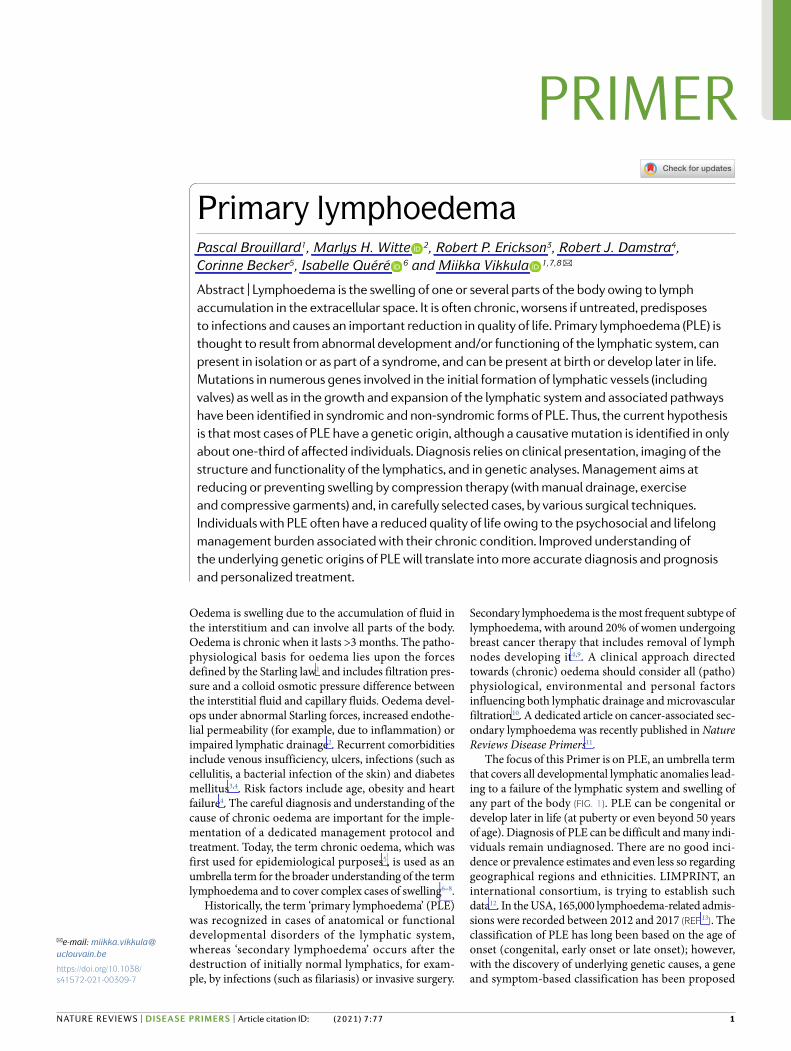
Fig. 1 | Examples of primary lymphoedema. Technetium (99mTc) albumin-aggregated lymphoscintigraphy (LSG) of hands (top) and feet (bottom) of a healthy person as a control (part a). Bilateral primary lymphoedema (PLE) in an individual carrying a VEGFR3 mutation, with visible large saphenous vein on top of the fascia (part b) and LSG showing no uptake (part c). Familial hyperplastic refluxing PLE on lower extremities in an individual carrying a FOXC2 mutation (lymphoedema–distichiasis) (part d), with LSG showing associated genital lymphoedema and irregular poorly transporting lymphatic collector channels on the left, with extensive dermal and genital tracer reflux after bilateral foot injections (part e). Congenital PLE with lymphatic hyperplasia involving the left lower extremity (part f), with LSG showing lymph stasis visible on left abnormal lymphatic nodes (blue arrows) and enlarged trunks (red arrows) as compared with normal right nodes and trunks (yellow arrows) (part g). Lympho-MRI showing central conducting lymphatic anomalies with enlargement of the Cysterna chyli (green arrow) and thoracic duct (red arrows), and large lymphatic anomalies on both sides of the upper mediastinum and upper thorax (blue arrows) (part h) similar to those in part g. Examples of superficial lymphatic hyperplasia visualized by lympho-MRI (part i) and visualization of superficial lymphatic network on the arm by injection of indocyanine green (part j).
3NATURE REVIEWS | DISEASE PRIMERS | Article citation ID: (2021) 7:77
P r i m e r
0123456789();:
rcprov
rcprov
rcprov
rcprov
rcprov
rcprov
rcprov
rcprov
rcprov
rcprov

Table 1 | Confirmed lymphoedema genes and loci
Genea Locus Disease or syndrome Major associated signs OMIM number
Inheritance Protein functionb Refs
(Prader–Willi) 15q11.2 Prader–Willi Obesity, developmental delay, short stature
176270 AD NA 233
(Aagenaes) 15q26.1 Cholestasis–lymphoedema Cholestasis 214900 AR NA 121
(TBX1?) 22q11.2 del 22q11 deletion Dysmorphism, cardiovascular anomalies
611867 De novo NA 120
(Phelan–McDermid)
22q13 del Phelan–McDermid Developmental delay, hypotonia
606232 AD NA 168
(Turner) Xp11.4/Yp11.2 del
Turner Cardiac anomalies, webbed neck, dysmorphism, slowed growth
NA De novo NA 114,133
ABCC9 12p12.1 Cantu Hypertrichosis, osteochon-drodysplasia, cardiomegaly
NA AR Anion transporter 234
ADAMTS3 4q13.3 Hennekam lymphangiectasia–lymphoedema syndrome 3
Dysmorphism, protein-losing enteropathy
618154 AR Extracellular enzyme
93,235
ANGPT2 8q23.1 Lymphoedema NA NA AD Ligand 23
CCBE1 18q21.32 Hennekam lymphangiectasia–lymphoedema syndrome 1
Dysmorphism, protein-losing enteropathy
235510 AR Adaptor protein 236
CELSR1 22q13.31 Lymphoedema NA NA AD Transmembrane 104
EPHB4 7q22.1 Hydrops fetalis, central conducting lymphatic anomaly (HFASD)
Hydrops fetalis 617300 AD Transmembrane receptor
109,237
FAT4 4q28.1 Hennekam lymphangiectasia–lymphoedema syndrome 2
Dysmorphism, protein-losing enteropathy
616006 AR Transmembrane protein
238
FLT4 (VEGFR3) 5q35.3 Nonne–Milroy lymphoedema Hydrops fetalis 153100 AD, AR, de novo
Transmembrane receptor
20,21
FOXC2 16q24.1 Lymphoedema–distichiasis Distichiasis, ptosis, varicose veins
602402 AD, de novo Transcription factor 73
GATA2 3q21.3 Emberger Myelodysplasia 614038 AD Transcription factor 171
GJC2 (Cx47) 1q42.13 Lymphoedema NA 613480 AD Connexin 108
IKBKG (NEMO) Xq28 Osteopetrosis with lymphoedema
Dental anomalies, ectodermal dysplasia, immunodeficiency
300291 X-linked Intracellular signalling molecule
239,240
KIF11 10q23.33 Microcephaly with or without chorioretinopathy, lymphoedema or mental retardation
Microcephaly with or without chorioretinopathy, mental retardation
152950 AD, de novo Intracellular signalling molecule
241
KRAS 12p12.1 Noonan syndrome 3, Gorham–Stout disease
Short stature, dysmorphism, cardiac anomaly, developmental delay
609942 AD, de novo Intracellular signalling molecule
242,243
NSD1 5q35.3 Sotos syndrome 1 Macrocephaly, rapid growth, cardiac anomaly
117550 De novo Histone methyltransferase
244,245
PIEZO1 16q24.3 Hydrops fetalis, generalized lymphatic dysplasia
Hydrops fetalis, short stature, facial dysmorphism
616843 AR Ion channel 246,247
PTPN11 (SHP2) 12q24.13 Noonan syndrome 1, hydrops Short stature, dysmorphism, cardiac anomaly, developmental delay
163950 AD Intracellular signalling molecule
248
RAF1 3qp25.2 Noonan syndrome 5 Short stature, dysmorphism, cardiac anomaly, developmental delay
611553 AD Intracellular signalling molecule
249,250
RASA1 5q14.3 Parkes–Weber (CM-AVM1), chylothorax
Capillary and arteriovenous malformations
139150 AD Intracellular signalling molecule
251,252,253
RIT1 1q22 Noonan syndrome 8 Short stature, dysmorphism, cardiac anomaly, developmental delay
615355 AD Intracellular signalling molecule
254–257
SOS1 2p22.1 Noonan syndrome 4 Short stature, dysmorphism, cardiac anomaly, developmental delay
610733 AD Intracellular signalling molecule
258,259
4 | Article citation ID: (2021) 7:77 www.nature.com/nrdp
P r i m e r
0123456789();:
molecules that cannot pass freely through the capillary barrier, which drives fluids towards the blood vessels and is therefore opposed by the hydrostatic pressure gradient. These so-called Starling forces and the lymphatic load are further modified by a filtration coefficient reflecting capillary surface area and permeability. Normally, in an oedema-free state, the volume of the lymphatic load is matched by the lymphatic capacity (the rate of lymph adsorption) to return lymph to the bloodstream (nor-mal thoracic duct lymph flow is approximately 1 ml per minute)45,49–52. There are regional variations in the abso-lute numerical value of the Starling forces and filtration coefficient (for example, the tight relatively impermeable blood–brain barrier in contrast to the low-hydrostatic pressure, highly permeable liver sinusoids) under nor-mal physiological conditions, which may be greatly exaggerated in disease states53,54.
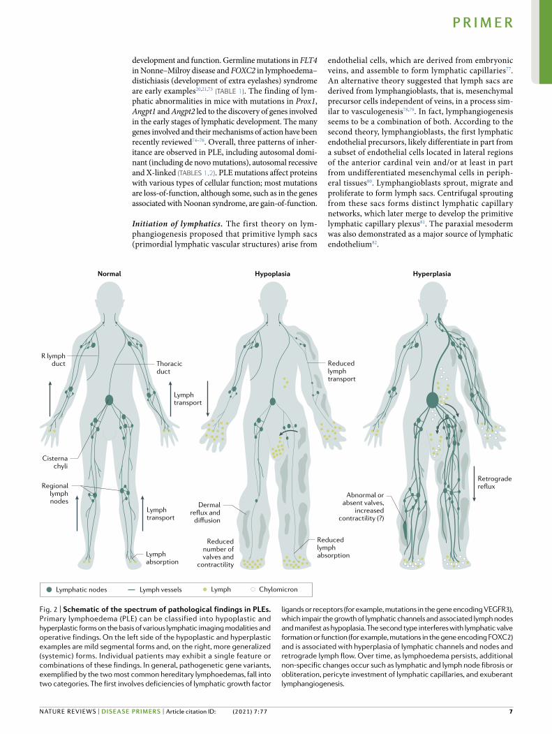
Pathophysiology of PLE. Congenital lymphatic mal-formation or malfunction anywhere along these con-tinuous pathways can be fatal during prenatal life or delayed or even silent after birth, until it leads to an imbalance between the processes of lymph formation (lymphatic load) and lymph absorption (lymphatic capacity)45,49–51 (Fig. 2). At that point, tissue swelling becomes manifest, presenting as PLE involving the limbs, chylous (chylomicra-containing intestinal lymph) or non-chylous lymph accumulations and/or effusions in the body cavities, as lymphostatic encephalopathy from brain oedema33, or even as external leaks (in which lymph exudates from the skin). Stasis of lymph (a high protein, hyaluronan-rich fluid-altering extracellular matrix (ECM)) reflects diminished lymphatic capa-city and sets into motion a localized tissue response. This response is characterized in varying degrees by inflammation, fibrosis, adipose deposition, immune dysregulation, susceptibility to infection, and both lym-phan giogenesis and haemangiogenesis as part of a progressive ‘overgrowth’ phenomenon45,49–51,55.
At the absorptive level, aberrations that could inter-fere with the relatively free passage of fluid and macro-mole cular and cellular lymph components into the
lymphatic capillaries include alterations in initial inter- endothelial open ‘buttons’ (loosely apposed perme-able junctions) and continuing closed impermeable ‘zipper’ junctions56 and related gap junction proteins, anchoring filaments45,49,50, tissue pressure mechanosen-sors (for example, Piezo-type mechanosensitive ion chan-nel component 1 (PIEZO1))57, and the newly described hyaluronan bulbs58 (structures composed of a large matrix of glycosaminoglycan and integrins, which are involved in cellular migration into the lymphatics dependent on lymphatic vessel endothelial hyaluranon receptor 1, Lyve1)59 (Fig. 2). Disturbances in lymphatic vessel growth (either reduced growth (aplasia or hypoplasia), increased size or number of the vessels (megalymphatics and hyperplasia, respectively), or growth in the wrong place, termed collectively as ‘lymphangiodysplasias’) could lead to or reflect anatomical or functional lymphatic obstruction (peripheral and/or central). These abnor-malities can be imaged dynamically most easily in PLE by whole-body lymphoscintigraphy (particularly when combined with resolution-enhanced and 3D-localized single-photon emission computed tomography (SPECT)) but also by other modalities such as MRI with or with-out contrast and indocyanine green (ICG) fluorescent lymphangiography45,47–50,55,60–66. Lymphangiodysplasias can also be caused or exacerbated by maldeveloped, hypoplastic or fibrotic regional lymph nodes67. Defective lymphatic valves45,48–50,55,60,68 can lead to valve incompe-tence, lymphangiectatic dilatations and lymph reflux into superficial valveless collaterals, tissues or body cav-ities or as external leakage from the skin. Heightened permeability can allow leakage from initial or collect-ing lymphatics and impaired contractility45,48–50,55 would delay lymph transport. Specific transgenic mouse models closely mimic these contrasting clinical and lymphatic imaging phenotypes (for example, lymphatic aplasia or hypoplasia in the angiopoietin 2 (Angpt2) knockout mouse69 and refluxing lymphatic hyperplasia in the Foxc2 haploinsufficient mouse70).
These pathogenetic mechanisms may stay latent and not manifest as tissue fluid accumulation and, more over, they might affect other cardiovascular or general systemic
Genea Locus Disease or syndrome Major associated signs OMIM number
Inheritance Protein functionb Refs
SOS2 14q31.1 Noonan syndrome 9 Short stature, dysmorphism, cardiac anomaly, developmental delay
616559 AD Intracellular signalling molecule
260–262
SOX18 20q13.33 Hypotrichosis–lymphoedema–telangiectasia–(renal defect) syndrome
Hypotrichosis, telangiectasia, ileal atresia, aortic dilation
607823, 137940
AD, AR, de novo
Transcription factor
228,263
THSD1 13q14.3 Hydrops, severe oedema Hydrops fetalis, cardiac anomaly
NA AR Transmembrane protein
264,265
TSC2 16p13.3 Tuberous sclerosis 2 Hamartomas, developmental delay
191092 AD Intracellular signalling molecule
266,267
VEGFC 4q34.3 Nonne–Milroy-like lymphoedema
NA 615907 AD Ligand 28,134,
268–270
OMIM numbers are only provided if the description mentions a lymphatic defect. When a maximum of five publications exists for primary lymphoedema-causing mutations in one gene, we listed them all; otherwise, if more than five, we listed only the original ones. AD, autosomal dominant; AR, autosomal recessive; CM-AVM1, capillary malformation-arteriovenous malformation 1; NA, not applicable. aMutated genes and loci associated with postnatal primary lymphoedema with or without non-immune hydrops fetalis with more than three index patients reported or, if only two index patients, supported by functional validation or linkage; in alphabetical order. bSee signalling in Fig. 3.
Table 1 (cont.) | Confirmed lymphoedema genes and loci
5NATURE REVIEWS | DISEASE PRIMERS | Article citation ID: (2021) 7:77
P r i m e r
0123456789();:
developmental events, resulting in multiorgan syndromes or, in select instances, contributing to lymph over-production (overload), for example, by venous pressure elevation, further overwhelming the diminished lym-phatic capacity. Indeed, specific initiating mechanisms controlling lymphatic growth, specialized lymphatic structures, and cell migration and adhesion (at an anato-mical, physiological or molecular level) have been pin-pointed in some forms of PLE (see Genetic basis below). These mechanisms have been observed in patients and/or mouse models and further elucidated in vitro. However, exactly how lymph transport is affected and compensated for (at the peripheral, visceral and central lymphatic system level) and what accounts for the variability in manifesting lymphoedema (site, severity, age of onset) remains elusive. Current techniques for non-invasive in vivo multimodal lymphatic imaging and histological study of diseased lymphatics are limited in assessing lym-phodynamics. Yet, determining the sequence, interaction
and impact of specific functional abnormalities in the integrated lymphatic system of vessels, fluid, nodes and trafficking of immune cells (lymphatic ‘systemomics’)71 is particularly crucial to fill in the gaps in our under-standing of uncomplicated PLE and of the pleiomor-phic manifestations in associated syndromes55,72 and to translating the findings of experimental models into the management of the human condition.
Genetic basisSingle genes. Our evolving knowledge of genes in which mutations cause lymphoedema has depended on syner-gistic findings in both human and mice. Zebrafish have more recently been studied and it is clear that the relative importance of the many shared essential gene products can differ between the three species. The first major discoveries were based on classical human genetic approaches using linkage studies in large fam-ilies to locate and clone genes involved in lymphatic
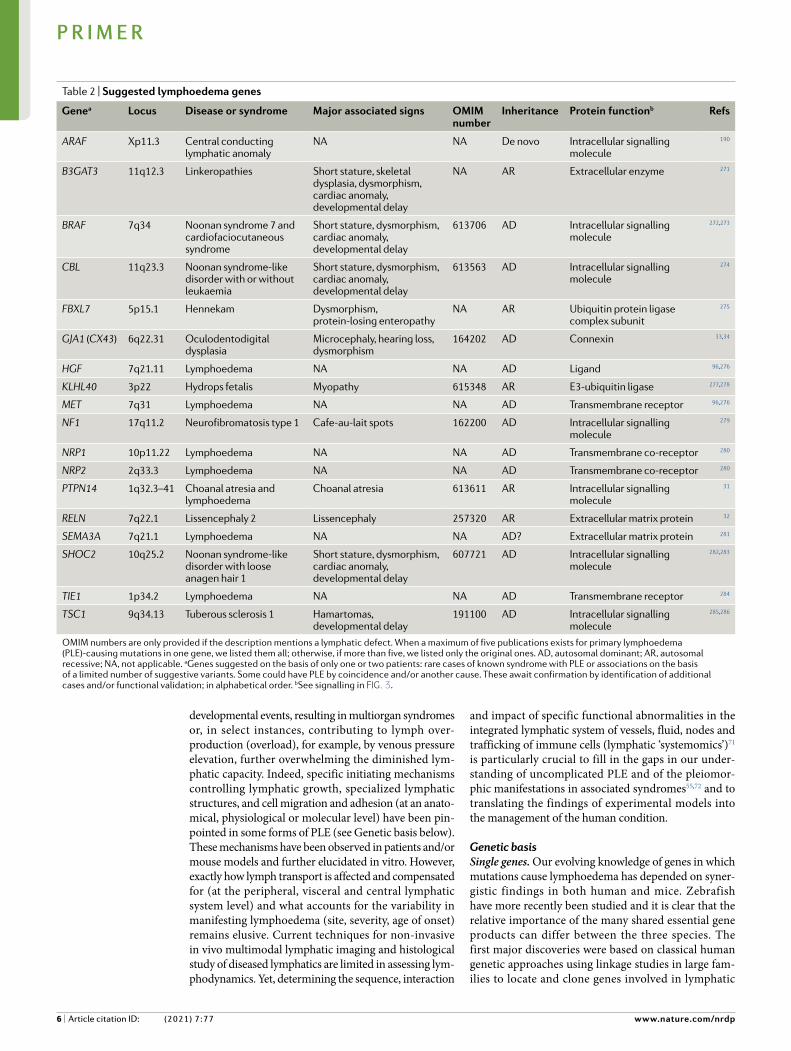
Table 2 | Suggested lymphoedema genes
Genea Locus Disease or syndrome Major associated signs OMIM number
Inheritance Protein functionb Refs
ARAF Xp11.3 Central conducting lymphatic anomaly
NA NA De novo Intracellular signalling molecule
190
B3GAT3 11q12.3 Linkeropathies Short stature, skeletal dysplasia, dysmorphism, cardiac anomaly, developmental delay
NA AR Extracellular enzyme 271
BRAF 7q34 Noonan syndrome 7 and cardiofaciocutaneous syndrome
Short stature, dysmorphism, cardiac anomaly, developmental delay
613706 AD Intracellular signalling molecule
272,273
CBL 11q23.3 Noonan syndrome-like disorder with or without leukaemia
Short stature, dysmorphism, cardiac anomaly, developmental delay
613563 AD Intracellular signalling molecule
274
FBXL7 5p15.1 Hennekam Dysmorphism, protein-losing enteropathy
NA AR Ubiquitin protein ligase complex subunit
275
GJA1 (CX43) 6q22.31 Oculodentodigital dysplasia
Microcephaly, hearing loss, dysmorphism
164202 AD Connexin 33,34
HGF 7q21.11 Lymphoedema NA NA AD Ligand 96,276
KLHL40 3p22 Hydrops fetalis Myopathy 615348 AR E3-ubiquitin ligase 277,278
MET 7q31 Lymphoedema NA NA AD Transmembrane receptor 96,276
NF1 17q11.2 Neurofibromatosis type 1 Cafe-au-lait spots 162200 AD Intracellular signalling molecule
279
NRP1 10p11.22 Lymphoedema NA NA AD Transmembrane co-receptor 280
NRP2 2q33.3 Lymphoedema NA NA AD Transmembrane co-receptor 280
PTPN14 1q32.3–41 Choanal atresia and lymphoedema
Choanal atresia 613611 AR Intracellular signalling molecule
31
RELN 7q22.1 Lissencephaly 2 Lissencephaly 257320 AR Extracellular matrix protein 32
SEMA3A 7q21.1 Lymphoedema NA NA AD? Extracellular matrix protein 281
SHOC2 10q25.2 Noonan syndrome-like disorder with loose anagen hair 1
Short stature, dysmorphism, cardiac anomaly, developmental delay
607721 AD Intracellular signalling molecule
282,283
TIE1 1p34.2 Lymphoedema NA NA AD Transmembrane receptor 284
TSC1 9q34.13 Tuberous sclerosis 1 Hamartomas, developmental delay
191100 AD Intracellular signalling molecule
285,286
OMIM numbers are only provided if the description mentions a lymphatic defect. When a maximum of five publications exists for primary lymphoedema (PLE)-causing mutations in one gene, we listed them all; otherwise, if more than five, we listed only the original ones. AD, autosomal dominant; AR, autosomal recessive; NA, not applicable. aGenes suggested on the basis of only one or two patients: rare cases of known syndrome with PLE or associations on the basis of a limited number of suggestive variants. Some could have PLE by coincidence and/or another cause. These await confirmation by identification of additional cases and/or functional validation; in alphabetical order. bSee signalling in Fig. 3.
6 | Article citation ID: (2021) 7:77 www.nature.com/nrdp
P r i m e r
0123456789();:
development and function. Germline mutations in FLT4 in Nonne–Milroy disease and FOXC2 in lymphoedema–distichiasis (development of extra eyelashes) syndrome are early examples20,21,73 (Table 1). The finding of lym-phatic abnormalities in mice with mutations in Prox1, Angpt1 and Angpt2 led to the discovery of genes involved in the early stages of lymphatic development. The many genes involved and their mechanisms of action have been recently reviewed74–76. Overall, three patterns of inher-itance are observed in PLE, including autosomal domi-nant (including de novo mutations), autosomal reces sive and X-linked (Tables 1,2). PLE mutations affect proteins with various types of cellular function; most mutations are loss-of-function, although some, such as in the genes associated with Noonan syndrome, are gain-of-function.
Initiation of lymphatics. The first theory on lym-phangiogenesis proposed that primitive lymph sacs (primordial lymphatic vascular structures) arise from
endothelial cells, which are derived from embryonic veins, and assemble to form lymphatic capillaries77. An alternative theory suggested that lymph sacs are derived from lymphangioblasts, that is, mesenchymal precursor cells independent of veins, in a process sim-ilar to vasculogenesis78,79. In fact, lymphangiogenesis seems to be a combination of both. According to the second theory, lymphangioblasts, the first lymphatic endothelial precursors, likely differentiate in part from a subset of endothelial cells located in lateral regions of the anterior cardinal vein and/or at least in part from undifferentiated mesenchymal cells in periph-eral tissues80. Lymphangioblasts sprout, migrate and proliferate to form lymph sacs. Centrifugal sprouting from these sacs forms distinct lymphatic capillary networks, which later merge to develop the primitive lymphatic capillary plexus81. The paraxial mesoderm was also demonstrated as a major source of lymphatic endothelium82.
Lymphatic nodes Lymph vessels Lymph
Reducedlymphtransport
Reducedlymphabsorption
Dermalreflux and
diffusion
Reducednumber ofvalves and
contractility
Hypoplasia Hyperplasia
Lymphtransport
Lymphtransport
Lymphabsorption
Thoracicduct
R lymphduct
Cisternachyli
Regionallymphnodes
Normal
Retrogradereflux
Abnormal orabsent valves,
increasedcontractility (?)
Chylomicron
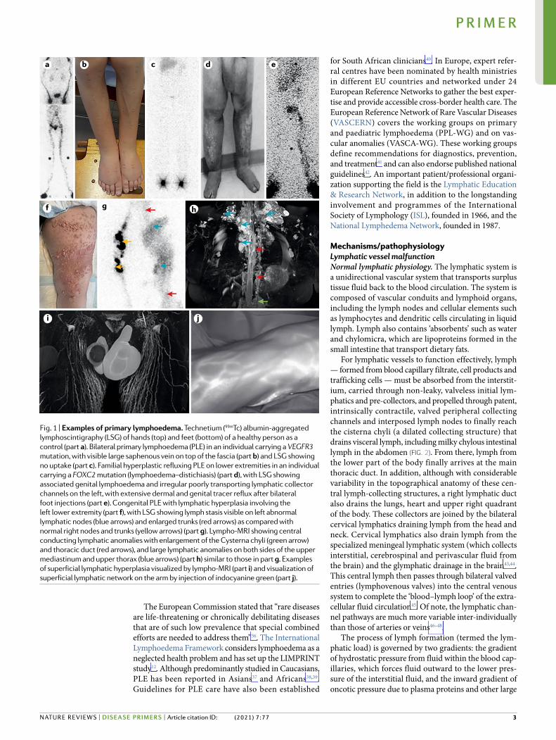
Fig. 2 | Schematic of the spectrum of pathological findings in PLEs. Primary lymphoedema (PLE) can be classified into hypoplastic and hyperplastic forms on the basis of various lymphatic imaging modalities and operative findings. On the left side of the hypoplastic and hyperplastic examples are mild segmental forms and, on the right, more generalized (systemic) forms. Individual patients may exhibit a single feature or combinations of these findings. In general, pathogenetic gene variants, exemplified by the two most common hereditary lymphoedemas, fall into two categories. The first involves deficiencies of lymphatic growth factor
ligands or receptors (for example, mutations in the gene encoding VEGFR3), which impair the growth of lymphatic channels and associated lymph nodes and manifest as hypoplasia. The second type interferes with lymphatic valve formation or function (for example, mutations in the gene encoding FOXC2) and is associated with hyperplasia of lymphatic channels and nodes and retrograde lymph flow. Over time, as lymphoedema persists, additional non-specific changes occur such as lymphatic and lymph node fibrosis or obliteration, pericyte investment of lymphatic capillaries, and exuberant lymphangiogenesis.
7NATURE REVIEWS | DISEASE PRIMERS | Article citation ID: (2021) 7:77
P r i m e r
0123456789();:
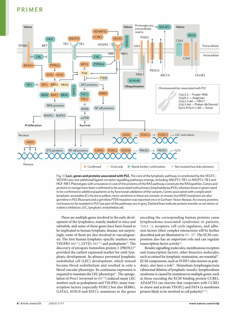
There are multiple genes involved in the early devel-opment of the lymphatics, mainly studied in mice and zebrafish, and some of these genes have been found to be implicated in human lymphatic disease; not surpris-ingly, some of them are also involved in vasculogene-sis. The first human lymphatic-specific markers were VEGFR3 (reF.83), LYVE1 (reF.84) and podoplanin85. The discovery of prospero homeobox protein 1 (PROX1)86 provided the earliest expressed marker for early lym-phatic development. Its absence prevented lymphatic endothelial cell (LEC) development, which instead became blood endothelium and resulted in only a blood vascular phenotype. Its continuous expression is required to maintain the LEC phenotype87. The upregu-lation of Prox1 (reviewed in reF.88) induced many LEC markers such as podoplanin and VEGFR3, many tran-scription factors (especially FOXC2 but also IKBKG, GATA2, SOX18 and KIF11; mutations in the genes
encoding the corresponding human proteins cause lymphoedema-associated syndromes in patients; Table 2), receptors, cell cycle regulators, and adhe-sion factors (their complex interactions will be further described and are illustrated in Fig. 3)89. The ECM com-position also has an important role and can regulate transcription factor activity90.
Besides signalling molecules, membranous receptors and transcription factors, other bioactive molecules, such as retinol for lymphatic maturation, are essential91. ECM components, such as SVEP1 (also known as poly-dom), also have a role92. Hennekam lymphangiectasia (abnormal dilation of lymphatic vessels)–lymphoedema syndrome is caused by mutations in multiple genes, such as those encoding the ECM-binding protein CCBE1, ADAMTS3 (an enzyme that cooperates with CCBE1 to cleave and activate VEGFC) and FAT4 (a membrane protein likely to be involved in cell polarity)93,94.
SOS1 SOS2
RIT1 KRAS NRAS
ARAF RAF1 BRAF
PTPN11
METEPHB4TIE2 TIE1 VEGFR3
NRP2 NRP1
Valves
KLHL40
Ubiquitination
FAT4
FBXL7PIEZO1
ABCC9
CX43
CX47
CELSR1
ValvesProteoglycans,extracellularmatrix
RASA1
NF1
Valves
HGF
CBL
KIF11
TSC2
TSC1
MEK proteins
MAPK1 MAPK3
ETS proteins NF-κB proteins
Proliferation Survival
mTOR
RHEB
IKBKG
AKT proteins
PI3K
PTEN
CBL
ANGPT2 VEGFC
PTPN14
CCBE1
ADAMTS3
SEM
A3A
RELN
B3GAT3
SHO
C2
Nucleus
NF-κB proteins
GATA2 FOXC2 SOX18 PROX1 PROX1 FLT4
GATA FOXC2 FOXC2 LEC and valves
NSD1
THSD1
Extracellular
Intracellular
HistoneConfirmed CLAs only Needs further confirmation Not mutated but links elements
Chromosomal loci associated with PLE
15q11.2 — Prader–Willi15q26.1 — Aagenaes22q11.2 del — TBX1?22q13 del — Phelan–McDermidXp11.4/Yp11.2 del — Turner
Fig. 3 | Loci, genes and proteins associated with PLE. The core of the lymphatic pathway is constituted by the VEGFC–VEGFR3 axis, but additional ligand–receptor signalling pathways emerge, including ANGPT2–TIE1 or ANGPT2–TIE2 and HGF–MET. Phenotypes with a mutation in one of the proteins of the RAS pathway constitute the RASopathies. Genes and proteins in orange have been confirmed to be associated with primary lymphoedema (PLE), whereas those in green need to be confirmed in additional patients or by functional validation of the variants. Genes associated with complicated lymphatic anomalies (CLAs) are in yellow; most variations in these are somatic or mosaic but KRAS mutations are also germline in PLE (Noonan) and a germline PTEN mutation was reported once in Gorham–Stout disease. Accessory proteins not known to be mutated in PLE but part of the pathways are in grey. Dashed lines indicate protein transfer or secretion or indirect inhibition. LEC, lymphatic endothelial cell.
8 | Article citation ID: (2021) 7:77 www.nature.com/nrdp
P r i m e r
0123456789();:

Rare mutations in VEGFC, the major stimulator of lymphatic growth and development, also cause lym-phoedema28. Its receptor, VEGFR3, is a tyrosine kinase receptor initiating PI3K–AKT signalling and the inter-acting RAS–MAPK cascade. Inactivating mutations in VEGFR3 are a cause of Nonne–Milroy disease (familial congenital bilateral lower-limb lymphoedema, the most frequently found genetic cause of lymphoedema)20,21 (Fig. 3; Table 2). Mutations in the genes encoding many components of these intracellular signalling pathways are associated with phenotypes exhibiting lymphoedema, including Noonan syndrome, cardiofaciocutaneous syndrome, lymphoedema–choanal atresia syndrome and rare cases of chylothorax (thoracic duct damage with chyle leakage surrounding the lungs) (see Signalling pathways below; Fig. 3; Tables 1,2). Mutations in some of these genes can serve as modifying genes; for example, mutations in GJC2 (encoding connexin 47) may inter-act with non-genetic factors to cause post-breast surgery lymphoedema95 or be causative in PLE96.
Lymphatic valve formation. In general, mutations in genes related to defective valve formation tend to pro-duce lymphatic hyperplasia or lymphangiectasia and lymph reflux. By contrast, genes involved in the initia-tion (above) or maintenance and proliferation (below) of lymphatics produce PLE with lymphatic hypoplasia or aplasia in the periphery and even centrally. The left-sided lymphovenous valve (and the frequent second right-sided lymphovenous valve) connecting the tho-racic duct (which is the central lymphatic collector) to the central vein is the only place (other than the much smaller right lymphatic duct valved entry) where the post nodal lymph fluid and blood normally come into contact. This may be the first ‘lymphatic’ valve to develop. The mechanotransducer PIEZO1 senses the laminar flow of lymphatic fluid and this detection is a major stimulus to valve formation97. Recessive mutations in PIEZO1 are associated with human lymphoedema98. The precursor cells also require transcription factor SOX18, NRF2, Coup transcription factor 2 and PROX1. These lymphovenous valves continue to develop with FOXC2 as the major activator99 for valve development, with planar cell polarity gene products and connexins in their maturation100–102.
Planar cell polarity refers to the coordinated orienta-tion of cells in epithelia in the direction perpendicular to the apical-basal orientation and is essential for cell orientation. In mice, two chemically induced, nonsense mutations in Celsr1 were found to affect planar cell polarity (spin cycle and crash mouse mutants). These two mutants and a conditional deletion of Celsr1, using a Prox1 promoter to specifically delete Celsr1 in endothe-lial cells of developing lymphatics, allowed study of the role of CELSR1 in later development103; it was shown, with VANGL2, to have a crucial role in lymphatic valve formation103. Families with lymphoedema due to muta-tions in the planar polarity gene CELSR1 have been described29,104.
Gap junction molecules (connexins) are also impor-tant for lymphatic development and three have been found to be expressed in most lymphatic vessels: Cx37,
Cx43 and Cx47 (reFs105–107). Knockouts of Cx37 and Cx43 disrupt lymphatic valve development and result in embryonic lymphoedema and chylothorax with mark-edly reduced postnatal survival105. Two GJC2 mutations were initially reported in two families with dominantly inherited lymphoedema108, followed by other families and one family with a mutation in GJA1 (encoding Cx43)33. In addition, mutations in EPHB4, encoding a member of the ephrin family of RTK receptors, which interact with connexins, have been found in cases of fatal NIHF109 (Fig. 3).
Expansion and proliferation of lymphatics. After the initiation of lymphangiogenesis, laminar fluid flow and interstitial pressure trigger lymphatic expansion and proliferation, at least in vitro. LECs exposed to laminar fluid flow stimulate a pore subunit of the s-activated calcium channel (ORAI1), which induces the upreg-ulation of Kruppel-like factor 2 (KLF2) and KLF4 and induces the expression of VEGFA, VEGFC, fibroblast growth factor receptor 3 (FGFR3), and p57 (also known as cyclin-dependent kinase inhibitor 1C)110. As with most of the genes mentioned in initiating the development of lymphatics (excluding VEGFC and VEGFR3), mutations in these genes causing human lymphoedema have not yet been found. However, recessive mutations in PIEZO1 are associated with human lymphoedema98. In addition, in a large multigeneration family with highly penetrant lym-phoedema, digenic inheritance has been documented with both FOXC2 and biallelic PIEZO1 mutations111.
Gap junction (connexin) proteins are not only involved in valve formation (Cx37 and Cx47) but are important for the function of lymphatics (Cx26) as they control the flow of fluid containing small and larger molecules between cells, which may be their role in valve formation. It is the leaked fluid from the blood vas cular system that the lymphatics return to the blood circula-tion112. Gap junctions are also important in coordinating smooth muscle-mediated contractility, which propels lymphatic fluid centripetally105,106,113.
Chromosomal loci associated with PLE. Several chro-mosomal disorders are associated with lymphoedema (Table 1). Turner syndrome (45,XO) frequently has infan-tile generalized lymphoedema (before the chromosomal cause was discovered, it was considered a separate syn-drome, known as Bonnevie–Ullrich) and can re-occur in children and adults. Although an X-chromosomal p11.4 location shared with Yp11.2 has been identified, a specific gene has not been pinpointed114,115. Moreover, trisomy 21 (and the rarer trisomies 13 and 18) is fre-quently associated with increased nuchal folds detected in utero by ultrasonography (posterior lateral neck swell-ings thought to be related to enlargement of the cervical lymphatic sacs)116. Old case reports based on classical karyotyping associated mosaic trisomies or intersti-tial deletions and duplications to nuchal translucency or PLE117–119. The cause of these phenotypes could be a mutation in one of the now known genes) localized in these regions (Tables 1,2). The frequently diagnosed Prader–Willi syndrome, which involves abnormal imprinting of a portion of chromosome 15 caused by
9NATURE REVIEWS | DISEASE PRIMERS | Article citation ID: (2021) 7:77
P r i m e r
0123456789();:
gene or chromosomal mutations, often presents with lymphoedema along with other syndromic features55. Additional loci include the locus of the Phelan–McDermid syndrome (22q13), the 22q11.2 deletion syndrome locus120 and the locus for the Aagenaes syn-drome (15q26.1)121. As the incidence of lymphoedema in various syndromes is unknown and case reports are scarce, epidemiological studies are needed.
Signalling pathwaysLymphangiogenesis. In addition to the proteins and signalling pathways implicated in human disease dis-cussed above, there are many other signalling pathways involved in lymphatic development and function122. These pathways control cell growth and proliferation, apoptosis, cell migration and differentiation, and cell adhesion. As mentioned, VEGFC is essential for the initial development and maintenance of lymphatics, whereas VEGFD, which stimulates adult lymphangio-genesis by binding to VEGFR3, is not essential123, at least in mice. The levels of VEGFR3 are strongly con-trolled by Notch signalling124. VEGFC stimulation results in receptor phosphorylation and downstream activation of multiple signalling pathways, which stim-ulate LEC proliferation and migration. Ephrin B2 sig-nalling at its tyrosine kinase-activating receptor, ephrin type B receptor 4 (EPHB4), is also essential for lym-phatic development (Fig. 3). This signalling pathway is unusual in that it involves ‘reverse signalling’, in which the ligand (ephrin) also functions as a receptor in the cell expressing it. This dual ligand–receptor function of the membrane protein mediates bi-directional signals between neighbouring cells; thus, intracellular signal-ling is induced in both cells (forward (in the neigh-bouring cell) and reverse (in the ligand-expressing cell)). The reverse signalling is essential for lymphatic remodelling and valve formation125. The ephrin signal-ling pathway as well as VEGFC and VEGFD signalling through neuropilins provide a connection between lymphatic network patterning and that of neurons. Vascular growth factors are secreted by neurons and neurotropins are secreted by developing vessels, ena-bling co-tracking of the development of both systems. Other signalling molecules, including semaphorins and Slits as well as their receptors plexin and roundabout homologue (Robo), also connect the two guidance pathways126. Their full role is beyond this Review but well covered in reF.126.
Another signalling pathway important for lymphatic development is that of the ANGPT1 and ANGPT2 ligands and the tyrosine-protein kinase receptors TIE1 and TIE2. The two tyrosine kinase receptors are differ-entially activated by the two angiopoietins. ANGPT1 activates TIE2, whereas ANGPT2 activates TIE2 only on LECs and blocks the activation of TIE2 on angiogenic blood endothelial cells because an inhibiting vascular endothelial protein tyrosine phosphatase (VEPTP) is expressed in blood endothelial cells but not LECs127. Knockout mouse models demonstrate that ANGPT2 is required for haemangiogenesis and lymphangio-genesis, with the lymphatic defects being corrected by the expression of ANGPT1 instead of ANGPT2 (reF.69).
Recently, loss-of-function or dominant-negative mutations were identified in ANGPT2 in PLE23.
Mutations in many genes of the RAF–MEK–ERK — MAPK cascade cause lymphoedema-associated syn-dromes. In mice, another pathway dependent on MAPK that is associated with lymphatic problems is the ternary complex factors pathway, which regulates immediate early genes through serum response elements. The knockout of the gene encoding one of these factors, Net, results in lymphovascular defects, including chylothorax128, but PLE-causing mutations in the corresponding human gene are not known. Finally, hepatocyte growth factor (HGF) and its tyrosine kinase-activating receptor MET promote lymphatic vessel formation and function129 (Fig. 3).
Overgrowth syndromes involving the lymphatics. A limited number of overgrowth syndromes involve the lymphatics. These syndromes can be quite disfigur-ing and, because germline mutations would be lethal, are due to somatic mutations. The famous case of the ‘Elephant Man’ involved an individual who was long thought to have neurofibromatosis type 1, whereas almost certainly he had Proteus syndrome, which is due to somatic, gain-of-function mutations in AKT1. The protein product of this gene, RAC-α serine/threonine-protein kinase (AKT1), is involved in a sig-nalling pathway involving several genes implicated in lymphatic malformations. PIK3CA produces phos-phatidylinositol 4,5-bisphosphate 3-kinase catalytic subunit-α isoform, which can be dephosphorylated by PTEN. This dephosphorylation decreases PI3K con-centrations and prevents it from translocating AKT1 to the cell membrane, where AKT1 is phosphorylated and activated by upstream kinases. PTEN loss-of-function mutations cause PTEN hamartoma tumour syndrome, whereas somatic gain-of-function mutations in PIK3CA are found in CLOVES syndrome, characterized by congenital lymphatic overgrowth, vascular malforma-tions, epidermal nevi and skeletal abnormalities, and in Klippel–Trenaunay–Weber syndrome of bone and angio-lymphatic overgrowth as well as in isolated lymphatic malformations55,76,130. Other PIK3CA-related overgrowth syndromes that may be associated with lymphatic anomalies also exist130.
In conclusion, our understanding of the pathophys-iological mechanisms underlying PLE is incomplete. Nonetheless, molecular discoveries over the past two decades have identified multiple genes, proteins and signalling pathways involved in lymphatic growth and development, with these findings providing funda-mental insights into PLE (Fig. 3). Further lymphatic imaging, particularly dynamic studies since the 1970s documenting the various peripheral and central lym-phatic system disturbances in PLE, has served to con-nect the proposed molecular events with physiological evidence of lymphatic maldevelopment and dysfunc-tion manifested in the clinical appearance and compli-cations of PLE. Opposite pathophysiological pathways are reflected in the hypoplastic form (inadequate peri-pheral lymphatic growth) and hyperplastic refluxing form (defective lymphatic valve formation and central lymphatic malformation) of PLE depicted in Fig. 2.
10 | Article citation ID: (2021) 7:77 www.nature.com/nrdp
P r i m e r
0123456789();:
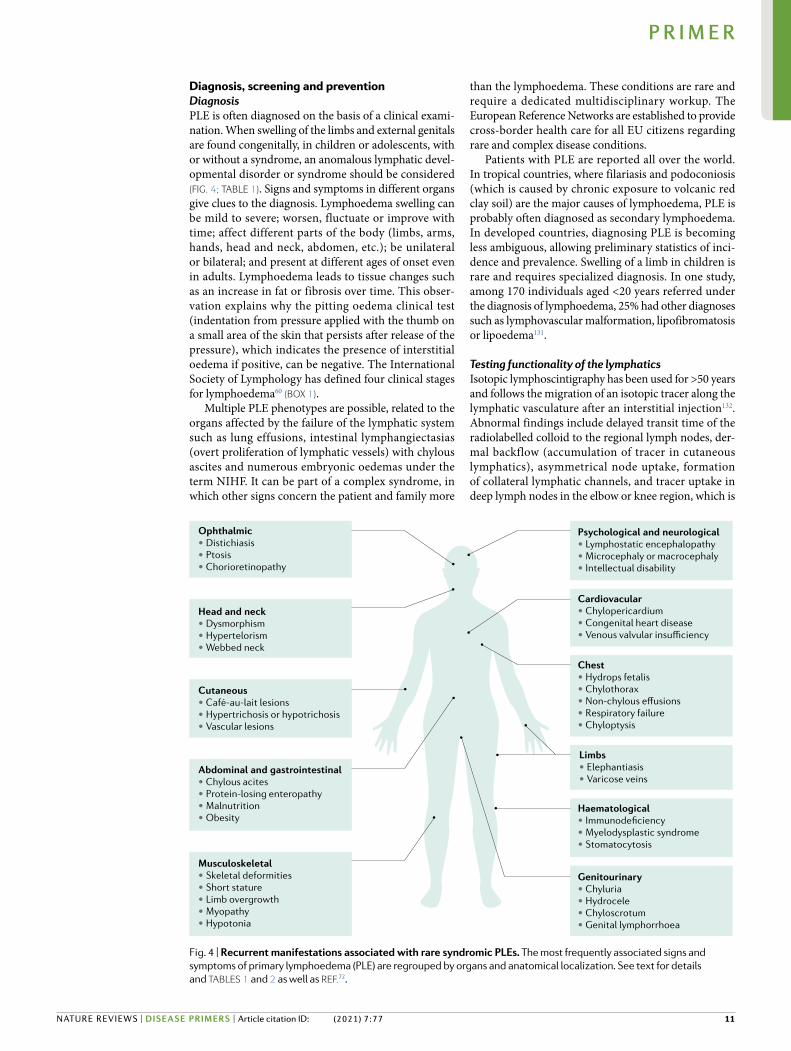
Diagnosis, screening and preventionDiagnosisPLE is often diagnosed on the basis of a clinical exami-nation. When swelling of the limbs and external genitals are found congenitally, in children or adolescents, with or without a syndrome, an anomalous lymphatic devel-opmental disorder or syndrome should be considered (Fig. 4; Table 1). Signs and symptoms in different organs give clues to the diagnosis. Lymphoedema swelling can be mild to severe; worsen, fluctuate or improve with time; affect different parts of the body (limbs, arms, hands, head and neck, abdomen, etc.); be unilateral or bilateral; and present at different ages of onset even in adults. Lymphoedema leads to tissue changes such as an increase in fat or fibrosis over time. This obser-vation explains why the pitting oedema clinical test (indentation from pressure applied with the thumb on a small area of the skin that persists after release of the pressure), which indicates the presence of interstitial oedema if positive, can be negative. The International Society of Lymphology has defined four clinical stages for lymphoedema60 (box 1).
Multiple PLE phenotypes are possible, related to the organs affected by the failure of the lymphatic system such as lung effusions, intestinal lymphangiectasias (overt proliferation of lymphatic vessels) with chylous ascites and numerous embryonic oedemas under the term NIHF. It can be part of a complex syndrome, in which other signs concern the patient and family more
than the lymphoedema. These conditions are rare and require a dedicated multidisciplinary workup. The European Reference Networks are established to provide cross-border health care for all EU citizens regarding rare and complex disease conditions.
Patients with PLE are reported all over the world. In tropical countries, where filariasis and podoconiosis (which is caused by chronic exposure to volcanic red clay soil) are the major causes of lymphoedema, PLE is probably often diagnosed as secondary lymphoedema. In developed countries, diagnosing PLE is becoming less ambiguous, allowing preliminary statistics of inci-dence and prevalence. Swelling of a limb in children is rare and requires specialized diagnosis. In one study, among 170 individuals aged <20 years referred under the diagnosis of lymphoedema, 25% had other diagnoses such as lymphovascular malformation, lipofibromatosis or lipoedema131.
Testing functionality of the lymphaticsIsotopic lymphoscintigraphy has been used for >50 years and follows the migration of an isotopic tracer along the lymphatic vasculature after an interstitial injection132. Abnormal findings include delayed transit time of the radiolabelled colloid to the regional lymph nodes, der-mal backflow (accumulation of tracer in cutaneous lymphatics), asymmetrical node uptake, formation of collateral lymphatic channels, and tracer uptake in deep lymph nodes in the elbow or knee region, which is
Ophthalmic• Distichiasis• Ptosis• Chorioretinopathy
Head and neck• Dysmorphism• Hypertelorism• Webbed neck
Cutaneous• Café-au-lait lesions• Hypertrichosis or hypotrichosis• Vascular lesions
Abdominal and gastrointestinal• Chylous acites• Protein-losing enteropathy• Malnutrition• Obesity
Limbs• Elephantiasis• Varicose veins
Musculoskeletal• Skeletal deformities• Short stature• Limb overgrowth• Myopathy • Hypotonia
Psychological and neurological• Lymphostatic encephalopathy• Microcephaly or macrocephaly• Intellectual disability
Cardiovacular• Chylopericardium• Congenital heart disease• Venous valvular insufficiency
Chest• Hydrops fetalis• Chylothorax• Non-chylous effusions• Respiratory failure• Chyloptysis
Haematological• Immunodeficiency• Myelodysplastic syndrome• Stomatocytosis
Genitourinary• Chyluria• Hydrocele • Chyloscrotum • Genital lymphorrhoea
Fig. 4 | Recurrent manifestations associated with rare syndromic PLEs. The most frequently associated signs and symptoms of primary lymphoedema (PLE) are regrouped by organs and anatomical localization. See text for details and Tables 1 and 2 as well as reF.72.
11NATURE REVIEWS | DISEASE PRIMERS | Article citation ID: (2021) 7:77
P r i m e r
0123456789();:
pathological. The spatial resolution of scintigraphy is poor and has been improved using simultaneous ana-tomical localization by SPECT–CT. The isotopic lym-phoscintigraphy patterns of gene-related PLE vary, from no intake in VEGFR3-related PLE to intake into large and numerous collateral lymphatics with dermal backflow in FOXC2-related PLE33,65,109,133–135. ICG fluor-escence lymphography is used to evaluate the real-time transport of a fluorescent tracer in the lymphatic vessels in the upper dermal space up to a maximum depth of 3–5 mm (reF.136).
Imaging the lymphaticsNowadays, non-contrast magnetic resonance lym-phangiography (MRL; also known as lympho-MRI)25 is a non-invasive technique that enables visualization of slow-moving non-bloody fluids such as those in large lymphatic vessels. It is based on heavily T2 weighted fast spin-echo sequences and maximum intensity projection reconstruction (Fig. 1). MRL has enabled the classifica-tion of the lymphatic system abnormalities in primary lymphoedema, considering lymph nodes and lymphatic vessel involvement137. Dynamic contrast-enhanced MRL enables static and dynamic visualization of the central lymphatic system by injecting gadolinium contrast agent in the groin lymph nodes in patients with PLE and/or CLA138 or intrahepatic lymphatic anomalies139. This technique enables understanding of the lymphatic flow disorders before planning interventional procedures140; however, it is not widely available.
Pedal lymphangiography imaging using oil-based iodinated agents injected into the lymphatic vessels, which were dissected and cannulated, was developed for surgical purposes and used for the anatomical classifica-tion of PLE141 but has been abandoned because of its vis-cosity and side effects. Lymphangiography based on the puncture of lymph nodes in the groin is now preferred to injection in the feet for the visualization of the central lymphatic conducting vessels in adults and children142.
Genetic analysesGenetic testing is being used as part of the clinical workup in highly specialized centres managing patients with lymphatic anomalies, especially for familial cases. In genetic centres, most often, next-generation sequencing techniques are used to screen blood-derived DNA using gene panels. However, with a growing number of known genes related to PLE to be tested, whole-exome sequenc-ing is becoming the best option. Chromosome analysis can be performed in detecting some syndromic PLEs. Tissue culture from affected tissue and next-generation sequencing can be helpful in finding somatic mutations but, so far, in most of the patients with PLE in whom a causative mutation has been identified, the mutations were germline (inherited or de novo). Yet, many patients remain undiagnosed and untested as they do not consult a specialized centre. Genetic testing increases our knowl-edge on phenotypic variability and genotype–phenotype correlations, enabling more specific genetic counselling as well as better stratification into subphenotypes and patient information.
Classification of lymphatic anomaliesA diagnostic algorithm based on detailed phenotyping, family history, age at onset, localization of the affected lymphatics, presence of visceral involvement, diag-nosis of concomitant syndromes and genotyping has been established15. We propose here a revised classifi-cation of lymphatic anomalies (Fig. 5). This classifica-tion, although not complete as the field evolves rapidly, allows stratification of patients into main subcategories and considers the evolution of phenotypes in time (more signs become apparent with age). It also clearly links CLAs and lymphatic malformations to the diagnostic workup. Eventually, this approach can have therapeu-tic consequences and can prevent unnecessary (inva-sive) diagnostic measures143. Genetic data allow further refinement of this algorithm.
ScreeningScreening for patients who are at risk of developing PLE is difficult in terms of selection. Three groups are at risk: relatives of a patient known to have PLE, patients with one of the syndromes associated with PLE but without clinical signs or symptoms of PLE, and patients who develop erysipelas (an infection of the superficial layers of the skin) without any preceding signs of chronic oedema, chronic venous insufficiency, lymphoedema, diabetes mellitus, overweight or previ-ous bouts of erysipelas144. There are no guidelines on pre-symptomatic screening and we lack estimations for the risk of developing PLE.
When a patient is diagnosed with a gene defect caus-ing lymphoedema, the possibility for family screening can be offered. Full information for the patient and consider-ation of the advantages and disadvantages are needed for shared decision-making. Owing to vari able pene-trance, the lymphoedema can be mild or even absent, which influences the relevance of screening for relatives. Despite all developments in the field of genetics and sophisticated techniques to visualize the anatomy and function of lymphatics, the role of clinicians within an
Box 1 | The four clinical stages for lymphoedema as defined by the International Society of Lymphology
A limb may exhibit more than one stage, which may reflect alterations in different lymphatic territories.
Stage 0 (or Ia)Latent or sub-clinical condition in which swelling is not yet evident despite impaired lymph transport, subtle alterations in tissue fluid and/or composition, and changes in subjective symptoms. It may exist months or years before overt oedema occurs. This assessment requires imaging techniques.
Stage IEarly accumulation of fluid relatively high in protein content (for example, compared with venous oedema) that subsides with limb elevation. Pitting may occur. An increase in various types of proliferating cells may also be observed.
Stage IILimb elevation alone rarely reduces the tissue swelling and pitting is manifest. Later in stage II, the limb may not pit, as excess subcutaneous fat and fibrosis develop.
Stage IIIComprises lymphostatic elephantiasis (enlargement of the limbs) in which pitting can be absent and trophic skin changes, such as acanthosis (overgrowth of the keratinocyte layer of the skin), alterations in skin character and thickness, further deposition of fat and fibrosis, and warty overgrowths, have developed.
12 | Article citation ID: (2021) 7:77 www.nature.com/nrdp
P r i m e r
0123456789();:

interdisciplinary expert team is crucial for meticulous phenotyping, selection of diagnostic tools and use of genetic techniques145.
Patients experiencing one episode of erysipelas of the leg, which presented without warning signs and without signs of previous lymphoedema, frequently (79%) show lymphatic impairment of both legs by scintigraphy30,146. In daily practice, bilateral scintigraphy can be useful to confirm lymphatic impairment.
PreventionPreventive medicine is often used in chronic conditions in which an overall cure is not possible. Three catego-ries are recognized: primary prevention, focusing on preventing the disease in the general population; sec-ondary prevention, intended for those with risk factors but clinical signs or symptoms not yet observed; and ter-tiary prevention, which is part of the treatment of active disease. The interventions used for these three types of prevention differ.
For relatives of patients with PLE who carry the gene defect but are asymptomatic, secondary prevention can be relevant to prevent lymphoedema. Genetic testing can give a decisive answer about the risk in such cases. When symptoms of PLE are present, tertiary prevention is important to support treatment regimens and to try
to minimize the negative impact of the disease, improve function and prevent complications. No study has been performed for PLE only.
The interventional parts of secondary and tertiary prevention have many elements in common, including staying active and maintaining a healthy lifestyle with enough physical exercise147–149, preventing obesity at all ages150,151 and preventing erysipelas3. In secondary pre-vention, clinimetrics (indexes, rating scales and other expressions used to describe or measure symptoms, physical signs and other clinical phenomena) for lifestyle (for example, pedometer, weight control) are performed by the patients themselves and there is no concomitant treatment related to lymphatic vascular diseases. In ter-tiary prevention, intervention is part of the treatment protocol for PLE and is monitored with clinimetrics in the International Classification of Functioning, Disability and Health (ICF) domains131,152.
ManagementNon-invasive medical and conservative managementLong-term management in all patients aims to min-imize the negative impact of the disease, improve function, and prevent short-term and long-term com-plications. Considering that PLE is a rare and chronic condition, attention is also paid to holistic management
Lymphatic anomaly
With visceral involvement
• Lymphatic malformation• Lymphatic–venous malformation• Capillary–lymphatic–venous malformation• Capillary–lymphatic malformation
• CLOVES• Klippel–Trenaunay syndrome• PROS
• Central conducting lymphatic anomaly• Generalized lymphatic dysplasia • Intestinal lymphangiectasia• Generalized lymphatic anomaly• Gorham–Stout disease• Kaposiform lymphangiomatosis
• Primary lymphoedema and distichiasis• Hennekam syndrome• HLTS• MCLMR• Noonan syndrome• Parkes–Weber• Others
• Hydrops fetalis• Congenital• Early onset• Puberty onset• Late onset
Overtime
OvertimeSyndromic
lymphoedemaIsolated primary
lymphoedemaComplex or complicated
lymphatic anomalyIsolated or combined with
vascular malformationsSyndromic
With associatedsigns or symptoms
in other organs
Lymphatic or vascular anomalies
With associated signs orsymptoms in other organs
Lymphoedema Lymphatic malformation
Fig. 5 | Proposed classification of lymphatic anomaly phenotypes. This practical algorithm allows the stratification of patients into main subcategories and considers the evolution of phenotypes in time (more signs become apparent with age). It also clearly links complicated lymphatic anomalies and lymphatic malformations to the diagnostic workup. Isolated primary lymphoedema (PLE) can have different time points of symptom onset; in isolated PLE, only peripheral lymphoedema (with or without varicose veins) is present. If signs or symptoms occur in other organs, diagnosis is more likely to be a lymphoedema-related syndrome (see Table 1 for genes). Complex or complicated lymphatic anomalies include phenotypes in which lymph and/or chyle accumulate centrally in the trunk, including, for example, chylous ascites, pleural
effusions and intestinal lymphangiectasias (see box 2 for genes). Lymphatic lesions that are more localized are defined as lymphatic malformations and, in these conditions, lymphoedema is rarely present. As phenotypes evolve postnatally, a diagnosis may move from isolated PLE or complex or complicated lymphatic anomalies towards syndromic lymphoedema. Diagnostic terms are those used by ISSVA (International Society for the Study of Vascular Anomalies). CLOVES, congenital lipomatous overgrowth with vascular malformation, epidermal nevi and scoliosis syndrome; HLTS, hypotrichosis–lymphoedema–telangiectasia syndrome; MCLMR, microcephaly with or without chorioretinopathy, lymphoedema or mental retardation; PROS, PIK3CA-related overgrowth syndrome. Adapted from reF.15, CC BY 4.0.
13NATURE REVIEWS | DISEASE PRIMERS | Article citation ID: (2021) 7:77
P r i m e r
0123456789();:
and long-term patient self-management. From a thera-peutic perspective, much research has focused on breast cancer-related secondary lymphoedema. These recom-mendations include wearing garments, skincare, and pre-venting personal factors that may mistakenly consider lymphoedema as weight gain or obesity153–155 or due to the lack of physical activity149,150. Although not studied for PLE, these factors can be extrapolated to be relevant for PLE. To further increase our knowledge, management of PLE should also include the identification of associated genetic mutations. Selected patients might benefit from gene-targeted drug therapies, especially within the group of CLAs (box 2). In tropical countries with low resources, the WHO recommends daily hygiene practices, such as washing and drying the skin to avoid infection, as the central component of long-term self-care for the manage-ment of filariasis-related lymphoedema, with limited benefit on swelling. Compression, a key component to reduce and prevent worsening of swelling, is commonly not available because of limited health-care resources. Similar considerations are expected to apply to PLE.
Control of swelling. PLE swelling occurs in any part of the body, although it affects the lower limbs more frequently131,156. The prevention of worsening of swelling and tissue changes is mainly achieved through the appli-cation of compression, prevention of skin infections and controlling weight (Fig. 6a–d; Supplementary Fig. 1a–g). Complex decongestive therapy aims at reducing swelling within a few weeks using multilayer compression band-aging, manual lymphatic drainage, skincare, deconges-tive exercises under compression and rehabilitation. Maintenance therapy aims to maintain the reduction of swelling long term after intensive complex decongestive therapy or can be the only therapy when swelling is mild, based on a randomized study in patients with cancer157 involving long-term wearing of compression hosiery, either regular or tailor-made, skincare, and exercise. There are few clinical practice guidelines158 (of which only two are international, by the International Lymphedema Framework and the American Venous Forum159,160) or international consensus statements55,60 and none specifically focuses on the management of PLE.
Conservative therapy can now be tailored according to natural history of PLE when the genotype is known.
As an example, swelling of the VEGFR3-associated Nonne–Milroy disease remains localized under the knees, mainly involving the forefoot, toes, ankles and the leg under the knee. Care focuses on distal bandages, skin moisturization, and prevention of ingrowing nails and related infections. The end of uncertainty about the future for these patients positively affects their quality of life.
In other cases, venous insufficiency due to incompe-tent venous valves, such as in lymphoedema–distichiasis161 or oedema secondary to hypoalbuminaemia42, may hamper swelling control. Specific approaches are used in patients with PLE of the genitals as effective com-pression is difficult and debulking surgery usually takes place early162,163.
Data related to compression use in children with lym-phoedema are rare164–166. There is a soft agreement on avoiding applying bandages or hosiery systematically on babies’ limbs as long as function and mobility are not impaired as stated on the VASCERN guideline adapted from the French national guideline42.
Treatment of lymphatic-related organ failure and associated syndromic comorbidities. Intestinal lym-phangiectasia results in an exudative enteropathy with hypoalbuminaemia and subsequent worsening of swell-ing and γ-globulin deficiency. The cornerstone of man-agement relies on a specific low-fat diet excluding the long-chain triglycerides that are absorbed by intestinal lymphatics and associated with drugs that reduce chyle flux and loss of albumin167. Infusion of γ-globulin and albumin is required in severe cases. Pulmonary lym-phangiectasia or reflux result in restrictive respiratory failure. Prevention of infections and oxygen supply are the only effective treatments. Syndromic comorbidities, such as learning difficulties in Phelan–McDermid syndrome168 or sensory-neural deficiencies in Emberger syndrome169, can make the application of compression therapy difficult. Growth hormone use in children with Noonan syndrome has no effect on lymphoedema itself170. Diseases such as Emberger syndrome are also associated with a specific risk of leukaemia or cancers; patients should be moni-tored through cancer surveillance programmes171,172 and benefit from haematopoietic stem cell transplants early in life172–176. In the case of CLAs (box 2), lymphatic organ fail-ure associated with pulmonary and intestinal lymphangi-ectasia, pleural or pericardial leakage or ascites, and bone destruction requires specific interventions.
Infections. Complications such as cellulitis starting early in life3, sometimes even before lymphoedema manifests146, require treatment with antibiotics (usu-ally penicillin). Long-term prophylaxis of recurrences or early self-initiated antibiotic treatment are proposed, along with careful skincare and treatment of fungal infections144. Warts are described in children177, are fre-quent in specific PLEs such as GATA2 deficiencies178–180, and may require specific treatment181,182.
Repurposing cancer drugs for CLAs. In a few patients with a CLA, a somatic gain-of-function mutation that activates an intracellular signalling pathway also
Box 2 | CLAs or lymphangiectasias
Complicated lymphatic anomalies (CLAs) are characterized by localized lymphatic malformations affecting bones and other tissues with chylothorax, chylopericardium and/or chylous ascites. Primary lymphoedema is present in some cases. Examples include the following.
•Generalized lymphatic anomaly, caused by a mosaic gain-of-function mutation in PIK3CA185
•Gorham–Stout disease, caused by a mosaic gain-of-function mutation in KRAS, possibly by a germline loss-of-function mutation in PTEN187,243,287
•Kaposiform lymphangiomatosis, caused by a mosaic gain-of-function mutation in NRAS183,184
•Generalized lymphatic dysplasia, caused by recessive mutations in ADAMTS3, CCBE1, FAT4, FBXL7, PIEZO1 (reFs93,235,236,238,246,247,275)
CLAs that can be associated with primary lymphoedema include central conducting lymphatic anomalies, for example, RASopathies and Hennekam syndromes188,189,237,288.
14 | Article citation ID: (2021) 7:77 www.nature.com/nrdp
P r i m e r
0123456789();:
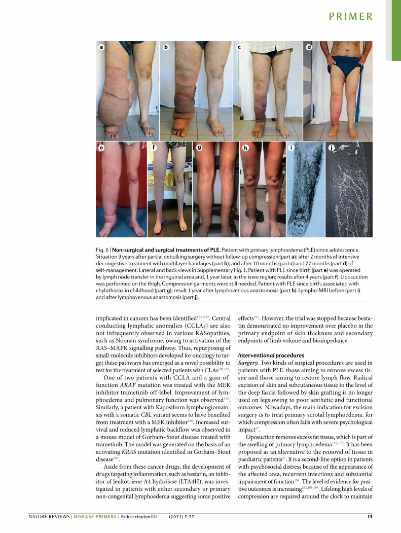
implicated in cancers has been identified183–187. Central conducting lymphatic anomalies (CCLAs) are also not infrequently observed in various RASopathies, such as Noonan syndrome, owing to activation of the RAS–MAPK signalling pathway. Thus, repurposing of small-molecule inhibitors developed for oncology to tar-get these pathways has emerged as a novel possibility to test for the treatment of selected patients with CLAs188,189.
One of two patients with CCLA and a gain-of- function ARAF mutation was treated with the MEK inhibitor trametinib off-label. Improvement of lym-phoedema and pulmonary function was observed190. Similarly, a patient with Kaposiform lymphangiomato-sis with a somatic CBL variant seems to have benefited from treatment with a MEK inhibitor186. Increased sur-vival and reduced lymphatic backflow was observed in a mouse model of Gorham–Stout disease treated with trametinib. The model was generated on the basis of an activating KRAS mutation identified in Gorham–Stout disease187.
Aside from these cancer drugs, the development of drugs targeting inflammation, such as bestatin, an inhib-itor of leukotriene A4 hydrolase (LTA4H), was inves-tigated in patients with either secondary or primary non-congenital lymphoedema suggesting some positive
effects191. However, the trial was stopped because besta-tin demonstrated no improvement over placebo in the primary endpoint of skin thickness and secondary endpoints of limb volume and bioimpedance.
Interventional proceduresSurgery. Two kinds of surgical procedures are used in patients with PLE: those aiming to remove excess tis-sue and those aiming to restore lymph flow. Radical excision of skin and subcutaneous tissue to the level of the deep fascia followed by skin grafting is no longer used on legs owing to poor aesthetic and functional outcomes. Nowadays, the main indication for excision surgery is to treat primary scrotal lymphoedema, for which compression often fails with severe psychological impact27.
Liposuction removes excess fat tissue, which is part of the swelling of primary lymphoedema192,193. It has been proposed as an alternative to the removal of tissue in paediatric patients27. It is a second-line option in patients with psychosocial distress because of the appearance of the affected area, recurrent infections and substantial impairment of function194. The level of evidence for posi-tive outcomes is increasing193,195,196. Lifelong high levels of compression are required around the clock to maintain
a c d
e f
b
g i jh
Fig. 6 | Non-surgical and surgical treatments of PLE. Patient with primary lymphoedema (PLE) since adolescence. Situation 9 years after partial debulking surgery without follow-up compression (part a); after 2 months of intensive decongestive treatment with multilayer bandages (part b); and after 10 months (part c) and 27 months (part d) of self-management. Lateral and back views in Supplementary Fig. 1. Patient with PLE since birth (part e) was operated by lymph node transfer in the inguinal area and, 1 year later, in the knee region; results after 4 years (part f). Liposuction was performed on the thigh. Compression garments were still needed. Patient with PLE since birth, associated with chylothorax in childhood (part g); result 1 year after lymphovenous anastomosis (part h). Lympho-MRI before (part i) and after lymphovenous anastomosis (part j).
15NATURE REVIEWS | DISEASE PRIMERS | Article citation ID: (2021) 7:77
P r i m e r
0123456789();:

the benefit. Liposuction is not indicated in patients seek-ing an alternative to compression or not complying with lifelong compression. Bariatric surgery improves limb volumes in patients with obesity with PLE197.
Lymphatic reconstructions aim to restore lymphatic flow and are proposed earlier during lymphoedema. Microsurgical procedures create lympho-lymphatic and lymphovenous anastomosis. These techniques have been proposed for >30 years, mostly in carefully selected groups of patients with local segmental obstruction of proximal lymphatics (for example, following cancer treatment) when peripheral lymph vessels were patent and with preserved contractility198. Supramicrosurgical reconstruction enables the creation of fine connec-tions (0.3–0.8 mm in diameter) between distal lym-phatics and subdermal venules (lymphaticovenular anastomosis). These techniques present good results if the lymphatic vessels are of good quality (autono-mous contraction and valves are still present) (Fig. 6g–j). Guidance with lymphoscintigraphy patterns and ICG fluorescence lymphangiography are being investigated to screen proper candidates and choose the dynamic vessels199–201. Only a few observational non-randomized studies include patients with PLE of lower limbs or genitals despite optimized compression decongestive management200,202,203. These approaches might not be useful in FOXC2-associated PLE or PLE associated with mutations in connexin genes as the venous system presents reflux or there is absence of contractility of functional capillaries100.
Free lymph node transfer (LNT) is based upon the transfer of a group of nodes and their own vessels from an unaffected extremity to a new location. It is a fascio(cutaneous) flap with arteries and veins that is transferred and revascularized by microsurgical anasto-mosis, like finger transplantation. In hypoplasia cases, the addition of nodes in a free fatty flap (autologous lymph node transplantation from patients themselves, avoiding graft rejection) enables neogenesis of lymphatic vessels owing to the presence of growth factors (VEGFs and cytokines). LNT and autologous lymph node trans-plantation are reconstructive techniques that have been developed mainly as prevention or treatment of second-ary lymphoedema after breast cancer treatment. The available case reports testify complete normalization in 20% of patients204 (Fig. 6e–f; Supplementary Fig. 1h–i). LNT has been associated with lymphoedema occurring on asymptomatic limbs in patients with PLE205. The combination of LNT with lymphovenous anastomosis is proposed in the management of complicated PLE in adults and children206.
Embolization of the lymphatics. The improvement of static and dynamic lymphangiography techniques enables the diagnosis of lymphatic flow anomalies and the guiding of lymphatic interventional procedures. Embolization of reflux from the thoracic duct into the pulmonary parenchyma and of leakages into the mesen-teric lymphatics was first developed as a palliative option in selected patients with single ventricle heart malforma-tion in whom heart transplantation could not be per-formed following failure of the surgical cavo-pulmonary
circulation, known as the Fontan circulation207,208. The resulting elevated central venous pressure and high lymphatic flow lead to protein-losing enteropathy and plastic bronchitis (characterized by expectoration of branching bronchial casts) in some patients with addi-tional lymphatic anomalies209. Interventional techniques, for example, catheterization of the thoracic duct and embolization, are increasingly used in the management of central conducting lymphatic anomalies associated with lymph and chyle reflux, such as chyluria210, genital leakage211, protein-losing enteropathy and ascites212, and pulmonary lymphatic anomalies46.
Quality of lifePLE is a chronic condition and its diagnosis is only one aspect of patient assessment. The large psychological burden of PLE and chronic oedemas has been evidenced using generic or specific health-related quality-of-life tools12,213 and is worse in patients in hospital than in those treated at the community level214, underscoring the necessity to include these aspects in the diagnosis and health profile assessment215,216. In 2001, the WHO introduced the ICF217. This bio-psycho-social ICF model includes five domains of functioning of a patient: bio-medical aspects, activities of daily living, participation in society, personal factors and environmental factors; the latter two influence the first three functional aspects and need to be considered. Validated clinimetrics218 can be established by a consensus group or during the creation of guidelines152 to design a treatment protocol and the measurement of its effects in all domains.
Most studies utilizing ICF health profiles219 and ded-icated ICF quality-of-life questionnaires (such as lymph ICF-lower limb220 and the Lymphedema Quality of Life Inventory (LYQLI)221) are based on secondary lymphoe-dema. A comparison of the validity of five different out-come measurement tools was performed in patients with lower-limb lymphoedema and suggested specific use according to the aim of the outcome measurement222.
Thus, it is not clear whether extrapolation of the validity of quality-of-life tools used for secondary lym-phoedema to PLE is acceptable. Because PLE is a rare disease, it is associated with delayed diagnosis, feelings of insecurity and mistrust of professionals, aspects that do not exist in secondary lymphoedema223. Growing with a different body shape also has different effects from secondary lymphoedema. Moreover, specific question-naires are being developed for children and young adults (NCT01922635).
Long-term management of PLE issuesDecongestive lymphoedema therapy is the first line of management of PLE to improve quality of life. However, compression maintenance therapy of lymphoedema is a lifelong process and remains the main treatment even after interventional procedures. It requires that the patient wears compression garments daily, exercises, performs skincare, and sometimes repetitively conduct self-massage and bandaging. The burden of long-term management is obvious and requires a commitment to self-management and a self-efficacy belief as shown in children and parents224. Compression is not mandatory
16 | Article citation ID: (2021) 7:77 www.nature.com/nrdp
P r i m e r
0123456789();:

in toddlers and even children might not cope with aggressive decongestive therapy or wear prescription garments. Professionals need to adapt compression to the patient’s life and not vice versa and take the time needed for patients to accept their condition and tailor the therapy. Education can be delivered in camps or dur-ing social and networking activities; it helps young peo-ple not to feel alone with their rare condition and is also useful for professionals to share their experience225,226 (boxes 3,4).
OutlookLymphoedema is a global problem (box 5). The man-agement of PLE is still in its infancy, although the dis-ease has been recognized for years. One of the reasons is the wide variety of presentations, from isolated forms to combinations with other features in syndromes. A second major reason is the lack of understanding of the pathogenetic events leading to the disease and the underlying genetic causes.
Genetic variabilityIn large-scale genetic screens, numerous variants of unknown significance are detected. If these were to be considered as disease-causing mutations (assuming that they would be validated as such by functional tests), the proportion of individuals in whom a causative mutation
is identified could be as high as 70% of patients accord-ing to our estimates (P.B. and M.V., unpublished work). Yet, most likely, the majority of variants of unknown sig-nificance do not have an effect on protein function and represent rare polymorphisms. Thus, novel genes asso-ciated with PLE need to be looked for as many pieces are still missing to reconstitute the full puzzle and finding additional pieces would lay down the basis for under-standing the multiple reasons for the development of PLE.
Additional challenges in identifying the genetic basis of PLE are based on the fact that the available techniques to identify causative intronic and intergenic variants are less developed than those for exonic variants. As the effect of disease-causing variants should present at protein level and therefore also at the mRNA level, RNA-based studies might be helpful. This approach could provide insights on deep intronic mutations, exonic splice-altering mutations that are not identified as pathogenetic as they are synonymous changes, pro-moter mutations, methylation disturbances, and other changes that would regulate gene expression level. After an exhausting search for intragenic mutations, we could look for alterations in the rest of the genome: mutations in non-coding parts of the DNA, microRNAs, long non-coding RNAs or DNA alterations that result from viral infection for viruses that integrate their genetic material into the genome. Occasionally, patients with unilateral PLE could carry a somatic mutation only detectable in the affected tissue. Moreover, PLE could also develop as a result of gene-to-gene interactions and therefore validated algorithms to study oligogenic diseases are needed.
Many of the mutations in genes causing Mendelian forms of PLE have incomplete penetrance. Sex-specific penetrance is also starting to emerge and could help provide a more accurate prognosis when taken into consideration. Hormones seem to have an important role in PLE development yet their influence is poorly understood and needs focused studies.
Another intriguing question is the overlap of PLE-causing genes with secondary lymphoedema as the same genes may be involved. Two studies have sug-gested such a link95,227. An association was found with LCP2 (encoding lymphocyte cytosolic protein 2), NRP2 (encoding neuropilin 2), SYK (encoding spleen tyros-ine kinase, also known as tyrosine-protein kinase SYK), VCAM1 (encoding vascular cell adhesion protein 1), FOXC2 and VEGFC. However, other studies are needed to confirm the significance of these associations.
When developing therapeutic approaches, it is important to visualize the interplay between the different genes that have already been identified for PLE. In 2014, 20 of them were grouped in a hypothetical LEC and many of the gene products were geared around a central VEGFC–VEGFR3 signalling pathway74. This model has now become more complex, with the inclusion of other ligand–receptor complexes (ANGPT2–TIE1/TIE2 and HGF–MET) and the effect of RAS–MAPK and PI3K–AKT signalling, which point to more severe syndromic forms (Fig. 3). Moreover, along the lymphatic vasculature, the impact of the gene mutations and mutated proteins
Box 3 | Patient perspective (part 1)
E. is a 24-year-old young woman born with a generalized lymphatic anomaly. These are her words about what it means to live with primary lymphoedema, recorded on December 2020 as a webinar.
The journey for a diagnosisI was 8 weeks old, my mum noticed that my left arm was bigger than my right arm, and they had no idea what was going on. There started a very long journey to diagnose it. We went to the general practitioner, who then referred diagnosis to a specialist paediatrician in the area. I was diagnosed with hemihypertrophy syndrome. At that time, 24 years ago, there was very little understanding of what was going on and my parents would feel a lot of confusion. It was only a few years later that they started to notice that it was my left arm and leg, but also that my lungs were involved. This led to beingabletoseeaspecialistteam,whodiagnosedpulmonarylymphangiectasiaanddetermined that I was having a sort of underlying generalized lymphatic anomaly.
I could talk for ages about how long this journey took. The essence of what I am saying is that growing with this, having lymphoedema, was not really understood at all at that time. My parents talk about the challenges and difficulties they had to be able to access the care I needed and just to be able to understand what was going on. This uncertainty went on for many years, until we were able to get the right diagnosis many years after. Being able to access specialized centres with doctors and professionals who know what is going on makes a huge difference. That is so life-changing.
Growing up with lymphoedemaUltimately, it is about understanding the needs of children and young people. They are quite different. Following the medical recommendations, you feel it is quite a big commitment. One of the hardest things for me was managing all of these different appointments, having to travel to a lot of different places and seeing many different doctors. At the same time, working at school and getting my degree was so important to me. I struggled a lot with not being at school because of lymphoedema.
Being now 24 years old, my advice would be to rely on a support network and get plans in place with school, colleges and universities. Being open to the reality of the challenges, and this is not easy to do. It is not easy to stand in front of people and talk about your challenges in life. In the end, it is about understanding that people can help you if they understand how they can help you. For example, [finding] shoes, clothes, garments, that is a daily struggle, and I still find that so challenging. One day hopefully I will get a pair of red high heel shoes; that is one of my aspirations in life, still a way to go.
17NATURE REVIEWS | DISEASE PRIMERS | Article citation ID: (2021) 7:77
P r i m e r
0123456789();:

may differ. Some mutations affect LEC proliferation or survival more globally, whereas others affect the devel-opment or stability of lymphatic valves or lymphovenous valves (such as EPHB4 mutations). Thus, we will need in vivo models to gather more detailed data for the var-ious pathways involved and unravel eventual ways to develop novel therapies and preventive measures.
Diagnosis and treatmentThe diversity of genes associated with PLE, both known (10 of them were identified during the past 5 years) and still to be unravelled, combined with technolog-ical advancements, can lead to the rapid evolution of diagnostic genetic testing of PLE. Gene panels have given the most reliable results but they rapidly become outdated. The recommended diagnostic approach is
moving towards whole-exome sequencing, for which the analysis can be re-processed for individuals in whom a causative mutation has not been identified when new genes are reported. With evolving copy number detec-tion algorithms, whole-exome sequencing data can also identify partial and whole-gene deletions, although there are still limitations. Overall, increased germline genetic testing of patients will enable more precise (molecular) diagnostics and genetic counselling, which in turn allows at-risk family members to be informed and establishment of preventive measures. Importantly, such data help to better stratify patients for clinical research studies.
Detailed clinical analysis of all patients, including re-evaluation after established genetic diagnosis, will enable calculation of the risks for additional signs and symptoms in each PLE subtype. For example, in hypo-trichosis–lymphoedema–telangiectasia syndrome (char-acterized by sparse hair, lymphoedema and small dilated cutaneous vessels), additional features are reported in almost every new case for this pleiotropic disease228. Such stratified epidemiological data will be paramount for the clinical diagnostics, genetic counselling, manage-ment and follow-up of patients. Such data will also help in the development of diagnostic algorithms15 (Fig. 5). Stratification will also enable the more precise study of response to conventional and surgical treatments and, eventually, to predict outcomes, which could have an important impact on patient care.
Imaging technologies are also developing rapidly, promising a more detailed evaluation of PLE. MRL is now increasingly used, also in combination with periph-eral ICG fluorescence lymphangiography. Combining these two techniques can help to reclassify the cause of PLE to peripheral, central or combined deficiency. Whole-body lymphangioscintigraphy associated with 3D localization by SPECT–CT can provide functional imaging of peripheral and central lymphatic flow60. Such multimodal approaches can be particularly useful in cases of PLE associated with CCLA and can guide the planning of interventional management. The Society of Interventional Radiology (SIR) and the Journal of Vascular Interventional Radiology selected lymphatic imaging as their focus topic of the year for 2020 (reF.46).
Photoacoustic imaging is another emerging imaging technique that provides unique scalability of optical res-olution and acoustic depth of penetration. When associ-ated with the use of light-absorbing biomolecules, such as oxy-haemoglobin and deoxyhaemoglobin and lipids, specific vascular structures can be targeted. Moreover, indocyanine green, which is taken up by lymphatic ves-sels, allows the use of photoacoustic imaging for lym-phangiography, which has a higher spatial resolution than other imaging modalities229–231.
The outlook for future personalized management of PLE is multifaceted. Diagnostics and interventional pro-cedures should benefit from the further development of sensitive imaging methods for lymphatic dysfunctions. Combined with large-scale genetic analyses and other omics-type approaches in research, it should gather data to decipher the underpinnings of PLE development. Understanding PLE pathophysiology is the foundation
Box 5 | Lymphoedema — the global picture
Lymphoedema is globally a Neglected Public Health issue6. In upper-middle-income and high-income countries, where cancer is one of the top 10 leading causes of death, lymphoedema is recognized as a sequala of cancer, associated with a solid reputation of being an untreatable adverse effect of the treatment of cancer11. Its incidence is increasing with ageing, immobility and obesity, which are leading risk factors for lymphoedema and related cellulitis in these countries289,290. The WHO recognizes lymphatic filariasis as one of the five preventable neglected tropical diseases threatening 859 million people in 50, mostly low-income, countries. The Global Programme to Eliminate Lymphatic Filariasis (GPELF) is aiming at providing anti-filarial medicines and a minimum package of care in all areas with known patients and progression of lymphoedema291. This package focuses on hygiene and misses the point of compression. PLE is hardly recognized in these countries292 but has been recently recognized in Europe as a rare disease (prevalence <1 per 2,000 individuals36) and benefits from programmes dedicated to Rare Disease Networks such as VASCERN. Yet, unequal access to diagnosis and treatment is the reality all patients face owing to the underdiagnosis of lymphoedema, lack of efficient management and the inadequacy of the reimbursement policies for medical devices even in Europe293. Dedicated research programmes are required as stated in the USA by the Lymphatic Education & Research Network.
Box 4 | Patient perspective (part 2)
FearsFear and anxiety around cellulitis are very real. They can come on very quickly, they can be severe and you can feel very unwell. It is a constant in the back of your mind. You have to be aware of it, that it is dangerous. But you still have to live and enjoy your life. It is a balance. Now, I learned my warning signs, and that is important and takes time. It is also about getting help from the network you have built around you, including professionals and clinicians.
Covering the information with the general practitioner and self-managementAs a patient, you can have so much control on your lymphoedema. I was the one sharing the knowledge with the general practitioner, using leaflets I was given by the support group. But with that comes a lot of responsibilities. You can in the end feel overwhelmed.
Psychological aspectsI would not cope with people seeing my leg for years. It took a lot of time to wear a dress when I wanted to wear a dress. We have to be in a place of self-acceptance. Lymphoedema is for life, and it takes time to understand and live with it.
I was not good at taking my medication and putting my compression on. Well, to be honest, we all get good and bad days. So it is also important that young people know that it is OK not to always feel OK with lymphoedema.
Sharing with peersWhen you see other patients as a young person, there is no better feeling. You do feel alone with a rare disease, and it can be hard. It has been hard to establish my identity as ‘E.’ and not only ‘E. with a bigger leg’.
18 | Article citation ID: (2021) 7:77 www.nature.com/nrdp
P r i m e r
0123456789();:

for the development of innovative PLE-specific thera-peutics such as modulators of lymphangiogenesis and/or gene therapy approaches, stem cell-based treat-ment, and the development of artificial lymphatics by tissue engineering. In addition to bestatin, VEGFC has been used in a clinical trial. It was combined with LNT to induce lymph vessel growth and connectivity to the transferred lymph nodes in patients with secondary lymphoedema after breast cancer232. However, the com-pany (Herantis Pharma) recently announced that the
results were inconclusive. The eventual benefit of this technique, aiming to induce lymphangiogenesis locally, remains unknown in PLE. Targeted therapies have already emerged for other vascular anomalies caused by somatic mutations that increase signalling via the PI3K–AKT–mTOR pathway or the RAS–RAF–MAPK pathway, including CLAs188. Thus, the future is full of hope also for patients with PLE.
Published online xx xx xxxx
1. Starling, E. H. On the absorption of fluids from the connective tissue spaces. J. Physiol. 19, 312–326 (1896).
2. Levick, J. R. & Michel, C. C. Microvascular fluid exchange and the revised Starling principle. Cardiovasc. Res. 87, 198–210 (2010).
3. Quere, I., Nagot, N. & Vikkula, M. Incidence of cellulitis among children with primary lymphedema. N. Engl. J. Med. 378, 2047–2048 (2018).
4. Moffatt, C. J. et al. Prevalence and risk factors for chronic edema in U.K. community nursing services. Lymphat. Res. Biol. 17, 147–154 (2019).
5. Moffatt, C. J. et al. Lymphoedema: an underestimated health problem. QJM 96, 731–738 (2003).
6. Moffatt, C., Keeley, V. & Quere, I. The concept of chronic edema — a neglected public health issue and an international response: The LIMPRINT Study. Lymphat. Res. Biol. 17, 121–126 (2019).
7. Rockson, S. G. & Rivera, K. K. Estimating the population burden of lymphedema. Ann. N. Y. Acad. Sci. 1131, 147–154 (2008).
8. Keast, D. H., Despatis, M., Allen, J. O. & Brassard, A. Chronic oedema/lymphoedema: under-recognised and under-treated. Int. Wound J. 12, 328–333 (2015).
9. DiSipio, T., Rye, S., Newman, B. & Hayes, S. Incidence of unilateral arm lymphoedema after breast cancer: a systematic review and meta-analysis. Lancet Oncol. 14, 500–515 (2013).
10. Mortimer, P. S. & Rockson, S. G. New developments in clinical aspects of lymphatic disease. J. Clin. Invest. 124, 915–921 (2014).
11. Rockson, S. G., Keeley, V., Kilbreath, S., Szuba, A. & Towers, A. Cancer-associated secondary lymphoedema. Nat. Rev. Dis. Prim. 5, 22 (2019). This recent detailed review explores various aspects of secondary lymphoedema, which, in developed countries, mostly results from the treatment of cancer as opposed to infection- related secondary lymphoedema in developing countries.
12. Mercier, G., Pastor, J., Moffatt, C., Franks, P. & Quere, I. LIMPRINT: health-related quality of life in adult patients with chronic edema. Lymphat. Res. Biol. 17, 163–167 (2019). This large multicentre study prospectively assessed the health-related quality of life of 1,094 adult patients with chronic oedema and underscored a poor disease-specific and generic health-related quality of life.
13. Lopez, M., Roberson, M. L., Strassle, P. D. & Ogunleye, A. Epidemiology of lymphedema-related admissions in the United States: 2012–2017. Surg. Oncol. 35, 249–253 (2020).
14. Biesecker, L. G. et al. A dyadic approach to the delineation of diagnostic entities in clinical genomics. Am. J. Hum. Genet. 108, 8–15 (2021).
15. Gordon, K. et al. Update and audit of the St George’s classification algorithm of primary lymphatic anomalies: a clinical and molecular approach to diagnosis. J. Med. Genet. 57, 653–659 (2020). This review proposes an updated clinical classification algorithm for primary lymphoedema to assist diagnostic workup and patient management, based on age of onset, areas affected by swelling and associated clinical features, in agreement with the International Society for the Study of Vascular Anomalies 2018 classification.
16. Nonne, M. & Vier Fälle, V. Elephantiasis congenita hereditarian. Arch. für pathologische anatomie und physiologie und für klinische Med. 125, 189–196 (1891).
17. Milroy, W. F. An undescribed variety of hereditary oedema. N. Y. Med. J. 56, 505–508 (1892).
18. Samman, P. D. & White, W. F. The “yellow nail” syndrome. Br. J. Dermatol. 76, 153–157 (1964).
19. Meige, H. Distrophie oedemateuse héréditaire. Presse Méd. 6, 341–343 (1898).
20. Irrthum, A., Karkkainen, M. J., Devriendt, K., Alitalo, K. & Vikkula, M. Congenital hereditary lymphedema caused by a mutation that inactivates VEGFR3 tyrosine kinase. Am. J. Hum. Genet. 67, 295–301 (2000).
21. Karkkainen, M. J. et al. Missense mutations interfere with VEGFR-3 signalling in primary lymphoedema. Nat. Genet. 25, 153–159 (2000).
22. Mendola, A. et al. Mutations in the VEGFR3 signaling pathway explain 36% of familial lymphedema. Mol. Syndromol. 4, 257–266 (2013).
23. Leppanen, V. M. et al. Characterization of ANGPT2 mutations associated with primary lymphedema. Sci. Transl Med. 12, eaax8013 (2020). This is the most recent discovery of a novel gene causing primary lymphoedema with functional validation of the mutations in vitro and in a mouse model, underscoring the heterogeneity within genetic causes of PLE.
24. Iacobas, I. et al. Multidisciplinary guidelines for initial evaluation of complicated lymphatic anomalies-expert opinion consensus. Pediatr. Blood Cancer 67, e28036 (2020).
25. Quinn, A. M., Valcarcel, B. N., Makhamreh, M. M., Al-Kouatly, H. B. & Berger, S. I. A systematic review of monogenic etiologies of nonimmune hydrops fetalis. Genet. Med. 23, 3–12 (2021). This systematic literature review of non-immune hydrops fetalis pinpointed 131 genes with strong evidence for an association with NIHF and 46 genes with emerging evidence, spanning the spectrum of multisystemic syndromes and cardiac, haematological and metabolic disorders.
26. Smeltzer, D. M., Stickler, G. B. & Schirger, A. Primary lymphedema in children and adolescents: a follow-up study and review. Pediatrics 76, 206–218 (1985).
27. Schook, C. C. et al. Primary lymphedema: clinical features and management in 138 pediatric patients. Plast. Reconstr. Surg. 127, 2419–2431 (2011).
28. Fastre, E. et al. Splice-site mutations in VEGFC cause loss of function and Nonne-Milroy-like primary lymphedema. Clin. Genet. 94, 179–181 (2018).
29. Erickson, R. P. et al. Sex-limited penetrance of lymphedema to females with CELSR1 haploinsufficiency: a second family. Clin. Genet. 96, 478–482 (2019).
30. Soo, J. K., Bicanic, T. A., Heenan, S. & Mortimer, P. S. Lymphatic abnormalities demonstrated by lymphoscintigraphy after lower limb cellulitis. Br. J. Dermatol. 158, 1350–1353 (2008).
31. Au, A. C. et al. Protein tyrosine phosphatase PTPN14 is a regulator of lymphatic function and choanal development in humans. Am. J. Hum. Genet. 87, 436–444 (2010).
32. Hong, S. E. et al. Autosomal recessive lissencephaly with cerebellar hypoplasia is associated with human RELN mutations. Nat. Genet. 26, 93–96 (2000).
33. Brice, G. et al. A novel mutation in GJA1 causing oculodentodigital syndrome and primary lymphoedema in a three generation family. Clin. Genet. 84, 378–381 (2013).
34. Gumus, E. A rare symptom of a very rare disease: a case report of a oculodentodigital dysplasia with lymphedema. Clin. Dysmorphol. 27, 91–93 (2018).
35. Schlogel, M. J. et al. No evidence of locus heterogeneity in familial microcephaly with or without chorioretinopathy, lymphedema, or mental retardation syndrome. Orphanet J. Rare Dis. 10, 52 (2015).
36. Commission, E. U. Useful Information on Rare Diseases from EU Perspective. C 151/157–C 151/110 (European Commission 2009).
37. Park, S. I. et al. Prevalence and epidemiological factors involved in cellulitis in Korean patients with
lymphedema. Ann. Rehabil. Med. 40, 326–333 (2016).
38. Aslam, A. F., Aslam, A. K., Qamar, M. U. & Levey, R. Primary lymphedema tarda in an 88-year-old African-American male. J. Natl Med. Assoc. 97, 1031–1035 (2005).
39. Ibrahim, A. Primary lymphedema tarda. Pan Afr. Med. J. 19, 16 (2014).
40. Davey, S. L. et al. The South African multi-disciplinary lymphoedema position statement. Wound Healing South. Afr. 11, 21–24 (2018).
41. Julkowska, D. et al. The importance of international collaboration for rare diseases research: a European perspective. Gene Ther. 24, 562–571 (2017).
42. Vignes, S. et al. Primary lymphedema French National Diagnosis and Care Protocol (PNDS; Protocole National de Diagnostic et de Soins). Orphanet J. Rare Dis. 16, 18 (2021).
43. Nedergaard, M. & Goldman, S. A. Glymphatic failure as a final common pathway to dementia. Science 370, 50–56 (2020).
44. Proulx, S. T. Cerebrospinal fluid outflow: a review of the historical and contemporary evidence for arachnoid villi, perineural routes, and dural lymphatics. Cell Mol. Life Sci. 78, 2429–2457 (2021).
45. Witte, M. & Bernas, M. in Rutherford’s Vascular Surgery and Endovascular Therapy (eds Sidaway, A. & Perler, B.) Ch. 10, 105–122 (Elsevier, 2019).
46. Itkin, M. et al. Research priorities in lymphatic interventions: recommendations from a multidisciplinary research consensus panel. J. Vasc. Interv. Radiol. 32, 762.e1–762.e7 (2021). This document, by a selected panel of experts in lymphatic medicine from the USA, New Zealand and Korea, identified seven priorities for research in the field, including lymphatic decompression in patients with congestive heart failure, detoxification of thoracic duct lymph in acute illness, development of newer agents for lymphatic imaging, characterization of organ-based lymph composition, and development of lymphatic interventions to treat ascites in liver cirrhosis.
47. Schwartz, F. R. et al. Lymphatic imaging: current noninvasive and invasive techniques. Semin. Intervent. Radiol. 37, 237–249 (2020).
48. Kinmonth, J. B. in The Lymphatic: Disease, Lymphography, and Surgery 114–155 (Edward Arnold, 1972).
49. Witte, M. H. et al. Structure function relationships in the lymphatic system and implications for cancer biology. Cancer Metastasis Rev. 25, 159–184 (2006).
50. Földi, M. & Földi, E. in Földi’s Textbook of Lymphology (eds Földi, M. & Földi, E.) Ch. 2, 135–273 (Urban & Fischer Verlag, 2012).
51. Executive Committee.The diagnosis and treatment of peripheral lymphedema: 2016 consensus document of the international society of lymphology. Lymphology 49, 170–184 (2016).
52. Witte, M. H., Dumont, A. E., Cole, W. R., Witte, C. L. & Kintner, K. Lymph circulation in hepatic cirrhosis: effect of portacaval shunt. Ann. Intern. Med. 70, 303–310 (1969).
53. Baish, J. W., Netti, P. A. & Jain, R. K. Transmural coupling of fluid flow in microcirculatory network and interstitium in tumors. Microvasc. Res. 53, 128–141 (1997).
54. Michel, C. C., Woodcock, T. E. & Curry, F. E. Understanding and extending the Starling principle. Acta Anaesthesiol. Scand. 64, 1032–1037 (2020).
55. Lee, B. B. et al. Diagnosis and treatment of primary lymphedema. Consensus document of the International Union of Phlebology (IUP)-2013. Int. Angiol. 32, 541–574 (2013).
19NATURE REVIEWS | DISEASE PRIMERS | Article citation ID: (2021) 7:77
P r i m e r
0123456789();:
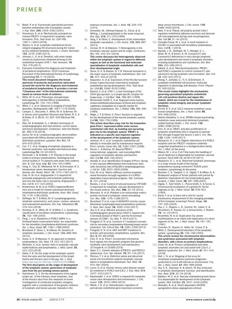
56. Baluk, P. et al. Functionally specialized junctions between endothelial cells of lymphatic vessels. J. Exp. Med. 204, 2349–2362 (2007).
57. Nonomura, K. et al. Mechanically activated ion channel PIEZO1 is required for lymphatic valve formation. Proc. Natl Acad. Sci. USA 115, 12817–12822 (2018).
58. Teijeira, A. et al. Lymphatic endothelium forms integrin-engaging 3D structures during DC transit across inflamed lymphatic vessels. J. Invest. Dermatol. 133, 2276–2285 (2013).
59. Johnson, L. A. et al. Dendritic cells enter lymph vessels by hyaluronan-mediated docking to the endothelial receptor LYVE-1. Nat. Immunol. 18, 762–770 (2017).
60. Executive Committee.The diagnosis and treatment of peripheral lymphedema: 2020 Consensus Document of the International Society of Lymphology. Lymphology 53, 3–19 (2020). This recent document integrates the broad spectrum of protocols and practices advocated around the world for the diagnosis and treatment of peripheral lymphoedema. It provides a current “Consensus view” of the international community based on various levels of evidence.
61. Witte, M. H. et al. Phenotypic and genotypic heterogeneity in familial Milroy lymphedema. Lymphology 31, 145–155 (1998).
62. Witte, C. L. et al. Advances in imaging of lymph flow disorders. Radiographics 20, 1697–1719 (2000).
63. Campisi, C., Boccardo, F., Witte, M. H. & Bernas, M. in Venous and Lymphatic Diseases (eds Dieter, R. S. Jr & Dieter, R. A. III) Ch. 42, 607–629 (McGraw Hill, 2011).
64. Itkin, M. & Nadolski, G. J. Modern techniques of lymphangiography and interventions: current status and future development. Cardiovasc. Intervent Radiol. 41, 366–376 (2018).
65. Sarica, M. et al. Lymphoscintigraphic abnormalities associated with Milroy disease and lymphedema- distichiasis syndrome. Lymphat. Res. Biol. 17, 610–619 (2019).
66. Cox, T. C. et al. Imaging of lymphatic dysplasia in Noonan syndrome: case studies and historical atlas. Lymphology 54, 23–40 (2021).
67. Kinmonth, J. B. & Wolfe, J. H. Fibrosis in the lymph nodes in primary lymphoedema. Histological and clinical studies in 74 patients with lower-limb oedema. Ann. R. Coll. Surg. Engl. 62, 344–354 (1980).
68. Geng, X., Cha, B., Mahamud, M. R. & Srinivasan, R. S. Intraluminal valves: development, function and disease. Dis. Model. Mech. 10, 1273–1287 (2017).
69. Gale, N. W. et al. Angiopoietin-2 is required for postnatal angiogenesis and lymphatic patterning, and only the latter role is rescued by Angiopoietin-1. Dev. Cell 3, 411–423 (2002).
70. Kriederman, B. M. et al. FOXC2 haploinsufficient mice are a model for human autosomal dominant lymphedema-distichiasis syndrome. Hum. Mol. Genet. 12, 1179–1185 (2003).
71. Dellinger, M. T. & Witte, M. H. Lymphangiogenesis, lymphatic systemomics, and cancer: context, advances and unanswered questions. Clin. Exp. Metastasis 35, 419–424 (2018).
72. Northup, K. A., Witte, M. H. & Witte, C. L. Syndromic classification of hereditary lymphedema. Lymphology 36, 162–189 (2003).
73. Fang, J. et al. Mutations in FOXC2 (MFH-1), a forkhead family transcription factor, are responsible for the hereditary lymphedema-distichiasis syndrome. Am. J. Hum. Genet. 67, 1382–1388 (2000).
74. Brouillard, P., Boon, L. & Vikkula, M. Genetics of lymphatic anomalies. J. Clin. Invest. 124, 898–904 (2014).
75. Jones, G. E. & Mansour, S. An approach to familial lymphoedema. Clin. Med. 17, 552–557 (2017).
76. Michelini, S. et al. Genetic tests in lymphatic vascular malformations and lymphedema. J. Med. Genet. 55, 222–232 (2018).
77. Sabin, F. R. On the origin of the lymphatic system from the veins and the development of the lymph hearts and thoracic duct in the pig. Am. J. Anat. https://doi.org/10.1002/aja.1000010310 (1902). The first description on the origin of development of the lymphatic system: generation of lymphatic sacs from the pre-existing venous system.
78. Hutchinson, G. S. On the development of the jugular lymph sac, of the tributary ulnar lymphatic, and of the thoracic duct from the viewpoint of recent investigations of vertebrate lymphatic ontogeny, together with a consideration of the genetic relations of lymphatic and hemal vascular channels in the
embryos of amniotes. Am. J. Anat. 16, 259–316 (1914).
79. Schneider, M., Othman-Hassan, K., Christ, B. & Wilting, J. Lymphangioblasts in the avian wing bud. Dev. Dyn. 216, 311–319 (1999).
80. Yang, Y. & Oliver, G. Development of the mammalian lymphatic vasculature. J. Clin. Invest. 124, 888–897 (2014).
81. Ulvmar, M. H. & Makinen, T. Heterogeneity in the lymphatic vascular system and its origin. Cardiovasc. Res. 111, 310–321 (2016). This review discusses the heterogeneity observed within the lymphatic system in regard to different organs as well as the functional and molecular specialization of lymphatic endothelial cells and their developmental origin.
82. Stone, O. A. & Stainier, D. Y. R. Paraxial mesoderm is the major source of lymphatic endothelium. Dev. Cell 50, 247–255.e3 (2019).
83. Kaipainen, A. et al. Expression of the fms-like tyrosine kinase 4 gene becomes restricted to lymphatic endothelium during development. Proc. Natl Acad. Sci. USA 92, 3566–3570 (1995).
84. Banerji, S. et al. LYVE-1, a new homologue of the CD44 glycoprotein, is a lymph-specific receptor for hyaluronan. J. Cell Biol. 144, 789–801 (1999).
85. Breiteneder-Geleff, S. et al. Angiosarcomas express mixed endothelial phenotypes of blood and lymphatic capillaries: podoplanin as a specific marker for lymphatic endothelium. Am. J. Pathol. 154, 385–394 (1999).
86. Wigle, J. T. & Oliver, G. Prox1 function is required for the development of the murine lymphatic system. Cell 98, 769–778 (1999). This article describes a key role for the homeobox gene Prox1, expressed within some venous endothelial cells that, by budding and sprouting, give rise to the lymphatic system. PROX1 is indicated as a specific and required regulator of the development of the lymphatic system.
87. Johnson, N. C. et al. Lymphatic endothelial cell identity is reversible and its maintenance requires Prox1 activity. Genes Dev. 22, 3282–3291 (2008).
88. Ducoli, L. & Detmar, M. Beyond PROX1: transcriptional, epigenetic, and noncoding RNA regulation of lymphatic identity and function. Dev. Cell 56, 406–426 (2021).
89. Harada, K. et al. Identification of targets of Prox1 during in vitro vascular differentiation from embryonic stem cells: functional roles of HoxD8 in lymphangiogenesis. J. Cell Sci. 122, 3923–3930 (2009).
90. Frye, M. et al. Matrix stiffness controls lymphatic vessel formation through regulation of a GATA2-dependent transcriptional program. Nat. Commun. 9, 1511 (2018).
91. Bowles, J. et al. Control of retinoid levels by CYP26B1 is important for lymphatic vascular development in the mouse embryo. Dev. Biol. 386, 25–33 (2014).
92. Morooka, N. et al. Polydom is an extracellular matrix protein involved in lymphatic vessel remodeling. Circ. Res. 120, 1276–1288 (2017).
93. Brouillard, P. et al. Loss of ADAMTS3 activity causes Hennekam lymphangiectasia-lymphedema syndrome 3. Hum. Mol. Genet. 26, 4095–4104 (2017).
94. Jha, S. K. et al. Efficient activation of the lymphangiogenic growth factor VEGF-C requires the C-terminal domain of VEGF-C and the N-terminal domain of CCBE1. Sci. Rep. 7, 4916 (2017).
95. Finegold, D. N. et al. Connexin 47 mutations increase risk for secondary lymphedema following breast cancer treatment. Clin. Cancer Res. 18, 2382–2390 (2012).
96. Finegold, D. N. et al. HGF and MET mutations in primary and secondary lymphedema. Lymphat. Res. Biol. 6, 65–68 (2008).
97. Choi, D. et al. Piezo1 incorporates mechanical force signals into the genetic program that governs lymphatic valve development and maintenance. JCI Insight 4, e125068 (2019).
98. Alper, S. L. Genetic diseases of PIEZO1 and PIEZO2 dysfunction. Curr. Top. Membr. 79, 97–134 (2017).
99. Petrova, T. V. et al. Defective valves and abnormal mural cell recruitment underlie lymphatic vascular failure in lymphedema distichiasis. Nat. Med. 10, 974–981 (2004).
100. Lyons, O. et al. Human venous valve disease caused by mutations in FOXC2 and GJC2. J. Exp. Med. 214, 2437–2452 (2017).
101. Kazenwadel, J. et al. GATA2 is required for lymphatic vessel valve development and maintenance. J. Clin. Invest. 125, 2979–2994 (2015).
102. Welsh, J. D. et al. Hemodynamic regulation of perivalvular endothelial gene expression prevents
deep venous thrombosis. J. Clin. Invest. 129, 5489–5500 (2019).
103. Tatin, F. et al. Planar cell polarity protein Celsr1 regulates endothelial adherens junctions and directed cell rearrangements during valve morphogenesis. Dev. Cell 26, 31–44 (2013).
104. Gonzalez-Garay, M. L. et al. A novel mutation in CELSR1 is associated with hereditary lymphedema. Vasc. Cell 8, 1 (2016).
105. Kanady, J. D., Dellinger, M. T., Munger, S. J., Witte, M. H. & Simon, A. M. Connexin37 and Connexin43 deficiencies in mice disrupt lymphatic valve development and result in lymphatic disorders including lymphedema and chylothorax. Dev. Biol. 354, 253–266 (2011).
106. Sabine, A. et al. Mechanotransduction, PROX1, and FOXC2 cooperate to control connexin37 and calcineurin during lymphatic-valve formation. Dev. Cell 22, 430–445 (2012).
107. Zhang, F., Zarkada, G., Yi, S. & Eichmann, A. Lymphatic endothelial cell junctions: molecular regulation in physiology and diseases. Front. Physiol. 11, 509 (2020). This recent review highlights the mechanisms governing specialized lymphatic endothelial cell–cell junctions (button and zipper-like states), which are crucial for the maintenance of lymphatic vessel integrity and proper lymphatic functions.
108. Ferrell, R. E. et al. GJC2 missense mutations cause human lymphedema. Am. J. Hum. Genet. 86, 943–948 (2010).
109. Martin-Almedina, S. et al. EPHB4 kinase-inactivating mutations cause autosomal dominant lymphatic- related hydrops fetalis. J. Clin. Invest. 126, 3080–3088 (2016).
110. Choi, D. et al. ORAI1 activates proliferation of lymphatic endothelial cells in response to laminar flow through kruppel-like factors 2 and 4. Circ. Res. 120, 1426–1439 (2017).
111. Mustacich, D. J. et al. Digenic inheritance of a FOXC2 mutation and two PIEZO1 mutations underlies congenital lymphedema in a multigeneration family. Am. J. Med. (in the press).
112. Meens, M. J. et al. Cx47 fine-tunes the handling of serum lipids but is dispensable for lymphatic vascular function. PLoS ONE 12, e0181476 (2017).
113. Mustacich, D. J., et al. Abnormal lymphatic phenotype in a crispr mouse model of the human lymphedema-causing connexin47 R260C point mutation. Lymphology (in the press).
114. Boucher, C. A., Sargent, C. A., Ogata, T. & Affara, N. A. Breakpoint analysis of Turner patients with partial Xp deletions: implications for the lymphoedema gene location. J. Med. Genet. 38, 591–598 (2001).
115. Ogata, T., Tyler-Smith, C., Purvis-Smith, S. & Turner, G. Chromosomal localisation of a gene(s) for Turner stigmata on Yp. J. Med. Genet. 30, 918–922 (1993).
116. Bardi, F. et al. Is there still a role for nuchal translucency measurement in the changing paradigm of first trimester screening? Prenat. Diagn. 40, 197–205 (2020).
117. Hsu, L. Y., Shapiro, L. R., Gertner, M., Lieber, E. & Hirschhorn, K. Trisomy 22: a clinical entity. J. Pediatr. 79, 12–19 (1971).
118. Rosenfeld, W. et al. Duplication 3q: severe manifestations in an infant with duplication of a short segment of 3q. Am. J. Med. Genet. 10, 187–192 (1981).
119. Greenlee, R., Hoyme, H., Witte, M., Crowe, P. & Witte, C. Developmental disorders of the lymphatic system. Lymphology 26, 156–168 (1993). This article reviews the chromosomal abnormalities and syndromes associated with lymphatic disorders, with a focus on primary lymphoedema.
120. Unolt, M. et al. Primary lymphedema and other lymphatic anomalies are associated with 22q11.2 deletion syndrome. Eur. J. Med. Genet. 61, 411–415 (2018).
121. Bull, L. N. et al. Mapping of the locus for cholestasis-lymphedema syndrome (Aagenaes syndrome) to a 6.6-cM interval on chromosome 15q. Am. J. Hum. Genet. 67, 994–999 (2000).
122. Jha, S. K., Rauniyar, K. & Jeltsch, M. Key molecules in lymphatic development, function, and identification. Ann. Anat. 219, 25–34 (2018).
123. Baldwin, M. E. et al. Vascular endothelial growth factor D is dispensable for development of the lymphatic system. Mol. Cell Biol. 25, 2441–2449 (2005).
124. Benedito, R. et al. Notch-dependent VEGFR3 upregulation allows angiogenesis without
20 | Article citation ID: (2021) 7:77 www.nature.com/nrdp
P r i m e r
0123456789();:

VEGF-VEGFR2 signalling. Nature 484, 110–114 (2012).
125. Makinen, T. et al. PDZ interaction site in ephrinB2 is required for the remodeling of lymphatic vasculature. Genes Dev. 19, 397–410 (2005).
126. Carmeliet, P. & Tessier-Lavigne, M. Common mechanisms of nerve and blood vessel wiring. Nature 436, 193–200 (2005).
127. Souma, T. et al. Context-dependent functions of angiopoietin 2 are determined by the endothelial phosphatase VEPTP. Proc. Natl Acad. Sci. USA 115, 1298–1303 (2018).
128. Ayadi, A., Suelves, M., Dolle, P. & Wasylyk, B. Net, an Ets ternary complex transcription factor, is expressed in sites of vasculogenesis, angiogenesis, and chondrogenesis during mouse development. Mech. Dev. 102, 205–208 (2001).
129. Kajiya, K., Hirakawa, S., Ma, B., Drinnenberg, I. & Detmar, M. Hepatocyte growth factor promotes lymphatic vessel formation and function. EMBO J. 24, 2885–2895 (2005).
130. Brouillard, P. et al. Non-hotspot PIK3CA mutations are more frequent in CLOVES than in common or combined lymphatic malformations. Orphanet J. Rare Dis. 16, 267 (2021).
131. Schook, C. C. et al. Differential diagnosis of lower extremity enlargement in pediatric patients referred with a diagnosis of lymphedema. Plast. Reconstr. Surg. 127, 1571–1581 (2011).
132. Szuba, A., Shin, W. S., Strauss, H. W. & Rockson, S. The third circulation: radionuclide lymphoscintigraphy in the evaluation of lymphedema. J. Nucl. Med. 44, 43–57 (2003).
133. Atton, G. et al. The lymphatic phenotype in Turner syndrome: an evaluation of nineteen patients and literature review. Eur. J. Hum. Genet. 23, 1634–1639 (2015).
134. Nadarajah, N. et al. A Novel splice-site mutation in VEGFC is associated with congenital primary lymphoedema of Gordon. Int. J. Mol. Sci. 19, 2259 (2018).
135. Burnier, P., Niddam, J., Bosc, R., Hersant, B. & Meningaud, J. P. Indocyanine green applications in plastic surgery: a review of the literature. J. Plast. Reconstr. Aesthet. Surg. 70, 814–827 (2017).
136. Unno, N. et al. A novel method of measuring human lymphatic pumping using indocyanine green fluorescence lymphography. J. Vasc. Surg. 52, 946–952 (2010).
137. Liu, N. F., Yan, Z. X. & Wu, X. F. Classification of lymphatic-system malformations in primary lymphoedema based on MR lymphangiography. Eur. J. Vasc. Endovasc. Surg. 44, 345–349 (2012).
138. Biko, D. M. et al. Imaging of central lymphatic abnormalities in Noonan syndrome. Pediatr. Radiol. 49, 586–592 (2019).
139. Biko, D. M. et al. Intrahepatic dynamic contrast MR lymphangiography: initial experience with a new technique for the assessment of liver lymphatics. Eur. Radiol. 29, 5190–5196 (2019).
140. Dori, Y. Novel lymphatic imaging techniques. Tech. Vasc. Interv. Radiol. 19, 255–261 (2016).
141. Kinmonth, J. B., Taylor, G. W., Tracy, G. D. & Marsh, J. D. Primary lymphedema: clinical and lymphangiographic studies of a series of 107 patients in which lower limbs were affected. Br. J. Surg. 45, 1 (1957).
142. Rajebi, M. R. et al. Intranodal lymphangiography: feasibility and preliminary experience in children. J. Vasc. Interv. Radiol. 22, 1300–1305 (2011).
143. Ho, B., Gordon, K. & Mortimer, P. S. A genetic approach to the classification of primary lymphoedema and lymphatic malformations. Eur. J. Vasc. Endovasc. Surg. 56, 465–466 (2018).
144. Dalal, A. et al. Interventions for the prevention of recurrent erysipelas and cellulitis. Cochrane Database Syst. Rev. 6, CD009758 (2017).
145. van Karnebeek, C. D. M. et al. The role of the clinician in the multi-omics era: are you ready? J. Inherit. Metab. Dis. 41, 571–582 (2018).
146. Damstra, R. J., van Steensel, M. A., Boomsma, J. H., Nelemans, P. & Veraart, J. C. Erysipelas as a sign of subclinical primary lymphoedema: a prospective quantitative scintigraphic study of 40 patients with unilateral erysipelas of the leg. Br. J. Dermatol. 158, 1210–1215 (2008).
147. Hayes, S. C. Role of exercise in the prevention and management of lymphedema after breast cancer. Exerc. Sport. Sci. Rev. 38, 2 (2010).
148. Hayes, S. C. et al. Exercise for health: a randomized, controlled trial evaluating the impact of a pragmatic, translational exercise intervention on the quality of
life, function and treatment-related side effects following breast cancer. Breast Cancer Res. Treat. 137, 175–186 (2013).
149. Wirtz, P. & Baumann, F. T. Physical activity, exercise and breast cancer - what is the evidence for rehabilitation, aftercare, and survival? A review. Breast Care 13, 93–101 (2018).
150. Dieli-Conwright, C. M. et al. Aerobic and resistance exercise improves physical fitness, bone health, and quality of life in overweight and obese breast cancer survivors: a randomized controlled trial. Breast Cancer Res. 20, 124 (2018).
151. Yumuk, V. et al. European guidelines for obesity management in adults. Obes. Facts 8, 402–424 (2015).
152. Damstra, R. J. & Halk, A.-B., Dutch Working Group on Lymphoedema.The Dutch lymphoedema guidelines based on the International Classification of Functioning, Disability, and Health and the chronic care model. J. Vasc. Surg. Venous Lymphat. Disord. 5, 756–765 (2017).
153. Leysen, L. et al. Risk factors of pain in breast cancer survivors: a systematic review and meta-analysis. Support. Care Cancer 25, 3607–3643 (2017).
154. Shahpar, H. et al. Risk factors of lymph edema in breast cancer patients. Int. J. Breast Cancer 2013, 641818 (2013).
155. Vieira, R. A. et al. Risk factors for arm lymphedema in a cohort of breast cancer patients followed up for 10 years. Breast Care 11, 45–50 (2016).
156. Watt, H., Singh-Grewal, D., Wargon, O. & Adams, S. Paediatric lymphoedema: a retrospective chart review of 86 cases. J. Paediatr. Child. Health 53, 38–42 (2017).
157. Badger, C. M., Peacock, J. L. & Mortimer, P. S. A randomized, controlled, parallel-group clinical trial comparing multilayer bandaging followed by hosiery versus hosiery alone in the treatment of patients with lymphedema of the limb. Cancer 88, 2832–2837 (2000).
158. O’Donnell, T. F. Jr, Allison, G. M. & Iafrati, M. D. A systematic review of guidelines for lymphedema and the need for contemporary intersocietal guidelines for the management of lymphedema. J. Vasc. Surg. Venous Lymphat. Disord. 8, 676–684 (2020).
159. Gloviczki, P. Handbook of Venous Disorders: Guidelines of the American Venous Forum (CRC Press, 2017).
160. Lymphoedema Framework. Best Practice for the Management of Lymphoedema. International Consensus (MEP Ltd., 2006).
161. Vreeburg, M. et al. Lymphedema-distichiasis syndrome: a distinct type of primary lymphedema caused by mutations in the FOXC2 gene. Int. J. Dermatol. 47 (Suppl. 1), 52–55 (2008).
162. Shenoy, V. G., Jawale, S. A., Oak, S. N. & Kulkarni, B. K. Primary lymphedema of the penis: surgical correction by preputial unfurling. Pediatr. Surg. Int. 17, 169–170 (2001).
163. Suehiro, K., Morikage, N., Murakami, M., Yamashita, O. & Hamano, K. Primary lymphedema complicated by weeping chylous vesicles in the leg and scrotum: report of a case. Surg. Today 42, 1100–1103 (2012).
164. Phillips, J. J. & Gordon, S. J. Conservative management of lymphoedema in children: a systematic review. J. Pediatr. Rehabil. Med. 7, 361–372 (2014).
165. Todd, M. Compression in young people living with lymphoedema. Br. J. Nurs. 28, 908–910 (2019).
166. Benoughidane, B., Simon, L., Fourgeaud, C. & Vignes, S. Low-stretch bandages to treat primary lower limb lymphoedema: a cohort of 48 children. Br. J. Dermatol. 179, 1203–1204 (2018).
167. Vignes, S. & Bellanger, J. Primary intestinal lymphangiectasia (Waldmann’s disease). Orphanet J. Rare Dis. 3, 5 (2008).
168. Sarasua, S. M. et al. Clinical and genomic evaluation of 201 patients with Phelan-McDermid syndrome. Hum. Genet. 133, 847–859 (2014).
169. Emberger, J. M., Navarro, M., Dejean, M. & Izarn, P. Deaf-mutism, lymphedema of the lower limbs and hematological abnormalities (acute leukemia, cytopenia) with autosomal dominant transmission. J. Genet. Hum. 27, 237–245 (1979).
170. Fuchs, S. et al. Vascular endothelial growth factor (VEGF) levels in short, GH treated children: a distinct pattern of VEGF-C in Noonan syndrome. J. Endocrinol. Invest. 38, 399–406 (2015).
171. Ostergaard, P. et al. Mutations in GATA2 cause primary lymphedema associated with a predisposition to acute myeloid leukemia (Emberger syndrome). Nat. Genet. 43, 929–931 (2011).
172. Wlodarski, M. W., Collin, M. & Horwitz, M. S. GATA2 deficiency and related myeloid neoplasms. Semin. Hematol. 54, 81–86 (2017).
173. Rastogi, N. et al. Successful nonmyeloablative allogeneic stem cell transplant in a child with Emberger syndrome and GATA2 mutation. J. Pediatr. Hematol. Oncol. 40, e383–e388 (2018).
174. Ramzan, M. et al. Successful myeloablative matched unrelated donor hematopoietic stem cell transplantation in a young girl with GATA2 deficiency and Emberger syndrome. J. Pediatr. Hematol. Oncol. 39, 230–232 (2017).
175. Saida, S. et al. Successful reduced-intensity stem cell transplantation for GATA2 deficiency before progression of advanced MDS. Pediatr. Transpl. 20, 333–336 (2016).
176. Lubking, A. et al. Young woman with mild bone marrow dysplasia, GATA2 and ASXL1 mutation treated with allogeneic hematopoietic stem cell transplantation. Leuk. Res. Rep. 4, 72–75 (2015).
177. Bishnoi, A. et al. Warty fingers and toes in a child with congenital lymphedema: elephantiasis nostras verrucosa. JAMA Dermatol. 154, 849–850 (2018).
178. Perez Botero, J. & Rodriguez, V. Primary lymphedema and viral warts in GATA2 haploinsufficiency. Mayo Clin. Proc. 92, 482 (2017).
179. Dorn, J. M. et al. WILD syndrome is GATA2 deficiency: a novel deletion in the GATA2 gene. J. Allergy Clin. Immunol. Pract. 5, 1149–1152.e1 (2017).
180. Kreuter, A. et al. A human papillomavirus-associated disease with disseminated warts, depressed cell-mediated immunity, primary lymphedema, and anogenital dysplasia: WILD syndrome. Arch. Dermatol. 144, 366–372 (2008).
181. Cusack, C., Fitzgerald, D., Clayton, T. M. & Irvine, A. D. Successful treatment of florid cutaneous warts with intravenous cidofovir in an 11-year-old girl. Pediatr. Dermatol. 25, 387–389 (2008).
182. Kreuter, A., Waterboer, T. & Wieland, U. Regression of cutaneous warts in a patient with WILD syndrome following recombinant quadrivalent human papillomavirus vaccination. Arch. Dermatol. 146, 1196–1197 (2010).
183. Manevitz-Mendelson, E. et al. Somatic NRAS mutation in patient with generalized lymphatic anomaly. Angiogenesis 21, 287–298 (2018).
184. Barclay, S. F. et al. A somatic activating NRAS variant associated with kaposiform lymphangiomatosis. Genet. Med. 21, 1517–1524 (2019).
185. Rodriguez-Laguna, L. et al. Somatic activating mutations in PIK3CA cause generalized lymphatic anomaly. J. Exp. Med. 216, 407–418 (2019).
186. Foster, J. B. et al. Kaposiform lymphangiomatosis effectively treated with MEK inhibition. EMBO Mol. Med. 12, e12324 (2020).
187. Homayun Sepehr, N. et al. KRAS-driven model of Gorham-Stout disease effectively treated with trametinib. JCI Insight https://doi.org/10.1172/jci.insight.149831 (2021).
188. Queisser, A., Seront, E., Boon, L. M. & Vikkula, M. Genetic basis and therapies for vascular anomalies. Circ. Res. 129, 155–173 (2021). This recent review describes the genetic and pathophysiological discoveries in the field of vascular anomalies and the current status of repurposing of cancer drugs for their targeted management (theranostics).
189. Makinen, T., Boon, L. M., Vikkula, M. & Alitalo, K. Lymphatic malformations: genetics, mechanisms and therapeutic strategies. Circ. Res. 129, 136–154 (2021). This recent review portrays the numerous discoveries made on the genetic and pathophysiological bases of lymphatic malformations and understanding of the molecular and cellular mechanisms involved. It also illustrates the fast progress made in the repurposing of small molecule inhibitors developed for oncology for the targeted management of lymphatic malformations.
190. Li, D. et al. ARAF recurrent mutation causes central conducting lymphatic anomaly treatable with a MEK inhibitor. Nat. Med. 25, 1116–1122 (2019).
191. Rockson, S. G. et al. Pilot studies demonstrate the potential benefits of antiinflammatory therapy in human lymphedema. JCI Insight 3, e123775 (2018). This small clinical trial for lymphoedema suggests the utility of anti-inflammatory therapy with ketoprofen for patients with lymphoedema.
21NATURE REVIEWS | DISEASE PRIMERS | Article citation ID: (2021) 7:77
P r i m e r
0123456789();:
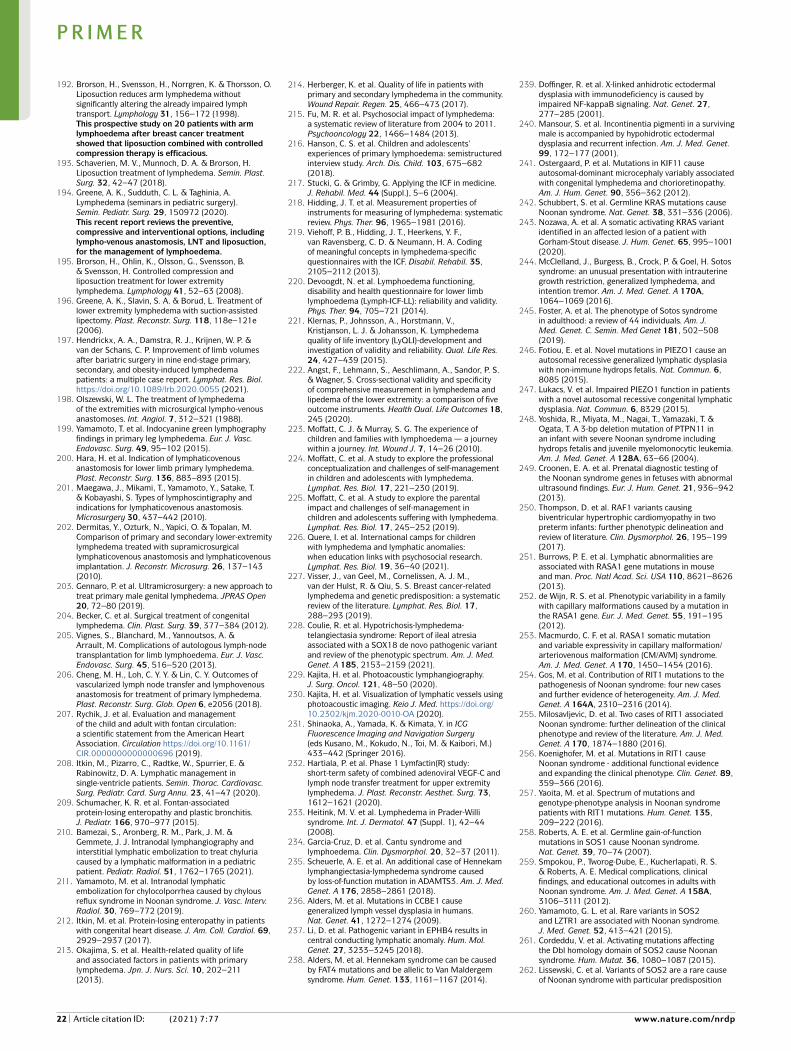
192. Brorson, H., Svensson, H., Norrgren, K. & Thorsson, O. Liposuction reduces arm lymphedema without significantly altering the already impaired lymph transport. Lymphology 31, 156–172 (1998). This prospective study on 20 patients with arm lymphoedema after breast cancer treatment showed that liposuction combined with controlled compression therapy is efficacious.
193. Schaverien, M. V., Munnoch, D. A. & Brorson, H. Liposuction treatment of lymphedema. Semin. Plast. Surg. 32, 42–47 (2018).
194. Greene, A. K., Sudduth, C. L. & Taghinia, A. Lymphedema (seminars in pediatric surgery). Semin. Pediatr. Surg. 29, 150972 (2020). This recent report reviews the preventive, compressive and interventional options, including lympho-venous anastomosis, LNT and liposuction, for the management of lymphoedema.
195. Brorson, H., Ohlin, K., Olsson, G., Svensson, B. & Svensson, H. Controlled compression and liposuction treatment for lower extremity lymphedema. Lymphology 41, 52–63 (2008).
196. Greene, A. K., Slavin, S. A. & Borud, L. Treatment of lower extremity lymphedema with suction-assisted lipectomy. Plast. Reconstr. Surg. 118, 118e–121e (2006).
197. Hendrickx, A. A., Damstra, R. J., Krijnen, W. P. & van der Schans, C. P. Improvement of limb volumes after bariatric surgery in nine end-stage primary, secondary, and obesity-induced lymphedema patients: a multiple case report. Lymphat. Res. Biol. https://doi.org/10.1089/lrb.2020.0055 (2021).
198. Olszewski, W. L. The treatment of lymphedema of the extremities with microsurgical lympho-venous anastomoses. Int. Angiol. 7, 312–321 (1988).
199. Yamamoto, T. et al. Indocyanine green lymphography findings in primary leg lymphedema. Eur. J. Vasc. Endovasc. Surg. 49, 95–102 (2015).
200. Hara, H. et al. Indication of lymphaticovenous anastomosis for lower limb primary lymphedema. Plast. Reconstr. Surg. 136, 883–893 (2015).
201. Maegawa, J., Mikami, T., Yamamoto, Y., Satake, T. & Kobayashi, S. Types of lymphoscintigraphy and indications for lymphaticovenous anastomosis. Microsurgery 30, 437–442 (2010).
202. Dermitas, Y., Ozturk, N., Yapici, O. & Topalan, M. Comparison of primary and secondary lower-extremity lymphedema treated with supramicrosurgical lymphaticovenous anastomosis and lymphaticovenous implantation. J. Reconstr. Microsurg. 26, 137–143 (2010).
203. Gennaro, P. et al. Ultramicrosurgery: a new approach to treat primary male genital lymphedema. JPRAS Open 20, 72–80 (2019).
204. Becker, C. et al. Surgical treatment of congenital lymphedema. Clin. Plast. Surg. 39, 377–384 (2012).
205. Vignes, S., Blanchard, M., Yannoutsos, A. & Arrault, M. Complications of autologous lymph-node transplantation for limb lymphoedema. Eur. J. Vasc. Endovasc. Surg. 45, 516–520 (2013).
206. Cheng, M. H., Loh, C. Y. Y. & Lin, C. Y. Outcomes of vascularized lymph node transfer and lymphovenous anastomosis for treatment of primary lymphedema. Plast. Reconstr. Surg. Glob. Open 6, e2056 (2018).
207. Rychik, J. et al. Evaluation and management of the child and adult with fontan circulation: a scientific statement from the American Heart Association. Circulation https://doi.org/10.1161/CIR.0000000000000696 (2019).
208. Itkin, M., Pizarro, C., Radtke, W., Spurrier, E. & Rabinowitz, D. A. Lymphatic management in single-ventricle patients. Semin. Thorac. Cardiovasc. Surg. Pediatr. Card. Surg Annu. 23, 41–47 (2020).
209. Schumacher, K. R. et al. Fontan-associated protein-losing enteropathy and plastic bronchitis. J. Pediatr. 166, 970–977 (2015).
210. Bamezai, S., Aronberg, R. M., Park, J. M. & Gemmete, J. J. Intranodal lymphangiography and interstitial lymphatic embolization to treat chyluria caused by a lymphatic malformation in a pediatric patient. Pediatr. Radiol. 51, 1762–1765 (2021).
211. Yamamoto, M. et al. Intranodal lymphatic embolization for chylocolporrhea caused by chylous reflux syndrome in Noonan syndrome. J. Vasc. Interv. Radiol. 30, 769–772 (2019).
212. Itkin, M. et al. Protein-losing enteropathy in patients with congenital heart disease. J. Am. Coll. Cardiol. 69, 2929–2937 (2017).
213. Okajima, S. et al. Health-related quality of life and associated factors in patients with primary lymphedema. Jpn. J. Nurs. Sci. 10, 202–211 (2013).
214. Herberger, K. et al. Quality of life in patients with primary and secondary lymphedema in the community. Wound Repair. Regen. 25, 466–473 (2017).
215. Fu, M. R. et al. Psychosocial impact of lymphedema: a systematic review of literature from 2004 to 2011. Psychooncology 22, 1466–1484 (2013).
216. Hanson, C. S. et al. Children and adolescents’ experiences of primary lymphoedema: semistructured interview study. Arch. Dis. Child. 103, 675–682 (2018).
217. Stucki, G. & Grimby, G. Applying the ICF in medicine. J. Rehabil. Med. 44 (Suppl.), 5–6 (2004).
218. Hidding, J. T. et al. Measurement properties of instruments for measuring of lymphedema: systematic review. Phys. Ther. 96, 1965–1981 (2016).
219. Viehoff, P. B., Hidding, J. T., Heerkens, Y. F., van Ravensberg, C. D. & Neumann, H. A. Coding of meaningful concepts in lymphedema-specific questionnaires with the ICF. Disabil. Rehabil. 35, 2105–2112 (2013).
220. Devoogdt, N. et al. Lymphoedema functioning, disability and health questionnaire for lower limb lymphoedema (Lymph-ICF-LL): reliability and validity. Phys. Ther. 94, 705–721 (2014).
221. Klernas, P., Johnsson, A., Horstmann, V., Kristjanson, L. J. & Johansson, K. Lymphedema quality of life inventory (LyQLI)-development and investigation of validity and reliability. Qual. Life Res. 24, 427–439 (2015).
222. Angst, F., Lehmann, S., Aeschlimann, A., Sandor, P. S. & Wagner, S. Cross-sectional validity and specificity of comprehensive measurement in lymphedema and lipedema of the lower extremity: a comparison of five outcome instruments. Health Qual. Life Outcomes 18, 245 (2020).
223. Moffatt, C. J. & Murray, S. G. The experience of children and families with lymphoedema — a journey within a journey. Int. Wound J. 7, 14–26 (2010).
224. Moffatt, C. et al. A study to explore the professional conceptualization and challenges of self-management in children and adolescents with lymphedema. Lymphat. Res. Biol. 17, 221–230 (2019).
225. Moffatt, C. et al. A study to explore the parental impact and challenges of self-management in children and adolescents suffering with lymphedema. Lymphat. Res. Biol. 17, 245–252 (2019).
226. Quere, I. et al. International camps for children with lymphedema and lymphatic anomalies: when education links with psychosocial research. Lymphat. Res. Biol. 19, 36–40 (2021).
227. Visser, J., van Geel, M., Cornelissen, A. J. M., van der Hulst, R. & Qiu, S. S. Breast cancer-related lymphedema and genetic predisposition: a systematic review of the literature. Lymphat. Res. Biol. 17, 288–293 (2019).
228. Coulie, R. et al. Hypotrichosis-lymphedema-telangiectasia syndrome: Report of ileal atresia associated with a SOX18 de novo pathogenic variant and review of the phenotypic spectrum. Am. J. Med. Genet. A 185, 2153–2159 (2021).
229. Kajita, H. et al. Photoacoustic lymphangiography. J. Surg. Oncol. 121, 48–50 (2020).
230. Kajita, H. et al. Visualization of lymphatic vessels using photoacoustic imaging. Keio J. Med. https://doi.org/ 10.2302/kjm.2020-0010-OA (2020).
231. Shinaoka, A., Yamada, K. & Kimata, Y. in ICG Fluorescence Imaging and Navigation Surgery (eds Kusano, M., Kokudo, N., Toi, M. & Kaibori, M.) 433–442 (Springer 2016).
232. Hartiala, P. et al. Phase 1 Lymfactin(R) study: short-term safety of combined adenoviral VEGF-C and lymph node transfer treatment for upper extremity lymphedema. J. Plast. Reconstr. Aesthet. Surg. 73, 1612–1621 (2020).
233. Heitink, M. V. et al. Lymphedema in Prader-Willi syndrome. Int. J. Dermatol. 47 (Suppl. 1), 42–44 (2008).
234. Garcia-Cruz, D. et al. Cantu syndrome and lymphoedema. Clin. Dysmorphol. 20, 32–37 (2011).
235. Scheuerle, A. E. et al. An additional case of Hennekam lymphangiectasia-lymphedema syndrome caused by loss-of-function mutation in ADAMTS3. Am. J. Med. Genet. A 176, 2858–2861 (2018).
236. Alders, M. et al. Mutations in CCBE1 cause generalized lymph vessel dysplasia in humans. Nat. Genet. 41, 1272–1274 (2009).
237. Li, D. et al. Pathogenic variant in EPHB4 results in central conducting lymphatic anomaly. Hum. Mol. Genet. 27, 3233–3245 (2018).
238. Alders, M. et al. Hennekam syndrome can be caused by FAT4 mutations and be allelic to Van Maldergem syndrome. Hum. Genet. 133, 1161–1167 (2014).
239. Doffinger, R. et al. X-linked anhidrotic ectodermal dysplasia with immunodeficiency is caused by impaired NF-kappaB signaling. Nat. Genet. 27, 277–285 (2001).
240. Mansour, S. et al. Incontinentia pigmenti in a surviving male is accompanied by hypohidrotic ectodermal dysplasia and recurrent infection. Am. J. Med. Genet. 99, 172–177 (2001).
241. Ostergaard, P. et al. Mutations in KIF11 cause autosomal-dominant microcephaly variably associated with congenital lymphedema and chorioretinopathy. Am. J. Hum. Genet. 90, 356–362 (2012).
242. Schubbert, S. et al. Germline KRAS mutations cause Noonan syndrome. Nat. Genet. 38, 331–336 (2006).
243. Nozawa, A. et al. A somatic activating KRAS variant identified in an affected lesion of a patient with Gorham-Stout disease. J. Hum. Genet. 65, 995–1001 (2020).
244. McClelland, J., Burgess, B., Crock, P. & Goel, H. Sotos syndrome: an unusual presentation with intrauterine growth restriction, generalized lymphedema, and intention tremor. Am. J. Med. Genet. A 170A, 1064–1069 (2016).
245. Foster, A. et al. The phenotype of Sotos syndrome in adulthood: a review of 44 individuals. Am. J. Med. Genet. C. Semin. Med Genet 181, 502–508 (2019).
246. Fotiou, E. et al. Novel mutations in PIEZO1 cause an autosomal recessive generalized lymphatic dysplasia with non-immune hydrops fetalis. Nat. Commun. 6, 8085 (2015).
247. Lukacs, V. et al. Impaired PIEZO1 function in patients with a novel autosomal recessive congenital lymphatic dysplasia. Nat. Commun. 6, 8329 (2015).
248. Yoshida, R., Miyata, M., Nagai, T., Yamazaki, T. & Ogata, T. A 3-bp deletion mutation of PTPN11 in an infant with severe Noonan syndrome including hydrops fetalis and juvenile myelomonocytic leukemia. Am. J. Med. Genet. A 128A, 63–66 (2004).
249. Croonen, E. A. et al. Prenatal diagnostic testing of the Noonan syndrome genes in fetuses with abnormal ultrasound findings. Eur. J. Hum. Genet. 21, 936–942 (2013).
250. Thompson, D. et al. RAF1 variants causing biventricular hypertrophic cardiomyopathy in two preterm infants: further phenotypic delineation and review of literature. Clin. Dysmorphol. 26, 195–199 (2017).
251. Burrows, P. E. et al. Lymphatic abnormalities are associated with RASA1 gene mutations in mouse and man. Proc. Natl Acad. Sci. USA 110, 8621–8626 (2013).
252. de Wijn, R. S. et al. Phenotypic variability in a family with capillary malformations caused by a mutation in the RASA1 gene. Eur. J. Med. Genet. 55, 191–195 (2012).
253. Macmurdo, C. F. et al. RASA1 somatic mutation and variable expressivity in capillary malformation/arteriovenous malformation (CM/AVM) syndrome. Am. J. Med. Genet. A 170, 1450–1454 (2016).
254. Gos, M. et al. Contribution of RIT1 mutations to the pathogenesis of Noonan syndrome: four new cases and further evidence of heterogeneity. Am. J. Med. Genet. A 164A, 2310–2316 (2014).
255. Milosavljevic, D. et al. Two cases of RIT1 associated Noonan syndrome: further delineation of the clinical phenotype and review of the literature. Am. J. Med. Genet. A 170, 1874–1880 (2016).
256. Koenighofer, M. et al. Mutations in RIT1 cause Noonan syndrome - additional functional evidence and expanding the clinical phenotype. Clin. Genet. 89, 359–366 (2016).
257. Yaoita, M. et al. Spectrum of mutations and genotype-phenotype analysis in Noonan syndrome patients with RIT1 mutations. Hum. Genet. 135, 209–222 (2016).
258. Roberts, A. E. et al. Germline gain-of-function mutations in SOS1 cause Noonan syndrome. Nat. Genet. 39, 70–74 (2007).
259. Smpokou, P., Tworog-Dube, E., Kucherlapati, R. S. & Roberts, A. E. Medical complications, clinical findings, and educational outcomes in adults with Noonan syndrome. Am. J. Med. Genet. A 158A, 3106–3111 (2012).
260. Yamamoto, G. L. et al. Rare variants in SOS2 and LZTR1 are associated with Noonan syndrome. J. Med. Genet. 52, 413–421 (2015).
261. Cordeddu, V. et al. Activating mutations affecting the Dbl homology domain of SOS2 cause Noonan syndrome. Hum. Mutat. 36, 1080–1087 (2015).
262. Lissewski, C. et al. Variants of SOS2 are a rare cause of Noonan syndrome with particular predisposition
22 | Article citation ID: (2021) 7:77 www.nature.com/nrdp
P r i m e r
0123456789();:
for lymphatic complications. Eur. J. Hum. Genet. 29, 51–60 (2021).
263. Irrthum, A. et al. Mutations in the transcription factor gene SOX18 underlie recessive and dominant forms of hypotrichosis-lymphedema-telangiectasia. Am. J. Hum. Genet. 72, 1470–1478 (2003).
264. Shamseldin, H. E. et al. Identification of embryonic lethal genes in humans by autozygosity mapping and exome sequencing in consanguineous families. Genome Biol. 16, 116 (2015).
265. Abdelrahman, H. A. et al. A recessive truncating variant in thrombospondin-1 domain containing protein 1 gene THSD1 is the underlying cause of nonimmune hydrops fetalis, congenital cardiac defects, and haemangiomas in four patients from a consanguineous family. Am. J. Med. Genet. A 176, 1996–2003 (2018).
266. Prato, G. et al. Congenital segmental lymphedema in tuberous sclerosis complex with associated subependymal giant cell astrocytomas treated with Mammalian target of rapamycin inhibitors. J. Child. Neurol. 29, NP54–NP57 (2014).
267. Geffrey, A. L., Shinnick, J. E., Staley, B. A., Boronat, S. & Thiele, E. A. Lymphedema in tuberous sclerosis complex. Am. J. Med. Genet. A 164A, 1438–1442 (2014).
268. Gordon, K. et al. Mutation in vascular endothelial growth factor-C, a ligand for vascular endothelial growth factor receptor-3, is associated with autosomal dominant Milroy-like primary lymphedema. Circ. Res. 112, 956–960 (2013).
269. Balboa-Beltran, E. et al. A novel stop mutation in the vascular endothelial growth factor-C gene (VEGFC) results in Milroy-like disease. J. Med. Genet. 51, 475–478 (2014).
270. Mukenge, S. et al. Investigation on the role of biallelic variants in VEGF-C found in a patient affected by Milroy-like lymphedema. Mol. Genet. Genomic Med. 8, e1389 (2020).
271. Jones, K. L., Schwarze, U., Adam, M. P., Byers, P. H. & Mefford, H. C. A homozygous B3GAT3 mutation causes a severe syndrome with multiple fractures, expanding the phenotype of linkeropathy syndromes. Am. J. Med. Genet. A 167A, 2691–2696 (2015).
272. Sekiguchi, K. et al. A transient myelodysplastic/myeloproliferative neoplasm in a patient with cardio-facio-cutaneous syndrome and a germline BRAF mutation. Am. J. Med. Genet. A 161A, 2600–2603 (2013).
273. Joyce, S. et al. The lymphatic phenotype in Noonan and Cardiofaciocutaneous syndrome. Eur. J. Hum. Genet. 24, 690–696 (2016).
274. Hanson, H. L. et al. Germline CBL mutation associated with a noonan-like syndrome with primary lymphedema and teratoma associated with acquired uniparental isodisomy of chromosome 11q23. Am. J. Med. Genet. A 164A, 1003–1009 (2014).
275. Boone, P. M. et al. Biallelic mutation of FBXL7 suggests a novel form of Hennekam syndrome. Am. J. Med. Genet. A 182, 189–194 (2020).
276. Michelini, S. et al. Genetic screening in a large cohort of italian patients affected by primary lymphedema using a next generation sequencing (NGS) Approach. Lymphology 49, 57–72 (2016).
277. Kawase, K. et al. Nemaline myopathy with KLHL40 mutation presenting as congenital totally locked-in state. Brain Dev. 37, 887–890 (2015).
278. Sparks, T. N. et al. Exome sequencing for prenatal diagnosis in nonimmune hydrops fetalis. N. Engl. J. Med. 383, 1746–1756 (2020).
279. Ponti, G. et al. Giant elephantiasis neuromatosa in the setting of neurofibromatosis type 1: a case report. Oncol. Lett. 11, 3709–3714 (2016).
280. Michelini, S. et al. Segregation analysis of rare NRP1 and NRP2 variants in families with lymphedema. Genes 11, 1361 (2020).
281. Ricci, M. et al. Review of the function of SEMA3A in lymphatic vessel maturation and its potential as a candidate gene for lymphedema: Analysis of three families with rare causative variants. Lymphology 53, 63–75 (2020).
282. Gargano, G. et al. Hydrops fetalis in a preterm newborn heterozygous for the c.4A>G SHOC2 mutation. Am. J. Med. Genet. A 164A, 1015–1020 (2014).
283. Takenouchi, T. et al. Severe craniosynostosis with Noonan syndrome phenotype associated with SHOC2 mutation: clinical evidence of crosslink between FGFR and RAS signaling pathways. Am. J. Med. Genet. A 164A, 2869–2872 (2014).
284. Michelini, S. et al. TIE1 as a candidate gene for lymphatic malformations with or without lymphedema. Int. J. Mol. Sci. 21, 6780 (2020).
285. Lucas, M. & Andrade, Y. Congenital lymphedema with tuberous sclerosis and clinical Hirschsprung disease. Pediatr. Dermatol. 28, 194–195 (2011).
286. Klinner, J. et al. Congenital lymphedema as a rare and first symptom of tuberous sclerosis complex. Gene 753, 144815 (2020).
287. Hopman, S. M. et al. PTEN hamartoma tumor syndrome and Gorham-Stout phenomenon. Am. J. Med. Genet. A 158A, 1719–1723 (2012).
288. Scarcella, A., De Lucia, A., Pasquariello, M. B. & Gambardella, P. Early death in two sisters with Hennekam syndrome. Am. J. Med. Genet. 93, 181–183 (2000).
289. Greene, A. K., Grant, F. D. & Slavin, S. A. Lower-extremity lymphedema and elevated body-mass index. N. Engl. J. Med. 366, 2136–2137 (2012).
290. Burian, E. A. et al. Cellulitis in chronic oedema of the lower leg: an international cross-sectional study. Br. J. Dermatol. 185, 110–118 (2021).
291. World Health Organization. Lymphatic filariasis — managing morbidity and preventing disability — an
aide-mémoire for national programme managers. Second edition (WHO, 2021).
292. Zanten, M. et al. A diagnostic dilemma: aetiological diagnosis of lymphoedema patients at an Indian multidisciplinary meeting. J. Lymphoedema 14, 43–46 (2019).
293. Mercier, G. et al. Out-of-pocket payments, vertical equity and unmet medical needs in France: A national multicenter prospective study on lymphedema. PLoS ONE 14, e0216386 (2019).
AcknowledgementsM.V.’s laboratories were financially supported by the Fonds de la Recherche Scientifique – FNRS Grants T.0026.14 and T.0247.19, the Fund Generet managed by the King Baudouin Foundation (Grant 2018-J1810250-211305), and by la Région wallonne dans le cadre du financement de l’axe stratégique FRFS-WELBIO (WELBIO-CR-2019C-06). M.V. has also received funding from the MSCA-ITN network V. A. Cure No. 814316 and the Lymphatic Malformation Institute, USA. M.H.W. has received research support from the University of Arizona Health Sciences Translational Imaging Program Projects Stimulus (TIPPS) Award and National Institutes of Health NHLBI R25HL108837 for diverse undergraduate research trainees (Luis Luy, Jasmine Jones, Reginald Myles); she is also Secretary-General, International Society of Lymphology, Tucson, AZ, USA, and Zurich, Switzerland. The authors are grateful to Grace Wagner and Juan Ruiz for programmatic assistance and to Liliana Niculescu for expert secretarial assistance.
Author contributionsIntroduction (M.V., P.B., M.H.W. and R.P.E.); Epidemiology (M.V., P.B., M.H.W., R.P.E. and I.Q.); Mechanisms/pathophys-iology (M.V., P.B., M.H.W., R.P.E. and I.Q.); Diagnosis, screen-ing and prevention (M.V., P.B., M.H.W., R.P.E., R.J.D., C.B. and I.Q.); Management (M.V., P.B., M.H.W., R.P.E., C.B. and I.Q.); Quality of life (M.V., P.B., M.H.W., R.P.E., R.J.D. and I.Q.); Outlook (M.V., P.B., M.H.W., R.P.E. and I.Q.); Overview of Primer (M.V.).
Competing interestsAll authors declare no competing interests.
Peer review informationNature Reviews Disease Primers thanks M.-H. Cheng, T. Yamamoto and the other, anonymous, reviewer(s) for their contribution to the peer review of this work.
Publisher’s noteSpringer Nature remains neutral with regard to jurisdictional claims in published maps and institutional affiliations.
Supplementary informationThe online version contains supplementary material available at https://doi.org/10.1038/s41572-021-00309-7. © Springer Nature Limited 2021
23NATURE REVIEWS | DISEASE PRIMERS | Article citation ID: (2021) 7:77
P r i m e r
0123456789();: